telomere dynamics in human cells reprogrammed to pluripotency
TRANSCRIPT
Telomere Dynamics in Human Cells Reprogrammed toPluripotencySteven T. Suhr1, Eun Ah Chang1, Ramon M. Rodriguez1, Kai Wang1, Pablo J. Ross1,2, Zeki Beyhan1,
Shashanka Murthy1, Jose B. Cibelli1,2*
1 Cellular Reprogramming Laboratory, Department of Animal Science, Michigan State University, East Lansing, Michigan, United States of America, 2 Programa Andaluz de
Terapia Celular y Medicina Regenerativa, Andalucı́a, Spain
Abstract
Background: Human induced pluripotent stem cells (IPSCs) have enormous potential in the development of cellular modelsof human disease and represent a potential source of autologous cells and tissues for therapeutic use. A question remainsas to the biological age of IPSCs, in particular when isolated from older subjects. Studies of cloned animals indicate thatsomatic cells reprogrammed to pluripotency variably display telomere elongation, a common indicator of cell‘‘rejuvenation.’’
Methodology/Principal Findings: We examined telomere lengths in human skin fibroblasts isolated from younger andolder subjects, fibroblasts converted to IPSCs, and IPSCs redifferentiated through teratoma formation and explant culture. InIPSCs analyzed at passage five (P5), telomeres were significantly elongated in 6/7 lines by .40% and approximatedtelomere lengths in human embryonic stem cells (hESCs). In cell lines derived from three IPSC-teratoma explants cultured toP5, two displayed telomeres shortened to lengths similar to input fibroblasts while the third line retained elongatedtelomeres.
Conclusions/Significance: While these results reveal some heterogeneity in the reprogramming process with respect totelomere length, human somatic cells reprogrammed to pluripotency generally displayed elongated telomeres that suggestthat they will not age prematurely when isolated from subjects of essentially any age.
Citation: Suhr ST, Chang EA, Rodriguez RM, Wang K, Ross PJ, et al. (2009) Telomere Dynamics in Human Cells Reprogrammed to Pluripotency. PLoS ONE 4(12):e8124. doi:10.1371/journal.pone.0008124
Editor: Martin Pera, University of Southern California, United States of America
Received August 12, 2009; Accepted November 11, 2009; Published December 2, 2009
Copyright: � 2009 Suhr et al. This is an open-access article distributed under the terms of the Creative Commons Attribution License, which permits unrestricteduse, distribution, and reproduction in any medium, provided the original author and source are credited.
Funding: Funding was obtained from the Naylor Family Foundation, Michigan State University Vice President for Research and Graduate Studies and TheMichigan Agricultural Experiment Station. The funders had no role in study design, data collection and analysis, decision to publish, or preparation of themanuscript.
Competing Interests: The authors have declared that no competing interests exist.
* E-mail: [email protected]
Introduction
Somatic animal cells can be reprogrammed to pluripotency by
nuclear transfer or introduction of reprogramming factors. These
cells not only gain the capacity for differentiation into the multiple cell
and tissue types that ultimately give rise to a complete embryo, but
they also gain the capacity for essentially indefinite ‘‘rejuvenation’’, or
self-renewal. Telomeres are special structures at the ends of
chromosomes that contain long tandem repeats of the DNA sequence
TTAGGG and that reduce genetic instability with the passage of
time (reviewed in [1–3]). Telomere length has emerged a critical
indicator of replicative capacity and advancement of the aging
process at both the cellular and organismal levels [4,5], and
elongation of telomeres has been reported in animal cells
reprogrammed to pluripotency by both nuclear transfer [6–8] and
direct reprogramming [9]. While reports such as these indicate that
telomere elongation is a common feature of nuclear reprogramming,
there are also reported exceptions. One such exception was Dolly the
sheep that did not reset telomeres and displayed indicators of
premature aging, revealing that in at least some cases, reprogram-
ming can occur in the absence of telomere lengthening [10].
Human embryonic stem cells (hESCs), like their animal
counterparts, also display elongated telomeres compared to
differentiated somatic cells [11,12]. Although there are relatively
few studies of telomere lengths in human ESCs, hESC lines at the
earliest passage reported – P15 – displayed average terminal
restriction fragment (TRF) lengths of approximately 14 Kb that
gradually declined and tended to level off at around 10 Kb
between passages 40–80 [11,13]. It has yet to be determined if
human somatic cells directly reprogrammed to pluripotency
respond similarly and display telomere lengths similar to hESCs.
Directly reprogrammed human cells known as ‘‘induced plurip-
otent stem cells’’ (IPSCs) [14–16] may display no telomere
elongation or shortened telomeres, elongation in some lines but
not others, or significantly elongated telomeres equal to or
exceeding those characteristic of hESCs. Before reprogrammed
human somatic cells can be used to best advantage as models of
disease in vitro or for therapeutic purposes, telomere dynamics need
to be examined for human IPSCs.
IPSCs, like ESCs, have been shown in several reports to display
increased activity of at least one important enzymatic component
of telomere homeostasis – the reverse transcriptase telomerase
PLoS ONE | www.plosone.org 1 December 2009 | Volume 4 | Issue 12 | e8124
(TERT) – compared to the activity seen in somatic cell types (i.e.
[14,15]). More recently it was shown that mouse fibroblasts
reprogrammed to pluripotency have both TERT activity and
elongated telomeres [9]. This group further demonstrated that
although one component of the reprogramming cocktail, the
oncogene c-myc, had been shown to directly activate telomerase
expression in human cells [17,18], it was not required for telomere
elongation in mouse IPSCs. Marion and colleagues further
demonstrated that fibroblasts from both young (6 month) and
old donor mice (2.3 yr) elongate telomeres to a similar degree
following IPSC conversion. Of note in this report is that although
mouse IPSCs at low passage (i.e.,P8) displayed multiple
indicators of reprogramming to pluripotency (including mESC-
like morphology, alkaline phosphatase staining, and expression of
mESC markers), they displayed only a small degree of telomere
elongation relative to input fibroblasts. From P8, however, mIPSC
telomeres progressively elongated until they had attained the
lengths of mESC telomeres at about P30 . It was also noted that
the inclusion of c-myc in the reprogramming mix accelerated
telomere lengthening at early passages, but did not impact the
overall length at later passage (Ref. [9], Figure 1I). Together, these
results led the authors to justifiably conclude that ‘‘most telomere
elongation occurs postreprogramming’’[9].
We examined telomere length in human skin fibroblasts from
young and old donor subjects, IPSCs derived from these cells, and
IPSCs returned to a differentiated phenotype. We found that like
animal cells reprogrammed by either somatic cell nuclear transfer or
direct reprogramming, human fibroblasts converted to the IPSC
phenotype generally displayed significantly elongated telomeres,
and after re-differentiation, displayed a loss of telomere length. Like
the mouse, this process was observed irrespective of the inclusion of
c-myc in the reprogramming cocktail, and occured to approxi-
mately the same degree in cells derived from either young and old
subjects. Unlike the mouse however, we observed greater
heterogeneity between cell lines, both in the magnitude of telomere
elongation during IPSC conversion and telomere shortening
following redifferentiation. Also unlike the slow and progressive
telomere elongation reported in mouse IPSCs, based on the seven
human IPSC lines we analyzed, hIPSC telomeres achieved the 14–
15 Kb length characteristic of human ESCs as early as P5.
Results
Input Fibroblasts, IPSCs, and Teratoma-Derived CellsDisplay Indicators of Their Respective Phenotypes
To determine if human somatic cells undergo telomere
elongation when reprogrammed to pluripotency, primary fibro-
blasts from human subjects of two age extremes – 16 weeks of
gestation (line FIBA), and 70-years of age (line FIBB) – were
converted to IPSCs by expression of reprogramming factor
combinations described by Yu et al. (2007)[14] and Takahashi
et al. (2007)[15] using lentiviral vectors shown in Figure 1. IPSC
and other cell lines used in this report are summarized in Table 1.
IPSCs derived from each fibroblast line displayed the character-
istic colonies of tightly packed round cells with relatively large
nucleus/cytoplasm ratios and prominent nucleoli characteristic of
hESCs (Figure 2). Immunochemical and qPCR analysis of
reprogramming factors in IPSCs from both donors revealed that
they differed dramatically from input fibroblasts in marker
expression, but displayed immunostaining patterns (Figure 3A)
and expression levels of reprogramming factors (Figure 3B) that
were very similar to hESCs. Microarray analysis of several IPSC
lines compared to input fibroblasts and hESCs further confirmed
their similarity to hESCs and loss of fibroblast identity (data not
shown).
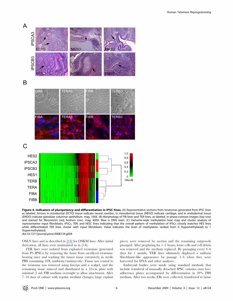
FIBA- and FIBB-derived IPSCs gave rise to teratomas with
tissues representing all three germ layers indicating that they are
pluripotent (Table 1 and Figure 4A). IPSCs were returned to a
differentiated phenotype by dissociating teratoma tissue in vitro and
culturing and passaging outgrowing cells under conditions that
favored the expansion of cells with fibroblast characteristics. These
teratoma-derived cells (TER cells) displayed a morphology similar
to input fibroblasts and many cells were immunopositive for
fibroblasts markers such as fibronectin (Figure 4B). DNA
methylation analysis of FIB lines, IPSCs, and TER cells revealed
that TER cells clearly clustered with input fibroblasts (r = 0.88)
while IPSCs clustered with human ESCs (r = 0.97) (Figure 4C).
Together, these data established the identity of the IPSC lines as
pluripotent cells and TER lines as differentiated cells sharing many
qualities of input FIB lines.
Telomere Elongation after IPSC Conversion and TelomereShortening after IPSC Differentiation
Initial analysis of telomeres was performed by fluorescent in situ
hybridization (FISH) [19,20] using a telomere peptide nucleic acid
(PNA) probe on cultures of each class of cell at P5–6. Samples
processed in parallel clearly revealed intensified punctate fluores-
cence characteristic of labeled telomeres in the IPSCs of both line
A and line B relative to either input cells or TER cells (Figure 5A).
Intensified fluorescence in the IPSC cultures relative to input cells
and redifferentiated TER cells suggested that telomeres in
pluripotent IPSCs were significantly elongated compared to the
differentiated cell types.
Telomeres in FIB, IPSC, and TER cells were further analyzed
and quantified using TRF analysis [9,21], a method based on
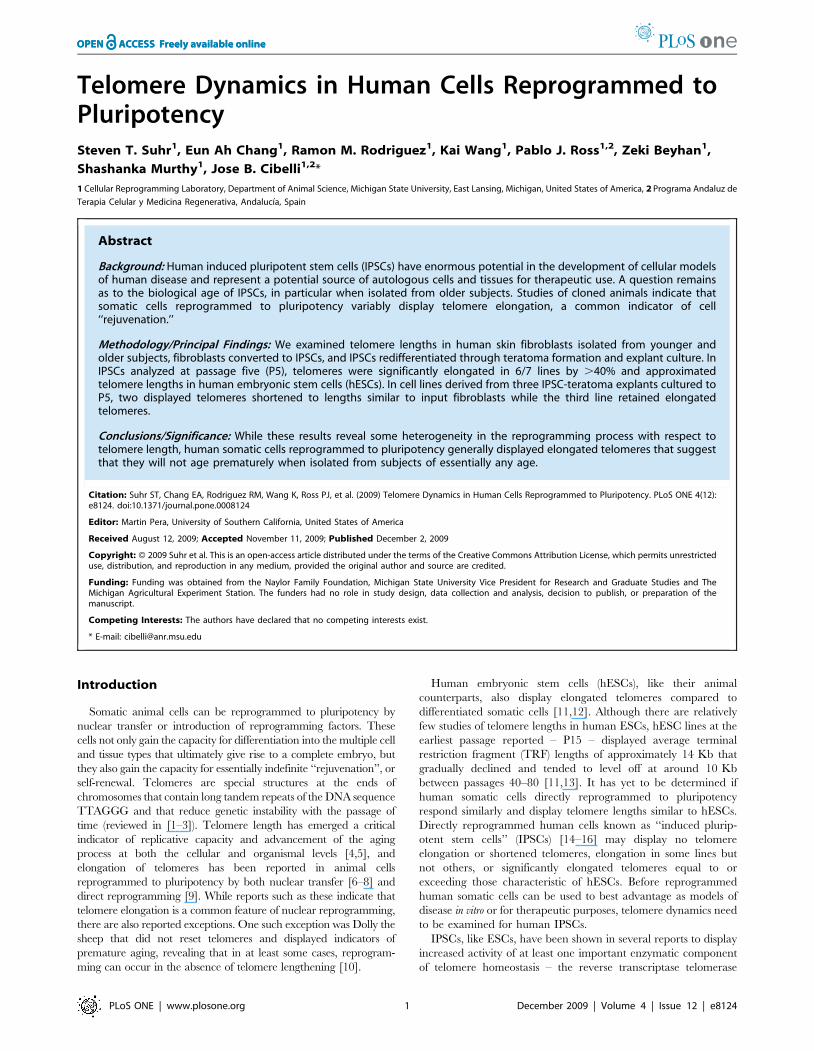
Figure 1. Lentiviral vectors used in IPSC production. LTR: viral LTRS, EF1A-IN: elongation factor 1-alpha promoter, PGK: PGK promoter, M: cMyc,O: Oct4, S: Sox2, K: KLF4, L: Lin28, N: Nanog transgenes. 2A: translation interruption element, IRES: internal ribosomal entry site, Puro: puromycinresistance gene.doi:10.1371/journal.pone.0008124.g001
Human Telomere Reprogramming
PLoS ONE | www.plosone.org 2 December 2009 | Volume 4 | Issue 12 | e8124

Southern blot hybridization of restriction endonuclease digested
genomic DNA with a labeled telomere-repeat probe. Fibroblast
lines A and B, seven IPSC lines, three TER lines, and two human
ESC lines were examined and quantified as shown in Figure 5B–
D. FIBA displayed a mean TRF length of approximately 10 Kb
and three IPSC lines derived from FIBA were found to have
significantly (P,0.05) elongated telomeres of between 13.6 and
16.1 Kb (Figure 5B). Line IPSCA1 also displayed indications of
elongation, but did not reach statistical significance. Line FIBB
displayed a telomere length that did not differ statistically from
FIBA (10.7 Kb), and telomere elongation was observed in all IPSC
lines derived from FIBB (Figure 5B) (because there was only
sufficient DNA for a single run of IPSCB2, statistical significance
could not be calculated for this sample).
In IPSCs differentiated into TER lines, TERA3 and TERB3
displayed a significant loss of telomere length relative to the
parental IPSC line (P,0.001 for TERA3 and P = 0.04 for
TERB3) and TERA3 even showed a slight but significant decrease
compared to FIBA (P = 0.02). Notably, however, the TERA2 line
displayed elongated telomeres relative to both the parental
fibroblast and IPSC lines. This difference was not statistically
significant due to variation in TRF lengths in different samples of
TERA2 even at approximately the same passage, suggesting that
the TERA2 is not homogenous and likely contains a subpopula-
tion of cells that are continuing to elongate telomeres and may be
more rapidly proliferating. Comparing all lines, we found that an
average increase of approximately 47% (from 10.5 Kb to 15.4 Kb)
accompanies conversion to the pluripotent phenotype, and
following differentiation, telomere length decreases to levels
approximating input fibroblasts (Figure 5D).
Discussion
Though the IPSC lines in this report were isolated from only
two subjects of very disparate ages, these results suggest that like
animal cells reprogrammed to pluripotency, reprogrammed
human somatic cells will generally restore telomeres to lengths
characteristic of human ESCs. It follows that IPSC-derived cells
will not reach replicative senescence prematurely, regardless of
donor subject age.
Similar to what was observed in mouse IPSCs, our results also
suggest that variables in the IPSC conversion process including the
age and sex of input cells, reprogramming factors used (Oct4 and
Sox2 coupled with either Nanog and Lin28 or KLF4 and c-Myc),
and media conditions for early establishment (HES medium with
either 100 ng/zFGF or 4 ng hFGF) do not preclude telomere
elongation in human IPSCs nor shortening following redifferentia-
tion.
In general, by P5, human IPSCs displayed TRFs that equaled
early passage hESCs. This differs from mouse IPSCs that, as
pointed out in the Introduction, did not attain mESC telomere
lengths until P30 [9]. Whether human IPSCs undergo a
progressive increase over a much shorter time frame (such as
between passage 1–5), or are essentially ‘‘born’’ with elongated
telomeres accompanying conversion to the pluripotent phenotype,
has not been determined; however, given that P5 approximates
the earliest point at which human IPSC lines for either the
generation of more differentiated cell types or therapeutic
application can reasonably be used, an answer to this question
has little practical relevance. Furthermore, it is possible that the
difference in telomere elongation in mouse IPSCs relative to
human IPSCs is a simple function of average telomere length. It
has been long-established that mouse chromosomes bear telomeres
an order of magnitude longer than their human counterparts [22],
and perhaps the elongation process in mouse takes more cell cycles
simply because the telomeres are much longer. In either event, the
lengths of telomeres in both the human IPSCs and hESCs
described in this report fall well within the range of human ESC
telomeres described previously [11,13]
Table 1. Cell lines used in this report.
Report Name Cell Type Pass Parental Line Vec/Factors Line Ref Name
IPSCA1 IPSC 5–6 FIBA OSNL MSUH-001
IPSCA2 IPSC 5–6 FIBA OSNL MSUH-002
IPSCA3 IPSC 5–6 FIBA (ON)(SL) MSUH-004
IPSCA4 IPSC 5–6 FIBA OSNL MSUH-005
IPSCB1* IPSC 5–6 FIBB OSKM MSUH-006*
IPSCB2* IPSC 5–6 FIBB OSKM MSUH-007*
IPSCB3 IPSC 5–6 FIBB OSKM MSUH-008
FIBA FIBROBLAST 15–16# NA NA IMR90
FIBB FIBROBLAST 5–6# NA NA MSUH-004F2
TERA2 TERAT-DERIVED 4–5 IPSCA2 OSNL MSUH-002T1
TERA3 TERAT-DERIVED 4–5 IPSCA3 (ON)(SL) MSUH-003T1
TERB3 TERAT-DERIVED 4–5 IPSCB3 OSKM MSUH-008T1
HES1 ES CELL 80 EMBRYO NA H7
HES2 ES CELL 44 EMBRYO NA H9
Report name: name used to reference the cell lines in this report. Cell Type: the general cell phenotype.Pass: passage of cells at time of telomere analysis. Parental Line: name of the input cell line used to produce the corresponding cell line.Vec/Factors: Reprogramming factor combination used to produce cell line (if applicable). Individual letters represent each of the 6 reprogramming factors as in Figure 1.Parentheses indicate coupling of factors into bicistronic pairs in the vector.Line Ref Name: Official name of each cell line used in the report.*Lines lost to contamination.#--also passage number used for production of IPSCs.doi:10.1371/journal.pone.0008124.t001
Human Telomere Reprogramming
PLoS ONE | www.plosone.org 3 December 2009 | Volume 4 | Issue 12 | e8124
One aspect of telomere dynamics that had not been studied for
IPSCs of any species prior to this report is resumption of telomere
shortening following redifferentiation of the cells. While both
ESCs and IPSCs can be differentiated in vitro by cell culture
methods with some success, we chose to use cells cultured from
teratomas because prior to plating, these cells undergo many
weeks of growth and replication in an environment conducive to
differentiation. Although cells of a variety of differentiated
phenotypes were observed in primary cultures from IPSC
teratomas (data not shown), repeated passage of the most adherent
population in fibroblast medium rapidly led to a relatively
homogenous population of cells with a flattened morphology
reminiscent of fibroblasts.
Although telomeres were reduced in two of the TER lines
isolated, in a third – TERA2 – elongated telomeres were
maintained. Given that only one line acted contrary to expectation
and retained elongated telomeres following extended growth in vivo
and additional culture in vitro, we can only speculate as to the
relevance of this observation. The two most likely explanations are
that either a subpopulation of IPSCs have retained their
pluripotent cell identity despite extended growth and differentia-
tion in conditions that do not support IPSC/ESC maintenance, or
a subpopulation of cells have taken on a non-pluripotent cell
phenotype that supports telomere elongation, such as a trans-
formed cell [23–25]. Analyses of the TERA2 line for indicators of
a pluripotent cell population have thus far proved negative (data
not shown). While the observation that the TERA2 cell population
retains elongated telomeresthe ovser may raise cautionary flags
regarding the use of uncharacterized IPSCs in vivo, in general, the
results with TER lines indicate the results with the that most IPSCs
will resume normal telomere physiology after differentiation.
Nevertheless, even if the emergence of cells with an extended
capacity for self-renewal from differentiated IPSC populations is a
rare event, this finding suggests that it may be prudent to examine
the telomere dynamics of individual IPSC lines destined for
extensive use in either cell culture models or future therapeutic
applications.
Materials and Methods
Cell Culture, IPSC Production, and EB/TeratomaFibroblasts and TER cell lines were cultured in DMEM+
10%FBS+antimycotic/antibiotic (FIB medium). FIBA (IMR90)
was obtained from ATCC and FIBB was grown out from a skin
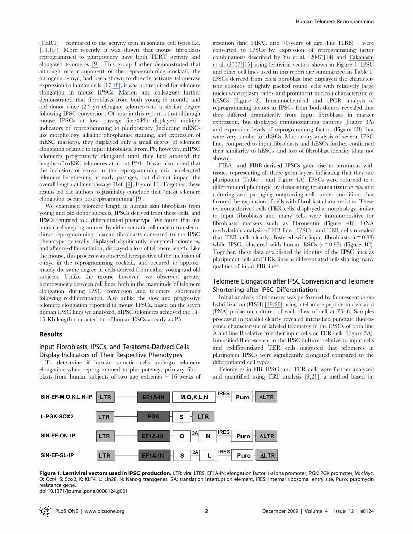
Figure 2. Phase-contrast images of FIB and IPSC lines used in this report (as labeled). FIBA and FIBB displayed a flat stellate cytoplasmwith irregular edges characteristic of fibroblast cell types (200X). IPSC lines displayed round colonies with regular edges evident in low magnification40X images that were composed of tightly packed cells with prominent nucleoli (200X, lower panels of IPSC lines) that appeared morphologicallyhomogenous within the center of the colony and flattened toward the edges where they were bounded by the MEF feeder layer. IPSCB1 and IPSB2are shown only at 40X magnification.doi:10.1371/journal.pone.0008124.g002
Human Telomere Reprogramming
PLoS ONE | www.plosone.org 4 December 2009 | Volume 4 | Issue 12 | e8124
biopsy from a 70-year old male subject with informed consent.
Human ES cell line HES2 (H9) was obtained the University of
Wisconsin and was used between passages 43–44 for analyses in
this report. DNA from HES1 cells (H7) at approximately P80 was
obtained from Sue O’Shea’s laboratory at the University of
Michigan.
IPSCs were produced from FIBA cells at P15 and FIBB cells at
P5. IPSCs were generated by infection with high-titer lentiviral
vectors encoding the six human reprogramming factors Oct4 (O),
Sox2 (S), KLF4 (K), cMyc (M), Nanog (N), and Lin28 (L)
expressed singly essentially as described [14,15] or in bi-cistronic
pairs (see Figure 1). The lentiviral vector used was either the SIN-
EF1a vector described by [14] and obtained from Addgene, a
lentiviral vector bearing a PGK promoter for Sox2 as shown in
Figure 1 (Sox2 expression appeared more robust from the PGK
vector). Vectors were modified to encode KLF4, cMyc (Obtained
from Open biosystems) or reprogramming factors were co-
expressed using 2A elements as shown in Figure 1. Infection,
identification of IPSCs, subcloning of IPSC colonies, and passage
onto MEFs, was performed essentially as described in [14] for
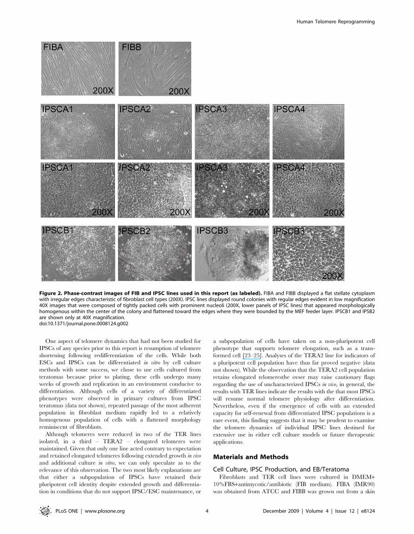
Figure 3. Indicators of cell phenotype and pluripotency in IPSC lines. (A) Immunofluorescent analysis of pluripotency markers as labeled.Label color corresponds to marker color in images. Blue staining in all panels is DAPI labeling of nuclei. All IPSC lines displayed strong positiveexpression of pluripotency markers that were not observed at high levels in input FIB cells with the exception of the Lin28 antibody that reproduciblyproduced faint fluorescence in the cytoplasm of fibroblasts. (B) Representative QPCR analysis of FIB lines and reprogrammed IPSCs for markers ofpluripotency and reprogramming factors relative to factor levels in hESCs. Input fibroblasts express very low levels of most factors whereas IPSCsdisplay levels very similar to hESCs.doi:10.1371/journal.pone.0008124.g003
Human Telomere Reprogramming
PLoS ONE | www.plosone.org 5 December 2009 | Volume 4 | Issue 12 | e8124
OSLN lines and as described in [15] for OSKM lines. After initial
derivation, all lines were maintained as in [14].
TER lines were isolated from explanted teratomas (generated
from P5 IPSCs) by removing the tissue from sacrificed teratoma-
bearing mice and washing the tumor tissue extensively in sterile
PBS containing 10X antibiotic/antimycotic. Tissue not central to
the teratoma was removed using forceps and a scalpel, and the
remaining tissue minced and distributed in a 10-cm plate with
minimal (5 ml) FIB medium overnight to allow attachment. After
7–10 days of culture with regular medium changes, large explant
pieces were removed by suction and the remaining outgrowth
passaged. After preplating for 1–2 hours, loose cells and cell debris
was removed and the medium replaced. By passaging every 4–6
days for 1 month, TER lines ultimately displayed a uniform
fibroblastic-like appearance by passage 5–6 when they were
harvested for DNA and other analyses.
Embryoid bodies were made using standard methods that
include transferal of manually detached IPSC colonies onto low-
adherence plates accompanied by differentiation in 20% FBS
medium. After two weeks, EBs were collected, transferred to tissue
Figure 4. Indicators of pluripotency and differentiation in IPSC lines. (A) Representative sections from teratomas generated from IPSC linesas labeled. Arrows in ectodermal (ECTO) tissue indicate neural rosettes, in mesodermal tissue (MESO) indicate cartilage, and in endodermal tissue(ENDO) indicate glandular columnar epithelium. mag. 100X. (B) Morphology of FIB lines and TER lines, as labeled, in phase-contrast images (top row)and stained for fibronectin (red, bottom row). mag. 400X. Blue is DNA stain. (C) Genome-wide methylation heat map and cluster analysis ofrepresentative input fibroblasts, IPSCs, TER, and hESC lines indicating that the overall pattern of methylation of IPSCs closely matches HES lineswhile differentiated TER lines cluster with input fibroblasts. Value indicates the level of methylation ranked from 0 (hypomethylated) to 1(hypermethylated).doi:10.1371/journal.pone.0008124.g004
Human Telomere Reprogramming
PLoS ONE | www.plosone.org 6 December 2009 | Volume 4 | Issue 12 | e8124
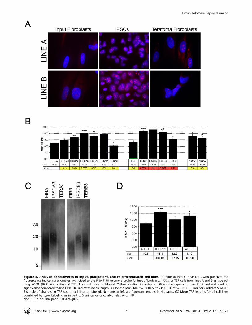
Figure 5. Analysis of telomeres in input, pluripotent, and re-differentiated cell lines. (A) Blue-stained nuclear DNA with punctate redfluorescence indicating telomeres hybridized to the PNA FISH telomere probe for input fibroblasts, IPSCs, or TER cells from lines A and B as labeled.mag. 400X. (B) Quantification of TRFs from cell lines as labeled. Yellow shading indicates significance compared to line FIBA and red shadingsignificance compared to line FIBB. TRF indicates mean length in kilobase pairs (Kb). * = P,0.05, ** = P,0.01, *** = P,.001. Error bars indicate SEM. (C)Example of changes in TRF size in cell lines as labeled. Numbers at left are fragment lengths in kilobases. (D) Mean TRF lengths for all cell linescombined by type. Labeling as in part B. Significance calculated relative to FIB.doi:10.1371/journal.pone.0008124.g005
Human Telomere Reprogramming
PLoS ONE | www.plosone.org 7 December 2009 | Volume 4 | Issue 12 | e8124
culture plates where they re-adhered, and ultimately passaged in
fibroblast medium. Teratomas were produced in nude mice using
standard methods. Briefly, 16106 IPSCs of each line at P5 were
suspended in DMEM/F12, and injected into Nude mice
intramuscularly. 6–8 weeks after injection, tumors were surgically
dissected and either processed as described above to isolate TER
lines, or fixed in 4% paraformaldehyde, embedded in paraffin,
sectioned, and processed for histological examination. Tissue
processing was performed by MSU histology core facility.
Analysis of Cell PhenotypesIPSC lines were cultured under standard conditions to passage
3–4 for harvest to produce RNA for QPCR analysis, perform
immunocytochemistry, or other analyses. FIB and TER lines were
analyzed at the passages listed in Table 1. Fibronectin monoclonal
antibody HFN7.1, SSEA3 and SSEA4 antibodies were obtained
from the Developmental Studies Hybridoma Bank at the
University of Iowa and Oct4, Nanog, and Lin28 antibodies were
obtained from Santa Cruz Biotechnology (Santa Cruz, CA). All
primary antibodies were used at 1:250. Secondary anti-mouse,
anti-goat, and anti-rabbit-Alexa conjugates were obtained from
Invitrogen and used at 1:1000. RNAs were isolated using Trizol
reagent (Invitrogen) following the manufacturers protocol. Immu-
nochemical analyses used to confirm cell phenotypes were
performed on cultured cells fixed with 4% paraformaldehyde
and processed using standard methods. Pluripotent cells used for
methylation analysis and for TRF analysis (below) were harvested
by manual microdissection of colonies with good ESC-like
morphology to remove cells at the edges that were partially
differentiated and most MEFs from the feeder layer. The genome-
wide methylation signature of bisulfite-converted DNA from all
cell lines was performed by the Applied Genomics Technology
Center, Wayne State University, using the Illumina Human-
Methylation27 BeadChip.
FISH and TRF AnalysisFor FISH analysis, cells at the passages listed in Table 1 were
washed with PBS and fixed with 4% paraformaldehyde for 10
minutes at room temperature. After washing, cells were treated
with 0.005% Pepsin (Sigma) for 10 minutes at 37uC and
immediately dehydrated with ice-cold ethanol. Fluorescent
telomere PNA probe (Panagene) was diluted in 10 mM Tris-
HCl containing 70% formamide to a final concentration of
180 nM. Samples were incubated with the probe for 2 hours at
room temperature and were visualized with an epifluorescence
system on a Nikon TE-2000 microscope.
Analysis of telomere restriction fragment lengths [9,23] was
performed using TeloTAGGG (Roche) reagents and following the
manufacturers protocol. !-2ug of DNA from cells at the passages
listed in Table 1 were digested overnight with RsaI/HinfI, run the
next day on a 0.8% agarose gel, and transferred to nylon
membrane for 48 hours for Southern analysis. Cell lines were
harvested for DNA on at least two separate occasions (except for
HES1 which was harvested only once) and run 2–6 times to
determine mean TRF length. There was only sufficient DNA for a
single run of IPSCB2 due to loss of this line to contamination.
Images were analyzed using NIHImageJ and TRF lengths were
calculated relative to labeled molecular weight markers. Statistical
significance was analyzed using Student’s T-test
Ethics StatementThis study was conducted according to the principles expressed
in the Declaration of Helsinki. The study was approved by the
Institutional Review Board of Michigan State University,
reference: #06-699 for the study titled: ‘‘Direct Dedifferentiation
of primary somatic cells’’. All patients provided written informed
consent for the collection of samples and subsequent analysis.
Acknowledgments
The authors thank members of the MSU CRL for comments and Dr. Sue
O’Shea and Crystal Pacut for assisting with H7 (HES1) DNA. The
monoclonal antibodies developed by RJ Klebe, D Solter, and BB Knowles
were obtained from the Developmental Studies Hybridoma Bank
developed under the auspices of the NICHD and maintained by The
University of Iowa, Department of Biology, Iowa City, IA 52242.
Author Contributions
Conceived and designed the experiments: STS EAC JC. Performed the
experiments: STS EAC RMR SM. Analyzed the data: STS EAC RMR
KW PJR ZB. Contributed reagents/materials/analysis tools: STS EAC
RMR KW. Wrote the paper: STS EAC JC.
References
1. Blackburn EH (2005) Telomeres and telomerase: their mechanisms of action and
the effects of altering their functions. FEBS Lett 579: 859–862.
2. Hug N, Lingner J (2006) Telomere length homeostasis. Chromosoma 115:
413–425.
3. Riethman H (2008) Human telomere structure and biology. Annu Rev
Genomics Hum Genet 9: 1–19.
4. Harrington L (2004) Does the reservoir for self-renewal stem from the ends?
Oncogene 23: 7283–7289.
5. Aubert G, Lansdorp PM (2008) Telomeres and aging. Physiol Rev 88: 557–579.
6. Lanza RP, Cibelli JB, Blackwell C, Cristofalo VJ, Francis MK, et al. (2000)
Extension of cell life-span and telomere length in animals cloned from senescent
somatic cells. Science 288: 665–669.
7. Wakayama T, Shinkai Y, Tamashiro KL, Niida H, Blanchard DC, et al. (2000)
Cloning of mice to six generations. Nature 407: 318–319.
8. Kubota C, Tian XC, Yang X (2004) Serial bull cloning by somatic cell nuclear
transfer. Nat Biotechnol 22: 693–694.
9. Marion RM, Strati K, Li H, Tejera A, Schoeftner S, et al. (2009) Telomeres
acquire embryonic stem cell characteristics in induced pluripotent stem cells.
Cell Stem Cell 4: 141–154.
10. Shiels PG, Kind AJ, Campbell KH, Waddington D, Wilmut I, et al. (1999)
Analysis of telomere lengths in cloned sheep. Nature 399: 316–317.
11. Amit M, Carpenter MK, Inokuma MS, Chiu CP, Harris CP, et al. (2000)
Clonally derived human embryonic stem cell lines maintain pluripotency and
proliferative potential for prolonged periods of culture. Dev Biol 227:
271–278.
12. Niida H, Shinkai Y, Hande MP, Matsumoto T, Takehara S, et al. (2000)
Telomere maintenance in telomerase-deficient mouse embryonic stem cells:
characterization of an amplified telomeric DNA. Mol Cell Biol 20: 4115–4127.
13. Rosler ES, Fisk GJ, Ares X, Irving J, Miura T, et al. (2004) Long-term culture of
human embryonic stem cells in feeder-free conditions. Dev Dyn. 229: 259–274.
14. Yu J, Vodyanik MA, Smuga-Otto K, Antosiewicz-Bourget J, Frane JL, et al.
(2007) Induced pluripotent stem cell lines derived from human somatic cells.
Science 318: 1917–1920.
15. Takahashi K, Tanabe K, Ohnuki M, Narita M, Ichisaka T, et al. (2007)
Induction of pluripotent stem cells from adult human fibroblasts by defined
factors. Cell 131: 861–872.
16. Park IH, Zhao R, West JA, Yabuuchi A, Huo H, et al. (2008) Reprogramming
of human somatic cells to pluripotency with defined factors. Nature 451:
141–146.
17. Greenberg RA, O’Hagan RC, Deng H, Xiao Q, Hann SR, et al. (1999)
Telomerase reverse transcriptase gene is a direct target of c-Myc but is not
functionally equivalent in cellular transformation. Oncogene 18: 1219–1226.
18. Wu KJ, Grandori C, Amacker M, Simon-Vermot N, Polack A, et al. (1999)
Direct activation of TERT transcription by c-MYC. Nat Genet 21: 220–224.
19. Moyzis RK, Buckingham JM, Cram LS, Dani M, Deaven LL, et al. (1988) A
highly conserved repetitive DNA sequence, (TTAGGG)n, present at the
telomeres of human chromosomes. Proc Natl Acad Sci U S A 85: 6622–6626.
20. Lansdorp PM, Verwoerd NP, van de Rijke FM, Dragowska V, Little MT, et al.
(1996) Heterogeneity in telomere length of human chromosomes. Hum Mol
Genet 5: 685–691.
Human Telomere Reprogramming
PLoS ONE | www.plosone.org 8 December 2009 | Volume 4 | Issue 12 | e8124

21. Harley CB, Futcher AB, Greider CW (1990) Telomeres shorten during ageing of
human fibroblasts. Nature 345: 458–460.22. Kipling D, Cooke HJ (1990) Hypervariable ultra-long telomeres in mice. Nature
347: 400–402.
23. Harley CB (2008) Telomerase and cancer therapeutics. Nat Rev Cancer 8:167–179.
24. Cheung AL, Deng W (2008) Telomere dysfunction, genome instability and
cancer. Front Biosci 13: 2075–2090.
25. Finkel T, Serrano M, Blasco MA (2007) The common biology of cancer and
ageing. Nature 448: 767–774.
Human Telomere Reprogramming
PLoS ONE | www.plosone.org 9 December 2009 | Volume 4 | Issue 12 | e8124