reconsidering pluripotency tests: do we still need teratoma assays?
TRANSCRIPT
Ava i l ab l e on l i ne a t www.sc i enced i r ec t . com
www.e l sev i e r . com/ l oca te / sc r
Stem Cell Research (2013) 11, 552–562
Reconsidering pluripotency tests: Do we stillneed teratoma assays?
Christiane Buta a, 1, Robert David b, Ralf Dressel c, Mia Emgård d,Christiane Fuchs e, 2, Ulrike Gross f, Lyn Healy g, Jürgen Hescheler h,Roman Kolar f, Ulrich Martin i, Harald Mikkers j, Franz-Josef Müller k,Rebekka K. Schneider l, Andrea E.M. Seiler m,Horst Spielmann n, Georg Weitzer o,⁎a SET Foundation, Frankfurt am Main, Germanyb Ludwig-Maximilians-Universität, München, Germanyc Universitätsmedizin Göttingen, Germanyd Cellartis, Göteborg and Karolinska Institute Stockholm, Swedene University of Applied Sciences, Technikum Wien, Austriaf Akademie für Tierschutz, Neubiberg, Germanyg UK Stem Cell Bank, London, UKh Universität Köln, Germanyi Medizinische Hochschule Hannover, Germanyj Universiteit Leiden, The Netherlandsk Universitätsklinikum Schleswig-Holstein, Campus Kiel, Germanyl RWTH, Aachen, Germanym Federal Institute for Risk Assessment, Berlin, Germanyn Freie Universität Berlin, Germanyo Medical University of Vienna, Austria
Received 1 February 2013; received in revised form 8 March 2013; accepted 16 March 2013Available online 26 March 2013
Abstract The induction of teratoma in mice by the transplantation of stem cells into extra-uterine sites has been used as aread-out for cellular pluripotency since the initial description of this phenomenon in 1954. Since then, the teratoma assay hasremained the assay of choice to demonstrate pluripotency, gaining prominence during the recent hype surrounding human stemcell research. However, the scientific significance of the teratoma assay has been debated due to the fact that transplanted cells
⁎ Corresponding author at: Max F. Perutz Laboratories, Department for Medical Biochemistry, Medical University of Vienna, Dr. Bohrgasse 9/2,A-1030, Austria. Fax: +43 1 4277 9618.
E-mail address: [email protected] (G. Weitzer).1 This author collected all contributions to the manuscript and organized the workshop of the German Foundation for the Promotion of
Alternatives to Animal Testing (Stiftung zur Förderung von Ersatz- und Ergänzungsmethoden zur Einschränkung von Tierversuchen, SET), onthe exploration of the 3R (refine, reduce and replace) applications in the context of testing pluripotency, together with J.H. in December2010. This positional paper summarizes the results of the workshop and subsequent discussions.2 This author prepared the manuscript together with G.W.; authors are in alphabetical order and contributed equally to this manuscript.
1873-5061/$ - see front matter © 2013 Elsevier B.V. All rights reserved.http://dx.doi.org/10.1016/j.scr.2013.03.001
553Reconsidering pluripotency tests: Do we still need teratoma assays?
are exposed to a non-physiological environment. Since many mice are used for a result that is heavily questioned, it is time toreconsider the teratoma assay from an ethical point of view. Candidate alternatives to the teratoma assay comprise the directeddifferentiation of pluripotent stem cells into organotypic cells, differentiation of cells in embryoid bodies, the analysis ofpluripotency-associated biomarkers with high correlation to the teratoma forming potential of stem cells, predictive epigeneticfootprints, or a combination of these technologies. Each of these assays is capable of addressing one or more aspects ofpluripotency, however it is essential that these assays are validated to provide an accepted robust, reproducible alternative. Inparticular, the rapidly expanding number of human induced pluripotent stem cell lines, requires the development of simple,affordable standardized in vitro and in silico assays to reduce the number of animal experiments performed.
© 2013 Elsevier B.V. All rights reserved.Contents
Introduction . . . . . . . . . . . . . . . . . . . . . . . . . . . . . . . . . . . . . . . . . . . . . . . . . . . . . . . . . . . 553Teratoma assays — when and why? . . . . . . . . . . . . . . . . . . . . . . . . . . . . . . . . . . . . . . . . . . . . . . . 555
Safety testing of transplants derived from pluripotent stem cells . . . . . . . . . . . . . . . . . . . . . . . . . . . . 555Why are teratoma assays questioned? . . . . . . . . . . . . . . . . . . . . . . . . . . . . . . . . . . . . . . . . . . . 555Animal welfare and ethical concerns . . . . . . . . . . . . . . . . . . . . . . . . . . . . . . . . . . . . . . . . . . . 556
Alternatives to the teratoma assay — their advantages and disadvantages . . . . . . . . . . . . . . . . . . . . . . . . 556Proof of pluripotency . . . . . . . . . . . . . . . . . . . . . . . . . . . . . . . . . . . . . . . . . . . . . . . . . . . . 556In vitro differentiation — directed and spontaneous differentiation . . . . . . . . . . . . . . . . . . . . . . . . . . 557Computer-based predictive models . . . . . . . . . . . . . . . . . . . . . . . . . . . . . . . . . . . . . . . . . . . . 558Chicken egg model — chorioallantoic membrane . . . . . . . . . . . . . . . . . . . . . . . . . . . . . . . . . . . . . 558Organotypic models . . . . . . . . . . . . . . . . . . . . . . . . . . . . . . . . . . . . . . . . . . . . . . . . . . . . . 558
An example of differential information content of embryoid bodies and teratoma assays . . . . . . . . . . . . . . . . 559Conclusions and future perspectives . . . . . . . . . . . . . . . . . . . . . . . . . . . . . . . . . . . . . . . . . . . . . . 559Disclosures . . . . . . . . . . . . . . . . . . . . . . . . . . . . . . . . . . . . . . . . . . . . . . . . . . . . . . . . . . . . 559References . . . . . . . . . . . . . . . . . . . . . . . . . . . . . . . . . . . . . . . . . . . . . . . . . . . . . . . . . . . . . 9560
Introduction
The experimental induction of teratoma (for definition ofterms see Box 1) in mammals, mostly mice, has been carriedout for decades (Evans and Kaufman, 1981; Skreb et al., 1971;Solter et al., 1970; Stevens, 1958, 1970; Stevens & Little,1954). In stem cell research and banking (ISCBI, 2009) thein vivo teratoma assay can be used to demonstrate thepluripotency of the stem cells in vivo (Gertow et al., 2007;Wesselschmidt, 2011). Basically stem cells, which are consid-ered to be pluripotent, are injected into various anatomicalsites e.g. sub-cutaneous, intra-muscular, under the capsule ofthe kidney, or intra-testicular, of immunocompromised micepotentially developing into an experimental tumor (also seeBox 1). The assayed cells are considered pluripotent ifthe resultant tumor shows characteristics of a teratoma,demonstrating the development of differentiated cells fromall three germ layers, namely ectoderm (such as nerve andskin), mesoderm (including bone, cartilage and muscle), andendoderm (liver and gut) (Brivanlou et al., 2003).
Although used regularly and frequently demanded byreviewers of manuscripts as proof of pluripotency, theteratoma assay has never been standardized in terms ofgraft site, age of mice, number of cells implanted and thecell preparation for a large number of pluripotent cell lines.These factors invariably influence the development of theteratoma (Hentze et al., 2009; Wesselschmidt, 2011). Groppet al. recently presented a systematic evaluation of some of
these factors for two ESC lines (Gropp et al., 2012). Inaddition to the lack of standardization of the teratomaassay, the assay is also regarded as time, cost and laborintensive. The assay certainly raises ethical concerns, as itmay induce pain and suffering of the animals used in theassay. This latter concern impacts on the current legislationon animal welfare and the following section will give anoverview on the current status in Europe.
From the animal welfare perspective the teratoma assayraises two major issues: first, the inoculation of geneticallymanipulated animals with potentially malignant cells thatcould initiate tumors, and second, the breeding of experimen-tal animals, especially if associated with the suffering and painof the animal. A classification system, comprising the degree ofpain, suffering and distress, was accepted in 1995 and has beenin general use throughout Europe. This system is known as theSeverity Catalogue of the Swiss Federal Veterinary Office. Inthe European Union (EU), a binding severity classificationsystem was approved in 2010 as Annex VIII of the new “EUDirective 2010/63/EU for the Protection of Animals used inScientific Procedures” (European Union, 2010). Accordantregulations and amendments are published by the CanadianCouncil on Animal Care in Science, the Australian and NewZealand Council for the Care of Animals in Research andTeaching, and in the U.S.A, under the Laboratory AnimalWelfare Act.
The Swiss Catalogue considers tumor models as moderatelysevere (grade 2) and severe (grade 3) procedures. The grade 2
Box 1 Explanation of terms used in this paper according to the NIH definitions.
Teratoma — a multi-layered benign tumor that grows from pluripotent cells injected into animals with adysfunctional immune system. Scientists test whether they have established a human embryonic stem(hES) cell line by injecting putative stem cells into such mice and verifying that the resulting teratomacontains cells derived from all three embryonic germ layers.Teratocarcinoma — a multi-layered malignant tumor that contains in addition to a teratoma embryonalcarcinoma cells, which either give rise to metastases or produce a malignant tumor after re-implantation ofthe primary tumor mass. The WHO recommended for this type of tumor the more complicated term“mixed embryonal carcinoma and teratoma”. A detailed consideration of the difference between teratomaand teratocarcinoma can be found in a series of comments in Nature Biotechnology Vol. 25, No. 11(Damjanov and Andrews, 2007). In a teratoma assay development of this type of tumor wouldimmediately lead to the exclusion of the putative pluripotent stem cells from any type of therapeuticalapplication.Severe combined immunodeficient (SCID) mice — SCID mice are important tools for researchinghematopoiesis, innate and adaptive immunity, autoimmunity, infectious diseases, cancer, vaccine develop-ment, and regenerative medicine in vivo. So-called because of their severe combined immunodeficiency, SCIDmice have reduced ability to reject allogeneic or xenogeneic tissue grafts, and are therefore excellent hosts forhuman cells and tissues.Teratoma assay— in this assay putative pluripotent stem cells are implanted into SCIDmicewhere they canproliferate and differentiate to form a teratoma. The pluripotent stem cells grow at the implantation site andare supported by factors of the local milieu and also circulating factors. After a certain time, when the tumorhas reached sufficient size, it is removed and subjected to histopathological analysis, immunocytochemistryand gene expression profiling.Tetraploid complementation assay— an assay that can be used to test a stem cell's potency. Fusing two2-cell embryos produces cells with 4 sets of chromosomes (tetraploid cells) that are biased towarddeveloping into extra-embryonic tissues only. The tetraploid cells are not able to generate a developmentallycompetent embryo itself; however, an embryo can develop properly from “sandwiched” diploid stem cells incase, the injected cells are pluripotent.
554 C. Buta et al.
classification represents tumor models, in which the inductionor transplantation of tumors does not cause cancerouscachexia or other progressively lethal disease, or modelswhich are discontinued before clinically manifest dysfunctionsoccur in the animal (e.g. the tumor model in mice and rats).Grade 3 covers tumor models that induce cancerous cachexiaor other progressive lethal diseases (Swiss Federal VeterinaryOffice, 2012). Analogous classifications in the future EUcatalogue aremoderate and severe. Classification as moderatedenotes models of induction of tumors or spontaneous tumors,that are expected to causemoderate pain, distress ormoderateinterference with normal behavior. Classification as severerefers to models with induction of tumors, that are expected tocause progressive lethal disease associated with long-lastingmoderate pain, distress or suffering like tumors causingcachexia, invasive bone tumors, tumors causing metastaticspread, and tumors that are allowed to ulcerate. Taking thisclassification system into account the actual teratoma assaydepends on the implementation of humane endpoints. Thegrowth of one or more tumors to a typical weight of 1–2 g orup to 10% of the body weight would classify as moderateseverity. If teratoma were allowed to grow beyond this pointseverity may increase to grade 3.
In 1959, William Russell and Rex Burch classified humaneanimal experimental techniques under the headings ofreplacement, reduction, and refinement — now commonlyknown as the three Rs. Replacement means the completesubstitution of a given animal experiment by one or severalalternative tests that, singly or taken together, will supply
the needed information, e.g. in vitro experiments, computermodeling, analysis of expression profile, proteome andepigenetic alterations. Reduction refers to animal numbers,which must be kept as low as possible yet still being consistentwith the delivery of robust statistical data. Other ways ofreducing animal numbers are the avoidance of duplication ofexperiments performed by other scientists, and the combina-tion of endpoints in toxicology testing. Refinement is thealleviation of experimental severities, e.g. animal-friendlyhousing and care, use of analgesics, and in general, keepingsuffering to a minimum. This includes the setting of humaneendpoints (Russell and Burch, 1959). Nowadays, the principlesof the 3Rs are widely adopted and the concepts have beenincorporated into the legal framework on animal experi-mentation in several countries, i.e. the German animalwelfare act and the Austrian law for animal protection(German Animal Welfare Act, §7 and Animal Protection Act,StF: BGBl. I Nr.80/2010, http://www.ris.bka.gv.at/Dokument.wxe?Abfrage=BgblAuth&Dokumentnummer=BGBLA_2010_I_80(02.27.2012)). Both the current effective EU Directive on theProtection of Animals Used for Scientific purposes (86/609/EEC) and its revised version (2010/63/EU), repeatedly refer tothe 3Rs (2010/63/EU, preamble 11; 27; 31; 38; 39; 48; 49; Arts.1a; 4; 27; 38; 39; 43; 48; 58; Annexes 5 and 6). In particular,“the use of animals for scientific or educational purposesshould […] only be considered where a non-animal alternativeis unavailable”, and “when choosing methods, the principlesof replacement, reduction and refinement should be im-plemented through a strict hierarchy of the requirement to
555Reconsidering pluripotency tests: Do we still need teratoma assays?
use alternative methods” (2010/63/EU, preamble 10; 11; 12etc.; Art 4 http://ec.europa.eu/foods/fs/aw/aw_legislation/scientific/86-609-eec_en.pdf).
Teratoma assays — when and why?
Safety testing of transplants derived from pluripotentstem cells
Stem cell research has developed in part from tumorresearch on teratocarcinoma cell lines (Martin and Evans,1975), therefore, the teratoma assay was originally a tumorassay before it became a useful technique to demonstrate thepluripotency of stem cell lines (Peterson et al., 2011). It is wellknown that pluripotency and tumorigenicity are closelyrelated phenomena in stem cells (Knoepfler, 2009). Theteratoma assay is not only a pluripotency assay but it is alsoan assay for tumorigenicity. The assay is required for theinvestigation of the tumor biology of teratoma and it can alsobe used to address additional questions in developmental ortumor biology (Li et al., 2009). Experimental teratoma can,e.g. provide insights into the in vivo development of humantissues (Gertow et al., 2011; Müller et al., 2010). It is largelyunknown which host factors guide the tissue differentiation interatoma and help to create three-dimensional tissue struc-tures. Further animal studies on these aspects of teratomagrowth will likely provide important information for thedevelopment of new in vitro tissue differentiation protocols.These protocols might in the end even help to replace theteratoma assay for pluripotency testing by improved in vitroassays.
The teratoma assay as a tumorigenicity assay is of majorimportance to address safety issues of new stem cell-basedtherapies or transplantation. The injection of pluripotentstem cell lines into immunodeficient or syngeneic recipientsleads usually to growth of benign teratomas (Dressel, 2011),however also the occurrence of teratocarcinomas (see Box 1)that infiltrate tissues and give rise to metastases has beenreported after transplantation of some stem cell lines (Erdöet al., 2003). The close link of pluripotency and tumorigenicityis a major challenge for regenerative medicine since it is oneimportant concept of regenerative medicine to generate cellsor tissues in vitro from pluripotent stem cells that can betransplanted to replace diseased tissues in patients. Notably,any graft that is derived from pluripotent stem cells is at risk ofcontaining tumorigenic cells. Numbers of pluripotent stemcells as low as 20 for mouse (Lawrenz et al., 2004) and 245 forhuman embryonic stem cells (Hentze et al., 2009) werereported to form tumors in immunodeficient hosts. Thecomparison of these numbers illustrates the enormous chal-lenge to provide grafts from pluripotent stem cells that do notcontain tumorigenic cells. Importantly, grafts should be freenot only of teratoma forming cells but also of other cellsleading to tumors of more restricted tissue composition or evenonly tissue overgrowth (Mauritz et al., 2011). For thesereasons, the teratoma assay or transplantation protocols,which could involve the formation of teratoma or other stemcell-derived tumors in experimental animals, will remainimportant to study the safety of new therapies that are basedon stem cells.
Teratoma assays could also identify pluripotent stem celllines with a lower intrinsic tumorigenic risk compared toothers. Notably, the tumorigenic potential of a stem cell lineor a transplant derived from stem cells is not sufficientlydescribed by features of these cells. Host factors thatsupport tumor growth need to be identified as well as factorsthat can reduce the risk of tumor formation after stem celltransplantation therapy. These host factors could include anumber and range of factors such as hormones, growth factors,and cytokines, interactions with the extracellular matrix andhost cells, paracrine effects of host cells, vascularization, andprovision of nutritive factors. The immune system of recipientscan contribute to the rejection of tumorigenic (Dressel et al.,2008) but also therapeutically effective cells (Saric et al.,2008). Therefore, how host factors influence tumor risk and theengraftment of stem cell-derived transplants needs to bestudied in greater depth.
Some of these questions can be answered in vitro butothers will require experiments in animal hosts. As stemcell-based therapies are developed, animal experiments willbe required to demonstrate the lack of tumorigenicpotential of these grafts before the start of clinical studies.It is a regulatory requirement for any new therapy thatinvolves the transplantation of stem cell-derived grafts todemonstrate convincingly by animal experiments that thegrafts are not at a detectable risk of tumor formation (Halmeand Kessler, 2006). In conclusion, the teratoma assay orvariations of the teratoma assay are used to address researchquestions that differ from the basic assessment of thepluripotency of stem cells and could not be answered usingalternative methods designed to assess the pluripotency ofstem cells.
The teratoma assay is used as an in vivo method to testpluripotency of cells, namely the ability of those cells togenerate cells/tissue of all three germ layers. If theteratoma assay is really needed to characterize a new cellline, there is an urgent requirement to analyze the teratomabeyond the simple identification of tissues from all threegerm layers, as this would provide a tremendous amount ofadditional information, e.g. embryonic development, differ-entiation potential, maturation status (Gertow et al., 2011).Although some potent in vitro models exist, it must be statedthat the teratoma assays may lead to new insights into theinteraction between the host and the injected stem cells and/or their in vivo differentiation products, which would have notbeen found in an ab initio designed in vitro model (Dresselet al., 2008).
Why are teratoma assays questioned?
Despite the ‘gold standard’ status of this assay, there is littleconsistency in either the methodology used or the reportingof results. Standardization may aid stem cell researchersto evaluate better and compare results across differentreprogramming strategies and differentiation protocols.Unfortunately the methods used for inducing teratoma arepoorly documented, frequently only by citing other publica-tions. Screening the literature for more than 1200 originalmanuscripts that were published between 1998 and 2009 injournals, indexed in the NCBI Medline, describing research onhESCs, as well as 124 original articles between 2007 and 2009
556 C. Buta et al.
that report on human iPSCs, revealed that the assay certainlylacks standardization. The description of the teratoma assayvaried widely and therefore we were not able to classify themin groups. As an example, the number of injected cells variedfrom clumps of 200–300 cells to 5 million cells in differentmanuscripts (Müller et al., 2010). As mentioned above, theniche/microenvironment influences the survival and differen-tiation of the injected pluripotent cells. It was shown that theinjection site and the grade of immunodeficiency of the hoststrongly influence the survival or differentiation of cells(Dressel, 2011).
To standardize this relatively simple assay, parameterssuch as the strain of mouse used, the number of cells injected,the passage of injected cells, the number of injections peranimal, the cell harvest method, the solution for the injectionof cells, as well as the time in vivo should be provided.Additionally, the histomorphological analysis and the formatof the results vary across the studies. Inmost cases teratoma isexamined by classic histologicalmethods via hematoxylin/eosinstainings, although immunohistochemistry can be helpful andsometimes even indispensable to quantify and definitelyidentify tissue types. An improvement to the teratoma assay,if required, would be to take biopsies in order to establish atime point at which cells of all three germ layers can bedemonstrated. At this point the assay should be terminated toavoid the development of large tumors and prevent suffering tothe animal. It may be possible therefore to establish cell growthand differentiation kinetics that ultimately serve to keep theperiod of tumor growth to a minimum.
Animal welfare and ethical concerns
The greatest disadvantage of the teratoma assay is that itrequires the use of experimental animals. According tocurrent legislation animal experiments must be ethicallyjustifiable. Such a justification is generally based on a cost–benefit analysis, in which the suffering of the animals is tobe weighed against the potential benefits for research andscientific significance of the results obtained. According toArticle 12 (2) of the EU Council Directive 86/609/EEC for theprotection of experimental animals the following aspectshave to be considered: “where it is planned to subject ananimal to an experiment in which it will, or may, experiencesevere pain which is likely to be prolonged, that experimentmust be specifically declared and justified to, or specificallyauthorized by, the authority.” The following sentence of thisarticle emphasizes that in case of potentially prolongedsevere pain the particular experiment, not the overallresearch goal, must meet high scientific requirements:“The authority shall take appropriate judicial or administrativeaction if it is not satisfied that the experiment is of sufficientimportance for meeting the essential needs of man or animal.”In the future, European legislation will go beyond thesedemands. Article 15 (2) of EU Directive 2010/63/EU laysdown, as a general rule, “that a procedure is not performedif it involves severe pain, suffering or distress that is likely tobe long-lasting and cannot be ameliorated.” In most countriesof the western hemisphere the teratoma assays, as animalexperiments in general, are performed in accord with theseregulations. Teratoma is not allowed to grow to an excessivesize and experiments are terminated before severe pain,
suffering, or distress occurs. Nonetheless, there is a need tofind alternatives for the in vivo teratoma assay to reduce theneed for animal experimentation.
The second issue regarding animal welfare is the breeding ofimmune deficient mammals, which leads to highly controver-sial discussions in both the public and scientific communities.Some ethicists argue that experimental animals have anintrinsic value independent of their use by humans and thattheir dignity and rights should be respected. An appreciation ofthe inherent value of animals means that no genetic manipu-lation should be carried out at all (Vorstenbosch, 1993), unlessa basic or very serious human or animal interest is involved,which cannot be met by any other means (Verhoog, 1992).However, many of the immune deficient mice used forteratoma assays do not result from a genetic manipulationbut occurred as natural mutations. Nevertheless, all SCID miceare lacking major elements of their immune system. Infectionsthat are not harmful to normal/healthy animals, can causesuffering or death in SCID mice. It should be noted that SCIDmice are bred under conditions that usually prevent thoseinfections.
In some countries the generation of genetically modifiedanimals is legally restricted. For example, the GermanAnimal Welfare Act, Article 11b states that it is prohibited tobreed vertebrates, or to change them through procedures ofbiotechnology, if this results in animals or their offspring,lacking parts of the body or organs for species-specific use orif they are unfit or deformed, thereby causing pain, sufferingor harm. Although animal experiments are exempted fromthis restriction, the law still highlights an awareness of theethical problems of breeding such animals.
Alternatives to the teratoma assay — theiradvantages and disadvantages
Proof of pluripotency
Stem cells exhibit some unique characteristics such as theability to self-renew, as well as to differentiate into celltypes of all three germ layers. They have been derived fromembryos and different sources of postnatal animals. It islogical to classify stem cells based on their developmentalpotential (Table 1). Embryonic and induced pluripotent stemcells represent the most prominent examples of pluripotentcells, bearing the second highest degree of developmentalpotential. These cells can give rise to tissue types in vivo aswellas in vitro, but they are not able to form the extraembryonictrophoblast lineage (Rossant, 2008).
Attributes such as the pluripotentcy and differentiationpotential are based on experimental criteria and need to bethoroughly addressed via functional and molecular assays.Therefore, approaches to increase the stringency of resultsshould be applied. From the standpoint of developmentalbiology many researchers regard in vitro differentiation e.g.in embryoid bodies (EBs) as the least stringent functional testof pluripotency of cultured stem cells. The generation ofteratoma is perceived as being the next level of stringency.While these two approaches are suitable for stem cells ofanimal and human origin, they are limited since they do nottest the ability of the cells to undergo normal development.The hESCs used in these assays are regarded as pluripotent
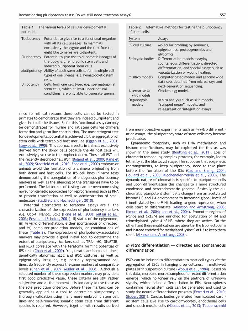
Table 2 Alternative methods for testing the pluripotencyof stem cells.
System Assays
ES cell culture Molecular profiling by genomics,epigenomics, proteogenomics andglycomics.
Embryoid bodies Differentiation models assayingspontaneous differentiation, directeddifferentiation, and special assays such asvascularization or wound healing.
In silico models Computer based models and genome widedata sets obtained from microarrays andnext-generation sequencing.
Alternative invivo models
Chicken egg model.
Organotypicmodels
In situ analysis such as skin models,“stripped organ” models, andre-aggregation/integration assays.
Table 1 The various levels of cellular developmentalpotential.
Totipotency Potential to give rise to a functional organismwith all its cell lineages. In mammalsexclusively the zygote and the first four toeight blastomeres are totipotent.
Pluripotency Potential to give rise to all somatic lineages ofthe body; e.g. embryonic stem cells andinduced pluripotent stem cells.
Multipotency Ability of adult stem cells to form multiple celltypes of one lineage; e.g. hematopoietic stemcells.
Unipotency Cells form one cell type; e.g. spermatogonialstem cells, which at least under naturalconditions, are only able to generate sperms.
557Reconsidering pluripotency tests: Do we still need teratoma assays?
since for ethical reasons these cells cannot be tested inprimates to demonstrate that they are indeed pluripotent andgive rise to all the tissues. So far this functional assay can onlybe demonstrated for murine and rat stem cells via chimeraformation and germ line contribution. The most stringent testfor developmental potential is achieved via the aggregation ofstem cells with tetraploid host morulae (Eggan et al., 2001;Nagy et al., 1990). This approach results in animals exclusivelyderived from the donor cells because the 4n host cells willexclusively give rise to the trophectoderm. These “all ES” andthe recently described “all iPS” (Boland et al., 2009; Kang etal., 2009; Stadtfeld et al., 2010; Zhao et al., 2009) embryos oranimals avoid the formation of a chimera originating fromboth donor and host cells. For iPS cell lines in vitro testsdemonstrating the upregulation of endogenous pluripotencymarkers as well as the silencing of the transgenes have to beperformed. The latter set of testing can be overcome usingnovel non-genetic approaches for reprogramming such as RNAor protein transfection as well as administration of smallmolecules (Stadtfeld and Hochedlinger, 2010).
Potential alternatives to teratoma assays are i) thecharacterization of the expression of pluripotency markerse.g. Oct-4, Nanog, Sox2 (Fong et al., 2008; Mitsui et al.,2003; Pesce and Scholer, 2001), ii) status of the epigenome,iii) in vitro differentiation, either spontaneous or directed,and iv) computer-prediction models, or combinations ofthese (Table 2). The expression of pluripotency-associatedmarkers may provide a good initial tool to determine theextent of pluripotency. Markers such as TRA-1-60, DNMT3B,and REX1 correlate with the teratoma forming potential ofiPS cells (Chan et al., 2009). Yet, teratocarcinoma cell lines,genetically abnormal hESC and iPSC cultures, as well asepigentically irregular, e.g. partially reprogrammed celllines, do frequently express the same markers at comparablelevels (Chan et al., 2009; Müller et al., 2008). Although aselected number of these expression markers may provide afirst good predictive value, these criteria are still rathersubjective and at the moment it is too early to use these asthe sole prediction criterion. Before these markers can begenerally applied as a tool to determine pluripotency athorough validation using many more embryonic stem celllines and self-renewing somatic stem cells from differentspecies is required. However, together with results derived
from more objective experiments such as in vitro differenti-ation assays, the pluripotency state of stem cells may becomepredictable.
Epigenomic footprints, such as DNA methylation andhistone modifications, may be exploited for this as wasshown in the same study (Boulting et al., 2011). Loss ofchromatin remodeling complex proteins, for example, led tolethality at the blastocyst stage. This supposes that epigeneticrearrangements, to keep pluripotency, need to take placebefore the formation of the ICM (Cao and Zhang, 2004;Houlard et al., 2006; Klochendler-Yeivin et al., 2000). Thedynamic nature of chromatin is specific to pluripotent cellsand upon differentiation this changes to a more structuredcondensed and heterochromatic genome. Basically the eu-chromatic pluripotent stem cells change from an acetylatedhistone H3 and H4 environment to increased global levels oftrimethylated lysine 9 H3 leading to gene repression, whencells start to differentiate (Atkinson and Armstrong, 2008;Kimura et al., 2004; Lee et al., 2004). Promoter regions ofNanog and Oct3/4 are enriched for acetylation of H4 andtrimethylated lysine 4 of H3, where they are active. On theother hand thesemodifications are absent in the trophectodermand instead enriched formethylated lysine 9 of H3 to keep themsilent (Atkinson and Armstrong, 2008).
In vitro differentiation — directed and spontaneousdifferentiation
ESCs can be induced to differentiate to most cell types via theaggregation of ESCs in hanging drop cultures, in multi-wellplates or in suspension culture (Wobus et al., 1984). Based onthis data, more andmore examples of directed differentiationemerge, which no longer rely on the plethora of unknownsignals, which induce differentiation in EBs. Neurospherescontaining neural stem cells can be generated and used tostudy the neural differentiation program (Ferrari et al., 2010;Studer, 2001). Cardiac bodies generated from isolated cardi-ac stem cells give rise to cardiomyocytes, endothelial cellsand smooth muscle cells (Höbaus et al., 2013; Taubenschmid
558 C. Buta et al.
and Weitzer, 2012). Definitive endoderm can be generatedwith high efficiency from ESCs in monolayers (Borowiak et al.,2009; Zhu et al., 2009). Murine ESC aggregates resemble theearly embryonic development of mouse for 7 to 8 days andspontaneously give rise to cells of ectodermal, endodermaland mesodermal origin. Until day 8, EBs undergo a morpho-logical development resembling early embryogenesis untilgastrulation commences (Bader et al., 2001). Later on,differentiation and development of other cell types appear tobe chaotic so far (Weitzer, 2006). However cardiomyogenesisseems to follow a morphological program at least until day 8 ofdifferentiation (Fuchs et al., 2012).
Furthermore, in EBs cellular function can be studied byelectrophysiology and cell–cell interaction can be studied byimmunofluorescence microscopy quite well; however, itseems that in teratoma cell–cell interaction resembles thesituation in a tissue much better than in EBs and somaticstem cell aggregates. Likewise, nutrition and blood supply interatoma reflect physiological conditions better than in EBsand monolayer cultures of stem cells. In teratoma, cells ofthe host, mainly the blood vessels growing into the tumor,influence the development of the tumor and thus alsosignificantly influence the results obtained from expressionanalysis etc. In teratoma new blood vessels supply the tissueswith nutrition and oxygen, however, in stem cell aggregatesthicker than 7 cell layers, no reproducible supply withnutrition and oxygen exists. Thus development of an in vitroangiogenesis model in combination with stem cell aggregationexperiments, as a possibility to improve the physiologicalrelevance of this model, is desirable.
In vitro models alone might be sensitive but are notspecific enough for the study of the genomics and epige-netics of hESC or hiPSC lines. To address this problem andthe gap between in vivo and in vitro models, in silicogenome wide methods such as whole genome transcriptomeprofiles in combination with complex biomarker models canidentify deviations from a defined “ideal” phenotype on aglobal scale (Müller et al., 2008, 2011; Williams et al.,2011).
Computer-based predictive models
Machine-learning based models can identify signaturescharacteristic of pluripotent stem cells in functional genomicdata sets (Brolen et al., 2010; Medine et al., 2010; Müller et al.,2008) and can also highlight deviations from an “ideal”pluripotent stem cell phenotype (Müller et al., 2011; Williamset al., 2011). In silico assays could be a cost effective alter-native to teratoma assays and would enable many exploratorybioinformatic downstream applications. Current challenges,in regard to in silico models, are i) standardization issues,ii) acceptance in the field, iii) regulatory issues and iv) mostimportantly the availability of comparable and multipledatasets of genomic, expression profile, proteomic and epige-netic analysis.
First, pluripotency models can be developed with reason-able funding for one microarray platform (e.g. Illumina) buttransfer to other platforms (e.g. RNA-seq) is a resourceintensive challenge. Secondly, even as bioinformatics is be-coming an important part in pluripotent stem cell research yetmost wet stem cell biologists have never received proper
training e.g. in using high-level bioinformatic tools such asBioconductor/R. Hence accessible ways of disseminatingbioinformatic assays for pluripotency to a non-expert audi-ence have to be developed. Reliable and standardized waysfor the effective communication of such bioinformatic resultshave to be agreed on by researchers, reviewers and journals,comparable to the Minimum Information About a MicroarrayExperiment (MIAME) standard required for the reporting ofmicroarray experiments by most peer reviewed journals(Brazma et al., 2001). Finally, global microarray datasetshave been widely used in preclinical, exploratory analyses,but rarely as defined outcome measure. Simple signature-based approaches are unable to identify stochastic, randomand unexpected events regularly emerging in stem cellcultures, such as epigenetic or even genetic alterations andabnormalities (Williams et al., 2011). As costs for generatinghigh-content datasets are currently dropping below a singleday of a postdocs salary due to the next-generation sequencingrevolution, a global, genome wide and data driven approachwill become more and more attractive and highly desirable forpluripotent stem cell research in spite of possible regulatorychallenges.
A first bioinformatic assay for pluripotency in human cells(PluriTest) has been recently developed and published(Müller et al., 2011) and can be used through a simple webinterface at www.pluritest.org. Up to date, more than 6200microarray data sets have been uploaded and analyzed.PluriTest currently supports Illumina gene expressionarrays and will be expanded to RNA-seq data in the nearfuture.
Chicken egg model — chorioallantoic membrane
Another option to the study of teratoma formation could bethe transplantation of stem cells onto the chorioallantoicmembrane (CAM) of avian/chicken embryos. The CAM issituated at the periphery of the chicken embryo as a denselyvascularized extraembryonic tissue. It is easily accessible byopening a hole in the egg shell. Experiments have beenperformed some 100 years ago and this model convinces bythe ease of access and the natural immunodeficient envi-ronment of the developing embryo. Tumor growth can beobserved within days and are of approximately 5–10 mm insize. They have sufficient similarity to teratoma and stronglyresemble clinical specimen of patient samples (Durupt et al.,2012; Hagedorn et al., 2005). The CAM model combines theadvantages of the in vivo environment with the simplicity ofan in vitro experiment, is cheap, fast and without seriousethical issues.
Organotypic models
The development of an in vitromodel of teratoma formation,such as a skin model would be of great importance, as it couldbe cultured in vitro via tissue engineering, where stem cellsare injected into skin pads and allowed to develop anddifferentiate. Another possibility would be to develop strat-egies for the testing of stem cells for their repopulationpotential of cell-free organ templates. These would serve asan ECM-template for directed differentiation. This has alreadybeen done with hearts and human trachea, where the organ
559Reconsidering pluripotency tests: Do we still need teratoma assays?
was completely depleted of cells leaving behind a grid ofextracellular material, which can be repopulated by stemcells (Ott and Taylor, 2006; Elliott et al., 2012). Further it ispossible to re-aggregate dissociated embryonic kidneys,which were still able to form organotypic renal structures(Unbekandt and Davies, 2010). This method was also used toform chimeric renal structures mixing murine embryonickidney cells with human amniotic fluid stem cells (hAFSCs).hAFSCs were able to integrate into and contribute to renalstructures. Using siRNA knock down technology they showedwhich genes contributed to renal structure formation(Siegel et al., 2010). This model can be used both as apluripotency assay and will allow us to gain new insight intoputative stem cell therapy in kidney disease models. Adultmurine ventricular slices serve as a new in vitro model ofadult myocardium with preserved in vivo structure. In thefuture, these could be used to study functional integration ofstem cells transplanted in infarcted hearts in vivo (Halbachet al., 2006).
An example of differential information contentof embryoid bodies and teratoma assays
Monitoring the development of cells in EBs allows theanalyses of dynamic aspects of cell differentiation and toidentify factors which differentially affect consecutivedevelopmental stages of cells or simple tissues. Furthermore,analysis of large numbers of EBs provides the basis for robuststatistical analysis of data. This type of information cannot beobtained from histological sections of teratoma because thereis only one endpoint per teratoma available and statisticalanalysis is limited by the rather small number of animalstypically used for teratoma assay. The following example willdemonstrate that the information obtained from in vitroexperiments in EBs may be more informative than that fromteratoma assays in mice.
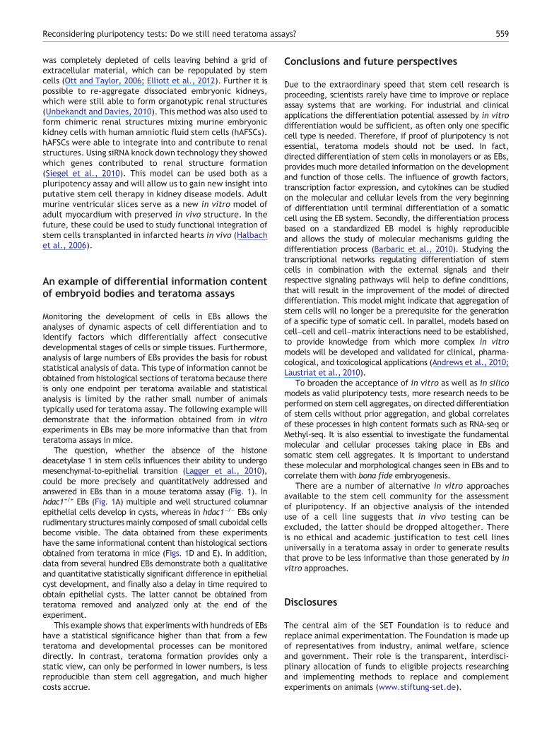
The question, whether the absence of the histonedeacetylase 1 in stem cells influences their ability to undergomesenchymal-to-epithelial transition (Lagger et al., 2010),could be more precisely and quantitatively addressed andanswered in EBs than in a mouse teratoma assay (Fig. 1). Inhdac1+/+ EBs (Fig. 1A) multiple and well structured columnarepithelial cells develop in cysts, whereas in hdac1−/− EBs onlyrudimentary structures mainly composed of small cuboidal cellsbecome visible. The data obtained from these experimentshave the same informational content than histological sectionsobtained from teratoma in mice (Figs. 1D and E). In addition,data from several hundred EBs demonstrate both a qualitativeand quantitative statistically significant difference in epithelialcyst development, and finally also a delay in time required toobtain epithelial cysts. The latter cannot be obtained fromteratoma removed and analyzed only at the end of theexperiment.
This example shows that experiments with hundreds of EBshave a statistical significance higher than that from a fewteratoma and developmental processes can be monitoreddirectly. In contrast, teratoma formation provides only astatic view, can only be performed in lower numbers, is lessreproducible than stem cell aggregation, and much highercosts accrue.
Conclusions and future perspectives
Due to the extraordinary speed that stem cell research isproceeding, scientists rarely have time to improve or replaceassay systems that are working. For industrial and clinicalapplications the differentiation potential assessed by in vitrodifferentiation would be sufficient, as often only one specificcell type is needed. Therefore, if proof of pluripotency is notessential, teratoma models should not be used. In fact,directed differentiation of stem cells in monolayers or as EBs,provides much more detailed information on the developmentand function of those cells. The influence of growth factors,transcription factor expression, and cytokines can be studiedon the molecular and cellular levels from the very beginningof differentiation until terminal differentiation of a somaticcell using the EB system. Secondly, the differentiation processbased on a standardized EB model is highly reproducibleand allows the study of molecular mechanisms guiding thedifferentiation process (Barbaric et al., 2010). Studying thetranscriptional networks regulating differentiation of stemcells in combination with the external signals and theirrespective signaling pathways will help to define conditions,that will result in the improvement of the model of directeddifferentiation. This model might indicate that aggregation ofstem cells will no longer be a prerequisite for the generationof a specific type of somatic cell. In parallel, models based oncell–cell and cell–matrix interactions need to be established,to provide knowledge from which more complex in vitromodels will be developed and validated for clinical, pharma-cological, and toxicological applications (Andrews et al., 2010;Laustriat et al., 2010).
To broaden the acceptance of in vitro as well as in silicomodels as valid pluripotency tests, more research needs to beperformed on stem cell aggregates, on directed differentiationof stem cells without prior aggregation, and global correlatesof these processes in high content formats such as RNA-seq orMethyl-seq. It is also essential to investigate the fundamentalmolecular and cellular processes taking place in EBs andsomatic stem cell aggregates. It is important to understandthese molecular and morphological changes seen in EBs and tocorrelate them with bona fide embryogenesis.
There are a number of alternative in vitro approachesavailable to the stem cell community for the assessmentof pluripotency. If an objective analysis of the intendeduse of a cell line suggests that in vivo testing can beexcluded, the latter should be dropped altogether. Thereis no ethical and academic justification to test cell linesuniversally in a teratoma assay in order to generate resultsthat prove to be less informative than those generated by invitro approaches.
Disclosures
The central aim of the SET Foundation is to reduce andreplace animal experimentation. The Foundation is made upof representatives from industry, animal welfare, scienceand government. Their role is the transparent, interdisci-plinary allocation of funds to eligible projects researchingand implementing methods to replace and complementexperiments on animals (www.stiftung-set.de).
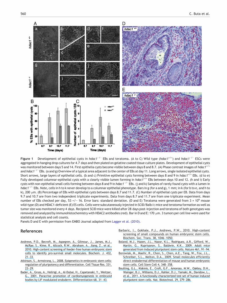
Figure 1 Development of epithelial cysts in hdac1−/− EBs and teratoma. (A to C) Wild type (hdac1+/+) and hdac1−/− ESCs wereaggregated in hanging drop cultures for 4.7 days and then plated on gelatine coated tissue culture plates. Development of epithelial cystswas monitored between days 5 and 14. First epithelia cysts become visible between days 8 and 8.7. (A) Phase contrast images of hdac1+/+
and hdac1−/− EBs. (a and g) Overview of a typical area adjacent to the center of EBs at day 11. Long arrows, single isolated epithelial cysts.Short arrows, large layers of epithelial cells. (b and c) Primitive epithelial cysts forming between days 8 and 9 in hdac1+/+ EBs. (d to e)Fully developed columnar-epithelial cysts with a clearly visible lumen forming in hdac1+/+ EBs between days 10 and 12. (h and i) Earlycysts with non-epithelial small cells forming between days 8 and 9 in hdac1−/− EBs. (j and k) Samples of rarely found cysts with a lumen inhdac1−/− EBs. Note, cells in h to k never develop to a columnar epithelial phenotype. Bars in g (for a and g), 1 mm; in k (for b to e, and h tok), 200 μm. (B) Percentage of EBs with epithelial cysts between days 8.7 and 11.7. (C) Number of epithelial cysts per EB. Data from days9.7 and 10.7 are from two independent triplicate experiments. Data from days 8.7 and 11.7 are from one triplicate experiment. Meannumber of EBs checked per day, 53 +/− 16. Error bars: standard deviation. (D and E) Teratoma were generated from 3 × 10⁶ mousewild-type (D) and HDAC1-deficient (E) ES cells. Cells were subcutaneously injected in SCID/Balb/cmice and teratoma formation aswell astumor size was monitored every 4 days. Recipient SCIDmice were killed after 28 days post-injection and teratoma of both genotypes wasremoved and analyzed by immunohistochemistry with HDAC2 antibodies (red). Bar in D and E: 170 μm. 3 tumors per cell linewere used forstatistical analysis and cell counts.Panels D and E with permission from EMBO Journal adapted from Lagger et al. (2010).
560 C. Buta et al.
References
Andrews, P.D., Becroft, M., Aspegren, A., Gilmour, J., James, M.J.,McRae, S., Kime, R., Allcock, R.W., Abraham, A., Jiang, Z., et al.,2010. High-content screening of feeder-free human embryonic stemcells to identify pro-survival small molecules. Biochem. J. 432,21–33.
Atkinson, S., Armstrong, L., 2008. Epigenetics in embryonic stem cells:regulation of pluripotency and differentiation. Cell Tissue Res. 331,23–29.
Bader, A., Gruss, A., Hollrigl, A., Al-Dubai, H., Capetanaki, Y., Weitzer,G., 2001. Paracrine promotion of cardiomyogenesis in embryoidbodies by LIF modulated endoderm. Differentiation 68, 31–43.
Barbaric, I., Gokhale, P.J., Andrews, P.W., 2010. High-contentscreening of small compounds on human embryonic stem cells.Biochem. Soc. Trans. 38, 1046–1050.
Boland, M.J., Hazen, J.L., Nazor, K.L., Rodriguez, A.R., Gifford, W.,Martin, G., Kupriyanov, S., Baldwin, K.K., 2009. Adult micegenerated from induced pluripotent stem cells. Nature 461, 91–94.
Borowiak, M., Maehr, R., Chen, S., Chen, A.E., Tang, W., Fox, J.L.,Schreiber, S.L., Melton, D.A., 2009. Small molecules efficientlydirect endodermal differentiation of mouse and human embryonicstem cells. Cell Stem Cell 4, 348–358.
Boulting, G.L., Kiskinis, E., Croft, G.F., Amoroso, M.W., Oakley, D.H.,Wainger, B.J., Williams, D.J., Kahler, D.J., Yamaki, M., Davidow, L.,et al., 2011. A functionally characterized test set of human inducedpluripotent stem cells. Nat. Biotechnol. 29, 279–286.
561Reconsidering pluripotency tests: Do we still need teratoma assays?
Brazma, A., Hingamp, P., Quackenbush, J., Sherlock, G., Spellman, P.,Stoeckert, C., Aach, J., Ansorge, W., Ball, C.A., Causton, H.C., etal., 2001. Minimum information about a microarray experiment(MIAME)-toward standards for microarray data. Nat. Genet. 29,365–371.
Brivanlou, A.H., Gage, F.H., Jaenisch, R., Jessell, T., Melton, D.,Rossant, J., 2003. Stem cells. Setting standards for humanembryonic stem cells. Science 300, 913–916.
Brolen, G., Sivertsson, L., Bjorquist, P., Eriksson, G., Ek, M., Semb, H.,Johansson, I., Andersson, T.B., Ingelman-Sundberg, M., Heins, N.,2010. Hepatocyte-like cells derived from human embryonic stemcells specifically via definitive endoderm and a progenitor stage. J.Biotechnol. 145, 284–294.
Cao, R., Zhang, Y., 2004. The functions of E(Z)/EZH2-mediatedmethylation of lysine 27 in histone H3. Curr. Opin. Genet. Dev.14, 155–164.
Chan, E.M., Ratanasirintrawoot, S., Park, I.H., Manos, P.D., Loh,Y.H., Huo, H., Miller, J.D., Hartung, O., Rho, J., Ince, T.A., et al.,2009. Live cell imaging distinguishes bona fide human iPS cellsfrom partially reprogrammed cells. Nat. Biotechnol. 27,1033–1037.
Damjanov, I., Andrews, P.W., 2007. The terminology of teratocarci-nomas and teratomas. Nat. Biotechnol. 25, 1212 (discussion 1212).
Dressel, R., 2011. Effects of histocompatibility and host immuneresponses on the tumorigenicity of pluripotent stem cells.Semin. Immunopathol. 33, 573–591.
Dressel, R., Schindehutte, J., Kuhlmann, T., Elsner, L., Novota, P.,Baier, P.C., Schillert, A., Bickeboller, H., Herrmann, T.,Trenkwalder, C., et al., 2008. The tumorigenicity of mouseembryonic stem cells and in vitro differentiated neuronal cells iscontrolled by the recipients' immune response. PLoS One 3,e2622.
Durupt, F., Koppers-Lalic, D., Balme, B., Budel, L., Terrier, O.,Lina, B., Thomas, L., Hoeben, R.C., Rosa-Calatrava, M., 2012.The chicken chorioallantoic membrane tumor assay as modelfor qualitative testing of oncolytic adenoviruses. Cancer GeneTher. 19, 58–68.
Eggan, K., Akutsu, H., Loring, J., Jackson-Grusby, L., Klemm, M.,Rideout III, W.M., Yanagimachi, R., Jaenisch, R., 2001. Hybridvigor, fetal overgrowth, and viability of mice derived by nuclearcloning and tetraploid embryo complementation. Proc. Natl.Acad. Sci. U. S. A. 98, 6209–6214.
Elliott, M.J., De Coppi, P., Speggiorin, S., Roebuck, D., Butler, C.R.,Samuel, E., Crowley, C., McLaren, C., Fierens, A., Vondrys, D.,Cochrane, L., Jephson, C., Janes, S., Beaumont, N.J., Cogan, T.,Bader, A., Seifalian, A.M., Hsuan, J.J., Lowdell, M.W., Birchall,M.A., 2012. Stem-cell-based, tissue engineered tracheal re-placement in a child: a 2-year follow-up study. Lancet 380,994–1000.
Erdö, F., Buhrle, C., Blunk, J., Hoehn, M., Xia, Y., Fleischmann, B.,Focking, M., Kustermann, E., Kolossov, E., Hescheler, J., et al.,2003. Host-dependent tumorigenesis of embryonic stem celltransplantation in experimental stroke. J. Cereb. Blood FlowMetab. 23, 780–785.
European Union, T.E.P.a.t.c.o.t., 2010. Directive 2010/63/EU onthe protection of animals used for scientific purposes. Off. J.Eur. Union L276, 33–79.
Evans, M.J., Kaufman, M.H., 1981. Establishment in culture ofpluripotential cells from mouse embryos. Nature 292, 154–156.
Ferrari, D., Binda, E., De Filippis, L., Vescovi, A.L., 2010. Isolationof neural stem cells from neural tissues using the neurospheretechnique. Curr. Protoc. Stem Cell Biol. (Chapter 2, Unit 2D 6).
Fong, H., Hohenstein, K.A., Donovan, P.J., 2008. Regulation of self-renewal and pluripotency by Sox2 in human embryonic stemcells. Stem Cells 26, 1931–1938.
Fuchs, C., Scheinast, M., Pasteiner, W., Lagger, S., Hofner, M.,Hoellrigl, A., Schultheis, M., Weitzer, G., 2012. Self-organizationphenomena in embryonic stem cell-derived embryoid bodies: axis
formation and breaking of symmetry during cardiomyogenesis.Cells Tissues Organs 195, 377–391.
Gertow, K., Przyborski, S., Loring, J.F., Auerbach, J.M., Epifano, O.,Otonkoski, T., Damjanov, I., Ahrlund-Richter, L., 2007. Isolationof human embryonic stem cell-derived teratomas for theassessment of pluripotency. Curr. Protoc. Stem Cell Biol. (Chapter1, Unit 1B 4).
Gertow, K., Cedervall, J., Jamil, S., Ali, R., Imreh, M.P., Gulyas, M.,Sandstedt, B., Ahrlund-Richter, L., 2011. Early events inxenograft development from the human embryonic stem cellline HS181—resemblance with an initial multiple epiblastformation. PLoS One 6, e27741.
Gropp, M., Shilo, V., Vainer, G., Gov, M., Gil, Y., Khaner, H.,Matzrafi, L., Idelson, M., Kopolovic, J., Zak, N.B., et al., 2012.Standardization of the teratoma assay for analysis of pluripotencyof human ES cells and biosafety of their differentiated progeny.PLoS One 7, e45532.
Hagedorn, M., Javerzat, S., Gilges, D., Meyre, A., de Lafarge, B.,Eichmann, A., Bikfalvi, A., 2005. Accessing key steps of humantumor progression in vivo by using an avian embryo model. Proc.Natl. Acad. Sci. U. S. A. 102, 1643–1648.
Halbach, M., Pillekamp, F., Brockmeier, K., Hescheler, J., Müller-Ehmsen, J., Reppel, M., 2006. Ventricular slices of adult mousehearts—a new multicellular in vitro model for electrophysiologicalstudies. Cell. Physiol. Biochem. 18, 1–8.
Halme, D.G., Kessler, D.A., 2006. FDA regulation of stem-cell-basedtherapies. N. Engl. J. Med. 355, 1730–1735.
Hentze, H., Soong, P.L., Wang, S.T., Phillips, B.W., Putti, T.C.,Dunn, N.R., 2009. Teratoma formation by human embryonicstem cells: evaluation of essential parameters for future safetystudies. Stem Cell Res. 2, 198–210.
Houlard, M., Berlivet, S., Probst, A.V., Quivy, J.P., Hery, P.,Almouzni, G., Gerard, M., 2006. CAF-1 is essential for hetero-chromatin organization in pluripotent embryonic cells. PLoS Genet.2, e181.
Höbaus, J., Heher, P., Gottschamel, T., Scheinast, M., Auner, H.,Walder, D., Wiedner, M., Taubenschmid, J., Miksch, M., Sauer,T., et al., 2013. Embryonic stem cells facilitate the isolation ofpersistent clonal cardiovascular progenitor cell lines andleukemia inhibitor factor maintains their self-renewal andmyocardial differentiation potential in vitro. Cells TissuesOrgans 197, 249–268.
ISCBI, T.I.S.C.B.I., 2009. Consensus guidance for banking and supplyof human embryonic stem cell lines for research purposes. StemCell Rev. 5, 301–314.
Kang, L., Wang, J., Zhang, Y., Kou, Z., Gao, S., 2009. iPS cells cansupport full-term development of tetraploid blastocyst-complemented embryos. Cell Stem Cell 5, 135–138.
Kimura, H., Tada, M., Nakatsuji, N., Tada, T., 2004. Histone codemodifications on pluripotential nuclei of reprogrammed somaticcells. Mol. Cell. Biol. 24, 5710–5720.
Klochendler-Yeivin, A., Fiette, L., Barra, J., Muchardt, C., Babinet, C.,Yaniv, M., 2000. Themurine SNF5/INI1 chromatin remodeling factoris essential for embryonic development and tumor suppression.EMBO Rep. 1, 500–506.
Knoepfler, P.S., 2009. Deconstructing stem cell tumorigenicity: aroadmap to safe regenerative medicine. Stem Cells 27, 1050–1056.
Lagger, S., Meunier, D., Mikula, M., Brunmeir, R., Schlederer, M.,Artaker, M., Pusch, O., Egger, G., Hagelkruys, A., Mikulits, W.,et al., 2010. Crucial function of histone deacetylase 1 fordifferentiation of teratomas in mice and humans. EMBO J. 29,3992–4007.
Laustriat, D., Gide, J., Peschanski, M., 2010. Human pluripotentstem cells in drug discovery and predictive toxicology. Biochem.Soc. Trans. 38, 1051–1057.
Lawrenz, B., Schiller, H., Willbold, E., Ruediger, M., Muhs, A., Esser, S.,2004. Highly sensitive biosafety model for stem-cell-derived grafts.Cytotherapy 6, 212–222.
562 C. Buta et al.
Lee, J.H., Hart, S.R., Skalnik, D.G., 2004. Histone deacetylaseactivity is required for embryonic stem cell differentiation.Genesis 38, 32–38.
Li, Z., Huang, H., Boland, P., Dominguez, M.G., Burfeind, P., Lai, K.M.,Lin, H.C., Gale, N.W., Daly, C., Auerbach, W., et al., 2009.Embryonic stem cell tumor model reveals role of vascularendothelial receptor tyrosine phosphatase in regulating Tie2pathway in tumor angiogenesis. Proc. Natl. Acad. Sci. U. S. A.106, 22399–22404.
Martin, G.R., Evans, M.J., 1975. Differentiation of clonal lines ofteratocarcinoma cells: formation of embryoid bodies in vitro.Proc. Natl. Acad. Sci. U. S. A. 72, 1441–1445.
Mauritz, C., Martens, A., Rojas, S.V., Schnick, T., Rathert, C.,Schecker, N., Menke, S., Glage, S., Zweigerdt, R., Haverich, A.,et al., 2011. Induced pluripotent stem cell (iPSC)-derived Flk-1progenitor cells engraft, differentiate, and improve heartfunction in a mouse model of acute myocardial infarction. Eur.Heart J. 32, 2634–2641.
Medine, C.N., Greenhough, S., Hay, D.C., 2010. Role of stem-cell-derived hepatic endoderm in human drug discovery. Biochem.Soc. Trans. 38, 1033–1036.
Mitsui, K., Tokuzawa, Y., Itoh, H., Segawa, K., Murakami, M.,Takahashi, K., Maruyama, M., Maeda, M., Yamanaka, S., 2003.The homeoprotein Nanog is required for maintenance ofpluripotency in mouse epiblast and ES cells. Cell 113, 631–642.
Müller, F.J., Laurent, L.C., Kostka, D., Ulitsky, I., Williams, R., Lu, C.,Park, I.H., Rao, M.S., Shamir, R., Schwartz, P.H., et al., 2008.Regulatory networks define phenotypic classes of human stem celllines. Nature 455, 401–405.
Müller, F.J., Goldmann, J., Loser, P., Loring, J.F., 2010. A call tostandardize teratoma assays used to define human pluripotent celllines. Cell Stem Cell 6, 412–414.
Müller, F.J., Schuldt, B.M., Williams, R., Mason, D., Altun, G.,Papapetrou, E.P., Danner, S., Goldmann, J.E., Herbst, A.,Schmidt, N.O., et al., 2011. A bioinformatic assay for pluripotencyin human cells. Nat. Methods 8, 315–317.
Nagy, A., Gocza, E., Diaz, E.M., Prideaux, V.R., Ivanyi, E., Markkula, M.,Rossant, J., 1990. Embryonic stem cells alone are able to supportfetal development in the mouse. Development 110, 815–821.
Ott, H.C., Taylor, D.A., 2006. From cardiac repair to cardiacregeneration—ready to translate? Expert Opin. Biol. Ther. 6,867–878.
Pesce, M., Scholer, H.R., 2001. Oct-4: gatekeeper in the beginningsof mammalian development. Stem Cells 19, 271–278.
Peterson, S.E., Tran, H.T., Garitaonandia, I., Han, S., Nickey, K.S.,Leonardo, T., Laurent, L.C., Loring, J.F., 2011. Teratoma genera-tion in the testis capsule. J. Vis. Exp. e3177 (http://www.jove.com/video/3177/teratoma-generation-in-the-testis-capsule).
Rossant, J., 2008. Stem cells and early lineage development. Cell132, 527–531.
Russell, W.M.S., Burch, R.L., 1959. The Principles of HumaneExperimental Technique. Methuen, London.
Saric, T., Frenzel, L.P., Hescheler, J., 2008. Immunological barriers toembryonic stem cell-derived therapies. Cells Tissues Organs 188,78–90.
Siegel, N., Rosner, M., Unbekandt, M., Fuchs, C., Slabina, N.,Dolznig, H., Davies, J.A., Lubec, G., Hengstschlager, M., 2010.
Contribution of human amniotic fluid stem cells to renaltissue formation depends on mTOR. Hum. Mol. Genet. 19,3320–3331.
Skreb, N., Svajger, A., Levak-Svajger, B., 1971. Growth anddifferentiation of rat egg-cylinders under the kidney capsule.J. Embryol. Exp. Morphol. 25, 47–56.
Solter, D., Skreb, N., Damjanov, I., 1970. Extrauterine growth ofmouse egg-cylinders results in malignant teratoma. Nature 227,503–504.
Stadtfeld, M., Hochedlinger, K., 2010. Induced pluripotency: history,mechanisms, and applications. Genes Dev. 24, 2239–2263.
Stadtfeld, M., Apostolou, E., Akutsu, H., Fukuda, A., Follett, P.,Natesan, S., Kono, T., Shioda, T., Hochedlinger, K., 2010.Aberrant silencing of imprinted genes on chromosome 12qF1 inmouse induced pluripotent stem cells. Nature 465, 175–181.
Stevens, L.C., 1958. Studies on transplantable testicular teratomasof strain 129 mice. J. Natl. Cancer Inst. 20, 1257–1275.
Stevens, L.C., 1970. The development of transplantable teratocarci-nomas from intratesticular grafts of pre- and postimplantationmouse embryos. Dev. Biol. 21, 364–382.
Stevens, L.C., Little, C.C., 1954. Spontaneous testicular teratomas inan inbred strain of mice. Proc. Natl. Acad. Sci. U. S. A. 40,1080–1087.
Studer, L., 2001. Stem cells with brainpower. Nat. Biotechnol. 19,1117–1118.
Swiss Federal Veterinary Office (Hsrg), 2012. Classification ofAnimal Experiments according to Grades of Severity prior tothe Experiment (Stress Categories). (http://www.tierversuch.ch/show=AWLaw&nav_id4104&lang=en (02.14.2012)).
Taubenschmid, J., Weitzer, G., 2012. Mechanisms of cardiogenesisin cardiovascular progenitor cells. Int. Rev. Cell Mol. Biol. 293,195–267.
Unbekandt, M., Davies, J.A., 2010. Dissociation of embryonickidneys followed by reaggregation allows the formation ofrenal tissues. Kidney Int. 77, 407–416.
Verhoog, H., 1992. The concept of intrinsic value and transgenicanimals. J. Agric. Ethics 5, 147–160.
Vorstenbosch, J., 1993. The concept of integrity. Its significance forthe ethical discussion on biotechnology and animals. Livest.Prod. Sci. 36, 109–112.
Weitzer, G., 2006. Embryonic stem cell-derived embryoid bodies: anin vitro model of eutherian pregastrulation development andearly gastrulation. Handb. Exp. Pharmacol. 21–51.
Wesselschmidt, R.L., 2011. The teratoma assay: an in vivoassessment of pluripotency. Methods Mol. Biol. 767, 231–241.
Williams, R., Schuldt, B., Müller, F.J., 2011. A guide to stem cellidentification: progress and challenges in system-wide predictivetesting with complex biomarkers. Bioessays 33, 880–890.
Wobus, A.M., Holzhausen, H., Jakel, P., Schoneich, J., 1984.Characterization of a pluripotent stem cell line derived from amouse embryo. Exp. Cell Res. 152, 212–219.
Zhao, X.Y., Li, W., Lv, Z., Liu, L., Tong, M., Hai, T., Hao, J., Guo, C.L.,Ma, Q.W., Wang, L., et al., 2009. iPS cells produce viable micethrough tetraploid complementation. Nature 461, 86–90.
Zhu, S., Wurdak, H., Wang, J., Lyssiotis, C.A., Peters, E.C., Cho, C.Y.,Wu, X., Schultz, P.G., 2009. A small molecule primes embryonicstem cells for differentiation. Cell Stem Cell 4, 416–426.