conditionally reprogrammed normal and transformed mouse mammary epithelial cells display a...
TRANSCRIPT
Conditionally Reprogrammed Normal and TransformedMouse Mammary Epithelial Cells Display aProgenitor-Cell–Like PhenotypeFrancisco R. Saenz1, Virginie Ory1, Maram AlOtaiby1, Sonia Rosenfield1, Mary Furlong2,
Luciane R. Cavalli1, Michael D. Johnson1, Xuefeng Liu2, Richard Schlegel2, Anton Wellstein1,
Anna T. Riegel1*
1 Department of Oncology, Lombardi Comprehensive Cancer Center, Georgetown University, Washington, District of Columbia, United States of America, 2 Department
of Pathology, Lombardi Comprehensive Cancer Center, Georgetown University, Washington, District of Columbia, United States of America
Abstract
Mammary epithelial (ME) cells cultured under conventional conditions senesce after several passages. Here, we demonstratethat mouse ME cells isolated from normal mammary glands or from mouse mammary tumor virus (MMTV)-Neu–inducedmammary tumors, can be cultured indefinitely as conditionally reprogrammed cells (CRCs) on irradiated fibroblasts in thepresence of the Rho kinase inhibitor Y-27632. Cell surface progenitor-associated markers are rapidly induced in normalmouse ME-CRCs relative to ME cells. However, the expression of certain mammary progenitor subpopulations, such asCD49f+ ESA+ CD44+, drops significantly in later passages. Nevertheless, mouse ME-CRCs grown in a three-dimensionalextracellular matrix gave rise to mammary acinar structures. ME-CRCs isolated from MMTV-Neu transgenic mouse mammarytumors express high levels of HER2/neu, as well as tumor-initiating cell markers, such as CD44+, CD49f+, and ESA+ (EpCam).These patterns of expression are sustained in later CRC passages. Early and late passage ME-CRCs from MMTV-Neu tumorsthat were implanted in the mammary fat pads of syngeneic or nude mice developed vascular tumors that metastasizedwithin 6 weeks of transplantation. Importantly, the histopathology of these tumors was indistinguishable from that of theparental tumors that develop in the MMTV-Neu mice. Application of the CRC system to mouse mammary epithelial cellsprovides an attractive model system to study the genetics and phenotype of normal and transformed mouse epithelium ina defined culture environment and in vivo transplant studies.
Citation: Saenz FR, Ory V, AlOtaiby M, Rosenfield S, Furlong M, et al. (2014) Conditionally Reprogrammed Normal and Transformed Mouse Mammary EpithelialCells Display a Progenitor-Cell–Like Phenotype. PLoS ONE 9(5): e97666. doi:10.1371/journal.pone.0097666
Editor: Nanette H Bishopric, University of Miami School of Medicine, United States of America
Received March 12, 2014; Accepted April 10, 2014; Published May 15, 2014
Copyright: � 2014 Saenz et al. This is an open-access article distributed under the terms of the Creative Commons Attribution License, which permitsunrestricted use, distribution, and reproduction in any medium, provided the original author and source are credited.
Data Availability: The authors confirm that all data underlying the findings are fully available without restriction. All data included within manuscript.
Funding: This work was funded in part by National Institutes of Health/National Cancer Institute grants CA113477 (ATR), Development Grant (ATR) under CCSGNumber P30CA051008 from the National Cancer Institute, CA177466 (RS and AW), CA180524 (XL) and by the Cherry Blossom Breast Cancer Foundation Grant (XL).FS is supported by Susan G Komen Training Grant PBTDR12228366. The funders had no role in study design, data collection and analysis, decision to publish, orpreparation of the manuscript.
Competing Interests: Georgetown University has a patent pending on the cell reprogramming technology (Immortalizing Epithelial Cells and Methods of Use,Application No. 13/885,078) and has licensed the technology to a new biotechnology company, Propagenix (www.propagenix.com). Georgetown University alsohas founding equity in Propagenix. RS and XL are Professors at Georgetown University who are consultants to Propagenix and RS has founding equity in thecompany. This does not alter the authors’ adherence to all the PLOS ONE policies on sharing data and materials.
* E-mail: [email protected]
Introduction
Studies utilizing primary normal and tumor epithelial cells are
frequently hampered by the fact that cells can only be cultured for
short periods of time before they cease proliferating and undergo
senescence [1]. In addition, the cultured cells frequently do not
retain lineage commitment or normal proliferation or differenti-
ation potential. Various methods have been used to immortalize
epithelial cells, such as introduction of viral oncogenes and the
telomerase reverse transcriptase [2], although these interventions
frequently disrupt normal differentiation. It has been recently
demonstrated that human epithelial cells from a variety of sources
(e.g., keratinocytes and human mammary epithelial cells) can be
cultured indefinitely and can bypass senescence when cultured on
irradiated fibroblast feeders in the presence of the Rho kinase
inhibitor Y-27632 [3]. Cells passaged in this system are known as
conditionally reprogrammed cells (CRCs). The CRC system has
been applied to epithelial cells from human tumor tissues, where
drug responsiveness in vivo can be predicted from the in vitro
responses of the CRCs [4]. Thus, the CRC system has potential
for studying normal and tumor cells from primary sources in
culture without utilizing overexpression of oncogenes and cell
cycle inhibitory factors. Further analysis of human CRCs revealed
that induction of the CRC phenotype is rapid and involves
reprogramming of most of the cell population [5]. However, the
CRC phenotype in keratinocytes can be reversed by the removal
of the Rho kinase inhibitor and cells can then differentiate
normally, as demonstrated by the ability of tracheal epithelium to
form a stratified epithelium in a three-dimensional culture system
[5]. Of note is that human CRCs share many properties of adult
stem cells but do not express markers of pluripotent progenitors
[5]. Thus, human CRCs can be used for in vitro and in vivo studies
PLOS ONE | www.plosone.org 1 May 2014 | Volume 9 | Issue 5 | e97666
of normal and tumor cells and may offer a system where drug
therapies can be tested on cells expanded from individual patients.
In the current study, we wished to determine if mouse
mammary epithelial (ME)-CRCs could be developed from normal
or tumor sources, and if their properties mirrored those of human
cells exposed to the CRC system. Although mouse epithelial cells
undergo senescence with serial passage, the mechanisms of
senescence differ from those of human cells [6]. In particular,
telomere shortening does not play a major role in driving
senescence of mouse cells [7,8]. Interestingly, despite these
differences, we report that both normal and tumor ME-CRCs
from mice can be passaged indefinitely. Similar to human
epithelial cells, normal mouse ME-CRCs expressed progenitor-
associated markers, but not pluripotent stem cell markers. ME-
CRCs were able to form mammary acinar structures when grown
in a three-dimensional (3D) Matrigel matrix. However, unlike
human cells, high expression levels of many progenitor cell
markers were maintained after CRC withdrawal, suggesting that,
in mouse cells, many of the effects of the CRC system are not
rapidly reversible. ME-CRCs derived from mouse mammary
tumors dissected from MMTV-Neu mice could also be passaged
indefinitely, and a large portion of the cells expressed markers
characteristic of tumor-initiating cells in vitro. Large tumors
developed in the mammary fat pad after implantation of these
cells. Thus, the CRC system can also be applied to normal and
transformed mouse epithelial cells and presents opportunities to
study properties of genetically manipulated cells in allograft
models.
Materials and Methods
Cell cultureNormal and tumor mouse ME cells were isolated from
mammary glands 3 and 4 of 6-week-old female FVB or
FVB.Cg-Tg(ACTB-EGFP)B5Nagy/J mice (The Jackson Labora-
tory, Bar Harbor, ME, www.jax.org) and from mammary tumors
of MMTV-Neu mice, respectively, as described previously [9].
ME-CRCs were maintained on irradiated 3T3-J2 fibroblasts as
described previously [3] and passaged in Dulbecco’s modified
Eagle medium (DMEM)/F12 containing 10 mM Y-27632 (Re-
agents Direct, Encinitas, CA, www.reagentsdirect.com). Co-
culture flasks were trypsinized in two steps by using 0.05%
Trypsin-EDTA. The initial 1–2 minutes trypsinization to remove
feeders was followed by a wash using phosphate-buffered saline
(PBS) and another 5 minutes to detached epithelial cells that were
subsequently reseeded at a 1:10 ratio in the CRC system and
cultured for 5–6 days before passaging. Freshly isolated primary
cells were defined as P0. Subsequent passages of the ME cells
(CRCs or non-CRCs) were referred to as P1 and greater. ME-
CRCs early passage was defined as P,10 and late passage as P$
10.
Array comparative genomic hybridization (cgh) analysisNormal ME-CRCs (P5, P18, and P76) and MMTV-Neu ME-
CRCs (P6, P38, and P73), as well as normal ME non-CRCs (P2)
and MMTV-HER2/neu ME non-CRCs (P2) were analyzed for
DNA copy number changes using the oligonucleotide-based
46180K mouse array CGH platform (Agilent Technologies,
Santa Clara, CA, www.agilent.com). Genomic DNA from CRC
and non-CRC cultures was isolated and enzymatically digested
according to the manufacturer’s protocol (Agilent Technologies).
Normal mouse DNA, isolated from female mouse tail, was used as
reference DNA. Equal amounts of CRC or non-CRC genomic
DNA and reference DNA were directly labeled with Cy3 or Cy5,
respectively, using the SureTag Labeling Kit (Agilent Technolo-
gies) and hybridized in the presence of mouse Cot1-DNA (Life
Technologies, Carlsbad, CA, www.lifetechnologies.com) to the
arrays for 40 hours. The arrays were scanned using an Agilent
array scanner and the data were analyzed using Feature
Extraction (FE) software v10.10 and Genome Workbench version
7.0 software (Agilent Technologies). For each sample, FE gave a
log10 ratio (log of tumor processed signal over reference processed
signal for each gene) that was imported into Genome Workbench
and transformed and viewed as a log2-based ratio. Outliers
detected by FE were excluded from the analysis. The algorithm
ADM-2 and a threshold value of 6.0 were applied with the
appropriate filters to analyze the data. Gene amplifications and
deletions were considered when the corresponding plotted
oligonucleotide probes presented values of log2 .7/6 and ,5/6,
respectively.
Flow cytometryCRCs were analyzed for expression of individual (monopara-
metric) or multiple (multiparametric) surface proteins using
fluorescence-labeled primary antibodies. Sca1/FITC anti-mouse
(Cat #553335), CD24/PE anti-mouse (Cat #553262), Rat IgG-
FITC anti-mouse (Cat #553929), and Rat IgG-PE (Cat #553989)
were all purchased from BD Biosciences (San Jose, CA, www.
bdbiosciences.com). Additionally, CD29/APC anti-mouse/rat
(Cat #102215), ESA/FITC anti-mouse (Cat #118207), CD49f/
APC anti-human/mouse (Cat # 313615) and CD44/PE anti-
mouse (Cat #103007) antibodies were purchased from BioLegend
(San Diego, CA, www.biolegend.com). In short, samples were
washed once with 3% bovine serum albumin (BSA), a second time
with PBS and then incubated with a viability assay kit (Life
Technologies Cat #L34955) according to manufacturer’s instruc-
tions. The excess reagent was removed by washing with PBS, then
incubated with labeled single antibody or antibody cocktail in PBS
at 4u C for one hour. Excess antibody was removed by washing
with PBS prior to analysis. Samples were analyzed using a BD
FACS Aria Cell Sorter (BD Biosciences). Gating was used to
restrict analysis to cells labeled for single or multiple markers, and
graphs were plotted using FCS Express 4 software.
Brightfield and Confocal Microscopic Analysis of CulturesGrowing in 3D Matrigel Matrices
ME-CRCs and MCF10A cultures were established in extracel-
lular matrix as previously described [10,11]. Briefly, 40 mL of cold
Matrigel (BD Bioscience Cat #354230) was added to the surface
of sterile pre-chilled, slide chambers (Lab-Tek II Chamber Slide
Cat #154534) purchased from ThermoFisher Scientific Inc.
(Waltham, MA, www.fishersci.com). The slides were then incu-
bated at 37uC for 15 minutes to allow polymerization of the
substratum. Subsequently, 36104 cells/mL were added to the
slides and resuspended in assay media containing 2% FBS, 2%
Matrigel, and 20 ng/mL EGF and incubated at 37uC until sphere
formation. For confocal microscopy, immunostaining of the acinar
structures was carried out as described [12].
Western blottingWhole-cell extracts were prepared from ME-CRCs, and
immunoblot analysis for proteins indicated was performed as
described previously [13].
Allograft analysisMMTV-Neu ME-CRCs were implanted in mammary fat pads
of nude or syngeneic FVB mice (The Jackson Laboratory). Briefly,
Reprogramming Progenitor-Like Cells
PLOS ONE | www.plosone.org 2 May 2014 | Volume 9 | Issue 5 | e97666

single-cell suspensions of early (P6) and late (P68) passage MMTV-
Neu ME-CRCs were injected orthotopically, with Matrigel, into
mammary gland 4 of 3-month-old MMTV-Neu female mice. Mice
were euthanized and tumors were removed 6 weeks later or when
the longest axis of the tumor reached 1 cm as described previously
[14]. Lung and liver tissue was collected at the same time as the
primary mammary tumor. Real-time polymerase chain reaction
(qRT PCR) for b-casein expression in lung and liver tissues was
performed as previously described [15].
Histological analysisHematoxylin and eosin (H&E) staining and immunohistochem-
ical and immunofluorescence analyses as indicated were per-
formed on paraffin-embedded 5- mm sections of mammary gland,
lung or liver using standard protocols described elsewhere [14].
Ethics statementAll procedures using animals were approved by the Georgetown
University IACUC and were conducted according to the NIH
guidelines for the care and use of laboratory animals (Public
Health Service: Assurance # A-3282-01). Isoflurane was used for
all animals undergoing surgery.
Statistical and Image AnalysesSamples were compared using two-tailed Student’s t-test. P,
0.05 was considered statistically significant. Phase contrast images
of serially passaged CRCs, micrographs showing immunohisto-
chemical staining of 3D mammosphere cultures, and images of
western blots were adjusted for brightness/contrast using Adobe
Photoshop CS5 software. All images were adjusted to the same
level. ImageJ was used to quantify colony subtypes. Prism 5 for PC
(GraphPad, San Diego, CA, www.graphpad.com) was used for all
statistical analyses. Figures were assembled using Adobe Illustrator
CS5 software.
Results
ME-CRCs Maintain a Phenotype Similar to the ParentalME Cells
We have reported that human epithelial cells can be propagated
indefinitely when co- cultured with irradiated fibroblast feeder cells
in the presence of the ROCK inhibitor Y-27632 [3]. To determine
if mouse ME cells can be similarly re-programmed, we prepared
ME cells from mammary glands of FVB mice. When grown on
plastic, these cells senesce after 3–4 passages (Figure S1A).
However, when grown on the irradiated fibroblasts in the
presence of Y-27632 as described [3,16], the mouse ME cells
maintained a normal cobblestone-like morphology in the CRC
system for .50 passages after one year in culture (Figure 1A and
B). This was also demonstrated with a ME preparation from
transgenic FVB mice that harbored a germ line insertion of the
green fluorescent protein (GFP) under the control of the actin
promoter. This strain assumed similar morphology and could also
be grown indefinitely in CRC culture (Figure 1A). To date we
have maintained these cells in culture for up to 150 days. Array
Figure 1. Culture of normal mouse mammary epithelial (ME) cells under conditions generating conditionally reprogrammed cells(CRCs). A) Continuous culture of ME-CRCs generated from mammary glands of FVB or FVB-green fluorescent protein (GFP) mice. B) Phase contrastimages of ME-CRCs and non-CRCs at indicated passages; magnification, 106; bar, 100 mm. C) Phase contrast and immunofluorescence images ofmammary acinar-like structures from P59 ME-CRCs and immortalized MCF10A cells stained for laminin and counterstained with DAPI. L, lumen; arrow,peripheral laminin. Magnification, 106 and 636; bar, 100 mm and 50 mm for phase contrast and fluorescence, respectively. D) Average number ofspheres per field of early and late ME-CRCs in four independent fields from four wells. Data are representative of two independent experiments.Values are mean6SEM; ns, not significant. A–D) All data were acquired after 5 days of culture. P, passage.doi:10.1371/journal.pone.0097666.g001
Reprogramming Progenitor-Like Cells
PLOS ONE | www.plosone.org 3 May 2014 | Volume 9 | Issue 5 | e97666

CGH analysis was carried out for ME-CRCs and ME-non CRCs
to determine if the CRC system impacted the genomic profile of
the cells. This analysis indicated no major changes in the normal
genome copy number composition of the CRCs at P5 (Figure S2A
and B). At later passages, despite the fact the morphology of the
cells appeared normal (Figure 1B), there was an increased level of
copy number alterations, with gains and losses on chromosomes 6,
11, and 18 at P18, and chromosomes 1, 3, 4, 7, 9, 13, 14, and 18 at
P76, respectively (Figure S2C and D). We next investigated
whether the later passage mouse CRCs were able to maintain the
differentiation properties of normal ME cells when grown in 3D
Matrigel culture. In Matrigel, the ME-CRCs formed acinar
structures with a well-defined cell/Matrigel interface and showed
polarized synthesis of laminin around the periphery of the colony,
similar to that observed with untransformed MCF10A human ME
cells (Figure 1C). Despite the increase in aneuploidy, the number
of spheres observed in Matrigel cultures in late vs. early passage
ME-CRCs was unchanged (Figure 1D). Thus, with serial passage,
the ME-CRCs maintain the ability to differentiate into a normal
phenotype and regenerate mammary acinar structures similar to
those formed by normal cells, in 3D Matrigel culture.
Conditionally Reprogramming ME Cells RestoresExpression of Progenitor-Associated Cell Surface Markers
Human keratinocytes, when exposed to the CRC system,
assume a stem-like state [5]. We investigated whether this was also
true of mouse ME-CRCs. We assessed the individual and
concomitant expression of the cell surface markers Sca1 (stem
cell antigen), CD24 (luminal cell adhesion P-selectin), CD29 (b1
integrin), CD49f (a6 integrin), ESA (epithelial specific marker
Epcam), and CD44 (hyaluronic acid receptor), which are
differentially expressed in mouse mammary progenitor (CD49f,
CD44, and Sca1) cells vs. differentiated luminal (CD24 and ESA)
and myoepithelial (CD29 and CD49f) cells [17,18]. To determine
whether expression levels of these markers changed with
conditional programming, we carried out FACS analysis of live
cells (Figure S1B). The expression of each cell surface marker in
freshly-isolated (non-CRC) ME cells was compared with that in
P3–7 ME-CRCs (Figure 2A). As shown in Figure 2A, with
exposure to the CRC system, there was an initial rapid rise in the
number of cells individually expressing Sca1, CD24, CD29, or
CD49f, with a less dramatic increase in cells expressing either ESA
or CD44. For Sca1 and CD49f, there were also initial increases in
overall protein expression per cell that were sustained in later
passages (Figure S1D and E). Also at later passages (.P10) the
Figure 2. Expression of cell surface markers in ME-CRCs. Monoparametric FACS analysis of expression of indicated markers in freshly isolatedME non-CRCs and early passage ME-CRCs A) and early and late passage ME-CRCs B). C) Three-marker profile identified by multiparametric FACSanalysis in early and late passage cells. D and E) Representative western blots showing expression of the indicated markers in non-CRCs (2) andCRCs (+) at the indicated passages. Values are mean6SEM of three independent experiments; ***P,0.001, **P,0.01, *P,0.05.doi:10.1371/journal.pone.0097666.g002
Reprogramming Progenitor-Like Cells
PLOS ONE | www.plosone.org 4 May 2014 | Volume 9 | Issue 5 | e97666

expression of most cell surface markers remained elevated, with a
large number of ME-CRCs expressing Sca1+, CD24+, CD29+,
and CD49f+ (Figure 2B). Of note is that the major difference
between early and late passage ME-CRCs is a greater than 50%
decrease in the number of cells expressing ESA (Figure 2B),
returning to levels seen in non-CRC ME cells. To analyze these
subpopulations further, multiparametric FACS analysis was
performed. The greatest change was in cells expressing ESA;
e.g., the level of CD49f+/ESA+/CD44+ cells dropped from
approximately 23% to 6% between early and late passage
(Figure 2C). Consistent with this, we observed significant increases
in the CD49f+/ESA2/CD44+ and CD44+/ESA2/CD49f+ sub-
populations in late vs. early ME-CRCs (Figures 2C and S3H). We
also noted that some ME-CRC cells that express Sca1, such as the
Sca1+/CD24+/CD29+ or Sca1+/CD24+/CD49f+ subpopulations
are equally expressed at a level greater than 80% at early and late
passages (Figure S3A–F). The subsets of mouse mammary
progenitor cells expressing CD29+/Sca12/CD24+ or CD49f+/
Sca12/CD24+ were unchanged by serial passage (Figure S3C and
F), but represented less than 1% of the population of ME-CRCs.
These subsets are of interest because cells expressing similar
patterns of markers can generate ductal outgrowths upon
mammary fat pad transplantation [19,20]. Overall, the results of
FACS analysis suggest that serial passage does not affect some
progenitor-like populations, while depleting others. In addition, a
substantial number of differentiated cells are present in the ME-
CRC population at early and late passage.
ME-CRCs Do Not Undergo Epithelial to MesenchymalTransition (EMT)
EMT induction with TGFb or overexpression of EMT factors
Snail or Twist can increase the number of cells with a stem-like
profile [21,22]. To investigate whether the CRC system was
inducing changes in progenitor characteristics through induction
of EMT, we investigated the intracellular expression of various
markers of differentiation and EMT [23]. Protein expression
patterns were examined in whole cell extracts of early- and late-
passage CRCs and compared to the expression patterns in P3 cells
that had not been exposed to the CRC system. Despite the fact
that Slug expression increased at later passages (Figure 2D and E),
we found that E-cadherin levels also increased between early (P6)
and late (P33 and 58) passage (Figure 2D and E), suggesting that
EMT is not occurring. b-catenin expression has also been
associated with mammary stem cells [17]. However, b-catenin
expression level was not changed on exposure to the CRC system
or upon serial CRC passage (Figure 2D and E) and N-cadherin
and vimentin levels were below detection limits in normal ME-
CRCs (data not shown). Consistent with results in human CRCs
[5] we found no evidence of reversion of ME-CRCs to pluripotent
progenitors, as demonstrated by the fact that markers, such as
Nanog and Oct 4, were not detected by western blotting at any
passage (data not shown). Overall, the results of western analysis of
ME-CRC protein expression are consistent with the preliminary
conclusions drawn from the morphological and FACS data
(Figures 1, 2 and S3), indicating that, as the ME cells are passaged
Figure 3: Removal of Y-27632 from the ME-CRCs alters expression of some cell surface markers. Early and late passage ME-CRCs wereseeded on irradiated feeders with (+Y) or were cultured under CRC conditions (+Y), followed by the removal of Y-27632 and culture for two morepassages (-Y). A) Phase contrast images of cells at indicated passages; magnification 106; scale, 100 mm. B and C) Monoparametric FACS analysis ofexpression of the indicated markers in (B) early and (C) late passage cells. Values are mean6SEM of three independent experiments; ***P,0.001,**P,0.01, *P,0.05. D) Representative western blots of indicated markers expressed in P80 ME-CRCs under conditions of (+Y) and (-Y).doi:10.1371/journal.pone.0097666.g003
Reprogramming Progenitor-Like Cells
PLOS ONE | www.plosone.org 5 May 2014 | Volume 9 | Issue 5 | e97666
in the CRC system, they maintain their epithelial phenotype.
However, there is enrichment of cells that exhibit progenitor
expression patterns (Figures 1, 2 and S3). This cell-type
distribution does not change dramatically with serial CRC
passage, despite increases in aneuploidy in the mouse ME-CRCs.
There are also clear changes in gene expression in the later-
passage ME-CRCs, most notably in the cell surface luminal
differentiation markers Sca1 and ESA.
Rho kinase inhibitor permanently reprogrammed mousemammary cells
We next examined whether the effects of Rho kinase inhibitor
Y-27632 on mouse ME cells were reversible by co-culturing early
and late passage ME-CRCs with feeder cells in the absence of Y-
27632 for two passages. Cells cultured in the absence of the
inhibitor proliferated more slowly, but the overall cobblestone
morphology was unaltered two weeks after withdrawal of Y-27632
(Figure 3A). Consistent with the minimal morphological change,
expression levels of most ME-CRC cell surface markers in the
absence of Y-27632 were unaltered or minimally changed at early
or late passage (Figure 3B and C); however, CD44 levels were
significantly increased in the absence of Y-27632 at late passage
(Figure 3C). Conversely, Sca1 levels were reduced in the early
passage cells after Y-27632 withdrawal (Figure 3B). Examination
of cellular protein expression after Y-27632 withdrawal indicated
that the cells continued to express E-cadherin, although Slug was
decreased (Figure 3D). Our data indicate that many, but not all,
cell surface hallmarks of a progenitor phenotype are maintained in
the mouse ME cells with culture in the absence of Y-27632 for 2
weeks.
Conditional Reprogramming of Mouse Mammary TumorEpithelial Cells
To determine if transformed mouse ME-CRCs could be
generated, we isolated epithelial cells from mammary tumors of
MMTV-Neu mice [14]. In this model, wild-type Neu is overex-
pressed in the mammary glands, resulting in an activating
transmembrane mutation in the Neu transgene that promotes
mammary tumorigenesis [15]. This model closely mimics the
progression of human breast cancer driven by the amplification
and overexpression of the human homologue of HER2/neu
(ErbB2). At an average age of 9 months, MMTV-Neu mice
develop highly vascular mammary adenocarcinomas [14]. Short-
term epithelial cultures can be prepared from cells isolated from
these tumors but, under our culture conditions, a large portion of
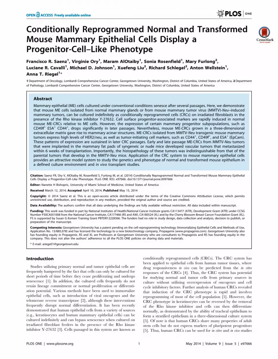
the cells senesce after several passages (Figure S4A). In contrast,
ME-CRCs generated from MMTV-Neu tumors were serially
passaged in the CRC system for .50 passages and did not senesce
(Figure 4A). Unlike the relatively stable morphology of normal
mouse ME-CRCs, the morphology of the MMTV-Neu ME-CRCs
changed with serial passage, with an increase in multilayer colony-
like structures at later passages (Figure 4B and C). We found also
that there were chromosomal gains (chromosomes 2 and 12) and
losses (chromosome 4) in these cells after extended passage (P38–
73) but no changes are observed in early non-CRC (P2) and CRC
(P6) passages, as determined by array CGH profiling (Figure S5A–
D). To further investigate the characteristics of the MMTV-Neu
MEC cells at early and late CRC passage, we determined the
proportions of progenitor and differentiated cells by FACS analysis
of cell surface markers known to be expressed by tumor-initiating
cells in mouse models of mammary cancer [24] or by more
differentiated tumor cells [17,25]. We used monoparametric and
multiparametric FACS approaches that were used for analysis of
normal ME-CRCs (Figure S4B and C). An initial increase in the
number of cells expressing Sca1, CD24, ESA, and CD44
(Figure 5A), as well as increased expression of each of these
proteins per cell (Figure S4D and E) compared to non-CRC
MMTV-Neu MECs freshly isolated from tumors was observed.
Elevated levels of cells expressing these surface markers were
sustained in late-passage MMTV-Neu ME-CRCs, with the notable
exception of Sca1, which decreased by greater than 50% (Fig. 5B).
Multiparametric FACS analysis indicated that early passage
MMTV-Neu ME-CRCs showed no change in the ESA/CD44/
CD49f subpopulation (Figure S6A–C). However, a significant
change was the decrease in the CD49f+/Sca1+/CD24+ subpop-
ulation which constituted ,17% of the late passage MMTV-Neu
ME-CRCs (Figures 5C and S6G–H). This combination of cell
surface markers has been described as defining a HER2+ tumor-
initiating population in some studies [26–29]. Of note, also, is that
a large portion of the Sca1+ MMTV-Neu CRCs were also positive
for CD24 and CD29 (Figure S6D–F) and, despite the overall
decrease in Sca1+ cells, the CD24+/CD29+/Sca1+ subpopulation
still constitutes only approximately 9% of the cell population at
later passages (Figure S6D–F). Western analysis of lysates of late-
passage MMTV-Neu ME-CRCs showed that HER2/neu contin-
ued to be expressed (Figure 5D and E). Interestingly, despite the
morphological changes in the MMTV-Neu ME-CRCs, there were
no major changes in expression of EMT markers, such as E-
cadherin and b-catenin, which continued to be expressed at
similar levels at early and late passage. Similar to the normal ME-
Figure 4. Properties of MMTV-Neu ME-CRCs in culture. A) Culture of tumor epithelial cells from a spontaneous MMTV-Neu mouse mammarygland tumor in the CRC system. B) Phase contrast images of MMTV-Neu ME-CRCs and non-CRCs at indicated passages on day 5 of culture (106; scale,100 mm). C) Quantification of colony subtypes of early (P2) vs. late (P23) passage MMTV-Neu ME-CRCs. Values are mean6SEM of three independentfields. *P,0.05.doi:10.1371/journal.pone.0097666.g004
Reprogramming Progenitor-Like Cells
PLOS ONE | www.plosone.org 6 May 2014 | Volume 9 | Issue 5 | e97666

CRCs, there was no detectable expression of pluripotent
progenitor markers at any of the passages examined (data not
shown).
Reprogramming of MMTV-Neu ME-crcs is PartiallyReversed by Removal of Y-27632
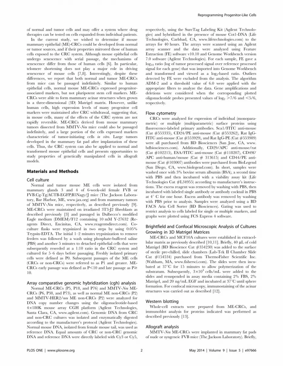
We also determined if removal of the inhibitor Y-27632 from
the MMTV-Neu ME-CRC cultures caused changes in the
populations of cells at early or late passages. The proliferation of
early-passage ME-CRCs was reduced by Y-27632 withdrawal, but
the overall morphology of the cells did not change significantly
(Figure 6A). FACS analysis of cell surface markers revealed that Y-
27632 withdrawal from early- and late-passage MMTV-Neu ME-
CRCs reduced the number of cells expressing Sca1, while
significantly increasing the number of ESA+ and CD49f+ cells in
early passage and the CD44+ and CD24+ cells in late passage
(Figure 6B and C). Western analysis of cell lysates revealed that
MMTV-Neu CRCs cultured in the presence or absence of Y-
27632 maintained expression of HER2/neu, E-cadherin, and b-
catenin (Figure 6D). Overall, these data suggest that there is some
permanent reprogramming of the MMTV-Neu ME-CRCs with
continued passage in the CRC system, although other changes in
gene expression that are dependent on Y-27632 are reversible.
MMTV-Neu ME-crcs form Mammary Tumors in vivoTo determine their tumorigenic potential, we injected early-
and late-passage MMTV-Neu ME-CRCs into the mammary fat
pads of syngeneic mice. After approximately 6 weeks, large tumors
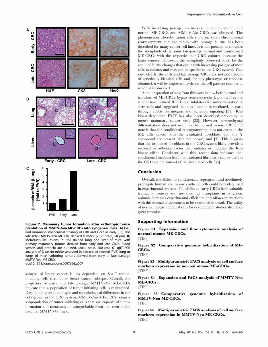
had developed and the mice were sacrificed. The histopathology of
the early and late passage MMTV-Neu ME-CRC allograft tumors
was indistinguishable from previous descriptions of tumors that
occur in the MMTV-Neu parental mice at approximately 7–9
months of age [14] (Figure 7A). These are highly vascular, largely
cribriform adenocarcinomas, with necrotic areas and increased
mitotic figures that stain uniformly for high expression of the
oncogene Her2/neu and CK8 (Figure 7A). The transgenic MMTV-
Neu mice also developed metastases at a later stage to the lung and
liver [15]. We found that a similar metastatic pattern, with
extensive lymphocyte infiltration, occurred in both lung and liver
of the mice harboring the transplanted MMTV-Neu ME-CRCs
(Figure 7B). The presence of ME cells in these metastatic lesions
was confirmed by detection of mammary-specific b-casein mRNA
Figure 5. Cell surface marker expression in MMTV-Neu ME-CRCs. A) Monoparametric FACS analysis of expression of indicated markers infreshly isolated MMTV-Neu ME non-CRCs and early passage MMTV-Neu ME-CRCs and B) early and late passage MMTV-Neu ME-CRCs. C) Three-markerprofile identified by multiparametric FACS analysis in early and late passage cells. D) & E) Representative western blots showing expression of theindicated markers in non-CRCs (2) and CRCs (+) at the indicated passages. Values are mean6SEM of three independent experiments; ***P,0.001,**P,0.01, *P,0.05.doi:10.1371/journal.pone.0097666.g005
Reprogramming Progenitor-Like Cells
PLOS ONE | www.plosone.org 7 May 2014 | Volume 9 | Issue 5 | e97666
by qRT-PCR (Figure 7C). Similar results were obtained with
MMTV-Neu ME-CRCs implanted in nude mice (data not shown).
Discussion
Here, we show that the CRC system [3] can be used for
indefinite passage of normal and transformed mouse ME cells.
There are similarities and notable differences between human and
mouse CRCs. Both human and mouse epithelial CRCs seem to
assume a progenitor-like phenotype, at least as assessed by changes
in cell surface markers, such as the integrin family members CD29
and CD49f, and the cell adhesion molecules CD24 and CD44.
Neither the human nor the mouse CRCs revert to pluripotent
progenitor cells, nor do they undergo mesenchymal transition.
Normal human conditionally reprogrammed keratinocytes main-
tain tissue-specific differentiation potential and have been shown
to form a stratified epithelial layer in a 3D culture system [5].
Likewise, the normal ME-CRCs can assume a mammary acinar-
like phenotype when grown in a 3D matrix of Matrigel. MMTV-
Neu ME-CRCs are able to form tumors when transplanted into
mice, and these tumors are histopathologically indistinguishable
from the tumors, and the metastases, that evolve in the MMTV-
Neu transgenic animals. Thus, there is significant overlap in the
properties of mouse and human CRCs.
A notable difference between the mouse and human CRCs is
the response to withdrawal of the Rho kinase inhibitor Y-27632. It
has been shown that removal of this compound from human
keratinocyte CRC cultures causes complete reversion to the non-
CRC phenotype [5]. In contrast, withdrawal of Y-27632 did not
alter the expression of many of the genes associated with mouse
mammary progenitor cells. We conjecture that the reason for this
may lie in fundamental differences in the regulation of the
reprogramming phenomenon in mouse and human CRCs in vitro.
Despite this, the lack of phenotypic reversion in the mouse CRCs
does not appear to impede the cells from driving the correct
differentiated phenotype in vivo. This suggests that the ME-CRCs,
when exposed to components of the normal stromal environment,
are capable of undergoing differentiation.
Differences in stem-like markers were observed, however, with
increasing passage number in both normal and transformed ME-
CRCs. In the normal ME- CRCs, frequency of expression of the
cell surface marker ESA reverts to that of non-CRC cells as the
cells are serially passaged; in particular, ESA+, CD44+ CD49f+
subpopulation is significantly decreased with progressive passage.
Although the reduction in ESA+ cells does not impede formation
of mammary acinar structures in Matrigel, it might impede other
aspects of normal mammary outgrowth development in vivo. With
serial passage of MMTV-Neu ME-CRCs, we observed loss of Sca1
expression by the cells. Loss of Sca1+ cells does not appear to
impede tumor formation in vivo, because tumors formed efficiently
with both late and early passage MMTV-Neu ME-CRCs. Sca1 has
been described as an important marker of tumor-initiating cells in
luminal breast cancer [30]. It may be that the HER2/neu-driven
Figure 6. Removal of Y-27632 from MMTV-Neu ME-CRCs alters expression of some cell surface markers. Early and late passage MMTV-Neu ME-CRCs were seeded on irradiated feeders with (+Y) or were cultured under CRC conditions (+Y), followed by the removal of Y-27632 andculture for two more passages (-Y). A) Phase contrast images of cells at indicated passages. A) Phase contrast images (106; scale, 100 mm). B and C)Monoparametric FACS analysis of expression of the indicated markers in early (B) and late (C) passage cells. Values are mean6SEM of threeindependent experiments; ***P,0.001, **P,0.01, *P,0.05. D) Representative western blots of indicated markers expressed in P62 ME-CRCs underconditions of (+Y) and (-Y).doi:10.1371/journal.pone.0097666.g006
Reprogramming Progenitor-Like Cells
PLOS ONE | www.plosone.org 8 May 2014 | Volume 9 | Issue 5 | e97666
subtype of breast cancer is less dependent on Sca1+ tumor-
initiating cells than other breast cancer subtypes. Overall, the
properties of early and late passage MMTV-Neu ME-CRCs
indicate that a population of tumor-initiating cells is maintained.
Despite the gross phenotypic and morphological differences in the
cells grown in the CRC system, MMTV-Neu ME-CRCs retain a
subpopulation of tumor-initiating cells that are capable of tumor
formation and metastasis indistinguishable from that seen in the
parental MMTV-Neu mice.
With increasing passage, an increase in aneuploidy in both
normal ME-CRCs and MMTV-Neu CRCs was observed. The
phenomenon whereby tumor cells show increased chromosomal
rearrangement and aneuploidy with passage in vitro has been
described for many cancer cell lines. It is not possible to compare
the aneuploidy of the same late-passage normal and transformed
ME-CRCs with the respective non-CRC cultures, because the
latter senesce. However, the aneuploidy observed could be the
result of in vitro changes that occur with increasing passage of most
cells in culture, and may not be specific to the CRC system. That
said, clearly, the early and late passage CRCs are not populations
of genetically identical cells and, for any phenotype or response
obtained, it will be important to define the cell passage number at
which it is observed.
A major question arising from this work is how both normal and
transformed ME-CRCs bypass senescence check points. Previous
studies have utilized Rho kinase inhibitors for immortalization of
stem cells and suggested that this function is mediated, in part,
through effects on integrin and adhesion signaling [31]. Rho
kinase-dependent EMT has also been described previously in
mouse mammary cancer cells [32]. However, mesenchymal
differentiation does not occur in the normal mouse CRCs. Of
note is that the conditional reprogramming does not occur in the
ME cells unless both the irradiated fibroblasts and the Y
compound are present (data not shown) and [5]. This suggests
that the irradiated fibroblasts in the CRC system likely provide a
secreted or adhesion factor that initiates or modifies the Rho
kinase effect. Consistent with this, recent data indicates that
conditioned medium from the irradiated fibroblasts can be used in
the CRC system instead of the irradiated cells [33].
Conclusion
Overall, the ability to conditionally reprogram and indefinitely
propagate human and mouse epithelial cells could be widely used
in experimental systems. The ability to carry CRCs from valuable
transgenic sources and use them as transplants in syngeneic
animals increases experimental efficiency and allows interactions
with the stromal environment to be examined in detail. The utility
of normal mouse epithelial cells for development studies also holds
great promise.
Supporting Information
Figure S1 Expansion and flow cytometric analysis ofnormal mouse ME-CRCs.
(TIFF)
Figure S2 Comparative genomic hybridization of ME-CRCs.
(TIFF)
Figure S3 Multiparametric FACS analysis of cell surfacemarkers expression in normal mouse ME-CRCs.
(TIFF)
Figure S4 Expansion and FACS analysis of MMTV-NeuME-CRCs.
(TIFF)
Figure S5 Comparative genomic hybridization ofMMTV-Neu ME-CRCs.
(TIFF)
Figure S6 Multiparametric FACS analysis of cell surfacemarkers expression in MMTV-Neu ME-CRCs.
(TIFF)
Figure 7. Mammary tumor formation after orthotopic trans-plantation of MMTV-Neu ME-CRCs into syngeneic mice. A) H&Eand Immunohistochemical staining of CK8 and Her2 in early (P6) andlate (P68) MMTV-Neu ME-CRC-derived tumors. (206; scale, 50 mm). B)Metastasis-like lesions in H&E-stained lung and liver of mice withprimary mammary tumors derived from early and late CRCs. Bloodvessels and bronchi are outlined. (206; scale, 200 mm). C) qRT PCRanalysis of b-casein mRNA assessed in extracts of normal (FVB) lung orlungs of mice harboring tumors derived from early or late passageMMTV-Neu ME-CRCs.doi:10.1371/journal.pone.0097666.g007
Reprogramming Progenitor-Like Cells
PLOS ONE | www.plosone.org 9 May 2014 | Volume 9 | Issue 5 | e97666
Acknowledgments
The content is solely the responsibility of the authors and does not
necessarily represent the official views of the National Institutes of Health.
Author Contributions
Conceived and designed the experiments: AR XL RS AW. Performed the
experiments: FS VO LC. Analyzed the data: MA SR MF LC MJ AR AW
FS. Contributed reagents/materials/analysis tools: MJ XL RS. Contrib-
uted to the writing of the manuscript: FS VO MA SR MF LC MJ AR XL
RS AW.
References
1. Kuilman T, Michaloglou C, Mooi WJ, Peeper DS (2010) The essence ofsenescence. Genes Dev 24: 2463–2479.
2. Roig AI, Eskiocak U, Hight SK, Kim SB, Delgado O, et al. (2010) Immortalized
epithelial cells derived from human colon biopsies express stem cell markers anddifferentiate in vitro. Gastroenterology 138: 1012–1021.e1011–1015.
3. Liu X, Ory V, Chapman S, Yuan H, Albanese C, et al. (2011) ROCK inhibitorand feeder cells induce the conditional reprogramming of epithelial cells.
Am J Pathol 180: 599–607.4. Yuan H, Myers S, Wang J, Zhou D, Woo JA, et al. (2012) Use of reprogrammed
cells to identify therapy for respiratory papillomatosis. N Engl J Med 367: 1220–
1227.5. Suprynowicz FA, Upadhyay G, Krawczyk E, Kramer SC, Hebert JD, et al.
(2012) Conditionally reprogrammed cells represent a stem-like state of adultepithelial cells. Proceedings of the National Academy of Sciences of the United
States of America 109: 20035–20040.
6. Itahana K, Campisi J, Dimri GP (2004) Mechanisms of cellular senescence inhuman and mouse cells. Biogerontology 5: 1–10.
7. Sherr CJ, DePinho RA (2000) Cellular senescence: mitotic clock or cultureshock? Cell 102: 407–410.
8. Wright WE, Shay JW (2000) Telomere dynamics in cancer progression andprevention: fundamental differences in human and mouse telomere biology. Nat
Med 6: 849–851.
9. Rosenfield SM, Bowden ET, Cohen-Missner S, Gibby KA, Ory V, et al. (2012)Pleiotrophin (PTN) expression and function and in the mouse mammary gland
and mammary epithelial cells. PLoS One 7: e47876.10. Debnath J, Brugge JS (2005) Modelling glandular epithelial cancers in three-
dimensional cultures. Nat Rev Cancer 5: 675–688.
11. Lee GY, Kenny PA, Lee EH, Bissell MJ (2007) Three-dimensional culturemodels of normal and malignant breast epithelial cells. Nat Methods 4: 359–365.
12. Ory V, Tassi E, Cavalli LR, Sharif GM, Saenz F, et al. (2013) The nuclearcoactivator amplified in breast cancer 1 maintains tumor-initiating cells during
development of ductal carcinoma in situ. Oncogene.
13. Oh A, List H-J, Reiter R, Mani A, Zhang Y, et al. (2004) The nuclear receptorcoactivator AIB1 mediates insulin-like growth factor I-induced phenotypic
changes in human breast cancer cells. Cancer Res 64: 8299–8308.14. Fereshteh MP, Tilli MT, Kim SE, Xu J, O’Malley BW, et al. (2008) The nuclear
receptor coactivator amplified in breast cancer-1 is required for Neu (ErbB2/HER2) activation, signaling, and mammary tumorigenesis in mice. Cancer Res
68: 3697–3706.
15. Guy CT, Webster MA, Schaller M, Parsons TJ, Cardiff RD, et al. (1992)Expression of the neu protooncogene in the mammary epithelium of transgenic
mice induces metastatic disease. Proc Natl Acad Sci U S A 89: 10578–10582.16. Chapman S, Liu X, Meyers C, Schlegel R, McBride AA (2010) Human
keratinocytes are efficiently immortalized by a Rho kinase inhibitor. J Clin Invest
120: 2619–2626.17. Li Y, Rosen JM (2005) Stem/progenitor cells in mouse mammary gland
development and breast cancer. J Mammary Gland Biol Neoplasia 10: 17–24.
18. Visvader JE (2009) Keeping abreast of the mammary epithelial hierarchy and
breast tumorigenesis. Genes Dev 23: 2563–2577.
19. Stingl J, Eirew P, Ricketson I, Shackleton M, Vaillant F, et al. (2006) Purification
and unique properties of mammary epithelial stem cells. Nature: 5.
20. Shackleton M, Vaillant F, Simpson KJ, Stingl J, Smyth GK, et al. (2006)
Generation of a functional mammary gland from a single stem cell. Nature 439:
84–88.
21. Mani SA, Guo W, Liao M-J, Eaton EN, Ayyanan A, et al. (2008) The epithelial-
mesenchymal transition generates cells with properties of stem cells. Cell 133:
704–715.
22. Morel A-P, Lievre M, Thomas C, Hinkal G, Ansieau S, et al. (2008) Generation
of breast cancer stem cells through epithelial-mesenchymal transition. PLoS One
3: e2888.
23. Lee JM, Dedhar S, Kalluri R, Thompson EW (2006) The epithelial-
mesenchymal transition: new insights in signaling, development, and disease.
The Journal of Cell Biology 172: 973–981.
24. Owens TW, Naylor MJ (2013) Breast cancer stem cells. Front Physiol 4: 225.
25. Lindeman GJ, Visvader JE (2010) Insights into the cell of origin in breast cancer
and breast cancer stem cells. Asia Pac J Clin Oncol 6: 89–97.
26. Matulka LA, Triplett AA, Wagner K-U (2007) Parity-induced mammary
epithelial cells are multipotent and express cell surface markers associated with
stem cells. Developmental Biology 303: 29–44.
27. Lo P-K, Kanojia D, Liu X, Singh UP, Berger FG, et al. (2012) CD49f and CD61
identify Her2/neu-induced mammary tumor-initiating cells that are potentially
derived from luminal progenitors and maintained by the integrin-TGFbsignaling. Oncogene 31: 2614–2626.
28. Jeselsohn R, Brown NE, Arendt L, Klebba I, Hu MG, et al. (2010) Cyclin D1
kinase activity is required for the self-renewal of mammary stem and progenitor
cells that are targets of MMTV-ErbB2 tumorigenesis. Cancer Cell 17: 65–76.
29. Liu JC, Deng T, Lehal RS, Kim J, Zacksenhaus E (2007) Identification of
tumorsphere- and tumor-initiating cells in HER2/Neu-induced mammary
tumors. Cancer Res 67: 8671–8681.
30. Batts TD, Machado HL, Zhang Y, Creighton CJ, Li Y, et al. (2011) Stem cell
antigen-1 (sca-1) regulates mammary tumor development and cell migration.
PLoS One 6: e27841.
31. Pakzad M, Totonchi M, Taei A, Seifinejad A, Hassani SN, et al. (2010) Presence
of a ROCK inhibitor in extracellular matrix supports more undifferentiated
growth of feeder-free human embryonic and induced pluripotent stem cells upon
passaging. Stem Cell Rev 6: 96–107.
32. Castro DJ, Maurer J, Hebbard L, Oshima RG (2013) ROCK1 inhibition
promotes the self-renewal of a novel mouse mammary cancer stem cell. Stem
Cells 31: 12–22.
33. Palechor-Ceron N, Suprynowicz FA, Upadhyay G, Dakic A, Minas T, et al.
(2013) Radiation induces diffusible feeder cell factor(s) that cooperate with
ROCK inhibitor to conditionally reprogram and immortalize epithelial cells.
Am J Pathol 183: 1862–1870.
Reprogramming Progenitor-Like Cells
PLOS ONE | www.plosone.org 10 May 2014 | Volume 9 | Issue 5 | e97666