targeting of αv integrin identifies a core molecular pathway that regulates fibrosis in several...
TRANSCRIPT
a r t i c l e s
nature medicine VOLUME 19 | NUMBER 12 | DECEMBER 2013 1617
Chronic tissue injury with fibrosis, loss of proper tissue architecture and organ dysfunction are characteristic features of many human diseases and major causes of morbidity and mortality worldwide. Currently, treatment of tissue fibrosis is severely limited, and trans-plantation is often the only effective option for end-stage fibrotic diseases. However, limited donor organ availability and the high cost and morbidity of transplantation underscore the urgent need for more effective therapies.
Secreted transforming growth factor-β (TGF-β) is arguably the major profibrogenic cytokine and a central mediator of fibrosis in multiple organs. The molecular pathways that regulate TGF-β activity and signaling are therefore attractive targets for new antifi-brotic treatments. TGF-β is secreted as a latent complex that is present at high concentrations and directly cross-linked to the extracellular matrix, and much of the regulation of TGF-β function in tissues is based on extracellular activation of this latent complex1,2. Two of the three mammalian TGF-β isoforms (TGF-β1 and TGF-β3) can be activated by members of the integrin family that interact with a linear arginine-glycine–aspartic acid (RGD) motif present in an N-terminal
fragment of the TGF-β gene product called the latency-associated peptide3–5. Inhibition and blockade of two of these integrins (αvβ6 and αvβ8) result in a phenotype similar to all of the developmen-tal effects of loss of TGF-β1 and TGF-β3 (ref. 6), suggesting that these two integrins are required for most or all important roles of these TGF-β isoforms in development. However, the mechanisms of TGF-β activation that contribute to tissue pathology in adults are less well understood.
We and others have previously shown that TGF-β activation by the αvβ6 integrin has an important role in models of fibrosis in the lungs, biliary tract and kidney3,7–9. However αvβ6 does not appear to be critical in all models of organ fibrogenesis, as ablation of αvβ6 was not protective in carbon tetrachloride (CCl4)-induced liver fibrosis7. Crucially, αvβ6 is largely restricted in its expression to a subset of epithelial cells7,10,11 and generates very localized activation of TGF-β that can only be detected by cells in direct contact with the integrin-expressing cell3. Moreover, tissue fibrosis in many organs is due to collagen production by myofibroblasts that are often at a substantial distance from any αvβ6-expressing epithelial cells with implications
1Lung Biology Center, Department of Medicine, University of California, San Francisco, San Francisco, California, USA. 2Medical Research Council Centre for Inflammation Research, The Queen’s Medical Research Institute, University of Edinburgh, Edinburgh, UK. 3Department of Pediatrics, University of California, San Francisco, San Francisco, California, USA. 4Department of Cancer Biology, University of Texas M.D. Anderson Cancer Center, Houston, Texas, USA. 5Department of Immunology, Genetics and Pathology, Uppsala University, Uppsala, Sweden. 6Department of Medical Biochemistry and Biophysics, Karolinska Institutet, Stockholm, Sweden. 7Center for World Health and Medicine, Saint Louis University, Edward A. Doisy Research Center, St. Louis, Missouri, USA. 8The Liver Center, Department of Medicine, University of California, San Francisco, San Francisco, California, USA. 9Program of Developmental Immunology, Department of Pediatrics, Massachusetts General Hospital and Harvard Medical School, Boston, Massachusetts, USA. 10Department of Tissue Morphogenesis, Faculty of Medicine, Max Planck Institute for Molecular Biomedicine, University of Münster, Münster, Germany. Correspondence should be addressed to N.H. ([email protected]) or D.S. ([email protected]).
Received 3 April 2012; accepted 17 June 2013; published online 10 November 2013; doi:10.1038/nm.3282
Targeting of αv integrin identifies a core molecular pathway that regulates fibrosis in several organsNeil C Henderson1,2, Thomas D Arnold3, Yoshio Katamura1, Marilyn M Giacomini1, Juan D Rodriguez1, Joseph H McCarty4, Antonella Pellicoro2, Elisabeth Raschperger5,6, Christer Betsholtz5,6, Peter G Ruminski7, David W Griggs7, Michael J Prinsen7, Jacquelyn J Maher8, John P Iredale2, Adam Lacy-Hulbert9, Ralf H Adams10 & Dean Sheppard1
Myofibroblasts are the major source of extracellular matrix components that accumulate during tissue fibrosis, and hepatic stellate cells (HSCs) are believed to be the major source of myofibroblasts in the liver. To date, robust systems to genetically manipulate these cells have not been developed. We report that Cre under control of the promoter of Pdgfrb (Pdgfrb-Cre) inactivates loxP-flanked genes in mouse HSCs with high efficiency. We used this system to delete the gene encoding av integrin subunit because various av-containing integrins have been suggested as central mediators of fibrosis in multiple organs. Such depletion protected mice from carbon tetrachloride–induced hepatic fibrosis, whereas global loss of b3, b5 or b6 integrins or conditional loss of b8 integrins in HSCs did not. We also found that Pdgfrb-Cre effectively targeted myofibroblasts in multiple organs, and depletion of the av integrin subunit using this system was protective in other models of organ fibrosis, including pulmonary and renal fibrosis. Pharmacological blockade of av-containing integrins by a small molecule (CWHM 12) attenuated both liver and lung fibrosis, including in a therapeutic manner. These data identify a core pathway that regulates fibrosis and suggest that pharmacological targeting of all av integrins may have clinical utility in the treatment of patients with a broad range of fibrotic diseases.
npg
© 2
013
Nat
ure
Am
eric
a, In
c. A
ll rig
hts
rese
rved
.
a r t i c l e s
1618 VOLUME 19 | NUMBER 12 | DECEMBER 2013 nature medicine
for therapeutic design. Myofibroblasts express several αv-containing integrins and are contractile cells capable of exerting force on teth-ered ligands. The recently solved crystal structure of the small latent complex of TGF-β demonstrates that mechanical force generated by the contractile actomyosin cytoskeleton and transmitted by integrins is a common mechanism for activating latent TGF-β12. Furthermore, in vitro studies of myofibroblasts have shown that they can use alterna-tive αv-containing integrins to activate TGF-β13, and we and others have shown that several integrins that share the αv subunit, including αvβ1, αvβ3, αvβ5, αvβ6 and αvβ8, can recognize the same RGD peptide motif and, at least in some circumstances, can activate latent TGF-β3,4,14–16. Together, these data suggest that myofibroblast αv expression may be critical to fibrosis and may be targetable. However, the precise importance in vivo of each of these integrins is undetermined.
The paucity of tools that allow reliable specific inactivation of genes in myofibroblasts in vivo has greatly hindered progress in under-standing the underlying biology of fibrotic diseases. In this study, we developed a strategy to inactivate genes in myofibroblasts in multiple organs. Platelet-derived growth factor receptor-β (PDGFR-β) induc-tion is an early feature of myofibroblast activation in a broad range of tissues17–24. We therefore evaluated the efficiency of targeting these cells using Pdgfrb-Cre mice25 and used this system to investigate the role of αv-containing integrins on myofibroblasts in regulating patho-logic fibrosis in multiple solid organs.
In the liver, HSCs (liver-specific pericytes) are believed to give rise to myofibroblasts upon their activation and thus are the major source of the additional extracellular matrix during hepatic fibrogenesis26,27. To date, no effective strategy has been developed to reliably delete genes specifically in HSCs. As PDGFR-β induction is an early feature of activation of HSCs to myofibroblasts17–21, we evaluated the effi-ciency of targeting HSCs using Pdgfrb-Cre mice. These mice, which express Cre recombinase under the control of a fragment of the gene encoding PDGFR-β, were previously developed to specifically target pericytes25. In the uninjured liver, HSCs are found in a perivascular location in close contact with underlying sinusoidal endothelial cells, and they morphologically resemble pericytes in other organs.
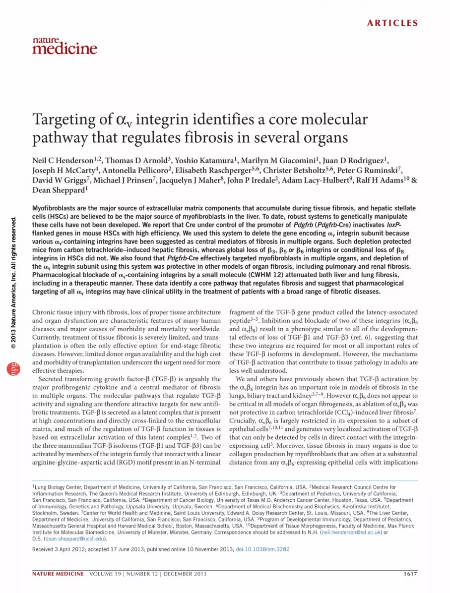
RESULTSPdgfrb-Cre effectively targets recombination in HSCsUsing mTmG reporter mice (double-fluorescent reporter mice that express membrane-targeted tandem dimer tomato (tdTomato) before Cre-mediated excision and membrane-targeted GFP (mGFP) after
excision)28 and Ai14 reporter mice (single-fluorescent reporter mice that express tdTomato after recombination)29, we found that Pdgfrb-Cre induced highly efficient recombination in a distribution appro-priate for HSCs both in uninjured control liver and after induction of hepatic fibrosis with repeated administration of CCl4 (Fig. 1a,b). To evaluate the specificity of recombination in HSCs, we stained uninjured control livers from Ai14; Pdgfrb-Cre mice for desmin (a well-characterized marker of HSCs), which was expressed in virtu-ally all of the reporter-expressing cells (Fig. 1c and Supplementary Fig. 1a). Staining for PDGFR-β confirmed both appropriate reporter expression by Pdgfrb-Cre and also a hitherto unknown expres-sion of PDGFR-β by quiescent HSCs in uninjured liver (Fig. 1c and Supplementary Fig. 1a). As expected, α-smooth muscle actin (α-SMA, a well-characterized marker of myofibroblasts) was seen only in larger blood vessel walls in uninjured liver (Fig. 1c). In fibrotic liver, almost all of the reporting cells also expressed desmin and PDGFR-β both in fibrous septae and in the surrounding liver paren-chyma (Fig. 1d and Supplementary Fig. 1a). Furthermore, virtually all of the α-SMA–positive cells observed in fibrotic livers were also reporter positive (Fig. 1d and Supplementary Fig. 1a), demonstrat-ing a high degree of recombination in activated liver myofibroblasts. To further assess specificity of Pdgfrb-Cre recombination, we stained uninjured control and fibrotic livers from Ai14; Pdgfrb-Cre mice with antibodies to CD31 (endothelial cells), F4/80 (Kupffer cells), CD3 (T cells) and pancytokeratin (biliary epithelium). No reporter-expressing cells expressed any of these markers (Supplementary Fig. 1b). As PDGFR-β expression by quiescent HSCs has not previously been reported, we used an alternative strategy to further assess PDGFR-β
f α-SMA + DAPIReporter + DAPI Merged
Des
Timp1
Mmp13
Mmp9
Mmp2
Col3a1
Col1a1
Acta2
Gfap
Pdgfrb
Tgfb1
Pparg
Itgav
Itgb3Itgb5
Itgb1
Itgb8
*OilCCl4
**
**** ** ****
*
* *** * * **
Fol
d ch
ange
0
20
15
10
5
***
e
Oil CCl4 Oil CCl4a
Reporter Desmin Rep + desminc d Reporter Desmin Rep + desmin
ReporterReporter α-SMAα-SMA Rep + α-SMARep + α-SMA
Reporter Rep + PDGFR-βPDGFR-βReporter Rep + PDGFR-βPDGFR-β
b
CC
l 4
Oil
Ai1
4; Pdgfrb
-Cre
TdT
om (
repo
rt)
mT
mG
; Pdgfrb
-Cre
GF
P (
repo
rt)
Figure 1 Pdgfrb-Cre effectively targets recombination in quiescent and activated HSCs. (a,b) Immunofluoresence micrographs of liver sections harvested from control (olive oil–treated) or chronic CCl4–treated (two injections per week for 6 weeks) mTmG; Pdgfrb-Cre reporter mice (a) or Ai14; Pdgfrb-Cre reporter mice (b) (n = 4 male mice per group). Scale bars, 100 µm. (c,d) Immunofluoresence micrographs of liver sections from control (olive oil–treated) (c) or CCl4 treated (d) Ai14; Pdgfrb-Cre mice (n = 4 male mice per group) stained for desmin, PDGFR-β or α-SMA (green) with endogenous (not enhanced) Ai14-tdTomato reporter in red. Scale bars, 25 µm (c) and 50 µm (d). Arrows indicate portal tracts in serial sections. Rep, reporter. (e) Gene expression profile of freshly sorted tdTomato-positive cells from control (olive oil–treated) or chronic CCl4–treated Ai14; Pdgfrb-Cre mice (n = 4 male mice per group). (f) Immunofluoresence staining of tdTomato-positive cells sorted from the uninjured livers of Ai14; Pdgfrb-Cre mice and plated on tissue culture plastic for 7 d. Left, Ai14 reporter (red) and DAPI (blue). Middle, α-SMA (green) and DAPI (blue). Right, merged image. Scale bar, 100 µm. Data are expressed as mean ± s.e.m. *P < 0.05, **P < 0.01, ***P < 0.001 (Student’s t-test).
npg
© 2
013
Nat
ure
Am
eric
a, In
c. A
ll rig
hts
rese
rved
.

a r t i c l e s
nature medicine VOLUME 19 | NUMBER 12 | DECEMBER 2013 1619
expression in such cells. Analysis of knock-in reporter mice that carry one copy of a bacterial artificial chromosome transgene expressing eGFP under the control of Pdgfrb regulatory elements (Pdgfrb-BAC-eGFP mice) demonstrated that eGFP-positive cells in the livers from these mice show an identical distribution to that of the tdTomato-positive cells in the livers of Ai14; Pdgfrb-Cre mice (Supplementary Fig. 1c). Furthermore, to evaluate the specificity of reporting in quiescent HSCs, we stained for desmin, which was expressed in virtually all of the reporter-expressing cells (Supplementary Fig. 1c,d). Finally, staining with PDGFR-β demonstrated both appropriate reporter expression by the Pdgfrb-BAC-eGFP system and confirmed expres-sion of PDGFR-β by quiescent HSCs (Supplementary Fig. 1c,d).
To further characterize reporter-expressing cells in Ai14; Pdgfrb-Cre mice, we isolated nonparenchymal cells from uninjured control or fibrotic livers and purified Ai14 (tdTomato)-positive cells by cell sort-ing. Quantitative PCR (qPCR) of mRNA obtained from live tdTomato-positive cells showed marked induction of multiple genes associated with the transition of quiescent HSCs to the activated myofibroblast phenotype, including Des (desmin), Pdgfrb, Acta2 (α-SMA), Col1a1 (collagen I), Col1a3 (collagen III), Mmp2 (matrix metallopeptidase 2), Mmp 9 (matrix metallopeptidase 9), Mmp13 (matrix metallopeptidase 13) and Timp1 (TIMP metallopeptidase inhibitor 1) (Fig. 1e), and higher expression of a number of these mRNAs was also confirmed at the protein level (Supplementary Fig. 1e–g). In addition, we found significantly higher expression of multiple β-subunit partners of αv following fibrosis induction (Fig. 1e). We also plated freshly isolated tdTomato-positive cells from uninjured Ai14; Pdgfrb-Cre mouse livers on tissue culture plastic for 5 d to induce HSC activation. All of the cells in these sorted cultures expressed the myofibroblast marker α-SMA (Fig. 1f). Taken together, these results suggest that Pdgfrb-Cre efficiently targets both quiescent and activated HSCs.
HSC av integrin depletion protects mice from hepatic fibrosisTo determine whether Pdgfrb-Cre could be used to identify molecular mechanisms driving hepatic fibrosis, we focused on the integrin αv subunit because of the suggested role of various αv-containing integrins in activating latent TGF-β, a central mediator of fibrosis3,4,13,15,16.
Confirming our qPCR data (Fig. 1e), we found that mouse HSCs express multiple αv-containing integrins (αvβ1, αvβ3, αvβ5 and αvβ8; Fig. 2a) and that αv integrin expression was significantly upregu-lated during activation ex vivo (Fig. 2b,c). We also found αv integrin expression on hepatic myofibroblasts in human fibrotic liver tissue (Supplementary Fig. 2a). Western blotting with an antibody raised against a portion of the αv extracellular domain revealed that αv integrin was efficiently deleted from Itgavflox/flox; Pdgfrb-Cre HSCs (Fig. 2d). Furthermore, Itgavflox/flox; Pdgfrb-Cre mice were sig-nificantly protected from CCl4-induced fibrosis, as determined by collagen staining, hydroxyproline content and α-SMA staining (Fig. 2e–h). This was not due to changes in the degree of initial injury caused by CCl4 (Supplementary Fig. 2b,c), the number of HSCs before injury (Supplementary Fig. 2d,e), differences in inflammatory infiltrates (Supplementary Fig. 2f–i) or changes in the vasculature (Supplementary Fig. 2j) of Itgavflox/flox; Pdgfrb-Cre livers com-pared to control (Itgavflox/flox lacking Pdgfrb-Cre) livers. In addition, αv integrin expression was not affected in purified hepatocytes, liver sinusoidal endothelial cells or Kupffer cells from Itgavflox/flox; Pdgfrb-Cre mice compared to control mice, further confirming the specific action of Pdgfrb-Cre in HSCs (Supplementary Fig. 3a). Further, HSCs cultured for 5 d from control and Itgavflox/flox; Pdgfrb-Cre mice demonstrated robust expression of PDGFR-β; however, as expected, hepatocytes, liver sinusoidal endothelial cells and Kupffer cells did not express PDGFR-β (Supplementary Fig. 3a). These data suggest that αv integrins on HSCs promote CCl4-induced liver fibrosis and that Pdgfrb-Cre effectively targets gene deletion in these cells during fibrogenesis.
HSC av integrins regulate fibrosis through TGF-b activationTo investigate whether loss of αv integrins affected activation-induced induction of extracellular matrix protein gene expression, control and αv-null (Itgavflox/flox; Pdgfrb-Cre) HSCs were activated in culture for 5 d. Acta2, Col1a1 and Col3a1expression was significantly reduced in Itgavflox/flox; Pdgfrb-Cre HSCs (Fig. 3a–c). Furthermore, treatment with an αv-blocking antibody (RMV-7) also inhibited expression of these profibrotic genes (Fig. 3d–f). HSCs activated in culture from
e Control αv Creb dαv
β-actin
0 5 10
αv
β-actin
Day PS
Rα-
SM
A
c
aβ3
130 kDa
100 kDa
70 kDa
β5
130 kDa
100 kDa
70 kDaβ8
130 kDa
100 kDa
70 kDa
130 kDa
100 kDa
70 kDa
130 kDa
100 kDa
70 kDa
130 kDa
100 kDa
70 kDa
130 kDa
100 kDa
70 kDa
β1
250 kDa
130 kDa
100 kDa
Lysa
te α vIg
G1
Lysa
te α vIg
G1
Lysa
te α vIg
G1
Lysa
te α vIg
G1
αv αvαv αv
0
6
4
2
0 5 10
α v (f
old
chan
ge)
*
**
Time (d)
0
6
4
2
8
**
α-S
MA
(%
are
a)
Oil CCl4
h
100
400
300
200
500
0
**
Hyd
roxy
prol
ine
(µg
per
g)Oil CCl4
gf
0Siri
us r
ed (
% a
rea)
***
6
4
2
Oil CCl4
Controlαv Cre
Contro
l
α v C
re
Figure 2 Depletion of the αv integrin on HSCs protects mice from CCl4-induced hepatic fibrosis. (a) Immunoprecipitation studies: sorted tdTomato-positive cells from uninjured livers of Ai14; Pdgfrb-Cre mice cultured for 7 d express αvβ1, αvβ3, αvβ5 and αvβ8. (b,c) Western blot (b) and qPCR (c) analyses of αv integrin expression during transition from the quiescent (day 0) to the culture-activated phenotype (days 5 and 10) in wild-type HSCs. (d) Western blot analysis of αv expression in control and Itgavflox/flox; Pdgfrb-Cre (αv Cre) HSCs culture-activated for 5 d. (e) Picrosirius red (PSR) staining (collagen deposition) (top; counterstain is fast green FCF) and α-SMA immunohistochemistry (bottom) of liver tissue after olive oil or chronic CCl4 treatment of control and Itgavflox/flox; Pdgfrb-Cre mice (n = 8 male mice per group). Scale bar, 200 µm. (f) Digital image analysis quantification of collagen staining. (g) Hydroxyproline analysis. (h) Digital image analysis quantification of α-SMA staining. Data are expressed as mean ± s.e.m. *P < 0.05, **P < 0.01, ***P < 0.001 (Student’s t-test).
npg
© 2
013
Nat
ure
Am
eric
a, In
c. A
ll rig
hts
rese
rved
.

a r t i c l e s
1620 VOLUME 19 | NUMBER 12 | DECEMBER 2013 nature medicine
control and Itgavflox/flox; Pdgfrb-Cre mice had similar Tgfb1 mRNA lev-els (Fig. 3g). However, culture of HSCs from control and Itgavflox/flox; Pdgfrb-Cre mice with mink lung epithelial reporter cells (TMLCs) expressing firefly luciferase under the control of the TGF-β–sensitive plasminogen activator inhibitor-1 promoter30 demonstrated a significant reduction in the levels of active TGF-β by αv-null HSCs compared to control HSCs. This difference was eliminated by TGF-β–blocking antibody and rescued with addition of acti-vated TGF-β1 (Fig. 3h), identifying a decrease in TGF-β activation by αv-null HSCs. Furthermore, we found no appreciable difference in levels of total TGF-β1 protein in cell lysates and supernatants from control and Itgavflox/flox; Pdgfrb-Cre HSCs cultured with and without TMLCs, confirming that the reduction in TGF-β activation by Itgavflox/flox; Pdgfrb-Cre HSCs is not secondary to a decrease in total TGF-β1 expression (Supplementary Fig. 3b,c). Moreover, addi-tion of TGF-β1 to control and Itgavflox/flox; Pdgfrb-Cre HSCs restored profibrotic gene expression in αv-null HSCs to TGF-β1–treated con-trol levels (Supplementary Fig. 3d,e). As multiple cell types can secrete TGF-β, we investigated the cellular sources of TGF-β dur-ing liver fibrosis by purifying HSCs, hepatocytes, liver sinusoidal endothelial cells and Kupffer cells from fibrotic livers of C57BL/6 mice and found that the major cellular sources of TGF-β were HSCs and Kupffer cells (Supplementary Fig. 4a). Previous studies have shown that TGF-β activation occurs in a highly spatially restricted manner3 but that integrins can activate TGF-β produced by any cells in the local vicinity.
We also examined whether adhesion and migration defects in αv-null HSCs might contribute to the protection from CCl4-induced fibrosis observed in Itgavflox/flox; Pdgfrb-Cre mice. However, there was no appreciable difference in adhesion to multiple matrix proteins present in normal or fibrotic liver and no difference in cell migration between control and Itgavflox/flox; Pdgfrb-Cre HSCs (Supplementary Fig. 4b,c). Furthermore the rate of apoptosis (assessed by TUNEL
staining) was similar in control and Itgavflox/flox; Pdgfrb-Cre HSCs (Supplementary Fig. 4d,e). We also assessed whether depletion of the αv integrin on HSCs might alter the expression of other β1 integrin subunit–containing integrins; however, expression of α1, α5 and β1 integrins was similar in control and Itgavflox/flox; Pdgfrb-Cre HSCs (Supplementary Fig. 5a–c).
As we had shown that Itgavflox/flox; Pdgfrb-Cre HSCs are defective in their ability to activate TGF-β in vitro, we extended our findings in vivo by examining canonical TGF-β signaling by immunostain-ing for phosphorylated SMAD3 (p-Smad3) (Fig. 3i). Digital image quantification demonstrated significantly reduced p-Smad3 signaling in the livers of Itgavflox/flox; Pdgfrb-Cre mice compared to controls fol-lowing chronic CCl4 administration (Fig. 3j). Taken together, these results strongly suggest that the protection from CCl4-induced hepatic fibrosis observed in Itgavflox/flox; Pdgfrb-Cre mice is at least in part a consequence of reduced TGF-β activation by αv-deficient HSCs.
Assessment of av integrin heterodimers in hepatic fibrosisThe αv integrin subunit has five possible β-subunit binding partners (β1, β3, β5, β6 and β8)31, each of which has been reported to bind and/or activate latent TGF-β3,4,14–16 and four of which we found were expressed on HSCs (αvβ1, αvβ3, αvβ5 and αvβ8, Fig. 2a).
To further assess the potential contribution of each αv integrin heterodimer during hepatic fibrogenesis, we evaluated the response to CCl4 in mice globally lacking β3, β5 and β6 integrins (previously been shown to be important in biliary tract fibrosis7) and in mice lacking β8 integrins on HSCs (Itgb8flox/flox; Pdgfrb-Cre). We used conditional deletion of Itgb8 from HSCs because global loss of Itgb8 is embryoni-cally lethal32. Individual depletion of any one of these β integrin sub-units failed to protect mice from CCl4-induced hepatic fibrosis (Fig. 4). These results suggest either that multiple αv-containing integrins con-tribute to hepatic fibrogenesis and TGF-β activation by HSCs or that the principal relevant integrin is αvβ1. It is not currently possible to
Col1a1
(fol
d ch
ange
) **b
0
0.5
1.0
1.5
Cont
α v C
reCon
t
α v C
re
Col3a1
(fol
d ch
ange
)
0
0.5
1.0
1.5**
c
Isotyp
e
Anti-α v
Isotyp
e
Anti-α v
Col1a1
(fol
d ch
ange
)
0
0.5
1.0
1.5e
**
Isotyp
e
Anti-α v
Isotyp
e
Anti-α v
Col3a1
(fol
d ch
ange
)
0
0.5
1.0
1.5f
*
Cont
α v C
re
g
0.5
0
1.5
1.0
2.0
Tgfb1
(fol
d ch
ange
)
NS
Luci
fera
se a
ctiv
ity(×
103 )
***
h
0
3
2
1
5
4
Control
Untre
ated
Anti–T
GF-β
TGF-β
αv Cre
Cont
α v C
re
j**
0
20
40
60
p-sm
ad3
(arb
itrar
y un
its)
a
Acta2
(fo
ld c
hang
e)
0
0.5
1.0
1.5 *
Cont
α v C
re
Cont
α v C
re
α-SMAβ-actin
0
0.5
1.0
1.5
Acta2
(fo
ld c
hang
e)
**
Isotyp
e
Anti-α v
Isotyp
e
Anti-α v
dControlαv Cre
Isot
ype
Ant
i-αv
Isot
ype
Ant
i-αv
α-SMAβ-actin
Cont
α v C
re
i PDGFR-β+ DAPI
p-smad3+ DAPI
PDGFR-β + p-smad3+ DAPI
Con
trol
α v C
re
Figure 3 αv integrin depletion on HSCs inhibits profibrotic gene expression through a reduction in TGF-β activation. (a) qPCR (left) and western blot analysis (right) of α-SMA expression in control (Cont) and Itgavflox/flox; Pdgfrb-Cre (αv Cre) HSCs that were activated in culture for 5 d. (b,c) qPCR analysis of Col1a1 (b) and Col3a1 (c) expression in control and Itgavflox/flox; Pdgfrb-Cre HSCs that were activated in culture for 5 d. (d) qPCR (left) and western blot analysis (right) of α-SMA expression in control and Itgavflox/flox; Pdgfrb-Cre HSCs treated with 20 µg ml−1 isotype control or αv integrin–specific (clone RMV-7) antibodies (Anti-αv) for 5 d after plating. (e,f) qPCR analysis of Col1a1 (e) and Col3a1 (f) expression in control and Itgavflox/flox; Pdgfrb-Cre HSCs treated with 20 µg ml−1 isotype control or αv integrin–specific antibodies for 5 d after plating. (g) qPCR analysis of Tgfb1 expression in control and Itgavflox/flox; Pdgfrb-Cre HSCs culture-activated for 5 d. NS, not significant. (h) TGF-β activation by control or Itgavflox/flox; Pdgfrb-Cre HSCs alone or in the presence of TGF-β–blocking antibody (clone 1D11, 40 µg ml−1) (Anti–TGF-β) or activated TGF-β (300 pg ml−1). (i) Immunofluoresence micrographs of liver sections from control and Itgavflox/flox; Pdgfrb-Cre mice following chronic CCl4 treatment (n = 8 male mice per group). Scale bars, 25 µm. (j) Digital image analysis quantification of p-Smad3 staining. Data are expressed as mean ± s.e.m. *P < 0.05, **P < 0.01, ***P < 0.001 (Student’s t-test).
npg
© 2
013
Nat
ure
Am
eric
a, In
c. A
ll rig
hts
rese
rved
.

a r t i c l e s
nature medicine VOLUME 19 | NUMBER 12 | DECEMBER 2013 1621
specifically evaluate the role of αvβ1 in liver fibrosis in vivo, as global loss of Itgb1 is lethal in mice after embryonic day 5.5 (refs. 33,34), and Itgb1flox/flox; Pdgfrb-Cre mice show early postnatal lethality35.
Selective av integrin depletion in lung and kidney fibrosisMyofibroblasts in the lung and kidney are also the major mediators of extracellular matrix deposition and organ scarring during tissue injury and have previously been shown to express high levels of PDGFR-β22–24,36. We therefore suspected that Pdgfrb-Cre might broadly mark myofibroblasts in multiple organs and allow manipula-tion of specific genes in these cells. To further evaluate this possibility,
we initially examined the lungs of mTmG; Pdgfrb-Cre reporter mice 28 d after intratracheal instillation of saline (control) or bleomycin. We found high levels of reporter expression in a distribution that closely resembled that of lung pericytes in control saline-treated lungs (Fig. 5a). We also saw a marked expansion of reporter cells in fibrotic regions following induction of pulmonary fibrosis with bleomycin (Fig. 5a). To confirm that reporter expression was occurring specifi-cally in lung pericytes in control saline-treated lung, we stained for desmin and PDGFR-β (two well-characterized markers of pericytes37) and found a high degree of colocalization of the reporter with each (Fig. 5b and Supplementary Fig. 5d). Following bleomycin treatment,
i β8
β 8 C
re
β-actin
Contro
l
a
Oil CCl4
Siri
us r
ed (
% a
rea)
0
6
4
2
8
Controlβ3 KO
0
6
4
2
8
b
Oil CCl4
Siri
us r
ed (
% a
rea)
Controlβ5 KO
e
100
400
300
200
0Hyd
roxy
prol
ine
(µg
per
g)
Oil CCl4
Controlβ3 KO
100
400300200
500
0
f
Hyd
roxy
prol
ine
(µg
per
g)
Oil CCl4
Controlβ5 KO
g
100
400300200
500
0Hyd
roxy
prol
ine
(µg
per
g)
Oil CCl4
Controlβ6 KO
100
400300200
500
0
h
Hyd
roxy
prol
ine
(µg
per
g)
Oil CCl4
Controlβ8 Cre
0
6
4
2
8
c
Oil CCl4
Siri
us r
ed (
% a
rea)
Controlβ6 KO
0
6
4
2
d
Oil CCl4
Siri
us r
ed (
% a
rea)
Controlβ8 Cre
Figure 4 Global loss of β3, β5 or β6 integrins or conditional loss of β8 integrins on HSCs does not protect mice from CCl4-induced hepatic fibrosis. (a–d) Digital image analysis quantification of collagen staining in control and β3 knockout (KO) (a), β5 KO (b), β6 KO (c) and Itgb8flox/flox; Pdgfrb-Cre (β8 Cre) (d) mice (n = 6 male mice per group) after control (olive oil) or chronic CCl4 treatment (two injections per week for 6 weeks). (e–h) Hydroxyproline analysis of liver tissue from control and β3 KO (e), β5 KO (f), β6 KO (g) and Itgb8flox/flox; Pdgfrb-Cre (h) mice. (i) Western blot analysis of integrin β8 subunit expression in control and Itgb8flox/flox; Pdgfrb-Cre HSCs culture-activated for 7 d. Data are expressed as mean ± s.e.m.
c Reporter Desmin Rep + desmin
Reporter α-SMA Rep + α-SMA
Reporter Rep + PDGFR-βPDGFR-β
Ble
o
a Saline Bleo
Reporter Desmin Rep + desminb
Reporter Rep + PDGFR-βPDGFR-β
Sal
ine
mT
mG
;Pdgfrb-C
re
eControl αv Cre
PS
R
αv CreControl
PS
R
i
0
20
10F
old
chan
ge
30
Des
Timp1
Mmp2
Col3a1
Col1a1
Acta2
Pdgfrb
****
**
**
** ***
**
** **** **
Saline Bleo 14 dBleo 28 d
d
Saline
Bleo
50
100
150
0
**
Hyd
roxy
prol
ine
(µg
ml–1
)
Controlαv Cre
f
j
αv Cre
0Siri
us r
ed (
% a
rea) 15
10
5
**
Control
Control UUO
0
20
15
10
5Fol
d ch
ange
ShamUUO
*
**
** **
**
**
Timp1
Mmp3
Mmp2
Col3a1
Col1a1
Acta2
Pdgfrb
hgSham UUO
mT
mG
; Pdgfrb
-Cre
Figure 5 Pdgfrb-Cre–mediated depletion of the αv integrin is protective in multiple models of solid organ fibrogenesis. (a) Immunofluoresence micrographs of lung sections from saline-treated (left) or bleomycin-treated (Bleo, right) mTmG; Pdgfrb-Cre mice (n = 4 female mice per group) 28 d after instillation. Scale bar, 50 µm. (b,c) Immunofluoresence micrographs of lung sections from saline-treated (b) or bleomycin-treated (c) Ai14; Pdgfrb-Cre mice (n = 4 female mice per group). Scale bars, 25 µm. (d) Gene expression profile of freshly sorted tdTomato-positive cells from saline-treated (28 d after instillation) or bleomycin-treated (14 or 28 d after instillation) Ai14; Pdgfrb-Cre mice (n = 4 female mice per group). (e) Picrosirius red staining of lung tissue 28 d after bleomycin instillation in control and Itgavflox/flox; Pdgfrb-Cre (αv Cre) mice (n = 12 female mice per group). Scale bar, 100 µm. (f) Hydroxyproline analysis of lung tissue. (g) Immunofluoresence micrographs of left kidney sections from mTmG; Pdgfrb-Cre mice (n = 4 male mice per group) following sham operation or UUO for 14 d. Scale bar, 100 µm. (h) Gene expression profile of freshly sorted tdTomato-positive cells from sham-operated or UUO (day 7) Ai14; Pdgfrb-Cre mice (n = 4 male mice per group). (i) Picrosirius red staining of kidney tissue 14 d after UUO in control and Itgavflox/flox; Pdgfrb-Cre mice (n = 6 male mice per group). Scale bar, 100 µm. (j) Digital image analysis of collagen staining. Data are expressed as mean ± s.e.m. *P < 0.05, **P < 0.01 (Student’s t-test).
npg
© 2
013
Nat
ure
Am
eric
a, In
c. A
ll rig
hts
rese
rved
.
a r t i c l e s
1622 VOLUME 19 | NUMBER 12 | DECEMBER 2013 nature medicine
almost all of the reporter cells expressed desmin and PDGFR-β (Fig. 5c and Supplementary Fig. 5d), and most expressed α-SMA (Fig. 5c and Supplementary Fig. 5d), demonstrating a high degree of recom-bination in lung myofibroblasts. We also found marked induction of multiple genes associated with lung fibrogenesis, including Acta2, Col1a1, Co3a1, Mmp2 and Timp1, at both day 14 and day 28 after bleomycin instillation as measured by qPCR of extracts from sorted Ai14-tdTomato–positive cells from control or bleomycin-treated animals (Fig. 5d). And higher expression of α-SMA protein confirmed myofibroblast induction in sorted Ai14-tdTomato–positive cells fol-lowing bleomycin treatment (Supplementary Fig. 6a). Furthermore, all sorted reporter cells (isolated from uninjured Ai14; Pdgfrb-Cre mouse lungs) that were activated in vitro by 7-d culture on tissue culture plastic expressed α-SMA (Supplementary Fig. 6b). Control or Itgavflox/flox; Pdgfrb-Cre mice were treated with saline or bleomycin, and lungs were harvested 28 d after instillation. Bleomycin induced the expected fibrosis in control mice (as determined by picrosirius red staining and hydroxyproline content), but Itgavflox/flox; Pdgfrb-Cre mice were completely protected in this model (Fig. 5e,f). These data demonstrate a central regulatory role for αv-containing integrins on lung pericytes and/or myofibroblasts during lung fibrogenesis.
As stated above, renal myofibroblasts express PDGFR-β and are key effector cells in the deposition of extracellular matrix during renal fibrogenesis24. To investigate whether Pdgfrb-Cre efficiently targets renal myofibroblasts, we used the unilateral ureteric obstruction (UUO) model of kidney fibrosis. We performed sham operation or UUO on mTmG; Pdgfrb-Cre reporter mice and harvested left kidneys 14 d after surgery. In sham-operated mice, mGFP-positive reporter cells were perivascular, intraglomerular and within the renal inter-stitium (Fig. 5g). UUO resulted in a massive expansion of reporter- positive cells throughout the renal interstitium (Fig. 5g). As in the liver and lung, qPCR of material from sorted live Ai14-tdTomato–positive cells from Ai14; Pdgfrb-Cre reporter mice showed induction of a panel of genes known to be induced when renal myofibroblasts become activated (Fig. 5h), and higher expression of α-SMA protein confirmed myofibroblast induction in sorted Ai14-tdTomato–positive cells following UUO (Supplementary Fig. 6c). Furthermore, freshly isolated Ai14-tdTomato–positive cells (isolated from uninjured Ai14; Pdgfrb-Cre mouse kidneys) cultured on tissue culture plastic for 7 d all expressed the myofibroblast marker α-SMA (Supplementary Fig. 6d). As in the liver and lung fibrosis models above, myofibroblast-specific deletion of Itgav was protective against UUO-induced kidney fibrosis, as determined by picrosirius red stain-ing and digital morphometric analysis (Fig. 5i,j). Taken together, these data demonstrate that αv-containing integrins on tissue myofibroblasts contribute to pathological tissue fibrosis in multiple solid organs.
We have previously shown an important role for the epithelial-expressed αvβ6 integrin in bleomycin-induced pulmonary fibrosis3 and UUO-induced kidney fibrosis8,9, and in this study we demon-strate that myofibroblast-expressed αv integrins also contribute to lung and kidney fibrosis. Together, our findings show that both cell lineages play an active part in pulmonary and renal fibrogenesis, fur-ther underlining the complex cellular interplay defining the program of tissue fibrosis. To exclude an indirect role of loss of αv on myofi-broblasts on αvβ6 expression on adjacent epithelial cells, we assessed αvβ6 expression in control and fibrotic lung, liver and kidney in con-trol and Itgavflox/flox; Pdgfrb-Cre mice. We found no appreciable dif-ference in αvβ6 integrin expression between control and Itgavflox/flox; Pdgfrb-Cre mice in uninjured or fibrotic lung, liver and kidney tissue (Supplementary Fig. 6e–j). These data demonstrate that the protection
from fibrosis seen in the Itgavflox/flox; Pdgfrb-Cre mice is not second-ary to altered αvβ6 integrin expression.
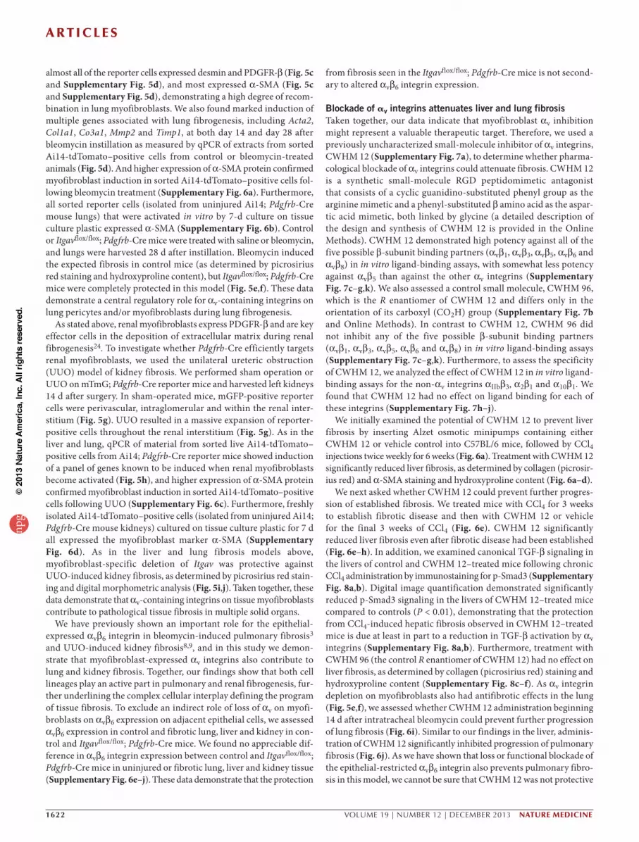
Blockade of av integrins attenuates liver and lung fibrosisTaken together, our data indicate that myofibroblast αv inhibition might represent a valuable therapeutic target. Therefore, we used a previously uncharacterized small-molecule inhibitor of αv integrins, CWHM 12 (Supplementary Fig. 7a), to determine whether pharma-cological blockade of αv integrins could attenuate fibrosis. CWHM 12 is a synthetic small-molecule RGD peptidomimetic antagonist that consists of a cyclic guanidino-substituted phenyl group as the arginine mimetic and a phenyl-substituted β amino acid as the aspar-tic acid mimetic, both linked by glycine (a detailed description of the design and synthesis of CWHM 12 is provided in the Online Methods). CWHM 12 demonstrated high potency against all of the five possible β-subunit binding partners (αvβ1, αvβ3, αvβ5, αvβ6 and αvβ8) in in vitro ligand-binding assays, with somewhat less potency against αvβ5 than against the other αv integrins (Supplementary Fig. 7c–g,k). We also assessed a control small molecule, CWHM 96, which is the R enantiomer of CWHM 12 and differs only in the orientation of its carboxyl (CO2H) group (Supplementary Fig. 7b and Online Methods). In contrast to CWHM 12, CWHM 96 did not inhibit any of the five possible β-subunit binding partners (αvβ1, αvβ3, αvβ5, αvβ6 and αvβ8) in in vitro ligand-binding assays (Supplementary Fig. 7c–g,k). Furthermore, to assess the specificity of CWHM 12, we analyzed the effect of CWHM 12 in in vitro ligand-binding assays for the non-αv integrins αIIbβ3, α2β1 and α10β1. We found that CWHM 12 had no effect on ligand binding for each of these integrins (Supplementary Fig. 7h–j).
We initially examined the potential of CWHM 12 to prevent liver fibrosis by inserting Alzet osmotic minipumps containing either CWHM 12 or vehicle control into C57BL/6 mice, followed by CCl4 injections twice weekly for 6 weeks (Fig. 6a). Treatment with CWHM 12 significantly reduced liver fibrosis, as determined by collagen (picrosir-ius red) and α-SMA staining and hydroxyproline content (Fig. 6a–d).
We next asked whether CWHM 12 could prevent further progres-sion of established fibrosis. We treated mice with CCl4 for 3 weeks to establish fibrotic disease and then with CWHM 12 or vehicle for the final 3 weeks of CCl4 (Fig. 6e). CWHM 12 significantly reduced liver fibrosis even after fibrotic disease had been established (Fig. 6e–h). In addition, we examined canonical TGF-β signaling in the livers of control and CWHM 12–treated mice following chronic CCl4 administration by immunostaining for p-Smad3 (Supplementary Fig. 8a,b). Digital image quantification demonstrated significantly reduced p-Smad3 signaling in the livers of CWHM 12–treated mice compared to controls (P < 0.01), demonstrating that the protection from CCl4-induced hepatic fibrosis observed in CWHM 12–treated mice is due at least in part to a reduction in TGF-β activation by αv integrins (Supplementary Fig. 8a,b). Furthermore, treatment with CWHM 96 (the control R enantiomer of CWHM 12) had no effect on liver fibrosis, as determined by collagen (picrosirius red) staining and hydroxyproline content (Supplementary Fig. 8c–f). As αv integrin depletion on myofibroblasts also had antifibrotic effects in the lung (Fig. 5e,f), we assessed whether CWHM 12 administration beginning 14 d after intratracheal bleomycin could prevent further progression of lung fibrosis (Fig. 6i). Similar to our findings in the liver, adminis-tration of CWHM 12 significantly inhibited progression of pulmonary fibrosis (Fig. 6j). As we have shown that loss or functional blockade of the epithelial-restricted αvβ6 integrin also prevents pulmonary fibro-sis in this model, we cannot be sure that CWHM 12 was not protective
npg
© 2
013
Nat
ure
Am
eric
a, In
c. A
ll rig
hts
rese
rved
.

a r t i c l e s
nature medicine VOLUME 19 | NUMBER 12 | DECEMBER 2013 1623
in the lung because of its inhibitory effects on αvβ6. Nonetheless, our findings suggest that inhibition of αv integrins by the small molecule CWHM 12 has potential clinical utility in the treatment of patients with a broad range of fibrotic diseases.
DISCUSSIONMyofibroblasts are a major source of extracellular matrix that accu-mulates during fibrosis36. However, the paucity of tools for reliable inactivation of genes in myofibroblasts in vivo has greatly impeded progress in dissecting the molecular mechanisms driving organ fibrosis, thereby slowing the discovery of new mechanistically tar-geted antifibrotic treatments. In this study, we developed a system (Pdgfrb-Cre) to genetically manipulate myofibroblasts in multiple organs. Using this system, we identified a key role for myofibroblast αv integrins in the regulation of fibrosis and then validated our approach by using a small molecule to target αv integrins. We have thus identi-fied a targeted approach to treat fibrosis.
As a first test of the effectiveness of this system, we deleted the αv integrin subunit from myofibroblasts and found that such depletion markedly inhibited fibrosis in the liver, lung and kidney, identifying myofibroblast αv-containing integrins as components of a core path-way widely shared by pathological fibrosis in multiple solid organs. Of note, we were unable to effectively inhibit liver fibrosis by indi-vidual depletion of four of the β-subunit partners of αv (β3, β5, β6 or β8), suggesting either that this protection was due to loss of αvβ1 (which cannot be studied with the tools currently available) or, more likely, that inhibition of multiple αv-containing integrins is required to effectively treat liver fibrosis. Using a small-molecule inhibitor, CWHM 12, we showed that therapeutically targeting all αv-containing
integrins effectively treated fibrosis in multiple organs. Notably, a recent study using cilengitide (an antagonist selective for mainly αvβ3 and αvβ5 with less potency toward αvβ6) demonstrated a 30% increase in hepatic collagen in two models of liver fibrosis38, supporting the idea that blockade of all αv-containing integrins may be required to obtain substantial antifibrotic effects. Furthermore, inhibition of TGF-β1 signaling through pharmacological blockade of αv-containing integrins with a small molecule such as CWHM 12 may yield the desired antifibrotic effects without the unwanted potential side effects of pan–TGF-β blockade (autoimmunity and carcinogenesis). However, comprehensive toxicology testing with CWHM 12 will be required before consideration of clinical trials.
As discussed above, one potential mechanism of protection for eliminating or blocking αv-containing integrins is inhibition of acti-vation of TGF-β. Our findings that loss of αv led to a reduction in in vivo p-Smad3 immunostaining, expression of TGF-β–inducible genes and TGF-β activation in culture by HSCs supports a role for this mechanism. TGF-β is a major profibrogenic cytokine and a cen-tral mediator of fibrosis in many tissues39–41. However, our results do not exclude other functions of αv integrins on myofibroblasts in contributing to tissue fibrosis.
It is of interest that activation of TGF-β appears to be a common function of multiple αv-containing integrins. Many of the functional roles for distinct β-subunit partners of αv are clearly unique, as demon-strated by the markedly different phenotypes described by us and oth-ers for mice globally lacking β3, β5, β6 and β8. Because of the absence of useful reagents to specifically examine the roles of αvβ1 in vivo, it remains possible that αvβ1 could be the major integrin responsible for TGF-β activation by myofibroblasts, but previously published in vitro
Figure 6 Blockade of αv integrins by a small molecule (CWHM 12) attenuates liver and lung fibrosis. (a) Left, dosing regime in the prophylactic liver fibrosis model. Alzet minipumps containing CWHM 12 or vehicle were inserted, followed by CCl4 intraperitoneal (i.p.) injection twice weekly for 6 weeks. Right, picrosirius red (top) and α-SMA immunohistochemistry (bottom) of liver tissue from control and CWHM 12–treated mice (n = 6 female mice per group) after chronic CCl4 treatment. Scale bar, 200 µm. (b) Digital image analysis of picrosirius red staining in the prophylactic liver fibrosis model. (c) Hydroxyproline analysis of liver tissue in the prophylactic liver fibrosis model. (d) Digital image analysis of α-SMA staining in the prophylactic liver fibrosis model. (e) Left, dosing regime in the therapeutic liver fibrosis model. Mice were given CCl4 i.p. twice weekly for 3 weeks, then Alzet minipumps containing either CWHM 12 or vehicle were inserted, followed by a further 3 weeks of CCl4 i.p. twice weekly. Right, picrosirius red (top) and α-SMA immunohistochemistry (bottom) of liver tissue from control and CWHM 12–treated mice (n = 14 female mice per group) after chronic CCl4 treatment. Scale bar, 200 µm. (f) Digital image analysis of picrosirius red staining in the therapeutic liver fibrosis model. (g) Hydroxyproline analysis of liver tissue in the therapeutic liver fibrosis model. (h) Digital image analysis of α-SMA staining in the therapeutic liver fibrosis model. (i) Dosing regime in the therapeutic lung fibrosis model. Alzet minipumps containing either CWHM 12 or vehicle were inserted 14 d after treatment with bleomycin or saline, and lungs were harvested at 28 d. (j) Left, picrosirius red staining of lung tissue from control and CWHM 12–treated mice 28 d after bleomycin instillation (n = 15 female mice per group). Scale bar, 100 µm. Right, hydroxyproline analysis. Data are expressed as mean ± s.e.m. *P < 0.05, **P < 0.01, ***P < 0.001, ****P < 0.0001 (Student’s t-test).
CWHM 12treatment
Day 0 Day 14 Day 28
Bleo orsaline
i
0
6
4
2
8
Siri
us r
ed (
% a
rea)
**
b
Con C12
6 weeks CCl4
CWHM12treatment
e Control CWHM 12
PS
Rα-
SM
A
6 weeks CCl4
CWHM12 treatment
a Control CWHM 12
PS
Rα-
SM
A
100
400
300
200
0
Hyd
roxy
prol
ine
(µg
per
g)
*c
Con C12
100
400
300
200
0
500
Hyd
roxy
prol
ine
(µg
per
g)
g
Con C12
***
d
Con C12
*
0
6
4
2
8
α-S
MA
(%
are
a)
h
0
6
4
2
8
α-S
MA
(%
are
a)
Con C12
**
f
Siri
us r
ed (
% a
rea)
****
0
6
4
2
8
Con C12
Control CWHM 12
PS
R
j*
Saline Bleo
25
50
100
0
75
Hyd
roxy
prol
ine
(µg
ml–1
) ControlC12
npg
© 2
013
Nat
ure
Am
eric
a, In
c. A
ll rig
hts
rese
rved
.

a r t i c l e s
1624 VOLUME 19 | NUMBER 12 | DECEMBER 2013 nature medicine
studies in different systems suggest that αvβ3, αvβ5 and αvβ8 all have the potential to partially mediate TGF-β activation by these cells.
In summary, we have developed a system that allows gene manip-ulation in myofibroblasts in multiple tissues and used this system to demonstrate that αv-containing integrins on myofibroblasts are components of a core cellular and molecular pathway that contributes to pathologic fibrosis in multiple solid organs, suggesting that target-ing this core pathway could have clinical utility in the treatment of patients with a broad range of fibrotic diseases.
METHODSMethods and any associated references are available in the online version of the paper.
Note: Any Supplementary Information and Source Data files are available in the online version of the paper.
ACKNoWLEDGMENTSThis work was supported by a Wellcome Trust Intermediate Clinical Fellowship (ref. 085187) to N.C.H., National Institutes of Health grants HL102292, HL53949 and AI077439 (to D.S.), a University of California, San Francisco (UCSF) Liver Center Tool and Technology grant (to N.C.H) and P30 DK026743 (UCSF Liver Center). We thank K. Thorn at the UCSF Nikon Imaging Center for assistance with image analysis. We also thank C. Her, N. Wu, S. Huling, D. Rodrigues and R. Aucott for expert technical assistance. We also acknowledge the contribution of M. Singh (chemical synthesis of compounds CWHM 12 and CWHM 96), D. Tajfirouz, S. Freeman and M. Yates at Saint Louis University for technical assistance in conducting integrin functional assays to characterize compound activities. L. Reichardt (UCSF) provided Itgb8flox/flox mice and R. Hynes (Massachusetts Institute of Technology) provided Itgb3−/− mice on 129/svJae background. S. Violette (Biogen Idec) provided antibody to αvβ6 (human/mouse chimeric 2A1), W. Stallcup (Sanford-Burnham Medical Research Institute) provided antibody to PDGFR-β and H. Yagita (Juntendo University) provided antibody to αv integrin (clone RMV-7).
AUTHoR CoNTRIBUTIoNSN.C.H. and D.S. conceived and designed the project. N.C.H. performed the experiments with assistance from T.D.A., Y.K., M.M.G., J.D.R. and A.P.; J.H.M. contributed reagents; P.G.R., D.W.G. and M.J.P. designed and synthesized the small molecule αv integrin inhibitor (CWHM 12) and performed the ligand-binding studies to characterize the in vitro potency of CWHM 12; J.J.M. and J.P.I. contributed reagents and provided substantial intellectual contribution; E.R. and C.B. contributed Pdgfrb-BAC-eGFP knock-in reporter mice; A.L.-H. contributed Itgavflox/flox mice; R.H.A. contributed Pdgfrb-Cre mice; N.C.H., T.D.A., Y.K., M.M.G. and D.S. analyzed data and N.C.H., J.P.I. and D.S. wrote the manuscript.
CoMPETING FINANCIAL INTERESTSThe authors declare competing financial interests: details are available in the online version of the paper.
Reprints and permissions information is available online at http://www.nature.com/reprints/index.html.
1. Gleizes, P.E. et al. TGF-β latency: biological significance and mechanisms of activation. Stem Cells 15, 190–197 (1997).
2. Munger, J.S. et al. Latent transforming growth factor-β: structural features and mechanisms of activation. Kidney Int. 51, 1376–1382 (1997).
3. Munger, J.S. et al. The integrin αvβ6 binds and activates latent TGFβ1: a mechanism for regulating pulmonary inflammation and fibrosis. Cell 96, 319–328 (1999).
4. Mu, D. et al. The integrin αvβ8 mediates epithelial homeostasis through MT1-MMP-dependent activation of TGF-β1. J. Cell Biol. 157, 493–507 (2002).
5. Annes, J.P., Rifkin, D.B. & Munger, J.S. The integrin αvβ6 binds and activates latent TGFβ3. FEBS Lett. 511, 65–68 (2002).
6. Aluwihare, P. et al. Mice that lack activity of αvβ6- and αvβ8-integrins reproduce the abnormalities of Tgfb1- and Tgfb3-null mice. J. Cell Sci. 122, 227–232 (2009).
7. Wang, B. et al. Role of αvβ6 integrin in acute biliary fibrosis. Hepatology 46, 1404–1412 (2007).
8. Hahm, K. et al. αv βa6 integrin regulates renal fibrosis and inflammation in Alport mouse. Am. J. Pathol. 170, 110–125 (2007).
9. Ma, L.J. et al. Transforming growth factor-β-dependent and -independent pathways of induction of tubulointerstitial fibrosis in β6−/− mice. Am. J. Pathol. 163, 1261–1273 (2003).
10. Breuss, J.M., Gillett, N., Lu, L., Sheppard, D. & Pytela, R. Restricted distribution of integrin β6 mRNA in primate epithelial tissues. J. Histochem. Cytochem. 41, 1521–1527 (1993).
11. Breuss, J.M. et al. Expression of the β6 integrin subunit in development, neoplasia and tissue repair suggests a role in epithelial remodeling. J. Cell Sci. 108, 2241–2251 (1995).
12. Shi, M. et al. Latent TGF-β structure and activation. Nature 474, 343–349 (2011).
13. Wipff, P.J., Rifkin, D.B., Meister, J.J. & Hinz, B. Myofibroblast contraction activates latent TGF-β1 from the extracellular matrix. J. Cell Biol. 179, 1311–1323 (2007).
14. Munger, J.S., Harpel, J.G., Giancotti, F.G. & Rifkin, D.B. Interactions between growth factors and integrins: latent forms of transforming growth factor-β are ligands for the integrin αvβ1. Mol. Biol. Cell 9, 2627–2638 (1998).
15. Asano, Y. et al. Increased expression of integrin αvβ3 contributes to the establishment of autocrine TGF-β signaling in scleroderma fibroblasts. J. Immunol. 175, 7708–7718 (2005).
16. Asano, Y., Ihn, H., Yamane, K., Jinnin, M. & Tamaki, K. Increased expression of integrin αvβ5 induces the myofibroblastic differentiation of dermal fibroblasts. Am. J. Pathol. 168, 499–510 (2006).
17. Friedman, S.L. & Arthur, M.J. Activation of cultured rat hepatic lipocytes by Kupffer cell conditioned medium. Direct enhancement of matrix synthesis and stimulation of cell proliferation via induction of platelet-derived growth factor receptors. J. Clin. Invest. 84, 1780–1785 (1989).
18. Pinzani, M., Gesualdo, L., Sabbah, G.M. & Abboud, H.E. Effects of platelet-derived growth factor and other polypeptide mitogens on DNA synthesis and growth of cultured rat liver fat-storing cells. J. Clin. Invest. 84, 1786–1793 (1989).
19. Wong, L., Yamasaki, G., Johnson, R.J. & Friedman, S.L. Induction of β-platelet–derived growth factor receptor in rat hepatic lipocytes during cellular activation in vivo and in culture. J. Clin. Invest. 94, 1563–1569 (1994).
20. Pinzani, M. et al. Expression of platelet-derived growth factor and its receptors in normal human liver and during active hepatic fibrogenesis. Am. J. Pathol. 148, 785–800 (1996).
21. Ikura, Y. et al. Expression of platelet-derived growth factor and its receptor in livers of patients with chronic liver disease. J. Gastroenterol. 32, 496–501 (1997).
22. Coin, P.G. et al. Lipopolysaccharide up-regulates platelet-derived growth factor (PDGF) α-receptor expression in rat lung myofibroblasts and enhances response to all PDGF isoforms. J. Immunol. 156, 4797–4806 (1996).
23. Bonner, J.C. Regulation of PDGF and its receptors in fibrotic diseases. Cytokine Growth Factor Rev. 15, 255–273 (2004).
24. Chen, Y.T. et al. Platelet-derived growth factor receptor signaling activates pericyte-myofibroblast transition in obstructive and post-ischemic kidney fibrosis. Kidney Int. 80, 1170–1181 (2011).
25. Foo, S.S. et al. Ephrin-B2 controls cell motility and adhesion during blood- vessel-wall assembly. Cell 124, 161–173 (2006).
26. de Leeuw, A.M., McCarthy, S.P., Geerts, A. & Knook, D.L. Purified rat liver fat-storing cells in culture divide and contain collagen. Hepatology 4, 392–403 (1984).
27. Friedman, S.L., Roll, F.J., Boyles, J. & Bissell, D.M. Hepatic lipocytes: the principal collagen-producing cells of normal rat liver. Proc. Natl. Acad. Sci. USA 82, 8681–8685 (1985).
28. Muzumdar, M.D., Tasic, B., Miyamichi, K., Li, L. & Luo, L. A global double-fluorescent Cre reporter mouse. Genesis 45, 593–605 (2007).
29. Madisen, L. et al. A robust and high-throughput Cre reporting and characterization system for the whole mouse brain. Nat. Neurosci. 13, 133–140 (2010).
30. Abe, M. et al. An assay for transforming growth factor-β using cells transfected with a plasminogen activator inhibitor-1 promoter-luciferase construct. Anal. Biochem. 216, 276–284 (1994).
31. Hynes, R.O. Integrins: bidirectional, allosteric signaling machines. Cell 110, 673–687 (2002).
32. Zhu, J. et al. β8 integrins are required for vascular morphogenesis in mouse embryos. Development 129, 2891–2903 (2002).
33. Fässler, R. & Meyer, M. Consequences of lack of β1 integrin gene expression in mice. Genes Dev. 9, 1896–1908 (1995).
34. Stephens, L.E. et al. Deletion of β1 integrins in mice results in inner cell mass failure and peri-implantation lethality. Genes Dev. 9, 1883–1895 (1995).
35. Abraham, S., Kogata, N., Fässler, R. & Adams, R.H. Integrin β1 subunit controls mural cell adhesion, spreading, and blood vessel wall stability. Circ. Res. 102, 562–570 (2008).
36. Hinz, B. et al. Recent developments in myofibroblast biology: paradigms for connective tissue remodeling. Am. J. Pathol. 180, 1340–1355 (2012).
37. Armulik, A., Genové, G. & Betsholtz, C. Pericytes: developmental, physiological, and pathological perspectives, problems, and promises. Dev. Cell 21, 193–215 (2011).
38. Patsenker, E. et al. Pharmacological inhibition of integrin αvβ3 aggravates experimental liver fibrosis and suppresses hepatic angiogenesis. Hepatology 50, 1501–1511 (2009).
39. Ignotz, R.A. & Massagué, J. Transforming growth factor-β stimulates the expression of fibronectin and collagen and their incorporation into the extracellular matrix. J. Biol. Chem. 261, 4337–4345 (1986).
40. Roberts, A.B. et al. Transforming growth factor type β: rapid induction of fibrosis and angiogenesis in vivo and stimulation of collagen formation in vitro. Proc. Natl. Acad. Sci. USA 83, 4167–4171 (1986).
41. Leask, A. & Abraham, D.J. TGF-β signaling and the fibrotic response. FASEB J. 18, 816–827 (2004).
npg
© 2
013
Nat
ure
Am
eric
a, In
c. A
ll rig
hts
rese
rved
.

nature medicinedoi:10.1038/nm.3282
ONLINE METHODSMice. mTmG (TdTomato-EGFP)28 and Ai14 (Rosa-CAG-LSL-tdTomato-WPRE)29 mice were obtained from the Jackson Laboratory and crossed with Pdgfrb-Cre mice25 (provided by R.H.A.). Itgavflox/flox mice42 were obtained from A.L.-H., Itgb8flox/flox mice43 were obtained from L. Reichardt and all were maintained on C57BL/6 background. Itgb3−/− mice on 129/svJae background44 were obtained from R. Hynes. Itgb5−/− mice on 129/svJae background45 and Itgb6−/− mice on a C57BL/6 background46 were generated and maintained in our laboratory as previously described. Pdgfrb-BAC-eGFP reporter mice (on a C57BL/6 background) carry one copy of a BAC transgene expressing enhanced eGFP under the control of Pdgfrb regulatory elements. They origi-nate from the GENSAT project (Gene Expression Nervous System Atlas) and were obtained from the MMRRC (Mutant Mouse Regional Resource Center, STOCK Tg(Pdgfrb-EGFP)JN169Gsat/Mmucd)). Genotyping of all mice was performed by PCR. Wild-type C57/BL6 mice were purchased from the Jackson Laboratory. Mice used for all experiments were 8–12 weeks old and were housed under specific-pathogen–free conditions in the Animal Barrier Facility of the University of California, San Francisco. All experiments were approved by the Institutional Animal Care and Use Committee of the University of California, San Francisco.
Fibrosis models. CCl4 liver injury was induced as described previously47 with 8- to 10-week-old sex-matched mice. For acute liver injury, mice were injected i.p. with 1 µl per g body weight sterile CCl4 in a 1:3 ratio in olive oil or olive oil alone (control) after overnight fasting (with free access to water). For chronic CCl4–induced liver fibrosis, mice were injected i.p. with 1 µl per g body weight CCl4 or olive oil as above twice weekly for 6 weeks. Livers were harvested 24 h after the last injection. To induce pulmonary fibrosis, 8- to 10-week-old sex-matched mice were anesthetized, and saline or bleomycin (1.5 U per kg Blenoxane, Sigma) was instilled intratracheally. Lungs were harvested 14 or 28 d later. For renal fibrosis, UUO was induced by ligation of the left ureter as described previously48. Sham-operated mice underwent an identical surgical procedure, except ligation of the ureter was not performed. 8- to 10-week-old male mice were used in these studies. Kidneys were harvested 7 and 14 d after surgery.
Primary cell isolation and fluorescence-activated cell sorting. Mouse liver was perfused through the inferior vena cava sequentially with liver perfusion medium (Invitrogen), 0.3% pronase (Roche) and 0.02% collagenase (Serva). The liver was excised, minced with scissors and further digested in 0.044% pronase and 0.008% DNase (Roche). The cell suspension was shaken (200–250 r.p.m.) at 37 °C for 10 min and strained through sterile gauze. To remove hepatocytes, the cell suspension was centrifuged at 90g for 2 min, supernatant collected and DNase added, and this procedure was repeated twice. Supernatant was centri-fuged at 700g for 7 min to collect the nonparenchymal cell fraction. Single-cell suspensions from mouse lung and kidney were prepared using a gentleMACS dissociator (Mitenyi Biotec) as per manufacturer’s instructions. Following live-dead staining with SYTOX blue (Invitrogen), live single tdTomato-positive cells from Ai14; Pdgfrb-Cre mice were sorted using a FACSAria (BD Biosciences). tdTomato-positive cells were cultured in DMEM (Invitrogen) supplemented with glucose, l-glutamine, penicillin-streptomycin and 15% FCS. Primary mouse HSCs were isolated and passaged as described previously47. Primary mouse hepatocytes were isolated by retrograde perfusion of the liver with Liver Perfusion Medium (Invitrogen) followed by Liver Digest Medium (Invitrogen) at 37 °C. When hepatocytes were visually dispersed within the liver capsule, the liver was removed to a sterile dish and minced with scissors to release the crude cell isolate. The cells were then suspended in DME/F-12 medium and pelleted twice. Hepatocytes were purified from the washed pellets by resuspension in culture medium and centrifugation through 50% Percoll (GE Healthcare). To isolate liver sinusoidal endothelial cells (LSECs) and Kupffer cells, single-cell suspensions from mouse liver were prepared using a gentleMACS dissociator (Mitenyi Biotec) followed by selection using CD146 (LSEC) or CD11b (Kupffer cells) microbeads as per manufacturer’s instructions (Miltenyi Biotec).
Immunohistochemistry and immunofluoresence. Paraffin-embedded sec-tions were processed for immunohistochemistry as described previously47.
The following primary antibodies were used for immunohistochemistry: α-SMA A5228, 1:1,000 (Sigma); GR1 MAB1037, 1:750 (R&D); F4/80 Ab6640, 1:100 (Abcam); CD31 sc1506, 1:80 (Santa Cruz Biotechnology) and αvβ6 mAb, 1:25 (human/mouse chimeric 2A1, a gift from S. Violette). 5-µM sections were stained with picrosirius red or antibody and results quantified using Nikon Elements software. Six random fields from each section were analyzed at a final magnification of 40×. For neutrophil counting, 20 random portal tracts per mouse liver were assessed. For immunofluoresence staining, liver tissue was fixed in 4% paraformaldehyde overnight at 4 °C, immersed in graded sucrose solutions, embedded in OCT (Tissue Tek) and stored at –80 °C. Frozen sections were incubated with the following antibodies: F4/80 MCA497R, 1:100; CD3 MCA500GT, 1:200 (Serotec); CD31 550274, 1:50 (BD Pharmingen); α-SMA A5228, 1:200 (Sigma); PDGFR-β, 1:200 (a gift from W. Stallcup); p-SMAD3 1880-1, 1:100 (Epitomics); cytokeratin WSS Z0622, 1:100 (Dako); αv integrin ab76609, 1:60; desmin ab8592, 1:250 (Abcam) and Alexa Fluor 488–conjugated and Alexa Fluor 555–conjugated secondary antibodies (Invitrogen). Confocal imaging was performed on a Zeiss LSM5 Pascal microscope. Digital morpho-metric measurements of p-SMAD3 were performed using ImageJ. Twelve random fields of areas of scar from each section were analyzed at a final magni-fication of 400×. Digital morphometric measurements of desmin and PDGFR-β immunostaining were performed using ImageJ. Eight random fields from each section were analyzed at a final magnification of 100×.
Human tissues. Human liver tissue was obtained from explant livers of patients with end-stage fibrotic liver disease. The use of human tissues for this study was approved by the Local Ethics Committee, University of Edinburgh. All samples collected were from subjects who gave informed consent for their tissues to be used for research purposes.
Hydroxyproline assays. Mouse liver tissue (200 mg) or the left lung was homog-enized, precipitated with trichloroacetic acid and baked overnight at 110 °C in HCl. Samples were reconstituted in water, and hydroxyproline content was measured using a colorimetric chloramine T assay.
Immunoprecipitation and western blotting. Cell-sorted tdTomato–positive cells from the uninjured livers of Ai14; Pdgfrb-Cre mice were plated on tissue culture plastic for 7 d and then lysed. Cell lysates were centrifuged at 14,000 r.p.m. for 10 min at 4 °C and the supernatant collected. 10 µg of antibody to αv integrin (clone RMV-7, a gift from H. Yagita) was added to the supernatant, and this was rotated at 4 °C for 2 h, followed by the addition of 30 µl of prewashed protein G sepharose slurry (GE Healthcare) for a further 1 h at 4 °C. Following centrifugation, the beads were washed three times with PBS–protease inhibitor mixture, and once with PBS only. Laemmli sample buffer was added, and the samples were boiled for 5 min followed by SDS-PAGE and western blotting using the following antibodies: αv integrin 611012, 1:500 (BD Biosciences); β1 integrin 1798-1, 1:1,000 (Epitomics); β3 integrin ab75872, 1:1,000; β5 integrin ab15459, 1:1,000 (Abcam) and β8 integrin 1:3,000 (a gift from J.H.M.)49. Western blotting was also undertaken using the following antibodies: α-SMA A5228, 1:5,000; β-actin A2228, 1:5,000 (Sigma); desmin MAB3430, 1:1,000 (Millipore) and PDGFR-β 1469-1, 1:10,000 (Epitomics).
TGF-b activation assay. Control and Itgavflox/flox; Pdgfrb-Cre HSCs were iso-lated and cultured for 5 d on tissue culture plastic and then plated at 50,000 cells per well in 96-well plates with mink lung epithelial cells expressing firefly luciferase downstream of a TGF-β sensitive portion of the plasminogen activator inhibitor-1 promoter30 (15,000 cells per well). Cells were cultured for 16 h, and TGF-β activity was calculated by measurement of luminescence. Recombinant human TGF-β1 was reconstituted as per manufacturer’s instructions (R&D).
In vivo CWHM 12 and CWHM 96 studies. For all studies, CWHM 12 and CWHM 96 were solubilized in 50% DMSO (in sterile water) and dosed to 100 mg per kg body weight per day. Drug or vehicle (50% DMSO) was delivered by implantable ALZET osmotic minipumps (Durect, Cupertino, CA). For CCl4-induced fibrosis, pumps were inserted subcutaneously either before the first dose of CCl4 (prophylactic) or after 3 weeks of treatment (therapeutic), and livers were harvested after 6 weeks. For bleomycin-induced fibrosis, pumps were inserted
npg
© 2
013
Nat
ure
Am
eric
a, In
c. A
ll rig
hts
rese
rved
.

nature medicine doi:10.1038/nm.3282
14 d after treatment with bleomycin or saline, and lungs were harvested at 28 d (therapeutic only).
qRT-PCR. Total RNA was isolated using an RNeasy kit (Qiagen). cDNA was analyzed by SYBR-Green RT-PCR with an ABI 7900HT thermocy-cler and normalized to β-actin or 18S expression. Primers used were as fol-lows: β-actin forward TGTTACCAACTGGGACGACA, β-actin reverse GGGGTGTTGAAGGTCTCAAA; 18S forward TAGAGGGACAAGTGGC GTTC, 18S reverse CGCTGAGCCAGTCAGTGT; Itgav forward CCGTGG ACTTCTTCGAGCC, Itgav reverse CTGTTGAATCAAACTCAATGGGC; Des forward GTGGATGCAGCCACTCTAGC, Des reverse TTAGCCGCG ATGGTCTCATAC; Pdgfrb forward TCCAGGAGTGATACCAGCTTT, Pdgfrb reverse CAGGAGCCATAACACGGACA; Gfap forward CGGAGA CGCATCACCTCTG, Gfap reverse TCTCGGAGGCATAGGAGCG; Acta2 forward GTCCCAGACATCAGGGAGTAA, Acta2 reverse TCGGATACT TCAGCGTCAGGA; Col1a1 forward GCTCCTCTTAGGGGCCACT, Col1a1 reverse CCACGTCTCACCATTGGGG; Col 3a1 forward AACCTGGTTTC TTCTCACCCTTC, Col 3a1 reverse ACTCATAGGACTGACCAAGGTGG; Tgfb1 forward CTCCCGTGGCTTCTAGTGC, Tgfb1 reverse GCCTTAG TTTGGACAGGATCTG; Mmp2 forward CAAGTTCCCCGGCGATGTC, Mmp2 reverse TTCTGGTCAAGGTCACCTGTC; Mmp3 forward ACATGGA GACTTTGTCCCTTTTG, Mmp3 reverse TTGGCTGAGTGGTAGAGTCCC; Mmp9 forward CTGGACAGCCAGACACTAAAG, Mmp9 reverse CTC GCGGCAAGTCTTCAGAG; Mmp13 forward CTTCTTCTTGTTGAGCTG GACTC, Mmp13 reverse CTGTGGAGGTCACTGTAGACT; Timp1 forward TGCAACTCGGACCTGGTCATA, Timp1 reverse CGCTGGTATAAGGTGG TCTCG; Pparg forward GGAAGACCACTCGCATTCCTT, Pparg reverse GTAATCAGCAACCATTGGGTCA; Itgb1 forward CTACTTCTGCAC GATGTGATGAT, Itgb1 reverse TTGGCTGGCAACCCTTCTTT; Itgb3 forward CCACACGAGGCGTGAACTC, Itgb3 reverse CTTCAGGTTAC ATCGGGGTGA; Itgb5 forward GAAGTGCCACCTCGTGTGAA, Itgb5 reverse GGACCGTGGATTGCCAAAGT; Itgb8 forward CTGAAGAAATA CCCCGTGGA, Itgb8 reverse ATGGGGAGGCATACAGTCT.
Adhesion assay. Control and Itgavflox/flox; Pdgfrb-Cre HSCs were cultured for 5 d and then seeded into 48-well tissue-culture plates precoated with fibronec-tin, collagen I, collagen IV, laminin and fibrinogen or BSA-treated controls (CytoSelect 48-well adhesion assay, ECM array, Cambridge Biosciences). Cells were allowed to adhere for 90 min, washed 3× with PBS and stained and eluted as per manufacturer’s instructions. Adhesion is expressed as a percentage of unwashed cells adhered to 1% poly-l-lysine.
Migration assay. Control and Itgavflox/flox; Pdgfrb-Cre HSCs were cultured for 5 d and then seeded into the upper chambers of 8-µm–pore sized modified Boyden chambers as per manufacturer’s instructions (CytoSelect 24-well cell migration assay, Cambridge Biosciences). Fetal calf serum (10%) was added to the lower chamber and cells were allowed to migrate for 6 h at 37 °C. Cells remaining in the upper chamber were wiped with a cotton tip, and cells attached to the underside of the membrane were fixed, stained and eluted as per manufacturer’s instruc-tions. Chemotaxis is expressed as percentage of an unwiped control.
ELISA. Total TGF-β1 and Timp-1 were measured using ELISA kits (R&D) as per manufacturer’s instructions.
TUNEL assay. Control and Itgavflox/flox; Pdgfrb-Cre HSCs were plated overnight on glass-chamber slides, and apoptosis was detected using the in situ death detection kit (Roche) as per manufacturer’s instructions. Cells were counter-stained with DAPI and viewed under a Zeiss fluorescent microscope.
Assessment of recombination efficiency. Six confocal images (225-µm2 single-optical sections) were randomly acquired throughout nonadjacent cryosections of liver or lung (n = 4 male mice per group). Antibody staining for desmin, PDGFR-β and α-SMA and Ai14-tdTomato recombination reporter were separately subjected to threshold processing using US National Institutes of Health ImageJ software, and then the percentage of Ai14-tdTomato–positive antibody-stained cells was quantified.
Flow cytometry. Cells were harvested with TrypLE Select (Life Technologies, UK), washed twice with PBS and resuspended in FACS buffer (PBS supple-mented with 2% FCS). 4 × 10⁵ cells were then incubated with a conjugated antibody (or isotype control) at 4 °C for 30 min in the dark. Cells were then washed, resuspended in FACS buffer and analyzed on a Becton Dickinson LSR Fortessa II. Antibodies used were phycoerythrin-conjugated antibody to CD49a (integrin α1) 562115, 1:50 and isotype control 553965, 1:50 (BD Pharmingen) and phycoerythrin-conjugated antibody to CD49e (integrin α5) 12-0493, 1:200 and antibody to CD29 (integrin β1) 12-0291, 1:200 and isotype control 12-4888, 1:200 (eBioscience).
In vitro integrin functional assays. The effects of CWHM 12 and CWHM 96 on cell adhesion mediated by αvβ3, αvβ5, αvβ6 and α5β1 were measured as previ-ously described with minor modifications50,51. Briefly, stably transfected human 293 cells overexpressing human αvβ3 or αvβ5 were preincubated in HBSS buffer (Sigma) containing 200 µM MnCl2 for 30 min at 37 °C with threefold dilutions of compound. Each sample was then added to triplicate wells of a 96-well plate that had been coated overnight at 4 °C with a predetermined optimal concentration of purified vitronectin (Calbiochem), washed, blocked by 1-h incubation with BSA and washed again. Cells were allowed to attach for 30 min at 37 °C, and nonad-herent cells were removed by washing. Remaining attached cells were measured by endogenous alkaline phosphatase activity using p-nitrophenyl phosphate and reading absorbance signal at 405 nM. The same procedure was used to measure adhesion of αvβ6-expressing human HT-29 cells to purified human latency-associated peptide (LAP; R&D Systems) and α5β1-expressing human K562 cells to human plasma fibronectin (Calbiochem). In all cell-based assays, binding by the expected integrin was verified by testing activity of corresponding isotype-matched positive (function-blocking) and negative control antibodies. Functions of integrins αvβ1, αvβ8, α2β1 and α10β1 were measured using cell-free receptor-ligand interaction assays using purified recombinant human integrins purchased from R&D Systems. Ligands used were human fibronectin (R&D Systems) for αvβ1, human LAP (R&D Systems) for αvβ8, bovine collagen II (Sigma) for α2β1 and murine laminin I (Cultrex, Trevigen) for α10β1. 96-well plates were coated with the predetermined optimal concentration of ligand overnight, washed 3× with TBS+++ (25 mM Tris, pH 7.4, 137 mM NaCl, 2.7 mM KCl, 1 mM MgCl2, 1 mM MnCl2 and 1mM CaCl2) and blocked with TBS+++/1% BSA. Purified integrin was diluted in TBS+++/0.1% BSA with or without compounds and the solution added to empty wells of the washed ligand-coated plate according to a standard template, with each sample repeated in triplicate. After incubation for 2 h at room temperature, the plate was washed 3× with TBS+++. Biotin-labeled antibody against the αv subunit (αvβ1 and αvβ8 assays) or β1 subunit (α2β1 and α10β1 assays) (R&D Systems) was applied for 1 h. The plate was washed 3× with TBS/0.1% BSA. Streptavidin-conjugated horseradish peroxidase (R&D Systems) was added to the wells and the plate incubated for 20 min at room temperature. Following a 3× TBS+++ wash, bound integrin was detected using streptavidin-conjugated horseradish peroxidase and TMB (tetramethylbenzidine) substrate with absorbance measured at 650 nm. For assay of αIIbβ3 (IIbIIIa) function, plates were coated with the purified human integrin (Abcam) overnight, washed 3× with TBS+++ and blocked with TBS+++/1% BSA. Alexa Fluor 647–labeled purified human fibrinogen (Life Technologies) was diluted in TBS+++/0.1% BSA with or without compounds, and the solutions were added to the integrin-coated plate. After 2-h incubation, the plate was washed 3× with TBS+++, and bound ligand was detected by absorbance measured at 640/668 nm. For all assays, concentration-response curves were constructed by nonlinear regres-sion analysis and half-maximum inhibitory concentration (IC50) values were calculated using GraphPad Prism software.
Statistical analyses. All data are presented as mean ± s.e.m. Statistical sig-nificance was calculated using a two-tailed Student’s t-test. Differences with a P value of less than 0.05 were considered statistically significant.
Synthesis of CWHM 12 and CWHM 96. Detailed methodology can be found in the Supplementary Methods.
42. Lacy-Hulbert, A. et al. Ulcerative colitis and autoimmunity induced by loss of myeloid αv integrins. Proc. Natl. Acad. Sci. USA 104, 15823–15828 (2007).
npg
© 2
013
Nat
ure
Am
eric
a, In
c. A
ll rig
hts
rese
rved
.

nature medicinedoi:10.1038/nm.3282
43. Proctor, J.M., Zang, K., Wang, D., Wang, R. & Reichardt, L.F. Vascular development of the brain requires β8 integrin expression in the neuroepithelium. J. Neurosci. 25, 9940–9948 (2005).
44. Hodivala-Dilke, K.M. et al. β3-integrin–deficient mice are a model for Glanzmann thrombasthenia showing placental defects and reduced survival. J. Clin. Invest. 103, 229–238 (1999).
45. Huang, X., Griffiths, M., Wu, J., Farese, R.V. Jr. & Sheppard, D. Normal development, wound healing, and adenovirus susceptibility in β5-deficient mice. Mol. Cell. Biol. 20, 755–759 (2000).
46. Huang, X.Z. et al. Inactivation of the integrin β6 subunit gene reveals a role of epithelial integrins in regulating inflammation in the lung and skin. J. Cell Biol. 133, 921–928 (1996).
47. Henderson, N.C. et al. Galectin-3 regulates myofibroblast activation and hepatic fibrosis. Proc. Natl. Acad. Sci. USA 103, 5060–5065 (2006).
48. Henderson, N.C. et al. Galectin-3 expression and secretion links macrophages to the promotion of renal fibrosis. Am. J. Pathol. 172, 288–298 (2008).
49. McCarty, J.H. et al. Selective ablation of αv integrins in the central nervous system leads to cerebral hemorrhage, seizures, axonal degeneration and premature death. Development 132, 165–176 (2005).
50. Nagarajan, S.R. et al. R-isomers of Arg-Gly-Asp (RGD) mimics as potent αvβ3 inhibitors. Bioorg. Med. Chem. 15, 3783–3800 (2007).
51. Shannon, K.E. et al. Anti-metastatic properties of RGD-peptidomimetic agents S137 and S247. Clin. Exp. Metastasis 21, 129–138 (2004).
npg
© 2
013
Nat
ure
Am
eric
a, In
c. A
ll rig
hts
rese
rved
.