chapter 4 central nervous system and sensory organs
TRANSCRIPT
Chapter 4
Central Nervous System and Sensory Organs
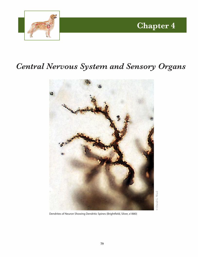
Dendrites of Neuron Showing Dendritic Spines (Bright�eld, Silver, x1880)
79
© D
avid
G. W
ard

80 Chapter 4: Central Nervous System and Sensory Organs
Sensory and Motor Neurons
Synaptic Bulb
Cell Body
Dendrite / Receptor
Axon (Central Process) Axon (Peripheral Process)
Synaptic Bulb
Cell Body
Dendrites
Dendrites
Dendrites
Axon
AxonHillock
Figure 4.1: Unipolar neuron (sensory). © David G. Ward.
Figure 4.2: Multipolar neuron (motor). © David G. Ward.
Chapter 4: Central Nervous System and Sensory Organs 81
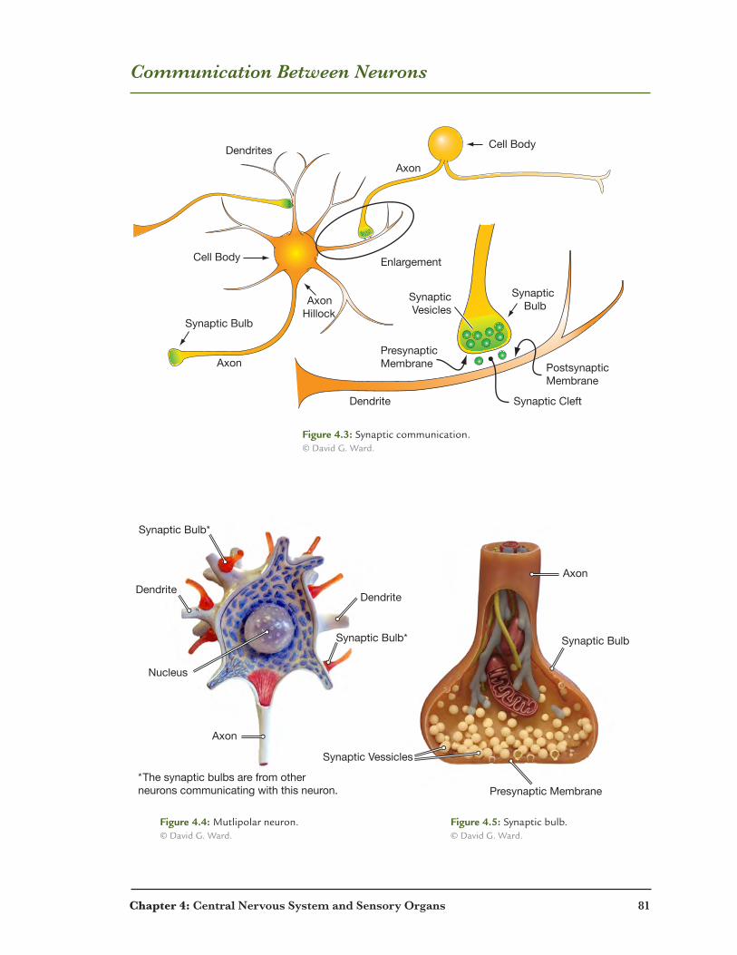
Communication Between Neurons
Synaptic Bulb
Synaptic Bulb
Cell Body
Cell BodyDendrites
Dendrite
Axon
Axon
AxonHillock
Enlargement
PresynapticMembrane
SynapticVesicles
PostsynapticMembrane
Synaptic Cleft
Axon
Axon
DendriteDendrite
Nucleus
Synaptic Bulb*
Synaptic Vessicles
Presynaptic Membrane
Synaptic Bulb
Synaptic Bulb*
*The synaptic bulbs are from other neurons communicating with this neuron.
Figure 4.3: Synaptic communication. © David G. Ward.
Figure 4.4: Mutlipolar neuron. © David G. Ward.
Figure 4.5: Synaptic bulb. © David G. Ward.
82 Chapter 4: Central Nervous System and Sensory Organs
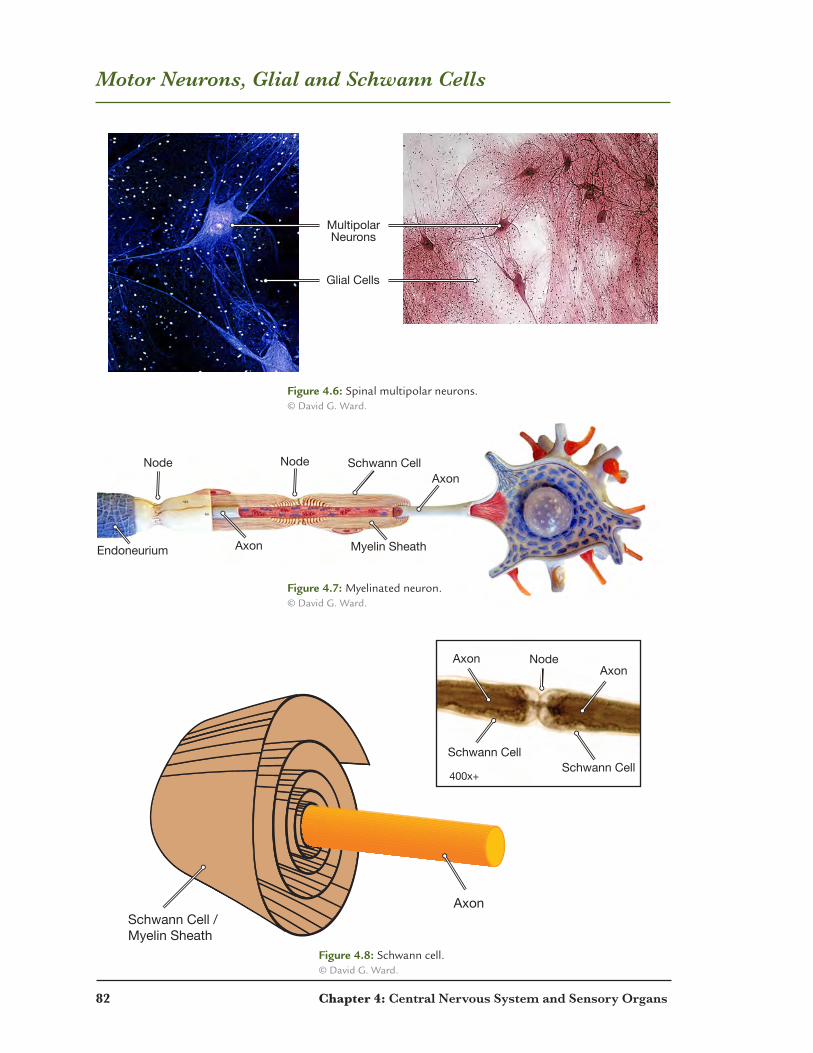
Motor Neurons, Glial and Schwann Cells
Glial Cells
MultipolarNeurons
NodeNodeAxon
Schwann Cell
Axon Myelin SheathEndoneurium
Node
Schwann Cell / Myelin Sheath
Axon
Schwann CellSchwann Cell
AxonAxon
400x+
Figure 4.6: Spinal multipolar neurons. © David G. Ward.
Figure 4.8: Schwann cell. © David G. Ward.
Figure 4.7: Myelinated neuron. © David G. Ward.
Chapter 4: Central Nervous System and Sensory Organs 83
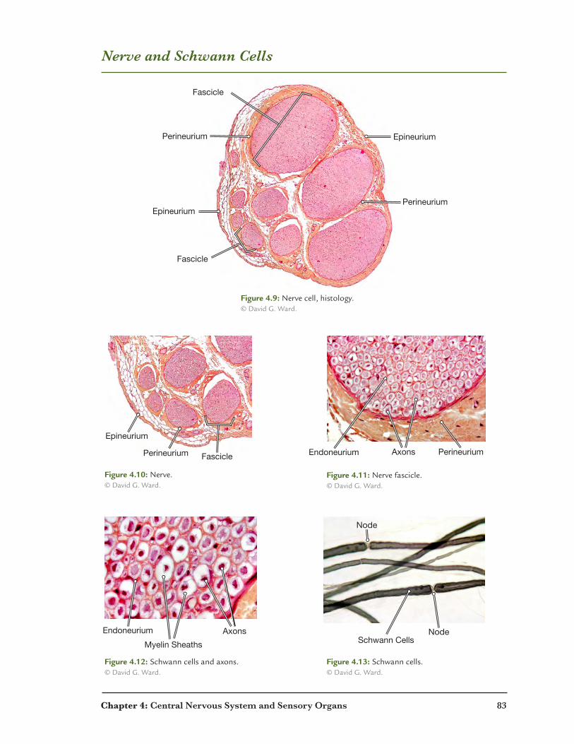
Nerve and Schwann Cells
Fascicle
Fascicle
Perineurium
Epineurium
Epineurium
Perineurium
Figure 4.10: Nerve. © David G. Ward.
Figure 4.9: Nerve cell, histology. © David G. Ward.
Figure 4.11: Nerve fascicle. © David G. Ward.
Figure 4.12: Schwann cells and axons. © David G. Ward.
Figure 4.13: Schwann cells. © David G. Ward.
Fascicle
Epineurium
Perineurium PerineuriumEndoneurium Axons
Endoneurium
Myelin Sheaths
Axons Node
NodeNode
Schwann Cells
Fascicle
Epineurium
Perineurium PerineuriumEndoneurium Axons
Endoneurium
Myelin Sheaths
Axons Node
NodeNode
Schwann Cells
84 Chapter 4: Central Nervous System and Sensory Organs
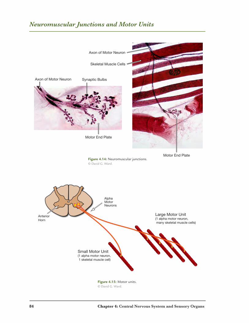
Neuromuscular Junctions and Motor Units
Skeletal Muscle Cells
Motor End Plate
Motor End Plate
Axon of Motor Neuron
Axon of Motor Neuron Synaptic Bulbs
AnteriorHorn
AlphaMotorNeurons
Small Motor Unit(1 alpha motor neuron, 1 skeletal muscle cell)
Large Motor Unit(1 alpha motor neuron, many skeletal muscle cells)
Figure 4.14: Neuromuscular junctions. © David G. Ward.
Figure 4.15: Motor units. © David G. Ward.

Chapter 4: Central Nervous System and Sensory Organs 85
Spinal Cord
Dorsal Horn
White Matter
Gray Matter
Ventral HornVentra Median
Sulcus
Dorsal Root
Central Canal
Ventral Root
Dorsal Root
Dorsal Horn
Dorsal Root Ganglion
Ventral Root Ventral Horn
Dura MaterPia MaterArachnoid
Figure 4.16: Cross section with meninges. © David G. Ward.
Figure 4.17: Meninges. © David G. Ward.
Figure 4.18: Horns and roots. © David G. Ward.

86 Chapter 4: Central Nervous System and Sensory Organs
Brain
Pyramids
Optic Chiasma
OpticNerve
TransverseFissure
Gyrus (Raised)LongitudinalFissure
Olfactory Blub Hypothalamus
ThalamusPons Medulla
Sulcus(Recessed)
Parietal Lobe
Frontal Lobe
Medulla
Pons
Temporal Lobe
Spinal Cord
Cerebrum
Cerebrum
Cerebrum
Cerebellum
Cerebellum
Cerebellum
Corpus Callosum
a. b.
c. d.
e. f.
Figure 4.19: Brain. © NCSU.
Chapter 4: Central Nervous System and Sensory Organs 87
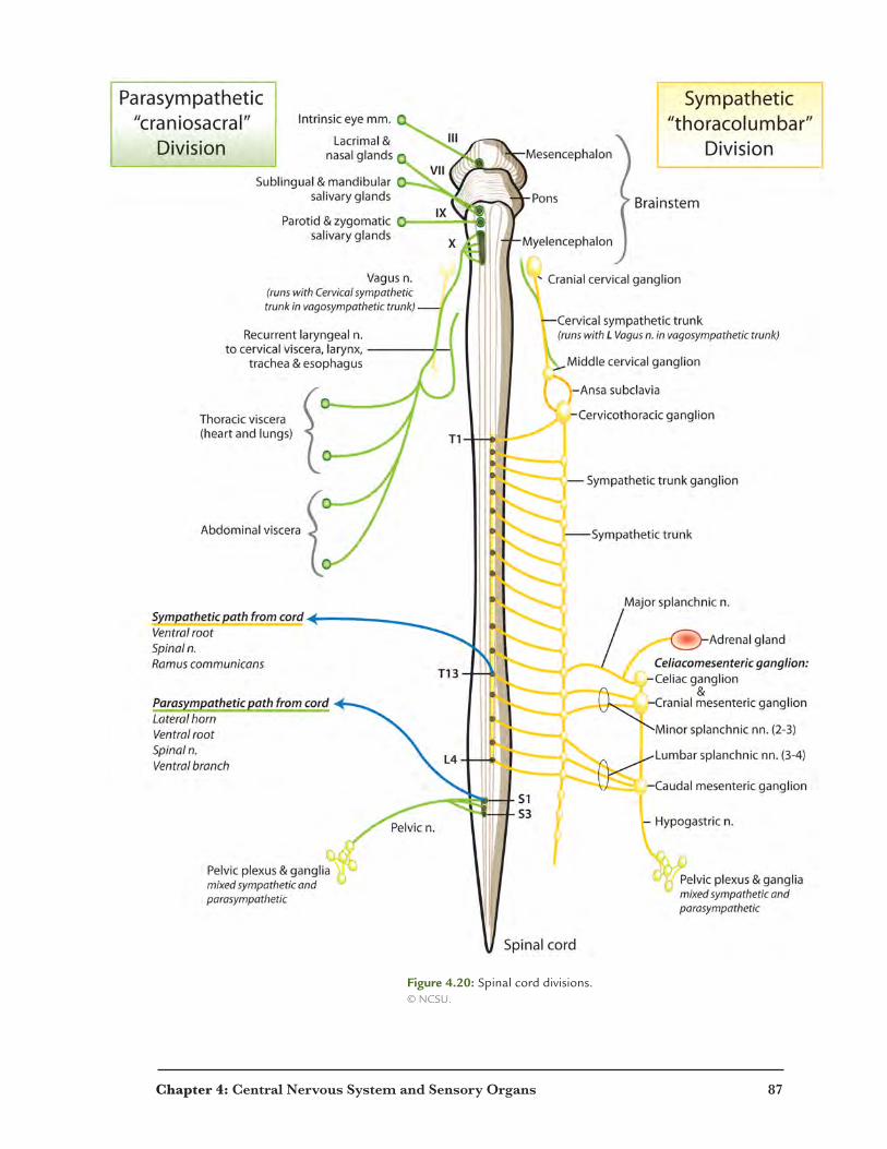
Figure 4.20: Spinal cord divisions. © NCSU.

88 Chapter 4: Central Nervous System and Sensory Organs
Parasympathetic Ganglia and Neurotransmitters
ParasympatheticPreganglionicNeurons
ParasympatheticPreganglionicNeurons
ParasympatheticPostganglionicNeuron
ParasympatheticPostganglionicNeuron
ParasympatheticPostganglionicNeuron
Thoracic and AbdominalIntramural
Ganglia
PelvicIntramural
Ganglia
Thoracic andAbdominalOrgans
PelvicOrgans
Middle Medulla
Sacral Spinal Cord
Ciliary, Sphenopalatine, Submandibular and OticGanglia
Organs of the Head
ParasympatheticPreganglionicNeuron
ParasympatheticPostganglionicNeuron
Intramural Ganglion
TargetOrgan
Enlargement
Enlargement
Acetylcholine
Acetylcholine
CholinergicNicotinic-NReceptors
CholinergicMuscarinicReceptorsMiddle Medulla
Figure 4.21: Parasympathetic ganglia. © David G. Ward.
Figure 4.22: Parasympathetic neurotransmitters. © David G. Ward.
Chapter 4: Central Nervous System and Sensory Organs 89
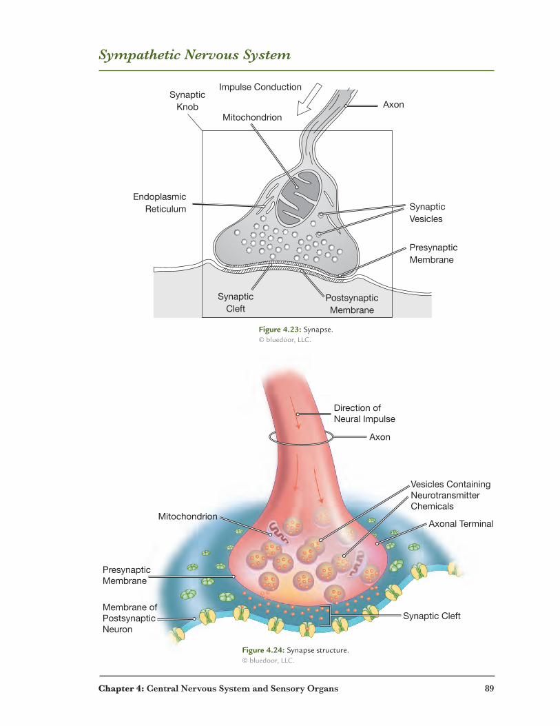
Sympathetic Nervous System
SynapticCleft
PostsynapticMembrane
PresynapticMembrane
SynapticVesicles
Axon
Impulse Conduction
Mitochondrion
SynapticKnob
EndoplasmicReticulum
Axon
PresynapticMembrane
Membrane ofPostsynapticNeuron
Vesicles ContainingNeurotransmitterChemicals
Axonal Terminal
Synaptic Cleft
Mitochondrion
Direction ofNeural Impulse
Figure 4.24: Synapse structure. © bluedoor, LLC.
Figure 4.23: Synapse. © bluedoor, LLC.
90 Chapter 4: Central Nervous System and Sensory Organs
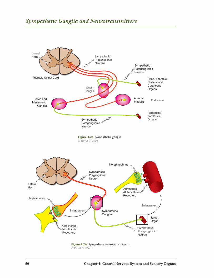
Sympathetic Ganglia and Neurotransmitters
LateralHorn Sympathetic
PreganglionicNeurons
SympatheticPostganglionicNeuron
SympatheticPostganglionicNeuron
Chain Ganglia
AdrenalMedulla
Celiac andMesenteric
Ganglia
Abdominaland PelvicOrgans
Head, Thoracic,Skeletal andCutaneousOrgans
Thoracic Spinal Cord
Endocrine
LateralHorn
SympatheticPreganglionicNeuron
SympatheticPostganglionicNeuron
Sympathetic Ganglion
TargetOrgan
Enlargement
Enlargement
Acetylcholine
Norepinephrine
CholinergicNicotinic-NReceptors
AdrenergicAlpha / BetaReceptors
Figure 4.25: Sympathetic ganglia. © David G. Ward.
Figure 4.26: Sympathetic neurotransmitters. © David G. Ward.

Chapter 4: Central Nervous System and Sensory Organs 91
Nerves
C6C7C8T1,2L4L5L6L7S1
Sacral Lumbar ThoracicCervical
Cranial
CervicalThoracic
CervicalThoracicLumbar
Sacral
Figure 4.27: Spinal nerves. © NCSU.
Figure 4.28: Spinal nerves. © NCSU.
a.
b.
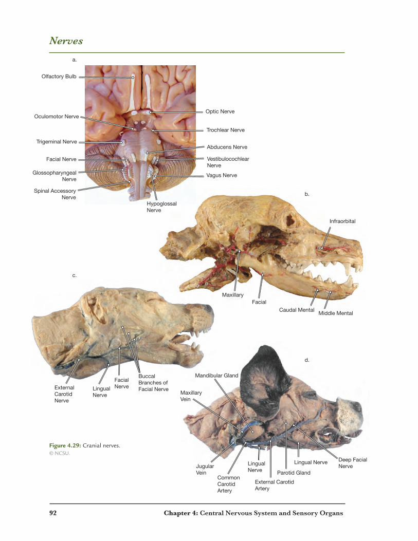
92 Chapter 4: Central Nervous System and Sensory Organs
Nerves
Olfactory Bulb
Oculomotor Nerve
Trigeminal Nerve
Facial Nerve
Hypoglossal Nerve
Vagus Nerve
Vestibulocochlear Nerve
Abducens Nerve
Trochlear Nerve
Optic Nerve
Glossopharyngeal Nerve
Spinal Accessory Nerve
Buccal Branches of Facial Nerve
FacialNerveLingual
NerveExternal Carotid Nerve
Deep FacialNerve
Parotid GlandLingualNerve
Mandibular Gland
JugularVein
Maxillary Vein
Lingual Nerve
External CarotidArtery
CommonCarotid Artery
a.
b.
c.
d.
FacialMaxillary
Infraorbital
Caudal Mental Middle Mental
Figure 4.29: Cranial nerves. © NCSU.

Chapter 4: Central Nervous System and Sensory Organs 93
Nerves of the Thoracic Limb
Brachial Plexus
Median Nerve
Ulnar Nerve
Axillary Nerve
Radial Nerve
Musculocutaneous Nerve
Figure 4.30: Nerves of the thoracic limb, lateral view. © NCSU.

94 Chapter 4: Central Nervous System and Sensory Organs
Nerves of Thoracic Limb
Figure 4.31: Ulnar nerve. © NCSU.
Figure 4.33: Radial nerve. © NCSU.
Figure 4.34: Axillary nerve. © NCSU.
Figure 4.35: Median nerve. © NCSU.
Figure 4.32: Musculocutaneous nerve. © NCSU.
Chapter 4: Central Nervous System and Sensory Organs 95
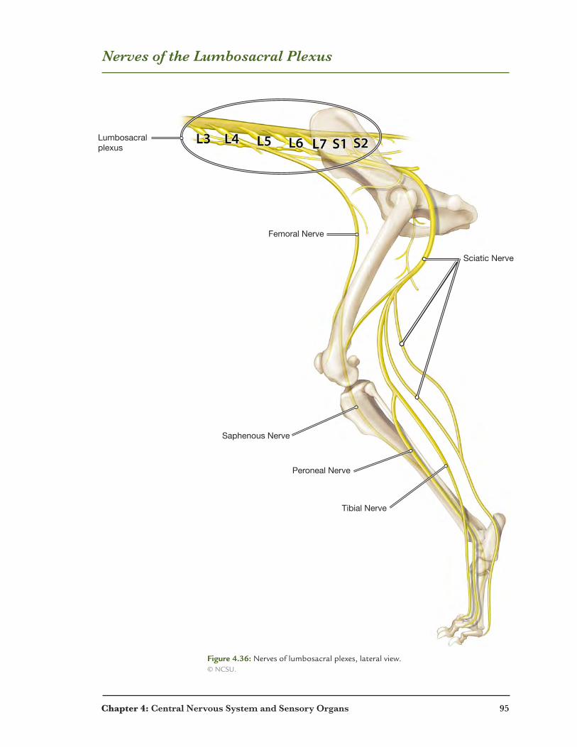
Nerves of the Lumbosacral Plexus
Sciatic Nerve
Femoral Nerve
Saphenous Nerve
Peroneal Nerve
Tibial Nerve
Lumbosacralplexus
Figure 4.36: Nerves of lumbosacral plexes, lateral view. © NCSU.
96 Chapter 4: Central Nervous System and Sensory Organs
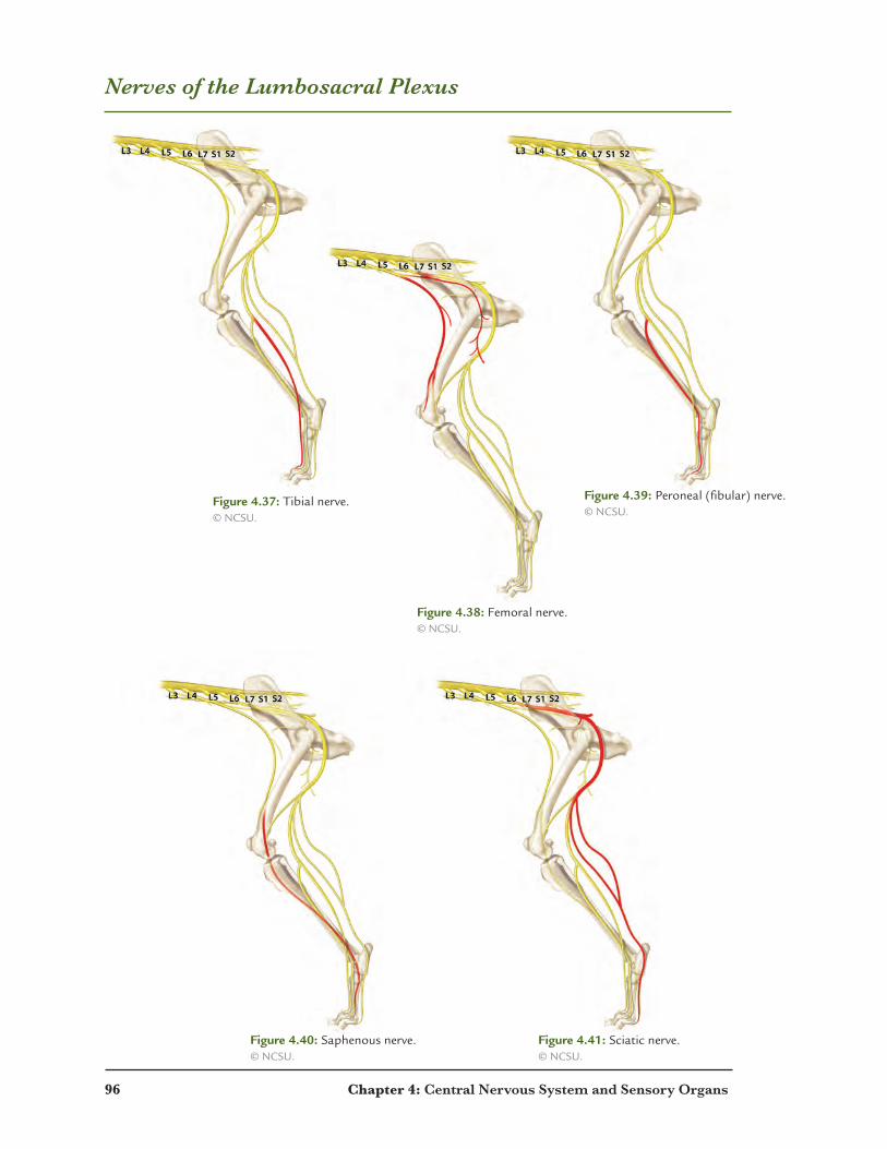
Nerves of the Lumbosacral Plexus
Figure 4.37: Tibial nerve. © NCSU.
Figure 4.38: Femoral nerve. © NCSU.
Figure 4.39: Peroneal (fibular) nerve. © NCSU.
Figure 4.40: Saphenous nerve. © NCSU.
Figure 4.41: Sciatic nerve. © NCSU.

Chapter 4: Central Nervous System and Sensory Organs 97
Dermatomes and Stretch Reflex
C2
C3
C4
C5C6
L4L5
L6L7
L7
L8
T1
C8
C7
C7
T2T3
T4
T5T6
T7
T8T9
T10
T11T12
T13
L1L2
L3/L4
L5
S1S2
SensoryNeuron(detects muscle stretch)
AnteriorHorn
AlphaMotorNeuron(contracts muscle)
MuscleSpindle
MotorUnit
Figure 4.42: Dermatomes. © NCSU.
Figure 4.43: Spinal stretch reflex. © David G. Ward.

98 Chapter 4: Central Nervous System and Sensory Organs
Sensory Organs: Ear
Pinna
Pinna and Temporlis Muscle
Pinna
Pinna and Temporlis Muscle
Cochlea
Ossicles
Tympanic Membrane
Eustachian Tube
SemicircularCanals
3
6
HorizontalEar Canal
Vertical Ear Canal
3
4
5
6
Figure 4.44: Inner ear model. © NCSU.
Figure 4.45: Heathly ear model. © NCSU.
Figure 4.46: Diseased ear model. © NCSU.
Chapter 4: Central Nervous System and Sensory Organs 99
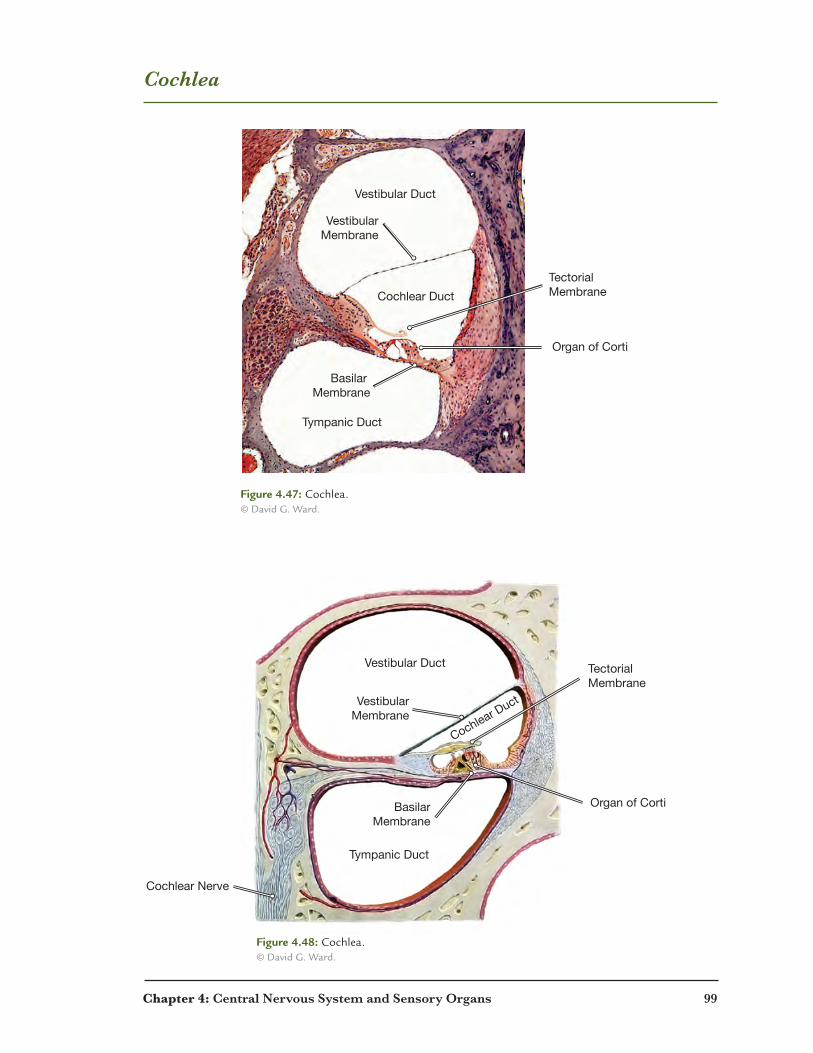
Cochlea
Cochlear Duct
Vestibular Duct
Tympanic Duct
Organ of Corti
VestibularMembrane
Basilar Membrane
TectorialMembrane
Cochlear Nerve
Vestibular Membrane
Basilar Membrane
TectorialMembrane
Organ of Corti
Vestibular Duct
Cochlear Duct
Tympanic Duct
Figure 4.47: Cochlea.© David G. Ward.
Figure 4.48: Cochlea.© David G. Ward.

100 Chapter 4: Central Nervous System and Sensory Organs
Cochlea and Vestibular Apparatus
Outer Hair Cells
Inner Hair Cell
BasilarMembrane
Stereocilia
Rods of Corti
Tectorial Membrane
Cochlear Duct(Filled with Endolymph)
VestibularMembrane
Organ ofCorti
Superior Semicircular Canal
Utricle and Saccule
Vestibular Ganglion
Vestibular Nerve
Cochlear nerve
Ampulla of Posterior Semicircular Canal
Posterior Semicircular Canal
Lateral Semicircular Canal
© D
avid
G. W
ard
Figure 4.49: Organ of corti.© David G. Ward.
Figure 4.50: Vestibular apparatus and cochlea, posterior view.© David G. Ward.

Chapter 4: Central Nervous System and Sensory Organs 101
Sensory Organs: Eye
Cornea
Iris
Suspensory Ligaments
Ciliary Body
Pupil
Aqueous HumorLens
Vitreous Humor
Optic Disk
Optic Nerve
Fovea
Retina
Choroid
Sclera
Iris
Lens
Upper eyelid
Cornea
Third Eyelid
Lower Eyelid
Iris
Third Eyelid
Medial Rectus Muscle
Superior Rectus Muscle
Lateral Rectus Muscle
Inferior Rectus Muscle
Left Eye
Lens
Cornea
VitreousHumor
Pupil
Figure 4.51: Eye, midsagittal.© David G. Ward.
Figure 4.52: Eye, anterior.© NCSU.
102 Chapter 4: Central Nervous System and Sensory Organs
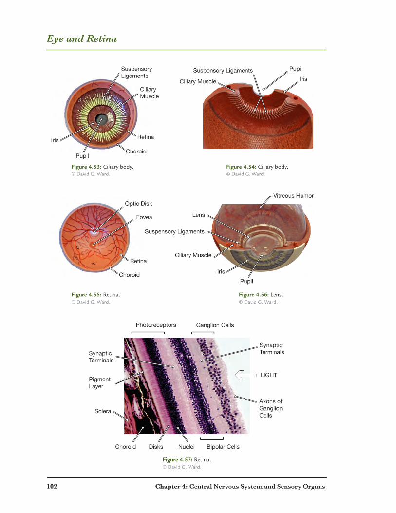
Eye and Retina
Figure 4.53: Ciliary body.© David G. Ward.
Figure 4.54: Ciliary body.© David G. Ward.
Figure 4.55: Retina.© David G. Ward.
Figure 4.56: Lens.© David G. Ward.
Figure 4.57: Retina.© David G. Ward.
Sclera
Choroid
Photoreceptors Ganglion Cells
Bipolar CellsDisks Nuclei
SynapticTerminals
SynapticTerminals
Axons ofGanglionCells
PigmentLayer
LIGHT
Retina Lens
Ciliary Body
CiliaryMuscle
RetinaIris
SuspensoryLigaments
Suspensory Ligaments
Suspensory Ligaments
Choroid
Retina
Choroid
Fovea
Pupil
Iris
Lens
Vitreous Humor
Pupil
Iris
Pupil
Ciliary Muscle
Ciliary Muscle
Ciliary Body
Optic Disk
Retina

Chapter 4: Central Nervous System and Sensory Organs 103
Review: Coloring Activity
Axon
Axon hillock
Axon terminals
Cell body
Cell body nucleolus
Cell body nucleus
Myelin sheath
Nissl bodies
Node of Ranvier
Schwann cell nucleus
Figure 4.58: Nerve coloring activity.© bluedoor, LLC.

104 Chapter 4: Central Nervous System and Sensory Organs
Review: Word Search
N I V P L W C D V R X O C M H F E U B T D
V O Z I S A N Q E E P U U B L J N E M Y I
T V I R T P T T F T R S P I A M A T E R O
M Y H T Y R A N I D O T Q L Z T R S W E N
A U C J C M E C O L F Y I O I H B F R L H
B V J Y A N C O L Z Y H R C E V M S A Y C
N W A R O H U A U X I E A B A F E Y D Q A
C Q U H I X C J D S G R O B Z L M N R X R
J D Q A U S L I R M H G O Z A Q C A E E A
Q G S S U X K B H A Q U G H N V I P T I P
P M L P F W B N S K L Y M W A P N T I B V
A S R I V V W O K I W U H O A N A I N O O
J O X L L E C N N A W H C S R D P C A A U
C P T L T Y D G Q L P C Q S I U M B M A V
R X P H N I W I S U S L W K U Y Y U S M J
P G R I K B L N W V W H Y Y V M T L R E H
A E T G H I V B A C C V E J K V O B S P K
E X M P V R E O B U N O B I V C F R R O B
S Y N A P T I C C L E F T Z K J X G U T F
V E Z Y T I C Y W A C Z A W X Q Z P F E N
D C B C I X W R C M R M X L W G Z G P W N
1. terminal end of a neuron 2. space of communication between two neurons 3. responsible for creating myelin sheath 4. nervous and skeletal muscle communication 5. innermost meninge layer 6. center meninge layer 7. outermost meninge layer 8. where the optic nerve enters the brain and crosses over 9. structure that joins the left and right hemisphere of the cerebrum 10. the eardrum11. where bipolar cells are located12. the number of eyelids in a dog eye13. the gelatinous substance posterior to the lens14. the ear canal most proximal to the pinna 15. the ear canal most proximal to the tympanic membrane

Chapter 4: Central Nervous System and Sensory Organs 105
Review: Crossword Puzzle
16
2
3
4 5
7 8
9
10 11 12
13 14
15
1 6
Across 1. contains the organ of corti 3. space on an axon in between myelin sheaths 9. receptor of a neuron10. conducting region between the cell body and synaptic bulb11. unipolar neuron13. regions of the skin innervated by specific spinal nerves15. layer surrounding a fascicle
Down 2. this fissure divides the cerebrum from the cerebellum 4. bundle of nerve fibers 5. this fissure divides the left from the right hemisphere of the cerebellum 6. multipolar neuron 7. dog’s ear 8. where neurotransmitters are stored12. thoracolumbar division14. outermost layer of a nerve16. craniosacral division

Chapter 7
GI Tract and the Digestive System
Intestinal Villi Showing Goblet Cells (Bright�eld, Trichrome; x350)
151
© D
avid
G. W
ard

152 Chapter 7: GI Tract and the Digestive System
Gastrointestinal Tract Overview
Mouth
Esophagus
Salivary Glands
Parotid Gland
Kidney
Large Intestine/Colon
Rectum
Anus
Liver
Gallbladder
Stomach
Spleen
Small Intestine
Figure 7.1: Gastrointestinal tract and related structures. © NCSU.

Chapter 7: GI Tract and the Digestive System 153
Esophagus
SubmucosaSubmucosa
Inner Circular Layer
Outer Longitudinal Layer
Muscularis Mucosae
Lamina Propria
Stratified SquamousEpithelium
Submucosa
Inner Circular Layer
Outer LongitudinalLayer
Muscularis Mucosae
Lamina Propria
Stratified SquamousEpithelium
Non-Keratinized Stratified Squamous Epithelium
MuscularisExterna
Mucosa
Submucosa
MuscularisExterna
Mucosa
Figure 7.2: Esophagus. © David G. Ward.
Figure 7.4: Esophagus. © David G. Ward.
Figure 7.3: Esophagus.(autofluorescence) © David G. Ward.
Figure 7.5: Esophagus.(autofluorescence) © David G. Ward.
154 Chapter 7: GI Tract and the Digestive System
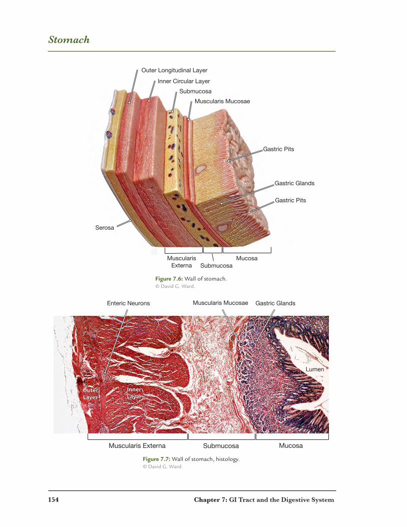
Stomach
Submucosa
Submucosa
Inner Circular Layer
Outer Longitudinal Layer
Muscularis Mucosae
MuscularisExterna
Mucosa
Serosa
Gastric Glands
Gastric Pits
Gastric Pits
Muscularis Externa MucosaSubmucosa
Gastric GlandsEnteric Neurons
InnerLayer
OuterLayer
Lumen
Muscularis Mucosae
Figure 7.6: Wall of stomach. © David G. Ward.
Figure 7.7: Wall of stomach, histology. © David G. Ward.

Chapter 7: GI Tract and the Digestive System 155
Stomach and Intestines
Goblet Cells(Mucus)
Chief Cells(Pepsinogen)
Parietal Cells(Orange) (HCl)
Parietal Cells(HCl)
x400 x200
Lumen
Muscularis Externa Mucosa
Villus
Submucosa
SubmucosaInner Circular Layer
Outer Longitudinal Layer
Muscularis Mucosae
Serosa
MyentericPlexus
(EntericNeaurons)
LymphaticNodule
DuodenalGland
Intestinal GlandsLamina Propria
Intestinal Capillaries
Goblet CellsColumnar Cells
Lacteals
Crypt
Figure 7.8: Mucosa of the stomach, histology, x400. © David G. Ward.
Figure 7.9: Mucosa of the stomach, histology, x200. © David G. Ward.
Figure 7.10: Small intesine. © David G. Ward.
156 Chapter 7: GI Tract and the Digestive System
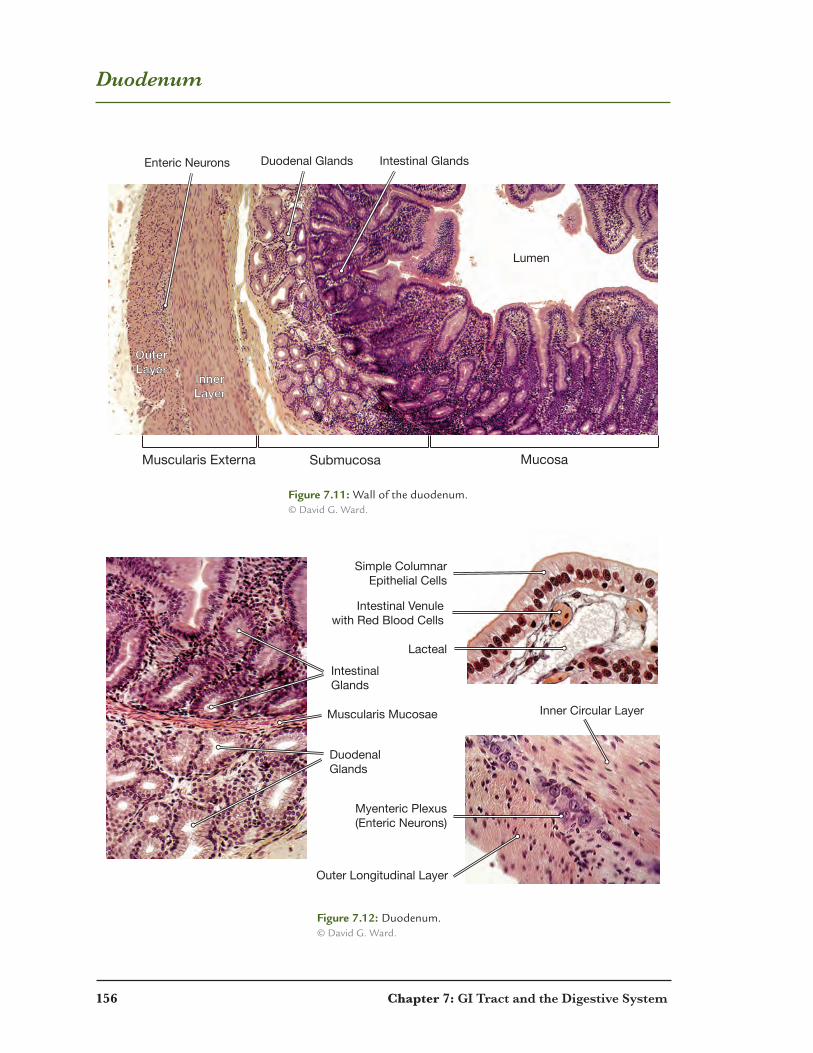
Duodenum
InnerLayer
OuterLayer
Enteric Neurons
Lumen
Duodenal Glands Intestinal Glands
Muscularis Externa MucosaSubmucosa
Myenteric Plexus (Enteric Neurons)
Simple ColumnarEpithelial Cells
Lacteal
Intestinal Venulewith Red Blood Cells
DuodenalGlands
IntestinalGlands
Inner Circular Layer
Outer Longitudinal Layer
Muscularis Mucosae
Figure 7.11: Wall of the duodenum. © David G. Ward.
Figure 7.12: Duodenum. © David G. Ward.
Chapter 7: GI Tract and the Digestive System 157
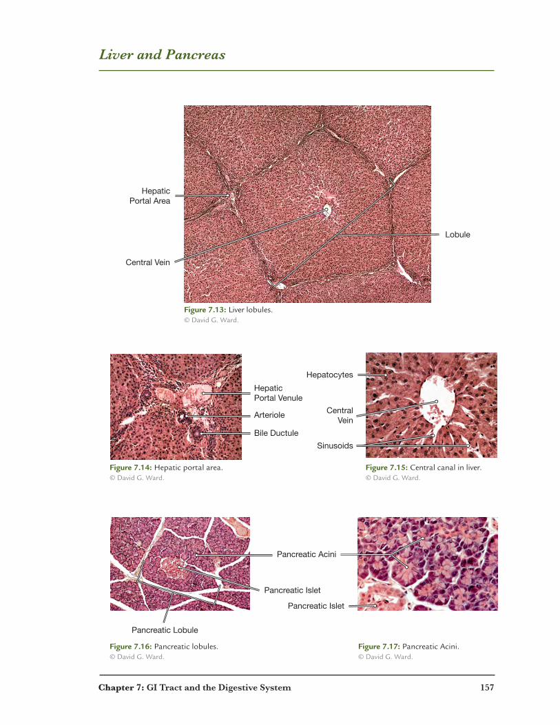
Liver and Pancreas
Central Vein
Lobule
HepaticPortal Area
HepaticPortal Venule
CentralVein
Hepatocytes
Sinusoids
Arteriole
Bile Ductule
Pancreatic Lobule
Pancreatic Islet
Pancreatic Islet
Pancreatic Acini
Figure 7.13: Liver lobules. © David G. Ward.
Figure 7.14: Hepatic portal area. © David G. Ward.
Figure 7.15: Central canal in liver. © David G. Ward.
Figure 7.16: Pancreatic lobules. © David G. Ward.
Figure 7.17: Pancreatic Acini. © David G. Ward.

158 Chapter 7: GI Tract and the Digestive System
Fundus
Body
Pyloris
Stomach
LargeIntestine
Colon
Rectum
Anus
Gallbladder
Duodenum
Jejunum
Ileum
SmallIntestine
Cecum
Figure 7.18: Digestive organs. © NCSU.

Chapter 7: GI Tract and the Digestive System 159
Digestive Organs
Liver
Liver
Small Intestine
Small Intestine
Small Intestine
Large Intestine
Large Intestine
Greater Omentum(Covering)
Greater Omentum(Covering)
Heart
Figure 7.19: Digestive organs.© NCSU.
a.
b.
c.
160 Chapter 7: GI Tract and the Digestive System
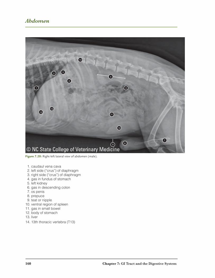
Abdomen
1
2 3
4
14
5
6
11
10
789
1213
1. caudaul vena cava 2. left side (“crus”) of diaphragm 3. right side (“crus”) of diaphragm 4. gas in fundus of stomach 5. left kidney 6. gas in descending colon 7. os penis 8. prepuce 9. teat or nipple10. ventral region of spleen11. gas in small bowel12. body of stomach13. liver14. 13th thoracic vertebra (T13)
Figure 7.20: Right-left lateral view of abdomen (male).
Chapter 7: GI Tract and the Digestive System 161
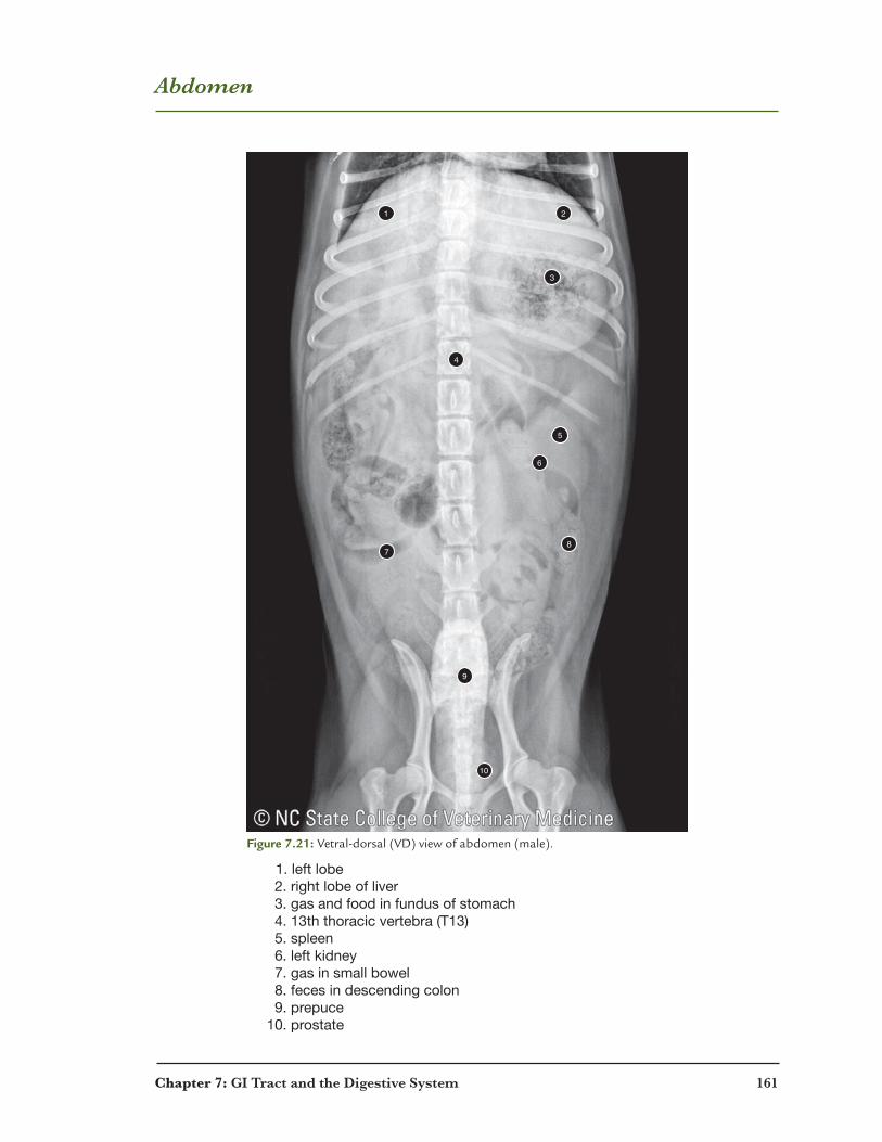
Abdomen
1 2
3
4
5
6
87
9
10
1. left lobe 2. right lobe of liver 3. gas and food in fundus of stomach 4. 13th thoracic vertebra (T13) 5. spleen 6. left kidney 7. gas in small bowel 8. feces in descending colon 9. prepuce 10. prostate
Figure 7.21: Vetral-dorsal (VD) view of abdomen (male).
162 Chapter 7: GI Tract and the Digestive System
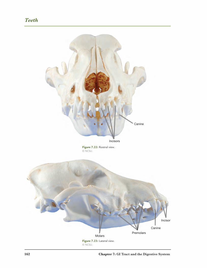
Teeth
MolarsPremolars
Canine
Incisor
Incisors
Canine
Figure 7.22: Rostral view. © NCSU.
Figure 7.23: Lateral view. © NCSU.

Chapter 7: GI Tract and the Digestive System 163
Incisors
Canine
Premolars
Molars
Incisors
Canine
Premolars
Molars
Upper 4th Premolar (Carnassial Tooth)
a.
b.
Figure 7.24: Teeth. a. Upper jaw. b. Lower jaw. © NCSU.
164 Chapter 7: GI Tract and the Digestive System
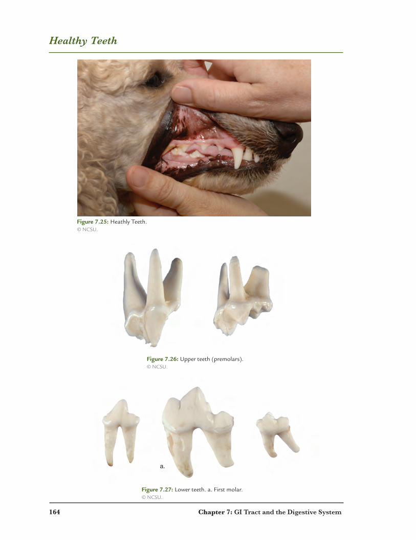
Healthy Teeth
Figure 7.25: Heathly Teeth. © NCSU.
Figure 7.26: Upper teeth (premolars). © NCSU.
Figure 7.27: Lower teeth. a. First molar. © NCSU.
a.
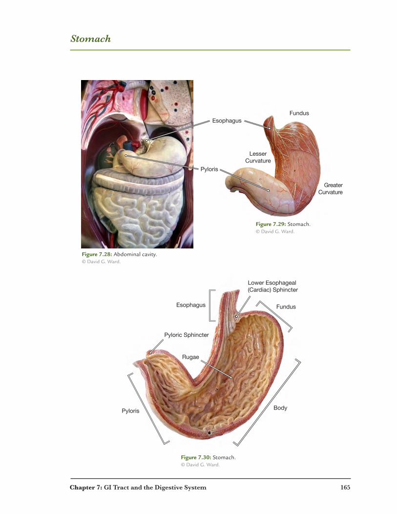
Chapter 7: GI Tract and the Digestive System 165
Stomach
Esophagus
Pyloris
Fundus
LesserCurvature
GreaterCurvature
Rugae
Lower Esophageal(Cardiac) Sphincter
Pyloric Sphincter
Esophagus
Pyloris
Fundus
Body
Figure 7.28: Abdominal cavity. © David G. Ward.
Figure 7.29: Stomach. © David G. Ward.
Figure 7.30: Stomach. © David G. Ward.

166 Chapter 7: GI Tract and the Digestive System
Small and Large Intestines
Transverse Mesocolon
Ascending Colon
DescendingColon
Figure 7.31: Small intestine. © blueboor, LLC.
Figure 7.32: Large intestine (colon). © Davis G. Ward.

Chapter 7: GI Tract and the Digestive System 167
Liver and Pancreas
Common Hepatic Duct Gallbladder
Cystic Duct
Hepatic Artery
Common Bile Duct
Hepatic Portal Vein
Lobule of PancreasPancreaticDuct
Duodenum
DuodenalAmpulla
Common Bile Duct
Adrenal glands
Kidney
Figure 7.33: Liver. © David G. Ward.
Figure 7.34: Pancreas. © David G. Ward.
168 Chapter 7: GI Tract and the Digestive System
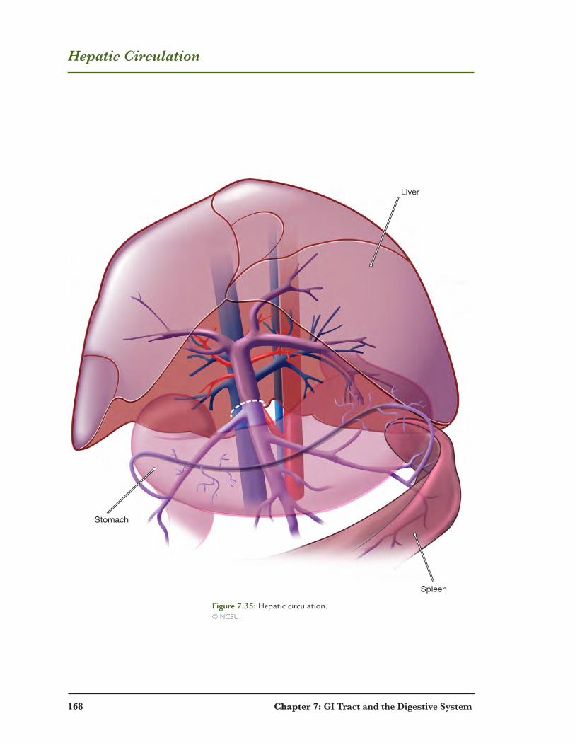
Hepatic Circulation
Liver
Stomach
Spleen
Figure 7.35: Hepatic circulation. © NCSU.
Chapter 7: GI Tract and the Digestive System 169
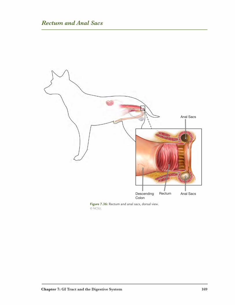
Rectum and Anal Sacs
Anal SacsRectumDescending Colon
Anal Sacs
Figure 7.36: Rectum and anal sacs, dorsal view. © NCSU.

170 Chapter 7: GI Tract and the Digestive System
Review: Stomach
The stomach is responsible for receiving the food you have swallowed. It then churns and mixes food with stomach juices to begin the digestion process. To accomplish this function, the stomach needs the following types of tissues.
1. This hollow organ needs a protective inner lining that is also able to secrete digestive juices.
A protective tissue that also secretes is ______________________ tissue.
2. In order to mix food and digestive juices the stomach contracts to squeeze its contents. The
tissue that produces movement of an internal organ is __________________ tissue.
3. The stomach is covered by a protective membrane. A protective tissue is
________________ tissue.
4. Thestomachisanchoredintheabdominalcavitybyfibroustissue.Tissuethatbindsand
hasstrongfibersis__________________tissue.
5. The stomach receives information regarding the digestive process. Tissue that is able to
carry information throughout the body is ____________________ tissue.
6. Put the following structures in order as food passes through the digestive system.
descending colon esophagus anus
ascending colon lower esophageal sphincter stomach
transverse colon pyloric sphincter rectum
1. mouth
2.
3.
4.
5.
6.
7.
8.
9.
10.
7. List the three sections of the small intestine.
1.
2.
3.
Chapter 7: GI Tract and the Digestive System 171
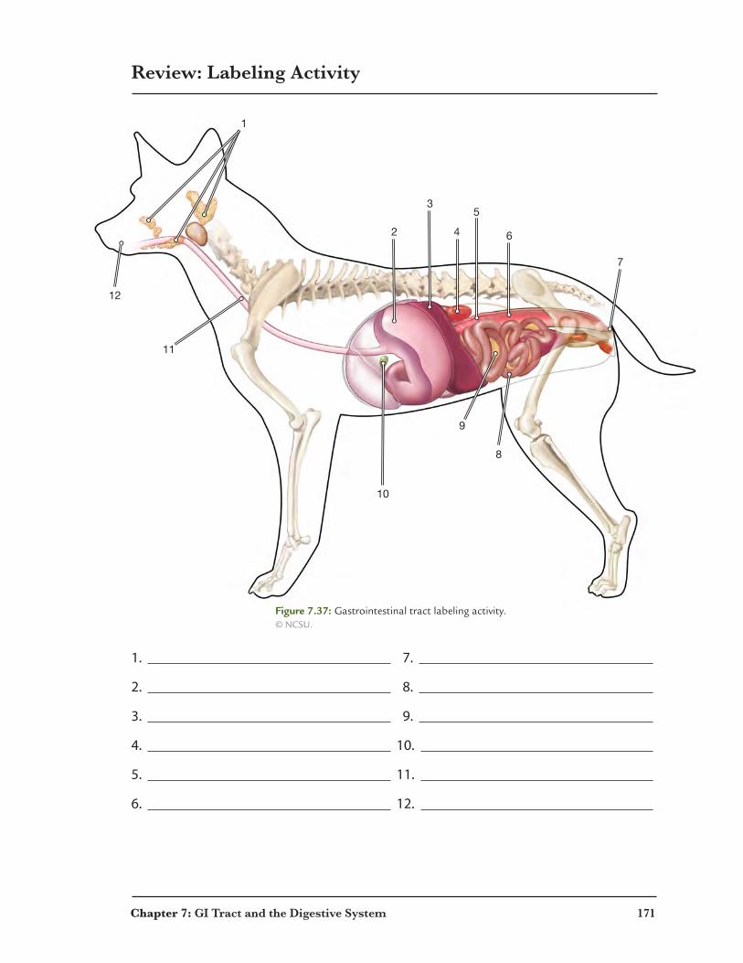
Review: Labeling Activity
12
11
1
10
2
3
4
5
6
7
9
8
1. 7.
2. 8.
3. 9.
4. 10.
5. 11.
6. 12.
Figure 7.37: Gastrointestinal tract labeling activity. © NCSU.

172 Chapter 7: GI Tract and the Digestive System
Review: Crossword Puzzle
Across 3. the lower region of the stomach 4. cell type located in the liver 6. the center region of the small intestine 7. the center region of the stomach10. the outermost layer of the muscularis externa12. folds located in the stomach that increase surface area13. another name for mechanical digestion14. this structure joins the descending colon and the anal area15. the final region of the small intestine that connects to the colon
Down 1. the upper region of the stomach 2. this acid starts the hydrolysis of proteins 5. the inactive precursor to gastric pepsin 8. the first region of the small intestine 9. where the digestive process begins11. the first section of the colon
1
2
3
4
5
6
97 8
10
12
14
11
13
15

Chapter 7: GI Tract and the Digestive System 173
Review: Word Search
V A A L Q I I E S C R F R M M I M D
Z Z P R A J E U P A S D N O Z U K I
O M D A S N B W L N N O L W S J O C
E E U E N M E O C I R A I C W X S A
A L Q C U C M D P N R H U V C P A C
H Z G C O E R H O E Y L Z P K F E I
U T O P R S B E E U A X K Y R P R R
A S K P H Y A W A R D O H A N E C O
A U B M D B X D I T W I A I Z P N L
S U G A H P O S E B I C R J P S A H
P J X C L L E G P R R C W R M I P C
C E K O H X O S Z O N V I P X N H O
L M Q F T J S B V F L I W S T O B R
U S W E V G C P O X V I Q D L G D D
A E R A L A T R O P C I T A P E H Y
Q N H T X X D L I M Z L Y Z Q N T H
A O Y R N V N C X T R I N C I S O R
P Y L O R I C S P H I N C T E R A L
1. produced by parietal cells
2. produced by chief cells
3. esophagus, stomach and small intestinal layer lining the lumen
4. layer between the mucosa and muscularis externa in the digestive system
5. muscular layer deep to the submucosa
6. a gland located in the small intestine submucosa
7. contains hepatic portal venule, arteriole and bile ductule
8. located in a pancreatic lobule
9. connects the stomach to the duodenum of the small intestine
10. muscular structure connecting the mouth to the stomach
11. organ that produces digestive enzymes released into the small intestine
12. small teeth in the front of the jaw
13. relatively long tooth also called a cuspid or fang
14. crushing teeth in the back of the jaw
15. four teeth on each side of the jaw located behind the canine
Chapter 8
Kidneys, Bladder and the Urinary System
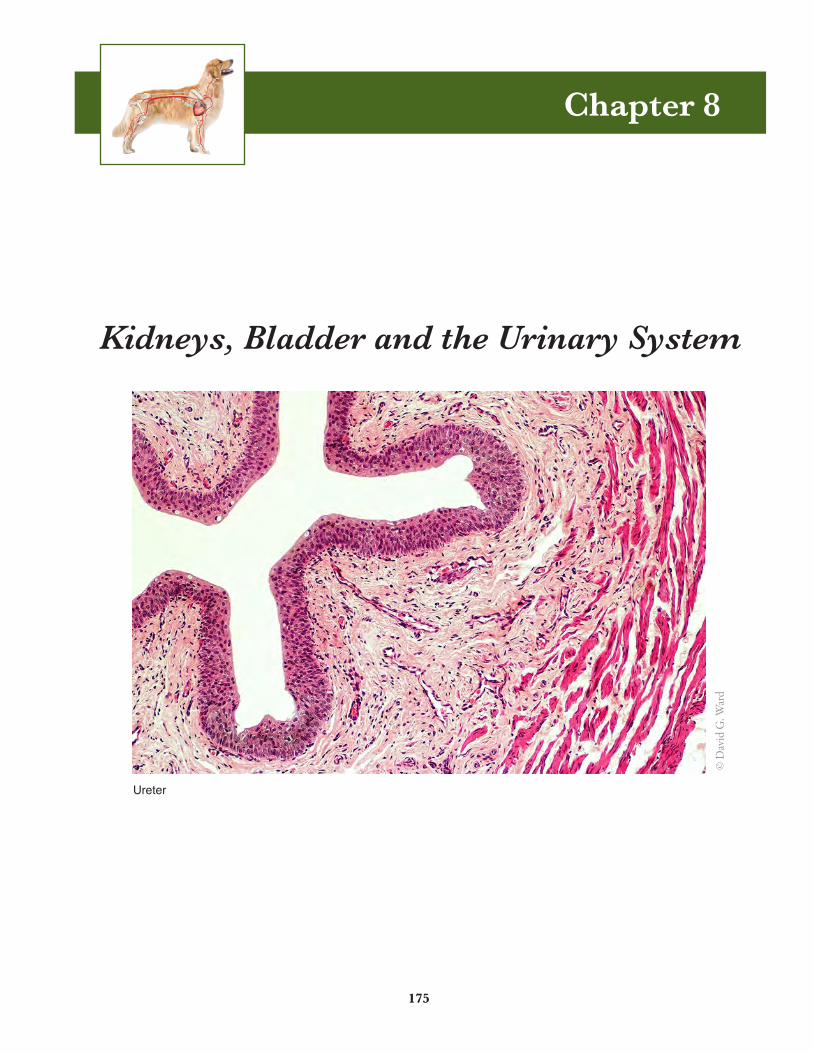
Ureter
175
© D
avid
G. W
ard
176 Chapter 8: Kidneys, Bladder and the Urinary System
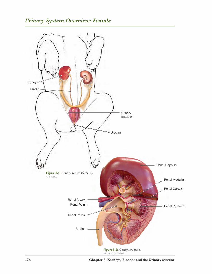
Urinary System Overview: Female
Ureter
Renal Pelvis
Renal ArteryRenal Vein Renal Pyramid
Renal Cortex
Renal Medulla
Renal Capsule
Kidney
Ureter
Urinary Bladder
Urethra
Figure 8.1: Urinary system (female). © NCSU.
Figure 8.2: Kidney structure. © David G. Ward.
Chapter 8: Kidneys, Bladder and the Urinary System 177
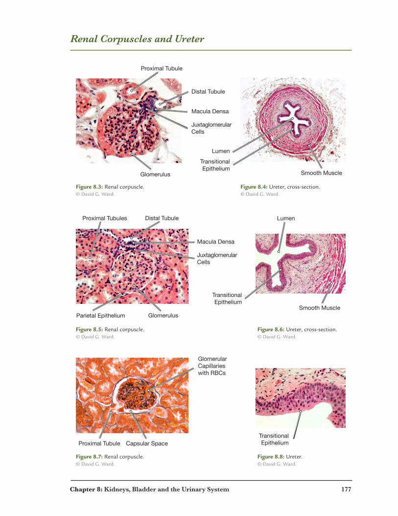
Renal Corpuscles and Ureter
Distal Tubule
Distal Tubule
Proximal Tubule
Proximal Tubules
Proximal Tubule Capsular Space
Glomerular Capillarieswith RBCs
Glomerulus Smooth Muscle
Smooth MuscleGlomerulusParietal Epithelium
Macula Densa
Lumen
TransitionalEpithelium
Lumen
TransitionalEpithelium
TransitionalEpithelium
JuxtaglomerularCells
Macula Densa
JuxtaglomerularCells
Figure 8.3: Renal corpuscle. © David G. Ward.
Figure 8.5: Renal corpuscle. © David G. Ward.
Figure 8.7: Renal corpuscle. © David G. Ward.
Figure 8.4: Ureter, cross-section. © David G. Ward.
Figure 8.6: Ureter, cross-section. © David G. Ward.
Figure 8.8: Ureter. © David G. Ward.
178 Chapter 8: Kidneys, Bladder and the Urinary System
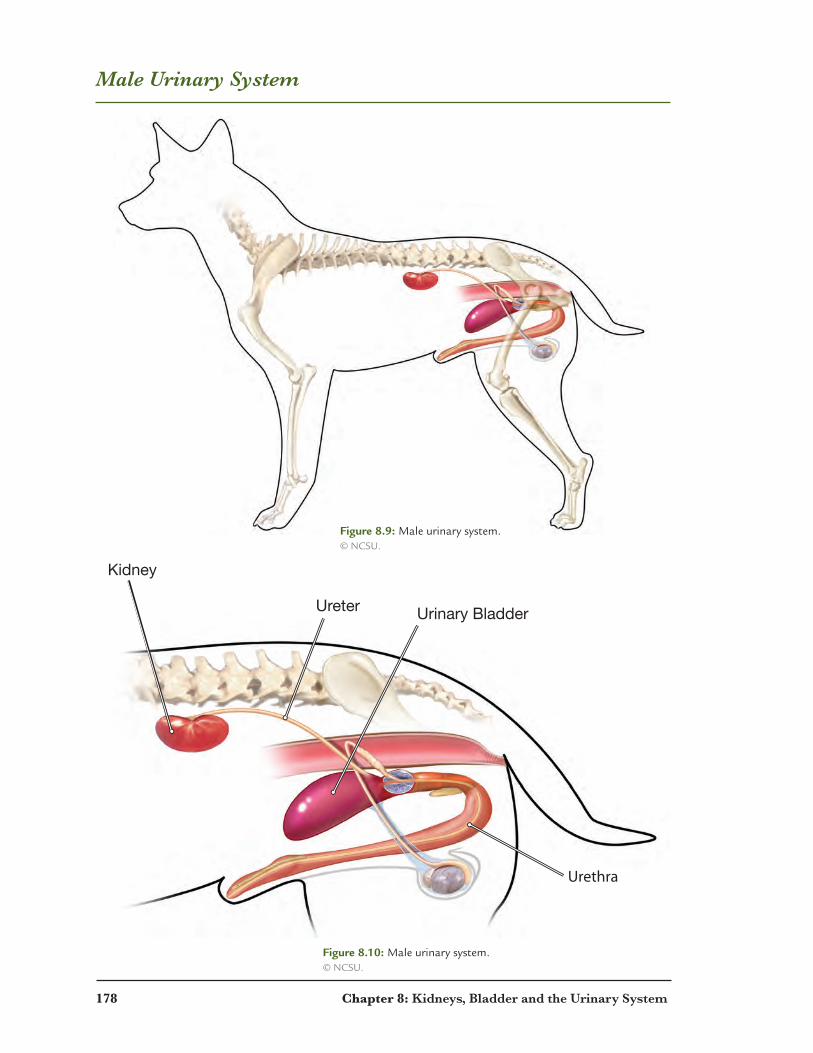
Male Urinary System
Urethra
Urinary BladderUreter
Kidney
Figure 8.9: Male urinary system. © NCSU.
Figure 8.10: Male urinary system. © NCSU.

Chapter 8: Kidneys, Bladder and the Urinary System 179
Female Urinary System
UrinaryBladder
Ureter
Kidney
Urethra
Figure 8.11: Female urinary system. © NCSU.
Figure 8.12: Female urinary system. © NCSU.
180 Chapter 8: Kidneys, Bladder and the Urinary System
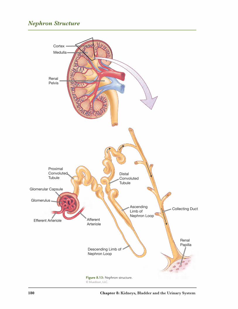
Nephron Structure
Cortex
Glomerular Capsule
Glomerulus
Descending Limb ofNephron Loop
Collecting DuctAscending Limb of Nephron Loop
AfferentArteriole
ProximalConvolutedTubule
Medulla
RenalPelvis
Efferent Arteriole
DistalConvolutedTubule
RenalPapilla
Figure 8.13: Nephron structure. © bluedoor, LLC.
Chapter 8: Kidneys, Bladder and the Urinary System 181
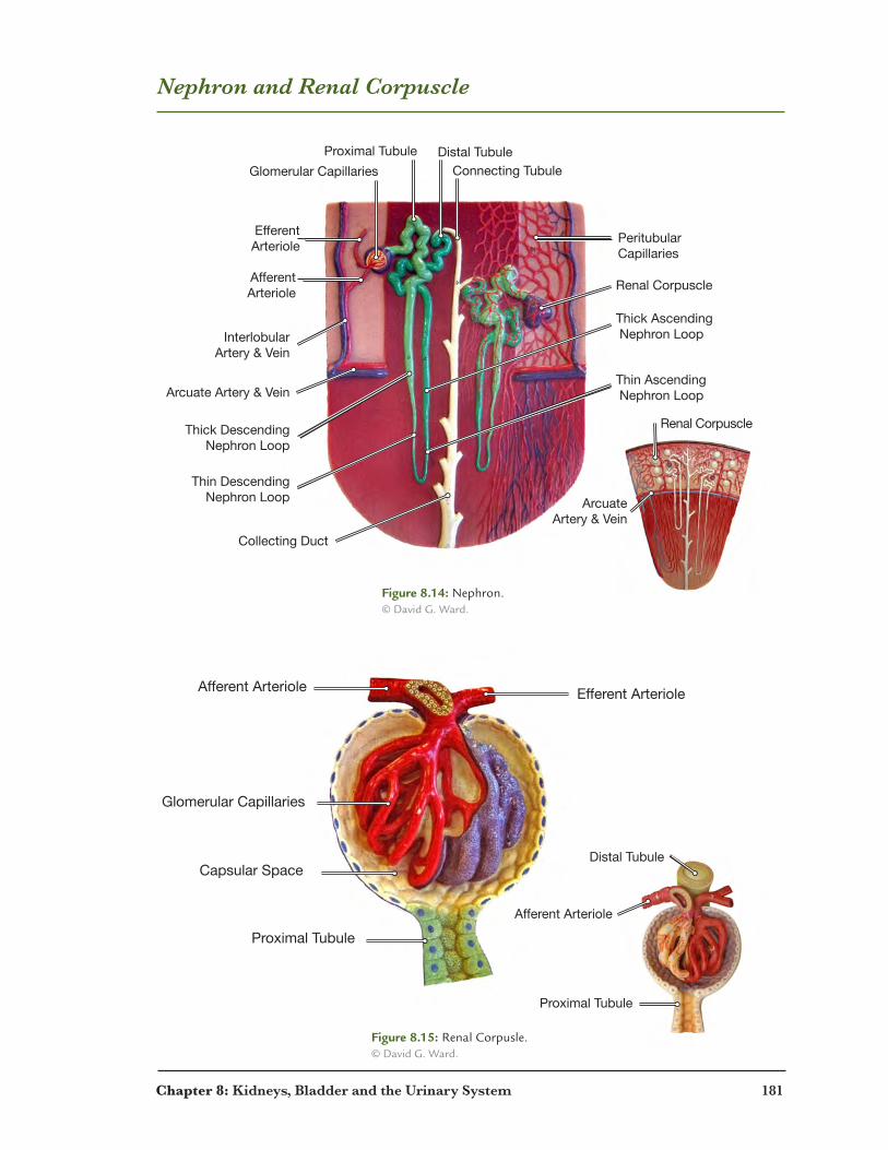
Nephron and Renal Corpuscle
Arcuate Artery & Vein
Arcuate Artery & Vein
Thick Descending Nephron Loop
Thin Descending Nephron Loop
Collecting Duct
Thin Ascending Nephron Loop
Thick Ascending Nephron Loop
Renal Corpuscle
Renal Corpuscle
Interlobular Artery & Vein
Afferent Arteriole
Glomerular Capillaries Connecting Tubule
Proximal Tubule Distal Tubule
Efferent Arteriole
PeritubularCapillaries
Proximal Tubule
Afferent Arteriole Efferent Arteriole
Glomerular Capillaries
Capsular SpaceDistal Tubule
Proximal Tubule
Afferent Arteriole
Figure 8.14: Nephron. © David G. Ward.
Figure 8.15: Renal Corpusle. © David G. Ward.

182 Chapter 8: Kidneys, Bladder and the Urinary System
Stages of Urine Production
1
2
2
3
3
GlomerularCapillaries
EfferentArteriole
GlomerularCapsule
Renal Tubule
PeritubularCapillaries
To InterlobularVein
Urine Flows to Collecting Duct
= Glomerular Filtration
= Tubular Reabsorption
= Tubular Secretion
Afferent Arteriole
InterlobularArtery
1
1. Glomerular filtration — the rate blood is filtered in the glomerulus
2. Tubular reabsorption — process of moving solutes and water from the renal tubule into the blood
3. Tubular secretion — process of moving solutes and water from the blood into the renal tubule
Figure 8.16: Stages of urine production. © bluedoor, LLC.
Chapter 8: Kidneys, Bladder and the Urinary System 183
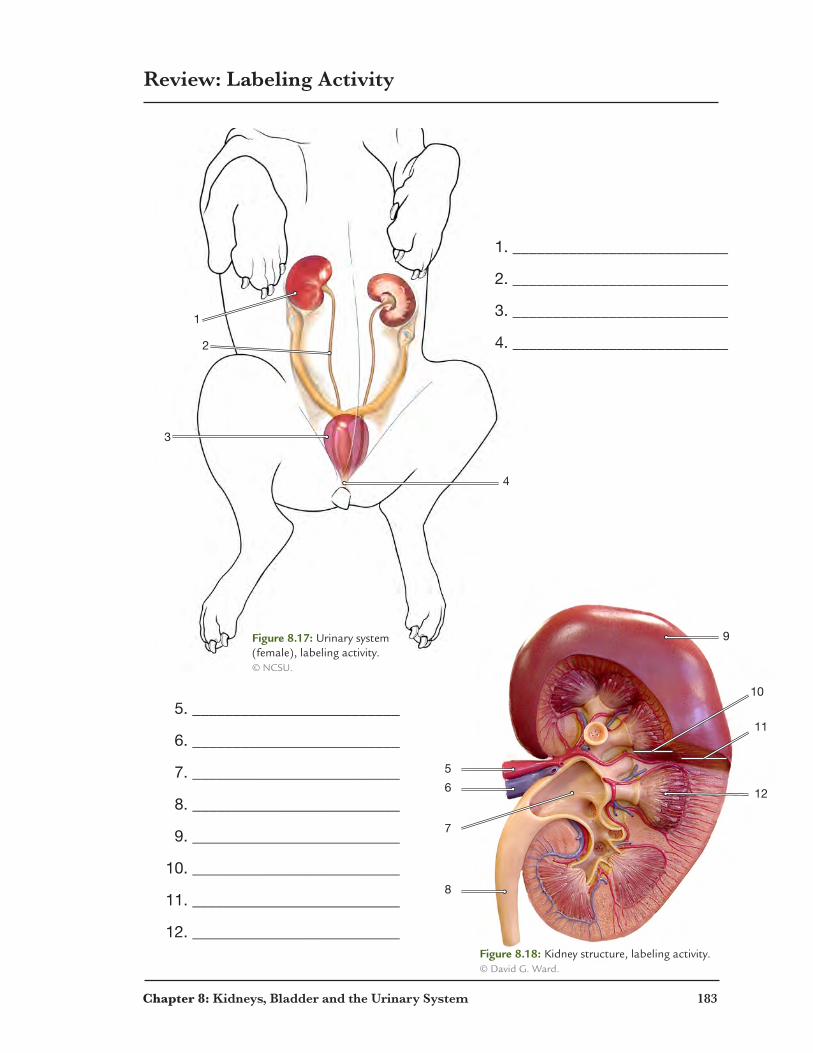
Review: Labeling Activity
8
7
5
6 12
11
10
9
1
2
3
1. ___________________________
2. ___________________________
3. ___________________________
4. ___________________________
5. __________________________
6. __________________________
7. __________________________
8. __________________________
9. __________________________
10. __________________________
11. __________________________
12. __________________________
4
Figure 8.17: Urinary system (female), labeling activity. © NCSU.
Figure 8.18: Kidney structure, labeling activity. © David G. Ward.

184 Chapter 8: Kidneys, Bladder and the Urinary System
Review: Crossword Puzzle
1
2
3 4
5
6 7
8
9 10
11
12
13
14 15
Across 3. filtrate passes through this structure immediately after the glomerulus 8. 25% of filtered sodium and chloride are reabsorbed into the blood 9. first stage of urine production11. the outermost region of the kidney12. where blood vessels, nerves and ureters enter and leave the kidney14. reabsorption of sodium and chloride by active transport
Down 1. the center region of the kidney 2. fibrous connective tissue surrounding the kidney 4. the structure that temporarily stores urine prior to elimination 5. the driving force for the filtration of water and small solutes in the renal corpuscle 6. connects the kidney to the bladder 7. vessel in which water channels are increased by vasopressin10. cone-shaped subdivision in the medulla containing nephrons 13. a tuft of capillaries located in Glomerular capsule 15. where reabsorption of water by diffusion through open water channels takes place
Chapter 8: Kidneys, Bladder and the Urinary System 185
Review: Word Search
T R V W Y W N L E S X N T S Z G Z W G N G L Z E
U E E K R C B O A Z P M X X T O M A B S R M T T
B N L X X Y G V I N U E S Y X M T Q V T B A S A
U A C Y R P D N N T E O C M M R L N C D R F F G
L L S N I E V L A N E R L Y O K Z O D N V F Y A
A A U G N D B I E V M R D A Q O B Z O Z E M P M
R R P K R U Z B P S G J C A I P T I Q R G X N C
R T R A N S I T I O N A L E P I T H E L I U M N
E E O G N D P G X A P S L T S A W N M Y E T G A
A R C L E V O W B S X Z C L R R T S U U Z L T Q
B Y L V M E G P Z V X U U T E A A G A R S B D W
S K A B U M D K G N D I L F R C N L Y U M C J S
O Z N O L X H W D G A I Z T L J E S U I Z Z L S
R F E W U Z K I N L F R E X H E X F U B S J O E
P Z R T N Q A I D R Y R F D G J Z Y O I U R E B
T N E U B F T O A M I L L I O S M O L E S T J T
I A Z I D C S L C O L D N T H I V O V A E M H F
O E D V E T U H L E O N T G X T I A V V Z S R P
N W W L E R H E U P X R F Q C L I K S Y B V Q Y
A B L R E V T Q P O A V J J I W M Q W J V I E S
C O O M E L O I R E T R A T N E R E F F E T K P
C N O C B Q T I L O F Q X I L F I V E A M U W D
E L S U E P K V A K C Q H V P B N W M Q F Z W M
G X G E O Z L M X H T Z I K J P K U P Y L N C R
1. cell type lining the lumen of the ureter
2. made up of the glomerulus and Bowman’s capsule
3. provides the blood supply to the kidney
4. distal tubule drains into this structure
5. provides blood supply to the glomerulus
6. measure of osmolarity in the kidney
7. the rate blood is filtered in the glomerulus (expressed mm/min and abbreviated GFR)
8. second stage of urine production
9. the open cavity in the ureter
10. the gland located superior to each kidney
11. the hormone affecting the DCT
12. drains the blood supply from the kidney
13. the outermost layer of the ureter
14. third stage of urine production
15. removes blood supply from the glomerulus

Chapter 9
Endocrine Glands and Reproductive Organs
Pancreas Showing Pancreatic Islet (Bright�eld, H&E; x430)
187
© D
avid
G. W
ard

188 Chapter 9: Endocrine Glands and Reproductive Organs
Overview of Endocrine Glands
Testis (male)
Ovary (Female)
Adrenal
Hypothalamus
Pituitary
Adrenal Pancreas
Thyroid and Parathyroid
Figure 9.1: Endocrine glands. © NCSU.

Chapter 9: Endocrine Glands and Reproductive Organs 189
Pancreas, Parathyroid, and Thyroid
LobuleLobule
Pancreatic Islet
Thyroid
Parathyroid
Thyroid Follicle
Thyroid Follicle
Thyroid Follicle
Thyroid Follicle
Figure 9.2: Pancreas. © David G. Ward.
Figure 9.4: Thyroid. © David G. Ward.
Figure 9.3: Parathyroid and Thyroid. © David G. Ward.

190 Chapter 9: Endocrine Glands and Reproductive Organs
Adrenal and Pituitary Gland
Anterior Pituitary
PosteriorPituitary
3rdVentricle
Adrenal Capsule
Adrenal Cortex
Adrenal MedullaZona Glomerulosa
Zona Fasciculata
Zona Reticularis
Aldosterone
Cortisol
Androstendione
EpinephrineNorepinephrine
Anterior Pituitary Posterior Pituitary
Infundibulum
Midbrain
3rdVentricle
Hypothalamus
Anterior Pituitary
PosteriorPituitary
3rdVentricle
Adrenal Capsule
Adrenal Cortex
Adrenal MedullaZona Glomerulosa
Zona Fasciculata
Zona Reticularis
Aldosterone
Cortisol
Androstendione
EpinephrineNorepinephrine
Anterior Pituitary Posterior Pituitary
Infundibulum
Midbrain
3rdVentricle
Hypothalamus
Anterior Pituitary
PosteriorPituitary
3rdVentricle
Adrenal Capsule
Adrenal Cortex
Adrenal MedullaZona Glomerulosa
Zona Fasciculata
Zona Reticularis
Aldosterone
Cortisol
Androstendione
EpinephrineNorepinephrine
Anterior Pituitary Posterior Pituitary
Infundibulum
Midbrain
3rdVentricle
Hypothalamus
Figure 9.5: Adrenal gland. © David G. Ward.
Figure 9.6: Pituitary gland. © David G. Ward.
Figure 9.7: Pituitary gland. © David G. Ward.

Chapter 9: Endocrine Glands and Reproductive Organs 191
Testes and Penis
SpermSperm
Spermatid
Nucleus of Sertoli Cell
SpermatogoniumPrimary Spermatocyte
Secondary Spermatocyte
Tails of Sperm
Heads of Sperm
CorporaCavernosa
CorpusSpongiosum
Urethra
Central Artery
Interstital Cells*
Spermatogonia
PrimarySpermatocytes
Spermatids
Sertoli Cells
*Interstitial cells produce testosterone.
Figure 9.8: Sperm are produced in the seminiferous tubules. © David G. Ward. Figure 9.9: Seminiferous tubules.
© David G. Ward.
Figure 9.11: Seminiferous tubules. © David G. Ward.
Figure 9.12: Sperm. © David G. Ward.
Figure 9.13: Penis, cross-section. © David G. Ward.
Figure 9.10: Interstitial cells produce testosterone. © David G. Ward.

192 Chapter 9: Endocrine Glands and Reproductive Organs
Oocytes, Ovarian Follicles and Corpus Luteum
Squamous Epithelial Cells around Oocytes
Primary OocytesGerminalEpithelium
Primary Oocyte
Secondary Oocyte
Zona Pellucida
Zona Pellucida
Granulosa Cells
Granulosa Cells
Theca Cells
Theca Cells
Corpus Luteum
Antrum
(Produces Progestin)
© N
CSU
Figure 9.14: Primary Oocytes. © David G. Ward.
Figure 9.15: Developing follicle. © David G. Ward.
Figure 9.16: Corpus luteum. © David G. Ward.
Figure 9.17: Mature follicle. © David G. Ward.
Chapter 9: Endocrine Glands and Reproductive Organs 193
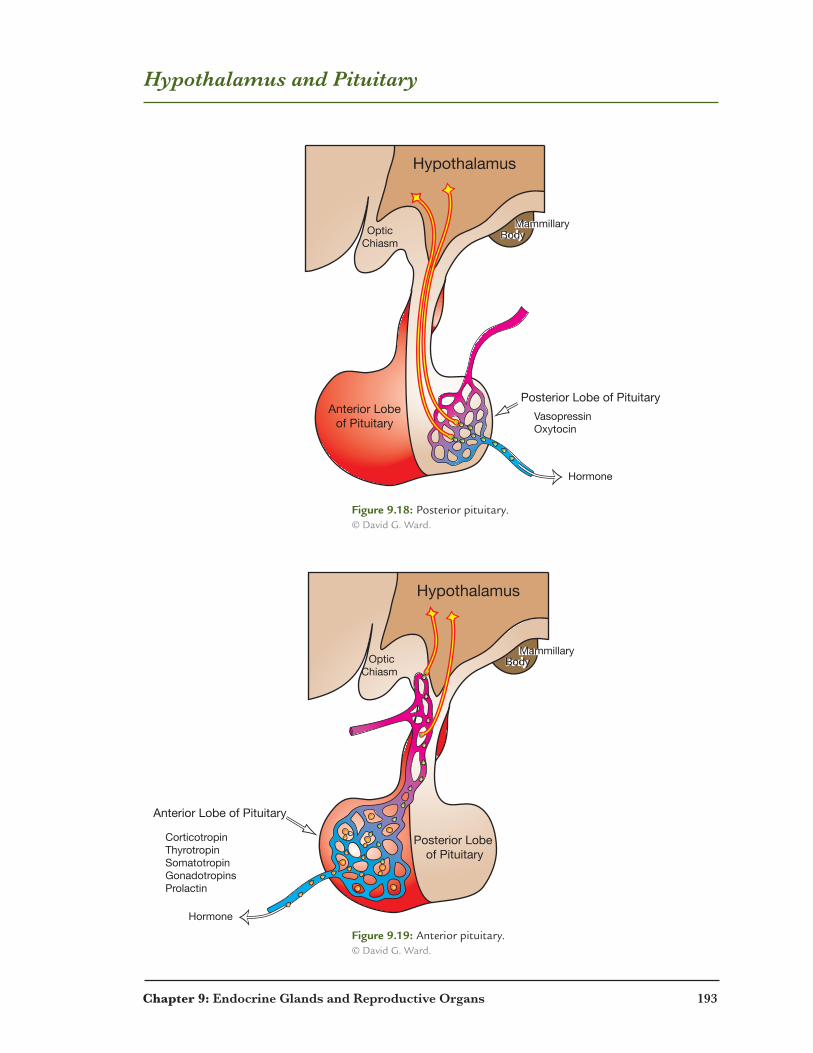
Hypothalamus and Pituitary
Hypothalamus
OpticChiasm
BodyMammillary
BodyMammillary
Posterior Lobe of PituitaryAnterior Lobe
of PituitaryVasopressinOxytocin
Hormone
Hypothalamus
Optic Chiasm
Body Mammillary
Body Mammillary
Posterior Lobe of Pituitary
Anterior Lobe of Pituitary
CorticotropinThyrotropinSomatotropinGonadotropinsProlactin
Hormone
Figure 9.18: Posterior pituitary. © David G. Ward.
Figure 9.19: Anterior pituitary. © David G. Ward.

194 Chapter 9: Endocrine Glands and Reproductive Organs
Female Reproductive Organs
Ovary
Broad ligamentUterine hornUterine Tube)
Body of Uterus
Figure 9.20: Female reproductive organs, ventral view. © NCSU.

Chapter 9: Endocrine Glands and Reproductive Organs 195
Female Reproductive Organs
Cervix
Vagina
Uterine Horn(Uterine Tube)
Oviduct
Body of Uterus
Ovary
Figure 9.21: Female reproductive organs. © NCSU.
Figure 9.22: Female reproductive organs. © NCSU.
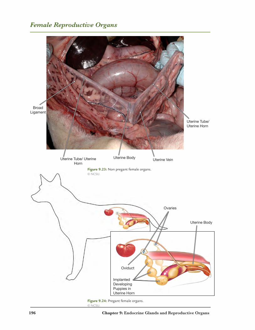
196 Chapter 9: Endocrine Glands and Reproductive Organs
Female Reproductive Organs
Uterine Tube/Uterine Horn
Uterine VeinUterine Body
BroadLigament
Uterine Tube/ Uterine Horn
Ovaries
Uterine Body
Oviduct
ImplantedDevelopingPuppies in Uterine Horn
Figure 9.23: Non pregant female organs. © NCSU.
Figure 9.24: Pregant female organs. © NCSU.

Chapter 9: Endocrine Glands and Reproductive Organs 197
Male Reproductive Organs
PenisUrethra
Ductus Deferens
Testicle
Prostate
Os Penis
Prepuce
Figure 9.25: Male reproductive organs. © NCSU.
Figure 9.26: Male reproductive organs. © NCSU.

198 Chapter 9: Endocrine Glands and Reproductive Organs
Review: Labeling Activity
1
2
3
6
5
8
4
7
1. 5.
2. 6.
3. 7.
4. 8.
Figure 9.27: Endocrine glands, labeling activity. © NCSU.

Chapter 9: Endocrine Glands and Reproductive Organs 199
Review: Labeling Activity
1. __________________________
2. __________________________
3. __________________________
4. __________________________
5. __________________________
6. __________________________
7. __________________________
8. __________________________
9. __________________________
10. __________________________
12
3 4
5
67
8
910
Figure 9.28: Male reproductive organs, labeling activity. © NCSU.
Figure 9.29: Female reproductive organs, labeling activty. © NCSU.
200 Chapter 9: Endocrine Glands and Reproductive Organs
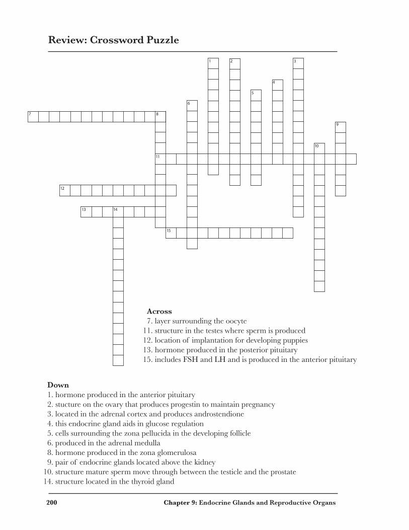
Review: Crossword Puzzle
Across 7. layer surrounding the oocyte11. structure in the testes where sperm is produced12. location of implantation for developing puppies13. hormone produced in the posterior pituitary15. includes FSH and LH and is produced in the anterior pituitary
Down 1. hormone produced in the anterior pituitary 2. stucture on the ovary that produces progestin to maintain pregnancy 3. located in the adrenal cortex and produces androstendione 4. this endocrine gland aids in glucose regulation 5. cells surrounding the zona pellucida in the developing follicle 6. produced in the adrenal medulla 8. hormone produced in the zona glomerulosa 9. pair of endocrine glands located above the kidney10. structure mature sperm move through between the testicle and the prostate14. structure located in the thyroid gland
1 2
5
4
6
87
3
9
10
11
12
13 14
15
Chapter 9: Endocrine Glands and Reproductive Organs 201
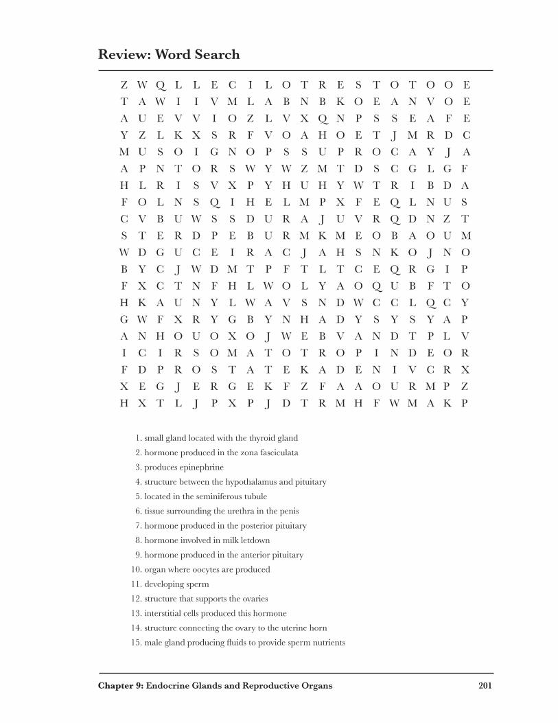
Review: Word Search
Z W Q L L E C I L O T R E S T O T O O E
T A W I I V M L A B N B K O E A N V O E
A U E V V I O Z L V X Q N P S S E A F E
Y Z L K X S R F V O A H O E T J M R D C
M U S O I G N O P S S U P R O C A Y J A
A P N T O R S W Y W Z M T D S C G L G F
H L R I S V X P Y H U H Y W T R I B D A
F O L N S Q I H E L M P X F E Q L N U S
C V B U W S S D U R A J U V R Q D N Z T
S T E R D P E B U R M K M E O B A O U M
W D G U C E I R A C J A H S N K O J N O
B Y C J W D M T P F T L T C E Q R G I P
F X C T N F H L W O L Y A O Q U B F T O
H K A U N Y L W A V S N D W C C L Q C Y
G W F X R Y G B Y N H A D Y S Y S Y A P
A N H O U O X O J W E B V A N D T P L V
I C I R S O M A T O T R O P I N D E O R
F D P R O S T A T E K A D E N I V C R X
X E G J E R G E K F Z F A A O U R M P Z
H X T L J P X P J D T R M H F W M A K P
1. small gland located with the thyroid gland
2. hormone produced in the zona fasciculata
3. produces epinephrine
4. structure between the hypothalamus and pituitary
5. located in the seminiferous tubule
6. tissue surrounding the urethra in the penis
7. hormone produced in the posterior pituitary
8. hormone involved in milk letdown
9. hormone produced in the anterior pituitary
10. organ where oocytes are produced
11. developing sperm
12. structure that supports the ovaries
13. interstitial cells produced this hormone
14. structure connecting the ovary to the uterine horn
15.maleglandproducingfluidstoprovidespermnutrients