taeniasis/cysticercosis in bali, indonesia
TRANSCRIPT
Taeniasis/cysTicercosis in indonesia
Vol 42 No. 4 July 2011 793
Correspondence: Toni Wandra, Directorate General Disease Control and Environmental Health, Ministry of Health, Indonesia, Jalan Percetakan Negara 29, Kotak Pos 223 Jakarta 10560, Indonesia.Tel: 62 21 4247608 # 119; Fax: 62 21 4200949E-mail: [email protected] Present affiliation: Escola de Medicina Veteri-nária e Zootecnia, Universidade Federal do Tocantins, Araguaina-TO, Brazil.bPresent affiliation: Primate Research Institute, Kyoto University, Inuyama, Japan.
REVIEW
TAENIASIS/CYSTICERCOSIS IN BALI, INDONESIA
Toni Wandra1,7, AA Raka Sudewi2, I Kadek Swastika3,7, Putu Sutisna3, Nyoman S Dharmawan4, Hemma Yulfi5, Dewi Masyithah Darlan5, I Nengah Kapti3,
Gina Samaan6, Marcello Otake Sato7,a, Munehiro Okamoto8,b, Yasuhito Sako7 and Akira Ito7
1Directorate General Disease Control and Environmental Health, Ministry of Health,
Indonesia; 2Department of Neurology, 3Department of Parasitology, Faculty of Medicine, 4Faculty of Veterinary Medicine, University of Udayana, Bali, Indonesia; 5Department of Parasitology, Faculty of Medicine, University of Sumatra Utara, Indonesia; 6National Centre for Epidemiology and Population Health, Australian National University, Canberra, Australia; 7Department of Parasitology, Asahikawa Medical University, Asahikawa, Japan;
8School of Veterinary Medicine, Faculty of Agriculture, Tottori University, Japan
Abstract. Taenia solium and Taenia saginata are found in humans in Bali, Indonesia. During a field survey of 660 people in Bali from 2002-2009 of taeniasis/cysticercosis cases using mitochondrial DNA confirmation of the species, we detected 80 cases of T. saginata taeniasis, 2 dual T. saginata/T. solium infections with T. solium meta-cestodes in the brain and 12 neurocysticercosis (NCC) cases at Sanglah Hospital, Denpasar. Although the prevalence of NCC in Bali is low, sporadic cases are still present. There is no Taenia asiatica in Bali. We summarize here the field survey findings of taeniasis, including 1 dual infection with taeniasis and cysticercosis in 2007, and the reason why there are no T. asiatica cases and we describe 3 NCC cases admitted to Sanglah Hospital, Denpasar, Bali in 2004. Diagnosis was based on anamnesis, clinical examination, including CT Scan, histopathological, sero-logical and mitochondrial DNA examinations. In order to prevent unexpected symptomatic NCC after treatment with praziquantel, we recommend introducing a rapid test to confirm taeniasis carriers and cysticercosis cases as a tool for real time diagnosis.
Keywords: Taenia solium, T. saginata, taeniasis, cysticercosis, Indonesia
INTRODUCTION
In Asia, there are 3 human Taenia species: Taenia solium (pork tapeworm), Taenia saginata (beef tapeworm) and Tae-nia asiatica (Chao and Fan, 1986; Fan et al, 1987; Fan 1988; 1988; Zarlenga et al, 1991; Eom and Rim, 1993; Bowles and McManus, 1994; Hoberg et al, 2000; Ito et al, 2003; Eom, 2006; Okamoto et al, 2010). The taxonomy of T. asiatica is still unclear, since hybrid T. saginata/T. asiatica worms
souTheasT asian J Trop Med public healTh
794 Vol 42 No. 4 July 2011
have been found in Thailand (Okamoto et al, 2010) and China (Nkouawa et al, in preparation).
Taeniasis in human due to T. saginata and T. asiatica is caused by eating un-cooked or undercooked beef and viscera of swine contaminated with metacestodes of these species, respectively. Metaces-todes of T. asiatica may develop not only in pigs but also in cattle and goats (Fan et al, 1987, 1989). In contrast, T. solium only causes two distinct clinical presentations: taeniasis due to the presence of adult tapeworm(s) in the small intestine of humans after eating uncooked or under-cooked pork contaminated with the meta-cestodes of this species, and cysticercosis caused by the presence of metacestode(s) in parenteral tissue after accidental oral ingestion of eggs of the parasite. Cysti-cercosis caused by metacestode(s) in the central nervous system (neurocysticer-cosis, NCC) is one of the most important causes of epilepsy, and a leading cause of late-onset epilepsy (Takayanagui and Odashima, 2006; Ito et al, 2006).
Cases of T. solium, T. saginata and T. asiatica have been reported from Indone-sia. To date, there have been 3 endemic provinces for taeniasis/cysticercosis in In-donesia: North Sumatra, Bali and Papua. North Sumatra is endemic for T. asiatica taeniasis (Kosin et al, 1972; Fan et al, 1990; Wandra et al, 2006). Bali is endemic for T. saginata taeniasis and T. solium taeniasis/cysticercosis (Sutisna et al, 1999, 2000; Wandra et al, 2006; Sudewi et al, 2008). Papua is endemic for T. solium taeniasis/cysticercosis (Tumada and Margono, 1973; Handali et al, 1997; Simanjuntak et al, 1997; Sutisna et al, 1999, 2000; Wandra et al, 2000, 2003; Subahar et al, 2001; Ito et al, 2002; Margono et al, 2003, 2006; Sa-lim et al, 2009). Taeniasis and cysticerosis are reported sporadically in Lampung,
Jakarta, East Java, West Kalimantan, East Kalimantan, North Sulawesi, South Sulawesi, South East Sulawesi, and East Nusa Tenggara Provinces of Indonesia (Simanjuntak et al, 1997; Margono et al, 2001; Wandra et al, 2003; Ito et al, 2004; Sudewi et al, unpublished).
Epidemiological surveys for taenia-sis/cysticercosis were conducted in all 9 districts of Bali from 2002 to 2009. During the field surveys, we examined a total of 660 people and simultaneously collected data regarding NCC cases at Sanglah Hospital, Denpasar. In this review, we summarize 80 cases of T. saginata taeniasis, 1 dual infection with T. solium/T. saginata, in 2007 and 3 NCC cases admitted to the hospital in 2004.
TAENIASIS/CYSTICERCOSIS IN BALI
Taenia saginata taeniasisDuring the epidemiological survey in
Bali from 2002 to 2009, we detected a total of 80 cases of T. saginata taeniasis (32, 24, 7, 3, 3, 4, and 7 cases in 2002, 2004, 2005, 2006, 2007, 2008, and 2009, respectively) using mitochondrial DNA confirmation by multiplex PCR and DNA sequencings (Yamasaki et al, 2004; Ito et al, 2009). The risk factor for T. saginata taeniasis is con-sumption of “beef lawar”, a traditional local food of raw minced beef. Butchers and their family members are often found to be infected with T. saginata in Bali and with T. asiatica in Lake Toba, North Suma-tra (Wandra et al, unpublished).
In Gianyar District, T. saginata taenia-sis cases were found yearly (32, 14, 5, 2, 3, 4, and 7 cases in 2002, 2004, 2005, 2006, 2007, 2008, and 2009, respectively). Ap-proximately 84% of taeniasis cases (67/80) were from Gianyar District. There were no cases of reinfection with T. saginata after treatment with tapeworm(s). Once most
Taeniasis/cysTicercosis in indonesia
Vol 42 No. 4 July 2011 795
people are confirmed to have an infection, the majority will stop eating raw beef. The high number of cases in Gianyar District may be due to the low impact of health education campaigns advising the com-munity to consume cooked beef only in order to prevent taeniasis. Further study is needed to determine the true prevalence of taeniasis in Gianyar District.
Nearly all taeniasis cases were sus-pected based on a question that asked about the expulsion of proglottids. Most people gave a reliable history, at least for T. saginata. T. asiatica is morphologically similar to T. saginata (Fan, 1988; Eom et al, 1993; Ito et al, 2003) and is fairly common in North Sumatra, Indonesia (Kosin et al, 1972; Fan et al, 1990; Wandra et al, 2006, 2007), but has never been reported from Bali. We believe the reason why Balinese people are not infected with T. asiatica is because they do not eat the uncooked vis-cera of swine, different from the people of North Sumatra (Wandra et al, 2006, 2007).
We found no cases of T. solium tae-niasis in Bali. However, there are cysti-cercosis cases in Bali. In order to detect T. solium, we would have to develop a rapid diagnostic test to screen for taeniasis car-riers. Serum samples of taeniasis cases may be useful for developing screening tests for taeniasis carriers (Wilkins et al, 1999; Levine et al, 2007; Nakao et al, un-published), but fecal samples are useful to confirm taeniasis carriers and identify the species (Yamasaki et al, 2004; Guezala et al, 2009; Nkouawa et al, 2009).
Dual infection with T. saginata taeniasis and T. solium neurocysticercosis
In 2007, we found a 47-year-old- Bali-nese male T. saginata carrier who appeared to be healthy and had no neurologic symptoms of cysticercosis. We treated him with praziquantel 15 mg/kg BW. A
single worm was expelled, examined mor-phologically and later confirmed to be T. saginata by multiplex PCR in Asahikawa, Japan. Unfortunately, he had seizure within 6 hours of treatment, requiring hospitalization for several days. A CT scan showed multiple cystic lesions in the brain. Serological examination carried out in Asahikawa, Japan, later showed weakly positive antibody responses to both na-tive and recombinant antigens (Sako et al, 2000; Sato et al, 2006; Ito et al, 2009) before treatment, and a stronger response post-treatment (data not shown). This case shows asymptomatic NCC cases may become symptomatic after treatment even with a low dose of praziquantel (Flisser et al, 1993; Sarti et al, 2000). In 2009, we treated another dual infection case in Bali (Wandra et al, in preparation).Cysticercosis due to T. solium
The first record of cysticercosis in Bali was reported from pigs more than 80 years ago (Le Coultre, 1928; Oemijati, 1977). Thirty-two years later, two human cases of subcutaneous cysticercosis (SCC) were reported (Soebroto at al, 1960). Since 1971, several cases of T. solium, epileptic seizures, SCC and NCC have been re-ported, including the seroprevalence of cysticercosis in Bali. Between 1960 and 1997, a total of six taeniasis cases due to T. solium have been confirmed. In contrast, a total of 44 cysticercosis cases have been reported. Both NCC and SCC are found in Bali, with a ratio of 2.6:1 for NCC (32 cases) and SCC (12 cases). However, there is no information about cases infected with both NCC and SCC (Soebroto et al., 1960; Hadidjaya, 1971; Ngoerah, 1975; Simanjuntak et al, 1977, 1997; Coker-Vann et al, 1981; Bakta et al, 1983; Theis et al, 1993; Sutisna, 1994, 1999; Sudewi and Nuartha, unpublished quoted from Sutisna et al, 2000). The seroprevalence
souTheasT asian J Trop Med public healTh
796 Vol 42 No. 4 July 2011
of cysticercosis in Bali ranged from 5.2 to 21% during 1981-1997 (Sutisna et al, 2000). One risk factor was consumption of pork lawar, an uncooked minced pork mixed with fresh pigs blood, a traditional cuisine among the Balinese people (Wandra et al, 2006, 2007). Pork lawar is more popular than beef lawar in Bali. This is based on Balinese Hinduism, which differs from Indian Hinduism (Simanjuntak et al, 1997; Ito et al, 2003; Wandra et al, 2007).
The number of cysticercosis cases has decreased dramatically, since only two sero-positive cases were detected from 9 districts in Bali among 660 people during 2002-2009. We did not detect any taeniasis cases due to T. solium. The decrease in the number of NCC and SCC cases due to T. solium may be due to improvement in household sanitation and pig hus-bandry through sustainable public health education in Bali and improvement in the economy. Most families have latrines and pigs are generally reared indoors (Wandra et al, 2007).
Although the prevalence of cysticer-cosis is currently low, sporadic cases are still detected at hospitals, especially at Sanglah Hospital, Denpasar. NCC cases included 1 case of disseminated cysticer-cosis (2003) (Sudewi et al, 2008), 3 cases of NCC (2004) summarized in this review, 1 case of NCC (2005), 2 cases of dual in-fection (2007, 2009), and 5 cases of NCC (2009) (Wandra et al, unpublished).
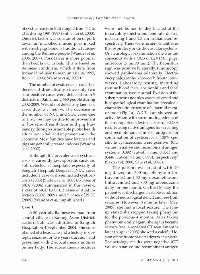
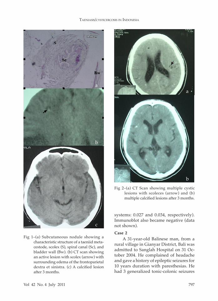
Case 1A 50-year-old Balinese woman, from
a rural village in Karang Asem District, eastern Bali was admitted to Sanglah Hospital on 4 September 2004. She com-plained of a headache and a history of epi-leptic seizures for two years duration, and presented with 2 subcutaneous nodules on her body. The subcutaneous nodules
were mobile, non-tender, located at the fossa cubity sinistra and femoralis dextra, measuring 1 and 1.5 cm in diameter, re-spectively. There were no abnormalities of the respiratory or cardiovascular systems. On neurological examination she was un-conscious with a GCS of E2V1M5, pupil anisocore (5 mm/3 mm). The Babinski’s sign was positive bilaterally, funduscopy showed papiledema bilaterally. Electro-encephalography showed bifrontal slow waves. Laboratory testing, including routine blood tests, eosinophils and fecal examination, were normal. Excision of the subcutaneous nodules was performed and histopathological examination revealed a characteristic structure of a taeniid meta-cestode (Fig 1a). A CT scan showed an active lesion with surrounding edema of the frontoparietal dextra et sinistra. ELISA results using native antigens for screening and recombinant chimeric antigens for confirmation of cysticercosis, 100% spe-cific to cysticercosis, were positive [(OD values in native and recombinant antigen systems: 0.392 (cut-off value: 0.051) and 0.446 (cut-off value: 0.093), respectively] (Sako et al, 2000; Sato et al, 2006).
The patient was treated with 10 mg diazepam, 300 mg phenytoin (in-travenous) and 50 mg dexamethasone (intravenous) and 800 mg albendazole daily for one month. On the 16th day, the patient was discharged in stable condition without neurological deficit and free from seizures. However, 8 months later (May, 2005), she had a focal seizure. The fam-ily stated she stopped taking phenytoin for the previous 4 months. After taking phenytoin orally again, she again became seizure free. A repeated CT scan 3 months later (August 2005) showed a calcified le-sion of the frontoparietal dextra et sinistra. The serology results were negative (OD values in native and recombinant antigen
Taeniasis/cysTicercosis in indonesia
Vol 42 No. 4 July 2011 797
systems: 0.027 and 0.034, respectively). Immunoblot also became negative (data not shown). Case 2
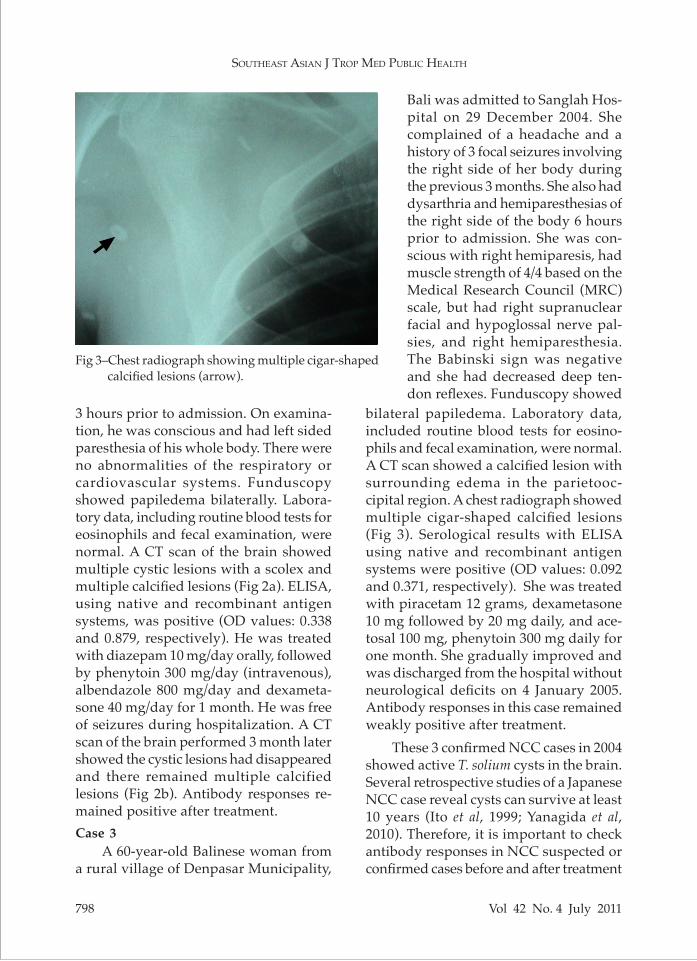
A 31-year-old Balinese man, from a rural village in Gianyar District, Bali was admitted to Sanglah Hospital on 31 Oc-tober 2004. He complained of headache and gave a history of epileptic seizures for 10 years duration with paresthesias. He had 3 generalized tonic-colonic seizures
Fig 1–(a) Subcutaneous nodule showing a characteristic structure of a taeniid meta-cestode, scolex (S), spiral canal (Sc), and bladder wall (Bw). (b) CT scan showing an active lesion with scolex (arrow) with surrounding edema of the frontoparietal dextra et sinistra. (c) A calcified lesion after 3 months.
Fig 2–(a) CT Scan showing multiple cystic lesions with scoleces (arrow) and (b) multiple calcified lesions after 3 months.
a
b
c
a
b
souTheasT asian J Trop Med public healTh
798 Vol 42 No. 4 July 2011
3 hours prior to admission. On examina-tion, he was conscious and had left sided paresthesia of his whole body. There were no abnormalities of the respiratory or cardiovascular systems. Funduscopy showed papiledema bilaterally. Labora-tory data, including routine blood tests for eosinophils and fecal examination, were normal. A CT scan of the brain showed multiple cystic lesions with a scolex and multiple calcified lesions (Fig 2a). ELISA, using native and recombinant antigen systems, was positive (OD values: 0.338 and 0.879, respectively). He was treated with diazepam 10 mg/day orally, followed by phenytoin 300 mg/day (intravenous), albendazole 800 mg/day and dexameta-sone 40 mg/day for 1 month. He was free of seizures during hospitalization. A CT scan of the brain performed 3 month later showed the cystic lesions had disappeared and there remained multiple calcified lesions (Fig 2b). Antibody responses re-mained positive after treatment. Case 3
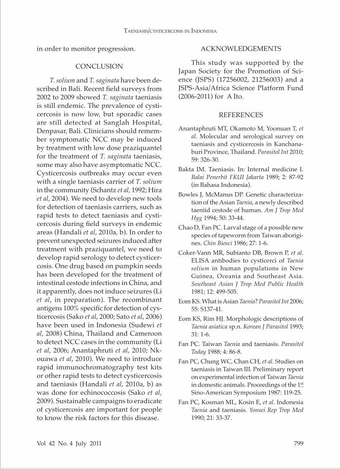
A 60-year-old Balinese woman from a rural village of Denpasar Municipality,
bilateral papiledema. Laboratory data, included routine blood tests for eosino-phils and fecal examination, were normal. A CT scan showed a calcified lesion with surrounding edema in the parietooc-cipital region. A chest radiograph showed multiple cigar-shaped calcified lesions (Fig 3). Serological results with ELISA using native and recombinant antigen systems were positive (OD values: 0.092 and 0.371, respectively). She was treated with piracetam 12 grams, dexametasone 10 mg followed by 20 mg daily, and ace-tosal 100 mg, phenytoin 300 mg daily for one month. She gradually improved and was discharged from the hospital without neurological deficits on 4 January 2005. Antibody responses in this case remained weakly positive after treatment.
These 3 confirmed NCC cases in 2004 showed active T. solium cysts in the brain. Several retrospective studies of a Japanese NCC case reveal cysts can survive at least 10 years (Ito et al, 1999; Yanagida et al, 2010). Therefore, it is important to check antibody responses in NCC suspected or confirmed cases before and after treatment
Fig 3–Chest radiograph showing multiple cigar-shaped calcified lesions (arrow).
Bali was admitted to Sanglah Hos-pital on 29 December 2004. She complained of a headache and a history of 3 focal seizures involving the right side of her body during the previous 3 months. She also had dysarthria and hemiparesthesias of the right side of the body 6 hours prior to admission. She was con-scious with right hemiparesis, had muscle strength of 4/4 based on the Medical Research Council (MRC) scale, but had right supranuclear facial and hypoglossal nerve pal-sies, and right hemiparesthesia. The Babinski sign was negative and she had decreased deep ten-don reflexes. Funduscopy showed
Taeniasis/cysTicercosis in indonesia
Vol 42 No. 4 July 2011 799
in order to monitor progression.
CONCLUSION
T. solium and T. saginata have been de-scribed in Bali. Recent field surveys from 2002 to 2009 showed T. saginata taeniasis is still endemic. The prevalence of cysti-cercosis is now low, but sporadic cases are still detected at Sanglah Hospital, Denpasar, Bali. Clinicians should remem-ber symptomatic NCC may be induced by treatment with low dose praziquantel for the treatment of T. saginata taeniasis, some may also have asymptomatic NCC. Cysticercosis outbreaks may occur even with a single taeniasis carrier of T. solium in the community (Schantz et al, 1992; Hira et al, 2004). We need to develop new tools for detection of taeniasis carriers, such as rapid tests to detect taeniasis and cysti-cercosis during field surveys in endemic areas (Handali et al, 2010a, b). In order to prevent unexpected seizures induced after treatment with praziquantel, we need to develop rapid serology to detect cysticer-cosis. One drug based on pumpkin seeds has been developed for the treatment of intestinal cestode infections in China, and it apparently, does not induce seizures (Li et al, in preparation). The recombinant antigens 100% specific for detection of cys-ticercosis (Sako et al, 2000; Sato et al, 2006) have been used in Indonesia (Sudewi et al, 2008) China, Thailand and Cameroon to detect NCC cases in the community (Li et al, 2006; Anantaphruti et al, 2010; Nk-ouawa et al, 2010). We need to introduce rapid immunochromatography test kits or other rapid tests to detect cysticercosis and taeniasis (Handali et al, 2010a, b) as was done for echinococcosis (Sako et al, 2009). Sustainable campaigns to eradicate of cysticercosis are important for people to know the risk factors for this disease.
ACKNOWLEDGEMENTS
This study was supported by the Japan Society for the Promotion of Sci-ence (JSPS) (17256002, 21256003) and a JSPS-Asia/Africa Science Platform Fund (2006-2011) for A Ito.
REFERENCES
Anantaphruti MT, Okamoto M, Yoonuan T, et al. Molecular and serological survey on taeniasis and cysticercosis in Kanchana-buri Province, Thailand. Parasitol Int 2010; 59: 326-30.
Bakta IM. Taeniasis. In: Internal medicine I. Balai Penerbit FKUI Jakarta 1989; 2: 87-92 (in Bahasa Indonesia).
Bowles J, McManus DP. Genetic characteriza-tion of the Asian Taenia, a newly described taeniid cestode of human. Am J Trop Med Hyg 1994; 50: 33-44.
Chao D, Fan PC. Larval stage of a possible new species of tapeworm from Taiwan aborigi-nes. Chin Biosci 1986; 27: 1-6.
Coker-Vann MR, Subianto DB, Brown P, et al. ELISA antibodies to cysticerci of Taenia solium in human populations in New Guinea, Oceania and Southeast Asia. Southeast Asian J Trop Med Public Health 1981; 12: 499-505.
Eom KS. What is Asian Taenia? Parasitol Int 2006; 55: S137-41.
Eom KS, Rim HJ. Morphologic descriptions of Taenia asiatica sp.n. Korean J Parasitol 1993; 31: 1-6.
Fan PC. Taiwan Taenia and taeniasis. Parasitol Today 1988; 4: 86-8.
Fan PC, Chung WC, Chan CH, et al. Studies on taeniasis in Taiwan III. Preliminary report on experimental infection of Taiwan Taenia in domestic animals. Proceedings of the 1st
Sino-American Symposium 1987: 119-25.Fan PC, Kosman ML, Kosin E, et al. Indonesia
Taenia and taeniasis. Yonsei Rep Trop Med 1990; 21: 33-37.
souTheasT asian J Trop Med public healTh
800 Vol 42 No. 4 July 2011
Fan PC, Lin CY, Wu CC. Experimental studies of Korea Taenia (Cheju strain) infection in domestic animals. Ann Trop Med Parasitol 1989; 83: 395-403.
Flisser A, Madrazo I, Plancarte A, et al. Neu-rological symptoms in occult neurocys-ticercosis after single taeniacidal dose of praziquantel. Lancet 1993; 342: 748.
Guezala MC, Rodriguez S, Zamora H, et al. Development of a spcies-specific coproan-tigen ELISA for human Taenia solium tae-niasis. Am J Trop Med Hyg 2009; 81: 433-7.
Hadidjaja P. Several cases of taeniasis in Jakarta: diagnosis and treatment methods. Madj Kedok Indon 1971; 21: 173-8 (in Bahasa Indonesia).
Handali S, Klarman M, Gaspark AN, et al. De-velopment and evaluation of a magnetic immunochromatographic test to detect Taenia solium which causes taeniasis and neurocysticercosis in humans. Clin Vaccine Immunol 2010a; 17: 631-7.
Handali S, Liying H, Lusikoy C, et al. A survey report – July 1993: Cysticercosis in the Grand Dani Vally, Jayawijiaya District, Irian Jaya Province, Indonesia. Southeast Asian J Trop Med Public Health 1997; 28 (suppl 1): 22-5.
Handali S, Pattabhi S, Lee YM, et al. Develop-ment and evaluation of porcine cysticerco-sis QuickELISA in Triturus EIA analyzer. J Immunoassay Immunochem 2010b; 31: 60-70.
Hira PR, Francis I, Abdella NA, et al. Cysticer-cosis: imported and autochthonous infec-tions in Kuwait. Trans R Soc Trop Med Hyg 2004; 98: 233-9.
Hoberg EP, Jones A, Rausch RL, et al. A phylo-genetic hypothesis for species of the genus Taenia (Eucestoda: Taeniidae). J Parasitol 2000; 86: 89-98.
Ito A, Nakao M, Ito Y, et al. Neurocysticercosis case with a single cyst in the brain showing dramatic drop in specific antibody titers within 1 year after curative surgical resec-tion. Parasitol Int 1999; 48: 95-9.
Ito A, Nakao M, Sako Y, et al. Taenia. Chapter
62. In: Liu D, ed. Molecular detection of foodborne pathogens. Boca Raton: CRC Press, 2009: 839-50.
Ito A, Nakao M, Wandra T. Human taeniasis and cysticercosis in Asia. Lancet 2003; 362: 1918-20.
Ito A, Putra MI, Subahar R, et al. Dogs as alter-native intermediate hosts of Taenia solium in Papua (Irian Jaya), Indonesia confirmed by highly specific ELISA and immunoblot using native and recombinant antigens and mitochondrial DNA analysis. J Hel-minthol 2002; 76: 311-4.
Ito A, Takayanagui OM, Sako Y, et al. Neuro-cysticercosis: clinical manifestation, neu-roimaging, serology and molecular con-firmation of histopathologic specimens. Southeast Asian J Trop Med Public Health 2006; 37 (suppl 3): 74-81.
Ito A, Wandra T, Yamasaki H, et al. Cysticerco-sis/taeniasis in Asia and the Pacific Vector-Borne Zoonot Dis 2004; 4: 95-107.
Kosin E, Depary A, Johansyah A. Taeniasis in Samosir island. Ma FK USU (Faculty Med Univ Sum Ut) 1972; 3: 5-11 (in Bahasa Indonesia).
Le Coultre AP. Cysticerci in beef and pork. A study on hygiene after special investiga-tion of these parasites on the island of Bali. Utrech, Netherlands: University of Utreach, 1928: 248 pp. Thesis.
Levine MS, Lewis MM, Rodriguez S, et al. Development of an enzyme-linked immu-noelectrotransfer blot (EITB) assay using two vaculovirus expressed recombinant antigens for diagnosis of Taenia solium taeniasis. J Parasitol 2007; 93: 409-17.
Li T, Craig PS, Ito A, et al. Taeniasis/cysticercosis in a Tibetan population in Sichuan Prov-ince, China. Acta Trop 2006; 100: 223-31.
Margono SS, Subahar R, Hamid A, et al. Cys-ticercosis in Indonesia: epidemiological aspects. Southeast Asian J Trop Med Public Health 2001; 32 (suppl 2): 79-84.
Margono SS, Ito A, Sato MO, et al. Taenia solium taeniasis/cysticercosis in Papua, Indonesia
Taeniasis/cysTicercosis in indonesia
Vol 42 No. 4 July 2011 801
in 2001: detection of human worm carriers. J Helminthol 2003; 77: 39-42.
Margono SS, Wandra T, Swasono F, et al. Tae-niasis/cysticercosis in Papua (Irian Jaya), Indonesia. Parasitol Int 2006; 55: S143-8.
Ngoeah IGNG. Cysticercosis of the central nervous system. Maj Ilmiah Univ Ud 1975: 31-8 (in Bahasa Indonesia).
Nkouawa A, Sako Y, Nakao M, et al. Loop-mediated isothermal amplification method for differentiation and rapid detection of Taenia species. J Clin Microbiol 2009; 47: 168-74.
Nkouawa A, Sako Y, Itoh S, et al. Serological studies of neurologic helminthic infections in rural areas of southwest Cameroon: toxocariasis, cysticercosis and paragoni-miasis. PLoS Negl Trop Dis 2010; 4: e732.
Oemjati S. Taeniasis and cysticercosis in Indo-nesia: a review. Southeast Asian J Trop Med Public Health 1977; 8: 494-7.
Okamoto M, Nakao M, Blair D, et al. Evidence of hybridization between Taenia saginata and Taenia asiatica. Parasitol Int 2010; 59; 70-4.
Sako Y, Nakao M, Ikejima T, et al. Molecular characterization and diagnostic value of Taenia solium low-molecular-weight antigen genes. J Clin Microbiol 2000; 38: 4439-44.
Sako Y, Fukuda K, Kobayashi Y, et al. Develop-ment of an immunochromatographic test to detect antibodies against recombinant Em18 for diagnosis of alveolar echinococ-cosis. J Clin Microbiol 2009; 47: 252-4.
Salim L, Ang A, Handali S, et al. Seroepidemio-logic survey of cysticercosis-taeniasis in four central highland districts of Papua, Indonesia. Am J Trop Med Hyg 2009; 80: 384-8.
Sarti E, Schantz PM, Avila G, et al. Mass treat-ment against human taeniasis for the con-trol for cysticercosis: a population-based intervention study. Trans R Soc Trop Med Hyg 2000; 94: 85-9.
Sato MO, Sako Y, Nakao M, et al. Evaluation of purified Taenia solium glycoproteins and
recombinant antigens in the serologic de-tection of human and swine cysticercosis. J Infect Dis 2006; 194: 1783-90.
Schantz PM, Moore AC, Muñoz JL, et al. Neu-rocysticercosis in an Orthodox Jewish community in New York City. N Engl J Med 1992; 327: 692-5.
Simanjuntak GM, Margono SS, Sachlan R, et al. An investigation on taeniasis and cysti-cercosis in Bali. Southeast Asian J Trop Med Public Health 1977; 8: 494-7.
Simanjuntak GM, Margono SS, Okamoto M, et al. Taeniasis/cysticercosis in Indonesia as an emerging disease. Parasitol Today 1997; 13: 321-3.
Soebroto FX, Njoo Tjing Hwa, Nmoeljono Djo-jopranoto. Subcutaneous cysticercosis in humans. Madj Kedok Indon 1960; 10: 460-3 (in Bahasa Indonesia).
Subahar R, Hamid A, Purba W, et al. Taenia so-lium infection in Irian Jaya (West Papua), Indonesia: a pilot serological survey of human and porcine cysticercosis in Jay-awijaya District. Trans R Soc Trop Med Hyg 2001; 95: 388-90.
Sudewi AAR, Wandra T, Artha A, et al. Taenia solium cysticercosis in Bali, Indonesia: se-rology and mtDNA analysis. Trans R Soc Trop Med Hyg 2008; 102: 96-8.
Sutisna P. Cysticercosis in Bali: Report of 6 cases. Maj Ilmiah Univ Ud 1994; 41: 5-9 (In Bahasa Indonesia).
Sutisna P, Flaser A, Kapti IN, et al. Community prevalence study of taeniasis and cysticer-cosis in Bali, Indonesia. Trop Med Int Health 1999; 4: 288-94.
Sutisna P, Kapti IN, Allan JC, et al. Prevalence of taeniasis and cysticercosis in Banjar Pamesan, Ketewel Village, Gianyar, Bali. Maj Kedok Ud 2000; 31: 226-34 (in Bahasa Indonesia).
Takayanagui OM, Odashima NS. Clinical as-pects of neurocysticercosis. Parasitol Int 2006; 55 (suppl): S111-5.
Theis JH, Goldsmith RS, Flisser A, et al. Detec-tion by immunoblot assay of antibodies
souTheasT asian J Trop Med public healTh
802 Vol 42 No. 4 July 2011
to Taenia solium cysticerci in sera from residents of rural communities and from epileptic patients in Bali, Indonesia. South-east Asian J Trop Med Public Health 1994; 25: 464-8.
Tumada LR, Margono SS. Cysticercosis in the area of the Wissel Lakes, West Irian. Southeast Asian J Trop Med Public Health 1973; 4: 371-6.
Wandra T, Depary AA, Sutisna P, et al. Tae-niasis and cysticercosis in Bali and North Sumatra, Indonesia. Parasitol Int 2006; 55: S155-60.
Wandra T, Ito A, Yamasaki H, et al. Taenia solium, cysticercosis, Irian Jaya, Indonesia. Emerg Infect Dis 2003; 9: 884-5.
Wandra T, Margono SS, Gafar MS, et al. Current situation of taeniasis and cysticercosis in Indonesia. Trop Med Health 2007; 35: 323-8.
Wandra T, Subahar R, Simanjuntak GM, et al. Resurgence of epileptic seizures and
burdens associated with cysticercosis in Assologaima, Jayawijaya, Irian Jaya, Indonesia, 1991-95. Trans R Soc Trop Med Hyg 2000; 94: 46-50.
Wilkins P, Allan JC, Verastegui M, et al. Devel-opment of a serologic assay to detect Taenia solium taeniasis. Am J Trop Med Hyg 1999; 60: 199-204.
Yamasaki H, Allan JC, Sato MO, et al. DNA dif-ferential diagnosis of taeniasis and cysti-cercosis by multiplex PCR. J Clin Microbiol 2004; 42: 548-53.
Yanagida T, Yuzawa I, Joshi D, et al. Neurocysti-cercosis: assessing where the infection was acquired from. J Travel Med 2010; 17: 206-8.
Zarlenga DS, McManus DP, Fan PC, et al. Characterization and detection of a newly described Asian taeniid using cloned ribosomal DNA fragments and sequence amplification by the polymerase chain reaction. Exp Parasitol 1991; 72: 174-83.