synthesis and characterization of lanthanum dicarbide-carbon targets for radioactive ion beams...
TRANSCRIPT
ARTICLE IN PRESS
0168-9002/$ - se
doi:10.1016/j.ni
�CorrespondE-mail addr
Nuclear Instruments and Methods in Physics Research A 583 (2007) 256–263
www.elsevier.com/locate/nima
Synthesis and characterization of lanthanum dicarbide-carbon targetsfor radioactive ion beams generation via the carbothermal reaction
S. Carturana,�, M. Tonezzera, L. Pigab, P. Zanonatob, P. Colomboc, A. Andrighettoa,L. Biasettoa, P. Di Bernardob, G. Maggionia, F. Gramegnaa, G. Pretea
aINFN Laboratori Nazionali di Legnaro, Viale dell’Universita 2, 35020 Legnaro, Padova, ItalybDipartimento di Scienze Chimiche, via Marzolo, 1-35131 Padova, Italy
cDipartimento di Ingegneria Meccanica, Settore Materiali, via Marzolo, 9-35131 Padova, Italy
Received 27 March 2007; received in revised form 21 September 2007; accepted 21 September 2007
Available online 29 September 2007
Abstract
In this study, we report the synthesis procedure for the attainment of thin pellets composed of lanthanum dicarbide (LaC2) grains and
graphite, as a candidate material for the production of targets for the generation of radioactive ion beams (RIBs). The samples were
obtained by thermal treatment of green pellets of lanthanum oxide or lanthanum oxalate (La2O3 and La2(C2O4)3, respectively) mixed
with pure graphite, in a selected amount, at high temperature and in vacuum or inert gas flux. Several treatments, carried out at different
temperatures and for different times, were applied and the obtained samples were characterized by scanning electron microscopy (SEM)
equipped with the X-ray probe for elemental analysis (EDS) in order to investigate their surface morphology, sintering degree, grain size
and chemical composition. X-ray diffraction (XRD) technique was adopted to unambiguously identify the final phases and their
crystallographic features. XRD analyses allowed to study the carburization reaction extent and LaC2 crystals growth as a function of the
treatment temperature. Moreover, the total porosity of the pellets was evaluated by measuring the bulk density after thermal treatment.
r 2007 Elsevier B.V. All rights reserved.
PACS: 81.05.Je; 81.05.Mh; 61.10.Nz
Keywords: Target materials; Target characterization; Thermal treatment; Radioactive ion beams; Lanthanum dicarbide; Carbon composite
1. Introduction
The production of targets for the generation of radio-active ion beams (RIBs) characterized by high powerresistance, fast diffusion release, high permeability andhigh ionization yield is a topic of great interest and theresearch in this field is currently very lively. Severalsuccessful techniques for the attainment of high releaserate targets have been developed at the Oak RidgeNational Laboratory (ORNL) [1–3] and at the ArgonneNational Laboratory (ANL) [4].
One of the key parameters to be considered in the choiceof the target materials is the limiting temperature, definedas the temperature at which the vapor pressure of the target
e front matter r 2007 Elsevier B.V. All rights reserved.
ma.2007.09.037
ing author. Tel.: +39049 8068365; fax: +39 049 641925.
ess: [email protected] (S. Carturan).
material begins to detrimentally affect the ionizationefficiency [5]. Among the possible choices for the targetconstituent materials, low-density, carbon-dispersed metalcarbides (MCx) are quite promising. In particular, uraniumand thorium dicarbides (UC2 and ThC2), which show veryhigh limiting temperature values (2373 and 2923K, forU- and Th-carbide, respectively [5]), seem to be the idealcandidates [4–6]. They can be produced in situ by reactionof a precursor (metal or metal oxide powder) with carbon(graphite powder or low-density carbon foam generators)according to Eqs. (1) and (2):
Mþ 2Cþ nC!MC2 þ nC (1)
MO2 þ 4Cþ nC!MC2 þ 2COþ nC; (2)
where nC represents the excess of carbon necessary for thedispersion of the metal carbide in the graphite matrix.
ARTICLE IN PRESSS. Carturan et al. / Nuclear Instruments and Methods in Physics Research A 583 (2007) 256–263 257
The formation of these ionic carbides (acetylides) occursunder high vacuum at temperatures ranging from 1400 to1700 1C [7]. However, the mechanical features required towithstand the extreme working conditions of the target arestrongly related to the sintering degree of the grains in thefinal composite. Thus, temperatures as high as 2000 1Cshould be applied in order to promote the sintering of theceramic grains and improve the effusivity.
Previous results [8] have shown how open porosity(interconnected structure) can improve the release ofisotopes. The open porosity was formed during carbother-mal reduction of uranium oxide in the presence of an excessof graphite powder. In other experiments, an intercon-nected porous matrix (fibrous SiC, graphite cloth orreticulated vitreous carbon) was used as support for thedeposition of a thin film of an appropriate uranium carbideprecursor [1,9].
The release of CO during carbothermal reduction isresponsible for the formation of pores. However, furthersintering can cause pores of small dimension to collapse, ifnot stopped at appropriate temperatures. Moreover, sinteringcan also cause particle size growth and consequently increasediffusion release time from the solid material. This short-coming makes proper control of the amount of porositygenerated during carbothermal reduction difficult.
In the case of uranium-based precursors, the resultingpellets are moisture and oxygen [10,11] sensitive and, mostimportantly, radioactive. Handling radioactive material(even if of low activity, like that of depleted uranium andnatural thorium) requires special precautions, authoriza-tions and dedicated radiological controlled area to avoidworkers and instrumentation contamination. In thiscontext, lanthanum dicarbide (LaC2) represents a valuablesubstitute of uranium and thorium compounds forpreliminary bench tests on the production and characteri-zation of the pellets.
As a matter of fact, beside the radiological aspect, thepreparation of pellets of lanthanum carbide dispersed in agraphite matrix presents similar challenges both in thesynthesis and in the characterization. Moreover, lantha-num dicarbide and uranium dicarbide present severalsimilarities with regard to crystalline structure, interatomicdistance and chemical reactivity [12,13]. It is also worthnoting that LaC2 displays the same limiting temperature asUC2 (2373K) [5].
The main goal of this work is to synthesize refractorycarbide-based materials by carbothermal reduction of ametal oxide precursor mixed with an excess of graphite andto investigate both the morphology and structure evolutionof the composite as a function of the thermal treatmentparameters. The parameters of the thermal treatmentprocess have been varied in terms of the heating atmo-sphere (vacuum or inert gas flux), maximum treatmenttemperature, intermediate thermal stops and treatmenttime, in order to evaluate the extent of carburization andsintering. Investigations on these phenomena have beencarried out by using several characterization techniques such
as X-ray diffraction (XRD) and scanning electron micro-scopy (SEM) equipped with energy dispersive analysis.On the other hand, the increase of the final porosity of
the target was pursued by choosing two different syntheticapproaches. In the first experiment, lanthanum oxalate wasused as precursor. Since a much greater amount of releasedCO must be expected from the stoichiometry of thereaction between the oxalate and graphite with respect tothe same reaction with oxide, as can be seen from Eqs. (3)and (4), the carbothermal reduction process could lead toenhanced pore formation.
La2O3 þ 11C! 2LaC2 þ 3COþ 4C (3)
La2ðC2O4Þ3 þ 14C! 2LaC2 þ 12COþ 4C: (4)
In order to increase the amount of porosity in the finalmaterial, thereby increasing the release rates upon irradia-tion, a novel technique has been also tested, which makesuse of phenolic resin hollow microbeads as filler. Thedecomposition of the microbeads during the thermalprocess should lead to the generation of microvoids, whichin turn could allow obtaining faster diffusion rates. As faras chemical reactivity towards ambient atmosphere isconcerned, the structural evolution of a lanthanumdicarbide-based pellet upon prolonged exposure to dryair has been investigated by XRD analyses.
2. Experimental
2.1. Materials
Lanthanum sesquioxide (La2O3, purity 99.9%) waspurchased from Aldrich and used as received. Lanthanumoxalate was obtained by reaction of a concentratedlanthanum nitrate (Aldrich, 99.9%) solution with sodiumoxalate (Aldrich). The precipitate was washed several timeswith water and dried under vacuum at 120 1C. Graphitepowder was purchased from SGL Carbon (U19F300,average grain size 4mm) and outgassed in vacuum at100 1C for 2 days prior to use. Phenol-formaldeide micro-beads were obtained from Asia Pacific Microspheres (EPO0360, average dimension 40mm) and used as received.
2.2. Green pellets production
The pellets were produced by loading in a die about300mg of lanthanum sesquioxide powders finely dispersedin graphite and mixed with a small percentage of apolyphenolic resin dissolved in acetone, as binder. In thecase of lanthanum sesquioxide-based systems, the startingcomposition in wt% was 71.15% of La2O3 and 28.85% ofgraphite, therefore the molar % of La and C was 15.5%and 84.5%, respectively. In the case of lathanum oxalate,the starting composition in wt% was 76.3% of La2(C2O4)3and 23.7% of graphite, so the molar % of La and C was12.4% and 87.6%, respectively. After pressing at 10 t for1 h in air, a green, unfired pellet with thickness of 1mm and
ARTICLE IN PRESSS. Carturan et al. / Nuclear Instruments and Methods in Physics Research A 583 (2007) 256–263258
diameter of 13mm was obtained. The green pellet was thenimmediately transferred into a furnace and heated undervacuum or in an argon flux.
2.3. Thermal treatments
Three different set-ups were used to perform thermaltreatment of the green pellets. The first series of sampleswas obtained by heat treatments in a Lindberg furnaceequipped with an alumina tube. This apparatus can reachtemperatures as high as 1500 1C in vacuum (1� 10�3 Pa) or1700 1C in an inert gas flux. The second series of sampleswas treated in an astro graphite furnace with upperworking temperature of 2000 1C in an argon flux. Thethird series was obtained by using a house-modifiedthermal evaporator Edwards E306A Coating System,capable of reaching a vacuum of 5� 10�4 Pa. The greenpellet was heated inside a graphite crucible which issurrounded by several tantalum thin foils acting as heatshields. The upper working temperature of this system is2200 1C. The temperature is monitored with a double-frequency pyrometer (Infrared Thermometer Sensor, Mod-line 5, IRCON Inc.) which can be used at the same time forcollecting information about the emissivity of the material.
In order to follow both the microstructure and compositionevolution during the heating process, different types ofthermal treatments were scheduled, varying the maximumtemperature, the reaction atmosphere and the dwelling time.A detailed description of each treatment is reported in Table 1.
2.4. Characterization techniques
Differential thermal (DTA) and thermogravimetric analyses(TGA) were performed at a heating rate of 5 1C/min in N2
(rate: 100 sccm) with a TA Instrument.
Table 1
Samples label and thermal treatments details
Label Heating
rate
(1C/min)
Treatment
temperature
(1C), time (h)
Atmosphere
La/C/12 2 1200, 10 N2 (15 sccm)
La/C/13 2 1300, 72 Vacuum (5� 10�4 Pa)
La/C/13ma 2 1300, 72 Vacuum (5� 10�4 Pa)
La/C/15 2 1000, 1 Vacuum (5� 10�4 Pa)
1500, 2 Ar (20 sccm)
La/C/15OXb 2 1000, 1 Vacuum (5� 10�4 Pa)
1500, 2 Ar (20 sccm)
La/C/16 2 1600, 10 Ar (15 sccm)
La/C/18 2 1800, 15 Ar (15 sccm)
La/C/18A 2 1300, 4 Vacuum (2� 10�4 Pa)
1400, 16
1500, 4
1800, 4
La/C/18B 2 1500, 24 Vacuum (2� 10�4 Pa)
1800, 24
aSample containing phenolic microspheres.bPrepared from lanthanum oxalate.
A Scanning Electron Microscope (Philips XL-30),equipped with elemental analysis (EDS) probe, was usedto investigate both the morphology and the composition ofthe samples.X-ray powder spectra of green and thermally treated
pellets were collected using a Rigaku D-Max III diffract-ometer: a Cu Ka source and a scan interval of 201 o2yo601 were adopted. The pellets were pulverized in a mortarand transferred to the powders sample holder. In the caseof the sample La/C/18A, XRD spectrum was collecteddirectly from the pellet surface. The samples weremanipulated under argon prior to SEM–EDS and XRDanalyses.
3. Results and discussion
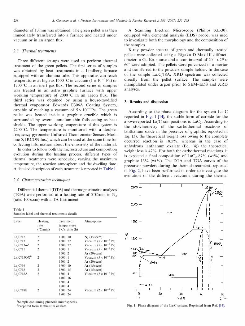
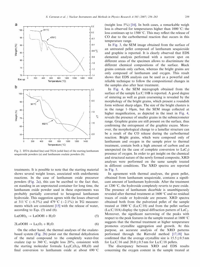
According to the phase diagram for the system La–Creported in Fig. 1 [14], the stable form of carbide for theabove-reported La/C compositions is LaC2. According tothe stoichiometry of the carbothermal reactions oflanthanum oxide in the presence of graphite, reported inEq. (3), the theoretical weight loss owing to the completeoccurred reaction is 18.5%, whereas in the case ofanhydrous lanthanum oxalate (Eq. (4)) the theoreticalweight loss is 47%. For both the carbothermal reactions, itis expected a final composition of LaC2 87% (wt%) andgraphite 13% (wt%). The DTA and TGA curves of theprecursor powders during the thermal treatment, reportedin Fig. 2, have been performed in order to investigate theevolution of the different reactions during the thermal
Fig. 1. Phase diagram of the La/C system. Reprinted from Ref. [14].
ARTICLE IN PRESS
Fig. 2. DTA (dashed line) and TGA (solid line) of the starting lanthanum
sesquioxide powders (a) and lanthanum oxalate powders (b).
S. Carturan et al. / Nuclear Instruments and Methods in Physics Research A 583 (2007) 256–263 259
treatments. It is possible to note that the starting materialshows several weight losses, associated with endothermicreactions. In the case of lanthanum oxide precursorpowders (Fig. 2a), this can be ascribed to the fact that,on standing in an unprotected container for long time, thelanthanum oxide powder used in these experiments wasprobably partially converted to hexagonal lanthanumhydroxide. This suggestion agrees with the losses observedat 311 1C (�6.3%) and 479 1C (�2.1%) in TG measure-ments which are consistent [15] with the release of water,according to Eqs. (5) and (6).
LaðOHÞ3 ! LaOOHþH2O (5)
2LaOOH! La2O3 þH2O: (6)
On the other hand, the thermal analyses of the oxalate-based system (Fig. 2b) point out the thermal dehydrationof the metal compound to the completely water-freeoxalate (up to 360 1C, weight loss 20%, consistent withthe starting molecular formula La2(C2O3)4 � 8H2O) andfinal conversion to lanthanum oxide at about 690 1C
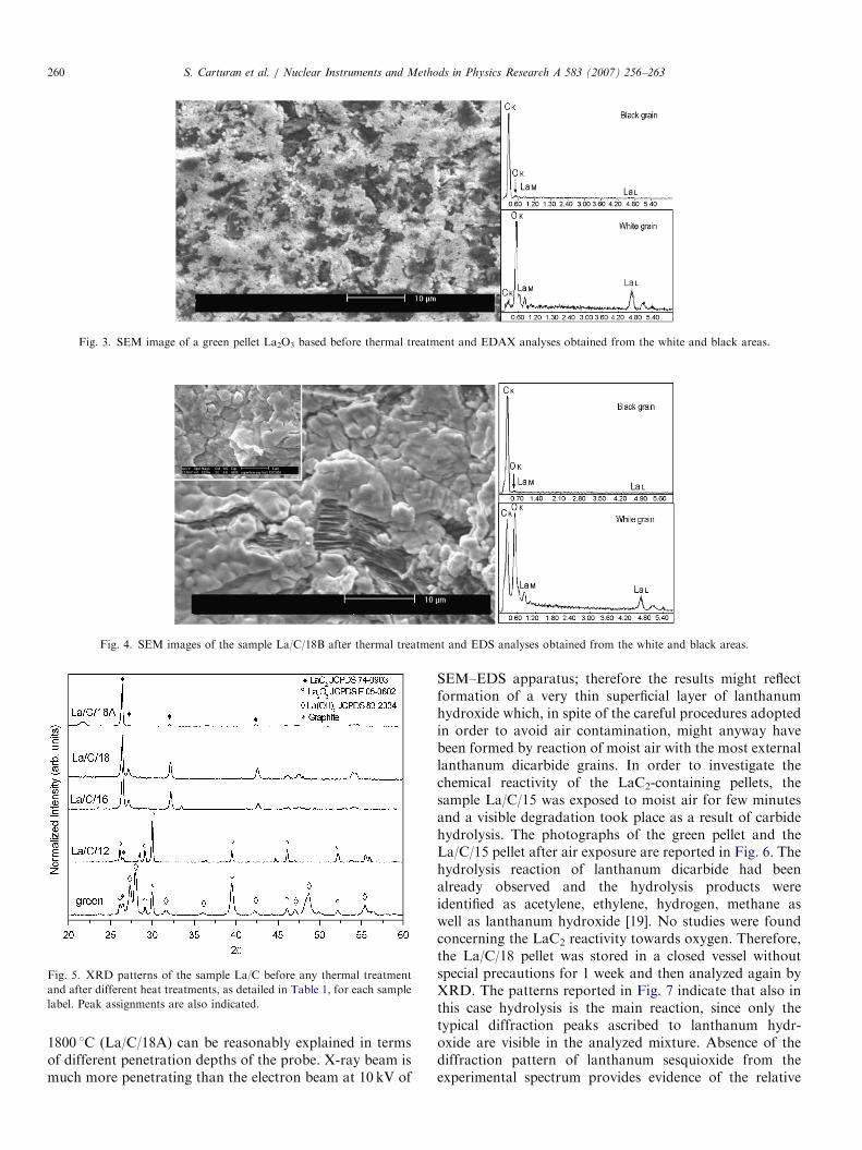
(weight loss 5%) [16]. In both cases, a remarkable weighloss is observed for temperatures higher than 1000 1C: theloss continues up to 1500 1C. This may reflect the release ofCO due to the carbothermal reaction that occurs in thistemperature range.In Fig. 3, the SEM image obtained from the surface of
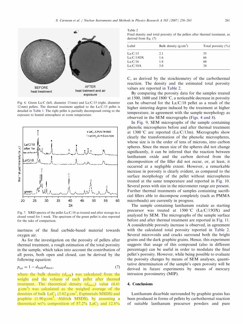
an untreated pellet composed of lanthanum sesquioxideand graphite is reported. It is clearly observed that EDSelemental analysis performed with a narrow spot ondifferent areas of the specimen allows to discriminate thedifferent chemical compositions of the surface. Blackgrains contain only carbon, whereas the bright grains areonly composed of lanthanum and oxygen. This resultshows that EDS analysis can be used as a powerful andreliable technique to follow the compositional changes inthe samples also after heat treatment.In Fig. 4, the SEM micrograph obtained from the
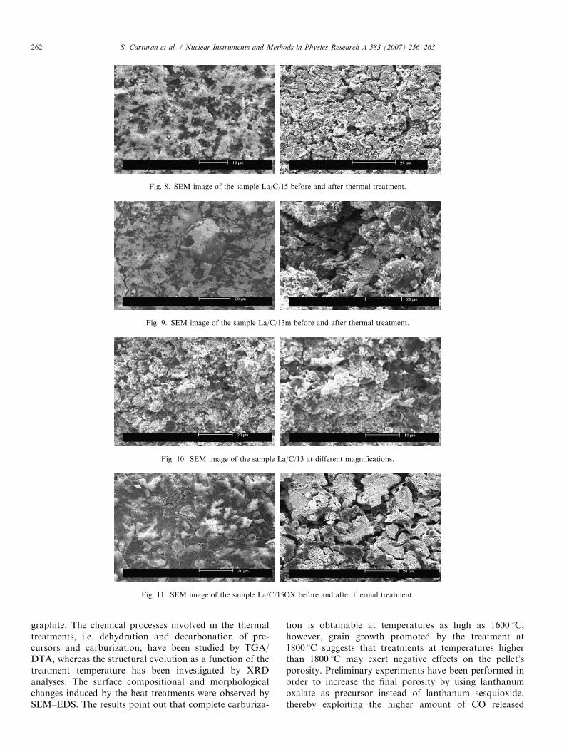
surface of the sample La/C/18B is reported. A good degreeof sintering as well as grain coarsening is revealed by themorphology of the bright grains, which present a roundishform without sharp edges. The size of the bright clusters isin the range 1–10mm, but the SEM image collected athigher magnification, as depicted in the inset in Fig. 4,reveals the presence of smaller grains in the submicrometerrange. Graphite grains are still present on the surface, thusconfirming the entrapment of the graphite excess. More-over, the morphological change to a lamellar structure canbe a result of the CO release during the carbothermalreaction. Bright grains, which were composed only oflanthanum and oxygen in the sample prior to thermaltreatment, contain both a high amount of carbon and anunexpected (in the case of complete conversion to LaC2)presence of oxygen. In order to get insight on the chemicaland structural nature of the newly formed composite, XRDanalyses were performed on the same sample treatedin different conditions. The XRD spectra are reportedin Fig. 5.In agreement with thermal analyses, the green pellet,
obtained from lanthanum sesquioxide, contains a signifi-cant amount of lanthanum hydroxide. After the treatmentat 1200 1C, the hydroxide completely reverts to pure oxide.The presence of lanthanum dicarbide is unambiguouslyrevealed after thermal treatment at 1600 1C and no residualtraces of oxide or hydroxide are present. XRD spectraobtained both from the pulverized pellet of the sampletreated at 1800 1C (La/C/18) and from the pellet surface(La/C/18A) display the typical diffraction pattern of LaC2.Moreover, the significant narrowing of the peaks withrespect to the peak features in the sample treated at 1600 1Csuggests that the thermal treatment at higher temperaturepromotes crystallite aggregation and growth. At thispurpose, an accurate analysis of the XRD patternsperformed through the Rietveld method [17,18] hasconfirmed that the average crystallite size is 15.570.5 nmfor La/C/16 and 20.070.5 nm for La/C/18 pellets.The discrepancy between XRD and EDS results
concerning the oxygen content in the sample treated at
ARTICLE IN PRESS
Fig. 3. SEM image of a green pellet La2O3 based before thermal treatment and EDAX analyses obtained from the white and black areas.
Fig. 4. SEM images of the sample La/C/18B after thermal treatment and EDS analyses obtained from the white and black areas.
Fig. 5. XRD patterns of the sample La/C before any thermal treatment
and after different heat treatments, as detailed in Table 1, for each sample
label. Peak assignments are also indicated.
S. Carturan et al. / Nuclear Instruments and Methods in Physics Research A 583 (2007) 256–263260
1800 1C (La/C/18A) can be reasonably explained in termsof different penetration depths of the probe. X-ray beam ismuch more penetrating than the electron beam at 10 kV of
SEM–EDS apparatus; therefore the results might reflectformation of a very thin superficial layer of lanthanumhydroxide which, in spite of the careful procedures adoptedin order to avoid air contamination, might anyway havebeen formed by reaction of moist air with the most externallanthanum dicarbide grains. In order to investigate thechemical reactivity of the LaC2-containing pellets, thesample La/C/15 was exposed to moist air for few minutesand a visible degradation took place as a result of carbidehydrolysis. The photographs of the green pellet and theLa/C/15 pellet after air exposure are reported in Fig. 6. Thehydrolysis reaction of lanthanum dicarbide had beenalready observed and the hydrolysis products wereidentified as acetylene, ethylene, hydrogen, methane aswell as lanthanum hydroxide [19]. No studies were foundconcerning the LaC2 reactivity towards oxygen. Therefore,the La/C/18 pellet was stored in a closed vessel withoutspecial precautions for 1 week and then analyzed again byXRD. The patterns reported in Fig. 7 indicate that also inthis case hydrolysis is the main reaction, since only thetypical diffraction peaks ascribed to lanthanum hydr-oxide are visible in the analyzed mixture. Absence of thediffraction pattern of lanthanum sesquioxide from theexperimental spectrum provides evidence of the relative
ARTICLE IN PRESS
Fig. 6. Green La/C (left, diameter 13mm) and La/C/15 (right, diameter
12mm) pellets. The thermal treatment applied to the La/C/15 pellet is
detailed in Table 1. The right pellet is partially decomposed owing to the
exposure to humid atmosphere at room temperature.
Fig. 7. XRD spectra of the pellet La/C/18 as treated and after storage in a
closed vessel for 1 week. The spectrum of the green pellet is also reported
for the sake of comparison.
Table 2
Final density and total porosity of the pellets after thermal treatment, as
derived from Eq. (7)
Label Bulk density (g/cm3) Total porosity (%)
La/C/15 2.1 55
La/C/15OX 1.6 66
La/C/16 1.8 60
La/C/18A 3.0 34
S. Carturan et al. / Nuclear Instruments and Methods in Physics Research A 583 (2007) 256–263 261
inertness of the final carbide-based material towardsoxygen air.
As for the investigation on the porosity of pellets afterthermal treatment, a rough estimation of the total porosityin the sample, which takes into account the contribution ofall pores, both open and closed, can be derived by thefollowing equation:
ptot ¼ 1� dbulkdtheor, (7)
where the bulk density (dbulk) was calculated from theweight and the volume of each pellet after thermaltreatment. The theoretical density (dtheor) value (4.61g/cm3) was calculated as the weighed average of thedensities of bulk LaC2 (5.02 g/cm
3, Espimetals MSDS) andgraphite (1.90 g/cm3, Aldrich MSDS), by assuming atheoretical wt% composition of 87.2% LaC2 and 12.8%
C, as derived by the stoichiometry of the carbothermalreaction. The density and the estimated total porosityvalues are reported in Table 2.By comparing the porosity data for the samples treated
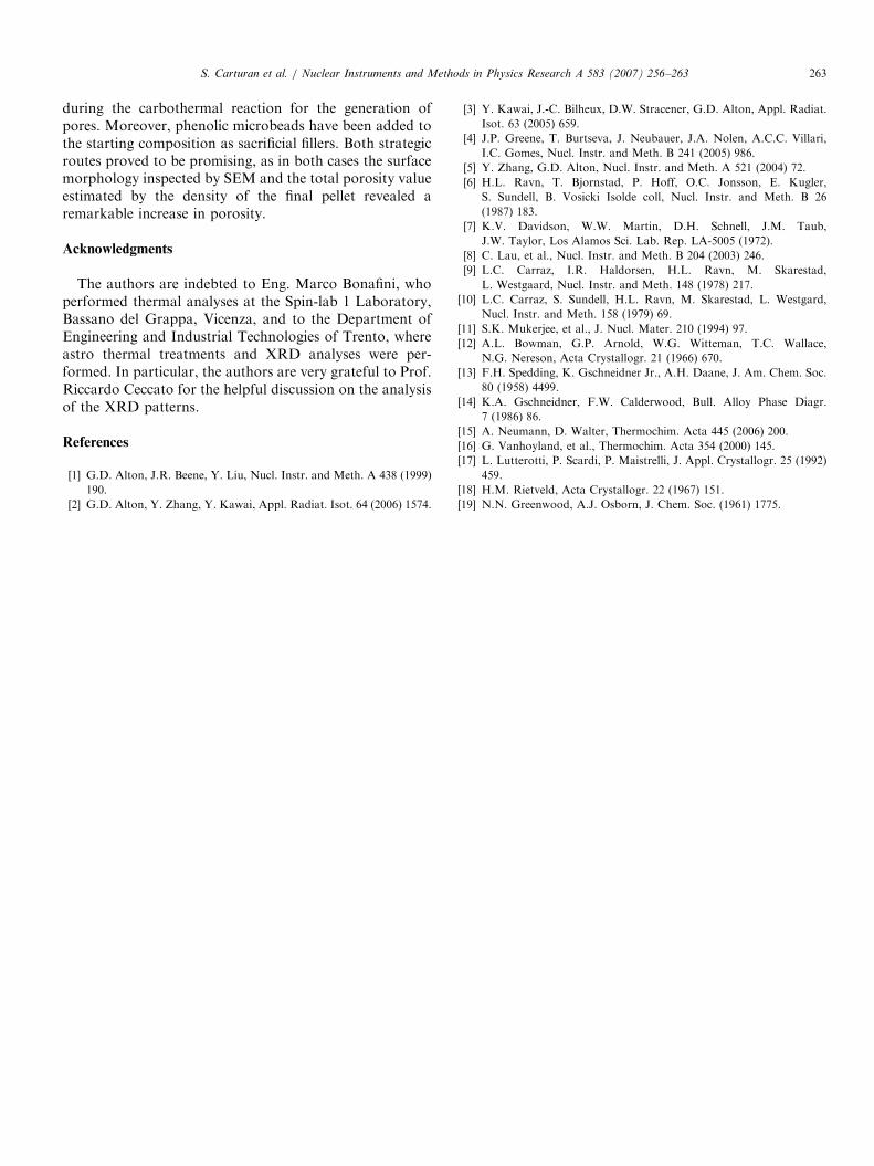
at 1500, 1600 and 1800 1C, a noticeable decrease in porositycan be observed for the La/C/18 pellet as a result of thehigher sintering degree induced by the treatment at highertemperature, in agreement with the sample morphology asobserved in the SEM micrographs (Figs. 4 and 8).In Fig. 9, SEM micrographs of the sample containing
phenolic microspheres before and after thermal treatmentat 1300 1C are reported (La/C/13m). Micrographs showclearly the transformation of the phenolic microspheres,whose size is in the order of tens of microns, into carbonspheres. Since the mean size of the spheres did not changesignificantly, it can be inferred that the reaction betweenlanthanum oxide and the carbon derived from thedecomposition of the filler did not occur, or, at least, itoccurred at a negligible extent. However, a remarkableincrease in porosity is clearly evident, as compared to thesurface morphology of the pellet without microspherestreated at the same temperature and reported in Fig. 10.Several pores with size in the micrometer range are present.Further thermal treatments of samples containing sacrifi-cial fillers able to decompose completely (such as PMMAmicrobeads) are currently in progress.The sample containing lanthanum oxalate as starting
precursor was treated at 1500 1C (La/C/15OX) andanalyzed by SEM. The micrographs of the sample surfacebefore and after thermal treatment are reported in Fig. 11.A considerable porosity increase is observed, in agreementwith the calculated total porosity reported in Table 2.Several microvoids and cracks surround both the brightgrains and the dark graphite grains. Hence, this experimentsuggests that usage of this compound (also in differentpercentage) can be useful in order to modulate the finalpellet’s porosity. However, while being possible to evaluatethe porosity changes by means of SEM analyses, quanti-tative determination of the sample’s open porosity will bederived in future experiments by means of mercuryintrusion porosimetry (MIP).
4. Conclusions
Lanthanum dicarbide surrounded by graphite grains hasbeen produced in forms of pellets by carbothermal reactionof suitable lanthanum precursor powders and pure
ARTICLE IN PRESS
Fig. 8. SEM image of the sample La/C/15 before and after thermal treatment.
Fig. 9. SEM image of the sample La/C/13m before and after thermal treatment.
Fig. 10. SEM image of the sample La/C/13 at different magnifications.
Fig. 11. SEM image of the sample La/C/15OX before and after thermal treatment.
S. Carturan et al. / Nuclear Instruments and Methods in Physics Research A 583 (2007) 256–263262
graphite. The chemical processes involved in the thermaltreatments, i.e. dehydration and decarbonation of pre-cursors and carburization, have been studied by TGA/DTA, whereas the structural evolution as a function of thetreatment temperature has been investigated by XRDanalyses. The surface compositional and morphologicalchanges induced by the heat treatments were observed bySEM–EDS. The results point out that complete carburiza-
tion is obtainable at temperatures as high as 1600 1C,however, grain growth promoted by the treatment at1800 1C suggests that treatments at temperatures higherthan 1800 1C may exert negative effects on the pellet’sporosity. Preliminary experiments have been performed inorder to increase the final porosity by using lanthanumoxalate as precursor instead of lanthanum sesquioxide,thereby exploiting the higher amount of CO released
ARTICLE IN PRESSS. Carturan et al. / Nuclear Instruments and Methods in Physics Research A 583 (2007) 256–263 263
during the carbothermal reaction for the generation ofpores. Moreover, phenolic microbeads have been added tothe starting composition as sacrificial fillers. Both strategicroutes proved to be promising, as in both cases the surfacemorphology inspected by SEM and the total porosity valueestimated by the density of the final pellet revealed aremarkable increase in porosity.
Acknowledgments
The authors are indebted to Eng. Marco Bonafini, whoperformed thermal analyses at the Spin-lab 1 Laboratory,Bassano del Grappa, Vicenza, and to the Department ofEngineering and Industrial Technologies of Trento, whereastro thermal treatments and XRD analyses were per-formed. In particular, the authors are very grateful to Prof.Riccardo Ceccato for the helpful discussion on the analysisof the XRD patterns.
References
[1] G.D. Alton, J.R. Beene, Y. Liu, Nucl. Instr. and Meth. A 438 (1999)
190.
[2] G.D. Alton, Y. Zhang, Y. Kawai, Appl. Radiat. Isot. 64 (2006) 1574.
[3] Y. Kawai, J.-C. Bilheux, D.W. Stracener, G.D. Alton, Appl. Radiat.
Isot. 63 (2005) 659.
[4] J.P. Greene, T. Burtseva, J. Neubauer, J.A. Nolen, A.C.C. Villari,
I.C. Gomes, Nucl. Instr. and Meth. B 241 (2005) 986.
[5] Y. Zhang, G.D. Alton, Nucl. Instr. and Meth. A 521 (2004) 72.
[6] H.L. Ravn, T. Bjornstad, P. Hoff, O.C. Jonsson, E. Kugler,
S. Sundell, B. Vosicki Isolde coll, Nucl. Instr. and Meth. B 26
(1987) 183.
[7] K.V. Davidson, W.W. Martin, D.H. Schnell, J.M. Taub,
J.W. Taylor, Los Alamos Sci. Lab. Rep. LA-5005 (1972).
[8] C. Lau, et al., Nucl. Instr. and Meth. B 204 (2003) 246.
[9] L.C. Carraz, I.R. Haldorsen, H.L. Ravn, M. Skarestad,
L. Westgaard, Nucl. Instr. and Meth. 148 (1978) 217.
[10] L.C. Carraz, S. Sundell, H.L. Ravn, M. Skarestad, L. Westgard,
Nucl. Instr. and Meth. 158 (1979) 69.
[11] S.K. Mukerjee, et al., J. Nucl. Mater. 210 (1994) 97.
[12] A.L. Bowman, G.P. Arnold, W.G. Witteman, T.C. Wallace,
N.G. Nereson, Acta Crystallogr. 21 (1966) 670.
[13] F.H. Spedding, K. Gschneidner Jr., A.H. Daane, J. Am. Chem. Soc.
80 (1958) 4499.
[14] K.A. Gschneidner, F.W. Calderwood, Bull. Alloy Phase Diagr.
7 (1986) 86.
[15] A. Neumann, D. Walter, Thermochim. Acta 445 (2006) 200.
[16] G. Vanhoyland, et al., Thermochim. Acta 354 (2000) 145.
[17] L. Lutterotti, P. Scardi, P. Maistrelli, J. Appl. Crystallogr. 25 (1992)
459.
[18] H.M. Rietveld, Acta Crystallogr. 22 (1967) 151.
[19] N.N. Greenwood, A.J. Osborn, J. Chem. Soc. (1961) 1775.