susi, a negative regulator of drosophila pi3kinase
TRANSCRIPT
University of ZurichZurich Open Repository and Archive
Winterthurerstr. 190
CH-8057 Zurich
http://www.zora.unizh.ch
Year: 2005
Susi, a negative regulator of Drosophila PI3-kinase
Wittwer, Franz; Jaquenoud, Malika; Brogiolo, Walter; Zarske, Marcel; Wüstemann,
Philipp; Fernandez, Rafael; Stocker, Hugo; Wymann, Matthias P; Hafen, Ernst
Wittwer, Franz; Jaquenoud, Malika; Brogiolo, Walter; Zarske, Marcel; Wüstemann, Philipp; Fernandez, Rafael;Stocker, Hugo; Wymann, Matthias P; Hafen, Ernst. Susi, a negative regulator of Drosophila PI3-kinase. Dev. Cell2005, 8(6):817-27.Postprint available at:http://www.zora.unizh.ch
Posted at the Zurich Open Repository and Archive, University of Zurich.http://www.zora.unizh.ch
Originally published at:Dev. Cell 2005, 8(6):817-27
Susi, a negative regulator of Drosophila PI3-kinase
Abstract
The Phosphatidylinositol-3 kinase/Protein Kinase B (PI3K/PKB) signaling pathway controls growth,metabolism, and lifespan in animals, and deregulation of its activity is associated with diabetes andcancer in humans. Here, we describe Susi, a coiled-coil domain protein that acts as a negative regulatorof insulin signaling in Drosophila. Whereas loss of Susi function increases body size, overexpression ofSusi reduces growth. We provide genetic evidence that Susi negatively regulates dPI3K activity. Susidirectly binds to dP60, the regulatory subunit of dPI3K. Since Susi has no overt similarity to knowninhibitors of PI3K/PKB signaling, it defines a novel mechanism by which this signaling cascade is keptin check. The fact that Susi is expressed in a circadian rhythm, with highest levels during the night,suggests that Susi attenuates insulin signaling during the fasting period.
Developmental Cell, Vol. 8, 817–827, June, 2005, Copyright ©2005 by Elsevier Inc. DOI 10.1016/j.devcel.2005.04.002
Susi, a Negative Regulatorof Drosophila PI3-Kinase
Franz Wittwer,1 Malika Jaquenoud,2 Walter Brogiolo,1
Marcel Zarske,1 Philipp Wüstemann,1,3
Rafael Fernandez,4 Hugo Stocker,1
Matthias P. Wymann,2 and Ernst Hafen1,*1Zoologisches InstitutUniversität ZürichWinterthurerstr. 190CH-8057 ZürichSwitzerland2 Institute of Biochemistry and GeneticsCenter of BiomedicineDepartment Clinical and Biological SciencesUniversity of BaselMattenstrasse 28CH-4058 BaselSwitzerland3The Genetics Company, Inc.Wagistrasse 278952 Zürich-SchlierenSwitzerland4Novartis Institute for Biomedical Research, Inc.Functional Genomics100 Technology Square, Room 6216Cambridge, Massachusetts 02139
Summary
The Phosphatidylinositol-3 kinase/Protein Kinase B(PI3K/PKB) signaling pathway controls growth, me-tabolism, and lifespan in animals, and deregulation ofits activity is associated with diabetes and cancer inhumans. Here, we describe Susi, a coiled-coil domainprotein that acts as a negative regulator of insulin sig-naling in Drosophila. Whereas loss of Susi functionincreases body size, overexpression of Susi reducesgrowth. We provide genetic evidence that Susi nega-tively regulates dPI3K activity. Susi directly binds todP60, the regulatory subunit of dPI3K. Since Susi hasno overt similarity to known inhibitors of PI3K/PKBsignaling, it defines a novel mechanism by which thissignaling cascade is kept in check. The fact that Susiis expressed in a circadian rhythm, with highestlevels during the night, suggests that Susi attenuatesinsulin signaling during the fasting period.
Introduction
PI3K/PKB signaling regulates growth and metabolismin mammals and Drosophila. In mammals, PI3K/PKBsignaling is activated by the insulin-like growth factorreceptor (IGFR) and by the insulin receptor (IR) (Nakaeet al., 2001; Yenush and White, 1997). Signaling in re-sponse to IGFR activation is required for cell growthand proliferation during development. The same cas-cade is activated by IR to regulate glucose levels in theblood in mammals. In Drosophila, the functions of IR
*Correspondence: [email protected]
and IGFR are fulfilled by a single prototypical receptor,the Drosophila insulin receptor (dINR). As in mammals,dINR regulates cell growth and metabolism (Stockerand Hafen, 2000).
The mechanisms of insulin receptor signaling aresimilar in mammals and Drosophila (Lizcano and Alessi,2002; Stocker and Hafen, 2000). The activated receptorphosphorylates insulin receptor substrate proteins (IRS1–4 or dIRS/Chico), thereby creating binding sites forSH2-domain-containing proteins including the regula-tory subunit of class IA phosphoinositide 3-kinase(class IA PI3K, hereafter called PI3K). PI3K is a lipidkinase consisting of the regulatory subunit P85 (dP60in Drosophila) and the catalytic subunit P110 (dP110 inDrosophila). By binding to IRS, PI3K is recruited to themembrane where it phosphorylates the 3# position ofphosphatidylinositol-(4,5)-bisphosphate (PIP2) andthereby generates phosphatidylinositol-(3,4,5)-tris-phosphate (PIP3). Elevated levels of PIP3 recruit the PH-domain-containing protein kinase B (PKB/AKT) to theplasma membrane and permit its further activation byphosphoinositide-dependent kinase 1 (PDK1). PKBtransmits the signal by the inhibition of transcriptionfactors of the FOXO (forkhead box, subgroup “O”) fam-ily and by phosphorylation of other downstream tar-gets.
The activity of PI3K/PKB signaling needs to be ad-justed continuously to the developmental and meta-bolic state of the organism. Therefore, the activity ofmost positively acting signaling components is held incheck by negative regulators. Several mechanisms ofhow PI3K/PKB signaling is regulated at the level ofPI3K are known in mammals. First, the level of PIP3,the product of PI3K activity, is negatively regulated bytwo lipid phosphatases that dephosphorylate PIP3.Whereas PTEN (the phosphatase and tensin homologon chromosome 10) dephosphorylates PIP3 at the 3#position (Di Cristofano and Pandolfi, 2000), SHIP (SH2-domain-containing inositol 5# phosphatase) dephos-phorylates PIP3 at the 5# position (Rohrschneider et al.,2000). PI3K activity is also modulated directly by thelevels of its adaptor P85. Under conditions where themolar ratio of P85/P110 is increased, P85 monomerscompete with P85/P110 heterodimers for binding tophosphorylated YxxM motifs on IRS (Kodaki et al.,1994; Rodriguez-Viciana et al., 1997). Similarly, overex-pression of the dP60 subunit of Drosophila PI3K alsoexerts a dominant-negative effect (Britton et al., 2002;Weinkove et al., 1999). Furthermore, activity of mam-malian PI3K is regulated by Ruk, a protein containingtwo SH3 domains, a proline-rich motif, and a coiled-coiled domain. Ruk binds to the SH3 domain of the reg-ulatory subunit of PI3K, inhibits its activity, and inducesapoptosis when overexpressed in cultured cells (Goutet al., 2000). Its role as negative regulator of insulin/IGFsignaling in an animal model has not been describedyet.
Here we describe Susi, a novel negative regulator ofinsulin signaling in Drosophila. Gain-of-function muta-tions of Susi were identified in a genetic screen for
Developmental Cell818
genes that suppress dINR-induced hyperplasia when dSoverexpressed. Loss-of-function alleles of Susi showed
that it encodes a negative regulator of insulin signaling faand that Susi protein acts by inhibiting dPI3K activity.
Susi encodes a coiled-coiled domain protein that in- meteracts with the dP60 regulatory subunit of dPI3K.t(Resultse1Overexpression of Susi SuppressesddINR-Induced OvergrowthdTo identify negative regulators of dINR signaling, weccarried out a misexpression screen for genes that sup-epress the overgrowth phenotype caused by overex-spression of wild-type dINR in the developing eye using
the UAS/Gal4 system (Figures 1A–1C). We tested 5,400ofly lines containing random insertions of an enhancer-Spromoter (EP) element that permits the transcription ofWgenes flanking the insertion in response to Gal4. Sev-teral independent lines that strongly suppressed thewdINR-induced eye phenotype (among them EP(7-66);TFigures 1D and 2A) contained EP insertions upstreamtof the first coding exon of the B4 gene (Sotillos et al.,h1997). The suppression is caused by the overexpres-ssion of the B4 gene, since overexpression of B4 fromca UAS transgene had the same effect. Therefore, weprenamed the B4 locus Suppressor of signaling by insu-
lin (Susi).tSusi encodes a novel protein with a predicted coiled-lcoil (CC) domain (amino acids 916–942 according to theupredicted amino acid sequence CG9239-PA, GenBankeaccession number NP 477325). Proteins that are obvi-lous orthologs of Susi exist in other insect species suchtas Drosophila pseudoobscura (75% identical aminoaacids, see Experimental Procedures) and Anopheleshgambiae (GenBank accession number EAA12152). Ow-Ping to the low sequence conservation and the large size
of the family of CC domain-containing proteins, wewere unable to resolve whether one of the CC domain Lproteins from higher organisms is a Susi ortholog. O
Susi overexpression suppresses dINR function in oother developmental processes also. For example, em- tbryonic lethality associated with the expression of dINR iby en-Gal4 was suppressed by the concomitant ex- opression of Susi. Importantly, the effects of Susi overex- ipression appear to be specific for dINR/dPI3K signal- oing. The complete set of EP lines was tested in parallel Pfor effects on other signaling pathways, including thegrowth promotion by dMyc, and the EP insertions in tthe Susi locus were not found in screens other than rour dINR screen (data not shown; P. Gallant, personal ccommunication). S
tpSusi Overexpression Reduces Growth
To further investigate the possibility that Susi is an in- fShibitor of PI3K/PKB signaling, we compared the pheno-
type of Susi-overexpressing flies with chico mutant tiflies, in which the activity of PI3K/PKB signaling is im-
paired (Bohni et al., 1999). Chico is the Drosophila or-ftholog of IRS1-4, and chico flies are small because of
a cell-autonomous reduction in the rate of cell prolifera- tation and cell growth. Ubiquitous overexpression of Susi
throughout development resulted in flies that were re- o
uced in size (Figures 1E and 1F). Overexpression ofusi in the developing eye resulted in smaller eyes with
ewer ommatidia (Figures 1G–1I). Susi overexpressionlso reduced photoreceptor cell size in a cell-autono-ous manner (Figure 1J). As for the eyes, wings over-
xpressing Susi during development were smaller dueo a reduction in cell number (−41%) and cell size−27%) (Figures 1K and 1L and data not shown). Over-xpression of the apoptosis inhibitor P35 (Hay et al.,994) together with Susi in the developing eye or wingid not suppress the growth-inhibition phenotypes, in-icating that Susi does not attenuate growth by in-reasing apoptosis. In all these experiments, Susi over-xpression reduced growth but did not affect cellpecification or patterning.To further analyze the effect of Susi overexpression
n cell division, we examined the growth capacity ofusi-overexpressing clones in the wing imaginal disc.e found that Susi-overexpressing clones were smaller
han control clones because they contained fewer cellshereas cell size was not changed (Figures 1M–1O).hus, Susi-overexpressing cells divided at a lower ratehan control cells. Whereas cell-doubling time was 11.5r (±0.48 hr, n = 10 clones) in control clones, it wasignificantly increased to 14.1 hr (±1.65 hr, n = 11lones) in Susi-overexpression clones (one-tailed t test:< 0.025).Apart from the growth phenotype, animals ubiqui-
ously overexpressing Susi were developmentally de-ayed and resistant to starvation (see Supplemental Fig-res S1A and S1B available with this article online). Theffects of Susi overexpression on cell growth, cell pro-
iferation, developmental timing, and starvation resis-ance are very similar to the Chico phenotype (Bohni etl., 1999; Oldham et al., 2002), further supporting theypothesis that Susi is a negative regulator of PI3K/KB signaling.
oss of Susi Function Increases Growthverexpression phenotypes may be caused by ectopicr increased gene activation and may thus not reflecthe physiological function of the gene. Therefore, it wasmportant to investigate the consequences of the lossf Susi function. We identified loss-of-function alleles
n Susi by screening for chemically induced reversionf the Susi gain-of-function phenotype (Experimentalrocedures).Sequencing of the revertants revealed that two mu-
ant alleles introduced stop codons within the openeading frame, thus resulting in the formation of trun-ated proteins (Figure 2B). In contrast to the wild-typeusi protein, overexpression of these truncated pro-
eins in the developing eye did not cause a detectablehenotype, suggesting that the isolated Susi loss-of-
unction alleles are functional nulls (data not shown).usi8 is a P element insertion in the locus and appears
o be a hypomorphic allele (see below, Figure 2A, andn Experimental Procedures).
All heteroallelic combinations of Susi resulted in adultlies with reduced viability and increased body size (de-ected by increased weight and wing size; Figures 2Cnd 2D and data not shown). The developmental timingf these flies was not altered. The same was observed
Susi Inhibits PI3K/PKB Signaling819
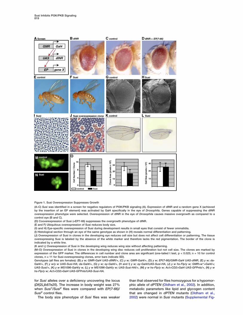
Figure 1. Susi Overexpression Suppresses Growth
(A–C) Susi was identified in a screen for negative regulators of PI3K/PKB signaling (A). Expression of dINR and a random gene X (achievedby the insertion of an EP element) was activated by Gal4 specifically in the eye of Drosophila. Genes capable of suppressing the dINRoverexpression phenotype were selected. Overexpression of dINR in the eye of Drosophila causes massive overgrowth as compared to acontrol eye (B and C).(D) Cooverexpression of Susi (=EP7-66) suppresses the overgrowth phenotype of dINR.(E and F) Ubiquitous overexpression of Susi reduces body size.(G and H) Eye-specific overexpression of Susi during development results in small eyes that consist of fewer ommatidia.(I) Histological section through an eye of the same genotype as shown in (H) reveals normal differentiation and patterning.(J) Overexpression of Susi in clones in the developing eye reduces cell size but does not affect cell differentiation or patterning. The tissueoverexpressing Susi is labeled by the absence of the white marker and therefore lacks the red pigmentation. The border of the clone isindicated by a white line.(K and L) Overexpression of Susi in the developing wing reduces wing size without affecting patterning.(M–O) Overexpression of Susi in clones in the developing wing disc reduces cell proliferation but not cell size. The clones are marked byexpression of the GFP marker. The differences in cell number and clone area are significant (one-tailed t test, p < 0.025; n = 10 for controlclones, n = 11 for Susi-overexpressing clones, error bars indicate SD).Genotypes (all flies are females): (B) y w; GMR-Gal4 UAS-dINR/+, (C) y w; GMR-Gal4/+, (D) y w; EP(7-66)/GMR-Gal4 UAS-dINR, (E) y w; da-Gal4/+, (F) y w/y w UAS-Susi-HA; da-Gal4/+, (G) y w; ey-Gal4/+, (H and I) y w; ey-Gal4/UAS-Susi-HA, (J) y w hs-Flp/y w; GMR>w+>Gal4/+;UAS-Susi/+, (K) y w MS1096-Gal4/y w, (L) y w MS1096-Gal4/y w; UAS-Susi-HA/+, (M) y w hs-Flp/y w; Act>CD2>Gal4 UAS-GFPnls/+, (N) y whs-Flp/y w; Act>CD2>Gal4 UAS-GFPnls/UAS-Susi-HA.
for Susi alleles over a deficiency uncovering the locus(Df(2L)b87e25). The increase in body weight was 27%when Susi1/Susi2 flies were compared with EP(7-66)/Susi2 control flies.
The body size phenotype of Susi flies was weaker
than that observed for flies homozygous for a hypomor-phic allele of dPTEN (Oldham et al., 2002). In addition,metabolic parameters like lipid and glycogen contentthat are changed in dPTEN mutants (Oldham et al.,2002) were normal in Susi mutants (Supplemental Fig-
Developmental Cell820
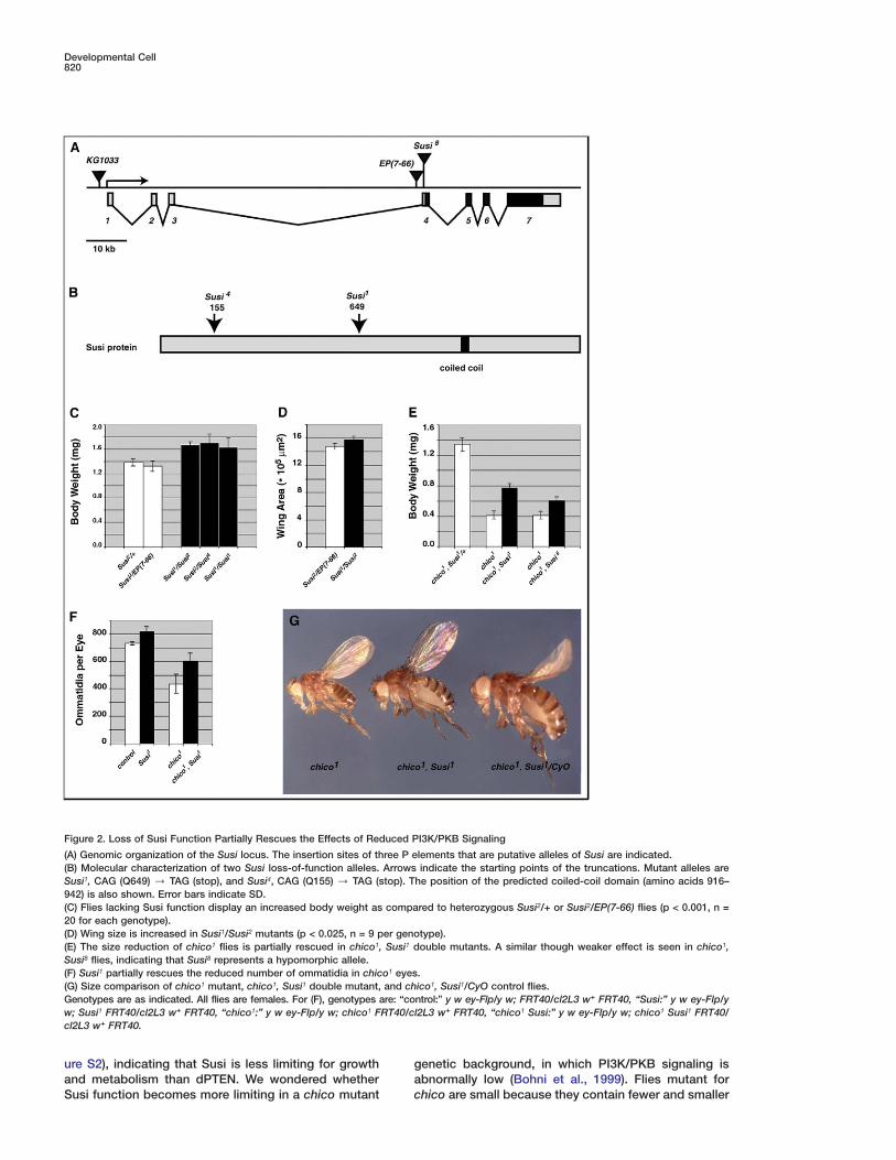
Figure 2. Loss of Susi Function Partially Rescues the Effects of Reduced PI3K/PKB Signaling
(A) Genomic organization of the Susi locus. The insertion sites of three P elements that are putative alleles of Susi are indicated.(B) Molecular characterization of two Susi loss-of-function alleles. Arrows indicate the starting points of the truncations. Mutant alleles areSusi1, CAG (Q649) / TAG (stop), and Susi4, CAG (Q155) / TAG (stop). The position of the predicted coiled-coil domain (amino acids 916–942) is also shown. Error bars indicate SD.(C) Flies lacking Susi function display an increased body weight as compared to heterozygous Susi2/+ or Susi2/EP(7-66) flies (p < 0.001, n =20 for each genotype).(D) Wing size is increased in Susi1/Susi2 mutants (p < 0.025, n = 9 per genotype).(E) The size reduction of chico1 flies is partially rescued in chico1, Susi1 double mutants. A similar though weaker effect is seen in chico1,Susi8 flies, indicating that Susi8 represents a hypomorphic allele.(F) Susi1 partially rescues the reduced number of ommatidia in chico1 eyes.(G) Size comparison of chico1 mutant, chico1, Susi1 double mutant, and chico1, Susi1/CyO control flies.Genotypes are as indicated. All flies are females. For (F), genotypes are: “control:” y w ey-Flp/y w; FRT40/cl2L3 w+ FRT40, “Susi:” y w ey-Flp/yw; Susi1 FRT40/cl2L3 w+ FRT40, “chico1:” y w ey-Flp/y w; chico1 FRT40/cl2L3 w+ FRT40, “chico1 Susi:” y w ey-Flp/y w; chico1 Susi1 FRT40/cl2L3 w+ FRT40.
ure S2), indicating that Susi is less limiting for growth gaand metabolism than dPTEN. We wondered whether
Susi function becomes more limiting in a chico mutant c
enetic background, in which PI3K/PKB signaling isbnormally low (Bohni et al., 1999). Flies mutant forhico are small because they contain fewer and smaller
Susi Inhibits PI3K/PKB Signaling821
cells (Bohni et al., 1999). chico, Susi double mutant flieswere significantly larger than chico flies but still smallerthan control flies (Figures 2E and 2G). The relative in-crease in body weight caused by removal of Susi func-tion was stronger in a chico1 genetic background thanin a wild-type background (Figures 2C and 2E). In ex-periments including Susi1, this increase was 27% in awild-type background and 88% in a chico1 back-ground. Similarly, loss of Susi function partially sup-pressed the reduction in ommatidial number of chicoflies (Figure 2F). Similar results were obtained withother independent alleles of Susi and chico (data notshown). Furthermore, Susi also suppressed the re-duced body size of mutants in which the function ofdINR or dPKB was reduced (the hypomorphic allelesdINR19 and dPKB3, data not shown).
Conversely, loss of Susi enhanced the phenotype ofweak dPTEN mutant alleles. Flies homozygous for thehypomorphic dPTEN2L100 allele are viable and in-creased in size (Oldham et al., 2002). Susi, dPTEN2L100
flies displayed synthetic lethality. Flies in which thefunction of both Susi and dPTEN was impaired by atissue-specific recombination system (Newsome et al.,2000) showed a strong enhancement of the big eyephenotype of dPTEN2L100. This size increase was atleast in part due to an increase in the number of omma-tidia (data not shown). Thus, Susi shows strong geneticinteractions with PI3K/PKB signaling components, andthe loss-of-function phenotype of Susi is complemen-tary to its gain-of-function phenotype. This indicatesthat the Susi gain-of-function phenotype reflects theoveractivation of the natural function of Susi and thatSusi is a negative regulator of PI3K/PKB signaling.
Susi Inhibits PI3K/PKB Signaling Downstreamof dINR but Upstream of dPI3KWe chose three different approaches to address whetherSusi indeed inhibits PI3K/PKB signaling and to estab-lish at which level of the cascade Susi acts. First, wetested the effect of Susi overexpression on the expres-sion of a target gene of PI3K/PKB signaling. Second,we tested whether overexpression of Susi reduced thelevels of PIP3, a key component of the PI3K/PKB sig-naling cascade. Finally, we checked whether Susi couldsuppress phenotypes caused by constitutive activationof components of PI3K/PKB signaling.
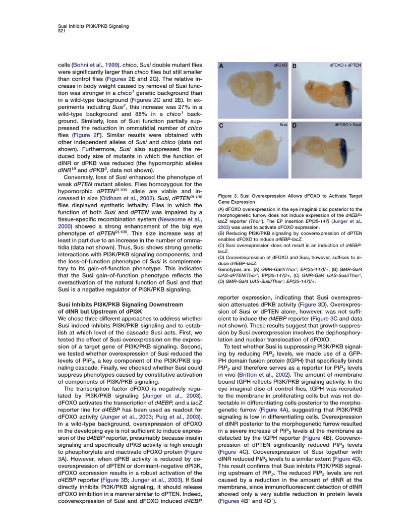
The transcription factor dFOXO is negatively regu-lated by PI3K/PKB signaling (Junger et al., 2003).dFOXO activates the transcription of d4EBP, and a lacZreporter line for d4EBP has been used as readout fordFOXO activity (Junger et al., 2003; Puig et al., 2003).In a wild-type background, overexpression of dFOXOin the developing eye is not sufficient to induce expres-sion of the d4EBP reporter, presumably because insulinsignaling and specifically dPKB activity is high enoughto phosphorylate and inactivate dFOXO protein (Figure3A). However, when dPKB activity is reduced by co-overexpression of dPTEN or dominant-negative dPI3K,dFOXO expression results in a robust activation of thed4EBP reporter (Figure 3B; Junger et al., 2003). If Susidirectly inhibits PI3K/PKB signaling, it should releasedFOXO inhibition in a manner similar to dPTEN. Indeed,cooverexpression of Susi and dFOXO induced d4EBP
Figure 3. Susi Overexpression Allows dFOXO to Activate TargetGene Expression
(A) dFOXO overexpression in the eye imaginal disc posterior to themorphogenetic furrow does not induce expression of the d4EBP-lacZ reporter (Thor1). The EP insertion EP(35-147) (Junger et al.,2003) was used to activate dFOXO expression.(B) Reducing PI3K/PKB signaling by cooverexpression of dPTENenables dFOXO to induce d4EBP-lacZ.(C) Susi overexpression does not result in an induction of d4EBP-lacZ.(D) Cooverexpression of dFOXO and Susi, however, suffices to in-duce d4EBP-lacZ.Genotypes are: (A) GMR-Gal4/Thor1; EP(35-147)/+, (B) GMR-Gal4UAS-dPTEN/Thor1; EP(35-147)/+, (C) GMR-Gal4 UAS-Susi/Thor1,(D) GMR-Gal4 UAS-Susi/Thor1; EP(35-147)/+.
reporter expression, indicating that Susi overexpres-sion attenuates dPKB activity (Figure 3D). Overexpres-sion of Susi or dPTEN alone, however, was not suffi-cient to induce the d4EBP reporter (Figure 3C and datanot shown). These results suggest that growth suppres-sion by Susi overexpression involves the dephosphory-lation and nuclear translocation of dFOXO.
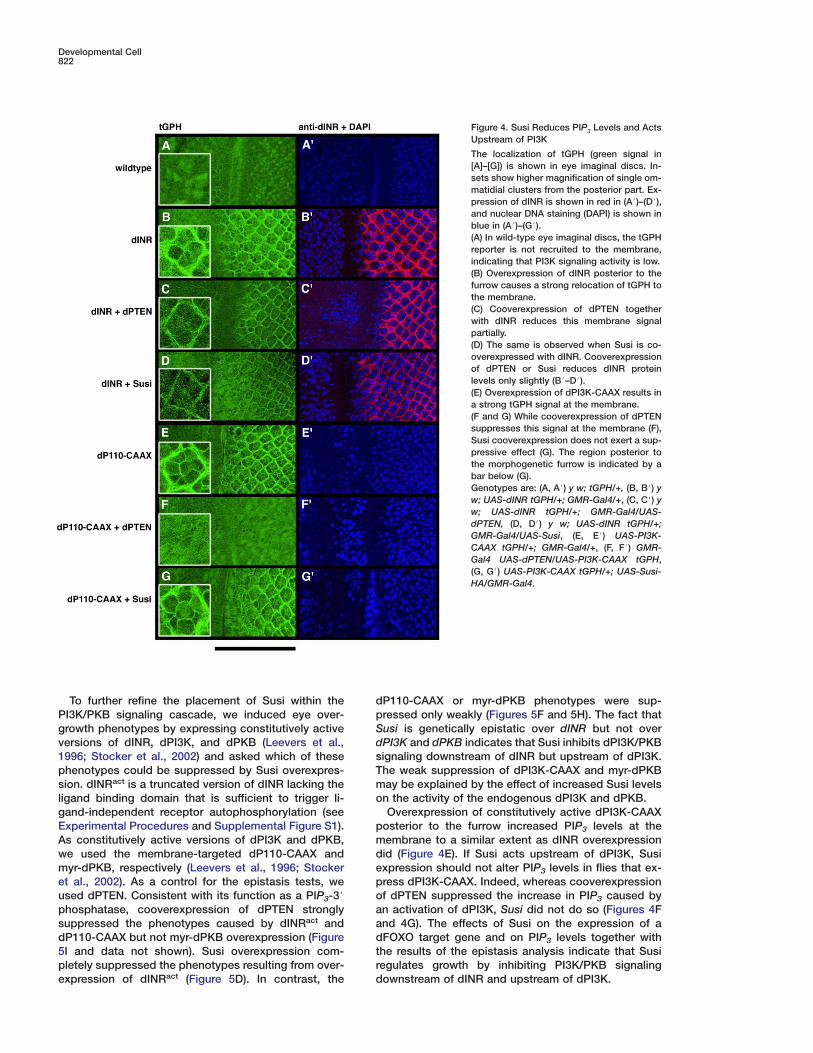
To test whether Susi is suppressing PI3K/PKB signal-ing by reducing PIP3 levels, we made use of a GFP-PH domain fusion protein (tGPH) that specifically bindsPIP3 and therefore serves as a reporter for PIP3 levelsin vivo (Britton et al., 2002). The amount of membranebound tGPH reflects PI3K/PKB signaling activity. In theeye imaginal disc of control flies, tGPH was recruitedto the membrane in proliferating cells but was not de-tectable in differentiating cells posterior to the morpho-genetic furrow (Figure 4A), suggesting that PI3K/PKBsignaling is low in differentiating cells. Overexpressionof dINR posterior to the morphogenetic furrow resultedin a severe increase of PIP3 levels at the membrane asdetected by the tGPH reporter (Figure 4B). Cooverex-pression of dPTEN significantly reduced PIP3 levels(Figure 4C). Cooverexpression of Susi together withdINR reduced PIP3 levels to a similar extent (Figure 4D).This result confirms that Susi inhibits PI3K/PKB signal-ing upstream of PIP3. The reduced PIP3 levels are notcaused by a reduction in the amount of dINR at themembrane, since immunofluorescent detection of dINRshowed only a very subtle reduction in protein levels(Figures 4B# and 4D#).
Developmental Cell822
Figure 4. Susi Reduces PIP3 Levels and ActsUpstream of PI3K
The localization of tGPH (green signal in[A]–[G]) is shown in eye imaginal discs. In-sets show higher magnification of single om-matidial clusters from the posterior part. Ex-pression of dINR is shown in red in (A#)–(D#),and nuclear DNA staining (DAPI) is shown inblue in (A#)–(G#).(A) In wild-type eye imaginal discs, the tGPHreporter is not recruited to the membrane,indicating that PI3K signaling activity is low.(B) Overexpression of dINR posterior to thefurrow causes a strong relocation of tGPH tothe membrane.(C) Cooverexpression of dPTEN togetherwith dINR reduces this membrane signalpartially.(D) The same is observed when Susi is co-overexpressed with dINR. Cooverexpressionof dPTEN or Susi reduces dINR proteinlevels only slightly (B#–D#).(E) Overexpression of dPI3K-CAAX results ina strong tGPH signal at the membrane.(F and G) While cooverexpression of dPTENsuppresses this signal at the membrane (F),Susi cooverexpression does not exert a sup-pressive effect (G). The region posterior tothe morphogenetic furrow is indicated by abar below (G).Genotypes are: (A, A#) y w; tGPH/+, (B, B#) yw; UAS-dINR tGPH/+; GMR-Gal4/+, (C, C#) yw; UAS-dINR tGPH/+; GMR-Gal4/UAS-dPTEN, (D, D#) y w; UAS-dINR tGPH/+;GMR-Gal4/UAS-Susi, (E, E#) UAS-PI3K-CAAX tGPH/+; GMR-Gal4/+, (F, F#) GMR-Gal4 UAS-dPTEN/UAS-PI3K-CAAX tGPH,(G, G#) UAS-PI3K-CAAX tGPH/+; UAS-Susi-HA/GMR-Gal4.
To further refine the placement of Susi within the dpPI3K/PKB signaling cascade, we induced eye over-
growth phenotypes by expressing constitutively active Sdversions of dINR, dPI3K, and dPKB (Leevers et al.,
1996; Stocker et al., 2002) and asked which of these sTphenotypes could be suppressed by Susi overexpres-
sion. dINRact is a truncated version of dINR lacking the moligand binding domain that is sufficient to trigger li-
gand-independent receptor autophosphorylation (seepExperimental Procedures and Supplemental Figure S1).
As constitutively active versions of dPI3K and dPKB, mdwe used the membrane-targeted dP110-CAAX and
myr-dPKB, respectively (Leevers et al., 1996; Stocker epet al., 2002). As a control for the epistasis tests, we
used dPTEN. Consistent with its function as a PIP3-3# oaphosphatase, cooverexpression of dPTEN strongly
suppressed the phenotypes caused by dINRact and addP110-CAAX but not myr-dPKB overexpression (Figure
5I and data not shown). Susi overexpression com- trpletely suppressed the phenotypes resulting from over-
expression of dINRact (Figure 5D). In contrast, the d
P110-CAAX or myr-dPKB phenotypes were sup-ressed only weakly (Figures 5F and 5H). The fact thatusi is genetically epistatic over dINR but not overPI3K and dPKB indicates that Susi inhibits dPI3K/PKBignaling downstream of dINR but upstream of dPI3K.he weak suppression of dPI3K-CAAX and myr-dPKBay be explained by the effect of increased Susi levelsn the activity of the endogenous dPI3K and dPKB.Overexpression of constitutively active dPI3K-CAAX
osterior to the furrow increased PIP3 levels at theembrane to a similar extent as dINR overexpressionid (Figure 4E). If Susi acts upstream of dPI3K, Susixpression should not alter PIP3 levels in flies that ex-ress dPI3K-CAAX. Indeed, whereas cooverexpressionf dPTEN suppressed the increase in PIP3 caused byn activation of dPI3K, Susi did not do so (Figures 4Fnd 4G). The effects of Susi on the expression of aFOXO target gene and on PIP3 levels together withhe results of the epistasis analysis indicate that Susiegulates growth by inhibiting PI3K/PKB signalingownstream of dINR and upstream of dPI3K.
Susi Inhibits PI3K/PKB Signaling823
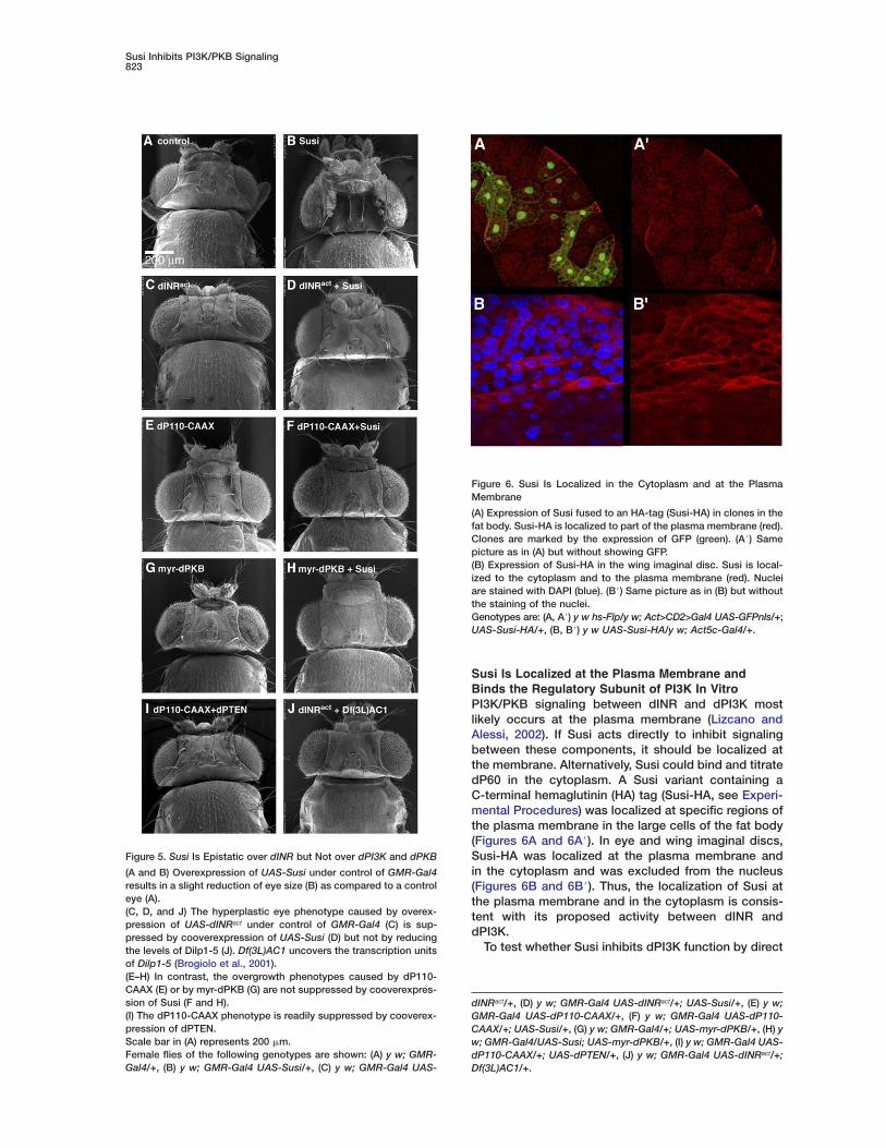
Figure 5. Susi Is Epistatic over dINR but Not over dPI3K and dPKB
(A and B) Overexpression of UAS-Susi under control of GMR-Gal4results in a slight reduction of eye size (B) as compared to a controleye (A).(C, D, and J) The hyperplastic eye phenotype caused by overex-pression of UAS-dINRact under control of GMR-Gal4 (C) is sup-pressed by cooverexpression of UAS-Susi (D) but not by reducingthe levels of Dilp1-5 (J). Df(3L)AC1 uncovers the transcription unitsof Dilp1-5 (Brogiolo et al., 2001).(E–H) In contrast, the overgrowth phenotypes caused by dP110-CAAX (E) or by myr-dPKB (G) are not suppressed by cooverexpres-sion of Susi (F and H).(I) The dP110-CAAX phenotype is readily suppressed by cooverex-pression of dPTEN.Scale bar in (A) represents 200 �m.Female flies of the following genotypes are shown: (A) y w; GMR-Gal4/+, (B) y w; GMR-Gal4 UAS-Susi/+, (C) y w; GMR-Gal4 UAS-
To test whether Susi inhibits dPI3K function by direct
dINRact/+, (D) y w; GMR-Gal4 UAS-dINRact/+; UAS-Susi/+, (E) y w;GMR-Gal4 UAS-dP110-CAAX/+, (F) y w; GMR-Gal4 UAS-dP110-CAAX/+; UAS-Susi/+, (G) y w; GMR-Gal4/+; UAS-myr-dPKB/+, (H) yw; GMR-Gal4/UAS-Susi; UAS-myr-dPKB/+, (I) y w; GMR-Gal4 UAS-dP110-CAAX/+; UAS-dPTEN/+, (J) y w; GMR-Gal4 UAS-dINRact/+;Df(3L)AC1/+.
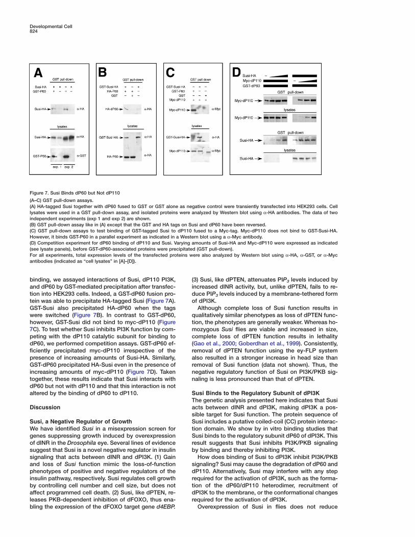
Susi Is Localized at the Plasma Membrane andBinds the Regulatory Subunit of PI3K In VitroPI3K/PKB signaling between dINR and dPI3K mostlikely occurs at the plasma membrane (Lizcano andAlessi, 2002). If Susi acts directly to inhibit signalingbetween these components, it should be localized atthe membrane. Alternatively, Susi could bind and titratedP60 in the cytoplasm. A Susi variant containing aC-terminal hemaglutinin (HA) tag (Susi-HA, see Experi-mental Procedures) was localized at specific regions ofthe plasma membrane in the large cells of the fat body(Figures 6A and 6A#). In eye and wing imaginal discs,Susi-HA was localized at the plasma membrane andin the cytoplasm and was excluded from the nucleus(Figures 6B and 6B#). Thus, the localization of Susi atthe plasma membrane and in the cytoplasm is consis-tent with its proposed activity between dINR anddPI3K.
Figure 6. Susi Is Localized in the Cytoplasm and at the PlasmaMembrane
(A) Expression of Susi fused to an HA-tag (Susi-HA) in clones in thefat body. Susi-HA is localized to part of the plasma membrane (red).Clones are marked by the expression of GFP (green). (A#) Samepicture as in (A) but without showing GFP.(B) Expression of Susi-HA in the wing imaginal disc. Susi is local-ized to the cytoplasm and to the plasma membrane (red). Nucleiare stained with DAPI (blue). (B#) Same picture as in (B) but withoutthe staining of the nuclei.Genotypes are: (A, A#) y w hs-Flp/y w; Act>CD2>Gal4 UAS-GFPnls/+;UAS-Susi-HA/+, (B, B#) y w UAS-Susi-HA/y w; Act5c-Gal4/+.
Developmental Cell824
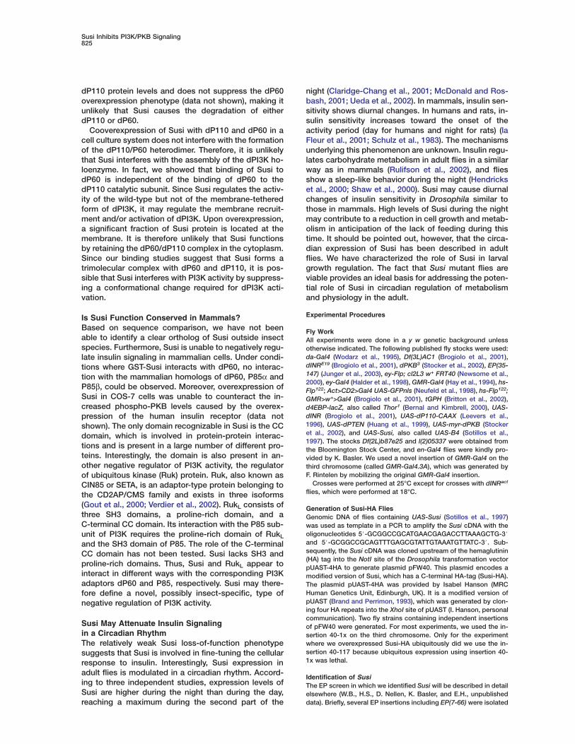
binding, we assayed interactions of Susi, dP110 PI3K, (iand dP60 by GST-mediated precipitation after transfec-
tion into HEK293 cells. Indeed, a GST-dP60 fusion pro- dotein was able to precipitate HA-tagged Susi (Figure 7A).
GST-Susi also precipitated HA-dP60 when the tagsqwere switched (Figure 7B). In contrast to GST-dP60,
however, GST-Susi did not bind to myc-dP110 (Figure tm7C). To test whether Susi inhibits PI3K function by com-
peting with the dP110 catalytic subunit for binding to c(dP60, we performed competition assays. GST-dP60 ef-
ficiently precipitated myc-dP110 irrespective of the rapresence of increasing amounts of Susi-HA. Similarly,
GST-dP60 precipitated HA-Susi even in the presence of rnincreasing amounts of myc-dP110 (Figure 7D). Taken
together, these results indicate that Susi interacts with ndP60 but not with dP110 and that this interaction is notaltered by the binding of dP60 to dP110. S
TaDiscussionsSSusi, a Negative Regulator of Growth
We have identified Susi in a misexpression screen for tSgenes suppressing growth induced by overexpression
of dINR in the Drosophila eye. Several lines of evidence rbsuggest that Susi is a novel negative regulator in insulin
signaling that acts between dINR and dPI3K. (1) Gainsand loss of Susi function mimic the loss-of-function
phenotypes of positive and negative regulators of the drinsulin pathway, respectively. Susi regulates cell growth
by controlling cell number and cell size, but does not tdaffect programmed cell death. (2) Susi, like dPTEN, re-
leases PKB-dependent inhibition of dFOXO, thus ena- rbling the expression of the dFOXO target gene d4EBP.
Figure 7. Susi Binds dP60 but Not dP110
(A–C) GST pull-down assays.(A) HA-tagged Susi together with dP60 fused to GST or GST alone as negative control were transiently transfected into HEK293 cells. Celllysates were used in a GST pull-down assay, and isolated proteins were analyzed by Western blot using α-HA antibodies. The data of twoindependent experiments (exp 1 and exp 2) are shown.(B) GST pull-down assay like in (A) except that the GST and HA tags on Susi and dP60 have been reversed.(C) GST pull-down assays to test binding of GST-tagged Susi to dP110 fused to a Myc-tag. Myc-dP110 does not bind to GST-Susi-HA.However, it binds GST-P60 in a parallel experiment as indicated in a Western blot using a α-Myc antibody.(D) Competition experiment for dP60 binding of dP110 and Susi. Varying amounts of Susi-HA and Myc-dP110 were expressed as indicated(see lysate panels), before GST-dP60-associated proteins were precipitated (GST pull-down).For all experiments, total expression levels of the transfected proteins were also analyzed by Western blot using α-HA, α-GST, or α-Mycantibodies (indicated as “cell lysates” in [A]–[D]).
3) Susi, like dPTEN, attenuates PIP3 levels induced byncreased dINR activity, but, unlike dPTEN, fails to re-uce PIP3 levels induced by a membrane-tethered formf dPI3K.Although complete loss of Susi function results in
ualitatively similar phenotypes as loss of dPTEN func-ion, the phenotypes are generally weaker. Whereas ho-ozygous Susi flies are viable and increased in size,
omplete loss of dPTEN function results in lethalityGao et al., 2000; Goberdhan et al., 1999). Consistently,emoval of dPTEN function using the ey-FLP systemlso resulted in a stronger increase in head size thanemoval of Susi function (data not shown). Thus, theegative regulatory function of Susi on PI3K/PKB sig-aling is less pronounced than that of dPTEN.
usi Binds to the Regulatory Subunit of dPI3Khe genetic analysis presented here indicates that Susicts between dINR and dPI3K, making dPI3K a pos-ible target for Susi function. The protein sequence ofusi includes a putative coiled-coil (CC) protein interac-
ion domain. We show by in vitro binding studies thatusi binds to the regulatory subunit dP60 of dPI3K. This
esult suggests that Susi inhibits PI3K/PKB signalingy binding and thereby inhibiting PI3K.How does binding of Susi to dPI3K inhibit PI3K/PKB
ignaling? Susi may cause the degradation of dP60 andP110. Alternatively, Susi may interfere with any step
equired for the activation of dPI3K, such as the forma-ion of the dP60/dP110 heterodimer, recruitment ofPI3K to the membrane, or the conformational changes
equired for the activation of dPI3K.Overexpression of Susi in flies does not reduce
Susi Inhibits PI3K/PKB Signaling825
dP110 protein levels and does not suppress the dP60overexpression phenotype (data not shown), making itunlikely that Susi causes the degradation of eitherdP110 or dP60.
Cooverexpression of Susi with dP110 and dP60 in acell culture system does not interfere with the formationof the dP110/P60 heterodimer. Therefore, it is unlikelythat Susi interferes with the assembly of the dPI3K ho-loenzyme. In fact, we showed that binding of Susi todP60 is independent of the binding of dP60 to thedP110 catalytic subunit. Since Susi regulates the activ-ity of the wild-type but not of the membrane-tetheredform of dPI3K, it may regulate the membrane recruit-ment and/or activation of dPI3K. Upon overexpression,a significant fraction of Susi protein is located at themembrane. It is therefore unlikely that Susi functionsby retaining the dP60/dP110 complex in the cytoplasm.Since our binding studies suggest that Susi forms atrimolecular complex with dP60 and dP110, it is pos-sible that Susi interferes with PI3K activity by suppress-ing a conformational change required for dPI3K acti-vation.
Is Susi Function Conserved in Mammals?Based on sequence comparison, we have not beenable to identify a clear ortholog of Susi outside insectspecies. Furthermore, Susi is unable to negatively regu-late insulin signaling in mammalian cells. Under condi-tions where GST-Susi interacts with dP60, no interac-tion with the mammalian homologs of dP60, P85α andP85β, could be observed. Moreover, overexpression ofSusi in COS-7 cells was unable to counteract the in-creased phospho-PKB levels caused by the overex-pression of the human insulin receptor (data notshown). The only domain recognizable in Susi is the CCdomain, which is involved in protein-protein interac-tions and is present in a large number of different pro-teins. Interestingly, the domain is also present in an-other negative regulator of PI3K activity, the regulatorof ubiquitous kinase (Ruk) protein. Ruk, also known asCIN85 or SETA, is an adaptor-type protein belonging tothe CD2AP/CMS family and exists in three isoforms(Gout et al., 2000; Verdier et al., 2002). RukL consists ofthree SH3 domains, a proline-rich domain, and aC-terminal CC domain. Its interaction with the P85 sub-unit of PI3K requires the proline-rich domain of RukL
and the SH3 domain of P85. The role of the C-terminalCC domain has not been tested. Susi lacks SH3 andproline-rich domains. Thus, Susi and RukL appear tointeract in different ways with the corresponding PI3Kadaptors dP60 and P85, respectively. Susi may there-fore define a novel, possibly insect-specific, type ofnegative regulation of PI3K activity.
Susi May Attenuate Insulin Signalingin a Circadian RhythmThe relatively weak Susi loss-of-function phenotypesuggests that Susi is involved in fine-tuning the cellularresponse to insulin. Interestingly, Susi expression inadult flies is modulated in a circadian rhythm. Accord-ing to three independent studies, expression levels ofSusi are higher during the night than during the day,reaching a maximum during the second part of the
night (Claridge-Chang et al., 2001; McDonald and Ros-bash, 2001; Ueda et al., 2002). In mammals, insulin sen-sitivity shows diurnal changes. In humans and rats, in-sulin sensitivity increases toward the onset of theactivity period (day for humans and night for rats) (laFleur et al., 2001; Schulz et al., 1983). The mechanismsunderlying this phenomenon are unknown. Insulin regu-lates carbohydrate metabolism in adult flies in a similarway as in mammals (Rulifson et al., 2002), and fliesshow a sleep-like behavior during the night (Hendrickset al., 2000; Shaw et al., 2000). Susi may cause diurnalchanges of insulin sensitivity in Drosophila similar tothose in mammals. High levels of Susi during the nightmay contribute to a reduction in cell growth and metab-olism in anticipation of the lack of feeding during thistime. It should be pointed out, however, that the circa-dian expression of Susi has been described in adultflies. We have characterized the role of Susi in larvalgrowth regulation. The fact that Susi mutant flies areviable provides an ideal basis for addressing the poten-tial role of Susi in circadian regulation of metabolismand physiology in the adult.
Experimental Procedures
Fly WorkAll experiments were done in a y w genetic background unlessotherwise indicated. The following published fly stocks were used:da-Gal4 (Wodarz et al., 1995), Df(3L)AC1 (Brogiolo et al., 2001),dINRE19 (Brogiolo et al., 2001), dPKB3 (Stocker et al., 2002), EP(35-147) (Junger et al., 2003), ey-Flp; cl2L3 w+ FRT40 (Newsome et al.,2000), ey-Gal4 (Halder et al., 1998), GMR-Gal4 (Hay et al., 1994), hs-Flp122; Act>CD2>Gal4 UAS-GFPnls (Neufeld et al., 1998), hs-Flp122;GMR>w+>Gal4 (Brogiolo et al., 2001), tGPH (Britton et al., 2002),d4EBP-lacZ, also called Thor1 (Bernal and Kimbrell, 2000), UAS-dINR (Brogiolo et al., 2001), UAS-dP110-CAAX (Leevers et al.,1996), UAS-dPTEN (Huang et al., 1999), UAS-myr-dPKB (Stockeret al., 2002), and UAS-Susi, also called UAS-B4 (Sotillos et al.,1997). The stocks Df(2L)b87e25 and l(2)05337 were obtained fromthe Bloomington Stock Center, and en-Gal4 flies were kindly pro-vided by K. Basler. We used a novel insertion of GMR-Gal4 on thethird chromosome (called GMR-Gal4.3A), which was generated byF. Rintelen by mobilizing the original GMR-Gal4 insertion.
Crosses were performed at 25°C except for crosses with dINRact
flies, which were performed at 18°C.
Generation of Susi-HA FliesGenomic DNA of flies containing UAS-Susi (Sotillos et al., 1997)was used as template in a PCR to amplify the Susi cDNA with theoligonucleotides 5#-GCGGCCGCATGAACGAGACCTTAAAGCTG-3#and 5#-GCGGCCGCAGTTTGAGCGTATTGTAAATGTTATC-3#. Sub-sequently, the Susi cDNA was cloned upstream of the hemaglutinin(HA) tag into the NotI site of the Drosophila transformation vectorpUAST-4HA to generate plasmid pFW40. This plasmid encodes amodified version of Susi, which has a C-terminal HA-tag (Susi-HA).The plasmid pUAST-4HA was provided by Isabel Hanson (MRCHuman Genetics Unit, Edinburgh, UK). It is a modified version ofpUAST (Brand and Perrimon, 1993), which was generated by clon-ing four HA repeats into the XhoI site of pUAST (I. Hanson, personalcommunication). Two fly strains containing independent insertionsof pFW40 were generated. For most experiments, we used the in-sertion 40-1x on the third chromosome. Only for the experimentwhere we overexpressed Susi-HA ubiquitously did we use the in-sertion 40-117 because ubiquitous expression using insertion 40-1x was lethal.
Identification of SusiThe EP screen in which we identified Susi will be described in detailelsewhere (W.B., H.S., D. Nellen, K. Basler, and E.H., unpublisheddata). Briefly, several EP insertions including EP(7-66) were isolated
Developmental Cell826
because they suppressed the eye phenotype of flies expressing tpdINR in the eye when cooverexpressed using the driver GMR-Gal4.
Plasmid rescue of EP(7-66) revealed that this EP element was in-serted 279 bp upstream of the predicted ATG of Susi (coding se- Squence as defined by CG9239-RB_CDS, Berkeley Drosophila Ge- Snome Project). t
t1Isolation of Susi AllelestWe performed a mutagenesis screen in which we selected mutatedcchromosomes carrying EP(7-66) that were no longer able to sup-1press the dINR overexpression phenotype in the eye. For this pur-
pose, EP(7-66) males were fed 27 mM EMS (ethylmethanesulfo-inate) (Lewis and Bacher, 1968) and subsequently crossed to GMR-0Gal4 UAS-dINR virgins. 7500 F1 flies were screened for a reversiontof the suppressive effect of EP(7-66) on the growth phenotypescaused by GMR-GAL4 UAS-dINR. 30 selected flies were subse-cquently backcrossed to GMR-Gal4 UAS-dINR, and 7 showed germ-mline transmission of the mutation. The molecular nature of the mu-stations was determined by amplifying genomic DNA of mutant fliespby PCR and sequencing the PCR products. We could identify mu-stations disrupting the Susi open reading frame for alleles Susi1 andwSusi4. For the other EMS alleles, the molecular nature is unknown.(The l(2)05337 chromosome contains a P element insertion in the
Susi locus (which we named Susi8) and a lethal second hit. Weremoved this second hit by meiotic recombination and obtained CSusi8 flies that were homozygous viable. Susi8 is likely to be a hy- Ipomorphic Susi allele because the P element is inserted 69 bp up- Gstream of the ATG. e
gNAnalysis of Susi Loss of Function and Gain of FunctionNBody weight and wing size were analyzed as described (Bohni etoal., 1999). To analyze the behavior of mutant cells in clones, alleles(of Susi were recombined onto FRT chromosomes, and clones inmthe eye imaginal disc were induced using the ey-FLP system (New-
some et al., 2000).Chromosomes doubly mutant for chico and Susi were generated
Sby meiotic recombination. Several independent recombinants wereanalyzed with similar results.
SOverexpression clones were generated by means of the “FLP-aout” technique using the lines hs-Flp122; Act>CD2>Gal4 UAS-fGFPnls (Neufeld et al., 1998) and hs-Flp122; GMR>w+>Gal4 (Brogi-
olo et al., 2001), respectively. FLP-out clones were induced in lar-vae either 56–80 hr after egg laying by a 14 min heat shock at 34°C A(Act>CD2>Gal4) and analyzed 40 hr after induction, or 24–48 hrafter egg laying by a 1 hr heat shock at 37°C (GMR>w+>Gal4). W
gJImmunohistochemistry and HistologyEAntibody staining was done using a mouse anti-HA antibodyT(1:1500, Roche) and an antibody against the C terminus of dINRB(αINRct, 1:10,000) (Fernandez et al., 1995). Secondary antibodiestwere anti-rabbit TRITC (1:200) and anti-mouse FITC (1:500). Nucleigwere stained with DAPI. Pictures were taken using a Leica SP2Kconfocal laser scanning microscope. To detect β-galactosidase ac-Stivity, third instar larval discs were fixed and subjected to a stan-
dard X-gal color reaction for 16 hr at 37°C. Histological sections ofadult eyes were done as described (Basler and Hafen, 1988). R
RdINRact Flies AdINRact was designed similarly to a modified version of mammalian PdINR that has been shown to be constitutively active (Lebwohl etal., 1991). The minigene of dINRwt (Brogiolo et al., 2001) was di- Rgested using Eco47III and Csp451 and subsequently blunt-end li-gated to remove the 5# part of the dINR cDNA encoding the extra- Bcellular domain (from amino acid 309 to 1110). This minigene, bwhich encodes a truncated version of dINR missing the ligand mbinding domain, was cloned into the pUAST vector, and the result-
Bing construct was used to generate transgenic flies. We confirmed
Jthat dINRact is constitutively active by a biochemical approach
m(Supplemental Figure S1) and by the following genetic experiment.
BThe deficiency Df(3L)AC1 uncovers the cluster encoding five of theiseven putative dINR ligands, Drosophila insulin-like peptidesi(DILPs) 1–5 (Brogiolo et al., 2001). Reducing the levels of DILP 1–5
by 50% by this deficiency suppresses the overexpression pheno- B
ype of wild-type dINR (Brogiolo et al., 2001), but it does not sup-ress the overexpression phenotype of dINRact (Figure 5J).
2 Cell Lines and Transfection Assays with dINRact
2 cells were cultured as described (Fernandez et al., 1995). Forransfection assays, we cloned the dINRact encoding sequence intohe EcoRI site of the pRmHa3 expression vector (Bunch et al.,988). This construct was transiently transfected into S2 cells usinghe N,N-bis(2-hydroxyethyl)-2 aminoethane sulfonic acid (Bes)/alcium phosphate coprecipitate method (Bunch and Brower,992).For induction of dINRact expression, overexpressing cells were
ncubated in Schneider medium containing 0.1% calf serum and.7 mM CuSO4 for 30 hr. After induction, cells were washed threeimes in PBS, resuspended in M3 medium containing 0.1% calferum, and left untreated (quiescent) or stimulated with insulin (re-ombinant porcine insulin). After insulin treatment, cells were im-ediately placed on ice and washed twice in ice-cold PBS. The
ubsequent cell extraction and analysis of cell extracts or immuno-recipitates by SDS-PAGE and immunoblotting were done as de-cribed (Fernandez et al., 1995). Antibodies used in this experimentere αINRct (Fernandez et al., 1995) and anti-phosphotyrosine
anti-pTyr) (Batzer et al., 1994).
ell Culture Experiments and Immunoprecipitation to Testnteraction between Susi and dPI3Kene constructs encoding the following fusion proteins were gen-rated and cloned into the pcDNA3.1 expression vector (Invitro-en): Susi with a C-terminal HA-tag and either with or without an-terminal GST-tag, dP60 with either an HA-tag or a GST-tag at itsterminus, and dP110 with an N-terminal Myc-tag. Combinations
f these constructs were transfected into mammalian cellsHEK293). These cells were then used for GST pull-down experi-
ents (Pirola et al., 2001).
upplemental Data
upplemental Data include three figures and can be found with thisrticle online at http://www.developmentalcell.com/cgi/content/ull/8/6/817/DC1/.
cknowledgments
e thank D. Nellen and K. Basler for their collaboration during theenetic screen, D. Prober for technical advice, P. Gallant and M.ünger for comments on the manuscript, and C. Hugentobler, M.gerszegi, B. Honegger, M. Spoerri, P. Faller, B. Schnüriger, and G.omio for technical assistance. We are grateful to S. Campuzano,. Edgar, I. Hanson, S. Leevers, S. Oldham, T. Radimerski, F. Rin-
elen, and the Bloomington Stock Center for fly stocks and rea-ents. This work was supported by grants from the Schweizerischeommission für Technologie und Innovation (KTI) to E.H. and thewiss National Science Foundation (SNF) to E.H. and M.P.W.
eceived: January 12, 2005evised: April 6, 2005ccepted: April 12, 2005ublished: June 6, 2005
eferences
asler, K., and Hafen, E. (1988). Control of photoreceptor cell fatey the sevenless protein requires a functional tyrosine kinase do-ain. Cell 54, 299–311.
atzer, A.G., Rotin, D., Urena, J.M., Skolnik, E.Y., and Schlessinger,. (1994). Hierarchy of binding sites for Grb2 and Shc on the epider-al growth factor receptor. Mol. Cell. Biol. 14, 5192–5201.
ernal, A., and Kimbrell, D.A. (2000). Drosophila Thor participatesn host immune defense and connects a translational regulator withnnate immunity. Proc. Natl. Acad. Sci. USA 97, 6019–6024.
ohni, R., Riesgo-Escovar, J., Oldham, S., Brogiolo, W., Stocker, H.,
Susi Inhibits PI3K/PKB Signaling827
Andruss, B.F., Beckingham, K., and Hafen, E. (1999). Autonomouscontrol of cell and organ size by CHICO, a Drosophila homolog ofvertebrate IRS1–4. Cell 97, 865–875.
Brand, A.H., and Perrimon, N. (1993). Targeted gene expression asa means of altering cell fates and generating dominant phenotypes.Development 118, 401–415.
Britton, J.S., Lockwood, W.K., Li, L., Cohen, S.M., and Edgar, B.A.(2002). Drosophila’s insulin/PI3-kinase pathway coordinates cellu-lar metabolism with nutritional conditions. Dev. Cell 2, 239–249.
Brogiolo, W., Stocker, H., Ikeya, T., Rintelen, F., Fernandez, R., andHafen, E. (2001). An evolutionarily conserved function of the Dro-sophila insulin receptor and insulin-like peptides in growth control.Curr. Biol. 11, 213–221.
Bunch, T.A., and Brower, D.L. (1992). Drosophila PS2 integrin medi-ates RGD-dependent cell-matrix interactions. Development 116,239–247.
Bunch, T.A., Grinblat, Y., and Goldstein, L.S. (1988). Characteriza-tion and use of the Drosophila metallothionein promoter in culturedDrosophila melanogaster cells. Nucleic Acids Res. 16, 1043–1061.
Claridge-Chang, A., Wijnen, H., Naef, F., Boothroyd, C., Rajewsky,N., and Young, M.W. (2001). Circadian regulation of gene expres-sion systems in the Drosophila head. Neuron 32, 657–671.
Di Cristofano, A., and Pandolfi, P.P. (2000). The multiple roles ofPTEN in tumor suppression. Cell 100, 387–390.
Fernandez, R., Tabarini, D., Azpiazu, N., Frasch, M., and Schles-singer, J. (1995). The Drosophila insulin receptor homolog: a geneessential for embryonic development encodes two receptor iso-forms with different signaling potential. EMBO J. 14, 3373–3384.
Gao, X., Neufeld, T.P., and Pan, D. (2000). Drosophila PTEN regu-lates cell growth and proliferation through PI3K-dependent and-independent pathways. Dev. Biol. 221, 404–418.
Goberdhan, D.C., Paricio, N., Goodman, E.C., Mlodzik, M., and Wil-son, C. (1999). Drosophila tumor suppressor PTEN controls cellsize and number by antagonizing the Chico/PI3-kinase signalingpathway. Genes Dev. 13, 3244–3258.
Gout, I., Middleton, G., Adu, J., Ninkina, N.N., Drobot, L.B., Filo-nenko, V., Matsuka, G., Davies, A.M., Waterfield, M., and Buchman,V.L. (2000). Negative regulation of PI 3-kinase by Ruk, a novel adap-tor protein. EMBO J. 19, 4015–4025.
Halder, G., Callaerts, P., Flister, S., Walldorf, U., Kloter, U., and Gehr-ing, W.J. (1998). Eyeless initiates the expression of both sine oculisand eyes absent during Drosophila compound eye development.Development 125, 2181–2191.
Hay, B.A., Wolff, T., and Rubin, G.M. (1994). Expression of baculovi-rus P35 prevents cell death in Drosophila. Development 120,2121–2129.
Hendricks, J.C., Finn, S.M., Panckeri, K.A., Chavkin, J., Williams,J.A., Sehgal, A., and Pack, A.I. (2000). Rest in Drosophila is a sleep-like state. Neuron 25, 129–138.
Huang, H., Potter, C.J., Tao, W., Li, D.M., Brogiolo, W., Hafen, E.,Sun, H., and Xu, T. (1999). PTEN affects cell size, cell proliferationand apoptosis during Drosophila eye development. Development126, 5365–5372.
Junger, M.A., Rintelen, F., Stocker, H., Wasserman, J.D., Vegh, M.,Radimerski, T., Greenberg, M.E., and Hafen, E. (2003). The Dro-sophila Forkhead transcription factor FOXO mediates the reductionin cell number associated with reduced insulin signaling. J. Biol.2, 20.
Kodaki, T., Woscholski, R., Hallberg, B., Rodriguez-Viciana, P.,Downward, J., and Parker, P.J. (1994). The activation of phosphati-dylinositol 3-kinase by Ras. Curr. Biol. 4, 798–806.
la Fleur, S.E., Kalsbeek, A., Wortel, J., Fekkes, M.L., and Buijs, R.M.(2001). A daily rhythm in glucose tolerance: a role for the suprachi-asmatic nucleus. Diabetes 50, 1237–1243.
Lebwohl, D.E., Nunez, I., Chan, M., and Rosen, O.M. (1991). Expres-sion of inducible membrane-anchored insulin receptor kinase en-hances deoxyglucose uptake. J. Biol. Chem. 266, 386–390.
Leevers, S.J., Weinkove, D., MacDougall, L.K., Hafen, E., and
Waterfield, M.D. (1996). The Drosophila phosphoinositide 3-kinaseDp110 promotes cell growth. EMBO J. 15, 6584–6594.
Lewis, E.B., and Bacher, F. (1968). Method of feeding ethyl methanes-ulphonate to Drosophila males. Drosophila Inf Serv 43, 193–194.
Lizcano, J.M., and Alessi, D.R. (2002). The insulin signalling path-way. Curr. Biol. 12, R236–R238.
McDonald, M.J., and Rosbash, M. (2001). Microarray analysis andorganization of circadian gene expression in Drosophila. Cell 107,567–578.
Nakae, J., Kido, Y., and Accili, D. (2001). Distinct and overlappingfunctions of insulin and IGF-I receptors. Endocr. Rev. 22, 818–835.
Neufeld, T.P., de la Cruz, A.F., Johnston, L.A., and Edgar, B.A.(1998). Coordination of growth and cell division in the Drosophilawing. Cell 93, 1183–1193.
Newsome, T.P., Asling, B., and Dickson, B.J. (2000). Analysis ofDrosophila photoreceptor axon guidance in eye-specific mosaics.Development 127, 851–860.
Oldham, S., Stocker, H., Laffargue, M., Wittwer, F., Wymann, M.,and Hafen, E. (2002). The Drosophila insulin/IGF receptor controlsgrowth and size by modulating PtdInsP(3) levels. Development 129,4103–4109.
Pirola, L., Zvelebil, M.J., Bulgarelli-Leva, G., Van Obberghen, E.,Waterfield, M.D., and Wymann, M.P. (2001). Activation loop se-quences confer substrate specificity to phosphoinositide 3-kinasealpha (PI3Kalpha). Functions of lipid kinase-deficient PI3Kalpha insignaling. J. Biol. Chem. 276, 21544–21554.
Puig, O., Marr, M.T., Ruhf, M.L., and Tjian, R. (2003). Control of cellnumber by Drosophila FOXO: downstream and feedback regulationof the insulin receptor pathway. Genes Dev. 17, 2006–2020.
Rodriguez-Viciana, P., Warne, P.H., Khwaja, A., Marte, B.M., Pappin,D., Das, P., Waterfield, M.D., Ridley, A., and Downward, J. (1997).Role of phosphoinositide 3-OH kinase in cell transformation andcontrol of the actin cytoskeleton by Ras. Cell 89, 457–467.
Rohrschneider, L.R., Fuller, J.F., Wolf, I., Liu, Y., and Lucas, D.M.(2000). Structure, function, and biology of SHIP proteins. GenesDev. 14, 505–520.
Rulifson, E.J., Kim, S.K., and Nusse, R. (2002). Ablation of insulin-producing neurons in flies: growth and diabetic phenotypes. Sci-ence 296, 1118–1120.
Schulz, B., Ratzmann, K.P., Albrecht, G., and Bibergeil, H. (1983).Diurnal rhythm of insulin sensitivity in subjects with normal andimpaired glucose tolerance. Exp. Clin. Endocrinol. 81, 263–272.
Shaw, P.J., Cirelli, C., Greenspan, R.J., and Tononi, G. (2000). Corre-lates of sleep and waking in Drosophila melanogaster. Science 287,1834–1837.
Sotillos, S., Roch, F., and Campuzano, S. (1997). The metallopro-tease-disintegrin Kuzbanian participates in Notch activation duringgrowth and patterning of Drosophila imaginal discs. Development124, 4769–4779.
Stocker, H., and Hafen, E. (2000). Genetic control of cell size. Curr.Opin. Genet. Dev. 10, 529–535.
Stocker, H., Andjelkovic, M., Oldham, S., Laffargue, M., Wymann,M.P., Hemmings, B.A., and Hafen, E. (2002). Living with lethal PIP3levels: viability of flies lacking PTEN restored by a PH domain muta-tion in Akt/PKB. Science 295, 2088–2091.
Ueda, H.R., Matsumoto, A., Kawamura, M., Iino, M., Tanimura, T.,and Hashimoto, S. (2002). Genome-wide transcriptional orchestra-tion of circadian rhythms in Drosophila. J. Biol. Chem. 277,14048–14052.
Verdier, F., Valovka, T., Zhyvoloup, A., Drobot, L.B., Buchman, V.,Waterfield, M., and Gout, I. (2002). Ruk is ubiquitinated but not de-graded by the proteasome. Eur. J. Biochem. 269, 3402–3408.
Weinkove, D., Neufeld, T.P., Twardzik, T., Waterfield, M.D., andLeevers, S.J. (1999). Regulation of imaginal disc cell size, cell num-ber and organ size by Drosophila class I(A) phosphoinositide3-kinase and its adaptor. Curr. Biol. 9, 1019–1029.
Wodarz, A., Hinz, U., Engelbert, M., and Knust, E. (1995). Expres-sion of crumbs confers apical character on plasma membrane do-mains of ectodermal epithelia of Drosophila. Cell 82, 67–76.
Yenush, L., and White, M.F. (1997). The IRS-signalling system dur-ing insulin and cytokine action. Bioessays 19, 491–500.