supraclavicular blocks of the brachial plexus
TRANSCRIPT

S
CM
FSA
bcnc
ttofcrulbwsshnf
M
1d
Techniques in Regional Anesthesia and Pain Management (2006) 10, 95-105
upraclavicular blocks of the brachial plexus
arlos A. Bollini, MD, Fernando Cacheiro, MD, Carlos Salgueiro, MD,iguel Moreno, MD
rom the Servicio de Anestesia del Instituto Argentino de Diagnostico y Tratamiento, Capital Federal, Argentina; theervicio de Anestesia del Hospital Municipal “Juan A. Fernandez,” Capital Federal, Argentina; and the Servicio de
nestesia del Hospital Universitario Austral, Pilar, Buenos Aires, Argentina.The concept of a continuous perineural and perivascular space surrounding the brachial plexus fromroots to terminal nerves, allows the injection of a local anesthetic at any level from the neck to the axilla.A complete anesthesia of the entire upper extremity can be simple, safe and effectively provided byblocking the brachial plexus using any supraclavicular approach. At the supraclavicular fossa the plexusis most compactly arranged and local anesthesia is delivered at the trunks level. The differentapproaches described, can be performed with the upper extremity in any position especially in thosepatients not suitable for an axillary block. All the supraclavicular approaches offer a high success rateand avoid the sparing of the ulnar nerve with the interscalene technique and the musculocutaneous oftenmissed with the axillary block. All these approaches carry a greater risk of pneumothorax. With the useof the peripheral nerve stimulator, the old Kulenkampff technique is now in a period of renaissance.© 2006 Elsevier Inc. All rights reserved.
KEYWORDS:Supraclavicular;Brachial plexus;Regional anesthesia
mc
H
MicspAtVtlbODntrlt
These blockades include all those approaches of therachial plexus that are performed immediately above thelavicle. They can be classified into supraclavicular tech-iques strictly so-called, low interscalene and paras-alene.
Supraclavicular blockades provide, with a single injec-ion, complete anesthesia of the upper extremity. Most ofhese techniques were originally described with the purposef reducing pneumothorax incidence. Pneumothorax is aeared complication typically associated with classic supra-lavicular blockade by Kulenkampff.1 By any of theseoutes, one can achieve appropriate surgical blockade of thepper extremity. However, supraclavicular injection of theocal anesthetic produces a quicker and more homogeneouslockade. The point of injection anatomically coincidesith the site where all the trunks are grouped among them-
elves (sandglass narrowing). This site includes nerves,uch as the circumflex and musculocutaneous ones, thatave not left the sheath yet. This is the reason why sucherves will be difficult to block with lower routes. There-ore, supraclavicular approaches have been proposed as the
Address reprint requests and correspondence: Carlos A. Bollini,D, J.D. Peron 2375, 1629 Pilar, Buenos Aires, Argentina.
(E-mail address: [email protected].
084-208X/$ -see front matter © 2006 Elsevier Inc. All rights reserved.oi:10.1053/j.trap.2006.07.010
ost effective and uniform procedures for the entire bra-hial plexus.2,3
istory
ultiple approaches of the brachial plexus above the clav-cle have been described. The first and best known is thelassic supraclavicular by Kulenkampff.1 Although iteemed to have fallen into disuse, recent publications sup-ort its popularity4,5 due to the use of the neurostimulator.fter this approach, many others appeared. Among them are:
he subclavian perivascular by Winnie,6 the parascalene byongvises and Panijayanond,7 the one by Dupré and Danel,8
he “plumb-bob” approach by Brown,9 the supraclavicularateral paravascular by Moorthy,10 the posterior approachy Pippa,11 the supraclavicular perivascular modified byrtells-Polo,12 and the intersternocleidomastoid by Pham-ang,13 among many others.14 The great diversity of tech-iques with minimum variants supports the idea that none ofhem is the perfect technique or is exempt from potentialisks. In fact, cases of major complications have been pub-ished with almost every technique. They include: pneumo-horax, radicular lesion, medullary lesion, vascular puncture
vertebral, subclavian, jugular), subarachnoid or epidural
ila
awie
l
bt
pbf
dcof
I
●
●
●
●
●
●
E
●
●
●
●
●
●
●
●
●
●
●
●
●
●
P
BsoR
d
Bl
I
dipfaale
A
ppic
c
tm
srt
F
96 Techniques in Regional Anesthesia and Pain Management, Vol 10, No 3, July 2006
njection, acute breathing insufficiency, cardiovascular col-apse, bilateral blockade, bronchospasm, unconsciousness,pnea, etc.
However, the techniques of supraclavicular approachre, beyond any doubt, the most effective techniques. Theyill provide the best quality, consistency, and shortest wait-
ng time of brachial plexus blockade for the entire upperxtremity.
With these procedures, the local anesthetic acts at theevel of the trunks and divisions of the brachial plexus.
These boarding routes allow blockade of the higher num-er of nerves that originate in this plexus, provided pares-hesia or neurolocalization were effective.
In case of using them for shoulder surgery, the cervicallexus that innervates the skin of the shoulder should belocked separately, even in the case of slight cephalic dif-usion of LA.
Although they are easy to execute, these techniques areifficult to teach and learn; maybe because all variants areircumscribed to a triangular surface of 5 cm and its anat-my and precise anatomical references can be easily con-used in the absence of adequate practice.
ndications
Surgeries below the shoulder (arm, forearm, and hand).These techniques are very useful when access to the axilla isimpossible regardless of the cause (tumor, infection, etc.).Shoulder surgery with appropriate complementation.Surgery of the elbow: fractures, arthroscopy, resection ofthe head of the radius.Transposition of the cubital nerve.The musculocutaneous and cubital nerves are blocked.However, in the axilla accessory to the internal brachialcutaneous, internal brachial cutaneous and intercostobra-chial nerves must be supplemented.Preventive analgesia, treatment of postoperative pain inshoulder surgery, reduction of shoulder luxations, sym-pathetic blockade for reflex, sympathetic Dystrophy, cau-salgia, vascular surgery, Raynaud syndrome.
quipment
Sterile compresses.Sterile gloves.One or two 20-mL syringes, one 10-mL syringe.Sterile pot for the local anesthetic solution.23-gauge, 1-inch (2.5 cm) needle, 23-gauge butterfly.Extension.Neurostimulator.Electrode.25-gauge needle for skin infiltration.22-gauge, 2-inch insulated needle for neurolocalization.Adrenaline.Sodium bicarbonate: 1 molar.Local anesthetic. From 40 to 50 mL (lidocaine or mepi-vacaine to 1%, 1.5%, 2% with epinephrine. Bupivacaineto 0.25%, 0.5%).
Tuberculine syringe. oreparation of patients before blockade
efore any regional blockade, one should always settle atandard ASA monitor (EKG, blood pressure and saturationf O2), and an IV line in the contralateral upper extremity.eanimation kit should be ready.
For most supraclavicular techniques, we prefer light se-ation with midazolam (from 0.01 to 0.02 mg/kg).
rachial plexus blockade at supraclavicularevel (technique by Kulenkampff)
ntroduction
The first percutaneous supraclavicular technique14 wasescribed by Kulenkampff in 1911. Its typical characteristics the high efficiency rate when approaching the brachiallexus at a point where the three trunks dispose in compactorm; it is also the boarding route with which fewer nervesre missed. The administration of a small volume of localnesthetic will produce a more complete blockade of loweratency and is effective for surgery of the arm, forearm,lbow, and hand with a single puncture.
natomy of the brachial plexus
Regional anesthesia of the upper extremity requires com-lete and sound knowledge of the anatomy of the brachiallexus. This facilitates technical aspects of the blockade,ncreases effectiveness, and diminishes the rate of compli-ations.
For a complete anatomical review, we go back to thehapter on anatomy of the brachial plexus.
Above the first rib, the three primary trunks are closeogether as in the narrowest part of a sandglass, whichakes this blockade more predictable.The area where the solution is injected is limited by the
ubclavian artery inwards, the clavicle outwards and the firstib below. Another significant aspect is the different rela-ionships that the plexus maintains at supraclavicular level
igure 1 Kulenkampff technique: Position of the patient the
perator and direction of the needle.
wap
P
sswwTteci
T
●
●
●
●
P
●
●
●
●
●
T
●
●
●
●
●
●
●
●
A
●
●
D
●
●
●
C
P
iRnac
rm
rswp
psd
H
s
97Bollini et al Supraclavicular Blocks of the Brachial Plexus
ith structures that can cause complications in the block-de: the phrenic nerve, the stellate ganglion, the cervicallexus and the pleural cupula, mainly.
osition of the patient
The patient can be either sitting down as originally de-cribed by Kulenkampff14 or in the supine position, with amall pillow between both scapulas. The head rotated to-ard the contralateral side. In this position, the plexus,hich is leaning on the first rib, becomes more superficial.he arm15 is placed in abduction with a moderate traction
oward the knee, for better identification of anatomical ref-rences. The midpoint of the clavicle, the beat of the sub-lavian artery and the spinous process of D2 should bedentified and marked.
echnique
The anesthesiologist (Figure 1) stands at the same side ofthe extremity to be blocked.The beat of the subclavian artery is located by means ofpalpation.The midpoint of the clavicle is marked. This is usuallylocated in the intersection of a projection of the itineraryof the external jugular vein with the clavicle.Then, a wheal of local anesthetic is performed exactlyabove the beat of the subclavian artery or, if the beat werenot palpable, 2 cm toward cephalic from the midpoint ofthe clavicle.
aresthesia technique1
A 22- or 23-gauge, 2.5-cm needle attached to an exten-sion is used.Direction of the needle should be backwards, downwards,and inwards, as if pointing the tip of the needle toward thespinous process of the second dorsal vertebra (D2).Needle is advanced slowly, until a paresthesia is elicited.Here, progression is stopped. The paresthesia is usuallyreported by the patient as “reaching the tip of the fingers.”It is of electric characteristics and quite unpleasant. It canalso be reported as “pain.”The tip of the needle is withdrawn a millimeter and, priorto negative blood aspiration, the total volume of LA isinjected.It is not uncommon for the patient to report a paresthesiaby pressure, which should be differentiated from an in-traneural injection. Onset is very quick; however, oneshould wait for about 20 minutes until sensitive blockadeis completed.
echnique with peripheral nerve stimulator4
A 22- or 24-gauge insulated needle is used, depending onthe thickness of the neck.One begins at an intensity of 1 mA, a duration of 0.1msec, and a frequency of 2 Hz, searching for an appro-priate motor response (MR). Namely, Grade II contrac-tion of the fingers and hand, corresponding to the median
nerve (middle trunk). wFrom 20 to 30 mL of anesthetic solution is injected,aspiring every 5 mL to avoid intravascular injection.Either lidocaine or mepivacaine to 1.5% with epinephrineis used for surgery, or combinations with bupivacaine.Originally, Kulenkampff pointed out that the goal was toreach the nerve trunks instead of the first rib. He statedthat the latter functioned as a barrier against contact withthe pleura.In the absence of paresthesia, MR, or contact with the firstrib, the needle has to be redirected toward posterior.If there is return of arterial blood in the puncture, thismeans the tip of the needle is located too anterior. Oneshould withdraw the needle, press during several minutesand, then, redirect the needle toward posterior. Remem-ber that the first rib has anterior–posterior direction in-stead of lateral–medial (like a chain hanging from theneck). If we move away from it toward medial, we willmake contact with the pleural cupula.The fact of obtaining a MR before a paresthesia givesrelative security with the neurostimulator.16 This has re-vitalized this technique.
dvantages
It produces complete blockade of the upper extremity,which is of better quality and lower latency.Less volume of anesthetic can be injected.
isadvantages
It requires a cooperative patient.If practiced by nonskilled hands, it is an inappropriatetechnique for ambulatory surgery.It is contraindicated for patients with pulmonarypathologies.
omplications17-21
neumothoraxIt is the most serious complication of this block. An
ncidence rate between 0.5% and 6.1% has been reported.isk decreases with experience of the operator, use of shorteedles, and sound knowledge of the region’s anatomy. Inddition, the puncture must be executed in aspiration. Spe-ial care should be exercised with thin tall patients.
One should be suspicious when the patient reports tho-acic pain, dyspnea, or cough. Diagnosis is confirmed byeans of a thorax x-ray.Most pneumothorax require 24 hours to develop; they
ange from small to moderate size and do not usually causeymptoms. A lower percentage of pneumothorax appearithin a few hours. These are usually extensive and accom-anied by symptoms.
Treatment depends on its magnitude and symptoms. Inrecocious and large pneumothorax, a pleural drainagehould be practiced. Those of smaller degree are onlyrained if symptoms are evidenced.
emidiaphragmatic paralysisIncidence is low in supraclavicular blockades. It is
maller than in interscalene blockade and is not associated
ith either symptoms or changes in breathing function.
N
dmti
I
ca
H
eb
C
Iepclh
“tp
Tb
Lb
I
biTicavm
cc
T
●
●
●
●
●
●
●
●
●
●
●
●
●
●
●
●
●
C
●
●
P(
I
(Tbb
tt
98 Techniques in Regional Anesthesia and Pain Management, Vol 10, No 3, July 2006
ervous lesionAlthough paresthesia-seeking implies a certain degree of
irect trauma on the nerve, the risk of nervous lesion can beore related to the use of long bevel needles, seeking more
han one paresthesia, and appearance of severe pain duringnjection of local anesthetic (intraneural).
ntravascular injectionThe vicinity of the brachial plexus to vascular structures
ontributes to intravascular injection. Frequent aspirationnd strict monitoring are essential.
ematomaIt usually has few consequences. Special care should be
xercised in patients with clotting dysfunctions wherelockade is contraindicated.
onclusions
t is a single puncture technique, ideal for arm, forearm,lbow, and hand surgeries. As approaching of the plexus iserformed where the three nervous trunks are in a moreompact form, they can be blocked with less volume ofocal anesthetic than the one necessary for other techniques,ence providing an excellent blockade of lower latency.
Modifications to the technique as time went by, such aswalk” the first rib, increasing the number of punctures, andhe volume of injection, only increased the number of com-lications.
Neurolocalization has given new life to this technique.oday it is another alternative at supraclavicular level forrachial plexus blockade in upper extremity surgeries.
ow interscalene blockade (techniquey Hadzic and Vloka22)
ntroduction
The authors prefer this approach to the classic approachy Winnie. They argue that their selected interscalene spacen the puncture site is not only wider but easier to identify.his blockade is performed immediately above the clavicle,
n the same supraclavicular space as for subclavian perivas-ular blockades, Kulenkampff blockade, parascalene block-de, and the “plumb-bob” technique. Differences amongariants of immediate supraclavicular blockades are mini-al. The major difference is in the direction of the needle.Each of these techniques has its pros and cons. The final
hoice is a matter of personal preference. There are noomparative works among techniques yet.
echnique
It is performed with a neurostimulator.For the direction of the needle, it is convenient to use ashorter one (1 inch, 2.5 cm).Patient without pillow, prone position, light sedation,
standard ASA monitoring, IV line. uIdentify cricoid cartilage (C6).Ask the patient to rotate the head slightly toward theopposite side.The patient should try to touch his/her knee with the handand relax the arm and shoulder.Then, he/she should lift the head.Two bundles (clavicular and sternal) of the sternocleido-mastoid muscle are identified.The soft flesh of the index and major fingers are placedbelow the clavicular bundle at the height of C6.At this point, the patient is asked to relax the head.Next, the operator slightly rolls the fingers from the an-terior scalene toward the middle scalene muscles.Descend the fingers leaning on the border of the middlescalene until the distal finger contacts the clavicle or thebeat of the subclavian artery is identified (the lowestpossible in the interscalene furrow).Place the major or annular finger on the beat of the artery(Figure 2).Insert the needle parallel to all planes and slightly towardcaudal, between both fingers.Advance slowly according to technique, until obtaining thesought MR at 2 Hz, 0.5 mA, and 0.1 msec. The direction ofthe needle is incorrect if, after surpassing 1.2 inches inlength, there has been no MR. Then, the needle should bewithdrawn and the tip reoriented, not toward caudal orcephalic, but toward anterior or posterior, preferably.The motor response of the median nerve is the best[middle trunk: response of the hand, flexion of the fingersand wrist (median), or extension of the fingers (radial)].Responses of the musculocutaneous and deltoids (supe-rior trunk) are frequently elicited.From 30 to 40 mL of LA is injected, that is lidocaine ormepivacaine to 1.5%, or 30 mL to 2% with epinephrine forsurgery and bupivacaine to 0.25% without epinephrine forpostoperative analgesia:. If quick onset and prolonged anes-thesia are sought, we use combinations of 15 mL bupiva-caine to 0.5% � 15 mL lidocaine 2% with epinephrine.
omplications
Possible pneumothorax.Paralysis of the phrenic nerve.
osterior approach of the brachial plexusPippa’s technique)
ntroduction and history
The posterior approach with peripheral nerve stimulatorPNS) is very popular in Holland for shoulder surgery.23
his technique is indicated when the typical access cannote performed and the patient must receive an interscalenelockade (local infections).
Kappis, in 1912, and Santoni, in 1916, described a pos-erior approach of the brachial plexus, but it required mul-iple punctures.14
Pippa and coworkers revitalized this posterior boarding
sing a loss of resistance technique with a single injection.24
T
●
●
●
●
●
●
●
●
●
●
L
99Bollini et al Supraclavicular Blocks of the Brachial Plexus
echnique
The patient is placed in the sitting position with the headflexed. In young, anxious, or vagotonic patients, blockadecan be made in the lateral position with the side to beblocked upwards.In the maximum flexion of the head, the seventh cervicalvertebra is the most prominent one and the spinous processis marked. The spinous process of C6 is marked above themark of C7. When the head is extended, this process movesforward and C7 remains as prominent (Figure 3).From the midpoint between the marks of C6 and C7, a3.5- to 4-cm horizontal line is traced toward lateral. Theend of this line is the needle insertion point (Figure 4).The PNS is connected at 2 Hz; 0.1 ms; 1 mA.A 10-cm insulated needle is inserted and sagittally di-rected, pointing toward the highest part of the cricoid
Figure 2 Hadzic and Vloka technique: Positio
cartilage. Do not go to medial plane (Figure 5). ●
At a depth of 5 to 6 cm, either contact is made with thetransverse process of C7 or the needle passes between thetransverse processes of C7–C6. If contact with the trans-verse process of C7 is made, the needle is withdrawn andredirected toward cephalic.At a depth of approximately 6 to 7 cm, muscular con-tractions to the brachial plexus are obtained.Intensity is diminished to 0.5 mA. Grade II MRs of theshoulder and/or arm should continue.After aspiration, a test dose of 2 mL of local anesthetic isinjected. The response disappears and there should be nopain.The total volume of local anesthetic is fractionallyinjected.
ocal anesthetic drugs
e operator’s fingers and direction of the needle.
n of th30 to 40 mL of LA is enough.

S
●
P
●
●
●
A
●
●
●
A
amttdpa
So
I
atTiwdfwvitif
T
●
FoC
Fl
Ft
F
100 Techniques in Regional Anesthesia and Pain Management, Vol 10, No 3, July 2006
hort procedures
Lidocaine or mepivacaine to 1.5% with 1:200.000epinephrine.
rolonged procedures
Bupivacaine to 0.5%, or ropivacaine to 0.75%.Other authors use 30 to 40 mL of ropivacaine to 0.75% forambulatory shoulder surgery.25 The time of onset is 5 to 10minutes and duration of analgesia from 12 to 15 hours.The patients can go home safely, with the arm in a slingand clear instructions.
dvantages
Anatomical references are clear and the technique is simple.The needle only goes through muscular planes, whichreduces complications.This approach has a high rate of success based on expe-rience and correct execution of the technique.
igure 3 The spinous process of C6 is marked above the markf C7. When the head is extended, this process moves forward and7 remains as prominent.
igure 4 At 3.5- to 4-cm an horizontal line is traced toward
ateral. At the end of this line is the needle insertion point. sdverse effects and complications
Dagli and coworkers26 found around 35% reduction inll functional breathing tests due to 100% of hemidiaphrag-atic paralysis produced. Rucci and coworkers27 compared
he area of analgesia of the classic technique by Winnnie tohe posterior approach and found that the analgesia area wasifferent. The cervical plexus is not usually blocked with theosterior approach, whereas with the lateral approach block-de is achieved.
ubclavian perivascular blockadef the brachial plexus
ntroduction and history
In 1964, it was described by Winnie and Collins.6 It islso known as Low Interscalene Blockade, as well as theechnique by Vloka and Hadzic described in this chapter.his blockade is performed in the supraclavicular fossa
mmediately above the clavicle. The puncture site coincidesith that of other techniques in that area, what varies is theirection of the needle that, in this approach, moves awayrom the pleural cupula. The puncture is at trunks’ level,hich is the most compact point of the plexus. A smallerolume of local anesthetic (30 mL) than with the classicnterscalene technique can be used. The advantage of thisechnique is that the needle accesses the fascia in its max-mum diameter. On the contrary, other techniques access theascia in its minimum diameter.
echnique
It can be performed either as described in the originaltechnique with search for a fascial “click,” trying to
igure 5 Needle is inserted and sagittally directed, pointingoward the highest part of the cricoid cartilage.
igure 6 Index finger on the beat of the subclavian artery in the
upraclavicular fossa.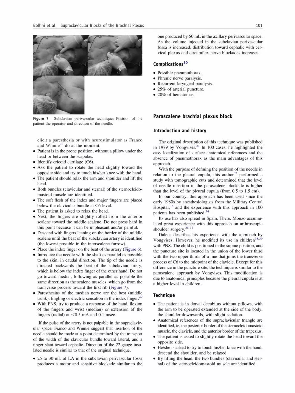
●
●
●
●
●
●
●
●
●
●
●
●
●
unofil
●
C
●
●
●
●
●
P
I
ieaa
rsot
eHp
ls
Vwtwpdpda
T
●
●
●
●
●
Fp
101Bollini et al Supraclavicular Blocks of the Brachial Plexus
elicit a paresthesia or with neurostimulator as Francoand Winnie28 do at the moment.Patient is in the prone position, without a pillow under thehead or between the scapulas.Identify cricoid cartilage (C6).Ask the patient to rotate the head slightly toward theopposite side and try to touch his/her knee with the hand.The patient should relax the arm and shoulder and lift thehead.Both bundles (clavicular and sternal) of the sternocleido-mastoid muscle are identified.The soft flesh of the index and major fingers are placedbelow the clavicular bundle at C6 level.The patient is asked to relax the head.Next, the fingers are slightly rolled from the anteriorscalene toward the middle scalene. Do not press hard inthis point because it can be unpleasant and/or painful.Descend with fingers leaning on the border of the middlescalene until the beat of the subclavian artery is identified(the lowest possible in the interscalene furrow).Place the index finger on the beat of the artery (Figure 6).Introduce the needle with the shaft as parallel as possibleto the skin, in caudal direction. The tip of the needle isdirected backwards the beat of the subclavian artery,which is below the index finger of the other hand. Do notgo toward medial, following as parallel as possible thesame direction as the scalene muscles, which go from thetransverse process toward the first rib (Figure 7).Paresthesias of the median nerve are the best (middletrunk), tingling or electric sensation in the index finger.29
With PNS, try to produce a response of the hand, flexionof the fingers and wrist (median) or extension of thefingers (radial) at �0.5 mA and 0.1 msec.
If the pulse of the artery is not palpable in the supraclavic-lar space, Franco and Winnie suggest that insertion of theeedle should be made at a point determined by the transportf the width of the clavicular bundle toward lateral, and anger slant toward cephalic. Direction of the 22-gauge insu-
ated needle is similar to that of the original technique.
25 to 30 mL of LA in the subclavian perivascular fossa
igure 7 Subclavian perivascular technique: Position of theatient the operator and direction of the needle.
produces a motor and sensitive blockade similar to the
one produced by 50 mL in the axillary perivascular space.As the volume injected in the subclavian perivascularfossa is increased, distribution toward cephalic with cer-vical plexus and circumflex nerve blockades increases.
omplications30
Possible pneumothorax.Phrenic nerve paralysis.Recurrent laryngeal paralysis.25% of arterial puncture.20% of hematomas.
arascalene brachial plexus block
ntroduction and history
The original description of this technique was publishedn 1979 by Vongvises.31 In 100 cases, he highlighted theasy localization of surface anatomical references and thebsence of pneumothorax as the main advantages of thispproach.
With the purpose of defining the position of the needle inelation to the pleural cupula, this author32 performed atudy with tomographic cuts and determined that the levelf needle insertion in the parascalene blockade is higherhan the level of the pleural cupula (from 0.5 to 1.5 cm).
In our country, this approach has been used since thearly 1980s by anesthesiologists from the Military Centralospital,33 and the experience with this approach in 100atients has been published.34
Its use has also spread in Spain. There, Monzo accumu-ated great experience with this approach on arthroscopichoulder surgery.35-37
Dalens describes his experience with the approach byongvises. However, he modified its use in children38,39
ith PNS. The child is positioned in the supine position, andhe puncture site is located in the union of the lower thirdith the two upper thirds of a line that joins the transverserocess of C6 to the midpoint of the clavicle. Except for thisifference in the puncture site, the technique is similar to thearascalene approach by Vongvises. This modification isue to anatomical principles because the pleural cupula is athigher level in children.
echnique
The patient is in dorsal decubitus without pillows, withthe arm to be operated extended at the side of the body,the shoulder downwards, with slight sedation.Anatomical references of the supraclavicular triangle areidentified, ie, the posterior border of the sternocleidomastoidmuscle, the clavicle, and the anterior border of the trapezius.The patient is asked to slightly rotate the head toward theopposite side.He/she is asked to try to touch his/her knee with the hand,descend the shoulder, and be relaxed.By lifting the head, the two bundles (clavicular and ster-
nal) of the sternocleidomastoid muscle are identified.
●
●
●
●
●
●
●
●
●
●
●
w0
nfi
●
●
●
●
A
●
●
F
102 Techniques in Regional Anesthesia and Pain Management, Vol 10, No 3, July 2006
With the soft flesh of the index and major fingers belowthe clavicular bundle, one palpates the interscalene fur-row located outside the posterior border of the sternoclei-domastoid muscle.We then descend palpating until the beat of the subcla-vian artery is found in the vicinities of the clavicle.Certain maneuvers can be performed to make the inter-scalene space evident. They include: deep inspiration onthe part of the patient, lateralization of the head toward theside of the puncture, observation or palpation of the beat ofthe subclavian artery in the supraclavicular space, etc.40
The key is accurate identification of the lateral border ofthe anterior scalene.Entrance to the sheath surrounding the plexus can beevident by means of the perception of a click, paresthesia,or motor response to neurostimulation.Skin antisepsis with iodized alcohol or iodopovidone.A dermic wheal is performed with a 25-gauge needle withlidocaine to 1% with 1:200.000 epinephrine in the punc-ture site, which is 1.5 to 2 cm above the clavicle in thelateral border of the anterior scalene above the subclavianartery and inside the external jugular vein (Figure 8).The authors suggest the use of the neurostimulator withan insulated needle of equal size.We continue using the immobile needle technique with anassistant who carries out the maneuvers with the PNS andinjects the anesthetic solution.The needle is inserted in the interscalene groove in an-teroposterior direction, ie, the needle tip perpendicularlydirected to the plane of the stretcher. Progression is slowuntil achieving a depth of about 1 to 1.5 cm (Figure 9).In the case of paresthesia technique, the appropriate re-sponse must be located either in the shoulder, elbow, orhand. These responses are generally brief, light, and soft.A paresthesia toward posterior (suprascapular) can beelicited; in this case, it is necessary to redirect the needletoward anterior. If the paresthesia is anterior (thorax), theneedle should be redirected backwards. If a PNS is avail-able, a motor response is sought in the same areas.
When we obtain contraction of biceps or deltoid musclesith an equal or inferior stimulus than 0.5 mA, a duration of
igure 8 Parascalene technique: Patient’s position.
.1 msec, and a frequency of 2 Hz, we inject, prior to F
egative aspiration, the anesthetic solution in increasingractional volumes of 5 mL. Aspiration among infiltrationss repeated to avoid inadvertent intravenous injection.
If response to PNS is contraction of the diaphragm, weare topographically on the body of the anterior scalene;thus, we should withdraw the needle and reinsert it closerto the median scalene.In general, we obtain a motor response before insertingthe needle 1.5 cm deep even in patients with voluminousnecks. If the needle goes deeper in, the puncture siteshould be reconsidered.If the immobile needle technique41 has been adequatelyfollowed, we should not worry about the pavilion of theneedle coming closer to the tissues since as we apply theinjection a supraclavicular “tumor” due to distension ofthe prevertebral fascia is formed.In the first minutes following injection, tingling or heatsensations appear with impossibility of lifting the arm. Amovement such as the one made when “counting money”may also be observed.
dverse effects and complications
Among complications of this approach are:
Possibility of vascular puncture.Claude Bernard Horner’s syndrome (which predicts suc-cess of the blockade). This syndrome disappears whenblockade is reverted.
igure 9 Parascalene technique: Needle direction.

●
●
●
S
I
tpon
C
●
●
●
T
●
●
●
●
Fr
F
Ftt
F
103Bollini et al Supraclavicular Blocks of the Brachial Plexus
Blockade of the inferior laryngeal nerve with bitonalvoice or hoarseness (less frequent).As there are no prospective studies on the incidence ofhomolateral phrenic nerve paralysis associated with thisblockade, we are unable to state whether the incidence islower than the one associated with the interscalene block-ade described by Winnie.Although we consider that the parascalene approach isnot ideal for placement of catheters due to perpendicularentrance to the major axis of the plexus, Monzo37 de-scribes placement of catheters for shoulder surgery.
upraclavicular technique by Dupré-Danel38
ntroduction
This technique is also known as “the surface reference”echnique. The authors propose a tangential boarding of thelexus that facilitates placement of catheters. Unlike inther techniques, they use an 18-gauge, 5-cm insulatedeedle and a 20-gauge catheter.
igure 10 Dupre and Danel technique: Anatomical surfaceeferences.
igure 11 Injection point and direction of the needle. d
utaneous repairs
A: External jugular.B: Apex of Sedillot triangle (it is given by the union ofboth bundles of the sternocleidomastoid muscle.C: Internal border of the clavicular insertion of the cla-vicular fasciculus of the trapezius (Figure 10).
echnique
The patient is asked to lift the head from the plane of thestretcher in slight rotation toward the contralateral side tomake anatomical references evident.A line is drawn from the cephalic vertex of the Sedillottriangle to the medial border of the clavicular insertion ofthe trapezius.The itinerary of the external jugular vein is marked (dot-ted line in Figure 10).The injection point is marked at the intersection of theline of the lateral border of the external jugular vein withthe line drawn between the apex of the triangle and themedial insertion of the trapezius in the clavicle. The axisof the puncture goes through the outer ear (Figure 11).
igure 12 Plumb bob: Insertion point is located 1 to 2 cm fromhe clavicle toward cephalic, tangentially to the lateral border ofhe sternocleidomastoid muscle.
igure 13 The needle is advanced in a strict anteroposterior
irection.
●
P
I
ptt
T
●
●
●
●
●
I
I
vtoksa
T
●
●
●
●
●
●
●
●
spjh
R
Ft
104 Techniques in Regional Anesthesia and Pain Management, Vol 10, No 3, July 2006
The usual motor response is that of the triceps. Contrac-tion of the biceps is also an excellent response. Thepuncture is 1 to 2.5 cm deep, and 30 mL of anestheticsolution is injected. The catheter is 2 cm deep.
lumb-bob technique
ntroduction
This technique was described by Brown.42,43 It shares theosition, indications, and volume of local anesthetics withhe parascalene technique. The only difference is given byhe entrance point of the needle.
echnique
The lateral border of the sternocleidomastoid muscleshould be identified at the level of its insertion in theclavicle. This becomes evident by asking the patient to liftthe head from the bed.A 5- to 6-cm blunt needle is inserted in an exact perpen-dicular and parasagittal plane to the patient and thestretcher (the horizontal one). The insertion point is lo-cated 1 to 2 cm from the clavicle toward cephalic, tan-gentially to the lateral border of the sternocleidomastoidmuscle (Figure 12).The needle is advanced in a strict anteroposterior direc-tion (Figure 13).If no contact is made with the first rib or if a pares-thesia is not elicited, direction of the needle tip shouldbe modified toward cephalic until a maximum 30°angle is achieved. If paresthesia is not obtained, theneedle is redirected toward the feet up to a maximum30° angle, until either a paresthesia is elicited or thefirst rib is found.44 The possibility of a pneumothoraxincreases in this last maneuver if performed by non-skilled hands.With regard to the classic description by Vongvises,31
the only similarity is the anteroposterior (perpendicular
igure 14 Intersternocleidomastoid technique: Needle direc-ion.
to the stretcher) direction of the needle. The difference
from the approach proposed by Brown is that a lowerand more medial puncture site is used, which, from ourstandpoint, increases the risk of puncture of the pleuralcupula.
ntersternocleidomastoid technique
ntroduction
In 1997, Pham-Dang13 described a really novel supracla-icular boarding. It was called the intersternocleidomastoidechnique, and it was based on a previous anatomical studyn cadavers. It stresses the simplicity of the references toeep in mind: the triangle formed by both heads of theternocleidomastoid muscle and the clavicle, usually visiblend palpable in most patients.
echnique
The patient rests in the supine position with the headrotated contralateral to the site of the blockade and withthe arm at the side of the body.The triangle formed by both bundles of the sternocleido-mastoid muscles and the clavicle is identified. The mid-point is marked.The puncture site is located in the internal border of theclavicular bundle, at two finger’s slant (3 cm) above thesternum.The neurostimulation needle is directed toward caudal,dorsal, and laterally to the midpoint of the clavicle, goingbelow the muscular stomach of the sternocleidomastoidmuscle, forming a 40° to 50° angle with the operatingtable (Figure 14).The plexus is located at a depth of 5 to 7 cm.The muscular responses obtained are those of the radialand musculocutaneous nerves. The latter is the most fre-quent initial response.30 mL of LA is injected when the intensity is of 0.5 mA.If the phrenic nerve is stimulated (which is evident bycontractions of the diaphragm), the needle is slightlyredirected toward lateral.
Although we lack experience with this approach, wehould highlight the potentiality of lesions either of thehrenic nerve or of the large vessels of the neck (internalugular, carotid). In our opinion, this technique does notave outstanding advantages over other approaches.
eferences
1. Kulenkampff D: Die Anästhesierung des plexus brachialis. Beitr KlinChir 79:550-552, 1912
2. Lanz E, Theiss D, Jankovic D: The extent of blockade followingvarious techniques plexus block. Anesth Analg 62:55-58, 1983
3. Dupré LJ: Blocking of the brachial plexus: which techniques should bechosen? Cahiers d’Anestesiologie 43:587-600, 1995
4. Pande R, Pande M, Bhadani U, et al: Supraclavicular brachial plexusblock as a sole anesthetic technique in children: an analysis of 200
cases. Anesthesia 55:798-802, 2000
1
1
1
1
1
1
1
1
1
1
2
2
2
2
2
2
2
2
2
2
3
3
3
3
3
3
3
3
3
3
4
4
4
4
4
105Bollini et al Supraclavicular Blocks of the Brachial Plexus
5. Mak PH, Irwin MG, Ooi CG, et al: Incidence of diaphragmatic paral-ysis following supraclavicular brachial plexus block and its effect onpulmonary function. Anesthesia 56:352-356, 2001
6. Winnie A, Collins V: The subclavian Perivascular technique of bra-chial plexus anesthesia. Anesthesiology 25:353-363, 1964
7. Vongvises P, Panijayanond T: A parascalene technique of brachialplexus anesthesia. Anesth Analg 58:267-273, 1979
8. Dupré JL, Danel V, Legrand JJ, et al: Surface landmarks for supra-clavicular block of the brachial plexus. Anesth Analg 61:28-31, 1982
9. Brown DL, Bridenbaugh LD: Physics applied to regional anesthesiaresults in an improved supraclavicular block: the “plumb bob” tech-nique. Anesthesiology 69:A376, 1988
0. Moorthy SS, Schmidt SL, Dierdorf SF: A supraclavicular lateral para-vascular approach for brachial plexus regional anesthesia. AnesthAnalg 72:241-244, 1991
1. Pippa P, Cominelli E, Marinelli C, et al: Brachial plexus block usingthe posterior approach. Eur J Anaesthesiol 7:411-420, 1990
2. Ortells-Polo MA, García-Guiral M, García-Amigueti FJ, et al: Anes-tesia del plexo braquial: resultados de una técnica supraclavicularperivascular modificada. Rev Esp Anestesiol Reanim 43:94-98, 1996
3. Pham-Dang C, Gunst JP, Gouin F, et al: A novel supraclavicularapproach to brachial plexus block. Anesth Analg 85:111-116, 1997
4. Winnie AP: Anestesia de Plexos Técnicas Perivasculares de bloqueodel Plexo Braquialqq. Salvat, Barcelona, Salvat, 1986, pp 68-75
5. Winnie AP, Franco CD: Supraclavicular approaches to brachial plexusanesthesia. Tech Reg Anesth Pain Manage 1:144-150, 1986
6. Bollini CA, Urmey W, Vascello L, et al: Relationship between evokedmotor response and sensory paresthesia in interscalene brachial plexusblock. Reg Anesth Pain Med 28:384-388, 2003
7. Cousins MJ, Bridenbaugh PO: Neural blockade in clinical anesthesiaand management of pain, second edition. Philadelphia, PA, J.B. Lip-pincott Company, 1988, pp 387-417
8. Neal JM, Hebe JR, Gerancher J, et al: Brachial plexus anesthesiaessentials of our current understanding. Reg Anesth Pain Med 27:402-428, 2002
9. Peláez X, Ornaque I: Bloqueo supraclavicular, in Anestesia RegionalHoy (2° Edición). Capitulo 17:265-280, 2001
0. Brown DL, Cahill DR, Bridenbaugh D: Supraclavicular nerve block:anatomic analysis of a method to prevent pneumothorax. Anesth Analg76:530-534, 1993
1. Bollini CA: Actas de V congreso Sudamericano y XXVII CongresoArgentino de Anestesiología, Iguazú Misiones, pp 171 y col, 1998
2. Hadzic A, Vloka JD: Peripheral Nerve Blocks Principles and Practice,New York, NY, McGraw Hill, 2004, pp 108-122
3. Jack NTM, Stienstra R: Anesthesie van de plexus brachialis: ervarin-gen met de posterieure benadering. Ned T Anesthesiol 11:50-52, 1998
4. Rettig HC, Gielen MJ, Jack NT, et al: A comparison of the lateral andposterior approach for brachial plexus block. Reg Anesth Pain Med
3:119-126, 20065. Neal JM, Hebl JR, Gerancher JC, et al: Brachial plexus anesthesia:essentials of our current understanding. Reg Anesth Pain Med 27:402-428, 2002
6. Dagli G, Guzeldemir ME, Volkan Acar H, et al: The effects and sideeffects of interscalene brachial plexus block by posterior approach.Reg Anesth Pain Med 23:87-91, 1998
7. Rucci FS, et al: How many interscalene blocks are there? A compar-ison between the lateral and posterior approach. Eur J Anaesthesiol10:303-307, 1993
8. Franco C, Winnie A: Subclavian perivascular block with a nervestimulator: outcome study after 1000 blocks. RAPM 24, 1999
9. Hickey R, Garland TA, Ramamurthy S: Subclavian perivascular block:influence of location of paresthesia. Anesth Analg 68:767-771, 1989
0. Wikinski J, Bollini C: Complicaciones neurológicas de la anestesiaregional periférica y central. Panamericana. 51-53, 1999
1. Vongvises P, Panijayanond T: A parascalene technique of brachialplexus anesthesia. Anesth Analg 58:267-273, 1979
2. Vongvises P: Beokhaimook Computed tomographic study of paras-calene block. Anesth Analg 842:379, 1997
3. Fernandez RO, Sanchez WJ: Nuestra experiencia con una nueva téc-nica de bloqueo del plexo braquial paraescalénica. Prensa Med Argent69:890, 1982
4. Salgueiro C, de la Peña R: Experiencia clínica con el abordaje par-aescalénico para el bloqueo del plexo braquial Indicaciones, ventajas ycomplicaciones. Rev Arg Anest 54:322-327, 1996
5. Monzó E, Baeza C, Sánchez ML, et al: Bloqueo paraescalénico con-tinuo en la cirugía del hombro. Rev Esp Anestesiol Reanim 45:377-383, 1998
6. Call Reigl L, de Vicente Sole J, Estany Raluy E: Parascalene block forshoulder arthroscopy surgery. Rev Esp Anestesiol Reanim 51:247-252, 2004
7. Monzo Abad E, Baeza Gil C, Galindo Sanchez F, et al: Parascalenebrachial plexus block: experience of 10 years. Rev Esp AnestesiolReanim 512:61-69, 2004
8. Dupré JL, Danel V, Legrand JJ, et al: Surface landmarks for supra-clavicular block of the brachial plexus. Anesth Analg 61:28, 1982
9. Dalens B, Vanneuville G, Tanguy A: A new parascalene approach tothe brachial plexus in children: comparison with the supraclavicularapproach. Anesth Analg 66:1264-1271, 1987
0. Bollini CA: Una Maniobra útil para identificar el espacio interes-calénico. Rev Arg Anest 55:310-312, 1997
1. Winnie AP: An immobile needle for nerve blocks. Anesthesiology31:577, 1969
2. Brown DL: Regional Anesthesia and Analgesia. Philadelphia, PA,W.B. Saunders Company, 1996, p 265
3. Brown DL: Atlas of Regional Anesthesia (ed 2). Philadelphia, PA, WBSaunder Company, 1999
4. VadeBoncouer TR, Weinberg GL: Supraclavicular brachial plexusanesthesia using the plumb bob method. Tech Reg Anesth 1:151-156,
1997