supports coated with pei as a new tool in chromatography
TRANSCRIPT

Enzyme and Microbial Technology 39 (2006) 711–716
Supports coated with PEI as a new tool in chromatography
Rodrigo Torres 1, Benevides C.C. Pessela, Manuel Fuentes, Cesar Mateo,Roberto Munilla, Roberto Fernandez-Lafuente ∗, Jose M. Guisan ∗
Departamento de Biocatalisis, Instituto de Catalisis-CSIC, Campus Universidad Autonoma, Cantoblanco, 28049 Madrid, Spain
Received 7 April 2005; received in revised form 30 November 2005; accepted 8 December 2005
Abstract
PEI coated supports are proposed in this manuscript as a matrix that may permit a new concept in chromatography. Standard supports have theiractive groups on a “plane” surface, and permit the interaction with an area accounting for not more than 15–20% of the protein surface. The PEIcoated supports permitted the interaction of the protein and a polymeric bead, where the protein may penetrate, permitting the interaction witha larger percentage of the protein surface. Here, we present that PEI coated supports are able to more strongly adsorb proteins that are adsorbedon conventional supports and to adsorb proteins that are not adsorbed on these supports. Moreover, more interestingly, the relative adsorptionstrength was found to be quite different when using PEI coated supports or conventional supports, because the distribution of charged groupsiacbdEc©
K
1
fAoTgdokot
j
Tm
0d
n the protein surface may affect very distinctly to the adsorption when using each of these supports. Thus, some proteins that are very stronglydsorbed on conventional supports (having a small area with many anionic groups) were not among the most strongly adsorbed proteins on PEIoated supports, while some proteins that could not be adsorbed on conventional supports (that is, not having a very rich area in anionic groups)ecome very strongly adsorbed on PEI supports (very likely because they have dispersed around the surface many anionic groups). That is, theesorption pattern from both kind of supports is very different. We presented the results obtained in the fractioning of crude proteins extract from. coli and whey protein concentrate. The sequential use of DEAE–agarose and PEI permit to improve the purity of the proteins adsorbed on PEIoated supports by more than a 10-fold factor and their further desorption may give only two to three bands in many instances.
2005 Elsevier Inc. All rights reserved.
eywords: Polymeric supports; Protein purification; Three-dimensional chromatography; Whey protein concentrate
. Introduction
Ion-exchange chromatography is a simple and very use-ul widespread technique used in protein purification [1–7].garose beads are composed of large trunks of agarose thatffers a “plane” surface in the interaction with small protein.hus, if these supports are activated with a layer of chargedroups [8–11] their interaction with the protein is in a bi-imensional plane, involving about an area that may be 15–20%f the total protein surface [11,12]. Protein adsorption in theseinds of supports requires the interaction between several groupsn the support and several groups on the protein. Therefore, pro-eins to be adsorbed on anionic exchangers should have in their
∗ Corresponding authors. Tel.: +34 91 585 4809;fax: +34 91 585 4760.E-mail addresses: [email protected] (R. Fernandez-Lafuente),
[email protected] (J.M. Guisan).1 Present address: Escuela de Quımica, Facultad de Ciencias, Edificio “Camiloorres”, Ciudad Universitaria, Universidad Industrial de Santander, Bucara-anga, Colombia.
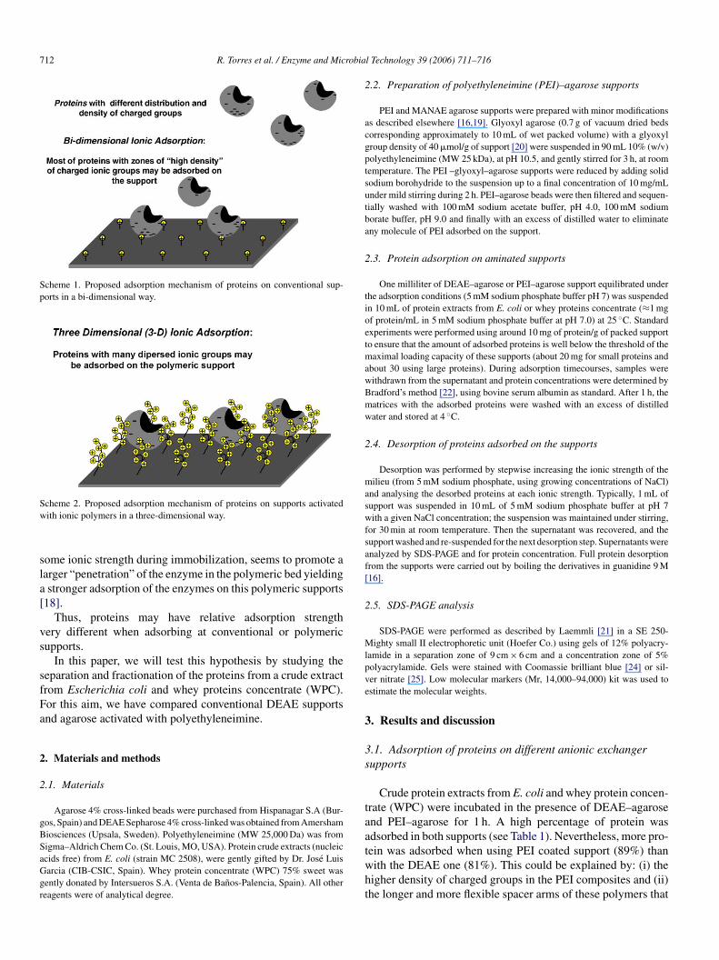
surface an area relatively enriched in groups able to interact withthose of the surface support (Scheme 1). Supports coated withPEI have been reported to permit more effective adsorption ofproteins as supports for reversible immobilization [13–15].
However, together to a higher amount of charged groups,these supports may have some additional differences when com-pared to conventional supports (Scheme 2). In this case, theproteins would interact with a bed formed by a flexible polymer,which has positive charges. Thus, proteins may penetrate in thecharged polymeric bed allowing that a greater percentage of theprotein surface may interact with the support. That is, now theinteraction is not in a plane surface, but in a certain volume. Ifthis hypothesis is right, now the proteins will not be adsorbedby the interaction between a reduced area of the protein andthe support, thus, they did not require to present a dense areaof charged groups in one area of the surface, but a number ofanionic groups disperse in the protein surface. In fact, it hasbeen reported that supports coated with polyethyleneimine areable to adsorb proteins that cannot be adsorbed on conventionalsupports [16,17]. Moreover, it has been reported that the use of
141-0229/$ – see front matter © 2005 Elsevier Inc. All rights reserved.oi:10.1016/j.enzmictec.2005.12.006
712 R. Torres et al. / Enzyme and Microbial Technology 39 (2006) 711–716
Scheme 1. Proposed adsorption mechanism of proteins on conventional sup-ports in a bi-dimensional way.
Scheme 2. Proposed adsorption mechanism of proteins on supports activatedwith ionic polymers in a three-dimensional way.
some ionic strength during immobilization, seems to promote alarger “penetration” of the enzyme in the polymeric bed yieldinga stronger adsorption of the enzymes on this polymeric supports[18].
Thus, proteins may have relative adsorption strengthvery different when adsorbing at conventional or polymericsupports.
In this paper, we will test this hypothesis by studying theseparation and fractionation of the proteins from a crude extractfrom Escherichia coli and whey proteins concentrate (WPC).For this aim, we have compared conventional DEAE supportsand agarose activated with polyethyleneimine.
2. Materials and methods
2.1. Materials
Agarose 4% cross-linked beads were purchased from Hispanagar S.A (Bur-gos, Spain) and DEAE Sepharose 4% cross-linked was obtained from AmershamBiosciences (Upsala, Sweden). Polyethyleneimine (MW 25,000 Da) was fromSigma–Aldrich Chem Co. (St. Louis, MO, USA). Protein crude extracts (nucleicacids free) from E. coli (strain MC 2508), were gently gifted by Dr. Jose LuisGarcia (CIB-CSIC, Spain). Whey protein concentrate (WPC) 75% sweet wasgently donated by Intersueros S.A. (Venta de Banos-Palencia, Spain). All otherreagents were of analytical degree.
2.2. Preparation of polyethyleneimine (PEI)–agarose supports
PEI and MANAE agarose supports were prepared with minor modificationsas described elsewhere [16,19]. Glyoxyl agarose (0.7 g of vacuum dried bedscorresponding approximately to 10 mL of wet packed volume) with a glyoxylgroup density of 40 �mol/g of support [20] were suspended in 90 mL 10% (w/v)polyethyleneimine (MW 25 kDa), at pH 10.5, and gently stirred for 3 h, at roomtemperature. The PEI –glyoxyl–agarose supports were reduced by adding solidsodium borohydride to the suspension up to a final concentration of 10 mg/mLunder mild stirring during 2 h. PEI–agarose beads were then filtered and sequen-tially washed with 100 mM sodium acetate buffer, pH 4.0, 100 mM sodiumborate buffer, pH 9.0 and finally with an excess of distilled water to eliminateany molecule of PEI adsorbed on the support.
2.3. Protein adsorption on aminated supports
One milliliter of DEAE–agarose or PEI–agarose support equilibrated underthe adsorption conditions (5 mM sodium phosphate buffer pH 7) was suspendedin 10 mL of protein extracts from E. coli or whey proteins concentrate (≈1 mgof protein/mL in 5 mM sodium phosphate buffer at pH 7.0) at 25 ◦C. Standardexperiments were performed using around 10 mg of protein/g of packed supportto ensure that the amount of adsorbed proteins is well below the threshold of themaximal loading capacity of these supports (about 20 mg for small proteins andabout 30 using large proteins). During adsorption timecourses, samples werewithdrawn from the supernatant and protein concentrations were determined byBradford’s method [22], using bovine serum albumin as standard. After 1 h, thematrices with the adsorbed proteins were washed with an excess of distilledwater and stored at 4 ◦C.
2.4. Desorption of proteins adsorbed on the supports
maswfsaf[
2
Mlpve
3
3s
taatwht
Desorption was performed by stepwise increasing the ionic strength of theilieu (from 5 mM sodium phosphate, using growing concentrations of NaCl)
nd analysing the desorbed proteins at each ionic strength. Typically, 1 mL ofupport was suspended in 10 mL of 5 mM sodium phosphate buffer at pH 7ith a given NaCl concentration; the suspension was maintained under stirring,
or 30 min at room temperature. Then the supernatant was recovered, and theupport washed and re-suspended for the next desorption step. Supernatants werenalyzed by SDS-PAGE and for protein concentration. Full protein desorptionrom the supports were carried out by boiling the derivatives in guanidine 9 M16].
.5. SDS-PAGE analysis
SDS-PAGE were performed as described by Laemmli [21] in a SE 250-ighty small II electrophoretic unit (Hoefer Co.) using gels of 12% polyacry-
amide in a separation zone of 9 cm × 6 cm and a concentration zone of 5%olyacrylamide. Gels were stained with Coomassie brilliant blue [24] or sil-er nitrate [25]. Low molecular markers (Mr, 14,000–94,000) kit was used tostimate the molecular weights.
. Results and discussion
.1. Adsorption of proteins on different anionic exchangerupports
Crude protein extracts from E. coli and whey protein concen-rate (WPC) were incubated in the presence of DEAE–agarosend PEI–agarose for 1 h. A high percentage of protein wasdsorbed in both supports (see Table 1). Nevertheless, more pro-ein was adsorbed when using PEI coated support (89%) thanith the DEAE one (81%). This could be explained by: (i) theigher density of charged groups in the PEI composites and (ii)he longer and more flexible spacer arms of these polymers that
R. Torres et al. / Enzyme and Microbial Technology 39 (2006) 711–716 713
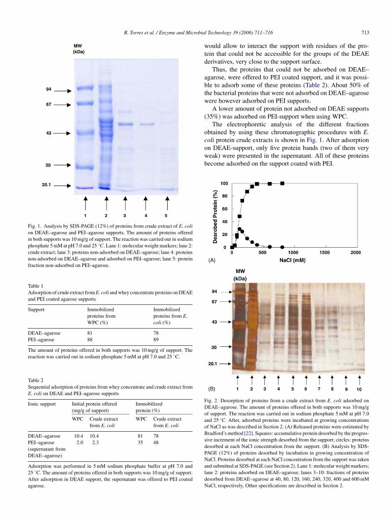
Fig. 1. Analysis by SDS-PAGE (12%) of proteins from crude extract of E. colion DEAE–agarose and PEI–agarose supports. The amount of proteins offeredin both supports was 10 mg/g of support. The reaction was carried out in sodiumphosphate 5 mM at pH 7.0 and 25 ◦C. Lane 1: molecular weight markers; lane 2:crude extract; lane 3: proteins non-adsorbed on DEAE–agarose; lane 4: proteinsnon-adsorbed on DEAE–agarose and adsorbed on PEI–agarose; lane 5: proteinfraction non-adsorbed on PEI–agarose.
Table 1Adsorption of crude extract from E. coli and whey concentrate proteins on DEAEand PEI coated agarose supports
Support Immobilizedproteins fromWPC (%)
Immobilizedproteins from E.coli (%)
DEAE–agarose 81 78PEI–agarose 88 89
The amount of proteins offered in both supports was 10 mg/g of support. Thereaction was carried out in sodium phosphate 5 mM at pH 7.0 and 25 ◦C.
Table 2Sequential adsorption of proteins from whey concentrate and crude extract fromE. coli on DEAE and PEI–agarose supports
Ionic support Initial protein offered(mg/g of support)
Immobilizedprotein (%)
WPC Crude extractfrom E. coli
WPC Crude extractfrom E. coli
DEAE–agarose 10.4 10.4 81 78PEI–agarose(supernatant fromDEAE–agarose)
2.0 2.3 35 48
Adsorption was performed in 5 mM sodium phosphate buffer at pH 7.0 and25 ◦C. The amount of proteins offered in both supports was 10 mg/g of support.After adsorption in DEAE support, the supernatant was offered to PEI coatedagarose.
would allow to interact the support with residues of the pro-tein that could not be accessible for the groups of the DEAEderivatives, very close to the support surface.
Thus, the proteins that could not be adsorbed on DEAE–agarose, were offered to PEI coated support, and it was possi-ble to adsorb some of these proteins (Table 2). About 50% ofthe bacterial proteins that were not adsorbed on DEAE–agarosewere however adsorbed on PEI supports.
A lower amount of protein not adsorbed on DEAE supports(35%) was adsorbed on PEI-support when using WPC.
The electrophoretic analysis of the different fractionsobtained by using these chromatographic procedures with E.coli protein crude extracts is shown in Fig. 1. After adsorptionon DEAE-support, only five protein bands (two of them veryweak) were presented in the supernatant. All of these proteinsbecome adsorbed on the support coated with PEI.
FDoaoBsdPNaldN
ig. 2. Desorption of proteins from a crude extract from E. coli adsorbed onEAE–agarose. The amount of proteins offered in both supports was 10 mg/gf support. The reaction was carried out in sodium phosphate 5 mM at pH 7.0nd 25 ◦C. After, adsorbed proteins were incubated at growing concentrationsf NaCl as was described in Section 2. (A) Released proteins were estimated byradford’s method [22]. Squares: accumulative protein desorbed by the progres-
ive increment of the ionic strength desorbed from the support; circles: proteinsesorbed at each NaCl concentration from the support. (B) Analysis by SDS-AGE (12%) of proteins desorbed by incubation in growing concentration ofaCl. Proteins desorbed at each NaCl concentration from the support was taken
nd submitted at SDS-PAGE (see Section 2). Lane 1: molecular weight markers;ane 2: proteins adsorbed on DEAE–agarose; lanes 3–10: fractions of proteinsesorbed from DEAE–agarose at 40, 80, 120, 160, 240, 320, 400 and 600 mMaCl, respectively. Other specifications are described in Section 2.

714 R. Torres et al. / Enzyme and Microbial Technology 39 (2006) 711–716
3.2. Desorption of proteins from E. coli on different anionicexchanger supports
Desorption of proteins adsorbed on PEI or DEAE supportsfollowed different elution profiles. Most of the proteins adsorbedon DEAE–agarose beads (≥90% of total immobilized proteins)could be eluted with 240 mM NaCl (Fig. 2). However, desorptionof proteins adsorbed on PEI–agarose supports required higherNaCl concentrations than for the DEAE–agarose counterpart,being detected traces of protein desorbed from the support evenat 2 M NaCl (Fig. 3). Due to the amount of bands, it may bedifficult to trace the desorption of some proteins to detect likelydifferences in relative adsorption strength.
Fig. 4 shows the desorption of the proteins that could notbe adsorbed on DEAE-supports but become adsorbed on PEI-supports. Surprisingly, the elution profile, of these proteins that
FPipapdspgf134
Fig. 4. Desorption of the proteins from a crude extract from E. coli non-adsorbedin DEAE–agarose but adsorbed on PEI–agarose. The initial amount of proteinsoffer to DEAE support was 10 mg/g of support. The reaction was carried outin sodium phosphate 5 mM at pH 7.0 and 25 ◦C. After, adsorbed proteins wereincubated at growing concentrations of NaCl as was described in Section 2. (A)Released proteins were followed by Bradford’s method [22]. Squares: accumula-tive protein desorbed by the progressive increment of the ionic strength desorbedfrom the support; circles: proteins desorbed at each NaCl concentration from thesupport. (B) Analysis by SDS-PAGE (12%) of proteins desorbed by incubationin growing concentration of NaCl. Proteins desorbed at each NaCl concentrationfrom the support was taken and submitted at SDS-PAGE (see Section 2). Lane1: molecular weight markers; lane 2: proteins adsorbed on PEI–agarose; lanes3–10: fractions of proteins desorbed from PEI–agarose at 80, 160, 240, 320,480, 800, 1200 and 1600 mM NaCl, respectively. Details in Section 2.
could be not adsorbed on DEAE (that is, theoretically with alower capacity to interact with an anionic exchanger), was sim-
ig. 3. Desorption of crude extract proteins from E. coli adsorbed onEI–agarose by increasing NaCl concentrations. The amount of proteins offered
n both supports was 10 mg/g of support. The reaction was carried out in sodiumhosphate 5 mM at pH 7.0 and 25 ◦C. After, adsorbed proteins were incubatedt growing concentrations of NaCl as was described in Section 2. (A) Releasedroteins followed by Bradford’s method [22]. Squares: accumulative proteinesorbed by the progressive increment of the ionic strength desorbed from theupport; circles: proteins desorbed at each NaCl concentration from the sup-ort. (B) Analysis by SDS-PAGE (12%) of proteins desorbed by incubation inrowing concentration of NaCl. Proteins desorbed at each NaCl concentrationrom the support was taken and submitted at SDS-PAGE (see Section 2). Lane: molecular weight markers; lane 2: proteins adsorbed on PEI–agarose; lanes–10: fractions of proteins desorbed from PEI–agarose at 40, 80, 160, 240, 320,00, 800 and 1200 mM NaCl, respectively.
ilar to that obtained when using the whole crude extract. Thatis, proteins that may be not adsorbed on supports activated onlywith amino groups on a plane surface could be adsorbed on thepolymeric ionic bed, and the average adsorption strength of theseproteins were similar to that of proteins that could be adsorbedon both supports. If the adsorption mechanisms of both fractionof proteins were similar, being the only difference the amount ofionic charges in the support, the fraction of proteins that cannotbe adsorbed on plane-activated supports should be adsorbed onPEI coated support in a weaker fashion that the proteins thatmay be adsorbed on these supports. This suggests that the pro-tein adsorption on PEI coated support may not follow exactlythe same mechanism than the protein adsorption on supportswith groups only in a plane surface. A protein become adsorbedon a plane surfaces by ionic interchange when it has a rich-
R. Torres et al. / Enzyme and Microbial Technology 39 (2006) 711–716 715
enough charged area, to yield several interactions [28] while theadsorption in a “volume” only requires the presence of charges,even very scattered, all around the protein surface. This seems toindicate that the PEI-support may yield very different desorptionpattern that the “bi-dimensional” chromatography.
Thus, proteins that cannot be adsorbed to plane-activatedmatrix but become adsorbed on PEI-supports become quite puri-fied. First, only around 10% of the proteins fulfil this requirement(that is, a 10-fold purification factor is achieved for all these pro-teins). Second, the desorption fractions account for a maximumof 20% of the adsorbed proteins, and in many instances is under10%, giving an additional purification factor of 5–10. Thus, theSDS-PAGE gels of the different fractions of the desorbed pro-teins showed a very low amount of bands (3–6), for the moststrongly adsorbed proteins even only 1–2 bands.
Fip5cfbcAcsuf6t
3.3. Desorption of proteins from whey protein concentrateon aminated supports
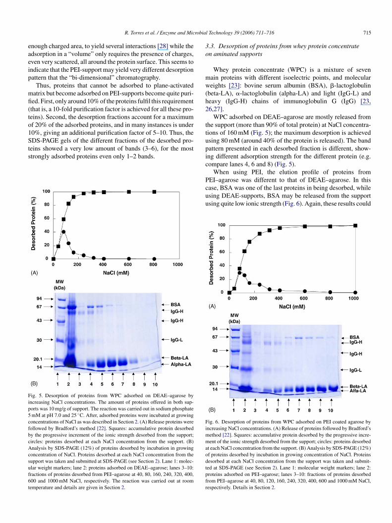
Whey protein concentrate (WPC) is a mixture of sevenmain proteins with different isoelectric points, and molecularweights [23]: bovine serum albumin (BSA), �-lactoglobulin(beta-LA), �-lactoglobulin (alpha-LA) and light (IgG-L) andheavy (IgG-H) chains of immunoglobulin G (IgG) [23,26,27].
WPC adsorbed on DEAE–agarose are mostly released fromthe support (more than 90% of total protein) at NaCl concentra-tions of 160 mM (Fig. 5); the maximum desorption is achievedusing 80 mM (around 40% of the protein is released). The bandpattern presented in each desorbed fraction is different, show-ing different adsorption strength for the different protein (e.g.compare lanes 4, 6 and 8) (Fig. 5).
When using PEI, the elution profile of proteins fromPEI–agarose was different to that of DEAE–agarose. In thiscase, BSA was one of the last proteins in being desorbed, whileusing DEAE-supports, BSA may be released from the supportusing quite low ionic strength (Fig. 6). Again, these results could
ig. 5. Desorption of proteins from WPC adsorbed on DEAE–agarose byncreasing NaCl concentrations. The amount of proteins offered in both sup-orts was 10 mg/g of support. The reaction was carried out in sodium phosphatemM at pH 7.0 and 25 ◦C. After, adsorbed proteins were incubated at growingoncentrations of NaCl as was described in Section 2. (A) Release proteins wereollowed by Bradford’s method [22]. Squares: accumulative protein desorbedy the progressive increment of the ionic strength desorbed from the support;ircles: proteins desorbed at each NaCl concentration from the support. (B)nalysis by SDS-PAGE (12%) of proteins desorbed by incubation in growing
oncentration of NaCl. Proteins desorbed at each NaCl concentration from theupport was taken and submitted at SDS-PAGE (see Section 2). Lane 1: molec-lar weight markers; lane 2: proteins adsorbed on DEAE–agarose; lanes 3–10:ractions of proteins desorbed from PEI–agarose at 40, 80, 160, 240, 320, 400,00 and 1000 mM NaCl, respectively. The reaction was carried out at roomemperature and details are given in Section 2.
Fimmaodtpfr
ig. 6. Desorption of proteins from WPC adsorbed on PEI coated agarose byncreasing NaCl concentrations. (A) Release of proteins followed by Bradford’s
ethod [22]. Squares: accumulative protein desorbed by the progressive incre-ent of the ionic strength desorbed from the support; circles: proteins desorbed
t each NaCl concentration from the support. (B) Analysis by SDS-PAGE (12%)f proteins desorbed by incubation in growing concentration of NaCl. Proteinsesorbed at each NaCl concentration from the support was taken and submit-ed at SDS-PAGE (see Section 2). Lane 1: molecular weight markers; lane 2:roteins adsorbed on PEI–agarose; lanes 3–10: fractions of proteins desorbedrom PEI–agarose at 40, 80, 120, 160, 240, 320, 400, 600 and 1000 mM NaCl,espectively. Details in Section 2.
716 R. Torres et al. / Enzyme and Microbial Technology 39 (2006) 711–716
be explained by the different adsorption mechanism existingbetween these two kinds of supports: BSA looks to have manynegative charges, but all around the surface of the protein (thatis, interaction with a polymer gave a strength adsorption higherthan any other protein, but not present a particulate area havinga strong negative charge, therefore, adsorption strength on planesurface is not very high).
4. Conclusions
The use of supports activated with polymers in a way that per-mits to form a polymeric bed where the proteins can penetrate,has been revealed as a new tool in chromatography, because theelution pattern of the proteins adsorbed on plane surfaces andvolumetric beds may be fully altered, permitting in this way toseparate proteins by combining both techniques. This could becalled a “three-dimensional” chromatography in contrapositionwith the standard bi-dimensional chromatography.
This way, polymeric beds are able to adsorb more proteinsnot only because the higher amounts of ionic groups, but alsobecause they can interact with a larger percentage of the proteinsurface. Thus, now the adsorption strength will be not related tothe presence of a cluster in the protein having negative charge,but with the presence in the protein surface of may spots pre-senting this negative charge.
This may open new opportunities to the development of sim-pt
bbm
A
2sTs
R
[7] Ghose S, Chase HA. Expanded bed chromatography of proteins in small-diameter columns. II. Methods development and scale up. Bioseparation2000;9:29–36.
[8] Baumeister A, Vogelmann S, Fischer L. Concentration and purificationof orotic acid directly from whey with an expanded bed adsorptionsystem. J Chromatogr A 2003;1006:261–70.
[9] Sun S, Yue Y, Huang X, Meng D. Protein adsorption on blood-contactmembranes. J Membr Sci 2003;222:3–11.
[10] Anspach FB, Curbelo D, Hartman R, Garke G, Deckwer W-D.Expanded-bed chromatography in primary protein purification. J Chro-matogr A 1999;865:129–44.
[11] Iberer G, Schwinn H, Josic D, Jungbauer A, Buchacher A. Improvedperformance of protein separation by continuous annular chromatogra-phy in the size-exclusion mode. J Chromatogr A 2001;921:15–24.
[12] Pessela BCC, Fernandez-Lafuente R, Fuentes M, Vian A, Garcia JL,Carrascosa AV, et al. Reversible immobilization of a thermophilic �-galactosidase via ionic adsorption on PEI coated Sepabeads. EnzymeMicrob Technol 2003;32:369–74.
[13] Li Y-M, Liao J-L, Zhang R, Henriksson H, Hjerten S. Continuous bedsfor microchromatography: chromatofocusing and anion exchange chro-matography. Anal Biochem 1999;267:121–4.
[14] Lucas D, Rabiller-Baudry M, Millesime L, Chaufer B, Daufin G. Extrac-tion of �-lactalbumin from whey protein concentrate with modifiedinorganic membranes. J Membr Sci 1998;148:1–12.
[15] Weaver LE, Carta G. Protein adsorption on cation exchangers: com-parison of macroporous and gel-composite media. Biotechnol Progr1996;12:342–55.
[16] Mateo C, Abian O, Fernandez-Lafuente R, Guisan JM. Reversibleenzyme immobilization via a very strong and non-distorting adsorp-tion on supports-polyethylene imine composites. Biotechnol Bioeng2000;68:98–105.
[
[
[
[
[
[
[
[
[
[
[
[
le protein purification using an apparent old methodology ashe ionic exchange.
By coupling sequentially the use of both supports, it is possi-le to get very pure proteins, mainly in the fraction that cannote adsorbed in a plane surface but become adsorbed in the poly-eric bed.
cknowledgements
Financial support from EC (Project MATINOES G5RD-CT-002-00752) is gratefully recognized. We thank a PhD fellow-hip from Universidad Industrial de Santander, Colombia for R.orres. Comunidad de Madrid is gratefully recognized for theupport to Dr. Pessela (Projects 07G/002/2003).
eferences
[1] Rao CS. Purification of large proteins using ion-exchange membranes.Proc Biochem 2001;37:247–56.
[2] Tong X-D, Dong X-Y, Sun Y. Lysozyme adsorption and purificationby expanded bed chromatography with a small-sized dense adsorbent.Biochem Eng J 2002;12:117–24.
[3] Lyddiatt A. Process chromatography: current constraints and futureoptions for the adsorptive recovery of bioproducts. Curr Opin Biotechnol2002;13:95–103.
[4] Ovsejevi K, Grazu V, Cuadra K, Batista-Viera F. Enzyme reductionon solid phase as a tool for the reversible immobilization of yeast�-galactosidase onto a thiol-reactive support. Enzyme Microb Technol2004;35:203–9.
[5] Scopes RK. Protein purification: principles and practice. 2nd ed. NewYork; 1987. p. 126–40.
[6] Shih Y-C, Prausnitz JM, Blanch HW. Some characteristics of proteinprecipitation by salts. Biotechnol Bioeng 1992;40:1155–64.
17] Kumar A, Galaev IY, Mattiasson B. Polymer displacement/shielding inprotein chromatography. J Chromatogr B 2000;741:103–13.
18] Pessela BCC, Betancor L, Lopez-Gallego F, Torres R, Dellamora-OrtizGM, Alonso-Morales N, et al. Improved immobilization of enzymes onPEI-coated supports by immobilizing at high ionic strength. EnzymeMicrob Technol 2005;37:295–9.
19] Fernandez-Lafuente R, Rosell CM, Rodriguez V, Santana C, Soler G,Bastida A, et al. Preparation of activated supports containing low pKamino groups. A new tool for protein immobilisation via the carboxylcoupling method. Enzyme Microb Technol 1993;15:546–50.
20] Guisan JM. Aldehyde–agarose gels as activated supports forimmobilization-stabilization of enzymes. Enzyme Microb Technol1988;10:375–82.
21] Laemmli UK. Cleavage of structural proteins during the assembly of thehead of bacteriophage T4. Nature 1970;277:680–5.
22] Bradford MM. A rapid and sensitive method for the quantitation ofmicrogram quantities of protein utilizing the principle of protein–dyebinding. Anal Biochem 1976;72:248–54.
23] Hahn RP, Schulz M, Schaupp C, Jungbauer A. Bovine whey frac-tionation based on cation-exchange chromatography. J Chromatogr A1998;795:277–87.
24] Swank RT, Munkres KD. Molecular weight analysis of oligopeptides byelectrophoresis in polyacrylamide gel with sodium dodecyl sulfate. AnalBiochem 1971;39:462–77.
25] Marshall T. Detection of protein in polyacrylamide gels using animproved silver stain. Anal Biochem 1984;136:340–6.
26] Rossano R, Dıelia A, Riccio P. One-step separation from lac-tose: recovery and purification of major cheese–whey proteins byhydroxyapatite—a flexible procedure suitable for small- and medium-scale preparations. Protein Express Purif 2001;21:165–9.
27] Alomirah HF, Alli I. Separation and characterization of �-lactoglobulinand �-lactalbumin from whey and whey protein preparations. Int DairyJ 2004;14:411–9.
28] Pessela BCC, Munilla R, Betancor L, Fuentes M, Carrascosa AV, VianA, et al. Ionic exchange using lowly activated supports: an easy wayfor purifying large proteins. J Chromatogr A 2004:155–9.