superantigens increase the survival of mice bearing t cell lymphomas by inducing apoptosis of...
TRANSCRIPT
Superantigens Increase the Survival of Mice Bearing TCell Lymphomas by Inducing Apoptosis of NeoplasticCellsJuliana Mundinano1, Paula M. Berguer2, Gabriel Cabrera1, Daniela Lorenzo1, Irene Nepomnaschy1.,
Isabel Piazzon1*.
1 ILEX-CONICET, Division Medicina Experimental, Instituto de Investigaciones Hematologicas, Academia Nacional de Medicina, Buenos Aires, Argentina, 2 Consejo
Nacional de Investigaciones Cientıficas y Tecnicas, Fundacion Instituto Leloir, Buenos Aires, Argentina
Abstract
Superantigens bind to major histocompatibility complex class II molecules and interact with T cells expressing a particular Tcell receptor Vb inducing a strong proliferation/deletion response of the superantigen-reactive T cells. However, there havebeen no attempts to investigate the ability of Sags to induce apoptosis in neoplastic T cells by signaling through the Vbregion of their TCR. In the present study we show that bacterial and MMTV-encoded superantigens induce the apoptosis ofAKR/J cognate lymphoma T cells both in vitro and in vivo. The Fas-Fas-L pathway was shown to be involved in the apoptosisof lymphoma T cells induced by bacterial superantigens. In vivo exposure to bacterial superantigens was able to improvethe survival of lymphoma bearing mice. Moreover, the permanent expression of a retroviral encoded superantigen inducedthe complete remission of an aggressive lymphoma in a high percentage of mice. The possibility of a therapeutic use ofsuperantigens in lymphoma/leukemia T cell malignancies is discussed.
Citation: Mundinano J, Berguer PM, Cabrera G, Lorenzo D, Nepomnaschy I, et al. (2010) Superantigens Increase the Survival of Mice Bearing T Cell Lymphomas byInducing Apoptosis of Neoplastic Cells. PLoS ONE 5(12): e15694. doi:10.1371/journal.pone.0015694
Editor: Syed A. Aziz, Health Canada, Canada
Received November 1, 2010; Accepted November 23, 2010; Published December 22, 2010
Copyright: � 2010 Mundinano et al. This is an open-access article distributed under the terms of the Creative Commons Attribution License, which permitsunrestricted use, distribution, and reproduction in any medium, provided the original author and source are credited.
Funding: This work was supported by Agencia Nacional de Promocion Cientıfica y Tecnologica PICT 506305, Consejo Nacional de Investigaciones Cientıficas yTecnicas PIP 20080102494, FUNDALEU, and Fundacion A. J. Roemmers. The funders had no role in study design, data collection and analysis, decision to publish,or preparation of the manuscript.
Competing Interests: The authors have declared that no competing interests exist.
* E-mail: ipiazzon@hematologıa.anm.edu.ar
. These authors contributed equally to this work.
Introduction
Superantigens (Sags) are bacterial and viral proteins that share
the ability to activate a large number of normal T cells. Sags bind
to major histocompatibility complex (MHC) class II molecules as
unprocessed proteins and subsequently interact with a high
number of T cells expressing particular T cell receptor (TCR)
Vb chains [1–4]. After several rounds of proliferation, Sag reactive
T cells undergo apoptosis or become anergic [2–4].
Bacterial Sags are a well described family of secreted protein
toxins produced mainly by Staphylococcus aureus and Streptococcus
pyogenes [5]. The capacity of bacterial Sags to induce the activation
and deletion of T cells expressing T cell receptors (TCR) with a
specific subset of TCR b-chain variable (Vb) regions in mice has
been extensively studied [2–3]. For instance, in mice, the
staphylococcal enterotoxins (SEs) A and E engage T cell receptors
bearing Vb 3, 7, and 17, albeit with different avidities. SEB and its
close sequence relatives, SECs 1-3, share reactivity with T cells
bearing members of the mouse Vb 3, 7 and 8.1-3 family [6–7]. We
have recently described that SEI significantly stimulates mouse T
cells bearing Vb 3, 5 and 13 [8].
Mouse mammary tumor virus (MMTV) is a type B retrovirus
which induces mammary adenocarcinomas in mice [9–10].
MMTV has two routes of infection in mice; susceptible strains
acquire the virus through milk-borne infection, while other strains
inherit endogenous copies of the provirus (Mtvs). For a review, see
Simpson E [11]. Mtvs are present in the germline of most of the
inbred mice and there are multiple proviral sequences found at
different chromosomal locations in different mouse strains.
Although the majority of these endogenous proviral sequences do
not produce viral particles because of mutations in their regulatory
or coding regions, all of them express a Sag which is encoded in
their LTR region [12–13]. Different exogenous and endogenous
proviruses cause the deletion of different classes of Vb-bearing T
cells, because they encode Sag proteins with different C-terminal
aminoacid sequences [14–16]. We have described two variants of
exogenous MMTVs, termed MMTV BALB14 and MMTV
BALB2. The former encodes for a Sag which is specifically
recognized by T cells bearing the Vb14 region; MMTV BALB2
encodes for a Sag which contacts with Vb2+ T cells [16–17].
Sags [18–20] and targeted Sags [21–28] have been used to
enhance immunogenicity of murine and human tumor cells in
different experimental models, mostly by fusing the Fab region of
tumor-reactive monoclonal antibodies with mutated SEA or SEB.
However, there have been no attempts to investigate the ability of
Sags to induce apoptosis in neoplastic T cells by signaling through
the Vb region of their TCR.
In the present study we show that MMTV-encoded and
bacterial Sags are able to induce both in vitro and in vivo the
apoptosis of AKR/J spontaneous lymphoma T cells expressing
PLoS ONE | www.plosone.org 1 December 2010 | Volume 5 | Issue 12 | e15694
cognate TCR Vb chains. Remarkably, we show that in vivo
exposure to Sags is able to significantly improve the survival of
mice bearing cognate lymphoma T cells.
Materials and Methods
Mice and lymphomasMale and female AKR/J mice bred in our animal facilities
(ILEX-CONICET, Division Medicina Experimental, Instituto de
Investigaciones Hematologicas, Academia Nacional de Medicina)
were maintained untreated until they developed spontaneous T
cell lymphomas at .6 months of age. Mice were sacrificed when
thymus enlargement was evident. One- to 3-mo-old male and
female AKR/J mice were used as hosts of sex-matched lymphoma
cells or as donors of splenocytes or macrophages. Two- to 14-mo-
old AKR/J mice without thymus enlargement nor a skewed
TCRVb repertory were used to determine the level of expression
of Fas, Fas-L and Bcl-2 molecules on thymocytes by fluorescence-
activated cell sorting (FACS).
The mice were housed according to the policies of the ILEX-
CONICET, Academia Nacional de Medicina based on Guide for
Care and Use of Laboratory Animals. Bethesda, MD: National Institutes of
Health; 1985. NIH publication N.85-23.
All experiments were approved by the ethical committee of the
ILEX-CONICET (Permit number 1008).
Lymphomas were characterized by FACS in order to determine
the expression of CD4, CD8, the TCRb chain and the Vb region,
Fas, Fas-L and Bcl-2. Lymphoma cells were maintained by
intraperitoneal passages in AKR/J mice. All lymphomas main-
tained their phenotype. Four T cell lymphomas expressing the Vbregion in .95% of the cells were chosen to investigate their
reactivity to Sags: 1) T14, bearing the Vb14 region and expressing
both CD4 and CD8 molecules; 2) T8, reactive with anti-Vb8.1,8.2
monoclonal antibody (MoAb) and expressing both CD4 and CD8
molecules; 3) T8.2, reactive with anti-Vb8.1,8.2 MoAb and
expressing the CD4 co-receptor and 4) T5, a CD4+ lymphoma
expressing the Vb5 region.
Monoclonal antibodiesThe following MoAbs conjugated to fluorescein isothiocyanate
(FITC), phycoerythrin (PE) or Cy-chrome 5 (all from Pharmingen)
were used for FACS analysis: anti-CD4 (clone H129.19); anti-
CD8a (clone 53-6.7), anti-TCRb (clone H57-597), anti-Vb14
(clone 14-2), anti-Vb17a (clone KJ23), anti-Vb13 (clone MR12-3),
anti-Vb12 (clone MR11-1), anti-Vb11 (clone RR3-15), anti-
Vb10b (clone B21.5), anti-Vb9 (MR10-2), anti-Vb8.3 (clone
1B3.3), anti-Vb8.1,8.2 (clone MR5-2), anti-Vb7 (clone TR310),
anti-Vb6 (clone RR4-7), anti-Vb5.1,5.2 (clone MR9-4), anti-Vb4
(clone KT4), anti-Vb3 (clone KJ25) or anti-Vb2 (clone B20.6),
anti-Fas (CD95 clone Jo2) and anti-Fas-L (CD178, clone MFL3).
Intracellular staining of Bcl-2 was performed using FITC- or PE-
conjugated anti-Bcl-2 (clone 3F11) and the Cytofix/Cytoperm kit
(both from Pharmingen) according to the manufacturer’s protocol.
Flow cytometric stainingFor double or triple staining, 16106 cells were incubated with
the appropriate MoAbs as previously described [29]. Acquisition
of 30,000 cells was performed using a FACScan flow cytometer
(BD Biosciences). Results were analyzed using Cell Quest software
(BD Immunocytometry Systems).
Toxins and MMTV infectionPurified staphylococcal enterotoxin B (SEB) and E (SEE) were
purchased from Toxin Technologies. Staphylococcal enterotoxin I
(SEI) was a kind gift from Dr. E. Malchiodi. The toxins were
diluted in PBS and kept frozen in aliquots at 220uC until use.
AKR/J mice were infected with MMTV BALB2 or MMTV
BALB14 by footpad injection of 50 ml of virus-containing milk, as
previously described [16]. Non-infected control mice were footpad
inoculated with 50 ml of virus-free milk.
CulturesDifferent numbers of lymphoma cells were co-cultured with
mytomicin-C pretreated splenocytes from MMTV-infected or
non-infected AKR/J mice. Alternatively, lymphoma cells were co-
cultured with intra-peritoneal macrophages obtained from thio-
glycolate-injected young AKR/J mice in the presence of 10 mg/ml
of SEB, SEE, SEI or PBS. All cultures were performed in 96-well
flat-bottom microculture plates (Corning Costar) in RPMI 1640
(Invitrogen Life Technologies) supplemented with 10% fetal
bovine serum, 1% L-glutamine, 1% antibiotic-antimycotic and
50 mM 2-mercaptoethanol (Gibco, Invitrogen Life Technologies)
and incubated in humidified 5% CO2 atmosphere at 37uC.
Proliferation assaysThe in vitro proliferative response of lymphoma cells to Sags was
determined by the incorporation of 3H-thymidine (PerkinElmer)
into DNA. Lymphoma cells were co-cultured with MMTV
infected splenocytes or with macrophages exposed to bacterial
Sags. At 24 hours of culture, cells were pulsed with 1 mCi of 3H-
thymidine and 18 hours later were harvested on a glass fiber filter.
Samples were counted in a scintillation beta-counter (Becton
Dickinson).
Additionally, lymphoma cells were labelled using 5,6 carboxi-
fluorescein diacetate succinimidyl ester (CFSE) (Molecular Probes)
and cultured with Sags. After 48 hours, cells were recovered and
proliferation was analyzed by CFSE dilution [30].
For in vivo proliferation assays, CFSE-stained lymphoma cells
were intraperitoneally inoculated in AKR/J mice. One hour later
mice received intraperitoneally 25 mg of SEB, SEI, SEE or PBS.
Alternatively, CFSE-stained lymphoma cells were intraperitone-
ally inoculated in MMTV-infected and non-infected AKR/J mice.
At different time intervals, cells were recovered from the
intraperitoneal cavity and analyzed by FACS.
Apoptosis assaysLymphoma cells were cultured with 10 mg/ml of SEB,
SEI, SEE o PBS. At different time intervals, cells were recovered,
washed with PBS at 4uC, resuspended in 150 ml of Anne-
xin V Binding Buffer and stained with 1 ml of Annexin V and
1 ml of 7AAD (all from Pharmingen). For DNA content analy-
sis, propidium iodide (PI) (Sigma) was used as described
previously [29].
For in vivo apoptosis assays, CFSE-stained lymphoma cells were
intraperitoneally inoculated in AKR/J mice and one hour later
mice received intraperitoneally 25 mg of SEB, SEI, SEE or PBS.
Alternatively, CFSE-stained lymphoma cells were intraperitone-
ally inoculated in MMTV infected and non-infected AKR/J mice.
Forty eight hours later cells were recovered from the intraperito-
neal cavity and stained with Annexin V.
Additionally, lymphoma cells were injected in the footpad and 3
to 6 days later, 10 mg of bacterial Sags or PBS were footpad
inoculated. Twenty four hours later, the draining popliteal lymph
nodes (PLN) were excised and apoptosis was assessed by FACS
using PI or by histology using the terminal deoxynucleotidyl
transferase (TdT)-mediated dUTP nick-end labelling (TUNEL)
assay. TUNEL was performed using the ApopTag In Situ
Apoptosis Detection Kit (Chemicon).
Superantigens and Apoptosis of T Cell Lymphomas
PLoS ONE | www.plosone.org 2 December 2010 | Volume 5 | Issue 12 | e15694
Assessment of mitochondrial membrane depolarizationTo analyze changes in mitochondrial membrane potential
(Dym) by FACS, lymphoma cells cultured during 72 hours with
10 mg/ml of SEE, SEB or PBS were stained with 3,39-
diethyloxacarbocyanine iodine (DiOC2(3)) (Molecular Probes) at
a final concentration of 10 nM according to the manufacturer’s
protocol. Increases in the percentage of DiOC2(3)low cells were
considered as indicative of mitochondrial depolarization. As a
positive control, cells were treated in parallel samples with the
protonophore uncoupling agent carbonyl cyanide 3-chlorophe-
nylhydrazone (CCCP) (50 mM).
Inhibition of Sag induced apoptosisLymphoma cells were cultured with or without 10 mg/ml of
bacterial Sags. At day 2, mouse Fas-Fc protein (10 mg/ml) or
human IgG (10 mg/ml) (both from Sigma) was added. Cells were
collected at day 4 and apoptosis was measured using Annexin V-
7AAD double staining.
For caspase inhibition assays, Z-IETD-FMK caspase-8 inhibitor
or Z-LEHD-FMK caspase-9 inhibitor (Calbiochem) was added
one hour prior to addition of Sags. Caspase inhibitors were used at
a final concentration of 25 mM in DMSO. DMSO (0.25%) diluted
in RPMI-1640 was used as vehicle control. At day 3, cells were
collected and apoptosis was measured using Annexin V-7AAD
double staining. The percentage of cells with mitochondrial
depolarization was determined using DiOC2(3).
Survival experimentsTo investigate whether bacterial Sags were able to improve the
survival of mice carrying cognate lymphoma T cells, 56103 T5 or
T8 lymphoma cells were inoculated into the tail vein of AKR/J
mice. At days 2 and 3 the mice were intraperitoneally treated with
50 mg of SEI or PBS. In another set of experiments, AKR/J mice
were intravenously inoculated with 16103 T8.2 or T14 lymphoma
cells. At days 2 and 3 the mice were intraperitoneally treated with
50 mg of SEB or PBS.
In order to investigate the effect of MMTV-encoded Sags,
16103 T14 lymphoma cells were intravenously inoculated in
MMTV BALB2-, MMTV BALB14-infected or non-infected
AKR/J mice. In another set of experiments T14-carrying mice
were MMTV infected 3 days after lymphoma cell inoculation.
All survival studies were conducted in a blind and random
fashion. Animals were monitored daily for general appearance and
weight change. Mice showing signs of pain and suffering were
killed.
Statistical analysisLevels of significance were determined using the two-tailed
Student’s t test.
Statistical analysis of TCRVb gene product representation in
normal thymocytes compared to thymic lymphoma cells was
performed using the exact binomial test.
Comparison of survival curves was performed using the Log-
rank test with Prism software. Survival rates were analyzed by chi-
square test.
Results
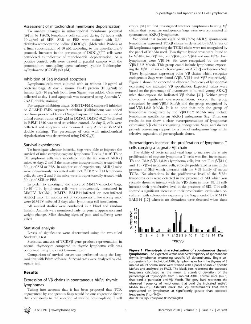
Expression of Vb chains in spontaneous AKR/J thymiclymphomas
Taking into account that it has been proposed that TCR
engagement by endogenous Sags would be one epigenetic factor
that contributes to the selection of murine pre-neoplastic T cell
clones [31] we first investigated whether lymphomas bearing Vbchains that recognize endogenous Sags were overrepresented in
spontaneous AKR/J lymphomas.
We found that twenty eight of 38 (74%) AKR/J spontaneous
lymphomas expressed TCRb chains as detected by FACS. Six of
28 lymphomas expressing the TCRb chain were not recognized by
the panel of MoAbs used. Two thymic lymphomas were found to
be Vb10+, two Vb14+, one Vb3+, one Vb4+ and one Vb2+. Five
lymphomas were Vb8.3+. Six were recognized by the anti-
Vb8.1,8.2 MoAb. This group could include lymphomas express-
ing the Vb8.1 chain which recognize an AKR/J endogenous Sag.
Three lymphomas expressing other Vb chains which recognize
endogenous Sags were found (Vb5, Vb11 and Vb7 respectively).
Figure 1 shows the expected vs observed frequency of lymphomas
expressing the indicated Vb specificities. Expected values were
based on the percentage of thymocytes in normal young AKR/J
mice that express the indicated Vb chains [31]. Neither group
showed a significant overrepresentation, except the group
recognized by anti-Vb8.3 MoAb and the group recognized by
anti-Vb8.1,8.2 MoAb. It is to note that only the group of
lymphomas recognized by the Vb8.1,8.2 MoAb may include
lymphomas specific for an AKR/J endogenous Sag. Thus, our
results do not show a clear overrepresentation of lymphomas
expressing Vb chains recognizing endogenous Sags, and do not
provide convincing support for a role of endogenous Sags in the
selective expansion of pre-neoplastic clones.
Superantigens increase the proliferation of lymphoma Tcells carrying a cognate Vb chain
The ability of bacterial and viral Sags to increase the in vitro
proliferation of cognate lymphoma T cells was first investigated.
T8 and T8.2 (Vb8.1,8.2+) lymphoma cells, but not T14 (Vb14+)
and T5 (Vb5+) neoplastic cells, strongly proliferated in vitro in the
presence of SEB which interacts with the Vb8 family of mouse
TCRs. No alterations in the proliferative level of the Vb8+lymphoma cells were detected in the presence of SEI which was
recently shown to interact with the Vb5 chain in mice [8]. T5 cells
increase their proliferative level in the presence of SEI. T14 cells
showed a significant increase in their proliferative levels when co-
cultured with splenocytes expressing the Sag encoded by MMTV
BALB14 [17] whereas no alterations were detected when these
Figure 1. Phenotypic characterization of spontaneous thymiclymphomas. The expected versus observed frequency of spontaneousthymic lymphomas expressing specific Vb determinants. Single cellsuspensions from individual AKR/J lymphomas or from the thymus of 3mo-old AKR/J normal mice were stained with a panel of anti-Vb specificMoAbs and analyzed by FACS. The black bars represent the expectedfrequency calculated as the mean 6 standard deviation of thepercentage of thymocytes from 3 mo-old AKR/J normal mice (n = 5)that bind a particular anti-Vb MoAb. The grey bars represent theobserved frequency of lymphomas that bind the indicated anti-VbMoAb (n = 28). Asterisks mark the Vb determinants that wererepresented on lymphomas at significantly greater than expectedfrequencies (* p,0.05).doi:10.1371/journal.pone.0015694.g001
Superantigens and Apoptosis of T Cell Lymphomas
PLoS ONE | www.plosone.org 3 December 2010 | Volume 5 | Issue 12 | e15694
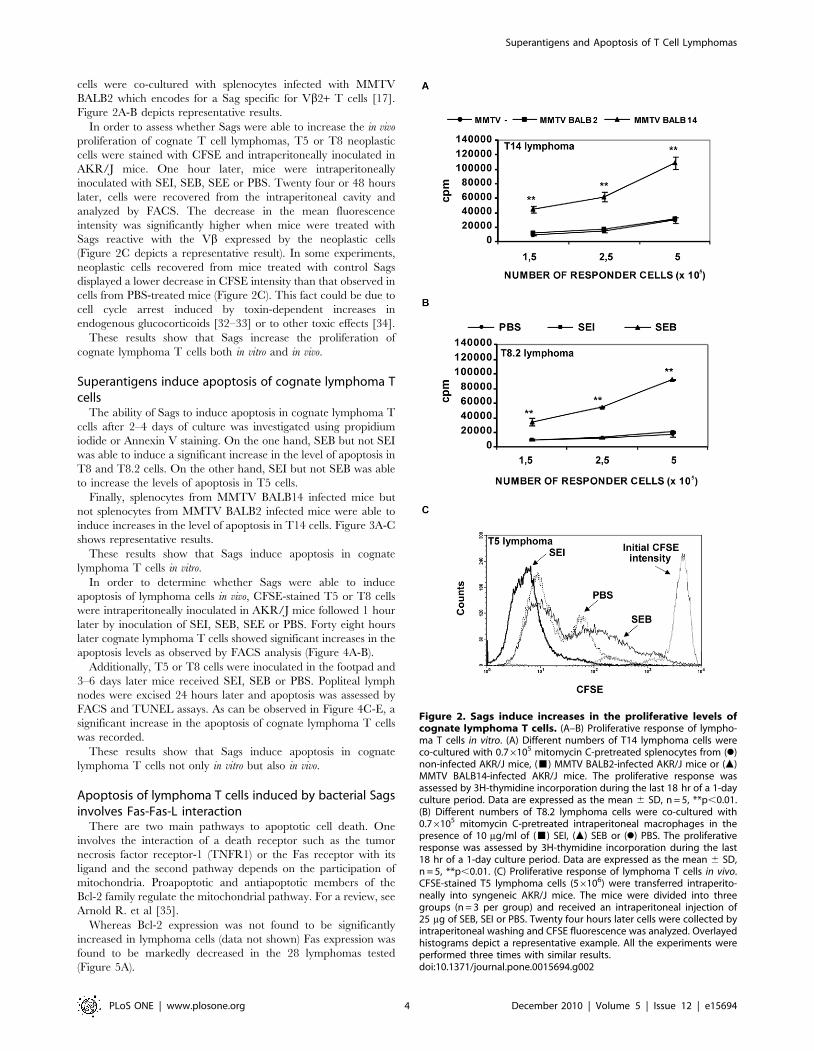
cells were co-cultured with splenocytes infected with MMTV
BALB2 which encodes for a Sag specific for Vb2+ T cells [17].
Figure 2A-B depicts representative results.
In order to assess whether Sags were able to increase the in vivo
proliferation of cognate T cell lymphomas, T5 or T8 neoplastic
cells were stained with CFSE and intraperitoneally inoculated in
AKR/J mice. One hour later, mice were intraperitoneally
inoculated with SEI, SEB, SEE or PBS. Twenty four or 48 hours
later, cells were recovered from the intraperitoneal cavity and
analyzed by FACS. The decrease in the mean fluorescence
intensity was significantly higher when mice were treated with
Sags reactive with the Vb expressed by the neoplastic cells
(Figure 2C depicts a representative result). In some experiments,
neoplastic cells recovered from mice treated with control Sags
displayed a lower decrease in CFSE intensity than that observed in
cells from PBS-treated mice (Figure 2C). This fact could be due to
cell cycle arrest induced by toxin-dependent increases in
endogenous glucocorticoids [32–33] or to other toxic effects [34].
These results show that Sags increase the proliferation of
cognate lymphoma T cells both in vitro and in vivo.
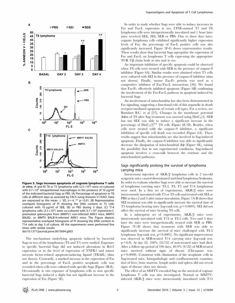
Superantigens induce apoptosis of cognate lymphoma Tcells
The ability of Sags to induce apoptosis in cognate lymphoma T
cells after 2–4 days of culture was investigated using propidium
iodide or Annexin V staining. On the one hand, SEB but not SEI
was able to induce a significant increase in the level of apoptosis in
T8 and T8.2 cells. On the other hand, SEI but not SEB was able
to increase the levels of apoptosis in T5 cells.
Finally, splenocytes from MMTV BALB14 infected mice but
not splenocytes from MMTV BALB2 infected mice were able to
induce increases in the level of apoptosis in T14 cells. Figure 3A-C
shows representative results.
These results show that Sags induce apoptosis in cognate
lymphoma T cells in vitro.
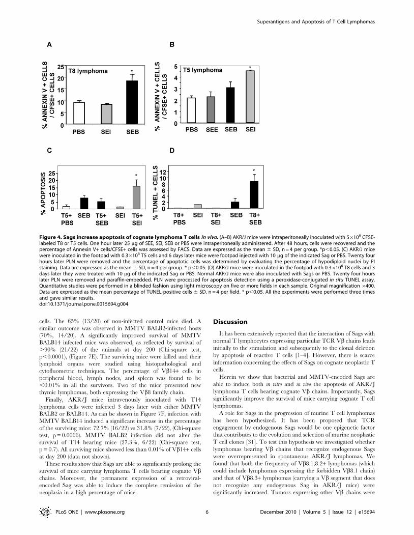
In order to determine whether Sags were able to induce
apoptosis of lymphoma cells in vivo, CFSE-stained T5 or T8 cells
were intraperitoneally inoculated in AKR/J mice followed 1 hour
later by inoculation of SEI, SEB, SEE or PBS. Forty eight hours
later cognate lymphoma T cells showed significant increases in the
apoptosis levels as observed by FACS analysis (Figure 4A-B).
Additionally, T5 or T8 cells were inoculated in the footpad and
3–6 days later mice received SEI, SEB or PBS. Popliteal lymph
nodes were excised 24 hours later and apoptosis was assessed by
FACS and TUNEL assays. As can be observed in Figure 4C-E, a
significant increase in the apoptosis of cognate lymphoma T cells
was recorded.
These results show that Sags induce apoptosis in cognate
lymphoma T cells not only in vitro but also in vivo.
Apoptosis of lymphoma T cells induced by bacterial Sagsinvolves Fas-Fas-L interaction
There are two main pathways to apoptotic cell death. One
involves the interaction of a death receptor such as the tumor
necrosis factor receptor-1 (TNFR1) or the Fas receptor with its
ligand and the second pathway depends on the participation of
mitochondria. Proapoptotic and antiapoptotic members of the
Bcl-2 family regulate the mitochondrial pathway. For a review, see
Arnold R. et al [35].
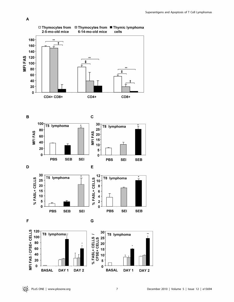
Whereas Bcl-2 expression was not found to be significantly
increased in lymphoma cells (data not shown) Fas expression was
found to be markedly decreased in the 28 lymphomas tested
(Figure 5A).
Figure 2. Sags induce increases in the proliferative levels ofcognate lymphoma T cells. (A–B) Proliferative response of lympho-ma T cells in vitro. (A) Different numbers of T14 lymphoma cells wereco-cultured with 0.76105 mitomycin C-pretreated splenocytes from (N)non-infected AKR/J mice, (&) MMTV BALB2-infected AKR/J mice or (m)MMTV BALB14-infected AKR/J mice. The proliferative response wasassessed by 3H-thymidine incorporation during the last 18 hr of a 1-dayculture period. Data are expressed as the mean 6 SD, n = 5, **p,0.01.(B) Different numbers of T8.2 lymphoma cells were co-cultured with0.76105 mitomycin C-pretreated intraperitoneal macrophages in thepresence of 10 mg/ml of (&) SEI, (m) SEB or (N) PBS. The proliferativeresponse was assessed by 3H-thymidine incorporation during the last18 hr of a 1-day culture period. Data are expressed as the mean 6 SD,n = 5, **p,0.01. (C) Proliferative response of lymphoma T cells in vivo.CFSE-stained T5 lymphoma cells (56106) were transferred intraperito-neally into syngeneic AKR/J mice. The mice were divided into threegroups (n = 3 per group) and received an intraperitoneal injection of25 mg of SEB, SEI or PBS. Twenty four hours later cells were collected byintraperitoneal washing and CFSE fluorescence was analyzed. Overlayedhistograms depict a representative example. All the experiments wereperformed three times with similar results.doi:10.1371/journal.pone.0015694.g002
Superantigens and Apoptosis of T Cell Lymphomas
PLoS ONE | www.plosone.org 4 December 2010 | Volume 5 | Issue 12 | e15694
The mechanisms underlying apoptosis induced by bacterial
Sags in two of the lymphomas (T8 and T5) were studied. Exposure
to specific bacterial Sags did not induced alterations in Bcl-2
expression or in the level of expression of TNFR1 and tumor
necrosis factor-related apoptosis-inducing ligand (TRAIL) (data
not shown). Contrarily, a marked increase in the expression of Fas
and in the percentage of Fas-L positive neoplastic cells was
recorded when cells were cultured with specific Sags (Figure 5B-E).
Occasionally in vitro exposure of lymphoma cells to non specific
bacterial Sags induced a slight but not significant increase in the
expression of Fas (Figure 5E).
In order to study whether Sags were able to induce increases in
Fas and Fas-L expression in vivo, CFSE-stained T5 and T8
lymphoma cells were intraperitoneally inoculated and 1 hour later
mice received SEE, SEI, SEB or PBS. One to three days later,
cognate lymphoma cells exhibited significantly higher expression
levels of Fas; the percentage of Fas-L positive cells was also
significantly increased. Figure 5F-G shows representative results.
These results show that bacterial Sags upregulate the expression of
Fas and Fas-L on lymphoma T cells expressing the appropriate
TCR Vb chain both in vitro and in vivo.
An important inhibition of specific apoptosis could be observed
when T8 cells were treated with SEB in the presence of caspase-8
inhibitor (Figure 6A). Similar results were obtained when T5 cells
were cultured with SEI in the presence of caspase-8 inhibitor (data
not shown). Finally, mouse Fas-Fc protein was used as a
competitive inhibitor of Fas/Fas-L interactions [36]. We found
that Fas-Fc effectively inhibited apoptosis (Figure 6B) confirming
the involvement of the Fas-Fas-L pathway in apoptosis induced by
bacterial Sags.
An involvement of mitochondria has also been demonstrated in
Fas signaling, suggesting a functional role of this organelle in death
receptor-mediated apoptosis of certain cell types. For a review, see
Barnhart B.C. et al [37]. Changes in the membrane potential
Dym of T8 after Sag treatment was assessed using DioC2(3). SEB
but not SEE was able to induce a significant increase in the
percentage of DioC2(3)low T8 cells (Figure 6C-D). Besides, when
cells were treated with the caspase-9 inhibitor, a significant
inhibition of specific cell death was recorded (Figure 6A). These
results suggest that mitochondria are also involved in Sag-induced
apoptosis. Finally, the caspase-8 inhibitor was able to significantly
decrease the dissipation of mitochondrial Dy (Figure 6E), raising
the possibility that in our experimental conditions, Sag-induced
apoptosis involves a cross-talk between the extrinsic and the
mitochondrial pathways.
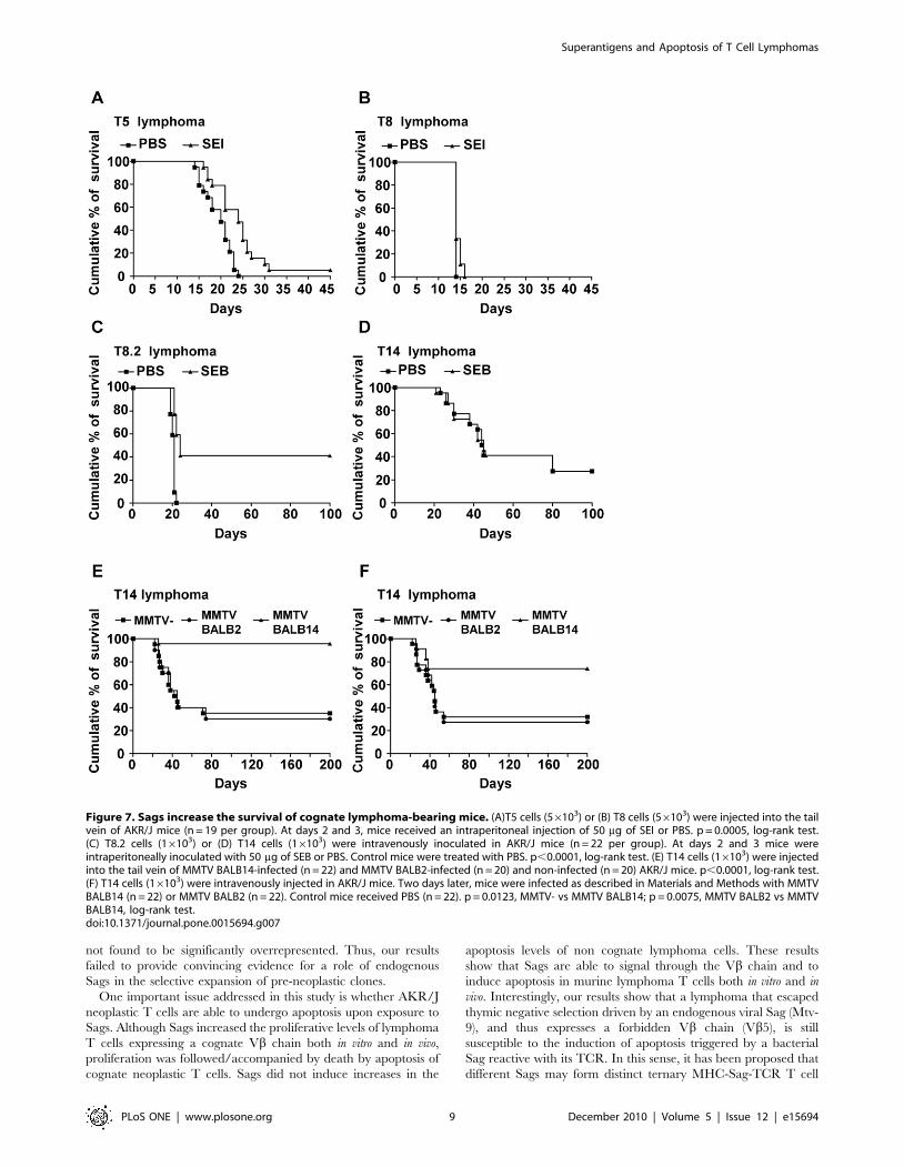
Sags significantly prolong the survival of lymphomacarrying mice
Intravenous injection of AKR/J lymphoma cells in 2 mo-old
syngeneic mice caused disseminated and fatal lymphoma/leukemia.
In order to evaluate whether Sags were able to increase the survival
of lymphoma carrying mice T8.2, T8, T5 and T14 lymphomas
were used. In a first set of experiments, AKR/J mice were
intravenously inoculated with T5 or T8 cells and treated with SEI or
PBS at days 2 and 3 after tumor inoculation. Figure 7A-B shows that
SEI treatment was able to significantly increase the survival time of
T5 lymphoma bearing mice (log-rank test, p = 0.0005). SEI did not
affect the survival of mice bearing T8 cells.
In a subsequent set of experiments, AKR/J mice were
intravenously inoculated with T14 or T8.2 cells. Two and 3 days
later the mice were intraperitoneally treated with SEB or PBS.
Figure 7C-D shows that treatment with SEB was able to
significantly increase the survival of mice challenged with T8.2
lymphoma (log-rank test, p,0.0001). No significant improvement
was observed in SEB-treated T14 carrying mice (log-rank test,
p = 0.8). At day 22, 100% (22/22) of non-treated mice had died.
After a follow-up period of 100 days, 40.9% (9/22) of SEB-treated
mice survived without signs of disease (Chi-square test,
p = 0.0008). Consistent with elimination of the neoplastic cells in
Sag-treated mice, histopathologic and cytofluorometric examina-
tion of liver, bone marrow, lymph nodes and spleen did not reveal
signs of disease (data not shown).
The effect of an MMTV-encoded Sag on the survival of cognate
lymphoma T cells was also investigated. Normal or MMTV-
infected AKR/J mice were intravenously inoculated with T14
Figure 3. Sags increase apoptosis of cognate lymphoma T cellsin vitro. (A and B) T8 or T5 lymphoma cells (2.56105) were co-culturedwith 0.76105 intraperitoneal macrophages in the presence of 10 mg/mlof the indicated bacterial Sags or PBS. (A) Percentage of apoptosis in T8cells at different days as assessed by FACS using Annexin V-7AAD. Dataare expressed as the mean 6 SD, n = 4, ** p,0.01. (B) Representativeoverlayed histograms of PI showing the DNA content in T5 cellscultured with 10 mg/ml of SEB, SEI or PBS during 3 days. (C) T14lymphoma cells (2.56105) were co-cultured with 0.76105 mytomicin-Cpretreated splenocytes from (MMTV-) non-infected AKR/J mice, MMTVBALB2- or MMTV BALB14-infected AKR/J mice. The Figure depictsrepresentative overlayed histograms of PI showing the DNA content inT14 cells at day 3 of culture. All the experiments were performed fivetimes with similar results.doi:10.1371/journal.pone.0015694.g003
Superantigens and Apoptosis of T Cell Lymphomas
PLoS ONE | www.plosone.org 5 December 2010 | Volume 5 | Issue 12 | e15694
cells. The 65% (13/20) of non-infected control mice died. A
similar outcome was observed in MMTV BALB2-infected hosts
(70%, 14/20). A significantly improved survival of MMTV
BALB14 infected mice was observed, as reflected by survival of
.90% (21/22) of the animals at day 200 (Chi-square test,
p,0.0001), (Figure 7E). The surviving mice were killed and their
lymphoid organs were studied using histopathological and
cytofluometric techniques. The percentage of Vb14+ cells in
peripheral blood, lymph nodes, and spleen was found to be
,0.01% in all the survivors. Two of the mice presented new
thymic lymphomas, both expressing the Vb8 family chain.
Finally, AKR/J mice intravenously inoculated with T14
lymphoma cells were infected 3 days later with either MMTV
BALB2 or BALB14. As can be shown in Figure 7F, infection with
MMTV BALB14 induced a significant increase in the percentage
of the surviving mice: 72.7% (16/22) vs 31.8% (7/22), (Chi-square
test, p = 0.0066). MMTV BALB2 infection did not alter the
survival of T14 bearing mice (27.3%, 6/22) (Chi-square test,
p = 0.7). All surviving mice showed less than 0.01% of Vb14+ cells
at day 200 (data not shown).
These results show that Sags are able to significantly prolong the
survival of mice carrying lymphoma T cells bearing cognate Vbchains. Moreover, the permanent expression of a retroviral-
encoded Sag was able to induce the complete remission of the
neoplasia in a high percentage of mice.
Discussion
It has been extensively reported that the interaction of Sags with
normal T lymphocytes expressing particular TCR Vb chains leads
initially to the stimulation and subsequently to the clonal deletion
by apoptosis of reactive T cells [1–4]. However, there is scarce
information concerning the effects of Sags on cognate neoplastic T
cells.
Herein we show that bacterial and MMTV-encoded Sags are
able to induce both in vitro and in vivo the apoptosis of AKR/J
lymphoma T cells bearing cognate Vb chains. Importantly, Sags
significantly improve the survival of mice carrying cognate T cell
lymphomas.
A role for Sags in the progression of murine T cell lymphomas
has been hypothesized. It has been proposed that TCR
engagement by endogenous Sags would be one epigenetic factor
that contributes to the evolution and selection of murine neoplastic
T cell clones [31]. To test this hypothesis we investigated whether
lymphomas bearing Vb chains that recognize endogenous Sags
were overrepresented in spontaneous AKR/J lymphomas. We
found that both the frequency of Vb8.1,8.2+ lymphomas (which
could include lymphomas expressing the forbidden Vb8.1 chain)
and that of Vb8.3+ lymphomas (carrying a Vb segment that does
not recognize any endogenous Sag in AKR/J mice) were
significantly increased. Tumors expressing other Vb chains were
Figure 4. Sags increase apoptosis of cognate lymphoma T cells in vivo. (A–B) AKR/J mice were intraperitoneally inoculated with 56106 CFSE-labeled T8 or T5 cells. One hour later 25 mg of SEE, SEI, SEB or PBS were intraperitoneally administered. After 48 hours, cells were recovered and thepercentage of Annexin V+ cells/CFSE+ cells was assessed by FACS. Data are expressed as the mean 6 SD, n = 4 per group. *p,0.05. (C) AKR/J micewere inoculated in the footpad with 0.36106 T5 cells and 6 days later mice were footpad injected with 10 mg of the indicated Sag or PBS. Twenty fourhours later PLN were removed and the percentage of apoptotic cells was determined by evaluating the percentage of hypodiploid nuclei by PIstaining. Data are expressed as the mean 6 SD, n = 4 per group. * p,0.05. (D) AKR/J mice were inoculated in the footpad with 0.36106 T8 cells and 3days later they were treated with 10 mg of the indicated Sag or PBS. Normal AKR/J mice were also inoculated with Sags or PBS. Twenty four hourslater PLN were removed and paraffin-embedded. PLN were processed for apoptosis detection using a peroxidase-conjugated in situ TUNEL assay.Quantitative studies were performed in a blinded fashion using light microscopy on five or more fields in each sample. Original magnification 6400.Data are expressed as the mean percentage of TUNEL-positive cells 6 SD, n = 4 per field. * p,0.05. All the experiments were performed three timesand gave similar results.doi:10.1371/journal.pone.0015694.g004
Superantigens and Apoptosis of T Cell Lymphomas
PLoS ONE | www.plosone.org 6 December 2010 | Volume 5 | Issue 12 | e15694
Superantigens and Apoptosis of T Cell Lymphomas
PLoS ONE | www.plosone.org 7 December 2010 | Volume 5 | Issue 12 | e15694
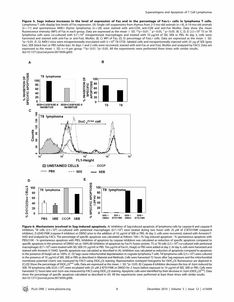
Figure 5. Sags induce increases in the level of expression of Fas and in the percentage of Fas-L+ cells in lymphoma T cells.Lymphoma T cells display low levels of Fas expression. (A) Single cell suspensions from thymus from 2-5-mo-old animals (n = 8), 6-14-mo-old animals(n = 11) and spontaneous AKR/J thymic lymphomas (n = 28) were stained with anti-CD4, anti-CD8 and anti-Fas MoAbs. Data show the meanfluorescence intensity (MFI) of Fas in each group. Data are expressed as the mean 6 SD. **p,0.01, { p,0.05, { p,0.05. (B, C, D, E) 2.56105 T5 or T8lymphoma cells were co-cultured with 0.76105 intraperitoneal macrophages and treated with 10 mg/ml of SEI, SEB or PBS. At day 3, cells wereharvested and stained with anti-Fas or anti-FasL MoAbs. (B, C) MFI of Fas, (D, E) percentage of FasL+ cells. Data are expressed as the mean 6 SD.*p,0.05. (F, G) AKR/J mice were intraperitoneally inoculated with 56106 T8 CFSE- labeled cells and intraperitoneally injected with 25 mg of SEE (greybar), SEB (black bar) or PBS (white bar). At days 1 and 2 cells were recovered, stained with anti-Fas or anti-FasL MoAbs and analyzed by FACS. Data areexpressed as the mean 6 SD, n = 4 per group. **p,0.01, *p,0.05. All the experiments were performed three times with similar results.doi:10.1371/journal.pone.0015694.g005
Figure 6. Mechanisms involved in Sag-induced apoptosis. A) Inhibition of Sag-induced apoptosis of lymphoma cells by caspase-8 and caspase-9inhibitors. T8 cells (2.56105) co-cultured with peritoneal macrophages (0.76105) were treated during two hours with 25 mM of Z-IETD-FMK (caspase-8inhibitor), Z-LEHD-FMK (caspase-9 inhibitor) or DMSO prior to the addition of 10 mg/ml of SEB or PBS. At day 3, cells were recovered, stained with Annexin/7-AAD and analyzed by FACS. The percentage of specific apoptosis was calculated as follows: 1006(% Sag induced apoptosis - % spontaneous apoptosis withPBS)/(100 - % spontaneous apoptosis with PBS). Inhibition of apoptosis by caspase inhibitors was calculated as reduction of specific apoptosis compared tospecific apoptosis in the presence of DMSO set as 100%.(B) Inhibition of apoptosis by Fas-Fc fusion protein. T5 or T8 cells (2.56105) co-cultured with peritonealmacrophages (0.76105) were treated with SEI, SEB (10 mg/ml) or PBS. Ten mg/ml of Fas-Fc, HuIgG or PBS were added at day 2. At day 4, cells were harvested andstained with Annexin V-7AAD. Specific apoptosis was calculated as described in (A). Inhibition was calculated as reduction of apoptosis compared to apoptosisin the presence of HuIgG set as 100%. (C–D) Sags cause mitochondrial depolarization in cognate lymphoma T cells. T8 lymphoma cells (2.56105) were culturedin the presence of 10 mg/ml of SEE, SEB or PBS as described in Material and Methods. Cells were harvested 72 hours after Sag exposure and the mitochondrialmembrane potential (Dym) was measured by FACS using DiOC2(3) staining. Representative overlayed histograms for DiOC2(3) fluorescence are depicted in(C).(D) Show the percentage of DiOC2(3)low cells. Data are expressed as the mean 6 SD. *p,0.05. (E) Caspase-8 inhibitor decreases the loss of Dym induced bySEB. T8 lymphoma cells (0.256106) were incubated with 25 mM Z-IETD-FMK or DMSO for 2 hours before exposure to 10 mg/ml of SEE, SEB or PBS. Cells wereharvested 72 hours later andDym was measured by FACS using DiOC2(3) staining. Apoptotic cells were identified by their decrease in Dym (DiOC2(3)low). Datashow the percentage of specific apoptosis calculated as described in (A). All the experiments were performed at least three times with similar results.doi:10.1371/journal.pone.0015694.g006
Superantigens and Apoptosis of T Cell Lymphomas
PLoS ONE | www.plosone.org 8 December 2010 | Volume 5 | Issue 12 | e15694
not found to be significantly overrepresented. Thus, our results
failed to provide convincing evidence for a role of endogenous
Sags in the selective expansion of pre-neoplastic clones.
One important issue addressed in this study is whether AKR/J
neoplastic T cells are able to undergo apoptosis upon exposure to
Sags. Although Sags increased the proliferative levels of lymphoma
T cells expressing a cognate Vb chain both in vitro and in vivo,
proliferation was followed/accompanied by death by apoptosis of
cognate neoplastic T cells. Sags did not induce increases in the
apoptosis levels of non cognate lymphoma cells. These results
show that Sags are able to signal through the Vb chain and to
induce apoptosis in murine lymphoma T cells both in vitro and in
vivo. Interestingly, our results show that a lymphoma that escaped
thymic negative selection driven by an endogenous viral Sag (Mtv-
9), and thus expresses a forbidden Vb chain (Vb5), is still
susceptible to the induction of apoptosis triggered by a bacterial
Sag reactive with its TCR. In this sense, it has been proposed that
different Sags may form distinct ternary MHC-Sag-TCR T cell
Figure 7. Sags increase the survival of cognate lymphoma-bearing mice. (A)T5 cells (56103) or (B) T8 cells (56103) were injected into the tailvein of AKR/J mice (n = 19 per group). At days 2 and 3, mice received an intraperitoneal injection of 50 mg of SEI or PBS. p = 0.0005, log-rank test.(C) T8.2 cells (16103) or (D) T14 cells (16103) were intravenously inoculated in AKR/J mice (n = 22 per group). At days 2 and 3 mice wereintraperitoneally inoculated with 50 mg of SEB or PBS. Control mice were treated with PBS. p,0.0001, log-rank test. (E) T14 cells (16103) were injectedinto the tail vein of MMTV BALB14-infected (n = 22) and MMTV BALB2-infected (n = 20) and non-infected (n = 20) AKR/J mice. p,0.0001, log-rank test.(F) T14 cells (16103) were intravenously injected in AKR/J mice. Two days later, mice were infected as described in Materials and Methods with MMTVBALB14 (n = 22) or MMTV BALB2 (n = 22). Control mice received PBS (n = 22). p = 0.0123, MMTV- vs MMTV BALB14; p = 0.0075, MMTV BALB2 vs MMTVBALB14, log-rank test.doi:10.1371/journal.pone.0015694.g007
Superantigens and Apoptosis of T Cell Lymphomas
PLoS ONE | www.plosone.org 9 December 2010 | Volume 5 | Issue 12 | e15694
signaling complexes which could lead to distinct physiological
outcomes [38].
Different mechanisms of apoptosis induced by bacterial Sags in
normal T cells have been reported. Several groups have reported
Fas-Fas-L independent apoptosis pathways after stimulation with
bacterial Sags. These pathways include members of the Bcl-2-
family [39–40] and/or reactive oxygen species [41]. Other studies,
however, have shown that Fas/Fas-L interaction is involved in
bacterial Sag mediated apoptosis [36,42–43]. Finally, it has been
proposed that the pathway involved would depend on the doses of
bacterial Sags or on the experimental model used. After
determining that the expression of Bcl-2 in the AKR/J
spontaneous lymphomas was not altered while Fas expression
was markedly decreased in all thymomas tested, the mechanisms
underlying apoptosis induced by bacterial Sags in two lymphomas
were studied. Whereas Sag exposure did not induce alterations in
the expression of Bcl-2, TRAIL or TNFR1, interaction of Sags
with the cognate Vb chain of lymphoma T cells revert the low
expression of Fas characteristic of these cells both in vitro and in
vivo. Inhibition of caspase-8 markedly decreased apoptosis induced
by Sags. Mouse Fas-Fc- a competitive inhibitor of Fas-Fas-L
interactions effectively inhibited apoptosis in the lymphomas
studied confirming the involvement of the Fas-Fas-L pathway in
apoptosis induced by bacterial Sags. It has been reported that in
certain cell types mitochondria is involved in Fas signaling. For a
review, see Barnhart B.C. et al [37]. Herein we show that Sags
induced loss of transmembrane potential in this organelle. Besides,
a significant inhibition of apoptosis was recorded in the presence of
a caspase-9 inhibitor. Finally, inhibition of caspase-8 significantly
decreased the dissipation of Dym, raising the possibility that Sag-
induced apoptosis involves a cross talk between the extrinsic and
the mitochondrial pathways.
Finally, the in vivo effect of retroviral and bacterial Sags on the
survival of lymphoma carrying mice was investigated. SEI was
able to significantly increase the survival time of mice carrying T5
neoplastic cells. Treatment with SEB was able to induce long-term
survival of hosts injected with T8.2 neoplastic cells. Whereas all
control mice died in three weeks, 40% of SEB treated mice were
still free of disease at day 100. The fact that toxins did not affect
the survival of mice bearing non-cognate lymphoma T cells
strongly suggests that the effect of these toxins is mainly due to
their superantigenic activity.
The effect of a retroviral-encoded Sag in the survival of
lymphoma bearing mice was assessed. Mice intravenously
inoculated with T14 cells showed a remarkable enhancement of
survival when the hosts were infected with MMTV BALB14
before or after tumor inoculation. No improvement in survival was
observed in MMTV BALB2 infected hosts or in mice treated with
SEB. These results clearly show that Sags are able to significantly
improve the survival of mice bearing cognate-T cell lymphomas.
The fate of the interaction between Sags and human
lymphoma/leukemia T cells is a matter of debate. It has been
reported that leukemic cells from patients with leukemia of T cell
origin have the ability to respond to TCR-dependent bacterial
Sags, as assessed by their proliferative response in vitro [44–45].
Based on these data, it has been proposed that bacterial infection
in such patients might contribute to the expansion of leukemic
cells. Noteworthy, apoptosis of leukemic cells was not assessed. It
has also been hypothesized that bacterial Sags could be involved in
malignant transformation and/or in the expansion and evolution
of cutaneous T cell lymphomas (CTCL), this hypothesis was based
in the restricted use of particular Vb by CTCL cells [46–48].
Contrarily, Vonderheid EC et al [49] hypothesized that chronic
stimulation of skin-homing normal T cells by staphylococcal Sags
would act to deplete Vb-responsive normal T cells prior to
neoplastic transformation and as a consequence the Vb usage by
the neoplastic T cells may be skewed and they would be more
likely to express Vb segments that are relatively unaffected by
staphylococcal Sags. It would be of great interest to determine
whether different human T cell malignancies expressing TCRs are
susceptible to the induction of apoptosis by Sags. Our unpublished
results show that cells from the human Jurkat T cell line derived
from an acute T cell leukemia are highly susceptible to Sag-
induced apoptosis (I.N. and I.P., manuscript in preparation).
Results reported herein clearly show that cells from AKR/J T
cell lymphomas are susceptible to be deleted as a consequence of
Sag signaling via TCR. Importantly, Sags were able to increase
the survival of mice bearing very aggressive lymphomas. If Sags
were able to induce apoptosis in human neoplastic T cells
expressing functional TCRs, they could be envisaged as
therapeutic agents. The use of Sags as therapeutic agents in T
cell malignancies would have the advantage of deleting restricted
T cell clones without causing the death of other normal cells. If
toxic Sags are to be used in humans, their toxic effects might be
separated from their superantigenic activity. In this sense, it has
been shown that carboxymethylation of SEB blocks its enterotoxic
but not its mitogenic properties [50]. Furthermore, many
retroviral-encoded Sags are not associated with toxic effects and
human T cells are able to recognize them, opening the possibility
of their use in gene therapy. Finally, treatments based on the
inoculation of dendritic cells transfected with mRNA coding for
MMTV Sags would avoid both toxicity and the risks associated
with genetic therapies.
Acknowledgments
We thank C. D. Pasqualini for helpful discussions.
Author Contributions
Conceived and designed the experiments: IP IN. Performed the
experiments: JM PMB GC DL. Analyzed the data: JM PMB GC DL IN
IP. Wrote the paper: JM PMB GC IP IN.
References
1. Marrack P, Blackman M, Kushnir E, Kappler J (1990) The toxicity of
staphylococcal enterotoxin B in mice is mediated by T cells. J Exp Med 171:
455–464.
2. Mac Donald HR, Baschieri S, Lees RK (1991) Clonal expansion precedes
anergy and death of Vb8+ peripheral T cells responding to staphylococcal
enterotoxin B in vivo. Eur J Immunol 21(8): 1963–1966.
3. Choi Y, Kappler JE, Marrack P (1991) A Superantigen encoded in the open
reading frame of the 3’ long terminal repeat of the mouse mammary tumor
virus. Nature 350;6315): 203–207.
4. Renno T, Attinger A, Locatelli S, Bakker T, Vacheron S, et al. (1999) Cutting
edge: Apoptosis of superantigen-activated T cells occurs preferentially after a
discrete number of cell divisions in vivo. J Immunol 162: 6312–6315.
5. Fraser JD, Proft T (2008) The bacterial superantigen and superantigen-like
proteins. Immunol Rev 225: 226–43.
6. Janeway CA, Jr., Yagi J, Conrad PJ, Katz ME, Jones B, et al. (1989) T-cell
responses to Mls and to bacterial proteins that mimic its behavior. Immunol Rev
107: 61–88.
7. Callahan JE, Herman A, Kappler JW, Marrack P (1990) Stimulation of B10.BR
T cells with superantigenic staphylococcal toxins. J Immunol 144(7): 2473–9.
8. Fernandez MM, De Marzi MC, Berguer P, Burzyn D, Langley RJ, et al. (2006)
Binding of natural variants of staphylococcal superantigens SEG and SEI to
TCR and MHC class II molecule. Mol Immunol 43(7): 927–38.
9. Bittner JJ (1936) Some possible effects of nursing on the mammary gland tumor
incidence in mice. Science 84(2172): 162.
Superantigens and Apoptosis of T Cell Lymphomas
PLoS ONE | www.plosone.org 10 December 2010 | Volume 5 | Issue 12 | e15694
10. Callahan R, Smith GH (2000) MMTV-induced mammary tumorigenesis: gene
discovery, progression to malignancy and cellular pathways. Oncogene 19(8):992–1001.
11. Simpson E (1993) T cell repertoire selection by mouse mammary tumour
viruses. Eur J Immunogenet 20(2): 137–49.12. Frankel WN, Rudy C, Coffin JM, Huber BT (1991) Linkage of Mls genes to
endogenous mammary tumor viruses of inbred mice. Nature 349(6309): 526–28.13. Acha-Orbea H, MacDonald HR (1995) Superantigens of mouse mammary
tumor virus. Annu Rev Immunol 13: 459–86.
14. Brandt-Carlson C, Butel JS, Wheeler D (1993) Phylogenetic and structuralanalyses of MMTV LTR ORF sequences of exogenous and endogenous origins.
Virology 193(1): 171–85.15. Yazdanbakhsh K, Park CG, Winslow GM, Choi Y (1993) Direct evidence for
the role of COOH terminus of mouse mammary tumor virus superantigen indetermining T cell receptor Vb specificity. J Exp Med 178(2): 737–41.
16. Golovkina TV, Piazzon I, Nepomnaschy I, Buggiano V, Olano Vela M, et al.
(1997) Generation of a tumorigenic milk-borne mouse mammary tumor virus byrecombination between endogenous and exogenous viruses. J Virol 71(5):
3895–3903.17. Buggiano V, Goldman A, Nepomnaschy I, Bekinschtein P, Berguer P, et al.
(1999) Characterization of two infectious mouse mammay tumor viruses:
superantigenicity and tumorigenicity. Scand J Immunol 49: 269–77.18. Newell KA, Ellernhorn JDI, Bru DS, Bluestone JA (1991) In vivo T cell activation
by staphylococcal enterotoxin B prevents outhgrowth of a malignant tumor.Proc Natl Acad Sci USA 88: 1074–78.
19. Kominsky SL, Torres BA, Honeika AC, Lake FA, Johnson H (2001)Superantigen enhanced protection against a weak tumor-specific melanoma
antigen implications for prophylactic vaccination against cancer. Int J Cancer
94(6): 834–841.20. Perabo FG, Willert PL, Wirger A, Schmidt DH, Wardelmann E, et al. (2005)
Preclinical evaluation of superantigen (staphylococcal enterotoxin B) in theintravesical immunotherapy of superficial bladder cancer. Int J Cancer 115(4):
591–8.
21. Gidlof C, Dohlsten M, Lando P, Kalland T, Sundstrom C, et al. (1997) Asuperantigen-antibody fusion protein for T-cell immunotherapy of human B-
lineage malignancies. Blood 89(6): 2089–97.22. Litton MJ, Dohlsten M, Rosendahl A, Ohlsson L, Søgaard M, et al. (1999) The
distinct role of CD4+ and CD8+ T-cells during the anti-tumour effects oftargeted Superantigens. British J Cancer 81(2): 359–366.
23. Tordsson JM, Ohlsson LG, Abrahmsen LB, Kalstrom PJ, Lando PA, et al.
(2000) Phage-selected primate antibodies fused to superantigens for immuno-therapy of malignant melanoma. Cancer Immunol Immunother 48: 691–702.
24. Forsberg G, Ohlsson L, Brodin T, Bjork P, Lando PA, et al. (2001) Therapy ofhuman non-small-cell lung carcinoma using antibody targeting of a modified
superantigen. British J Cancer 85(1): 129–136.
25. Ragnarsson L, Stromberg T, Wijdenes J, Totterman TH, Weigelt C (2001)Multiple myeloma cells are killed by syndecan-1-directed superantigen-activated
T cells. Cancer Immunol Immunother 50: 382–390.26. Ueno A, Arakawa F, Abe H, Matsumoto H, Kudo T, et al. (2002) T-cell
immunotherapy for human MK-1-expressing tumors using a fusion protein ofthe superantigen SEA and anti-MK-1 scFv antibody. Anticancer Res 22(2A):
769–76.
27. Terman DS, Bohach G, Vandenesch F, Etienne J, Lina G, et al. (2006)Staphylococcal superantigens of the enterotoxin gene cluster (egc) for treatment
of stage IIIb non-small cell lung cancer with pleural effusion. Clin Chest Med27(2): 321–34.
28. Sundstedt A, Celander M, Ohman MW, Forsberg G, Hedlund G (2009)
Immunotherapy with tumor-targeted superantigens (TTS) in combination withdocetaxel results in synergistic anti-tumor effects. Int Immunopharmacol 9(9):
1063–70.29. Lombardi G, Burzyn D, Mundinano J, Berguer P, Bekinschtein P, et al. (2005)
Cathepsin-L influences the expression of extracellular matrix in lymphoid organs
and plays a role in the regulation of thymic output and of peripheral T cellnumber. J Immunol 174(11): 7022–32.
30. Cabrera G, Burzyn D, Mundinano J, Courreges MC, Camicia G, et al. (2008)Early increases in superantigen-specific Foxp3+ regulatory T cells during mouse
mammary tumor virus infection. J Virol 82(15): 7422–31.
31. Gomez G, Clarkin KZ, Kraig E, Infante AJ, Richie ER (2000) TCR Vbrepertoire restriction and lack of CDR3 conservation implicate TCR-superantigen interactions in promoting the clonal evolution of murine thymic
lymphomas. Int Immunol 12(3): 263–70.
32. Shurin G, Shanks N, Nelson L, Hoffman G, Huang L, et al. (1997)
Hypothalamic-pituitary-adrenal activation by the bacterial superantigen staph-
ylococcal enterotoxin B: role of macrophages and T cells. Neuroendocrinology65(1): 18–28.
33. Gonzalo JA, Gonzalez-Garcıa A, Martınez C, Kroemer G (1993) Glucocorti-coid-mediated control of the activation and clonal deletion of peripheral T cells
in vivo. J Exp Med 177(5): 1239–46.
34. Fleming SD, Iandolo JJ, Chapes SK (1991) Murine macrophage activation by
staphylococcal exotoxins. Infect Immun 59(11): 4049–55.
35. Arnold R, Brenner D, Becker M, Frey CR, Krammer PH (2006) How T
lymphocytes switch between life and death. Eur J Immunol 36(7): 1654–8.
36. Dhein J, Walczak H, Baumler C, Debatin KM, Krammer PH (1995) Autocrine
T-cell suicide mediated by APO-1/(Fas/CD95). Nature 373(6513): 438–41.
37. Barnhart BC, Alappat EC, Peter ME (2003) The CD95 type I/type II model.
Semin Immunol 15(3): 185–93.
38. Sundberg EJ, Li Y, Mariuzza RA (2002) So many ways of getting in the way:
diversity in the molecular architecture of superantigen-dependent T-cell
signaling complexes. Current Opinion in Immunology 14: 36–44.
39. Hildeman DA, Zhu Y, Mitchell TC, Bouillet P, Strasser A, et al. (2002)
Activated T cell death in vivo mediated by proapoptotic bcl-2 family memberbim. Immunity 16(6): 759–67.
40. Brenner D, Krammer PH, Arnold R (2008) Concepts of activated T cell death.Crit Rev Oncol Hematol 66(1): 52–64.
41. Hildeman DA, Mitchell T, Teague TK, Henson P, Day BJ, et al. (1999)Reactive oxygen species regulate activation-induced T cell apoptosis. Immunity
10(6): 735–44.
42. Ettinger R, Panka DJ, Wang JK, Stanger BZ, Ju ST, et al. (1995) Fas ligand-
mediated cytotoxicity is directly responsible for apoptosis of normal CD4+ Tcells responding to a bacterial superantigen. J Immunol 154(9): 4302–8.
43. Renno T, Hahne M, Tschopp J, MacDonald HR (1996) Peripheral T cellsundergoing superantigen-induced apoptosis in vivo express B220 and upregulate
Fas and Fas ligand. J Exp Med 183(2): 431–7.
44. Metzger R, Melmer G, Schondelmaier S, Heckl-Ostreicher B, Nerl C, et al.
(1993) Leukaemic T cells from patients with chronic lymphocytic leukemia of T-
cell origin respond to Staphylococcus aereus enterotoxin Superantigens.Scand J Immunol 37: 245–50.
45. Ogata M, Kikuchi H, Ohtsuka E, Kohno K, Ito M, et al. (1998) Stimulation ofleukaemic cells from adult T-cell leukaemia patients with bacterial superanti-
gens. Br J Haematol 100(3): 490–500.
46. Tokura Y, Heald PW, Yan SL, Edelson RL (1992) Stimulation of cutaneous T-
cell lymphoma cells with superantigenic staphylococcal toxins. J Invest Dermatol98(1): 33–7.
47. Jackow CM, Cather JC, Hearne V, Asano AT, Musser JM, et al. (1997)Association of erythrodermic cutaneous T-cell lymphoma, superantigen-positive
Staphylococcus aureus, and oligoclonal T-cell receptor Vb gene expansion.
Blood 89(1): 32–40.
48. Linnemann T, Gellrich S, Lukowsky A, Mielke A, Audring H, et al. (2004)
Polyclonal expansion of T cells with the TCR Vb type of the tumour cell inlesions of cutaneous T-cell lymphoma: evidence for possible superantigen
involvement. Br J Dermatol 150(5): 1013–7.
49. Vonderheid EC, Boselli CM, Conroy M, Casaus L, Espinoza LC, et al. (2005)
Evidence for restricted Vb usage in the leukemic phase of cutaneous T celllymphoma. J Invest Dermatol 124(3): 651–61.
50. Alber G, Hammer DK, Fleischer B (1990) Relationship between enterotoxic-and T lymphocyte-stimulating activity of staphylococcal enterotoxin.
B J Immunol 144(12): 4501–6.
Superantigens and Apoptosis of T Cell Lymphomas
PLoS ONE | www.plosone.org 11 December 2010 | Volume 5 | Issue 12 | e15694