studying the active-site loop movement of the são paolo metallo-β-lactamase-1
TRANSCRIPT

ChemicalScience
EDGE ARTICLE
Ope
n A
cces
s A
rtic
le. P
ublis
hed
on 2
4 O
ctob
er 2
014.
Dow
nloa
ded
on 2
9/01
/201
5 09
:07:
32.
Thi
s ar
ticle
is li
cens
ed u
nder
a C
reat
ive
Com
mon
s A
ttrib
utio
n 3.
0 U
npor
ted
Lic
ence
.
View Article OnlineView Journal | View Issue
Studying the act
aDepartment of Chemistry, University of Oxfo
UK. E-mail: [email protected] of Chemistry, Physical and The
of Oxford, South Parks Road, Oxford, OX1 3
ac.ukcSchool of Cellular and Molecular Medicin
Building, Bristol, BS8 1TD, UK
† Electronic supplementary informationexpression and purication, 19F-labellinstructure determination, table of crystalloparameters and renement statistics,distances, procedures for mass specscanning uorimetry measurements, stkinetics measurements. See DOI: 10.1039
‡ These authors contribute equally to this
Cite this: Chem. Sci., 2015, 6, 956
Received 12th June 2014Accepted 24th October 2014
DOI: 10.1039/c4sc01752h
www.rsc.org/chemicalscience
956 | Chem. Sci., 2015, 6, 956–963
ive-site loop movement of theSao Paolo metallo-b-lactamase-1†
Jurgen Brem,‡a Weston B. Struwe,‡b Anna M. Rydzik,a Hanna Tarhonskaya,a
Inga Pfeffer,a Emily Flashman,a Sander S. van Berkel,a James Spencer,c
Timothy D. W. Claridge,a Michael A. McDonough,a Justin L. P. Benesch*b
and Christopher J. Schofield*a
Metallo-b-lactamases (MBLs) catalyse the hydrolysis of almost all b-lactam antibiotics. We report
biophysical and kinetic studies on the Sao Paulo MBL (SPM-1), which reveal its Zn(II) ion usage and
mechanism as characteristic of the clinically important di-Zn(II) dependent B1 MBL subfamily. Biophysical
analyses employing crystallography, dynamic 19F NMR and ion mobility mass spectrometry, however,
reveal that SPM-1 possesses loop and mobile element regions characteristic of the B2 MBLs. These
include a mobile a3 region which is important in catalysis and determining inhibitor selectivity. SPM-1
thus appears to be a hybrid B1/B2 MBL. The results have implications for MBL evolution and inhibitor design.
Introduction
The b-lactams (BLAs) remain the most important class of anti-biotics, representing �60% of those used in the clinic.1
However, b-lactam antibiotic (BLA) utility is threatened byresistance mechanisms, most importantly those involvingplasmid encoded b-lactamases (BLs).2 BLs can be divided intotwomechanistic classes – the serine-BLs (SBLs) and themetallo-BLs (MBLs).2,3 Although for many decades the SBLs were themost clinically relevant BLs, global dissemination of MBLs, e.g.the New Delhi MBLs (NDMs) and the Sao Paulo MBL (SPM-1), isnow occurring.4–6 This is concerning because, with the excep-tion of the monobactams, MBLs hydrolyze all known b-lactamantibiotics (BLAs) (Fig. 1A), and, whilst there are clinicallyuseful SBL inhibitors, there are no such compounds for MBLs.
The MBL fold is structurally unrelated to those of SBLs.MBLs (class B of the Ambler b-lactamase classication)7 aredivided into the B1, B2 and B3 subclasses reecting sequence/
rd, 12 Manseld Road, Oxford, OX1 3TA,
ac.uk
oretical Chemistry Laboratory, University
QZ, UK. E-mail: [email protected].
e, University of Bristol, Medical Sciences
(ESI) available: Procedures for proteing, crystallisation, data collection, andgraphic data, table of crystallographicgures showing binding mode andtrometry measurements, differentialopped-ow measurements and other/c4sc01752h
work.
structural relationships and substrate selectivities (Table S1,Fig. S1 and S2†)2 with the B1 and B2 MBLs being most closelyrelated to each other. Most, but not all, clinically relevant MBLsare from subclass B1 (Table S1†).8
SPM-1 was rst identied in a Pseudomonas aeruginosa strainfrom a paediatric leukaemia patient9 and is a major clinicalproblem in South America;10,11 in Brazil SPM-1 occurs in �35%of clinically identied carbapenem resistant P. aeruginosaisolates and is a major cause of infection mediated mortality.12
Unusually, SPM-1 has high sequence similarity with MBLs fromboth B1 (35% identity with IMP-1 and 30% with BcII) and B2(32% with ImiS and CphA) MBL subclasses.13,14 Studies on MBLZn(II) ion use reveal that the B1 MBLs use two zinc ions forefficient catalysis, whereas the B2 MBLs are inhibited bybinding of a second Zn(II) ion (Fig. 1 and Table S1†).8 Interest-ingly, SPM-1 is reported to have a Zn(II) : MBL ratio of 1.5 : 1 insolution (by atomic absorption spectrometry); however, thesingle reported SPM-1 structure reveals only one zinc ion,bound at the tri-histidine or Zn1 site.13
Structural studies on MBLs have revealed the importance ofmobile regions in catalysis and substrate selectivity.8 Under-standing how these loops inuence binding to MBL active sitesis important with respect to identifying MBL inhibitors with asufficiently broad spectrum of activity for clinical utility. Thedifferent MBL subfamilies are distinguished by their use ofdifferent loops/mobile regions for interactions with substrates(Table S1†). B1 MBLs are characterised by the presence of a L3loop, located between strands b3 and b4 (residues 61–66, usingstandard numbering scheme for the class B b-lactamases (BBLnumbering)),15 and a L10 loop (residues 223–241 (BBLnumbering)), located between strand b11 and the helix a4,which includes the Arg/Lys224 and Asn233 residues that are
This journal is © The Royal Society of Chemistry 2015

Fig. 1 (A) Outline mode of action of metallo-b-lactamases (MBLs); (B–E) views from MBL crystal structures highlighting potentially mobileregions. Views of (B) IMP-1 (B1 MBL, PDB ID: 1JJT),20 (C) CphA (B2 MBL, PDB ID: 1X8I)17 and the (D) ‘open’ and (E) ‘closed’ forms of SPM-1 (B1 MBL,‘open’ PDB ID: 2FHX14 and ‘closed’ PDB ID: 4BP0) highlighting the different mobile regions (L3 loop (blue), L10 loop (red) and a3 region (green))that characterize the MBL subfamilies. A longer L3 loop (blue) is characteristic of the di-Zn B1 MBLs. The B2 MBLs using mono-zinc ion arecharacterised by an elongated a3 region (green) and a shorter L3 loop (blue). In the case of the B1 MBLs only SPM-1 has an elongated a3 loop(green) and a short L3 loop (blue).
Edge Article Chemical Science
Ope
n A
cces
s A
rtic
le. P
ublis
hed
on 2
4 O
ctob
er 2
014.
Dow
nloa
ded
on 2
9/01
/201
5 09
:07:
32.
Thi
s ar
ticle
is li
cens
ed u
nder
a C
reat
ive
Com
mon
s A
ttrib
utio
n 3.
0 U
npor
ted
Lic
ence
.View Article Online
involved in substrate binding.16 The B2 MBLs also have a L10loop, but feature a shorter L3 loop that forms a tight turn betweenb3 and b4, and an extended region adjacent to the active site (‘a3region’) that compromises the N-terminal section of a ‘kinked’ a-helix (a3), followed by a loop17 (Fig. 1 and S1–S4†).
This journal is © The Royal Society of Chemistry 2015
In all reported B2 MBL crystal structures the a3 region hasbeen observed in a ‘closed’ conformation, enabling the a3 loopto form a ‘hydrophobic wall’18 that denes the active site andcontributes to binding of carbapenem substrates.14,17 However,EPR studies imply that b-lactam hydrolysis by B2 MBLs involves
Chem. Sci., 2015, 6, 956–963 | 957

Chemical Science Edge Article
Ope
n A
cces
s A
rtic
le. P
ublis
hed
on 2
4 O
ctob
er 2
014.
Dow
nloa
ded
on 2
9/01
/201
5 09
:07:
32.
Thi
s ar
ticle
is li
cens
ed u
nder
a C
reat
ive
Com
mon
s A
ttrib
utio
n 3.
0 U
npor
ted
Lic
ence
.View Article Online
conformational changes that may be associated with their a3regions.19
SPM-1 is unique amongst known B1 MBLs because itcontains an extended a3 region (24 residues), and a relativelyshort L3 loop, both features characteristic of B2 enzymes (Fig. 1,S1 and S3–S4†).9,13,14 However, the only reported SPM-1 crystalstructure (PDB ID: 2FHX; 1.9 A resolution; space group R3)reveals the SPM-1 a3 region in an apparently ‘open’ conforma-tion in which the a3 kink is not evident; instead this regionforms an extended helix rendering the a3 loop sequence �30 Afrom the active site.14 The SPM-1 a3 region also contains anadditional a-helix (a4) that is not present in known B2 MBLs.14
Despite these apparent structural differences, deletion of the a3region in SPM-1 causes a substantial decrease in catalyticactivity against carbapenems,14 implicating this region inbinding and/or hydrolysis of substrates. SPM-1 thus uniquelypresents structural/sequence similarities to both B1 and B2subclasses21 and in consequence is challenging from theperspective of developing broad spectrum MBL inhibitors.
Here we report solution and crystallographic studies of therole of the a3 region in the binding and hydrolysis of b-lactamsby SPM-1, and in its interactions with inhibitors.
Results and discussion
To investigate whether the conformational exibility of theSPM-1 a3 region is involved in substrate binding as wasobserved for B2 MBLs,19 we performed crystallisation experi-ments to obtain an alternative SPM-1 structure (Fig. S5, S7 andS8†). A resultant SPM-1 structure, (2.2 A resolution; space groupP212121, Table S2†) reveals a profound rearrangement of the a3region resulting in a ‘closed’ form of the enzyme reminiscent ofa B2 MBL. The new structure reveals a ‘closed’ a3 region with akink in helix a3 that is very similar to B2 MBLs (Fig. 1). With theexception of the a3 region (Ca atoms of residues, 141–166,mean RMSD � 4.0 A), the remainder of the SPM-1 ab/ba corefold that denes the MBL superfamily enzymes is very similar tothe ‘open’ SPM-1 structure (all backbone Ca atoms minus thea3 loop region RMSD 0.29 A).
In addition to the ‘closed’ conformation of the a3 region, thenew SPM-1 structure differs from that previously reported inthat both zinc binding sites are fully occupied (Fig. 2B) (with thetri-histidine (Zn1) and Cys-His-Asp (Zn2) sites connected by a“bridging” water/hydroxide molecule with four- and ve-foldcoordination, respectively).
This observation is consistent with the presence of two Zn(II)ion equivalents in the puried recombinant protein, asmeasured by non-denaturing mass spectrometry (Fig. S7C†).The difference in the Zn(II) : SPM-1 ratio that we observed (i.e.2 : 1), compared to that previously reported (1 : 1),14 likelyreects (partial) oxidation of the active site Cys-residue (Cys221)that was observed in the ‘open’ conformation SPM-1 structure.14
Such oxidation has been shown to be linked to inhibition ofmetal binding to B1 MBLs.22 Thus, at least with respect to Zn(II)-binding characteristics, SPM-1 appears to behave as a typical B1MBL (see below for solution studies). The positions of the a3helix and a3 loop in the ‘closed’ SPM-1 structure are strikingly
958 | Chem. Sci., 2015, 6, 956–963
similar to those observed for the a3 region in the B2 MBL, CphA(Fig. 2C, 1 and S3†). As for CphA,17 the ‘closed’ conformationenables SPM-1 to form a hydrophobic wall, as observed for B2MBLs, formed by residues Ala148, Phe151, Tyr152 and Leu157(from the a3 loop) and Phe57 (b2), Tyr58 (b3) and Phe79 (fromthe loop linking b5 and a1) (Fig. 2C). However, the closed SPM-1structure differs from the CphA, and other B2 structures, in thata3 is extended by 7 residues and by the presence of the a4 helix.
In contrast with the a3 helix, the a4 helix not only anks theactive site of the enzyme but also provides direct contact with it.In particular, the salt bridge between Arg160 (a4), Asp56 (shortL3 loop) and Glu80 (a1) is notable. In addition, the SPM-1 activesite is notably wider than those of the B2 MBLs (Fig. S3†),possibly contributing to the broader substrate specicity ofSPM-1 (which hydrolyzes all b-lactams except monobactams)13
compared to the B2 MBLs (which are narrow-spectrumcarbapenemases).
In order to examine whether SPM-1 undergoes conforma-tional changes involving the a3 region on ligand binding, asproposed for B2 MBLs19 and implied by the SPM-1 crystalstructures, we used non-denaturing ion mobility mass spec-trometry (IM-MS) to investigate the conformational exibility ofSPM-1. IM-MS is based on the separation (or dri time) of aparticular protein ion in a neutral gas (He) under the inuenceof a weak electric eld. The dri time of a gas-phase ion isrelated to its rotationally averaged collisional cross section(CCS) which is specic to each protein charge state.23,24
IM-MS was used to investigate different conformationalstates of SPM-1 with and without Zn bound through reportingtheir corresponding CCS values. An IM-MS spectrum underconditions that preserve non-covalent interaction shows threemajor charge states (9+ / 11+) of SPM-1 with distributions of z¼ 9+ (10%), 10+ (60%), 11+ (30%), corresponding to a mass of28 030.5 Da (Fig. 2A). The CCS of the charge states are: 2119 A2
(9+), 2167 A2 (10+) and 2247 A2 (11+) (Fig. 3B). The calculatedCCS from X-ray structures of ‘open’ and ‘closed’ forms of SPM-1are 2237 A2 (open) and 2171 A2 (closed). The ‘open’ crystal CCSthus correlates with the CCS of the 11+ SPM-1 ion and the‘closed’ form matched the CCS of the 10+ charge state, sug-gesting the presence of both forms.
CCS measurements of apo-SPM-1, generated by EDTA treat-ment (Fig. 3B and S9†), showed a more compact conformationthan the ‘closed’ form. The experimentally determined valuesfor apo-SPM-1 are (2050 A2 (9+), 2043 A2 (10+), 2032 A2 (11+)) –the difference from the di-Zn(II)-SPM-1 likely reect changes inconformation apparent in the gas phase due to metal binding.In contrast to SPM-1, IM-MS measurements for di-Zn(II)-IMP-1,the B1 MBL with the highest sequence similarity to SPM-1 (35%identity), shows minimal differences in the CCSs of the 3observed charge states, i.e. 2023 A2 (10+), 1991 (9+), 1994 (8+),with a standard deviation of 17.5%, suggesting reduced loopexibility for the di-Zn(II)-IMP-1 (Fig. S9†), compared to SPM-1,consistent with the relatively large a3 region in di-Zn(II)-SPM-1compared to IMP-1.
To investigate whether the conformational exibilityobserved by IM-MS is related to the a3 loop movement insolution, we then employed 19F NMR, which has been reported
This journal is © The Royal Society of Chemistry 2015

Fig. 2 Crystallographic evidence for conformational changes in SPM-1. (A) Comparison of SPM-1 structures with the a3 region in an ‘open’ (PDBID: 2FHX) and ‘closed’ (PDB ID: 4BP0) conformation. (B) Representative 2Fo–Fc electron density contoured to 1s (blue mesh) of the active siteresidues and Zn atoms for the ‘closed’ SPM-1 structure. (C) Superimposition of CphA (purple, PDB ID: 1X8I) and the ‘closed’ SPM-1 structure(green), showing the position of hydrolysed biapenem in the active site.
Edge Article Chemical Science
Ope
n A
cces
s A
rtic
le. P
ublis
hed
on 2
4 O
ctob
er 2
014.
Dow
nloa
ded
on 2
9/01
/201
5 09
:07:
32.
Thi
s ar
ticle
is li
cens
ed u
nder
a C
reat
ive
Com
mon
s A
ttrib
utio
n 3.
0 U
npor
ted
Lic
ence
.View Article Online
to be useful for monitoring loop movements in MBLs.25 Tointroduce a 19F label, Tyr152 at the C-terminal end of the a3helix was substituted for a cysteine (Y152C) (Fig. 4, S10 and 11†);the Y152C variant was then selectively alkylated using 3-bromo-1,1,1-triuoroacetone (BFA) to give the desired 19F-labeled
Fig. 3 Non-denaturing ion mobility MS reveals different conformationsection (CCS) values of the ‘open’ and ‘closed’ crystal forms.
This journal is © The Royal Society of Chemistry 2015
di-Zn(II)-SPM-1 variant (SPM-1*), as veried by MS analyses(Fig. S13 and S14†).
In the 19F-NMR spectrum of SPM-1*, distinct signals at d�72ppm, and d �83 ppm, respectively (in a 2 : 3 ratio, Fig. S17†)were observed, indicating that the BFA label is sampling two
s of di-Zn(II)-SPM-1 as compared to the theoretical collisional cross
Chem. Sci., 2015, 6, 956–963 | 959

Chemical Science Edge Article
Ope
n A
cces
s A
rtic
le. P
ublis
hed
on 2
4 O
ctob
er 2
014.
Dow
nloa
ded
on 2
9/01
/201
5 09
:07:
32.
Thi
s ar
ticle
is li
cens
ed u
nder
a C
reat
ive
Com
mon
s A
ttrib
utio
n 3.
0 U
npor
ted
Lic
ence
.View Article Online
distinct environments and consistent with the presence of twoconformational species which are in slow exchange on the NMRtimescale (Fig. 4B). Denaturation with 2 M guanidine resultedin observation of two species at d �84 ppm (Fig. 4A and S15†),supporting the proposal that the two signals observed withfolded protein correspond to different conformations, note ananalogous shi at d�84 ppm was observed for denatured M67CNDM-1, Fig. S15.†
Removal of both Zn(II) ions from SPM-1* using 5 mM EDTA,led to the observation of distinct signals at d �72 ppm, and d
�83 ppm respectively, (in a 1 : 4 ratio, Fig. S16 and 17†) asobserved for the di-Zn(II)-SPM-1*, indicating the presence of twoconformational species for apo-SPM-1* in solution. Note, thisobservation contrasts with the IM-MS results, where twoconformations were observed for di-Zn(II)-SPM-1, but only onefor apo-SPM-1, possibly due to the overall more compact natureof the latter, as analysed by IM-MS.
Investigation of solvent accessibility of the 19F label revealsthe d �72 ppm uorine of SPM-1* as being much more solventaccessible (99% compared to CF3COOH standard) than the d
�83 ppm uorine (58% compared to CF3COOH), suggestingthat the d�83 ppm peak corresponds to the closed form of SPM-1 (Fig. S18†.).
19F NMR saturation transfer experiments were then con-ducted; the results are consistent with the existence of onespecies (SPM-1*) in two different conformational states (assignedas ‘open’ and ‘closed’) (Fig. S19 and Table S3†). The saturationtransfer results reveal that the exchange between the two states isslow on the NMR timescale (Table S3†). At 298 K a small decreasein signal intensity arising from chemical exchange was observed(d�72 ppm, 28%; d�83 ppm, 5%); at 320 K a larger decrease wasobserved (d �72 ppm, 70%; d �83 ppm, 25%), consistent withincreased mobility at higher temperature.
Fig. 4 The loop of SPM-1 adopts distinct conformations in solution. (A) 19
(bottom) revealed distinct signals (The peak labeled * may correspond to(B) views of the SPM-1 a3 region in ‘closed’ and ‘open’ conformationsubstituted for Cys, derivatized with CF3COCH2Br, to give a Enz–SCH2C
960 | Chem. Sci., 2015, 6, 956–963
Following the initial observation of exchange between thetwo uorine resonances of SPM-1* by direct saturation transfer,the exchange rate was determined through 1D selectivemagnetization transfer experiments as appropriate for slowexchange systems;26–28 for signicant exchange to be observed itwas necessary to warm the sample to 310 K. Selective inversionof one resonance was followed by a variable mixing time inwhich exchange between the two sites could occur (in additionto longitudinal spin relaxation), aer which a read pulseproduced the 1D spectrum. The resulting magnetizationexchange data are summarized in Table S5† and shown inFig. 5. The selective magnetization transfer experiments revealan exchange rate constant of 0.6 � 0.1 s�1.
The combined X-ray crystallographic, ion mobility massspectrometry and 19F NMR experiments, thus reveal that the a3region of SPM-1 can adopt a conformation where it structurallyresembles its equivalent in B2 MBLs in both the crystalline andsolution states. This conclusion supports the proposal that,despite possession of a binuclear zinc center typical of B1MBLs,in structural respects SPM-1 more closely resembles the B2rather than the B1 MBLs.
To further test this hypothesis we investigated inhibition ofSPM-1 with compounds of known selectivity for B1 and B2 MBLsubclasses. We found that L-(S,S)-captopril, which is an estab-lished B1 MBL subclass inhibitor,29 but which does not potentlyinhibit the B2 MBL CphA,30 does not inhibit SPM-1 at concen-trations up to 100 mM. Furthermore, 3,5-bis(mercaptomethyl)benzoic acid, which is a more potent inhibitor of B2 MBLs(CphA IC50 ¼ 0.09 mM)30 than B1 MBLs, inhibits SPM-1 with asub-micromolar potency (IC50 0.7 mM).31 19F NMR revealed thatwhilst L-(S,S)-captopril does not bind to SPM-1* within limits ofdetection (at 1 mM), 3,5-bis(mercaptomethyl)benzoic acid andanother thiol inhibitor (thiosalicylic acid, IC50 > 100 mM) cause
F-NMR spectra of folded di-Zn(II)-SPM-1* (top) and denatured SPM-1*an intermediate state between non-denatured and denatured states);s, showing the different position of Tyr-152 (the residue which wasOCF3 species).
This journal is © The Royal Society of Chemistry 2015
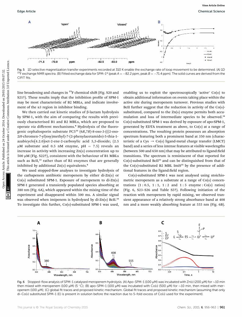
Fig. 5 1D selective magnetization transfer experiments recorded at 310 K enable the exchange rate of loop movement to be determined. (A) 1D19F exchangeNMR spectra. (B) Fitted exchange data for SPM-1* (peak A¼�82.2 ppm, peak B¼�71.4 ppm). The solid curves are derived from theCIFIT fits.
Edge Article Chemical Science
Ope
n A
cces
s A
rtic
le. P
ublis
hed
on 2
4 O
ctob
er 2
014.
Dow
nloa
ded
on 2
9/01
/201
5 09
:07:
32.
Thi
s ar
ticle
is li
cens
ed u
nder
a C
reat
ive
Com
mon
s A
ttrib
utio
n 3.
0 U
npor
ted
Lic
ence
.View Article Online
line broadening and changes in 19F chemical shi (Fig. S20 andS21†). These results imply that the inhibition prole of SPM-1may be most characteristic of B2 MBLs, and indicate involve-ment of the a3 region in inhibitor binding.
We then carried out kinetic studies of b-lactam hydrolysisby SPM-1, with the aim of comparing the results with previ-ously characterised B1 and B2 MBLs, which are proposed tooperate via different mechanisms.8 Hydrolysis of the uoro-genic cephalosporin substrate FC532 (6R,7R)-8-oxo-3-(((2-oxo-2H-chromen-7-yl)oxy)methyl)-7-(2-phenylacetamido)-5-thia-1-azabicyclo[4.2.0]oct-2-ene-2-carboxylic acid 5,5-dioxide; (2.5mM substrate and 0.5 nM enzyme, pH ¼ 7.5) reveals anincrease in activity with increasing Zn(II) concentration up to500 mM (Fig. S22†), consistent with the behaviour of B1 MBLssuch as BcII,33 rather than of B2 enzymes that are generallyinhibited by additional Zn(II) equivalents.8
We used stopped-ow analyses to investigate hydrolysis ofthe carbapenem antibiotic meropenem by either di-Zn(II) orCo(II) substituted SPM-1. Exposure of meropenem to di-Zn(II)SPM-1 generated a transiently populated species absorbing at380 nm (Fig. 6A), which appeared within the mixing time of theexperiment and disappeared within 500 ms. A similar signalwas observed when imipenem is hydrolysed by di-Zn(II) BcII.34
To investigate this further, Co(II)-substituted SPM-1 was used,
Fig. 6 Stopped-flow analysis of SPM-1 catalysedmeropenemhydrolysis.then mixed with meropenem (100 mM) (5 �C); (B) apo-SPM-1 (100 mM) wopenem (100 mM); (C) global fit traces and proposed kinetic mechanism.di-Co(II) substituted SPM-1 (E) is present in solution before the reaction
This journal is © The Royal Society of Chemistry 2015
enabling us to exploit the spectroscopically ‘active’ Co(II) toobtain additional information on events taking place within theactive site during meropenem turnover. Previous studies withBcII further suggest that the reduction in activity of the Co(II)-substituted, compared to the Zn(II) enzyme permits both accu-mulation and loss of intermediate species to be observed.34
Co(II)-substituted SPM-1 was derived by exposure of apo-SPM-1,generated by EDTA treatment as above, to Co(II) at a range ofconcentrations. The resulting protein possesses an absorptionspectrum featuring both a prominent band at 350 nm (charac-teristic of a Cys / Co(II) ligand-metal charge transfer (LMCT)band) and a series of less intense features at visible wavelengths(between 500 and 650 nm) that may be attributed to ligand:eldtransitions. The spectrum is reminiscent of that reported forCo(II)-substituted BcII33 and can be distinguished from that ofthe Co(II)-substituted B2 MBL ImiS35 by the presence of addi-tional features in the ligand:eld region.
Co(II)-substituted SPM-1 was next analysed using stoichio-metric meropenem as a substrate at a range of Co(II) concen-trations (1 : 0.5, 1 : 1, 1 : 2 and 1 : 5 enzyme : Co(II) ratios)(Fig. 6, S23–S26 and Table S5†). Following initiation of thereaction with meropenem by rapid mixing, we observed tran-sient appearance of a relatively strong absorbance band at 408nm and a more weakly absorbing feature at 555 nm (Fig. 6B).
(A) Apo-SPM-1 (100 mM) was incubatedwith Zn(II) (200 mM) for�10minas incubated with Co(II) (500 mM) for �10 min, then mixed with mer-Global fit traces and proposed kinetic mechanism (assuming that onlydue to 5-fold excess of Co(II) used for the experiment).
Chem. Sci., 2015, 6, 956–963 | 961

Chemical Science Edge Article
Ope
n A
cces
s A
rtic
le. P
ublis
hed
on 2
4 O
ctob
er 2
014.
Dow
nloa
ded
on 2
9/01
/201
5 09
:07:
32.
Thi
s ar
ticle
is li
cens
ed u
nder
a C
reat
ive
Com
mon
s A
ttrib
utio
n 3.
0 U
npor
ted
Lic
ence
.View Article Online
We propose these observations are analogous to the absorbancebands at 407 nm and 575 nm reported for the reaction of Co(II)complexes of the B1 MBL BcII with imipenem and mer-openem.34 Accumulation of the intermediate species wasdependent on Co(II) concentration and reached a maximum at aSPM-1 : Co(II) ratio of 1 : 2 (Fig. S23–S26 and Table S5†),implicating the di-Co(II) species in stabilisation of the putativeintermediate(s). Traces at 310, 408 and 555 nm were then ttedto evaluate the ability of a range of possible kinetic schemes todescribe the experimental observations (Tables S5 and S6†). Thets utilised data collected at excess Co(II) (1 : 5 SPM-1 : Co(II)ratio), simplifying the analysis by permitting the assumptionthat the reaction mixture contains only di-Co(II) SPM-1 and thatno other enzyme species is contributing to the reaction.
The best tting model (Fig. 6C and S26†) involves a branchedmechanism where a populated intermediate EI1, distinguishedby strong absorbance at 408 nm, can both yield product andequilibrate with an unproductive species EI2. Such a branchedmechanism is in broad agreement with previous proposals forthe reaction of Co(II)-substituted BcII with a range of b-lactamsubstrates,34,36 but differs substantially from the linear schemethat has been proposed to account for the reaction of the B2MBL ImiS with the carbapenem substrate imipenem.37 Weconclude that, at least for reaction of di-Co(II) enzyme withmeropenem, SPM-1 should be considered as mechanisticallyclose to B1, rather than B2, MBLs.
Conclusions
The combined results reveal that, in terms of its Zn(II) usage andcatalytic mechanism, SPM-1 is best categorized as a B1 MBL.However, in structural terms, and in particular with respect toits exible a3 region, SPM-1 has features typical of a B2 MBL.Interestingly, while SPM-1 is structurally unique amongstknown MBLs, the a3 region that it shares with B2 MBLs hasrecently been observed in other members of the MBL super-family, including an unusual type II glyoxylase from Salmonellaenterica38 and the MBL fold protein TTHA163 from the marinethermophile Thermus thermophilus HB8.39 The growing preva-lence of SPM-1 in carbapenem-resistant P. aeruginosa isolates inSouth America makes it one of the most clinically importantMBLs, yet its unusual structural properties distinguish it fromother B1 enzymes of equivalent medical relevance. In conse-quence, SPM-1 presents a unique challenge to inhibitordiscovery programmes, and its inclusion in such studies has thepotential to signicantly complicate the search for inhibitors ofall clinically signicant enzymes. The results presented here gosome way to identifying interaction sites, including a ‘hydro-phobic wall’ at the active site that may be exploited in SPM-1inhibitor discovery.
Acknowledgements
We thank the Medical Research Council (MRC)/Canadian GrantG1100135 for support of J.B. and C.J.S. and Cancer Research UK(CRUK) for support of S.S.v.B and C.J.S. We thank the DulvertonTrust (A.M.R.), the Engineering and Physical Sciences Research
962 | Chem. Sci., 2015, 6, 956–963
Council (EPSRC) (W.B.S.), a Clarendon–St Hugh's College-LoueyScholarship (H.T.), a Royal Society Dorothy Hodgkin Fellowship(E.F.) and a Royal Society University Research Fellow (J.L.P.B.).
References
1 W. Lee, M. A. McDonough, L. Kotra, Z. H. Li, N. R. Silvaggi,Y. Takeda, J. A. Kelly and S. Mobashery, Proc. Natl. Acad.Sci. U. S. A., 2001, 98, 1427–1431.
2 T. R. Walsh, M. A. Toleman, L. Poirel and P. Nordmann, Clin.Microbiol. Rev., 2005, 18, 306–325.
3 K. Bush and G. A. Jacoby, Antimicrob. Agents Chemother.,2010, 54, 969–976.
4 L. N. Andrade, N. Woodford and A. L. Darini, Int. J.Antimicrob. Agents, 2014, 43, 196–197.
5 A. E. Salabi, M. A. Toleman, J. Weeks, T. Bruderer, R. Frei andT. R. Walsh, Antimicrob. Agents Chemother., 2010, 54, 582.
6 G. Cornaglia, H. Giamarellou and G. M. Rossolini, LancetInfect. Dis., 2011, 11, 381–393.
7 M. Galleni, J. Lamotte-Brasseur, G. M. Rossolini, J. Spencer,O. Dideberg and J. M. Frere, Antimicrob. Agents Chemother.,2001, 45, 660–663.
8 A. I. Karsisiotis, C. F. Damblon and G. C. K. Roberts,Metallomics, 2014, 6, 1881–1197.
9 M. A. Toleman, A. M. Simm, T. A. Murphy, A. C. Gales,D. J. Biedenbach, R. N. Jones and T. R. Walsh, J.Antimicrob. Chemother., 2002, 50, 673–679.
10 M. Polotto, T. Casella, M. G. de Lucca Oliveira, F. G. Rubio,M. L. Nogueira, M. T. de Almeida and M. C. Nogueira,BMC Infect. Dis., 2012, 12, 176.
11 M. A. Toleman and T. R. Walsh, FEMS Microbiol. Rev., 2011,35, 912–935.
12 A. P. Zavascki, P. B. Gaspareto, A. F. Martins, A. L. Goncalvesand A. L. Barth, J. Antimicrob. Chemother., 2005, 56, 1148–1151.
13 T. A. Murphy, A. M. Simm, M. A. Toleman, R. N. Jones andT. R. Walsh, Antimicrob. Agents Chemother., 2003, 47, 582–587.
14 T. A. Murphy, L. E. Catto, S. E. Halford, A. T. Hadeld,W. Minor, T. R. Walsh and J. Spencer, J. Mol. Biol., 2006,357, 890–903.
15 C. Moali, C. Anne, J. Lamotte-Brasseur, S. Groslambert,B. Devreese, J. Van Beeumen, M. Galleni and J.-M. Frere,Chem. Biol., 2003, 10, 319–329.
16 H. Zhang and Q. Hao, FASEB J., 2011, 25, 2574–2582.17 G. Garau, C. Bebrone, C. Anne, M. Galleni, J. M. Frere and
O. Dideberg, J. Mol. Biol., 2005, 345, 785–795.18 C. Bebrone, C. Anne, F. Kerff, G. Garau, K. De Vriendt,
R. Lantin, B. Devreese, J. Van Beeumen, O. Dideberg,J. M. Frere and M. Galleni, Biochem. J., 2008, 414, 151–159.
19 N. Sharma, Z. Hu, M. W. Crowder and B. Bennett, J. Am.Chem. Soc., 2008, 130, 8215–8222.
20 J. H. Toney, G. G. Hammond, P. M. Fitzgerald, N. Sharma,J. M. Balkovec, G. P. Rouen, S. H. Olson, M. L. Hammond,M. L. Greenlee and Y. D. Gao, J. Biol. Chem., 2001, 276,31913–31918.
21 C. Bebrone, Biochem. Pharmacol., 2007, 74, 1686–1701.
This journal is © The Royal Society of Chemistry 2015

Edge Article Chemical Science
Ope
n A
cces
s A
rtic
le. P
ublis
hed
on 2
4 O
ctob
er 2
014.
Dow
nloa
ded
on 2
9/01
/201
5 09
:07:
32.
Thi
s ar
ticle
is li
cens
ed u
nder
a C
reat
ive
Com
mon
s A
ttrib
utio
n 3.
0 U
npor
ted
Lic
ence
.View Article Online
22 A. M. Davies, R. M. Rasia, A. J. Vila, B. J. Sutton andS. M. Fabiane, Biochemistry, 2005, 44, 4841–4849.
23 J. L. Benesch and B. T. Ruotolo, Curr. Opin. Struct. Biol., 2011,21, 641–649.
24 J. L. Benesch and C. V. Robinson, Curr. Opin. Struct. Biol.,2006, 16, 245–251.
25 A. M. Rydzik, J. Brem, S. S. van Berkel, I. Pfeffer, A. Makena,T. D. W. Claridge and C. J. Schoeld, Angew. Chem., Int. Ed.,2014, 53, 3129–3133.
26 A. D. Bain, Prog. Nucl. Magn. Reson. Spectrosc., 2003, 43, 63–103.27 A. D. Bain, Ann. Rep. NMR Spectrosc., 2008, 63, 23–48.28 A. D. Bain and J. A. Cramer, J. Magn. Reson., Ser. A, 1993, 103,
217–222.29 U. Heinz, R. Bauer, S. Wommer, W. Meyer-Klaucke,
C. Papamichaels, J. Bateson and H. W. Adolph, J. Biol.Chem., 2003, 278, 20659–20666.
30 B. M. Lienard, G. Garau, L. Horsfall, A. I. Karsisiotis,C. Damblon, P. Lassaux, C. Papamicael, G. C. Roberts,M. Galleni, O. Dideberg, J. M. Frere and C. J. Schoeld,Org. Biomol. Chem., 2008, 6, 2282–2294.
31 A. Makena, S. S. van Berkel, C. Lejeune, R. J. Owens,A. Verma, R. Salimraj, J. Spencer, J. Brem andC. J. Schoeld, ChemMedChem, 2013, 8, 1923–1929.
This journal is © The Royal Society of Chemistry 2015
32 S. S. van Berkel, J. Brem, A. M. Rydzik, R. Salimraj, R. Cain,A. Verma, R. J. Owens, C. W. G. Fishwick, J. Spencer andC. J. Schoeld, J. Med. Chem., 2013, 56, 6945–6953.
33 E. G. Orellano, J. E. Girardini, J. A. Cricco, E. A. Ceccarelliand A. J. Vila, Biochemistry, 1998, 37, 10173–10180.
34 M. F. Tioni, L. I. Llarrull, A. A. Poeylaut-Palena, M. A. Marti,M. Saggu, G. R. Periyannan, E. G. Mata, B. Bennett,D. H. Murgida and A. J. Vila, J. Am. Chem. Soc., 2008, 130,15852–15863.
35 P. A. Crawford, K. W. Yang, N. Sharma, B. Bennett andM. W. Crowder, Biochemistry, 2005, 44, 5168–5176.
36 R. Bicknell, E. L. Emanuel, J. Gagnon and S. G. Waley,Biochem. J., 1985, 229, 791–797.
37 N. P. Sharma, C. Hajdin, S. Chandrasekar, B. Bennett,K. W. Yang and M. W. Crowder, Biochemistry, 2006, 45,10729–10738.
38 A. L. Stamp, P. Owen, K. El Omari, C. E. Nichols, M. Lockyer,H. K. Lamb, I. G. Charles, A. R. Hawkins and D. K. Stammers,Protein Sci., 2010, 19, 1897–1905.
39 A. Yamamura, A. Okada, Y. Kameda, J. Ohtsuka,N. Nakagawa, A. Ebihara, K. Nagata and M. Tanokura, ActaCrystallogr., Sect. F: Struct. Biol. Cryst. Commun., 2009, 65,455–459.
Chem. Sci., 2015, 6, 956–963 | 963