staphylococcus aureus cell wall maintenance
TRANSCRIPT

FEMS Microbiology Reviews, 2022, 1–19
DOI: 10.1093/femsre/fuac025Advance access publication date: 8 June 2022
Review article
Staphylococcus aureus cell wall maintenance – themultifaceted roles of peptidoglycan hydrolases inbacterial growth, fitness, and virulenceMin Wang, Girbe Buist†, Jan Maarten van Dijl*†
Department of Medical Microbiology, University of Groningen, University Medical Center Groningen, Hanzeplein 1, PO Box 30001, 9700 RB Groningen, theNetherlands∗Corresponding author: Department of Medical Microbiology, University of Groningen, University Medical Center Groningen, Hanzeplein 1, P.O. box 30001, HPCEB80, 9700 RB Groningen, the Netherlands, Tel. +31-50-3615187; Fax. +31-50-3619105; E-mail: [email protected] sentence summary: Here, we review the roles of peptidoglycan hydrolases of the Gram-positive bacterial pathogen Staphylococcus aureus in bacterial growthand division, cell wall maintenance, protein localization, pathogenesis and antimicrobial susceptibility.Editor: Aimee Shen†equal contributions‡Jan Maarten van Dijl, https://orcid.org/0000-0002-5688-8438
Abstract
Staphylococcus aureus is an important human and livestock pathogen that is well-protected against environmental insults by a thickcell wall. Accordingly, the wall is a major target of present-day antimicrobial therapy. Unfortunately, S. aureus has mastered the artof antimicrobial resistance, as underscored by the global spread of methicillin-resistant S. aureus (MRSA). The major cell wall compo-nent is peptidoglycan. Importantly, the peptidoglycan network is not only vital for cell wall function, but it also represents a bacterialAchilles’ heel. In particular, this network is continuously opened by no less than 18 different peptidoglycan hydrolases (PGHs) en-coded by the S. aureus core genome, which facilitate bacterial growth and division. This focuses attention on the specific functionsexecuted by these enzymes, their subcellular localization, their control at the transcriptional and post-transcriptional levels, theircontributions to staphylococcal virulence and their overall importance in bacterial homeostasis. As highlighted in the present review,our understanding of the different aspects of PGH function in S. aureus has been substantially increased over recent years. This isimportant because it opens up new possibilities to exploit PGHs as innovative targets for next-generation antimicrobials, passive oractive immunization strategies, or even to engineer them into effective antimicrobial agents.
Keywords: Staphylococcus aureus, peptidoglycan hydrolase, cell wall, subcellular protein localization, pathogenesis, antimicrobial sus-ceptibility
IntroductionStaphylococcus aureus is a small and spherical Gram-positive bac-terium (Fig. 1), belonging to the phylum of the Firmicutes. As acommensal, S. aureus frequently colonizes asymptomatically thenares, skin and gut of humans (Mainous et al. 2006, Raineri, Al-tulea and van Dijl 2022). Nonetheless, S. aureus is also one of themost renowned opportunistic pathogens, causing many differentdiseases that range from relatively mild skin and soft tissue in-fections to life-threatening diseases, such as bacteremia, pneu-monia and endocarditis. The invasive behavior of S. aureus is of-ten triggered through injury or medical interventions, where thebarrier function of the host cells or tissues is compromised. How-ever, S. aureus also produces a variety of virulence factors thatallow this bacterium to invade the human body. Beyond the ep-ithelial or endothelial barriers, S. aureus is able to bind to the ex-tracellular matrix of host cells and tissues, to invade host cells,or to form biofilms on tissues and medical implants (Raineri, Al-tulea and van Dijl 2022). At the same time, it is able to effectivelyevade innate and adaptive immune defenses of the host, eitherthrough the secretion of particular virulence factors or the forma-tion of biofilms (Thammavongsa et al. 2015, Goldmann and Med-
ina 2018). Ultimately, this may result in persistent or chronic infec-tions (Josse et al. 2017). In recent years, the treatment of S. aureusinfections has become increasingly difficult due to the fact thatthis pathogen has acquired many different antibiotic resistances,as critically exemplified by the emergence of methicillin-resistantS. aureus (MRSA) lineages in hospitals and the community (Turneret al. 2019).
Gram-positive bacteria, like S. aureus, are surrounded by a thickcell wall, which plays vital roles in maintaining cell shape, cellintegrity, cell viability and the protection against osmotic stressunder different environmental conditions (Vollmer et al. 2008, Sil-havy et al. 2010). The structure and composition of the cell wallvaries between different Gram-positive bacterial species (Do et al.2020). However, peptidoglycan is invariably the major cell wallcomponent that surrounds the bacterial cytoplasmic membrane(Turner et al. 2014). S. aureus peptidoglycan contains relativelyshort glycan strands that are highly cross-linked through species-specific peptide bridges; 80%–90% of the so-called stem peptidesare cross-linked via a pentaglycine bridge (Snowden and Perkins1990, Monteiro et al. 2019, Sobral and Tomasz 2019). These cross-links are important for the integrity and physical strength of the
Received: December 8, 2021. Accepted: May 25, 2022C© The Author(s) 2022. Published by Oxford University Press on behalf of FEMS. This is an Open Access article distributed under the terms of the CreativeCommons Attribution-NonCommercial License (https://creativecommons.org/licenses/by-nc/4.0/), which permits non-commercial re-use, distribution, andreproduction in any medium, provided the original work is properly cited. For commercial re-use, please contact [email protected]
Dow
nloaded from https://academ
ic.oup.com/fem
sre/advance-article/doi/10.1093/femsre/fuac025/6604383 by guest on 05 July 2022
2 | FEMS Microbiology Reviews, 2022
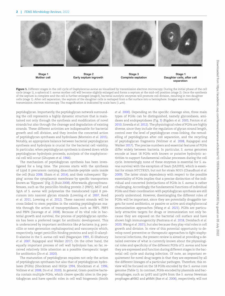
Figure 1. Different stages in the cell cycle of Staphylococcus aureus as visualized by transmission electron microscopy. During the initial phase of the cellcycle (stage 1), a spherical S. aureus mother cell will become slightly enlarged and forms a septum at the mid-cell position (stage 2). Once the synthesisof the septum is complete and the cell is further enlarged (stage3), bacterial autolytic enzymes will promote cell division, resulting in two daughtercells (stage 1). After cell separation, the septum of the daughter cells is reshaped from a flat surface into a hemisphere. Images were recorded bytransmission electron microscopy. The magnification is indicated by scale bars (1 μm).
peptidoglycan. Importantly, the peptidoglycan network surround-ing the cell represents a highly dynamic structure that is main-tained not only through the synthesis and modification of novelstrands but also through the cleavage and degradation of existingstrands. These different activities are indispensable for bacterialgrowth and cell division, and they involve the concerted actionof peptidoglycan synthases and hydrolases (Monteiro et al. 2015).Notably, an appropriate balance between bacterial peptidoglycansynthesis and hydrolysis is crucial for the bacterial cell viability.In particular, when peptidoglycan synthesis is slowed down whilepeptidoglycan hydrolysis proceeds, autolysis of the staphylococ-cal cell will occur (Ghuysen et al. 1966).
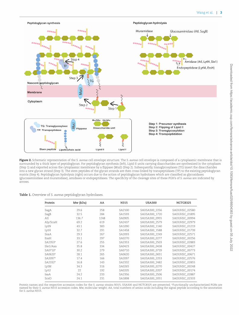
The mechanism of peptidoglycan synthesis has been inves-tigated for a long time. The process starts with the synthesisof Lipid II precursors carrying disaccharide-peptide units insidethe cell (Ruiz 2008, Sham et al. 2014), and their subsequent ‘flip-ping’ across the cytoplasmic membrane by specific transportersknown as ‘flippases’ (Fig. 2) (Ruiz 2008). Afterwards, glycosyltrans-ferases, such as the penicillin-binding protein 2 (PBP2), MGT andSgtA of S. aureus will polymerize the translocated Lipid II pre-cursors into nascent glycan strands (Lovering et al. 2007, Reedet al. 2011, Lovering et al. 2012). These nascent strands will becross-linked to stem peptides in the existing peptidoglycan ma-trix through the action of transpeptidases, such as PBP1, PBP3and PBP4 (Sauvage et al. 2008). Because of its vital role in bac-terial growth and survival, the process of peptidoglycan synthe-sis has been a preferred target for antibiotic development. Thisis underscored by important antibiotics like β-lactams (e.g. peni-cillin or next-generation cephalosporins) and vancomycin which,respectively, target penicillin-binding proteins and acyl-D-alanyl-D-alanine in the S. aureus cell wall (Nagarajan 1991, El-Shabouryet al. 2007, Rajagopal and Walker 2017). On the other hand, theequally important process of cell wall hydrolysis has, so far, re-ceived relatively little attention as a possible therapeutic targetfor antibiotics (Do et al. 2020).
The maturation of peptidoglycan requires not only the actionof peptidoglycan synthases but also that of peptidoglycan hydro-lases (PGHs) (Shockman and Höltje 1994, Shockman et al. 1996,Vollmer et al. 2008, Do et al. 2020). In general, Gram-positive bacte-ria contain multiple PGHs, which cleave specific sites in the pep-tidoglycan and have specific roles in cell wall biogenesis (Smith
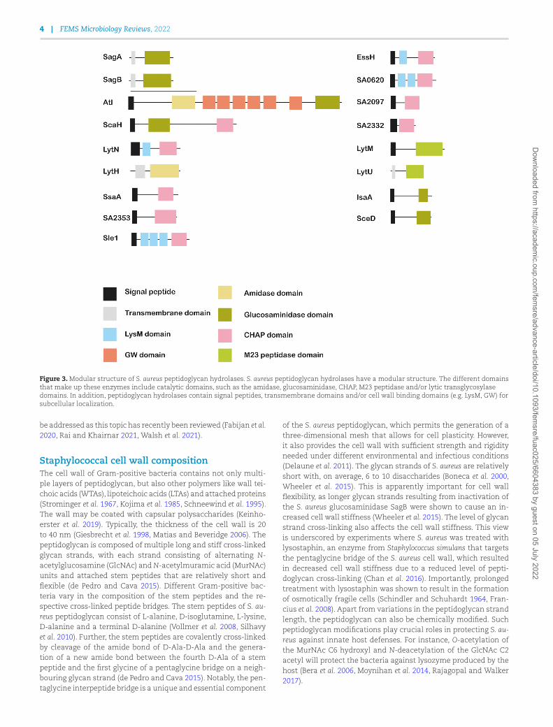
et al. 2000). Depending on the specific cleavage sites, three maintypes of PGHs can be distinguished, namely glycosidases, ami-dases and endopeptidases (Fig. 3) (Rigden et al. 2003, Fenton et al.2010, Szweda et al. 2012). The physiological roles of PGHs are highlydiverse, since they include the regulation of glycan strand length,control over the level of peptidoglycan cross-linking, the remod-elling of peptidoglycan after cell separation, and the recyclingof peptidoglycan fragments (Vollmer et al. 2008, Rajagopal andWalker 2017). The precise numbers and essential features of PGHsdiffer widely between bacteria. In particular, S. aureus genomesencode at least 18 PGHs with known or putative hydrolytic ac-tivities to support fundamental cellular processes during the cellcycle. Interestingly, none of these enzymes is essential for S. au-reus survival with the exception of SsaA (SA2093), which is essen-tial for strain NTCT8325, but not for strain N315 (Chaudhuri et al.2009). The latter strain dependency with respect to the possibleessentiality of PGHs implies that a precise distinction of the indi-vidual and concerted (inter)actions of PGHs in S. aureus is ratherchallenging. Accordingly, the fundamental functions of individualPGHs and their coordination with peptidoglycan synthesis are stillpoorly understood. However, disentangling the different roles ofPGHs will be important, since they are potentially druggable tar-gets for novel antibiotics, or passive or active anti-staphylococcalimmunization approaches (Wang et al. 2021). PGHs are particu-larly attractive targets for drugs or immunization not only be-cause they are exposed on the bacterial cell surface and haveshown high immunogenicity (Pastrana et al. 2018, Dreisbach et al.2020, Wang et al. 2021), but also because they play vital roles in cellgrowth and division. In view of this potential opportunity to de-velop novel preventive or therapeutic approaches to fight staphy-lococcal infections, the present review is aimed at providing a de-tailed overview of what is currently known about the physiologi-cal roles and specificity of the different PGHs of S. aureus and howthey are expressed and function during different stages in the bac-terial cell cycle and during infection. Of note, an important re-quirement for novel drug targets is that they are expressed by allthe different lineages of a particular pathogen. Therefore, this re-view will be focused on the 18 PGHs encoded by the S. aureus coregenome (Table 1). In contrast, PGHs encoded by plasmids and bac-teriophages, such as LytP2 and LytP4 from the S. aureus Newmanprophages φNM2 and φNM4 (Bae et al. 2006), respectively, will not
Dow
nloaded from https://academ
ic.oup.com/fem
sre/advance-article/doi/10.1093/femsre/fuac025/6604383 by guest on 05 July 2022
Wang et al. | 3
Figure 2. Schematic representation of the S. aureus cell envelope structure. The S. aureus cell envelope is composed of a cytoplasmic membrane that issurrounded by a thick layer of peptidoglycan. For peptidoglycan synthesis (left), Lipid II units carrying disaccharides are synthesized in the cytoplasm(Step 1) and exported across the cytoplasmic membrane by a flippase (MurJ) (Step 2). Subsequently, transglycosylases (TG) insert the disaccharidesinto a new glycan strand (Step 3). The stem peptides of the glycan strands are then cross-linked by transpeptidases (TP) to the existing peptidoglycanmatrix (Step 4). Peptidoglycan hydrolysis (right) occurs due to the action of peptidoglycan hydrolases which are classified as glycosidases(glucosaminidase and muramidase), amidases or endopeptidases. The specificity of the cleavage sites of these PGH’s of S. aureus are indicated byarrows.
Table 1. Overview of S. aureus peptidoglycan hydrolases.
Protein Mw (kDa) AA N315 USA300 NCTC8325
SagA 29.6 258 SA2100 SAUSA300_2256 SAOUHSC_02580SagB 32.5 284 SA1593 SAUSA300_1720 SAOUHSC_01895Atl 136.7 1248 SA0905 SAUSA300_0955 SAOUHSC_00994Aly/ScaH 69.2 619 SA2437 SAUSA300_2579 SAOUHSC_02979LytN 43.1 383 SA1090 SAUSA300_1140 SAOUHSC_01219LytH 32.7 291 SA1458 SAUSA300_1588 SAOUHSC_01739SsaA 29.3 267 SA2093 SAUSA300_2249 SAOUHSC_02571EssH 33.1 297 SA0270 SAUSA300_0277 SAOUHSC_00256SA2353∗ 27.6 255 SA2353 SAUSA300_2503 SAOUHSC_02883Sle1/Aaa 35.8 334 SA0423 SAUSA300_0438 SAOUHSC_00427SA0710∗ 30.2 279 SA0710 SAUSA300_0739 SAOUHSC_00773SA0620∗ 28.1 265 SA0620 SAUSA300_0651 SAOUHSC_00671SA2097∗ 17.4 166 SA2097 SAUSA300_2253 SAOUHSC_02576SA2332∗ 16.8 143 SA2332 SAUSA300_2482 SAOUHSC_02855LytM 34.3 316 SA0265 SAUSA300_0270 SAOUHSC_00248LytU 22 192 SA0205 SAUSA300_0207 SAOUHSC_00174IsaA 24.2 233 SA2356 SAUSA300_2506 SAOUHSC_02887SceD 24.1 231 SA1898 SAUSA300_2051 SAOUHSC_02333
Protein names and the respective accession codes for the S. aureus strains N315, USA300 and NCTC8325 are presented. ∗Functionally uncharacterized PGHs arenamed by their S. aureus N315 accession codes. Mw, molecular weight; AA, total numbers of amino acids including the signal peptide according to the annotationfor S. aureus N315.
Dow
nloaded from https://academ
ic.oup.com/fem
sre/advance-article/doi/10.1093/femsre/fuac025/6604383 by guest on 05 July 2022
4 | FEMS Microbiology Reviews, 2022
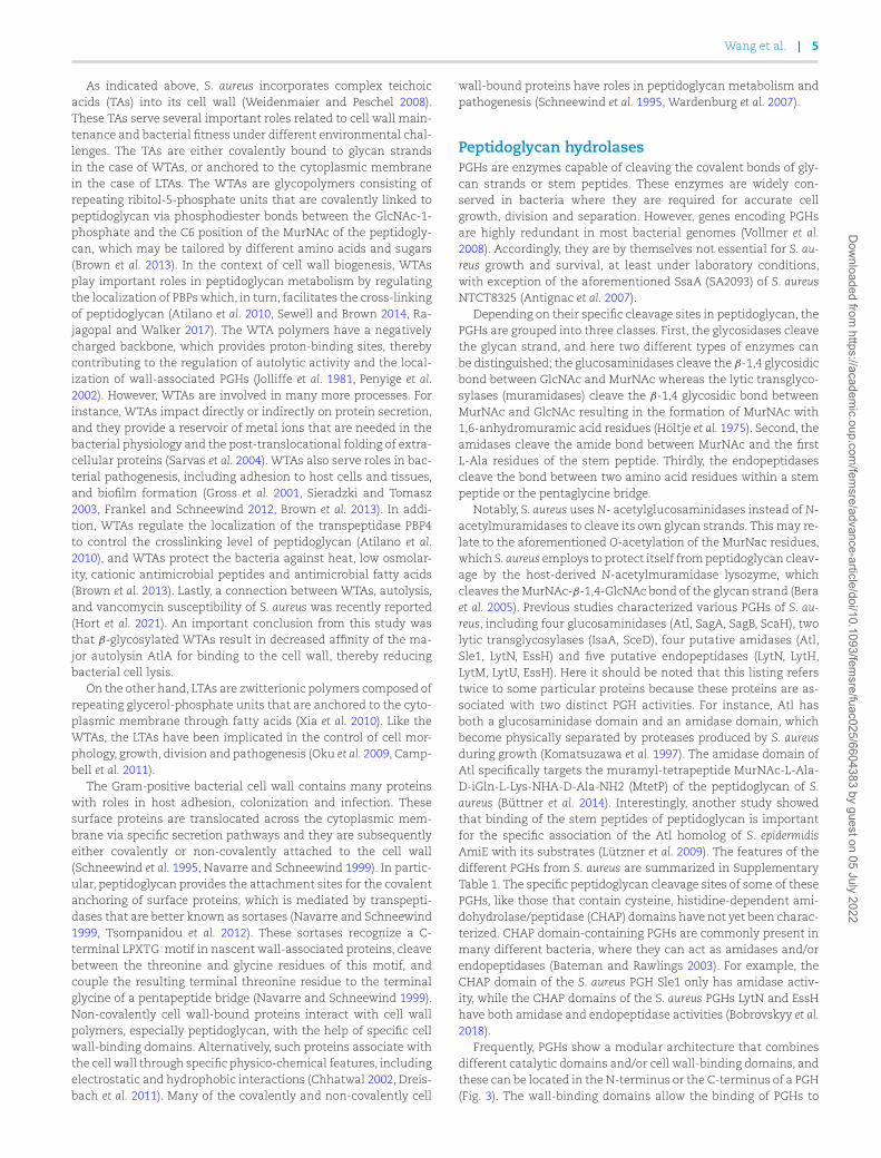
Figure 3. Modular structure of S. aureus peptidoglycan hydrolases. S. aureus peptidoglycan hydrolases have a modular structure. The different domainsthat make up these enzymes include catalytic domains, such as the amidase, glucosaminidase, CHAP, M23 peptidase and/or lytic transglycosylasedomains. In addition, peptidoglycan hydrolases contain signal peptides, transmembrane domains and/or cell wall binding domains (e.g. LysM, GW) forsubcellular localization.
be addressed as this topic has recently been reviewed (Fabijan et al.2020, Rai and Khairnar 2021, Walsh et al. 2021).
Staphylococcal cell wall compositionThe cell wall of Gram-positive bacteria contains not only multi-ple layers of peptidoglycan, but also other polymers like wall tei-choic acids (WTAs), lipoteichoic acids (LTAs) and attached proteins(Strominger et al. 1967, Kojima et al. 1985, Schneewind et al. 1995).The wall may be coated with capsular polysaccharides (Keinho-erster et al. 2019). Typically, the thickness of the cell wall is 20to 40 nm (Giesbrecht et al. 1998, Matias and Beveridge 2006). Thepeptidoglycan is composed of multiple long and stiff cross-linkedglycan strands, with each strand consisting of alternating N-acetylglucosamine (GlcNAc) and N-acetylmuramic acid (MurNAc)units and attached stem peptides that are relatively short andflexible (de Pedro and Cava 2015). Different Gram-positive bac-teria vary in the composition of the stem peptides and the re-spective cross-linked peptide bridges. The stem peptides of S. au-reus peptidoglycan consist of L-alanine, D-isoglutamine, L-lysine,D-alanine and a terminal D-alanine (Vollmer et al. 2008, Silhavyet al. 2010). Further, the stem peptides are covalently cross-linkedby cleavage of the amide bond of D-Ala-D-Ala and the genera-tion of a new amide bond between the fourth D-Ala of a stempeptide and the first glycine of a pentaglycine bridge on a neigh-bouring glycan strand (de Pedro and Cava 2015). Notably, the pen-taglycine interpeptide bridge is a unique and essential component
of the S. aureus peptidoglycan, which permits the generation of athree-dimensional mesh that allows for cell plasticity. However,it also provides the cell wall with sufficient strength and rigidityneeded under different environmental and infectious conditions(Delaune et al. 2011). The glycan strands of S. aureus are relativelyshort with, on average, 6 to 10 disaccharides (Boneca et al. 2000,Wheeler et al. 2015). This is apparently important for cell wallflexibility, as longer glycan strands resulting from inactivation ofthe S. aureus glucosaminidase SagB were shown to cause an in-creased cell wall stiffness (Wheeler et al. 2015). The level of glycanstrand cross-linking also affects the cell wall stiffness. This viewis underscored by experiments where S. aureus was treated withlysostaphin, an enzyme from Staphylococcus simulans that targetsthe pentaglycine bridge of the S. aureus cell wall, which resultedin decreased cell wall stiffness due to a reduced level of pepti-doglycan cross-linking (Chan et al. 2016). Importantly, prolongedtreatment with lysostaphin was shown to result in the formationof osmotically fragile cells (Schindler and Schuhardt 1964, Fran-cius et al. 2008). Apart from variations in the peptidoglycan strandlength, the peptidoglycan can also be chemically modified. Suchpeptidoglycan modifications play crucial roles in protecting S. au-reus against innate host defenses. For instance, O-acetylation ofthe MurNAc C6 hydroxyl and N-deacetylation of the GlcNAc C2acetyl will protect the bacteria against lysozyme produced by thehost (Bera et al. 2006, Moynihan et al. 2014, Rajagopal and Walker2017).
Dow
nloaded from https://academ
ic.oup.com/fem
sre/advance-article/doi/10.1093/femsre/fuac025/6604383 by guest on 05 July 2022
Wang et al. | 5
As indicated above, S. aureus incorporates complex teichoicacids (TAs) into its cell wall (Weidenmaier and Peschel 2008).These TAs serve several important roles related to cell wall main-tenance and bacterial fitness under different environmental chal-lenges. The TAs are either covalently bound to glycan strandsin the case of WTAs, or anchored to the cytoplasmic membranein the case of LTAs. The WTAs are glycopolymers consisting ofrepeating ribitol-5-phosphate units that are covalently linked topeptidoglycan via phosphodiester bonds between the GlcNAc-1-phosphate and the C6 position of the MurNAc of the peptidogly-can, which may be tailored by different amino acids and sugars(Brown et al. 2013). In the context of cell wall biogenesis, WTAsplay important roles in peptidoglycan metabolism by regulatingthe localization of PBPs which, in turn, facilitates the cross-linkingof peptidoglycan (Atilano et al. 2010, Sewell and Brown 2014, Ra-jagopal and Walker 2017). The WTA polymers have a negativelycharged backbone, which provides proton-binding sites, therebycontributing to the regulation of autolytic activity and the local-ization of wall-associated PGHs (Jolliffe et al. 1981, Penyige et al.2002). However, WTAs are involved in many more processes. Forinstance, WTAs impact directly or indirectly on protein secretion,and they provide a reservoir of metal ions that are needed in thebacterial physiology and the post-translocational folding of extra-cellular proteins (Sarvas et al. 2004). WTAs also serve roles in bac-terial pathogenesis, including adhesion to host cells and tissues,and biofilm formation (Gross et al. 2001, Sieradzki and Tomasz2003, Frankel and Schneewind 2012, Brown et al. 2013). In addi-tion, WTAs regulate the localization of the transpeptidase PBP4to control the crosslinking level of peptidoglycan (Atilano et al.2010), and WTAs protect the bacteria against heat, low osmolar-ity, cationic antimicrobial peptides and antimicrobial fatty acids(Brown et al. 2013). Lastly, a connection between WTAs, autolysis,and vancomycin susceptibility of S. aureus was recently reported(Hort et al. 2021). An important conclusion from this study wasthat β-glycosylated WTAs result in decreased affinity of the ma-jor autolysin AtlA for binding to the cell wall, thereby reducingbacterial cell lysis.
On the other hand, LTAs are zwitterionic polymers composed ofrepeating glycerol-phosphate units that are anchored to the cyto-plasmic membrane through fatty acids (Xia et al. 2010). Like theWTAs, the LTAs have been implicated in the control of cell mor-phology, growth, division and pathogenesis (Oku et al. 2009, Camp-bell et al. 2011).
The Gram-positive bacterial cell wall contains many proteinswith roles in host adhesion, colonization and infection. Thesesurface proteins are translocated across the cytoplasmic mem-brane via specific secretion pathways and they are subsequentlyeither covalently or non-covalently attached to the cell wall(Schneewind et al. 1995, Navarre and Schneewind 1999). In partic-ular, peptidoglycan provides the attachment sites for the covalentanchoring of surface proteins, which is mediated by transpepti-dases that are better known as sortases (Navarre and Schneewind1999, Tsompanidou et al. 2012). These sortases recognize a C-terminal LPXTG motif in nascent wall-associated proteins, cleavebetween the threonine and glycine residues of this motif, andcouple the resulting terminal threonine residue to the terminalglycine of a pentapeptide bridge (Navarre and Schneewind 1999).Non-covalently cell wall-bound proteins interact with cell wallpolymers, especially peptidoglycan, with the help of specific cellwall-binding domains. Alternatively, such proteins associate withthe cell wall through specific physico-chemical features, includingelectrostatic and hydrophobic interactions (Chhatwal 2002, Dreis-bach et al. 2011). Many of the covalently and non-covalently cell
wall-bound proteins have roles in peptidoglycan metabolism andpathogenesis (Schneewind et al. 1995, Wardenburg et al. 2007).
Peptidoglycan hydrolasesPGHs are enzymes capable of cleaving the covalent bonds of gly-can strands or stem peptides. These enzymes are widely con-served in bacteria where they are required for accurate cellgrowth, division and separation. However, genes encoding PGHsare highly redundant in most bacterial genomes (Vollmer et al.2008). Accordingly, they are by themselves not essential for S. au-reus growth and survival, at least under laboratory conditions,with exception of the aforementioned SsaA (SA2093) of S. aureusNTCT8325 (Antignac et al. 2007).
Depending on their specific cleavage sites in peptidoglycan, thePGHs are grouped into three classes. First, the glycosidases cleavethe glycan strand, and here two different types of enzymes canbe distinguished; the glucosaminidases cleave the β-1,4 glycosidicbond between GlcNAc and MurNAc whereas the lytic transglyco-sylases (muramidases) cleave the β-1,4 glycosidic bond betweenMurNAc and GlcNAc resulting in the formation of MurNAc with1,6-anhydromuramic acid residues (Höltje et al. 1975). Second, theamidases cleave the amide bond between MurNAc and the firstL-Ala residues of the stem peptide. Thirdly, the endopeptidasescleave the bond between two amino acid residues within a stempeptide or the pentaglycine bridge.
Notably, S. aureus uses N- acetylglucosaminidases instead of N-acetylmuramidases to cleave its own glycan strands. This may re-late to the aforementioned O-acetylation of the MurNac residues,which S. aureus employs to protect itself from peptidoglycan cleav-age by the host-derived N-acetylmuramidase lysozyme, whichcleaves the MurNAc-β-1,4-GlcNAc bond of the glycan strand (Beraet al. 2005). Previous studies characterized various PGHs of S. au-reus, including four glucosaminidases (Atl, SagA, SagB, ScaH), twolytic transglycosylases (IsaA, SceD), four putative amidases (Atl,Sle1, LytN, EssH) and five putative endopeptidases (LytN, LytH,LytM, LytU, EssH). Here it should be noted that this listing referstwice to some particular proteins because these proteins are as-sociated with two distinct PGH activities. For instance, Atl hasboth a glucosaminidase domain and an amidase domain, whichbecome physically separated by proteases produced by S. aureusduring growth (Komatsuzawa et al. 1997). The amidase domain ofAtl specifically targets the muramyl-tetrapeptide MurNAc-L-Ala-D-iGln-L-Lys-NHA-D-Ala-NH2 (MtetP) of the peptidoglycan of S.aureus (Büttner et al. 2014). Interestingly, another study showedthat binding of the stem peptides of peptidoglycan is importantfor the specific association of the Atl homolog of S. epidermidisAmiE with its substrates (Lützner et al. 2009). The features of thedifferent PGHs from S. aureus are summarized in SupplementaryTable 1. The specific peptidoglycan cleavage sites of some of thesePGHs, like those that contain cysteine, histidine-dependent ami-dohydrolase/peptidase (CHAP) domains have not yet been charac-terized. CHAP domain-containing PGHs are commonly present inmany different bacteria, where they can act as amidases and/orendopeptidases (Bateman and Rawlings 2003). For example, theCHAP domain of the S. aureus PGH Sle1 only has amidase activ-ity, while the CHAP domains of the S. aureus PGHs LytN and EssHhave both amidase and endopeptidase activities (Bobrovskyy et al.2018).
Frequently, PGHs show a modular architecture that combinesdifferent catalytic domains and/or cell wall-binding domains, andthese can be located in the N-terminus or the C-terminus of a PGH(Fig. 3). The wall-binding domains allow the binding of PGHs to
Dow
nloaded from https://academ
ic.oup.com/fem
sre/advance-article/doi/10.1093/femsre/fuac025/6604383 by guest on 05 July 2022
6 | FEMS Microbiology Reviews, 2022
the peptidoglycan at an adequate concentration. They also prop-erly position the respective PGH active sites towards the PG sub-strate cleavage site for efficient formation of enzyme-substratecomplexes (Vermassen et al. 2019). The type and number of cellwall-binding domains differ among PGHs (Steen et al. 2005). Thisis exemplified by the cell wall-binding Lysin Motif (LysM), whichis widely distributed in both prokaryotes and eukaryotes (Buistet al. 2008). Bacterial LysM domains recognize and non-covalentlybind to the N-acetylglucosamine moiety of peptidoglycan (Frankeland Schneewind 2012). LysM domains also direct the mureinhydrolases Sle1 and LytN to the cross wall of S. aureus, wherethey assume different positions (Frankel et al. 2011, Frankel andSchneewind 2012, Pastrana et al. 2017). LytN contains a YSIRK/GSsignal which also directs this protein to the septum of dividingcells (Frankel et al. 2011). SH3b, another cell wall-binding domain,targets proteins to the pentaglycine bridge with high affinity andspecificity, and SH3b-mediated peptidoglycan binding is actuallya key step in peptidoglycan hydrolysis (Visweswaran et al. 2014,Gonzalez-Delgado et al. 2020). The GW domain of Atl is a repeat-ing cell wall-binding domain that binds to LTA (Sugai et al. 1995,Baba and Schneewind 1998, Zoll et al. 2012), which is responsiblefor the correct protein docking (Komatsuzawa et al. 1997, Frankeland Schneewind 2012, Zoll et al. 2012). Some PGHs are capable ofcleaving cross-links of soluble muropeptides and intact peptido-glycan sacculi. For example, mutanolysin from Streptomyces glo-bisporus cleaves the MurNAc-GlcNAc that carries a stem peptide(Kawata et al. 1983). In contrast, the glucosaminidase domain ofAtl specifically cleaves the glycan strand without attached stempeptides (Nega et al. 2020). Compared to Atl, the glucosaminidaseSagB prefers longer glycan strands as substrates which, in fact,implies an important role for SagB in controlling the glycan strandlength (Wheeler et al. 2015, Chan et al. 2016). The amidase LytHexclusively cleaves peptides from non-cross-linked nascent pep-tidoglycan (Do et al. 2020).
Altogether, studies on the PGHs of S. aureus have shown thatthese enzymes are exported proteins that localize either to thebacterial septum (Sle1 [Frankel and Schneewind 2012], Atl [Schlaget al. 2010], LytN [Frankel et al. 2011], IsaA [Sakata et al. 2005])or the peripheral wall region (LytM [Ramadurai et al. 1999] andLytU [Raulinaitis et al. 2017]) during cell growth and division. SomePGHs, like Atl and Sle1, are also secreted into the extracellu-lar milieu and, subsequently, these PGHs cleave the septum atthe cross wall (Komatsuzawa et al. 1997, Frankel and Schneewind2012). Moreover, PGHs may even contain both a membrane an-chor and an extracytoplasmic catalytic domain, as exemplified byLytU, LytH, SagA, and SagB. Their positioning close to the extra-cytoplasmic side of the membrane suggests that the latter fourenzymes preferentially target nascent peptidoglycan.
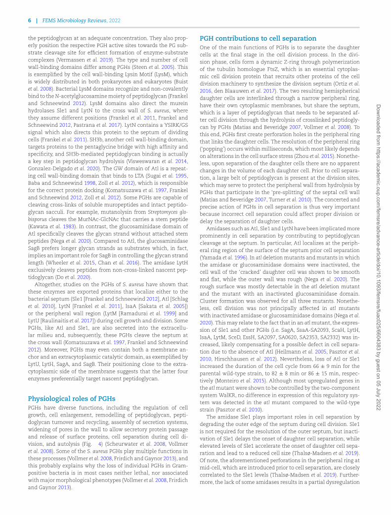
Physiological roles of PGHsPGHs have diverse functions, including the regulation of cellgrowth, cell enlargement, remodelling of peptidoglycan, pepti-doglycan turnover and recycling, assembly of secretion systems,widening of pores in the wall to allow secretory protein passageand release of surface proteins, cell separation during cell di-vision, and autolysis (Fig. 4) (Scheurwater et al. 2008, Vollmeret al. 2008). Some of the S. aureus PGHs play multiple functions inthese processes (Vollmer et al. 2008, Frirdich and Gaynor 2013), andthis probably explains why the loss of individual PGHs in Gram-positive bacteria is in most cases neither lethal, nor associatedwith major morphological phenotypes (Vollmer et al. 2008, Frirdichand Gaynor 2013).
PGH contributions to cell separationOne of the main functions of PGHs is to separate the daughtercells at the final stage in the cell division process. In the divi-sion phase, cells form a dynamic Z-ring through polymerizationof the tubulin homologue FtsZ, which is an essential cytoplas-mic cell division protein that recruits other proteins of the celldivision machinery to synthesize the division septum (Ortiz et al.2016, den Blaauwen et al. 2017). The two resulting hemisphericaldaughter cells are interlinked through a narrow peripheral ring,have their own cytoplasmic membranes, but share the septum,which is a layer of peptidoglycan that needs to be separated af-ter cell division through the hydrolysis of crosslinked peptidogly-can by PGHs (Matias and Beveridge 2007, Vollmer et al. 2008). Tothis end, PGHs first create perforation holes in the peripheral ringthat links the daughter cells. The resolution of the peripheral ring(‘popping’) occurs within milliseconds, which most likely dependson alterations in the cell surface stress (Zhou et al. 2015). Nonethe-less, upon separation of the daughter cells there are no apparentchanges in the volume of each daughter cell. Prior to cell separa-tion, a large belt of peptidoglycan is present at the division sites,which may serve to protect the peripheral wall from hydrolysis byPGHs that participate in the ‘pre-splitting’ of the septal cell wall(Matias and Beveridge 2007, Turner et al. 2010). The concerted andprecise action of PGHs in cell separation is thus very importantbecause incorrect cell separation could affect proper division ordelay the separation of daughter cells.
Amidases such as Atl, Sle1 and LytN have been implicated moreprominently in cell separation by contributing to peptidoglycancleavage at the septum. In particular, Atl localizes at the periph-eral ring region of the surface of the septum prior cell separation(Yamada et al. 1996). In atl deletion mutants and mutants in whichthe amidase or glucosaminidase domains were inactivated, thecell wall of the ‘cracked’ daughter cell was shown to be smoothand flat, while the outer wall was rough (Nega et al. 2020). Therough surface was mostly detectable in the atl deletion mutantand the mutant with an inactivated glucosaminidase domain.Cluster formation was observed for all three mutants. Nonethe-less, cell division was not principally affected in atl mutantswith inactivated amidase or glucosaminidase domains (Nega et al.2020). This may relate to the fact that in an atl mutant, the expres-sion of Sle1 and other PGHs (i.e. SagA, SsaA-SA2093, ScaH, LytH,IsaA, LytM, SceD, EssH, SA2097, SA0620, SA2353, SA2332) was in-creased, likely compensating for a possible defect in cell separa-tion due to the absence of Atl (Heilmann et al. 2005, Pasztor et al.2010, Hirschhausen et al. 2012). Nevertheless, loss of Atl or Sle1increased the duration of the cell cycle from 66 ± 9 min for theparental wild-type strain, to 82 ± 8 min or 86 ± 15 min, respec-tively (Monteiro et al. 2015). Although most upregulated genes inthe atl mutant were shown to be controlled by the two-componentsystem WalKR, no difference in expression of this regulatory sys-tem was detected in the atl mutant compared to the wild-typestrain (Pasztor et al. 2010).
The amidase Sle1 plays important roles in cell separation bydegrading the outer edge of the septum during cell division. Sle1is not required for the resolution of the outer septum, but inacti-vation of Sle1 delays the onset of daughter cell separation, whileelevated levels of Sle1 accelerate the onset of daughter cell sepa-ration and lead to a reduced cell size (Thalsø-Madsen et al. 2019).Of note, the aforementioned perforations in the peripheral ring atmid-cell, which are introduced prior to cell separation, are closelycorrelated to the Sle1 levels (Thalsø-Madsen et al. 2019). Further-more, the lack of some amidases results in a partial dysregulation
Dow
nloaded from https://academ
ic.oup.com/fem
sre/advance-article/doi/10.1093/femsre/fuac025/6604383 by guest on 05 July 2022
Wang et al. | 7
Figure 4. Physiological roles of S. aureus peptidoglycan hydrolases. Peptidoglycan hydrolases play different essential roles during bacterial growth anddivision. PGHs are involved in the entire cell cycle including cell enlargement (i), cell enlargement during septum formation (ii), daughter cellseparation upon cell division (iii), and remodelling of the peptidoglycan network after daughter cell separation (iv). In addition, peptidoglycanhydrolases serve in peptidoglycan turnover (v) and they modulate cell wall passage of cell surface-located proteins (vi).
of peptidoglycan synthesis, and the respective mutant bacterialcells increase in size and show cell division defects. The loss ofglucosaminidases, such as Atl and SagB, impact cell separation(Wheeler et al. 2015, Chan et al. 2016, Sutton et al. 2021). Bacte-ria lacking SceD and IsaA displayed impaired cell separation andclumping (Sakata et al. 2005, Stapleton et al. 2007).
PGH contributions to cell enlargementDuring the cell cycle, the expansion of cells is important for main-taining the bacterial shape. This expansion probably starts at thecell periphery and also occurs at the division sites (Pinho et al.2013, Lund et al. 2018). The proper subcellular location of FtsZ de-termines the final cell size (Jorge et al. 2011, Veiga et al. 2011). Af-ter cell division, S. aureus undergoes slight elongation at mid-cellalong the lateral wall. This is mediated by PBP3 and RodA, whichpromote the sidewall insertion of peptidoglycan (Reichmann et al.2019). S. aureus reshapes the flat septum into the curved hemi-spheres of two daughter cells once the cell separation has beencompleted (Pinho et al. 2013). PGHs drive the peptidoglycan remod-elling after splitting by breaking existing bonds in the peptidogly-can, thereby increasing the peptidoglycan surface area and allow-ing the peptidoglycan to stretch. In turn, this allows the incorpora-tion of new peptidoglycan units into the nascent glycan strands. Agroup of PGHs with glucosaminidase activity (i.e. Atl, SagA, ScaHand SagB) is required for cell enlargement after cell separation,and for reaching the mature cell shape (Wheeler et al. 2015). Asmentioned above, physical properties, such as the cell wall stiff-ness and glycan strand length, may affect the cell wall elasticity,thereby impacting cell enlargement. Peptidoglycan strand lengthand cross-linking levels also affect the cell wall stiffness (Loskillet al. 2014, Wheeler et al. 2015). Accordingly, cell enlargement maydepend on the reduction of cell wall stiffness. One model proposesthat initially a dense and stiff peptidoglycan is formed. However,hydrolysis by PGHs makes it less dense, less cross-linked and less
stiff, thereby allowing the insertion of new peptidoglycan and en-abling the enlargement of cells into their mature shape (Loskillet al. 2014, Wheeler et al. 2015). SagB is dominantly responsiblefor determining the length of glycan strands, producing a GlcNAcat the end of one resulting strand and a MurNAc at the start ofthe other strand (Schaefer et al 2021, Willing et al. 2021). Accord-ingly, the inactivation of SagB results in long unprocessed glycanstrands, which cause increased cell wall stiffness and decreasedcell wall elasticity. Other glucosaminidases, like Atl, SagA andScaH have an additional, though modest, impact on glycan strandlength and cell enlargement, which may be due to reduced pep-tidoglycan cross-linking, especially by SagA (Wheeler et al. 2015,Chan et al. 2016, Pasquina-Lemonche et al. 2020).
PGH contributions to cell wall turnoverThe turnover or recycling of bacterial peptidoglycan is constantlyprogressing during cell growth. Different bacteria have distinctstrategies to breakdown the peptidoglycan and reutilize the liber-ated cell wall fragments for peptidoglycan recycling (Mayer et al.2019). In fact, it has been reported that around 45% of the peptido-glycan is turned over in one generation in bacteria (Goodell 1985).In S. aureus, Atl plays an important role in cell wall turnover. Inparticular, Atl hydrolyzes peptidoglycan, thereby producing pep-tides and the disaccharide MurNAc-GlcNAc (Kluj et al. 2018). Thisunique disaccharide unit is transported into the bacterial cell andphosphorylated by the phosphotransferase system transporterMurP. The intracellular 6-phospho-N-acetylmuramidase MupGcleaves the MurNAc 6-phosphate-GlcNAc and produces MurNAc6-phosphate and GlcNAc (Kluj et al. 2018). The MurQ esterase thenconverts MurNAc 6-phosphate into GlcNAc 6-phosphate (Borisovaet al. 2016). Such cell wall turnover products may subsequentlybe perceived as signalling molecules for bacterial recognition byother (micro)organisms or mammalian hosts. In some bacteriathis will lead to the induction of β-lactamase production (Jacobs
Dow
nloaded from https://academ
ic.oup.com/fem
sre/advance-article/doi/10.1093/femsre/fuac025/6604383 by guest on 05 July 2022
8 | FEMS Microbiology Reviews, 2022
et al. 1997). Endopeptidases could also mediate the turnover ofpeptidoglycan. Importantly, the peptidoglycan turnover caused byPGHs regulates the release of some surface proteins that may beinvolved in host cell adhesion and invasion (Boneca 2005). Theimmunodominant staphylococcal antigen A (IsaA) and its par-alogue SceD with putative soluble lytic transglycosylase domainsmay participate in peptidoglycan turnover and cell wall hydrolysis(Stapleton et al. 2007).
Role of PGHs in extracellular protein localizationThe Sec pathway is the primary secretion pathway of S. aureus,which directs signal peptide-dependent translocation of proteinsacross the cytoplasmic membrane into the cell wall and extracel-lular milieu (Sibbald et al. 2006, Dreisbach et al. 2011, Schneewindand Missiakas 2014). However, among the surface-associated andextracellular proteins of S. aureus, many typically cytoplasmic pro-teins can be encountered, which are nowadays referred to as ex-tracellular cytoplasmic proteins (ECPs) (Sibbald et al. 2006). PGHshave been implicated in the extracellular localization of theseECPs by modulating the peptidoglycan complexity and cross-linking (Ebner et al. 2015, 2017, Ebner et al. 2016, Ebner and Götz2019, Zhao et al. 2020). For instance, this applies to SagB, which isevidenced by the fact that sagB mutant bacteria display defects incell wall anchoring and protein secretion, whereas the liberationof ECPs is enhanced (Chan et al. 2016). Nonetheless, the release ofECPs by sagB mutant bacteria does not seem to be due to autol-ysis or membrane leakage (Chan et al. 2016). On the other hand,atl mutant bacteria display a significant decrease in the releaseof ECPs to the bacterial cell surface and into the extracellular mi-lieu. This suggests a role of Atl-mediated cell wall weakening inthe release of ECPs (Pasztor et al. 2010). In this context it is note-worthy that various ECPs have known roles in staphylococcal vir-ulence, such as the adhesion to host cells. Importantly, there arealso PGHs known to operate in close concert with dedicated pro-tein secretion systems. This is exemplified by the EssH protein,which is a CHAP domain-containing PGH encoded by the ess lo-cus. The activity of EssH is associated with the ESAT-6-like secre-tion system, also referred to as the type 7 secretion system (T7SS).In particular, EssH is required for assembly of the T7SS secretionsystem and secretion of the EsxC and EssD proteins by the T7SS-dependent pathway (Bobrovskyy et al. 2018). Through its amidaseand endopeptidase activities EssH cleaves the stem peptides andpentaglycine bridges of the peptidoglycan to create space for pro-tein passage across the bacterial cell wall. Likewise, PGHs involvedin the assembly of secretion systems have been reported in otherbacteria, such as Escherichia coli (Koraimann 2003).
Possible cross-talk of PGHs and peptidoglycansynthasesThe peptidoglycan synthases and PGHs of Gram-positive bacteriaare located at distinct subcellular positions, which sets limits todirect interactions between these enzymes (Vollmer et al. 2008).In particular, peptidoglycan synthases are positioned in the in-ner peptidoglycan layer, while PGHs are mostly positioned in theouter layers. However, there are examples of indirect cross-talk be-tween peptidoglycan synthases and PGHs. For instance, the ami-dase LytH is a determinant for the subcellular localization of pep-tidoglycan synthases by controlling the density of peptidoglycanassembly sites. Consequently, the inactivation of LytH results in adisplacement of division sites (Do et al. 2020). This can be compen-sated by a reduced activity of the PBP2 polymerase, which impliesthat LytH controls cell growth by regulating the PBP2-mediated
synthesis of new peptidoglycan (Do et al. 2020). FtsZ is frequentlymislocalized in lytH mutant cells, which implies that LytH is alsoimportant for FtsZ assembly (Do et al. 2020). Similarly, deletion ofthe glucosaminidase domain of Atl was also shown to result ina defective localization of the division septum, since in 25%–35%of glucosaminidase-mutated bacteria an asymmetric cell divisionwas detected (Nega et al. 2020).
Regulation of PGHsControl of PGH expression by gene regulatorysystemsTo maintain the rigid shape and structure of the peptidoglycannetwork, the expression and activity of PGHs needs to be spatiallyand temporally controlled (Egan et al. 2017). Several direct and in-direct mechanisms involved in the regulation of PGHs are knownto exist (Typas et al. 2012, Egan et al. 2017). These involve transcrip-tional control by gene regulatory systems of S. aureus (e.g. sigmafactors, RNA binding proteins and small regulatory RNAs), alteredbiological activity of PGHs and their substrate binding by the pres-ence of LTA and WTA or peptidoglycan modification, proteolyticprocessing by proteases, protein-protein interactions that activateor block the activity of PGHs, as well as environmental factors.
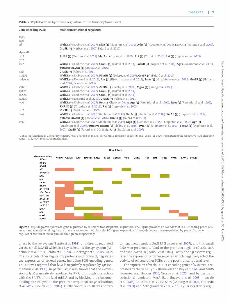
At the transcriptional level, two-component signal transduc-tion systems and global regulators are involved in regulating theproduct of PGHs (Table 2 and Fig. 5). S. aureus is known to possess16 so-called two-component regulatory systems (TCSs; e.g. WalKR,AgrAC, and ArlRS), which are composed of a sensor module withhistidine kinase activity and a cognate DNA-binding response reg-ulator that modulates transcription. Such TCSs monitor particu-lar stresses or extracellular stimuli (Szurmant et al. 2007) and reg-ulate the appropriate physiological responses through autophos-phorylation of the sensor module and subsequent transfer of thephosphoryl group to the cognate response regulator, which leadsto activation of the regulator (Kleerebezem et al. 1997). Particu-lar TCSs may control the expression of multiple PGH-encodinggenes. For instance, the WalR response regulator of the essentialWalKR (YycGF) TCS specifically binds to the promoter regions ofgenes encoding the S. aureus PGHs Atl, LytM, SsaA (SA2093), IsaA,SceD, EssH, the three CHAP domain-containing proteins SA0620,SA2097, and SA2353) (Table 2) (Dubrac and Msadek 2004, Dubracet al. 2007), Sle1 and SA2332 (Delauné et al. 2012), thereby posi-tively controlling their expression. In addition, WalKR also posi-tively regulates peptidoglycan biosynthesis and turnover (Dubracet al. 2007). Importantly, WalKR can both regulate and be regulatedby other regulatory components. For example, the YycH and YycIproteins stimulate WalK activity through WalKR phosphorylation(Gajdiss et al. 2020). Also, the WalR regulator stimulates the SaeSRTCS (Delauné et al. 2012), whereas SaeSR negatively regulates thessaA gene (SA2093) (Rogasch et al. 2006). The expression of ssaA ispositively regulated by GraSR (Falord et al. 2011), a TCS that has animportant function in the regulation of the bacterial metabolismunder oxidative stress conditions, at low pH and at higher temper-ature. Nine genes encoding PGHs are regulated by both GraSR andWalKR (Dubrac et al. 2007, Falord et al. 2011). Thus, the TCSs playmultiple roles in the regulation of S. aureus cell wall metabolism(Dubrac et al. 2007, Delaune et al. 2011).
One of the key TCSs of S. aureus, the accessory gene regula-tion (agr) quorum-sensing system that modulates the expressionof over 100 genes, is also involved in the regulation of cell wallmetabolism (Dunman et al. 2001). Various genes for cell wall-associated proteins are repressed in the late exponential growth
Dow
nloaded from https://academ
ic.oup.com/fem
sre/advance-article/doi/10.1093/femsre/fuac025/6604383 by guest on 05 July 2022
Wang et al. | 9
Table 2. Peptidoglycan hydrolase regulation at the transcriptional level.
Gene encoding PGHs Main transcriptional regulators
sagA -sagB -atl WalKR (↑) (Dubrac et al. 2007), SigB (↑) (Houston et al. 2011), AtlR (↓) (Houston et al. 2011), SarA (↓) (Trotonda et al. 2009),
GraSR (↑) (Herbert et al. 2007, Falord et al. 2011)aly/scaH -lytN ArlRS (↓) (Memmi et al. 2012), MgrA (↓) (Luong et al. 2006), Rot (↓) (Chu et al. 2013), Rat (↓) (Ingavale et al. 2003)lytH -ssaA WalKR (↑) (Dubrac et al. 2007), GraSR (↑) (Falord et al. 2011), SaeSR (↓) (Rogasch et al. 2006), Agr (↓) (Dunman et al. 2001),
putative RNAIII (↓) (Lioliou et al. 2016)essH GraSR (↑) (Falord et al. 2011)sa2353 WalKR (↑) (Dubrac et al. 2007), RNAIII (↓) (Boisset et al. 2007), GraSR (↑) (Falord et al. 2011)sle1/aaa WalKR (↑) (Delauné et al. 2012), Agr (↓) (Hirschhausen et al. 2012), SarA (↓) (Hirschhausen et al. 2012), GraSR (↑) (Herbert
et al. 2007, Falord et al. 2011)sa0710 WalKR (↑) (Dubrac et al. 2007), ArlRS (↓) (Crosby et al. 2020), MgrA (↓) (Luong et al. 2006)sa0620 WalKR (↑) (Dubrac et al. 2007), GraSR (↑) (Falord et al. 2011)sa2097 WalKR (↑) (Dubrac et al. 2007), GraSR (↑) (Falord et al. 2011)sa2332 WalKR (↑) (Delauné et al. 2012), GraSR (↑) (Falord et al. 2011)lytM WalKR (↑) (Dubrac et al. 2007), Rot (↓) (Chu et al. 2013), Agr (↓) (Ramadurai et al. 1999), SarA (↓) (Ramadurai et al. 1999),
RNA III (↓) (Chunhua et al. 2012), Rat (↓) (Ingavale et al. 2003)lytU VraSR (↑) (Pietiäinen et al. 2009)isaA WalKR (↑) (Dubrac et al. 2007, Stapleton et al. 2007), SarA (↑) (Stapleton et al. 2007), SrrAB (↑) (Stapleton et al. 2007),
putative RNAIII (↓) (Lioliou et al. 2016), GraSR (↑) (Falord et al. 2011)sceD WalKR (↑) (Dubrac et al. 2007, Stapleton et al. 2007), SigB (↑) (Ziebandt et al. 2001, Stapleton et al. 2007), Agr (↑)
(Stapleton et al. 2007), putative RNAIII (↓) (Lioliou et al. 2016), LytSR (↓) (Stapleton et al. 2007), SaeSR (↓) (Stapleton et al.2007), GraSR (↑) (Falord et al. 2011), SarA (↓) (Stapleton et al. 2007)
∗Genes for functionally uncharacterized PGHs are named by their S. aureus N315 accession codes; (↑) and (↓), up- or down-regulation of the respective PGH-encodinggene; -, unknown regulation mechanism.
Figure 5. Peptidoglycan hydrolase gene regulation by different transcriptional regulators. The Figure provides an overview of PGH-encoding genes of S.aureus and translational regulators that are known to modulate the PGH gene expression. Up-regulation or down-regulation by particular generegulators are indicated in pink or olive green, respectively.
phase by the agr system (Benito et al. 1998), or indirectly regulatedvia the small RNA III which is a key effector of the agr system (Ab-delnour et al. 1993, Benito et al. 1998, Huntzinger et al. 2005). RNAIII also targets other regulatory proteins and indirectly regulatesthe expression of several genes, including PGH-encoding genes.Thus, it was reported that lytM is negatively regulated by agr (Ra-madurai et al. 1999). In particular, it was shown that the expres-sion of lytM is negatively regulated by RNA III through interactionwith the 5’UTR of the lytM mRNA and by blocking the ribosome-binding site of lytM at the post-transcriptional stage (Chunhuaet al. 2012, Lioliou et al. 2016). Furthermore, RNA III was shown
to negatively regulate SA2353 (Boisset et al. 2007), and this smallRNA was predicted to bind to the promoter regions of sceD, isaAand ssaA (SA2093) (Lioliou et al. 2016). Lastly, the agr system regu-lates the expression of protease genes, which negatively affect theactivity of Atl and other PGHs at the post-transcriptional level.
The expression of various PGH-encoding genes of S. aureus is re-pressed by the TCSs LytSR (Brunskill and Bayles 1996a) and ArlRS(Fournier and Hooper 2000, Crosby et al. 2020), and by the tran-scriptional regulators MgrA (Rat) (Ingavale et al. 2003, Ingavaleet al. 2005), Rot (Chu et al. 2013), SarA (Cheung et al. 2004, Trotondaet al. 2009) and AtlR (Houston et al. 2011). LytSR negatively regu-
Dow
nloaded from https://academ
ic.oup.com/fem
sre/advance-article/doi/10.1093/femsre/fuac025/6604383 by guest on 05 July 2022
10 | FEMS Microbiology Reviews, 2022
lates PGH activity by regulating the expression of the lytSR operonand its downstream lrgAB operon (Brunskill and Bayles 1996b). Inturn, the lrgAB operon, and also the paralogous cidABC operonapparently control the transport of PGHs across the membrane,thereby facilitating PGH regulation at the post-transcriptionallevel (Groicher et al. 2000, Rice et al. 2003, Rice and Bayles 2008,Bayles 2014). Here it should be noted that cidA encodes a putativeholin that may disrupt the membrane integrity thereby promot-ing bacterial cell death and lysis, while lrgA encodes an anti-holinthat counteracts cell death and lysis (Groicher et al. 2000, Rice et al.2003, Rice and Bayles 2008). ArlRS and MgrA constitute a regula-tory cascade that controls expression of a large number of genes(Crosby et al. 2016). ArlRS regulates autolysis by activating the ex-pression of mgrA (Crosby et al. 2016, Crosby et al. 2020), whereArlRS and MgrA jointly mediate the repression of lytN (Luong andLee 2006, Memmi et al. 2012). Furthermore, ArlRS, is a negative reg-ulator of the aforementioned essH gene, but this does not involveMgrA (Crosby et al. 2020).
The regulator Rot, a DNA-binding protein from the SarA fam-ily of S. aureus regulators, can bind to the promoter regions oflytM and lytN to directly regulate the expression of these PGH-encoding genes (Chu et al. 2013). Rot also regulates the transcrip-tion of lryAB, which leads to an indirect regulation of PGH pro-duction (Chu et al. 2013). The negative regulation of autolysis byMgrA and SarA may be mediated indirectly by downregulation ofsarV, which encodes a positive regulator of several PGHs (Mannaet al. 2004, Trotonda et al. 2009). MgrA represses the expressionof at least two transcriptional regulators, SarV and AtlR (Crosbyet al. 2016). The repressor AtlR downregulates the transcriptionof atl (Houston et al. 2011), whereas the sigma factor SigB neg-atively regulates the agr locus, thereby positively influencing atltranscription (Houston et al. 2011). Lastly, the TCS VraSR is able tosense perturbations in cell wall synthesis and negatively regulatesautolytic activity (Kuroda et al. 2003, Antignac et al. 2007).
Several factors have been reported to positively regulate the ex-pression of PGH-encoding genes. For instance, the TCS SrrAB regu-lates gene expression in S. aureus in response to oxygen availability(Yarwood et al. 2001) and it positively regulates the transcriptionof isaA (Stapleton et al. 2007). Like the agr system, the regulatorsSarA, Rot, MgrA and SaeR also regulate the expression of extracy-toplasmic proteases that can modulate PGH activity through pro-teolysis of, for instance, Atl (Gimza et al. 2019).
Lastly, some regulators may exert both positive and negativeregulation of the expression of PGH-encoding genes. This is ex-emplified by the small RNA SprX of S. aureus, which exerts its reg-ulatory function by base-pairing with cis- or trans-encoded targetmRNAs, thereby influencing the mRNA stability and translation(Caldelari et al. 2013). Thus, SprX can directly interact with theWalR mRNA to positively regulate the expression of lytM and atl.On the other hand, SprX also binds to the isaA mRNA, thereby neg-atively influencing its stability (Buchad and Nair 2021). Anothertarget of SprX is the spoVG gene, which encodes a site-specificDNA-binding protein responsible for the inhibition of cell walldegradation (Eyraud et al. 2014). In particular, it was shown thatSpoVG can directly bind to the putative promoter region of lytN,femA and lytSR to repress their transcription (Liu et al. 2016).
Regulation of PGH activity by cell wallcomponentsWTA and LTA are known to influence the activity of PGHs and theirsubstrate binding capability. In particular, WTA was shown to actas a temporal and spatial regulator of peptidoglycan metabolism
(Atilano et al. 2010, Schlag et al. 2010), being a key determinantfor the localization and activity of Atl (Weidenmaier et al. 2004).WTA also prevents the association of the GW cell wall-bindingdomain of Atl with the side wall (also referred to as the lateralwall), but not with the septum where WTA is less abundant oraltered in structure, which results in a more acidic milieu thatreduces the activity of Atl (Schlag et al. 2010, Biswas et al. 2012, Ti-wari et al. 2018). TagO is an N-acetylglucosamine-phosphate trans-ferase, which catalyzes the first step of WTA biosynthesis. Accord-ingly, a tagO mutant was shown to be completely devoid of WTA.The absence of WTA resulted in an even distribution of Atl on thesurface of a tagO mutant leading to increased autolytic activity(Schlag et al. 2010). WTA also mediates the specific binding of Sle1and LytN to the cross-wall of the septum (Schlag et al. 2010) and,accordingly, a lack of WTA results in a disordered localization ofSle1 and LytN (Chan et al. 2013). Consistent with this finding, itwas shown that Sle1 lacking the LysM domain is unable to fulfillits function. In contrast, the function of LytN was not affected byabsence of the LysM domain (Frankel and Schneewind 2012). Thelatter finding can be explained by the presence of the YSIRK/GSmotif in the signal peptide of LytN, which contributes to the local-ization of LytN to the cross-wall (DeDent et al. 2008). Of note, theGW cell wall-binding domain of Atl can also bind to LTA, whichis highly abundant at the septum, resulting in Atl targeting to theseptum (Biswas et al. 2012, Zoll et al. 2012). Similar to WTA, LTA hasbeen implicated in controlling the activity of various PGHs (Fedtkeet al. 2007, Tiwari et al. 2018).
Regulation of PGH activity by protein–proteininteractionsProtein-protein interactions can affect the activity and localiza-tion of PGHs. For instance, PGHs can form complexes with otherproteins, such as peptidoglycan synthases, to execute their biolog-ical functions (Ho¨ltje 1998, Typas et al. 2012). Thus, SagB and thecytoplasmic membrane protein SpdC (LyrA) were shown to forma complex and cleave nascent glycan strands to release newlysynthesized glycan strands from the cell membrane and to pro-duce lipid II-attached oligomers for further peptidoglycan elonga-tion (Schaefer et al. 2021). The SagB partner protein SpdC orientsthe active site of SagB for cleaving glycan strands (Schaefer et al.2021). SpdC also antagonizes the WalR-dependent expression ofSceD, LytM, Atl, and SA2097 (Poupel et al. 2018). Another exam-ple of protein-protein interactions relevant for PGH activity con-cerns the membrane-localized amidase LytH, which requires cat-alytic activation by the polytopic membrane protein ActH (Schae-fer et al. 2021). The LytH-ActH amidase complex can also regulatethe activity of peptidoglycan synthases. In particular, this com-plex removes the stem peptides from a nascent glycan strandat the cell periphery and promotes the relocation of peptidogly-can synthases to the septum during cell division (Do et al. 2020).Intramolecular protein-protein interactions also appear to deter-mine PGH activity. This is exemplified by the LytM protein thatshows weak PGH activity (Osipovitch et al. 2015). In contrast, thePGH activity is strongly increased upon truncation of LytM to theso-called M23 endopeptidase domain (Fig. 3). This suggests thatthe activity of LytM’s M23 domain is suppressed by the so-calledoccluding domain of LytM (Odintsov et al. 2004). Until now, it isnot known how the intact LytM becomes activated, but this mostlikely occurs through the interaction with partner proteins ratherthan proteolytic processing since the full-size LytM is readily de-tectable in the growth medium of S. aureus (Wang et al. 2021).
Dow
nloaded from https://academ
ic.oup.com/fem
sre/advance-article/doi/10.1093/femsre/fuac025/6604383 by guest on 05 July 2022
Wang et al. | 11
Regulation of PGHs by proteolytic activityStaphylococcus aureus possesses several cytoplasmic and secretedproteolytic enzymes that play important functions in regulatingthe expression and activities of PGHs (Shaw et al. 2004, Kolar et al.2013). For example, Sle1 is a substrate of the highly conserved pro-tease ClpXP, which is composed of the peptidase subunit ClpP andthe chaperone subunit ClpX. Inactivation of ClpXP was shown toresult in increased Sle1 activity (Feng et al. 2013, Thalsø-Madsenet al. 2019), which seems to be due to ClpXP mediated process-ing of the signal peptide-bearing Sle1 precursor protein (Feng et al.2013, Liu et al. 2017). Likewise, enhanced levels of IsaA, SceD, LytM,LytN, SsaA (SA2093) were detected in both clpP and clpX mutantstrains (Kirsch et al. 2020). Cytoplasmic proteases, such as ClpXP,have been implicated in the activation of some regulatory factors,such as Rot, thereby indirectly regulating the expression of PGHs(Frees et al. 2005). Importantly, also extracellular proteases seemto modulate the PGH levels by proteolytic processing, in order todispose of damaged or no longer needed proteins, or in responseto external or internal signals. These extracellular proteases in-clude the metalloprotease Aur, the serine proteases SspA, SplA,and SplC, the serine protease-like proteins SplB, SplD, SplE, andSplF, as well as the cysteine proteases ScpA and SspB (Dubin 2002,Kolar et al. 2013). In particular, the SspA protease was shown to beinvolved in the proteolytic processing of Atl, thereby regulating Atlactivity and autolysis (Chen et al. 2013, Sahukhal et al. 2015).
Regulation of PGHs by environmental factorsLastly, the activity of PGHs can be modulated by environmen-tal factors, such as the salt and metal concentrations, or the pH(Gilpin et al. 1972, Foster 1995, Calamita and Doyle 2002). The im-pact of metals is exemplified by the M23 domain of LytM, LytUand the amidase domain of Atl, which all display zinc-dependentenzymatic activity (Büttner et al. 2014, Raulinaitis et al. 2017). ThePGH activity of LytM is strongly inhibited in the presence of Fe2+,Cu2+, and Mg2+ ions and low concentrations of NaCl or KCL (Ra-madurai et al. 1999). In contrast, the presence of Cu2+ or Co2+ wasshown to increase the activity of LytU (Raulinaitis et al. 2017). Aclear pH dependency was reported for SagB, which is more ac-tive at pH 5 than at pH 7.5 (Wheeler et al. 2015). LytM shows opti-mal activity at pH values between 5 and 8 (Ramadurai et al. 1999),whereas Atl has optimal activity above pH 6.5. Of note, Atl activ-ity seems to be enhanced at low temperature and high NaCl con-centrations (Foster 1995). Accordingly, it has been proposed thatchanges in the ionic composition of the cell wall affect PGH ac-tivity (Cheung and Freese 1985). In particular, it was shown thatWTA affects the concentration of proton-binding sites in the cellwall, thereby modulating the activity of Atl (Biswas et al. 2012).The optimal pH for activity may relate to the specific localizationof a PGH within the cell wall, where a high concentration of pro-tons is encountered at the membrane-cell wall interface due tothe proton-motive force, while the proton concentration can belower towards the cell surface (Calamita et al. 2001).
Impact of antibiotics on PGH activityInhibition of peptidoglycan synthesis by antibiotics can lead tobacterial autolysis. This was exemplified by perturbations of thepeptidoglycan synthesis in S. aureus, either by growing the bacte-ria in the presence of sub-inhibitory concentrations of β-lactamantibiotics, or by downregulating the expression of the peptido-glycan synthase PBP2 (Antignac et al. 2007). However, in responseto the respective cell wall stress, the synthesis of PGHs is repressed
at the transcriptional level, leading to a decreased susceptibilityto autolysis (Antignac et al. 2007). In turn, this may result in areduced susceptibility for β-lactams, as evidenced by the obser-vation that PGH inactivation typically enhances the resistance toβ-lactam antibiotics (Tomasz et al. 1970). In different staphylococ-cal lineages, the antibiotic resistance profiles can be very differ-ent, which will impact on the correlation of antibiotic activity andPGHs to different extents. Accordingly, some PGH mutants showactually an increased susceptibility to antibiotics. For example, asagB mutant of the MRSA USA300 LAC strain was shown to bemore susceptible to the β-lactam antibiotic oxacillin (Chan et al.2016). This may be due to the increased length of glycan strands asa consequence of the SagB-deficiency. The increased glycan strandlength may, in fact, cause a perturbed recognition and transpep-tidation by the PBP2a that is responsible for the methicillin re-sistance of the USA300 LAC strain (Chan et al. 2016). Further-more, β-lactam resistance can be correlated to the level of Sle1 inMRSA isolates of the USA300 lineage (Thalsø-Madsen et al. 2019).In particular, in oxacillin-treated cells the expression of sle1 is de-creased, which leads to a cell separation defect. Conversely, highSle1 levels accelerate the splitting of daughter cells and decreasethe cell size under oxacillin stress conditions. This implies that theSle1 level may be positively coupled to the transpeptidase activityof PBPs, and that an increased Sle1 level contributes to oxacillinresistance by promoting cell separation. In contrast, sle1 mutantcells are more susceptible to oxacillin, which may be linked to asynthetically lethal effect on septum synthesis (Thalsø-Madsenet al. 2019). Another example where an antibiotic impacts on PGHactivity concerns targocil, which targets the TarG subunit of theWTA translocase. Targocil treatment of methicillin-susceptible S.aureus was shown to decrease the cell lysis by sequestering Atlat the membrane instead of the septal surface, which was a con-sequence of the accumulation of untranslocated WTA molecules(Tiwari et al. 2018).
Role of PGHs in PathogenesisS. aureus can thrive and survive inside and outside a wide vari-ety of eukaryotic cells. Accordingly, the peptidoglycan structureand dynamics can be altered in different niches provided by thehost, which seems to be very important for bacterial survivaland pathogenesis (Chamaillard et al. 2003, Boneca 2005). For in-stance, during biofilm formation, which frequently occurs on tis-sues and implanted biomaterials, the cross-linking of the pep-tidoglycan was found to be reduced (Kim et al. 2018). Also herethe non-crosslinked pentaglycine bridges were shown to providea support for surface proteins that promote the biofilm formation(Kim et al. 2018). During a S. aureus kidney infection, smaller bac-terial cells with thicker cell walls and less crosslinking of the pep-tidoglycan were observed compared to in vitro cultured bacteria.It was suggested that the non-crosslinked pentaglycine bridgescould be used as attachment sites for surface proteins that areneeded in abscess formation (Sutton et al. 2021). In addition, dur-ing infection, the structure of peptidoglycan showed less com-plexity, which may relate to an adjustment of the peptidoglycanmetabolism. This implies that the energy gained by the adjustedpeptidoglycan metabolism could be used for other processes inorder to allow the bacterial proliferation in a limited environment(Sutton et al. 2021). In extreme cases, S. aureus may even partiallyor completely lose its cell wall, thereby converting to the L-formas has been reported for recurrent and persistent infections. How-ever, the L-form bacteria are unstable under normal growth condi-tions, so when the environment changes or the inducer (e.g. peni-
Dow
nloaded from https://academ
ic.oup.com/fem
sre/advance-article/doi/10.1093/femsre/fuac025/6604383 by guest on 05 July 2022
12 | FEMS Microbiology Reviews, 2022
cillin or oxacillin) is removed, these bacteria will convert again tothe normal state with a protective cell wall (Xu et al. 2020).
Importantly, peptidoglycan fragments can be perceived by thehost as bacterial virulence factors. For instance, peptidoglycanturnover mediated by PGHs was shown to lead to the sheddingof peptidoglycan fragments into the host environment, therebyeliciting a proinflammatory response in the host (Girardin et al.2003). In particular, it is known that the products of peptidogly-can turnover, muropeptides, can target the human intracellularimmune receptors Nod1 and Nod2 which, in turn, leads to NF-κBactivation and the release of proinflammatory factors, such as cy-tokines, chemokines and various antimicrobial compounds (Davisand Weiser 2011).
The pathogenicity of S. aureus largely relies on secreted and cellwall-localized proteinaceous virulence factors that promote theinteraction between bacteria and the host. Accordingly, variousPGHs exposed on the bacterial cell surface also play importantroles in bacterial pathogenesis, including host cell adhesion, hostcell internalization, inflammatory responses, immune activationand the aforementioned biofilm formation (McCarthy et al. 2016,Schlesier et al. 2020, Sutton et al. 2021). PGH activities may alterthe overall charge of the bacterial surface, or they may modifythe exposure of adhesins which, in turn, leads to altered bacterialaggregation, adhesive properties or altered biofilm formation. Forexample, the major autolysin Atl also acts as a host adhesin andinteracts with the extracellular matrix of host cells during attach-ment, colonization and infection. Atl has been shown to bind tofibronectin and vitronectin mainly via its GW-peptidoglycan bind-ing domain (Kohler et al. 2014, Porayath et al. 2018), and it alsobinds to the human heat shock cognate protein (Hsc70) with highaffinity (Schlesier et al. 2020), thereby promoting the efficient inter-nalization into host cells, including non-professional phagocytes(Hirschhausen et al. 2010), such as endothelial cells (Schlesier et al.2020) and human epithelial cells (Dziewanowska et al. 2000). Atlalso binds to the extracellular matrix components heparin andgelatine, thereby promoting bacterial binding to multiple differ-ent host cells during an invasive infection (Porayath et al. 2018).An atl mutant was shown to be attenuated in nanopore propa-gation in implant materials in vitro (which mimics infection ofbone canaliculi) and in abscess formation in vivo, which may beattributed to an altered promotion of cell clustering and roughcell surface formation (Masters et al. 2021). Intriguingly, atl mu-tant bacteria excreted almost no ECPs, such as aldolase (FbaA) andglyceraldehyde 3-phosphate dehydrogenase (GAPDH). This couldalso explain the attenuation of atl mutants at least in part, be-cause FbaA and GAPDH are known to promote the adherence ofS. aureus to host cells (Pasztor et al. 2010, Ebner et al. 2016). ThePGH Sle1 can act as an adhesin by binding to human fibrinogen,fibronectin and vitronectin (Heilmann et al. 2005). Both the LysMdomain and CHAP domain of Sle1 have been shown to play rolesin host adhesion (Scheurwater et al. 2008, Kim et al. 2018). Further-more, both the sle1 and the atl mutant bacteria showed a signif-icant decrease in pathogenesis in acute murine infection models(Kajimura et al. 2005). In addition, loss of the glucosaminidase do-main of Atl leads to cell clustering and attenuation in a murinemodel of osteomyelitis (Varrone et al. 2014). Nonetheless, anotherstudy obtained no evidence for a role of Atl in murine infection(McCarthy et al. 2016), and the lack of Atl by itself was not suffi-cient to cause attenuation in a Zebrafish infection model (Suttonet al. 2021). On the other hand, inactivation of SagA and/or ScaHdid lead to attenuation in a Zebrafish infection model (Sutton et al.2021). A sagB mutant also displayed significant attenuation in amurein sepsis model, which implies that the absence of SagB sig-
nificantly reduces the fitness of S. aureus (Sutton et al. 2021). Alto-gether, the reason(s) why PGH mutations cause attenuation of S.aureus appear to be rather complex. For instance, inactivation ofSagB results initially in thinner cell walls that take longer transi-tion time to reach maturity which, in turn, may lead to altered,less effective interactions with the host (Balraadjsing et al. 2019,Pasquina-Lemonche et al. 2020, Sutton et al. 2021). However, sagBmutants also display aberrant excretion of cytoplasmic proteinsand reduced release of membrane and secreted proteins (Chanet al. 2016), which may also be a cause of attenuation. Bacteriawith isaA or sceD mutations did not show significant attenuationin a murine infection model, whereas an isaA sceD double mutantdisplayed clear attenuation (Stapleton et al. 2007). On the otherhand, isaA single mutant bacteria were attenuated in a Galleriamellonella larval infection model (Zhao et al. 2019), whereas SceDwas shown to be important for nasal colonization and dissemina-tion of S. aureus in host tissues (Stapleton et al. 2007).
PGHs are frequently involved in the aforementioned formationof biofilms that, in general, consist of proteins, polysaccharidesand extracellular DNA (i.e. eDNA) (Schilcher and Horswill 2020)and that also contribute to the bacterial pathogenesis (Archeret al. 2011). Importantly, PGHs can mediate the release of eDNAwhich is a key factor in biofilm formation. Both through its ami-dase and glucosaminidase domains, Atl is involved in the primaryattachment of the bacteria to surfaces and, consequently, thisPGH serves an essential function in biofilm formation (Biswas et al.2006, Bose et al. 2012). Specifically, unprocessed Atl promotes theprimary attachment of S. aureus to surfaces, while the proteolyti-cally activated Atl promotes cell lysis, the release of eDNA and cellaccumulation for early biofilm development (Biswas et al. 2006).Atl plays an essential role in the development of so-called ica-independent biofilms. The formation of such biolfilms is broughtabout by a diverse array of surface proteins, such as Aap and Bap.These proteins serve as adhesins that bind to the host’s extracellu-lar matrix, instead of extracellular polysaccharide adhesins, suchas the polysaccharide intracellular adhesin or polymeric N-acetyl-glucosamine that are synthesized by enzymes encoded in the icaoperon (O’Gara 2007, Houston et al. 2011). Next to Atl, previous re-search has also shown that Sle1 may have a role in the formationof bacterial biofilms (Liu et al. 2017). Lastly, an elevated expressionof isaA as induced by the anti-biofilm reagent tannic acid or byprotein overexpression may also lead to biofilm formation (Payneet al. 2013).
PGHs as potential targets for active or passiveimmunizationPGHs are commonly considered as promising targets for activeand passive immunization against S. aureus infections, becausemost of these proteins are exposed on the bacterial cell surface,where they play important roles in cell wall metabolism. Pro-teomic analyses of the cell surface of S. aureus have shown thatScaH (Aly), LytM, IsaA, SA0620, SA2097 are nearly always presentat this subcellular location of clinical S. aureus isolates (Ziebandtet al. 2010). For active immunization, PGHs could thus poten-tially be used to elicit protective host immune responses eitherthrough the generation of specific antibodies that opsonize S. au-reus and lead to its elimination by complement and/or profes-sional phagocytes, or by inducing protective cellular immune re-sponses (Bredius et al. 1993). Importantly, there is an increasingneed for vaccines against S. aureus, due to the wide-spread antibi-otic resistance displayed by this pathogen. The idea that PGHs aresuitable targets for vaccination is corroborated by the observation
Dow
nloaded from https://academ
ic.oup.com/fem
sre/advance-article/doi/10.1093/femsre/fuac025/6604383 by guest on 05 July 2022
Wang et al. | 13
that the amidase domain of the major PGH Atl can elicit protectiveimmunity against S. aureus in a murine infection model (Nair et al.2015). It is, however, presently unknown whether this protectiontakes place through the staphylococcal opsonization by anti-Atlantibodies and subsequent immune clearance, or through the in-activation of Atl by such antibodies. Another study showed thatthe amidase domain of Atl in combination with an Alum adjuvantefficiently protects mice against an S. aureus infection (Suresh et al.2021). An octa-valent S. aureus antigen mixture was shown to in-duce significant IgG responses against LytM, IsaA and the propep-tide of Atl in immunized mice, although this did not result in aprotection against death upon S. aureus infection (van den Berget al. 2015b). However, patients with the genetic blistering diseaseepidermolysis bullosa, whose wounds are highly colonized withmultiple S. aureus lineages and who rarely develop serious invasivestaphylococcal infections, do show high IgG levels against theseantigens (van der Kooi-Pol et al. 2012,van der Kooi-Pol et al. 2013,van der Kooi-pol et al. 2013, van der Kooi-pol et al. 2014, van den
Berg et al. 2015b, Pastrana et al. 2018). This seems to suggest thatIgGs against LytM, IsaA and Atl may also confer some degree ofprotection against S. aureus infections in humans. Likewise, in ascreen for immunogenic non-covalently cell wall-bound proteins,it was observed that Atl, IsaA, EssH, Sle1 and SsaA (SA2093) elicithigh IgG levels in sera of patients with EB (Pastrana et al. 2018).These findings imply that surface-exposed PGHs could be poten-tial targets for anti-staphylococcal vaccines. This idea is also sup-ported by passive immunization experiments. For instance, thehuman monoclonal antibody 1D9, which binds to the N-terminalregion of IsaA, can protect mice at least partially against S. au-reus bacteraemia and skin infection (van den Berg et al. 2015a).Likewise the humanized monoclonal antibody hUK-66 that specif-ically binds IsaA was shown to protect against killing by S. aureusin murine infection models, and this monoclonal antibody alsoeffectively enhanced S. aureus killing in whole blood (Lorenz et al.2011, Oesterreich et al. 2014).
Interestingly, so-called lysibodies that were engineered by fus-ing the cell wall-binding domain of Atl and the Fc region of hu-man IgG were reported to efficiently bind the S. aureus cell wall.In turn, this was shown to lead to the fixation of complementon the bacterial surface, the promotion of phagocytosis by neu-trophils and macrophages, and the protection of mice from anMRSA infection (Raz et al. 2017). Another lysibody composed ofthe cell wall-binding domain of lysostaphin and the Fc region ofhuman IgG was shown to promote phagocytosis by neutrophilsand macrophages even more efficiently, as well as the protectionof mice against MRSA infection (Raz et al. 2018). These observa-tions indicate that PGHs can be effectively applied as targets foractive or passive immunization and also for the development ofcompletely new generations of engineered antimicrobials.
ConclusionThe Gram-positive bacterial cell wall is essentially one moleculeof cross-linked peptidoglycan that shelters a protoplast within itsboundaries, thereby providing solid support against high turgorpressures. On the other hand, the cell wall is highly dynamic,allowing the cell to grow and divide and, at the same time, itserves as a semi-porous filter that allows the uptake of essen-tial nutrients, the exclusion of toxic compounds from the bacte-rial environment, and the excretion of metabolic wastes. Yet an-other function of the cell wall is to serve as a matrix that binds
and retains proteins with key functions in nutrient acquisition,cell adhesion to surfaces and bacterial virulence (van Dijl and duTeil Espina 2021). To make all these vital cell wall functions hap-pen, PGHs play central roles as highlighted in our present review.The PGHs are key in peptidoglycan modulation and, therefore, es-sential for the bacterial survival in widely differing environmentswhere staphylococci can thrive, ranging from fomites to contami-nated food products and different niches in the bodies of livestockand humans. To avoid the destruction of their essential cell wallstructures and functions, bacterial cells need to carefully regulatethe expression of PGHs both spatially and temporally. This is not atrivial challenge, as underscored by S. aureus, which needs to care-fully control the activities of its 18 core PGHs. Our present reviewsummarizes the current understanding of how S. aureus managesto control PGH activity, how the bacterium takes advantage ofthe different properties and localizations of its PGHs, and how itsuppresses their potentially cytotoxic effects. Importantly, PGHsare much more than enzymes that degrade cell walls, since theyserve multiple biological functions in bacterial growth and sur-vival in widely varying environments. Moreover, they participatein the release of ECPs needed for the bacterial adhesion to sur-faces of medical implants or host tissues, or in the regulated bac-terial cell death to generate eDNA needed for biofilm formation.PGHs are also critical for the cell cycle and allow S. aureus to exe-cute various functions in human pathogenesis. Altogether, thesevital functions of PGHs imply that they can potentially be used astargets for the development of anti-staphylococcal therapies. Thisis exemplified by the possible application of PGHs to lyse bacteria,to target PGHs through passive or active anti-staphylococcal im-munization approaches, or even to use parts of the staphylococ-cal PGHs in the engineering of novel antimicrobial drugs. Yet, inspite of the fact that our understanding of PGH functions has sub-stantially increased over recent years, there are still many openquestions that need to be answered before we can fully appre-ciate their possible application for novel antimicrobial therapies.For instance, when and where exactly in the bacterial cell do thedifferent PGHs play their physiological roles, or how exactly dothey facilitate the sorting and secretion of virulence factors (vanDijl and du Teil Espina 2021)? Also, we need to know more aboutthe plethora of either cooperative or antagonistic interactions be-tween these enzymes that are responsible for cell wall homeosta-sis, and about how PGH function is regulated by the levels of pepti-doglycan crosslinking and other major cell wall components likeWTA and LTA. A better understanding of this complex interplaybetween cell wall synthesis, the synthesized polymers and PGHsmay ultimately allow us to define new targets for new-generationantibiotics, vaccines and therapeutic antibodies that can help usto disrupt the essential processes of cell growth and division inimportant multi-drug resistant pathogens like S. aureus.
Availability of data and MaterialsNot applicable
Authors’ contributionsM.W. drafted the manuscript. MW, GB, and JMvD collected litera-ture and contributed manuscript sections. MW prepared the Fig-ures. GB and JMvD supervised the project. All authors, criticallyrevised the manuscript, gave final approval and agree to be ac-countable for all aspects of the review.
Dow
nloaded from https://academ
ic.oup.com/fem
sre/advance-article/doi/10.1093/femsre/fuac025/6604383 by guest on 05 July 2022
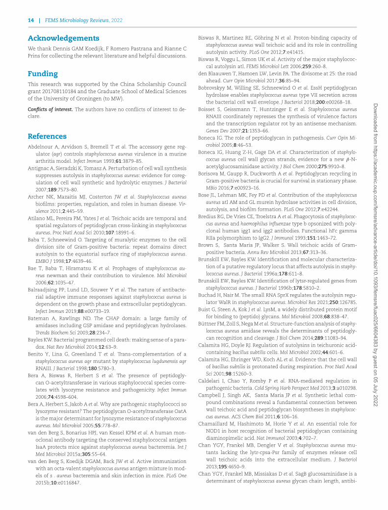
14 | FEMS Microbiology Reviews, 2022
AcknowledgementsWe thank Dennis GAM Koedijk, F Romero Pastrana and Rianne CPrins for collecting the relevant literature and helpful discussions.
FundingThis research was supported by the China Scholarship Councilgrant 201708110184 and the Graduate School of Medical Sciencesof the University of Groningen (to MW).
Conflicts of interest. The authors have no conflicts of interest to de-clare.
ReferencesAbdelnour A, Arvidson S, Bremell T et al. The accessory gene reg-
ulator (agr) controls staphylococcus aureus virulence in a murinearthritis model. Infect Immun 1993;61:3879–85.
Antignac A, Sieradzki K, Tomasz A. Perturbation of cell wall synthesissuppresses autolysis in staphylococcus aureus: evidence for coreg-ulation of cell wall synthetic and hydrolytic enzymes. J Bacteriol2007;189:7573–80.
Archer NK, Mazaitis MJ, Costerton JW et al. Staphylococcus aureusbiofilms: properties, regulation, and roles in human disease. Vir-ulence 2011;2:445–59.
Atilano ML, Pereira PM, Yates J et al. Teichoic acids are temporal andspatial regulators of peptidoglycan cross-linking in staphylococcusaureus. Proc Natl Acad Sci 2010;107:18991–6.
Baba T, Schneewind O. Targeting of muralytic enzymes to the celldivision site of Gram-positive bacteria: repeat domains directautolysin to the equatorial surface ring of staphylococcus aureus.EMBO J 1998;17:4639–46.
Bae T, Baba T, Hiramatsu K et al. Prophages of staphylococcus au-reus newman and their contribution to virulence. Mol Microbiol2006;62:1035–47.
Balraadjsing PP, Lund LD, Souwer Y et al. The nature of antibacte-rial adaptive immune responses against staphylococcus aureus isdependent on the growth phase and extracellular peptidoglycan.Infect Immun 2019;88:e00733–19.
Bateman A, Rawlings ND. The CHAP domain: a large family ofamidases including GSP amidase and peptidoglycan hydrolases.Trends Biochem Sci 2003;28:234–7.
Bayles KW. Bacterial programmed cell death: making sense of a para-dox. Nat Rev Microbiol 2014;12:63–9.
Benito Y, Lina G, Greenland T et al. Trans-complementation of astaphylococcus aureus agr mutant by staphylococcus lugdunensis agrRNAIII. J Bacteriol 1998;180:5780–3.
Bera A, Biswas R, Herbert S et al. The presence of peptidogly-can O-acetyltransferase in various staphylococcal species corre-lates with lysozyme resistance and pathogenicity. Infect Immun2006;74:4598–604.
Bera A, Herbert S, Jakob A et al. Why are pathogenic staphylococci solysozyme resistant? The peptidoglycan O-acetyltransferase OatAis the major determinant for lysozyme resistance of staphylococcusaureus. Mol Microbiol 2005;55:778–87.
van den Berg S, Bonarius HPJ, van Kessel KPM et al. A human mon-oclonal antibody targeting the conserved staphylococcal antigenIsaA protects mice against staphylococcus aureus bacteremia. Int JMed Microbiol 2015a;305:55–64.
van den Berg S, Koedijk DGAM, Back JW et al. Active immunizationwith an octa-valent staphylococcus aureus antigen mixture in mod-els of s . aureus bacteremia and skin infection in mice. PLoS One2015b;10:e0116847.
Biswas R, Martinez RE, Göhring N et al. Proton-binding capacity ofstaphylococcus aureus wall teichoic acid and its role in controllingautolysin activity. PLoS One 2012;7:e41415.
Biswas R, Voggu L, Simon UK et al. Activity of the major staphylococ-cal autolysin atl. FEMS Microbiol Lett 2006;259:260–8.
den Blaauwen T, Hamoen LW, Levin PA. The divisome at 25: the roadahead. Curr Opin Microbiol 2017;36:85–94.
Bobrovskyy M, Willing SE, Schneewind O et al. EssH peptidoglycanhydrolase enables staphylococcus aureus type VII secretion acrossthe bacterial cell wall envelope. J Bacteriol 2018;200:e00268–18.
Boisset S, Geissmann T, Huntzinger E et al. Staphylococcus aureusRNAIII coordinately represses the synthesis of virulence factorsand the transcription regulator rot by an antisense mechanism.Genes Dev 2007;21:1353–66.
Boneca IG. The role of peptidoglycan in pathogenesis. Curr Opin Mi-crobiol 2005;8:46–53.
Boneca IG, Huang Z-H, Gage DA et al. Characterization of staphylo-coccus aureus cell wall glycan strands, evidence for a new β-N-acetylglucosaminidase activity. J Biol Chem 2000;275:9910–8.
Borisova M, Gaupp R, Duckworth A et al. Peptidoglycan recycling inGram-positive bacteria is crucial for survival in stationary phase.MBio 2016;7:e00923–16.
Bose JL, Lehman MK, Fey PD et al. Contribution of the staphylococcusaureus atl AM and GL murein hydrolase activities in cell division,autolysis, and biofilm formation. PLoS One 2012;7:e42244.
Bredius RG, De Vries CE, Troelstra A et al. Phagocytosis of staphylococ-cus aureus and haemophilus influenzae type b opsonized with poly-clonal human igg1 and igg2 antibodies. Functional hFc gammaRIIa polymorphism to IgG2. J Immunol 1993;151:1463–72.
Brown S, Santa Maria JP, Walker S. Wall teichoic acids of Gram-positive bacteria. Annu Rev Microbiol 2013;67:313–36.
Brunskill EW, Bayles KW. Identification and molecular characteriza-tion of a putative regulatory locus that affects autolysis in staphy-lococcus aureus. J Bacteriol 1996a;178:611–8.
Brunskill EW, Bayles KW. Identification of lytsr-regulated genes fromstaphylococcus aureus. J Bacteriol 1996b;178:5810–2.
Buchad H, Nair M. The small RNA SprX regulates the autolysin regu-lator WalR in staphylococcus aureus. Microbiol Res 2021;250:126785.
Buist G, Steen A, Kok J et al. LysM, a widely distributed protein motiffor binding to (peptido) glycans. Mol Microbiol 2008;68:838–47.
Büttner FM, Zoll S, Nega M et al. Structure-function analysis of staphy-lococcus aureus amidase reveals the determinants of peptidogly-can recognition and cleavage. J Biol Chem 2014;289:11083–94.
Calamita HG, Doyle RJ. Regulation of autolysins in teichuronic acid-containing bacillus subtilis cells. Mol Microbiol 2002;44:601–6.
Calamita HG, Ehringer WD, Koch AL et al. Evidence that the cell wallof bacillus subtilis is protonated during respiration. Proc Natl AcadSci 2001;98:15260–3.
Caldelari I, Chao Y, Romby P et al. RNA-mediated regulation inpathogenic bacteria. Cold Spring Harb Perspect Med 2013;3:a010298.
Campbell J, Singh AK, Santa Maria JP et al. Synthetic lethal com-pound combinations reveal a fundamental connection betweenwall teichoic acid and peptidoglycan biosyntheses in staphylococ-cus aureus. ACS Chem Biol 2011;6:106–16.
Chamaillard M, Hashimoto M, Horie Y et al. An essential role forNOD1 in host recognition of bacterial peptidoglycan containingdiaminopimelic acid. Nat Immunol 2003;4:702–7.
Chan YGY, Frankel MB, Dengler V et al. Staphylococcus aureus mu-tants lacking the lytr-cpsa-Psr family of enzymes release cellwall teichoic acids into the extracellular medium. J Bacteriol2013;195:4650–9.
Chan YGY, Frankel MB, Missiakas D et al. SagB glucosaminidase is adeterminant of staphylococcus aureus glycan chain length, antibi-
Dow
nloaded from https://academ
ic.oup.com/fem
sre/advance-article/doi/10.1093/femsre/fuac025/6604383 by guest on 05 July 2022
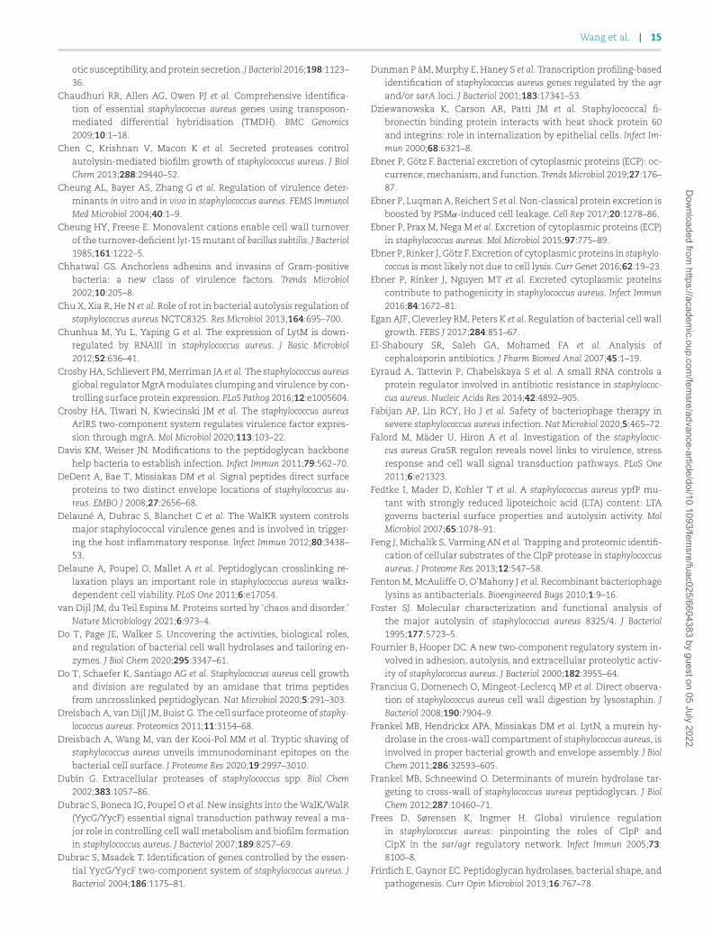
Wang et al. | 15
otic susceptibility, and protein secretion. J Bacteriol 2016;198:1123–36.
Chaudhuri RR, Allen AG, Owen PJ et al. Comprehensive identifica-tion of essential staphylococcus aureus genes using transposon-mediated differential hybridisation (TMDH). BMC Genomics2009;10:1–18.
Chen C, Krishnan V, Macon K et al. Secreted proteases controlautolysin-mediated biofilm growth of staphylococcus aureus. J BiolChem 2013;288:29440–52.
Cheung AL, Bayer AS, Zhang G et al. Regulation of virulence deter-minants in vitro and in vivo in staphylococcus aureus. FEMS ImmunolMed Microbiol 2004;40:1–9.
Cheung HY, Freese E. Monovalent cations enable cell wall turnoverof the turnover-deficient lyt-15 mutant of bacillus subtilis. J Bacteriol1985;161:1222–5.
Chhatwal GS. Anchorless adhesins and invasins of Gram-positivebacteria: a new class of virulence factors. Trends Microbiol2002;10:205–8.
Chu X, Xia R, He N et al. Role of rot in bacterial autolysis regulation ofstaphylococcus aureus NCTC8325. Res Microbiol 2013;164:695–700.
Chunhua M, Yu L, Yaping G et al. The expression of LytM is down-regulated by RNAIII in staphylococcus aureus. J Basic Microbiol2012;52:636–41.
Crosby HA, Schlievert PM, Merriman JA et al. The staphylococcus aureusglobal regulator MgrA modulates clumping and virulence by con-trolling surface protein expression. PLoS Pathog 2016;12:e1005604.
Crosby HA, Tiwari N, Kwiecinski JM et al. The staphylococcus aureusArlRS two-component system regulates virulence factor expres-sion through mgrA. Mol Microbiol 2020;113:103–22.
Davis KM, Weiser JN. Modifications to the peptidoglycan backbonehelp bacteria to establish infection. Infect Immun 2011;79:562–70.
DeDent A, Bae T, Missiakas DM et al. Signal peptides direct surfaceproteins to two distinct envelope locations of staphylococcus au-reus. EMBO J 2008;27:2656–68.
Delauné A, Dubrac S, Blanchet C et al. The WalKR system controlsmajor staphylococcal virulence genes and is involved in trigger-ing the host inflammatory response. Infect Immun 2012;80:3438–53.
Delaune A, Poupel O, Mallet A et al. Peptidoglycan crosslinking re-laxation plays an important role in staphylococcus aureus walkr-dependent cell viability. PLoS One 2011;6:e17054.
van Dijl JM, du Teil Espina M. Proteins sorted by ‘chaos and disorder.’Nature Microbiology 2021;6:973–4.
Do T, Page JE, Walker S. Uncovering the activities, biological roles,and regulation of bacterial cell wall hydrolases and tailoring en-zymes. J Biol Chem 2020;295:3347–61.
Do T, Schaefer K, Santiago AG et al. Staphylococcus aureus cell growthand division are regulated by an amidase that trims peptidesfrom uncrosslinked peptidoglycan. Nat Microbiol 2020;5:291–303.
Dreisbach A, van Dijl JM, Buist G. The cell surface proteome of staphy-lococcus aureus. Proteomics 2011;11:3154–68.
Dreisbach A, Wang M, van der Kooi-Pol MM et al. Tryptic shaving ofstaphylococcus aureus unveils immunodominant epitopes on thebacterial cell surface. J Proteome Res 2020;19:2997–3010.
Dubin G. Extracellular proteases of staphylococcus spp. Biol Chem2002;383:1057–86.
Dubrac S, Boneca IG, Poupel O et al. New insights into the WalK/WalR(YycG/YycF) essential signal transduction pathway reveal a ma-jor role in controlling cell wall metabolism and biofilm formationin staphylococcus aureus. J Bacteriol 2007;189:8257–69.
Dubrac S, Msadek T. Identification of genes controlled by the essen-tial YycG/YycF two-component system of staphylococcus aureus. JBacteriol 2004;186:1175–81.
Dunman P áM, Murphy E, Haney S et al. Transcription profiling-basedidentification of staphylococcus aureus genes regulated by the agrand/or sarA loci. J Bacteriol 2001;183:17341–53.
Dziewanowska K, Carson AR, Patti JM et al. Staphylococcal fi-bronectin binding protein interacts with heat shock protein 60and integrins: role in internalization by epithelial cells. Infect Im-mun 2000;68:6321–8.
Ebner P, Götz F. Bacterial excretion of cytoplasmic proteins (ECP): oc-currence, mechanism, and function. Trends Microbiol 2019;27:176–87.
Ebner P, Luqman A, Reichert S et al. Non-classical protein excretion isboosted by PSMα-induced cell leakage. Cell Rep 2017;20:1278–86.
Ebner P, Prax M, Nega M et al. Excretion of cytoplasmic proteins (ECP)in staphylococcus aureus. Mol Microbiol 2015;97:775–89.
Ebner P, Rinker J, Götz F. Excretion of cytoplasmic proteins in staphylo-coccus is most likely not due to cell lysis. Curr Genet 2016;62:19–23.
Ebner P, Rinker J, Nguyen MT et al. Excreted cytoplasmic proteinscontribute to pathogenicity in staphylococcus aureus. Infect Immun2016;84:1672–81.
Egan AJF, Cleverley RM, Peters K et al. Regulation of bacterial cell wallgrowth. FEBS J 2017;284:851–67.
El-Shaboury SR, Saleh GA, Mohamed FA et al. Analysis ofcephalosporin antibiotics. J Pharm Biomed Anal 2007;45:1–19.
Eyraud A, Tattevin P, Chabelskaya S et al. A small RNA controls aprotein regulator involved in antibiotic resistance in staphylococ-cus aureus. Nucleic Acids Res 2014;42:4892–905.
Fabijan AP, Lin RCY, Ho J et al. Safety of bacteriophage therapy insevere staphylococcus aureus infection. Nat Microbiol 2020;5:465–72.
Falord M, Mäder U, Hiron A et al. Investigation of the staphylococ-cus aureus GraSR regulon reveals novel links to virulence, stressresponse and cell wall signal transduction pathways. PLoS One2011;6:e21323.
Fedtke I, Mader D, Kohler T et al. A staphylococcus aureus ypfP mu-tant with strongly reduced lipoteichoic acid (LTA) content: LTAgoverns bacterial surface properties and autolysin activity. MolMicrobiol 2007;65:1078–91.
Feng J, Michalik S, Varming AN et al. Trapping and proteomic identifi-cation of cellular substrates of the ClpP protease in staphylococcusaureus. J Proteome Res 2013;12:547–58.
Fenton M, McAuliffe O, O’Mahony J et al. Recombinant bacteriophagelysins as antibacterials. Bioengineered Bugs 2010;1:9–16.
Foster SJ. Molecular characterization and functional analysis ofthe major autolysin of staphylococcus aureus 8325/4. J Bacteriol1995;177:5723–5.
Fournier B, Hooper DC. A new two-component regulatory system in-volved in adhesion, autolysis, and extracellular proteolytic activ-ity of staphylococcus aureus. J Bacteriol 2000;182:3955–64.
Francius G, Domenech O, Mingeot-Leclercq MP et al. Direct observa-tion of staphylococcus aureus cell wall digestion by lysostaphin. JBacteriol 2008;190:7904–9.
Frankel MB, Hendrickx APA, Missiakas DM et al. LytN, a murein hy-drolase in the cross-wall compartment of staphylococcus aureus, isinvolved in proper bacterial growth and envelope assembly. J BiolChem 2011;286:32593–605.
Frankel MB, Schneewind O. Determinants of murein hydrolase tar-geting to cross-wall of staphylococcus aureus peptidoglycan. J BiolChem 2012;287:10460–71.
Frees D, Sørensen K, Ingmer H. Global virulence regulationin staphylococcus aureus: pinpointing the roles of ClpP andClpX in the sar/agr regulatory network. Infect Immun 2005;73:8100–8.
Frirdich E, Gaynor EC. Peptidoglycan hydrolases, bacterial shape, andpathogenesis. Curr Opin Microbiol 2013;16:767–78.
Dow
nloaded from https://academ
ic.oup.com/fem
sre/advance-article/doi/10.1093/femsre/fuac025/6604383 by guest on 05 July 2022
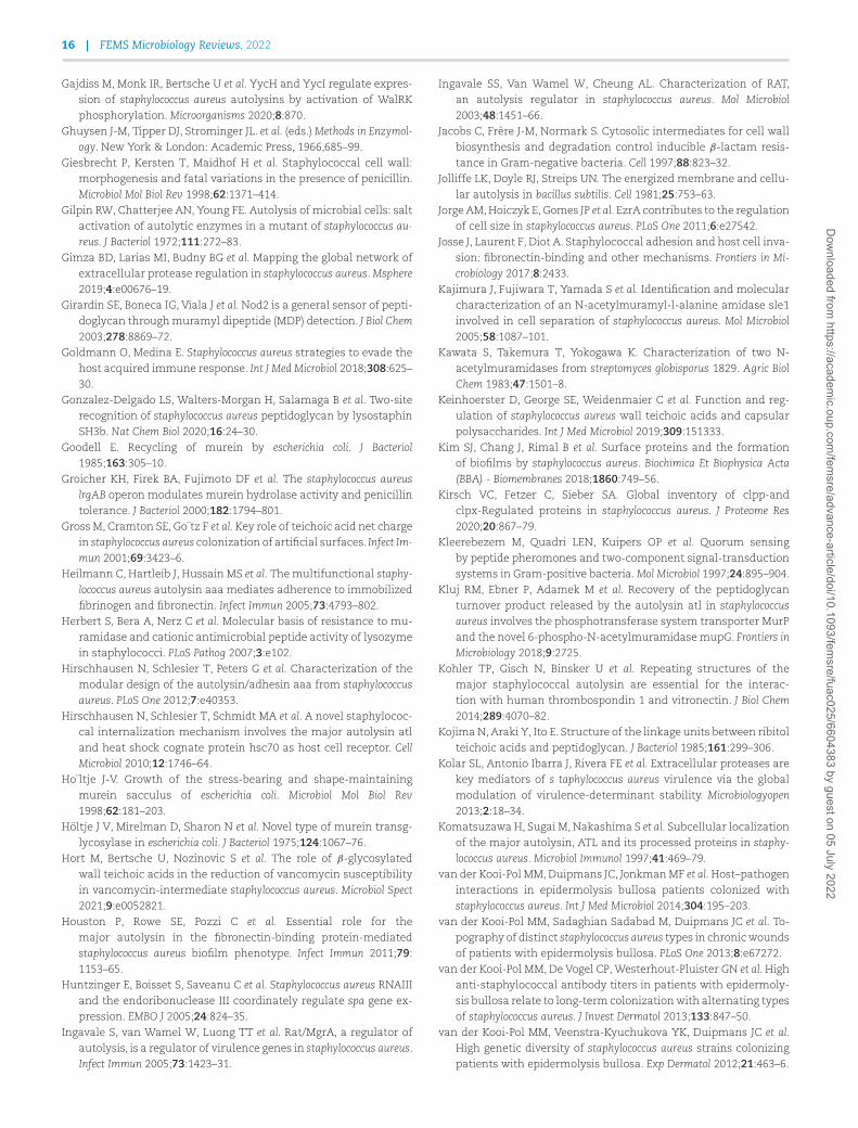
16 | FEMS Microbiology Reviews, 2022
Gajdiss M, Monk IR, Bertsche U et al. YycH and YycI regulate expres-sion of staphylococcus aureus autolysins by activation of WalRKphosphorylation. Microorganisms 2020;8:870.
Ghuysen J-M, Tipper DJ, Strominger JL. et al. (eds.) Methods in Enzymol-ogy. New York & London: Academic Press, 1966,685–99.
Giesbrecht P, Kersten T, Maidhof H et al. Staphylococcal cell wall:morphogenesis and fatal variations in the presence of penicillin.Microbiol Mol Biol Rev 1998;62:1371–414.
Gilpin RW, Chatterjee AN, Young FE. Autolysis of microbial cells: saltactivation of autolytic enzymes in a mutant of staphylococcus au-reus. J Bacteriol 1972;111:272–83.
Gimza BD, Larias MI, Budny BG et al. Mapping the global network ofextracellular protease regulation in staphylococcus aureus. Msphere2019;4:e00676–19.
Girardin SE, Boneca IG, Viala J et al. Nod2 is a general sensor of pepti-doglycan through muramyl dipeptide (MDP) detection. J Biol Chem2003;278:8869–72.
Goldmann O, Medina E. Staphylococcus aureus strategies to evade thehost acquired immune response. Int J Med Microbiol 2018;308:625–30.
Gonzalez-Delgado LS, Walters-Morgan H, Salamaga B et al. Two-siterecognition of staphylococcus aureus peptidoglycan by lysostaphinSH3b. Nat Chem Biol 2020;16:24–30.
Goodell E. Recycling of murein by escherichia coli. J Bacteriol1985;163:305–10.
Groicher KH, Firek BA, Fujimoto DF et al. The staphylococcus aureuslrgAB operon modulates murein hydrolase activity and penicillintolerance. J Bacteriol 2000;182:1794–801.
Gross M, Cramton SE, Go¨tz F et al. Key role of teichoic acid net chargein staphylococcus aureus colonization of artificial surfaces. Infect Im-mun 2001;69:3423–6.
Heilmann C, Hartleib J, Hussain MS et al. The multifunctional staphy-lococcus aureus autolysin aaa mediates adherence to immobilizedfibrinogen and fibronectin. Infect Immun 2005;73:4793–802.
Herbert S, Bera A, Nerz C et al. Molecular basis of resistance to mu-ramidase and cationic antimicrobial peptide activity of lysozymein staphylococci. PLoS Pathog 2007;3:e102.
Hirschhausen N, Schlesier T, Peters G et al. Characterization of themodular design of the autolysin/adhesin aaa from staphylococcusaureus. PLoS One 2012;7:e40353.
Hirschhausen N, Schlesier T, Schmidt MA et al. A novel staphylococ-cal internalization mechanism involves the major autolysin atland heat shock cognate protein hsc70 as host cell receptor. CellMicrobiol 2010;12:1746–64.
Ho¨ltje J-V. Growth of the stress-bearing and shape-maintainingmurein sacculus of escherichia coli. Microbiol Mol Biol Rev1998;62:181–203.
Höltje J V, Mirelman D, Sharon N et al. Novel type of murein transg-lycosylase in escherichia coli. J Bacteriol 1975;124:1067–76.
Hort M, Bertsche U, Nozinovic S et al. The role of β-glycosylatedwall teichoic acids in the reduction of vancomycin susceptibilityin vancomycin-intermediate staphylococcus aureus. Microbiol Spect2021;9:e0052821.
Houston P, Rowe SE, Pozzi C et al. Essential role for themajor autolysin in the fibronectin-binding protein-mediatedstaphylococcus aureus biofilm phenotype. Infect Immun 2011;79:1153–65.
Huntzinger E, Boisset S, Saveanu C et al. Staphylococcus aureus RNAIIIand the endoribonuclease III coordinately regulate spa gene ex-pression. EMBO J 2005;24:824–35.
Ingavale S, van Wamel W, Luong TT et al. Rat/MgrA, a regulator ofautolysis, is a regulator of virulence genes in staphylococcus aureus.Infect Immun 2005;73:1423–31.
Ingavale SS, Van Wamel W, Cheung AL. Characterization of RAT,an autolysis regulator in staphylococcus aureus. Mol Microbiol2003;48:1451–66.
Jacobs C, Frère J-M, Normark S. Cytosolic intermediates for cell wallbiosynthesis and degradation control inducible β-lactam resis-tance in Gram-negative bacteria. Cell 1997;88:823–32.
Jolliffe LK, Doyle RJ, Streips UN. The energized membrane and cellu-lar autolysis in bacillus subtilis. Cell 1981;25:753–63.
Jorge AM, Hoiczyk E, Gomes JP et al. EzrA contributes to the regulationof cell size in staphylococcus aureus. PLoS One 2011;6:e27542.
Josse J, Laurent F, Diot A. Staphylococcal adhesion and host cell inva-sion: fibronectin-binding and other mechanisms. Frontiers in Mi-crobiology 2017;8:2433.
Kajimura J, Fujiwara T, Yamada S et al. Identification and molecularcharacterization of an N-acetylmuramyl-l-alanine amidase sle1involved in cell separation of staphylococcus aureus. Mol Microbiol2005;58:1087–101.
Kawata S, Takemura T, Yokogawa K. Characterization of two N-acetylmuramidases from streptomyces globisporus 1829. Agric BiolChem 1983;47:1501–8.
Keinhoerster D, George SE, Weidenmaier C et al. Function and reg-ulation of staphylococcus aureus wall teichoic acids and capsularpolysaccharides. Int J Med Microbiol 2019;309:151333.
Kim SJ, Chang J, Rimal B et al. Surface proteins and the formationof biofilms by staphylococcus aureus. Biochimica Et Biophysica Acta(BBA) - Biomembranes 2018;1860:749–56.
Kirsch VC, Fetzer C, Sieber SA. Global inventory of clpp-andclpx-Regulated proteins in staphylococcus aureus. J Proteome Res2020;20:867–79.
Kleerebezem M, Quadri LEN, Kuipers OP et al. Quorum sensingby peptide pheromones and two-component signal-transductionsystems in Gram-positive bacteria. Mol Microbiol 1997;24:895–904.
Kluj RM, Ebner P, Adamek M et al. Recovery of the peptidoglycanturnover product released by the autolysin atl in staphylococcusaureus involves the phosphotransferase system transporter MurPand the novel 6-phospho-N-acetylmuramidase mupG. Frontiers inMicrobiology 2018;9:2725.
Kohler TP, Gisch N, Binsker U et al. Repeating structures of themajor staphylococcal autolysin are essential for the interac-tion with human thrombospondin 1 and vitronectin. J Biol Chem2014;289:4070–82.
Kojima N, Araki Y, Ito E. Structure of the linkage units between ribitolteichoic acids and peptidoglycan. J Bacteriol 1985;161:299–306.
Kolar SL, Antonio Ibarra J, Rivera FE et al. Extracellular proteases arekey mediators of s taphylococcus aureus virulence via the globalmodulation of virulence-determinant stability. Microbiologyopen2013;2:18–34.
Komatsuzawa H, Sugai M, Nakashima S et al. Subcellular localizationof the major autolysin, ATL and its processed proteins in staphy-lococcus aureus. Microbiol Immunol 1997;41:469–79.
van der Kooi-Pol MM, Duipmans JC, Jonkman MF et al. Host–pathogeninteractions in epidermolysis bullosa patients colonized withstaphylococcus aureus. Int J Med Microbiol 2014;304:195–203.
van der Kooi-Pol MM, Sadaghian Sadabad M, Duipmans JC et al. To-pography of distinct staphylococcus aureus types in chronic woundsof patients with epidermolysis bullosa. PLoS One 2013;8:e67272.
van der Kooi-Pol MM, De Vogel CP, Westerhout-Pluister GN et al. Highanti-staphylococcal antibody titers in patients with epidermoly-sis bullosa relate to long-term colonization with alternating typesof staphylococcus aureus. J Invest Dermatol 2013;133:847–50.
van der Kooi-Pol MM, Veenstra-Kyuchukova YK, Duipmans JC et al.High genetic diversity of staphylococcus aureus strains colonizingpatients with epidermolysis bullosa. Exp Dermatol 2012;21:463–6.
Dow
nloaded from https://academ
ic.oup.com/fem
sre/advance-article/doi/10.1093/femsre/fuac025/6604383 by guest on 05 July 2022
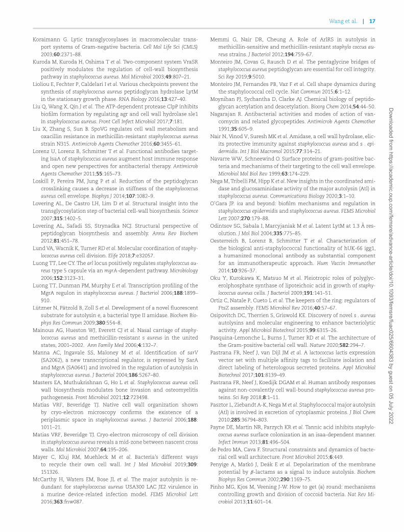
Wang et al. | 17
Koraimann G. Lytic transglycosylases in macromolecular trans-port systems of Gram-negative bacteria. Cell Mol Life Sci (CMLS)2003;60:2371–88.
Kuroda M, Kuroda H, Oshima T et al. Two-component system VraSRpositively modulates the regulation of cell-wall biosynthesispathway in staphylococcus aureus. Mol Microbiol 2003;49:807–21.
Lioliou E, Fechter P, Caldelari I et al. Various checkpoints prevent thesynthesis of staphylococcus aureus peptidoglycan hydrolase LytMin the stationary growth phase. RNA Biology 2016;13:427–40.
Liu Q, Wang X, Qin J et al. The ATP-dependent protease ClpP inhibitsbiofilm formation by regulating agr and cell wall hydrolase sle1in staphylococcus aureus. Front Cell Infect Microbiol 2017;7:181.
Liu X, Zhang S, Sun B. SpoVG regulates cell wall metabolism andoxacillin resistance in methicillin-resistant staphylococcus aureusstrain N315. Antimicrob Agents Chemother 2016;60:3455–61.
Lorenz U, Lorenz B, Schmitter T et al. Functional antibodies target-ing IsaA of staphylococcus aureus augment host immune responseand open new perspectives for antibacterial therapy. AntimicrobAgents Chemother 2011;55:165–73.
Loskill P, Pereira PM, Jung P et al. Reduction of the peptidoglycancrosslinking causes a decrease in stiffness of the staphylococcusaureus cell envelope. Biophys J 2014;107:1082–9.
Lovering AL, De Castro LH, Lim D et al. Structural insight into thetransglycosylation step of bacterial cell-wall biosynthesis. Science2007;315:1402–5.
Lovering AL, Safadi SS, Strynadka NCJ. Structural perspective ofpeptidoglycan biosynthesis and assembly. Annu Rev Biochem2012;81:451–78.
Lund VA, Wacnik K, Turner RD et al. Molecular coordination of staphy-lococcus aureus cell division. Elife 2018;7:e32057.
Luong TT, Lee CY. The arl locus positively regulates staphylococcus au-reus type 5 capsule via an mgrA-dependent pathway. Microbiology2006;152:3123–31.
Luong TT, Dunman PM, Murphy E et al. Transcription profiling of theMgrA regulon in staphylococcus aureus. J Bacteriol 2006;188:1899–910.
Lützner N, Pätzold B, Zoll S et al. Development of a novel fluorescentsubstrate for autolysin e, a bacterial type II amidase. Biochem Bio-phys Res Commun 2009;380:554–8.
Mainous AG, Hueston WJ, Everett CJ et al. Nasal carriage of staphy-lococcus aureus and methicillin-resistant s aureus in the unitedstates, 2001–2002. Ann Family Med 2006;4:132–7.
Manna AC, Ingavale SS, Maloney M et al. Identification of sarV(SA2062), a new transcriptional regulator, is repressed by SarAand MgrA (SA0641) and involved in the regulation of autolysis instaphylococcus aureus. J Bacteriol 2004;186:5267–80.
Masters EA, Muthukrishnan G, Ho L et al. Staphylococcus aureus cellwall biosynthesis modulates bone invasion and osteomyelitispathogenesis. Front Microbiol 2021;12:723498.
Matias VRF, Beveridge TJ. Native cell wall organization shownby cryo-electron microscopy confirms the existence of aperiplasmic space in staphylococcus aureus. J Bacteriol 2006;188:1011–21.
Matias VRF, Beveridge TJ. Cryo-electron microscopy of cell divisionin staphylococcus aureus reveals a mid-zone between nascent crosswalls. Mol Microbiol 2007;64:195–206.
Mayer C, Kluj RM, Muehleck M et al. Bacteria’s different waysto recycle their own cell wall. Int J Med Microbiol 2019;309:151326.
McCarthy H, Waters EM, Bose JL et al. The major autolysin is re-dundant for staphylococcus aureus USA300 LAC JE2 virulence ina murine device-related infection model. FEMS Microbiol Lett2016;363:fnw087.
Memmi G, Nair DR, Cheung A. Role of ArlRS in autolysis inmethicillin-sensitive and methicillin-resistant staphylo coccus au-reus strains. J Bacteriol 2012;194:759–67.
Monteiro JM, Covas G, Rausch D et al. The pentaglycine bridges ofstaphylococcus aureus peptidoglycan are essential for cell integrity.Sci Rep 2019;9:5010.
Monteiro JM, Fernandes PB, Vaz F et al. Cell shape dynamics duringthe staphylococcal cell cycle. Nat Commun 2015;6:1–12.
Moynihan PJ, Sychantha D, Clarke AJ. Chemical biology of peptido-glycan acetylation and deacetylation. Bioorg Chem 2014;54:44–50.
Nagarajan R. Antibacterial activities and modes of action of van-comycin and related glycopeptides. Antimicrob Agents Chemother1991;35:605–9.
Nair N, Vinod V, Suresh MK et al. Amidase, a cell wall hydrolase, elic-its protective immunity against staphylococcus aureus and s . epi-dermidis. Int J Biol Macromol 2015;77:314–21.
Navarre WW, Schneewind O. Surface proteins of gram-positive bac-teria and mechanisms of their targeting to the cell wall envelope.Microbiol Mol Biol Rev 1999;63:174–229.
Nega M, Tribelli PM, Hipp K et al. New insights in the coordinated ami-dase and glucosaminidase activity of the major autolysin (Atl) instaphylococcus aureus. Communications Biology 2020;3:1–10.
O’Gara JP. ica and beyond: biofilm mechanisms and regulation instaphylococcus epidermidis and staphylococcus aureus. FEMS MicrobiolLett 2007;270:179–88.
Odintsov SG, Sabala I, Marcyjaniak M et al. Latent LytM at 1.3 A res-olution. J Mol Biol 2004;335:775–85.
Oesterreich B, Lorenz B, Schmitter T et al. Characterization ofthe biological anti-staphylococcal functionality of hUK-66 igg1,a humanized monoclonal antibody as substantial componentfor an immunotherapeutic approach. Hum Vaccin Immunother2014;10:926–37.
Oku Y, Kurokawa K, Matsuo M et al. Pleiotropic roles of polyglyc-erolphosphate synthase of lipoteichoic acid in growth of staphy-lococcus aureus cells. J Bacteriol 2009;191:141–51.
Ortiz C, Natale P, Cueto L et al. The keepers of the ring: regulators ofFtsZ assembly. FEMS Microbiol Rev 2016;40:57–67.
Osipovitch DC, Therrien S, Griswold KE. Discovery of novel s . aureusautolysins and molecular engineering to enhance bacteriolyticactivity. Appl Microbiol Biotechnol 2015;99:6315–26.
Pasquina-Lemonche L, Burns J, Turner RD et al. The architecture ofthe Gram-positive bacterial cell wall. Nature 2020;582:294–7.
Pastrana FR, Neef J, van Dijl JM et al. A lactococcus lactis expressionvector set with multiple affinity tags to facilitate isolation anddirect labeling of heterologous secreted proteins. Appl MicrobiolBiotechnol 2017;101:8139–49.
Pastrana FR, Neef J, Koedijk DGAM et al. Human antibody responsesagainst non-covalently cell wall-bound staphylococcus aureus pro-teins. Sci Rep 2018;8:1–11.
Pasztor L, Ziebandt A-K, Nega M et al. Staphylococcal major autolysin(Atl) is involved in excretion of cytoplasmic proteins. J Biol Chem2010;285:36794–803.
Payne DE, Martin NR, Parzych KR et al. Tannic acid inhibits staphylo-coccus aureus surface colonization in an isaa-dependent manner.Infect Immun 2013;81:496–504.
de Pedro MA, Cava F. Structural constraints and dynamics of bacte-rial cell wall architecture. Front Microbiol 2015;6:449.
Penyige A, Matkó J, Deák E et al. Depolarization of the membranepotential by β-lactams as a signal to induce autolysis. BiochemBiophys Res Commun 2002;290:1169–75.
Pinho MG, Kjos M, Veening J-W. How to get (a) round: mechanismscontrolling growth and division of coccoid bacteria. Nat Rev Mi-crobiol 2013;11:601–14.
Dow
nloaded from https://academ
ic.oup.com/fem
sre/advance-article/doi/10.1093/femsre/fuac025/6604383 by guest on 05 July 2022
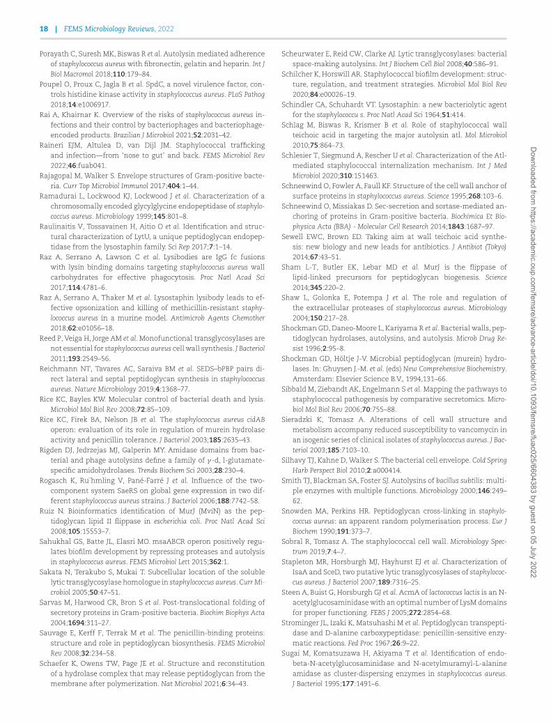
18 | FEMS Microbiology Reviews, 2022
Porayath C, Suresh MK, Biswas R et al. Autolysin mediated adherenceof staphylococcus aureus with fibronectin, gelatin and heparin. Int JBiol Macromol 2018;110:179–84.
Poupel O, Proux C, Jagla B et al. SpdC, a novel virulence factor, con-trols histidine kinase activity in staphylococcus aureus. PLoS Pathog2018;14:e1006917.
Rai A, Khairnar K. Overview of the risks of staphylococcus aureus in-fections and their control by bacteriophages and bacteriophage-encoded products. Brazilian J Microbiol 2021;52:2031–42.
Raineri EJM, Altulea D, van Dijl JM. Staphylococcal traffickingand infection—from ‘nose to gut’ and back. FEMS Microbiol Rev2022;46:fuab041.
Rajagopal M, Walker S. Envelope structures of Gram-positive bacte-ria. Curr Top Microbiol Immunol 2017;404:1–44.
Ramadurai L, Lockwood KJ, Lockwood J et al. Characterization of achromosomally encoded glycylglycine endopeptidase of staphylo-coccus aureus. Microbiology 1999;145:801–8.
Raulinaitis V, Tossavainen H, Aitio O et al. Identification and struc-tural characterization of LytU, a unique peptidoglycan endopep-tidase from the lysostaphin family. Sci Rep 2017;7:1–14.
Raz A, Serrano A, Lawson C et al. Lysibodies are IgG fc fusionswith lysin binding domains targeting staphylococcus aureus wallcarbohydrates for effective phagocytosis. Proc Natl Acad Sci2017;114:4781–6.
Raz A, Serrano A, Thaker M et al. Lysostaphin lysibody leads to ef-fective opsonization and killing of methicillin-resistant staphy-lococcus aureus in a murine model. Antimicrob Agents Chemother2018;62:e01056–18.
Reed P, Veiga H, Jorge AM et al. Monofunctional transglycosylases arenot essential for staphylococcus aureus cell wall synthesis. J Bacteriol2011;193:2549–56.
Reichmann NT, Tavares AC, Saraiva BM et al. SEDS–bPBP pairs di-rect lateral and septal peptidoglycan synthesis in staphylococcusaureus. Nature Microbiology 2019;4:1368–77.
Rice KC, Bayles KW. Molecular control of bacterial death and lysis.Microbiol Mol Biol Rev 2008;72:85–109.
Rice KC, Firek BA, Nelson JB et al. The staphylococcus aureus cidABoperon: evaluation of its role in regulation of murein hydrolaseactivity and penicillin tolerance. J Bacteriol 2003;185:2635–43.
Rigden DJ, Jedrzejas MJ, Galperin MY. Amidase domains from bac-terial and phage autolysins define a family of γ -d, l-glutamate-specific amidohydrolases. Trends Biochem Sci 2003;28:230–4.
Rogasch K, Ru¨hmling V, Pané-Farré J et al. Influence of the two-component system SaeRS on global gene expression in two dif-ferent staphylococcus aureus strains. J Bacteriol 2006;188:7742–58.
Ruiz N. Bioinformatics identification of MurJ (MviN) as the pep-tidoglycan lipid II flippase in escherichia coli. Proc Natl Acad Sci2008;105:15553–7.
Sahukhal GS, Batte JL, Elasri MO. msaABCR operon positively regu-lates biofilm development by repressing proteases and autolysisin staphylococcus aureus. FEMS Microbiol Lett 2015;362:1.
Sakata N, Terakubo S, Mukai T. Subcellular location of the solublelytic transglycosylase homologue in staphylococcus aureus. Curr Mi-crobiol 2005;50:47–51.
Sarvas M, Harwood CR, Bron S et al. Post-translocational folding ofsecretory proteins in Gram-positive bacteria. Biochim Biophys Acta2004;1694:311–27.
Sauvage E, Kerff F, Terrak M et al. The penicillin-binding proteins:structure and role in peptidoglycan biosynthesis. FEMS MicrobiolRev 2008;32:234–58.
Schaefer K, Owens TW, Page JE et al. Structure and reconstitutionof a hydrolase complex that may release peptidoglycan from themembrane after polymerization. Nat Microbiol 2021;6:34–43.
Scheurwater E, Reid CW, Clarke AJ. Lytic transglycosylases: bacterialspace-making autolysins. Int J Biochem Cell Biol 2008;40:586–91.
Schilcher K, Horswill AR. Staphylococcal biofilm development: struc-ture, regulation, and treatment strategies. Microbiol Mol Biol Rev2020;84:e00026–19.
Schindler CA, Schuhardt VT. Lysostaphin: a new bacteriolytic agentfor the staphylococcu s. Proc Natl Acad Sci 1964;51:414.
Schlag M, Biswas R, Krismer B et al. Role of staphylococcal wallteichoic acid in targeting the major autolysin atl. Mol Microbiol2010;75:864–73.
Schlesier T, Siegmund A, Rescher U et al. Characterization of the Atl-mediated staphylococcal internalization mechanism. Int J MedMicrobiol 2020;310:151463.
Schneewind O, Fowler A, Faull KF. Structure of the cell wall anchor ofsurface proteins in staphylococcus aureus. Science 1995;268:103–6.
Schneewind O, Missiakas D. Sec-secretion and sortase-mediated an-choring of proteins in Gram-positive bacteria. Biochimica Et Bio-physica Acta (BBA) - Molecular Cell Research 2014;1843:1687–97.
Sewell EWC, Brown ED. Taking aim at wall teichoic acid synthe-sis: new biology and new leads for antibiotics. J Antibiot (Tokyo)2014;67:43–51.
Sham L-T, Butler EK, Lebar MD et al. MurJ is the flippase oflipid-linked precursors for peptidoglycan biogenesis. Science2014;345:220–2.
Shaw L, Golonka E, Potempa J et al. The role and regulation ofthe extracellular proteases of staphylococcus aureus. Microbiology2004;150:217–28.
Shockman GD, Daneo-Moore L, Kariyama R et al. Bacterial walls, pep-tidoglycan hydrolases, autolysins, and autolysis. Microb Drug Re-sist 1996;2:95–8.
Shockman GD, Höltje J-V. Microbial peptidoglycan (murein) hydro-lases. In: Ghuysen J.-M. et al. (eds) New Comprehensive Biochemistry.Amsterdam: Elsevier Science B.V., 1994,131–66.
Sibbald M, Ziebandt AK, Engelmann S et al. Mapping the pathways tostaphylococcal pathogenesis by comparative secretomics. Micro-biol Mol Biol Rev 2006;70:755–88.
Sieradzki K, Tomasz A. Alterations of cell wall structure andmetabolism accompany reduced susceptibility to vancomycin inan isogenic series of clinical isolates of staphylococcus aureus. J Bac-teriol 2003;185:7103–10.
Silhavy TJ, Kahne D, Walker S. The bacterial cell envelope. Cold SpringHarb Perspect Biol 2010;2:a000414.
Smith TJ, Blackman SA, Foster SJ. Autolysins of bacillus subtilis: multi-ple enzymes with multiple functions. Microbiology 2000;146:249–62.
Snowden MA, Perkins HR. Peptidoglycan cross-linking in staphylo-coccus aureus: an apparent random polymerisation process. Eur JBiochem 1990;191:373–7.
Sobral R, Tomasz A. The staphylococcal cell wall. Microbiology Spec-trum 2019;7:4–7.
Stapleton MR, Horsburgh MJ, Hayhurst EJ et al. Characterization ofIsaA and SceD, two putative lytic transglycosylases of staphylococ-cus aureus. J Bacteriol 2007;189:7316–25.
Steen A, Buist G, Horsburgh GJ et al. AcmA of lactococcus lactis is an N-acetylglucosaminidase with an optimal number of LysM domainsfor proper functioning. FEBS J 2005;272:2854–68.
Strominger JL, Izaki K, Matsuhashi M et al. Peptidoglycan transpepti-dase and D-alanine carboxypeptidase: penicillin-sensitive enzy-matic reactions. Fed Proc 1967;26:9–22.
Sugai M, Komatsuzawa H, Akiyama T et al. Identification of endo-beta-N-acetylglucosaminidase and N-acetylmuramyl-L-alanineamidase as cluster-dispersing enzymes in staphylococcus aureus.J Bacteriol 1995;177:1491–6.
Dow
nloaded from https://academ
ic.oup.com/fem
sre/advance-article/doi/10.1093/femsre/fuac025/6604383 by guest on 05 July 2022
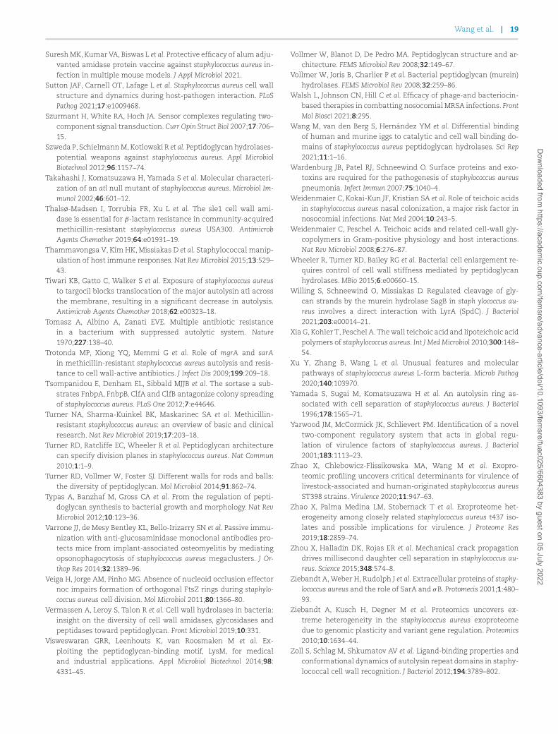
Wang et al. | 19
Suresh MK, Kumar VA, Biswas L et al. Protective efficacy of alum adju-vanted amidase protein vaccine against staphylococcus aureus in-fection in multiple mouse models. J Appl Microbiol 2021.
Sutton JAF, Carnell OT, Lafage L et al. Staphylococcus aureus cell wallstructure and dynamics during host-pathogen interaction. PLoSPathog 2021;17:e1009468.
Szurmant H, White RA, Hoch JA. Sensor complexes regulating two-component signal transduction. Curr Opin Struct Biol 2007;17:706–15.
Szweda P, Schielmann M, Kotlowski R et al. Peptidoglycan hydrolases-potential weapons against staphylococcus aureus. Appl MicrobiolBiotechnol 2012;96:1157–74.
Takahashi J, Komatsuzawa H, Yamada S et al. Molecular characteri-zation of an atl null mutant of staphylococcus aureus. Microbiol Im-munol 2002;46:601–12.
Thalsø-Madsen I, Torrubia FR, Xu L et al. The sle1 cell wall ami-dase is essential for β-lactam resistance in community-acquiredmethicillin-resistant staphylococcus aureus USA300. AntimicrobAgents Chemother 2019;64:e01931–19.
Thammavongsa V, Kim HK, Missiakas D et al. Staphylococcal manip-ulation of host immune responses. Nat Rev Microbiol 2015;13:529–43.
Tiwari KB, Gatto C, Walker S et al. Exposure of staphylococcus aureusto targocil blocks translocation of the major autolysin atl acrossthe membrane, resulting in a significant decrease in autolysis.Antimicrob Agents Chemother 2018;62:e00323–18.
Tomasz A, Albino A, Zanati EVE. Multiple antibiotic resistancein a bacterium with suppressed autolytic system. Nature1970;227:138–40.
Trotonda MP, Xiong YQ, Memmi G et al. Role of mgrA and sarAin methicillin-resistant staphylococcus aureus autolysis and resis-tance to cell wall-active antibiotics. J Infect Dis 2009;199:209–18.
Tsompanidou E, Denham EL, Sibbald MJJB et al. The sortase a sub-strates FnbpA, FnbpB, ClfA and ClfB antagonize colony spreadingof staphylococcus aureus. PLoS One 2012;7:e44646.
Turner NA, Sharma-Kuinkel BK, Maskarinec SA et al. Methicillin-resistant staphylococcus aureus: an overview of basic and clinicalresearch. Nat Rev Microbiol 2019;17:203–18.
Turner RD, Ratcliffe EC, Wheeler R et al. Peptidoglycan architecturecan specify division planes in staphylococcus aureus. Nat Commun2010;1:1–9.
Turner RD, Vollmer W, Foster SJ. Different walls for rods and balls:the diversity of peptidoglycan. Mol Microbiol 2014;91:862–74.
Typas A, Banzhaf M, Gross CA et al. From the regulation of pepti-doglycan synthesis to bacterial growth and morphology. Nat RevMicrobiol 2012;10:123–36.
Varrone JJ, de Mesy Bentley KL, Bello-Irizarry SN et al. Passive immu-nization with anti-glucosaminidase monoclonal antibodies pro-tects mice from implant-associated osteomyelitis by mediatingopsonophagocytosis of staphylococcus aureus megaclusters. J Or-thop Res 2014;32:1389–96.
Veiga H, Jorge AM, Pinho MG. Absence of nucleoid occlusion effectornoc impairs formation of orthogonal FtsZ rings during staphylo-coccus aureus cell division. Mol Microbiol 2011;80:1366–80.
Vermassen A, Leroy S, Talon R et al. Cell wall hydrolases in bacteria:insight on the diversity of cell wall amidases, glycosidases andpeptidases toward peptidoglycan. Front Microbiol 2019;10:331.
Visweswaran GRR, Leenhouts K, van Roosmalen M et al. Ex-ploiting the peptidoglycan-binding motif, LysM, for medicaland industrial applications. Appl Microbiol Biotechnol 2014;98:4331–45.
Vollmer W, Blanot D, De Pedro MA. Peptidoglycan structure and ar-chitecture. FEMS Microbiol Rev 2008;32:149–67.
Vollmer W, Joris B, Charlier P et al. Bacterial peptidoglycan (murein)hydrolases. FEMS Microbiol Rev 2008;32:259–86.
Walsh L, Johnson CN, Hill C et al. Efficacy of phage-and bacteriocin-based therapies in combatting nosocomial MRSA infections. FrontMol Biosci 2021;8:295.
Wang M, van den Berg S, Hernández YM et al. Differential bindingof human and murine iggs to catalytic and cell wall binding do-mains of staphylococcus aureus peptidoglycan hydrolases. Sci Rep2021;11:1–16.
Wardenburg JB, Patel RJ, Schneewind O. Surface proteins and exo-toxins are required for the pathogenesis of staphylococcus aureuspneumonia. Infect Immun 2007;75:1040–4.
Weidenmaier C, Kokai-Kun JF, Kristian SA et al. Role of teichoic acidsin staphylococcus aureus nasal colonization, a major risk factor innosocomial infections. Nat Med 2004;10:243–5.
Weidenmaier C, Peschel A. Teichoic acids and related cell-wall gly-copolymers in Gram-positive physiology and host interactions.Nat Rev Microbiol 2008;6:276–87.
Wheeler R, Turner RD, Bailey RG et al. Bacterial cell enlargement re-quires control of cell wall stiffness mediated by peptidoglycanhydrolases. MBio 2015;6:e00660–15.
Willing S, Schneewind O, Missiakas D. Regulated cleavage of gly-can strands by the murein hydrolase SagB in staph ylococcus au-reus involves a direct interaction with LyrA (SpdC). J Bacteriol2021;203:e00014–21.
Xia G, Kohler T, Peschel A. The wall teichoic acid and lipoteichoic acidpolymers of staphylococcus aureus. Int J Med Microbiol 2010;300:148–54.
Xu Y, Zhang B, Wang L et al. Unusual features and molecularpathways of staphylococcus aureus L-form bacteria. Microb Pathog2020;140:103970.
Yamada S, Sugai M, Komatsuzawa H et al. An autolysin ring as-sociated with cell separation of staphylococcus aureus. J Bacteriol1996;178:1565–71.
Yarwood JM, McCormick JK, Schlievert PM. Identification of a noveltwo-component regulatory system that acts in global regu-lation of virulence factors of staphylococcus aureus. J Bacteriol2001;183:1113–23.
Zhao X, Chlebowicz-Flissikowska MA, Wang M et al. Exopro-teomic profiling uncovers critical determinants for virulence oflivestock-associated and human-originated staphylococcus aureusST398 strains. Virulence 2020;11:947–63.
Zhao X, Palma Medina LM, Stobernack T et al. Exoproteome het-erogeneity among closely related staphylococcus aureus t437 iso-lates and possible implications for virulence. J Proteome Res2019;18:2859–74.
Zhou X, Halladin DK, Rojas ER et al. Mechanical crack propagationdrives millisecond daughter cell separation in staphylococcus au-reus. Science 2015;348:574–8.
Ziebandt A, Weber H, Rudolph J et al. Extracellular proteins of staphy-lococcus aureus and the role of SarA and σB. Protomecis 2001;1:480–93.
Ziebandt A, Kusch H, Degner M et al. Proteomics uncovers ex-treme heterogeneity in the staphylococcus aureus exoproteomedue to genomic plasticity and variant gene regulation. Proteomics2010;10:1634–44.
Zoll S, Schlag M, Shkumatov AV et al. Ligand-binding properties andconformational dynamics of autolysin repeat domains in staphy-lococcal cell wall recognition. J Bacteriol 2012;194:3789–802.
Dow
nloaded from https://academ
ic.oup.com/fem
sre/advance-article/doi/10.1093/femsre/fuac025/6604383 by guest on 05 July 2022