methicillin-resistant staphylococcus aureus: a novel approach
TRANSCRIPT
1
Methicillin-resistant Staphylococcus aureus: a novel approach to molecular
detection and a US countywide study of strain diversity and distribution among
healthcare facilities
Lyndsey Olivia Hudson
Imperial College London
Department of Infectious Disease Epidemiology
Thesis submitted for the degree of Doctor of Philosophy of Imperial College
London

2
I declare that all work presented in this thesis is my own, unless otherwise stated.
3
ABSTRACT
Methicillin-resistant Staphylococcus aureus (MRSA) is a global public health
problem and is a major cause of morbidity and mortality worldwide, imposing serious
economic costs on patients and hospitals. Prior to the mid-1990s, MRSA was largely
a healthcare-associated pathogen, causing infection predominantly in people with
frequent or recent contact with healthcare facilities (HA-MRSA). Since then,
community-associated MRSA (CA-MRSA), which often causes infection among
healthy children and young adults with no exposure to the healthcare setting, has
become increasingly prevalent. Worryingly, there is evidence that CA-MRSA is
penetrating the healthcare MRSA reservoir, and even replacing traditional HA-MRSA
strains. This highlights the need to keep abreast of the changing epidemiology of
MRSA in order to implement effective infection control strategies. To investigate the
composition of the healthcare MRSA reservoir and ascertain the extent to which CA-
MRSA has penetrated this reservoir, a countywide, population-based cohort study of
MRSA in hospital inpatients and nursing home residents was conducted in Orange
County (OC), California, covering a total of 46 facilities. CA-MRSA was found to be
fully mixed with HA-MRSA in the hospital setting. The predominant CA-MRSA
clone in the US, USA300, was the most commonly isolated MRSA clone in OC
hospitals. In OC nursing homes, HA-MRSA (specifically a variant of USA100 that is
also very common in OC hospitals but has not been reported elsewhere)
predominates, but USA300 made up just over a quarter of the isolates and was the
second most frequently isolated clone. Both OC hospitals and nursing homes were
dominated by the same three strains: USA300, USA100 and a variant of USA100.
Not only are community-based infection control strategies needed to stem the influx
of community associated strains, in particular USA300, into the hospital setting, but
also strategies tailored to the complex problem of MRSA transmission and infection
4
in nursing homes, to minimise the impact of the unique nursing home MRSA
reservoir on overall regional MRSA burden. A key component of effective infection
control strategies is prompt isolation of MRSA carriers, facilitated by rapid
diagnostics. PCR-based methods of MRSA detection offer a much faster alternative to
traditional culture techniques, but are expensive and often complex to operate. A
novel nucleic acid amplification technique developed by my industrial sponsor,
TwistDx Ltd, called recombinase polymerase amplification (RPA), has been
incorporated into a probe based detection system called TwistAmp MRSA, and offers
a simple and cheap alternative to current commercial PCR-based assays, amplifying
MRSA to detectable levels within 20 minutes. I tested the assay with diverse
collections of MRSA and discovered that 4% of isolates from a UK MRSA collection
could not be detected by the assay. I subsequently developed RPA primers for their
detection. Nonetheless, TwistAmp MRSA was able to detect most MRSA strains, and
was comparable to current commercial assays in this respect. Despite a very high
analytical sensitivity of approximately 20 CFU/swab, the clinical sensitivity of
TwistAmp MRSA was lower than expected with respect to the current market leader,
Xpert MRSA. I investigated lysis and filtration methods to improve the assay's
clinical sensitivity, but found that such methods did not currently warrant inclusion in
the TwistAmp MRSA protocol. While TwistAmp MRSA performance is in line with
current assays, and is a faster, cheaper and simpler assay, a problem faced by all
molecular methods of MRSA detection is the constant emergence of undetectable
MRSA strains, necessitating continual assay evaluation and improvement where
possible.

5
ACKNOWLEDGEMENTS
I thank my supervisors Professor Brian G. Spratt and Doctor Matthew Forrest
for their continual support throughout my PhD, and their invaluable subject
knowledge. They have both been a pleasure to work with. I also thank my family,
particularly my mother, for unwavering support throughout the course of my
university career. Without my family I would not have been able to go to university,
and for that I am extremely grateful.
My friends have also been a great source of motivation and inspiration over
the years, and so a special thank you goes to them. My friends and colleagues within
the Department of Infectious Disease Epidemiology have been a great pleasure to
work with, and I am very grateful to have been part of such a friendly and
academically excellent group of researchers.
I also thank my various collaborators, in particular Doctor Susan S. Huang and
Courtney R. Murphy from the University of California, Irvine, for giving me the
opportunity to work on such an exciting project, and for their immense enthusiasm
and hard-working spirit over the course of my PhD. A big thank you also goes to the
people at TwistDx Ltd, who provided a very friendly and supportive working
environment during my industrial placement.
Finally, I acknowledge the financial support of the Biotechnology and
Biological Sciences Research Council, and TwistDx Ltd, without which I could not
have pursued a PhD.
6
CONTENTS Abstract.........................................................................................................................3
Acknowledgements ......................................................................................................5
List of Tables ..............................................................................................................11
List of Figures.............................................................................................................13
List of Abbreviations .................................................................................................15
Chapter 1: Introduction ............................................................................................19
1.1 Staphylococcus aureus carriage and disease ..................................................19
1.2 Antibiotic resistance: emergence and mechanisms.......................................21
1.2.1 Mechanism of methicillin resistance ..........................................................25
1.3 Molecular epidemiology of S. aureus .............................................................30
1.3.1 Pulsed field gel electrophoresis (PFGE) .....................................................32
1.3.2 Multi locus sequence typing (MLST) .........................................................32
1.3.3 spa typing....................................................................................................33
1.3.4 SCCmec typing ...........................................................................................35
1.3.5 Advances in S. aureus molecular epidemiology.........................................35
1.4 The changing epidemiology of MRSA............................................................37
1.5 MRSA prevalence and burden .......................................................................45
1.6 Preventative measures .....................................................................................50
1.7 MRSA diagnosis ...............................................................................................52
1.6 Thesis objectives...............................................................................................60
Chapter 2: Methods ...................................................................................................62
2.1 Bacterial culture...............................................................................................62
2.2 DNA extraction.................................................................................................62
2.3 PCR amplification............................................................................................62
2.4 Sequencing of PCR products ..........................................................................63
2.5 MLST ................................................................................................................64
2.6 spa typing ..........................................................................................................64
2.7 SCCmec typing .................................................................................................65
Part 1 Introduction: RPA-based Detection of MRSA ............................................69
Chapter 3: TwistAmp MRSA ...................................................................................75

7
3.1 Introduction......................................................................................................75
3.1.1 The mec Right Extremity Junction (MREJ)................................................75
3.1.2 Chapter Objectives......................................................................................79
3.1.2.1 TwistAmp MRSA performance...........................................................79
3.1.2.2 MREJ typing of MRSA isolates ..........................................................79
3.1.2.3 Characterisation of false negative isolates ...........................................81
3.1.2.4 Scope of TwistAmp MRSA.................................................................82
3.2 Methods.............................................................................................................84
3.2.1 The UK MRSA collection ..........................................................................84
3.2.2 Kansas City paediatric hospital MRSA collection......................................85
3.2.3 Laboratory collection of Staphylococci ......................................................85
3.2.4 TwistAmp MRSA protocol.........................................................................86
3.2.5 MREJ typing protocol.................................................................................89
3.2.6 Characterisation of isolates .........................................................................89
3.2.7 Primer design and primer screening for novel MREJ types .......................90
3.2.8 Detection of SCCmec variants and problematic strains..............................93
3.3 Results ...............................................................................................................95
3.3.1 TwistAmp MRSA performance..................................................................95
3.3.1.1 UK MRSA collection...........................................................................95
3.3.1.2 KC MRSA collection...........................................................................95
3.3.1.3 Laboratory collection of Staphylococci ...............................................97
3.3.2 MREJ typing ...............................................................................................98
3.3.2.1 UK MRSA collection...........................................................................98
3.3.2.2 KC MRSA collection.........................................................................101
3.3.2.3 Laboratory MRSA collection.............................................................102
3.3.2.4 Correlation of MREJ type with SCCmec type...................................102
3.3.3 Characterisation of isolates .......................................................................106
3.3.3.1 False negative MRSA isolates from TwistAmp MRSA screen of UK
collection........................................................................................................106
3.3.3.2 Characterisation of the 49 KC MRSA isolates .................................115
3.3.4 Scope of TwistAmp MRSA......................................................................118
3.3.4.1 Testing a variant of a common MRSA clone in Copenhagen, Denmark
........................................................................................................................118
8
3.3.4.2 Testing common Australian MRSA clones not detected by BD
GeneOhm MRSA...........................................................................................119
3.3.4.3 Testing the livestock-associated MRSA clone ST398-MRSA-V......120
3.3.4.4 Testing prototype strains for SCCmec types I-XI with TwistAmp
MRSA ............................................................................................................121
3.4 Discussion........................................................................................................124
3.4.1 TwistAmp MRSA performance and scope ...............................................124
3.4.2 MREJ and SCCmec type...........................................................................139
Chapter 4: Improving Assay Performance............................................................142
4.1 Introduction....................................................................................................142
4.1.2 Chapter Objectives....................................................................................143
4.1.2.1 Limit of detection...............................................................................143
4.1.2.2 Inhibition and sub-sampling ..............................................................145
4.2 Methods and results.......................................................................................147
4.2.1 Limit of detection......................................................................................147
4.2.2 Inhibition and sub-sampling .....................................................................151
4.3 Discussion........................................................................................................172
4.3.1 Limit of detection......................................................................................172
4.3.2 Inhibition and sub-sampling .....................................................................174
4.3.3 Conclusions...............................................................................................180
Part 1 Final Discussion: RPA-Based Detection of MRSA....................................181
Part 2 Introduction: MRSA in Orange County, California.................................186
Chapter 5: Differences in MRSA Strains Isolated from Paediatric and Adult
Hospital Inpatients...................................................................................................191
5.1 Introduction....................................................................................................191
5.2 Methods...........................................................................................................192
5.2.1 Study .........................................................................................................192
5.2.2 Isolate collection .......................................................................................192
5.2.3 Specimen data and hospital characteristics...............................................193
5.2.4 Laboratory methods and molecular typing ...............................................193
5.2.5 Statistical analyses ....................................................................................194
5.3 Results .............................................................................................................195
9
5.4 Discussion........................................................................................................203
Chapter 6: Diversity of MRSA Strains Isolated from Inpatients of 30 Hospitals
....................................................................................................................................208
6.1 Introduction....................................................................................................208
6.2 Methods...........................................................................................................209
6.2.1 Study .........................................................................................................209
6.2.2 Isolate collection .......................................................................................209
6.2.3 Specimen data and hospital characteristics...............................................210
6.2.4 Laboratory methods and molecular typing ...............................................210
6.2.5 Statistical analyses ....................................................................................210
6.3 Results .............................................................................................................212
6.3.1 Overview...................................................................................................212
6.3.2 spa typing and MLST ...............................................................................214
6.3.3 Inter-hospital differences ..........................................................................220
6.3.4 Community onset versus hospital onset MRSA .......................................221
6.4 Discussion........................................................................................................224
Chapter 7: Diversity of MRSA Strains Isolated from Residents of 25 Nursing
Homes........................................................................................................................229
7.1 Introduction....................................................................................................229
7.2 Methods...........................................................................................................231
7.2.1 Study .........................................................................................................231
7.2.2 Isolate collection .......................................................................................231
7.2.3 Specimen data and nursing home characteristics......................................231
7.2.4 Laboratory methods and molecular typing ...............................................232
7.2.5 Statistical analyses ....................................................................................232
7.3 Results .............................................................................................................233
7.3.1 Overview...................................................................................................233
7.3.2 spa typing and MLST ...............................................................................235
7.3.3 Differences among nursing homes............................................................241
7.3.4 Admissions versus Point Prevalence MRSA ............................................245
7.4 Discussion........................................................................................................245
Part 2 Final Discussion: MRSA Diversity in Orange County, California ..........251
10
References.................................................................................................................256
Appendices................................................................................................................292
Appendix 1: Laboratory MRSA collection........................................................292
Appendix 2: DNA quantification protocol using the Twista machine ............293
Appendix 3: RPA-based multiplex SCCmec typing assay ...............................295
Appendix 4: spa types by OC hospital ...............................................................299
Appendix 5: spa types by OC nursing home .....................................................312
Appendix 6: Publications ....................................................................................319
11
LIST OF TABLES
Table 1.1 Common healthcare-associated MRSA strains...........................................23
Table 1.2 Currently identified ccr and mec gene complexes in S. aureus ..................28
Table 1.3 SCCmec types defined by the combination of mec and ccr gene complexes
..............................................................................................................................29
Table 1.4 Common community-associated MRSA strains .........................................42
Table 1.5 Common livestock-associated MRSA strains.............................................45
Table 1.6 Advantages and disadvantages of culture and molecular methods.............53
Table 1.7 Performance characteristics of the two FDA-approved molecular assays for
MRSA detection...................................................................................................58
Table 2.1 PCR Primers used for spa typing and MLST..............................................65
Table 2.2 PCR primers for SCCmec typing 1 .............................................................66
Table 2.3 PCR primers for SCCmec typing 2 .............................................................67
Table 3.1 SCCmec prototype strains ...........................................................................94
Table 3.2 Test characteristics of TwistAmp MRSA for the laboratory Staphylococci
collection..............................................................................................................98
Table 3.3 MREJ typing results of the UK MRSA collection......................................99
Table 3.4 MREJ typing results of the UK MRSA collection, by hospital ................100
Table 3.5 MREJ typing results of the laboratory MRSA collection .........................102
Table 3.6 Comparison of MREJ type and SCCmec type for the 57 MRSA strains
from our laboratory collection ...........................................................................104
Table 3.7 Comparison of MREJ type and SCCmec type for 49 MRSA strains from
the KC collection ...............................................................................................105
Table 3.8 MREJ types of the 15 prototypic SCCmec strains ....................................106
Table 3.9 Strain types of the 42 false negative isolates from the UK MRSA collection
............................................................................................................................110
Table 3.10 Optimal MREJ-specific primers for novel MREJ types a to d ...............113
Table 3.11 Strain types of the 28 false negative CMFT isolates and associated novel
MREJ types........................................................................................................115
Table 3.12 Strain types of the 57 isolates of the KC collection ................................117
Table 3.13 Strain types of the 28 false negative CMFT isolates, novel MREJ types,
and SCCmec types as determined by whole genome sequencing......................132
12
Table 4.1 Colony counts for serial dilutions of viable MRSA in RPA resuspension
buffer..................................................................................................................149
Table 4.2 Millipore microporous 13mm membrane filters tested and their
characteristics.....................................................................................................155
Table 4.3 Colony counts for dilutions of MRSA before and after filtration with
microporous membrane filters ...........................................................................169
Table II.1 Size of healthcare facilities in OC, CA....................................................187
Table 5.1 Characteristics of clinical MRSA strains isolated from adult and paediatric
patients ...............................................................................................................196
Table 5.2 Ten most frequently found spa types among adult and paediatric patients in
OC......................................................................................................................199
Table 5.3 Bivariate analyses of variables associated with spa type t008..................202
Table 5.4 Multivariate analysis of variables associated with spa type t008 .............202
Table 6.1 Summary of the 30 participating hospitals and the clinical MRSA isolates
from hospital inpatients in OC, CA ...................................................................213
Table 6.2 Ten most frequently found spa types among isolates from OC hospital
inpatients ............................................................................................................215
Table 6.3 Relatedness of MLST sequence types among 284 hospital MRSA isolates
............................................................................................................................219
Table 6.4 Correlation of hospital and isolate variables with hospital genetic diversity
............................................................................................................................223
Table 6.5 Multivariate analysis of variables associated with hospital genetic diversity
............................................................................................................................223
Table 7.1 Summary of the 25 nursing homes and 837 MRSA carriage isolates from
nursing home residents in OC, CA ....................................................................234
Table 7.2 Ten most frequently found spa types among 835 MRSA isolates from OC
nursing home residents ......................................................................................236
Table 7.3 Relatedness of MLST sequence types among 138 nursing home MRSA
isolates................................................................................................................240
Table 7.4 Correlation of nursing home and isolate variables with nursing home
genetic diversity .................................................................................................244
Table 7.5 Multivariate analysis of variables associated with nursing home genetic
diversity..............................................................................................................244
13
LIST OF FIGURES
Figure 1.1 Staphylococcus aureus structure: surface and secreted proteins/virulence
factors...................................................................................................................21
Figure 1.2 Structural comparison of SCCmec types I-VIII ........................................30
Figure 1.3 Global prevalence of healthcare-acquired MRSA.....................................47
Figure 1.4 Frequency of MRSA among S. aureus isolates.........................................47
Figure 1.5 Number of HA-MRSA infections reported annually in the US between
1999 and 2005......................................................................................................52
Figure 2.1 Amplification patterns obtained with two SCCmec multiplex PCR
strategies ..............................................................................................................68
Figure I.1 Schematic of recombinase polymerase amplification (RPA) ....................71
Figure I.2 The dynamic reaction environment of RPA ..............................................72
Figure I.3 RPA as a probe-based detection system ....................................................74
Figure 3.1 The mec right extremity junction (MREJ).................................................76
Figure 3.2 Schematic showing relative positions of proprietary primers and probe
used by TwistAmp MRSA...................................................................................78
Figure 3.3 Twista machine and Twista Studio software.............................................88
Figure 3.4 TwistAmp MRSA output showing weak reaction curves produced by two
isolates from the KC MRSA collection ...............................................................96
Figure 3.5 TwistAmp MRSA output showing the effect of the C/T SNP in the orfX
primer binding region ..........................................................................................97
Figure 3.6 Twista Studio output for MREJ typing results showing a weakly positive
reaction curve.....................................................................................................100
Figure 3.7 SNPs found in the orfX primer binding region of the 25 odd-performing
CMFT isolates....................................................................................................101
Figure 3.8 Amplification patterns for ST254-MRSA-IV and ST5-MRSA-III .........104
Figure 3.9 Relatedness of spa types among the 42 false negative isolates from the UK
MRSA collection ...............................................................................................111
Figure 3.10 Amplification patterns for 14 of 42 false negative isolates from the UK
MRSA collection ...............................................................................................112
Figure 3.11 Example output for primer screen of novel MREJ type d.....................114
Figure 3.12 MREJ typing results for the SCCmec IV/t024 isolate...........................119
Figure 3.13 MREJ typing results for the ST398 isolate............................................121
Figure 3.14 RPA output for SCCmec type V prototype strain, WIS ........................123
Figure 4.1 TwistAmp MRSA detection of MREJ ii at varying CFU per reaction ...144
14
Figure 4.2 TwistAmp MRSA results of determining the LOD.................................148
Figure 4.3 TwistAmp MRSA output for lysed and unlysed samples of QCMD MRSA
............................................................................................................................150
Figure 4.4 TwistAmp MRSA output for lysed samples of previously viable MRSA
............................................................................................................................150
Figure 4.5 TwistAmp MRSA output: constant concentration of MRSA and increasing
concentrations of MSSA and MR-CNS.............................................................152
Figure 4.6 TwistAmp MRSA output: no MRSA and increasing concentrations of
MSSA and MR-CNS..........................................................................................153
Figure 4.7 Schematic showing buffer/template filtration process prior to filtrate
testing with TwistAmp MRSA ..........................................................................156
Figure 4.8 Schematic showing buffer/template filtration process prior to filter testing
with TwistAmp MRSA......................................................................................157
Figure 4.9 TwistAmp MRSA output for filtrate of filters with differing pore sizes.159
Figure 4.10 Example of TwistAmp MRSA output for reactions containing 0.4µm
filters ..................................................................................................................160
Figure 4.11 TwistAmp MRSA output showing the effect of filters in the reaction
tubes ...................................................................................................................162
Figure 4.12 Photograph depicting the interaction between RPA resuspension buffer
and 0.4µm microporous membrane filters .........................................................163
Figure 4.13 Schematic showing filtration process prior to filtrate testing with
TwistAmp MRSA, with buffer containing no PEG...........................................165
Figure 4.14 TwistAmp MRSA results of testing filter retention of PEG .................166
Figure 4.15 TwistAmp MRSA results of testing the effect of PEG in resuspension
buffer prior to filtration......................................................................................170
Figure 4.16 TwistAmp MRSA results of testing filtrates of MRSA diluted to
1:10,000 and 1:1 million in RPA resuspension buffer.......................................171
Figure 5.1 Relatedness of spa types among adult and paediatric MRSA isolates ....200
Figure 6.1 Relatedness of spa types among hospital MRSA isolates.......................217
Figure 6.2 Proportion of isolates belonging to spa-CC242 versus spa-CC008, by
hospital ...............................................................................................................222
Figure 7.1 Relatedness of spa types among nursing home MRSA isolates..............238
Figure 7.2 Proportion of isolates belonging to spa-CC002 versus spa-CC008, by
nursing home......................................................................................................243
Figure II.1 OC-Maps ................................................................................................255
15
LIST OF ABBREVIATIONS
1−D Simpson's index of diversity
5′ or 3′ 5-prime (upstream) or 3-prime (downstream) of DNA
°C Degrees centigrade
χ2 Chi-square
µg Microgram
µl Microlitre
µm Micrometre
µM Micromolar
ABCs Active Bacterial Core surveillance
ACME Argenine catabolic mobile element
AIDS Acquired immunodeficiency syndrome
ATP/ADP/ Adenosine tri-/di-/mono-phosphate
AMP
β Beta
BLAST Basic local alignment search tool
bp base pair
BURP Based upon repeat pattern
BURST Based upon related sequence type
CA-MRSA Community-associated MRSA
CC clonal complex
ccr Cassette chromosome recombinase
CDC US Centers for Disease Control and Prevention
CFU Colony forming unit
CI Confidence Interval
cm Centimetre
CMFT Central Manchester University Hospitals NHS Foundation Trust
CNS Coagulase negative staphylococci
dATP Deoxyadenosine triphosphate
dCTP Deoxycytidine triphosphate
dGTP Deoxyguanosine triphosphate
DLV Double locus variant
DNA Deoxyribonucleic acid
dNTP Deoxynucleotide triphosphate (dATP, dCTP, dGTP, dTTP)
16
dTTP Deoxythymidine triphosphate
EARSnet European Antimicrobial Resistance Surveillance network
EDSI Excision, duplication, substitution and indels (insertion/deletions)
EDTA Ethylenediaminetetraacetic acid
EMRSA Epidemic MRSA
EQA External quality assessment
FAM Carboxyfluorescin
FDA US Food and Drug Administration
GISA Glycopeptide-intermediate Staphylococcus aureus
HA-MRSA Healthcare-associated MRSA
HEX Hexachlorofluorescein
ICU Intensive care unit
IgG Immunoglobulin G
IQR Interquartile range
IS Insertion sequence
IWG-SCC International Working Group on the Classification of SCC Elements
kb kilobase
KC Kansas City, US
L Litre
LA-MRSA Livestock-associated MRSA
LOD Limit of detection
LTAC Long-term acute care
MEGA Molecular evolutionary genetics analysis
mer Length of an oligonucleotide e.g. a 35-mer is 35bp in length
mg Milligram
Mg Magnesium
Mg Magnesium acetate
MgCl2 Magnesium chloride
ml Millilitre
mM Millimolar
MLST Multi-locus sequence typing
MR-CNS Methicillin-resistant coagulase negative staphylococci
MREP mec right extremity polymorphism
MREJ mec right extremity junction
MRSA Methicillin-resistant Staphylococcus aureus
17
MS-CNS Methicillin-sensitive Staphylococcus aureus
MSCRAMM Microbial surface components recognising adhesive matrix molecules
MSSA Methicillin-susceptible Staphylococcus aureus
mV MilliVolt
NF No filter/unfiltered
ng Nanogram
NGS Next generation sequencing
(NH4)2SO4 Ammonium sulphate
NPV Negative predictive value
NT Non-typeable
NTC No template control
OC Orange County
ORF Open reading frame
p P-value (probability)
PBP Penicillin binding protein
PCR Polymerase chain reaction
PEG Polyethylene glycol
PFGE Pulsed field gel elctrophoresis
Pol I Polymerase I
PPV Positive predictive value
PTFE Polytetrafluoroethylene
PVDF Polyvinylidene fluoride
PVL Panton-Valentine leukocidin
QCMD Quality Control for Molecular Diagnostics
r Correlation coefficient
ROX Carboxy-x-rhodamine
RPA Recombinase polymerase amplification
s Second
S. aureus Staphylococcus aureus
SCC Staphylococcal cassette chromosome
SCCmec Staphylococcal cassette chromosome mec
SDW Sterile distilled water
SE Standard error
SLV Single locus variant
SNP Single nucleotide polymorphism
18
spa Staphylococcal protein A
spa-CC spa-clonal complex
SSB Single-stranded DNA binding protein
SSTI Skin and sof tissue infection
ST Sequence type
SWP Southwest Pacific
TAMRA Carboxytetramethylrhodamine
TAT Turnaround time
TBE Tris-Borate-EDTA
Tris-Cl /HCL Tris-chloride/hydrochloride
TSB Tryptic soy broth
TSST Toxic shock syndrome toxin
u Unified atomic mass unit
UCI University of California, Irvine
V Volt
VISA Vancomycin-resistant Staphylococcus aureus
VRSA Vancomycin-intermediate Staphylococcus aureus
w/v weight/volume
19
CHAPTER 1: INTRODUCTION
1.1 STAPHYLOCOCCUS AUREUS CARRIAGE AND DISEASE
Staphylococcus aureus is the most important pathogenic species of the
Staphylococcus genus, which contains more than 30 species. In contrast to most other
Staphylococci, S. aureus has pathogenic potential even in the absence of clear host
conditions that predispose them to infection, such as immunodeficiency. S. aureus is
a non-motile, non-spore-forming, gram-positive, catalase-positive and primarily
coagulase-positive facultative anaerobe. Occuring as cluster-forming cocci, and
forming white-grey to golden-yellow colonies, S. aureus bacteria are often
haemolytic on blood agar and most ferment mannitol.
S. aureus is one of the most important human pathogens, occurring
worldwide, and responsible for healthcare-, community- and livestock-associated
colonisation and infection. It is an opportunistic pathogen that colonises the human
skin and mucosa, the primary reservoir being the anterior nares, and is present in 30%
to 50% of healthy adults, about 20% of which are persistently colonised [Lowy 1998,
Wertheim et al. 2005]. Extra-nasal sites include the groin, pharynx, axillae, skin,
perineum and vagina [Wertheim et al. 2005]. Those colonised with S. aureus are at
increased risk of subsequent infection and disease, ranging from mild skin and soft
tissue infections (SSTIs) such as folliculitis and furunculosis to life-threatening,
invasive infections such as pneumonia, deep abscesses and sepsis [Lowy 1998,
Wertheim et al. 2005]. S. aureus also colonises several different animal species,
where it can cause disease such as bovine mastitis [Annemuller, Lammler and
Zschock 1999].
The diverse range of S. aureus disease has been attributed to its ability to
20
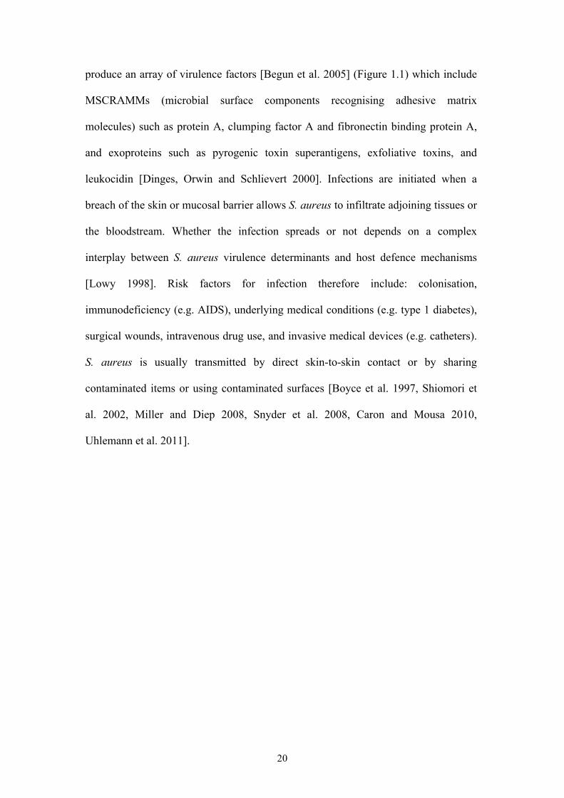
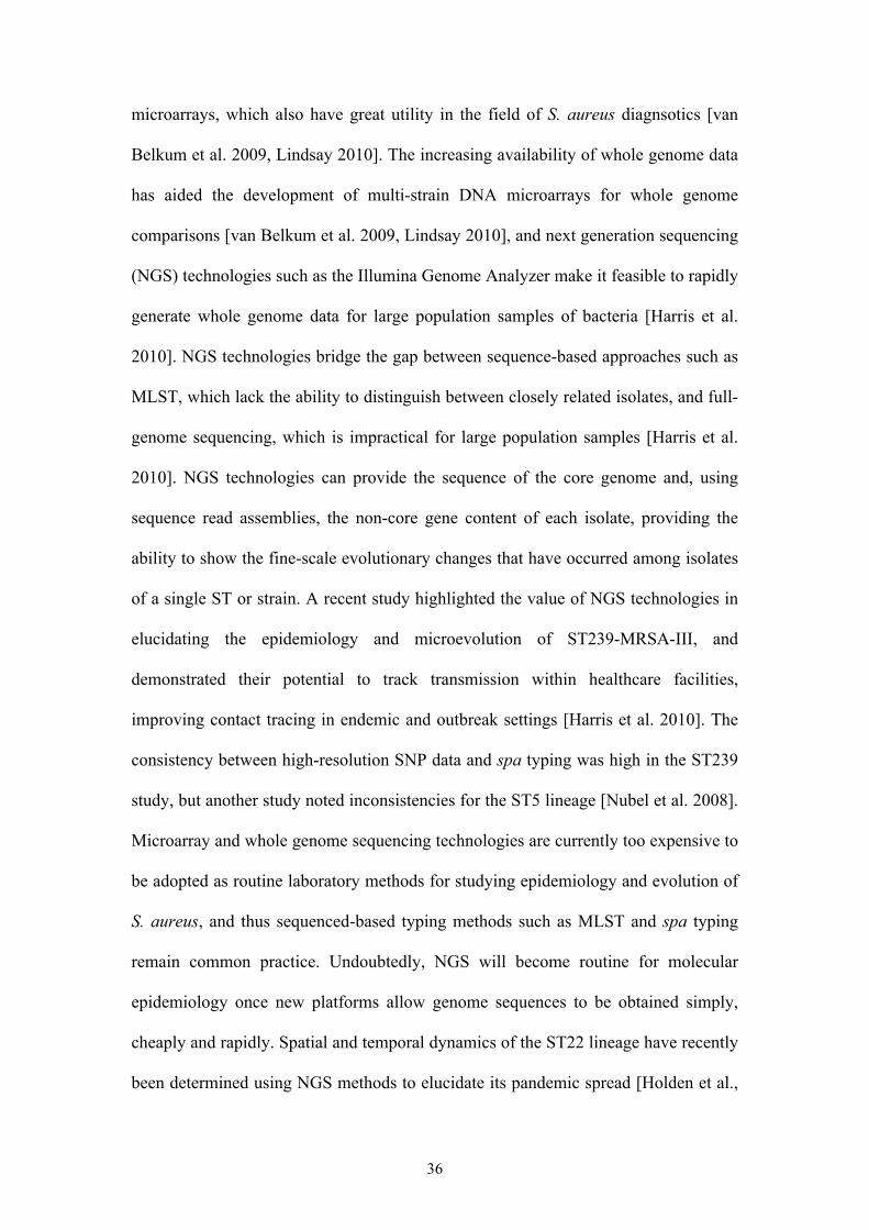
produce an array of virulence factors [Begun et al. 2005] (Figure 1.1) which include
MSCRAMMs (microbial surface components recognising adhesive matrix
molecules) such as protein A, clumping factor A and fibronectin binding protein A,
and exoproteins such as pyrogenic toxin superantigens, exfoliative toxins, and
leukocidin [Dinges, Orwin and Schlievert 2000]. Infections are initiated when a
breach of the skin or mucosal barrier allows S. aureus to infiltrate adjoining tissues or
the bloodstream. Whether the infection spreads or not depends on a complex
interplay between S. aureus virulence determinants and host defence mechanisms
[Lowy 1998]. Risk factors for infection therefore include: colonisation,
immunodeficiency (e.g. AIDS), underlying medical conditions (e.g. type 1 diabetes),
surgical wounds, intravenous drug use, and invasive medical devices (e.g. catheters).
S. aureus is usually transmitted by direct skin-to-skin contact or by sharing
contaminated items or using contaminated surfaces [Boyce et al. 1997, Shiomori et
al. 2002, Miller and Diep 2008, Snyder et al. 2008, Caron and Mousa 2010,
Uhlemann et al. 2011].
21
Figure 1.1 Staphylococcus aureus structure: surface and secreted
proteins/virulence factors. A: surface protein synthesis is usually dependent on
the growth phase and secreted protein synthesis on the stationary phase. B:
cross section of the cell envelope. C: many of the surface proteins have a
structural organization similar to that of clumping factor, including repeated
segments of amino acids. TSST-1=toxic shock syndrome toxin 1 (a pyrogenic
toxin superantigen). Figure taken from [Lowy 1998].
1.2 ANTIBIOTIC RESISTANCE: EMERGENCE AND MECHANISMS
The first clinical isolate of methicillin-resistant S. aureus (MRSA) was
reported in 1961 in the UK. Now often called the 'archaic' clone, it carries
staphylococcal cassette chromosome mec type I (SCCmec I). SCCmec is a mobile
genetic element that carries mecA, the gene conferring methicillin resistance in S.
aureus (see section 1.2.1). The archaic MRSA clone was reported just 1 year after the
introduction of methicillin, a β-lactam antibiotic developed to counter the increasing
prevalence of penicillin resistance in gram-positive bacteria [Jevons 1961]. Since
then, S. aureus has developed or acquired resistance mechanisms to almost all
22
antibiotics that have been introduced over the past decades, including β-lactams,
aminoglycosides, quinolones and glycopepetides [Lowy 2003]. After the first reports
of MRSA in the 1960s, it gradually disseminated around Europe [Crisostomo et al.
2001], and began causing serious hospital infections worldwide in the 1970s
[Hiramatsu et al. 2001]. By the 1980s, the archaic clone had largely disappeared from
European hospitals, and descendents of this clone (e.g. the Iberian clone) as well as
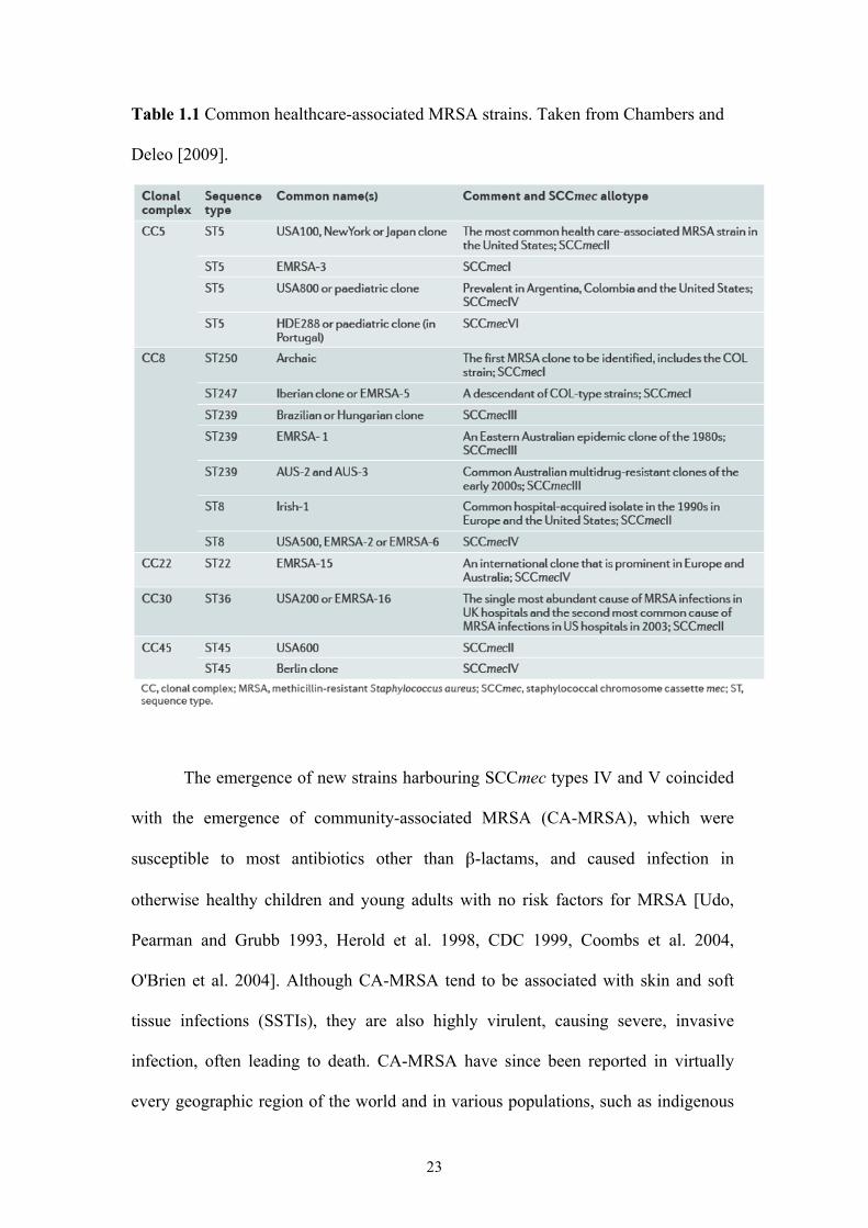
new lineages of MRSA had emerged (Table 1.1), causing significant clinical and
epidemiological problems in hospitals [Oliveira, Tomasz and de Lencastre 2002]. In
1982, the New York/Japan Clone (SCCmec II) was discovered and also spread
worldwide, followed by the discovery in 1985 of the 85/2082 MRSA strain in New
Zealand (SCCmec III). These and new MRSA strains disseminated around the world
during the 1990s, contributing to the worldwide healthcare-associated MRSA (HA-
MRSA) pandemic in hospitals and other healthcare facilities such as nursing homes,
that continues today (Table 1.1). From the 1990s, MRSA harbouring a new SCCmec
element, type IV, had emerged, and the WIS MRSA strain (SCCmec V) was
described in Australia [Udo, Pearman and Grubb 1993, Ma et al. 2002, Vandenesch
et al. 2003, Ito et al. 2004].
23
Table 1.1 Common healthcare-associated MRSA strains. Taken from Chambers and
Deleo [2009].
The emergence of new strains harbouring SCCmec types IV and V coincided
with the emergence of community-associated MRSA (CA-MRSA), which were
susceptible to most antibiotics other than β-lactams, and caused infection in
otherwise healthy children and young adults with no risk factors for MRSA [Udo,
Pearman and Grubb 1993, Herold et al. 1998, CDC 1999, Coombs et al. 2004,
O'Brien et al. 2004]. Although CA-MRSA tend to be associated with skin and soft
tissue infections (SSTIs), they are also highly virulent, causing severe, invasive
infection, often leading to death. CA-MRSA have since been reported in virtually
every geographic region of the world and in various populations, such as indigenous
24
peoples, competitive athletes, prison inmates, men who have sex with men, military
recruits and personnel, children in day care centres, contacts of patients with CA-
MRSA infection, and adult emergency room patients [Adcock et al. 1998, Shahin et
al. 1999, Groom et al. 2001, CDC 2003c, CDC 2003b, CDC 2003a, Baggett et al.
2004, CDC 2004b, Zinderman et al. 2004, Aiello et al. 2006, Johansson, Gustafsson
and Ringberg 2007, Tristan et al. 2007a, Diep et al. 2008a, Wallin, Hern and Frazee
2008]. CA-MRSA strains are also being increasingly reported as a cause of hospital-
onset and healthcare-associated infections [O'Brien et al. 1999, Saiman et al. 2003,
Bratu et al. 2005, Klevens et al. 2006, Seybold et al. 2006, Liu et al. 2008, Patel et al.
2008, Park et al. 2009b].
In the last decade, MRSA strains harbouring three new SCCmec elements
were reported, in Portugal (a healthcare-associated paediatric clone; SCCmec VI)
[Oliveira, Milheirico and de Lencastre 2006], Sweden (a community-associated
strain; SCCmec VII) [Berglund et al. 2008] and Canada (a healthcare-associated
strain; SCCmec VIII). Most recently, a further three SCCmec elements have been
described, SCCmec types IX, X and XI, which are associated with livestock-
associated MRSA (LA-MRSA) [Garcia-Alvarez et al. 2011, Li et al. 2011]. Livestock
are an increasingly recognised reservoir for MRSA, with LA-MRSA carriage and
infection reported in both farm animals and human beings [de Neeling et al. 2007,
van Loo et al. 2007, Mulders et al. 2010, Van Cleef et al. 2010, van Cleef et al. 2010,
Garcia-Alvarez et al. 2011].
The continual discovery of novel SCCmec elements represents an ongoing
evolution of antibiotic resistance in S. aureus (although novel SCCmec acquisition is
not necessarily driven by antibiotic resistance). Since β-lactams (such as methicillin)
have been the first-line antibiotics for treatment of S. aureus infections, such
25
evolution may likely impact on therapeutic options [Monecke et al. 2011]. S. aureus
has quickly acquired resistance to all antibiotics introduced for clinical use, and many
MRSA isolates are multiply antibiotic-resistant. Alternative antibiotics to β-lactams
for treatment of MRSA infections include daptomycin, linezolid, vancomycin and
rifampicin, but they are expensive or have problems with tissue penetration and
toxicity [Monecke et al. 2011]. Until recently, all MRSA were considered susceptible
to glycopeptide antibiotics such as vancomycin - considered the antibiotic of last
resort for MRSA infections - and investigational drugs [Enright et al. 2002, Lowy
2003]. However, due to intensive selective pressure as a result of increased
glycopeptide use, MRSA isolates increasingly resistant to vancomycin have been
reported worldwide (vancomycin intermediate/resistant S. aureus; VISA/VRSA)
[Hiramatsu et al. 1997a, Hiramatsu et al. 1997b, Howe et al. 1998, Ploy et al. 1998,
Sieradzki et al. 1999, Smith et al. 1999, Ferraz et al. 2000, Kim et al. 2000, Wong et
al. 2000, Boyle-Vavra, Carey and Daum 2001, Hageman et al. 2001, Oliveira et al.
2001, CDC 2002a, CDC 2002b, Weigel et al. 2003, CDC 2004a, Howe et al. 2004],
leading to treatment failures and poor outcomes [Fridkin et al. 2003, Moore,
Perdreau-Remington and Chambers 2003, Charles et al. 2004, Howden et al. 2004].
MRSA resistant to linezolid, daptomycin and rifampicin have also been reported
[Schmitz et al. 2000, Mangili et al. 2005, Long et al. 2006, Marty et al. 2006, Skiest
2006, Murthy et al. 2008, Kehrenberg et al. 2009, Shore et al. 2010, Tan et al. 2011],
posing a great problem for antimicrobial therapy.
1.2.1 Mechanism of methicillin resistance
MRSA produces a modified penicillin-binding protein, PBP2A, which has a
low affinity for β-lactam antibiotics [Hartman and Tomasz 1984, Reynolds and
26
Brown 1985, Utsui and Yokota 1985], conferring resistance to all β-lactams
(including penicillins, cephalosporins (except ceftobiprole [Stein, Goetz and Ganea
2009]), carbapenems, and monobactams), the most commonly used antibiotics to
treat S. aureus infections. β-lactams bind to PBPs in the cell wall, inhibiting
peptidoglycan synthesis in susceptible microbes, but PBP2A retains effective
transpeptidase activity in the presence of β-lactams, unlike the PBPs native to S.
aureus, allowing cell wall synthesis to continue. The transpeptidase domain of
PBP2A functions cooperatively with the transglycosylase domain of the native
staphylococcal PBP2 to achieve cell wall synthesis in the presence of β-lactams
[Pinho, de Lencastre and Tomasz 2001]. PBP2A is encoded by the mecA gene,
carried on the mobile genetic element SCCmec. MRSA arises when methicillin-
susceptible S. aureus (MSSA) acquires SCCmec. Evidence suggests this acquisition
comes from coagulase negative staphylococci (CNS) [Archer et al. 1996, Kobayashi
et al. 1999, Wielders et al. 2001, Robinson and Enright 2003, Wisplinghoff et al.
2003, Qi et al. 2005, Grundmann et al. 2006], and has occurred several times into
different S. aureus lineages [Musser and Kapur 1992, Crisostomo et al. 2001,
Fitzgerald et al. 2001, Oliveira, Tomasz and de Lencastre 2001, Enright et al. 2002,
Gomes, Westh and de Lencastre 2006] i.e. the multi-clone theory.
The SCCmec element contains the mec gene complex (the mecA gene and its
regulators, mecI, encoding a repressor protein, and mecR1, encoding a signal
transducer protein, both of which are sometimes truncated) and the ccr (cassette
chromosome recombinase) gene complex, which encodes site-specific recombinases
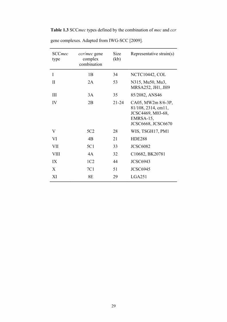
(ccrA, ccrB and ccrC) responsible for the mobility of SCCmec [Ito et al. 2004]. There
are curently 11 different SCCmec elements (21-53kb in size) formed by different
combinations of the mec and ccr gene complexes (Tables 1.2 and 1.3, Figure 1.2).
27
The recombinases catalyse the insertion/excision of SCCmec into/from the S. aureus
genome at a specific site (the bacterial chromosome attachment site for SCCmec
DNA, attBscc) at the 3′ end of an open reading frame (ORF) of unknown function,
orfX, located near the origin of replication of S. aureus. The various SCCmec types
can be further classified into subtypes based upon variations in the so-called J regions
(or 'joining regions'), J1 (the region between ccr and the chromosomal region
flanking SCCmec), J2 (the region between mec and ccr), and J3 (the region between
orfX and mec), which constitute nonessential components of the cassette [IWG-SCC
2009]. The presence of specific DNA sequences in these J regions are used to define
SCCmec subtypes, including mobile genetic elements such as insertion sequences
(ISs), plasmids or transposons, most of which encode antibiotic resistance (e.g. to
aminoglycosides or macrolides), resistance to heavy metals (e.g. Cd and Hg), or other
determinants, and characteristic genes, pseudogenes or non-coding regions in the J
regions [Oliveira, Wu and de Lencastre 2000, Ito et al. 2001, IWG-SCC 2009].
Mobile genetic elements encoding antibiotic resistance are mainly integrated into the
J2 or J3 regions, while subtype-specific ORFs are used to distinguish the several
different J1 regions in SCCmec types II and IV [Chongtrakool et al. 2006].
Horizontal transfer of DNA from other strains or species plays an important part in
antibiotic resistance in S. aureus, despite S. aureus evolution being regarded as
predominantly clonal [Enright et al. 2000, Grundmann et al. 2002, Feil et al. 2003,
Murchan et al. 2003, Melles et al. 2004]. For example, the recent emergence of
VRSA is due to the acquisition by conjugative transposition of vanA-containing
elements from vancomycin-resistant enterococci [CDC 2002b, CDC 2002a, Chang et
al. 2003].
28
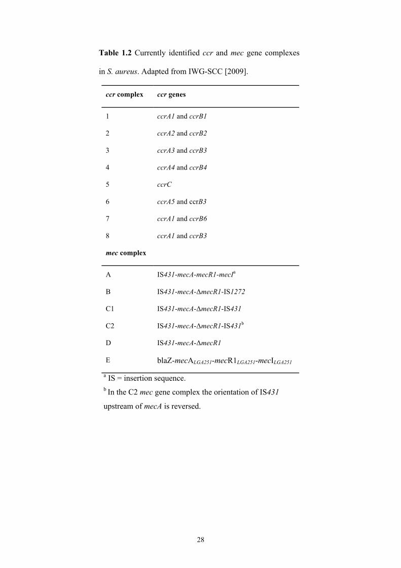
Table 1.2 Currently identified ccr and mec gene complexes
in S. aureus. Adapted from IWG-SCC [2009].
ccr complex ccr genes
1 ccrA1 and ccrB1
2 ccrA2 and ccrB2
3 ccrA3 and ccrB3
4 ccrA4 and ccrB4
5 ccrC
6 ccrA5 and ccrB3
7 ccrA1 and ccrB6
8 ccrA1 and ccrB3
mec complex
A IS431-mecA-mecR1-mecIa
B IS431-mecA-ΔmecR1-IS1272
C1 IS431-mecA-ΔmecR1-IS431
C2 IS431-mecA-ΔmecR1-IS431b
D IS431-mecA-ΔmecR1
E blaZ-mecALGA251-mecR1LGA251-mecILGA251
a IS = insertion sequence. b In the C2 mec gene complex the orientation of IS431
upstream of mecA is reversed.
29
Table 1.3 SCCmec types defined by the combination of mec and ccr
gene complexes. Adapted from IWG-SCC [2009].
SCCmec type
ccr/mec gene complex
combination
Size (kb)
Representative strain(s)
I 1B 34 NCTC10442, COL II 2A 53 N315, Mu50, Mu3,
MRSA252, JH1, JH9 III 3A 35 85/2082, ANS46
IV 2B 21-24 CA05, MW2m 8/6-3P, 81/108, 2314, cm11, JCSC4469, M03-68, EMRSA-15, JCSC6668, JCSC6670
V 5C2 28 WIS, TSGH17, PM1
VI 4B 21 HDE288 VII 5C1 33 JCSC6082
VIII 4A 32 C10682, BK20781 IX 1C2 44 JCSC6943
X 7C1 51 JCSC6945 XI 8E 29 LGA251
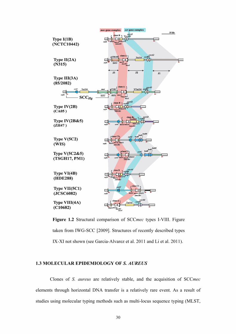
30
Figure 1.2 Structural comparison of SCCmec types I-VIII. Figure
taken from IWG-SCC [2009]. Structures of recently described types
IX-XI not shown (see Garcia-Alvarez et al. 2011 and Li et al. 2011).
1.3 MOLECULAR EPIDEMIOLOGY OF S. AUREUS
Clones of S. aureus are relatively stable, and the acquisition of SCCmec
elements through horizontal DNA transfer is a relatively rare event. As a result of
studies using molecular typing methods such as multi-locus sequence typing (MLST,
31
see section 1.3.2), it is thought that a limited number of genetically distinct epidemic
clones circulate and disseminate worldwide (Table 1.1), with SCCmec and other
mobile genetic elements conferring enhanced virulence and antibiotic resistance,
maintained in the predominantly clonal genomic background [Oliveira, Tomasz and
de Lencastre 2002, Enright 2003, Robinson and Enright 2003]. However, a more
recent study, using a highly discriminatory single nucleotide polymorphism (SNP)-
discovery method, provided evidence that the population of MRSA comprising the so-
called EMRSA-3, New York/Japan and Paediatric clones (multi-locus sequence type
ST5) is geographically structured, and that MRSA could have emerged very
frequently in different parts of the world through independent imports of the
methicillin resistance determinant into their genomes [Deurenberg and Stobberingh
2008, Nubel et al. 2008]. Studies on the population structure of a different lineage,
ST239, have suggested dissemination rather than repeated emergence is the cause of
its global prevalence, although phylogeographic structure was also found as in the
ST5 group [Harris et al. 2010, Smyth et al. 2010, Gray et al. 2011]. A study of the
emerging ST225 clone also suggests long-distance dissemination as opposed to
repeated importation of SCCmec [Nubel et al. 2010]. More studies employing SNP-
discovery and genomic comparison methods are required to elucidate the relative
contributions of dissemination and local emergence to global MRSA population
structure.
In the absence of frequent inter-strain recombination, S. aureus clones mainly
diversify through the accumulation of single nucleotide polymorphisms (SNPs), thus
making it possible to distinguish clones and clonal lineages using genetic markers
[Feil et al. 2003, Grundmann et al. 2010]. Molecular typing methods use these genetic
markers to not only track the transmission and spread of clones, but answer questions
32
regarding their evolution and epidemiology, in order to develop effective strategies
for controlling the spread of MRSA. The most commonly used molecular typing
methods are pulsed field gel electrophoresis (PFGE), MLST, spa typing and SCCmec
typing. All allow the typing of unrelated strains but do so with different accuracy,
discriminatory power, and reproducibility [Melles et al. 2007]. Recent advances in
whole genome sequencing have shown that almost all isolates of a single strain differ
to some extent in nucleotide sequence, allowing the detailed spread and
microevolution of S. aureus strains to be studied.
1.3.1 Pulsed field gel electrophoresis (PFGE)
PFGE is the most commonly used and one of the most discriminatory typing
methods for studying local MRSA epidemiology such as outbreaks and nosocomial
transmission [Cookson et al. 2007]. PFGE is based on digestion of chromosomal
DNA with restriction enzyme SmaI followed by agarose gel electrophoresis. The
resulting banding patterns are analysed using software such as Bionumerics (Applied
Maths), compared to banding patterns of reference strains, and PFGE types defined
based on a similarity coefficient. Due to the nature of the method, efforts to
standardize PFGE at an international level have not been successful in terms of
reproducibility, speed and analysis costs. A common nomenclature is needed, which
has been achieved only at a national level [Deurenberg and Stobberingh 2008].
1.3.2 Multi locus sequence typing (MLST)
MLST has become established as an important tool for unambiguously defining
strains and for studying MRSA clonal evolution, although whole genome sequencing
will inevitably supersede it. MLST is based on the sequence analysis of seven S.
33
aureus housekeeping genes (arcC, aroE, glpF, gmk, pta, tpi and yqiL) [Enright et al.
2000]. For each locus, different sequences are assigned as different alleles, and each
isolate is assigned a sequence type (ST) based on its allelic profile (the set of alleles
at all seven loci) (www.mlst.net). S. aureus STs are grouped within clonal complexes
(CCs), which are groups of STs where every member of the group has a 6/7 allelic
match to at least one other ST in the group. The putative ancestor or founder of each
CC is the ST with the largest number of single-locus variants (SLVs), and sub-
founders are SLVs or double locus variants (DLVs) of a predicted founder that has
become prevalent in a population and diversified to produce its own SLVs and DLVs
[Enright and Spratt 1999, Enright et al. 2000, Enright et al. 2002, Spratt et al. 2004].
CCs are defined using the based upon related sequence types (BURST) algorithm
(www.eburst.mlst.net) and are named by the ST number of the predicted founder
[Spratt et al. 2004].
Although highly discriminatory, MLST may lack the necessary power to
discriminate between epidemiologically unrelated strains [Cooper and Feil 2004], and
is laborious and time-consuming. Nonetheless, MLST offers a major advantage over
pulsed field gel electrophoresis (PFGE) as a reference method due to the
unambiguous nature of the procedure allowing excellent reproducibility.
A common nomenclature for MRSA is the combination of ST and SCCmec
type. For example the New York/Japan clone is ST5-MRSA-II and EMRSA-15 is
ST22-MRSA-IV.
1.3.3 spa typing
The spa locus of S. aureus encodes staphylococcal protein A, a species-
specific protein known for its immunoglobulin G (IgG) binding capacity. spa typing
34
targets the highly polymorphic region X of the spa gene. This region consists of a
variable number of mainly 24-bp repeats, the variation being largely due to deletions
and duplications of the different repeats [Shopsin et al. 1999]. spa typing is simple
compared to MLST as it requires the sequencing of just one locus, and its
discriminatory power lies between that of PFGE and MLST [Malachowa et al. 2005].
Unlike MLST, both molecular evolution and hospital outbreaks can be studied with
spa typing, and comparability and a common nomenclature is possible thanks to
dedicated software [Harmsen et al. 2003, Deurenberg et al. 2007]. Because it is a
single-locus typing method, it is less expensive, less laborious and less time
consuming than MLST. The spa typing database, spaserver.ridom.de (Ridom GmbH
and SeqNet), synchronises public spa typing data and currently comprises over
10,000 spa types that consist of different combinations of over 500 spa repeats from
over 200,000 S. aureus isolates typed in 90 countries worldwide.
Cluster analysis of spa typing data groups spa types into spa-clonal
complexes (spa-CCs) using the based upon repeat pattern (BURP) algorithm in the
StaphType software (Ridom GmbH). spa typing has a higher discriminatory power
than MLST and so a single MLST ST can be resolved into several spa types,
typically within the same spa-CC. Good concordance has been reported between
MLST and spa typing but anomalies can occur with spa typing/BURP [Cookson et
al. 2007, Mellmann et al. 2008, Strommenger et al. 2008]. For example, the same or
related spa types or spa repeat patterns can occur in different clonal lineages, maybe
due to recombination events involving the spa gene, or recombination within the spa
locus [Robinson and Enright 2004, Strommenger et al. 2008]. It has been suggested
that the discriminatory power of spa typing/BURP can be improved by combining it
35
with an additional genetic marker, e.g. SCCmec for MRSA [Strommenger et al.
2008].
1.3.4 SCCmec typing
SCCmec typing exploits differences in the various SCCmec elements of
MRSA. SCCmec typing methods are able to detect various ranges of SCCmec types
and subtypes, including I-IV [Okuma et al. 2002, Oliveira and de Lencastre 2002,
Francois et al. 2004, Motoshima et al. 2010], subtypes of IV [Milheirico, Oliveira and
de Lencastre 2007b], I-V [Ito et al. 2001, Zhang et al. 2005, Boye et al. 2007, Kondo
et al. 2007, Valvatne et al. 2009, Ghaznavi-Rad et al. 2010b], I-VI [Milheirico,
Oliveira and de Lencastre 2007a, Cai et al. 2009] and even I-VI and VIII [Chen et al.
2009]. These methods are based on the mec complex and ccr genes, or the mecA gene
and other loci on SCCmec, mainly using multiplex PCR. However, each method
determines different structural properties of SCCmec, with some methods giving
different results for the same MRSA isolate [Shore et al. 2005, Kim et al. 2007].
There is no single universal method available for the classification of this mobile
element, but the most commonly used methods are those developed by Milheirico,
Oliveira and de Lencastre [2007a] and Ito et al. [Ito et al. 2001, Okuma et al. 2002]
that target several loci. Simpler methods have been developed by Boye et al. [2007]
and Zhang et al. [2005] but they only target a single locus for most SCCmec types,
and thus have less discriminatory power.
1.3.5 Advances in S. aureus molecular epidemiology
The field of S. aureus molecular epidemiology has really advanced in the last
few years, with the advent of new technologies such as whole genome sequencing and
36
microarrays, which also have great utility in the field of S. aureus diagnsotics [van
Belkum et al. 2009, Lindsay 2010]. The increasing availability of whole genome data
has aided the development of multi-strain DNA microarrays for whole genome
comparisons [van Belkum et al. 2009, Lindsay 2010], and next generation sequencing
(NGS) technologies such as the Illumina Genome Analyzer make it feasible to rapidly
generate whole genome data for large population samples of bacteria [Harris et al.
2010]. NGS technologies bridge the gap between sequence-based approaches such as
MLST, which lack the ability to distinguish between closely related isolates, and full-
genome sequencing, which is impractical for large population samples [Harris et al.
2010]. NGS technologies can provide the sequence of the core genome and, using
sequence read assemblies, the non-core gene content of each isolate, providing the
ability to show the fine-scale evolutionary changes that have occurred among isolates
of a single ST or strain. A recent study highlighted the value of NGS technologies in
elucidating the epidemiology and microevolution of ST239-MRSA-III, and
demonstrated their potential to track transmission within healthcare facilities,
improving contact tracing in endemic and outbreak settings [Harris et al. 2010]. The
consistency between high-resolution SNP data and spa typing was high in the ST239
study, but another study noted inconsistencies for the ST5 lineage [Nubel et al. 2008].
Microarray and whole genome sequencing technologies are currently too expensive to
be adopted as routine laboratory methods for studying epidemiology and evolution of
S. aureus, and thus sequenced-based typing methods such as MLST and spa typing
remain common practice. Undoubtedly, NGS will become routine for molecular
epidemiology once new platforms allow genome sequences to be obtained simply,
cheaply and rapidly. Spatial and temporal dynamics of the ST22 lineage have recently
been determined using NGS methods to elucidate its pandemic spread [Holden et al.,
37
submitted for publication], and high-resolution SNP data from over 1,000 isolates of
the USA300 clone (ST8-MRSA-IV) are currently being generated to investigate its
transmission and spread across healthcare facilities in a US county (see Part 2 final
discussion).
1.4 THE CHANGING EPIDEMIOLOGY OF MRSA
As highlighted in section 1.2, the emergence of novel SCCmec elements has
generally coincided with the emergence of epidemiologically novel MRSA strains.
Over the last few decades, MRSA has largely been a nosocomial pathogen, causing
infection in people with frequent or recent contact with healthcare facilities. The
epidemic MRSA clones that currently pose a major public health problem in
healthcare facilities worldwide, termed HA-MRSA, are listed in Table 1.1. However,
MRSA isolated from young, otherwise healthy patients with no identifiable risk
factors (including recent hospitalisation, surgery, residency in a long-term care
facility, dialysis or invasive medical devices), termed CA-MRSA, have become
increasingly prevalent since the 1990s and are now seen worldwide. The major CA-
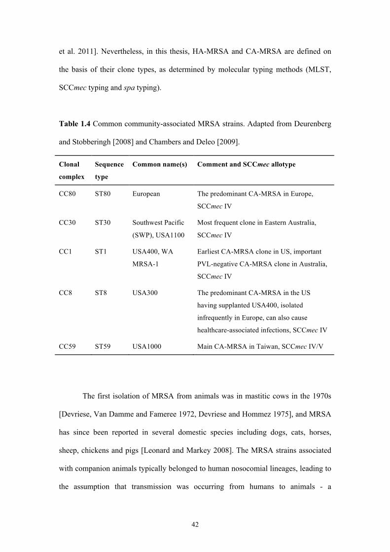
MRSA clones currently circulating are listed in Table 1.4.
SCCmec types I, II and III are typically associated with HA-MRSA and are
not frequent among the healthy, younger population, while the smaller SCCmec types
IV and V are commonly associated with CA-MRSA that not only infect hospitalised
patients but also healthy contact persons, and spread easily in the community
[Kazakova et al. 2005, Hota et al. 2007, Larsen et al. 2007, Huang et al. 2008, Tong et
al. 2008]. These latter SCCmec allotypes are more readily transmissible between
staphylococci than the larger elements and may provide a lower fitness cost to the
pathogen [Grundmann et al. 2006]. This could lead to competitive exclusion of HA-
38
MRSA by CA-MRSA if a reservoir of the latter was established in hospitals [D'Agata
et al. 2009], for which there is increasing evidence [Moran et al. 2006, Seybold et al.
2006, Huang et al. 2007b, Patel et al. 2008, Popovich, Weinstein and Hota 2008, Song
et al. 2011]. The larger HA-MRSA SCCmec allotypes correlate with a slower growth
rate, and strains with these elements may be at a selective disadvantage in the absence
of antibiotics, i.e. in community settings [Ender et al. 2004, Lee et al. 2007].
Successful HA-MRSA clones harbour the SCCmec IV element however. For
example, one of the most common healthcare-associated clones in the UK is ST22-
MRSA-IV, and the Paediatric clone (ST5) harbours SCCmec IV [Holmes et al. 2005].
Furthermore, SCCmec types I, II and III have been observed in CA-MRSA isolates
[Chung et al. 2004, Wannet et al. 2005].
CA-MRSA are considered more virulent than HA-MRSA due to the presence
of various virulence factors [Chambers 2001, Davis et al. 2007, Otto 2010], which has
clear implications in terms of morbidity and mortality in the healthcare setting. While
frequently associated with chronic or recurrent SSTIs, CA-MRSA can also cause
septic arthritis, bacteraemia, toxic shock syndrome, necrotising fasciitis and
necrotising pneumonia [Mongkolrattanothai et al. 2003, Francis et al. 2005, Gonzalez
et al. 2005a, Gonzalez et al. 2005b, Miller et al. 2005, Bocchini et al. 2006, King et al.
2006, Moran et al. 2006, Davis et al. 2007, Tristan et al. 2007b, Lo and Wang 2011,
Shilo and Quach 2011]. Panton-Valentine leukocidin (PVL) is a virulence factor that
can cause tissue necrosis and destruction of leukocytes by forming pores in the
cellular membrane [Bassetti, Nicco and Mikulska 2009], and is directly associated
with staphylococcal necrotising pneumonia [Gillet et al. 2002, Labandeira-Rey et al.
2007], but its association with other CA-MRSA invasive disease is debatable [Lina et
al. 1999, Voyich et al. 2006, Ellington et al. 2007]. Despite the predominant CA-
39
MRSA clone in the US being PVL-positive (USA300) [Okuma et al. 2002,
Vandenesch et al. 2003] several CA-MRSA lineages are PVL-negative, so PVL
cannot be considered a marker for CA-MRSA [Nimmo et al. 2006, Rossney et al.
2007b, Otter and French 2008, Zhang et al. 2008]. Another factor that may contribute
to the virulence of CA-MRSA is the argenine catabolic mobile element (ACME),
which has also been shown to contribute to the growth and survival of USA300, the
clone in which it seems to be exclusively observed [Diep et al. 2006b, Goering et al.
2007, Diep et al. 2008b, Ellington et al. 2008]. The pore-forming toxin α-haemolysin
has also been shown essential for USA300 and USA400 to cause lethal pneumonia in
a mouse model of the disease, and increasing severity of the disease has been shown
in vitro to correlate with increasing amounts of the toxin produced by these strains
[Bubeck Wardenburg et al. 2007, Burlak et al. 2007, Montgomery et al. 2008].
CA-MRSA is particularly well established in the US, with USA300 the
predominant cause of MRSA infection in North America [Gonzalez et al. 2006,
Moran et al. 2006, Klevens et al. 2007]. In contrast, USA300 is uncommon in Europe
despite being reported in most countries. However, USA300 appears to be increasing
in prevalence there [Larsen et al. 2007, Witte et al. 2007a], and CA-MRSA is partly
responsible for the increase in MRSA prevalence in northern European countries that
have a traditionally low prevalence of HA-MRSA [Bartels et al. 2007, Stam-Bolink et
al. 2007, Fang et al. 2008, Larsen et al. 2008]. European CA-MRSA are more clonally
diverse and vary geographically, but the European clone (ST80-MRSA-IV) is
widespread on this continent (Table 1.4) [Otter and French 2010]. In stark contrast to
the US that is dominated by a single clone, there is considerable CA-MRSA diversity
in Australia, with over 100 clones described there [Chua et al. 2011]. Nonetheless,
CA-MRSA prevalence in other parts of the world still remains much lower than in the
40
US [Otter and French 2010, Otto 2010, Johnson 2011]. The USA300 clone, so
predominant in North America, is already disseminating globally, which could lead to
a rapid worldwide increase in CA-MRSA. Other than Europe, it is also present in
Latin America and the Caribbean, the Middle East and the Western Pacific [Nimmo
2012].
It has been shown that CA-MRSA lineages are distinct from those of HA-
MRSA, and CA-MRSA are associated with several specific S. aureus lineages
[Groom et al. 2001, Naimi et al. 2001, Tristan et al. 2007a, David and Daum 2010]. In
addition, the larger clonal diversity of CA-MRSA compared to HA-MRSA suggests
that more MSSA lineages have the ability to become CA-MRSA [Enright et al. 2002,
Okuma et al. 2002, Feng et al. 2008, Francois et al. 2008]. It is unclear whether CA-
MRSA was originally MSSA that acquired SCCmec, or CA-MRSA originated from
HA-MRSA, but more evidence suggests that it descended from virulent strains of
MSSA via horizontal phage transfer and integration of SCCmec from CNS [Okuma et
al. 2002, Aires de Sousa and de Lencastre 2003, Bradley 2005, Robinson et al. 2005,
Ma et al. 2006, Boyle-Vavra and Daum 2007, Monecke et al. 2007c, Wallin, Hern and
Frazee 2008].
In contrast to HA-MRSA, CA-MRSA are susceptible to most non β-lactam
antibiotics, have a faster growth rate and express methicillin resistance at lower levels
and heterogeneously [Laurent et al. 2001, Okuma et al. 2002], but multidrug-resistant
CA-MRSA have started to emerge [Boyle-Vavra et al. 2005, Ramdani-Bouguessa et
al. 2006, Diep et al. 2008a], posing a serious public health concern because of their
associated virulence and their ability to cause outbreaks in otherwise healthy
individuals, as well as their rapid spread in countries worldwide [Monecke et al.
2011]. Moreover, the lack of clear definitions for HA-MRSA and CA-MRSA due to
41
increasingly blurred molecular and epidemiological distinctions between the two
groups, make it difficult to develop effective infection control strategies in healthcare
and community settings. One study in the US highlighted this blurred line by
obtaining MRSA isolates from patients and classifying them as either HA-MRSA or
CA-MRSA based on both epidemiological and molecular definitions, which were
performed separately by blinded investigators. Sixty percent of strains classified as
HA-MRSA based on epidemiological definitions were identified as CA-MRSA based
on molecular definitions, and CA-MRSA was found to cause healthcare-associated
bloodstream infection just as likely as it causes community-associated infection
[Gonzalez et al. 2006].
The common epidemiological definition for CA-MRSA is that used by the
Active Bacterial Core surveillance (ABCs) program of the US Centers for Disease
Control and Prevention (CDC): any MRSA infection diagnosed in an outpatient or
within 48 hours of hospitalisation if the patient lacks healthcare-associated MRSA
risk factors (haemodialysis, surgery, residence in a long-term care facility, recent
hospitalisation, or invasive medical devices) [Morrison, Hageman and Klevens 2006,
Klevens et al. 2007]. All other MRSA are considered HA-MRSA. A simpler temporal
definition of CA-MRSA is often used, without considering the presence of MRSA
risk factors [David and Daum 2010], while the use of a strictly molecular definition is
becoming increasingly problematic, for example because HA-MRSA also possess the
traditionally community-associated SCCmec IV element [Miller and Kaplan 2009].
One such strain, ST22-MRSA-IV has been reported in the community in Ireland
[Mollaghan et al. 2010]. In East Asia, CA-MRSA with SCCmec IV (ST59, ST30 and
ST72) have spread from the community into the hospital, while healthcare-associated
strains ST239-MRSA-III and ST5-MRSA-II have been found in the community [Song
42
et al. 2011]. Nevertheless, in this thesis, HA-MRSA and CA-MRSA are defined on
the basis of their clone types, as determined by molecular typing methods (MLST,
SCCmec typing and spa typing).
Table 1.4 Common community-associated MRSA strains. Adapted from Deurenberg
and Stobberingh [2008] and Chambers and Deleo [2009].
Clonal
complex
Sequence
type
Common name(s) Comment and SCCmec allotype
CC80 ST80 European The predominant CA-MRSA in Europe,
SCCmec IV
CC30 ST30 Southwest Pacific
(SWP), USA1100
Most frequent clone in Eastern Australia,
SCCmec IV
CC1 ST1 USA400, WA
MRSA-1
Earliest CA-MRSA clone in US, important
PVL-negative CA-MRSA clone in Australia,
SCCmec IV
CC8 ST8 USA300 The predominant CA-MRSA in the US
having supplanted USA400, isolated
infrequently in Europe, can also cause
healthcare-associated infections, SCCmec IV
CC59 ST59 USA1000 Main CA-MRSA in Taiwan, SCCmec IV/V
The first isolation of MRSA from animals was in mastitic cows in the 1970s
[Devriese, Van Damme and Fameree 1972, Devriese and Hommez 1975], and MRSA
has since been reported in several domestic species including dogs, cats, horses,
sheep, chickens and pigs [Leonard and Markey 2008]. The MRSA strains associated
with companion animals typically belonged to human nosocomial lineages, leading to
the assumption that transmission was occurring from humans to animals - a

43
'humanosis' [Morgan 2008]. Since 2003, MRSA strains carrying SCCmec types IV
and V have been reported among livestock, termed LA-MRSA [Nemati et al. 2008],
predominantly belonging to the clonal lineage CC398. CC398 first emerged as a
coloniser among farmed pigs, and later in other livestock such as calves and poultry.
It also colonises humans in contact with these livestock, where it infrequently causes
infection, ranging from SSTIs to severe, invasive infections such as endocarditis,
necrotising fasciitis and pneumonia [Ekkelenkamp et al. 2006, van Rijen, Van Keulen
and Kluytmans 2008, Pan et al. 2009, Catry et al. 2010, Hartmeyer et al. 2010,
Mammina et al. 2010, Schijffelen et al. 2010, Soavi et al. 2010], demonstrating the
zoonotic potential and virulence of this lineage [Voss et al. 2005, van Loo et al. 2007,
Witte et al. 2007b, Khanna et al. 2008, Smith et al. 2008, van Belkum et al. 2008,
Cuny et al. 2009, Mulders et al. 2010, van Cleef et al. 2010]. CC398 is now reported
in different countries around the world, with livestock representing another reservoir
for MRSA colonisation and infection in humans [Armand-Lefevre, Ruimy and
Andremont 2005, Witte et al. 2007b, Lewis et al. 2008, Smith et al. 2008, Bhat et al.
2009, Denis et al. 2009b, Krziwanek, Metz-Gercek and Mittermayer 2009, Loeffler et
al. 2009, Pan et al. 2009, Mulders et al. 2010, Potel et al. 2010, Soavi et al. 2010, van
Cleef et al. 2010, Vanderhaeghen et al. 2010, Haenni et al. 2011]. It has been shown
however that CC398 MRSA are poor persistent human colonisers in the absence of
animal contact [Graveland et al. 2011].
Studies of S. aureus population genetics have demonstrated the existence of
host-specific clonal lineages, with the majority of LA-MRSA belonging to a small
number of animal-associated clones (Table 1.5) [Sung, Lloyd and Lindsay 2008,
McCarthy et al. 2011, Fitzgerald 2012]. Most ruminant lineages are host-specific, for
example ST97, ST705, ST126, ST151 and ST133 [Cuny et al. 2011, Fitzgerald 2012].
44
However, ST398 and human lineages such as ST5, ST239 and ST1 have been
associated with bovine mastitis [Cuny et al. 2011], and CC130, which was recently
found to harbour the novel divergent mecALGA251 gene carried by SCCmec XI, appears
to have no host restrictions, infecting both bovine and human populations [Cuny et al.
2011, Garcia-Alvarez et al. 2011, Shore et al. 2011]. Clones such as ST398 appear to
have the ability to colonise and infect multiple host species. The novel mecALGA251
found in CC130 clones was also identified in CC705 and ST425, from bovine and
human populations in the UK, Denmark, Ireland and Germany, highlighting the
possibility of additional circulating novel mecA alleles that could be acquired by S.
aureus to create new MRSA strains [Cuny et al. 2011, Garcia-Alvarez et al. 2011,
Shore et al. 2011, Fitzgerald 2012]. In addition, mecALGA251 cannot be detected by
molecular diagnostic tests for MRSA, leading to false negative results, and the
ramifications of this should be considered by diagnostic protocols [Garcia-Alvarez et
al. 2011].
Not only do livestock serve as a potential source of zoonotic S. aureus
infection, but also humans represent an important source of new pathogenic strains
affecting economically important livestock [Lowder et al. 2009, Guinane et al. 2010,
Sakwinska et al. 2011, Fitzgerald 2012], with a recent study providing strong
evidence that LA-MRSA CC398 originated in humans as MSSA [Price et al. 2012].
While the potential impact of the community MRSA reservoir on current infection
control strategies is clear, the impact of the livestock MRSA reservoir on public
health is less so. Current evidence suggests LA-MRSA have not spread significantly
into healthcare settings in Europe, but CC398 spread seems to be dependent on the
region and the intensity of pig farming [Stefani et al. 2012]. A better understanding of
the origin, evolution and epidemiology of both CA-MRSA and LA-MRSA is required
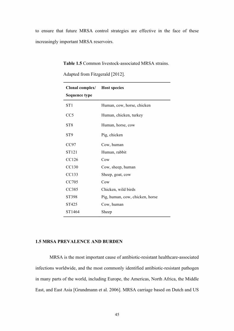
45
to ensure that future MRSA control strategies are effective in the face of these
increasingly important MRSA reservoirs.
Table 1.5 Common livestock-associated MRSA strains.
Adapted from Fitzgerald [2012].
Clonal complex/
Sequence type
Host species
ST1 Human, cow, horse, chicken
CC5 Human, chicken, turkey
ST8 Human, horse, cow
ST9 Pig, chicken
CC97 Cow, human
ST121 Human, rabbit
CC126 Cow
CC130 Cow, sheep, human
CC133 Sheep, goat, cow
CC705 Cow
CC385 Chicken, wild birds
ST398 Pig, human, cow, chicken, horse
ST425 Cow, human
ST1464 Sheep
1.5 MRSA PREVALENCE AND BURDEN
MRSA is the most important cause of antibiotic-resistant healthcare-associated
infections worldwide, and the most commonly identified antibiotic-resistant pathogen
in many parts of the world, including Europe, the Americas, North Africa, the Middle
East, and East Asia [Grundmann et al. 2006]. MRSA carriage based on Dutch and US
46
prevalence figures is conservatively estimated at 0.1-2.65% of the expected 2 billion
S. aureus carriers globally [Grundmann et al. 2006]. MRSA prevalence varies greatly
between countries, as well as within (Figures 1.3 and 1.4). It is hard to make
international comparisons of MRSA prevalence however, due to the differences in the
way prevalence rates are obtained between studies, and the scarcity of routine
surveillance systems for MRSA. In general, the US, South America and East Asia
(including South Korea, Japan and Taiwan) appear to have the highest MRSA rates,
(>50%), with intermediate rates in Canada, Latin America, Australia, southern and
central Europe, and parts of Africa and the Middle East (mostly between 25 and 50%)
[Bouchillon et al. 2004, Grundmann et al. 2006, Perovic et al. 2006, Laxminarayan
and Malani 2007, Reinert et al. 2007, Mejia, Zurita and Guzman-Blanco 2010, Song
et al. 2011]. Scandinavia and the Netherlands have the lowest MRSA rates, at <5%,
while rates in resource-poor countries, including most of Asia and Africa, are largely
unknown, due to a dearth of prevalence studies in these regions [Nickerson et al.
2009], although MRSA rates appear to be intermediate [Kesah et al. 2003, Ramdani-
Bouguessa et al. 2006, Song et al. 2011]. High MRSA rates tend to be seen in
countries with high rates of antibiotic use (current or historical) and poor infection
control strategies, whereas the low MRSA rates seen for example in the Netherlands
are attributable to national 'search and destroy' policies to limit MRSA spread. Such
policies are expensive to implement, but in countries with low endemic MRSA
incidence, the benefits outweigh the costs [Laxminarayan and Malani 2007, Simoens,
Ophals and Schuermans 2009, van Rijen and Kluytmans 2009].
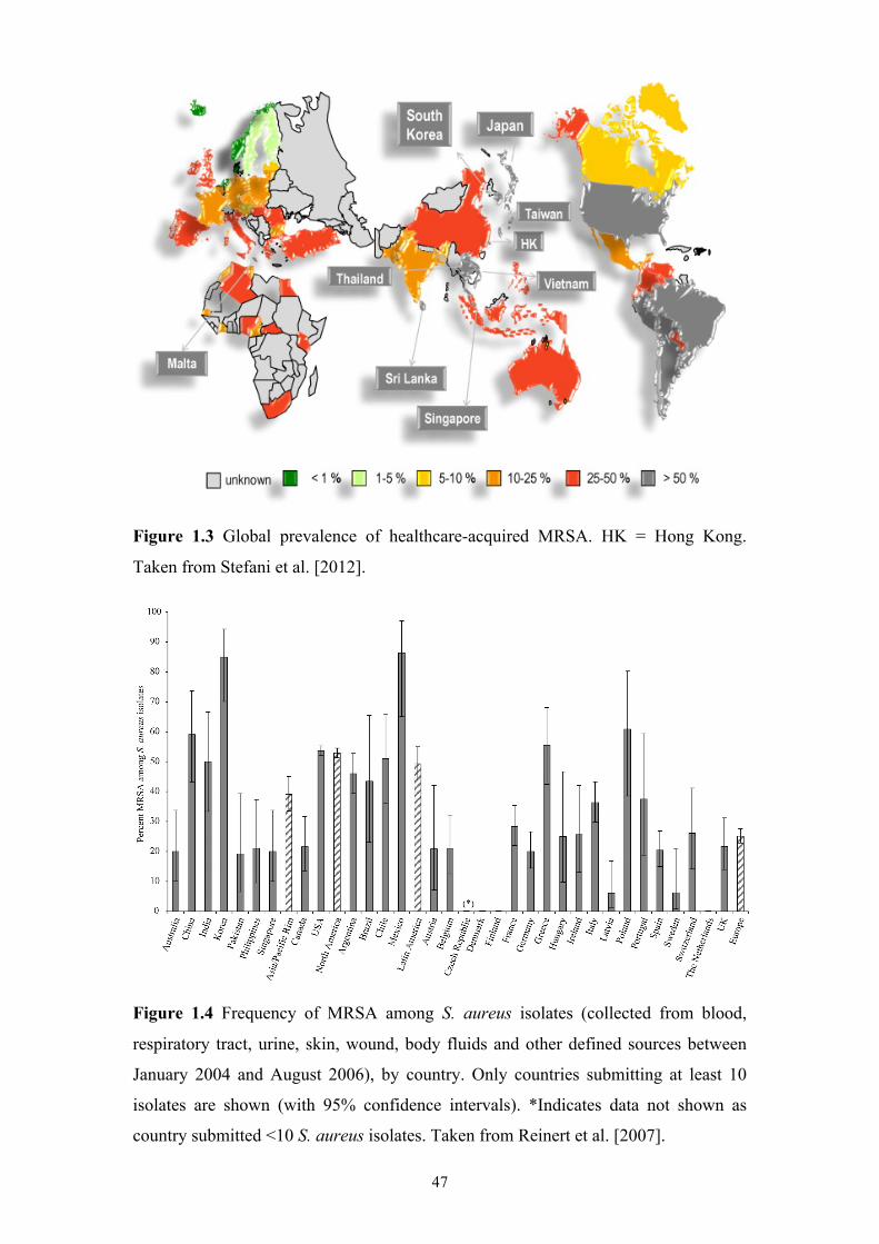
47
Figure 1.3 Global prevalence of healthcare-acquired MRSA. HK = Hong Kong.
Taken from Stefani et al. [2012].
Figure 1.4 Frequency of MRSA among S. aureus isolates (collected from blood,
respiratory tract, urine, skin, wound, body fluids and other defined sources between
January 2004 and August 2006), by country. Only countries submitting at least 10
isolates are shown (with 95% confidence intervals). *Indicates data not shown as
country submitted <10 S. aureus isolates. Taken from Reinert et al. [2007].
48
The European Antimicrobial Resistance Surveillance Network (EARSnet) was
set up in 1999 using sentinel hospital laboratories across the continent to survey cases
of bacteraemia caused by several bacterial species including S. aureus. Although
bacteraemia is less common than other types of infection such as SSTIs, surveillance
systems tend to focus on bloodstream infections as they are clinically significant and
are likely to be investigated microbiologically [Johnson 2011]. Through EARSnet, in
2009, nine European countries reported less than 10% invasive MRSA isolates, nine
countries reported 10-25%, another nine reported 25-50%, and one country reported
over 50% invasive MRSA isolates (Figure 1.3) [ECDC 2011]. Eight countries
reported a decreasing trend for MRSA while just one country reported an increase
[ECDC 2011]. In the UK, the percentage of invasive MRSA isolates was between 25
and 50% in 2009, but a significantly sustained decrease in this percentage was
observed between 2006 and 2009 [ECDC 2011]. This decrease likely reflects the
government action in England to make reporting of MRSA bacteraemia mandatory
for all hospitals, and setting hospitals the target of halving their MRSA rates
[Liebowitz 2009, Pearson, Chronias and Murray 2009]. Although proportions of
MRSA seem to be stabilising, and even decreasing in some European countries, the
percentage of MRSA is still more than 25% in 10 of 28 reporting countries, and thus
MRSA control remains a public health priority in Europe [ECDC 2011].
In the US, the ABCs population-based sentinel surveillance program was used
to evaluate the incidence of healthcare-associated MRSA infections between 2005 and
2008 [Kallen et al. 2010]. Over the four-year period, rates of hospital onset and
healthcare-associated, community onset invasive MRSA infections decreased among
US hospital inpatients [Kallen et al. 2010], supporting a previous study on
bloodstream infections in intensive care units (ICUs) [Burton et al. 2009], and
49
mirroring the pattern observed in some European countries like the UK. Despite this
decrease, MRSA prevalence remains high in the US [Grundmann et al. 2006], likely
due to the rapid increase in rates of CA-MRSA infection in the last decade and its
emergence as a major cause of healthcare-associated infection [Carleton et al. 2004,
King et al. 2006, Moran et al. 2006, Patel et al. 2008].
MRSA is a large and increasing global burden on healthcare resources, and is
associated with increased morbidity and a higher risk of mortality [Abramson and
Sexton 1999, Cosgrove et al. 2003, Engemann et al. 2003, Chu et al. 2005, Cosgrove
et al. 2005, Gould 2005, Klein, Smith and Laxminarayan 2007, Shurland et al. 2007].
The poorer therapeutic outcome of MRSA infections compared to MSSA infections
can be attributed to the underlying medical problems of the often sicker, older patients
that are infected with MRSA, plus the use of more toxic or ineffective antibiotics
[Cosgrove et al. 2003, Lowy 2003, Simoens, Ophals and Schuermans 2009]. The
additional burden imposed by resistance not only aggravates the clinical outcome but
also adds to the overall caseload of patients with invasive S. aureus infections [de
Kraker et al. 2011]. Increased risk of treatment failure, implementation of isolation
measures, antimicrobial treatment and extended hospital stay all add to the financial
burden on healthcare facilities [Shorr 2007]. Additional financial costs can be accrued
through the containment of outbreaks and changes in antibiotic prescribing habits
[Grundmann et al. 2006]. The average excess costs per MRSA-positive patient have
been estimated to range from €5,700 to €10,000 [Monecke et al. 2011]. MRSA
infections also impact on sufferers psychologically (e.g. due to isolation) and
financially, through loss of productivity and long-term disability, in turn impacting on
societal costs [Tarzi et al. 2001, Grundmann et al. 2006].
50
1.6 PREVENTATIVE MEASURES
Although HA-MRSA prevalence is stabilising and even decreasing in some
regions, notably Europe, the need for effective MRSA control strategies remains, as
HA-MRSA prevalence is still high and increasing in many countries around the
world. Several hurdles to effective control exist however, including the increasing
prevalence of CA-MRSA, which appears to be emerging as a potent combination of
transmissibility, virulence and resistance, the ever-increasing healthcare MRSA
reservoir, to which CA-MRSA is increasingly contributing, the likely impossibility of
eradicating endemic MRSA, and the significant costs and disruption to patient care
associated with active surveillance and control [Marshall et al. 2004].
There are four main approaches to MRSA control: reduction in antibiotic use
through education and restriction, transmission prevention through hand hygiene,
identification of carriers through screening and isolation, and elimination of reservoirs
through decontamination [Harbarth 2006]. There is much debate regarding the most
cost-effective strategy for MRSA control, but it is clear that strategies developed with
HA-MRSA pathogenesis in mind may not work for prevention of CA-MRSA
infection, attenuating our ability to control overall MRSA burden [Miller and Diep
2008]. The increasing non-β-lactam resistance in CA-MRSA clones, particularly
USA300, highlights the need for accurate therapeutic decisions, guided by active
screening and surveillance [Chua et al. 2011].
The limited choice of therapeutic options available, and the declining
investment by pharmaceutical companies in antimicrobial research and development
[Talbot et al. 2006] (Figure 1.5), has shifted the spotlight onto infection prevention
and control measures such as hand hygiene, protective clothing and equipment, and
accommodating patients in isolation rooms or wards [Monecke et al. 2011]. As well
51
as a better understanding of S. aureus pathogenesis and improved non-antimicrobial
approaches, an important aspect of effective MRSA control is the rapid screening for
early identification of MRSA carriers, using accurate and rapid MRSA diagnostics.
The stringent search and destroy policies employed in Scandinavian countries
and the Netherlands, where MRSA prevalence is low, involve a combination of
measures, such as isolation of identified MRSA carriers, patient decolonisation,
admission screening of high-risk patients with pre-emptive isolation, screening of all
contact patients, healthcare workers and index cases, temporarily sending healthcare
workers home, and closing wards to prevent outbreaks. A study investigating the
individual contribution of each of these measures found that admission screening
combined with pre-emptive isolation could be the most beneficial, even in high
MRSA prevalence settings [Bootsma, Diekmann and Bonten 2006b]. Rapid MRSA
detection is crucial in such a strategy, to limit transmission risk and isolation costs, as
well as the impact on patient care [Wassenberg et al. 2010]. Conventional
microbiological cultures have a turnaround time (TAT) of at least 48 hours, and
longer if a broth enrichment technique is used. Novel and faster diagnostic tests for
MRSA screening have been introduced in recent years that reduce the TAT to a
diagnostic result, and these are discussed in section 1.7.
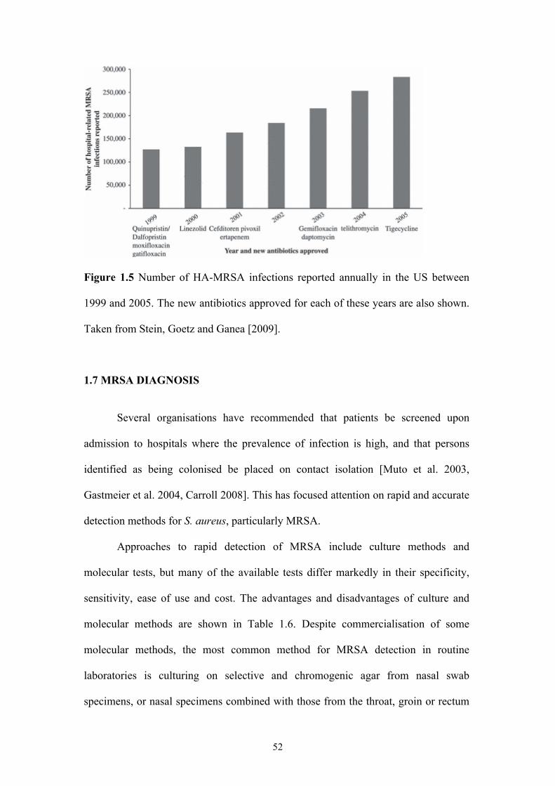
52
Figure 1.5 Number of HA-MRSA infections reported annually in the US between
1999 and 2005. The new antibiotics approved for each of these years are also shown.
Taken from Stein, Goetz and Ganea [2009].
1.7 MRSA DIAGNOSIS
Several organisations have recommended that patients be screened upon
admission to hospitals where the prevalence of infection is high, and that persons
identified as being colonised be placed on contact isolation [Muto et al. 2003,
Gastmeier et al. 2004, Carroll 2008]. This has focused attention on rapid and accurate
detection methods for S. aureus, particularly MRSA.
Approaches to rapid detection of MRSA include culture methods and
molecular tests, but many of the available tests differ markedly in their specificity,
sensitivity, ease of use and cost. The advantages and disadvantages of culture and
molecular methods are shown in Table 1.6. Despite commercialisation of some
molecular methods, the most common method for MRSA detection in routine
laboratories is culturing on selective and chromogenic agar from nasal swab
specimens, or nasal specimens combined with those from the throat, groin or rectum
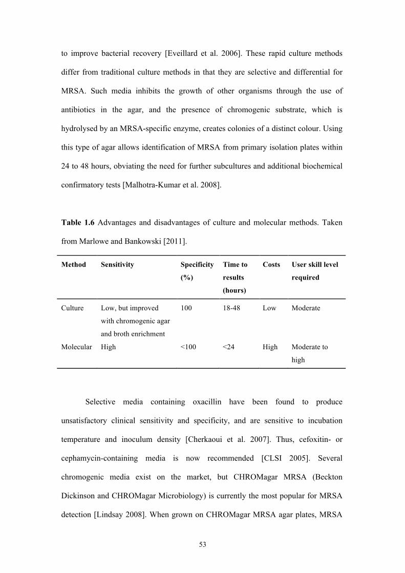
53
to improve bacterial recovery [Eveillard et al. 2006]. These rapid culture methods
differ from traditional culture methods in that they are selective and differential for
MRSA. Such media inhibits the growth of other organisms through the use of
antibiotics in the agar, and the presence of chromogenic substrate, which is
hydrolysed by an MRSA-specific enzyme, creates colonies of a distinct colour. Using
this type of agar allows identification of MRSA from primary isolation plates within
24 to 48 hours, obviating the need for further subcultures and additional biochemical
confirmatory tests [Malhotra-Kumar et al. 2008].
Table 1.6 Advantages and disadvantages of culture and molecular methods. Taken
from Marlowe and Bankowski [2011].
Method Sensitivity Specificity
(%)
Time to
results
(hours)
Costs User skill level
required
Culture Low, but improved
with chromogenic agar
and broth enrichment
100 18-48 Low Moderate
Molecular High <100 <24 High Moderate to
high
Selective media containing oxacillin have been found to produce
unsatisfactory clinical sensitivity and specificity, and are sensitive to incubation
temperature and inoculum density [Cherkaoui et al. 2007]. Thus, cefoxitin- or
cephamycin-containing media is now recommended [CLSI 2005]. Several
chromogenic media exist on the market, but CHROMagar MRSA (Beckton
Dickinson and CHROMagar Microbiology) is currently the most popular for MRSA
detection [Lindsay 2008]. When grown on CHROMagar MRSA agar plates, MRSA
54
will grow in the presence of cefoxitin (6mg/L) and produce mauve colonies as a
result of hydrolysis of the chromogenic substrate. Additional selective agents are
incorporated for the suppression of gram negative organisms, yeast and some other
gram positive cocci, but other bacteria may grow and produce differently coloured
colonies, e.g. bacteria that utilise chromogenic substrates in the medium produce blue
or blue/green colonies and those that don't utilise chromogenic substrates will
produce white or colourless colonies [BD Diagnostics 2010]. Other cefoxitin- or
cephomycin-containing media include chromID MRSA (bioMérieux), MRSA Select
(Bio-Rad) and Chromogenic MRSA/Denim Blue agar (Oxoid). All produce
uniformly high specificities after 24 hours, but sensitivities tend to vary both between
media and studies [Cherkaoui et al. 2007, Malhotra-Kumar et al. 2008, Luteijn et al.
2011], although they are higher than non-chromogenic media (93-99%) [Malhotra-
Kumar et al. 2010a]. Sensitivities can be improved by 48 hour incubation, but then
specificities are affected, requiring confirmatory MRSA tests [Malhotra-Kumar et al.
2008]. The variable performances highlight the need for a gold standard media for
MRSA screening [Cherkaoui et al. 2007].
Molecular techniques for direct detection of MRSA have become increasingly
commonplace [Marlowe and Bankowski 2011]. MRSA may be hetero-resistant to β-
lactam antibiotics due to repression of mecA by mecI and consequently the gold
standard method for molecular detection of MRSA is PCR detection of mecA in S.
aureus. However, coagulase-negative staphylococci (CNS), such as S. epidermidis
and S. haemolyticus, can be positive for mecA and are frequently carried in the human
population. Commercial PCR kits for detection of mecA have been available for
several years but these are not widely used due to the problem of false-positives
caused by mecA-carrying CNS isolates. In CNS, SCCmec elements can be identical
55
to those in S. aureus, but the location of the integration site (attBscc) differs. In the S.
aureus chromosome, attBscc is located near the S. aureus origin of replication, in an
open reading frame of unknown function, orfX, which is highly conserved among
clinical strains of S. aureus. Multiplex PCR approaches have been developed in
recent years to take advantage of this fact, in an attempt to overcome the problem of
false positives. Specifically, the approach involves detection of a single amplicon,
which includes the right junction of the SCCmec element and a part of the adjacent S.
aureus-specific orfX gene. This amplified region is termed the mec right extremity
junction (MREJ) (section 3.1.1).
Commercial assays like GenoType MRSA Direct (Hain Lifescience), BD
GeneOhm MRSA (BD Diagnostics) and Xpert MRSA (Cepheid Diagnostics) have
successfully utilised the approach, with the latter two being US Food and Drug
Administration (FDA)-approved for detection of MRSA from nasal surveillance
samples. Colonisation of the nose has been shown to be a risk factor for subsequent
infection [Carroll 2008].
GenoType MRSA Direct targets SCCmec types I to V in a multiplex PCR
using biotinylated primers followed by a reverse hybridisation step [Malhotra-Kumar
et al. 2008]. Direct detection of MRSA from diverse body sties (nose, throat, groin,
axilla, wound, and other sites) gives a sensitivity and specificity ranging from 68% to
95% and 96% to 99%, respectively [Harbarth et al. 2011]. There is a newer version of
this assay, the GenoQuick MRSA dipstick assay, which excludes the reverse
hybridisation step, reducing the total assay time from 4 hours to 2 hours 20 minutes
[Carroll 2008, Malhotra-Kumar et al. 2008]. This assay has been shown to have very
high sensitivity, specificity, positive predictive value (PPV) and negative predictive
value (NPV) (100%, 99.4%, 96% and 100% respectively) [Eigner et al. 2007].
56
BD GeneOhm MRSA is a multiplex qualitative real-time PCR assay that can
be semi-automated using Cepheid’s SmartCycler instrument and has been extensively
evaluated [Huletsky et al. 2004, Warren et al. 2004, Bishop et al. 2006, Desjardins et
al. 2006, Drews et al. 2006, Oberdorfer et al. 2006, Francois et al. 2007, Paule et al.
2007, Rossney et al. 2007a, van Hal et al. 2007, Zhang et al. 2007, Bartels et al. 2009,
Kelley et al. 2009, Kimura et al. 2009, Park et al. 2009a, Hombach et al. 2010, Lucke
et al. 2010, Peterson et al. 2010, Snyder, Munier and Johnson 2010, Hassan and
Shorman 2011, Patel et al. 2011]. The assay contains primers targeting the right
junction sequences of SCCmec types I to VI and VIII [Boyle-Vavra and Daum 2010],
combined with a consensus primer and three molecular beacons specific for orfX
[Malhotra-Kumar et al. 2008]. Since the assay is able to simultaneously link
identification with resistance detection, it can differentiate MRSA from MSSA and
mecA-positive CNS in clinical samples. Clinical performance characteristics are
compared with those of the Xpert MRSA assay in Table 1.7. In general, the
sensitivity and NPVs are equivalent or slightly better than culture, so the assay can be
used to decide which patients should be put into or removed from isolation
[Malhotra-Kumar et al. 2008]. However, the PPV appears poor (Table 1.7) so the
impact of isolating non-colonised patients should be assessed before using this assay
[Carroll 2008]. Studies show that BD GeneOhm MRSA, Genotype MRSA Direct and
Xpert MRSA assays fail to detect nontypeable SCCmec elements or certain variants
of known SCCmec types [Francois et al. 2007, Rossney et al. 2007a, Bartels et al.
2009, Boyle-Vavra and Daum 2010, Laurent et al. 2010], suggesting that these assays
should be evaluated against the local MRSA diversity, and in areas where
problematic SCCmec elements are prevalent, perhaps an alternative assay or culture-
based method should be employed.

57
A new version of the BD GeneOhm MRSA assay, BD GeneOhm ACP
MRSA, incorporates a novel lysis method for specimen preparation, reducing the
preparation steps and time needed to perform them, thus facilitating a high-
throughput, automated procedure for MRSA detection. Performance characteristics of
the ACP version were comparable to the original assay, providing good sensitivity
[Patel et al. 2011].
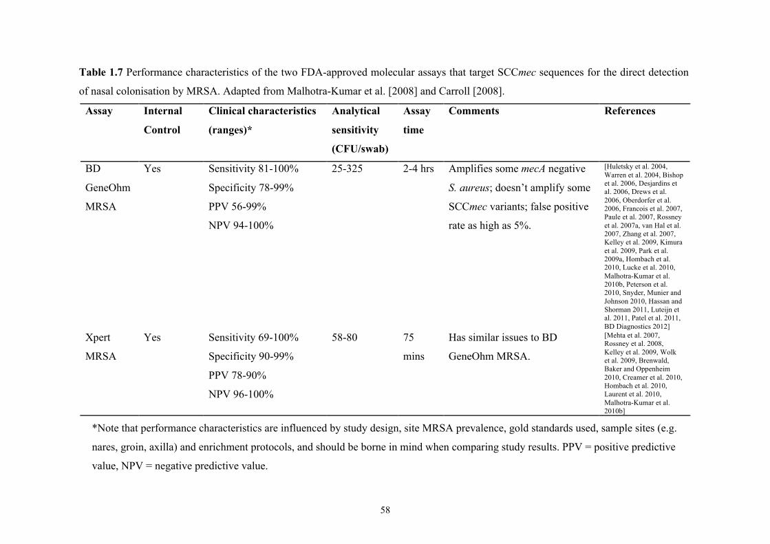
58
Table 1.7 Performance characteristics of the two FDA-approved molecular assays that target SCCmec sequences for the direct detection
of nasal colonisation by MRSA. Adapted from Malhotra-Kumar et al. [2008] and Carroll [2008].
Assay Internal
Control
Clinical characteristics
(ranges)*
Analytical
sensitivity
(CFU/swab)
Assay
time
Comments References
BD
GeneOhm
MRSA
Yes Sensitivity 81-100%
Specificity 78-99%
PPV 56-99%
NPV 94-100%
25-325 2-4 hrs Amplifies some mecA negative
S. aureus; doesn’t amplify some
SCCmec variants; false positive
rate as high as 5%.
[Huletsky et al. 2004, Warren et al. 2004, Bishop et al. 2006, Desjardins et al. 2006, Drews et al. 2006, Oberdorfer et al. 2006, Francois et al. 2007, Paule et al. 2007, Rossney et al. 2007a, van Hal et al. 2007, Zhang et al. 2007, Kelley et al. 2009, Kimura et al. 2009, Park et al. 2009a, Hombach et al. 2010, Lucke et al. 2010, Malhotra-Kumar et al. 2010b, Peterson et al. 2010, Snyder, Munier and Johnson 2010, Hassan and Shorman 2011, Luteijn et al. 2011, Patel et al. 2011, BD Diagnostics 2012]
Xpert
MRSA
Yes Sensitivity 69-100%
Specificity 90-99%
PPV 78-90%
NPV 96-100%
58-80 75
mins
Has similar issues to BD
GeneOhm MRSA.
[Mehta et al. 2007, Rossney et al. 2008, Kelley et al. 2009, Wolk et al. 2009, Brenwald, Baker and Oppenheim 2010, Creamer et al. 2010, Hombach et al. 2010, Laurent et al. 2010, Malhotra-Kumar et al. 2010b]
*Note that performance characteristics are influenced by study design, site MRSA prevalence, gold standards used, sample sites (e.g.
nares, groin, axilla) and enrichment protocols, and should be borne in mind when comparing study results. PPV = positive predictive
value, NPV = negative predictive value.
59
Xpert MRSA is a real-time PCR assay that works on a fully automated
platform, and is the most sophisticated system available for MRSA detection. It is
fully automated with minimal front-end processing, the level of expertise required to
operate it are minimal, and it is a random access instrument allowing flexible testing
of samples [Carroll 2008, Malhotra-Kumar et al. 2008, Rossney et al. 2008]. Xpert
MRSA is able to detect SCCmec types I, II, III, IVa, V and VI. A comparison of
Xpert MRSA and BD GeneOhm MRSA showed similar sensitivities and specificities
(98.5% and 97.1%, and 90.4% and 89.2%, respectively) for MRSA detection from
nasal samples [Mehta et al. 2007] (Table 1.7), and Wolk et al. found no statistical
difference in performance between the two assays [Wolk et al. 2009]. The Xpert
MRSA assay is able to detect both MSSA and MRSA and is thought to reliably detect
MRSA in mixed cultures as the relative quantities of gene products is measured, thus
making it a semi-quantitative assay.
These molecular assays show consistently high NPVs, making them ideal
tools for the rapid isolation of MRSA carriers, and in turn dramatically reducing
isolation time [Hassan and Shorman 2011]. The potential value of using such assays
in point of care settings such as hospital wards has also been demonstrated by a major
reduction in TAT (by more than ten hours) compared to their use in laboratories
[Brenwald, Baker and Oppenheim 2010].
A study evaluating the savings made if Xpert MRSA, at a cost of €50 per test,
was used for MRSA detection in patients and healthcare workers, found that at least
€925 per exposed healthcare worker and €550 per exposed patient that were MRSA
negative, would be saved [Andersen et al. 2010]. Another study found that decision-
making based on molecular tests added between €154 (BD GeneOhm MRSA) and
€194 (Xpert MRSA) per patient to overall costs, while chromogenic tests saved €31,
60
leading the authors to conclude that rapid diagnostic tests safely reduce the number of
unnecessary isolation days, but only screening based on chromogenic testing, can be
considered cost saving [Wassenberg et al. 2010]. This is in contrast to a Swiss study
in a single healthcare facility that found replacement of culture-based testing with
PCR-based methods more than halved the number of pre-emptive isolation days and
was thus considered cost-effective [Uckay et al. 2008].
It is clear that molecular tests allow for prompt MRSA detection, but they are
expensive, and so the effectiveness of their use must be carefully evaluated for each
setting. In populations with low MRSA endemicity, broad use of molecular tests may
not be cost-effective [Harbarth et al. 2011, Wassenberg et al. 2011], but in settings of
low MRSA prevalence, rapid isolation of MRSA is crucial for effective MRSA
control [Diederen 2010]. Rapid screening with chromogenic media is preferred due to
the expense of molecular tests, but the latter could be cost-effective for high-risk
healthcare units and critically ill patients [Harbarth et al. 2011]. While it is suggested
in some populations that transmission rates do not differ between patients screened
with culture-based methods and those screened with molecular-based methods, the
real value of the latter lies in their ability to free up bed space more readily in the
hospital [Marlowe and Bankowski 2011].
1.6 THESIS OBJECTIVES
My thesis draws on two themes identified in the introduction: the need for a
simpler, cheaper and quicker MRSA diagnostic test that enables widespread use of a
molecular test for MRSA screening (addressed in Part 1); and the need for a better
understanding of healthcare MRSA reservoirs due to the ever-changing epidemiology
of MRSA, specifically investigating the extent to which HA-MRSA and CA-MRSA

61
strains have merged in these reservoirs (Part 2 of the thesis). These themes are
introduced more fully in their respective parts.
62
CHAPTER 2: METHODS
Note: This chapter outlines methods used throughout my thesis. Methods specific to a
chapter are described in that chapter.
2.1 BACTERIAL CULTURE
All bacterial isolates were stored in 15% glycerol (TSB-glycerol) at −80°C.
Cells were harvested on Oxoid blood agar base No.2 and incubated at 37°C
overnight. Single colonies from the overnight growth were picked and subcultured,
from which DNA extractions were performed.
2.2 DNA EXTRACTION
DNA was extracted from subcultured growth using a DNeasy Blood & Tissue
Kit (Qiagen), according to guidelines for the purification of total DNA from gram-
positive bacteria. Briefly, this involved the standard purification procedure but with a
pretreatment step of incubation with enzymatic lysis buffer to lyse cell walls (20mM
Tris-Cl pH 8.0, 2mM sodium EDTA, 1.2% Triton X-100, lysostaphin to 2mg/ml and
immediately before use, lysozyme to 20mg/ml). DNA samples were eluted in 200µl
of AE buffer (10 mM Tris-Cl and 0.5 mM EDTA, pH 9.0) and stored at −20°C.
2.3 PCR AMPLIFICATION
Unless otherwise stated, all PCR amplification was performed in a total
volume of 29µl, containing 25µl of 1.1x ReddyMix PCR Master Mix (Thermo
Scientific; 1.25u Thermoprime Plus DNA Polymerase, 75mM Tris-HCl (pH8.8 at
25°C), 20mM (NH4)2SO4, 1.5mM MgCl2, 0.01% (v/v) Tween 20, 0.2mM each of
dATP, dCTP, dGTP and dTTP, plus precipitant and red dye for electrophoresis), 1µl
63
each of 100µM forward and reverse primer, and 2µl of cleaned DNA template. 5µl of
each PCR product was checked by electrophoresis on a 1% agarose gel containing
5µl of SafeView nucleic acid stain (NBS Biologicals) at 15V/cm for 40 min in 1x
TBE buffer. The remaining volume of each PCR product was then purified by
polyethylene glycol (PEG) precipitation plus an ethanol wash and suspended in 12µl
of sterile distilled water (SDW).
2.4 SEQUENCING OF PCR PRODUCTS
All purified PCR products were diluted to 2.5-5ng/µl using SDW prior to
sequencing reactions. Products were sequenced using the BigDye Terminator v1.1
Cycle Sequencing Kit (Applied Biosystems). A total reaction volume of 10µl
contained 2µl of purified PCR product, 4µl of 1µM forward or reverse primer, 0.5µl
of BigDye Terminator, 1.75µl of BigDye Terminator 5x Sequencing Buffer, and
1.75µl of SDW). Thermal cycling conditions for sequencing reactions included 25
cycles of denaturing, annealing and extension (10s at 96°C, 5s at 50°C and 2 min at
60°C) and a ramp of 0.1°C/s to 4°C. Reaction cleanup was performed by ethanol
precipitation. When ready to sequence, cleaned reaction products were re-suspended
in 10µl of Hi-Di Formamide (Applied Biosystems) and sequences determined using
the ABI 3730xl DNA Analyser. All sequence data were assembled, trimmed, edited
and aligned using Molecular Evolutionary Genetics Analysis (MEGA) version 4.1
[Tamura et al. 2007], except data for the spa repeat region, which were analysed
using software described in section 2.6.
64
2.5 MLST
Primer sequences used for PCR amplification of the seven housekeeping genes
are listed in Table 2.1. Thermal cycling conditions consisted of an initial denaturation
of 3 min at 95°C, 35 cycles of denaturation, annealing and extension (30s at 95°C,
30s at 60°C and 1 min at 72°C), and a final extension of 10 min at 72°C. Following
sequencing of the seven loci and building of consensus sequences using MEGA
version 4.1 [Tamura et al. 2007], consensus sequences were queried using the MLST
S. aureus database (http://saureus.mlst.net/) to determine sequence types (STs).
Relationships between STs were visualised using the eBURST (Based Upon Related
Sequence Types) algorithm [Feil et al. 2004, Spratt et al. 2004]
(http://saureus.mlst.net/eburst/).
2.6 spa TYPING
Primer sequences used for PCR amplification of the spa repeat region are
listed in Table 2.1. Thermal cycling conditions consisted of an initial denaturation of
5 min at 80°C, 35 cycles of denaturation, annealing and extension (45s at 94°C, 45s
at 60°C and 90s at 72°C), and a final extension of 10 min at 72°C. Following
sequencing of the spa repeat region, spa types were determined using Ridom
StaphType v1.5-2.2 (Ridom GmbH, Würzburg, Germany) [Harmsen et al. 2003]. To
assess spa type diversity and relatedness, cluster analysis of spa types was performed
using the Based Upon Repeat Pattern (BURP) algorithm, a built-in feature of the
StaphType software [Mellmann et al. 2007]. The BURP algorithm is a heuristic
variant of the Excision, Duplication, Substitution and Indels (EDSI) algorithm
[Sammeth and Stoye 2006], and is the first automated and objective tool to infer
clonal relatedeness from spa repeat regions [Mellmann et al. 2007].
65
Table 2.1 PCR Primers used for spa typing and MLST.
Primer 5′-3′ sequence Amplicon Size (bp) Reference
spa_1113F spa_1514R
TAAAGACGATCCTTCGGTGAGC CAGCAGTAGTGCCGTTTGCTT
Variable [Ridom GmbH 2006]
arC_fwd arcC_rev
TTGATTCACCAGCGCGTATTGTC AGGTATCTGCTTCAATCAGCG
456 [Enright et al. 2000]
aroE_fwd aroE_rev
ATCGGAAATCCTATTTCACATTC GGTGTTGTATTAATAACGATATC
456 [Enright et al. 2000]
glpF_fwd glpF_rev
CTAGGAACTGCAATCTTAATCC TGGTAAAATCGCATGTCCAATTC
465 [Enright et al. 2000]
gmk_fwd gmk_rev
ATCGTTTTATCGGGACCATC TCATTAACTACAACGTAATCGTA
429 [Enright et al. 2000]
pta_fwd pta_rev
GTTAAAATCGTATTACCTGAAGG GACCCTTTTGTTGAAAAGCTTAA
474 [Enright et al. 2000]
tpi_fwd tpi_rev yqiL_fwd yqiL_rev
TCGTTCATTCTGAACGTCGTGAA TTTGCACCTTCTAACAATTGTAC CAGCATACAGGACACCTATTGGC CGTTGAGGAATCGATACTGGAAC
402
516
[Enright et al. 2000] [Enright et al. 2000]
2.7 SCCmec TYPING
SCCmec typing was carried out according to either the method of Boye et al.
[2007] or Milheirico, Oliveira and de Lencastre [2007a] using the primers listed in
Tables 2.2 and 2.3. Multiplex PCR products were viewed on agarose gels
(percentages method-specific) containing 5µl of SafeView nucleic acid stain (NBS
Biologicals), run in 1x TBE buffer and at the rate recommended by each method.
SCCmec types were determined on the basis of the amplification pattern obtained
(Figure 2.1). Isolates with no visible bands, or with an amplification pattern not in
agreement with one of the predicted patterns, were classified as non-typeable (NT).
66
Table 2.2 Primers used in the multiplex SCCmec PCR assay of Boye et al. [2007] for SCCmec types I-V.
Name Primer sequence (5′3′) Amplicon
size (bp)
Target Primer specificity (SCCmec
type)
β α
ATTGCCTTCATAATAGCCYTCTa
TAAAGGCATCAATGCACAAACACTa
937
ccrA2-B
II and IV
ccrCF ccrCR
CGTCTATTACAAGATGTTAAGGATAATb
CCTTTATAGACTGGATTATTCAAAATATb
518 ccrC III and V
1272F1 1272R1
GCCACTCATAACATATGGAAc
CATCCGAGTGAAACCCAAAc
415 IS1272 I and IV
5RmecA 5R431
TATACCAAACCCGACAACTACc
CGGCTACAGTGATAACATCCc
359 mecA–IS431
V
a[Ito et al. 2001]; b[Ito et al. 2004]; c[Boye et al. 2007].
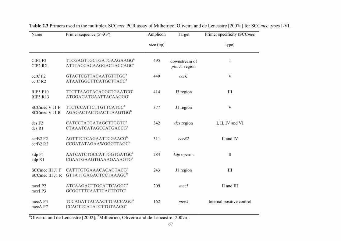
67
Table 2.3 Primers used in the multiplex SCCmec PCR assay of Milheirico, Oliveira and de Lencastre [2007a] for SCCmec types I-VI.
Name Primer sequence (5′3′) Amplicon
size (bp)
Target Primer specificity (SCCmec
type)
CIF2 F2 CIF2 R2
TTCGAGTTGCTGATGAAGAAGGa ATTTACCACAAGGACTACCAGCa
495
downstream of pls, J1 region
I
ccrC F2 ccrC R2
GTACTCGTTACAATGTTTGGb ATAATGGCTTCATGCTTACCb
449 ccrC V
RIF5 F10 RIF5 R13
TTCTTAAGTACACGCTGAATCGa ATGGAGATGAATTACAAGGGa
414 J3 region III
SCCmec V J1 F SCCmec V J1 R
TTCTCCATTCTTGTTCATCCb AGAGACTACTGACTTAAGTGGb
377 J1 region V
dcs F2 dcs R1
CATCCTATGATAGCTTGGTCa
CTAAATCATAGCCATGACCGa
342 dcs region I, II, IV and VI
ccrB2 F2 ccrB2 R2
AGTTTCTCAGAATTCGAACGb
CCGATATAGAAWGGGTTAGCb
311 ccrB2 II and IV
kdp F1 kdp R1
AATCATCTGCCATTGGTGATGCa
CGAATGAAGTGAAAGAAAGTGa
284 kdp operon II
SCCmec III J1 F SCCmec III J1 R
CATTTGTGAAACACAGTACGb
GTTATTGAGACTCCTAAAGCb
243 J1 region III
mecI P2 mecI P3
ATCAAGACTTGCATTCAGGCa
GCGGTTTCAATTCACTTGTCa
209 mecI II and III
mecA P4 mecA P7
TCCAGATTACAACTTCACCAGGa
CCACTTCATATCTTGTAACGa
162 mecA Internal positive control
aOliveira and de Lencastre [2002]; bMilheirico, Oliveira and de Lencastre [2007a].
68
Figure 2.1 Amplification patterns obtained with the SCCmec multiplex PCR strategies of A Boye et
al. [2007] (examples of SCCmec types I-V with 100bp DNA ladders) and B Milheirico, Oliveira and
de Lencastre [2007a] (SCCmec types (prototype strains) as follows: lane 1, I (COL); lane 2, II
(N315); lane 3, III (ANS46), lane 4, IVa (MW2); lane 5, IVb (8/6-3P); lane 6, IVc (Q2314); lane 7,
IVd (JCSC4469); lane 8, IVE (AR43/3330.1); lane 9, IVg (M03-68); lane 10, IVh (HAR22); lane 11,
V (WIS); lane 12, VI (HDE288); and M, 1kb DNA ladder). Gel images taken directly from Boye et
al. [2007] and Milheirico, Oliveira and de Lencastre [2007a].
69
PART 1 INTRODUCTION: RPA-BASED DETECTION OF MRSA
The major advantage of rapid MRSA detection is the time saved over
traditional culture methods. Although advances have been made with culture-based
detection methods, particularly those based on chromogenic media, which have
decreased the time to detection, molecular tests can provide results in a few hours; BD
GeneOhm MRSA takes 2-4 hours, and Xpert MRSA under 70 minutes. However,
both systems require expensive specialist platforms, the costs per test are high (BD
GeneOhm MRSA excluding/including platform, personnel and additional costs
(swabs, gloves and consumables), around €30/€56; Xpert MRSA around €43/€70) and
the operator skill requirement for the assays is also relatively high, preventing their
widespread routine use [Malhotra-Kumar et al. 2008, Wassenberg et al. 2010]. The
training required and the size and expense of these platforms means they are typically
employed only in central laboratories, significantly increasing the TAT for a
diagnostic MRSA result. Nonetheless, PCR-based molecular detection methods allow
a clinically relevant turnaround time for a diagnostic result, with comparable clinical
sensitivity and specificity to culture-based methods [Luteijn et al. 2011]. There is still
a need for a standalone rapid assay that can detect all MRSA strains, and a faster and
cheaper diagnostic test that can be deployed at the point of care. This chapter
describes and evaluates one such assay that is based on a novel method of nucleic acid
amplification.
Recombinase polymerase amplification (RPA), developed by my industrial
sponsor TwistDx, is a novel alternative to PCR for the amplification and detection of
nucleic acids. It couples isothermal recombinase-driven primer targeting of template
material with strand-displacement DNA synthesis (Figure I.1) [Piepenburg et al.
2006]. The key to RPA is the dynamic reaction environment that balances
70
recombinase-primer complex formation with disassembly (Figure I.2). UvsX
recombinase of bacteriophage T4 binds cooperatively to primers in the presence of
ATP. The resulting ATP-bound nucleoprotein complex (Figure I.2A) actively
hydrolyses ATP and the consequent depletion of ATP substrate and accumulation of
ADP/AMP products leads to replacement of UvsX with gp32 (Figure I.2B), the single
stranded DNA binding protein (SSB) of T4 necessary for RPA. gp32 protects single-
stranded DNA from nuclease digestion, removes secondary structure and promotes
assembly of recombinase-primer complexes [Liu, Qian and Morrical 2006]. However,
at high gp32 and salt concentrations, recombinase-primer complex formation is
inhibited by the high affinity of gp32-primer interactions [Ando and Morrical 1998].
gp32 thus competes with UvsX for binding sites on the primer. Assembly of UvsX-
primer complexes relies on the mediator function of the T4 UvsY protein, a
recombinase loading factor that weakens gp32-primer interactions and strengthens
UvsX-primer interactions, thus helping UvsX to displace gp32 and shift the
equilibrium in favour of recombinase loading (Figure I.2C) [Jiang, Salinas and
Kodadek 1997, Sweezy and Morrical 1999, Bleuit et al. 2004, Liu, Bond and Morrical
2006]. The presence of a crowding agent such as polyethylene glycol (PEG) further
establishes reaction conditions that support RPA, by strengthening the interactions
between the proteins and DNA. As well as a role in presynaptic filament formation,
gp32 facilitates DNA strand exchange by binding to the displaced strand generated
during UvsX-catalysed D-loop formation, as shown in Figure I.1 [Liu, Qian and
Morrical 2006].
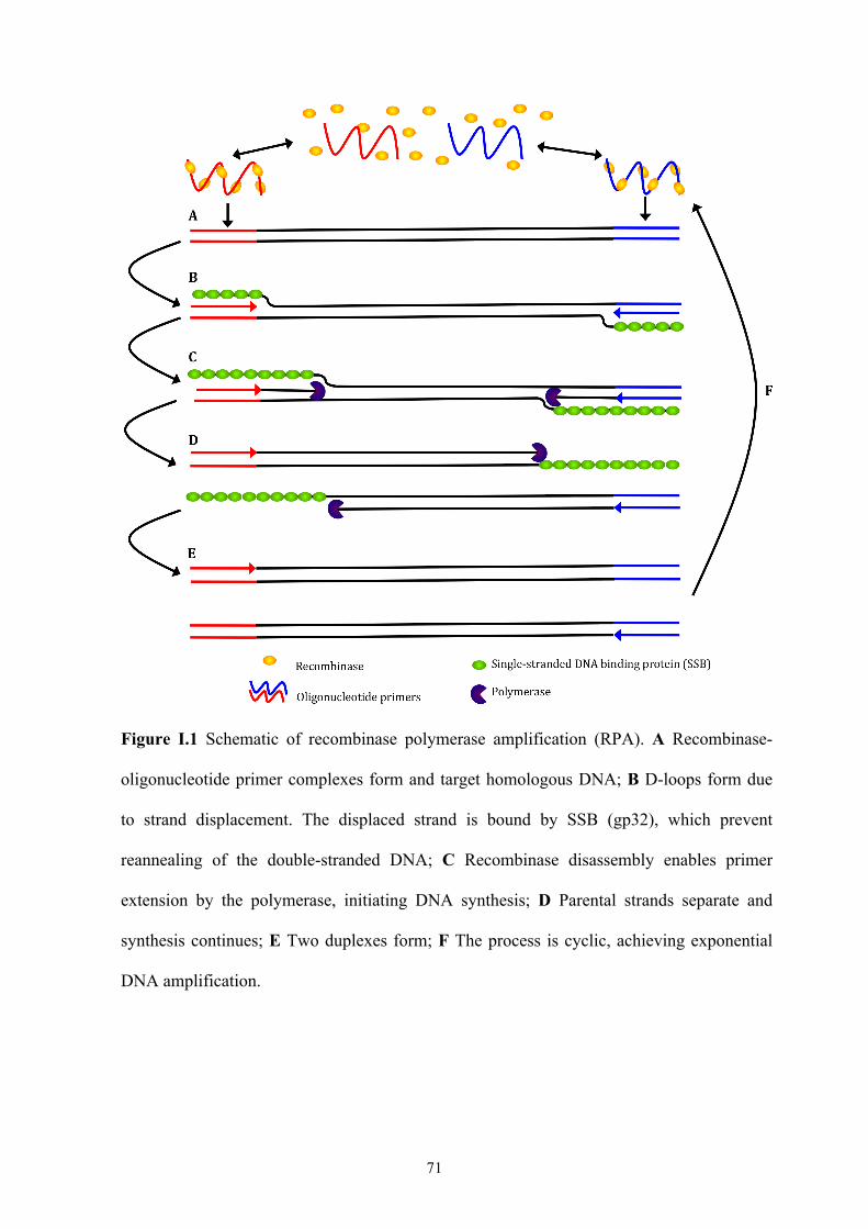
71
Figure I.1 Schematic of recombinase polymerase amplification (RPA). A Recombinase-
oligonucleotide primer complexes form and target homologous DNA; B D-loops form due
to strand displacement. The displaced strand is bound by SSB (gp32), which prevent
reannealing of the double-stranded DNA; C Recombinase disassembly enables primer
extension by the polymerase, initiating DNA synthesis; D Parental strands separate and
synthesis continues; E Two duplexes form; F The process is cyclic, achieving exponential
DNA amplification.
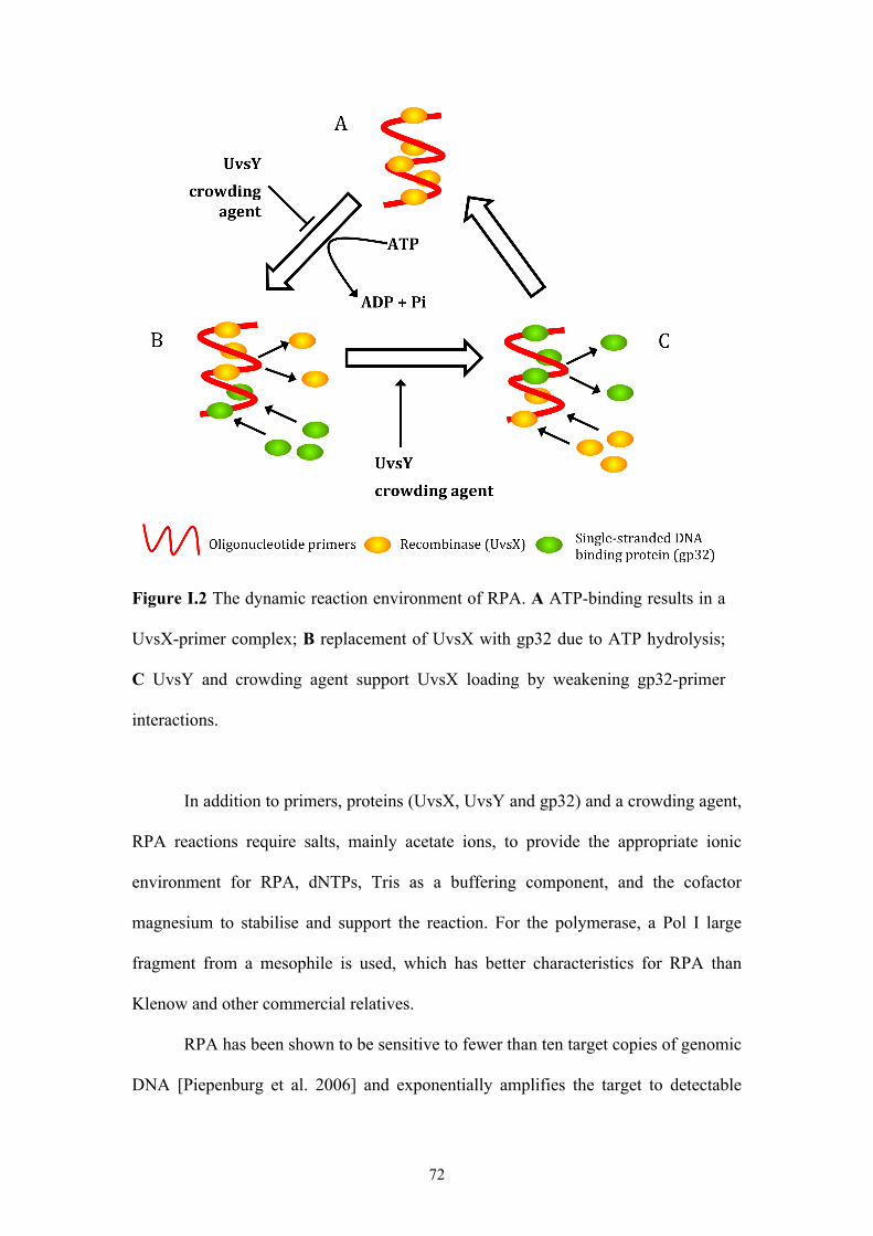
72
Figure I.2 The dynamic reaction environment of RPA. A ATP-binding results in a
UvsX-primer complex; B replacement of UvsX with gp32 due to ATP hydrolysis;
C UvsY and crowding agent support UvsX loading by weakening gp32-primer
interactions.
In addition to primers, proteins (UvsX, UvsY and gp32) and a crowding agent,
RPA reactions require salts, mainly acetate ions, to provide the appropriate ionic
environment for RPA, dNTPs, Tris as a buffering component, and the cofactor
magnesium to stabilise and support the reaction. For the polymerase, a Pol I large
fragment from a mesophile is used, which has better characteristics for RPA than
Klenow and other commercial relatives.
RPA has been shown to be sensitive to fewer than ten target copies of genomic
DNA [Piepenburg et al. 2006] and exponentially amplifies the target to detectable
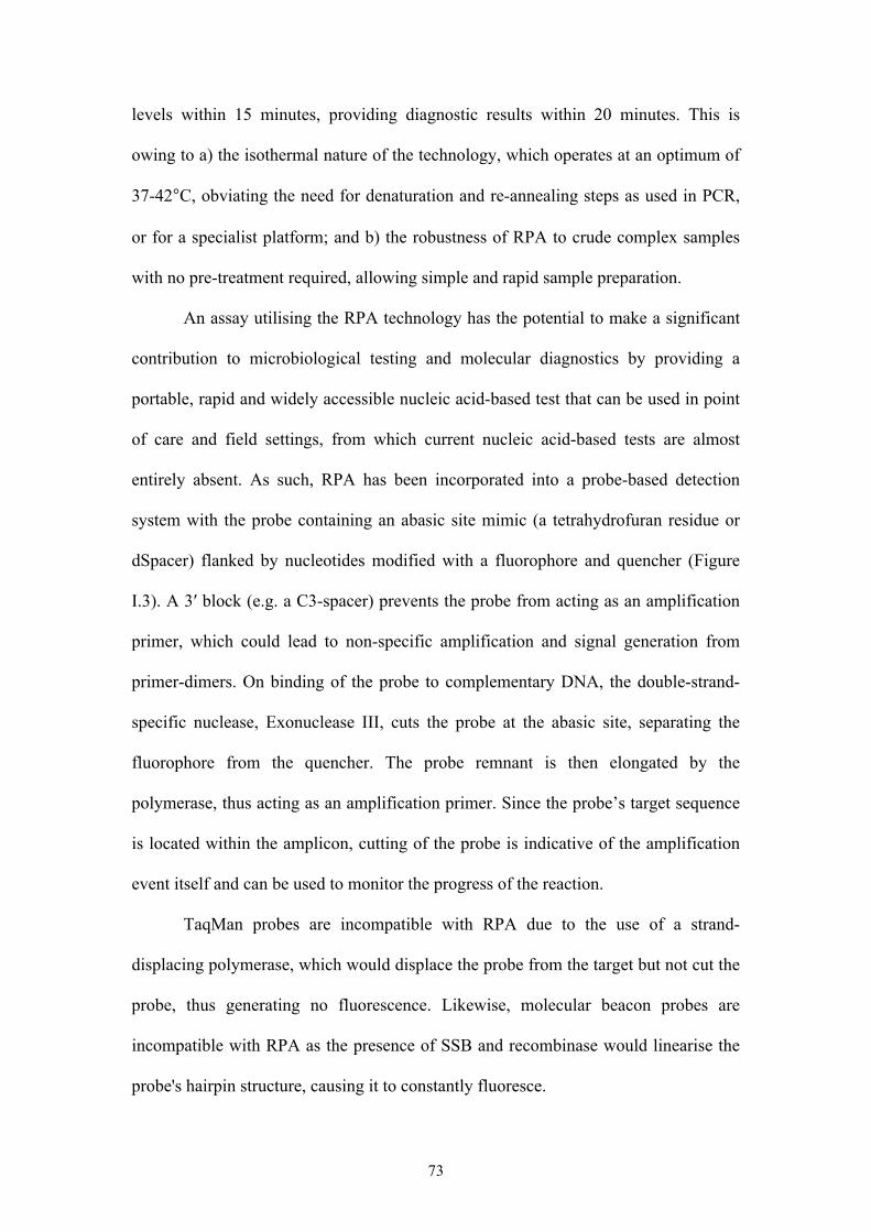
73
levels within 15 minutes, providing diagnostic results within 20 minutes. This is
owing to a) the isothermal nature of the technology, which operates at an optimum of
37-42°C, obviating the need for denaturation and re-annealing steps as used in PCR,
or for a specialist platform; and b) the robustness of RPA to crude complex samples
with no pre-treatment required, allowing simple and rapid sample preparation.
An assay utilising the RPA technology has the potential to make a significant
contribution to microbiological testing and molecular diagnostics by providing a
portable, rapid and widely accessible nucleic acid-based test that can be used in point
of care and field settings, from which current nucleic acid-based tests are almost
entirely absent. As such, RPA has been incorporated into a probe-based detection
system with the probe containing an abasic site mimic (a tetrahydrofuran residue or
dSpacer) flanked by nucleotides modified with a fluorophore and quencher (Figure
I.3). A 3′ block (e.g. a C3-spacer) prevents the probe from acting as an amplification
primer, which could lead to non-specific amplification and signal generation from
primer-dimers. On binding of the probe to complementary DNA, the double-strand-
specific nuclease, Exonuclease III, cuts the probe at the abasic site, separating the
fluorophore from the quencher. The probe remnant is then elongated by the
polymerase, thus acting as an amplification primer. Since the probe’s target sequence
is located within the amplicon, cutting of the probe is indicative of the amplification
event itself and can be used to monitor the progress of the reaction.
TaqMan probes are incompatible with RPA due to the use of a strand-
displacing polymerase, which would displace the probe from the target but not cut the
probe, thus generating no fluorescence. Likewise, molecular beacon probes are
incompatible with RPA as the presence of SSB and recombinase would linearise the
probe's hairpin structure, causing it to constantly fluoresce.
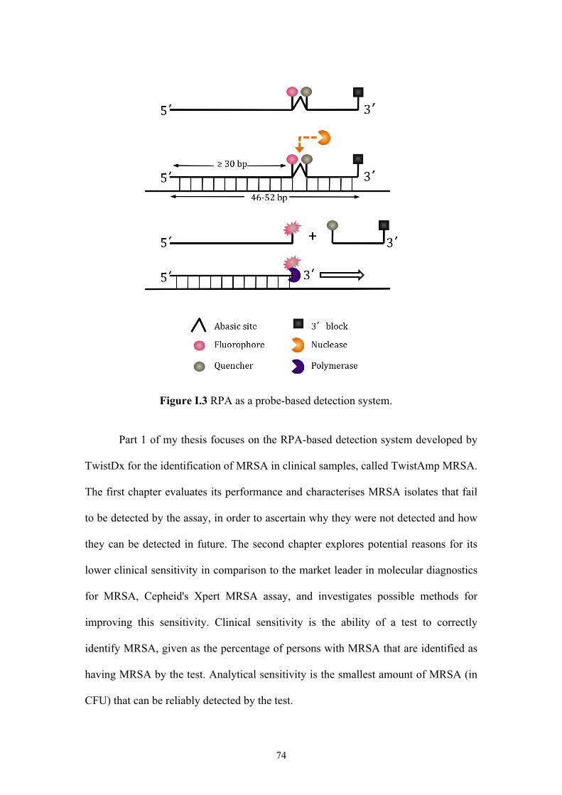
74
Figure I.3 RPA as a probe-based detection system.
Part 1 of my thesis focuses on the RPA-based detection system developed by
TwistDx for the identification of MRSA in clinical samples, called TwistAmp MRSA.
The first chapter evaluates its performance and characterises MRSA isolates that fail
to be detected by the assay, in order to ascertain why they were not detected and how
they can be detected in future. The second chapter explores potential reasons for its
lower clinical sensitivity in comparison to the market leader in molecular diagnostics
for MRSA, Cepheid's Xpert MRSA assay, and investigates possible methods for
improving this sensitivity. Clinical sensitivity is the ability of a test to correctly
identify MRSA, given as the percentage of persons with MRSA that are identified as
having MRSA by the test. Analytical sensitivity is the smallest amount of MRSA (in
CFU) that can be reliably detected by the test.
75
CHAPTER 3: TWISTAMP MRSA
3.1 INTRODUCTION
Having recognised the public health implications of MRSA worldwide and
thus the need for a faster, simpler and cheaper diagnostic test compared to current
commercial assays, TwistDx have developed an RPA-based diagnostic assay for
MRSA, called TwistAmp MRSA. Proprietary primers amplify the different sequences
between the SCCmec element and orfX (the mec right extremity junction; MREJ)
found in MRSA strains, an approach similar to that used in the BD GeneOhm MRSA
and Xpert MRSA assays. Proprietary FAM- and TAMRA-labelled TwistAmp exo
probes then bind to the MREJ amplicons and internal control respectively and
generate a fluorescence signal as a real-time readout.
3.1.1 The mec Right Extremity Junction (MREJ)
The MREJ is approximately 1kb in length and comprises the right extremity of
SCCmec, the SCCmec integration site attBscc and the 3′ end of the orfX gene of the S.
aureus chromosome (Figure 3.1) [Huletsky et al. 2004, Cuny and Witte 2005]. The
first assay that utilised the MREJ as a MRSA-specific target was developed in 2000,
using PCR primers that targeted the MREJs of SCCmec types I-III [Hiramatsu et al.
2000]. This led to the development of mec right extremity polymorphism (MREP)
typing for SCCmec DNA, on the basis that SCCmec types I-III each have a different
MREJ (MREJ types i-iii) [Hiramatsu, Kondo and Ito 1996, Ito et al. 2001]. After the
discovery of SCCmec IV [Ma et al. 2002], it was found that the MREP typing method
could not discriminate it from SCCmec II and therefore both were associated with
MREJ ii [Huletsky and Rossbach 2002].
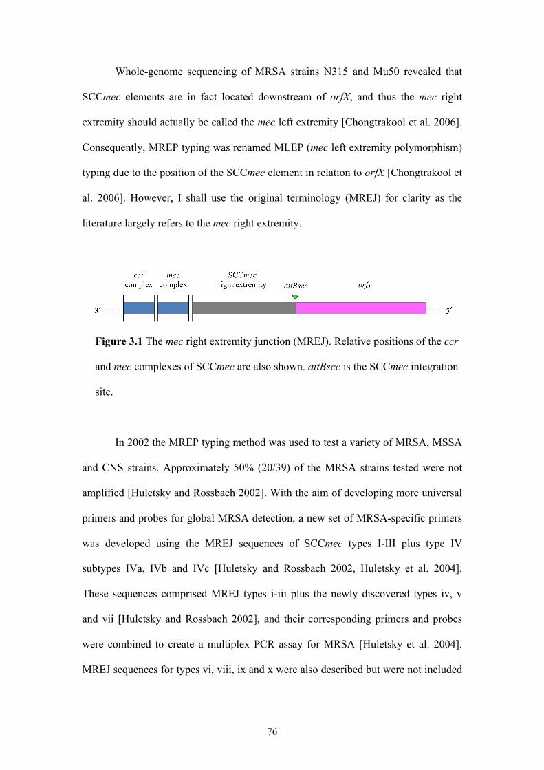
76
Whole-genome sequencing of MRSA strains N315 and Mu50 revealed that
SCCmec elements are in fact located downstream of orfX, and thus the mec right
extremity should actually be called the mec left extremity [Chongtrakool et al. 2006].
Consequently, MREP typing was renamed MLEP (mec left extremity polymorphism)
typing due to the position of the SCCmec element in relation to orfX [Chongtrakool et
al. 2006]. However, I shall use the original terminology (MREJ) for clarity as the
literature largely refers to the mec right extremity.
Figure 3.1 The mec right extremity junction (MREJ). Relative positions of the ccr
and mec complexes of SCCmec are also shown. attBscc is the SCCmec integration
site.
In 2002 the MREP typing method was used to test a variety of MRSA, MSSA
and CNS strains. Approximately 50% (20/39) of the MRSA strains tested were not
amplified [Huletsky and Rossbach 2002]. With the aim of developing more universal
primers and probes for global MRSA detection, a new set of MRSA-specific primers
was developed using the MREJ sequences of SCCmec types I-III plus type IV
subtypes IVa, IVb and IVc [Huletsky and Rossbach 2002, Huletsky et al. 2004].
These sequences comprised MREJ types i-iii plus the newly discovered types iv, v
and vii [Huletsky and Rossbach 2002], and their corresponding primers and probes
were combined to create a multiplex PCR assay for MRSA [Huletsky et al. 2004].
MREJ sequences for types vi, viii, ix and x were also described but were not included
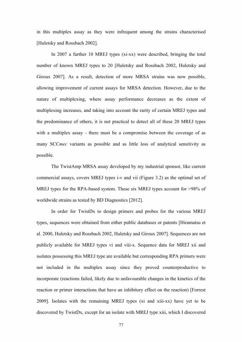
77
in this multiplex assay as they were infrequent among the strains characterised
[Huletsky and Rossbach 2002].
In 2007 a further 10 MREJ types (xi-xx) were described, bringing the total
number of known MREJ types to 20 [Huletsky and Rossbach 2002, Huletsky and
Giroux 2007]. As a result, detection of more MRSA strains was now possible,
allowing improvement of current assays for MRSA detection. However, due to the
nature of multiplexing, where assay performance decreases as the extent of
multiplexing increases, and taking into account the rarity of certain MREJ types and
the predominance of others, it is not practical to detect all of these 20 MREJ types
with a multiplex assay - there must be a compromise between the coverage of as
many SCCmec variants as possible and as little loss of analytical sensitivity as
possible.
The TwistAmp MRSA assay developed by my industrial sponsor, like current
commercial assays, covers MREJ types i-v and vii (Figure 3.2) as the optimal set of
MREJ types for the RPA-based system. These six MREJ types account for >98% of
worldwide strains as tested by BD Diagnostics [2012].
In order for TwistDx to design primers and probes for the various MREJ
types, sequences were obtained from either public databases or patents [Hiramatsu et
al. 2000, Huletsky and Rossbach 2002, Huletsky and Giroux 2007]. Sequences are not
publicly available for MREJ types vi and viii-x. Sequence data for MREJ xii and
isolates possessing this MREJ type are available but corresponding RPA primers were
not included in the multiplex assay since they proved counterproductive to
incorporate (reactions failed, likely due to unfavourable changes in the kinetics of the
reaction or primer interactions that have an inhibitory effect on the reaction) [Forrest
2009]. Isolates with the remaining MREJ types (xi and xiii-xx) have yet to be
discovered by TwistDx, except for an isolate with MREJ type xiii, which I discovered
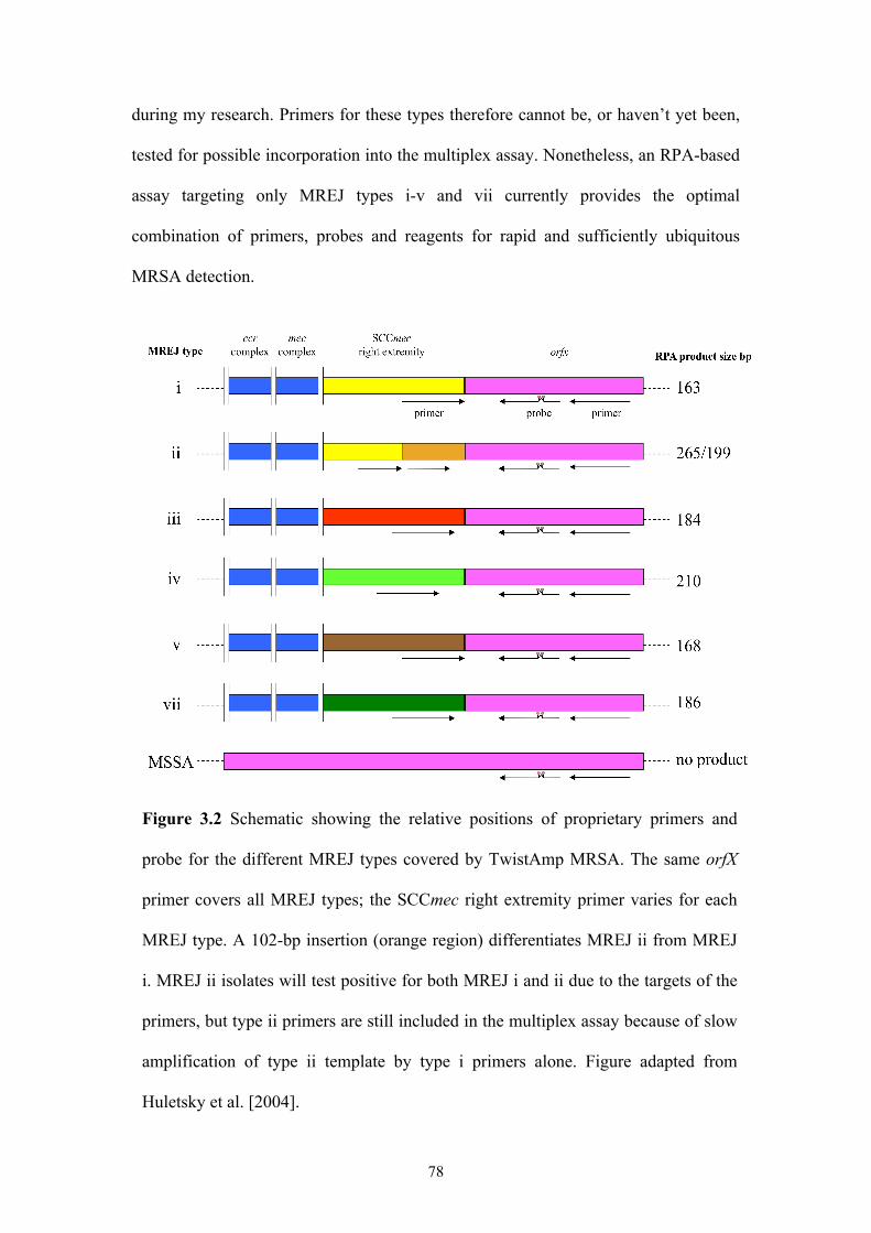
78
during my research. Primers for these types therefore cannot be, or haven’t yet been,
tested for possible incorporation into the multiplex assay. Nonetheless, an RPA-based
assay targeting only MREJ types i-v and vii currently provides the optimal
combination of primers, probes and reagents for rapid and sufficiently ubiquitous
MRSA detection.
Figure 3.2 Schematic showing the relative positions of proprietary primers and
probe for the different MREJ types covered by TwistAmp MRSA. The same orfX
primer covers all MREJ types; the SCCmec right extremity primer varies for each
MREJ type. A 102-bp insertion (orange region) differentiates MREJ ii from MREJ
i. MREJ ii isolates will test positive for both MREJ i and ii due to the targets of the
primers, but type ii primers are still included in the multiplex assay because of slow
amplification of type ii template by type i primers alone. Figure adapted from
Huletsky et al. [2004].
79
3.1.2 Chapter Objectives
3.1.2.1 TwistAmp MRSA performance
The aim of TwistAmp MRSA is to not only provide a more accurate and rapid
alternative to traditional culturing methods for MRSA detection, but also to rival the
current commercial PCR-based MRSA assays. A collection of Staphylococci from
our laboratory, including MRSA, MSSA and CNS, was used to interrogate the assay
in order to assess its performance (sensitivity, specificity, PPV and NPV). Two
collections of MRSA isolates obtained from standard screening procedures at
hospitals in the UK and US (hereafter called the UK and KC collections respectively;
see section 3.2) were used to further determine the performance of the assay.
3.1.2.2 MREJ typing of MRSA isolates
TwistAmp MRSA uses a multiplex format with proprietary primers and
probes to detect MRSA with MREJ types i-v and vii. Any MRSA with one of these
MREJ types will produce a fluorescence signal, which provides the user with a
positive identification of MRSA. Unlike MREP typing however, the MREJ type of
the isolate is not provided by the method. MREJ types can, however, be obtained
using a series of singleplex RPA-based assays with the primers and probes for the
individual MREJ types, an approach called MREJ typing.
The MREJ has not been studied a great deal in an epidemiological sense.
Since the prevalence of SCCmec types at the local and global level are well
documented, MREJ typing was performed on the UK and KC MRSA collections in
order to assess the diversity and distribution of MREJ types among local populations
of MRSA. Similarly, MREJ typing of our laboratory collection of MRSA was
80
performed to provide a snapshot of the diversity and distribution of MREJ types
among geographically diverse strains of HA- and CA-MRSA.
The commercial assays BD GeneOhm MRSA, Xpert MRSA and Genotype
MRSA Direct all target the MREJ, but like TwistAmp MRSA only produce a yes/no
output; they do not report the specific MREJ type. In their product literature, these
three commercial assays state the different SCCmec types that they are able to detect,
and the associated limit of detection (LOD) for each type. The LODs are based on
testing of an MRSA strain of each SCCmec type, but the product literature does not
clearly specify how SCCmec type correlates with MREJ type. The BD GeneOhm
MRSA package insert lists the LOD for each of six strains tested, representing MREJ
types i-v and vii and SCCmec types I-IV, but also states the ability to detect SCCmec
types V and VI without specifying the associated MREJ type [BD Diagnostics 2012].
The Xpert MRSA and Genotype MRSA Direct assays can detect SCCmec types I-V
but the associated MREJ types are not stated [Cepheid Diagnostics 2009, Hain
Lifescience GmbH 2012].
To my knowledge only two comparisons of MREJ typing and SCCmec typing
have been made. Huletsky et al. [2004] made three key findings: 1) no correlation was
found between MREJ and SCCmec type for the MRSA strains described in the study;
2) exceptions were found to the typical association between SCCmec types I-III and
MREJ types i-iii; and 3) strains with new MREJ types did not carry a new SCCmec
element but rather have structural variations at the SCCmec right extremity, since
known SCCmec types were assigned for most of the MRSA strains with the MREJ
types described in the study [Huletsky et al. 2004]. The other study found a strong
correlation between SCCmec type and MREJ type, but there were discrepancies and
non-typeable isolates [Chongtrakool et al. 2006]. For example, of 370 SCCmec III
81
isolates, 86.4% were MREJ iii but 10.6% were MREJ ii and 3% could not be assigned
a MREJ type [Chongtrakool et al. 2006]. Since TwistAmp MRSA also targets the
MREJ, the correlation between SCCmec type and MREJ type was explored further
using our laboratory and the KC collections of MRSA, as well as prototypic strains
for SCCmec types I-XI.
3.1.2.3 Characterisation of false negative isolates
The evaluation of the performance of TwistAmp MRSA in identifying MRSA
isolates in the UK and KC collections, followed by MREJ typing of these collections,
resulted in MRSA that were not detected by this assay (false negatives) and an MSSA
that was identified as MRSA (false positive). In order to contribute to the
development and improvement of TwistAmp MRSA, these false positive and false
negative isolates were characterised using MLST, spa typing and SCCmec typing. In
this way it was hoped that STs, spa types or SCCmec types could then be identified
that might pose a problem in terms of MRSA detection using TwistAmp MRSA. In
the case of false negative isolates, sequencing of the MREJ region was performed to
try and determine why these isolates were not detected, and if their MREJ sequences
could be incorporated into the assay through modification of the primers and/or
probes for currently covered MREJ types, or whether new primers and probes needed
to be developed for novel MREJ types.
In the latter case, primers and/or probes were designed for any novel MREJ
types and full primer screens conducted for potential incorporation into and therefore
improvement of the multiplex assay. If RPA primers are already developed for all
MREJ types encountered, whether known or novel, then incorporating them into the
diagnostic assay prospectively, where required, will prove more efficient than
82
discovering the assay does not detect a percentage of MRSA strains in a certain region
and designing them retrospectively.
3.1.2.4 Scope of TwistAmp MRSA
TwistDx aim to develop TwistAmp MRSA such that it can be marketed
widely, as opposed to an assay that after marketing in a certain region, requires
development of primers and probes for false negative variants discovered there. To
assess whether the current set of primers and probes in TwistAmp MRSA is sufficient
to detect most MRSA strain types, the assay was tested with strains possessing
different SCCmec elements. All isolates from the collections described above that
were characterised by SCCmec typing were tested, as well as prototypic strains for
SCCmec types I-XI.
Recent literature and personal communications suggest some MRSA strains
can be problematic with current commercial diagnostic assays such as BD GeneOhm
MRSA and Xpert MRSA [Thomas et al. 2008, Bartels et al. 2009, Snyder et al. 2009,
Voss 2009, Malhotra-Kumar et al. 2010b]. Bartels et al. [2009] hypothesised that the
same SCCmec type might have minor variations in different MRSA lineages and that
this variation could be in the primer regions. While studies of BD GeneOhm MRSA
performance suggest misidentifications are uncommon [Stamper et al. 2007, Grobner
et al. 2009], other studies observe low assay sensitivities due to the prevailing
SCCmec types [Thomas et al. 2008, Bartels et al. 2009]. TwistAmp MRSA was
therefore tested with identical strains or ones similar to those that have been reported
to be problematic, to see if it too was unable to detect them. Similarly, MRSA isolates
of clonal lineage ST398, a major livestock-associated MRSA able to cause infections
in humans, have been reported to be missed by current diagnostic MRSA assays

83
[Reischl et al. 2009, Voss 2009, Malhotra-Kumar et al. 2010b]. ST398 has been
isolated mainly from livestock but also other animals including horses, chickens and
pets [Voss et al. 2005, de Neeling et al. 2007, Monecke et al. 2007b, Cuny et al. 2008,
Nemati et al. 2008, Loeffler et al. 2009, Nienhoff et al. 2009, Walther et al. 2009,
Mulders et al. 2010]. It has also been isolated from humans with or without a history
of contact with livestock [Aubry-Damon et al. 2004, Sergio et al. 2007, van Loo et al.
2007, Witte et al. 2007b, Khanna et al. 2008, Smith et al. 2008, Wulf et al. 2008, Yu
et al. 2008, Cuny et al. 2009, Krziwanek, Metz-Gercek and Mittermayer 2009].
ST398 has been reported in both community and healthcare settings in several
European countries [Nemati et al. 2008, Krziwanek, Metz-Gercek and Mittermayer
2009, Loeffler et al. 2009, Lozano et al. 2009, Pan et al. 2009, Battisti et al. 2010,
Potel et al. 2010, Vanderhaeghen et al. 2010], the Americas [Khanna et al. 2008,
Smith et al. 2008, Bhat et al. 2009], Australia [Monecke et al. 2011], Singapore
[Sergio et al. 2007] and China [Yu et al. 2008]. Most worryingly, ST398 has shown
the ability to cause severe infection in humans, such as bacteraemia and pneumonia,
including necrotising pneumonia [Witte et al. 2007b, Nulens et al. 2008, van Belkum
et al. 2008, van Rijen, Van Keulen and Kluytmans 2008, Hartmeyer et al. 2010,
Mammina et al. 2010, Rasigade et al. 2010, Soavi et al. 2010]. TwistAmp MRSA was
therefore tested with a reference ST398 isolate (SO385, provided by Angela Kearns,
Staphylococcal Reference Unit, HPA, UK) to assess its ability to detect a strain from
this important lineage.
84
3.2 METHODS
Note: I performed all methods unless otherwise stated. 3.2.1 The UK MRSA collection
MRSA isolates were obtained from the Central Manchester University
Hospitals NHS Foundation Trust (CMFT) and Addenbrooke’s Hospital (Cambridge
University Hospitals NHS Foundation Trust) as part of collaborations with TwistDx.
MRSA isolates were obtained from clinical samples collected via standard hospital
screening procedures (nasal and groin swabs were combined and processed according
to a gold standard broth enrichment culture technique). CMFT isolates were collected
between December 2008 and June 2009 (n=580); Addenbrooke’s isolates included a
random selection of MRSA collected throughout 2008, plus all MRSA samples
collected in December 2008 (n=550). The 1,130 MRSA isolates were sub-cultured by
TwistDx and collaborators, and one colony from each sub-culture resuspended in
sterile distilled water (SDW) in a 1.5ml cryovial or eppendorf tube and boiled in
water for 20 minutes to kill the bacteria. Boiled bacteria were then diluted 1:1000 in
SDW in new 1.5ml tubes. A separate collaboration between TwistDx and CMFT (July
2009 to November 2009) obtained a further 146 culture-positive MRSA isolates from
nasal and groin screening swabs that were cultured via standard methods employed at
CMFT and stored as glycerol stocks. A total of 1,276 MRSA isolates - 1,130 boiled
isolates in SDW and 146 viable isolates in glycerol - were provided to me for MREJ
typing.
85
3.2.2 Kansas City paediatric hospital MRSA collection
Fifty-two hospital screening samples (nasal swabs) positive for MRSA by
culture were collected from a paediatric hospital in Kansas City, US, between May
2011 and August 2011, as part of a further collaboration with TwistDx. Samples were
determined as MRSA using CHROMagar plates as part of the standard screening
protocol employed at the hospital. Collaborators collected 35 isolates positive for
MRSA by culture from outpatients visiting a dermatology clinic and 17 from hospital
inpatients. These 52 MRSA isolates were provided to me as glycerol stocks. One MR-
CNS and four MSSA isolates from clinic outpatients were also provided to me as
negative controls. All 57 isolates were tested with TwistAmp MRSA and the MRSA
isolates were also MREJ typed.
3.2.3 Laboratory collection of Staphylococci
Our laboratory collection of Staphylococci comprises 57 MRSA isolates, 59
MSSA isolates and three CNS isolates (one MR-CNS and two MS-CNS), totalling
119. This collection contains members of previously described EMRSA clones -
including glycopeptide-intermediate S. aureus (GISA) isolates - and MSSA isolates
from disease and carriage [Enright et al. 2002, Feil et al. 2003]. The isolates represent
lineages of geographically diverse HA- and CA- MRSA (Appendix 1). The MSSA
isolates represent 52 different STs and are mainly from the UK, but also include one
isolate from the Netherlands (ST281), one from Canada (ST289), a putative ancestor
to the Berlin clone from Germany (ST46), two isolates from Cuba (ST30 and ST94)
and two MSSA isolates recovered in the early 1960s from Denmark (ST30 and
ST250) [Crisostomo et al. 2001, Robinson and Enright 2003]. The CNS isolates
86
include one positive for the type III SCCmec element. These 119 isolates were used as
a screening collection for TwistAmp MRSA, to assess the sensitivity, specificity, PPV
and NPV of the assay. The 57 MRSA isolates were also used to further investigate
MREJ types.
In summary I had available for study 1,276 MRSA isolates from the UK, 52 MRSA
isolates from KC, US, and 57 MRSA isolates from our own laboratory collection.
3.2.4 TwistAmp MRSA protocol
TwistAmp MRSA reactions come freeze-dried in strips of 8x0.2ml tubes, so
up to 7 samples plus a control reaction can be run at once. The freeze-dried reactions
contain all the proteins needed for RPA and the proprietary primer/probe set used for
MRSA detection. RPA resuspension buffer containing salts, crowding agent and Mg,
as well as the test DNA, is added to the freeze-dried reactions to start RPA. The same
standard protocol was used throughout my thesis for testing clinical isolates, culture
or DNA, and is as follows:
1. One colony taken using a sterile inoculating loop from an agar plate OR one
loop scrape of frozen isolate stock OR 1µl of DNA (at a concentration of
approximately 50ng/µl unless otherwise stated), was added to 50µl of RPA
resuspension buffer in a 1.5ml eppendorf tube. For a no template control
(NTC), 1µl of SDW was added. The resuspension buffer containing template
DNA was briefly spun down, vortexed and spun down again.
87
2. The 50µl of the resuspension buffer containing template DNA was added to a
0.2ml TwistAmp MRSA reaction tube, sealed firmly with a lid, and briefly
vortexed and spun down.
3. The TwistAmp MRSA reaction tube was placed in a Twista machine (Figure
3.3A), a fluorometer that detects the TwistAmp exo FAM- and TAMRA-
labelled probes. Reaction conditions were set up and monitored in real-time
using Twista Studio software (Figure 3.3B) via a Twista-connected computer.
The standard conditions for RPA reactions are 38 or 39°C for 20 minutes, with
a 4 min reminder to shake (see below).
4. Because RPA is so rapid, operates at a constant temperature, and the reactions
are so viscous (due to the RPA resuspension buffer), diffusion and convection
do not adequately mix the amplicons throughout the reaction mixture. At 4
min, the reaction tube was therefore removed from the machine, briefly
agitated by vortexing, and spun down before replacing in the machine, to
disperse the amplicons throughout the reaction mixture so the reagents were
not locally limiting.
5. The Twista machine was left to run for the remaining 16 minutes to generate
the reaction curves (Figure 3.3B).
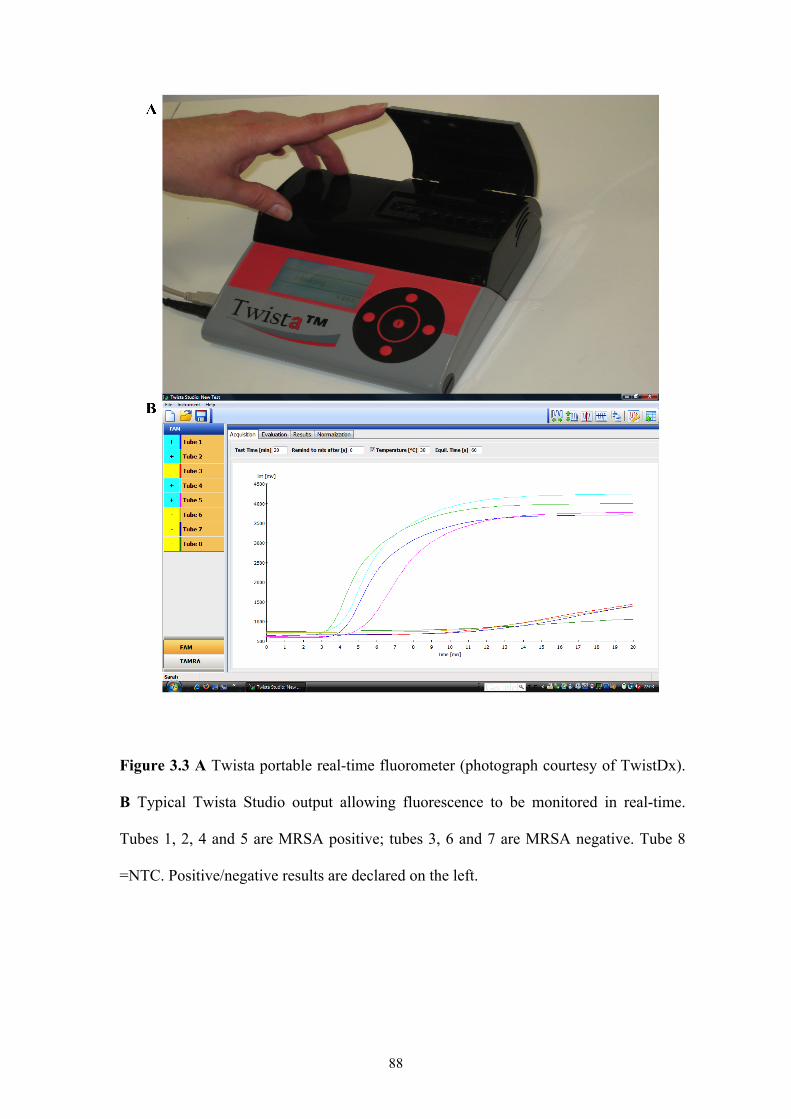
88
Figure 3.3 A Twista portable real-time fluorometer (photograph courtesy of TwistDx).
B Typical Twista Studio output allowing fluorescence to be monitored in real-time.
Tubes 1, 2, 4 and 5 are MRSA positive; tubes 3, 6 and 7 are MRSA negative. Tube 8
=NTC. Positive/negative results are declared on the left.
89
The TwistAmp MRSA protocol was used for screening all 119 isolates from
our laboratory collection of Staphylococci. I also performed TwistAmp MRSA testing
of the MRSA culture positive KC isolates direct from TSB-glycerol stocks diluted
1:1000 in SDW. Prototypic strains for SCCmec types I-XI were tested with
TwistAmp MRSA from DNA samples.
3.2.5 MREJ typing protocol
The MREJ typing protocol was used to type all MRSA isolates from the UK,
KC and laboratory collections. Prototypic strains for SCCmec types I-XI were also
MREJ typed. The protocol used was as in 3.2.4, but using singleplex RPA reactions
for the individual MREJ types. MREJ typing was performed in the following order to
first distinguish type i isolates from type ii isolates and to process the isolates further
in the most efficient manner (reactions for more common MREJ types performed
first): ii, i, iii, vii, v, xii, iv. Isolates negative for these MREJ types were then tested
with PCR reactions for types xi and xiii-xx (as RPA assays for these types had not
been developed). Statistical analyses were performed using STATA (release 11,
StataCorp). Fisher’s exact test was used a) to compare the distributions of MREJ
types among CMFT and Addenbrooke’s isolates from the UK MRSA collection; and
b) to investigate the correlation between SCCmec type and MREJ type among the 57
laboratory MRSA isolates.
3.2.6 Characterisation of isolates
I characterised the false negative isolates from the UK collection using MLST,
SCCmec typing (Boye et al. [2007] method then Milheirico, Oliveira and de Lencastre
90
[2007a] method to retest unexpected banding patterns) and spa typing, all as
described in Chapter 2, in order to ascertain their diversity and define clones based on
the currently used nomenclature. All 57 isolates from the KC collection were
characterised by Alere Technologies GmbH using their StaphyType DNA microarray,
as previously described [Monecke and Ehricht 2005, Monecke et al. 2006, Monecke
et al. 2007a, Monecke et al. 2008, Monecke, Slickers and Ehricht 2008, Monecke et
al. 2011]. The array allows comprehensive genotyping of S. aureus isolates by
simultaneous detection of a large number of genes and alleles thereof, including
species markers, regulatory genes, and genes related to antibiotic resistance, virulence
and pathogenicity. Markers for typing SCCmec were also included in the array,
covering mecA and its regulatory genes as well as the different recombinase and
accessory genes that make up the various SCCmec elements [Monecke et al. 2007a].
Alere Technlogies determined affiliation of isolates to clonal complexes or STs, as
defined by MLST, by comparison of hybridisation profiles to a collection of reference
strains previously characterised by MLST [Monecke and Ehricht 2005, Monecke et
al. 2007a, Monecke et al. 2011]. Results of the microarray testing were sent to me for
analysis and epidemiological interpretation.
3.2.7 Primer design and primer screening for novel MREJ types
False negative isolates from the Addenbrooke's MRSA collection were
unavailable for whole genome sequencing as permission for their use could not be
obtained. Collaborators at King Abdullah University of Science and Technology,
Saudi Arabia, obtained the genome sequences of the false negative MRSA isolates
from the CMFT collection, by assembly of 110-nucleotide reads from an Illumina
Genome Analyzer IIx (Illumina, Inc.). The assemblies obtained from the 110-
91
nucleotide sequence reads were then sent to me by these collaborators, and for each
sequenced isolate, the contig containing the MREJ was identified by BLAST using a
segment of orfX (position 33692-34111 of GenBank entry BA000018). This contig
was compared to all known MREJ sequences by ClustalW2 alignment. Positive
control isolates of known MREJ types were also sequenced and assembled to confirm
the ability of the procedure to correctly assemble MREJ sequences.
For novel MREJ sequences (18 successfully sequenced in total), I grouped
together identical sequences and for each group (i.e. each novel MREJ sequence)
chose 35bp-primers from within the SCCmec element a sufficient distance
downstream from the TwistAmp MRSA orfX primer (~100bp), to amplify the
SCCmec-orfX junction region. I then selected a further 14 primers, each time 5bp
further downstream of orfX than the previous one. Thus, 15 candidate RPA primers
for each novel MREJ were generated and tested.
I prepared template DNA for each novel MREJ using PCR primers flanking
the RPA MREJ primers (primer sequences are commercially sensitive and cannot be
shown), to generate amplicons of at least 500bp that span the TwistAmp MRSA target
region within each novel MREJ. The orfX PCR primer was positioned approximately
200bp upstream of the orfX RPA primer. PCR reactions were performed using
genomic DNA preparations of isolates with novel MREJ types, in a total volume of
50µl using DreamTaq DNA polymerase (Fermentas), according to the manufacturer’s
protocol. PCR products were viewed on a 1% agarose gel and extracted and purified
using a QIAquick Gel Extraction Kit (Qiagen). DNA quantification of the purified
products was performed using a TwistDx protocol (Appendix 2) and each product was
serially diluted in Eppendorf LoBind tubes to ~50 copies/µl in T0.1E buffer (10 mM
Tris–HCl, pH 8 and 0.1 mM EDTA, pH 8) supplemented with 1ng/µl of human DNA
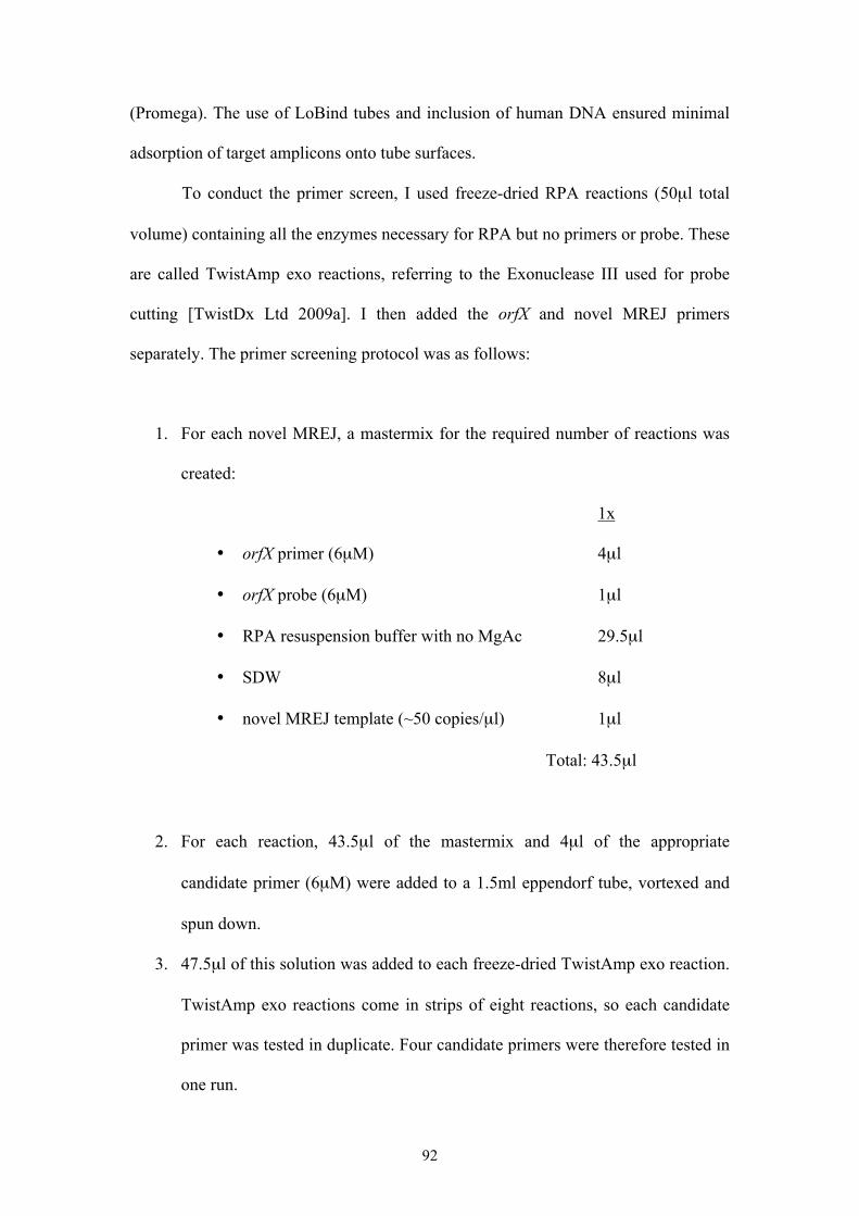
92
(Promega). The use of LoBind tubes and inclusion of human DNA ensured minimal
adsorption of target amplicons onto tube surfaces.
To conduct the primer screen, I used freeze-dried RPA reactions (50µl total
volume) containing all the enzymes necessary for RPA but no primers or probe. These
are called TwistAmp exo reactions, referring to the Exonuclease III used for probe
cutting [TwistDx Ltd 2009a]. I then added the orfX and novel MREJ primers
separately. The primer screening protocol was as follows:
1. For each novel MREJ, a mastermix for the required number of reactions was
created:
1x
• orfX primer (6µM) 4µl
• orfX probe (6µM) 1µl
• RPA resuspension buffer with no MgAc 29.5µl
• SDW 8µl
• novel MREJ template (~50 copies/µl) 1µl
Total: 43.5µl
2. For each reaction, 43.5µl of the mastermix and 4µl of the appropriate
candidate primer (6µM) were added to a 1.5ml eppendorf tube, vortexed and
spun down.
3. 47.5µl of this solution was added to each freeze-dried TwistAmp exo reaction.
TwistAmp exo reactions come in strips of eight reactions, so each candidate
primer was tested in duplicate. Four candidate primers were therefore tested in
one run.
93
4. 2.5µl of 280mM MgAc was added to the inside of the lid of each reaction
tube, the lids on the reaction tubes secured, and the reaction tubes briefly spun,
vortexed and spun again (50µl total reaction volume).
5. The TwistAmp protocol was then followed from step 3, as in section 3.2.4.
The best candidate primer from the first stage of the primer screen was
selected and tested alongside a further eight primers, staggered by 1bp downstream
(four primers) and 1bp upstream (four primers) from the initial best candidate primer.
The best 35bp-candidate from this second stage was tested in duplicate with no
template and 106 copies of MSSA to ensure specificity for MRSA detection, then
modified in length to create another 8 primers 30-38bp long. The length was modified
at the 3′ end of the primer sequence i.e. towards orfX. This final stage of the primer
screen determined the optimal MREJ-specific RPA primer to use with the universal
orfX RPA primer for detection of each novel MREJ type.
3.2.8 Detection of SCCmec variants and problematic strains
To determine the range of SCCmec types that TwistAmp MRSA was able to
detect, prototype strains for SCCmec types I-XI (Table 3.1) were tested with the
assay. A number of strains that have been reported not to be detected by other MREJ-
based diagnostic assays were also examined, as follows. Bartels et al. [2009] found a
common variant of SCCmec type IVa (ST8-MRSA-IVa/t024) in Copenhagen,
Denmark, that is not detected by the BD GeneOhm MRSA assay. This variant is the
most abundant MRSA clone in Copenhagen, affecting mainly people in nursing
homes and causing small outbreaks in local hospitals [Bartels et al. 2009]. One isolate
with a similar SCCmec and spa type (IVh and t024 respectively) to the Copenhagen
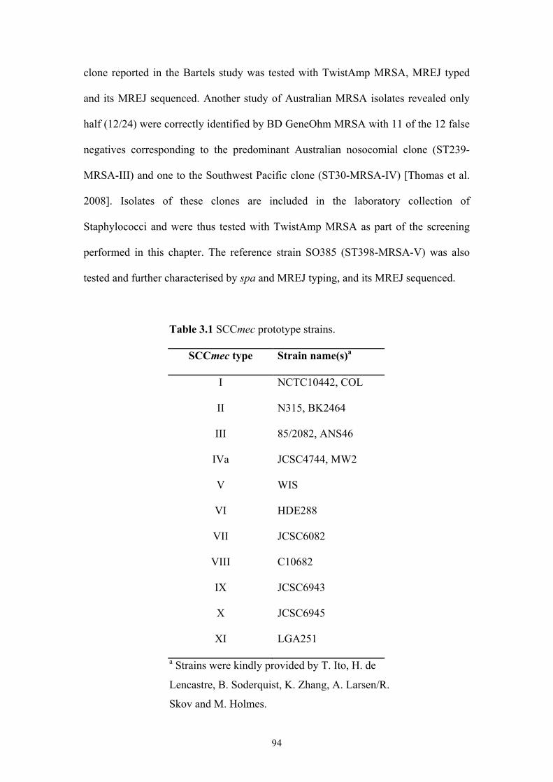
94
clone reported in the Bartels study was tested with TwistAmp MRSA, MREJ typed
and its MREJ sequenced. Another study of Australian MRSA isolates revealed only
half (12/24) were correctly identified by BD GeneOhm MRSA with 11 of the 12 false
negatives corresponding to the predominant Australian nosocomial clone (ST239-
MRSA-III) and one to the Southwest Pacific clone (ST30-MRSA-IV) [Thomas et al.
2008]. Isolates of these clones are included in the laboratory collection of
Staphylococci and were thus tested with TwistAmp MRSA as part of the screening
performed in this chapter. The reference strain SO385 (ST398-MRSA-V) was also
tested and further characterised by spa and MREJ typing, and its MREJ sequenced.
Table 3.1 SCCmec prototype strains.
SCCmec type Strain name(s)a
I NCTC10442, COL
II N315, BK2464
III 85/2082, ANS46
IVa JCSC4744, MW2
V WIS
VI HDE288
VII JCSC6082
VIII C10682
IX JCSC6943
X JCSC6945
XI LGA251
a Strains were kindly provided by T. Ito, H. de
Lencastre, B. Soderquist, K. Zhang, A. Larsen/R.
Skov and M. Holmes.
95
3.3 RESULTS
3.3.1 TwistAmp MRSA performance
3.3.1.1 UK MRSA collection
Of the 1,276 MRSA isolates from the UK collection, 1,232 were MREJ typed
as i-v or vii (the types covered by TwistAmp MRSA) (Table 3.3), giving TwistAmp
MRSA a sensitivity of 96.6%.
3.3.1.2 KC MRSA collection
Of the 52 isolates that were culture positive for MRSA from the KC
collection, 50 gave a positive result when tested with TwistAmp MRSA. Despite a
culture positive result for MRSA, the two remaining isolates were found to be MSSA
and MR-CNS (S. succinus) when characterised by Alere Technologies using their
StaphyType DNA microarray (see section 3.3.3.2). The five isolates included in the
KC collection as negative controls (four MSSA isolates and one MR-CNS (S.
haemolyticus) isolate) correctly gave a negative result with TwistAmp MRSA. Thus,
TwistAmp MRSA correctly identified all 50 MRSA isolates, giving the assay a
sensitivity of 100%.
Two KC isolates that were culture positive for MRSA produced weak
TwistAmp MRSA reaction curves compared to all other isolates (Figure 3.4). One of
these was an isolate that was later found to be MSSA by microarray analysis
(performed by Alere Technologies; see section 3.3.3.2) and therefore gave a false
positive result with TwistAmp MRSA, perhaps due to an SCC remnant or mec-less
cassette. I sequenced the MREJ of these two isolates, both of which were MREJ i,
using PCR primers flanking the RPA orfX and RPA MREJ i-specific primers.
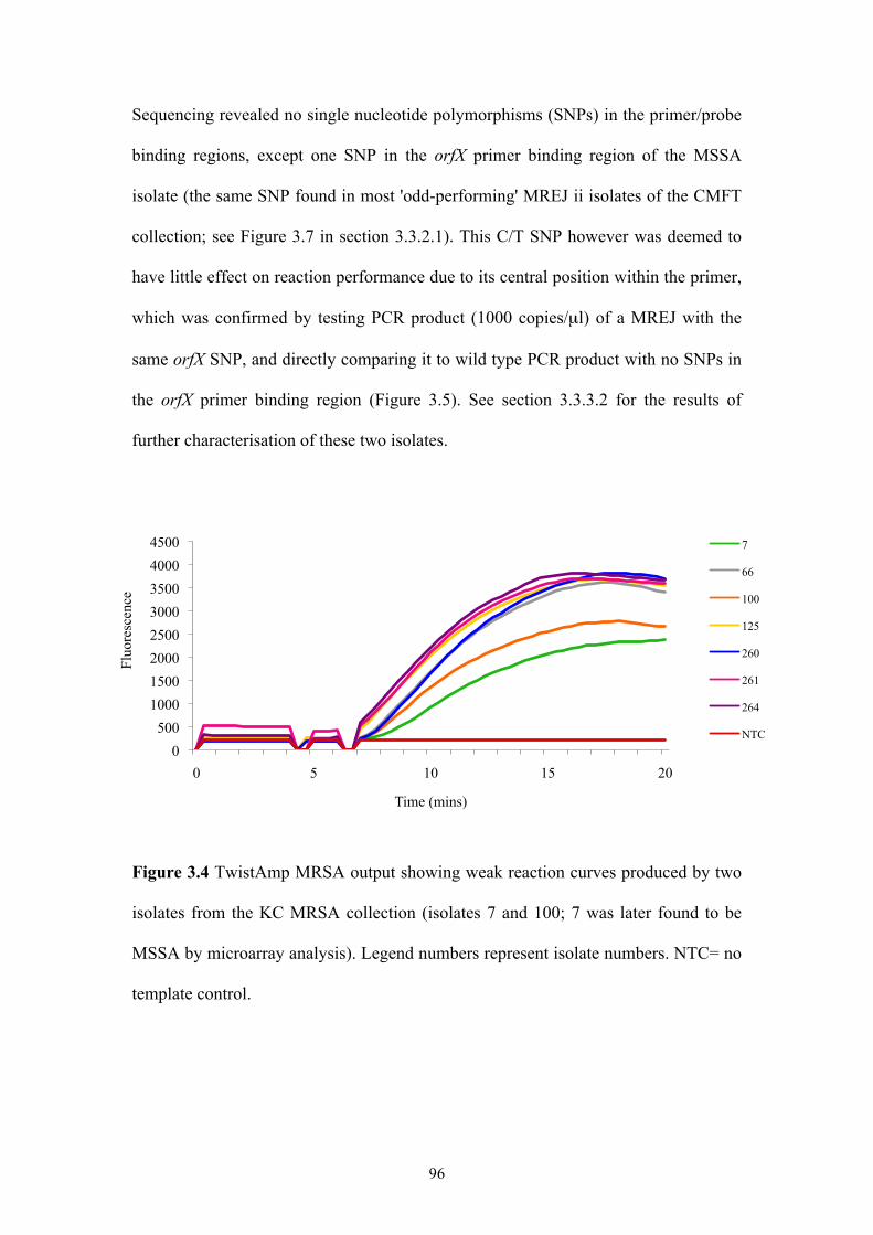
96
Sequencing revealed no single nucleotide polymorphisms (SNPs) in the primer/probe
binding regions, except one SNP in the orfX primer binding region of the MSSA
isolate (the same SNP found in most 'odd-performing' MREJ ii isolates of the CMFT
collection; see Figure 3.7 in section 3.3.2.1). This C/T SNP however was deemed to
have little effect on reaction performance due to its central position within the primer,
which was confirmed by testing PCR product (1000 copies/µl) of a MREJ with the
same orfX SNP, and directly comparing it to wild type PCR product with no SNPs in
the orfX primer binding region (Figure 3.5). See section 3.3.3.2 for the results of
further characterisation of these two isolates.
Figure 3.4 TwistAmp MRSA output showing weak reaction curves produced by two
isolates from the KC MRSA collection (isolates 7 and 100; 7 was later found to be
MSSA by microarray analysis). Legend numbers represent isolate numbers. NTC= no
template control.
0 500
1000 1500 2000 2500 3000 3500 4000 4500
0 5 10 15 20
Fluo
resc
ence
Time (mins)
7
66
100
125
260
261
264
NTC
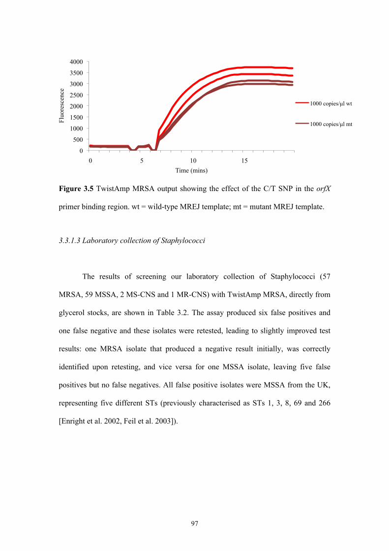
97
Figure 3.5 TwistAmp MRSA output showing the effect of the C/T SNP in the orfX
primer binding region. wt = wild-type MREJ template; mt = mutant MREJ template.
3.3.1.3 Laboratory collection of Staphylococci
The results of screening our laboratory collection of Staphylococci (57
MRSA, 59 MSSA, 2 MS-CNS and 1 MR-CNS) with TwistAmp MRSA, directly from
glycerol stocks, are shown in Table 3.2. The assay produced six false positives and
one false negative and these isolates were retested, leading to slightly improved test
results: one MRSA isolate that produced a negative result initially, was correctly
identified upon retesting, and vice versa for one MSSA isolate, leaving five false
positives but no false negatives. All false positive isolates were MSSA from the UK,
representing five different STs (previously characterised as STs 1, 3, 8, 69 and 266
[Enright et al. 2002, Feil et al. 2003]).
0
500
1000
1500
2000
2500
3000
3500
4000
0 5 10 15
Fluo
resc
ence
Time (mins)
1000 copies/µl wt
1000 copies/µl mt
98
Table 3.2 Test characteristics of TwistAmp MRSA for the laboratory Staphylococci
collection (N=119). The first numbers/percentages in each box represent results of the
initial screen, and the second numbers/percentages represent results of the repeat
screen.
Isolate
MRSA MSSA or CNS
TwistAmp
MRSA positive
True positive
56
57
False positive
6
5
PPV
90.3%
91.9% Test
outcome TwistAmp
MRSA negative
False negative
1
0
True negative
56
57
NPV
98.2%
100%
Sensitivity
98.2%
100%
Specificity
90.3%
91.9%
3.3.2 MREJ typing
3.3.2.1 UK MRSA collection
1,232 of the 1,276 MRSA isolates were typed as MREJ i-v or vii (Tables 3.3
and 3.4) using singleplex RPA reactions. A further two isolates were MREJ typed
using PCR reactions for types xiii and xvi, but the latter was later found by whole
genome sequencing to be incorrectly assigned and was re-assigned as a MREJ non-
typeable (NT) isolate (see section 3.3.3.1). Forty-two isolates (3.3%; 28 from CMFT
and 14 from Addenbrooke's) were negative for all MREJ types tested (i-v, vii and xi-
xx). The predominant MREJ type was ii, making up 90.8% of all isolates. The
distribution of MREJ types, including NT MREJs, was not significantly different
between CMFT and Addenbrooke’s isolates (Table 3.4, p = 0.485).

99
25 CMFT isolates were assigned MREJ types (23 MREJ ii, one MREJ i and
one MREJ iii) but produced weakly positive or lagging reaction curves (Figure 3.6).
SNPs in the orfX primer binding region were observed among 19 of the 25 isolates,
with one SNP in the MREJ i/ii isolates and a different SNP in the MREJ iii isolate. A
further two SNPs were identified in the MREJ iii-specific primer binding region of
the single odd-performing MREJ iii isolate (for orfX SNPs see Figure 3.7). Upon
retesting the 25 isolates all produced strong reaction curves.
Table 3.3 MREJ typing results of the UK
MRSA collection.
MREJ
type Frequency %
i 21 1.6
ii 1159 90.8
iii 19 1.5
iv 2 0.2
v 3 0.2
vii 28 2.2
xiii 1 0.1
xvi/NTa 1 0.1
NT 42 3.3
Total 1,276 100
a NT = MREJ non-typeable. MREJ xvi
isolate incorrectly assigned and later
found to be NT (see section 3.3.3.1).
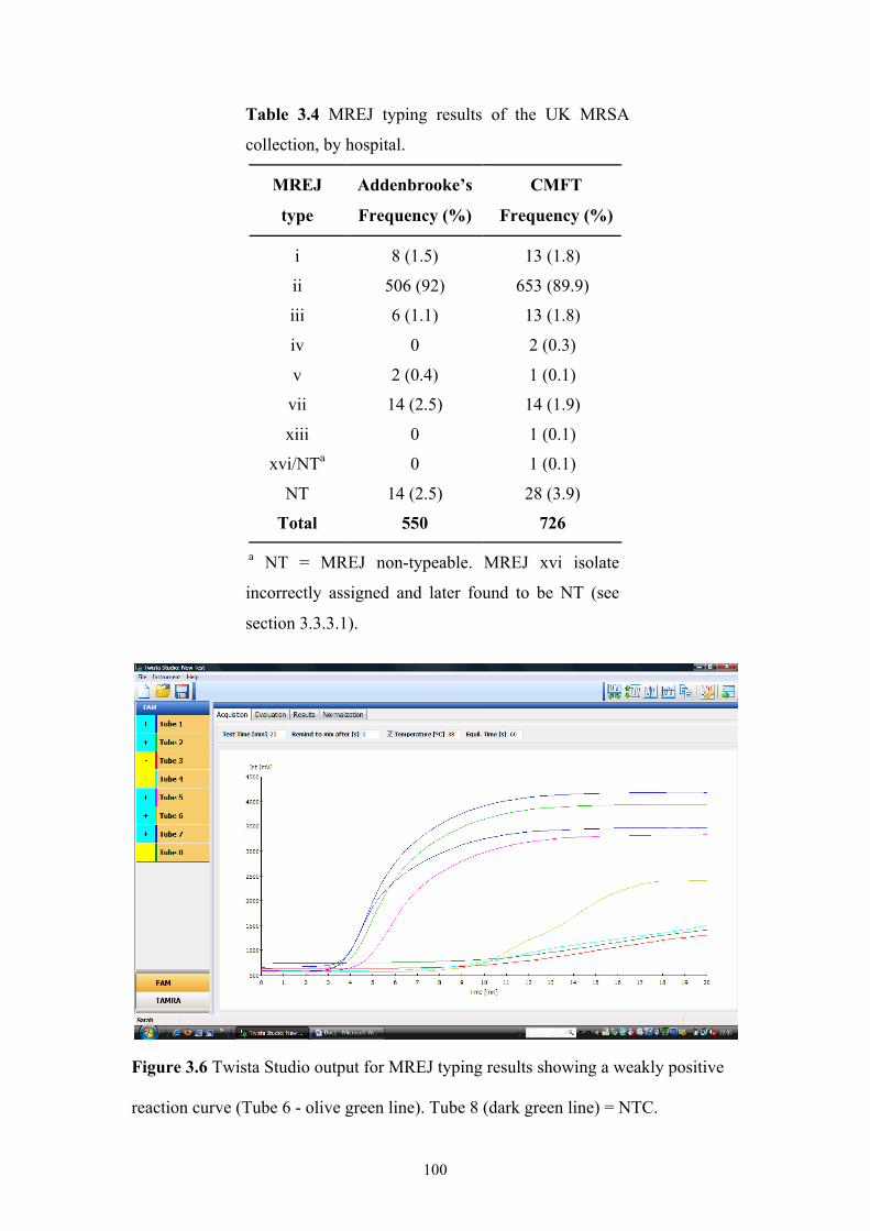
100
Table 3.4 MREJ typing results of the UK MRSA
collection, by hospital.
MREJ
type
Addenbrooke’s
Frequency (%)
CMFT
Frequency (%)
i 8 (1.5) 13 (1.8)
ii 506 (92) 653 (89.9)
iii 6 (1.1) 13 (1.8)
iv 0 2 (0.3)
v 2 (0.4) 1 (0.1)
vii 14 (2.5) 14 (1.9)
xiii 0 1 (0.1)
xvi/NTa 0 1 (0.1)
NT 14 (2.5) 28 (3.9)
Total 550 726
a NT = MREJ non-typeable. MREJ xvi isolate
incorrectly assigned and later found to be NT (see
section 3.3.3.1).
Figure 3.6 Twista Studio output for MREJ typing results showing a weakly positive
reaction curve (Tube 6 - olive green line). Tube 8 (dark green line) = NTC.
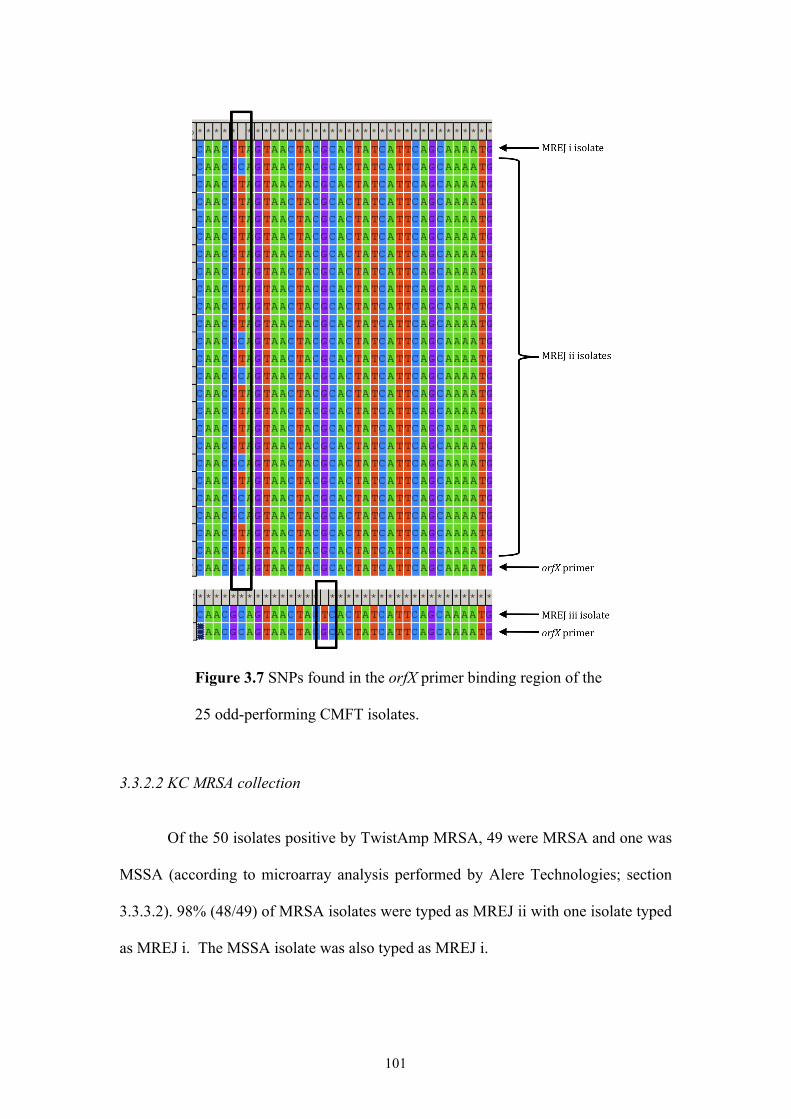
101
Figure 3.7 SNPs found in the orfX primer binding region of the
25 odd-performing CMFT isolates.
3.3.2.2 KC MRSA collection Of the 50 isolates positive by TwistAmp MRSA, 49 were MRSA and one was
MSSA (according to microarray analysis performed by Alere Technologies; section
3.3.3.2). 98% (48/49) of MRSA isolates were typed as MREJ ii with one isolate typed
as MREJ i. The MSSA isolate was also typed as MREJ i.
102
3.3.2.3 Laboratory MRSA collection
MREJ typing of the 57 MRSA isolates in our laboratory collection of
Staphylococci revealed the most common MREJ type was ii (80.7%;), with all
isolates typed as either i, ii or iii (Table 3.5).
Table 3.5 MREJ typing results of the
laboratory MRSA collection.
MREJ
type Frequency %
i 7 12.3
ii 46 80.7 iii 4 7.0
Total 57 100
3.3.2.4 Correlation of MREJ type with SCCmec type
I compared the MREJ types determined for the 57 MRSA isolates from our
laboratory collection of Staphylococci with the SCCmec types already established for
these strains and confirmed the SCCmec types by multiplex PCR (Table 3.6). Only
MREJ types i, ii and iii were detected among the isolates tested, using singleplex RPA
reactions. A significant correlation was found between MREJ and SCCmec type
(p<0.001), with 78.3% of MREJ ii isolates either SCCmec II or IV (91.7%). Most
MREJ iii isolates (75%) were SCCmec III and most MREJ i isolates (71.4%) were
SCCmec I. The single SCCmec V isolate was MREJ iii.
When confirming SCCmec types by multiplex PCR [Boye et al. 2007], one
isolate gave an unusual amplification pattern (ST254-MRSA-IV/t009), producing

103
bands for both ccrA2-B and ccrC. Another isolate produced no bands (ST5-MRSA-
III/t045). Retesting with the alternative multiplex PCR (Milheirico, Oliveira and de
Lencastre 2007a) gave the expected amplification pattern for ST254-MRSA-IV but an
unusual amplification pattern for ST5-MRSA-III (Figure 3.8). The ST254-MRSA-IV
isolate likely carries SCCmec subtype IVk (type IV and ccrC (2B&5)), explaining the
presence of ccrA2-B and ccrC bands using the Boye et al. [2007] method. ST254-
MRSA-IV, otherwise known as EMRSA-10 or the Hannover clone, has already been
reported to carry this SCCmec element [Chongtrakool et al. 2006, Monecke et al.
2011]. The lack of amplification pattern for the ST5-MRSA-III isolate using the Boye
et al. [2007] method indicated the absence of ccrC in its SCCmec element. The
Milheirico, Oliveira and de Lencastre [2007a] method produced an amplification
pattern for this isolate consisting of bands for mecA, mecI, the SCCmec III-specific JI
region and dcs (Figure 3.8). The dcs (downstream common sequence) is shared by
SCCmec types I, II and IV, but previous studies have identified SCCmec III MRSA
and MR-CNS isolates positive for dcs [Aires de Sousa and de Lencastre 2003, Qi et
al. 2005, Budimir et al. 2006, Chongtrakool et al. 2006, Mombach Pinheiro Machado
et al. 2007].
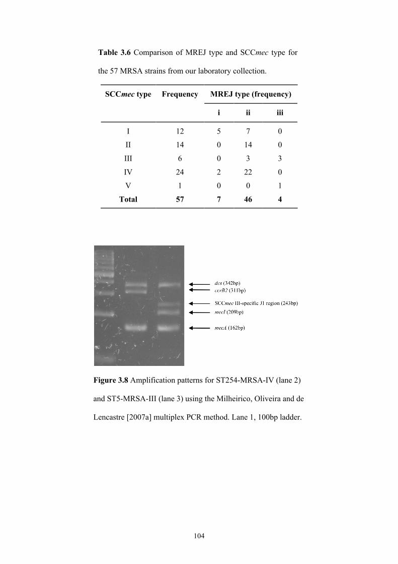
104
Table 3.6 Comparison of MREJ type and SCCmec type for
the 57 MRSA strains from our laboratory collection.
MREJ type (frequency) SCCmec type Frequency
i ii iii
I 12 5 7 0
II 14 0 14 0
III 6 0 3 3
IV 24 2 22 0
V 1 0 0 1
Total 57 7 46 4
Figure 3.8 Amplification patterns for ST254-MRSA-IV (lane 2)
and ST5-MRSA-III (lane 3) using the Milheirico, Oliveira and de
Lencastre [2007a] multiplex PCR method. Lane 1, 100bp ladder.
105
Only two SCCmec types were found among the 49 KC MRSA isolates - II and
IV. All SCCmec II isolates and most SCCmec IV isolates (97.4%) were MREJ ii
(Table 3.7).
Table 3.7 Comparison of MREJ type and SCCmec type
for 49 MRSA strains from the KC collection.
MREJ type (frequency) SCCmec type Frequency
i ii
II 11 0 11
IV 38 1 37
Total 49 1 48
Among the 15 prototypic MRSA strains for the 11 currently known SCCmec
types, MREJ types i, ii, iii and xii were identified (Table 3.8). Strains with SCCmec
types II, IV and VI were MREJ ii, SCCmec types I, VII and VIII were MREJ i, and
the SCCmec type III strains were MREJ iii. The prototypic SCCmec type V strain,
WIS, was typed as MREJ xii. The recently described SCCmec types IX, X and XI
were MREJ non-typeable for types i-v, vii and xi-xx, but SCCmec types IX and X
were found to be novel MREJ types c and g, respectively, after BLAST alignment of
their GenBank entries against sequences for novel MREJ types discovered in section
3.3.3.1.
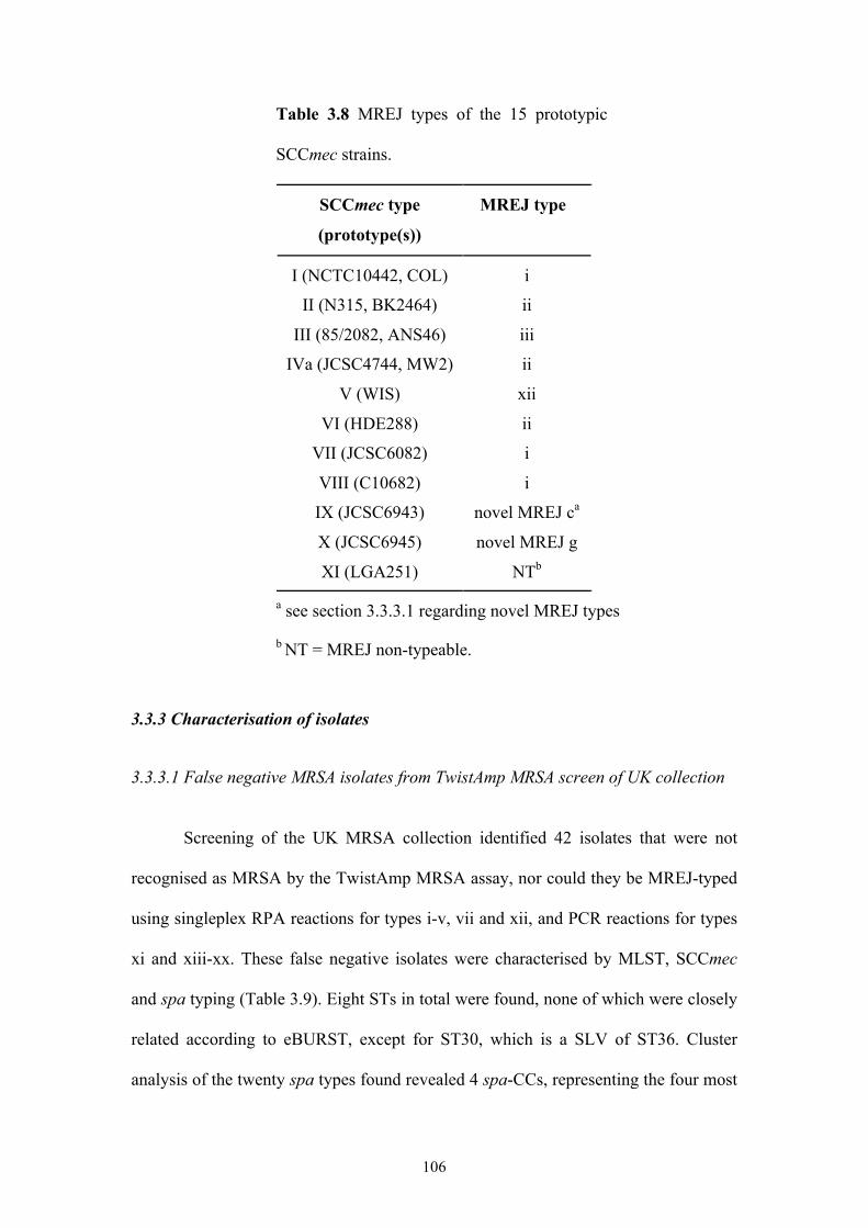
106
Table 3.8 MREJ types of the 15 prototypic
SCCmec strains.
SCCmec type
(prototype(s))
MREJ type
I (NCTC10442, COL) i
II (N315, BK2464) ii
III (85/2082, ANS46) iii
IVa (JCSC4744, MW2) ii
V (WIS) xii
VI (HDE288) ii
VII (JCSC6082) i
VIII (C10682) i
IX (JCSC6943) novel MREJ ca
X (JCSC6945) novel MREJ g
XI (LGA251) NTb
a see section 3.3.3.1 regarding novel MREJ types
b NT = MREJ non-typeable.
3.3.3 Characterisation of isolates
3.3.3.1 False negative MRSA isolates from TwistAmp MRSA screen of UK collection
Screening of the UK MRSA collection identified 42 isolates that were not
recognised as MRSA by the TwistAmp MRSA assay, nor could they be MREJ-typed
using singleplex RPA reactions for types i-v, vii and xii, and PCR reactions for types
xi and xiii-xx. These false negative isolates were characterised by MLST, SCCmec
and spa typing (Table 3.9). Eight STs in total were found, none of which were closely
related according to eBURST, except for ST30, which is a SLV of ST36. Cluster
analysis of the twenty spa types found revealed 4 spa-CCs, representing the four most
107
prevalent STs, ST22 (50% of isolates), ST36 (23.8%), ST149 (11.9%) and ST130
(4.8%) (Figure 3.9). Four isolates possessing novel spa types were found and
submitted to the SpaServer database (http://spaserver2.ridom.de/index.shtml) via the
StaphType software. These were designated types t5626 and t6419-21. t6419 also
contains a novel repeat, designated r377.
Isolates with spa types t084, t657, t6419 and t1258 were classified as
singletons because their repeat patterns differed by more than five repeats from those
of all other spa types. The first three singleton spa types correspond to the single
ST15, ST772 and ST59 isolates found among the false negative isolates. t1258
matched the first five repeats of t032 and would belong to spa-CC906 (ST22) if the
BURP algorithm criteria were relaxed. spa type t5829 was excluded from BURP
analysis because it was only 4 repeats in length and no reliable evolutionary history
can be inferred from ‘short’ spa types [Mellmann et al. 2007]. SCCmec typing by the
Boye et al. [2007] method described 45.2% of the false negative isolates as type IV,
23.8% as type II and one isolate each as type I and V. Eleven isolates (26.2%) were
non-typeable (NT) for SCCmec.
ST22-MRSA-IV and ST36-MRSA-II, also known as EMRSA-15 and
EMRSA-16 respectively, the two predominant nosocomial clones in the UK, were the
two most common clones among the 42 false negative isolates (Table 3.9). The five
ST149-MRSA-IV isolates, belonging to the major MLST clonal complex CC5
(ST149 is a SLV of ST5), have previously been reported only in Malta as an epidemic
strain [Scicluna et al. 2010], and in a Libyan patient in Switzerland [Francois et al.
2008]. The single ST772-MRSA-V/t657 isolate is known as the Bengal Bay clone or
WA MRSA-60, a multiply-resistant PVL-positive CA-MRSA that is becoming
increasingly prevalent in India, where it has spread into hospitals [D'Souza, Rodrigues
108
and Mehta 2010]. It has also been identified in Malaysia, the UK, Italy, Australia,
Germany, Hong Kong and Abu Dhabi [Neela et al. 2009, D'Souza, Rodrigues and
Mehta 2010, Ellington et al. 2010, Monecke et al. 2011, Sanchini et al. 2011], and
most recently in Ireland, where several ST772-MRSA-V/t657 isolates were reported
[Brennan et al. 2012]. Many patients infected with ST772-MRSA-V outside of India
had familial or travel links to India [Ellington et al. 2010, Brennan et al. 2012]. A
single ST15-MRSA-I isolate was also found among the false negative isolates. MRSA
from the CC15 lineage are extremely rare, with just one study reporting CC15-MRSA
isolated in Italy in 1980 [Campanile et al. 2009, Monecke et al. 2011].
The two ST130-MRSA-NT isolates (spa types t843 and t1736) belong to the
clonal lineage CC130 that has previously been reported in livestock (predominantly
from bovine sources) and more recently in humans, in the UK, Denmark, Ireland and
Germany [Cuny et al. 2011, Garcia-Alvarez et al. 2011, Shore et al. 2011]. These
isolates were non-typeable using the SCCmec typing method of Boye et al. [2007],
which was confirmed using the method of Milheirico, Oliveira and de Lencastre
[2007a]. However, the most recently described SCCmec element - type XI - has been
associated with ST130/t843 and ST130/t1736 strains [Cuny et al. 2011, Garcia-
Alvarez et al. 2011, Shore et al. 2011].
Seven ST22-MRSA isolates (spa types t032 and t492) exhibited a novel
amplification pattern with the Boye et al. [2007] typing method that combined the
expected bands for SCCmec types III (ccrC) and IV (ccrA2-B and IS1272) (Figure
3.10). Re-typing using the Milheirico, Oliveira and de Lencastre [2007a] method
showed they were SCCmec type IV (data not shown). A further two isolates,
ST30/t017 and ST59/t6419 generated the single band expected for SCCmec type III
(ccrC) plus one of the bands expected for SCCmec type IV (ccrA2-B) (Figure 3.10).
109
These 9 SCCmec NT isolates by the Boye et al. [2007] method could contain the most
recently described SCCmec subtype, IVk (type IV and ccrC (2B&5)). This composite
element, represented by MRSA strain ZH47, comprises a class B2 mec gene complex
and a type 2 ccr gene complex, plus an SCC carrying ccrC located in the J3 region
(between the right chromosomal junction and mec complex) [Heusser et al. 2007,
IWG-SCC 2009]. If so, these isolates would be variants of ST22-MRSA-IV, ST30-
MRSA-IV and ST59-MRSA-IV, respectively.
ST30-MRSA-IV is a CA-MRSA strain known as the Southwest Pacific clone
or USA1100, and belongs to the major clonal complex CC30 of HA- and CA-MRSA,
which includes ST36. The variant of ST59-MRSA-IV could be WA MRSA-15, the
second most common CC59-MRSA strain in Australia [Coombs et al. 2010] that has a
composite (SCCmec IV and V) or novel (SCCmec IV plus ccrC) SCCmec element
(type IV (2B&5)) [Monecke et al. 2011]. Its spa type is t976, which differs from
t6419 by two repeats. However, performing a BLAST alignment of the mobile
element of MRSA strain ZH47 (accession number AM292304) against the MREJ
amplicons of TwistAmp MRSA revealed this strain possesses MREJ iii and this strain
would therefore be detected by the assay.

110
Table 3.9 Strain types of the 42 false negative isolates from the UK MRSA collection, according to
MLST, SCCmec typing (using the method of Boye et al. [2007]), and spa typing.
Strain (ST and SCCmec type) Frequency spa types (frequency)
CMFT
ST36-MRSA-II 10 t018 (10)
ST22-MRSA-IV 8 t906 (4), t032 (2), t6420 (1), t6421 (1)
ST149-MRSA-IV 5 t002 (1), t5181 (1), t5626 (1), t1062 (1), t5829 (1)
ST30-MRSA-NTa 1 t017
ST772-MRSA-V 1 t657
ST15-MRSA-I 1 t084
ST59-MRSA-NT 1 t6419
ST130-MRSA-NT 1 t843
Addenbrooke's
ST22-MRSA-IV 6 t032 (2), t020 (2), t022 (1), t1258 (1)
ST22-MRSA-NT(IV)b 7 t032 (6), t492 (1)
ST130-MRSA-NT 1 t1736
a NT = non-typeable SCCmec element.
b NT by the method of Boye et al. [2007] but SCCmec type IV after re-typing by the method of
Milheirico, Oliveira and de Lencastre [2007a].
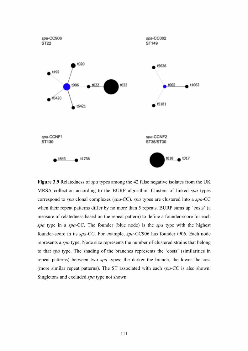
111
Figure 3.9 Relatedness of spa types among the 42 false negative isolates from the UK
MRSA collection according to the BURP algorithm. Clusters of linked spa types
correspond to spa clonal complexes (spa-CC). spa types are clustered into a spa-CC
when their repeat patterns differ by no more than 5 repeats. BURP sums up ‘costs’ (a
measure of relatedness based on the repeat pattern) to define a founder-score for each
spa type in a spa-CC. The founder (blue node) is the spa type with the highest
founder-score in its spa-CC. For example, spa-CC906 has founder t906. Each node
represents a spa type. Node size represents the number of clustered strains that belong
to that spa type. The shading of the branches represents the ‘costs’ (similarities in
repeat patterns) between two spa types; the darker the branch, the lower the cost
(more similar repeat patterns). The ST associated with each spa-CC is also shown.
Singletons and excluded spa type not shown.
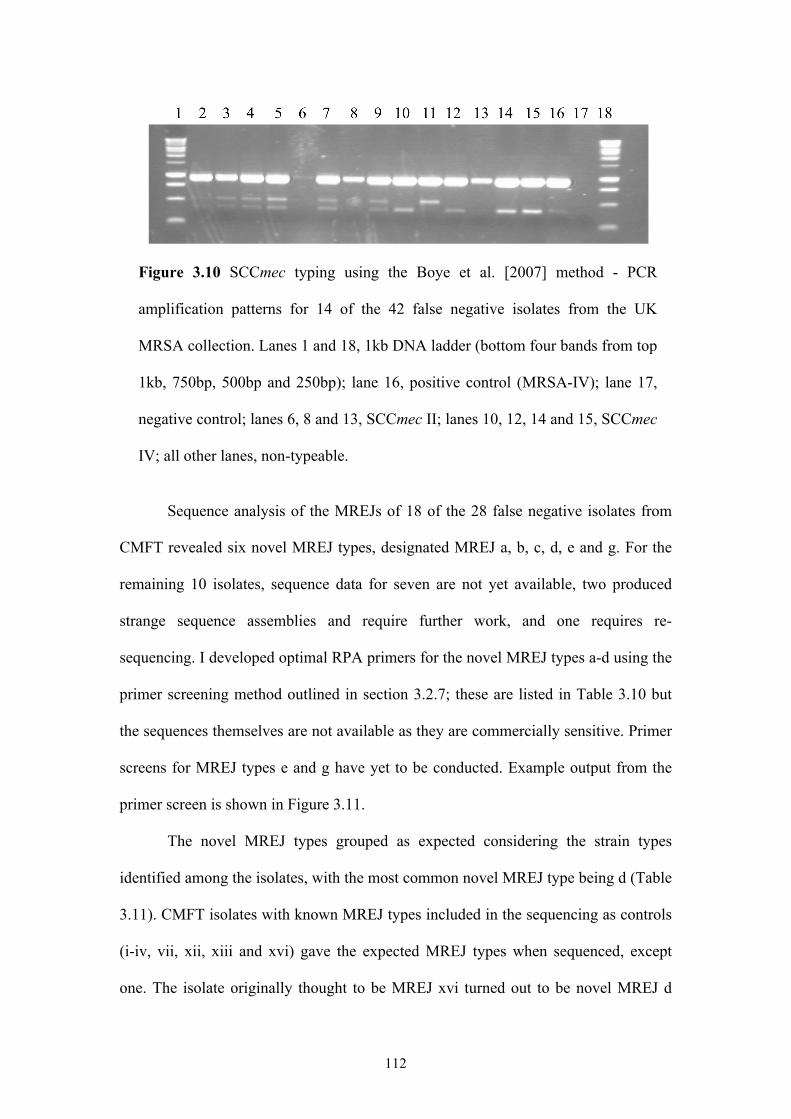
112
Figure 3.10 SCCmec typing using the Boye et al. [2007] method - PCR
amplification patterns for 14 of the 42 false negative isolates from the UK
MRSA collection. Lanes 1 and 18, 1kb DNA ladder (bottom four bands from top
1kb, 750bp, 500bp and 250bp); lane 16, positive control (MRSA-IV); lane 17,
negative control; lanes 6, 8 and 13, SCCmec II; lanes 10, 12, 14 and 15, SCCmec
IV; all other lanes, non-typeable.
Sequence analysis of the MREJs of 18 of the 28 false negative isolates from
CMFT revealed six novel MREJ types, designated MREJ a, b, c, d, e and g. For the
remaining 10 isolates, sequence data for seven are not yet available, two produced
strange sequence assemblies and require further work, and one requires re-
sequencing. I developed optimal RPA primers for the novel MREJ types a-d using the
primer screening method outlined in section 3.2.7; these are listed in Table 3.10 but
the sequences themselves are not available as they are commercially sensitive. Primer
screens for MREJ types e and g have yet to be conducted. Example output from the
primer screen is shown in Figure 3.11.
The novel MREJ types grouped as expected considering the strain types
identified among the isolates, with the most common novel MREJ type being d (Table
3.11). CMFT isolates with known MREJ types included in the sequencing as controls
(i-iv, vii, xii, xiii and xvi) gave the expected MREJ types when sequenced, except
one. The isolate originally thought to be MREJ xvi turned out to be novel MREJ d
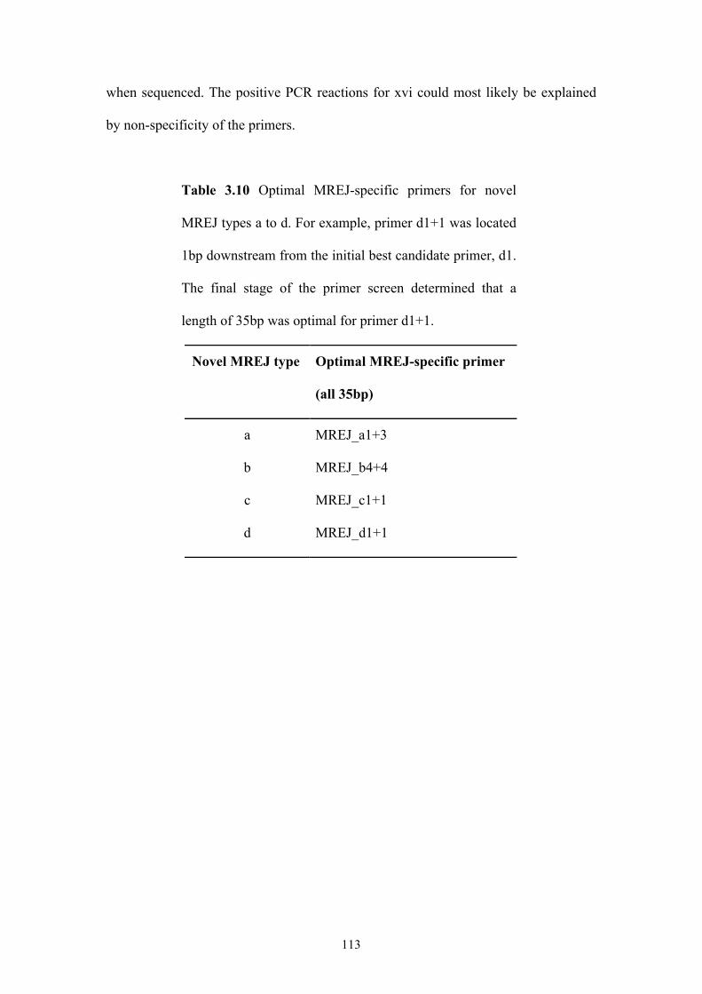
113
when sequenced. The positive PCR reactions for xvi could most likely be explained
by non-specificity of the primers.
Table 3.10 Optimal MREJ-specific primers for novel
MREJ types a to d. For example, primer d1+1 was located
1bp downstream from the initial best candidate primer, d1.
The final stage of the primer screen determined that a
length of 35bp was optimal for primer d1+1.
Novel MREJ type Optimal MREJ-specific primer
(all 35bp)
a MREJ_a1+3
b MREJ_b4+4
c MREJ_c1+1
d MREJ_d1+1
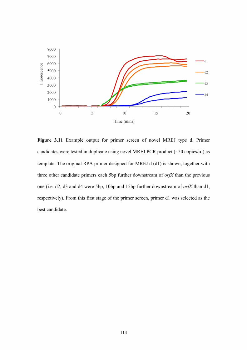
114
Figure 3.11 Example output for primer screen of novel MREJ type d. Primer
candidates were tested in duplicate using novel MREJ PCR product (~50 copies/µl) as
template. The original RPA primer designed for MREJ d (d1) is shown, together with
three other candidate primers each 5bp further downstream of orfX than the previous
one (i.e. d2, d3 and d4 were 5bp, 10bp and 15bp further downstream of orfX than d1,
respectively). From this first stage of the primer screen, primer d1 was selected as the
best candidate.
0
1000
2000
3000
4000
5000
6000
7000
8000
0 5 10 15 20
Fluo
resc
ence
Time (mins)
d1
d2
d3
d4
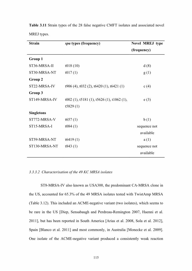
115
Table 3.11 Strain types of the 28 false negative CMFT isolates and associated novel
MREJ types.
Strain spa types (frequency) Novel MREJ type
(frequency)
Group 1
ST36-MRSA-II t018 (10) d (8)
ST30-MRSA-NT t017 (1) g (1)
Group 2
ST22-MRSA-IV t906 (4), t032 (2), t6420 (1), t6421 (1) c (4)
Group 3
ST149-MRSA-IV t002 (1), t5181 (1), t5626 (1), t1062 (1),
t5829 (1)
e (3)
Singletons
ST772-MRSA-V t657 (1) b (1)
ST15-MRSA-I t084 (1) sequence not
available
ST59-MRSA-NT t6419 (1) a (1)
ST130-MRSA-NT t843 (1) sequence not
available
3.3.3.2 Characterisation of the 49 KC MRSA isolates
ST8-MRSA-IV also known as USA300, the predominant CA-MRSA clone in
the US, accounted for 65.3% of the 49 MRSA isolates tested with TwistAmp MRSA
(Table 3.12). This included an ACME-negative variant (two isolates), which seems to
be rare in the US [Diep, Sensabaugh and Perdreau-Remington 2007, Haenni et al.
2011], but has been reported in South America [Arias et al. 2008, Sola et al. 2012],
Spain [Blanco et al. 2011] and most commonly, in Australia [Monecke et al. 2009].
One isolate of the ACME-negative variant produced a consistently weak reaction
116
curve with TwistAmp MRSA (see Figure 3.4) but when MREJ typed from DNA was
strongly positive for MREJ i. The MREJ i false positive isolate mentioned in section
3.3.1.2, CC1-MSSA [PVL+], was also weakly positive with TwistAmp MRSA but
strongly positive in singleplex RPA reactions for MREJ i. CC1 is a major worldwide
MSSA lineage strongly associated with CA-MRSA, including USA400. All SCCmec-
related microarray targets were negative for this isolate, removing the possibility of a
mec-less cassette. However, the isolate could contain a remnant of an SCC element
(mec-containing or otherwise) at the integration site (see section 3.4.1 for related
discussion).
ST5-MRSA-II, including its SLV ST225-MRSA-II, made up 22.4% of the
MRSA isolates tested. Also known as the NY/Japan clone, or USA100, this strain is
the predominant HA-MRSA in the US. Four isolates belonging to the CC5-MRSA-
IV, also known as the Paediatric clone, were found. This clone is prevalent in
Argentina, Colombia and the US [Chambers and Deleo 2009].
Of the seven isolates that produced a negative result with TwistAmp MRSA,
five were MSSA belonging to CC8, CC30, CC45, CC59 and CC152. CC8, CC30 and
CC45 are major international MRSA lineages, and CC59 is a common CA-MRSA
lineage. The CC8-MSSA isolate was PVL+ and is likely a USA300 that lost its
SCCmec element [Brown et al. 2012]. The CC152-MSSA isolate was also PVL+.
This clone is prevalent in West Africa [Ruimy et al. 2008, Okon et al. 2009, Breurec
et al. 2011, Shittu et al. 2011], but the epidemic CC152-MRSA clone prevails
elsewhere [Perez-Roth et al. 2010]. The two remaining isolates were MR-CNS, one of
which, Staphylococcus succinus, was classified as MRSA by culture. There is only
one report of S. succinus being isolated from human clinical material [Novakova et al.
2006].
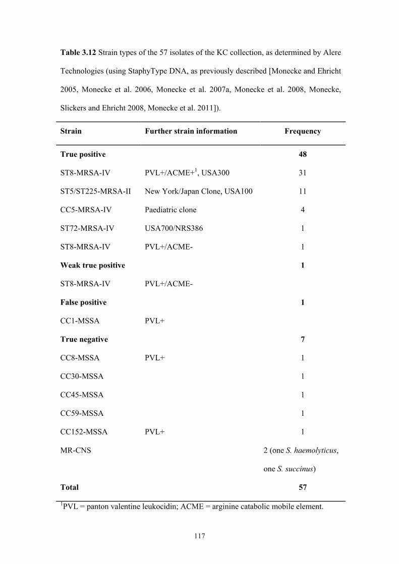
117
Table 3.12 Strain types of the 57 isolates of the KC collection, as determined by Alere
Technologies (using StaphyType DNA, as previously described [Monecke and Ehricht
2005, Monecke et al. 2006, Monecke et al. 2007a, Monecke et al. 2008, Monecke,
Slickers and Ehricht 2008, Monecke et al. 2011]).
Strain Further strain information Frequency
True positive 48
ST8-MRSA-IV PVL+/ACME+1, USA300 31
ST5/ST225-MRSA-II New York/Japan Clone, USA100 11
CC5-MRSA-IV Paediatric clone 4
ST72-MRSA-IV USA700/NRS386 1
ST8-MRSA-IV PVL+/ACME- 1
Weak true positive 1
ST8-MRSA-IV PVL+/ACME-
False positive 1
CC1-MSSA PVL+
True negative 7
CC8-MSSA PVL+ 1
CC30-MSSA 1
CC45-MSSA 1
CC59-MSSA 1
CC152-MSSA PVL+ 1
MR-CNS 2 (one S. haemolyticus,
one S. succinus)
Total 57
1PVL = panton valentine leukocidin; ACME = arginine catabolic mobile element.
118
3.3.4 Scope of TwistAmp MRSA
3.3.4.1 Testing a variant of a common MRSA clone in Copenhagen, Denmark The isolate with similar ST, SCCmec and spa type (ST8, SCCmec IVh and
t024 respectively) to the common variant of the Copenhagen clone (ST8-MRSA-
IVa/t024) that was not detected by BD GeneOhm MRSA, was detected using
TwistAmp MRSA and when tested with singleplex RPA reactions for MREJ types i-
v, vii and xii it tested positive for MREJ ii (Figure 3.12). Since MREJ i and ii differ
by a 102-bp insertion, an isolate with MREJ ii will test positive for both types.
Sequencing of the MREJ amplicon of this isolate (data not shown) revealed three
SNPs compared to the reference MREJ amplicon for type ii. One of these SNPs was
in the probe binding region, but is accounted for in the probe design using a wobble
base, so should not cause any detection problems. The other two SNPs were not
present in the primer/probe binding regions so should not have any effect on RPA
reaction performance. The preliminary data show that this variant is not problematic
for TwistAmp MRSA, although the SCCmec type of the tested isolate was subtype
IVh, whereas the problem variant was SCCmec subtype IVa. It is therefore likely that
the tested isolate does not have the MREJ in question in the Bartels paper. Indeed, 23
MRSA isolates with SCCmec type IVh were tested by Bartels et al. [2009] and all
were detected by BD GeneOhm MRSA. Two prototypic strains for SCCmec type IVa
tested positive with TwistAmp MRSA and were MREJ typed as ii (section 3.3.2.4
above and 3.3.4.4 below) but had different STs and spa types to the variant of the
Copenhagen clone (ST1/t128 and ST379/t375 versus ST8/t024). The PCR primers
and/or probes used in BD GeneOhm MRSA may overlap SNPs in the MREJ of the
Copenhagen clone, causing amplification and/or detection problems for their assay, or
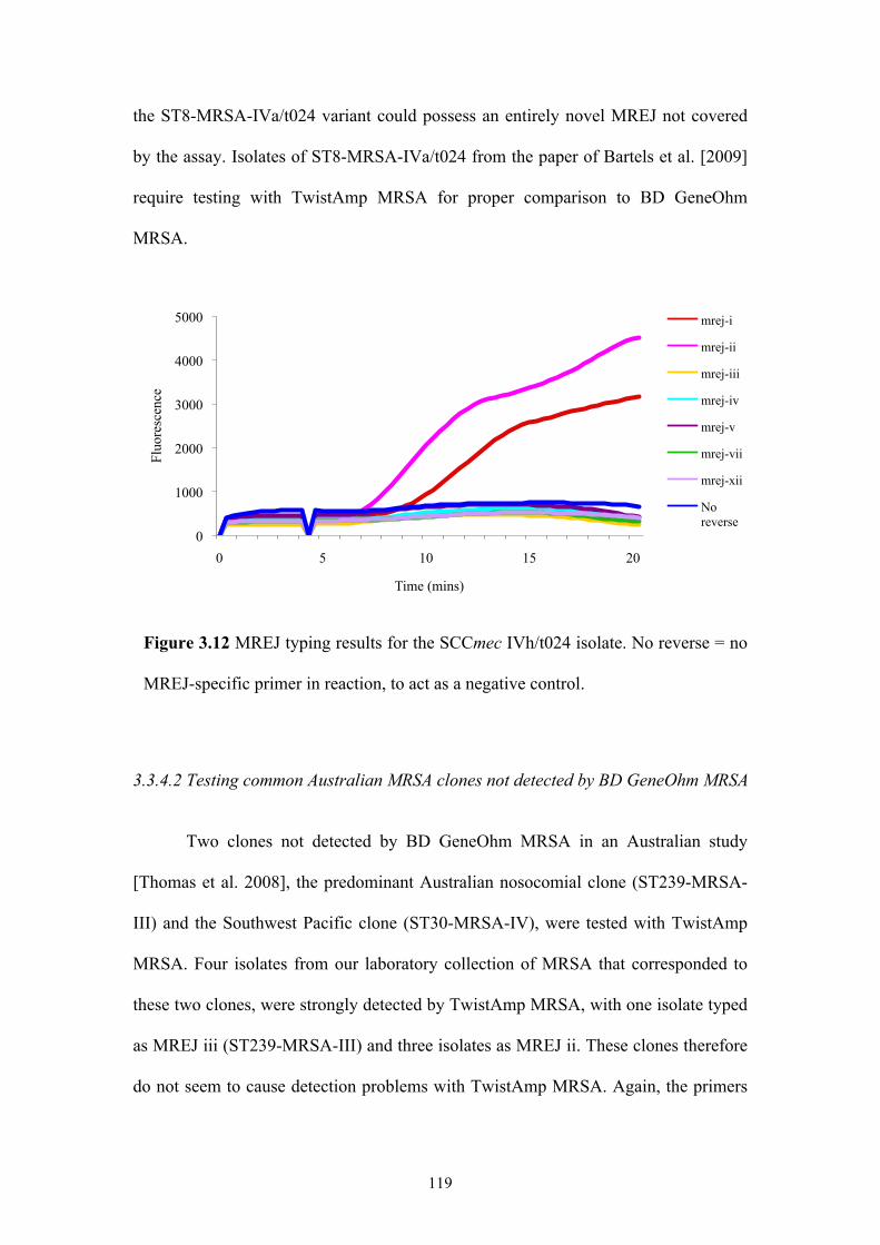
119
the ST8-MRSA-IVa/t024 variant could possess an entirely novel MREJ not covered
by the assay. Isolates of ST8-MRSA-IVa/t024 from the paper of Bartels et al. [2009]
require testing with TwistAmp MRSA for proper comparison to BD GeneOhm
MRSA.
Figure 3.12 MREJ typing results for the SCCmec IVh/t024 isolate. No reverse = no
MREJ-specific primer in reaction, to act as a negative control.
3.3.4.2 Testing common Australian MRSA clones not detected by BD GeneOhm MRSA Two clones not detected by BD GeneOhm MRSA in an Australian study
[Thomas et al. 2008], the predominant Australian nosocomial clone (ST239-MRSA-
III) and the Southwest Pacific clone (ST30-MRSA-IV), were tested with TwistAmp
MRSA. Four isolates from our laboratory collection of MRSA that corresponded to
these two clones, were strongly detected by TwistAmp MRSA, with one isolate typed
as MREJ iii (ST239-MRSA-III) and three isolates as MREJ ii. These clones therefore
do not seem to cause detection problems with TwistAmp MRSA. Again, the primers
0
1000
2000
3000
4000
5000
0 5 10 15 20
Fluo
resc
ence
Time (mins)
mrej-i
mrej-ii
mrej-iii
mrej-iv
mrej-v
mrej-vii
mrej-xii
No reverse

120
and/or probes used in BD GeneOhm MRSA may target more variable regions than
those in TwistAmp MRSA.
3.3.4.3 Testing the livestock-associated MRSA clone ST398-MRSA-V
ST398 has previously been reported to cause detection problems with current
commercial diagnostic assays [Reischl et al. 2009, Voss 2009, Malhotra-Kumar et al.
2010b]. Strain SO385 (ST398-MRSA-V) was therefore tested with TwistAmp
MRSA, giving a weak positive result (data not shown). MREJ typing of this strain
with RPA singeplex reactions for MREJ types i-v, vii and xii gave a weak positive
result for MREJ type iii (Figure 3.13). Sequencing of the type iii amplicon revealed
two SNPs in the probe binding region, compared to the typical MREJ iii sequence
(sequences not shown as commercially sensitive). These two SNPs could well explain
the weak positive result for the SO385 isolate compared to typical MREJ iii isolates,
as well as the general ST398 detection problems seen with current commercial PCR
diagnostic assays [Voss 2009]. The SO385 strain has since been whole genome-
sequenced (GenBank accession number AM990992 [Schijffelen et al. 2010]).
Aligning the SO385 MREJ iii amplicon I sequenced against the complete genome
sequence of SO385 confirmed the presence of these two SNPs. Aligning the SO385
MREJ iii amplicon against other available ST398 sequences (all SCCmec V) revealed
that the same two SNPs were consistent across all ST398-MRSA-V strains (GenBank
accession numbers AB505629, FJ830606 (both SCCmec V (5C2)) and GQ902038
(SCCmec V (5C2&5)). It is likely that ST398 harbours more than one MREJ type
since it is associated with more than one SCCmec type (mostly type V (shown to be
MREJ iii) but also type IV; type III has also been reported but due to the typing
method used may actually be type V [van Loo et al. 2007, Nemati et al. 2008, Jansen,
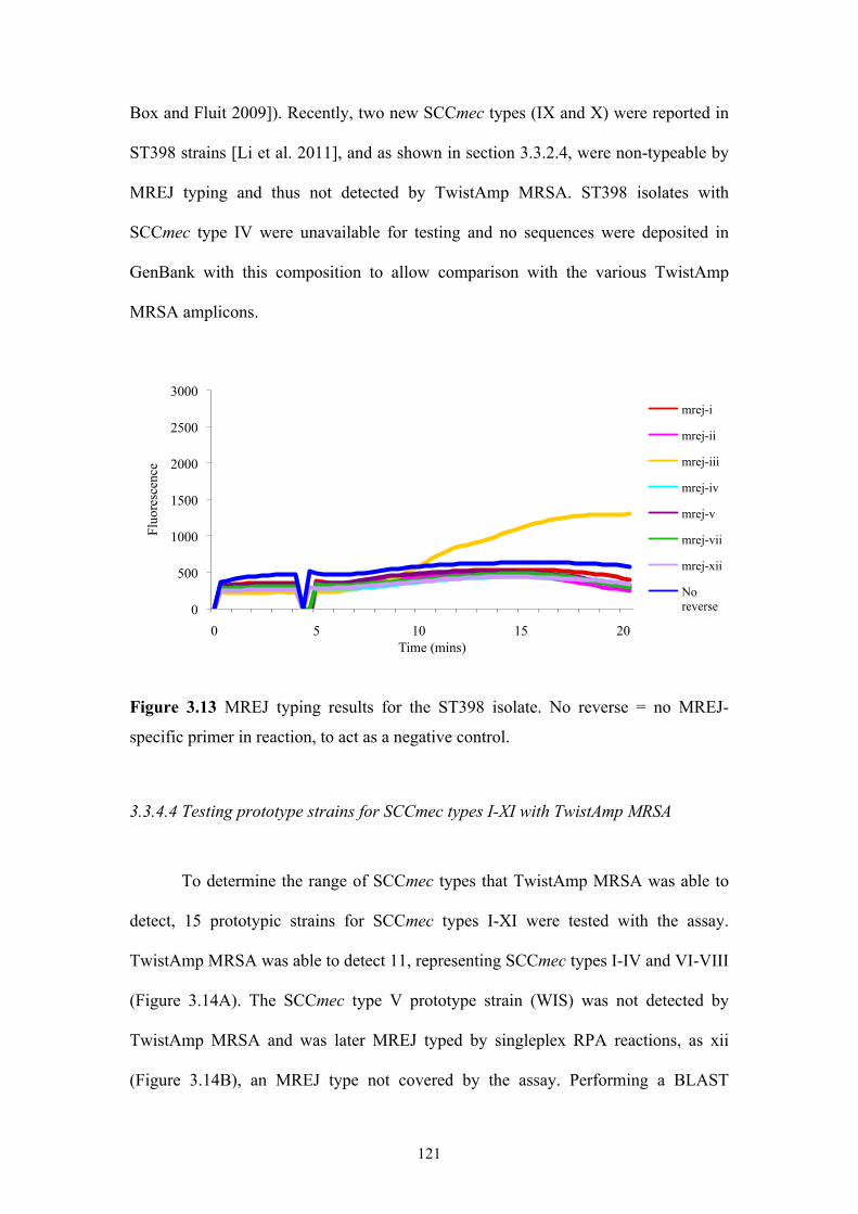
121
Box and Fluit 2009]). Recently, two new SCCmec types (IX and X) were reported in
ST398 strains [Li et al. 2011], and as shown in section 3.3.2.4, were non-typeable by
MREJ typing and thus not detected by TwistAmp MRSA. ST398 isolates with
SCCmec type IV were unavailable for testing and no sequences were deposited in
GenBank with this composition to allow comparison with the various TwistAmp
MRSA amplicons.
Figure 3.13 MREJ typing results for the ST398 isolate. No reverse = no MREJ-
specific primer in reaction, to act as a negative control.
3.3.4.4 Testing prototype strains for SCCmec types I-XI with TwistAmp MRSA
To determine the range of SCCmec types that TwistAmp MRSA was able to
detect, 15 prototypic strains for SCCmec types I-XI were tested with the assay.
TwistAmp MRSA was able to detect 11, representing SCCmec types I-IV and VI-VIII
(Figure 3.14A). The SCCmec type V prototype strain (WIS) was not detected by
TwistAmp MRSA and was later MREJ typed by singleplex RPA reactions, as xii
(Figure 3.14B), an MREJ type not covered by the assay. Performing a BLAST
0
500
1000
1500
2000
2500
3000
0 5 10 15 20
Fluo
resc
ence
Time (mins)
mrej-i
mrej-ii
mrej-iii
mrej-iv
mrej-v
mrej-vii
mrej-xii
No reverse
122
alignment of all SCCmec type V entries in GenBank other than WIS (both types 5C2
and 5C2&5; accession numbers AB505629, AM990992, GQ902038, FJ830606,
AB478780, AB512767, AB462393 and CP003166) against the MREJ sequences for
all known MREJ types, revealed that they were all MREJ iii, rather than xii. As
mentioned, all ST398-associated SCCmec type V sequences have two SNPs in the
probe binding region compared to the typical MREJ iii sequence, which are likely to
produce only weak positive results using TwistAmp MRSA, as shown by the ST398-
MRSA-V reference strain SO385 above (Figure 3.13). The remaining four SCCmec
type V sequences in GenBank possessed no SNPs in the primer or probe binding
regions for MREJ iii, suggesting that TwistAmp MRSA would succesfully detect
strains possessing this variant. Thus, it appears that some SCCmec type V strains will
be detected by TwistAmp MRSA and others will not (or will give weak positive
results), although the proportion that will be detected is at present unclear.
The recently described prototypic strains for SCCmec types IX-XI were not
detected by TwistAmp MRSA and were MREJ non-typeable for types i-v, vii and xi-
xx. However, a BLAST alignment of the sequences of SCCmec types IX, X and XI
(GenBank accession numbers AB505628, AB505630 and FR823292, respectively)
against the novel MREJ types a-e and g discovered in this chapter, revealed that the
prototype strains for SCCmec types IX and X were novel MREJ types c and g,
respectively. While the MREJ of the prototype strain for SCCmec X had no SNPs
compared to the novel MREJ g sequence, SCCmec IX had one SNP in the orfX primer
binding region compared to the novel MREJ c sequence (the same C/T SNP found in
several CMFT MREJ ii isolates in section 3.3.2.1). SCCmec XI was not typeable by
the novel MREJ types a-e and g.
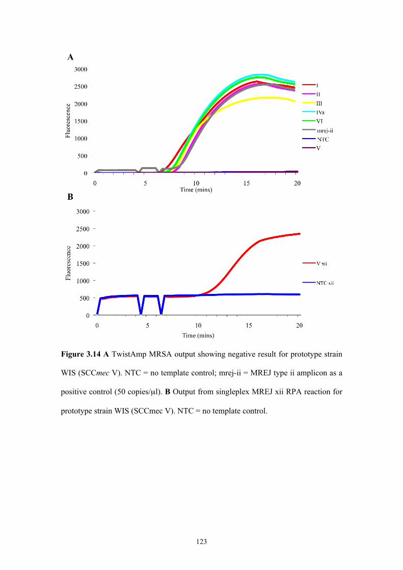
123
Figure 3.14 A TwistAmp MRSA output showing negative result for prototype strain
WIS (SCCmec V). NTC = no template control; mrej-ii = MREJ type ii amplicon as a
positive control (50 copies/µl). B Output from singleplex MREJ xii RPA reaction for
prototype strain WIS (SCCmec V). NTC = no template control.
124
3.4 DISCUSSION
3.4.1 TwistAmp MRSA performance and scope
TwistAmp MRSA was shown to have a sensitivity, specificity, NPV and PPV
of 98.2%, 90.3%, 98.2% and 90.3% respectively when tested with our laboratory
collection of 119 geographically and clonally diverse MRSA and MSSA (and three
CNS). Although not relevant to the use of the method in a clinical setting, all test
characteristics were improved upon retesting those that gave false positive and false
negative results (to 100%, 91.9%, 100% and 91.9% respectively). The one false
negative isolate was correctly identified as MRSA upon retesting, suggesting a
problem with the initial detection by TwistAmp MRSA (lack of assay sensitivity or
perhaps poor protocol implementation). Five false positive isolates (all previously
well-defined MSSA [Enright et al. 2002, Feil et al. 2003]) remained after retesting,
indicating a lack of assay specificity, or possible contamination of the samples or
reactions. The latter could occur as a result of MRSA-containing aerosols, for
example created by pipetting, although negative controls should have detected this
problem. Another possibility is MSSA containing mec-less cassettes or non-mecA
SCC elements, which is discussed in greater detail below.
A limitation of using the laboratory collection of Staphylococci to test
TwistAmp MRSA is that it does not include recently isolated or emerging strains, nor
is it representative of a distinct region as it included MRSA isolates from 16 countries
representing strains with an international distribution. Nonetheless, screening of the
collection that includes diverse MRSA, has provided an initial insight into the
performance of the assay, which is comparable to that of current commercial assays
(see Table 1.7). A more up to date collection of MRSA, including diverse examples of
125
MR-CNS and more geographically diverse MSSA strains, will allow more
comprehensive testing of TwistAmp MRSA.
Testing the MRSA collections from hospitals in the US and UK with
TwistAmp MRSA (singleplex MREJ reactions for the UK collection) gave an assay
sensitivity of 100% and 96.6% respectively. This showed that the assay delivered
good coverage of the MRSA strains prevalent at each site. However, 25 isolates from
the UK collection produced weakly positive or lagging reaction curves, but upon
retesting were strongly positive, suggesting lack of assay sensitivity, or poor protocol
implementation when first tested. The lower sensitivity observed for the UK
collection of MRSA is explained by the presence of MREJ types not covered by
TwistAmp MRSA, and a number of novel MREJ types. No such MREJ types were
identified in the KC collection, although this may be an artefact of sample size - only
49 MRSA isolates in the latter compared to 1,276 from the UK collection. Testing
more isolates from the site of the KC MRSA, or from a second US site, may reveal
the presence of more MREJ types, including novel types, but it could in fact be that
MREJ diversity, possibly as a consequence of the strain diversity or the presence of
more conserved SCCmec elements among MRSA, is lower in the US compared to the
UK (see section 3.4.2 for more on MREJ diversity and distribution). Indeed, a recent
study of BD GeneOhm MRSA tested on a diverse range of MRSA genotypes and
SCCmec types from the US and Taiwan, gave a very high assay sensitivity (99.7%)
[Boyle-Vavra and Daum 2010]. The TwistAmp MRSA sensitivities determined for
both the UK and KC MRSA collections may be overestimated because subculture of
the isolates was used, not the swabs directly (which would contain mixed flora and
potential reaction inhibitors).
126
Furthermore, two hospitals were involved in the UK collection compared to
the single hospital in the KC collection (although MREJ typing revealed no
significant difference between the two UK hospitals; see section 3.4.2 below). The
nature of the hospitals could also play a role in the differing sensitivities seen. The US
hospital served paediatric patients only. Differences in MRSA strain types have been
observed between adult and paediatric patients [David et al. 2006b, Park et al. 2007,
Hudson et al. 2012] (see also Chapter 5); for example, in the US a lower strain
diversity and significantly more USA300 is associated with the latter group [Hudson
et al. 2012] (see also discussion of KC isolate characterisation below and Chapter 5).
An isolate collection from solely paediatric patients may affect the MRSA diversity
observed and not represent the true diversity in the region. Simultaneously comparing
TwistAmp MRSA with other diagnostic methods would allow more accurate
assessment of assay sensitivity in a given region.
Although these preliminary test characteristics provide useful information
about the general ability of TwistAmp MRSA to detect most MRSA, the performance
of TwistAmp MRSA in a clinically relevant situation has been evaluated in two pre-
clinical (feasibility) studies to obtain more clinically relevant performance data.
TwistDx has used the assay to test clinical samples obtained as part of routine
screening procedures at hospitals in the UK and US. Specifically, their aim was to
assess whether the assay provided adequate coverage of the MRSA strains prevalent
at a certain hospital/in a certain region and compare its performance to gold standards
of MRSA detection.
In 2009 the UK study collected 5,433 nasal and groin swab samples from
routine MRSA screening of patients admitted over a 12-week period at Manchester
Royal Infirmary, CMFT [DoH 2011]. TwistAmp MRSA performed well when
127
compared to the gold standard method of broth enrichment and culture on
chromogenic media. This was particularly the case for specificity and NPV, with
clinical sensitivity at almost 75% (exact data confidential). However the PPV was
only around 50%, which could be attributed to mec-less cassettes or non-mecA SCC
elements, sampling error, or MRSA positive patients receiving systemic or topical
MRSA therapy [DoH 2011]. The confidential US study was conducted in 2011 at a
paediatric medical centre to simultaneously compare the clinical sensitivity and
specificity of the assay to Xpert MRSA, using chromogenic agar and blood agar plate-
based bacterial culture as the gold standard. TwistAmp MRSA performed similarly to
Xpert MRSA, but showed a lower clinical sensitivity (data confidential).
Despite pre-clinical studies demonstrating the good performance of TwistAmp
MRSA compared to current gold standard culture techniques and the market leader in
molecular diagnostic assays, further assay development is required before the assay
can be used clinically. There is potential to improve the assay further both by
covering more MREJ types enabling detection of more MRSA strains and SCCmec
variants, and by improving assay functionality (particularly since 25 isolates from the
UK collection required retesting in order to produce strong positive results). The
former was addressed in part in this chapter and is discussed below; the latter is
addressed in Chapter 4.
In a screening situation where the goal is to detect MRSA colonised patients
and to prevent nosocomial MRSA infections, an MRSA diagnostic assay with a high
NPV is important [Bartels et al. 2009], especially in countries with a low MRSA
prevalence (<1%), such as in northern Europe, where few MRSA carriers are
expected [Tiemersma et al. 2004]. In these countries, hospitalised patients with
MRSA are always isolated. If a patient is at risk of being colonised by MRSA, they
128
too are kept in isolation until a negative MRSA result is obtained. Thus, a false
negative result would release patients with MRSA from isolation, which could lead to
an MRSA outbreak [Bartels et al. 2009]. The proportion of invasive S. aureus isolates
that are MRSA has been high in the UK (25-50% [ECDC 2011]), where patient
isolation procedures are less stringent, but rates of MRSA bacteraemia are decreasing
[HPA 2011]. It is important that for use in this country TwistAmp MRSA be adequate
for screening the local diversity. Although a high NPV for TwistAmp MRSA has thus
far been reported, this can be further improved by reducing the number of false
negative results.
Several MRSA strains were identified among the 42 false negative isolates
from the UK collection, including strains that, while occurring at a frequency of
<0.5% in the MRSA population tested, harboured non-typeable SCCmec elements.
The most salient finding however was that the majority of isolates not detected by
TwistAmp MRSA (73.8%) belonged to the dominant UK clones EMRSA-15 and -16.
It was therefore important to sequence the MREJs of all available false negative
isolates (the 28 from CMFT) in order to determine why these strains or variants
thereof were not detected, for subsequent coverage by the assay where possible. Six
novel MREJ types were found among 18 of the 28 isolates where genome data were
available. Genome data for the remaining ten isolates would likely reveal two more
novel MREJ types based on the association of the current novel MREJ types with the
strain types identified (Table 3.11). Given that sequence data for MREJ types vi and
viii-x are not publicly available, up to four of the novel MREJ types discovered here
could be one of these. It was not possible to modify the current set of primers and
probe to account for the novel MREJ types due to great sequence variability, and so
new RPA primers were designed and developed to detect them. Incorporation of these
129
into the current multiplex has not yet been attempted, but the most likely scenario
would be incorporation of novel MREJ type d since it was the most common. Given
that the current primer/probe set is optimal for the RPA-based system, and that
previous attempts by TwistDx to include MREJ xii in the assay were
counterproductive, the feasibility of adding a new primer pair to the set is
questionable. Nevertheless, having optimised RPA primers that exist for all novel
MREJ types identified will allow TwistDx to quickly and easily respond to future
changes in the prevalences of these MREJ types, by developing as necessary separate
primer/probe sets for MREJ types other than the 'core set', or by changing the 'core
set' itself as a result of further research and development that facilitates easy
incorporation of more MREJ types into TwistAmp MRSA. A recent study slightly
adapted the PCR-based detection method of Huletsky et al. [2004] (the forerunner of
BD GeneOhm MRSA) and added additional primers to allow detection of more
MRSA types based on a literature search of strains and on the authors' discovery of 14
novel MREJ types that are not detected by commercially available assays, resulting in
a megaplex PCR [van der Zee et al. 2011]. A total of 21 MREJ-specific primers were
included in the megaplex, with no adverse effects on PCR reported [van der Zee et al.
2011]. PCR clearly works with many primers in a single reaction, but RPA is a new
technology, and more research is needed into its multiplexing capabilities (see Part 1
summary).
In section 3.2.7 I used the whole genome sequence data provided by
collaborators in Saudi Arabia to characterise the MREJs of 18 of the false negative
CMFT isolates. My collaborators have subsequently used these genome data to
assemble their SCCmec elements and compare them with the sequences of known
SCCmec types using BLAST. Their results corroborate the findings of this chapter, in
130
terms of the non-typeable SCCmec elements I identified by SCCmec typing. Of the 18
isolates that were sequenced, most carried SCCmec IVk or variants thereof (Table
3.13). Four non-typeable (NT) SCCmec isolates were identified among the 42 UK
false negatives, after re-typing the seven ST22 isolates as type IV using a second
SCCmec typing method [Milheirico, Oliveira and de Lencastre 2007a]. Only three of
these four were sequenced, with the remaining one from Addenbrooke's Hospital
unavailable for sequencing (one ST130-MRSA-NT isolate). Of the three that were
sequenced, the ST130-MRSA-NT isolate produced a novel sequence assembly that
requires further analysis by collaborators, adding weight to the prediction that the two
ST130 isolates identified could harbour the most recently described SCCmec element,
type XI [Garcia-Alvarez et al. 2011, Shore et al. 2011]. ST59-MRSA-NT carried a
IVk variant as predicted, and ST30-MRSA-NT carried an unusual composite SCCmec
element primarily consisting of type IV.
Interestingly, the eight ST36 isolates and four ST22 isolates sequenced, which
were SCCmec typed as II and IV respectively, all in fact carried IVk elements, or
variants thereof, and in one case a composite element with some similarity to type
IVk, but primarily to type I. This shows that SCCmec IVk, present in dominant UK
clones, is clearly a problematic variant for TwistAmp MRSA, accounting for at least
82% of the false negatives identified in the UK collection and associated with four of
the six novel MREJ types. This also highlights the problem of misidentifications
experienced by PCR-based SCCmec typing methods, as discussed further in the Part 1
summary. Based on the SCCmec typing results of the Boye et al. [2007] method, the
isolate representing the Hannover clone (ST254-MRSA-IV) in our laboratory
collection of MRSA, and reported to carry SCCmec subtype IVk [Chongtrakool et al.
2006, Monecke et al. 2011], produced an identical, non-typeable amplification pattern
131
to the ST22-MRSA-NT false negative isolates of the UK collection, that were also
thought to carry SCCmec subtype IVk. However, TwistAmp MRSA detected the
isolate representing the Hannover clone. Perhaps, for some unknown reason, the
ST254-MRSA-IVk clone, or German isolates containing SCCmec subtype IVk (since
the isolate was obtained from Germany), have a detectable MREJ type (MREJ i)
unlike the ST22-MRSA-IVk clone, or UK isolates containing this SCCmec subtype.
Finally, the MREJ xvi isolate included in the sequencing protocol as a control
strain actually contained novel MREJ d upon sequencing, and consisted of a
composite SCCmec element of types IVk and II. More detailed analysis of the
SCCmec elements of these false negative isolates will be published at a later date.
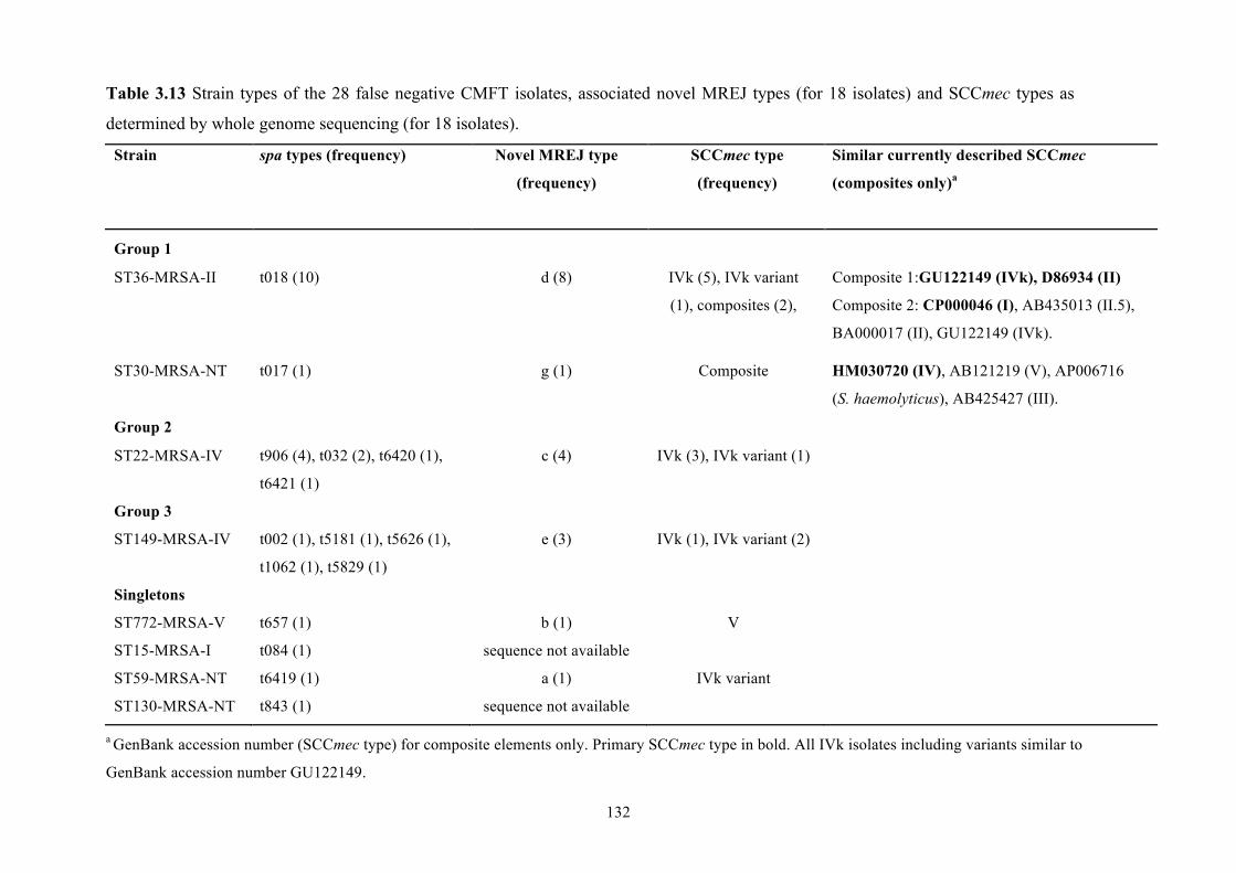
132
Table 3.13 Strain types of the 28 false negative CMFT isolates, associated novel MREJ types (for 18 isolates) and SCCmec types as
determined by whole genome sequencing (for 18 isolates).
Strain spa types (frequency) Novel MREJ type
(frequency)
SCCmec type
(frequency)
Similar currently described SCCmec
(composites only)a
Group 1
ST36-MRSA-II t018 (10) d (8) IVk (5), IVk variant
(1), composites (2),
Composite 1:GU122149 (IVk), D86934 (II)
Composite 2: CP000046 (I), AB435013 (II.5),
BA000017 (II), GU122149 (IVk).
ST30-MRSA-NT t017 (1) g (1) Composite HM030720 (IV), AB121219 (V), AP006716
(S. haemolyticus), AB425427 (III).
Group 2
ST22-MRSA-IV t906 (4), t032 (2), t6420 (1),
t6421 (1)
c (4) IVk (3), IVk variant (1)
Group 3
ST149-MRSA-IV t002 (1), t5181 (1), t5626 (1),
t1062 (1), t5829 (1)
e (3) IVk (1), IVk variant (2)
Singletons
ST772-MRSA-V t657 (1) b (1) V
ST15-MRSA-I t084 (1) sequence not available
ST59-MRSA-NT t6419 (1) a (1) IVk variant
ST130-MRSA-NT t843 (1) sequence not available
a GenBank accession number (SCCmec type) for composite elements only. Primary SCCmec type in bold. All IVk isolates including variants similar to
GenBank accession number GU122149.
133
All 49 MRSA isolates from the KC collection were detected by TwistAmp
MRSA. Characterisation of these isolates by StaphyType DNA microarray analysis
[Monecke and Ehricht 2005, Monecke et al. 2006, Monecke et al. 2007a, Monecke et
al. 2008, Monecke, Slickers and Ehricht 2008, Monecke et al. 2011] showed 32
(65.3%) were USA300 and 11 (22.4%) were USA100, representing the predominant
CA- and HA-MRSA clones in the US, respectively. This suggests that TwistAmp
MRSA would perform well in the US given its ability to detect the major clones
circulating there. One false positive isolate was detected however. This isolate was
culture positive for MRSA, detected by TwistAmp MRSA and typed as MREJ i, yet
when analysed by microarray was found to be CC1-MSSA and positive for the PVL
locus. There are several possibilities. First, the isolate could have contained a mix of
MRSA and MSSA, and when tested by microarray an MSSA colony was picked.
Second, the isolate could have a novel mecA homologue not detected by mircoarray
analysis, similar to that recently found in LA-MRSA, but combined with a detectable
MREJ. Third, the isolate could be MSSA with a mec-less cassette or other SCC
element. Given the culture positive result for MRSA this could only be possible if the
first point was also true. Furthermore the negative microarray results for all SCCmec-
related targets mean an intact SCC element is unlikely.
The isolate could have lost mecA while retaining PVL for fitness reasons (lack
of antibiotic use in its environment) and because of evolutionary pressure [Ender et al.
2004, Lee et al. 2007, Brown et al. 2012], and there is evidence that some clinical
MRSA isolates have lost all or part of the SCCmec element [Donnio et al. 2005, Wong
et al. 2010]. Remnants of SCCmec in MSSA have been reported in several studies, but
many refer to multi-resistant MSSA [Corkill et al. 2004, Huletsky et al. 2004, Donnio
et al. 2005, Rupp et al. 2006, Donnio et al. 2007, Shore et al. 2008, Wong et al. 2010,
134
Lindqvist et al. 2011]. The false positive isolate was not positive for any antibiotic
resistance determinants according to microarray analysis (except for penicillinase).
Rupp et al. [Rupp et al. 2006] described an MSSA isolate containing only small
fragments of the right extremity of SCCmec that tested positive with the GenoType
MRSA Direct assay, speculating that the isolate arose from MRSA that experienced
the deletion of large parts of its SCCmec element.
There has also been a report of an MRSA isolate rapidly losing SCCmec upon
sub-culturing [Ciardo et al. 2010], which could have occurred in the false positive
isolate found here, between the initial chromogenic culturing to determine MRSA and
microarray analysis. Freezing-thawing of stored isolates has also been suggested as a
possible cause of mecA loss [van Griethuysen et al. 2005]. If the false positive CC1-
MSSA isolate did contain a mec-less element, then one would expect it to be
genetically related to the predominant MRSA clones in the local area. No CC1-MRSA
were identified among the KC isolates. Further investigation, ideally by whole genome
sequencing, would reveal whether the isolate harbours remnants of a non-mecA-
containing SCC element or indeed of an SCCmec element that is detected by
TwistAmp MRSA. Since the isolate is CC1, it could harbour a remnant of SCCmec
type IV present in USA400 (ST1) or a remnant of the mobile element SCC476 which is
present in MSSA476 (ST1) [Holden et al. 2004]. One study reported 17 false positive
isolates resembling USA400 and/or MSSA476 that contained an intact SCC integration
site and a duplicate dcs. The binding region for one of the MREJ-specific primers in
BD GeneOhm MRSA was identified in this duplicate dcs, which in the absence of
SCC would generate a 176bp amplicon and thus a false positive result [Wong et al.
2010]. Again, further testing of the CC1-MSSA false positive isolate would reveal if it
too possesses a duplicate dcs.
135
False positive isolates containing mec-less cassettes, non-mecA-containing SCC
elements and SCC remnants that all possess a detectable MREJ are clearly a problem
for current commercial assays [Huletsky et al. 2004, Huletsky et al. 2005, Desjardins et
al. 2006, Oberdorfer et al. 2006, Rupp et al. 2006, Rossney et al. 2007a, Zhang et al.
2007, Farley et al. 2008, Shore et al. 2008, Snyder, Munier and Johnson 2010,
Arbefeville et al. 2011, Blanc et al. 2011, Lindqvist et al. 2011, Stamper et al. 2011],
and TwistAmp MRSA appears to be no exception. False positive results can lead to
inappropriate patient care through unnecessary treatment (e.g. vancomycin therapy)
and additional precautionary measures and costs. Further, current MREJ-based assays
may not be suitable in regions with a high prevalence of multi-resistant or SCC
remnant-containing MSSA [Lindqvist et al. 2011, Stamper et al. 2011]. For these
reasons it is important that current and next generation tests minimise or even eradicate
the limitation of false positives [Blanc et al. 2011].
To assess whether the current set of primers and probes in TwistAmp MRSA is
sufficient to detect most MRSA strain types, the assay was tested with strains
possessing different SCCmec elements as well as those possessing SCCmec variants
that cause problems with current commerical assays. Of the 15 prototypic strains for
known SCCmec types I-XI, TwistAmp MRSA was able to detect 11 strains, covering
types I-IV and VI-VIII. WIS, the prototypic strain for SCCmec type V, was MREJ xii
and therefore not covered by the assay. Rossney et al. [2007a] found BD GeneOhm
MRSA also gave a false negative result for WIS, but was positive when tested from
genomic DNA. Another study had to add an additional primer to the assay of Huletsky
et al. [2004] in order to detect the MREJ of WIS [van der Zee et al. 2011]. A BLAST
alignment of SCCmec type V sequences against TwistAmp MRSA amplicons showed
all but the WIS type V element possessed MREJ type iii. A type V-containing isolate
136
from the laboratory collection of MRSA (ST59-MRSA-V) was also successfully
detected by TwistAmp MRSA. With the exception of the element represented by WIS,
these findings suggest that TwistAmp MRSA should be able to detect most type V
elements, though it appears the variant in ST398-MRSA-V strains would produce
weak results compared to others. Furthermore, the ST772-MRSA-V isolate from the
CMFT collection could not be detected at all, due to the presence of the novel MREJ b,
suggesting TwistAmp MRSA is unable to detect this emerging CA-MRSA clone.
These findings warrant further investigation using more SCCmec type V isolates to
directly test the assay.
SCCmec types IX-XI were not detected by TwistAmp MRSA due to the
presence of novel MREJ types. Xpert MRSA also failed to detect SCCmec type XI
(not yet tested with BD GeneOhm MRSA) [Shore et al. 2011]. This is unsurprising as
SCCmec type XI not only contains a novel mecA homologue, mecALGA251, which is
undetectable by PCR tests for mecA, but also does not have a J3 joining region
[Garcia-Alvarez et al. 2011], the region of the SCCmec element that is targeted by
MREJ-based molecular assays. Diagnostic protocols involving such assays, including
TwistAmp MRSA, should consider the importance of not being able to detect this
novel SCCmec type, particularly since LA-MRSA containing this element have been
isolated from human blood and infected wound sites, demonstrating its lack of host-
specificity and ability to cause clinical disease [Garcia-Alvarez et al. 2011]. Culturing
and antimicrobial susceptibility testing were able to detect isolates containing this
novel mecA homologue as MRSA [Garcia-Alvarez et al. 2011], perhaps providing
further impetus for commercial assays to consider detection of its associated SCCmec
element, although the potential to incorporate additional primers into current assays is
137
limited [Fluit 2011]. No commercial molecular assays have yet been tested with
SCCmec types IX and X.
Testing TwistAmp MRSA with the laboratory collection of MRSA confirmed
that it was able to detect a diverse collection of MRSA genotypes covering SCCmec
types I-V. Similarly, TwistAmp MRSA detected all MRSA of the KC collection,
which included types II and IV. Thus, TwistAmp MRSA is able to detect SCCmec
types I-VIII, with the clear exception of WIS (SCCmec V/MREJ xii).
Xpert MRSA has been shown to detect SCCmec types I-VI [Rossney et al.
2008], and BD GeneOhm MRSA types I-VI and VIII [Boyle-Vavra and Daum 2010].
The coverage of TwistAmp MRSA is thus comparable to that of current commercial
assays, however the various SCCmec subtypes, most notably those of type IV, have yet
to be tested with the assay. While subtyping of SCCmec IV is mainly based on
differences in the J1 region [IWG-SCC 2009] and MREJ-based assays use primers that
amplify part of the J3 region, Bartels et al. [2009] showed that at least in type IVa, the
J3 region exhibits some variability. Moreover, there are several subtype-specific
targets present in the J3 region that may interfere with MREJ-based assay detection,
for example the presence of an SCC carrying ccrC in subtype IVk (2B&5), which may
be the cause of several false negative isolates identified in the UK collection of MRSA
in this chapter. SCCmec IV subtypes are thus important to test with TwistAmp MRSA.
Despite the coverage of most known SCCmec types by current assays, many
SCCmec variants, frequently of common SCCmec types, have been found that give
false-negative results, often representing MRSA strains prevalent in the study location
[Francois et al. 2007, Thomas et al. 2008, Bartels et al. 2009, Reischl et al. 2009,
Sissonen et al. 2009, Snyder et al. 2009, Voss 2009, Laurent et al. 2010, Malhotra-
Kumar et al. 2010b]. Furthermore, the results of this chapter and other studies suggest
138
composite SCCmec elements are more likely to test negative with MREJ-based
detection assays [Boyle-Vavra and Daum 2010]. The implications of false negative
isolates are clear, with the result that the assays cannot be relied upon in certain
epidemiological situations, and must be combined with more MRSA-specific tests
[Sissonen et al. 2009]. There are currently 20 known MREJ types (i-xx), the work
presented in this chapter has identified at least six more (though up to four of these
could be types vi and viii-x, for which sequence data are publicly unavailable), and the
recent study of van der Zee et al. [2011] revealed several further MREJ types. These
points highlight the importance of constantly developing and improving MREJ-based
detection assays so they may accommodate the ever-changing diversity of SCCmec
elements and thus maintain a high sensitivity, as well as continuous awareness of
possible SCCmec variants giving false negative results [Laurent et al. 2010, Malhotra-
Kumar et al. 2010b, van der Zee et al. 2011]. Ongoing evaluations of assays in regions
where they are used are of great importance so that assays stay up to date with regional
changes in MRSA epidemiology [Boyle-Vavra and Daum 2010]. Nonetheless, current
assays including TwistAmp MRSA successfully cover the most common MREJ types
and therefore SCCmec types, and currently provide the best compromise between
coverage and multiplexing capacity [Reischl et al. 2009].
Preliminary testing of TwistAmp MRSA with strains or variants that prove
problematic with current commerical assays, showed the former is potentially robust to
polymorphisms that render some strains and variants thereof undetectable by other
assays. However, much more work is required to fully substantiate the findings of
these initial results. Specifically, testing more examples of ST398 will provide greater
insight into the detectability of this lineage. Testing isolates representative of HA- and
CA-MRSA for a specific region, for example, a collection representative of MRSA in
139
a given country, will allow the performance of TwistAmp MRSA to be assessed for
that country before marketing it there, and perhaps enable the development of region-
specific primer/probe sets. The laboratory collection of MRSA used to test TwistAmp
only covers SCCmec types I-V, so a collection of MRSA chosen to represent a broad
range of genetic backgrounds and harbouring variants of as many of the eleven
currently described SCCmec types as possible, plus reported composite SCCmec
elements undetectable by other assays, will more thoroughly test the performance of
TwistAmp MRSA for today’s use.
No MR-CNS were discovered that gave a false positive result with TwistAmp
MRSA, but the sample size was very small (only three MR-CNS isolates were tested,
all of which were assay-negative). It would therefore be of great interest to test more
MR-CNS strains, as well as multi-resistant MSSA (MR-MSSA), to investigate
potential cross-reactivity that would affect assay specificity. Cross-reactivity has been
well described for MSSA and has also been reported for MR-CNS [Shore et al. 2008,
Malhotra-Kumar et al. 2010b].
3.4.2 MREJ and SCCmec type
Few studies have explored the diversity and distribution of MREJ types and
compared this to SCCmec type [Huletsky et al. 2004, Chongtrakool et al. 2006]. By
far the most common MREJ type found among all collections of MRSA tested was
MREJ ii, accounting for 80.7% to 98% of the isolates in each collection, followed by
type vii (2.2% of the UK collection) or type i (1.6%, 2% and 12.3% of the UK, KC and
laboratory collections respectively). In the UK collection, 96.6% of all isolates were
covered by the MREJ types i-v and vii. Comparing the distribution of MREJ types
between CMFT and Addenbrooke's hospital showed no significant difference (p =
140
0.485), suggesting that the MREJ diversity and distribution shown in the UK collection
is representative of the MREJ types present in UK hospitals. Less diversity was seen
among both the smaller KC (all MREJ ii except one MREJ i isolate) and laboratory
collections (MREJs i-iii). This suggests that MREJ diversity differs between countries,
perhaps reflecting the diversity of strain types, and also that examples of the major
global HA- and CA-MRSA clones do not show great diversity in MREJ type. The
laboratory collection represents MRSA isolates collected up to 2004; more modern
examples of MRSA clones may reveal more MREJ types, since with the discovery of
more SCCmec types and variants of known SCCmec types over time, more variation in
the MREJ has been observed.
A strong correlation between SCCmec type and MREJ type was observed in
this chapter. While variation exists, most SCCmec I-III strains are correspondingly
MREJ i-iii. Most SCCmec IV strains are MREJ ii and most SCCmec V strains MREJ
iii. This correlation was tested statistically and found to be highly significant among
the UK MRSA collection (p<0.001). Chongtrakool et al. [2006] also found a strong
correlation that matched the patterns seen here. While Huletsky et al. [2004] claimed
no such correlation was found, performing a Fisher's exact test on their data reveals a
significant correlation between SCCmec type and MREJ type (p<0.001), although
slightly different patterns were found (for example MREJ iv isolates associated with
SCCmec III and MREJ v isolates associated with SCCmec IV).
In conclusion, the work in this chapter is in agreement with two of the three key
findings of Huletsky et al. [2004]. There is an association between SCCmec type and
MREJ type, but exceptions to this typical association exist, and strains with new MREJ
types do not necessarily carry a new SCCmec element but rather have structural
variations at the SCCmec right extremity. It must therefore be borne in mind that when
141
reading reports of the SCCmec types detected by MREJ-based assays, this does not
mean that all isolates of these SCCmec types will necessarily be successfully detected,
or detected in the same way (for example detected strongly) by the same MREJ
primers. MREJ and SCCmec typing much larger collections of MRSA would provide a
much more detailed picture of the relationship between SCCmec elements and the
sequences at their right extremity.
142
CHAPTER 4: IMPROVING ASSAY PERFORMANCE
4.1 INTRODUCTION
The TwistAmp MRSA diagnostic assay, the mechanisms of which are outlined
in detail in the previous chapter, has been shown to work well when tested with diverse
and representative collections of MRSA (Chapter 3). However, its clinical sensitivity
still falls below that of the market leader, Cepheid’s Xpert MRSA (TwistAmp MRSA
confidential data; see Table 1.7 for Xpert MRSA performance). TwistDx collaborated
with a hospital in Manchester, UK (Manchester Royal Infirmary, CMFT) to assess the
performance of TwistAmp MRSA in comparison with microbiological culture, using
nasal and groin swabs (Copan flocked swabs). A total of 5,433 patients were screened
between July 2009 and November 2009. A lower than expected clinical sensitivity
(nearly 75% [DoH 2011]) highlighted four possibilities: poor analytical limit of
detection, prevalence of novel MREJ types, RPA inhibition and testing only a small
fraction of the total sample (sub-sampling).
After the CMFT collaboration, TwistDx determined that greater sample
dilution in RPA resuspension buffer, as well as changes to the buffer itself, could
overcome most of the problems with inhibition (data not shown). These assay
improvements were incorporated prior to a second collaboration with a paediatric
hospital in Kansas City, MO. TwistAmp MRSA was compared to culture and Xpert
MRSA using nasal swabs (custom Copan flocked swabs) collected from 250
dermatology clinic outpatients and 50 hospital inpatients between May 2011 and
August 2011. Although specificities, PPVs and NPVs were similar between the two
assays, sensitivity of TwistAmp MRSA was still lower than that of Xpert MRSA
(confidential data), highlighting the need for further assay improvement.
143
Despite the discovery of several novel MREJ types (Chapter 3), and known
MREJ types not covered by TwistAmp MRSA among the 726 CMFT MRSA isolates
that I examined, the assay should have been able to detect 696 (96%) of CMFT
MRSA. Among these 696, 25 (4%) isolates produced weak or lagging reaction curves,
and required retesting to achieve a definitive diagnostic result. However, the sensitivity
during the performance evaluation carried out by TwistDx in collaboration with CMFT
was significantly lower than 96% (nearly 75% (p<0.001) [DoH 2011]).
In my study of KC MRSA, all isolates were successfully MREJ-typed as either
i-v or vii, the types covered by TwistAmp MRSA, and therefore no novel MREJ types
were detected among these isolates. Novel MREJ types, or known MREJ types not
covered by the assay, are therefore unlikely to be a major reason for the observed
performance gap seen during performance evaluations of TwistAmp MRSA by
TwistDx and collaborators at CMFT and KC. This chapter investigates other potential
reasons for the comparatively low TwistAmp MRSA clinical sensitivity observed as a
result of TwistDx's work with their collaborators: poor analytical limit of detection,
sub-sampling and RPA inhibition. While the latter was largely overcome by further
sample dilution and changes to the RPA resuspension buffer by TwistDx (see above),
some inhibition still remained and thus was investigated further.
4.1.2 Chapter Objectives
4.1.2.1 Limit of detection
The lower sensitivity observed for TwistAmp MRSA compared to Xpert
MRSA could simply be due to a poorer limit of detection (LOD). TwistDx tested a
sample of strain N315 (MREJ ii) from the Quality Control for Molecular Diagnostics
(QCMD) 2008 MRSA external quality assessment (EQA) programme panel (Qnostics
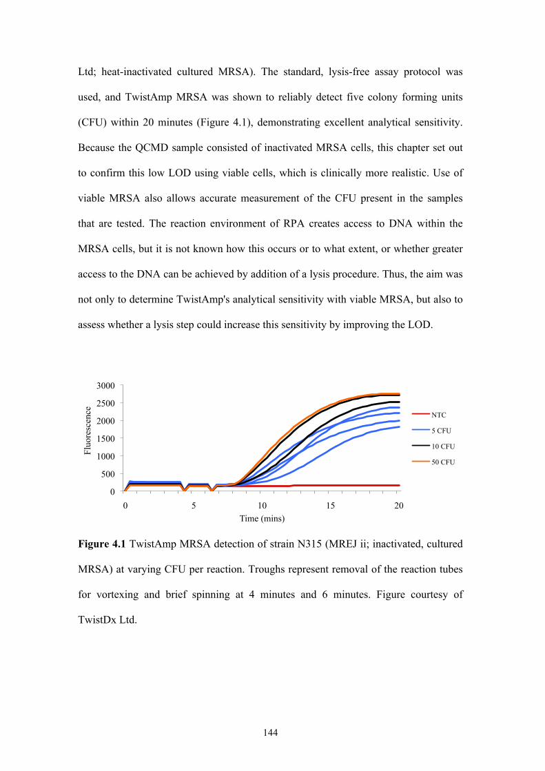
144
Ltd; heat-inactivated cultured MRSA). The standard, lysis-free assay protocol was
used, and TwistAmp MRSA was shown to reliably detect five colony forming units
(CFU) within 20 minutes (Figure 4.1), demonstrating excellent analytical sensitivity.
Because the QCMD sample consisted of inactivated MRSA cells, this chapter set out
to confirm this low LOD using viable cells, which is clinically more realistic. Use of
viable MRSA also allows accurate measurement of the CFU present in the samples
that are tested. The reaction environment of RPA creates access to DNA within the
MRSA cells, but it is not known how this occurs or to what extent, or whether greater
access to the DNA can be achieved by addition of a lysis procedure. Thus, the aim was
not only to determine TwistAmp's analytical sensitivity with viable MRSA, but also to
assess whether a lysis step could increase this sensitivity by improving the LOD.
Figure 4.1 TwistAmp MRSA detection of strain N315 (MREJ ii; inactivated, cultured
MRSA) at varying CFU per reaction. Troughs represent removal of the reaction tubes
for vortexing and brief spinning at 4 minutes and 6 minutes. Figure courtesy of
TwistDx Ltd.
0
500
1000
1500
2000
2500
3000
0 5 10 15 20
Fluo
resc
ence
Time (mins)
NTC
5 CFU
10 CFU
50 CFU
145
4.1.2.2 Inhibition and sub-sampling
The reduced clinical sensitivity of TwistAmp MRSA is not due to molecular
reasons such as an inability to detect certain MREJ types, or problems with the RPA
chemistry. If the analytical LOD as determined in section 4.2.1 using viable MRSA is
very low, and therefore does not affect clinical sensitivity, then sub-sampling and/or
RPA inhibition may be the problem.
For the CMFT collaboration, duplicate swabs were taken from each patient:
one for testing using the gold standard broth enrichment culture technique employed at
the hospital, and one for testing with TwistAmp MRSA. The swabs for testing with
TwistAmp MRSA were each eluted in 800µl of RPA resuspension buffer and 50µl of
this solution was added to the reaction tube. For the KC collaboration, duplicate swabs
were also taken from each patient. One swab was tested with Xpert MRSA and one
swab was eluted in 1000µl of RPA resuspension buffer and 150µl of this solution
tested with TwistAmp MRSA. The remaining solution was used to inoculate
chromogenic and blood agar plates for detection of MRSA and MSSA, respectively.
A total reaction volume of 150µl was used in the KC trial after TwistDx found
that greater sample dilution in RPA resuspension buffer solved most of the inhibition
problems experienced during the CMFT trial, which used a total reaction volume of
50µl (see section 4.1). To compensate for the greater sample dilution, a greater
reaction volume (greater volume of RPA resuspension buffer mixed with sample) was
used. All TwistAmp MRSA reactions used in this chapter have a total volume of 150µl
to replicate the reactions used in the KC trial. In Chapter 3, I used reactions with a
smaller total volume of 50µl, since they require a third of the required RPA
components, making them cheaper for TwistDx to produce.
146
The culturing procedure at CMFT involved breaking off the swab in broth,
incubating for 24 hours at 37°C, and plating an amount of this broth on chromogenic
media. The initial 24-hour broth culture allows any MRSA on a swab to grow and
replicate such that when some of the broth is plated out it should contain MRSA (this
also removes any inconsistencies caused by users, for example only one side of a swab
being streaked directly onto a plate). With TwistAmp MRSA, the swab is placed in
buffer and agitated for a few seconds, and the eluate tested. Thus, if the entire swab
only holds small numbers of MRSA, e.g. 10 CFU, culturing may detect them, but
TwistAmp MRSA may not, due to insufficient CFU (below the assay's LOD) being
present in the fraction of eluate tested. The even distribution of cells in solution could
be further affected by the cluster-forming nature of S. aureus, whose cells do not fully
separate upon division. Cepheid's Xpert MRSA assay uses a larger swab (Copan
Venturi Transystem double swab) and tests the entire eluate [Cepheid Diagnostics
2009] - procedures that are both likely to improve the possibility of detection
compared to the TwistAmp MRSA protocol. However, in the KC collaboration,
identical swabs were used for both TwistAmp and Xpert MRSA, and any difference
would be due only to the fraction of eluate used in the test procedure.
Since the TwistAmp MRSA tested does not use any sample preparation,
inhibitory substances or interfering organisms (e.g. MSSA and MR-CNS) in the
sample may affect TwistAmp MRSA sensitivity. In contrast, Xpert MRSA includes
DNA extraction and purification steps and thus tests cleaner samples. In order to
ascertain the effects of potential inhibition and sub-sampling, experiments were
performed to answer the following questions:
• Does the presence of MSSA or MR-CNS in the sample inhibit detection of
MRSA thereby giving a false negative result? For example, the high sequence
147
similarity of closely related Staphylococcal species to MRSA could lead to the
depletion of primer reserves required for MRSA-specific amplification.
• Do high levels of MSSA or CNS in samples with no MRSA present, cause
false positive results, for example due to snap-back DNA synthesis as a result
of the homology between orfX moieties in S. aureus and CNS, such as S.
haemolyticus or S. epidermidis (GenBank accession numbers AY751823 and
AY751825, respectively) [Francois et al. 2007]? MR-CNS, and MSSA and
MS-CNS containing mec-less SCC cassettes such as SCCcap1, could also
produce false-positive results due to the presence of MREJ sequences identical
to those found in MRSA [Luong et al. 2002, Katayama et al. 2003,
Mongkolrattanothai et al. 2004, Cuny and Witte 2005, Malhotra-Kumar et al.
2010b, Arbefeville et al. 2011].
• Does isolation of MRSA using filters reduce inhibition and thus improve
detection? Which filters retain and therefore concentrate MRSA for subsequent
testing, and which filters release MRSA?
4.2 METHODS AND RESULTS
Note: The troughs seen in all graphs at four minutes and six minutes represent removal
of the reaction tubes for vortexing and brief spinning.
4.2.1 Limit of detection
To determine the analytical sensitivity of TwistAmp MRSA with viable,
cultured MRSA, an isolate with the most common MREJ type (type ii), was cultured as
in section 2.1. A colony from overnight growth on blood agar (Oxoid) was suspended
in 1ml of RPA resuspension buffer and thoroughly mixed by vortexing, repeated
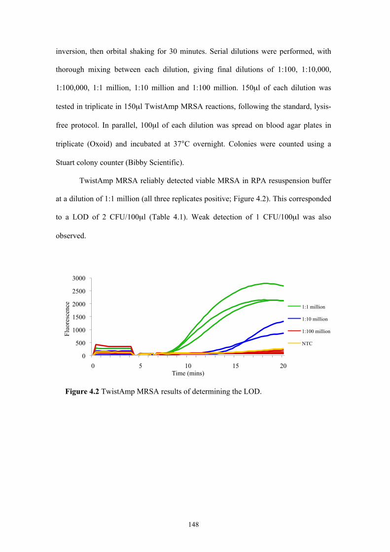
148
inversion, then orbital shaking for 30 minutes. Serial dilutions were performed, with
thorough mixing between each dilution, giving final dilutions of 1:100, 1:10,000,
1:100,000, 1:1 million, 1:10 million and 1:100 million. 150µl of each dilution was
tested in triplicate in 150µl TwistAmp MRSA reactions, following the standard, lysis-
free protocol. In parallel, 100µl of each dilution was spread on blood agar plates in
triplicate (Oxoid) and incubated at 37°C overnight. Colonies were counted using a
Stuart colony counter (Bibby Scientific).
TwistAmp MRSA reliably detected viable MRSA in RPA resuspension buffer
at a dilution of 1:1 million (all three replicates positive; Figure 4.2). This corresponded
to a LOD of 2 CFU/100µl (Table 4.1). Weak detection of 1 CFU/100µl was also
observed.
Figure 4.2 TwistAmp MRSA results of determining the LOD.
0
500
1000
1500
2000
2500
3000
0 5 10 15 20
Fluo
resc
ence
Time (mins)
1:1 million
1:10 million
1:100 million
NTC
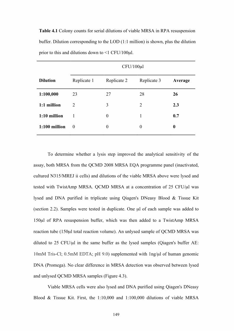
149
Table 4.1 Colony counts for serial dilutions of viable MRSA in RPA resuspension
buffer. Dilution corresponding to the LOD (1:1 million) is shown, plus the dilution
prior to this and dilutions down to <1 CFU/100µl.
CFU/100µl
Dilution Replicate 1 Replicate 2 Replicate 3 Average
1:100,000 23 27 28 26
1:1 million 2 3 2 2.3
1:10 million 1 0 1 0.7
1:100 million 0 0 0 0
To determine whether a lysis step improved the analytical sensitivity of the
assay, both MRSA from the QCMD 2008 MRSA EQA programme panel (inactivated,
cultured N315/MREJ ii cells) and dilutions of the viable MRSA above were lysed and
tested with TwistAmp MRSA. QCMD MRSA at a concentration of 25 CFU/µl was
lysed and DNA purified in triplicate using Qiagen's DNeasy Blood & Tissue Kit
(section 2.2). Samples were tested in duplicate. One µl of each sample was added to
150µl of RPA resuspension buffer, which was then added to a TwistAmp MRSA
reaction tube (150µl total reaction volume). An unlysed sample of QCMD MRSA was
diluted to 25 CFU/µl in the same buffer as the lysed samples (Qiagen's buffer AE:
10mM Tris-Cl; 0.5mM EDTA; pH 9.0) supplemented with 1ng/µl of human genomic
DNA (Promega). No clear difference in MRSA detection was observed between lysed
and unlysed QCMD MRSA samples (Figure 4.3).
Viable MRSA cells were also lysed and DNA purified using Qiagen's DNeasy
Blood & Tissue Kit. First, the 1:10,000 and 1:100,000 dilutions of viable MRSA
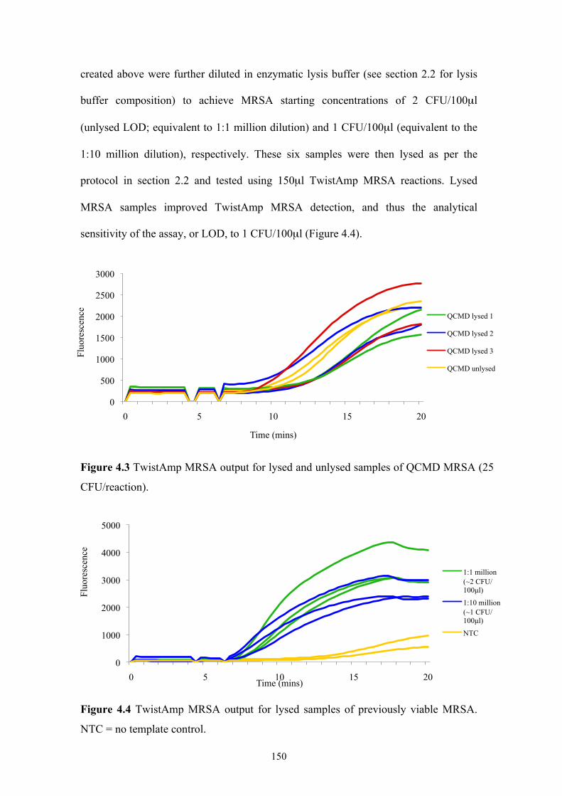
150
created above were further diluted in enzymatic lysis buffer (see section 2.2 for lysis
buffer composition) to achieve MRSA starting concentrations of 2 CFU/100µl
(unlysed LOD; equivalent to 1:1 million dilution) and 1 CFU/100µl (equivalent to the
1:10 million dilution), respectively. These six samples were then lysed as per the
protocol in section 2.2 and tested using 150µl TwistAmp MRSA reactions. Lysed
MRSA samples improved TwistAmp MRSA detection, and thus the analytical
sensitivity of the assay, or LOD, to 1 CFU/100µl (Figure 4.4).
Figure 4.3 TwistAmp MRSA output for lysed and unlysed samples of QCMD MRSA (25
CFU/reaction).
Figure 4.4 TwistAmp MRSA output for lysed samples of previously viable MRSA.
NTC = no template control.
0
500
1000
1500
2000
2500
3000
0 5 10 15 20
Fluo
resc
ence
Time (mins)
QCMD lysed 1
QCMD lysed 2
QCMD lysed 3
QCMD unlysed
0
1000
2000
3000
4000
5000
0 5 10 15 20
Fluo
resc
ence
Time (mins)
1:1 million (~2 CFU/100µl)
1:10 million (~1 CFU/100µl)
NTC
151
4.2.2 Inhibition and sub-sampling
To determine if TwistAmp MRSA is inhibited by the presence of large
concentrations of MSSA or MR-CNS, leading to false negative results, MRSA, MSSA
and MR-CNS samples from the QCMD 2008 MRSA EQA programme panel were
used as template and tested with TwistAmp MRSA reactions as per the protocol in
section 3.2.4. All samples were diluted to the necessary concentrations in S. aureus
negative medium (Mueller-Hinton Broth) from the QCMD panel. MRSA template was
added to each reaction at 50 CFU/reaction. MSSA and MR-CNS were tested
separately, in duplicate, and at varying concentrations in combination with MRSA:
104, 105 and 106 CFU/reaction. To test for the possibility of false positive results in the
presence of large concentrations of MSSA or MR-CNS but absence of MRSA, the
same reactions were performed as above, but with no MRSA template (0
CFU/reaction).
Low concentrations of MRSA (50 CFU/reaction) in combination with high
concentrations of MSSA or MR-CNS (104-106 CFU/reaction) in a sample did not
inhibit TwistAmp MRSA reactions and cause false negative results (Figure 4.5).
Shallower reaction curves with increasing concentrations of MR-CNS were observed,
but did not affect definitive detection of MRSA (Figure 4.5B). High concentrations of
MSSA or MR-CNS in a sample with no MRSA did not cause false positive results
(Figure 4.6).
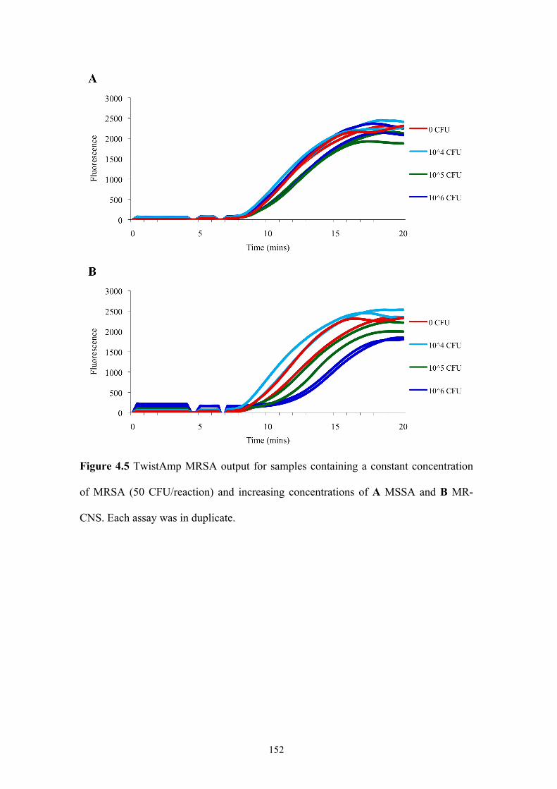
152
Figure 4.5 TwistAmp MRSA output for samples containing a constant concentration
of MRSA (50 CFU/reaction) and increasing concentrations of A MSSA and B MR-
CNS. Each assay was in duplicate.
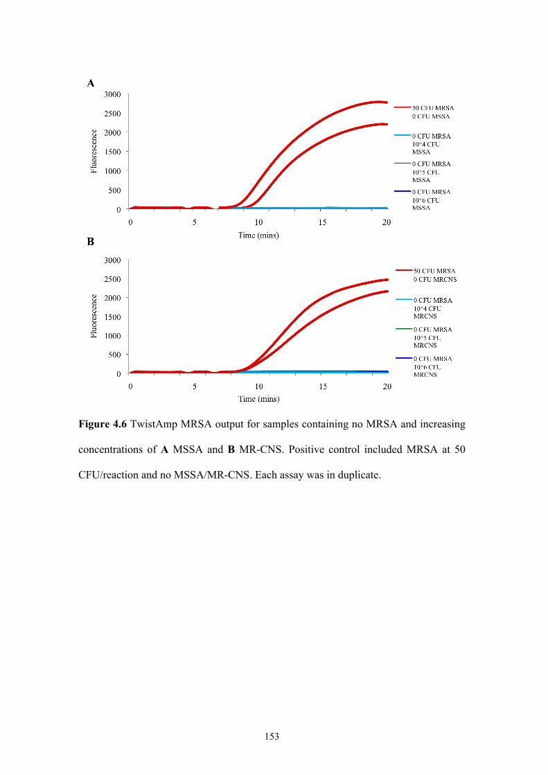
153
Figure 4.6 TwistAmp MRSA output for samples containing no MRSA and increasing
concentrations of A MSSA and B MR-CNS. Positive control included MRSA at 50
CFU/reaction and no MSSA/MR-CNS. Each assay was in duplicate.
154
Several different filters manufactured by Millipore (Table 4.2) were tested with
TwistAmp MRSA to assess whether MRSA detection is improved with filtration.
These microporous membrane filters can be used for microfiltration of particles,
including bacteria. S. aureus cocci are approximately 0.5-1µm in diameter, so any
filters with a pore size larger than this should allow MRSA to pass through into the
filtrate. Filters with pores smaller than 0.5µm should retain MRSA. Larger pore filters
were tested for their porosity to MRSA with a view to their suitability as pre-filters that
could remove larger particulate matter that might inhibit RPA, but not trap MRSA.
This was tested by checking that filtration through these filters did not impact on
TwistAmp MRSA assay performance. Smaller pore filters were tested for their ability
to retain MRSA, evidenced by a negative reaction curve when the filtrate was added to
TwistAmp MRSA reactions. MRSA from the QCMD 2008 MRSA EQA programme
panel was used as template and mixed with RPA resuspension buffer at a concentration
of 25 MRSA CFU/136µl, then filtered (Figure 4.7). The filtrate was then tested with
150µl TwistAmp MRSA reactions as per the protocol in section 3.2.4. Since MgAc
immediately starts the RPA reaction, RPA resuspension buffer without MgAc was
used for the filtering. Fourteen µl of 280mM MgAc was added separately to the
reaction as a final step before placing reaction tubes in the Twista machine, to ensure
simultaneous initiation of compared reactions.
To further test capture of MRSA, the filters themselves were included in
TwistAmp MRSA reactions rather than the filtrate (Figure 4.8). Any filters that retain
MRSA should produce a reaction curve; any filters that do not capture MRSA should
produce no reaction curve.
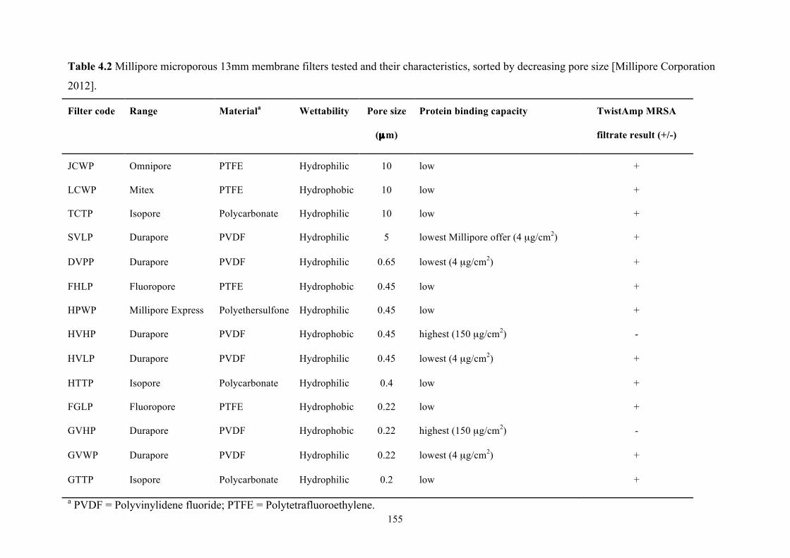
155
Table 4.2 Millipore microporous 13mm membrane filters tested and their characteristics, sorted by decreasing pore size [Millipore Corporation
2012].
Filter code Range Materiala Wettability Pore size
(µm)
Protein binding capacity TwistAmp MRSA
filtrate result (+/-)
JCWP Omnipore PTFE Hydrophilic 10 low +
LCWP Mitex PTFE Hydrophobic 10 low +
TCTP Isopore Polycarbonate Hydrophilic 10 low +
SVLP Durapore PVDF Hydrophilic 5 lowest Millipore offer (4 µg/cm2) +
DVPP Durapore PVDF Hydrophilic 0.65 lowest (4 µg/cm2) +
FHLP Fluoropore PTFE Hydrophobic 0.45 low +
HPWP Millipore Express Polyethersulfone Hydrophilic 0.45 low +
HVHP Durapore PVDF Hydrophobic 0.45 highest (150 µg/cm2) -
HVLP Durapore PVDF Hydrophilic 0.45 lowest (4 µg/cm2) +
HTTP Isopore Polycarbonate Hydrophilic 0.4 low +
FGLP Fluoropore PTFE Hydrophobic 0.22 low +
GVHP Durapore PVDF Hydrophobic 0.22 highest (150 µg/cm2) -
GVWP Durapore PVDF Hydrophilic 0.22 lowest (4 µg/cm2) +
GTTP Isopore Polycarbonate Hydrophilic 0.2 low +
a PVDF = Polyvinylidene fluoride; PTFE = Polytetrafluoroethylene.
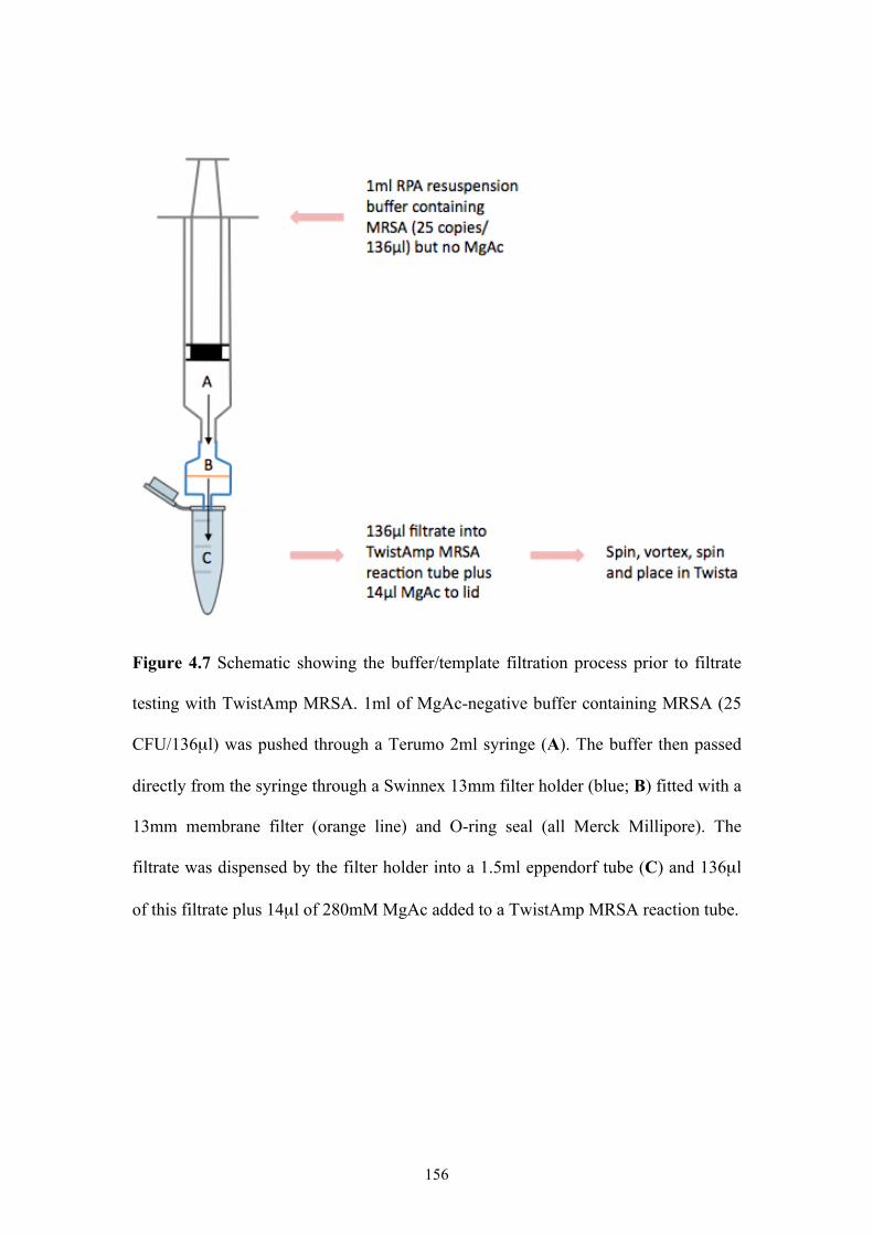
156
Figure 4.7 Schematic showing the buffer/template filtration process prior to filtrate
testing with TwistAmp MRSA. 1ml of MgAc-negative buffer containing MRSA (25
CFU/136µl) was pushed through a Terumo 2ml syringe (A). The buffer then passed
directly from the syringe through a Swinnex 13mm filter holder (blue; B) fitted with a
13mm membrane filter (orange line) and O-ring seal (all Merck Millipore). The
filtrate was dispensed by the filter holder into a 1.5ml eppendorf tube (C) and 136µl
of this filtrate plus 14µl of 280mM MgAc added to a TwistAmp MRSA reaction tube.
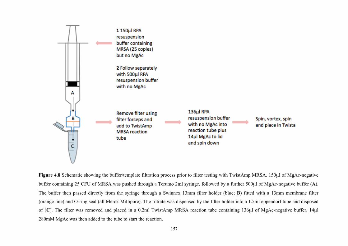
157
Figure 4.8 Schematic showing the buffer/template filtration process prior to filter testing with TwistAmp MRSA. 150µl of MgAc-negative
buffer containing 25 CFU of MRSA was pushed through a Terumo 2ml syringe, followed by a further 500µl of MgAc-negative buffer (A).
The buffer then passed directly from the syringe through a Swinnex 13mm filter holder (blue; B) fitted with a 13mm membrane filter
(orange line) and O-ring seal (all Merck Millipore). The filtrate was dispensed by the filter holder into a 1.5ml eppendorf tube and disposed
of (C). The filter was removed and placed in a 0.2ml TwistAmp MRSA reaction tube containing 136µl of MgAc-negative buffer. 14µl
280mM MgAc was then added to the tube to start the reaction.
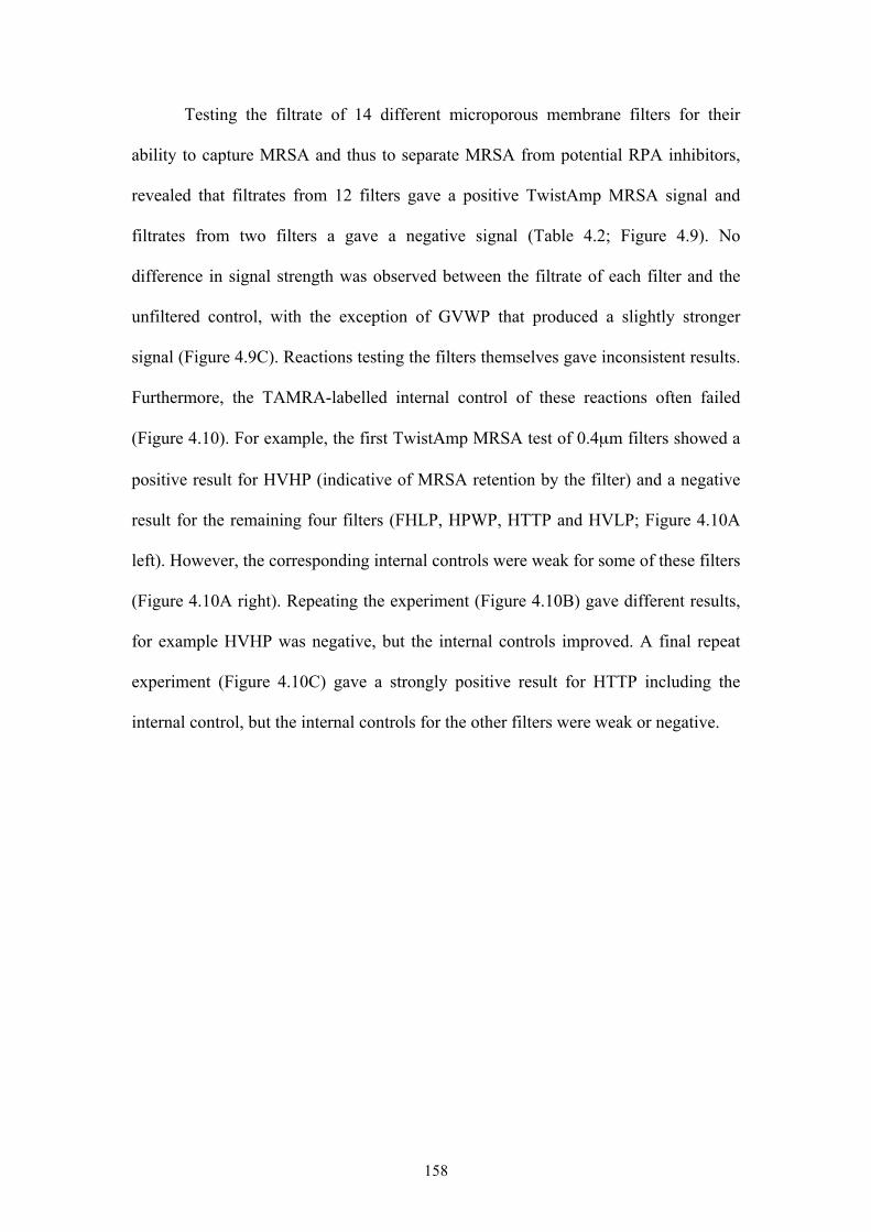
158
Testing the filtrate of 14 different microporous membrane filters for their
ability to capture MRSA and thus to separate MRSA from potential RPA inhibitors,
revealed that filtrates from 12 filters gave a positive TwistAmp MRSA signal and
filtrates from two filters a gave a negative signal (Table 4.2; Figure 4.9). No
difference in signal strength was observed between the filtrate of each filter and the
unfiltered control, with the exception of GVWP that produced a slightly stronger
signal (Figure 4.9C). Reactions testing the filters themselves gave inconsistent results.
Furthermore, the TAMRA-labelled internal control of these reactions often failed
(Figure 4.10). For example, the first TwistAmp MRSA test of 0.4µm filters showed a
positive result for HVHP (indicative of MRSA retention by the filter) and a negative
result for the remaining four filters (FHLP, HPWP, HTTP and HVLP; Figure 4.10A
left). However, the corresponding internal controls were weak for some of these filters
(Figure 4.10A right). Repeating the experiment (Figure 4.10B) gave different results,
for example HVHP was negative, but the internal controls improved. A final repeat
experiment (Figure 4.10C) gave a strongly positive result for HTTP including the
internal control, but the internal controls for the other filters were weak or negative.
159
Figure 4.9 TwistAmp MRSA output for the filtrate of filters with pore sizes A
>0.5µm; B 0.4µm; and C 0.2µm in diameter.
0
500
1000
1500
2000
2500
0 5 10 15 20
Fluo
resc
ence
Time (mins)
JCWP
TCTP
LCWP
SVLP
DVPP
No filter
0
500
1000
1500
2000
2500
0 5 10 15 20
Fluo
resc
ence
Time (mins)
HVLP
HTTP
HPWP
HVHP
FHLP
No filter
0
500
1000
1500
2000
2500
0 5 10 15 20
Fluo
resc
ence
Time (mins)
GVHP
FGLP
GTTP
GVWP
No filter
A
C
B
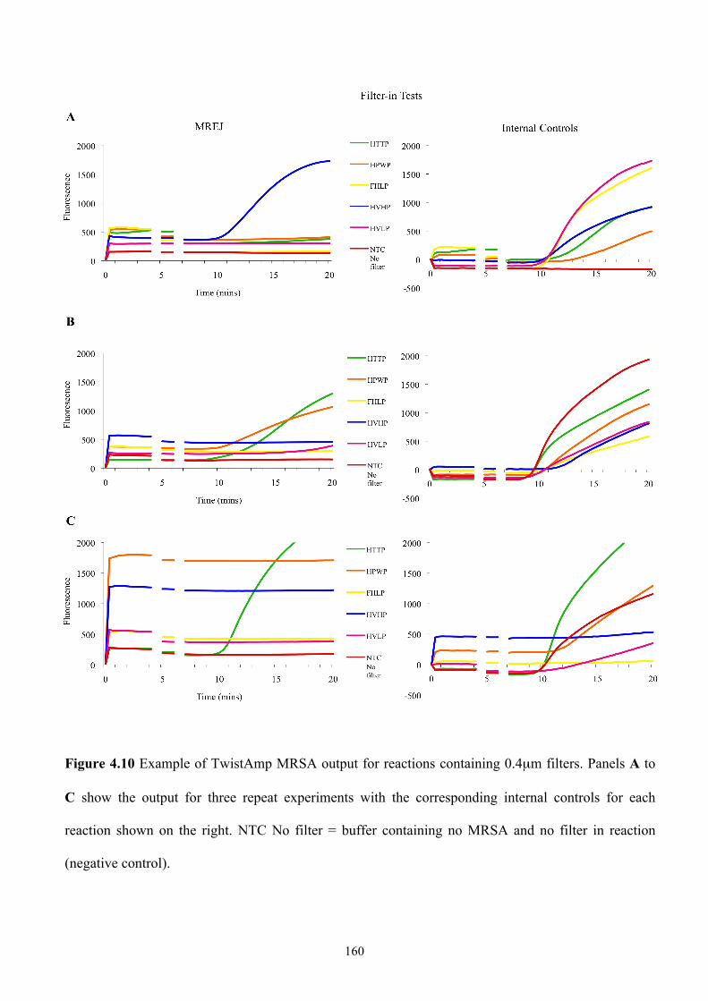
160
Figure 4.10 Example of TwistAmp MRSA output for reactions containing 0.4µm filters. Panels A to
C show the output for three repeat experiments with the corresponding internal controls for each
reaction shown on the right. NTC No filter = buffer containing no MRSA and no filter in reaction
(negative control).
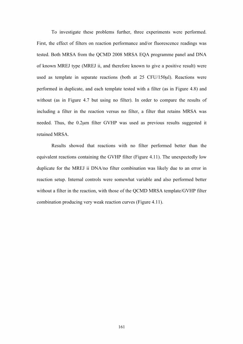
161
To investigate these problems further, three experiments were performed.
First, the effect of filters on reaction performance and/or fluorescence readings was
tested. Both MRSA from the QCMD 2008 MRSA EQA programme panel and DNA
of known MREJ type (MREJ ii, and therefore known to give a positive result) were
used as template in separate reactions (both at 25 CFU/150µl). Reactions were
performed in duplicate, and each template tested with a filter (as in Figure 4.8) and
without (as in Figure 4.7 but using no filter). In order to compare the results of
including a filter in the reaction versus no filter, a filter that retains MRSA was
needed. Thus, the 0.2µm filter GVHP was used as previous results suggested it
retained MRSA.
Results showed that reactions with no filter performed better than the
equivalent reactions containing the GVHP filter (Figure 4.11). The unexpectedly low
duplicate for the MREJ ii DNA/no filter combination was likely due to an error in
reaction setup. Internal controls were somewhat variable and also performed better
without a filter in the reaction, with those of the QCMD MRSA template/GVHP filter
combination producing very weak reaction curves (Figure 4.11).
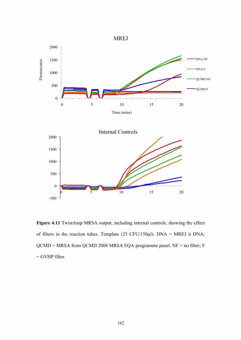
162
Figure 4.11 TwistAmp MRSA output, including internal controls, showing the effect
of filters in the reaction tubes. Template (25 CFU/150µl): DNA = MREJ ii DNA;
QCMD = MRSA from QCMD 2008 MRSA EQA programme panel. NF = no filter; F
= GVHP filter.
0
500
1000
1500
2000
0 5 10 15 20
Fluo
resc
ence
Time (mins)
MREJ
DNA NF
DNA F
QCMD NF
QCMD F
-500
0
500
1000
1500
2000
0 5 10 15 20
Internal Controls
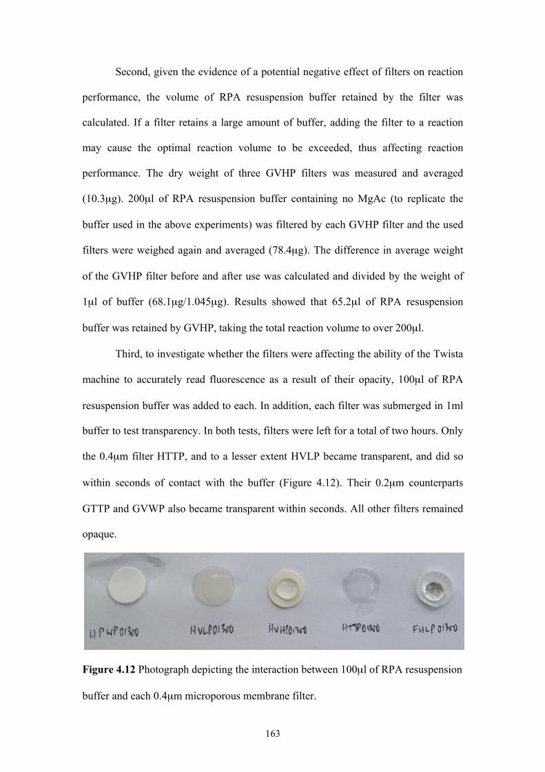
163
Second, given the evidence of a potential negative effect of filters on reaction
performance, the volume of RPA resuspension buffer retained by the filter was
calculated. If a filter retains a large amount of buffer, adding the filter to a reaction
may cause the optimal reaction volume to be exceeded, thus affecting reaction
performance. The dry weight of three GVHP filters was measured and averaged
(10.3µg). 200µl of RPA resuspension buffer containing no MgAc (to replicate the
buffer used in the above experiments) was filtered by each GVHP filter and the used
filters were weighed again and averaged (78.4µg). The difference in average weight
of the GVHP filter before and after use was calculated and divided by the weight of
1µl of buffer (68.1µg/1.045µg). Results showed that 65.2µl of RPA resuspension
buffer was retained by GVHP, taking the total reaction volume to over 200µl.
Third, to investigate whether the filters were affecting the ability of the Twista
machine to accurately read fluorescence as a result of their opacity, 100µl of RPA
resuspension buffer was added to each. In addition, each filter was submerged in 1ml
buffer to test transparency. In both tests, filters were left for a total of two hours. Only
the 0.4µm filter HTTP, and to a lesser extent HVLP became transparent, and did so
within seconds of contact with the buffer (Figure 4.12). Their 0.2µm counterparts
GTTP and GVWP also became transparent within seconds. All other filters remained
opaque.
Figure 4.12 Photograph depicting the interaction between 100µl of RPA resuspension
buffer and each 0.4µm microporous membrane filter.
164
Since no conclusive results could be obtained from testing the filters
themselves with TwistAmp MRSA, the focus was turned to the filtrate experiments
and the reasons why GVHP (0.2µm) and HVHP (0.4µm) showed evidence of MRSA
retention. Filter retention would be expected since S. aureus cells are larger than
0.5µm, but given that all other 0.2µm and 0.4µm filters tested appeared to release
MRSA cells, GVHP and HVHP were investigated. A component of the RPA
resuspension buffer, polyethylene glycol (PEG), may somehow be retained by these
filters, thus leading to a reaction failure. Buffer containing MRSA (25 CFU/110µl)
but no PEG and no MgAc, was filtered through GVHP and HVHP. 20% w/v PEG
(confidential molecular weight) and 280mM MgAc were then added to the reaction
tubes prior to filtrate testing (Figure 4.13). The same reactions were performed with
no filtration. Filters GVHP and HVHP did not appear to retain PEG, as shown by
negative TwistAmp MRSA output for these filters, regardless of the presence of PEG
in the buffer prior to filtration (Figure 4.14).
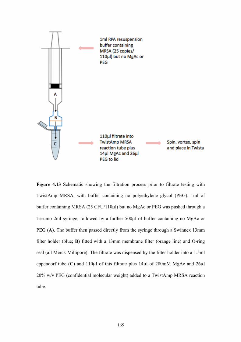
165
Figure 4.13 Schematic showing the filtration process prior to filtrate testing with
TwistAmp MRSA, with buffer containing no polyethylene glycol (PEG). 1ml of
buffer containing MRSA (25 CFU/110µl) but no MgAc or PEG was pushed through a
Terumo 2ml syringe, followed by a further 500µl of buffer containing no MgAc or
PEG (A). The buffer then passed directly from the syringe through a Swinnex 13mm
filter holder (blue; B) fitted with a 13mm membrane filter (orange line) and O-ring
seal (all Merck Millipore). The filtrate was dispensed by the filter holder into a 1.5ml
eppendorf tube (C) and 110µl of this filtrate plus 14µl of 280mM MgAc and 26µl
20% w/v PEG (confidential molecular weight) added to a TwistAmp MRSA reaction
tube.
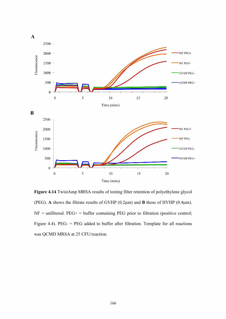
166
Figure 4.14 TwistAmp MRSA results of testing filter retention of polyethylene glycol
(PEG). A shows the filtrate results of GVHP (0.2µm) and B those of HVHP (0.4µm).
NF = unfiltered. PEG+ = buffer containing PEG prior to filtration (positive control;
Figure 4.4). PEG- = PEG added to buffer after filtration. Template for all reactions
was QCMD MRSA at 25 CFU/reaction.
167
The QCMD MRSA sample used as template throughout the filtration
experiments consisted of heat-inactivated cultured cells. Heat has previously been
shown to cause leakage and lysis of S. aureus cells [Allwood and Russell 1969], so it
may be that free DNA was present, leading to RPA detection of MRSA in the filtrate.
If this were the case, GVHP and HVHP retain DNA as well as S. aureus cells. In
order to confirm that all other filters released cells and not just free DNA, filtration
experiments were performed with live MRSA cells. Three filters with different
characteristics were selected: HPWP (0.4µm), FGLP (0.2µm) and GVWP (0.2µm)
(Table 4.2). MRSA of an MREJ type known to give a positive TwistAmp MRSA
signal (MREJ ii) was cultured as in section 2.1. For each filter, a colony from
overnight growth was suspended in 1.2ml of RPA resuspension buffer and thoroughly
mixed by vortexing, repeated inversion, then orbital shaking for 30 minutes. Serial
dilutions were performed from 1:100 up to the approximate TwistAmp MRSA LOD
(1:1 million), with thorough mixing between each dilution. In parallel, and also for
each filter, MRSA was suspended in sterile distilled water (SDW) and diluted up to
1:1 million. Three 100µl replicates of each 1:1 million and also 1:10,000 dilution
(RPA resuspension buffer and SDW) were plated separately on blood agar plates
(Oxoid) and incubated overnight at 37°C. Colonies were counted using a Stuart
colony counter (Bibby Scientific). Filtration of the 1:1 million and 1:10,000 dilutions
(buffer and SDW) was performed for each of the three filters. 600µl of each dilution
was filtered, and 100µl replicates of each filtrate plated separately on blood agar
plates and incubated overnight at 37°C. Comparing the filtrate of both the buffer and
SDW dilutions by culture allowed the assessment of the effect of the buffer and/or
filtration process on cell viability. 100µl of each buffer filtrate was tested with
168
TwistAmp MRSA (100µl reactions) and 100µl of each unfiltered buffer dilution was
tested for direct comparison.
No difference was observed in the colony counts between buffer and SDW
dilutions prior to filtration (Table 4.3), suggesting that RPA resuspension buffer does
not lyse cells or cause cell leakage, and thus presumably creates no free DNA that
may pass through the filters and cause a positive TwistAmp MRSA result. This was
confirmed by testing the main component of RPA resuspension buffer besides SDW -
PEG - for an effect during filtration (protocol Figure 4.13). No difference in
TwistAmp MRSA output was observed between reactions with buffer that contained
PEG prior to filtration, to reactions where PEG was added after filtration, using 0.2µm
filters FGLP and GVWP (Figure 4.15).
No MRSA growth was observed from plating the filtrates. This was the case
for both 1:10,000 and 1:1 million MRSA dilutions in SDW and resuspension buffer,
and for all three filters tested (Table 4.3). TwistAmp MRSA output showed the buffer
filtrates were MRSA positive at the 1:10,000 dilution but weaker than the unfiltered
dilutions (Figure 4.16). Filter FGLP demonstrated the smallest difference in output
before and after filtration, and at the 1:1 million dilution no difference was observed
for this filter. Filtrates of HPWP and GVWP were negative at the 1:1 million dilution
(Figure 4.16).
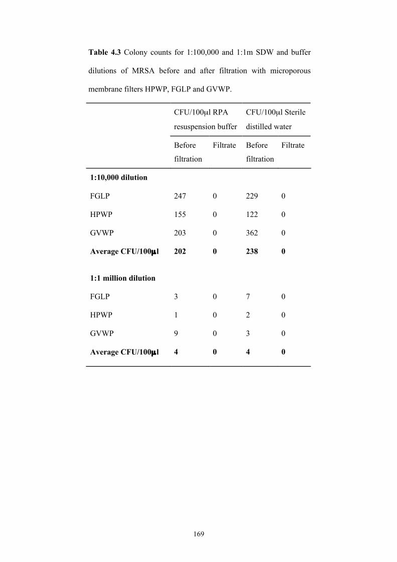
169
Table 4.3 Colony counts for 1:100,000 and 1:1m SDW and buffer
dilutions of MRSA before and after filtration with microporous
membrane filters HPWP, FGLP and GVWP.
CFU/100µl RPA
resuspension buffer
CFU/100µl Sterile
distilled water
Before
filtration
Filtrate Before
filtration
Filtrate
1:10,000 dilution
FGLP 247 0 229 0
HPWP 155 0 122 0
GVWP 203 0 362 0
Average CFU/100µ l 202 0 238 0
1:1 million dilution
FGLP 3 0 7 0
HPWP 1 0 2 0
GVWP 9 0 3 0
Average CFU/100µ l 4 0 4 0
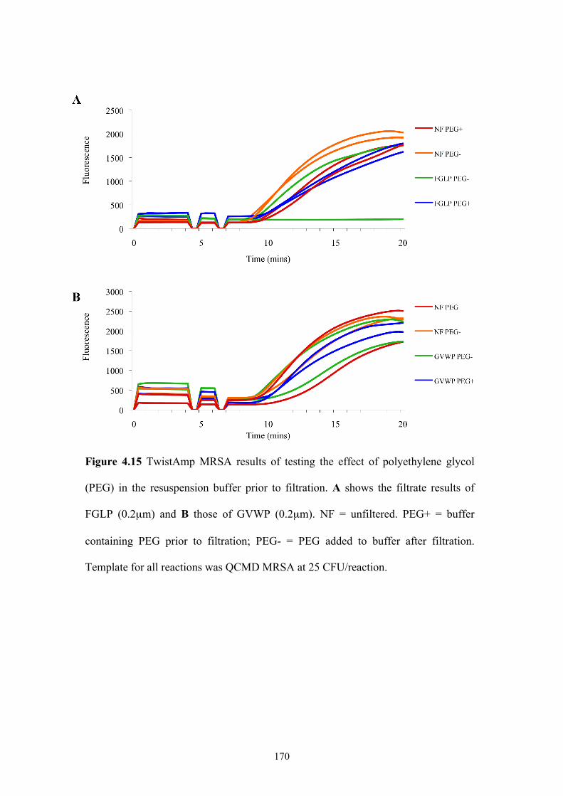
170
Figure 4.15 TwistAmp MRSA results of testing the effect of polyethylene glycol
(PEG) in the resuspension buffer prior to filtration. A shows the filtrate results of
FGLP (0.2µm) and B those of GVWP (0.2µm). NF = unfiltered. PEG+ = buffer
containing PEG prior to filtration; PEG- = PEG added to buffer after filtration.
Template for all reactions was QCMD MRSA at 25 CFU/reaction.
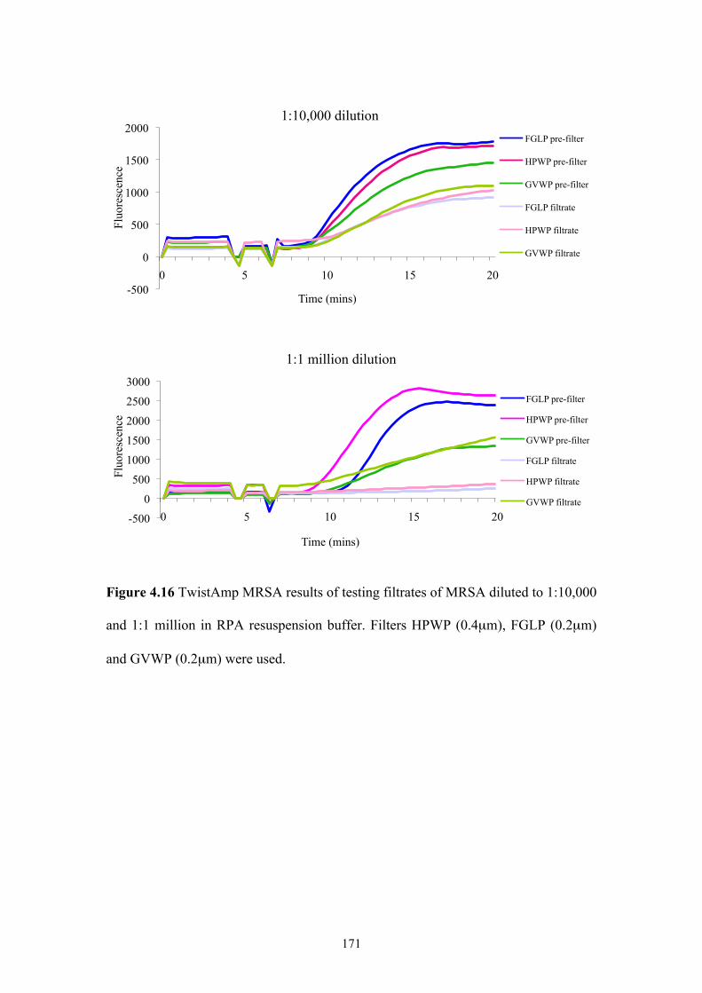
171
Figure 4.16 TwistAmp MRSA results of testing filtrates of MRSA diluted to 1:10,000
and 1:1 million in RPA resuspension buffer. Filters HPWP (0.4µm), FGLP (0.2µm)
and GVWP (0.2µm) were used.
-500
0
500
1000
1500
2000
0 5 10 15 20
Fluo
resc
ence
Time (mins)
1:10,000 dilution FGLP pre-filter
HPWP pre-filter
GVWP pre-filter
FGLP filtrate
HPWP filtrate
GVWP filtrate
-500
0
500
1000
1500
2000
2500
3000
0 5 10 15 20
Fluo
resc
ence
Time (mins)
1:1 million dilution
FGLP pre-filter
HPWP pre-filter
GVWP pre-filter
FGLP filtrate
HPWP filtrate
GVWP filtrate
172
4.3 DISCUSSION
TwistAmp MRSA demonstrates a lower clinical sensitivity than the market
leader in molecular MRSA diagnostics, Cepheid's Xpert MRSA. The purpose of this
chapter was to investigate possible reasons for this (poor analytical LOD, RPA
inhibition and sub-sampling) and assess whether methods such as lysis and/or
filtration could improve the assay's analytical LOD and its ability to detect MRSA in
clinical samples.
4.3.1 Limit of detection
The LOD of TwistAmp MRSA was determined as approximately 2
CFU/100µl using cultured MRSA. The assay sporadically and weakly detected 1
CFU/100µl. This clearly demonstrates the assay is very sensitive. Cepheid states an
overall LOD of 80 CFU/swab for Xpert MRSA, and provides the LODs for individual
SCCmec types. For Xpert MRSA, the LOD is 10 CFU/swab for SCCmec types I-III
and V, 50 CFU/swab for type IV, and 100 CFU/swab for type IVa [Cepheid
Diagnostics 2009]. The LOD of TwistAmp MRSA was determined using an MREJ
type ii isolate, which accounted for about 90% of all MRSA tested in Chapter 3.
SCCmec type II was exclusively associated with MREJ type ii in the MRSA strains
typed in Chapter 3. Thus, results show TwistAmp MRSA has a slightly lower
analytical sensitivity than Xpert MRSA (2 CFU/100µl, equivalent to 20 CFU/swab,
versus 10 CFU/swab).
Xpert MRSA's LOD of 80 CFU/swab was determined with 95% confidence
i.e. the lowest number of MRSA CFU per swab that can be reproducibly distinguished
from negative samples with 95% confidence. A 95% confidence interval for the
analytical LOD of TwistAmp MRSA is yet to be determined. Further work is also
173
needed to determine the LOD for each of the less prevalent MREJ types covered by
TwistAmp MRSA, as well as for each of the prototypic strains for the different
SCCmec elements detected by the assay (types I-IV and VI-VIII). Proprietary primers
for MREJ types iv, v and vii performed less well than those for MREJs i-iii (data not
shown), so the LOD is likely to be poorer for the former types. The SCCmec type V
element represented by strain WIS (MREJ xii) was not detected by TwistAmp MRSA,
and one other type V strain tested (ST398) produced only weak positive results for
MREJ iii (Figure 3.16). All GenBank entries of type V elements with the exception of
WIS are MREJ iii (see Chapter 3 section 3.3.4.4). TwistAmp MRSA would
successfully detect half of these but the other half, all belonging to the livestock-
associated lineage ST398, would likely produce weak positive results due to SNPs in
the probe-binding region. Given this fact, it would be interesting to ascertain the
SCCmec type V strain used in Cepheid's performance testing of Xpert MRSA.
Because detection of SCCmec type V strains by TwistAmp MRSA is not reliable,
determining a LOD for this element would only be informative if all variants were
tested. Despite a correlation between MREJ type and SCCmec type existing, there are
exceptions, so caution should be taken when interpreting SCCmec type-specific
LODs.
A lysis and purification step was incorporated into the TwistAmp MRSA
protocol in an attempt to further improve the assay's analytical sensitivity. No
improvement was observed with inactivated N315/MREJ ii MRSA cells, however the
limit of detection was improved to approximately 1 CFU/100µl after lysis of viable
MREJ ii MRSA. This is equivalent to 10 CFU/swab, matching the LOD of Xpert
MRSA for MREJ ii MRSA. The lack of difference observed between unlysed and
lysed QCMD MRSA could be a result of free DNA already present in the heat-
174
inactivated sample. Heat has previously been shown to cause cell shrinkage and
leakage of intracellular constituents from Staphylococcal cells [Allwood and Russell
1969, Berkman and Wyatt 1970]. Despite the improved LOD with lysis and
purification of viable MRSA, high analytical sensitivity is still achieved with no lysis.
It was therefore concluded that the addition of a lysis step would not improve the
analytical sensitivity to an extent that would warrant such an addition, given the
modifications that would have to be made to the current assay.
4.3.2 Inhibition and sub-sampling
Given the high analytical sensitivity of TwistAmp MRSA (2 CFU/100µl), the
potential effects of RPA inhibition and sub-sampling on the assay's clinical sensitivity
were investigated. Testing RPA with samples of MRSA mixed with high
concentrations of MSSA or MR-CNS did not inhibit amplification of MRSA DNA
and cause false negative results. A similar experiment performed for a rival assay, BD
GeneOhm MRSA, showed that it too was not inhibited by MSSA or CNS [Huletsky
et al. 2004]. While increasing concentrations of MR-CNS in MRSA samples did not
produce false negative results, they did appear to weaken detection although not to an
extent that would affect a definitive positive MRSA result. This is likely due to the
depletion of MREJ-specific primer reserves in the reaction as they bind to the
SCCmec element conferring methicillin resistance in the CNS, leaving less MREJ
primer for MRSA DNA in the sample. The MR-CNS likely had the same MREJ type
as the MRSA in the sample (MREJ ii), which explains the weaker reaction curves for
MRSA as the concentration of MR-CNS increased.
Samples containing high concentrations of MSSA or MR-CNS but no MRSA
did not produce false positive results, demonstrating good analytical specificity. More
175
comprehensive testing of MSSA, MS-CNS and MR-CNS, plus strains representing
species phylogenetically related to S. aureus and members of the nasal commensal
flora, is required for accurate determination of the analytical specificity of TwistAmp
MRSA [Cepheid Diagnostics 2009]. Five MSSA isolates from our laboratory
collection of Staphylococci, and one MSSA isolate from the KC collection gave false
positive results with the assay (Chapter 3). The KC MSSA false positive appears to
contain a remnant of an SCC element because it is MREJ positive (type i) but mecA
negative. The QCMD 2008 MRSA EQA programme panel was used in this chapter,
but the 2009 EQA panel contains two samples of MSSA with mec-less cassettes
(SCCmec I and III). These samples would likely cause a similar effect to MR-CNS on
MRSA detection, and would cause false positive results in the absence of MRSA.
mec-less SCC elements are known to cause false positive results with Xpert
MRSA and BD GeneOhm MRSA [Francois et al. 2007, Arbefeville et al. 2011]. te
Witt et al. [2010] found commercial real-time PCR tests incorrectly reported the two
QCMD 2009 MSSA containing mec-less SCC elements as positive in 89% of datasets
(40 of 45 datasets, including 13 tested with GeneOhm MRSA and 11 with Xpert
MRSA). Xpert MRSA, GeneOhm MRSA and TwistAmp MRSA all target the MREJ
and so false positive results are inevitable with MREJ-positive MSSA. If these assays
were used in laboratories with high false positive rates or in regions with low MRSA
prevalence, confirmation by culture or a second molecular test would be necessary
[Kerremans et al. 2008, te Witt et al. 2010]. The only way to improve specificity with
such samples would be to incorporate a mecA internal control into the assays.
However, mecA is not unique to MRSA, so false positives could still arise due to co-
colonisation by MSSA and MR-CNS. Overall, MREJ-negative MSSA and MR-CNS
do not affect accurate MRSA detection by TwistAmp MRSA.
176
When directly using swabs, interfering substances such as blood or mucus
present in clinical samples could cause RPA inhibition, but this has not yet been
investigated. Cepheid evaluated these potentially interfering substances and concluded
that they did not significantly inhibit PCR or cause false negative results with Xpert
MRSA [Cepheid Diagnostics 2009], but this assay filters and lyses the sample, and
the released DNA is eluted prior to PCR testing, thus separating reaction inhibitors
such as blood or mucus from the target DNA. TwistAmp MRSA does not include any
sample preparation.
RPA resuspension buffer containing MRSA was filtered using various
microporous membrane filters to assess whether filtration improved detection of
MRSA with TwistAmp MRSA. Filters and filtrates were tested with the assay for
both retention and passage of MRSA. Of 14 filters, only two appeared to retain
MRSA: GVHP (0.2µm) and HVHP (0.4µm). This was surprising given that S. aureus
cells are at least 0.5µm in diameter - all 0.4µm and 0.2µm filters should retain MRSA.
This highlighted the possibility of either free DNA present in the sample, or
lysis/leakage of cells at some point during the filtration process, causing positive
results despite the retention of cells by the filters. Since the MRSA used as template
consisted of heat-inactivated cells, free DNA was likely present, since heat can cause
cell leakage [Allwood and Russell 1969]. This was confirmed by filtering viable
MRSA cells through selected 0.2µm and 0.4µm filters. The cultured filtrates grew no
MRSA, yet TwistAmp MRSA testing of the same filtrates gave MRSA-positive
results, albeit weak. It is likely therefore that all of the filters with pores less then
0.5µm in diameter retained MRSA cells. Alternatively, the filtration process could
have affected cell viability in an unknown way. Millipore states that fluid viscosity
and chemical interactions between filter membranes and particles in the solution could
177
affect bacterial retention [Millipore Corporation 2012]. However, RPA resuspension
buffer, which is fairly viscous owing to PEG, was confirmed not to have an effect on
filtration of MRSA cells.
MRSA retention by the 0.2µm and 0.4µm filters could not be confirmed with
TwistAmp MRSA reactions containing the filters themselves. The idea was that
adding filters to the reactions could test for the presence of MRSA cells on the filter
membranes. These experiments could not produce consistent results, and internal
controls failed. TwistAmp MRSA reaction tubes are 0.2ml in size, and so the 13mm
filters were not only difficult to insert into these but likely interfered with the RPA
reactions or measurements thereof due to their size and/or composition. Optical
properties of the filters, such as autofluorescence or transmittance, could have affected
fluorescence readings [Millipore Corporation 2012]. Of the filters tested for opacity
when in contact with RPA resuspension buffer, only HTTP (0.4µm) and GTTP
(0.2µm) filters were fully transparent. These filters are both part of Millipore's Isopore
range, recommended for analyses in which the sample is viewed on the surface of the
membrane, for example using optical or electron microscopy [Millipore Corporation
2012]. However, the optical compatibility of this filter type with the FAM and
TAMRA fluorophores used in TwistAmp MRSA is unknown, so it's possible that
despite their transparency to the naked eye, they still cause optical interference with
this assay.
Filter GVHP was found to retain a significant volume of RPA resuspension
buffer, which would have caused the optimal reaction volume to be exceeded,
resulting in reaction failure. This could explain the negative results obtained when
testing filters GVHP and HVHP (they were thought to retain MRSA so testing the
filters were expected to give positive results). Indeed, both GVHP and HVHP filters
178
were hydrophobic. Retention of buffer as well as MRSA cells due to hydrophobicity
renders these filters unsuitable for use with TwistAmp MRSA. The only filter
characteristic GVHP and HVHP had in common that also differed from all other
filters tested was their high protein binding capacity. It is not clear why this filter
property would result in bacterial retention. One possibility is the surface proteins
present on S. aureus cell walls binding to the filter.
Millipore describe a 0.2µm filter (GPWP) that is bacterially retentive and low
protein-binding, meaning that MRSA would be retained by the filter, but the liquid
sample in which it is suspended and any potential RPA inhibitors would pass through
the filter [Millipore Corporation 2003]. This Millipore Express filter is hydrophilic
and sterilising-grade, the latter feature meaning that it can reproducibly retain viable
microorganisms. A 0.4µm Millipore Express filter was tested in this chapter - HPWP
- but could not be accurately tested by the filtration experiments performed. Using
viable MRSA cells however did provide evidence of its bacterial retention property.
This was also the case for GVWP (0.22µm), a hydrophilic Durapore membrane with
the lowest protein-binding capacity offered by Millipore. Thus, these filters would
likely be the most appropriate for use with TwistAmp MRSA. Confirmation that these
filters retain MRSA cells could be achieved by visualising the filters through a
fluorescence microscope, but most bacterial retention studies measure the number of
viable microorganisms present in the filtrate, rather than the number retained. One
study used Millipore membrane filters spanning pore sizes of 0.4µm to 12µm to test
their retention of S. aureus. Only 1µm filters or larger allowed passage of S. aureus, at
both high (107 cells per ml) and low (102 cells per ml) cell concentrations [Bobbitt and
Betts 1992]. If more time were available, the remaining 11 filters in this chapter
would be tested with viable MRSA, and retention tested by measuring the CFU
179
present in solution before and after filtration (FGLP, GVWP and HPWP already
tested here).
Despite evidence that 0.4µm and 0.2µm filters retain MRSA cells, no
improvement in MRSA detection was observed as a result of filtration, suggesting that
addition of a filtration step would not improve the clinical sensitivity of TwistAmp
MRSA. Testing with clinical samples instead of cultured MRSA cells would allow
more accurate evaluation of a filtration process for removal of potential RPA
inhibitors, since it is clinical samples that most likely contain inhibitory substances,
e.g. blood or mucus.
MRSA cells successfully retained on a membrane filter would not only be
purified, but also concentrated. If a method for detecting the entire bacterial retention
on a filter could be devised, the potential problem of sub-sampling would also be
removed. Such a method could involve a double-filtration process whereby a swab is
eluted in RPA resuspension buffer, and the resulting sample processed through a
>0.5µm filter, allowing the passage of MRSA but retaining large sample constituents,
followed by a 0.4µm filter that retains MRSA and allows passage of smaller sample
constituents. The retained MRSA cells could then be eluted in more RPA
resuspension buffer and the solution added to a TwistAmp MRSA reaction tube for
testing. A similar process occurs in Cepheid's Xpert MRSA that uses a number of
filters, ranging from a 5µm filter to a 0.2µm Durapore filter (Millipore) for cell
capture, combined with glass beads and an ultrasonic horn for cell lysis to release
DNA [Pourahmadi et al. 2000]. The released DNA is then eluted and the solution
mixed with dried PCR reagents for testing [FDA 2007]. However, addition of a
filtration step would complicate the TwistAmp MRSA assay and would have to
provide a very substantial increase in performance to be considered.
180
4.3.3 Conclusions
Possible reasons for lower TwistAmp MRSA clinical sensitivity compared to
that of Xpert MRSA were tested, but neither a poor analytical sensitivity, nor RPA
interference due to the presence of MSSA or MR-CNS in the sample, were found to
be significant causes. It is clear that filters retain MRSA, the main advantage of this
being the concentration of MRSA cells for improved detection, but without testing
clinical samples of MRSA that may contain inhibitory substances, it remains unclear
to what extent clinical sensitivity is affected by reaction inhibition. It is also unclear
what is causing positive TwistAmp MRSA results despite bacterial retention by
filters. More comprehensive testing and development of the assay is needed to
identify and minimise the causes of the lower than expected clinical sensitivity.
Multiple manual steps are required in the current TwistAmp MRSA prorotcol,
including pipetting, vortexing and centrifugation, whereas Xpert MRSA is largely
self-contained and automated. Reducing manual input may go some way towards
improving sensitivity by minimising errors introduced from the external environment.
TwistDx have developed a second-generation fluorometer that removes some manual
steps, including automated mixing through the use of magnetic beads, but the machine
was not available for use with this thesis work.
181
PART 1 FINAL DISCUSSION: RPA-BASED DETECTION OF MRSA
The RPA-based diagnostic assay, TwistAmp MRSA, offers a rapid, simple
and accurate alternative to PCR-based systems such as BD GeneOhm MRSA and
Xpert MRSA that currently lead the market in molecular detection of MRSA. The
work presented in Chapter 3 of this thesis has shown that the performance of
TwistAmp MRSA is comparable to that of commercial assays. However in the UK,
false negative MRSA isolates were reported with the assay due to the presence of
novel MREJ types identified among epidemic MRSA clones. False negative results
were also obtained for recently described SCCmec types IX-XI, while certain variants
of existing SCCmec types appeared to cause detection problems for the assay. This
highlights the problem of ever-increasing SCCmec variation that is faced by all assays
targeting the MREJ. MREJ-based detection methods must therefore be continually
evaluated and modified to keep abreast of such variation, particularly in areas where
problematic MRSA strains are common. Testing of a more genotypically and
geographically diverse range of MRSA strains is required to fully assess the ability of
TwistAmp MRSA to detect most MRSA.
Despite comparable TwistAmp MRSA performance to current commercial
assays and its ability to detect a wide range of MRSA strains, its clinical sensitivity
was lower than expected from initial pre-clinical studies in the US and UK, and so
Chapter 4 assessed potential reasons for this. Analytical sensitivity was found to be
extremely high, at approximately 20 CFU/swab, and thus was not considered a cause
of lower than expected clinical sensitivity. While there was some evidence for the
benefit of incorporating lysis and filtration steps into the assay protocol, doing so
would be, at present, unlikely to sufficiently improve assay performance. More
comprehensive testing using clinical samples is required to fully assess and improve
182
TwistAmp MRSA performance, and thus bring its clinical sensitivity more into line
with that of the leading commercial assay, Xpert MRSA.
An aspect of TwistAmp MRSA that was not explored in this thesis was the
optimisation of its current primers and the multiplex assay as a whole. Further work
on the assay would therefore involve investigating the possibility of splitting the
multiplex into a two-tube system, for example with primers for MREJ types i-iii in
one tube and primers for MREJ types iv, v and vii in a second tube. A new internal
control would need to be designed for the second tube, and the addition of more
MREJ types to each tube could be explored, for example novel MREJ d that was
found to be the most common MREJ type among false negative isolates of the UK
MRSA collection. Since primers for MREJ types iv, v and vii performed less well
than those for types i-iii (data not shown), the former primers could be redesigned or
an alternative orfX primer designed to improve their performance. A primer screen of
30-mers for MREJ types iv, v and vii could also be conducted to see if their
performance is improved compared to the original 35-mers used in the multiplex
assay.
RPA could also be used as the basis of other diagnostic assays for MRSA, for
example for specific clones such as USA300 (ST8-MRSA-IV), the highly successful
community-associated strain that predominates in the US. Targets for an RPA-based
USA300 test could include the enterotoxin genes sek and seq, or the arginine catabolic
mobile element (ACME), both of which would distinguish USA300 from its epidemic
progenitor USA500 [Li et al. 2009].
Knowledge regarding the nature of MRSA clones that are disseminating
globally is crucial for implementation of infection control strategies in both
nosocomial and community settings [Boucher and Corey 2008, Chen et al. 2009].
183
Consequently, rapid characterisation of MRSA strains by SCCmec typing is an
important issue [Boucher and Corey 2008]. With the advent of RPA as a novel
alternative to PCR, it is possible to apply RPA to any area of MRSA epidemiology
that uses PCR-based methods. As such, I began work on developing an RPA-based
multiplex assay for SCCmec typing, as a rapid alternative to PCR-based SCCmec
typing methods. I designed RPA primers (Eurogentec) and TwistAmp exo probes
(Biosearch Technologies) for a two-tube assay that detects SCCmec types I-VIII using
a four-dye system (see Appendix 3 for primer and probe sequences and the proposed
multiplex format). Primers and probes were designed according to TwistDx
recommendations [TwistDx Ltd 2009b] and using Primer-BLAST. Primers and
probes were also tested for secondary structures using AutoDimer [Vallone and Butler
2004], and tested for specificity using prototypic strains for SCCmec types I-VIII in
singleplex TwistAmp exo reactions (Table 3.1; see Appendix 3 for figures
demonstrating specificity). Singleplex TwistAmp exo reactions were analysed in real-
time using the Twista machine for FAM- and TAMRA-labelled probes, as well as the
second-generation fluorometer developed by TwistDx, capable of detecting FAM-,
HEX- and ROX-labelled probes.
Although these singleplex reactions detected these SCCmec types, due to time
constraints, it was not possible to combine the primers and probes into an optimal
multiplex format, or validate the assay using a collection of previously characterised,
geographically, temporally, and genotypically diverse MRSA. Further work is
therefore required to fully develop the SCCmec typing method, but it is clear that it
would provide a rapid alternative to current methods, giving results within 20
minutes. A machine capable of detecting four dyes and adapted for RPA reaction
conditions (i.e. constant temperature) would be required, such as the ABI 7500 real-
184
time PCR system (Applied Biosystems), which would also allow the method to be
high-throughput - up to 48 samples (48 two-tube reactions) in one run.
The proposed RPA-based SCCmec typing method would detect two loci for
each of the SCCmec types I-VI and VIII, and one locus for type VII, in addition to the
mecA locus for all types. Because of the lack of discriminatory power associated with
targeting just one locus for an SCCmec type, a confirmatory test would be necessary
for SCCmec type VII, for example targeting ccrC8. The method of Boye et al. [2007]
used in this thesis for SCCmec typing, as well as that of Zhang et al. [2005], are easy
to use multiplex PCR assays for SCCmec types I-V, but they detect only a single
locus for the majority of the SCCmec types, and therefore lack discriminatory power.
The RPA-based method would offer superior discriminatory power in this respect.
A disadvantage of all SCCmec typing methods, including the RPA-based
method proposed here, is that they determine different structural properties of the
element. A single universal assay for the determination of SCCmec type therefore
needs to be developed, that is based on a universally accepted nomenclature. In 2006,
Chongtrakool et al. [2006] proposed a novel nomenclature based on the ccr genes
(indicated by a number) and the mec complex (indicated by an uppercase letter).
Application of this nomenclature results in SCCmec type 1A (type I), type 2A (type
II), type 3A (type III), type 2B (type IV), type 4B (type VI) and type 5C (type V and
VII). In addition, the nomenclature designates the differences in the J1 and J2-J3
regions by numbers such that for example type IVb is type 2B.2.1. The ccr genes and
J regions are also numbered in chronological order according to their time of
discovery [Chongtrakool et al. 2006]. Based on this novel nomenclature, Kondo et al.
[2007] developed a PCR scheme using five multiplex PCR reactions, but the need for
185
multiple multiplex PCR reactions makes the method time consuming and thus not
ideal for routine applications [Deurenberg and Stobberingh 2008].
Recently, Chen et al. [2009] developed a rapid molecular beacon real-time
PCR (MB-PCR) assay for SCCmec typing, based on the established definition of
SCCmec types, i.e. the combination of mec and ccr complexes, and by following the
recommendations of the IWG-SCC [2009]. The assay consists of two multiplex
panels, the combination of which results in two targets for each SCCmec type. The
assay can detect types I-VI and the recently described type VIII. Up to 96 isolates can
be classified within 3-4 hours, including DNA isolation, PCR cycling, and analysis
[Chen et al. 2009]. This novel assay is faster, more robust and more sensitive than
previously published typing schemes. The use of molecular beacons in the assay has
obvious advantages over current typing methods that would also be seen with the
proposed RPA probe-based method, namely speed and specificity.
RPA is a rapid and simple nucleic acid amplification technology that can
easily be applied to any method that traditionally uses PCR. In the field of molecular
characterisation of MRSA, an RPA-based real-time SCCmec typing method such as
the one proposed in this thesis, would obviate the need for lengthy thermal cycling
and electrophoretic analysis of amplicons, providing an attractive, potentially high-
throughput alternative to the popular typing method of Milheirico, Oliveira and de
Lencastre [2007a], and the most recently described real-time method of Chen et al.
[2009]. In molecular diagnostics for MRSA, RPA performs comparably to PCR-based
detection methods, and TwistAmp MRSA has the potential to provide a fast, simple
and cheap alternative to current commercial assays.
186
PART 2 INTRODUCTION: MRSA IN ORANGE COUNTY, CALIFORNIA
Orange County (OC), California, provides a unique and diverse population
from which to draw valuable data regarding MRSA epidemiology. OC is the sixth
largest US County according to 2011 estimates [US Census Bureau 2011a]. This
metropolitan county covers 790 square miles of land and has a population of just over
3 million people [US Census Bureau 2010], isolated on three sides by the ocean to the
west, forest to the east, and miles of undeveloped land to the south. Traffic is
considered a major barrier to driving north into Los Angeles County for routine
healthcare. It is not only an ethnically diverse county, with a population comprising
the following persons: 44% non-Hispanic White, 34% Hispanic, 18% Asian, 2%
Black and 2% other race; but is also an economically diverse one, with 10% of the
population living below the poverty line [US Census Bureau 2010]. OC has 39
hospitals and 74 nursing homes (Table II.1) [OSHPD 2011]. The hospitals range from
small, long term, acute care facilities that care for chronically ill patients, to large
academic medical centres. Nursing homes, which include sub-acute and chronic care
facilities, range from small facilities with as few as ten licensed beds for long-term
care, to large facilities with more than 300 beds.
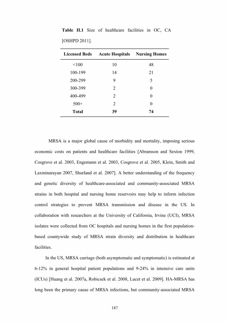
187
Table II.1 Size of healthcare facilities in OC, CA
[OSHPD 2011].
Licensed Beds Acute Hospitals Nursing Homes
<100 10 48
100-199 14 21
200-299 9 5
300-399 2 0
400-499 2 0
500+ 2 0
Total 39 74
MRSA is a major global cause of morbidity and mortality, imposing serious
economic costs on patients and healthcare facilities [Abramson and Sexton 1999,
Cosgrove et al. 2003, Engemann et al. 2003, Cosgrove et al. 2005, Klein, Smith and
Laxminarayan 2007, Shurland et al. 2007]. A better understanding of the frequency
and genetic diversity of healthcare-associated and community-associated MRSA
strains in both hospital and nursing home reservoirs may help to inform infection
control strategies to prevent MRSA transmission and disease in the US. In
collaboration with researchers at the University of California, Irvine (UCI), MRSA
isolates were collected from OC hospitals and nursing homes in the first population-
based countywide study of MRSA strain diversity and distribution in healthcare
facilities.
In the US, MRSA carriage (both asymptomatic and symptomatic) is estimated at
6-12% in general hospital patient populations and 9-24% in intensive care units
(ICUs) [Huang et al. 2007a, Robicsek et al. 2008, Lucet et al. 2009]. HA-MRSA has
long been the primary cause of MRSA infections, but community-associated MRSA
188
(CA-MRSA), which often causes infections among healthy children and young adults
with no exposure to the healthcare setting, is becoming increasingly prevalent. The
first reports of MRSA isolated from patients with no identifiable risk factors came
from Australia and the US in the 1990s [Udo, Pearman and Grubb 1993, Herold et al.
1998, CDC 1999]. Since then CA-MRSA prevalence has rapidly increased, with
reports of CA-MRSA infection from virtually every geographic region of the world
[Tristan et al. 2007a, Wallin, Hern and Frazee 2008]. The incidence of life-threatening
invasive infections owing to CA-MRSA is increasing, and CA-MRSA appears to be
particularly virulent among children [Moellering 2006]. Moreover, CA-MRSA has
caused outbreaks in the hospital setting [O'Brien et al. 1999, Saiman et al. 2003, Bratu
et al. 2005] with some reports suggesting it may be replacing HA-MRSA [Seybold et
al. 2006, Patel et al. 2008, Popovich, Weinstein and Hota 2008, D'Agata et al. 2009].
The predominant community-associated MRSA clone in the US is now USA300
(defined by spa typing and multilocus sequence typing (MLST) as t008 and ST8
respectively), having rapidly disseminated and replaced USA400 (t128/ST1) since its
isolation in 2000. USA300 has several characteristics that may offer a selective
advantage over other MRSA clones, both community-associated (e.g. USA400) and
healthcare-associated (e.g. USA100 (t002/ST5)). These advantages include (i) a
smaller SCCmec element (usually type IV) than those of healthcare-associated strains
(usually SCCmec types I-III), which is more readily transmissible and may be an
advantage in terms of DNA replication speed; (ii) fewer antibiotic resistance genes
than healthcare-associated strains, resulting in a fitness benefit due to the carriage of
smaller or fewer genes; and (iii) a higher growth rate in vitro that may lead to
successful colonization by outcompeting healthcare-associated strains [Okuma et al.
2002, D'Agata et al. 2009]. Furthermore, the linkage of ACME with SCCmec type IV
189
in USA300 likely confers increased fitness and/or pathogenicity [Diep et al. 2008b].
Finally, greater expression of regulatory genes associated with the virulence factors
PVL and α-haemolysin has been shown in USA300 versus USA400 isolates, which
may contribute to the invasiveness of USA300 [Montgomery et al. 2008]. However,
there is evidence that CA-MRSA do not need PVL to cause nosocomial infections
[Regev-Yochay et al. 2005, David et al. 2006a, Gould et al. 2009, Otter and French
2011].
In addition, as CA-MRSA strains move into the healthcare setting and are
exposed to nosocomial antibiotic pressure, they have developed greater antibiotic
resistance. In one US study, USA300 isolates classified as healthcare-associated were
significantly more likely to be ciprofloxacin-resistant than CA-MRSA USA300
isolates [Huang et al. 2006], and another study reported a USA300 isolate with
intermediate vancomycin susceptibility and reduced daptomycin susceptibility from a
hospital in San Francisco in 2007 [Graber et al. 2007]. As CA-MRSA strains continue
to encroach on healthcare MRSA reservoirs, they may come to resemble the antibiotic
resistance profiles of HA-MRSA, as well as behave more like HA-MRSA clinically
[Davis et al. 2006, Benoit et al. 2008, Moore et al. 2009, Otter and French 2011].
While it is not clear if CA-MRSA cause more severe disease in the healthcare setting
and whether they are more transmissible than HA-MRSA, their higher fitness and
growth rate could lead to increasing prevalence in hospitals.
Chapters 5 and 6 of Part 2 of my thesis focus on MRSA in OC hospitals, in
order to determine the frequency of hospital-associated and community-associated
clones, particularly USA300, among adult and paediatric inpatients, and to gain a
better understanding of the nature of the hospital MRSA reservoir by assessing the
diversity and distribution of MRSA across 30 hospitals. This knowledge may better
190
inform infection control strategies employed in hospitals.
Very little is known about the diversity and distribution of MRSA in long-term
care facilities. A recent longitudinal analysis by Tattevin et al. [2009] attributed an
increasing incidence of MRSA infections in a long-term care facility in San Francisco,
CA, to two clonal groups - ST5-MRSA-II and ST8-MRSA-IV. The study highlights
the need for further investigation into the epidemiology of MRSA in long-term care
facilities in order to minimise further MRSA transmission, particularly since such
facilities, long thought to be a reservoir for nosocomial MRSA clones, are now
emerging as an important reservoir for the community associated clone USA300
(ST8-MRSA-IV), and could play a role in the emergence of multidrug-resistant
USA300 [Tattevin et al. 2009]. Chapter 7 therefore assesses the diversity and
distribution of all carriage MRSA collected from residents of 25 nursing homes in
OC, in order to gain a better understanding of the nature of the MRSA reservoir in
these unique facilities.
Note: All work presented in Chapter 5 has been published previously, and co-authors
are listed in Appendix 6 as part of the official citation. Apart from study design and
isolate collection, I performed all work, including laboratory methods and molecular
typing, statistical analyses, and manuscript writing. For Chapters 6 and 7, apart from
study design and isolate collection which were performed by my collaborators at UCI
and the Orange County Public Health Laboratory, I performed all work, including
laboratory methods and molecular typing, statistical analyses, and writing.
191
CHAPTER 5: DIFFERENCES IN MRSA STRAINS ISOLATED FROM PAEDIATRIC AND ADULT HOSPITAL INPATIENTS
5.1 INTRODUCTION
The phenotypic and genotypic differences between HA- and CA-MRSA strains
have been well documented [Ma et al. 2002, Okuma et al. 2002, Eady and Cove 2003,
Diep et al. 2006a, Bassetti, Nicco and Mikulska 2009], yet there are few studies that
have directly explored the differences in MRSA strains isolated from adults and those
isolated from children. Park et al. [2007] previously compared a small number of
adult and paediatric MRSA isolates in a South Korean hospital and found a
predominance of CA-MRSA isolates among children. A better understanding of the
frequency of community- versus healthcare-associated MRSA clones among adults
and children, and in particular the USA300 clone, may inform strategies to prevent
transmission and disease.
Children may have different exposures to MRSA, as they constitute a largely
healthy population that is most likely to incur MRSA infection through skin and soft
tissue injuries related to sports and other play activities [Frei et al. 2010]. This is in
contrast to the chronically and critically ill adult population, which frequents hospitals
and may encounter healthcare-associated MRSA strains more readily. Furthermore,
children may experience different antimicrobial drug selection pressure compared to
that of adults due to differences in common disease syndromes and different guidance
on antibiotic therapy [David et al. 2006b, Park et al. 2007].
Defining the characteristics of MRSA strains in adults and children would
provide insight into the spread of MRSA strains, particularly since there is growing
evidence that community and healthcare MRSA reservoirs are mixing [Kourbatova et
al. 2005, Seybold et al. 2006, Maree et al. 2007, Liu et al. 2008, Popovich, Weinstein
192
and Hota 2008]. Furthermore, few studies of adult or paediatric MRSA strains have
involved a population-based sample of strains. We conducted a prospective cohort
study of inpatients in a large metropolitan county to characterise differences in
paediatric versus adult MRSA strains.
5.2 METHODS
5.2.1 Study
A population-based, prospective collection of clinical isolates of MRSA from
30 hospitals in OC, California, was conducted. This study was approved by the
Institutional Review Board of the University of California Regents.
5.2.2 Isolate collection
Clinical (non-screening) isolates of MRSA from unique adult patients (>18
years of age) and unique paediatric patients (<18 years of age) were collected from
hospital microbiology laboratories. Hospitals were instructed to collect MRSA
isolates from unique patients up to a total of 100 isolates or for a duration of 12
months, whichever came first. In order to have a representative sample of Orange
County MRSA isolates, we limited isolates in this study to those collected for a
uniform duration of time from adult hospitals. Since the largest adult hospitals
reached 100 isolates over a 5-month period, we restricted the period of all adult isolate
collections to 5 months. All paediatric hospitals required a 12-month collection
period. Nearly all adult isolates were collected between December 2008 and April
2009. Paediatric isolates were collected between October 2008 and September 2009.
Isolates from patients not admitted to hospitals were excluded from the study.
Samples were batched and delivered to the Orange County Public Health Laboratory
193
using soy agar slants. For the repeated confirmation of MRSA, isolates were plated on
selective media for MRSA (BD CHROMagar). MRSA strains were stored at −65°C in
15% glycerol Brucella broth.
5.2.3 Specimen data and hospital characteristics
Specimen data, including patient age in years, specimen source (wound, blood,
urine, sputum, or other), specimen location (ICU or non-ICU), and time of specimen
collection with respect to admission date (hospital onset, ≥3 days after admission;
community onset, <3 days after admission), were collected. Hospital characteristics
were obtained from a California hospital dataset [OSHPD 2005], which included
annual admissions, hospital type (acute care versus long-term acute care (LTAC)
facility), percentage of Medicaid-insured patients, and percentage of Hispanic
patients. Population estimates of adults and children in OC were obtained from the
2010 US Census [US Census Bureau 2011b].
5.2.4 Laboratory methods and molecular typing
All strains were shipped to me for spa typing, and stored at −80°C. Cells were
harvested on blood agar plates (Oxoid) and incubated at 37°C overnight. DNA was
extracted using a Qiagen DNeasy Blood & Tissue Kit. DNA samples were eluted in
200µl of elution buffer (10 mM Tris-Cl, 0.5 mM EDTA (pH 9.0)) and stored at
−20°C. Following sequencing of the spa region, spa types were determined using
Ridom StaphType v2.1 (Ridom GmbH) [Harmsen et al. 2003]. To assess spa type
diversity and relatedness, cluster analysis of spa types was performed separately for
adult and paediatric isolates using the Based Upon Repeat Pattern (BURP) algorithm,
a built-in feature of the StaphType software [Mellmann et al. 2007]. MLST and SmaI
194
PFGE were performed on a subset of the isolates (n = 171) to confirm MRSA strain
types, according to methods described previously [Pfaller 1998, Enright et al. 2000].
This subset included one isolate of each spa type and, for the ten most common spa
types, one isolate from each of the hospitals in which these spa types were present.
Isolates were selected using a random number generator. I performed MLST, but
collaborators at the University of Iowa performed PFGE. For PFGE, DNA profiles
were analyzed using BioNumerics software (version 5.0, 2007; Applied Maths).
PFGE types were defined using a similarity coefficient of 78%, and USA100 to
USA800 strains were used as references.
5.2.5 Statistical analyses
Annual adult and paediatric population estimates of hospitalised patients with
clinical MRSA cultures were calculated by spa type, accounting for the duration of
countywide collection. I further calculated the percentage of MRSA strains from adult
versus paediatric patients that were due to the most common spa types (t008, t242,
t002) and compared them using χ2 tests. Specimen data for t008, t242 and t002
isolates were compared using χ2 or Fisher’s exact tests and, for patient age, the
Wilcoxon Mann-Whitney test. Simpson’s index of diversity (1−D) was used to
compare the genetic diversities of MRSA strains among adults and children. 1−D
gives an unbiased measure of the probability of drawing two different spa types given
the distribution of spa types in a sample [Grundmann et al. 2010]. The 95%
confidence intervals (CIs) were calculated as described previously [Grundmann, Hori
and Tanner 2001]. I conducted bivariate tests to evaluate the association of spa type
t008 with individual variables, including age (adult/paediatric), specimen source
(specifically wound and blood), time of specimen collection (community or hospital
195
onset), and ward type (non-ICU/ICU). I also tested hospital-level variables, including
annual admissions (greater or less than 10,000), LTAC facility, percentage of
Hispanic patients, and percentage of Medicaid-insured patients. For multivariate
analyses, variables with a p value of <0.1 were entered into a generalized linear mixed
model clustered by hospital and were retained at an α value of ≤0.05 (xtmelogit,
STATA release 11, StataCorp. 2009).
5.3 RESULTS
A total of 1,124 adult and 159 paediatric MRSA isolates were collected over
the 5- and 12-month periods, respectively. A summary of the characteristics of the
clinical MRSA strains collected is shown in Table 5.1. The median age of adults was
67 years (interquartile range (IQR), 50 to 81 years) and that of children was 2 years
(IQR, 1 to 9 years).
t008, t242 and t002 were the predominant spa types in OC, accounting for
83% of all isolates (Table 5.2). The distribution of these spa types among adults (t008,
41%; t242, 23%; t002, 19%) was significantly different from that among children
(t008, 69%; t242, 9%; t002, 6%) (χ²=52.29, p<0.001). Annual population estimates of
clinical inpatient MRSA cultures were 119/100,000 adults and 22/100,000 children.
Annual estimates by spa type were 48/100,000 adults and 15/100,000 children for
t008, 27/100,000 adults and 2/100,000 children for t242, and 22/100,000 adults and
1/100,000 children for t002.
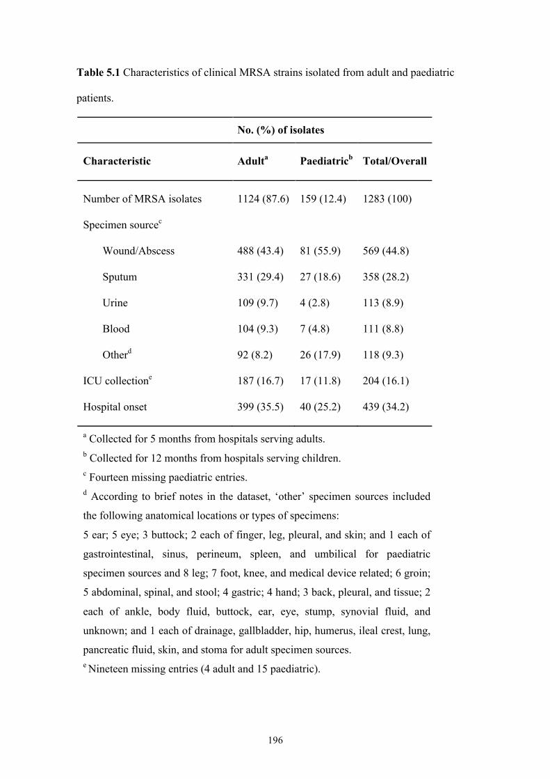
196
Table 5.1 Characteristics of clinical MRSA strains isolated from adult and paediatric
patients.
No. (%) of isolates
Characteristic Adulta Paediatricb Total/Overall
Number of MRSA isolates 1124 (87.6) 159 (12.4) 1283 (100)
Specimen sourcec
Wound/Abscess 488 (43.4) 81 (55.9) 569 (44.8)
Sputum 331 (29.4) 27 (18.6) 358 (28.2)
Urine 109 (9.7) 4 (2.8) 113 (8.9)
Blood 104 (9.3) 7 (4.8) 111 (8.8)
Otherd 92 (8.2) 26 (17.9) 118 (9.3)
ICU collectione 187 (16.7) 17 (11.8) 204 (16.1)
Hospital onset 399 (35.5) 40 (25.2) 439 (34.2)
a Collected for 5 months from hospitals serving adults. b Collected for 12 months from hospitals serving children. c Fourteen missing paediatric entries. d According to brief notes in the dataset, ‘other’ specimen sources included
the following anatomical locations or types of specimens:
5 ear; 5 eye; 3 buttock; 2 each of finger, leg, pleural, and skin; and 1 each of
gastrointestinal, sinus, perineum, spleen, and umbilical for paediatric
specimen sources and 8 leg; 7 foot, knee, and medical device related; 6 groin;
5 abdominal, spinal, and stool; 4 gastric; 4 hand; 3 back, pleural, and tissue; 2
each of ankle, body fluid, buttock, ear, eye, stump, synovial fluid, and
unknown; and 1 each of drainage, gallbladder, hip, humerus, ileal crest, lung,
pancreatic fluid, skin, and stoma for adult specimen sources. e Nineteen missing entries (4 adult and 15 paediatric).
197
According to MLST, the t008 isolates in this study were the prototypic
community clone USA300 (t008/ST8), and the t002 isolates were the prototypic
hospital clone USA100 (t002/ST5), with t242 isolates identified as ST5 (Table 5.2).
Comparison of t242 and t002 isolates for the following parameters revealed no
significant difference: the proportion from each specimen source, the proportion of
hospital and community onset, the proportion collected on ICU and non-ICU wards,
and the age distribution of patients (all p>0.05). Conversely, t008 isolates were
significantly different from t242 and t002 isolates in the same tests (p<0.001). t242
and t002 isolates shared the most common specimen source, sputum (34% and 38%,
respectively), whereas wounds were the most common specimen source of t008
isolates (56%). PFGE of a sample of t242 and t002 isolates showed them to be
predominantly USA100 isolates (data not shown).
BURP analysis of the spa types clustered the majority of adult isolates (97%)
into three spa clonal complexes (spa-CC) and most paediatric isolates (96%) into two
spa-CCs (Figure 5.1). spa types were clustered with either t008 (spa-CC008;
community-associated strains) or t002 (spa-CC002; healthcare-associated strains), but
in adults, a further spa-CC with founder t324 was identified (spa-CC324). Isolates in
this spa-CC were characterised as ST72. For both adult and paediatric MRSA isolates,
MLST results showed that all isolates in spa-CC008 were ST8 and all isolates in spa-
CC002 were either ST5 or a SLV, ST105 (spa types t045, t088 and t1791 for the
latter). According to the BURP algorithm, spa types that differ from all other spa
types in the sample by more than 4 repeats, and thus which cannot be clustered into a
spa-CC, are termed singletons. For adults, 10 (11.2%) spa types (40 (3.6%) isolates)
were classified as singletons, and for children 4 (18.2%) spa types (6 (3.8%) isolates)
were classified as singletons. spa types of less than five repeats in length were
198
excluded from the BURP analysis because no reliable evolutionary history can be
inferred from ‘short’ spa types [Mellmann et al. 2007]. For adults, two (2.2%) spa
types (two (0.2%) isolates) were excluded and for children, one (4.5%) spa type (one
(0.6%) isolate) was excluded. The estimated genetic diversity of MRSA isolates was
significantly higher among adults than among children (1−D = 75% versus 51%)
(Table 5.2).
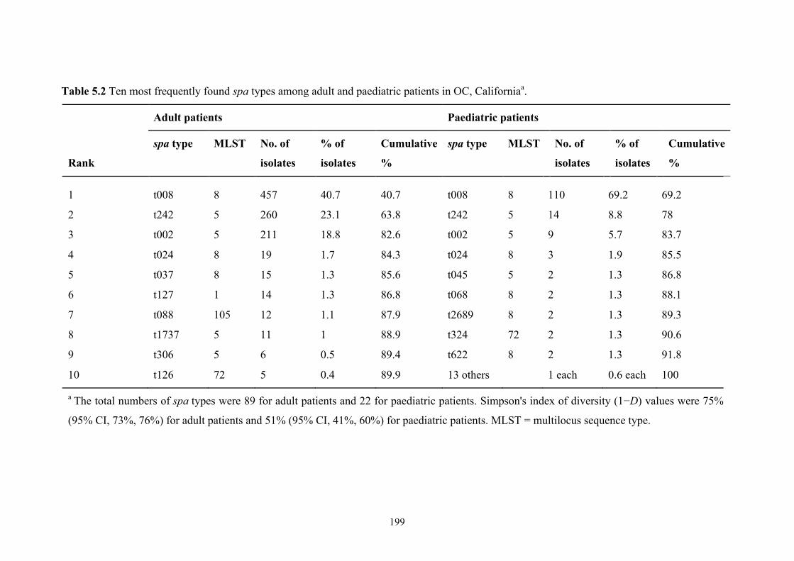
199
Table 5.2 Ten most frequently found spa types among adult and paediatric patients in OC, Californiaa.
Adult patients Paediatric patients
Rank
spa type MLST No. of
isolates
% of
isolates
Cumulative
%
spa type MLST No. of
isolates
% of
isolates
Cumulative
%
1 t008 8 457 40.7 40.7 t008 8 110 69.2 69.2
2 t242 5 260 23.1 63.8 t242 5 14 8.8 78
3 t002 5 211 18.8 82.6 t002 5 9 5.7 83.7
4 t024 8 19 1.7 84.3 t024 8 3 1.9 85.5
5 t037 8 15 1.3 85.6 t045 5 2 1.3 86.8
6 t127 1 14 1.3 86.8 t068 8 2 1.3 88.1
7 t088 105 12 1.1 87.9 t2689 8 2 1.3 89.3
8 t1737 5 11 1 88.9 t324 72 2 1.3 90.6
9 t306 5 6 0.5 89.4 t622 8 2 1.3 91.8
10 t126 72 5 0.4 89.9 13 others 1 each 0.6 each 100
a The total numbers of spa types were 89 for adult patients and 22 for paediatric patients. Simpson's index of diversity (1−D) values were 75%
(95% CI, 73%, 76%) for adult patients and 51% (95% CI, 41%, 60%) for paediatric patients. MLST = multilocus sequence type.
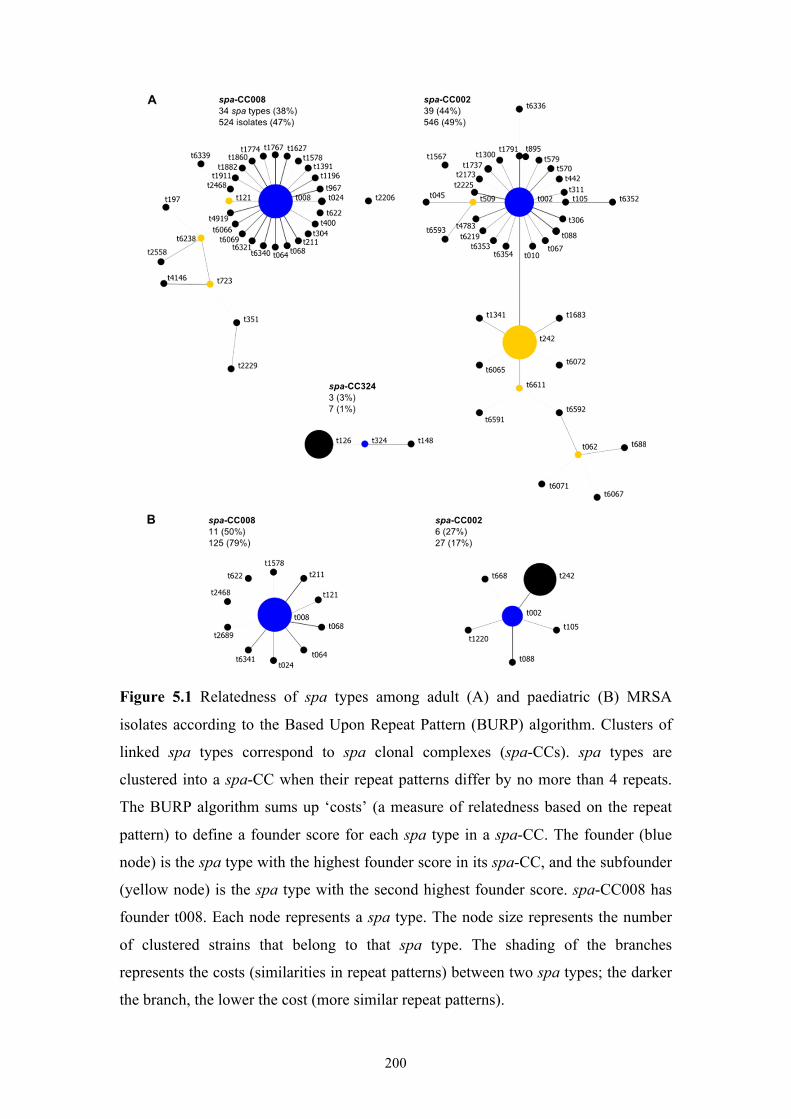
200
Figure 5.1 Relatedness of spa types among adult (A) and paediatric (B) MRSA
isolates according to the Based Upon Repeat Pattern (BURP) algorithm. Clusters of
linked spa types correspond to spa clonal complexes (spa-CCs). spa types are
clustered into a spa-CC when their repeat patterns differ by no more than 4 repeats.
The BURP algorithm sums up ‘costs’ (a measure of relatedness based on the repeat
pattern) to define a founder score for each spa type in a spa-CC. The founder (blue
node) is the spa type with the highest founder score in its spa-CC, and the subfounder
(yellow node) is the spa type with the second highest founder score. spa-CC008 has
founder t008. Each node represents a spa type. The node size represents the number
of clustered strains that belong to that spa type. The shading of the branches
represents the costs (similarities in repeat patterns) between two spa types; the darker
the branch, the lower the cost (more similar repeat patterns).
201
In bivariate analyses, paediatric patients, wound specimens, isolation in a non-
ICU ward, community onset timing of collection, and isolation from a hospital with
>10,000 annual admissions were associated with t008 (USA300) isolates (Table 5.3).
In addition, admission to a hospital with a high proportion of Medicaid-insured
patients or a high proportion of Hispanic patients was linearly associated with the
recovery of t008 isolates. In multivariate analyses, isolates from paediatric patients,
wounds, non-ICU wards, and hospitals with a high proportion of Medicaid-insured
patients remained significantly associated with spa type t008 (Table 5.4). Isolates
from hospitals with a high proportion of Hispanic patients were significantly more
likely to be t008 isolates (20% higher odds of being a spa type t008 isolate per 10%
increase in numbers of Hispanic patients). However, this finding was collinear with
hospitals with a high proportion of Medicaid-insured patients and thus was removed
from the multivariate model.
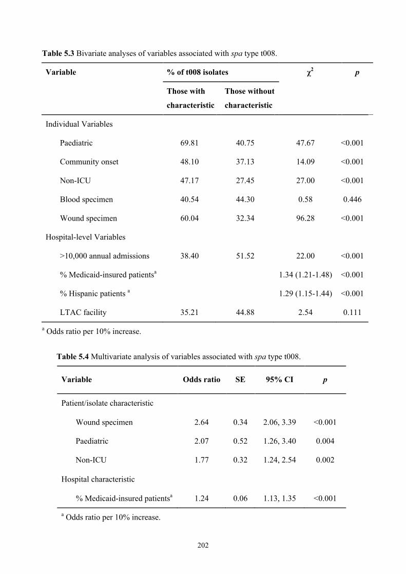
202
Table 5.3 Bivariate analyses of variables associated with spa type t008.
Variable % of t008 isolates χ2 p
Those with
characteristic
Those without
characteristic
Individual Variables
Paediatric 69.81 40.75 47.67 <0.001
Community onset 48.10 37.13 14.09 <0.001
Non-ICU 47.17 27.45 27.00 <0.001
Blood specimen 40.54 44.30 0.58 0.446
Wound specimen 60.04 32.34 96.28 <0.001
Hospital-level Variables
>10,000 annual admissions 38.40 51.52 22.00 <0.001
% Medicaid-insured patientsa 1.34 (1.21-1.48) <0.001
% Hispanic patients a 1.29 (1.15-1.44) <0.001
LTAC facility 35.21 44.88 2.54 0.111
a Odds ratio per 10% increase.
Table 5.4 Multivariate analysis of variables associated with spa type t008.
Variable Odds ratio SE 95% CI p
Patient/isolate characteristic
Wound specimen
Paediatric
Non-ICU
2.64
2.07
1.77
0.34
0.52
0.32
2.06, 3.39
1.26, 3.40
1.24, 2.54
<0.001
0.004
0.002
Hospital characteristic
% Medicaid-insured patientsa 1.24 0.06 1.13, 1.35 <0.001
a Odds ratio per 10% increase.
203
5.4 DISCUSSION
A prospective cohort study of inpatients in a large metropolitan county in
California was conducted, collecting all clinical MRSA isolates from 30 hospitals in
order to characterise differences in paediatric versus adult MRSA strains. To my
knowledge, this is the first study to assess adult and paediatric MRSA isolates from a
population-based sample across a large region.
Countywide, adult and paediatric clinical MRSA isolates were dominated by
three spa types, two of which were consistent with the prototypic community- and
healthcare-associated clones prevalent in the US (t008 (USA300) and t002
(USA100)). t008 (USA300) was the most common single clone among both adult and
paediatric isolates. Nevertheless, t008 comprised a large majority of paediatric
isolates, whereas adult isolates were nearly equally divided among community- and
healthcare-associated clones. Most other spa types were shown by BURP to be related
to these two dominant clones. The two spa clonal complexes spa-CC008 and spa-
CC002 can therefore be thought of as two distinct groups of isolates representing the
major community- and healthcare-associated MRSA strains prevalent in the US.
Interestingly, t242/ST5 was slightly more common than t002/ST5 among both
adult and paediatric isolates, despite the predominance of the t002/ST5 hospital clone
in the US. Given the similarities of t242 and t002 isolates in this study, and the fact
that t242 differs from t002 by only one nucleotide (resulting in a different spa repeat
pattern by one spa repeat), t242/ST5 presumably represents a minor variant of
USA100 that has become prevalent in OC hospitals. t242 has been reported
infrequently in the literature [Kinnevey et al. 2010, Johnson et al. 2007, Weese, Avery
and Reid-Smith 2010], with just one study reporting t242 at an endemic level in an
Italian hospital [Parlato et al. 2009].
204
The additional spa-CC identified among adult isolates included a community-
onset isolate identified as t324/ST72, an invasive community-associated MRSA clone
reported in elderly patients in South Korea from 2006 to 2007, just before our isolate
collection began [Lee et al. 2010]. According to the US Census Bureau, 17.9% of the
OC population is Asian, approximately 2.9% of which is Korean [US Census Bureau
2011b].
There was significantly more genetic diversity among adult MRSA isolates
than among paediatric isolates. This could simply represent the greater time that
healthcare-associated clones have had to diversify at the spa locus than community-
associated clones, which have emerged only in the past two decades. The greater
MRSA diversity among adults could also be due to different degrees of contact; for
example, adults may have more diverse MRSA encounters (travel, work, social
venues, and healthcare facilities) than young children (schools and day care centres).
The population estimates of clinical MRSA isolates in OC show that there was
a 6-fold-higher frequency of inpatient MRSA clinical cultures among adults than
among children. This pattern was consistent among the three most common spa types
t008, t242 and t002, and is likely a combination of more frequent hospitalisations
among adults (many of whom were elderly, with a median age of 67 years) and more
frequent MRSA carriage.
In multivariate analyses, the community-associated MRSA clone t008
(USA300) was associated with paediatric patients. In contrast to adults, children are
often healthier and are more likely to encounter MRSA in the community through
exposure to high-density environments, such as schools, day care centres, camps and
sporting activities, where close contact may facilitate the spread of community MRSA
strains. In agreement with data from previous studies, I found that USA300 was
205
associated with wounds, which is the most common presentation for hospitalization
due to community-acquired MRSA infection [Fridkin et al. 2005, Bassetti, Nicco and
Mikulska 2009]. USA300 was also associated with hospitals that treat a large fraction
of Medicaid-insured patients, suggesting that community MRSA infections may be
more prevalent among patients from economically disadvantaged or high-density
areas.
USA300 was also associated with isolation from non-ICU wards, suggesting
that this community strain is occurring in healthier hosts or is producing infections
that are less severe than those caused by traditional healthcare-associated strains.
Nevertheless, there is ample evidence that community strains are capable of
producing fulminant infections [Frazee et al. 2005, Miller et al. 2005, Seybold et al.
2006]. An understanding of what component of invasiveness is due to host
comorbidities versus pathogen virulence factors is an area of active research.
Interestingly, I did not find that the isolation of t008 was associated with
community onset clinical isolates (clinical culture isolated less than 3 days after
admission). This finding is likely due to the fact that the majority of healthcare-
associated carriage or infection is found on readmission to hospitals [Klevens et al.
2007]. It could also be explained by community-associated strains that have become
endemic in some hospitals [Seybold et al. 2006, Popovich, Weinstein and Hota 2008].
Community- and healthcare-associated MRSA strains are becoming
increasingly difficult to distinguish epidemiologically as community-associated
strains continue to penetrate hospital MRSA reservoirs. Furthermore, it remains
unclear whether community clones are adding to or replacing traditional healthcare
MRSA strains [Bootsma et al. 2006a, Hota et al. 2007, Popovich, Weinstein and Hota
2008, D'Agata et al. 2009]. The implication of the blurred line between community-
206
and healthcare-associated MRSA strains may be that efforts to control MRSA
transmission within hospitals will not be effective in controlling community influx
into hospitals. Simultaneous community strategies to limit MRSA spread are needed.
However, much is still unknown about the acquisition and transmission of CA-
MRSA, so improved knowledge is needed to better guide infection control strategies.
Further studies are needed to ascertain whether community strategies to reduce
transmission in children and young adults would produce benefits across the entire
age spectrum.
One limitation of this study is that few individual-level characteristics were
available. Also, this study did not account for the different policies in place at each
hospital with regard to when to obtain clinical cultures. These differences could affect
MRSA detection at each hospital and, possibly, the type of MRSA strains isolated, if
clinical cultures were more likely to be obtained for sicker, older patients. Moreover,
the results could have been affected by the potential seasonality of MRSA infections
and infection types due to the different collection periods for adult and paediatric
isolates (largely winter and spring for adult collections, compared to all seasons for
paediatric collections). Seasonality of S. aureus infections, particularly skin
infections, has been observed in paediatric and adult patients in temperate and tropical
environments, with a predominance of infections during summer and autumn [Loffeld
et al. 2005, Szczesiul et al. 2007, Van De Griend et al. 2009, Mermel, Machan and
Parenteau 2011]. A recent study in Rhode Island found a two- to three-fold-increased
incidence of MRSA infections (both CA- and HA-MRSA) in paediatric patients
during the second two quarters of the year, over the last decade [Mermel, Machan and
Parenteau 2011]. However, in the same study, adult CA-MRSA infections showed
less seasonal variation than did paediatric infections, and no variation was observed
207
among adult HA-MRSA infections. Some studies observed no significant seasonality
of S. aureus infections, but those studies focused on bacteremia [Morin and Hadler
2001, Perencevich et al. 2008]. The collection of both adult and paediatric MRSA
isolates for the same time period i.e. twelve months, would have accounted for any
potential seasonality effects and/or other factors that could affect the type and
diversity of MRSA strains isolated.
Mandatory screening of high-risk inpatients was not in place in California
until 2009; therefore, my population estimates are likely underestimates. In addition,
my estimates should not be construed as measures of MRSA infection among
inpatients. Clinical isolates often represent carriage without infection. Finally, my
estimates of the index of diversity for adult and paediatric MRSA isolates may have
been influenced by differing sample sizes [Grundmann, Hori and Tanner 2001].
In conclusion, this study found that in a large county, MRSA isolates from
hospitalised children were more likely to be spa type t008 (USA300). This
community-associated spa type was associated with children, wounds, non-ICU care,
and admission to a hospital with a high percentage of Medicaid-insured patients.
Despite the association of t008 isolates with children, t008 was still the most common
spa type among adult patients, suggesting that community-based interventions are
needed to stem the influx of t008 isolates into hospitals. The study also found
evidence for a prevalent variant of the USA100 clone (t242/ST5), which has not been
reported elsewhere. While community- and hospital-associated MRSA reservoirs
have begun to merge, significant differences remain in paediatric versus adult patient
populations, which may provide an impetus for different age-based strategies to
reduce transmission and disease.
208
CHAPTER 6: DIVERSITY OF MRSA STRAINS ISOLATED FROM INPATIENTS OF 30 HOSPITALS
6.1 INTRODUCTION
HA-MRSA has long been the primary cause of MRSA infections, but CA-
MRSA, which often causes infections among healthy children and young adults with
no exposure to the healthcare setting, has become increasingly prevalent across the
globe, particularly in the US [Udo, Pearman and Grubb 1993, Herold et al. 1998,
CDC 1999, Eady and Cove 2003, Mongkolrattanothai et al. 2003, Francis et al. 2005,
Gonzalez et al. 2005a, Miller et al. 2005, Moran et al. 2005, Moellering 2006, Tristan
et al. 2007a, Wallin, Hern and Frazee 2008, Otter and French 2010, Otter and French
2011]. While well documented in the community, there is increasing evidence that
CA-MRSA is penetrating healthcare MRSA reservoirs [O'Brien et al. 1999, Saiman et
al. 2003, Bratu et al. 2005, Kourbatova et al. 2005, David et al. 2006a, Gonzalez et al.
2006, Otter and French 2006, Saunders et al. 2007, Boyce 2008, Otter and French
2008, Sonnevend et al. 2012]. CA-MRSA has caused outbreaks in the hospital setting
since 2003, often in paediatrics and obstetrics where HA-MRSA prevalence is low
and community influx of patients without prior healthcare exposure is common [Otter
and French 2011]. Furthermore, some reports suggest CA-MRSA may be replacing
HA-MRSA [Seybold et al. 2006, Maree et al. 2007, Patel et al. 2008, Popovich,
Weinstein and Hota 2008, D'Agata et al. 2009].
Most prior studies of CA-MRSA penetration into hospital reservoirs involve a
single centre. Regional evaluation of healthcare facilities may provide further
information about the extent of reservoir mixing of CA-MRSA and HA-MRSA
strains across community and academic healthcare facilities, as well as paediatric
hospitals and LTAC facilities. A prospective, population-based study of clinical
209
MRSA isolates across nine medical centers in San Francisco, California, found that
USA300 was the predominant clone in both the community and hospital setting [Liu
et al. 2008]. While this city-based study collected almost 4000 MRSA isolates, only a
fifth of these were selected for molecular analysis, with the primary goal to determine
clonal groupings based upon the isolate collection date (hospital onset or community
onset). Further comprehensive evaluations of the diversity of isolates within and
across clonal complexes will provide valuable information about how exact strain
types are evolving and being shared across facilities. It is also unclear how much
hospital onset disease is caused by CA-MRSA. Given the increasing dominance of
USA300 and the growing evidence that community and healthcare MRSA reservoirs
are mixing [Kourbatova et al. 2005, Seybold et al. 2006, Maree et al. 2007, Liu et al.
2008, Popovich, Weinstein and Hota 2008], a better understanding of the frequency
and diversity of community- and healthcare-associated MRSA clones in hospitals may
inform strategies to prevent MRSA transmission and disease in the US. We conducted
a prospective cohort study of inpatients in a large metropolitan county to investigate
the frequency and genetic diversity of MRSA at a population level.
6.2 METHODS
6.2.1 Study
As described in section 5.2.1.
6.2.2 Isolate collection
Clinical (non-screening) isolates of MRSA from unique patients were
collected from hospital microbiology laboratories by my collaborators, between
October 2008 and April 2010. Hospitals were instructed to collect non-blood MRSA
210
isolates from unique patients up to a total of 100 or for a duration of 12 months,
whichever came first. In addition, hospitals were instructed to collect all blood
isolates from unique patients until collection ended. Isolates from patients not
admitted to hospital were excluded from the study. Samples were then processed and
stored by the Orange County Public Health Laboratory as described in section 5.2.2.
6.2.3 Specimen data and hospital characteristics
Specimen data, hospital characteristics and the OC population estimate were
obtained as described previously (section 5.2.3) [Hudson et al. 2012].
6.2.4 Laboratory methods and molecular typing
All strains were shipped to me for spa typing, and processed as before (section
5.2.4). To assess spa type diversity and relatedness, cluster analysis of spa types was
performed using the Based Upon Repeat Pattern (BURP) algorithm, a built-in feature
of the StaphType software [Mellmann et al. 2007]. MLST was also performed on a
subset of the isolates (n=284) to confirm MRSA strain types, according to methods
described previously [Enright et al. 2000]. This subset was selected as described in
section 5.2.4.
6.2.5 Statistical analyses
I calculated the number of hospitalized patients with MRSA clinical cultures
among both the total population of OC and total annual admissions across all 30
hospitals, accounting for duration of isolate collection within each hospital. The
number of community onset MRSA clinical cultures among the total OC population
was also calculated. χ2 tests were performed to compare isolate characteristics
211
(community onset versus hospital onset, and number of isolates belonging to different
spa-CCs) between hospitals. One-sample z-tests for equality of proportions were
conducted, to compare isolate characteristics (community onset versus hospital onset,
and number of isolates belonging to different spa-CCs) within each hospital.
I also used Simpson’s index of diversity (1−D) to estimate inter- and intra-
hospital genetic diversity of the MRSA strains collected, as well as the genetic
diversity of the two major spa-CCs, and genetic diversity among hospital and
community onset isolates. 1−D gives an unbiased measure of the probability of
drawing two different spa types given the distribution of spa types in a sample
[Grundmann et al. 2010]. 95% confidence intervals (CIs) were calculated as described
previously [Grundmann, Hori and Tanner 2001]. For comparison of diversity indices,
a significant difference (p<0.05) was determined by non-overlapping 95% CIs.
I computed Pearson's correlation coefficients to determine the relationship
between hospital-level and isolate variables, and genetic diversity. Due to the small
sample size (28 hospitals; two were excluded as they collected <10 MRSA isolates
and thus their diversity estimates were unreliable) and the number of potential
predictor variables for genetic diversity, variables were considered for entry into a
bootstrapped multiple linear regression model based on a combination of their
correlation coefficient and current knowledge regarding their association with MRSA.
Only variables with p<0.1 in correlation tests were considered for the exploratory
model. I also tested the correlation between community onset and spa-CC008 isolates.
All statistical tests were performed using STATA (release 11, StataCorp 2009).
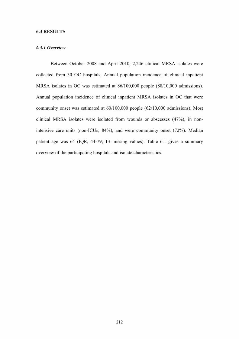
212
6.3 RESULTS
6.3.1 Overview
Between October 2008 and April 2010, 2,246 clinical MRSA isolates were
collected from 30 OC hospitals. Annual population incidence of clinical inpatient
MRSA isolates in OC was estimated at 86/100,000 people (88/10,000 admissions).
Annual population incidence of clinical inpatient MRSA isolates in OC that were
community onset was estimated at 60/100,000 people (62/10,000 admissions). Most
clinical MRSA isolates were isolated from wounds or abscesses (47%), in non-
intensive care units (non-ICUs; 84%), and were community onset (72%). Median
patient age was 64 (IQR, 44-79; 13 missing values). Table 6.1 gives a summary
overview of the participating hospitals and isolate characteristics.
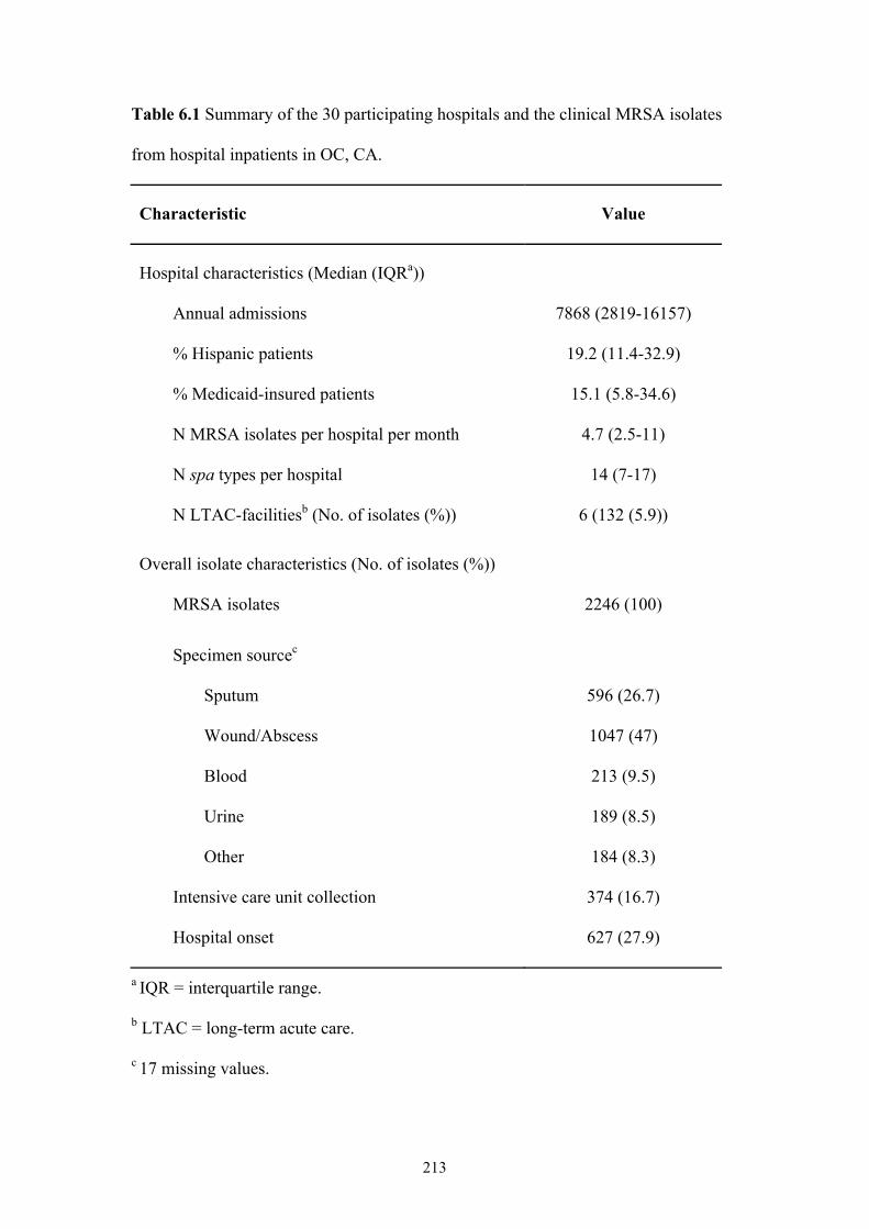
213
Table 6.1 Summary of the 30 participating hospitals and the clinical MRSA isolates
from hospital inpatients in OC, CA.
Characteristic Value
Hospital characteristics (Median (IQRa))
Annual admissions 7868 (2819-16157)
% Hispanic patients 19.2 (11.4-32.9)
% Medicaid-insured patients 15.1 (5.8-34.6)
N MRSA isolates per hospital per month 4.7 (2.5-11)
N spa types per hospital 14 (7-17)
N LTAC-facilitiesb (No. of isolates (%)) 6 (132 (5.9))
Overall isolate characteristics (No. of isolates (%))
MRSA isolates 2246 (100)
Specimen sourcec
Sputum 596 (26.7)
Wound/Abscess 1047 (47)
Blood 213 (9.5)
Urine 189 (8.5)
Other 184 (8.3)
Intensive care unit collection 374 (16.7)
Hospital onset 627 (27.9)
a IQR = interquartile range.
b LTAC = long-term acute care.
c 17 missing values.
214
6.3.2 spa typing and MLST
Among the 2,246 MRSA isolates collected, 134 spa types were identified,
including one non-typeable (NT) isolate and 28 spa types (1.6% of all isolates) that
did not match any known spa sequence. These novel spa sequences were
automatically submitted to the Ridom SpaServer via the Ridom StaphType software
and were assigned new spa types. The isolate with the NT spa type was re-tested to
confirm the result was not due to a processing error, and the sequence quality was
deemed excellent by the StaphType software. The NT spa type bore closest
resemblance to t008, with a missing nucleotide in the ninth repeat, making the repeat
23-bp long. This is surprising since it would put the spa coding region out of frame
although others have reported spa repeats with an unexpected length [Rothganger
2010]. The NT spa type was submitted to Ridom for their records. The three most
common spa types were t008, t242 and t002, representing 83% of all isolates
collected (Table 6.2).
BURP analysis of the spa types clustered 96% of isolates into two large spa-
CCs and 1.2% of isolates into six smaller spa-CCs (Figure 6.1). 78% of spa types
were clustered into either spa-CC242 (founder t242) or spa-CC008 (founder t008),
including 18 and 8 novel spa types, respectively. Under the BURP algorithm, spa
types that differ from all other spa types in the sample by more than 4 repeats cannot
reasonably be clustered into a spa-CC, and are termed singletons. Nine spa types (56
isolates) were classed as singletons, including two novel spa types. Six isolates
represented six spa types that were less than five repeats in length and were excluded
from BURP analysis because no reliable evolutionary history can be inferred from
‘short’ spa types [Mellmann et al. 2007]. The NT isolate could not be included in the
215
BURP algorithm. Estimated genetic diversity of MRSA in OC hospitals using spa
typing was high, at 72% (Table 6.2).
Table 6.2 Ten most frequently found spa types among isolates from OC hospital
inpatientsa.
Rank spa type MLST Freq % Cumulative %
1 t008 8 1034 46 46
2 t242 5 478 21.3 67.3
3 t002 5 347 15.4 82.8
4 t024 8 33 1.5 84.2
5 t037 8 25 1.1 85.4
6 t045 5 22 1.0 86.3
7 t088 105 21 0.9 87.3
8 t127 474b 18 0.8 88.1
9 t306 5 14 0.6 88.7
10 t1737 5 12 0.5 89.2
- Other - 242 10.8 100.0
a The total number of spa types was 134, including one non-typeable isolate.
Simpson's index of diversity (1−D) value was 72% (95% CI, 70%, 73%).
MLST = multilocus sequence type.
b t127 isolates were also ST1 and ST1900, both SLVs of ST474.
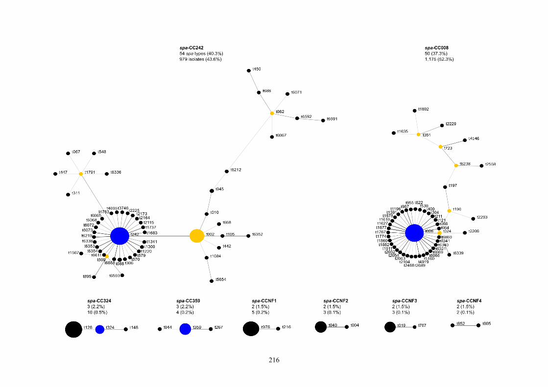
216
217
Figure 6.1 Relatedness of spa types among hospital MRSA isolates according to the Based Upon Repeat Pattern (BURP) algorithm. Clusters of
linked spa types correspond to spa clonal complexes (spa-CCs). spa types are clustered into a spa-CC when their repeat patterns differ by no
more than 4 repeats. The BURP algorithm sums up ‘costs’ (a measure of relatedness based on the repeat pattern) to define a founder score for
each spa type in a spa-CC. The founder (blue node) is the spa type with the highest founder score in its spa-CC, and the subfounder (yellow
node) is the spa type with the second highest founder score. spa-CC008 has founder t008, and spa-CCNF refers to a spa-CC with no founder.
Each node represents a spa type. The node size represents the number of clustered strains that belong to that spa type. The shading of the
branches represents the ‘costs’ (similarities in repeat patterns) between two spa types; the darker the branch, the lower the cost (more similar
repeat patterns).
218
To confirm strain types, 284 isolates were selected for MLST. Among the 23
unique sequence types (STs) identified, ST5 (45%), ST8 (38%) and ST105 (4%) were
the three most common, with the vast majority of isolates (90%) belonging to one of
two major MLST CCs: CC5 (50%; four STs) and CC8 (40%; three STs) (Table 6.3).
The remaining 10% of isolates comprised sporadic incidences of both HA- and CA-
MRSA clones, but mostly the latter (7.4%). According to MLST, t008 isolates were
the prototypic community clone USA300 (t008/ST8) and t002 isolates were the
prototypic hospital clone USA100 (t002/ST5), with t242 isolates identified as ST5
(Tables 6.2 and 6.3). spa type t242 differs from t002 by one spa repeat, as a result of a
single nucleotide difference. The non-typeable spa isolate was ST8, with 64% of the
novel spa types being ST5 and 36% ST8.
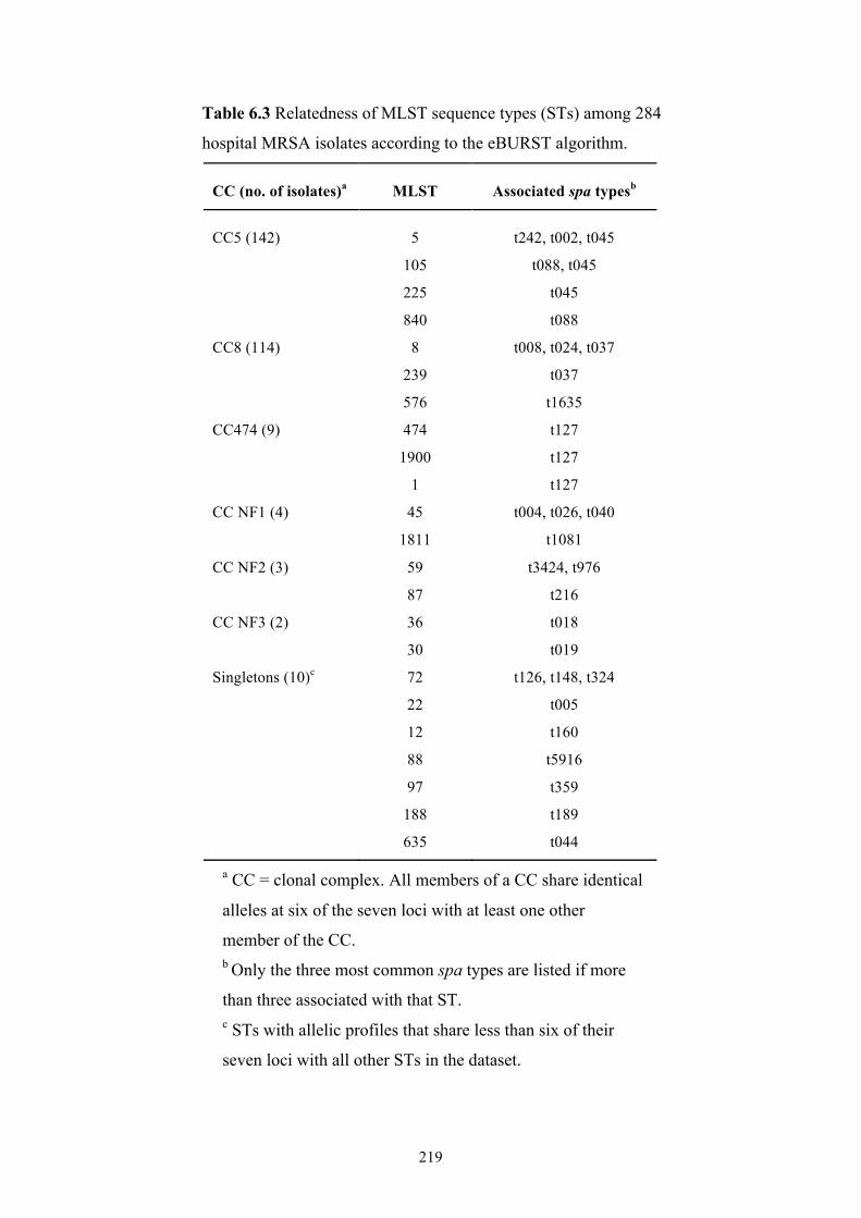
219
Table 6.3 Relatedness of MLST sequence types (STs) among 284
hospital MRSA isolates according to the eBURST algorithm.
CC (no. of isolates)a MLST Associated spa typesb
CC5 (142) 5 t242, t002, t045
105 t088, t045
225 t045
840 t088
CC8 (114) 8 t008, t024, t037
239 t037
576 t1635
CC474 (9) 474 t127
1900 t127
1 t127
CC NF1 (4) 45 t004, t026, t040
1811 t1081
CC NF2 (3) 59 t3424, t976
87 t216
CC NF3 (2) 36 t018
30 t019
Singletons (10)c 72 t126, t148, t324
22 t005
12 t160
88 t5916
97 t359
188 t189
635 t044
a CC = clonal complex. All members of a CC share identical
alleles at six of the seven loci with at least one other
member of the CC. b Only the three most common spa types are listed if more
than three associated with that ST. c STs with allelic profiles that share less than six of their
seven loci with all other STs in the dataset.
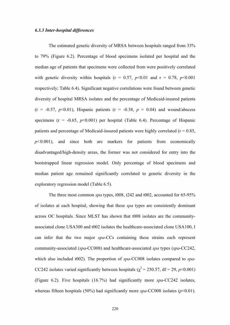
220
6.3.3 Inter-hospital differences
The estimated genetic diversity of MRSA between hospitals ranged from 33%
to 79% (Figure 6.2). Percentage of blood specimens isolated per hospital and the
median age of patients that specimens were collected from were positively correlated
with genetic diversity within hospitals (r = 0.57, p<0.01 and r = 0.78, p<0.001
respectively; Table 6.4). Significant negative correlations were found between genetic
diversity of hospital MRSA isolates and the percentage of Medicaid-insured patients
(r = -0.57, p<0.01), Hispanic patients (r = -0.38, p = 0.04) and wound/abscess
specimens (r = -0.65, p<0.001) per hospital (Table 6.4). Percentage of Hispanic
patients and percentage of Medicaid-insured patients were highly correlated (r = 0.85,
p<0.001), and since both are markers for patients from economically
disadvantaged/high-density areas, the former was not considered for entry into the
bootstrapped linear regression model. Only percentage of blood specimens and
median patient age remained significantly correlated to genetic diversity in the
exploratory regression model (Table 6.5).
The three most common spa types, t008, t242 and t002, accounted for 65-95%
of isolates at each hospital, showing that these spa types are consistently dominant
across OC hospitals. Since MLST has shown that t008 isolates are the community-
associated clone USA300 and t002 isolates the healthcare-associated clone USA100, I
can infer that the two major spa-CCs containing these strains each represent
community-associated (spa-CC008) and healthcare-associated spa types (spa-CC242,
which also included t002). The proportion of spa-CC008 isolates compared to spa-
CC242 isolates varied significantly between hospitals (χ2 = 250.57, df = 29, p<0.001)
(Figure 6.2). Five hospitals (16.7%) had significantly more spa-CC242 isolates,
whereas fifteen hospitals (50%) had significantly more spa-CC008 isolates (p<0.01).
221
Diversity of spa types among spa-CC008 (1−D = 22% (95% CI, 19-26%)) was
significantly lower than diversity among spa-CC242 (1−D = 63% (95% CI, 61-66%)).
6.3.4 Community onset versus hospital onset MRSA
MRSA spa type genetic diversity was significantly higher among hospital
onset isolates (1−D = 75% (95% CI, 73-77%)) than among community onset isolates
(1−D = 70% (95% CI, 68-72%)). The proportion of community onset and hospital
onset isolates was also significantly different between hospitals (χ2 = 127.4, df = 29,
p<0.001); all but two hospitals isolated significantly more community onset MRSA
(p<0.001), with seven hospitals only isolating community onset MRSA. The
remaining two hospitals showed no significant difference in the proportions of
community onset and hospital onset MRSA (p>0.01). No significant correlation was
found between the proportions of community onset isolates per hospital and the
proportions of spa-CC008 isolates per hospital (r = 0.08, p = 0.69). Among hospital
onset isolates, 42.3% belonged to spa-CC008 (18.2-100% per hospital, ignoring
community onset only hospitals), while 39.4% of community onset isolates were spa-
CC242 (6.9%-82.9% per hospital).
222
Figure 6.2 Proportion of isolates belonging to spa-CC242 versus spa-CC008, by hospital. *indicates a significant
difference at the 99% level in the proportion of isolates belonging to spa-CC242 and spa-CC008 at that hospital. The
black bars show the point estimates and 95% confidence intervals of hospital-specific genetic diversity expressed as
Simpson's index of diversity (1−D) of spa types (as a percentage). Diversity indices for hospitals 11 and 13 were excluded
from the figure as these hospitals had spa type data on less than ten isolates. Diversity indices with non-overlapping 95%
CIs were considered significantly different (p<0.05).
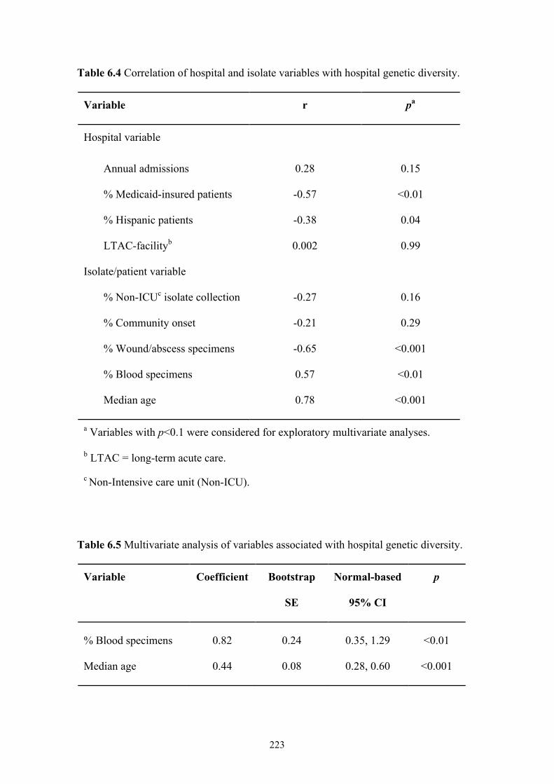
223
Table 6.4 Correlation of hospital and isolate variables with hospital genetic diversity.
Variable r pa
Hospital variable
Annual admissions 0.28 0.15
% Medicaid-insured patients -0.57 <0.01
% Hispanic patients -0.38 0.04
LTAC-facilityb 0.002 0.99
Isolate/patient variable
% Non-ICUc isolate collection -0.27 0.16
% Community onset -0.21 0.29
% Wound/abscess specimens -0.65 <0.001
% Blood specimens 0.57 <0.01
Median age 0.78 <0.001
a Variables with p<0.1 were considered for exploratory multivariate analyses.
b LTAC = long-term acute care.
c Non-Intensive care unit (Non-ICU).
Table 6.5 Multivariate analysis of variables associated with hospital genetic diversity.
Variable Coefficient Bootstrap
SE
Normal-based
95% CI
p
% Blood specimens 0.82 0.24 0.35, 1.29 <0.01
Median age 0.44 0.08 0.28, 0.60 <0.001
224
6.4 DISCUSSION
A prospective cohort study of inpatients in a large metropolitan county was
conducted, collecting all clinical MRSA isolates from 30 hospitals in order to
investigate the frequency and genetic diversity of MRSA at a population level. To my
knowledge, this is the first study to assess MRSA isolates from a population-based
sample across a large region. While Liu et al. [2008] conducted a large population-
based study of clinical MRSA isolates in both hospital inpatients and outpatients, they
sampled from a single city and characterized only 20% of all MRSA isolates
collected. Our countywide study was more comprehensive, encompassing 30
hospitals and characterizing all inpatient clinical MRSA isolates (over 2000).
Countywide, three spa types dominated clinical MRSA isolates. USA300
(t008/ST8), the prototypic community-associated clone prevalent in the US, was the
most common clone, making up just under half of all clinical MRSA isolates.
USA100 (t002/ST5), the prototypic healthcare-associated clone, was also common,
but interestingly, t242/ST5 isolates were slightly more common than t002/ST5
isolates. Given the clinical similarities of t242 and t002 isolates found in our previous
study (Chapter 5) [Hudson et al. 2012], and that pulsed field gel electrophoresis
showed a sample of t242 and t002 isolates to be predominantly USA100, t242/ST5
likely represents a minor variant of USA100 that has become prevalent in OC
hospitals (Chapter 5) [Hudson et al. 2012]. t242 has been reported sporadically
elsewhere, but was endemic in one hospital in Italy [Kinnevey et al. 2010, Parlato et
al. 2009, Johnson et al. 2007, Weese, Avery and Reid-Smith 2010].
Most spa types were closely related to either the USA300 or USA100 clone,
creating two spa-CCs each representing CA- and HA-MRSA strains. The remaining
unrelated spa types were clustered into six small spa-CCs representing several
225
community- and healthcare-associated clones, but occurred only sporadically. Four of
the six smaller spa-CCs were community-associated. The largest represented ST72,
an invasive community-associated clone that was reported in elderly patients in South
Korea just before our isolate collection began [Lee et al. 2010]. According to the US
Census Bureau, 17.9% of the OC population is Asian of which approximately 2.9%
are Korean [US Census Bureau 2011b]. ST72 strains belong to the USA700 clone and
have also been reported in Australia and Europe [Monecke et al. 2011]. The other
three community-associated spa-CCs represented clones including USA1000 (ST59),
USA1100 (ST30/ Southwest Pacific clone) and a rare CA-MRSA clone (ST97) only
reported once before in the US [Chung et al. 2004] and recently as a clone transmitted
among neonates [Udo et al. 2011], although this clone was isolated from two adults in
this study.
The remaining two small spa-CCs represented the hospital-associated clones
USA600 (ST45/Berlin clone) and the pandemic HA-MRSA clone EMRSA-15
(ST22), however the latter has recently been reported in the community setting
[Mollaghan et al. 2010]. Among the few isolates not belonging to a spa-CC was the
HA-MRSA clone USA200 (ST36/EMRSA-16), isolates representing a pandemic HA-
MRSA clone (ST239) and isolates representing strains of MLST CC1, a CA-MRSA
lineage that includes USA400. Most isolates of this latter group were spa type t127
and ST474, a SLV of ST1. ST1/t127 is one of the most common CA-MRSA strains in
the UK [Otter et al. 2009], but to my knowledge has not been reported in the US
previously. While there is MRSA diversity in the OC population, USA300 and
USA100 continue to dominate, with most diversity caused by their close spa-type
relatives.
226
Overall genetic diversity of MRSA in OC was relatively high, but
heterogeneous between hospitals. This variation in diversity was mostly non-
significant, with all hospitals dominated by the three most common spa types t008,
t242 and t002. Only one regional study of spa type diversity has been performed
previously, in Europe. This study found that spa type genetic diversity of MRSA
causing invasive infections is much higher in Europe (94%), ranging from 62% to
91% between countries, indicating the presence of less dominant MRSA spa types
than in OC [Grundmann et al. 2010]. Invasive infections are traditionally caused by
HA-MRSA, which could explain the higher diversity seen in the European study.
However, HA-MRSA in OC still exhibited low diversity (63%) in comparison.
Diversity was significantly lower among spa-CC008 isolates than spa-CC242
isolates, indicating overall MRSA diversity in OC is driven by HA-MRSA, perhaps
simply due to the greater time healthcare-associated strains have had to diversify
compared to community-associated strains. Genetic diversity was significantly
associated with older patient age and isolation of MRSA from blood specimens. HA-
MRSA are typically associated with older patients, whereas CA-MRSA are associated
with children and young adults (Chapter 5) [Hudson et al. 2012]. Blood infection is
more commonly associated with hospital onset MRSA, which are more likely to be
healthcare-associated strains.
Genetic diversity was also significantly lower among community onset MRSA
than hospital onset MRSA, but this difference was marginal. Furthermore, the lack of
correlation between spa-CC008 isolates (CA-MRSA) and community onset suggests
that isolation of a community-associated strain does not imply community onset.
Indeed, among community onset isolates (obtained within the first two days of
hospitalization), large numbers of HA-MRSA strains were found, and, conversely,
227
among hospital onset isolates, there were large numbers of CA-MRSA strains. In fact,
nearly half of hospital onset isolates were CA-MRSA strains. This suggests full
mixing of CA-MRSA and HA-MRSA reservoirs among the majority of hospitals in
OC.
The vast majority of MRSA isolates were obtained within the first two days of
hospitalization, suggesting that MRSA hospital reservoirs are mainly maintained by
importation. Over a third of all community onset isolates were spa-CC242, which
could partly be explained by the fact that healthcare-associated carriage or infection is
often found on readmission to hospitals [Tacconelli et al. 2004, Klevens et al. 2007].
A history of healthcare exposure however does not exclude the possibility of MRSA
acquisition and onset in the community [Klevens et al. 2007].
The high penetration of CA-MRSA among hospital-onset isolates highlights
the needs for community-based strategies to be implemented in an effort to address
the MRSA epidemic in the community and minimize the ability of community-
associated MRSA strains to become endemic in hospitals. The consequences of CA-
MRSA continuing to infiltrate the healthcare setting include 1) the emergence of
multidrug resistant CA-MRSA due to nosocomial antibiotic pressure [Kardas-Sloma
et al. 2011], 2) the potential of increased virulence of healthcare-associated infections
due to PVL-positive CA-MRSA strains, although studies suggest CA-MRSA are
clinically similar to HA-MRSA once in the healthcare setting [Moore et al. 2009], and
3) the risk of hospital outbreaks due to the influx of CA-MRSA from the ever-
expanding community reservoir [D'Agata et al. 2009, Skov and Jensen 2009, Kardas-
Sloma et al. 2011].
A limitation of this study was that few individual level characteristics were
available. Also, variation among hospitals in obtaining clinical cultures could not be
228
accounted for. Screening cultures were excluded since mandatory screening of high-
risk inpatients was not in place in California until 2009 and capture would have been
inconsistent across facilities. Therefore, my population estimate of MRSA isolates
from hospital inpatients is likely an underestimate. In addition, my estimate should
not be construed as a measure of MRSA infection among inpatients. Clinical isolates
often represent carriage without infection. Finally, my estimates of the indices of
diversity for hospital onset and community onset MRSA isolates may have been
influenced by differing sample sizes [Grundmann, Hori and Tanner 2001].
In conclusion this study found that in a large county, CA-MRSA strains
accounted for 56% of community onset isolates and 42% of hospital onset isolates.
No correlation was found between community onset isolates and CA-MRSA,
providing strong regional evidence that community and healthcare MRSA reservoirs
have fully mixed. Genetic diversity of MRSA was still driven by HA-MRSA, with a
highly prevalent, previously unreported USA100 variant found across all OC
hospitals. Community-based MRSA strategies are needed to stem the influx of
community-associated strains, particularly USA300, into the healthcare setting.
229
CHAPTER 7: DIVERSITY OF MRSA STRAINS ISOLATED FROM RESIDENTS OF 25 NURSING HOMES
7.1 INTRODUCTION
Residence in a nursing home, which typically provides long-term care for
chronically ill and/or elderly people, is a well-established risk factor for MRSA
carriage and infection [Hsu et al. 1988, Bradley 1997, O'Sullivan and Keane 2000,
Eveillard et al. 2008], and MRSA carriage in nursing home residents is associated
with increased mortality [Suetens et al. 2006]. Nursing homes represent a unique and
important MRSA reservoir. People colonized with MRSA tend to introduce the
organism into nursing homes via the hospital setting, and MRSA can also be
transported back into hospitals and the community from the nursing home. The
reservoir represented by colonized patients is often large due to the high MRSA
prevalence in nursing homes, sometimes higher than 30%, which increases the risk of
MRSA transmission in these facilities [Eveillard et al. 2008, Eveillard and Joly-
Guillou 2009, Li, Arnsberger and Miller 2010]. Furthermore, once colonized, nursing
home residents seem to carry the same MRSA strain for prolonged periods of time;
asymptomatic colonization has been reported to last anything from 3 months to 3
years [Bradley et al. 1991, Sanford et al. 1994]. Studies suggest that multiple strains,
not a single strain, circulate within nursing homes [Bradley et al. 1991, Fraise et al.
1997, Eveillard et al. 2008].
Not only does the complex operational structure of nursing homes, that act as
both a healthcare setting and a resident's home, make it difficult for standard MRSA
control practices to be implemented in these facilities, but a standardized MRSA
control strategy for nursing homes is yet to be agreed on, largely due to the lack of
studies aimed at identifying appropriate strategies [Hughes, Smith and Tunney 2008].
230
There is also a general dearth of studies, particularly regional ones, investigating the
makeup of the nursing home MRSA reservoir. A study of 60 nursing homes in
Belgium identified hospital care, co-morbidities and a lack of coordinated MRSA
surveillance and control activities as risk factors for MRSA carriage in nursing home
residents [Denis et al. 2009a]. It also found that the predominant MRSA strains
among nursing home residents were identical to those found in hospital inpatients,
highlighting the need for synergistic infection control between nursing homes and
hospitals [Denis et al. 2009a]. A better understanding of the frequency and diversity
of nursing home MRSA strains and predictors thereof, will help to form strategies for
minimizing MRSA transmission and infection in nursing homes, and thus reduce the
impact of the nursing home MRSA reservoir on hospitals.
Assessing the extent to which CA-MRSA has penetrated the nursing home
reservoir is also of interest. CA-MRSA has become increasingly dominant in recent
years, and USA300 in particular has several characteristics that may offer a selective
advantage over HA-MRSA, including higher transmissibility and increased
pathogenicity [Okuma et al. 2002, Diep et al. 2008b]. There is also growing evidence
that community and healthcare reservoirs are mixing [Kourbatova et al. 2005,
Seybold et al. 2006, Maree et al. 2007, Liu et al. 2008, Popovich, Weinstein and Hota
2008] (Chapter 6).
We conducted a prospective study of MRSA isolates in nursing home
residents in a large metropolitan county to investigate the frequency and genetic
diversity of MRSA in these facilities, and thus gain a better understanding of the
nature of the nursing home MRSA reservoir.
231
7.2 METHODS
7.2.1 Study
A population-based prospective study of carriage (symptomatic and
asymptomatic) isolates of MRSA from 26 nursing homes in OC, California, was
conducted. This study was approved by the Institutional Review Board of the
University of California Regents.
7.2.2 Isolate collection
Carriage isolates of MRSA from unique residents were collected from
participating nursing homes between January 2009 and April 2011. Each nursing
home was instructed to swab the nares of 100 consecutive residents upon admission
(within three days of arrival), and 100 residents on a single day (point prevalence
screening), using bilateral nares swabs (BD Culture Swabs, Fisher Scientific). For
nursing homes with a low bed turnover, fewer residents were screened (30-50). For
nursing homes with an average length of stay in years, admission screening was not
performed. Swabs were cultured for MRSA using selective media (BD CHROMagar).
MRSA strains were stored at −65°C in 15% glycerol Brucella broth.
7.2.3 Specimen data and nursing home characteristics
Specimen data including swab type (admission or point prevalence), swab day
since admission, room type (shared or single resident room) and whether the swabbed
resident had prior MRSA, were collected. Demographic and co-morbidity data for
participating nursing homes were derived from the Centers for Medicare and
Medicaid Services (CMS) Long Term Care Minimum Data Set for 2009 [CMS 2009],
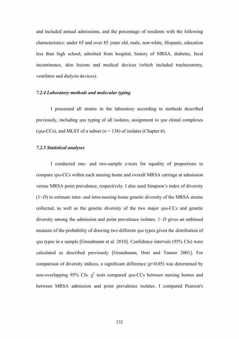
232
and included annual admissions, and the percentage of residents with the following
characteristics: under 65 and over 85 years old, male, non-white, Hispanic, education
less than high school, admitted from hospital, history of MRSA, diabetes, fecal
incontinence, skin lesions and medical devices (which included tracheostomy,
ventilator and dialysis devices).
7.2.4 Laboratory methods and molecular typing
I processed all strains in the laboratory according to methods described
previously, including spa typing of all isolates, assignment to spa clonal complexes
(spa-CCs), and MLST of a subset (n = 138) of isolates (Chapter 6).
7.2.5 Statistical analyses
I conducted one- and two-sample z-tests for equality of proportions to
compare spa-CCs within each nursing home and overall MRSA carriage at admission
versus MRSA point prevalence, respectively. I also used Simpson’s index of diversity
(1−D) to estimate inter- and intra-nursing home genetic diversity of the MRSA strains
collected, as well as the genetic diversity of the two major spa-CCs and genetic
diversity among the admission and point prevalence isolates. 1−D gives an unbiased
measure of the probability of drawing two different spa types given the distribution of
spa types in a sample [Grundmann et al. 2010]. Confidence intervals (95% CIs) were
calculated as described previously [Grundmann, Hori and Tanner 2001]. For
comparison of diversity indices, a significant difference (p<0.05) was determined by
non-overlapping 95% CIs. χ2 tests compared spa-CCs between nursing homes and
between MRSA admission and point prevalence isolates. I computed Pearson's
233
correlation coefficients to determine the relationship between nursing home and
isolate variables, and genetic diversity.
Due to the small sample size of nursing homes (21, since one nursing home
did not isolate any MRSA and four were excluded in this analysis as they collected
<10 MRSA isolates and thus their diversity estimates were unreliable) and the large
number of potential predictor variables for genetic diversity, I considered variables for
entry into a bootstrapped multiple linear regression model based on a combination of
their correlation coefficient and current knowledge regarding their association with
MRSA. Only variables with p<0.1 in correlation tests were considered for the
exploratory model. All statistical tests were performed using STATA (release 11,
StataCorp 2009).
7.3 RESULTS
7.3.1 Overview
Between January 2009 and April 2011, 3,806 nasal swabs were taken from
residents of 26 OC nursing homes either on admission or for estimating MRSA point
prevalence. Of these, 837 swabs (22%) isolated MRSA. One nursing home did not
isolate any MRSA. Overall admission prevalence was 16%, and point prevalence was
significantly higher at 27% (p<0.001). The majority of the 837 MRSA isolates were
from point prevalence testing (68%), from residents with no prior history of MRSA
(76%), and from residents sharing a room (95%). Median swab day since admission
was 53 (IQR, 4-265). A third of all admissions swabs were collected at day 4 since
some nursing homes could not swab earlier. Table 7.1 gives a summary overview of
the 25 nursing homes and the characteristics of their MRSA isolates.
234
Table 7.1 Summary of the 25 nursing homes and 837 MRSA carriage isolates
from nursing home residents in OC, CA.
Characteristic Value
Nursing home characteristics Median (IQRa)
Annual admissions 264 (144-520)
% Residents under 65 years old 20 (4-39)
% Male 40.8 (31.6-48.2)
% Education less than high school 23.8 (7.4-30.3)
% Hispanic residents 11.9 (3.7-23)
% Non-white residents 15 (7.8-21.7)
% Residents admitted from hospital 82.2 (58.5-93.8)
% Diabetes 27.1 (23.4-42.1)
% Fecal incontinence 43.8 (29.2-54.8)
% Skin lesions 72.7 (50.7-86.5)
% Devices 2.2 (1.4-7.1)
% Residents with MRSA history 12 (6-19)
MRSA admission prevalence 16 (10.2-22)
MRSA point prevalence 26.7 (19-34)
N spa types per nursing home 5 (4-8)
Overall MRSA isolate characteristics No. of isolates (%)
MRSA isolates 837 (100)
Admissions swab 269 (32.1)
Resident had prior MRSA 201 (24.0)
Resident shared room 795 (95.0)
a IQR = interquartile range.
235
7.3.2 spa typing and MLST
Of the 837 MRSA isolates collected, 835 were spa typed. Two isolates could
not be spa typed as one did not grow upon culturing and a spa PCR product was not
obtained from the other. Among the 835 MRSA isolates, 60 spa types were identified,
including nine novel spa types (1.4% of all isolates) with hitherto unknown spa repeat
sequences. One isolate that was non-typeable (NT) by spa typing was identical to spa
type t002, except for two extra nucleotides in the third repeat, making the repeat 26-
bp long and putting the spa coding region out of frame. A clinical MRSA isolate from
an OC hospital inpatient was similarly NT, and others have reported spa repeats of
unexpected length (see also Chapter 6) [Rothganger 2010]. The three most common
spa types were t242, t008 and t002, representing 83% of all isolates collected (Table
7.2).
BURP analysis of the spa types clustered 94% of isolates into two large spa-
CCs and 3% of isolates into two smaller spa-CCs (Figure 7.1). Half of all spa types
were clustered into spa-CC002 (predicted founder t002) and 20% into spa-CC008
(founder t008), including six and one novel spa type(s), respectively. Under the
BURP algorithm, singletons are spa types that differ from all other spa types in the
sample by more than 4 repeats, and thus cannot reasonably be clustered into a spa-
CC. Ten spa types (17 isolates) were classed as singletons, including one novel spa
type. Since no reliable evolutionary history can be inferred from short spa types
[Mellmann et al. 2007], two isolates representing two spa types (t026 and t8606) were
excluded from BURP analysis. The NT isolate could also not be included in the
BURP analysis. Estimated genetic diversity of MRSA in OC nursing homes using spa
typing was high, at 77% (Table 7.2).
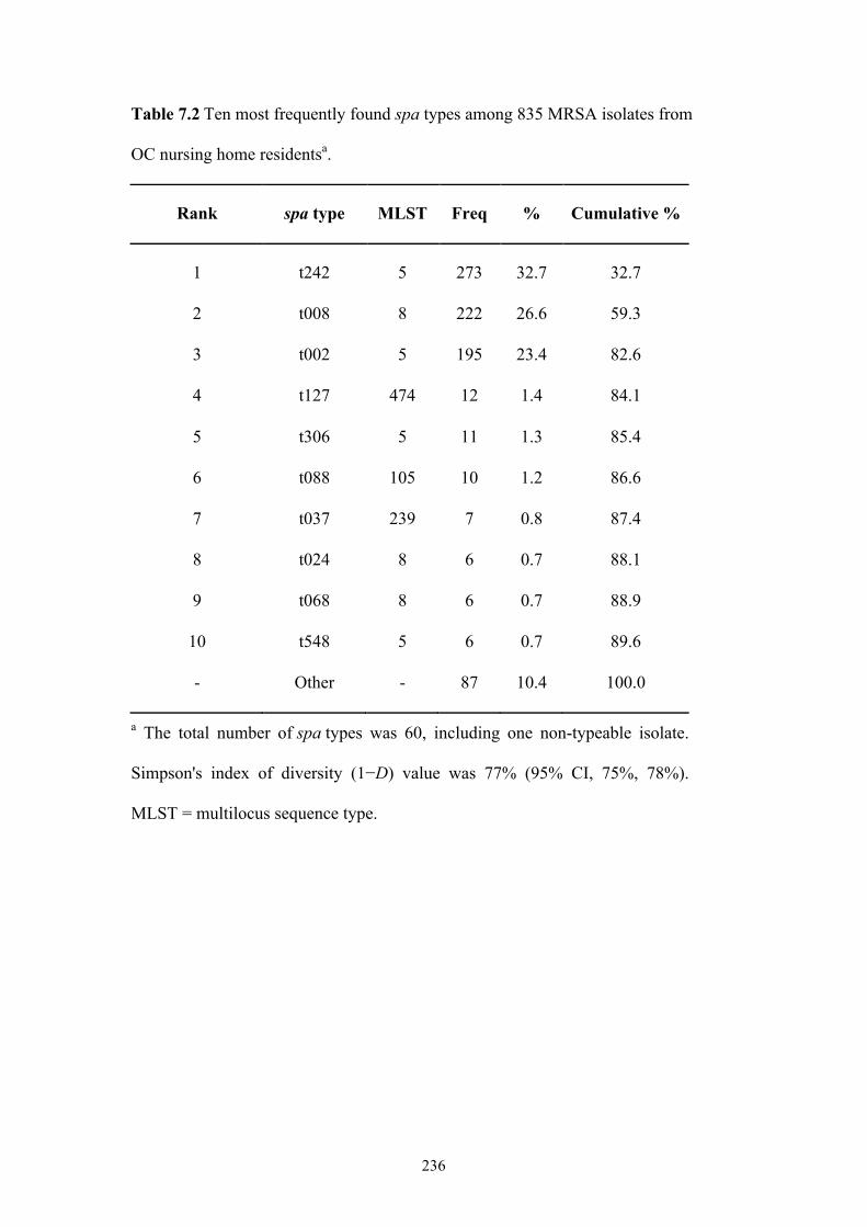
236
Table 7.2 Ten most frequently found spa types among 835 MRSA isolates from
OC nursing home residentsa.
Rank spa type MLST Freq % Cumulative %
1 t242 5 273 32.7 32.7
2 t008 8 222 26.6 59.3
3 t002 5 195 23.4 82.6
4 t127 474 12 1.4 84.1
5 t306 5 11 1.3 85.4
6 t088 105 10 1.2 86.6
7 t037 239 7 0.8 87.4
8 t024 8 6 0.7 88.1
9 t068 8 6 0.7 88.9
10 t548 5 6 0.7 89.6
- Other - 87 10.4 100.0
a The total number of spa types was 60, including one non-typeable isolate.
Simpson's index of diversity (1−D) value was 77% (95% CI, 75%, 78%).
MLST = multilocus sequence type.
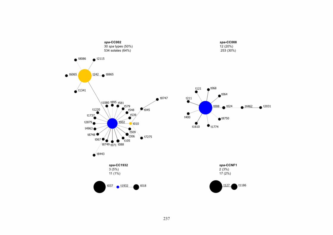
237
238
Figure 7.1 Relatedness of spa types among nursing home MRSA isolates according to the Based Upon Repeat Pattern (BURP) algorithm.
Clusters of linked spa types correspond to spa clonal complexes (spa-CCs). spa types are clustered into a spa-CC when their repeat patterns
differ by no more than 4 repeats. The BURP algorithm sums up ‘costs’ (a measure of relatedness based on the repeat pattern) to define a founder
score for each spa type in a spa-CC. The founder (blue node) is the spa type with the highest founder score in its spa-CC, and the subfounder
(yellow node) is the spa type with the second highest founder score. spa-CC008 has founder t008, and spa-CCNF refers to a spa-CC with no
founder. Each node represents a spa type. The node size represents the number of clustered strains that belong to that spa type. The shading of
the branches represents the ‘costs’ (similarities in repeat patterns) between two spa types; the darker the branch, the lower the cost (more similar
repeat patterns).
239
To confirm strain types, 138 isolates were selected for MLST. Among the 15
unique sequence types (STs) identified, ST5 (54%) and ST8 (28%) were the most
predominant, with the majority of isolates belonging to one of two major MLST CCs:
CC5 (60%; five STs) and CC8/239 (29%; two STs) (Table 7.3). The remaining 11%
of isolates comprised sporadic incidences of both HA-MRSA (4%) and CA-MRSA
(7%) clones. According to MLST, t008 isolates were the prototypic community clone
USA300 (ST8/t008) and t002 isolates were the prototypic hospital clone USA100
(ST5/t002). t242 isolates, which differ from t002 isolates by one spa repeat as a result
of a single nucleotide difference, were identified as ST5 (Tables 7.2 and 7.3). The NT
spa isolate and one novel, singleton spa type were ST105, with seven of the other
novel spa types being ST5 and one ST8.
Since the founders of the two large spa-CCs represent a community-associated
clone (the predominant US clone USA300 for spa-CC008) and a healthcare-
associated clone (the dominant US hospital clone USA100 for spa-CC002), spa-
CC008 effectively represents CA-MRSA and spa-CC002 HA-MRSA. The two
smaller spa-CCs each represented HA-MRSA and CA-MRSA, and the remaining
2.4% of isolates that could not be assigned to a spa-CC also included both HA-MRSA
and CA-MRSA.
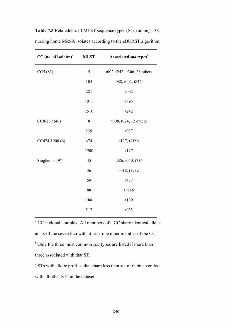
240
Table 7.3 Relatedness of MLST sequence types (STs) among 138
nursing home MRSA isolates according to the eBURST algorithm.
CC (no. of isolates)a MLST Associated spa typesb
CC5 (83) 5 t002, t242, t306, 28 others
105 t088, t002, t8444
221 t002
1011 t895
1510 t242
CC8/239 (40) 8 t008, t024, 12 others
239 t037
CC474/1900 (6) 474 t127, t1186
1900 t127
Singletons (9)c 45 t026, t040, t736
36 t018, t1932
59 t437
88 t5916
188 t189
217 t032
a CC = clonal complex. All members of a CC share identical alleles
at six of the seven loci with at least one other member of the CC.
b Only the three most common spa types are listed if more than
three associated with that ST.
c STs with allelic profiles that share less than six of their seven loci
with all other STs in the dataset.
241
7.3.3 Differences among nursing homes
The estimated genetic diversity of MRSA within nursing homes ranged from
43% to 84% (Figure 7.2), but due to relatively small numbers these differences were
mostly non-significant, with the clear exception of hospital 23 that exhibited
significantly higher diversity (84%) than nine other nursing homes. Four nursing
homes collected <10 MRSA isolates and so their genetic diversities could not be
reliably estimated. For the 21 remaining nursing homes, MRSA genetic diversity
within nursing homes was positively correlated with the percentage of residents
admitted from hospital (r = 0.52, p = 0.02), percentage of residents with diabetes (r =
0.57, p <0.01), percentage of residents with skin lesions (r = 0.46, p = 0.03), MRSA
admission prevalence (r = 0.50, p = 0.03) and MRSA point prevalence (r = 0.47, p =
0.03), and negatively correlated with the percentage of residents under 65 years old (r
= -0.57, p<0.01) and the percentage of male residents (r = -0.43, p = 0.05) (Table 7.4).
The percentage of residents with skin lesions was positively correlated with the
percentage of residents admitted from hospital (r = 0.84, p<0.001) and negatively
correlated with the percentage of residents under 65 (r = -0.58, p<0.01). Skin lesions
are very common among elderly people, and are often caused by disease or trauma
that requires hospital treatment. The percentage of residents with skin lesions showed
the weakest correlation with genetic diversity and thus was not considered for entry
into the bootstrapped linear regression model. Similarly, the percentage of male
residents and the percentage of residents under 65 were highly correlated (r = 0.88,
p<0.001). Since age was more strongly correlated with MRSA diversity than gender,
and differences in MRSA strain types have been observed between age groups in our
previous study (Chapter 5) [Hudson et al. 2012], the percentage of male residents was
not considered for entry into the multiple regression model. Finally, point prevalence
242
was highly correlated with admission prevalence (r = 0.74, p<0.001) and diabetic
residents (r = 0.69, p<0.001), thus only admission prevalence was considered for
regression model entry. Only the percentage of residents 65 or over and the
percentage of diabetic residents remained significant predictors of spa type genetic
diversity in the exploratory regression model (Table 7.5).
The three most common spa types - t242, t008 and t002 - accounted for 55-
96% of isolates at each of 23 nursing homes. Two further nursing homes only isolated
t008, although the sample sizes were <10. Twelve nursing homes isolated mostly t242
(36-63%). The proportion of spa-CC008 isolates, representing community-associated
spa types, compared to spa-CC002 isolates, representing healthcare-associated spa
types, varied significantly between nursing homes (χ2 = 69.2, df = 24, p<0.001)
(Figure 7.2). Two nursing homes (8%) had significantly more spa-CC008 isolates,
and 16 nursing homes (64%) had significantly more spa-CC002 isolates (p<0.01).
Diversity of spa types among spa-CC008 isolates (1−D = 23% (95% CI, 12-33%))
was significantly lower than diversity among spa-CC002 isolates (1−D = 60% (95%
CI, 58-63%)).
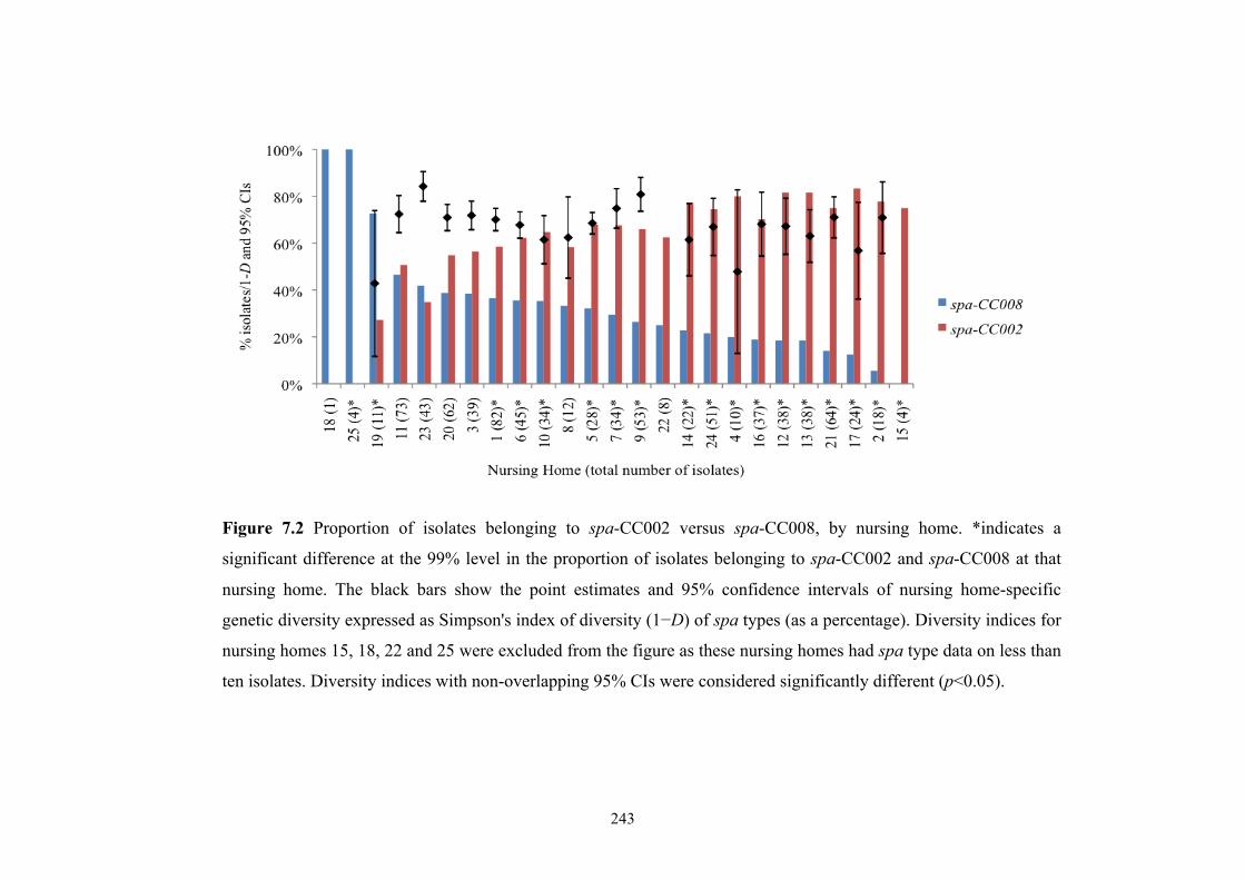
243
Figure 7.2 Proportion of isolates belonging to spa-CC002 versus spa-CC008, by nursing home. *indicates a
significant difference at the 99% level in the proportion of isolates belonging to spa-CC002 and spa-CC008 at that
nursing home. The black bars show the point estimates and 95% confidence intervals of nursing home-specific
genetic diversity expressed as Simpson's index of diversity (1−D) of spa types (as a percentage). Diversity indices for
nursing homes 15, 18, 22 and 25 were excluded from the figure as these nursing homes had spa type data on less than
ten isolates. Diversity indices with non-overlapping 95% CIs were considered significantly different (p<0.05).
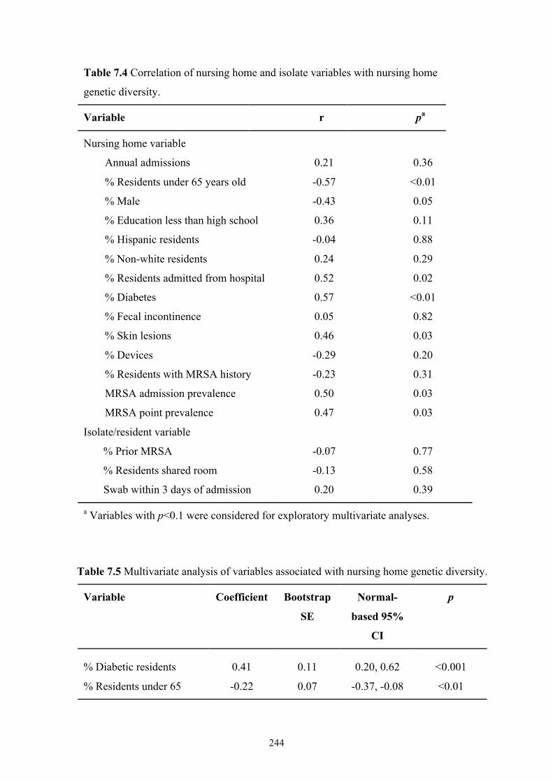
244
Table 7.4 Correlation of nursing home and isolate variables with nursing home
genetic diversity.
Variable r pa
Nursing home variable
Annual admissions 0.21 0.36
% Residents under 65 years old -0.57 <0.01
% Male -0.43 0.05
% Education less than high school 0.36 0.11
% Hispanic residents -0.04 0.88
% Non-white residents 0.24 0.29
% Residents admitted from hospital 0.52 0.02
% Diabetes 0.57 <0.01
% Fecal incontinence 0.05 0.82
% Skin lesions 0.46 0.03
% Devices -0.29 0.20
% Residents with MRSA history -0.23 0.31
MRSA admission prevalence 0.50 0.03
MRSA point prevalence 0.47 0.03
Isolate/resident variable
% Prior MRSA -0.07 0.77
% Residents shared room -0.13 0.58
Swab within 3 days of admission 0.20 0.39
a Variables with p<0.1 were considered for exploratory multivariate analyses.
Table 7.5 Multivariate analysis of variables associated with nursing home genetic diversity.
Variable Coefficient Bootstrap
SE
Normal-
based 95%
CI
p
% Diabetic residents 0.41 0.11 0.20, 0.62 <0.001
% Residents under 65 -0.22 0.07 -0.37, -0.08 <0.01
245
7.3.4 Admissions versus Point Prevalence MRSA
MRSA spa type genetic diversity was not significantly different among
isolates collected at admission (1−D = 76% (95% CI, 74-79%)) versus isolates
collected during point prevalence testing (1−D = 77% (95% CI, 75-79%)). No
significant correlation was found between admission prevalence and the proportion of
spa-CC008 isolates (r = -0.20, p = 0.34), however nursing homes with a higher
percentage of residents admitted from hospital had significantly lower percentages of
spa-CC008 isolates (r = -0.58, p<0.01). Proportions of spa-CC008, spa-CC002 and
other isolates were not significantly different between admissions and point
prevalence MRSA (26%, 68% and 6% among admissions MRSA versus 32%, 62%
and 6% among point prevalence MRSA, respectively, χ2 = 3.3, df = 2, p = 0.2).
7.4 DISCUSSION
A prospective collection of carriage isolates of MRSA from 26 nursing homes
in OC, CA, was conducted. The study investigated the frequency and genetic diversity
of MRSA in these little-studied healthcare facilities, to better inform nursing home-
based infection control strategies. This is the first study to assess MRSA isolates in
nursing homes at a population level and across a large region.
Countywide, nursing home carriage MRSA isolates were dominated in
approximately equal proportions by three strains: the predominant community-
associated clone in the US, USA300 (ST8/t008); the healthcare-associated clone
USA100 (ST5/t002); and ST5/t242 isolates, likely a minor variant of USA100 that
has become prevalent in OC healthcare facilities (Chapters 5 and 6) [Hudson et al.
2012]. ST5/t242 isolates were slightly more common than USA300 and USA100
246
however, representing a third of all carriage MRSA isolates. The same three strains
dominated in OC hospitals, but in this setting USA300 was the most common clone
(Chapter 6).
As in OC hospitals, most spa types were closely related to either USA300 or
USA100, creating two large spa-CCs each representing CA-MRSA and HA-MRSA
strains. The remaining, sporadically occurring spa types were both community-
associated and healthcare-associated, representing several known clones. Of the two
smaller spa-CCs, one represented strains of the CA-MRSA lineage MLST CC1, with
most isolates typed as ST474/t127. ST474 is a single-locus variant (SLV) of ST1, and
ST1/t127 is a common CA-MRSA strain in the UK [Otter et al. 2009]. ST474/t127
isolates were also found among OC hospital inpatients (Chapter 6), but no ST1/t127
isolates have yet been identified in the US. The other small spa-CC, spa-CC1932,
represented HA-MRSA and included the epidemic clone USA200/EMRSA-16 (ST36)
and the pandemic clone ST239.
The isolates that could not be assigned to a spa-CC included both HA-MRSA
and CA-MRSA. The healthcare-associated strains were USA600/Berlin clone (ST45)
and ST217/t032, a SLV of ST22, the pandemic HA-MRSA clone EMRSA-15 that has
also recently been reported in the community [Mollaghan et al. 2010]. The
community-associated strains were USA1000 (ST59), ST188 (a double-locus variant
of ST1 and ST474 reported sporadically in Australia and Asia [Nimmo and Coombs
2008, Peck et al. 2009, Ghaznavi-Rad et al. 2010a]) and ST88, a clone closely related
to CC1 that has been reported in several countries, particularly Nigeria, but has not
been previously reported in the US [Ghebremedhin et al. 2009, Monecke et al. 2011].
It is clear that USA300 and USA100 dominate healthcare facilities in OC, in line with
the MRSA picture seen nationwide. However, it would be interesting to investigate
247
whether the USA100 variant seen in this county is also more common than USA100
elsewhere in the US, particularly since t242 has been reported rarely in the literature
[Kinnevey et al. 2010, Parlato et al. 2009, Johnson et al. 2007, Weese, Avery and
Reid-Smith 2010].
Overall genetic diversity of MRSA in OC nursing homes was significantly
higher than that seen in OC hospitals (Chapter 6). The higher proportion of HA-
MRSA strains present in nursing homes likely drives this, which could be a result of
the high proportion of residents directly admitted to nursing homes from a number of
different OC hospitals. Diversity was significantly lower among spa-CC008 (CA-
MRSA) isolates than spa-CC002 (HA-MRSA) isolates, probably due to the greater
time HA-MRSA have had to diversify compared to CA-MRSA. This suggests that
MRSA diversity in OC is driven by healthcare-associated strains.
In exploratory analyses, greater MRSA genetic diversity was significantly
associated with older resident age and diabetic residents. Diabetic foot ulcers are a
known risk factor for MRSA, and in particular HA-MRSA, with MRSA found to be
present in 10-30% of diabetic wounds [Goldstein, Citron and Nesbit 1996,
Tentolouris et al. 1999, Shankar et al. 2005, Wang et al. 2010]. Diabetic
complications such as neuropathy, osteomyelitis and peripheral vascular disease may
result in a prolonged hospital stay, increasing the exposure of diabetic people to HA-
MRSA [Wang et al. 2010]. Older age is a well-established risk factor for HA-MRSA,
as elderly patients tend to be sicker and require hospital treatment. Older age was a
significant predictor of genetic diversity in OC hospitals (Chapter 6), and was
associated with non-t008 strains in a study comparing adult and paediatric OC
inpatients (Chapter 5) [Hudson et al. 2012].
248
Genetic diversity was not significantly different between admissions MRSA
and MRSA collected during point prevalence, with no correlation found between spa-
CC008 isolates (CA-MRSA) and admissions MRSA. In fact, over two-thirds of
admissions MRSA belonged to spa-CC002 (HA-MRSA). The vast majority of
residents in this study were admitted to nursing homes directly from hospital and thus
were not recently exposed to the community MRSA reservoir, thus reducing the
likelihood of isolating a community-associated strain at admission. Community-
associated strains are present in nursing homes, although to a lesser extent than in
hospitals (Chapter 6), with the majority of nursing homes isolating significantly more
HA-MRSA. This is probably due to the resident demographic - older, sicker people
generally have a history of healthcare exposure and thus tend to have HA-MRSA,
which is associated with more invasive infections and serious illness. The long-term
care provided by nursing homes means that the turnover rate in nursing homes is far
lower than the patient turnover rate in hospitals. This results in a lower frequency of
possible introductions of MRSA from outside the healthcare setting.
The CA-MRSA seen among residents upon admission to nursing homes and
during point prevalence sampling likely comes from the hospital MRSA reservoir, in
which it is clearly becoming dominant (Chapter 6). The slightly higher proportion of
CA-MRSA from point prevalence sampling compared to admission sampling and
consequently the lower proportion of HA-MRSA, although non-significant, possibly
suggests that CA-MRSA could be more easily spread in nursing homes than HA-
MRSA, perhaps due to a more community-like environment that CA-MRSA is well-
adapted to, e.g. communal areas and shared rooms. It could also possibly mean that
additional exposures to CA-MRSA are occurring via contact with visitors and/or
healthcare workers. Given the non-significant result however, it could just be due to
249
the different sample of residents that were tested at point prevalence versus at
admission (some residents would have been tested at both times, while others would
have been tested only at point prevalence and not at admission, and vice versa).
The significantly higher point prevalence of MRSA compared to admissions
prevalence may be due to the transmission of MRSA imported by residents upon
admission, evidenced by the lack of significant change in genetic diversity or
proportions of CA-MRSA and HA-MRSA from admissions to point prevalence.
Factors influencing transmission in nursing homes and predictors of MRSA carriage
at point prevalence have been reported previously [Reynolds et al. 2011].
A limitation of this study was that few individual-level characteristics were
available and so facility-level data were primarily used. Also, only a single site was
swabbed for MRSA. Although nasal screening is thought to detect the majority of
MRSA carriers [Manian et al. 2002, Lucet et al. 2003], anatomical sites such as the
rectum and throat have been shown to be important in the detection of MRSA carriage
[Eveillard et al. 2006, Batra et al. 2008, Eveillard et al. 2008]. The point prevalence
study design also limited the accuracy of carriage estimates. In using genetic diversity
as the outcome, the sample size was reduced considerably, and thus multivariate
analysis of predictor variables could only be exploratory. Also, the diversity indices
estimated for admissions MRSA versus point prevalence MRSA, and spa-CC008
isolates versus spa-CC002 isolates, may have been influenced by differing sample
sizes [Grundmann, Hori and Tanner 2001].
In conclusion this study found that in a large county, the diversity of carriage
MRSA isolates among nursing home residents, although heterogeneous between
facilities, was significantly higher than the diversity among clinical MRSA isolates
from hospital inpatients in the same county. MRSA diversity in both hospitals and
250
nursing homes appears to be driven by HA-MRSA. The proportions of older, diabetic
residents were significant predictors of nursing home MRSA diversity. Nursing home
strains were dominated by a healthcare-associated, OC-prevalent variant of USA100
(ST5/t242). The CA-MRSA strain USA300 was the second most common clone
isolated from the nares of nursing home residents, suggesting substantial penetration
of community-associated strains into the nursing home reservoir, however to a lesser
extent than seen in the hospitals, where USA300 was predominant. Nursing home
MRSA burden appeared to be largely due to importation of diverse strains from
hospitals and subsequent transmission of these imported strains, leading to high
MRSA point prevalence. One of the OC nursing homes in this study was previously
reported to have an MRSA point prevalence of 52% [Reynolds et al. 2011]. Nursing
homes therefore represent a significant reservoir for MRSA, and as such a consensual,
regionally implemented control strategy tailored to this unique setting is required.
However, due to the complexity of MRSA control in these facilities, further studies
evaluating the contribution of nursing homes to regional MRSA transmission are
needed before developing such a strategy.
251
PART 2 FINAL DISCUSSION: MRSA DIVERSITY IN ORANGE COUNTY, CALIFORNIA
In collaboration with researchers at the University of California, Irvine (UCI),
MRSA isolates were collected from OC hospitals and nursing homes in the first
population-based countywide study of MRSA strain diversity and distribution in
healthcare facilities. The aim of this work was to gain a better understanding of the
frequency and genetic diversity of HA-MRSA and CA-MRSA in hospital and nursing
home MRSA reservoirs in the hope that such knowledge would better inform regional
infection control strategies.
While MRSA diversity was observed among OC healthcare facilities, three
strains dominated both hospital and nursing home MRSA reservoirs in this
metropolitan county: the increasingly prevalent community-associated clone USA300,
a surprisingly common variant of the hospital-associated clone USA100, and USA100
itself. USA300 accounted for almost half of all MRSA isolates collected from OC
hospitals and over a quarter of isolates from OC nursing homes. Despite significantly
higher numbers of USA300 in hospitalised children, USA300 was still the most
common strain among adult inpatients, and the second most common strain among
nursing home residents. No correlation was found between CA-MRSA and
community onset among hospital MRSA isolates, with 42% of hospital onset isolates
CA-MRSA, 39% of community onset isolates HA-MRSA, suggesting that community
and healthcare MRSA reservoirs are mixing. Other US studies have reported such
mixing. The Centers for Disease Control and Prevention ABC surveillance system for
invasive MRSA infections found that the USA300 clone accounted for 22% of
community onset HA-MRSA infection and 16% of hospital onset HA-MRSA
infection in 2004-2005 [Klevens et al. 2007]. It also found that 23% of CA-MRSA
252
infections were caused by USA100. Another study involving a single urban hospital
and its associated outpatient clinics in Atlanta, GA, found that previous
hospitalization was a risk factor for community onset MRSA infections, 99% of
which were caused by USA300 [King et al. 2006]. Finally, among cases of hospital
onset MRSA bacteraemia between 2000 and 2006 in a single hospital in Chicago, IL,
community-genotype strains caused an increasing proportion of cases from 24% to
49% while the proportion of hospital-genotype strains decreased [Popovich,
Weinstein and Hota 2008].
Because of their changing epidemiology and increasingly indistinguishable
clinical characteristics, the definitions of HA- and CA-MRSA need to be revised in
order to better inform infection control strategies [Popovich and Weinstein 2009,
David and Daum 2010, McCarthy et al. 2010]. The predominance of community
onset isolates among hospital inpatients highlights the need for community-based
interventions to stem the influx of community-associated strains, particularly the
highly successful USA300 clone, into the healthcare setting.
In addition to the spa typing and MLST I performed as part of my thesis,
PFGE was also performed on the OC MRSA isolates by my collaborators. PFGE
results suggest that the USA300 strain may be diverging (data not shown). While
these typing methods for characterising MRSA isolates are useful for identifying
preliminary differences among strains, they are also limited. For example, MLST
evaluates housekeeping genes, so while excellent at unambiguously assigning a strain,
it is unlikely to capture recent or subtle changes in USA300 strains. Whole genome
sequencing would allow us to characterise this predominant MRSA strain in OC on a
finer scale, helping us to elucidate the rapidly evolving differences in this strain and
thus better understand its epidemiology and pathogenicity. In collaboration with the
253
Wellcome Trust Sanger Institute, all USA300 strains from the large prospective study
of MRSA in OC are currently being whole genome sequenced using NGS technology,
allowing us to assess the extent to which USA300 has spread across the 30 OC
hospitals and into the 25 OC nursing homes included in the study, and to possibly
determine its major transmission pathways. Other collaborators working with the spa
typing data from this OC MRSA project found that hospitals that share patients share
strains [Ke et al. 2012]. Integrating genome data into the network models they have
created will help to quantify USA300 spread between hospitals.
Work linking this part of my thesis with that involving my industrial sponsor,
TwistDx, could involve using the various MRSA strains identified in the OC study to
test the performance of their MRSA diagnostic assay, TwistAmp MRSA. Also, MREJ
typing all MRSA isolates collected from OC hospitals and nursing homes would
allow comparison to MREJ typing data from the KC MRSA isolates in Chapter 3, as
well as MRSA isolates from the UK collection, to form a better picture of the
frequencies and diversity of MREJ types.
I am currently involved in the development of OC-Maps, with collaborators
from the Department of Infectious Disease Epidemiology, Imperial College London.
OC-Maps will be a web-based interactive mapping tool for the interrogation of the
geographic distribution of different spa types isolated from hospitals in OC. It will be
based on the publicly accessible mapping tool SRL-Maps
(http://www.spatialepidemiology.net/SRL-Maps) [Grundmann et al. 2010]. OC-Maps
will be countywide, covering 30 hospitals, whereas SRL-Maps spans a continent
covering 450 hospitals, so it is unlikely that any clear geographical patterns could be
visualized using the former. However, it will offer an elegant way to summarise the
spa typing and related clinical data both across and within hospitals in OC, CA and
254
aid the formation of hypotheses regarding the spread of MRSA strains within this
county.
All hospital locations will be represented as placemarks on a Google map.
Clicking on a placemark will display, below the map, all spa types identified at that
hospital including frequencies (Figure II.1). The number of isolates found elsewhere
(if any) for each of these spa types, along with the number of hospitals each spa type
has been isolated from, would also be displayed. There will also be the option of
viewing the countywide distribution of any spa type, and placemarks on the map will
be colour-coded on the basis of the percentage of isolates of that spa type at each
hospital, with the number of isolates shown inside the placemark (Figure II.1).
Graphical charts will be displayed showing either spa-type specific, hospital-specific
or overall age distributions, specimen types, onset and ICU/non-ICU isolate collection
(Figure II.1). This tool, once developed, would allow the identification and mapping
of strains with particular public health importance, for example USA300.
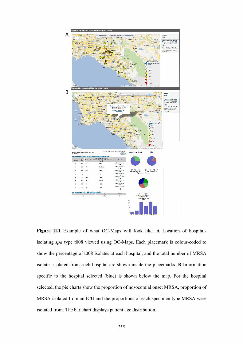
255
Figure II.1 Example of what OC-Maps will look like. A Location of hospitals
isolating spa type t008 viewed using OC-Maps. Each placemark is colour-coded to
show the percentage of t008 isolates at each hospital, and the total number of MRSA
isolates isolated from each hospital are shown inside the placemarks. B Information
specific to the hospital selected (blue) is shown below the map. For the hospital
selected, the pie charts show the proportion of nosocomial onset MRSA, proportion of
MRSA isolated from an ICU and the proportions of each specimen type MRSA were
isolated from. The bar chart displays patient age distribution.
!"
#"
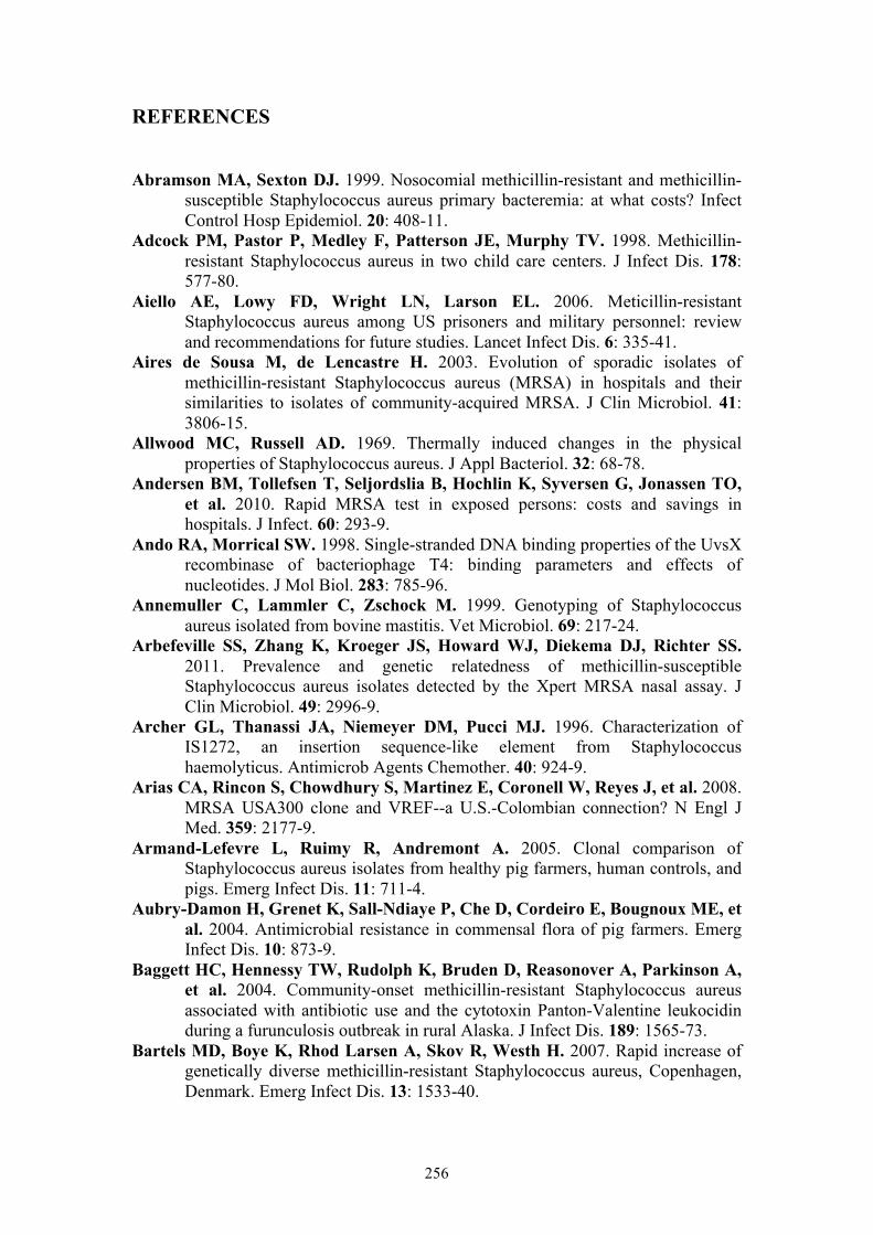
256
REFERENCES
Abramson MA, Sexton DJ. 1999. Nosocomial methicillin-resistant and methicillin-
susceptible Staphylococcus aureus primary bacteremia: at what costs? Infect Control Hosp Epidemiol. 20: 408-11.
Adcock PM, Pastor P, Medley F, Patterson JE, Murphy TV. 1998. Methicillin-resistant Staphylococcus aureus in two child care centers. J Infect Dis. 178: 577-80.
Aiello AE, Lowy FD, Wright LN, Larson EL. 2006. Meticillin-resistant Staphylococcus aureus among US prisoners and military personnel: review and recommendations for future studies. Lancet Infect Dis. 6: 335-41.
Aires de Sousa M, de Lencastre H. 2003. Evolution of sporadic isolates of methicillin-resistant Staphylococcus aureus (MRSA) in hospitals and their similarities to isolates of community-acquired MRSA. J Clin Microbiol. 41: 3806-15.
Allwood MC, Russell AD. 1969. Thermally induced changes in the physical properties of Staphylococcus aureus. J Appl Bacteriol. 32: 68-78.
Andersen BM, Tollefsen T, Seljordslia B, Hochlin K, Syversen G, Jonassen TO, et al. 2010. Rapid MRSA test in exposed persons: costs and savings in hospitals. J Infect. 60: 293-9.
Ando RA, Morrical SW. 1998. Single-stranded DNA binding properties of the UvsX recombinase of bacteriophage T4: binding parameters and effects of nucleotides. J Mol Biol. 283: 785-96.
Annemuller C, Lammler C, Zschock M. 1999. Genotyping of Staphylococcus aureus isolated from bovine mastitis. Vet Microbiol. 69: 217-24.
Arbefeville SS, Zhang K, Kroeger JS, Howard WJ, Diekema DJ, Richter SS. 2011. Prevalence and genetic relatedness of methicillin-susceptible Staphylococcus aureus isolates detected by the Xpert MRSA nasal assay. J Clin Microbiol. 49: 2996-9.
Archer GL, Thanassi JA, Niemeyer DM, Pucci MJ. 1996. Characterization of IS1272, an insertion sequence-like element from Staphylococcus haemolyticus. Antimicrob Agents Chemother. 40: 924-9.
Arias CA, Rincon S, Chowdhury S, Martinez E, Coronell W, Reyes J, et al. 2008. MRSA USA300 clone and VREF--a U.S.-Colombian connection? N Engl J Med. 359: 2177-9.
Armand-Lefevre L, Ruimy R, Andremont A. 2005. Clonal comparison of Staphylococcus aureus isolates from healthy pig farmers, human controls, and pigs. Emerg Infect Dis. 11: 711-4.
Aubry-Damon H, Grenet K, Sall-Ndiaye P, Che D, Cordeiro E, Bougnoux ME, et al. 2004. Antimicrobial resistance in commensal flora of pig farmers. Emerg Infect Dis. 10: 873-9.
Baggett HC, Hennessy TW, Rudolph K, Bruden D, Reasonover A, Parkinson A, et al. 2004. Community-onset methicillin-resistant Staphylococcus aureus associated with antibiotic use and the cytotoxin Panton-Valentine leukocidin during a furunculosis outbreak in rural Alaska. J Infect Dis. 189: 1565-73.
Bartels MD, Boye K, Rhod Larsen A, Skov R, Westh H. 2007. Rapid increase of genetically diverse methicillin-resistant Staphylococcus aureus, Copenhagen, Denmark. Emerg Infect Dis. 13: 1533-40.
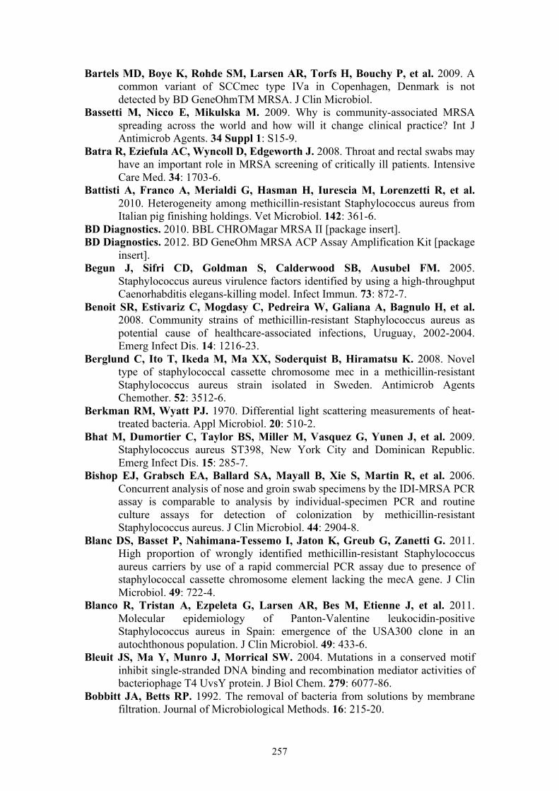
257
Bartels MD, Boye K, Rohde SM, Larsen AR, Torfs H, Bouchy P, et al. 2009. A common variant of SCCmec type IVa in Copenhagen, Denmark is not detected by BD GeneOhmTM MRSA. J Clin Microbiol.
Bassetti M, Nicco E, Mikulska M. 2009. Why is community-associated MRSA spreading across the world and how will it change clinical practice? Int J Antimicrob Agents. 34 Suppl 1: S15-9.
Batra R, Eziefula AC, Wyncoll D, Edgeworth J. 2008. Throat and rectal swabs may have an important role in MRSA screening of critically ill patients. Intensive Care Med. 34: 1703-6.
Battisti A, Franco A, Merialdi G, Hasman H, Iurescia M, Lorenzetti R, et al. 2010. Heterogeneity among methicillin-resistant Staphylococcus aureus from Italian pig finishing holdings. Vet Microbiol. 142: 361-6.
BD Diagnostics. 2010. BBL CHROMagar MRSA II [package insert]. BD Diagnostics. 2012. BD GeneOhm MRSA ACP Assay Amplification Kit [package
insert]. Begun J, Sifri CD, Goldman S, Calderwood SB, Ausubel FM. 2005.
Staphylococcus aureus virulence factors identified by using a high-throughput Caenorhabditis elegans-killing model. Infect Immun. 73: 872-7.
Benoit SR, Estivariz C, Mogdasy C, Pedreira W, Galiana A, Bagnulo H, et al. 2008. Community strains of methicillin-resistant Staphylococcus aureus as potential cause of healthcare-associated infections, Uruguay, 2002-2004. Emerg Infect Dis. 14: 1216-23.
Berglund C, Ito T, Ikeda M, Ma XX, Soderquist B, Hiramatsu K. 2008. Novel type of staphylococcal cassette chromosome mec in a methicillin-resistant Staphylococcus aureus strain isolated in Sweden. Antimicrob Agents Chemother. 52: 3512-6.
Berkman RM, Wyatt PJ. 1970. Differential light scattering measurements of heat-treated bacteria. Appl Microbiol. 20: 510-2.
Bhat M, Dumortier C, Taylor BS, Miller M, Vasquez G, Yunen J, et al. 2009. Staphylococcus aureus ST398, New York City and Dominican Republic. Emerg Infect Dis. 15: 285-7.
Bishop EJ, Grabsch EA, Ballard SA, Mayall B, Xie S, Martin R, et al. 2006. Concurrent analysis of nose and groin swab specimens by the IDI-MRSA PCR assay is comparable to analysis by individual-specimen PCR and routine culture assays for detection of colonization by methicillin-resistant Staphylococcus aureus. J Clin Microbiol. 44: 2904-8.
Blanc DS, Basset P, Nahimana-Tessemo I, Jaton K, Greub G, Zanetti G. 2011. High proportion of wrongly identified methicillin-resistant Staphylococcus aureus carriers by use of a rapid commercial PCR assay due to presence of staphylococcal cassette chromosome element lacking the mecA gene. J Clin Microbiol. 49: 722-4.
Blanco R, Tristan A, Ezpeleta G, Larsen AR, Bes M, Etienne J, et al. 2011. Molecular epidemiology of Panton-Valentine leukocidin-positive Staphylococcus aureus in Spain: emergence of the USA300 clone in an autochthonous population. J Clin Microbiol. 49: 433-6.
Bleuit JS, Ma Y, Munro J, Morrical SW. 2004. Mutations in a conserved motif inhibit single-stranded DNA binding and recombination mediator activities of bacteriophage T4 UvsY protein. J Biol Chem. 279: 6077-86.
Bobbitt JA, Betts RP. 1992. The removal of bacteria from solutions by membrane filtration. Journal of Microbiological Methods. 16: 215-20.
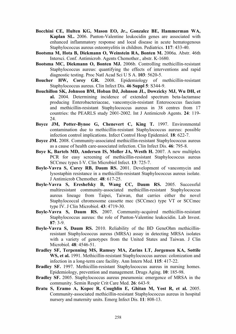
258
Bocchini CE, Hulten KG, Mason EO, Jr., Gonzalez BE, Hammerman WA, Kaplan SL. 2006. Panton-Valentine leukocidin genes are associated with enhanced inflammatory response and local disease in acute hematogenous Staphylococcus aureus osteomyelitis in children. Pediatrics. 117: 433-40.
Bootsma M, Hota B, Diekmann O, Weinstein RA, Bonten M. 2006a. Abstr. 46th Intersci. Conf. Antimicrob. Agents Chemother., abstr. K-1680.
Bootsma MC, Diekmann O, Bonten MJ. 2006b. Controlling methicillin-resistant Staphylococcus aureus: quantifying the effects of interventions and rapid diagnostic testing. Proc Natl Acad Sci U S A. 103: 5620-5.
Boucher HW, Corey GR. 2008. Epidemiology of methicillin-resistant Staphylococcus aureus. Clin Infect Dis. 46 Suppl 5: S344-9.
Bouchillon SK, Johnson BM, Hoban DJ, Johnson JL, Dowzicky MJ, Wu DH, et al. 2004. Determining incidence of extended spectrum beta-lactamase producing Enterobacteriaceae, vancomycin-resistant Enterococcus faecium and methicillin-resistant Staphylococcus aureus in 38 centres from 17 countries: the PEARLS study 2001-2002. Int J Antimicrob Agents. 24: 119-24.
Boyce JM, Potter-Bynoe G, Chenevert C, King T. 1997. Environmental contamination due to methicillin-resistant Staphylococcus aureus: possible infection control implications. Infect Control Hosp Epidemiol. 18: 622-7.
Boyce JM. 2008. Community-associated methicillin-resistant Staphylococcus aureus as a cause of health care-associated infection. Clin Infect Dis. 46: 795-8.
Boye K, Bartels MD, Andersen IS, Moller JA, Westh H. 2007. A new multiplex PCR for easy screening of methicillin-resistant Staphylococcus aureus SCCmec types I-V. Clin Microbiol Infect. 13: 725-7.
Boyle-Vavra S, Carey RB, Daum RS. 2001. Development of vancomycin and lysostaphin resistance in a methicillin-resistant Staphylococcus aureus isolate. J Antimicrob Chemother. 48: 617-25.
Boyle-Vavra S, Ereshefsky B, Wang CC, Daum RS. 2005. Successful multiresistant community-associated methicillin-resistant Staphylococcus aureus lineage from Taipei, Taiwan, that carries either the novel Staphylococcal chromosome cassette mec (SCCmec) type VT or SCCmec type IV. J Clin Microbiol. 43: 4719-30.
Boyle-Vavra S, Daum RS. 2007. Community-acquired methicillin-resistant Staphylococcus aureus: the role of Panton-Valentine leukocidin. Lab Invest. 87: 3-9.
Boyle-Vavra S, Daum RS. 2010. Reliability of the BD GeneOhm methicillin-resistant Staphylococcus aureus (MRSA) assay in detecting MRSA isolates with a variety of genotypes from the United States and Taiwan. J Clin Microbiol. 48: 4546-51.
Bradley SF, Terpenning MS, Ramsey MA, Zarins LT, Jorgensen KA, Sottile WS, et al. 1991. Methicillin-resistant Staphylococcus aureus: colonization and infection in a long-term care facility. Ann Intern Med. 115: 417-22.
Bradley SF. 1997. Methicillin-resistant Staphylococcus aureus in nursing homes. Epidemiology, prevention and management. Drugs Aging. 10: 185-98.
Bradley SF. 2005. Staphylococcus aureus pneumonia: emergence of MRSA in the community. Semin Respir Crit Care Med. 26: 643-9.
Bratu S, Eramo A, Kopec R, Coughlin E, Ghitan M, Yost R, et al. 2005. Community-associated methicillin-resistant Staphylococcus aureus in hospital nursery and maternity units. Emerg Infect Dis. 11: 808-13.

259
Brennan GI, Shore AC, Corcoran S, Tecklenborg S, Coleman DC, O'Connell B. 2012. Emergence of Hospital- and Community-Associated Panton-Valentine Leukocidin-Positive Methicillin-Resistant Staphylococcus aureus Genotype ST772-MRSA-V in Ireland and Detailed Investigation of a ST772-MRSA-V Cluster in a Neonatal Intensive Care Unit. J Clin Microbiol. 50: 841-7.
Brenwald NP, Baker N, Oppenheim B. 2010. Feasibility study of a real-time PCR test for meticillin-resistant Staphylococcus aureus in a point of care setting. J Hosp Infect. 74: 245-9.
Breurec S, Fall C, Pouillot R, Boisier P, Brisse S, Diene-Sarr F, et al. 2011. Epidemiology of methicillin-susceptible Staphylococcus aureus lineages in five major African towns: high prevalence of Panton-Valentine leukocidin genes. Clin Microbiol Infect. 17: 633-9.
Brown ML, O'Hara FP, Close NM, Mera RM, Miller LA, Suaya JA, et al. 2012. Prevalence and sequence variation of panton-valentine leukocidin in methicillin-resistant and methicillin-susceptible staphylococcus aureus strains in the United States. J Clin Microbiol. 50: 86-90.
Bubeck Wardenburg J, Bae T, Otto M, Deleo FR, Schneewind O. 2007. Poring over pores: alpha-hemolysin and Panton-Valentine leukocidin in Staphylococcus aureus pneumonia. Nat Med. 13: 1405-6.
Budimir A, Deurenberg RH, Plecko V, Vink C, Kalenic S, Stobberingh EE. 2006. Molecular characterization of methicillin-resistant Staphylococcus aureus bloodstream isolates from Croatia. J Antimicrob Chemother. 57: 331-4.
Burlak C, Hammer CH, Robinson MA, Whitney AR, McGavin MJ, Kreiswirth BN, et al. 2007. Global analysis of community-associated methicillin-resistant Staphylococcus aureus exoproteins reveals molecules produced in vitro and during infection. Cell Microbiol. 9: 1172-90.
Burton DC, Edwards JR, Horan TC, Jernigan JA, Fridkin SK. 2009. Methicillin-resistant Staphylococcus aureus central line-associated bloodstream infections in US intensive care units, 1997-2007. JAMA. 301: 727-36.
Cai L, Kong F, Wang Q, Wang H, Xiao M, Sintchenko V, et al. 2009. A new multiplex PCR-based reverse line-blot hybridization (mPCR/RLB) assay for rapid staphylococcal cassette chromosome mec (SCCmec) typing. J Med Microbiol. 58: 1045-57.
Campanile F, Bongiorno D, Borbone S, Stefani S. 2009. Hospital-associated methicillin-resistant Staphylococcus aureus (HA-MRSA) in Italy. Ann Clin Microbiol Antimicrob. 8: 22.
Carleton HA, Diep BA, Charlebois ED, Sensabaugh GF, Perdreau-Remington F. 2004. Community-adapted methicillin-resistant Staphylococcus aureus (MRSA): population dynamics of an expanding community reservoir of MRSA. J Infect Dis. 190: 1730-8.
Caron WP, Mousa SA. 2010. Prevention strategies for antimicrobial resistance: a systematic review of the literature. Infect Drug Resist. 3: 25-33.
Carroll KC. 2008. Rapid diagnostics for methicillin-resistant Staphylococcus aureus: current status. Mol Diagn Ther. 12: 15-24.
Catry B, Van Duijkeren E, Pomba MC, Greko C, Moreno MA, Pyorala S, et al. 2010. Reflection paper on MRSA in food-producing and companion animals: epidemiology and control options for human and animal health. Epidemiol Infect. 138: 626-44.
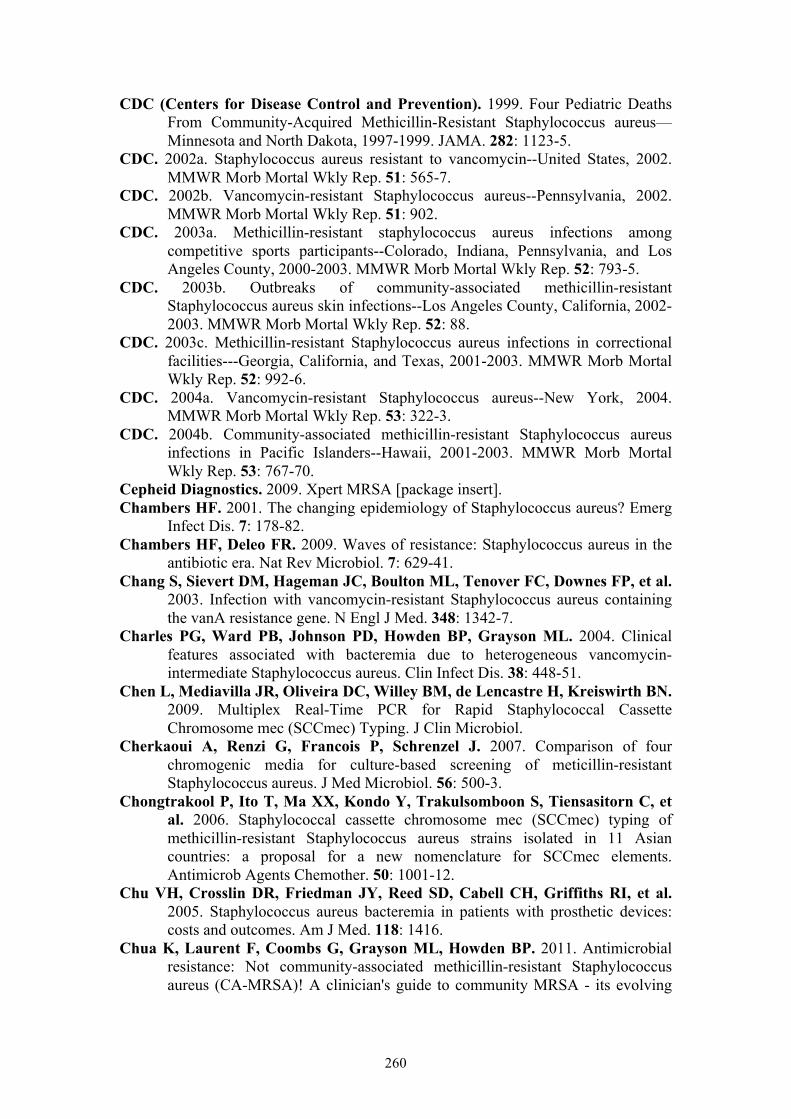
260
CDC (Centers for Disease Control and Prevention). 1999. Four Pediatric Deaths From Community-Acquired Methicillin-Resistant Staphylococcus aureus—Minnesota and North Dakota, 1997-1999. JAMA. 282: 1123-5.
CDC. 2002a. Staphylococcus aureus resistant to vancomycin--United States, 2002. MMWR Morb Mortal Wkly Rep. 51: 565-7.
CDC. 2002b. Vancomycin-resistant Staphylococcus aureus--Pennsylvania, 2002. MMWR Morb Mortal Wkly Rep. 51: 902.
CDC. 2003a. Methicillin-resistant staphylococcus aureus infections among competitive sports participants--Colorado, Indiana, Pennsylvania, and Los Angeles County, 2000-2003. MMWR Morb Mortal Wkly Rep. 52: 793-5.
CDC. 2003b. Outbreaks of community-associated methicillin-resistant Staphylococcus aureus skin infections--Los Angeles County, California, 2002-2003. MMWR Morb Mortal Wkly Rep. 52: 88.
CDC. 2003c. Methicillin-resistant Staphylococcus aureus infections in correctional facilities---Georgia, California, and Texas, 2001-2003. MMWR Morb Mortal Wkly Rep. 52: 992-6.
CDC. 2004a. Vancomycin-resistant Staphylococcus aureus--New York, 2004. MMWR Morb Mortal Wkly Rep. 53: 322-3.
CDC. 2004b. Community-associated methicillin-resistant Staphylococcus aureus infections in Pacific Islanders--Hawaii, 2001-2003. MMWR Morb Mortal Wkly Rep. 53: 767-70.
Cepheid Diagnostics. 2009. Xpert MRSA [package insert]. Chambers HF. 2001. The changing epidemiology of Staphylococcus aureus? Emerg
Infect Dis. 7: 178-82. Chambers HF, Deleo FR. 2009. Waves of resistance: Staphylococcus aureus in the
antibiotic era. Nat Rev Microbiol. 7: 629-41. Chang S, Sievert DM, Hageman JC, Boulton ML, Tenover FC, Downes FP, et al.
2003. Infection with vancomycin-resistant Staphylococcus aureus containing the vanA resistance gene. N Engl J Med. 348: 1342-7.
Charles PG, Ward PB, Johnson PD, Howden BP, Grayson ML. 2004. Clinical features associated with bacteremia due to heterogeneous vancomycin-intermediate Staphylococcus aureus. Clin Infect Dis. 38: 448-51.
Chen L, Mediavilla JR, Oliveira DC, Willey BM, de Lencastre H, Kreiswirth BN. 2009. Multiplex Real-Time PCR for Rapid Staphylococcal Cassette Chromosome mec (SCCmec) Typing. J Clin Microbiol.
Cherkaoui A, Renzi G, Francois P, Schrenzel J. 2007. Comparison of four chromogenic media for culture-based screening of meticillin-resistant Staphylococcus aureus. J Med Microbiol. 56: 500-3.
Chongtrakool P, Ito T, Ma XX, Kondo Y, Trakulsomboon S, Tiensasitorn C, et al. 2006. Staphylococcal cassette chromosome mec (SCCmec) typing of methicillin-resistant Staphylococcus aureus strains isolated in 11 Asian countries: a proposal for a new nomenclature for SCCmec elements. Antimicrob Agents Chemother. 50: 1001-12.
Chu VH, Crosslin DR, Friedman JY, Reed SD, Cabell CH, Griffiths RI, et al. 2005. Staphylococcus aureus bacteremia in patients with prosthetic devices: costs and outcomes. Am J Med. 118: 1416.
Chua K, Laurent F, Coombs G, Grayson ML, Howden BP. 2011. Antimicrobial resistance: Not community-associated methicillin-resistant Staphylococcus aureus (CA-MRSA)! A clinician's guide to community MRSA - its evolving
261
antimicrobial resistance and implications for therapy. Clin Infect Dis. 52: 99-114.
Chung M, Dickinson G, De Lencastre H, Tomasz A. 2004. International clones of methicillin-resistant Staphylococcus aureus in two hospitals in Miami, Florida. J Clin Microbiol. 42: 542-7.
Ciardo DE, Burger S, Payer M, Lee C, McCallum N. 2010. GeneXpert captures unstable methicillin-resistant Staphylococcus aureus prone to rapidly losing the mecA gene. J Clin Microbiol. 48: 3030-2.
CLSI (Clinical and Laboratory Standards Institute). 2005. Performance Standards for Anitmicrobial Susceptibility Testing. 15th informational supplement, M100-S15. Clinical and Laboratory Standards Institute. Wayne, PA.
CMS (Centers for Medicare and Medicaid Services). 2009. Long Term Care Minimum Data Set. Available from: http://www.resdac.org/MDS/data_available.asp. [Accessed 10th April 2012].
Cookson BD, Robinson DA, Monk AB, Murchan S, Deplano A, de Ryck R, et al. 2007. Evaluation of molecular typing methods in characterizing a European collection of epidemic methicillin-resistant Staphylococcus aureus strains: the HARMONY collection. J Clin Microbiol. 45: 1830-7.
Coombs GW, Nimmo GR, Bell JM, Huygens F, O'Brien FG, Malkowski MJ, et al. 2004. Genetic diversity among community methicillin-resistant Staphylococcus aureus strains causing outpatient infections in Australia. J Clin Microbiol. 42: 4735-43.
Coombs GW, Monecke S, Ehricht R, Slickers P, Pearson JC, Tan HL, et al. 2010. Differentiation of clonal complex 59 community-associated methicillin-resistant Staphylococcus aureus in Western Australia. Antimicrob Agents Chemother. 54: 1914-21.
Cooper JE, Feil EJ. 2004. Multilocus sequence typing--what is resolved? Trends Microbiol. 12: 373-7.
Corkill JE, Anson JJ, Griffiths P, Hart CA. 2004. Detection of elements of the staphylococcal cassette chromosome (SCC) in a methicillin-susceptible (mecA gene negative) homologue of a fucidin-resistant MRSA. J Antimicrob Chemother. 54: 229-31.
Cosgrove SE, Sakoulas G, Perencevich EN, Schwaber MJ, Karchmer AW, Carmeli Y. 2003. Comparison of mortality associated with methicillin-resistant and methicillin-susceptible Staphylococcus aureus bacteremia: a meta-analysis. Clin Infect Dis. 36: 53-9.
Cosgrove SE, Qi Y, Kaye KS, Harbarth S, Karchmer AW, Carmeli Y. 2005. The impact of methicillin resistance in Staphylococcus aureus bacteremia on patient outcomes: mortality, length of stay, and hospital charges. Infect Control Hosp Epidemiol. 26: 166-74.
Creamer E, Dolan A, Sherlock O, Thomas T, Walsh J, Moore J, et al. 2010. The effect of rapid screening for methicillin-resistant Staphylococcus aureus (MRSA) on the identification and earlier isolation of MRSA-positive patients. Infect Control Hosp Epidemiol. 31: 374-81.
Crisostomo MI, Westh H, Tomasz A, Chung M, Oliveira DC, de Lencastre H. 2001. The evolution of methicillin resistance in Staphylococcus aureus: similarity of genetic backgrounds in historically early methicillin-susceptible and -resistant isolates and contemporary epidemic clones. Proc Natl Acad Sci U S A. 98: 9865-70.
262
Cuny C, Witte W. 2005. PCR for the identification of methicillin-resistant Staphylococcus aureus (MRSA) strains using a single primer pair specific for SCCmec elements and the neighbouring chromosome-borne orfX. Clin Microbiol Infect. 11: 834-7.
Cuny C, Strommenger B, Witte W, Stanek C. 2008. Clusters of infections in horses with MRSA ST1, ST254, and ST398 in a veterinary hospital. Microb Drug Resist. 14: 307-10.
Cuny C, Nathaus R, Layer F, Strommenger B, Altmann D, Witte W. 2009. Nasal colonization of humans with methicillin-resistant Staphylococcus aureus (MRSA) CC398 with and without exposure to pigs. PLoS One. 4: e6800.
Cuny C, Layer F, Strommenger B, Witte W. 2011. Rare occurrence of methicillin-resistant Staphylococcus aureus CC130 with a novel mecA homologue in humans in Germany. PLoS One. 6: e24360.
D'Agata EM, Webb GF, Horn MA, Moellering RC, Jr., Ruan S. 2009. Modeling the invasion of community-acquired methicillin-resistant Staphylococcus aureus into hospitals. Clin Infect Dis. 48: 274-84.
D'Souza N, Rodrigues C, Mehta A. 2010. Molecular characterization of methicillin-resistant Staphylococcus aureus with emergence of epidemic clones of sequence type (ST) 22 and ST 772 in Mumbai, India. J Clin Microbiol. 48: 1806-11.
David MD, Kearns AM, Gossain S, Ganner M, Holmes A. 2006a. Community-associated meticillin-resistant Staphylococcus aureus: nosocomial transmission in a neonatal unit. J Hosp Infect. 64: 244-50.
David MZ, Crawford SE, Boyle-Vavra S, Hostetler MA, Kim DC, Daum RS. 2006b. Contrasting pediatric and adult methicillin-resistant Staphylococcus aureus isolates. Emerg Infect Dis. 12: 631-7.
David MZ, Daum RS. 2010. Community-associated methicillin-resistant Staphylococcus aureus: epidemiology and clinical consequences of an emerging epidemic. Clin Microbiol Rev. 23: 616-87.
Davis SL, Rybak MJ, Amjad M, Kaatz GW, McKinnon PS. 2006. Characteristics of patients with healthcare-associated infection due to SCCmec type IV methicillin-resistant Staphylococcus aureus. Infect Control Hosp Epidemiol. 27: 1025-31.
Davis SL, Perri MB, Donabedian SM, Manierski C, Singh A, Vager D, et al. 2007. Epidemiology and outcomes of community-associated methicillin-resistant Staphylococcus aureus infection. J Clin Microbiol. 45: 1705-11.
de Kraker ME, Wolkewitz M, Davey PG, Koller W, Berger J, Nagler J, et al. 2011. Clinical impact of antimicrobial resistance in European hospitals: excess mortality and length of hospital stay related to methicillin-resistant Staphylococcus aureus bloodstream infections. Antimicrob Agents Chemother. 55: 1598-605.
de Neeling AJ, van den Broek MJ, Spalburg EC, van Santen-Verheuvel MG, Dam-Deisz WD, Boshuizen HC, et al. 2007. High prevalence of methicillin resistant Staphylococcus aureus in pigs. Vet Microbiol. 122: 366-72.
Denis O, Jans B, Deplano A, Nonhoff C, De Ryck R, Suetens C, et al. 2009a. Epidemiology of methicillin-resistant Staphylococcus aureus (MRSA) among residents of nursing homes in Belgium. J Antimicrob Chemother. 64: 1299-306.
263
Denis O, Suetens C, Hallin M, Catry B, Ramboer I, Dispas M, et al. 2009b. Methicillin-resistant Staphylococcus aureus ST398 in swine farm personnel, Belgium. Emerg Infect Dis. 15: 1098-101.
Department of Health (DoH) Smart Solutions for HCAI Programme. 2011. TwistDx Evaluation Report. Available from: http://hcai.dh.gov.uk/technologieswelcome/evaluation-reports/. [Accessed 5th April 2012].
Desjardins M, Guibord C, Lalonde B, Toye B, Ramotar K. 2006. Evaluation of the IDI-MRSA assay for detection of methicillin-resistant staphylococcus aureus from nasal and rectal specimens pooled in a selective broth. J Clin Microbiol. 44: 1219-23.
Deurenberg RH, Vink C, Kalenic S, Friedrich AW, Bruggeman CA, Stobberingh EE. 2007. The molecular evolution of methicillin-resistant Staphylococcus aureus. Clin Microbiol Infect. 13: 222-35.
Deurenberg RH, Stobberingh EE. 2008. The evolution of Staphylococcus aureus. Infect Genet Evol. 8: 747-63.
Devriese LA, Van Damme LR, Fameree L. 1972. Methicillin (cloxacillin)-resistant Staphylococcus aureus strains isolated from bovine mastitis cases. Zentralbl Veterinarmed B. 19: 598-605.
Devriese LA, Hommez J. 1975. Epidemiology of methicillin-resistant Staphylococcus aureus in dairy herds. Res Vet Sci. 19: 23-7.
Diederen BM. 2010. Comparison of the Cepheid Xpert MRSA assay with culture in a low prevalence setting in The Netherlands. J Infect. 61: 509-10.
Diep BA, Carleton HA, Chang RF, Sensabaugh GF, Perdreau-Remington F. 2006a. Roles of 34 virulence genes in the evolution of hospital- and community-associated strains of methicillin-resistant Staphylococcus aureus. J Infect Dis. 193: 1495-503.
Diep BA, Gill SR, Chang RF, Phan TH, Chen JH, Davidson MG, et al. 2006b. Complete genome sequence of USA300, an epidemic clone of community-acquired meticillin-resistant Staphylococcus aureus. Lancet. 367: 731-9.
Diep BA, Sensabaugh GF, Perdreau-Remington F. 2007. Diversity of community-associated strains of methicillin-resistant Staphylococcus aureus in Hawaii - Reply to Seifried et al. Journal of Infectious Diseases. 195: 305-7.
Diep BA, Chambers HF, Graber CJ, Szumowski JD, Miller LG, Han LL, et al. 2008a. Emergence of multidrug-resistant, community-associated, methicillin-resistant Staphylococcus aureus clone USA300 in men who have sex with men. Ann Intern Med. 148: 249-57.
Diep BA, Stone GG, Basuino L, Graber CJ, Miller A, des Etages SA, et al. 2008b. The arginine catabolic mobile element and staphylococcal chromosomal cassette mec linkage: convergence of virulence and resistance in the USA300 clone of methicillin-resistant Staphylococcus aureus. J Infect Dis. 197: 1523-30.
Dinges MM, Orwin PM, Schlievert PM. 2000. Exotoxins of Staphylococcus aureus. Clin Microbiol Rev. 13: 16-34.
Donnio PY, Oliveira DC, Faria NA, Wilhelm N, Le Coustumier A, de Lencastre H. 2005. Partial excision of the chromosomal cassette containing the methicillin resistance determinant results in methicillin-susceptible Staphylococcus aureus. J Clin Microbiol. 43: 4191-3.
Donnio PY, Fevrier F, Bifani P, Dehem M, Kervegant C, Wilhelm N, et al. 2007. Molecular and epidemiological evidence for spread of multiresistant
264
methicillin-susceptible Staphylococcus aureus strains in hospitals. Antimicrob Agents Chemother. 51: 4342-50.
Drews SJ, Willey BM, Kreiswirth N, Wang M, Ianes T, Mitchell J, et al. 2006. Verification of the IDI-MRSA assay for detecting methicillin-resistant Staphylococcus aureus in diverse specimen types in a core clinical laboratory setting. J Clin Microbiol. 44: 3794-6.
Eady EA, Cove JH. 2003. Staphylococcal resistance revisited: community-acquired methicillin resistant Staphylococcus aureus--an emerging problem for the management of skin and soft tissue infections. Curr Opin Infect Dis. 16: 103-24.
European Centre for Disease Prevention and Control (ECDC). 2011. Annual epidemiological report: reporting on 2009 surveillance data and 2010 epidemic intelligence data. Available from: http://ecdc.europa.eu/en/publications/Publications/1111_SUR_Annual_Epidemiological_Report_on_Communicable_Diseases_in_Europe.pdf. [Accessed 18th April 2012].
Eigner U, Holfelder M, Wild U, Bender C, Kirstahler M, Turnwald AM, et al. 2007. Evaluation of a rapid molecular dipstick assay for the direct detection of methicillin-resistant Staphylococcus aureus in clinical specimens, abstr. O-21. 17th Eur Congr Clin microbiol Infect Dis; Munich, Germany.
Ekkelenkamp MB, Sekkat M, Carpaij N, Troelstra A, Bonten MJ. 2006. [Endocarditis due to meticillin-resistant Staphylococcus aureus originating from pigs]. Ned Tijdschr Geneeskd. 150: 2442-7.
Ellington MJ, Hope R, Ganner M, East C, Brick G, Kearns AM. 2007. Is Panton-Valentine leucocidin associated with the pathogenesis of Staphylococcus aureus bacteraemia in the UK? J Antimicrob Chemother. 60: 402-5.
Ellington MJ, Yearwood L, Ganner M, East C, Kearns AM. 2008. Distribution of the ACME-arcA gene among methicillin-resistant Staphylococcus aureus from England and Wales. J Antimicrob Chemother. 61: 73-7.
Ellington MJ, Ganner M, Warner M, Cookson BD, Kearns AM. 2010. Polyclonal multiply antibiotic-resistant methicillin-resistant Staphylococcus aureus with Panton-Valentine leucocidin in England. J Antimicrob Chemother. 65: 46-50.
Ender M, McCallum N, Adhikari R, Berger-Bachi B. 2004. Fitness cost of SCCmec and methicillin resistance levels in Staphylococcus aureus. Antimicrob Agents Chemother. 48: 2295-7.
Engemann JJ, Carmeli Y, Cosgrove SE, Fowler VG, Bronstein MZ, Trivette SL, et al. 2003. Adverse clinical and economic outcomes attributable to methicillin resistance among patients with Staphylococcus aureus surgical site infection. Clin Infect Dis. 36: 592-8.
Enright MC, Spratt BG. 1999. Multilocus sequence typing. Trends Microbiol. 7: 482-7.
Enright MC, Day NP, Davies CE, Peacock SJ, Spratt BG. 2000. Multilocus sequence typing for characterization of methicillin-resistant and methicillin-susceptible clones of Staphylococcus aureus. J Clin Microbiol. 38: 1008-15.
Enright MC, Robinson DA, Randle G, Feil EJ, Grundmann H, Spratt BG. 2002. The evolutionary history of methicillin-resistant Staphylococcus aureus (MRSA). Proc Natl Acad Sci U S A. 99: 7687-92.
Enright MC. 2003. The evolution of a resistant pathogen--the case of MRSA. Curr Opin Pharmacol. 3: 474-9.
265
Eveillard M, de Lassence A, Lancien E, Barnaud G, Ricard JD, Joly-Guillou ML. 2006. Evaluation of a strategy of screening multiple anatomical sites for methicillin-resistant Staphylococcus aureus at admission to a teaching hospital. Infect Control Hosp Epidemiol. 27: 181-4.
Eveillard M, Charru P, Rufat P, Hippeaux MC, Lancien E, Benselama F, et al. 2008. Methicillin-resistant Staphylococcus aureus carriage in a long-term care facility: hypothesis about selection and transmission. Age Ageing. 37: 294-9.
Eveillard M, Joly-Guillou ML. 2009. Methicillin-resistant Staphylococcus aureus (MRSA) in the institutionalized older patient. Reviews in Clinical Gerontology. 19: 13-23.
Fang H, Hedin G, Li G, Nord CE. 2008. Genetic diversity of community-associated methicillin-resistant Staphylococcus aureus in southern Stockholm, 2000-2005. Clin Microbiol Infect. 14: 370-6.
Farley JE, Stamper PD, Ross T, Cai M, Speser S, Carroll KC. 2008. Comparison of the BD GeneOhm methicillin-resistant Staphylococcus aureus (MRSA) PCR assay to culture by use of BBL CHROMagar MRSA for detection of MRSA in nasal surveillance cultures from an at-risk community population. J Clin Microbiol. 46: 743-6.
FDA (US Food and Drug Administration). 2007. Substantial Equivalence Determination: Decision Summary. Available from: http://www.accessdata.fda.gov/cdrh_docs/reviews/K070462.pdf. [Accessed 14th April 2012].
Feil EJ, Cooper JE, Grundmann H, Robinson DA, Enright MC, Berendt T, et al. 2003. How clonal is Staphylococcus aureus? J Bacteriol. 185: 3307-16.
Feil EJ, Li BC, Aanensen DM, Hanage WP, Spratt BG. 2004. eBURST: inferring patterns of evolutionary descent among clusters of related bacterial genotypes from multilocus sequence typing data. J Bacteriol. 186: 1518-30.
Feng Y, Chen CJ, Su LH, Hu S, Yu J, Chiu CH. 2008. Evolution and pathogenesis of Staphylococcus aureus: lessons learned from genotyping and comparative genomics. FEMS Microbiol Rev. 32: 23-37.
Ferraz V, Duse AG, Kassel M, Black AD, Ito T, Hiramatsu K. 2000. Vancomycin-resistant Staphylococcus aureus occurs in South Africa. S Afr Med J. 90: 1113.
Fitzgerald JR, Sturdevant DE, Mackie SM, Gill SR, Musser JM. 2001. Evolutionary genomics of Staphylococcus aureus: insights into the origin of methicillin-resistant strains and the toxic shock syndrome epidemic. Proc Natl Acad Sci U S A. 98: 8821-6.
Fitzgerald JR. 2012. Livestock-associated Staphylococcus aureus: origin, evolution and public health threat. Trends Microbiol. 20: 192-8.
Fluit AC. 2011. What to do with MRSA with a novel mec gene? The Lancet Infectious Diseases. 11: 580-1.
Forrest M. TwistDx Ltd. Personal communication. 2009. Fraise AP, Mitchell K, O'Brien SJ, Oldfield K, Wise R. 1997. Methicillin-resistant
Staphylococcus aureus (MRSA) in nursing homes in a major UK city: an anonymized point prevalence survey. Epidemiol Infect. 118: 1-5.
Francis JS, Doherty MC, Lopatin U, Johnston CP, Sinha G, Ross T, et al. 2005. Severe community-onset pneumonia in healthy adults caused by methicillin-resistant Staphylococcus aureus carrying the Panton-Valentine leukocidin genes. Clin Infect Dis. 40: 100-7.
266
Francois P, Renzi G, Pittet D, Bento M, Lew D, Harbarth S, et al. 2004. A novel multiplex real-time PCR assay for rapid typing of major staphylococcal cassette chromosome mec elements. J Clin Microbiol. 42: 3309-12.
Francois P, Bento M, Renzi G, Harbarth S, Pittet D, Schrenzel J. 2007. Evaluation of three molecular assays for rapid identification of methicillin-resistant Staphylococcus aureus. J Clin Microbiol. 45: 2011-3.
Francois P, Harbarth S, Huyghe A, Renzi G, Bento M, Gervaix A, et al. 2008. Methicillin-resistant Staphylococcus aureus, Geneva, Switzerland, 1993-2005. Emerg Infect Dis. 14: 304-7.
Frazee BW, Salz TO, Lambert L, Perdreau-Remington F. 2005. Fatal community-associated methicillin-resistant Staphylococcus aureus pneumonia in an immunocompetent young adult. Ann Emerg Med. 46: 401-4.
Frei CR, Makos BR, Daniels KR, Oramasionwu CU. 2010. Emergence of community-acquired methicillin-resistant Staphylococcus aureus skin and soft tissue infections as a common cause of hospitalization in United States children. J Pediatr Surg. 45: 1967-74.
Fridkin SK, Hageman J, McDougal LK, Mohammed J, Jarvis WR, Perl TM, et al. 2003. Epidemiological and microbiological characterization of infections caused by Staphylococcus aureus with reduced susceptibility to vancomycin, United States, 1997-2001. Clin Infect Dis. 36: 429-39.
Fridkin SK, Hageman JC, Morrison M, Sanza LT, Como-Sabetti K, Jernigan JA, et al. 2005. Methicillin-resistant Staphylococcus aureus disease in three communities. N Engl J Med. 352: 1436-44.
Garcia-Alvarez L, Holden MT, Lindsay H, Webb CR, Brown DF, Curran MD, et al. 2011. Meticillin-resistant Staphylococcus aureus with a novel mecA homologue in human and bovine populations in the UK and Denmark: a descriptive study. Lancet Infect Dis. 11: 595-603.
Gastmeier P, Schwab F, Geffers C, Ruden H. 2004. To isolate or not to isolate? Analysis of data from the German Nosocomial Infection Surveillance System regarding the placement of patients with methicillin-resistant Staphylococcus aureus in private rooms in intensive care units. Infect Control Hosp Epidemiol. 25: 109-13.
Ghaznavi-Rad E, Nor Shamsudin M, Sekawi Z, Khoon LY, Aziz MN, Hamat RA, et al. 2010a. Predominance and emergence of clones of hospital-acquired methicillin-resistant Staphylococcus aureus in Malaysia. J Clin Microbiol. 48: 867-72.
Ghaznavi-Rad E, Nor Shamsudin M, Sekawi Z, van Belkum A, Neela V. 2010b. A simplified multiplex PCR assay for fast and easy discrimination of globally distributed staphylococcal cassette chromosome mec types in meticillin-resistant Staphylococcus aureus. J Med Microbiol. 59: 1135-9.
Ghebremedhin B, Olugbosi MO, Raji AM, Layer F, Bakare RA, Konig B, et al. 2009. Emergence of a community-associated methicillin-resistant Staphylococcus aureus strain with a unique resistance profile in Southwest Nigeria. J Clin Microbiol. 47: 2975-80.
Gillet Y, Issartel B, Vanhems P, Fournet JC, Lina G, Bes M, et al. 2002. Association between Staphylococcus aureus strains carrying gene for Panton-Valentine leukocidin and highly lethal necrotising pneumonia in young immunocompetent patients. Lancet. 359: 753-9.
Goering RV, McDougal LK, Fosheim GE, Bonnstetter KK, Wolter DJ, Tenover FC. 2007. Epidemiologic distribution of the arginine catabolic mobile element
267
among selected methicillin-resistant and methicillin-susceptible Staphylococcus aureus isolates. J Clin Microbiol. 45: 1981-4.
Goldstein EJ, Citron DM, Nesbit CA. 1996. Diabetic foot infections. Bacteriology and activity of 10 oral antimicrobial agents against bacteria isolated from consecutive cases. Diabetes Care. 19: 638-41.
Gomes AR, Westh H, de Lencastre H. 2006. Origins and evolution of methicillin-resistant Staphylococcus aureus clonal lineages. Antimicrob Agents Chemother. 50: 3237-44.
Gonzalez BE, Hulten KG, Dishop MK, Lamberth LB, Hammerman WA, Mason EO, Jr., et al. 2005a. Pulmonary manifestations in children with invasive community-acquired Staphylococcus aureus infection. Clin Infect Dis. 41: 583-90.
Gonzalez BE, Martinez-Aguilar G, Hulten KG, Hammerman WA, Coss-Bu J, Avalos-Mishaan A, et al. 2005b. Severe Staphylococcal sepsis in adolescents in the era of community-acquired methicillin-resistant Staphylococcus aureus. Pediatrics. 115: 642-8.
Gonzalez BE, Rueda AM, Shelburne SA, 3rd, Musher DM, Hamill RJ, Hulten KG. 2006. Community-associated strains of methicillin-resistant Staphylococccus aureus as the cause of healthcare-associated infection. Infect Control Hosp Epidemiol. 27: 1051-6.
Gould IM. 2005. The clinical significance of methicillin-resistant Staphylococcus aureus. J Hosp Infect. 61: 277-82.
Gould IM, Girvan EK, Browning RA, MacKenzie FM, Edwards GF. 2009. Report of a hospital neonatal unit outbreak of community-associated methicillin-resistant Staphylococcus aureus. Epidemiol Infect. 137: 1242-8.
Graber CJ, Wong MK, Carleton HA, Perdreau-Remington F, Haller BL, Chambers HF. 2007. Intermediate vancomycin susceptibility in a community-associated MRSA clone. Emerg Infect Dis. 13: 491-3.
Graveland H, Wagenaar JA, Bergs K, Heesterbeek H, Heederik D. 2011. Persistence of livestock associated MRSA CC398 in humans is dependent on intensity of animal contact. PLoS One. 6: e16830.
Gray RR, Tatem AJ, Johnson JA, Alekseyenko AV, Pybus OG, Suchard MA, et al. 2011. Testing spatiotemporal hypothesis of bacterial evolution using methicillin-resistant Staphylococcus aureus ST239 genome-wide data within a bayesian framework. Mol Biol Evol. 28: 1593-603.
Grobner S, Dion M, Plante M, Kempf VA. 2009. Evaluation of the BD GeneOhm StaphSR assay for detection of methicillin-resistant and methicillin-susceptible Staphylococcus aureus isolates from spiked positive blood culture bottles. J Clin Microbiol. 47: 1689-94.
Groom AV, Wolsey DH, Naimi TS, Smith K, Johnson S, Boxrud D, et al. 2001. Community-acquired methicillin-resistant Staphylococcus aureus in a rural American Indian community. JAMA. 286: 1201-5.
Grundmann H, Hori S, Tanner G. 2001. Determining confidence intervals when measuring genetic diversity and the discriminatory abilities of typing methods for microorganisms. J Clin Microbiol. 39: 4190-2.
Grundmann H, Hori S, Enright MC, Webster C, Tami A, Feil EJ, et al. 2002. Determining the genetic structure of the natural population of Staphylococcus aureus: a comparison of multilocus sequence typing with pulsed-field gel electrophoresis, randomly amplified polymorphic DNA analysis, and phage typing. J Clin Microbiol. 40: 4544-6.
268
Grundmann H, Aires-de-Sousa M, Boyce J, Tiemersma E. 2006. Emergence and resurgence of meticillin-resistant Staphylococcus aureus as a public-health threat. Lancet. 368: 874-85.
Grundmann H, Aanensen DM, van den Wijngaard CC, Spratt BG, Harmsen D, Friedrich AW. 2010. Geographic distribution of Staphylococcus aureus causing invasive infections in Europe: a molecular-epidemiological analysis. PLoS Med. 7: e1000215.
Guinane CM, Ben Zakour NL, Tormo-Mas MA, Weinert LA, Lowder BV, Cartwright RA, et al. 2010. Evolutionary genomics of Staphylococcus aureus reveals insights into the origin and molecular basis of ruminant host adaptation. Genome Biol Evol. 2: 454-66.
Haenni M, Saras E, Chatre P, Medaille C, Bes M, Madec JY, et al. 2011. A USA300 variant and other human-related methicillin-resistant Staphylococcus aureus strains infecting cats and dogs in France. J Antimicrob Chemother. 67: 326-9.
Hageman JC, Pegues DA, Jepson C, Bell RL, Guinan M, Ward KW, et al. 2001. Vancomycin-intermediate Staphylococcus aureus in a home health-care patient. Emerg Infect Dis. 7: 1023-5.
Hain Lifescience GmbH. 2012. GenoType MRSA Direct - Rapid diagnosis even for mixed infections. Available from: http://www.hain-lifescience.de/en/products/microbiology/mrsa/genotype-mrsa-direct.html. [Accessed 18th April 2012].
Harbarth S. 2006. Control of endemic methicillin-resistant Staphylococcus aureus-recent advances and future challenges. Clin Microbiol Infect. 12: 1154-62.
Harbarth S, Hawkey PM, Tenover F, Stefani S, Pantosti A, Struelens MJ. 2011. Update on screening and clinical diagnosis of meticillin-resistant Staphylococcus aureus (MRSA). Int J Antimicrob Agents. 37: 110-7.
Harmsen D, Claus H, Witte W, Rothganger J, Claus H, Turnwald D, et al. 2003. Typing of methicillin-resistant Staphylococcus aureus in a university hospital setting by using novel software for spa repeat determination and database management. J Clin Microbiol. 41: 5442-8.
Harris SR, Feil EJ, Holden MT, Quail MA, Nickerson EK, Chantratita N, et al. 2010. Evolution of MRSA during hospital transmission and intercontinental spread. Science. 327: 469-74.
Hartman BJ, Tomasz A. 1984. Low-affinity penicillin-binding protein associated with beta-lactam resistance in Staphylococcus aureus. J Bacteriol. 158: 513-6.
Hartmeyer GN, Gahrn-Hansen B, Skov RL, Kolmos HJ. 2010. Pig-associated methicillin-resistant Staphylococcus aureus: family transmission and severe pneumonia in a newborn. Scand J Infect Dis. 42: 318-20.
Hassan H, Shorman M. 2011. Evaluation of the BD GeneOhm MRSA and VanR Assays as a Rapid Screening Tool for Detection of Methicillin-Resistant Staphylococcus aureus and Vancomycin-Resistant Enterococci in a Tertiary Hospital in Saudi Arabia. Int J Microbiol. 2011: 861514.
Health Protection Agency (HPA). 2011. Summary Points on Methicillin Resistant Staphylococcus aureus (MRSA) Bacteraemia. Available from: http://www.hpa.org.uk/webc/HPAwebFile/HPAweb_C/1278944283762. [Accessed 18th April 2012].
Herold BC, Immergluck LC, Maranan MC, Lauderdale DS, Gaskin RE, Boyle-Vavra S, et al. 1998. Community-acquired methicillin-resistant
269
Staphylococcus aureus in children with no identified predisposing risk. JAMA. 279: 593-8.
Heusser R, Ender M, Berger-Bachi B, McCallum N. 2007. Mosaic staphylococcal cassette chromosome mec containing two recombinase loci and a new mec complex, B2. Antimicrob Agents Chemother. 51: 390-3.
Hiramatsu K, Kondo N, Ito T. 1996. Genetic Basis for Molecular Epidemiology of MRSA. J Infect Chemother. 2: 117-29.
Hiramatsu K, Aritaka N, Hanaki H, Kawasaki S, Hosoda Y, Hori S, et al. 1997a. Dissemination in Japanese hospitals of strains of Staphylococcus aureus heterogeneously resistant to vancomycin. Lancet. 350: 1670-3.
Hiramatsu K, Hanaki H, Ino T, Yabuta K, Oguri T, Tenover FC. 1997b. Methicillin-resistant Staphylococcus aureus clinical strain with reduced vancomycin susceptibility. J Antimicrob Chemother. 40: 135-6.
Hiramatsu K, Ito T, Awaya A, Ohno H, Hayashi T. 2000. Method of identifying methicillin-resistant Staphylococcus aureus or methicillin-resistant coagulase-negative staphylococci. Patent US6156507.
Hiramatsu K, Cui L, Kuroda M, Ito T. 2001. The emergence and evolution of methicillin-resistant Staphylococcus aureus. Trends Microbiol. 9: 486-93.
Holden MT, Hsu LY, Kurt K, Weinert LA, Mather AE, Harris SR, et al. Evolution of antibiotic resistance and the genesis of an MRSA pandemic [submitted for publication].
Holden MT, Feil EJ, Lindsay JA, Peacock SJ, Day NP, Enright MC, et al. 2004. Complete genomes of two clinical Staphylococcus aureus strains: evidence for the rapid evolution of virulence and drug resistance. Proc Natl Acad Sci U S A. 101: 9786-91.
Holmes A, Ganner M, McGuane S, Pitt TL, Cookson BD, Kearns AM. 2005. Staphylococcus aureus isolates carrying Panton-Valentine leucocidin genes in England and Wales: frequency, characterization, and association with clinical disease. J Clin Microbiol. 43: 2384-90.
Hombach M, Pfyffer GE, Roos M, Lucke K. 2010. Detection of methicillin-resistant Staphylococcus aureus (MRSA) in specimens from various body sites: performance characteristics of the BD GeneOhm MRSA assay, the Xpert MRSA assay, and broth-enriched culture in an area with a low prevalence of MRSA infections. J Clin Microbiol. 48: 3882-7.
Hota B, Ellenbogen C, Hayden MK, Aroutcheva A, Rice TW, Weinstein RA. 2007. Community-associated methicillin-resistant Staphylococcus aureus skin and soft tissue infections at a public hospital: do public housing and incarceration amplify transmission? Arch Intern Med. 167: 1026-33.
Howden BP, Ward PB, Charles PG, Korman TM, Fuller A, du Cros P, et al. 2004. Treatment outcomes for serious infections caused by methicillin-resistant Staphylococcus aureus with reduced vancomycin susceptibility. Clin Infect Dis. 38: 521-8.
Howe RA, Bowker KE, Walsh TR, Feest TG, MacGowan AP. 1998. Vancomycin-resistant Staphylococcus aureus. The Lancet. 351: 602-.
Howe RA, Monk A, Wootton M, Walsh TR, Enright MC. 2004. Vancomycin susceptibility within methicillin-resistant Staphylococcus aureus lineages. Emerg Infect Dis. 10: 855-7.
Hsu CC, Macaluso CP, Special L, Hubble RH. 1988. High rate of methicillin resistance of Staphylococcus aureus isolated from hospitalized nursing home patients. Arch Intern Med. 148: 569-70.
270
Huang H, Flynn NM, King JH, Monchaud C, Morita M, Cohen SH. 2006. Comparisons of community-associated methicillin-resistant Staphylococcus aureus (MRSA) and hospital-associated MSRA infections in Sacramento, California. J Clin Microbiol. 44: 2423-7.
Huang H, Cohen SH, King JH, Monchaud C, Nguyen H, Flynn NM. 2008. Injecting drug use and community-associated methicillin-resistant Staphylococcus aureus infection. Diagn Microbiol Infect Dis. 60: 347-50.
Huang SS, Rifas-Shiman SL, Warren DK, Fraser VJ, Climo MW, Wong ES, et al. 2007a. Improving methicillin-resistant Staphylococcus aureus surveillance and reporting in intensive care units. J Infect Dis. 195: 330-8.
Huang YH, Tseng SP, Hu JM, Tsai JC, Hsueh PR, Teng LJ. 2007b. Clonal spread of SCCmec type IV methicillin-resistant Staphylococcus aureus between community and hospital. Clin Microbiol Infect. 13: 717-24.
Hudson LO, Murphy CR, Spratt BG, Enright MC, Terpstra L, Gombosev A, et al. 2012. Differences in Methicillin-Resistant Staphylococcus aureus Strains Isolated from Pediatric and Adult Patients from Hospitals in a Large County in California. J Clin Microbiol. 50: 573-9.
Hughes CM, Smith MB, Tunney MM. 2008. Infection control strategies for preventing the transmission of meticillin-resistant Staphylococcus aureus (MRSA) in nursing homes for older people. Cochrane Database Syst Rev. CD006354.
Huletsky A, Rossbach V. 2002. Sequences for detection and identification of methicillin-resistant Staphylococcus aureus (MRSA). Patent WO02099034.
Huletsky A, Giroux R, Rossbach V, Gagnon M, Vaillancourt M, Bernier M, et al. 2004. New real-time PCR assay for rapid detection of methicillin-resistant Staphylococcus aureus directly from specimens containing a mixture of staphylococci. J Clin Microbiol. 42: 1875-84.
Huletsky A, Lebel P, Picard FJ, Bernier M, Gagnon M, Boucher N, et al. 2005. Identification of methicillin-resistant Staphylococcus aureus carriage in less than 1 hour during a hospital surveillance program. Clin Infect Dis. 40: 976-81.
Huletsky A, Giroux R. 2007. Sequences for detection and identification of methicillin-resistant Staphylococcus aureus (MRSA) of MREJ types xi-xx. Patent WO2007044873.
Ito T, Katayama Y, Asada K, Mori N, Tsutsumimoto K, Tiensasitorn C, et al. 2001. Structural comparison of three types of staphylococcal cassette chromosome mec integrated in the chromosome in methicillin-resistant Staphylococcus aureus. Antimicrob Agents Chemother. 45: 1323-36.
Ito T, Ma XX, Takeuchi F, Okuma K, Yuzawa H, Hiramatsu K. 2004. Novel type V staphylococcal cassette chromosome mec driven by a novel cassette chromosome recombinase, ccrC. Antimicrob Agents Chemother. 48: 2637-51.
IWG-SCC. 2009. Classification of Staphylococcal Cassette Chromosome mec (SCCmec): Guidelines for Reporting Novel SCCmec Elements. Antimicrob Agents Chemother. 53: 4961-7.
Jansen MD, Box AT, Fluit AC. 2009. SCCmec typing in methicillin-resistant Staphylococcus aureus strains of animal origin. Emerg Infect Dis. 15: 136-7; author reply -7.
Jevons MP. 1961. "Celbenin" - resistant Staphylococci. BMJ. 1: 124-5.
271
Jiang H, Salinas F, Kodadek T. 1997. The gene 32 single-stranded DNA-binding protein is not bound stably to the phage T4 presynaptic filament. Biochem Biophys Res Commun. 231: 600-5.
Johansson PJ, Gustafsson EB, Ringberg H. 2007. High prevalence of MRSA in household contacts. Scand J Infect Dis. 39: 764-8.
Johnson AP. 2011. Methicillin-resistant Staphylococcus aureus: the European landscape. J Antimicrob Chemother. 66 Suppl 4: iv43-iv8.
Johnson JK, Khoie T, Shurland S, Kreisel K, Stine OC, Roghmann MC. 2007. Skin and soft tissue infections caused by methicillin-resistant Staphylococcus aureus USA300 clone. Emerg Infect Dis. 13: 1195-200.
Kallen AJ, Mu Y, Bulens S, Reingold A, Petit S, Gershman K, et al. 2010. Health care-associated invasive MRSA infections, 2005-2008. JAMA. 304: 641-8.
Kardas-Sloma L, Boelle PY, Opatowski L, Brun-Buisson C, Guillemot D, Temime L. 2011. Impact of antibiotic exposure patterns on selection of community-associated methicillin-resistant Staphylococcus aureus in hospital settings. Antimicrob Agents Chemother. 55: 4888-95.
Katayama Y, Takeuchi F, Ito T, Ma XX, Ui-Mizutani Y, Kobayashi I, et al. 2003. Identification in methicillin-susceptible Staphylococcus hominis of an active primordial mobile genetic element for the staphylococcal cassette chromosome mec of methicillin-resistant Staphylococcus aureus. J Bacteriol. 185: 2711-22.
Kazakova SV, Hageman JC, Matava M, Srinivasan A, Phelan L, Garfinkel B, et al. 2005. A clone of methicillin-resistant Staphylococcus aureus among professional football players. N Engl J Med. 352: 468-75.
Ke W, Huang SS, Hudson LO, Elkins KR, Nguyen CC, Spratt BG, et al. 2012. Patient sharing and population genetic structure of methicillin-resistant Staphylococcus aureus. Proc Natl Acad Sci U S A. 109: 6763-8.
Kehrenberg C, Cuny C, Strommenger B, Schwarz S, Witte W. 2009. Methicillin-resistant and -susceptible Staphylococcus aureus strains of clonal lineages ST398 and ST9 from swine carry the multidrug resistance gene cfr. Antimicrob Agents Chemother. 53: 779-81.
Kelley PG, Grabsch EA, Howden BP, Gao W, Grayson ML. 2009. Comparison of the Xpert methicillin-resistant Staphylococcus aureus (MRSA) assay, BD GeneOhm MRSA assay, and culture for detection of nasal and cutaneous groin colonization by MRSA. J Clin Microbiol. 47: 3769-72.
Kerremans JJ, Maaskant J, Verbrugh HA, van Leeuwen WB, Vos MC. 2008. Detection of methicillin-resistant Staphylococcus aureus in a low-prevalence setting by polymerase chain reaction with a selective enrichment broth. Diagn Microbiol Infect Dis. 61: 396-401.
Kesah C, Ben Redjeb S, Odugbemi TO, Boye CS, Dosso M, Ndinya Achola JO, et al. 2003. Prevalence of methicillin-resistant Staphylococcus aureus in eight African hospitals and Malta. Clin Microbiol Infect. 9: 153-6.
Khanna T, Friendship R, Dewey C, Weese JS. 2008. Methicillin resistant Staphylococcus aureus colonization in pigs and pig farmers. Vet Microbiol. 128: 298-303.
Kim J, Jeong JH, Cha HY, Jin JS, Lee JC, Lee YC, et al. 2007. Detection of diverse SCCmec variants in methicillin-resistant Staphylococcus aureus and comparison of SCCmec typing methods. Clin Microbiol Infect. 13: 1128-30.
Kim MN, Pai CH, Woo JH, Ryu JS, Hiramatsu K. 2000. Vancomycin-intermediate Staphylococcus aureus in Korea. J Clin Microbiol. 38: 3879-81.
272
Kimura T, Komori T, Hirose Y, Kurahashi S, Yamada Y, Kyotani N, et al. 2009. [Evaluation of the MRSA rapid detection assay (BD GeneOhm MRSA detection kit) by a real-time PCR]. Rinsho Byori. 57: 425-30.
King MD, Humphrey BJ, Wang YF, Kourbatova EV, Ray SM, Blumberg HM. 2006. Emergence of community-acquired methicillin-resistant Staphylococcus aureus USA 300 clone as the predominant cause of skin and soft-tissue infections. Ann Intern Med. 144: 309-17.
Kinnevey P, Shore A, Rossney A, Coleman D. 2010. Abstr. 20th Eur. Cong. Clin. Microbiol. Infect. Dis., abstr. P1712.
Klein E, Smith DL, Laxminarayan R. 2007. Hospitalizations and deaths caused by methicillin-resistant Staphylococcus aureus, United States, 1999-2005. Emerg Infect Dis. 13: 1840-6.
Klevens RM, Morrison MA, Fridkin SK, Reingold A, Petit S, Gershman K, et al. 2006. Community-associated methicillin-resistant Staphylococcus aureus and healthcare risk factors. Emerg Infect Dis. 12: 1991-3.
Klevens RM, Morrison MA, Nadle J, Petit S, Gershman K, Ray S, et al. 2007. Invasive methicillin-resistant Staphylococcus aureus infections in the United States. JAMA. 298: 1763-71.
Kobayashi N, Urasawa S, Uehara N, Watanabe N. 1999. Distribution of insertion sequence-like element IS1272 and its position relative to methicillin resistance genes in clinically important Staphylococci. Antimicrob Agents Chemother. 43: 2780-2.
Kondo Y, Ito T, Ma XX, Watanabe S, Kreiswirth BN, Etienne J, et al. 2007. Combination of multiplex PCRs for staphylococcal cassette chromosome mec type assignment: rapid identification system for mec, ccr, and major differences in junkyard regions. Antimicrob Agents Chemother. 51: 264-74.
Kourbatova EV, Halvosa JS, King MD, Ray SM, White N, Blumberg HM. 2005. Emergence of community-associated methicillin-resistant Staphylococcus aureus USA 300 clone as a cause of health care-associated infections among patients with prosthetic joint infections. Am J Infect Control. 33: 385-91.
Krziwanek K, Metz-Gercek S, Mittermayer H. 2009. Methicillin-Resistant Staphylococcus aureus ST398 from human patients, upper Austria. Emerg Infect Dis. 15: 766-9.
Labandeira-Rey M, Couzon F, Boisset S, Brown EL, Bes M, Benito Y, et al. 2007. Staphylococcus aureus Panton-Valentine leukocidin causes necrotizing pneumonia. Science. 315: 1130-3.
Larsen A, Stegger M, Goering R, Sorum M, Skov R. 2007. Emergence and dissemination of the methicillin resistant Staphylococcus aureus USA300 clone in Denmark (2000-2005). Euro Surveill. 12.
Larsen AR, Bocher S, Stegger M, Goering R, Pallesen LV, Skov R. 2008. Epidemiology of European community-associated methicillin-resistant Staphylococcus aureus clonal complex 80 type IV strains isolated in Denmark from 1993 to 2004. J Clin Microbiol. 46: 62-8.
Laurent C, Bogaerts P, Schoevaerdts D, Denis O, Deplano A, Swine C, et al. 2010. Evaluation of the Xpert MRSA assay for rapid detection of methicillin-resistant Staphylococcus aureus from nares swabs of geriatric hospitalized patients and failure to detect a specific SCCmec type IV variant. Eur J Clin Microbiol Infect Dis. 29: 995-1002.
Laurent F, Lelievre H, Cornu M, Vandenesch F, Carret G, Etienne J, et al. 2001. Fitness and competitive growth advantage of new gentamicin-susceptible
273
MRSA clones spreading in French hospitals. J Antimicrob Chemother. 47: 277-83.
Laxminarayan R, Malani A. 2007. Extending the Cure: Policy Responses to the Growing Threat of Antibiotic Resistance. Resources for the Future. Available from: http://www.extendingthecure.org/sites/default/files/ETC_FULL.pdf. [Accessed 26th April 2012].
Lee SM, Ender M, Adhikari R, Smith JM, Berger-Bachi B, Cook GM. 2007. Fitness cost of staphylococcal cassette chromosome mec in methicillin-resistant Staphylococcus aureus by way of continuous culture. Antimicrob Agents Chemother. 51: 1497-9.
Lee SS, Kim YJ, Chung DR, Jung KS, Kim JS. 2010. Invasive infection caused by a community-associated methicillin-resistant Staphylococcus aureus strain not carrying Panton-Valentine leukocidin in South Korea. J Clin Microbiol. 48: 311-3.
Leonard FC, Markey BK. 2008. Meticillin-resistant Staphylococcus aureus in animals: a review. Vet J. 175: 27-36.
Lewis HC, Molbak K, Reese C, Aarestrup FM, Selchau M, Sorum M, et al. 2008. Pigs as source of methicillin-resistant Staphylococcus aureus CC398 infections in humans, Denmark. Emerg Infect Dis. 14: 1383-9.
Li F, Arnsberger P, Miller FD. 2010. Profile of methicillin-resistant Staphylococcus aureus among nursing home residents in Hawai'i. Hawaii Med J. 69: 126-9.
Li M, Diep BA, Villaruz AE, Braughton KR, Jiang X, DeLeo FR, et al. 2009. Evolution of virulence in epidemic community-associated methicillin-resistant Staphylococcus aureus. Proc Natl Acad Sci U S A. 106: 5883-8.
Li S, Skov RL, Han X, Larsen AR, Larsen J, Sorum M, et al. 2011. Novel types of staphylococcal cassette chromosome mec elements identified in clonal complex 398 methicillin-resistant Staphylococcus aureus strains. Antimicrob Agents Chemother. 55: 3046-50.
Liebowitz LD. 2009. MRSA burden and interventions. Int J Antimicrob Agents. 34 Suppl 3: S11-3.
Lina G, Piemont Y, Godail-Gamot F, Bes M, Peter MO, Gauduchon V, et al. 1999. Involvement of Panton-Valentine leukocidin-producing Staphylococcus aureus in primary skin infections and pneumonia. Clin Infect Dis. 29: 1128-32.
Lindqvist M, Isaksson B, Grub C, Jonassen TO, Hallgren A. 2011. Detection and characterisation of SCCmec remnants in multiresistant methicillin-susceptible Staphylococcus aureus causing a clonal outbreak in a Swedish county. Eur J Clin Microbiol Infect Dis. 31: 141-7.
Lindsay J. 2008. Staphylococcus: molecular genetics. Caister Academic Press, Norfolk.
Lindsay JA. 2010. Genomic variation and evolution of Staphylococcus aureus. Int J Med Microbiol. 300: 98-103.
Liu C, Graber CJ, Karr M, Diep BA, Basuino L, Schwartz BS, et al. 2008. A population-based study of the incidence and molecular epidemiology of methicillin-resistant Staphylococcus aureus disease in San Francisco, 2004-2005. Clin Infect Dis. 46: 1637-46.
Liu J, Bond JP, Morrical SW. 2006. Mechanism of presynaptic filament stabilization by the bacteriophage T4 UvsY recombination mediator protein. Biochemistry. 45: 5493-502.
274
Liu J, Qian N, Morrical SW. 2006. Dynamics of bacteriophage T4 presynaptic filament assembly from extrinsic fluorescence measurements of Gp32-single-stranded DNA interactions. J Biol Chem. 281: 26308-19.
Lo WT, Wang CC. 2011. Panton-Valentine leukocidin in the pathogenesis of community-associated methicillin-resistant Staphylococcus aureus infection. Pediatr Neonatol. 52: 59-65.
Loeffler A, Kearns AM, Ellington MJ, Smith LJ, Unt VE, Lindsay JA, et al. 2009. First isolation of MRSA ST398 from UK animals: a new challenge for infection control teams? J Hosp Infect. 72: 269-71.
Loffeld A, Davies P, Lewis A, Moss C. 2005. Seasonal occurrence of impetigo: a retrospective 8-year review (1996-2003). Clin Exp Dermatol. 30: 512-4.
Long KS, Poehlsgaard J, Kehrenberg C, Schwarz S, Vester B. 2006. The Cfr rRNA methyltransferase confers resistance to Phenicols, Lincosamides, Oxazolidinones, Pleuromutilins, and Streptogramin A antibiotics. Antimicrob Agents Chemother. 50: 2500-5.
Lowder BV, Guinane CM, Ben Zakour NL, Weinert LA, Conway-Morris A, Cartwright RA, et al. 2009. Recent human-to-poultry host jump, adaptation, and pandemic spread of Staphylococcus aureus. Proc Natl Acad Sci U S A. 106: 19545-50.
Lowy FD. 1998. Staphylococcus aureus infections. N Engl J Med. 339: 520-32. Lowy FD. 2003. Antimicrobial resistance: the example of Staphylococcus aureus. J
Clin Invest. 111: 1265-73. Lozano C, Lopez M, Gomez-Sanz E, Ruiz-Larrea F, Torres C, Zarazaga M.
2009. Detection of methicillin-resistant Staphylococcus aureus ST398 in food samples of animal origin in Spain. J Antimicrob Chemother. 64: 1325-6.
Lucet JC, Chevret S, Durand-Zaleski I, Chastang C, Regnier B. 2003. Prevalence and risk factors for carriage of methicillin-resistant Staphylococcus aureus at admission to the intensive care unit: results of a multicenter study. Arch Intern Med. 163: 181-8.
Lucet JC, Paoletti X, Demontpion C, Degrave M, Vanjak D, Vincent C, et al. 2009. Carriage of methicillin-resistant Staphylococcus aureus in home care settings: prevalence, duration, and transmission to household members. Arch Intern Med. 169: 1372-8.
Lucke K, Hombach M, Hug M, Pfyffer GE. 2010. Rapid detection of methicillin-resistant Staphylococcus aureus (MRSA) in diverse clinical specimens by the BD GeneOhm MRSA assay and comparison with culture. J Clin Microbiol. 48: 981-4.
Luong TT, Ouyang S, Bush K, Lee CY. 2002. Type 1 capsule genes of Staphylococcus aureus are carried in a staphylococcal cassette chromosome genetic element. J Bacteriol. 184: 3623-9.
Luteijn JM, Hubben GA, Pechlivanoglou P, Bonten MJ, Postma MJ. 2011. Diagnostic accuracy of culture-based and PCR-based detection tests for methicillin-resistant Staphylococcus aureus: a meta-analysis. Clin Microbiol Infect. 17: 146-54.
Ma XX, Ito T, Tiensasitorn C, Jamklang M, Chongtrakool P, Boyle-Vavra S, et al. 2002. Novel type of staphylococcal cassette chromosome mec identified in community-acquired methicillin-resistant Staphylococcus aureus strains. Antimicrob Agents Chemother. 46: 1147-52.
Ma XX, Ito T, Chongtrakool P, Hiramatsu K. 2006. Predominance of clones carrying Panton-Valentine leukocidin genes among methicillin-resistant
275
Staphylococcus aureus strains isolated in Japanese hospitals from 1979 to 1985. J Clin Microbiol. 44: 4515-27.
Malachowa N, Sabat A, Gniadkowski M, Krzyszton-Russjan J, Empel J, Miedzobrodzki J, et al. 2005. Comparison of multiple-locus variable-number tandem-repeat analysis with pulsed-field gel electrophoresis, spa typing, and multilocus sequence typing for clonal characterization of Staphylococcus aureus isolates. J Clin Microbiol. 43: 3095-100.
Malhotra-Kumar S, Haccuria K, Michiels M, Ieven M, Poyart C, Hryniewicz W, et al. 2008. Current trends in rapid diagnostics for methicillin-resistant Staphylococcus aureus and glycopeptide-resistant enterococcus species. J Clin Microbiol. 46: 1577-87.
Malhotra-Kumar S, Abrahantes JC, Sabiiti W, Lammens C, Vercauteren G, Ieven M, et al. 2010a. Evaluation of chromogenic media for detection of methicillin-resistant Staphylococcus aureus. J Clin Microbiol. 48: 1040-6.
Malhotra-Kumar S, Van Heirstraeten L, Lee A, Abrahantes JC, Lammens C, Vanhommerig E, et al. 2010b. Evaluation of molecular assays for rapid detection of methicillin-resistant Staphylococcus aureus. J Clin Microbiol. 48: 4598-601.
Mammina C, Cala C, Plano MR, Bonura C, Vella A, Monastero R, et al. 2010. Ventilator-associated pneumonia and MRSA ST398, Italy. Emerg Infect Dis. 16: 730-1.
Mangili A, Bica I, Snydman DR, Hamer DH. 2005. Daptomycin-resistant, methicillin-resistant Staphylococcus aureus bacteremia. Clin Infect Dis. 40: 1058-60.
Manian FA, Senkel D, Zack J, Meyer L. 2002. Routine screening for methicillin-resistant Staphylococcus aureus among patients newly admitted to an acute rehabilitation unit. Infect Control Hosp Epidemiol. 23: 516-9.
Maree CL, Daum RS, Boyle-Vavra S, Matayoshi K, Miller LG. 2007. Community-associated methicillin-resistant Staphylococcus aureus isolates causing healthcare-associated infections. Emerg Infect Dis. 13: 236-42.
Marlowe EM, Bankowski MJ. 2011. Conventional and Molecular Methods for the Detection of Methicillin-Resistant Staphylococcus aureus. J Clin Microbiol. 49: S53-S6.
Marshall C, Wesselingh S, McDonald M, Spelman D. 2004. Control of endemic MRSA-what is the evidence? A personal view. J Hosp Infect. 56: 253-68.
Marty FM, Yeh WW, Wennersten CB, Venkataraman L, Albano E, Alyea EP, et al. 2006. Emergence of a clinical daptomycin-resistant Staphylococcus aureus isolate during treatment of methicillin-resistant Staphylococcus aureus bacteremia and osteomyelitis. J Clin Microbiol. 44: 595-7.
McCarthy AJ, Witney AA, Gould KA, Moodley A, Guardabassi L, Voss A, et al. 2011. The distribution of mobile genetic elements (MGEs) in MRSA CC398 is associated with both host and country. Genome Biol Evol. 3: 1164-74.
McCarthy NL, Sullivan PS, Gaynes R, Rimland D. 2010. Health care-associated and community-associated methicillin-resistant Staphylococcus aureus infections: A comparison of definitions. Am J Infect Control. 38: 600-6.
Mehta MS, Gonzales T, Hacek DM, Burdsall D, Ng C, Peterson LR. 2007. Performance of two FDA-cleared PCR tests for MRSA compared to microbiological cultures, abstr. D-880. 47th Intersci Conf Antimicrob Agents Chemother; Chicago, IL.
276
Mejia C, Zurita J, Guzman-Blanco M. 2010. Epidemiology and surveillance of methicillin-resistant staphylococcus aureus in Latin America. Braz J Infect Dis. 14 Suppl 2: S79-86.
Melles DC, Gorkink RF, Boelens HA, Snijders SV, Peeters JK, Moorhouse MJ, et al. 2004. Natural population dynamics and expansion of pathogenic clones of Staphylococcus aureus. J Clin Invest. 114: 1732-40.
Melles DC, van Leeuwen WB, Snijders SV, Horst-Kreft D, Peeters JK, Verbrugh HA, et al. 2007. Comparison of multilocus sequence typing (MLST), pulsed-field gel electrophoresis (PFGE), and amplified fragment length polymorphism (AFLP) for genetic typing of Staphylococcus aureus. J Microbiol Methods. 69: 371-5.
Mellmann A, Weniger T, Berssenbrugge C, Rothganger J, Sammeth M, Stoye J, et al. 2007. Based Upon Repeat Pattern (BURP): an algorithm to characterize the long-term evolution of Staphylococcus aureus populations based on spa polymorphisms. BMC Microbiol. 7: 98.
Mellmann A, Weniger T, Berssenbrugge C, Keckevoet U, Friedrich AW, Harmsen D, et al. 2008. Characterization of clonal relatedness among the natural population of Staphylococcus aureus strains by using spa sequence typing and the BURP (based upon repeat patterns) algorithm. J Clin Microbiol. 46: 2805-8.
Mermel LA, Machan JT, Parenteau S. 2011. Seasonality of MRSA infections. PLoS One. 6: e17925.
Milheirico C, Oliveira DC, de Lencastre H. 2007a. Update to the multiplex PCR strategy for assignment of mec element types in Staphylococcus aureus. Antimicrob Agents Chemother. 51: 3374-7.
Milheirico C, Oliveira DC, de Lencastre H. 2007b. Multiplex PCR strategy for subtyping the staphylococcal cassette chromosome mec type IV in methicillin-resistant Staphylococcus aureus: 'SCCmec IV multiplex'. J Antimicrob Chemother. 60: 42-8.
Miller LG, Perdreau-Remington F, Rieg G, Mehdi S, Perlroth J, Bayer AS, et al. 2005. Necrotizing fasciitis caused by community-associated methicillin-resistant Staphylococcus aureus in Los Angeles. N Engl J Med. 352: 1445-53.
Miller LG, Diep BA. 2008. Clinical practice: colonization, fomites, and virulence: rethinking the pathogenesis of community-associated methicillin-resistant Staphylococcus aureus infection. Clin Infect Dis. 46: 752-60.
Miller LG, Kaplan SL. 2009. Staphylococcus aureus: a community pathogen. Infect Dis Clin North Am. 23: 35-52.
Millipore Corporation. 2003. Application Note: Using a new, fast flow, low protein binding membrane for sterile filtration. Available from: http://www.millipore.com/publications.nsf/a73664f9f981af8c852569b9005b4eee/8447504c5259cf40852568f60067bd04/$FILE/PF2001EN00.pdf. [Accessed 14th April 2012].
Millipore Corporation. 2012. Membrane Resources for Researchers and Decision Makers. Available from: http://www.millipore.com/membrane/flx4/millipore_filters_hm. [Accessed 11th April 2012].
Moellering RC, Jr. 2006. The growing menace of community-acquired methicillin-resistant Staphylococcus aureus. Ann Intern Med. 144: 368-70.
277
Mollaghan AM, Lucey B, Coffey A, Cotter L. 2010. Emergence of MRSA clone ST22 in healthy young adults in the community in the absence of risk factors. Epidemiol Infect. 138: 673-6.
Mombach Pinheiro Machado AB, Reiter KC, Paiva RM, Barth AL. 2007. Distribution of staphylococcal cassette chromosome mec (SCCmec) types I, II, III and IV in coagulase-negative staphylococci from patients attending a tertiary hospital in southern Brazil. J Med Microbiol. 56: 1328-33.
Monecke S, Ehricht R. 2005. Rapid genotyping of methicillin-resistant Staphylococcus aureus (MRSA) isolates using miniaturised oligonucleotide arrays. Clin Microbiol Infect. 11: 825-33.
Monecke S, Slickers P, Hotzel H, Richter-Huhn G, Pohle M, Weber S, et al. 2006. Microarray-based characterisation of a Panton–Valentine leukocidin-positive community-acquired strain of methicillin-resistant Staphylococcus aureus. Clinical Microbiology and Infection. 12: 718-28.
Monecke S, Berger-Bachi B, Coombs G, Holmes A, Kay I, Kearns A, et al. 2007a. Comparative genomics and DNA array-based genotyping of pandemic Staphylococcus aureus strains encoding Panton-Valentine leukocidin. Clin Microbiol Infect. 13: 236-49.
Monecke S, Kuhnert P, Hotzel H, Slickers P, Ehricht R. 2007b. Microarray based study on virulence-associated genes and resistance determinants of Staphylococcus aureus isolates from cattle. Vet Microbiol. 125: 128-40.
Monecke S, Slickers P, Ellington MJ, Kearns AM, Ehricht R. 2007c. High diversity of Panton-Valentine leukocidin-positive, methicillin-susceptible isolates of Staphylococcus aureus and implications for the evolution of community-associated methicillin-resistant S. aureus. Clin Microbiol Infect. 13: 1157-64.
Monecke S, Jatzwauk L, Weber S, Slickers P, Ehricht R. 2008. DNA microarray-based genotyping of methicillin-resistant Staphylococcus aureus strains from Eastern Saxony. Clin Microbiol Infect. 14: 534-45.
Monecke S, Slickers P, Ehricht R. 2008. Assignment of Staphylococcus aureus isolates to clonal complexes based on microarray analysis and pattern recognition. FEMS Immunol Med Microbiol. 53: 237-51.
Monecke S, Ehricht R, Slickers P, Tan HL, Coombs G. 2009. The molecular epidemiology and evolution of the Panton-Valentine leukocidin-positive, methicillin-resistant Staphylococcus aureus strain USA300 in Western Australia. Clin Microbiol Infect. 15: 770-6.
Monecke S, Coombs G, Shore AC, Coleman DC, Akpaka P, Borg M, et al. 2011. A field guide to pandemic, epidemic and sporadic clones of methicillin-resistant Staphylococcus aureus. PLoS One. 6: e17936.
Mongkolrattanothai K, Boyle S, Kahana MD, Daum RS. 2003. Severe Staphylococcus aureus infections caused by clonally related community-acquired methicillin-susceptible and methicillin-resistant isolates. Clin Infect Dis. 37: 1050-8.
Mongkolrattanothai K, Boyle S, Murphy TV, Daum RS. 2004. Novel non-mecA-containing staphylococcal chromosomal cassette composite island containing pbp4 and tagF genes in a commensal staphylococcal species: a possible reservoir for antibiotic resistance islands in Staphylococcus aureus. Antimicrob Agents Chemother. 48: 1823-36.
Montgomery CP, Boyle-Vavra S, Adem PV, Lee JC, Husain AN, Clasen J, et al. 2008. Comparison of virulence in community-associated methicillin-resistant
278
Staphylococcus aureus pulsotypes USA300 and USA400 in a rat model of pneumonia. J Infect Dis. 198: 561-70.
Moore CL, Hingwe A, Donabedian SM, Perri MB, Davis SL, Haque NZ, et al. 2009. Comparative evaluation of epidemiology and outcomes of methicillin-resistant Staphylococcus aureus (MRSA) USA300 infections causing community- and healthcare-associated infections. Int J Antimicrob Agents. 34: 148-55.
Moore MR, Perdreau-Remington F, Chambers HF. 2003. Vancomycin treatment failure associated with heterogeneous vancomycin-intermediate Staphylococcus aureus in a patient with endocarditis and in the rabbit model of endocarditis. Antimicrob Agents Chemother. 47: 1262-6.
Moran GJ, Amii RN, Abrahamian FM, Talan DA. 2005. Methicillin-resistant Staphylococcus aureus in community-acquired skin infections. Emerg Infect Dis. 11: 928-30.
Moran GJ, Krishnadasan A, Gorwitz RJ, Fosheim GE, McDougal LK, Carey RB, et al. 2006. Methicillin-resistant S. aureus infections among patients in the emergency department. N Engl J Med. 355: 666-74.
Morgan M. 2008. Methicillin-resistant Staphylococcus aureus and animals: zoonosis or humanosis? J Antimicrob Chemother. 62: 1181-7.
Morin CA, Hadler JL. 2001. Population-based incidence and characteristics of community-onset Staphylococcus aureus infections with bacteremia in 4 metropolitan Connecticut areas, 1998. J Infect Dis. 184: 1029-34.
Morrison MA, Hageman JC, Klevens RM. 2006. Case definition for community-associated methicillin-resistant Staphylococcus aureus. J Hosp Infect. 62: 241.
Motoshima M, Yanagihara K, Morinaga Y, Matsuda J, Sugahara K, Yamada Y, et al. 2010. Genetic diagnosis of community-acquired MRSA: a multiplex real-time PCR method for Staphylococcal cassette chromosome mec typing and detecting toxin genes. Tohoku J Exp Med. 220: 165-70.
Mulders MN, Haenen AP, Geenen PL, Vesseur PC, Poldervaart ES, Bosch T, et al. 2010. Prevalence of livestock-associated MRSA in broiler flocks and risk factors for slaughterhouse personnel in The Netherlands. Epidemiol Infect. 138: 743-55.
Murchan S, Kaufmann ME, Deplano A, de Ryck R, Struelens M, Zinn CE, et al. 2003. Harmonization of pulsed-field gel electrophoresis protocols for epidemiological typing of strains of methicillin-resistant Staphylococcus aureus: a single approach developed by consensus in 10 European laboratories and its application for tracing the spread of related strains. J Clin Microbiol. 41: 1574-85.
Murthy MH, Olson ME, Wickert RW, Fey PD, Jalali Z. 2008. Daptomycin non-susceptible meticillin-resistant Staphylococcus aureus USA 300 isolate. J Med Microbiol. 57: 1036-8.
Musser JM, Kapur V. 1992. Clonal analysis of methicillin-resistant Staphylococcus aureus strains from intercontinental sources: association of the mec gene with divergent phylogenetic lineages implies dissemination by horizontal transfer and recombination. J Clin Microbiol. 30: 2058-63.
Muto CA, Jernigan JA, Ostrowsky BE, Richet HM, Jarvis WR, Boyce JM, et al. 2003. SHEA guideline for preventing nosocomial transmission of multidrug-resistant strains of Staphylococcus aureus and enterococcus. Infect Control Hosp Epidemiol. 24: 362-86.
279
Naimi TS, LeDell KH, Boxrud DJ, Groom AV, Steward CD, Johnson SK, et al. 2001. Epidemiology and clonality of community-acquired methicillin-resistant Staphylococcus aureus in Minnesota, 1996-1998. Clin Infect Dis. 33: 990-6.
Neela V, Ehsanollah GR, Zamberi S, Van Belkum A, Mariana NS. 2009. Prevalence of Panton-Valentine leukocidin genes among carriage and invasive Staphylococcus aureus isolates in Malaysia. Int J Infect Dis. 13: e131-2.
Nemati M, Hermans K, Lipinska U, Denis O, Deplano A, Struelens M, et al. 2008. Antimicrobial resistance of old and recent Staphylococcus aureus isolates from poultry: first detection of livestock-associated methicillin-resistant strain ST398. Antimicrob Agents Chemother. 52: 3817-9.
Nickerson EK, West TE, Day NP, Peacock SJ. 2009. Staphylococcus aureus disease and drug resistance in resource-limited countries in south and east Asia. Lancet Infect Dis. 9: 130-5.
Nienhoff U, Kadlec K, Chaberny IF, Verspohl J, Gerlach GF, Schwarz S, et al. 2009. Transmission of methicillin-resistant Staphylococcus aureus strains between humans and dogs: two case reports. J Antimicrob Chemother. 64: 660-2.
Nimmo GR, Coombs GW, Pearson JC, O'Brien FG, Christiansen KJ, Turnidge JD, et al. 2006. Methicillin-resistant Staphylococcus aureus in the Australian community: an evolving epidemic. Med J Aust. 184: 384-8.
Nimmo GR, Coombs GW. 2008. Community-associated methicillin-resistant Staphylococcus aureus (MRSA) in Australia. Int J Antimicrob Agents. 31: 401-10.
Nimmo GR. 2012. USA300 abroad: global spread of a virulent strain of community-associated methicillin-resistant Staphylococcus aureus. Clin Microbiol Infect.
Novakova D, Sedlacek I, Pantucek R, Stetina V, Svec P, Petras P. 2006. Staphylococcus equorum and Staphylococcus succinus isolated from human clinical specimens. J Med Microbiol. 55: 523-8.
Nubel U, Roumagnac P, Feldkamp M, Song JH, Ko KS, Huang YC, et al. 2008. Frequent emergence and limited geographic dispersal of methicillin-resistant Staphylococcus aureus. Proc Natl Acad Sci U S A. 105: 14130-5.
Nubel U, Dordel J, Kurt K, Strommenger B, Westh H, Shukla SK, et al. 2010. A timescale for evolution, population expansion, and spatial spread of an emerging clone of methicillin-resistant Staphylococcus aureus. PLoS Pathog. 6: e1000855.
Nulens E, Stobberingh EE, van Dessel H, Sebastian S, van Tiel FH, Beisser PS, et al. 2008. Molecular characterization of Staphylococcus aureus bloodstream isolates collected in a Dutch University Hospital between 1999 and 2006. J Clin Microbiol. 46: 2438-41.
O'Brien FG, Pearman JW, Gracey M, Riley TV, Grubb WB. 1999. Community strain of methicillin-resistant Staphylococcus aureus involved in a hospital outbreak. J Clin Microbiol. 37: 2858-62.
O'Brien FG, Lim TT, Chong FN, Coombs GW, Enright MC, Robinson DA, et al. 2004. Diversity among community isolates of methicillin-resistant Staphylococcus aureus in Australia. J Clin Microbiol. 42: 3185-90.
O'Sullivan NP, Keane CT. 2000. Risk factors for colonization with methicillin-resistant Staphylococcus aureus among nursing home residents. J Hosp Infect. 45: 206-10.
Oberdorfer K, Pohl S, Frey M, Heeg K, Wendt C. 2006. Evaluation of a single-locus real-time polymerase chain reaction as a screening test for specific
280
detection of methicillin-resistant Staphylococcus aureus in ICU patients. Eur J Clin Microbiol Infect Dis. 25: 657-63.
Office of Statewide Health Planning and Development (OSHPD). 2005. Patient Discharge Data Public Data Set 2005. Available from: http://www.oshpd.ca.gov/HID/Products/PatDischargeData/PublicDataSet/index.html. [Accessed 6th June 2011].
Office of Statewide Health Planning and Development (OSHPD). 2011. Facility Listings. Available from: http://www.oshpd.ca.gov/HID/Products/Listings.html. [Accessed 23rd April 2012].
Okon KO, Basset P, Uba A, Lin J, Oyawoye B, Shittu AO, et al. 2009. Cooccurrence of predominant Panton-Valentine leukocidin-positive sequence type (ST) 152 and multidrug-resistant ST 241 Staphylococcus aureus clones in Nigerian hospitals. J Clin Microbiol. 47: 3000-3.
Okuma K, Iwakawa K, Turnidge JD, Grubb WB, Bell JM, O'Brien FG, et al. 2002. Dissemination of new methicillin-resistant Staphylococcus aureus clones in the community. J Clin Microbiol. 40: 4289-94.
Oliveira DC, Wu SW, de Lencastre H. 2000. Genetic organization of the downstream region of the mecA element in methicillin-resistant Staphylococcus aureus isolates carrying different polymorphisms of this region. Antimicrob Agents Chemother. 44: 1906-10.
Oliveira DC, Tomasz A, de Lencastre H. 2001. The evolution of pandemic clones of methicillin-resistant Staphylococcus aureus: identification of two ancestral genetic backgrounds and the associated mec elements. Microb Drug Resist. 7: 349-61.
Oliveira DC, de Lencastre H. 2002. Multiplex PCR strategy for rapid identification of structural types and variants of the mec element in methicillin-resistant Staphylococcus aureus. Antimicrob Agents Chemother. 46: 2155-61.
Oliveira DC, Tomasz A, de Lencastre H. 2002. Secrets of success of a human pathogen: molecular evolution of pandemic clones of meticillin-resistant Staphylococcus aureus. Lancet Infect Dis. 2: 180-9.
Oliveira DC, Milheirico C, de Lencastre H. 2006. Redefining a structural variant of staphylococcal cassette chromosome mec, SCCmec type VI. Antimicrob Agents Chemother. 50: 3457-9.
Oliveira GA, Dell'Aquila AM, Masiero RL, Levy CE, Gomes MS, Cui L, et al. 2001. Isolation in Brazil of nosocomial Staphylococcus aureus with reduced susceptibility to vancomycin. Infect Control Hosp Epidemiol. 22: 443-8.
Otter JA, French GL. 2006. Nosocomial transmission of community-associated methicillin-resistant Staphylococcus aureus: an emerging threat. Lancet Infect Dis. 6: 753-5.
Otter JA, French GL. 2008. The emergence of community-associated methicillin-resistant Staphylococcus aureus at a London teaching hospital, 2000-2006. Clin Microbiol Infect. 14: 670-6.
Otter JA, Havill NL, Boyce JM, French GL. 2009. Comparison of community-associated methicillin-resistant Staphylococcus aureus from teaching hospitals in London and the USA, 2004-2006: where is USA300 in the UK? Eur J Clin Microbiol Infect Dis. 28: 835-9.
Otter JA, French GL. 2010. Molecular epidemiology of community-associated meticillin-resistant Staphylococcus aureus in Europe. Lancet Infect Dis. 10: 227-39.
281
Otter JA, French GL. 2011. Community-associated meticillin-resistant Staphylococcus aureus strains as a cause of healthcare-associated infection. J Hosp Infect. 79: 189-93.
Otto M. 2010. Basis of virulence in community-associated methicillin-resistant Staphylococcus aureus. Annu Rev Microbiol. 64: 143-62.
Pan A, Battisti A, Zoncada A, Bernieri F, Boldini M, Franco A, et al. 2009. Community-acquired methicillin-resistant Staphylococcus aureus ST398 infection, Italy. Emerg Infect Dis. 15: 845-7.
Park JY, Jin JS, Kang HY, Jeong EH, Lee JC, Lee YC, et al. 2007. A comparison of adult and pediatric methicillin-resistant Staphylococcus aureus isolates collected from patients at a university hospital in Korea. J Microbiol. 45: 447-52.
Park SH, Jang YH, Sung H, Kim MN, Kim JS, Park YJ. 2009a. [Performance evaluation of BD GeneOhm MRSA PCR assay for detection of nasal colonization of methicillin-resistant Staphylococcus aureus at endemic intensive care units]. Korean J Lab Med. 29: 439-47.
Park SH, Park C, Yoo JH, Choi SM, Choi JH, Shin HH, et al. 2009b. Emergence of community-associated methicillin-resistant Staphylococcus aureus strains as a cause of healthcare-associated bloodstream infections in Korea. Infect Control Hosp Epidemiol. 30: 146-55.
Parlato C, Cavallerio P, Fossati L, Allice T, Serra R. 2009. Abstr. 19th Eur. Cong. Clin. Microbiol. Infect. Dis., abstr. P1574.
Patel M, Waites KB, Hoesley CJ, Stamm AM, Canupp KC, Moser SA. 2008. Emergence of USA300 MRSA in a tertiary medical centre: implications for epidemiological studies. J Hosp Infect. 68: 208-13.
Patel PA, Ledeboer NA, Ginocchio CC, Condon S, Bouchard S, Qin P, et al. 2011. Performance of the BD GeneOhm MRSA achromopeptidase assay for real-time PCR detection of methicillin-resistant Staphylococcus aureus in nasal specimens. J Clin Microbiol. 49: 2266-8.
Paule SM, Hacek DM, Kufner B, Truchon K, Thomson RB, Jr., Kaul KL, et al. 2007. Performance of the BD GeneOhm methicillin-resistant Staphylococcus aureus test before and during high-volume clinical use. J Clin Microbiol. 45: 2993-8.
Pearson A, Chronias A, Murray M. 2009. Voluntary and mandatory surveillance for methicillin-resistant Staphylococcus aureus (MRSA) and methicillin-susceptible S. aureus (MSSA) bacteraemia in England. J Antimicrob Chemother. 64 Suppl 1: i11-7.
Peck KR, Baek JY, Song JH, Ko KS. 2009. Comparison of genotypes and enterotoxin genes between Staphylococcus aureus isolates from blood and nasal colonizers in a Korean hospital. J Korean Med Sci. 24: 585-91.
Perencevich EN, McGregor JC, Shardell M, Furuno JP, Harris AD, Morris JG, Jr., et al. 2008. Summer Peaks in the Incidences of Gram-Negative Bacterial Infection Among Hospitalized Patients. Infect Control Hosp Epidemiol. 29: 1124-31.
Perez-Roth E, Alcoba-Florez J, Lopez-Aguilar C, Gutierrez-Gonzalez I, Rivero-Perez B, Mendez-Alvarez S. 2010. Familial furunculosis associated with community-acquired leukocidin-positive methicillin-susceptible Staphylococcus aureus ST152. J Clin Microbiol. 48: 329-32.
282
Perovic O, Koornhof H, Black V, Moodley I, Duse A, Galpin J. 2006. Staphylococcus aureus bacteraemia at two academic hospitals in Johannesburg. S Afr Med J. 96: 714-7.
Peterson LR, Liesenfeld O, Woods CW, Allen SD, Pombo D, Patel PA, et al. 2010. Multicenter evaluation of the LightCycler methicillin-resistant Staphylococcus aureus (MRSA) advanced test as a rapid method for detection of MRSA in nasal surveillance swabs. J Clin Microbiol. 48: 1661-6.
Pfaller MA. 1998. Chromosomal restriction fragment analysis by pulsed-field gel electrophoresis: application to molecular epidemiology, p.651-7. In: Isenberg HD (editor), Essential procedures for clinical microbiology. ASM Press, Washington, DC.
Piepenburg O, Williams CH, Stemple DL, Armes NA. 2006. DNA detection using recombination proteins. PLoS Biol. 4: e204.
Pinho MG, de Lencastre H, Tomasz A. 2001. An acquired and a native penicillin-binding protein cooperate in building the cell wall of drug-resistant staphylococci. Proc Natl Acad Sci U S A. 98: 10886-91.
Ploy MC, Grelaud C, Martin C, de Lumley L, Denis F. 1998. First clinical isolate of vancomycin-intermediate Staphylococcus aureus in a French hospital. Lancet. 351: 1212.
Popovich KJ, Weinstein RA, Hota B. 2008. Are community-associated methicillin-resistant Staphylococcus aureus (MRSA) strains replacing traditional nosocomial MRSA strains? Clin Infect Dis. 46: 787-94.
Popovich KJ, Weinstein RA. 2009. Commentary: The graying of methicillin-resistant Staphylococcus aureus. Infect Control Hosp Epidemiol. 30: 9-12.
Potel C, Alvarez-Fernandez M, Constenla L, Alvarez P, Perez S. 2010. First human isolates of methicillin-resistant Staphylococcus aureus sequence type 398 in Spain. Eur J Clin Microbiol Infect Dis. 29: 351-2.
Pourahmadi F, Taylor M, Kovacs G, Lloyd K, Sakai S, Schafer T, et al. 2000. Toward a rapid, integrated, and fully automated DNA diagnostic assay for chlamydia trachomatis and neisseria gonorrhoeae. Clin Chem. 46: 1511-3.
Price LB, Stegger M, Hasman H, Aziz M, Larsen J, Andersen PS, et al. 2012. Staphylococcus aureus CC398: host adaptation and emergence of methicillin resistance in livestock. MBio. 3.
Qi W, Ender M, O'Brien F, Imhof A, Ruef C, McCallum N, et al. 2005. Molecular epidemiology of methicillin-resistant Staphylococcus aureus in Zurich, Switzerland (2003): prevalence of type IV SCCmec and a new SCCmec element associated with isolates from intravenous drug users. J Clin Microbiol. 43: 5164-70.
Ramdani-Bouguessa N, Bes M, Meugnier H, Forey F, Reverdy ME, Lina G, et al. 2006. Detection of methicillin-resistant Staphylococcus aureus strains resistant to multiple antibiotics and carrying the Panton-Valentine leukocidin genes in an Algiers hospital. Antimicrob Agents Chemother. 50: 1083-5.
Rasigade JP, Laurent F, Hubert P, Vandenesch F, Etienne J. 2010. Lethal necrotizing pneumonia caused by an ST398 Staphylococcus aureus strain. Emerg Infect Dis. 16: 1330.
Regev-Yochay G, Rubinstein E, Barzilai A, Carmeli Y, Kuint J, Etienne J, et al. 2005. Methicillin-resistant Staphylococcus aureus in neonatal intensive care unit. Emerg Infect Dis. 11: 453-6.
Reinert RR, Low DE, Rossi F, Zhang X, Wattal C, Dowzicky MJ. 2007. Antimicrobial susceptibility among organisms from the Asia/Pacific Rim,
283
Europe and Latin and North America collected as part of TEST and the in vitro activity of tigecycline. J Antimicrob Chemother. 60: 1018-29.
Reischl U, Frick J, Hoermansdorfer S, Melzl H, Bollwein M, Linde HJ, et al. 2009. Single-nucleotide polymorphism in the SCCmec-orfX junction distinguishes between livestock-associated MRSA CC398 and human epidemic MRSA strains. Euro Surveill. 14.
Reynolds C, Quan V, Kim D, Peterson E, Dunn J, Whealon M, et al. 2011. Methicillin-resistant Staphylococcus aureus (MRSA) carriage in 10 nursing homes in Orange County, California. Infect Control Hosp Epidemiol. 32: 91-3.
Reynolds PE, Brown DF. 1985. Penicillin-binding proteins of beta-lactam-resistant strains of Staphylococcus aureus. Effect of growth conditions. FEBS Lett. 192: 28-32.
Ridom GmbH. 2006. DNA sequencing of the spa gene. Available from: http://www.ridom.de/staphtype/spa_sequencing.shtml. [Accessed 7th February 2012].
Robicsek A, Beaumont JL, Paule SM, Hacek DM, Thomson RB, Jr., Kaul KL, et al. 2008. Universal surveillance for methicillin-resistant Staphylococcus aureus in 3 affiliated hospitals. Ann Intern Med. 148: 409-18.
Robinson DA, Enright MC. 2003. Evolutionary models of the emergence of methicillin-resistant Staphylococcus aureus. Antimicrob Agents Chemother. 47: 3926-34.
Robinson DA, Enright MC. 2004. Evolution of Staphylococcus aureus by large chromosomal replacements. J Bacteriol. 186: 1060-4.
Robinson DA, Kearns AM, Holmes A, Morrison D, Grundmann H, Edwards G, et al. 2005. Re-emergence of early pandemic Staphylococcus aureus as a community-acquired meticillin-resistant clone. Lancet. 365: 1256-8.
Rossney AS, Herra CM, Fitzgibbon MM, Morgan PM, Lawrence MJ, O'Connell B. 2007a. Evaluation of the IDI-MRSA assay on the SmartCycler real-time PCR platform for rapid detection of MRSA from screening specimens. Eur J Clin Microbiol Infect Dis. 26: 459-66.
Rossney AS, Shore AC, Morgan PM, Fitzgibbon MM, O'Connell B, Coleman DC. 2007b. The emergence and importation of diverse genotypes of methicillin-resistant Staphylococcus aureus (MRSA) harboring the Panton-Valentine leukocidin gene (pvl) reveal that pvl is a poor marker for community-acquired MRSA strains in Ireland. J Clin Microbiol. 45: 2554-63.
Rossney AS, Herra CM, Brennan GI, Morgan PM, O'Connell B. 2008. Evaluation of the Xpert methicillin-resistant Staphylococcus aureus (MRSA) assay using the GeneXpert real-time PCR platform for rapid detection of MRSA from screening specimens. J Clin Microbiol. 46: 3285-90.
Rothganger J. Ridom GmbH. Personal communication. 2010. Ruimy R, Maiga A, Armand-Lefevre L, Maiga I, Diallo A, Koumare AK, et al.
2008. The carriage population of Staphylococcus aureus from Mali is composed of a combination of pandemic clones and the divergent Panton-Valentine leukocidin-positive genotype ST152. J Bacteriol. 190: 3962-8.
Rupp J, Fenner I, Solbach W, Gieffers J. 2006. Be aware of the possibility of false-positive results in single-locus PCR assays for methicillin-resistant Staphylococcus aureus. J Clin Microbiol. 44: 2317.
Saiman L, O'Keefe M, Graham PL, 3rd, Wu F, Said-Salim B, Kreiswirth B, et al. 2003. Hospital transmission of community-acquired methicillin-resistant Staphylococcus aureus among postpartum women. Clin Infect Dis. 37: 1313-9.
284
Sakwinska O, Giddey M, Moreillon M, Morisset D, Waldvogel A, Moreillon P. 2011. Staphylococcus aureus host range and human-bovine host shift. Appl Environ Microbiol. 77: 5908-15.
Sammeth M, Stoye J. 2006. Comparing tandem repeats with duplications and excisions of variable degree. IEEE/ACM Trans Comput Biol Bioinform. 3: 395-407.
Sanchini A, Campanile F, Monaco M, Cafiso V, Rasigade JP, Laurent F, et al. 2011. DNA microarray-based characterisation of Panton-Valentine leukocidin-positive community-acquired methicillin-resistant Staphylococcus aureus from Italy. Eur J Clin Microbiol Infect Dis. 30: 1399-408.
Sanford MD, Widmer AF, Bale MJ, Jones RN, Wenzel RP. 1994. Efficient detection and long-term persistence of the carriage of methicillin-resistant Staphylococcus aureus. Clin Infect Dis. 19: 1123-8.
Saunders A, Panaro L, McGeer A, Rosenthal A, White D, Willey BM, et al. 2007. A nosocomial outbreak of community-associated methicillin-resistant Staphylococcus aureus among healthy newborns and postpartum mothers. Can J Infect Dis Med Microbiol. 18: 128-32.
Schijffelen MJ, Boel CH, van Strijp JA, Fluit AC. 2010. Whole genome analysis of a livestock-associated methicillin-resistant Staphylococcus aureus ST398 isolate from a case of human endocarditis. BMC Genomics. 11: 376.
Schmitz FJ, Fluit AC, Hafner D, Beeck A, Perdikouli M, Boos M, et al. 2000. Development of resistance to ciprofloxacin, rifampin, and mupirocin in methicillin-susceptible and -resistant Staphylococcus aureus isolates. Antimicrob Agents Chemother. 44: 3229-31.
Scicluna EA, Shore AC, Thurmer A, Ehricht R, Slickers P, Borg MA, et al. 2010. Characterisation of MRSA from Malta and the description of a Maltese epidemic MRSA strain. Eur J Clin Microbiol Infect Dis. 29: 163-70.
Sergio DM, Koh TH, Hsu LY, Ogden BE, Goh AL, Chow PK. 2007. Investigation of meticillin-resistant Staphylococcus aureus in pigs used for research. J Med Microbiol. 56: 1107-9.
Seybold U, Kourbatova EV, Johnson JG, Halvosa SJ, Wang YF, King MD, et al. 2006. Emergence of community-associated methicillin-resistant Staphylococcus aureus USA300 genotype as a major cause of health care-associated blood stream infections. Clin Infect Dis. 42: 647-56.
Shahin R, Johnson IL, Jamieson F, McGeer A, Tolkin J, Ford-Jones EL. 1999. Methicillin-resistant Staphylococcus aureus carriage in a child care center following a case of disease. Toronto Child Care Center Study Group. Arch Pediatr Adolesc Med. 153: 864-8.
Shankar EM, Mohan V, Premalatha G, Srinivasan RS, Usha AR. 2005. Bacterial etiology of diabetic foot infections in South India. Eur J Intern Med. 16: 567-70.
Shilo N, Quach C. 2011. Pulmonary infections and community associated methicillin resistant Staphylococcus aureus: a dangerous mix? Paediatr Respir Rev. 12: 182-9.
Shiomori T, Miyamoto H, Makishima K, Yoshida M, Fujiyoshi T, Udaka T, et al. 2002. Evaluation of bedmaking-related airborne and surface methicillin-resistant Staphylococcus aureus contamination. J Hosp Infect. 50: 30-5.
Shittu AO, Okon K, Adesida S, Oyedara O, Witte W, Strommenger B, et al. 2011. Antibiotic resistance and molecular epidemiology of Staphylococcus aureus in Nigeria. BMC Microbiol. 11: 92.
285
Shopsin B, Gomez M, Montgomery SO, Smith DH, Waddington M, Dodge DE, et al. 1999. Evaluation of protein A gene polymorphic region DNA sequencing for typing of Staphylococcus aureus strains. J Clin Microbiol. 37: 3556-63.
Shore A, Rossney AS, Keane CT, Enright MC, Coleman DC. 2005. Seven novel variants of the staphylococcal chromosomal cassette mec in methicillin-resistant Staphylococcus aureus isolates from Ireland. Antimicrob Agents Chemother. 49: 2070-83.
Shore AC, Rossney AS, O'Connell B, Herra CM, Sullivan DJ, Humphreys H, et al. 2008. Detection of staphylococcal cassette chromosome mec-associated DNA segments in multiresistant methicillin-susceptible Staphylococcus aureus (MSSA) and identification of Staphylococcus epidermidis ccrAB4 in both methicillin-resistant S. aureus and MSSA. Antimicrob Agents Chemother. 52: 4407-19.
Shore AC, Brennan OM, Ehricht R, Monecke S, Schwarz S, Slickers P, et al. 2010. Identification and characterization of the multidrug resistance gene cfr in a Panton-Valentine leukocidin-positive sequence type 8 methicillin-resistant Staphylococcus aureus IVa (USA300) isolate. Antimicrob Agents Chemother. 54: 4978-84.
Shore AC, Deasy EC, Slickers P, Brennan G, O'Connell B, Monecke S, et al. 2011. Detection of staphylococcal cassette chromosome mec type XI carrying highly divergent mecA, mecI, mecR1, blaZ, and ccr genes in human clinical isolates of clonal complex 130 methicillin-resistant Staphylococcus aureus. Antimicrob Agents Chemother. 55: 3765-73.
Shorr AF. 2007. Epidemiology and economic impact of meticillin-resistant Staphylococcus aureus: review and analysis of the literature. Pharmacoeconomics. 25: 751-68.
Shurland S, Zhan M, Bradham DD, Roghmann MC. 2007. Comparison of mortality risk associated with bacteremia due to methicillin-resistant and methicillin-susceptible Staphylococcus aureus. Infect Control Hosp Epidemiol. 28: 273-9.
Sieradzki K, Roberts RB, Haber SW, Tomasz A. 1999. The development of vancomycin resistance in a patient with methicillin-resistant Staphylococcus aureus infection. N Engl J Med. 340: 517-23.
Simoens S, Ophals E, Schuermans A. 2009. Search and destroy policy for methicillin-resistant Staphylococcus aureus: cost-benefit analysis. J Adv Nurs. 65: 1853-9.
Sissonen S, Pasanen T, Salmenlinna S, Vuopio-Varkila J, Tarkka E, Vaara M, et al. 2009. Evaluation of a commercial MRSA assay when multiple MRSA strains are causing epidemics. Eur J Clin Microbiol Infect Dis. 28: 1271-3.
Skiest DJ. 2006. Treatment failure resulting from resistance of Staphylococcus aureus to daptomycin. J Clin Microbiol. 44: 655-6.
Skov RL, Jensen KS. 2009. Community-associated meticillin-resistant Staphylococcus aureus as a cause of hospital-acquired infections. J Hosp Infect. 73: 364-70.
Smith TC, Male MJ, Harper AL, Kroeger JS, Tinkler GP, Moritz ED, et al. 2008. Methicillin-resistant Staphylococcus aureus (MRSA) strain ST398 is present in midwestern U.S. swine and swine workers. PLoS One. 4: e4258.
Smith TL, Pearson ML, Wilcox KR, Cruz C, Lancaster MV, Robinson-Dunn B, et al. 1999. Emergence of vancomycin resistance in Staphylococcus aureus.
286
Glycopeptide-Intermediate Staphylococcus aureus Working Group. N Engl J Med. 340: 493-501.
Smyth DS, McDougal LK, Gran FW, Manoharan A, Enright MC, Song JH, et al. 2010. Population structure of a hybrid clonal group of methicillin-resistant Staphylococcus aureus, ST239-MRSA-III. PLoS One. 5: e8582.
Snyder GM, Thom KA, Furuno JP, Perencevich EN, Roghmann MC, Strauss SM, et al. 2008. Detection of methicillin-resistant Staphylococcus aureus and vancomycin-resistant enterococci on the gowns and gloves of healthcare workers. Infect Control Hosp Epidemiol. 29: 583-9.
Snyder JW, Munier GK, Heckman SA, Camp P, Overman TL. 2009. Failure of the BD GeneOhm StaphSR assay for direct detection of methicillin-resistant and methicillin-susceptible Staphylococcus aureus isolates in positive blood cultures collected in the United States. J Clin Microbiol. 47: 3747-8.
Snyder JW, Munier GK, Johnson CL. 2010. Comparison of the BD GeneOhm methicillin-resistant Staphylococcus aureus (MRSA) PCR assay to culture by use of BBL CHROMagar MRSA for detection of MRSA in nasal surveillance cultures from intensive care unit patients. J Clin Microbiol. 48: 1305-9.
Soavi L, Stellini R, Signorini L, Antonini B, Pedroni P, Zanetti L, et al. 2010. Methicillin-resistant Staphylococcus aureus ST398, Italy. Emerg Infect Dis. 16: 346-8.
Sola C, Paganini H, Egea AL, Moyano AJ, Garnero A, Kevric I, et al. 2012. Spread of Epidemic MRSA-ST5-IV Clone Encoding PVL as a Major Cause of Community Onset Staphylococcal Infections in Argentinean Children. PLoS One. 7: e30487.
Song JH, Hsueh PR, Chung DR, Ko KS, Kang CI, Peck KR, et al. 2011. Spread of methicillin-resistant Staphylococcus aureus between the community and the hospitals in Asian countries: an ANSORP study. J Antimicrob Chemother. 66: 1061-9.
Sonnevend A, Blair I, Alkaabi M, Jumaa P, Al Haj M, Ghazawi A, et al. 2012. Change in meticillin-resistant Staphylococcus aureus clones at a tertiary care hospital in the United Arab Emirates over a 5-year period. J Clin Pathol. 65: 178-82.
Spratt BG, Hanage WP, Li B, Aanensen DM, Feil EJ. 2004. Displaying the relatedness among isolates of bacterial species -- the eBURST approach. FEMS Microbiol Lett. 241: 129-34.
Stam-Bolink EM, Mithoe D, Baas WH, Arends JP, Moller AV. 2007. Spread of a methicillin-resistant Staphylococcus aureus ST80 strain in the community of the northern Netherlands. Eur J Clin Microbiol Infect Dis. 26: 723-7.
Stamper PD, Cai M, Howard T, Speser S, Carroll KC. 2007. Clinical validation of the molecular BD GeneOhm StaphSR assay for direct detection of Staphylococcus aureus and methicillin-resistant Staphylococcus aureus in positive blood cultures. J Clin Microbiol. 45: 2191-6.
Stamper PD, Louie L, Wong H, Simor AE, Farley JE, Carroll KC. 2011. Genotypic and phenotypic characterization of methicillin-susceptible Staphylococcus aureus isolates misidentified as methicillin-resistant Staphylococcus aureus by the BD GeneOhm MRSA assay. J Clin Microbiol. 49: 1240-4.
Stefani S, Chung DR, Lindsay JA, Friedrich AW, Kearns AM, Westh H, et al. 2012. Meticillin-resistant Staphylococcus aureus (MRSA): global
287
epidemiology and harmonisation of typing methods. Int J Antimicrob Agents. 39: 273-82.
Stein RA, Goetz RM, Ganea GM. 2009. Ceftobiprole: a new beta-lactam antibiotic. Int J Clin Pract. 63: 930-43.
Strommenger B, Braulke C, Heuck D, Schmidt C, Pasemann B, Nubel U, et al. 2008. spa Typing of Staphylococcus aureus as a frontline tool in epidemiological typing. J Clin Microbiol. 46: 574-81.
Suetens C, Niclaes L, Jans B, Verhaegen J, Schuermans A, Van Eldere J, et al. 2006. Methicillin-resistant Staphylococcus aureus colonization is associated with higher mortality in nursing home residents with impaired cognitive status. J Am Geriatr Soc. 54: 1854-60.
Sung JM, Lloyd DH, Lindsay JA. 2008. Staphylococcus aureus host specificity: comparative genomics of human versus animal isolates by multi-strain microarray. Microbiology. 154: 1949-59.
Sweezy MA, Morrical SW. 1999. Biochemical interactions within a ternary complex of the bacteriophage T4 recombination proteins uvsY and gp32 bound to single-stranded DNA. Biochemistry. 38: 936-44.
Szczesiul JM, Shermock KM, Murtaza UI, Siberry GK. 2007. No decrease in clindamycin susceptibility despite increased use of clindamycin for pediatric community-associated methicillin-resistant Staphylococcus aureus skin infections. Pediatr Infect Dis J. 26: 852-4.
Tacconelli E, Venkataraman L, De Girolami PC, EM DA. 2004. Methicillin-resistant Staphylococcus aureus bacteraemia diagnosed at hospital admission: distinguishing between community-acquired versus healthcare-associated strains. J Antimicrob Chemother. 53: 474-9.
Talbot GH, Bradley J, Edwards JE, Jr., Gilbert D, Scheld M, Bartlett JG. 2006. Bad bugs need drugs: an update on the development pipeline from the Antimicrobial Availability Task Force of the Infectious Diseases Society of America. Clin Infect Dis. 42: 657-68.
Tamura K, Dudley J, Nei M, Kumar S. 2007. MEGA4: Molecular Evolutionary Genetics Analysis (MEGA) software version 4.0. Mol Biol Evol. 24: 1596-9.
Tan CK, Lai CC, Liao CH, Lin SH, Huang YT, Hsueh PR. 2011. Increased rifampicin resistance in blood isolates of meticillin-resistant Staphylococcus aureus (MRSA) amongst patients exposed to rifampicin-containing antituberculous treatment. Int J Antimicrob Agents. 37: 550-3.
Tarzi S, Kennedy P, Stone S, Evans M. 2001. Methicillin-resistant Staphylococcus aureus: psychological impact of hospitalization and isolation in an older adult population. J Hosp Infect. 49: 250-4.
Tattevin P, Diep BA, Jula M, Perdreau-Remington F. 2009. Methicillin-resistant Staphylococcus aureus USA300 clone in long-term care facility. Emerg Infect Dis. 15: 953-5.
te Witt R, van Belkum A, MacKay WG, Wallace PS, van Leeuwen WB. 2010. External quality assessment of the molecular diagnostics and genotyping of meticillin-resistant Staphylococcus aureus. Eur J Clin Microbiol Infect Dis. 29: 295-300.
Tentolouris N, Jude EB, Smirnof I, Knowles EA, Boulton AJ. 1999. Methicillin-resistant Staphylococcus aureus: an increasing problem in a diabetic foot clinic. Diabet Med. 16: 767-71.
Thomas L, van Hal S, O'Sullivan M, Kyme P, Iredell J. 2008. Failure of the BD GeneOhm StaphS/R assay for identification of Australian methicillin-resistant
288
Staphylococcus aureus strains: duplex assays as the "gold standard" in settings of unknown SCCmec epidemiology. J Clin Microbiol. 46: 4116-7.
Tiemersma EW, Bronzwaer SL, Lyytikainen O, Degener JE, Schrijnemakers P, Bruinsma N, et al. 2004. Methicillin-resistant Staphylococcus aureus in Europe, 1999-2002. Emerg Infect Dis. 10: 1627-34.
Tong SY, McDonald MI, Holt DC, Currie BJ. 2008. Global implications of the emergence of community-associated methicillin-resistant Staphylococcus aureus in Indigenous populations. Clin Infect Dis. 46: 1871-8.
Tristan A, Bes M, Meugnier H, Lina G, Bozdogan B, Courvalin P, et al. 2007a. Global distribution of Panton-Valentine leukocidin--positive methicillin-resistant Staphylococcus aureus, 2006. Emerg Infect Dis. 13: 594-600.
Tristan A, Ferry T, Durand G, Dauwalder O, Bes M, Lina G, et al. 2007b. Virulence determinants in community and hospital meticillin-resistant Staphylococcus aureus. J Hosp Infect. 65 Suppl 2: 105-9.
TwistDx Ltd. 2009a. TwistAmp DNA amplification kit [package insert]. TwistDx Ltd. 2009b. Appendix to the TwistAmp reaction kit manuals. Uckay I, Sax H, Iten A, Camus V, Renzi G, Schrenzel J, et al. 2008. Effect of
screening for methicillin-resistant Staphylococcus aureus carriage by polymerase chain reaction on the duration of unnecessary preemptive contact isolation. Infect Control Hosp Epidemiol. 29: 1077-9.
Udo EE, Pearman JW, Grubb WB. 1993. Genetic analysis of community isolates of methicillin-resistant Staphylococcus aureus in Western Australia. J Hosp Infect. 25: 97-108.
Udo EE, Aly NY, Sarkhoo E, Al-Sawan R, Al-Asar AS. 2011. Detection and characterization of an ST97-SCCmec-V community-associated meticillin-resistant Staphylococcus aureus clone in a neonatal intensive care unit and special care baby unit. J Med Microbiol. 60: 600-4.
Uhlemann AC, Knox J, Miller M, Hafer C, Vasquez G, Ryan M, et al. 2011. The environment as an unrecognized reservoir for community-associated methicillin resistant Staphylococcus aureus USA300: a case-control study. PLoS One. 6: e22407.
United States Census Bureau. 2010. QuickFacts for Orange County, California. Available from: http://quickfacts.census.gov/qfd/states/06/06059.html [Accessed 30th August 2011].
United States Census Bureau. 2011a. 100 Largest Counties. Available from: http://www.census.gov/popest/data/counties/totals/2011/index.html. [Accessed 23rd April 2012].
United States Census Bureau. 2011b. United States Census 2010 Demographic Profile Data for Orange County, California. Available from: http://factfinder2.census.gov [Accessed 6th June 2011].
Utsui Y, Yokota T. 1985. Role of an altered penicillin-binding protein in methicillin- and cephem-resistant Staphylococcus aureus. Antimicrob Agents Chemother. 28: 397-403.
Vallone PM, Butler JM. 2004. AutoDimer: a screening tool for primer-dimer and hairpin structures. Biotechniques. 37: 226-31.
Valvatne H, Rijnders MI, Budimir A, Boumans ML, de Neeling AJ, Beisser PS, et al. 2009. A rapid, 2-well, multiplex real-time polymerase chain reaction assay for the detection of SCCmec types I to V in methicillin-resistant Staphylococcus aureus. Diagn Microbiol Infect Dis.
289
van Belkum A, Melles DC, Peeters JK, van Leeuwen WB, van Duijkeren E, Huijsdens XW, et al. 2008. Methicillin-resistant and -susceptible Staphylococcus aureus sequence type 398 in pigs and humans. Emerg Infect Dis. 14: 479-83.
van Belkum A, Melles DC, Nouwen J, van Leeuwen WB, van Wamel W, Vos MC, et al. 2009. Co-evolutionary aspects of human colonisation and infection by Staphylococcus aureus. Infect Genet Evol. 9: 32-47.
Van Cleef BA, Broens EM, Voss A, Huijsdens XW, Zuchner L, Van Benthem BH, et al. 2010. High prevalence of nasal MRSA carriage in slaughterhouse workers in contact with live pigs in The Netherlands. Epidemiol Infect. 138: 756-63.
van Cleef BA, Verkade EJ, Wulf MW, Buiting AG, Voss A, Huijsdens XW, et al. 2010. Prevalence of livestock-associated MRSA in communities with high pig-densities in The Netherlands. PLoS One. 5: e9385.
Van De Griend P, Herwaldt LA, Alvis B, DeMartino M, Heilmann K, Doern G, et al. 2009. Community-associated methicillin-resistant Staphylococcus aureus, Iowa, USA. Emerg Infect Dis. 15: 1582-9.
van der Zee A, Roorda L, Hendriks WD, Ossewaarde JM, Buitenwerf J. 2011. Detection of novel chromosome-SCCmec variants in Methicillin Resistant Staphylococcus aureus and their inclusion in PCR based screening. BMC Res Notes. 4: 150.
van Griethuysen A, van Loo I, van Belkum A, Vandenbroucke-Grauls C, Wannet W, van Keulen P, et al. 2005. Loss of the mecA gene during storage of methicillin-resistant Staphylococcus aureus strains. J Clin Microbiol. 43: 1361-5.
van Hal SJ, Stark D, Lockwood B, Marriott D, Harkness J. 2007. Methicillin-resistant Staphylococcus aureus (MRSA) detection: comparison of two molecular methods (IDI-MRSA PCR assay and GenoType MRSA Direct PCR assay) with three selective MRSA agars (MRSA ID, MRSASelect, and CHROMagar MRSA) for use with infection-control swabs. J Clin Microbiol. 45: 2486-90.
van Loo I, Huijsdens X, Tiemersma E, de Neeling A, van de Sande-Bruinsma N, Beaujean D, et al. 2007. Emergence of methicillin-resistant Staphylococcus aureus of animal origin in humans. Emerg Infect Dis. 13: 1834-9.
van Rijen MM, Van Keulen PH, Kluytmans JA. 2008. Increase in a Dutch hospital of methicillin-resistant Staphylococcus aureus related to animal farming. Clin Infect Dis. 46: 261-3.
van Rijen MM, Kluytmans JA. 2009. Costs and benefits of the MRSA Search and Destroy policy in a Dutch hospital. Eur J Clin Microbiol Infect Dis. 28: 1245-52.
Vandenesch F, Naimi T, Enright MC, Lina G, Nimmo GR, Heffernan H, et al. 2003. Community-acquired methicillin-resistant Staphylococcus aureus carrying Panton-Valentine leukocidin genes: worldwide emergence. Emerg Infect Dis. 9: 978-84.
Vanderhaeghen W, Cerpentier T, Adriaensen C, Vicca J, Hermans K, Butaye P. 2010. Methicillin-resistant Staphylococcus aureus (MRSA) ST398 associated with clinical and subclinical mastitis in Belgian cows. Vet Microbiol. 144: 166-71.
Voss A, Loeffen F, Bakker J, Klaassen C, Wulf M. 2005. Methicillin-resistant Staphylococcus aureus in pig farming. Emerg Infect Dis. 11: 1965-6.
290
Voss A. Infection Control Best Practice from Europe 2009 meeting, London. Personal communication. 2009.
Voyich JM, Otto M, Mathema B, Braughton KR, Whitney AR, Welty D, et al. 2006. Is Panton-Valentine leukocidin the major virulence determinant in community-associated methicillin-resistant Staphylococcus aureus disease? J Infect Dis. 194: 1761-70.
Wallin TR, Hern HG, Frazee BW. 2008. Community-associated methicillin-resistant Staphylococcus aureus. Emerg Med Clin North Am. 26: 431-55.
Walther B, Monecke S, Ruscher C, Friedrich AW, Ehricht R, Slickers P, et al. 2009. Comparative molecular analysis substantiates zoonotic potential of equine methicillin-resistant Staphylococcus aureus. J Clin Microbiol. 47: 704-10.
Wang SH, Sun ZL, Guo YJ, Yang BQ, Yuan Y, Wei Q, et al. 2010. Meticillin-resistant Staphylococcus aureus isolated from foot ulcers in diabetic patients in a Chinese care hospital: risk factors for infection and prevalence. J Med Microbiol. 59: 1219-24.
Wannet WJ, Spalburg E, Heck ME, Pluister GN, Tiemersma E, Willems RJ, et al. 2005. Emergence of virulent methicillin-resistant Staphylococcus aureus strains carrying Panton-Valentine leucocidin genes in The Netherlands. J Clin Microbiol. 43: 3341-5.
Warren DK, Liao RS, Merz LR, Eveland M, Dunne WM, Jr. 2004. Detection of methicillin-resistant Staphylococcus aureus directly from nasal swab specimens by a real-time PCR assay. J Clin Microbiol. 42: 5578-81.
Wassenberg MW, Kluytmans JA, Box AT, Bosboom RW, Buiting AG, van Elzakker EP, et al. 2010. Rapid screening of methicillin-resistant Staphylococcus aureus using PCR and chromogenic agar: a prospective study to evaluate costs and effects. Clin Microbiol Infect. 16: 1754-61.
Wassenberg MW, Kluytmans JA, Bosboom RW, Buiting AG, van Elzakker EP, Melchers WJ, et al. 2011. Rapid diagnostic testing of methicillin-resistant Staphylococcus aureus carriage at different anatomical sites: costs and benefits of less extensive screening regimens. Clin Microbiol Infect. 17: 1704-10.
Weese JS, Avery BP, Reid-Smith RJ. 2010. Detection and quantification of methicillin-resistant Staphylococcus aureus (MRSA) clones in retail meat products. Lett Appl Microbiol. 51: 338-42.
Weigel LM, Clewell DB, Gill SR, Clark NC, McDougal LK, Flannagan SE, et al. 2003. Genetic analysis of a high-level vancomycin-resistant isolate of Staphylococcus aureus. Science. 302: 1569-71.
Wertheim HF, Melles DC, Vos MC, van Leeuwen W, van Belkum A, Verbrugh HA, et al. 2005. The role of nasal carriage in Staphylococcus aureus infections. Lancet Infect Dis. 5: 751-62.
Wielders CL, Vriens MR, Brisse S, de Graaf-Miltenburg LA, Troelstra A, Fleer A, et al. 2001. In-vivo transfer of mecA DNA to Staphylococcus aureus [corrected]. Lancet. 357: 1674-5.
Wisplinghoff H, Rosato AE, Enright MC, Noto M, Craig W, Archer GL. 2003. Related clones containing SCCmec type IV predominate among clinically significant Staphylococcus epidermidis isolates. Antimicrob Agents Chemother. 47: 3574-9.
Witte W, Strommenger B, Cuny C, Heuck D, Nuebel U. 2007a. Methicillin-resistant Staphylococcus aureus containing the Panton-Valentine leucocidin gene in Germany in 2005 and 2006. J Antimicrob Chemother. 60: 1258-63.
291
Witte W, Strommenger B, Stanek C, Cuny C. 2007b. Methicillin-resistant Staphylococcus aureus ST398 in humans and animals, Central Europe. Emerg Infect Dis. 13: 255-8.
Wolk DM, Picton E, Johnson D, Davis T, Pancholi P, Ginocchio CC, et al. 2009. Multicenter evaluation of the Cepheid Xpert methicillin-resistant Staphylococcus aureus (MRSA) test as a rapid screening method for detection of MRSA in nares. J Clin Microbiol. 47: 758-64.
Wong H, Louie L, Lo RY, Simor AE. 2010. Characterization of Staphylococcus aureus isolates with a partial or complete absence of staphylococcal cassette chromosome elements. J Clin Microbiol. 48: 3525-31.
Wong SS, Ng TK, Yam WC, Tsang DN, Woo PC, Fung SK, et al. 2000. Bacteremia due to Staphylococcus aureus with reduced susceptibility to vancomycin. Diagn Microbiol Infect Dis. 36: 261-8.
Wulf MW, Sorum M, van Nes A, Skov R, Melchers WJ, Klaassen CH, et al. 2008. Prevalence of methicillin-resistant Staphylococcus aureus among veterinarians: an international study. Clin Microbiol Infect. 14: 29-34.
Yu F, Chen Z, Liu C, Zhang X, Lin X, Chi S, et al. 2008. Prevalence of Staphylococcus aureus carrying Panton-Valentine leukocidin genes among isolates from hospitalised patients in China. Clin Microbiol Infect. 14: 381-4.
Zhang K, McClure JA, Elsayed S, Louie T, Conly JM. 2005. Novel multiplex PCR assay for characterization and concomitant subtyping of staphylococcal cassette chromosome mec types I to V in methicillin-resistant Staphylococcus aureus. J Clin Microbiol. 43: 5026-33.
Zhang K, McClure JA, Elsayed S, Tan J, Conly JM. 2008. Coexistence of Panton-Valentine leukocidin-positive and -negative community-associated methicillin-resistant Staphylococcus aureus USA400 sibling strains in a large Canadian health-care region. J Infect Dis. 197: 195-204.
Zhang SX, Drews SJ, Tomassi J, Katz KC. 2007. Comparison of two versions of the IDI-MRSA assay using charcoal swabs for prospective nasal and nonnasal surveillance samples. J Clin Microbiol. 45: 2278-80.
Zinderman CE, Conner B, Malakooti MA, LaMar JE, Armstrong A, Bohnker BK. 2004. Community-acquired methicillin-resistant Staphylococcus aureus among military recruits. Emerg Infect Dis. 10: 941-4.
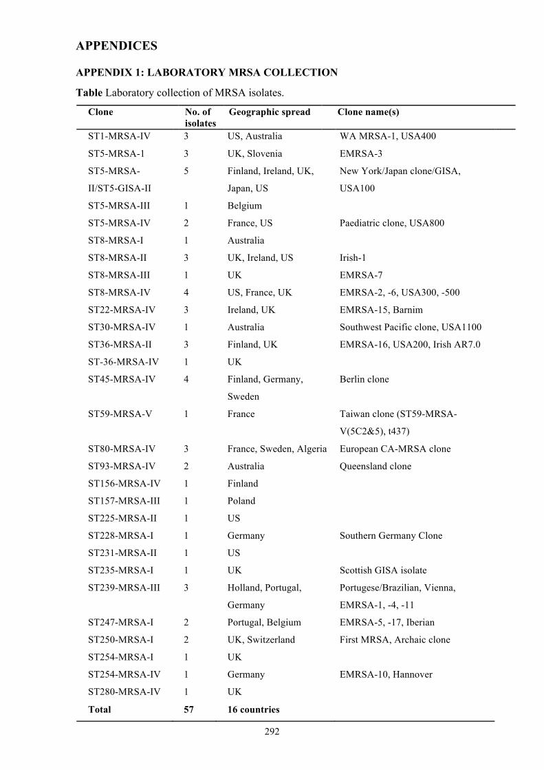
292
APPENDICES
APPENDIX 1: LABORATORY MRSA COLLECTION
Table Laboratory collection of MRSA isolates.
Clone No. of isolates
Geographic spread Clone name(s)
ST1-MRSA-IV 3 US, Australia WA MRSA-1, USA400
ST5-MRSA-1 3 UK, Slovenia EMRSA-3
ST5-MRSA-
II/ST5-GISA-II
5 Finland, Ireland, UK,
Japan, US
New York/Japan clone/GISA,
USA100
ST5-MRSA-III 1 Belgium
ST5-MRSA-IV 2 France, US Paediatric clone, USA800
ST8-MRSA-I 1 Australia
ST8-MRSA-II 3 UK, Ireland, US Irish-1
ST8-MRSA-III 1 UK EMRSA-7
ST8-MRSA-IV 4 US, France, UK EMRSA-2, -6, USA300, -500
ST22-MRSA-IV 3 Ireland, UK EMRSA-15, Barnim
ST30-MRSA-IV 1 Australia Southwest Pacific clone, USA1100
ST36-MRSA-II 3 Finland, UK EMRSA-16, USA200, Irish AR7.0
ST-36-MRSA-IV 1 UK
ST45-MRSA-IV 4 Finland, Germany,
Sweden
Berlin clone
ST59-MRSA-V 1 France Taiwan clone (ST59-MRSA-
V(5C2&5), t437)
ST80-MRSA-IV 3 France, Sweden, Algeria European CA-MRSA clone
ST93-MRSA-IV 2 Australia Queensland clone
ST156-MRSA-IV 1 Finland
ST157-MRSA-III 1 Poland
ST225-MRSA-II 1 US
ST228-MRSA-I 1 Germany Southern Germany Clone
ST231-MRSA-II 1 US
ST235-MRSA-I 1 UK Scottish GISA isolate
ST239-MRSA-III 3 Holland, Portugal,
Germany
Portugese/Brazilian, Vienna,
EMRSA-1, -4, -11
ST247-MRSA-I 2 Portugal, Belgium EMRSA-5, -17, Iberian
ST250-MRSA-I 2 UK, Switzerland First MRSA, Archaic clone
ST254-MRSA-I 1 UK
ST254-MRSA-IV 1 Germany EMRSA-10, Hannover
ST280-MRSA-IV 1 UK
Total 57 16 countries
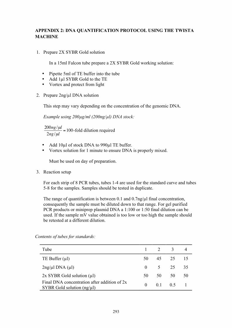
293
APPENDIX 2: DNA QUANTIFICATION PROTOCOL USING THE TWISTA MACHINE
1. Prepare 2X SYBR Gold solution In a 15ml Falcon tube prepare a 2X SYBR Gold working solution:
• Pipette 5ml of TE buffer into the tube • Add 1µl SYBR Gold to the TE • Vortex and protect from light
2. Prepare 2ng/µl DNA solution
This step may vary depending on the concentration of the genomic DNA.
Example using 200µg/ml (200ng/µl) DNA stock:
€
200ng /µl2ng /µl
=100-fold dilution required
• Add 10µl of stock DNA to 990µl TE buffer. • Vortex solution for 1 minute to ensure DNA is properly mixed. Must be used on day of preparation.
3. Reaction setup For each strip of 8 PCR tubes, tubes 1-4 are used for the standard curve and tubes 5-8 for the samples. Samples should be tested in duplicate. The range of quantification is between 0.1 and 0.7ng/µl final concentration, consequently the sample must be diluted down to that range. For gel purified PCR products or miniprep plasmid DNA a 1:100 or 1:50 final dilution can be used. If the sample mV value obtained is too low or too high the sample should be retested at a different dilution.
Contents of tubes for standards:
Tube 1 2 3 4
TE Buffer (µl) 50 45 25 15
2ng/µl DNA (µl) 0 5 25 35
2x SYBR Gold solution (µl) 50 50 50 50 Final DNA concentration after addition of 2x SYBR Gold solution (ng/µl) 0 0.1 0.5 1
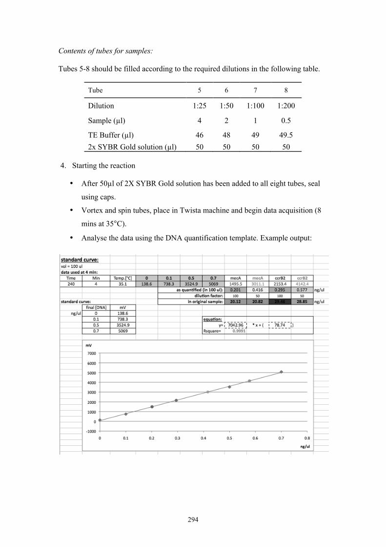
294
Contents of tubes for samples: Tubes 5-8 should be filled according to the required dilutions in the following table.
Tube 5 6 7 8
Dilution 1:25 1:50 1:100 1:200
Sample (µl) 4 2 1 0.5
TE Buffer (µl) 46 48 49 49.5 2x SYBR Gold solution (µl) 50 50 50 50
4. Starting the reaction
• After 50µl of 2X SYBR Gold solution has been added to all eight tubes, seal
using caps.
• Vortex and spin tubes, place in Twista machine and begin data acquisition (8
mins at 35°C).
• Analyse the data using the DNA quantification template. Example output:
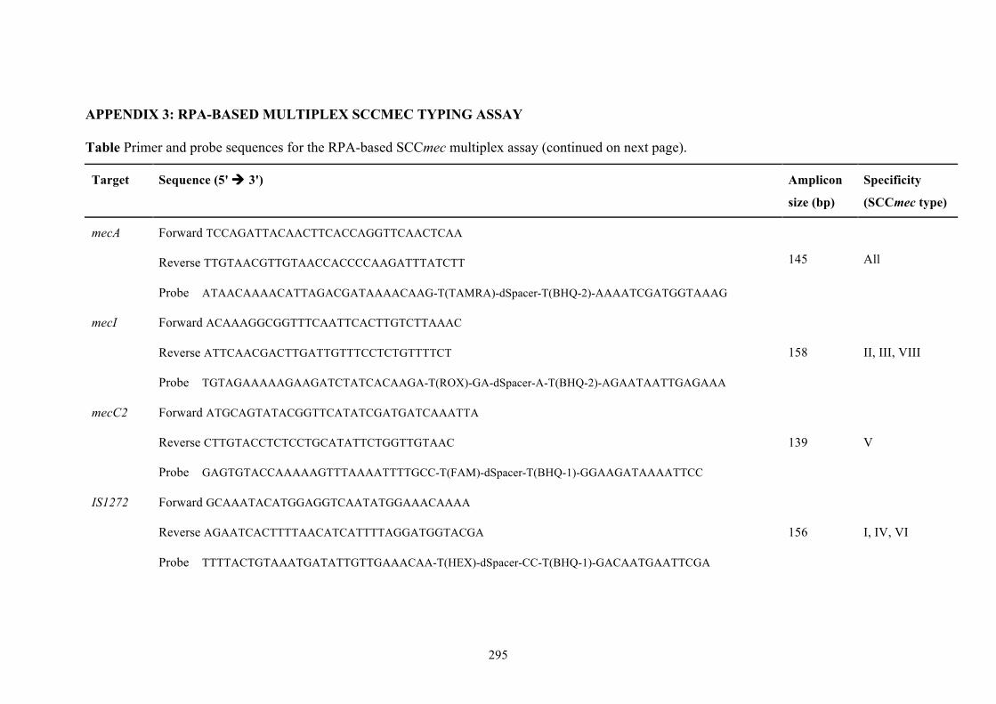
295
APPENDIX 3: RPA-BASED MULTIPLEX SCCMEC TYPING ASSAY
Table Primer and probe sequences for the RPA-based SCCmec multiplex assay (continued on next page).
Target Sequence (5' 3') Amplicon
size (bp)
Specificity
(SCCmec type)
mecA Forward TCCAGATTACAACTTCACCAGGTTCAACTCAA
Reverse TTGTAACGTTGTAACCACCCCAAGATTTATCTT
Probe ATAACAAAACATTAGACGATAAAACAAG-T(TAMRA)-dSpacer-T(BHQ-2)-AAAATCGATGGTAAAG
145 All
mecI Forward ACAAAGGCGGTTTCAATTCACTTGTCTTAAAC
Reverse ATTCAACGACTTGATTGTTTCCTCTGTTTTCT
Probe TGTAGAAAAAGAAGATCTATCACAAGA-T(ROX)-GA-dSpacer-A-T(BHQ-2)-AGAATAATTGAGAAA
158 II, III, VIII
mecC2 Forward ATGCAGTATACGGTTCATATCGATGATCAAATTA
Reverse CTTGTACCTCTCCTGCATATTCTGGTTGTAAC
Probe GAGTGTACCAAAAAGTTTAAAATTTTGCC-T(FAM)-dSpacer-T(BHQ-1)-GGAAGATAAAATTCC
139 V
IS1272 Forward GCAAATACATGGAGGTCAATATGGAAACAAAA
Reverse AGAATCACTTTTAACATCATTTTAGGATGGTACGA
Probe TTTTACTGTAAATGATATTGTTGAAACAA-T(HEX)-dSpacer-CC-T(BHQ-1)-GACAATGAATTCGA
156 I, IV, VI

296
ccrB1 Forward TGAACGTATGGAAGTCAAAAATTCAACAGGCA
Reverse GGAAGATTGCCTTGATAATAGCCCTCTAAAGC
Probe GCAAGTTTTTCCGAATTCGAAAGAAA-T(ROX)-ACA-dSpacer-T(BHQ-2)-TTTAGAGAATATTTAC
150 I
ccrB2 Forward ACCACGGTCAGCGTATATATCTTTAACTTCAA
Reverse TGCAACAACTTAAAACAAAACGTGTCGGTATC
Probe TATCAACAGAAATGCAAAGCACAGAAGGT-T(HEX)-dSpacer-T(BHQ-1)-AGTATCGACGGACAAA
158 II, IV
ccrB4 Forward TGCAAACGGATGGTTACAGTATTCAWGGTCAAT
Reverse CTTTTAACATACGTTGTAATTCWGGACGTTG
Probe ACGACTTCATAACCTTGAAATTGACAATAC-T(TAMRA)-C-dSpacer-G-T(BHQ-2)-AAGTTGATTYAATTGACC
150 VI, VIII
ccrC Forward CATCCAATGCTTCTGGAGAAGTACTCGTTACA
Reverse TGATAATTTGTAGCCTAAAACACGACCGGTGA
Probe TGGAGAAGTACTCGTTACAATGTTTGGGT-T(FAM)-dSpacer-A-T(BHQ-1)-AGGATCTATMGAACG
155 III, V, VII
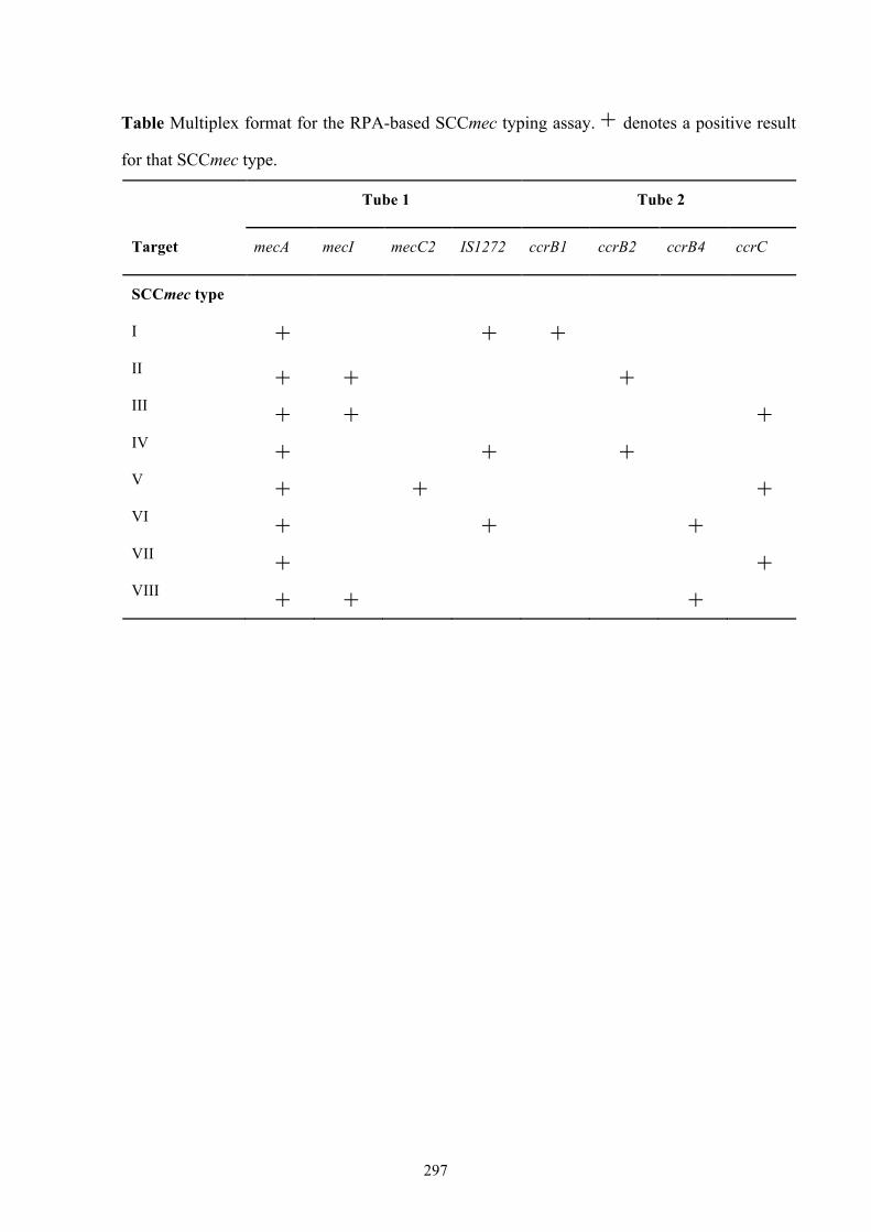
297
Table Multiplex format for the RPA-based SCCmec typing assay. + denotes a positive result
for that SCCmec type.
Tube 1 Tube 2
Target mecA mecI mecC2 IS1272 ccrB1 ccrB2 ccrB4 ccrC
SCCmec type
I
+ +
+
II + + + III + + + IV + + + V + + + VI + + + VII + + VIII + + +
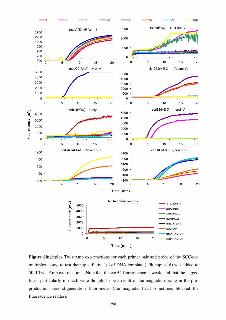
298
Figure Singleplex TwistAmp exo reactions for each primer pair and probe of the SCCmec
multiplex assay, to test their specificity. 1µl of DNA template (~9k copies/µl) was added to
50µl TwistAmp exo reactions. Note that the ccrB4 fluorescence is weak, and that the jagged
lines, particularly in mecI, were thought to be a result of the magnetic mixing in the pre-
production, second-generation fluorometer (the magnetic bead sometimes blocked the
fluorescence reader).
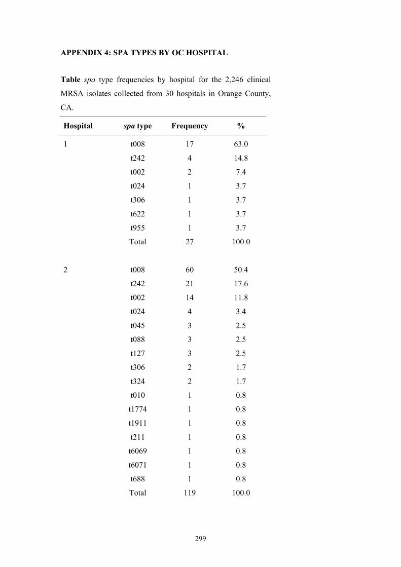
299
APPENDIX 4: SPA TYPES BY OC HOSPITAL
Table spa type frequencies by hospital for the 2,246 clinical
MRSA isolates collected from 30 hospitals in Orange County,
CA.
Hospital spa type Frequency %
1 t008 17 63.0
t242 4 14.8
t002 2 7.4
t024 1 3.7
t306 1 3.7
t622 1 3.7
t955 1 3.7
Total 27 100.0
2 t008 60 50.4
t242 21 17.6
t002 14 11.8
t024 4 3.4
t045 3 2.5
t088 3 2.5
t127 3 2.5
t306 2 1.7
t324 2 1.7
t010 1 0.8
t1774 1 0.8
t1911 1 0.8
t211 1 0.8
t6069 1 0.8
t6071 1 0.8
t688 1 0.8
Total 119 100.0
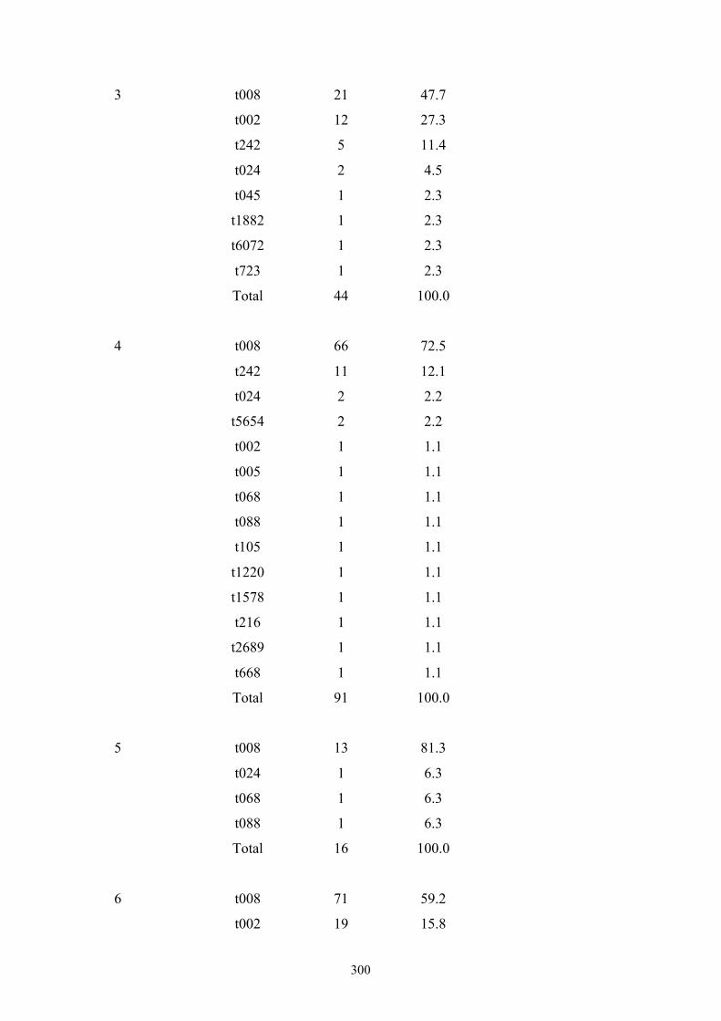
300
3 t008 21 47.7
t002 12 27.3
t242 5 11.4
t024 2 4.5
t045 1 2.3
t1882 1 2.3
t6072 1 2.3
t723 1 2.3
Total 44 100.0
4 t008 66 72.5
t242 11 12.1
t024 2 2.2
t5654 2 2.2
t002 1 1.1
t005 1 1.1
t068 1 1.1
t088 1 1.1
t105 1 1.1
t1220 1 1.1
t1578 1 1.1
t216 1 1.1
t2689 1 1.1
t668 1 1.1
Total 91 100.0
5 t008 13 81.3
t024 1 6.3
t068 1 6.3
t088 1 6.3
Total 16 100.0
6 t008 71 59.2
t002 19 15.8
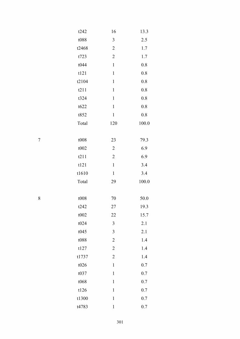
301
t242 16 13.3
t088 3 2.5
t2468 2 1.7
t723 2 1.7
t044 1 0.8
t121 1 0.8
t2104 1 0.8
t211 1 0.8
t324 1 0.8
t622 1 0.8
t852 1 0.8
Total 120 100.0
7 t008 23 79.3
t002 2 6.9
t211 2 6.9
t121 1 3.4
t1610 1 3.4
Total 29 100.0
8 t008 70 50.0
t242 27 19.3
t002 22 15.7
t024 3 2.1
t045 3 2.1
t088 2 1.4
t127 2 1.4
t1737 2 1.4
t026 1 0.7
t037 1 0.7
t068 1 0.7
t126 1 0.7
t1300 1 0.7
t4783 1 0.7
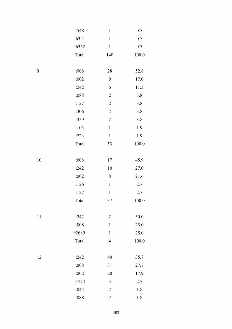
302
t548 1 0.7
t6321 1 0.7
t6352 1 0.7
Total 140 100.0
9 t008 28 52.8
t002 9 17.0
t242 6 11.3
t088 2 3.8
t127 2 3.8
t306 2 3.8
t359 2 3.8
t105 1 1.9
t723 1 1.9
Total 53 100.0
10 t008 17 45.9
t242 10 27.0
t002 8 21.6
t126 1 2.7
t127 1 2.7
Total 37 100.0
11 t242 2 50.0
t008 1 25.0
t2689 1 25.0
Total 4 100.0
12 t242 40 35.7
t008 31 27.7
t002 20 17.9
t1774 3 2.7
t045 2 1.8
t088 2 1.8
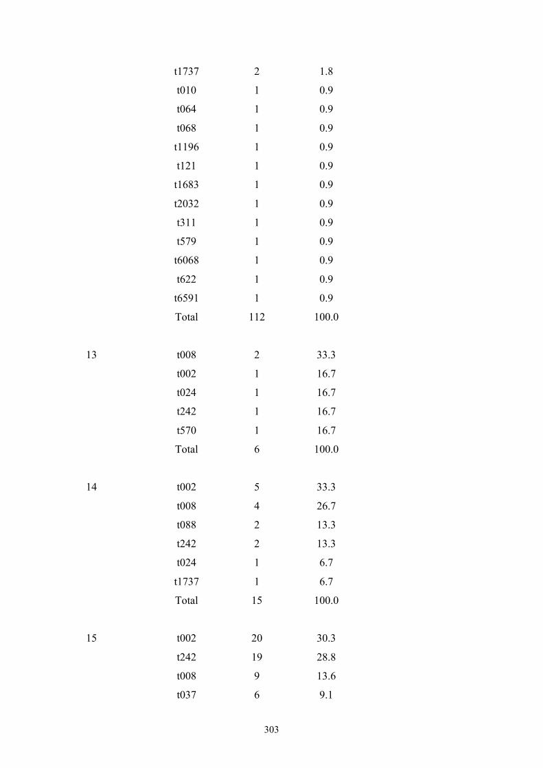
303
t1737 2 1.8
t010 1 0.9
t064 1 0.9
t068 1 0.9
t1196 1 0.9
t121 1 0.9
t1683 1 0.9
t2032 1 0.9
t311 1 0.9
t579 1 0.9
t6068 1 0.9
t622 1 0.9
t6591 1 0.9
Total 112 100.0
13 t008 2 33.3
t002 1 16.7
t024 1 16.7
t242 1 16.7
t570 1 16.7
Total 6 100.0
14 t002 5 33.3
t008 4 26.7
t088 2 13.3
t242 2 13.3
t024 1 6.7
t1737 1 6.7
Total 15 100.0
15 t002 20 30.3
t242 19 28.8
t008 9 13.6
t037 6 9.1
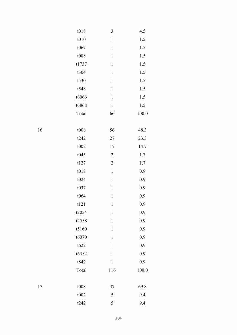
304
t018 3 4.5
t010 1 1.5
t067 1 1.5
t088 1 1.5
t1737 1 1.5
t304 1 1.5
t530 1 1.5
t548 1 1.5
t6066 1 1.5
t6868 1 1.5
Total 66 100.0
16 t008 56 48.3
t242 27 23.3
t002 17 14.7
t045 2 1.7
t127 2 1.7
t018 1 0.9
t024 1 0.9
t037 1 0.9
t064 1 0.9
t121 1 0.9
t2054 1 0.9
t2558 1 0.9
t5160 1 0.9
t6070 1 0.9
t622 1 0.9
t6352 1 0.9
t842 1 0.9
Total 116 100.0
17 t008 37 69.8
t002 5 9.4
t242 5 9.4
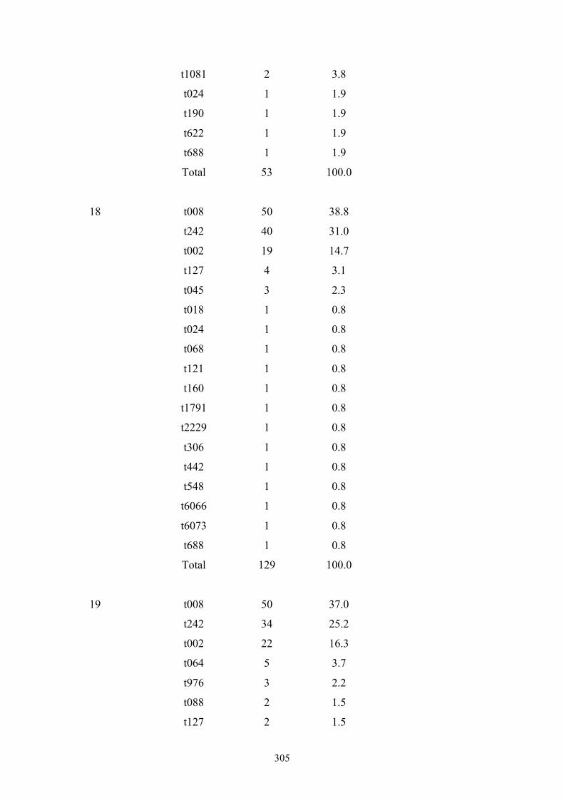
305
t1081 2 3.8
t024 1 1.9
t190 1 1.9
t622 1 1.9
t688 1 1.9
Total 53 100.0
18 t008 50 38.8
t242 40 31.0
t002 19 14.7
t127 4 3.1
t045 3 2.3
t018 1 0.8
t024 1 0.8
t068 1 0.8
t121 1 0.8
t160 1 0.8
t1791 1 0.8
t2229 1 0.8
t306 1 0.8
t442 1 0.8
t548 1 0.8
t6066 1 0.8
t6073 1 0.8
t688 1 0.8
Total 129 100.0
19 t008 50 37.0
t242 34 25.2
t002 22 16.3
t064 5 3.7
t976 3 2.2
t088 2 1.5
t127 2 1.5
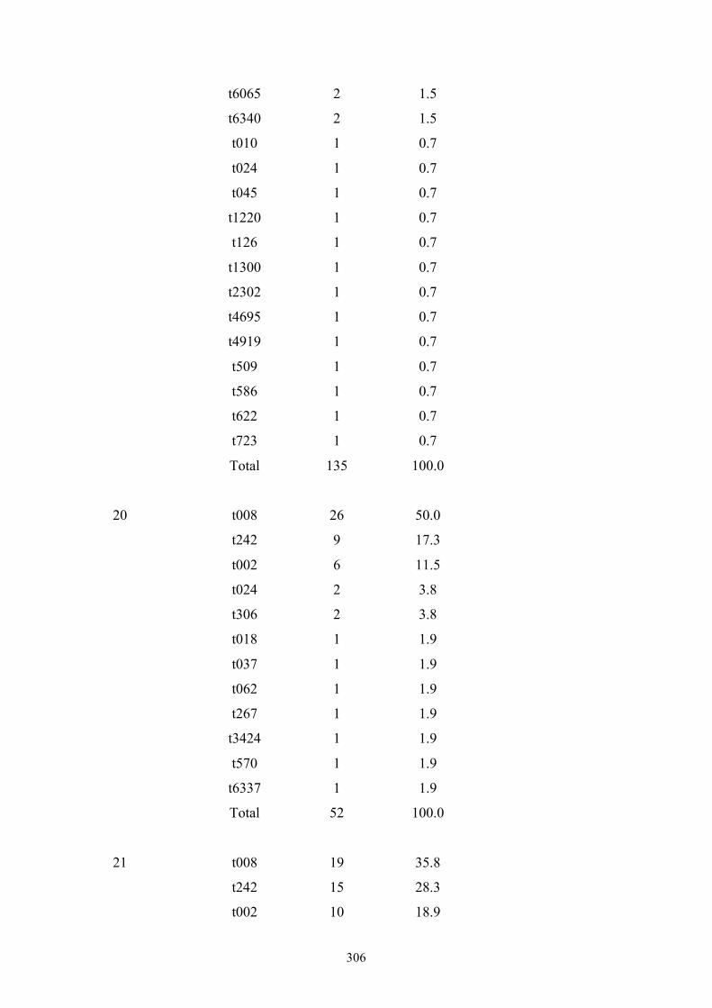
306
t6065 2 1.5
t6340 2 1.5
t010 1 0.7
t024 1 0.7
t045 1 0.7
t1220 1 0.7
t126 1 0.7
t1300 1 0.7
t2302 1 0.7
t4695 1 0.7
t4919 1 0.7
t509 1 0.7
t586 1 0.7
t622 1 0.7
t723 1 0.7
Total 135 100.0
20 t008 26 50.0
t242 9 17.3
t002 6 11.5
t024 2 3.8
t306 2 3.8
t018 1 1.9
t037 1 1.9
t062 1 1.9
t267 1 1.9
t3424 1 1.9
t570 1 1.9
t6337 1 1.9
Total 52 100.0
21 t008 19 35.8
t242 15 28.3
t002 10 18.9

307
t304 2 3.8
t306 2 3.8
t018 1 1.9
t045 1 1.9
t121 1 1.9
t1578 1 1.9
t6212 1 1.9
Total 53 100.0
22 t008 8 40.0
t242 4 20.0
t2468 2 10.0
t002 1 5.0
t024 1 5.0
t037 1 5.0
t6238 1 5.0
t967 1 5.0
t976 1 5.0
Total 20 100.0
23 t008 42 35.0
t242 35 29.2
t002 20 16.7
t037 7 5.8
t1860 3 2.5
t024 2 1.7
t105 2 1.7
t126 1 0.8
t1627 1 0.8
t1737 1 0.8
t2173 1 0.8
t6072 1 0.8
t6338 1 0.8
t6339 1 0.8
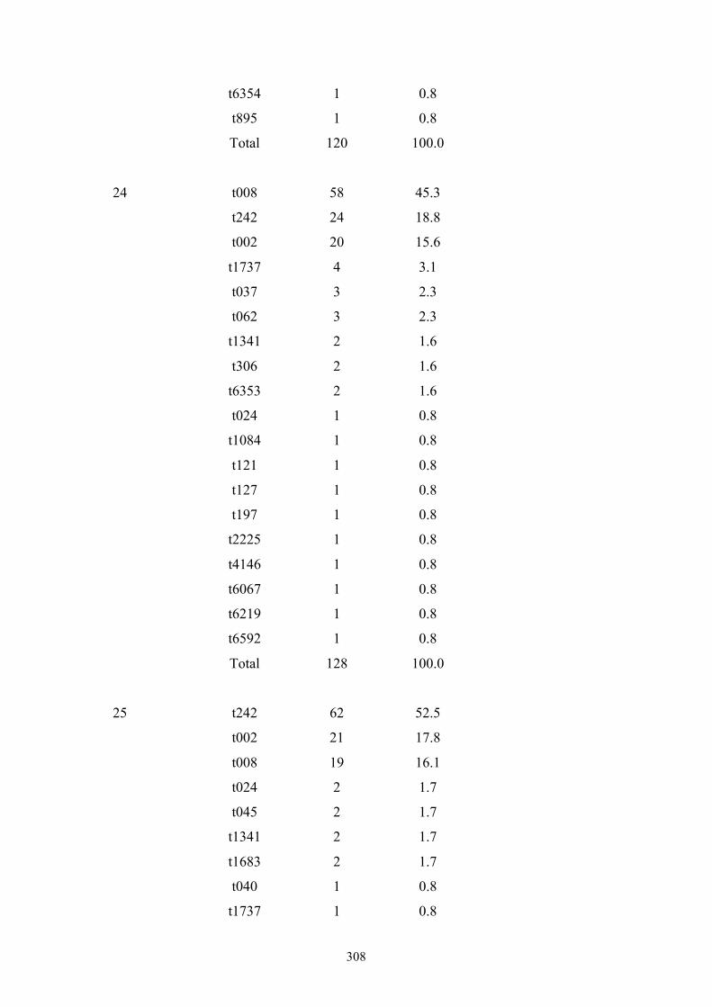
308
t6354 1 0.8
t895 1 0.8
Total 120 100.0
24 t008 58 45.3
t242 24 18.8
t002 20 15.6
t1737 4 3.1
t037 3 2.3
t062 3 2.3
t1341 2 1.6
t306 2 1.6
t6353 2 1.6
t024 1 0.8
t1084 1 0.8
t121 1 0.8
t127 1 0.8
t197 1 0.8
t2225 1 0.8
t4146 1 0.8
t6067 1 0.8
t6219 1 0.8
t6592 1 0.8
Total 128 100.0
25 t242 62 52.5
t002 21 17.8
t008 19 16.1
t024 2 1.7
t045 2 1.7
t1341 2 1.7
t1683 2 1.7
t040 1 0.8
t1737 1 0.8
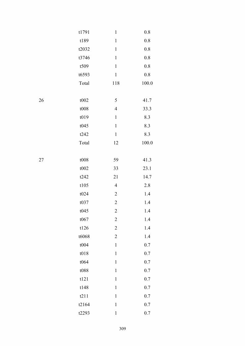
309
t1791 1 0.8
t189 1 0.8
t2032 1 0.8
t3746 1 0.8
t509 1 0.8
t6593 1 0.8
Total 118 100.0
26 t002 5 41.7
t008 4 33.3
t019 1 8.3
t045 1 8.3
t242 1 8.3
Total 12 100.0
27 t008 59 41.3
t002 33 23.1
t242 21 14.7
t105 4 2.8
t024 2 1.4
t037 2 1.4
t045 2 1.4
t067 2 1.4
t126 2 1.4
t6068 2 1.4
t004 1 0.7
t018 1 0.7
t064 1 0.7
t088 1 0.7
t121 1 0.7
t148 1 0.7
t211 1 0.7
t2164 1 0.7
t2293 1 0.7
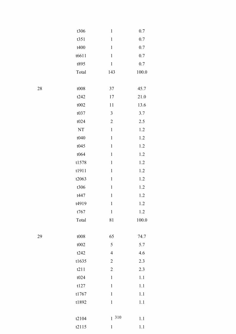
310
t306 1 0.7
t351 1 0.7
t400 1 0.7
t6611 1 0.7
t895 1 0.7
Total 143 100.0
28 t008 37 45.7
t242 17 21.0
t002 11 13.6
t037 3 3.7
t024 2 2.5
NT 1 1.2
t040 1 1.2
t045 1 1.2
t064 1 1.2
t1578 1 1.2
t1911 1 1.2
t2063 1 1.2
t306 1 1.2
t447 1 1.2
t4919 1 1.2
t767 1 1.2
Total 81 100.0
29 t008 65 74.7
t002 5 5.7
t242 4 4.6
t1635 2 2.3
t211 2 2.3
t024 1 1.1
t127 1 1.1
t1767 1 1.1
t1892 1 1.1
t2104 1 1.1
t2115 1 1.1
t2206 1 1.1
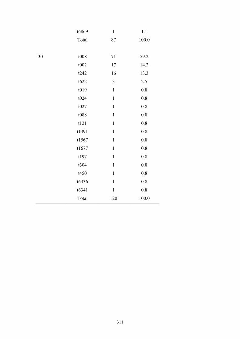
311
t6869 1 1.1
Total 87 100.0
30 t008 71 59.2
t002 17 14.2
t242 16 13.3
t622 3 2.5
t019 1 0.8
t024 1 0.8
t027 1 0.8
t088 1 0.8
t121 1 0.8
t1391 1 0.8
t1567 1 0.8
t1677 1 0.8
t197 1 0.8
t304 1 0.8
t450 1 0.8
t6336 1 0.8
t6341 1 0.8
Total 120 100.0
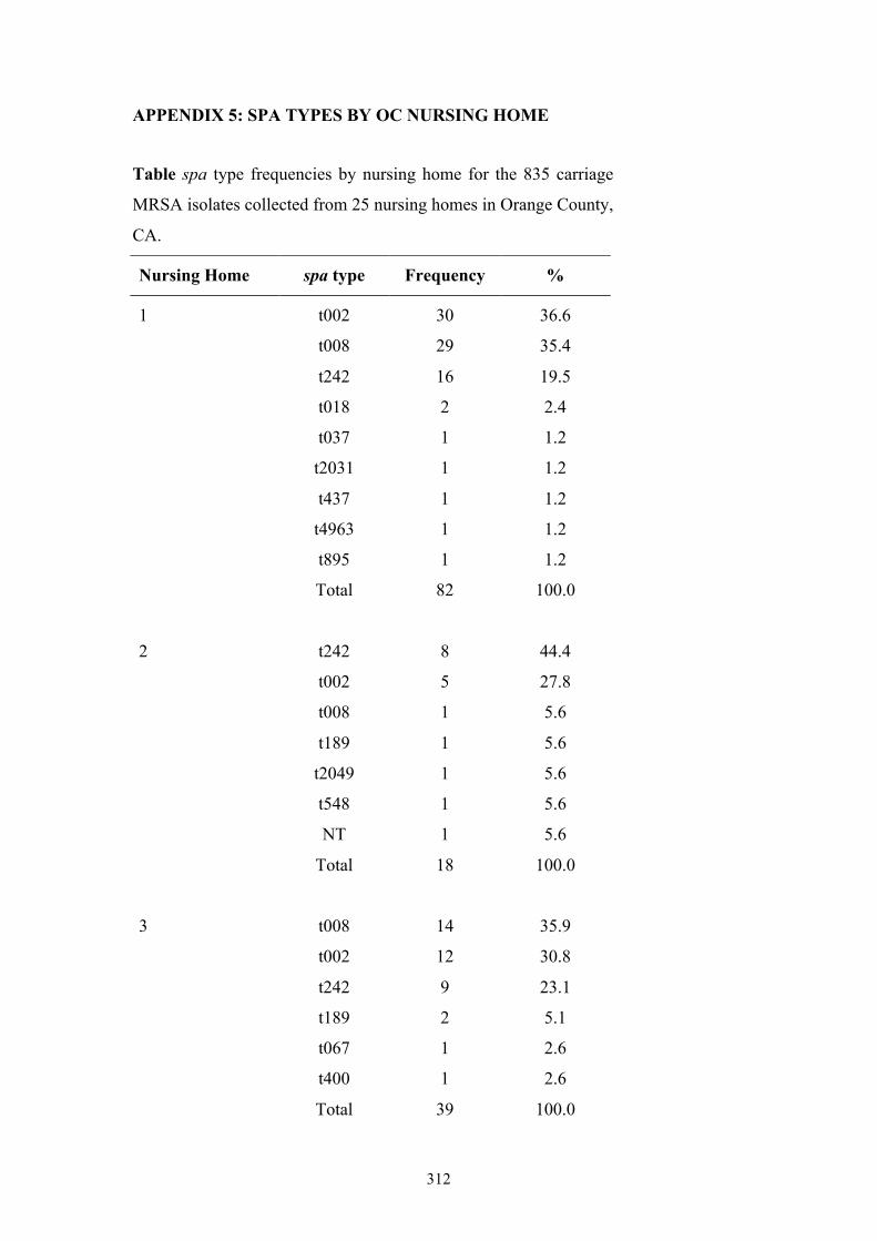
312
APPENDIX 5: SPA TYPES BY OC NURSING HOME
Table spa type frequencies by nursing home for the 835 carriage
MRSA isolates collected from 25 nursing homes in Orange County,
CA.
Nursing Home spa type Frequency %
1 t002 30 36.6
t008 29 35.4
t242 16 19.5
t018 2 2.4
t037 1 1.2
t2031 1 1.2
t437 1 1.2
t4963 1 1.2
t895 1 1.2
Total 82 100.0
2 t242 8 44.4
t002 5 27.8
t008 1 5.6
t189 1 5.6
t2049 1 5.6
t548 1 5.6
NT 1 5.6
Total 18 100.0
3 t008 14 35.9
t002 12 30.8
t242 9 23.1
t189 2 5.1
t067 1 2.6
t400 1 2.6
Total 39 100.0
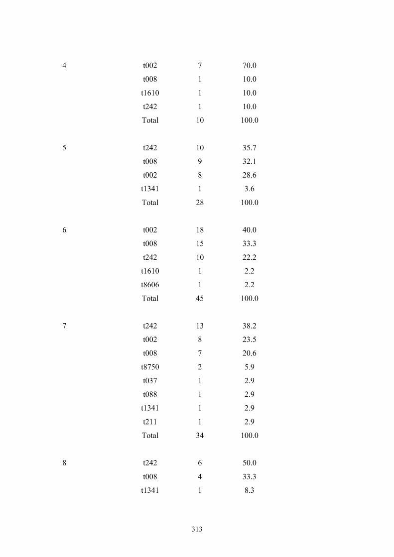
313
4 t002 7 70.0
t008 1 10.0
t1610 1 10.0
t242 1 10.0
Total 10 100.0
5 t242 10 35.7
t008 9 32.1
t002 8 28.6
t1341 1 3.6
Total 28 100.0
6 t002 18 40.0
t008 15 33.3
t242 10 22.2
t1610 1 2.2
t8606 1 2.2
Total 45 100.0
7 t242 13 38.2
t002 8 23.5
t008 7 20.6
t8750 2 5.9
t037 1 2.9
t088 1 2.9
t1341 1 2.9
t211 1 2.9
Total 34 100.0
8 t242 6 50.0
t008 4 33.3
t1341 1 8.3
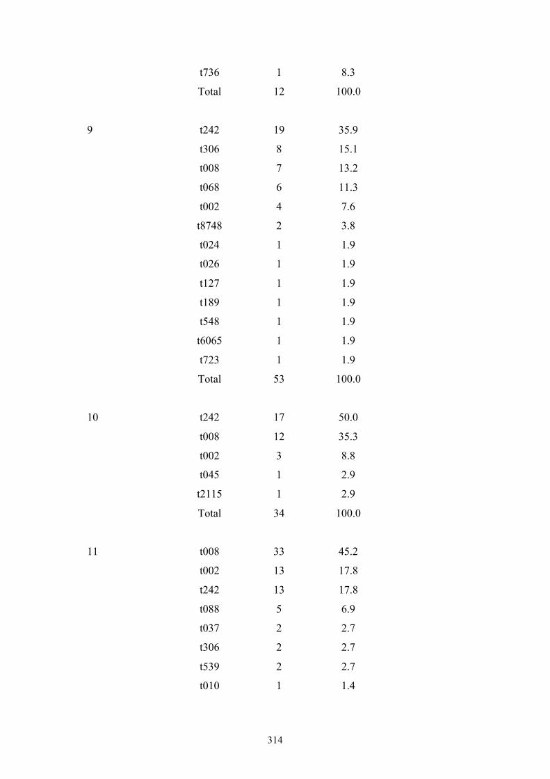
314
t736 1 8.3
Total 12 100.0
9 t242 19 35.9
t306 8 15.1
t008 7 13.2
t068 6 11.3
t002 4 7.6
t8748 2 3.8
t024 1 1.9
t026 1 1.9
t127 1 1.9
t189 1 1.9
t548 1 1.9
t6065 1 1.9
t723 1 1.9
Total 53 100.0
10 t242 17 50.0
t008 12 35.3
t002 3 8.8
t045 1 2.9
t2115 1 2.9
Total 34 100.0
11 t008 33 45.2
t002 13 17.8
t242 13 17.8
t088 5 6.9
t037 2 2.7
t306 2 2.7
t539 2 2.7
t010 1 1.4
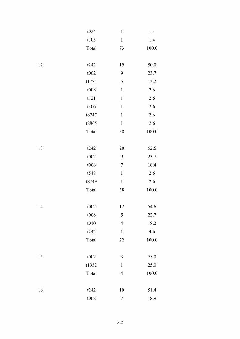
315
t024 1 1.4
t105 1 1.4
Total 73 100.0
12 t242 19 50.0
t002 9 23.7
t1774 5 13.2
t008 1 2.6
t121 1 2.6
t306 1 2.6
t8747 1 2.6
t8865 1 2.6
Total 38 100.0
13 t242 20 52.6
t002 9 23.7
t008 7 18.4
t548 1 2.6
t8749 1 2.6
Total 38 100.0
14 t002 12 54.6
t008 5 22.7
t010 4 18.2
t242 1 4.6
Total 22 100.0
15 t002 3 75.0
t1932 1 25.0
Total 4 100.0
16 t242 19 51.4
t008 7 18.9
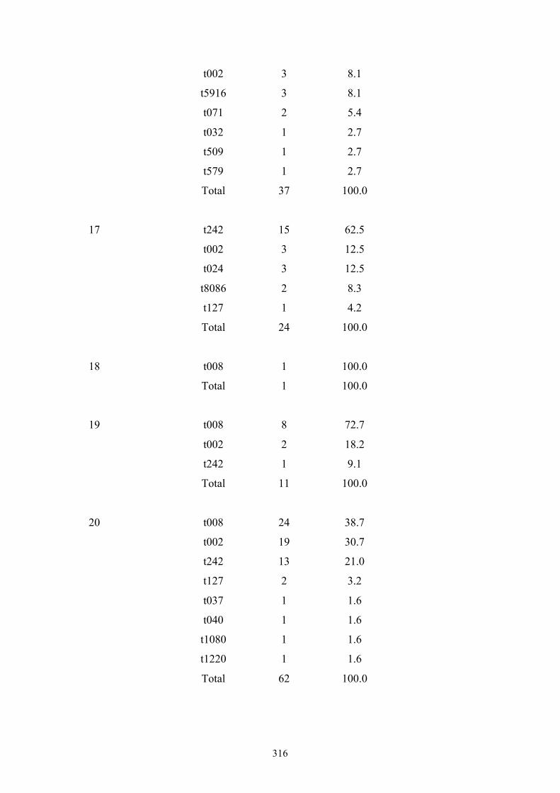
316
t002 3 8.1
t5916 3 8.1
t071 2 5.4
t032 1 2.7
t509 1 2.7
t579 1 2.7
Total 37 100.0
17 t242 15 62.5
t002 3 12.5
t024 3 12.5
t8086 2 8.3
t127 1 4.2
Total 24 100.0
18 t008 1 100.0
Total 1 100.0
19 t008 8 72.7
t002 2 18.2
t242 1 9.1
Total 11 100.0
20 t008 24 38.7
t002 19 30.7
t242 13 21.0
t127 2 3.2
t037 1 1.6
t040 1 1.6
t1080 1 1.6
t1220 1 1.6
Total 62 100.0
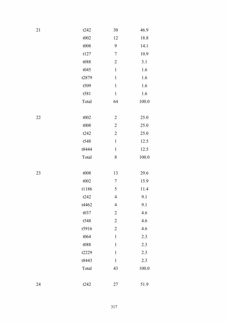
317
21 t242 30 46.9
t002 12 18.8
t008 9 14.1
t127 7 10.9
t088 2 3.1
t045 1 1.6
t2879 1 1.6
t509 1 1.6
t581 1 1.6
Total 64 100.0
22 t002 2 25.0
t008 2 25.0
t242 2 25.0
t548 1 12.5
t8444 1 12.5
Total 8 100.0
23 t008 13 29.6
t002 7 15.9
t1186 5 11.4
t242 4 9.1
t4462 4 9.1
t037 2 4.6
t548 2 4.6
t5916 2 4.6
t064 1 2.3
t088 1 2.3
t2229 1 2.3
t8443 1 2.3
Total 43 100.0
24 t242 27 51.9
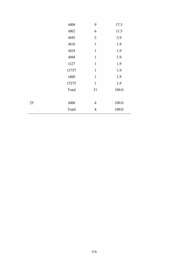
318
t008 9 17.3
t002 6 11.5
t045 2 3.9
t018 1 1.9
t024 1 1.9
t088 1 1.9
t127 1 1.9
t1737 1 1.9
t400 1 1.9
t7275 1 1.9
Total 51 100.0
25 t008 4 100.0
Total 4 100.0
319
APPENDIX 6: PUBLICATIONS
Hudson LO, Murphy CR, Spratt BG, Enright MC, Terpstra L, Gombosev A, et
al. 2012. Differences in Methicillin-Resistant Staphylococcus aureus Strains Isolated
from Paediatric and Adult Patients from Hospitals in a Large County in California. J
Clin Microbiol. 50: 573-9.
Ke W, Huang SS, Hudson LO, Elkins KR, Nguyen CC, Spratt BG, et al. 2012.
Patient sharing and population genetic structure of methicillin-resistant
Staphylococcus aureus. Proc Natl Acad Sci U S A. 109: 6763-8.
The above manuscripts are presented in their published form on the following pages:
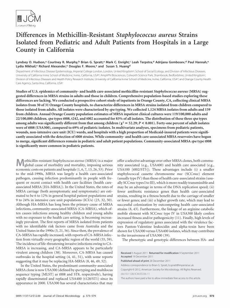
Differences in Methicillin-Resistant Staphylococcus aureus StrainsIsolated from Pediatric and Adult Patients from Hospitals in a LargeCounty in California
Lyndsey O. Hudson,a Courtney R. Murphy,b Brian G. Spratt,a Mark C. Enright,c Leah Terpstra,d Adrijana Gombosev,d Paul Hannah,e
Lydia Mikhail,e Richard Alexander,e Douglas F. Moore,e and Susan S. Huangd
Department of Infectious Disease Epidemiology, Imperial College London, London, United Kingdoma; School of Social Ecology, and Division of Infectious Diseases,University of California Irvine School of Medicine, Irvine, California, USAb; AmpliPhi Biosciences, Colworth Science Park, Sharnbrook, Bedfordshire, United Kingdomc;Division of Infectious Diseases and Health Policy Research Institute, University of California Irvine School of Medicine, Irvine, California, USAd; and Orange County HealthCare Agency, Santa Ana, California, USAe
Studies of U.S. epidemics of community- and health care-associated methicillin-resistant Staphylococcus aureus (MRSA) sug-gested differences in MRSA strains in adults and those in children. Comprehensive population-based studies exploring thesedifferences are lacking. We conducted a prospective cohort study of inpatients in Orange County, CA, collecting clinical MRSAisolates from 30 of 31 Orange County hospitals, to characterize differences in MRSA strains isolated from children compared tothose isolated from adults. All isolates were characterized by spa typing. We collected 1,124 MRSA isolates from adults and 159from children. Annual Orange County population estimates of MRSA inpatient clinical cultures were 119/100,000 adults and22/100,000 children. spa types t008, t242, and t002 accounted for 83% of all isolates. The distribution of these three spa typesamong adults was significantly different from that among children (!2 " 52.29; P < 0.001). Forty-one percent of adult isolateswere of t008 (USA300), compared to 69% of pediatric isolates. In multivariate analyses, specimens from pediatric patients,wounds, non-intensive care unit (ICU) wards, and hospitals with a high proportion of Medicaid-insured patients were signifi-cantly associated with the detection of t008 strains. While community- and health care-associated MRSA reservoirs have begunto merge, significant differences remain in pediatric and adult patient populations. Community-associated MRSA spa type t008is significantly more common in pediatric patients.
Methicillin-resistant Staphylococcus aureus (MRSA) is a majorglobal cause of morbidity and mortality, imposing serious
economic costs on patients and hospitals (1, 6, 7, 13, 26, 53). Priorto the mid-1990s, MRSA was largely a health care-associatedpathogen, causing infection predominantly in people with fre-quent or recent contact with health care facilities (health care-associated MRSA [HA-MRSA]). In the United States, the rates ofMRSA carriage (both asymptomatic and symptomatic) are esti-mated to be 6 to 12% in general hospital patient populations and9 to 24% in intensive care unit populations (ICUs) (23, 32, 50).Although HA-MRSA has long been the primary cause of MRSAinfections, community-associated MRSA (CA-MRSA), which of-ten causes infections among healthy children and young adultswith no exposure to the health care setting, is becoming increas-ingly prevalent. The first reports of MRSA isolated from patientswith no identifiable risk factors came from Australia and theUnited States in the 1990s (5, 21, 56). Since then, the prevalence ofCA-MRSA has rapidly increased, with reports of CA-MRSA infec-tion from virtually every geographic region of the world (55, 59).The incidence of life-threatening invasive infections owing to CA-MRSA is increasing, and CA-MRSA appears to be particularlyvirulent among children (38). Moreover, CA-MRSA has causedoutbreaks in the hospital setting (4, 41, 51), with some reportssuggesting that it may be replacing HA-MRSA (8, 46, 49, 52).
In the United States, the predominant community-associatedMRSA clone is now USA300 (defined by spa typing and multilocussequence typing [MLST] as t008 and ST8, respectively), havingrapidly disseminated and replaced USA400 (t128/ST1) since itsappearance in 2000. USA300 has several characteristics that may
offer a selective advantage over other MRSA clones, both commu-nity associated (e.g., USA400) and health care associated (e.g.,USA100 [t002/ST5]). These advantages include (i) a smallerstaphylococcal cassette chromosome mec (SCCmec) element(usually type IV) than those of health care-associated strains (usu-ally SCCmec types I to III), which is more readily transmissible andmay be an advantage in terms of the DNA replication speed; (ii)fewer antibiotic resistance genes than health care-associatedstrains, resulting in a fitness benefit due to the carriage of smalleror fewer genes; and (iii) a higher growth rate, which may lead tosuccessful colonization by outcompeting health care-associatedstrains (8, 43). Furthermore, the linkage of an arginine catabolicmobile element with SCCmec type IV in USA300 likely confersincreased fitness and/or pathogenicity (11). Finally, high levels ofexpression of regulatory genes associated with the virulence fac-tors Panton-Valentine leukocidin and alpha-toxin have beenshown for USA300 versus USA400 isolates, which may contributeto the invasiveness of USA300 (39).
The phenotypic and genotypic differences between HA- and
Received 3 August 2011 Returned for modification 9 September 2011Accepted 19 December 2011
Published ahead of print 28 December 2011
Address correspondence to Brian G. Spratt, [email protected].
Copyright © 2012, American Society for Microbiology. All Rights Reserved.
doi:10.1128/JCM.05336-11
The authors have paid a fee to allow immediate free access to this article.
0095-1137/12/$12.00 Journal of Clinical Microbiology p. 573–579 jcm.asm.org 573
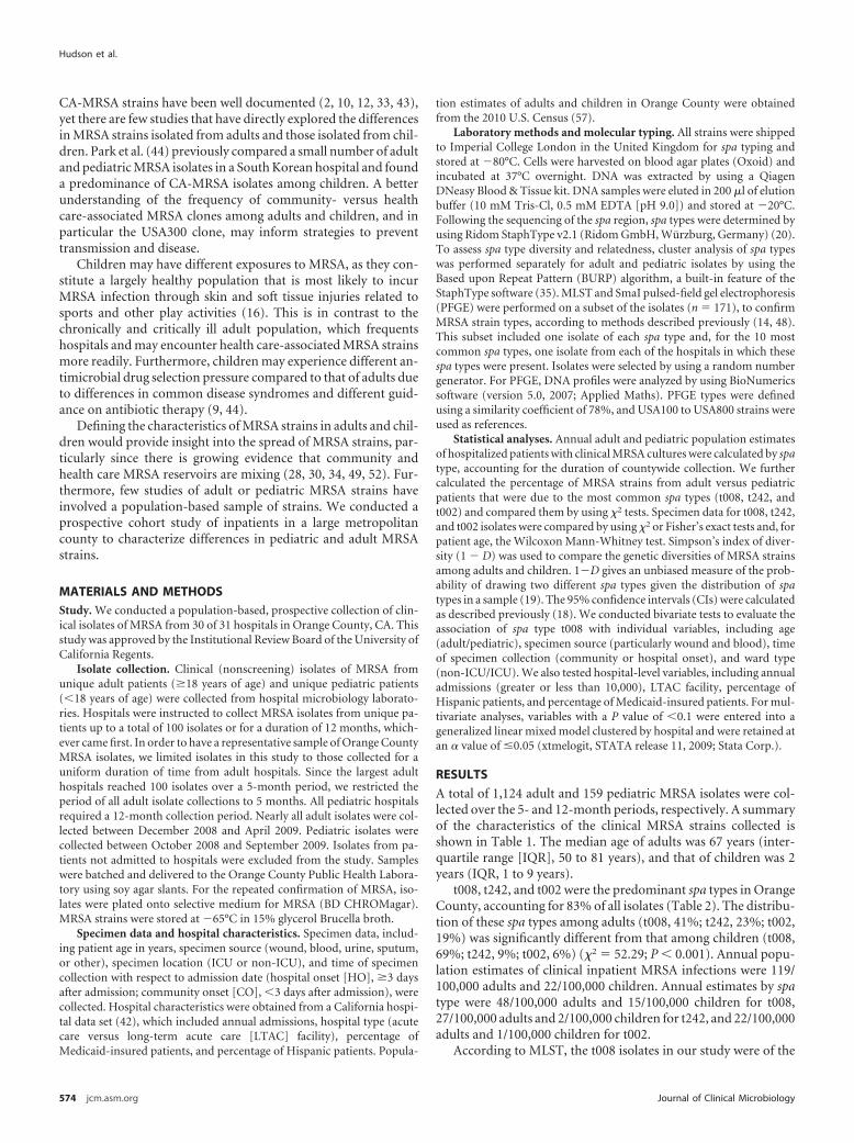
CA-MRSA strains have been well documented (2, 10, 12, 33, 43),yet there are few studies that have directly explored the differencesin MRSA strains isolated from adults and those isolated from chil-dren. Park et al. (44) previously compared a small number of adultand pediatric MRSA isolates in a South Korean hospital and founda predominance of CA-MRSA isolates among children. A betterunderstanding of the frequency of community- versus healthcare-associated MRSA clones among adults and children, and inparticular the USA300 clone, may inform strategies to preventtransmission and disease.
Children may have different exposures to MRSA, as they con-stitute a largely healthy population that is most likely to incurMRSA infection through skin and soft tissue injuries related tosports and other play activities (16). This is in contrast to thechronically and critically ill adult population, which frequentshospitals and may encounter health care-associated MRSA strainsmore readily. Furthermore, children may experience different an-timicrobial drug selection pressure compared to that of adults dueto differences in common disease syndromes and different guid-ance on antibiotic therapy (9, 44).
Defining the characteristics of MRSA strains in adults and chil-dren would provide insight into the spread of MRSA strains, par-ticularly since there is growing evidence that community andhealth care MRSA reservoirs are mixing (28, 30, 34, 49, 52). Fur-thermore, few studies of adult or pediatric MRSA strains haveinvolved a population-based sample of strains. We conducted aprospective cohort study of inpatients in a large metropolitancounty to characterize differences in pediatric and adult MRSAstrains.
MATERIALS AND METHODSStudy. We conducted a population-based, prospective collection of clin-ical isolates of MRSA from 30 of 31 hospitals in Orange County, CA. Thisstudy was approved by the Institutional Review Board of the University ofCalifornia Regents.
Isolate collection. Clinical (nonscreening) isolates of MRSA fromunique adult patients (!18 years of age) and unique pediatric patients(!18 years of age) were collected from hospital microbiology laborato-ries. Hospitals were instructed to collect MRSA isolates from unique pa-tients up to a total of 100 isolates or for a duration of 12 months, which-ever came first. In order to have a representative sample of Orange CountyMRSA isolates, we limited isolates in this study to those collected for auniform duration of time from adult hospitals. Since the largest adulthospitals reached 100 isolates over a 5-month period, we restricted theperiod of all adult isolate collections to 5 months. All pediatric hospitalsrequired a 12-month collection period. Nearly all adult isolates were col-lected between December 2008 and April 2009. Pediatric isolates werecollected between October 2008 and September 2009. Isolates from pa-tients not admitted to hospitals were excluded from the study. Sampleswere batched and delivered to the Orange County Public Health Labora-tory using soy agar slants. For the repeated confirmation of MRSA, iso-lates were plated onto selective medium for MRSA (BD CHROMagar).MRSA strains were stored at "65°C in 15% glycerol Brucella broth.
Specimen data and hospital characteristics. Specimen data, includ-ing patient age in years, specimen source (wound, blood, urine, sputum,or other), specimen location (ICU or non-ICU), and time of specimencollection with respect to admission date (hospital onset [HO], !3 daysafter admission; community onset [CO], !3 days after admission), werecollected. Hospital characteristics were obtained from a California hospi-tal data set (42), which included annual admissions, hospital type (acutecare versus long-term acute care [LTAC] facility), percentage ofMedicaid-insured patients, and percentage of Hispanic patients. Popula-
tion estimates of adults and children in Orange County were obtainedfrom the 2010 U.S. Census (57).
Laboratory methods and molecular typing. All strains were shippedto Imperial College London in the United Kingdom for spa typing andstored at "80°C. Cells were harvested on blood agar plates (Oxoid) andincubated at 37°C overnight. DNA was extracted by using a QiagenDNeasy Blood & Tissue kit. DNA samples were eluted in 200 "l of elutionbuffer (10 mM Tris-Cl, 0.5 mM EDTA [pH 9.0]) and stored at "20°C.Following the sequencing of the spa region, spa types were determined byusing Ridom StaphType v2.1 (Ridom GmbH, Würzburg, Germany) (20).To assess spa type diversity and relatedness, cluster analysis of spa typeswas performed separately for adult and pediatric isolates by using theBased upon Repeat Pattern (BURP) algorithm, a built-in feature of theStaphType software (35). MLST and SmaI pulsed-field gel electrophoresis(PFGE) were performed on a subset of the isolates (n # 171), to confirmMRSA strain types, according to methods described previously (14, 48).This subset included one isolate of each spa type and, for the 10 mostcommon spa types, one isolate from each of the hospitals in which thesespa types were present. Isolates were selected by using a random numbergenerator. For PFGE, DNA profiles were analyzed by using BioNumericssoftware (version 5.0, 2007; Applied Maths). PFGE types were definedusing a similarity coefficient of 78%, and USA100 to USA800 strains wereused as references.
Statistical analyses. Annual adult and pediatric population estimatesof hospitalized patients with clinical MRSA cultures were calculated by spatype, accounting for the duration of countywide collection. We furthercalculated the percentage of MRSA strains from adult versus pediatricpatients that were due to the most common spa types (t008, t242, andt002) and compared them by using #2 tests. Specimen data for t008, t242,and t002 isolates were compared by using #2 or Fisher’s exact tests and, forpatient age, the Wilcoxon Mann-Whitney test. Simpson’s index of diver-sity (1 " D) was used to compare the genetic diversities of MRSA strainsamong adults and children. 1"D gives an unbiased measure of the prob-ability of drawing two different spa types given the distribution of spatypes in a sample (19). The 95% confidence intervals (CIs) were calculatedas described previously (18). We conducted bivariate tests to evaluate theassociation of spa type t008 with individual variables, including age(adult/pediatric), specimen source (particularly wound and blood), timeof specimen collection (community or hospital onset), and ward type(non-ICU/ICU). We also tested hospital-level variables, including annualadmissions (greater or less than 10,000), LTAC facility, percentage ofHispanic patients, and percentage of Medicaid-insured patients. For mul-tivariate analyses, variables with a P value of !0.1 were entered into ageneralized linear mixed model clustered by hospital and were retained atan $ value of %0.05 (xtmelogit, STATA release 11, 2009; Stata Corp.).
RESULTSA total of 1,124 adult and 159 pediatric MRSA isolates were col-lected over the 5- and 12-month periods, respectively. A summaryof the characteristics of the clinical MRSA strains collected isshown in Table 1. The median age of adults was 67 years (inter-quartile range [IQR], 50 to 81 years), and that of children was 2years (IQR, 1 to 9 years).
t008, t242, and t002 were the predominant spa types in OrangeCounty, accounting for 83% of all isolates (Table 2). The distribu-tion of these spa types among adults (t008, 41%; t242, 23%; t002,19%) was significantly different from that among children (t008,69%; t242, 9%; t002, 6%) (#2 # 52.29; P ! 0.001). Annual popu-lation estimates of clinical inpatient MRSA infections were 119/100,000 adults and 22/100,000 children. Annual estimates by spatype were 48/100,000 adults and 15/100,000 children for t008,27/100,000 adults and 2/100,000 children for t242, and 22/100,000adults and 1/100,000 children for t002.
According to MLST, the t008 isolates in our study were of the
Hudson et al.
574 jcm.asm.org Journal of Clinical Microbiology
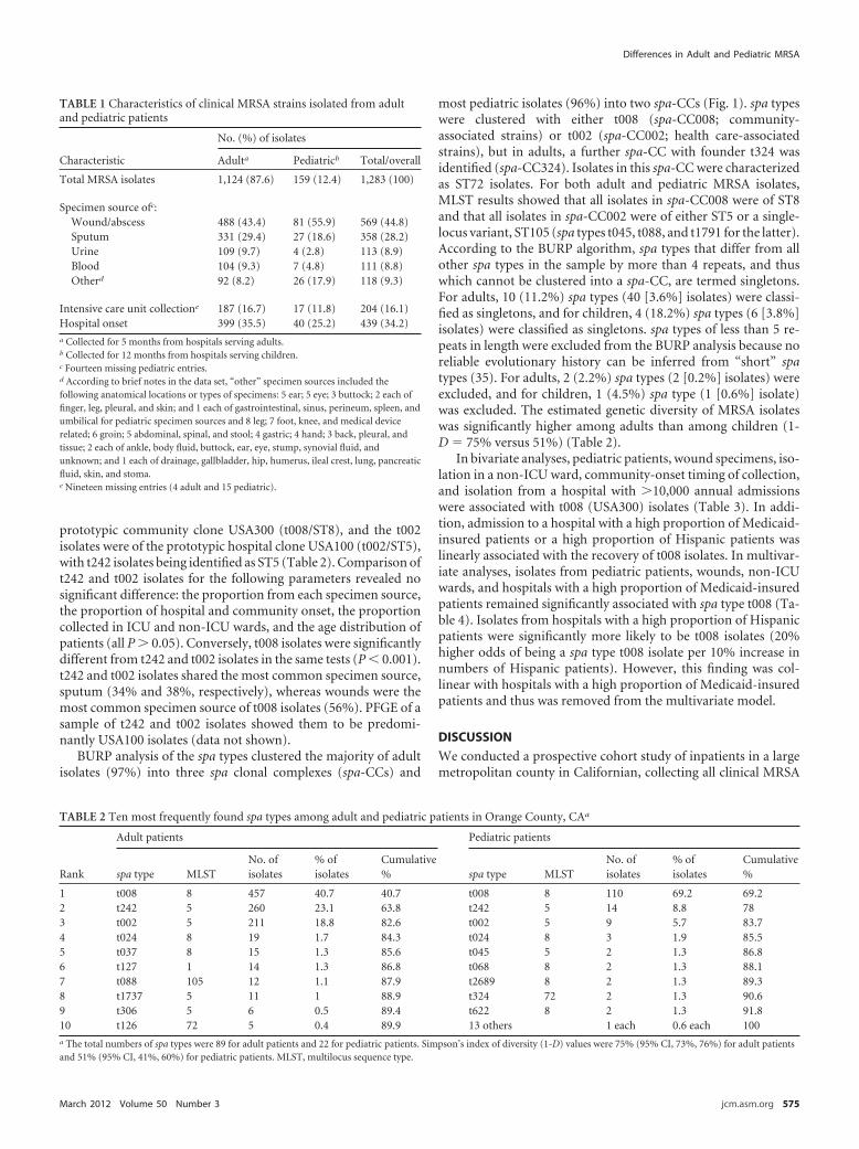
prototypic community clone USA300 (t008/ST8), and the t002isolates were of the prototypic hospital clone USA100 (t002/ST5),with t242 isolates being identified as ST5 (Table 2). Comparison oft242 and t002 isolates for the following parameters revealed nosignificant difference: the proportion from each specimen source,the proportion of hospital and community onset, the proportioncollected in ICU and non-ICU wards, and the age distribution ofpatients (all P $ 0.05). Conversely, t008 isolates were significantlydifferent from t242 and t002 isolates in the same tests (P ! 0.001).t242 and t002 isolates shared the most common specimen source,sputum (34% and 38%, respectively), whereas wounds were themost common specimen source of t008 isolates (56%). PFGE of asample of t242 and t002 isolates showed them to be predomi-nantly USA100 isolates (data not shown).
BURP analysis of the spa types clustered the majority of adultisolates (97%) into three spa clonal complexes (spa-CCs) and
most pediatric isolates (96%) into two spa-CCs (Fig. 1). spa typeswere clustered with either t008 (spa-CC008; community-associated strains) or t002 (spa-CC002; health care-associatedstrains), but in adults, a further spa-CC with founder t324 wasidentified (spa-CC324). Isolates in this spa-CC were characterizedas ST72 isolates. For both adult and pediatric MRSA isolates,MLST results showed that all isolates in spa-CC008 were of ST8and that all isolates in spa-CC002 were of either ST5 or a single-locus variant, ST105 (spa types t045, t088, and t1791 for the latter).According to the BURP algorithm, spa types that differ from allother spa types in the sample by more than 4 repeats, and thuswhich cannot be clustered into a spa-CC, are termed singletons.For adults, 10 (11.2%) spa types (40 [3.6%] isolates) were classi-fied as singletons, and for children, 4 (18.2%) spa types (6 [3.8%]isolates) were classified as singletons. spa types of less than 5 re-peats in length were excluded from the BURP analysis because noreliable evolutionary history can be inferred from “short” spatypes (35). For adults, 2 (2.2%) spa types (2 [0.2%] isolates) wereexcluded, and for children, 1 (4.5%) spa type (1 [0.6%] isolate)was excluded. The estimated genetic diversity of MRSA isolateswas significantly higher among adults than among children (1-D # 75% versus 51%) (Table 2).
In bivariate analyses, pediatric patients, wound specimens, iso-lation in a non-ICU ward, community-onset timing of collection,and isolation from a hospital with $10,000 annual admissionswere associated with t008 (USA300) isolates (Table 3). In addi-tion, admission to a hospital with a high proportion of Medicaid-insured patients or a high proportion of Hispanic patients waslinearly associated with the recovery of t008 isolates. In multivar-iate analyses, isolates from pediatric patients, wounds, non-ICUwards, and hospitals with a high proportion of Medicaid-insuredpatients remained significantly associated with spa type t008 (Ta-ble 4). Isolates from hospitals with a high proportion of Hispanicpatients were significantly more likely to be t008 isolates (20%higher odds of being a spa type t008 isolate per 10% increase innumbers of Hispanic patients). However, this finding was col-linear with hospitals with a high proportion of Medicaid-insuredpatients and thus was removed from the multivariate model.
DISCUSSIONWe conducted a prospective cohort study of inpatients in a largemetropolitan county in Californian, collecting all clinical MRSA
TABLE 1 Characteristics of clinical MRSA strains isolated from adultand pediatric patients
Characteristic
No. (%) of isolates
Adulta Pediatricb Total/overall
Total MRSA isolates 1,124 (87.6) 159 (12.4) 1,283 (100)
Specimen source ofc:Wound/abscess 488 (43.4) 81 (55.9) 569 (44.8)Sputum 331 (29.4) 27 (18.6) 358 (28.2)Urine 109 (9.7) 4 (2.8) 113 (8.9)Blood 104 (9.3) 7 (4.8) 111 (8.8)Otherd 92 (8.2) 26 (17.9) 118 (9.3)
Intensive care unit collectione 187 (16.7) 17 (11.8) 204 (16.1)Hospital onset 399 (35.5) 40 (25.2) 439 (34.2)a Collected for 5 months from hospitals serving adults.b Collected for 12 months from hospitals serving children.c Fourteen missing pediatric entries.d According to brief notes in the data set, “other” specimen sources included thefollowing anatomical locations or types of specimens: 5 ear; 5 eye; 3 buttock; 2 each offinger, leg, pleural, and skin; and 1 each of gastrointestinal, sinus, perineum, spleen, andumbilical for pediatric specimen sources and 8 leg; 7 foot, knee, and medical devicerelated; 6 groin; 5 abdominal, spinal, and stool; 4 gastric; 4 hand; 3 back, pleural, andtissue; 2 each of ankle, body fluid, buttock, ear, eye, stump, synovial fluid, andunknown; and 1 each of drainage, gallbladder, hip, humerus, ileal crest, lung, pancreaticfluid, skin, and stoma.e Nineteen missing entries (4 adult and 15 pediatric).
TABLE 2 Ten most frequently found spa types among adult and pediatric patients in Orange County, CAa
Rank
Adult patients Pediatric patients
spa type MLSTNo. ofisolates
% ofisolates
Cumulative% spa type MLST
No. ofisolates
% ofisolates
Cumulative%
1 t008 8 457 40.7 40.7 t008 8 110 69.2 69.22 t242 5 260 23.1 63.8 t242 5 14 8.8 783 t002 5 211 18.8 82.6 t002 5 9 5.7 83.74 t024 8 19 1.7 84.3 t024 8 3 1.9 85.55 t037 8 15 1.3 85.6 t045 5 2 1.3 86.86 t127 1 14 1.3 86.8 t068 8 2 1.3 88.17 t088 105 12 1.1 87.9 t2689 8 2 1.3 89.38 t1737 5 11 1 88.9 t324 72 2 1.3 90.69 t306 5 6 0.5 89.4 t622 8 2 1.3 91.810 t126 72 5 0.4 89.9 13 others 1 each 0.6 each 100a The total numbers of spa types were 89 for adult patients and 22 for pediatric patients. Simpson’s index of diversity (1-D) values were 75% (95% CI, 73%, 76%) for adult patientsand 51% (95% CI, 41%, 60%) for pediatric patients. MLST, multilocus sequence type.
Differences in Adult and Pediatric MRSA
March 2012 Volume 50 Number 3 jcm.asm.org 575
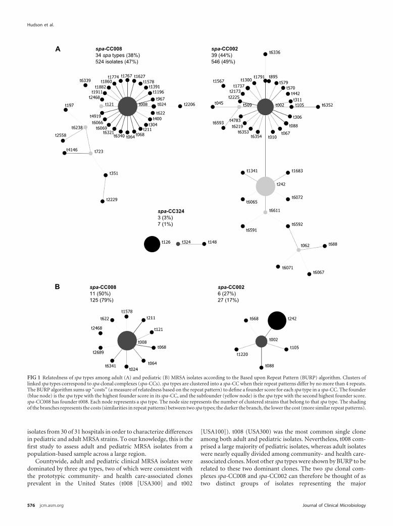
isolates from 30 of 31 hospitals in order to characterize differencesin pediatric and adult MRSA strains. To our knowledge, this is thefirst study to assess adult and pediatric MRSA isolates from apopulation-based sample across a large region.
Countywide, adult and pediatric clinical MRSA isolates weredominated by three spa types, two of which were consistent withthe prototypic community- and health care-associated clonesprevalent in the United States (t008 [USA300] and t002
[USA100]). t008 (USA300) was the most common single cloneamong both adult and pediatric isolates. Nevertheless, t008 com-prised a large majority of pediatric isolates, whereas adult isolateswere nearly equally divided among community- and health care-associated clones. Most other spa types were shown by BURP to berelated to these two dominant clones. The two spa clonal com-plexes spa-CC008 and spa-CC002 can therefore be thought of astwo distinct groups of isolates representing the major
FIG 1 Relatedness of spa types among adult (A) and pediatric (B) MRSA isolates according to the Based upon Repeat Pattern (BURP) algorithm. Clusters oflinked spa types correspond to spa clonal complexes (spa-CCs). spa types are clustered into a spa-CC when their repeat patterns differ by no more than 4 repeats.The BURP algorithm sums up “costs” (a measure of relatedness based on the repeat pattern) to define a founder score for each spa type in a spa-CC. The founder(blue node) is the spa type with the highest founder score in its spa-CC, and the subfounder (yellow node) is the spa type with the second highest founder score.spa-CC008 has founder t008. Each node represents a spa type. The node size represents the number of clustered strains that belong to that spa type. The shadingof the branches represents the costs (similarities in repeat patterns) between two spa types; the darker the branch, the lower the cost (more similar repeat patterns).
Hudson et al.
576 jcm.asm.org Journal of Clinical Microbiology
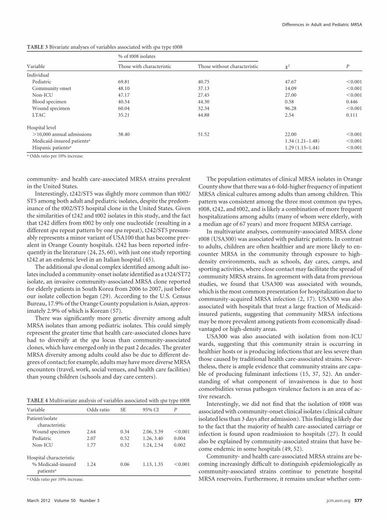
community- and health care-associated MRSA strains prevalentin the United States.
Interestingly, t242/ST5 was slightly more common than t002/ST5 among both adult and pediatric isolates, despite the predom-inance of the t002/ST5 hospital clone in the United States. Giventhe similarities of t242 and t002 isolates in this study, and the factthat t242 differs from t002 by only one nucleotide (resulting in adifferent spa repeat pattern by one spa repeat), t242/ST5 presum-ably represents a minor variant of USA100 that has become prev-alent in Orange County hospitals. t242 has been reported infre-quently in the literature (24, 25, 60), with just one study reportingt242 at an endemic level in an Italian hospital (45).
The additional spa clonal complex identified among adult iso-lates included a community-onset isolate identified as a t324/ST72isolate, an invasive community-associated MRSA clone reportedfor elderly patients in South Korea from 2006 to 2007, just beforeour isolate collection began (29). According to the U.S. CensusBureau, 17.9% of the Orange County population is Asian, approx-imately 2.9% of which is Korean (57).
There was significantly more genetic diversity among adultMRSA isolates than among pediatric isolates. This could simplyrepresent the greater time that health care-associated clones havehad to diversify at the spa locus than community-associatedclones, which have emerged only in the past 2 decades. The greaterMRSA diversity among adults could also be due to different de-grees of contact; for example, adults may have more diverse MRSAencounters (travel, work, social venues, and health care facilities)than young children (schools and day care centers).
The population estimates of clinical MRSA isolates in OrangeCounty show that there was a 6-fold-higher frequency of inpatientMRSA clinical cultures among adults than among children. Thispattern was consistent among the three most common spa types,t008, t242, and t002, and is likely a combination of more frequenthospitalizations among adults (many of whom were elderly, witha median age of 67 years) and more frequent MRSA carriage.
In multivariate analyses, community-associated MRSA clonet008 (USA300) was associated with pediatric patients. In contrastto adults, children are often healthier and are more likely to en-counter MRSA in the community through exposure to high-density environments, such as schools, day cares, camps, andsporting activities, where close contact may facilitate the spread ofcommunity MRSA strains. In agreement with data from previousstudies, we found that USA300 was associated with wounds,which is the most common presentation for hospitalization due tocommunity-acquired MRSA infection (2, 17). USA300 was alsoassociated with hospitals that treat a large fraction of Medicaid-insured patients, suggesting that community MRSA infectionsmay be more prevalent among patients from economically disad-vantaged or high-density areas.
USA300 was also associated with isolation from non-ICUwards, suggesting that this community strain is occurring inhealthier hosts or is producing infections that are less severe thanthose caused by traditional health care-associated strains. Never-theless, there is ample evidence that community strains are capa-ble of producing fulminant infections (15, 37, 52). An under-standing of what component of invasiveness is due to hostcomorbidities versus pathogen virulence factors is an area of ac-tive research.
Interestingly, we did not find that the isolation of t008 wasassociated with community-onset clinical isolates (clinical cultureisolated less than 3 days after admission). This finding is likely dueto the fact that the majority of health care-associated carriage orinfection is found upon readmission to hospitals (27). It couldalso be explained by community-associated strains that have be-come endemic in some hospitals (49, 52).
Community- and health care-associated MRSA strains are be-coming increasingly difficult to distinguish epidemiologically ascommunity-associated strains continue to penetrate hospitalMRSA reservoirs. Furthermore, it remains unclear whether com-
TABLE 3 Bivariate analyses of variables associated with spa type t008
Variable
% of t008 isolates
#2 PThose with characteristic Those without characteristic
IndividualPediatric 69.81 40.75 47.67 !0.001Community onset 48.10 37.13 14.09 !0.001Non-ICU 47.17 27.45 27.00 !0.001Blood specimen 40.54 44.30 0.58 0.446Wound specimen 60.04 32.34 96.28 !0.001LTAC 35.21 44.88 2.54 0.111
Hospital level$10,000 annual admissions 38.40 51.52 22.00 !0.001Medicaid-insured patientsa 1.34 (1.21–1.48) !0.001Hispanic patientsa 1.29 (1.15–1.44) !0.001
a Odds ratio per 10% increase.
TABLE 4 Multivariate analysis of variables associated with spa type t008
Variable Odds ratio SE 95% CI P
Patient/isolatecharacteristic
Wound specimen 2.64 0.34 2.06, 3.39 !0.001Pediatric 2.07 0.52 1.26, 3.40 0.004Non-ICU 1.77 0.32 1.24, 2.54 0.002
Hospital characteristic% Medicaid-insured
patientsa
1.24 0.06 1.13, 1.35 !0.001
a Odds ratio per 10% increase.
Differences in Adult and Pediatric MRSA
March 2012 Volume 50 Number 3 jcm.asm.org 577
munity clones are adding to or replacing traditional health care-associated MRSA strains (3, 8, 22, 49). The implication of theblurred line between community- and health care-associatedMRSA strains may be that efforts to control MRSA transmissionwithin hospitals will not be effective in controlling communityinflux into hospitals. Simultaneous community strategies to limitMRSA spread are needed. However, much is still unknown aboutthe acquisition and transmission of CA-MRSA, so improvedknowledge is needed to better guide infection control strategies.Further studies are needed to ascertain whether community strat-egies to reduce transmission in children and young adults wouldproduce benefits across the entire age spectrum.
One limitation of our study is that few individual-level charac-teristics were available. Also, our study did not account for thedifferent policies in place at each hospital with regard to when toobtain clinical cultures. These differences could affect MRSA de-tection at each hospital and, possibly, the type of MRSA strainsisolated, if clinical cultures were more likely to be obtained forsicker, older patients. Moreover, our results could have been af-fected by the potential seasonality of MRSA infections and infec-tion types due to the different collection periods for adult andpediatric isolates (largely winter and spring for adult collections,compared to all seasons for pediatric collections). A seasonality ofS. aureus infections, particularly skin infections, has been ob-served in pediatric and adult patients in temperate and tropicalenvironments, with a predominance of infections during summerand autumn (31, 36, 54, 58). A recent study in Rhode Island founda 2- to 3-fold-increased incidence of MRSA infections (both CA-and HA-MRSA) in pediatric patients during the second two quar-ters of the year over the last decade (36). However, in that samestudy, adult CA-MRSA infections showed less seasonal variationthan did pediatric infections, and no variation was observedamong adult HA-MRSA infections. Some studies observed no sig-nificant seasonality of S. aureus infections, but those studies fo-cused on bacteremia (40, 47). The collection of both adult andpediatric MRSA isolates for the same time period, i.e., 12 months,would have accounted for any potential seasonality effects and/orother factors that could affect the type and diversity of MRSAstrains isolated.
Mandatory screening of high-risk inpatients was not in place inCalifornia until 2009; therefore, our population estimates arelikely underestimates. In addition, our estimates should not beconstrued as measures of MRSA infection among inpatients. Clin-ical isolates often represent carriage without infection. Finally, ourestimates of the index of diversity for adult and pediatric MRSAisolates may have been influenced by differing sample sizes (18).
In conclusion, our study found that in a large county, MRSAisolates from hospitalized children were more likely to be of spatype t008 (USA300). This community-associated spa type was as-sociated with children, wounds, non-ICU care, and admission to ahospital with a high percentage of Medicaid-insured patients. De-spite the association of t008 isolates with children, t008 was stillthe most common spa type among adult patients, suggesting thatcommunity-based interventions are needed to stem the influx oft008 isolates into hospitals. We also found evidence for a prevalentvariant of the USA100 clone (t242/ST5), which has not been re-ported elsewhere. While community- and hospital-associatedMRSA reservoirs have begun to merge, significant differences re-main in pediatric and adult patient populations, which may pro-
vide an impetus for different age-based strategies to reduce trans-mission and disease.
ACKNOWLEDGMENTSThis work was supported by the University of California Irvine School ofMedicine, the Biotechnology and Biological Sciences Research Council(grant number BB/D52637X/1), and the Wellcome Trust (grant number089472/Z/09/Z).
We thank Diane Kim for her contributions to this study. We alsoextend special thanks to the participating microbiology laboratoriesthroughout Orange County.
We do not have an association that might pose a conflict of interest.
REFERENCES1. Abramson MA, Sexton DJ. 1999. Nosocomial methicillin-resistant and
methicillin-susceptible Staphylococcus aureus primary bacteremia: atwhat costs? Infect. Control Hosp. Epidemiol. 20:408 – 411.
2. Bassetti M, Nicco E, Mikulska M. 2009. Why is community-associatedMRSA spreading across the world and how will it change clinical practice?Int. J. Antimicrob. Agents 34(Suppl 1):S15–S19.
3. Bootsma M, Hota B, Diekmann O, Weinstein RA, Bonten M. 2006.Abstr. 46th Intersci. Conf. Antimicrob. Agents Chemother., abstr K-1680.
4. Bratu S, et al. 2005. Community-associated methicillin-resistant Staph-ylococcus aureus in hospital nursery and maternity units. Emerg. Infect.Dis. 11:808 – 813.
5. Centers for Disease Control and Prevention. 1999. Four pediatric deathsfrom community-acquired methicillin-resistant Staphylococcus au-reus—Minnesota and North Dakota, 1997–1999. JAMA 282:1123–1125.
6. Cosgrove SE, et al. 2003. Comparison of mortality associated withmethicillin-resistant and methicillin-susceptible Staphylococcus aureusbacteremia: a meta-analysis. Clin. Infect. Dis. 36:53–59.
7. Cosgrove SE, et al. 2005. The impact of methicillin resistance in Staphy-lococcus aureus bacteremia on patient outcomes: mortality, length of stay,and hospital charges. Infect. Control Hosp. Epidemiol. 26:166 –174.
8. D’Agata EM, Webb GF, Horn MA, Moellering RC, Jr, Ruan S. 2009.Modeling the invasion of community-acquired methicillin-resistantStaphylococcus aureus into hospitals. Clin. Infect. Dis. 48:274 –284.
9. David MZ, et al. 2006. Contrasting pediatric and adult methicillin-resistant Staphylococcus aureus isolates. Emerg. Infect. Dis. 12:631– 637.
10. Diep BA, Carleton HA, Chang RF, Sensabaugh GF, Perdreau-Remington F. 2006. Roles of 34 virulence genes in the evolution ofhospital- and community-associated strains of methicillin-resistantStaphylococcus aureus. J. Infect. Dis. 193:1495–1503.
11. Diep BA, et al. 2008. The arginine catabolic mobile element and staphy-lococcal chromosomal cassette mec linkage: convergence of virulence andresistance in the USA300 clone of methicillin-resistant Staphylococcusaureus. J. Infect. Dis. 197:1523–1530.
12. Eady EA, Cove JH. 2003. Staphylococcal resistance revisited:community-acquired methicillin resistant Staphylococcus aureus—anemerging problem for the management of skin and soft tissue infections.Curr. Opin. Infect. Dis. 16:103–124.
13. Engemann JJ, et al. 2003. Adverse clinical and economic outcomes at-tributable to methicillin resistance among patients with Staphylococcusaureus surgical site infection. Clin. Infect. Dis. 36:592–598.
14. Enright MC, Day NP, Davies CE, Peacock SJ, Spratt BG. 2000. Multi-locus sequence typing for characterization of methicillin-resistant andmethicillin-susceptible clones of Staphylococcus aureus. J. Clin. Micro-biol. 38:1008 –1015.
15. Frazee BW, Salz TO, Lambert L, Perdreau-Remington F. 2005. Fatalcommunity-associated methicillin-resistant Staphylococcus aureus pneu-monia in an immunocompetent young adult. Ann. Emerg. Med. 46:401–404.
16. Frei CR, Makos BR, Daniels KR, Oramasionwu CU. 2010. Emergence ofcommunity-acquired methicillin-resistant Staphylococcus aureus skinand soft tissue infections as a common cause of hospitalization in UnitedStates children. J. Pediatr. Surg. 45:1967–1974.
17. Fridkin SK, et al. 2005. Methicillin-resistant Staphylococcus aureus dis-ease in three communities. N. Engl. J. Med. 352:1436 –1444.
18. Grundmann H, Hori S, Tanner G. 2001. Determining confidence inter-vals when measuring genetic diversity and the discriminatory abilities oftyping methods for microorganisms. J. Clin. Microbiol. 39:4190 – 4192.
Hudson et al.
578 jcm.asm.org Journal of Clinical Microbiology
19. Grundmann H, et al. 2010. Geographic distribution of Staphylococcusaureus causing invasive infections in Europe: a molecular-epidemiologicalanalysis. PLoS Med. 7:e1000215.
20. Harmsen D, et al. 2003. Typing of methicillin-resistant Staphylococcusaureus in a university hospital setting by using novel software for sparepeat determination and database management. J. Clin. Microbiol. 41:5442–5448.
21. Herold BC, et al. 1998. Community-acquired methicillin-resistantStaphylococcus aureus in children with no identified predisposing risk.JAMA 279:593–598.
22. Hota B, et al. 2007. Community-associated methicillin-resistant Staph-ylococcus aureus skin and soft tissue infections at a public hospital: dopublic housing and incarceration amplify transmission? Arch. Intern.Med. 167:1026 –1033.
23. Huang SS, et al. 2007. Improving methicillin-resistant Staphylococcusaureus surveillance and reporting in intensive care units. J. Infect. Dis.195:330 –338.
24. Johnson JK, et al. 2007. Skin and soft tissue infections caused bymethicillin-resistant Staphylococcus aureus USA300 clone. Emerg. Infect.Dis. 13:1195–1200.
25. Kinnevey P, Shore A, Rossney A, Coleman D. 2010. Abstr. 20th Eur.Congr. Clin. Microbiol. Infect. Dis., abstr P1712.
26. Klein E, Smith DL, Laxminarayan R. 2007. Hospitalizations and deathscaused by methicillin-resistant Staphylococcus aureus, United States,1999 –2005. Emerg. Infect. Dis. 13:1840 –1846.
27. Klevens RM, et al. 2007. Invasive methicillin-resistant Staphylococcusaureus infections in the United States. JAMA 298:1763–1771.
28. Kourbatova EV, et al. 2005. Emergence of community-associatedmethicillin-resistant Staphylococcus aureus USA300 clone as a cause ofhealth care-associated infections among patients with prosthetic joint in-fections. Am. J. Infect. Control 33:385–391.
29. Lee SS, Kim YJ, Chung DR, Jung KS, Kim JS. 2010. Invasive infectioncaused by a community-associated methicillin-resistant Staphylococcusaureus strain not carrying Panton-Valentine leukocidin in South Korea. J.Clin. Microbiol. 48:311–313.
30. Liu C, et al. 2008. A population-based study of the incidence and molec-ular epidemiology of methicillin-resistant Staphylococcus aureus diseasein San Francisco, 2004-2005. Clin. Infect. Dis. 46:1637–1646.
31. Loffeld A, Davies P, Lewis A, Moss C. 2005. Seasonal occurrence ofimpetigo: a retrospective 8-year review (1996 –2003). Clin. Exp. Dermatol.30:512–514.
32. Lucet JC, et al. 2009. Carriage of methicillin-resistant Staphylococcusaureus in home care settings: prevalence, duration, and transmission tohousehold members. Arch. Intern. Med. 169:1372–1378.
33. Ma XX, et al. 2002. Novel type of staphylococcal cassette chromosomemec identified in community-acquired methicillin-resistant Staphylococ-cus aureus strains. Antimicrob. Agents Chemother. 46:1147–1152.
34. Maree CL, Daum RS, Boyle-Vavra S, Matayoshi K, Miller LG. 2007.Community-associated methicillin-resistant Staphylococcus aureus iso-lates causing healthcare-associated infections. Emerg. Infect. Dis. 13:236 –242.
35. Mellmann A, et al. 2007. Based upon Repeat Pattern (BURP): an algo-rithm to characterize the long-term evolution of Staphylococcus aureuspopulations based on spa polymorphisms. BMC Microbiol. 7:98.
36. Mermel LA, Machan JT, Parenteau S. 2011. Seasonality of MRSA infec-tions. PLoS One 6:e17925.
37. Miller LG, et al. 2005. Necrotizing fasciitis caused by community-associated methicillin-resistant Staphylococcus aureus in Los Angeles. N.Engl. J. Med. 352:1445–1453.
38. Moellering RC, Jr. 2006. The growing menace of community-acquiredmethicillin-resistant Staphylococcus aureus. Ann. Intern. Med. 144:368 –370.
39. Montgomery CP, et al. 2008. Comparison of virulence in community-associated methicillin-resistant Staphylococcus aureus pulsotypesUSA300 and USA400 in a rat model of pneumonia. J. Infect. Dis. 198:561–570.
40. Morin CA, Hadler JL. 2001. Population-based incidence and character-istics of community-onset Staphylococcus aureus infections with bactere-
mia in 4 metropolitan Connecticut areas, 1998. J. Infect. Dis. 184:1029 –1034.
41. O’Brien FG, Pearman JW, Gracey M, Riley TV, Grubb WB. 1999.Community strain of methicillin-resistant Staphylococcus aureus in-volved in a hospital outbreak. J. Clin. Microbiol. 37:2858 –2862.
42. Office of Statewide Health Planning and Development. 6 June 2011, acces-sion date. Patient discharge data public data set 2005. Office of StatewideHealth Planning and Development, Sacramento, CA. http://www.oshpd.ca.gov/HID/Products/PatDischargeData/PublicDataSet/index.html.
43. Okuma K, et al. 2002. Dissemination of new methicillin-resistant Staph-ylococcus aureus clones in the community. J. Clin. Microbiol. 40:4289 –4294.
44. Park JY, et al. 2007. A comparison of adult and pediatric methicillin-resistant Staphylococcus aureus isolates collected from patients at a uni-versity hospital in Korea. J. Microbiol. 45:447– 452.
45. Parlato C, Cavallerio P, Fossati L, Allice T, Serra R. 2009. Abstr. 19thEur. Congr. Clin. Microbiol. Infect. Dis., abstr P1574.
46. Patel M, et al. 2008. Emergence of USA300 MRSA in a tertiary medicalcentre: implications for epidemiological studies. J. Hosp. Infect. 68:208 –213.
47. Perencevich EN, et al. 2008. Summer peaks in the incidences of Gram-negative bacterial infection among hospitalized patients. Infect. ControlHosp. Epidemiol. 29:1124 –1131.
48. Pfaller MA. 1998. Chromosomal restriction fragment analysis by pulsed-field gel electrophoresis: application to molecular epidemiology, p 651–657. In Isenberg HD (ed), Essential procedures for clinical microbiology.ASM Press, Washington, DC.
49. Popovich KJ, Weinstein RA, Hota B. 2008. Are community-associatedmethicillin-resistant Staphylococcus aureus (MRSA) strains replacing tra-ditional nosocomial MRSA strains? Clin. Infect. Dis. 46:787–794.
50. Robicsek A, et al. 2008. Universal surveillance for methicillin-resistantStaphylococcus aureus in 3 affiliated hospitals. Ann. Intern. Med. 148:409 – 418.
51. Saiman L, et al. 2003. Hospital transmission of community-acquiredmethicillin-resistant Staphylococcus aureus among postpartum women.Clin. Infect. Dis. 37:1313–1319.
52. Seybold U, et al. 2006. Emergence of community-associated methicillin-resistant Staphylococcus aureus USA300 genotype as a major cause ofhealth care-associated blood stream infections. Clin. Infect. Dis. 42:647–656.
53. Shurland S, Zhan M, Bradham DD, Roghmann MC. 2007. Comparisonof mortality risk associated with bacteremia due to methicillin-resistantand methicillin-susceptible Staphylococcus aureus. Infect. Control Hosp.Epidemiol. 28:273–279.
54. Szczesiul JM, Shermock KM, Murtaza UI, Siberry GK. 2007. No de-crease in clindamycin susceptibility despite increased use of clindamycinfor pediatric community-associated methicillin-resistant Staphylococcusaureus skin infections. Pediatr. Infect. Dis. J. 26:852– 854.
55. Tristan A, et al. 2007. Global distribution of Panton-Valentineleukocidin-positive methicillin-resistant Staphylococcus aureus, 2006.Emerg. Infect. Dis. 13:594 – 600.
56. Udo EE, Pearman JW, Grubb WB. 1993. Genetic analysis of communityisolates of methicillin-resistant Staphylococcus aureus in Western Austra-lia. J. Hosp. Infect. 25:97–108.
57. U.S. Census Bureau. 6 June 2011, accession date. United States census 2010demographic profile data for Orange County, California. US Census Bureau,Washington, DC. http://factfinder2.census.gov/faces/tableservices/jsf/pages/productview.xhtml?pid#DEC_10_DP_DPDP1&prodType#table.
58. Van De Griend P, et al. 2009. Community-associated methicillin-resistant Staphylococcus aureus, Iowa, USA. Emerg. Infect. Dis. 15:1582–1589.
59. Wallin TR, Hern HG, Frazee BW. 2008. Community-associatedmethicillin-resistant Staphylococcus aureus. Emerg. Med. Clin. NorthAm. 26:431– 455.
60. Weese JS, Avery BP, Reid-Smith RJ. 2010. Detection and quantificationof methicillin-resistant Staphylococcus aureus (MRSA) clones in retailmeat products. Lett. Appl. Microbiol. 51:338 –342.
Differences in Adult and Pediatric MRSA
March 2012 Volume 50 Number 3 jcm.asm.org 579
Patient sharing and population genetic structure ofmethicillin-resistant Staphylococcus aureusWeixiong Kea, Susan S. Huangb, Lyndsey O. Hudsonc, Kristen R. Elkinsb, Christopher C. Nguyenb, Brian G. Sprattc,Courtney R. Murphyb, Taliser R. Averyd, and Marc Lipsitcha,e,1
aDepartment of Epidemiology and eCenter for Communicable Disease Dynamics and Department of Immunology and Infectious Diseases, Harvard School ofPublic Health, Boston, MA 02115; bDivision of Infectious Diseases and Health Policy Research Institute, Irvine School of Medicine, University of California,Irvine, CA 92617; cDepartment of Infectious Disease Epidemiology, Imperial College London, London W2 1PG, United Kingdom; and dDepartment ofPopulation Medicine, Harvard Pilgrim Health Care Institute and Harvard Medical School, Boston, MA 02215
Edited by Simon A. Levin, Princeton University, Princeton, NJ, and approved February 16, 2012 (received for review August 26, 2011)
Rates of hospital-acquired infections, speci!callymethicillin-resistantStaphylococcus aureus (MRSA), are increasingly being used as indi-cators for quality of hospital hygiene. There has beenmuch effort onunderstanding the transmission process at the hospital level; how-ever, interhospital population-based transmission remainspoorly de-!ned. We evaluated whether the proportion of shared patientsbetween hospitals was correlated with genetic similarity of MRSAstrains from those hospitals. Using data collected from 30 of 32 hos-pitals in Orange County, California, multivariate linear regressionshowed that for each twofold increase in the proportion of patientsshared between 2 hospitals, there was a 7.7% reduction in geneticheterogeneity between the hospitals’ MRSA populations (permuta-tion P value = 0.0356). Pairs of hospitals that both served adults hadmore similar MRSA populations than pairs including a pediatric hos-pital. These !ndings suggest that concerted efforts among hospitalsthat share large numbers of patients may be synergistic to preventMRSA transmission.
Methicillin-resistant Staphylococcus aureus (MRSA), one ofthemost common and virulent nosocomial pathogens, is also
an increasingly important cause of community-acquired disease.MRSA strains, particularly those associated with hospitals, areoften resistant to multiple antibiotics, limiting treatment options.S. aureus is carried asymptomatically in!30% of healthy adults andis shown to be amajor cause of invasive disease among hospitalizedpatients (1); MRSAmakes up a growing proportion of nosocomialS. aureus infections in many countries. The circulation of a smallnumber ofMRSA clones that characterizes the current epidemic isthought to be mainly the result of between-patient transmissionrather than de novo appearance of resistance in patients exposed toantibiotics (2), because the appearance of a new MRSA strainrequires acquisition of a mec resistance element, a relatively rareevent. The level of genetic variation occurring in S. aureus withinidenti!able clonal lineages allows the use of genetic markers totrack transmission of these lineages and sublineages (3, 4).The prevalence of MRSA varies considerably both within and
between countries (5, 6). About 30% of the S. aureus causingbloodstream infections is methicillin resistant in the United King-dom, whereas that proportion is !1% in The Netherlands andScandinavian countries (7). Among countries with high endemicMRSA infection rates, the proportion is highest in large teachinghospitals (6, 8), where the highest frequency of new emergingMRSA clones has also been reported (9–12). The proposed reasonsinclude increased antibiotic use and increased prevalence of med-ical procedures and serious medical conditions associated withMRSA acquisition and disease (13). BecauseMRSA can be carriedasymptomatically for a long time (14), readmission could introdu-ce a previously acquired strain into a newhospital (15). Thus, failureof one hospital’s infection control could in principle affect theprevalence of MRSA in other hospitals that share patients with it(16). Previous studies have suggested that patient transfer or patientreferral patterns (17) could affect the prevalence of MRSA inhospitals (1, 2, 16, 18, 19), on the basis of theoretical arguments and
observations that clones of MRSA appear in neighboring hospitals.Population genetics can provide a test of the hypothesis that patientsharing plays an important role inMRSAdynamics: If so, onemightexpect that hospitals that share large numbers of patients would alsotend to share genetically similar populations of MRSA.In the current study, we sought to investigate whether the pattern
of genetic relatedness among MRSA isolates from hospitals withinOrange County (OC), California, was consistent with a signi!cantrole for patient sharing in determining the population of MRSAstrains within a hospital. OC is well suited for this study because it isthe !fth largest county in theUnited States, and it has relatively lowpopulation "ow from three of its four sides. A !nding that MRSAisolates from hospitals that share more patients tend to be relatedto one another would provide an independent line of evidence forthe importance of patient transfer in spreading MRSA from hos-pital to hospital. For S. aureus genotyping, we used the spa locus,which encodes protein A, a species-speci!c protein known for itsIgG binding capacity (3). This locus features highly polymorphicinternal regions due to short tandem repeats (STRs) (20) andtherefore serves as a good target for molecular genotyping (spatyping). This genotyping method has been demonstrated to beuseful in researching transmission, outbreaks, or geographic dis-tributions (3, 21, 22). Using spa typing, we sought evidence on howMRSA strains “travel” with patient "ow, to infer how patienttransfer might in"uence MRSA spread among hospitals.
ResultsSummary of Overall Approach. We used Wright’s F statistics tomeasure genetic heterogeneity between MRSA populations inhospitals and groups of hospitals and used pairwise regressionanalysis supplemented by group-level analysis as our methods, asdescribed in Materials and Methods.In the pairwise regression analysis, we calculated the heterozy-
gosity of each pair of hospitals (HR) (for each pair we regard the twohospitals as a group) and the heterozygosity of each single hospitalwithin a pair (HS). FSR was calculated for each pair of hospitalsaccordingly and was used as our response variable to measure thegenetic dissimilarity between a pair of hospitals. A positive coef!-cient for a predictor of FSR indicates that hospitals are more diver-gent from one another, whereas a negative coef!cient indicates thathospitals are more similar to one another.
Author contributions: W.K., S.S.H., B.G.S., and M.L. designed research; W.K., C.R.M., andM.L. performed research; L.O.H. contributed new reagents/analytic tools; W.K., L.O.H.,K.R.E., C.C.N., C.R.M., and T.R.A. analyzed data; and W.K., S.S.H., B.G.S., C.R.M., and M.L.wrote the paper.
The authors declare no con!ict of interest.
This article is a PNAS Direct Submission.
See Commentary on page 6364.1To whom correspondence should be addressed. E-mail: [email protected].
This article contains supporting information online at www.pnas.org/lookup/suppl/doi:10.1073/pnas.1113578109/-/DCSupplemental.
www.pnas.org/cgi/doi/10.1073/pnas.1113578109 PNAS | April 24, 2012 | vol. 109 | no. 17 | 6763–6768
POPU
LATION
BIOLO
GY
SEECO
MMEN
TARY
The distribution of FSR is shown in Fig. S1A. We found thatthe distribution of this pairwise FSR is signi!cantly skewed, so welog-transformed this variable in the regression analysis, and thenew distribution is shown in Fig. S1B. This procedure was alsocarried out for the main predictor variable, patient "ow, asshown in Fig. S1 C and D.In group-level analysis, we used the FST statistics calculated from
heterozygosity of all 30 hospitals (HT) and heterozygosity of eachindividual hospital (HS) to measure heterogeneity or the reductionof heterozygosity from the total population level to the individualhospital level. For this study, FST = 0.0853, and HT and HS fromwhich it is calculated = 0.719 and 0.658, respectively. As FRT mea-sures the reduction of heterozygosity when hospitals are groupedcompared with the total 30 hospitals, this result implies that the bestpossible grouping (that is, each singlehospital is viewedasonegroup)could do no better than to achieve an FRT = 0.0853 (FRT = FST).
Predictors of Similarity Between Pairs of Hospitals. Individual categorizingvariables. Table S1 shows the characteristics of all 30 hospitals. Forpairwise analyses described below, dichotomous variables for pri-vate insurance proportion, Medicaid coverage, hospital size, andproportion Hispanic were created to re"ect above-cut point orbelow-cut point values.Pairwise analysis of similarity. For each of the 435 hospital pairs, wecalculated and log-transformed FSR to get log-FSR, serving as ameasure of population differentiation between them. We per-formed multivariate regression on the relationship of log-FSR tolog-transformed patient "ow and to pairwise geographic distancesbetween hospitals, average isolates collected, and the dichotomousvariables mentioned above. The P values presented in all of ourregression analyses are multiple Mantel test permutation P values,used to account for dependency between observations involvingpairs of hospitals. The results are presented in Table 1. Greaterpatient "ow between a pair of hospitals (log "ow) was associatedwith reduced pairwise FSR, i.e., greater similarity in MRSA pop-ulations between hospitals (coef!cient = !0.115, P value =0.0356). Another predictor of similarity in MRSA populations wasfor both to be nonpediatric (ped00, coef!cient =!0.801, P value=0.0448). Univariate analyses are shown in Table S2.In addition, we calculated Pearson correlation coef!cients for
each pair of the 13 variables to assess collinearity of predictors. Wefound relatively large correlation between the proportion of His-panic patients and Medicaid coverage (coef!cient for ethnicity11and medicaid11 = 0.581 and for ethnicity00 and medicaid00 =0.564), and between the average number of isolates collected andhospital size (coef!cient for SSize and size11 = 0.616 and for SSizeand size00=!0.680). This result suggests that these variablesmighthave had small and insigni!cant effects in our models due to
collinearity. We thus performed multivariate analyses with each(group) of these variables removed in turn, as shown in Tables S3–S6. The results of these models were consistent with those in theprimary analysis. In these alternative models, the coef!cient of log"ow ranged from !0.110 to !0.134 (vs. !0.115 in the base model)and remained statistically signi!cant. Exclusion of sample size led toa statistically signi!cant increase in similarity between pairs of largehospitals, compared with pairs containing a small and a large hos-pital (Table S3). Exclusion of hospital size variables led to a statis-tically signi!cant association between larger sample size and greatersimilarity between the hospitals (Table S4). Neither exclusion of theethnicity variables (Table S5) nor exclusion of the Medicaid varia-bles (Table S6) produced a statistically signi!cant association withthe other, but with Medicaid excluded there was a trend towardgreater similarity of hospitals having >20% Hispanic patients.Although the correlation between patient "ow—our main
variable of interest—and geographical distance was not as large(coef!cient for log "ow and dist = !0.378), we conducted an-other multivariate analysis with distance excluded, as shown inTable S7. The result suggested that by removing the distancepredictor, a slightly greater degree of similarity was associatedwith greater patient "ow (coef!cient = !0.128).Two of the hospitals have fewer isolates available (hospital 21,
six isolates; hospital 29, four isolates). To verify that our resultswere not driven by them, we removed them from the dataset andran the full multivariate regression again. The coef!cient onpatient "ow was almost unchanged (!0.114) although theMantel P value increased to 0.053 (Table S8).As we hypothesized, our results based on the full multivariate
model showed that hospitals sharing more patients have signi!-cantly more similar MRSA populations, after adjustment forother possible confounders. For each factor of 2 increase in pa-tient "ow (see Materials and Methods for de!nition), there is anassociated 1–2!0.115 = 7.7% reduction in pairwise FSR betweenthe hospitals, whereas the interquartile range of hospital pairs forlog "ow was !11.51 to !8.74, which corresponds to a 19.8% re-duction in pairwise FSR. Another variable was also statisticallyassociated with increased similarity of MRSA populations: Hos-pitals that both served adult patients tended to have populationsthat were more similar to one another. Visual inspection sug-gested that none of these results were driven by individual outliers.
Predictors of Similarity at the Group Level. Estimating groupingef!ciency. We used group-level analysis to supplement our resultsin pairwise analysis. This method divides hospitals into groups bya given criterion and calculates FRT for the grouping. FRT heremeasures the reduction of heterozygosity caused by groupingrelative to all hospitals without grouping and serves as a mea-
Table 1. Results of pairwise multivariate analysis
Variable no. Variable name Description Coef!cient P value*
1 Log_"ow Log-transformed pairwise patient "ow !0.115 0.03562 SSize Average sample size of MRSA isolates provided by a pair of hospitals !0.010 0.24743 Medicaid11 Indicator of both hospitals having Medicaid >10% !0.342 0.24764 Medicaid00 Indicator of both hospitals having Medicaid no more than 10% !0.191 0.55105 Private11 Indicator of both hospitals having private insurance >35% 0.041 0.87426 Private00 Indicator of both hospitals having private insurance no more than 35% 0.081 0.70687 Size11 Indicator of both hospitals having annual admission >10,000 !0.133 0.71988 Size00 Indicator of both hospitals having annual admission no more than 10,000 !0.026 0.94049 Ethnicity11 Indicator of both hospitals having >20% Hispanic patients !0.077 0.810210 Ethnicity00 Indicator of both hospitals having no more than 20% Hispanic patients !0.166 0.571411 Ped00 Indicator of both hospitals being mainly nonpediatric !0.801 0.044812 Ped11 Indicator of both hospitals being pediatric 0.368 0.502213 Dist Distance between a pair of hospitals in kilometers 0.004 0.7066
*Permutation P values were calculated by multiple Mantel test permutation. P values that reached signi!cance are in boldface type.
6764 | www.pnas.org/cgi/doi/10.1073/pnas.1113578109 Ke et al.
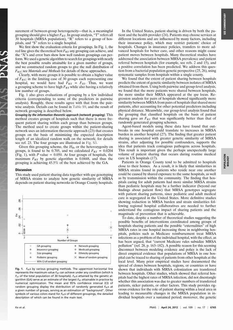
surement of between-group heterogeneity—that is, a meaningfulgrouping should give a higher FRT. In group analysis, “T” refers all30 hospitals (MRSA population), “R” refers to a group of hos-pitals, and “S” refers to a single hospital.We !rst show the evaluation criteria for groupings. In Fig. 1, the
red line gives the theoretical bestFRT any grouping can achieve, andthe “X”s and error bars show how well random groupings can per-form.Weuseda genetic algorithm to search for groupingswithnearlythe best possible results attainable for a given number of groups,and we constructed random groups to give the null distribution ofFRT; seeMaterials and Methods for details of these approaches.Clearly, with more groups it is possible to obtain a higher value
of FRT; in the limiting case of 30 groups each representing onehospital, we would have had FRT = FST. Thus, we wanta grouping scheme to have high FRT while also having a relativelylow number of groups.Fig. 1 also gives evaluations of grouping by a few individual
criteria (corresponding to some of the predictors in pairwiseanalysis). Roughly, these results agree with that from the pair-wise analysis. Details can be found in Table S9, and the result ofnetwork grouping is described below.Grouping by the information theoretic approach (network grouping). Thismethod creates groups of hospitals such that there is more fre-quent patient sharing within each group than between groups.The method used to create groups within the patient-sharingnetwork uses an information theoretic approach (23) that createsgroups on the basis of minimizing the expected descriptionlength of an idealized random walk on the network; for detailssee ref. 23. The four groups are illustrated in Fig. S3.Given this grouping scheme, the HR, or the heterozygosity on
groups, is found to be 0.705, and we calculated FRT = 0.0204(Fig. 1), with P value = 0.050. At four groups of hospitals, themaximum FRT by genetic algorithm is 0.0448, and thus thegrouping is achieving 45.5% of the best achieved by the GA.
DiscussionThis study used patient sharing data together with spa genotypingof MRSA strains to analyze how genetic similarity of MRSAdepends on patient sharing networks in Orange County hospitals.
In the United States, patient sharing is driven by both the pa-tient and the health provider (24). Patients may choose services atdifferent locations and are in"uenced by many factors, includingtheir insurance policy, which may restrict patients’ choice ofhospitals. Changes in insurance policies, transfers to more ad-vanced hospitals for better care, and other reasons might causepatient moves between hospitals. Many theoretical studies haveaddressed the association between MRSA prevalence and patientreferral between hospitals (for example, see refs. 2 and 15), anda positive correlation has been predicted. We address this ques-tion from a bacterial population genetic perspective (25, 26), usingsystematic samples from hospitals within a single county.We found that the extent of patient sharing between hospitals
predicts the extent of genetic similarity between isolates ofMRSAobtained from them.Using both pairwise and group-level analysis,we found that the more patients were shared between hospitals,the more similar their MRSA appeared at the spa locus. Re-gression analysis for pairs of hospitals showed signi!cantly moresimilarity betweenMRSA frompairs of hospitals that sharedmorepatients, after accounting for other potential predictors includingphysical distance. Meanwhile, our group-level analysis found thatthe grouping that classi!ed hospitals on the basis of patientsharing gave an FRT that was signi!cantly better than that ofrandomly generated grouping schemes.Agent-based models using these data have found that out-
breaks in one hospital could translate to increases in MRSAburden in another hospital (27). The !nding that greater patientsharing is associated with greater genetic similarity of MRSAstrains, after adjusting for possible confounders, supports theidea that patients track contagious pathogens across hospitals.This result is important given the perhaps unexpectedly largevolume of patient sharing that occurs during routine medicalcare in US hospitals (17).Patients in Orange County tend to be admitted to hospitals
close to their homes. As a result, it is likely that similarities inMRSA strains found in patients who reside near one anothercould be caused by shared exposure to the same hospitals, as wellas by transmission within the community. The !nding that hos-pitals caring for adult patients had more similar MRSA strainsthan pediatric hospitals may be a further indicator (beyond our!ndings about patient "ow) that MRSA genotypes segregatewith patient sharing patterns, since pediatric and adult medicalcare is segregated in the United States. More de!nitive studiesshowing reduction in MRSA burden and strain similarities fol-lowing regional hospital collaboratives are needed to furtherunderstand the contagious impact of sharing patients and themagnitude of prevention that is achievable.To date, despite a number of theoretical studies suggesting the
possible bene!ts of interventions coordinated among groups ofhospitals sharing patients and the possible “externalities” of highMRSA rates in one hospital increasing those in neighboring hos-pitals, policies such as Medicare reimbursement treat MRSAinfections as a problem of the individual hospital, with the effect, ashas been argued, that “current Medicare rules subsidize MRSApollution” (ref. 28, p. 163–182). A possible reason for this seemingdisconnect between modeling evidence and policy is the lack ofdirect empirical evidence that populations of MRSA in one hos-pital can be traced to sharing of patients from other hospitals at thelocal level. Many prior empirical studies have documented thespread of clones between hospitals, regions, or countries or haveshown that individuals with MRSA colonization are transferredbetween hospitals. Other studies, which showed that referral hos-pitals had the highest rates of MRSA infection, did not disentanglewhether this association was due to greater numbers of transferredpatients, sicker patients, or other factors. This study provides rig-orous evidence for the role of patient sharing within a local area inleading to measurable changes in the MRSA population in in-dividual hospitals over a sustained period; moreover, the genetic
Fig. 1. FRTs by various grouping methods. The uppermost horizontal linerepresents the maximum value FRT can achieve under any condition (which isFST of the total population of 30 hospitals). FRTs attained by the genetic al-gorithm (GA) serve as an estimate of the largest FRT attainable in practice bynumerical optimization. The mean and 95% con!dence interval (CI) ofrandom grouping display the distribution of randomly generated FRTs ata given number of groups, serving as an estimation of “background”. Othersymbols of various colors stand for FRTs of different groupings, the detaileddescription of which can be found in the main text.
Ke et al. PNAS | April 24, 2012 | vol. 109 | no. 17 | 6765
POPU
LATION
BIOLO
GY
SEECO
MMEN
TARY
evidence provided here is an independent line of evidence thatcon!rms the importance of patient sharing. This !nding impliesthat there is hope for synergistic impact to reduce MRSA, withconcerted efforts by hospitals to implement prevention strategiestogether. Had we found that MRSA strains were indiscriminatelyfound throughout all hospitals, this result would have suggestedthat a county-wide approach to MRSA containment would benecessary. Instead, it appears possible that targeted approachesmight well produce substantial impact when applied to a smallgroup of hospitals that are strongly connected by patient sharing.There are several limitations to our study. First, our measure of
patient sharing considered transfer of patients between hospitals, inboth directions and regardless of theirMRSA colonization status. Amore directly relevant measure, if it were available, would be thetransfer of patients colonized withMRSA from each hospital to theother (19). On the other hand, the demonstration here that overallpatient transfer is a predictor ofMRSA similarity between hospitalssuggests that overall patient transfer is an adequate surrogate for theeffect of sharing of MRSA-colonized patients, at least for analysesof this type. A related limitation is that whereas 92% of adults in thedata set for patient transfer had identi!ers (thus only 8% wereuntracked), the majority (86%) of children lacked such identi!ers.However, of these, 63% were<6 mo of age, and a large proportionof these would have been hospitalized at birth and would not havebeen readmitted (17). Second, the current study did not assess theimpact of strains categorized as hospital-onset (HO)-MRSA vs.community-onset (CO)-MRSA (3). We did not assess this distinc-tion partly because community and healthcare reservoirs are mixingand because, at a hospital level, there were often too few strains tomake these types of evaluations. Moreover, all strains were treatedequally, assessing only whether they were distinguishable by ourtypingmethod. Third, patient sharing data could be used in differentways that take into account patient sharing directedness, time oftransfer, and length of stay in hospitals, which might provide otherinteresting !ndings but were not implemented due to data limi-tations. Fourth, unmeasured or residual confounding of the asso-ciation between patient "ow and MRSA population similarity isa possibility, as in all such studies. A potential confounder of par-ticular concern is that hospitals that transfer many patients may alsodraw from the same patient population, so that similarity of thecatchment populations leads to importation of similar strains, whichcould explain MRSA population similarities independent of anycausal effect of patient sharing. To address this problem, we in-cluded in our model a variable for distance between the two hos-pitals; compared with a model omitting distance (Table S7), thebaseline model (Table 1) had a similar, but slightly smaller effect ofpatient sharing. Moreover, distance between hospitals was nota signi!cant predictor in the baselinemodel, nor was it signi!cant inunivariate analysis, whereas patient sharing was. In addition, weincluded variables for shared demographic characteristics ofpatients, to further eliminate spurious associations with patientsharing that are in fact caused by similar patient populations.Nonetheless, because none of these variables perfectly capturessimilarities in patient populations, it is possible that the associationbetween patient sharing and genetic similarity of MRSA remainsbiased by some of these factors. Speci!c spa types associated withpediatric and adult patients in this population have been describedrecently (29). Fifth, although the spa genotypingmethod used in thecurrent study is widely used as a fast and reliable genotyping tech-nique for S. aureus, we might obtain more meaningful results if weused higher-resolution typing systems. Finally, we noticed that someof our hospitals have relatively fewer isolates available. Althoughthe exclusion of these hospitals did not qualitatively affect the resultsof our pairwise analysis, we found that removing hospitals 21 and 29made our network grouping result insigni!cant. Themain reason forthis loss of statistical signi!cance is that each hospital has a spa typethat is rare among all hospitals (appearing only in the hospitals thatare in the same networking group), and its removal, combined with
the fact that these hospitals have fewer isolates, made the distri-bution of random FRTs generated by random grouping higher. Al-though we preserved these hospitals in group-level analysis as webelieve these rare isolates indicated within-group similarity, moreisolates from these hospitals, if possible, are strongly desired.In summary, we found that patient sharing patterns across
hospitals are likely to be correlated with MRSA genetic hetero-geneity, along with several other hospital characteristics. Thisstudy is a unique regional analysis of a relatively enclosed largemetropolitan region of 3 million people. It performs a compre-hensive analysis of whether hospitals that share patients also shareMRSA strains. It provides evidence of local ecosystems withina single region that are associated with shared patients and sug-gests that certain groups of local hospitals could make concertedand synergistic efforts to reduce the prevalence of important re-sistant pathogens and reduce healthcare-associated disease.
Materials and MethodsStudy. We conducted a population-based, prospective collection of clinicalisolates of MRSA from 30 of 32 hospitals in OC, California as described else-where (17). The geographical distribution of these hospitals is shown in Fig. S4.This study was approved by the Institutional Review Board of the University ofCalifornia Regents.
Isolate collection, specimen- and hospital-level data, and laboratorymethods are described in SI Text.
Measuring the Genetic Similarity in MRSA Between Pairs or Groups ofHospitals. We adopted a standard measure of genetic similarity: Wright’s Fstatistics (30). F statistics detect population substructure measured by a givengenetic locus of interest. One!rst calculates the “heterozygosities” of this locusondifferent levels; here, heterozygosity corresponds to theprobability that tworandomly chosen isolates will differ at the locus of interest. Three hierarchicallevels of population were used in this study: (i) subpopulations (S) refer to thebacteria isolated from a single hospital, (ii) “regions” (R) refer to the bacteriafrom a subset (group) of hospitals less than the 30 total hospitals in our study,and (iii) the total population (T) refers to all bacteria included in our study. Notethat in pairwise analysis, we only have R (a pair of hospitals) and S (a singlehospital)—T is not used in the pairwise analysis. If there is any populationsubstructure, then the heterozygosity calculated for the total populationwill behigher than the weighted average of that calculated for each group in-dividually. In this report, we use the term “heterogeneity” to refer to high Fstatistics implying genetic differentiation between different populations.
Formally, heterozygosity of a population (in terms of one genetic locus) isde!ned as oneminus the sum of squared allele frequencies. Let pi (i = 1, 2, . . .)represent the frequency of allele i; then the heterozygosity of this locus isgiven by
H ! 1!Xk
i!1
p2i ;
whereH stands for heterozygosity, and k is the total number of alleles present.Here, in the total population T (the MRSA population of all hospitals
involved) we calculate the heterozygosity of this total population (HT), using
HT ! 1!Xk
i!1
p2Ti
with each allele’s frequency (pTi ) and the number of different alleles (k) inthe total population. HS, the heterozygosity of subpopulations (individualhospitals) is calculated similarly to HT, except that !rst, we do the calculationrestricted to each subpopulation and then calculate HS as the average of allof the computed subpopulation heterozygosities,
HS !1n
Xn
i!1
HSi
HSi ! 1!Xki
j!1
p2ij ;
where n is the total number of subpopulations, HSi is the heterozygosity ofsubpopulation i, ki is the number of different alleles in subpopulation i, andpij is allele j’s frequency in subpopulation i.
6766 | www.pnas.org/cgi/doi/10.1073/pnas.1113578109 Ke et al.
Between these two levels, another level, the regional heterozygosityHR (inthe current study, a region means a group of hospitals we classi!ed), is alsocalculated similarly: We take a group of hospitals and compute the het-erozygosity of that group. Afterward, HR is just the weighted average of allthese regional heterozygosities, with the number of hospitals in each groupbeing the weights. It can be shown that HT ! HR ! HS (equal signs hold whenthere is no population substructure). Then, the F statistics we used arede!ned as
FSR ! HR "HS
HR
FRT ! HT "HR
HT
FST ! HT "HS
HT:
Measurement of MRSA Population Similarity—Pairwise Analysis of Hospitals.Potential individual predictors. To assess patient demographic factors that mightaccount for genetic similarity of MRSA found in pairs of hospitals, we de!nedthe following dichotomous variables. For dichotomized proportions, thesubscript zero indicates a proportion less than or equal to the break point.Hospitals were classi!ed for whether they were or had the following:
Over 35% of patients privately insuredOver 10% of patients on MedicaidOver 20% of patients HispanicOver 10,000 admissions per yearPediatric hospital (vs. adult).
Pairs of hospitals were classi!ed as 00, 01/10, or 11 on each of thesevariables, and genetic similarity was assessed for hospitals that were similaron these variables (00 or 11) compared with pairs containing hospitals thatwere different (01/10).
The predictor of primary interest was patient "ow. Using previouslypublished data (17) on the number of times any patient was transferredbetween two hospitals (including possible multiple transfers of the samepatient or discharge from the !rst before admission to the second, with anintervening stay at home), the "ow of patients from hospital A to hospital B,TAB, was de!ned as the proportion of hospital B’s patients in a year who hada previous stay in hospital A during the year. The average "ow betweenhospital A and B was then de!ned as (TAB + TBA)/2 and was used in ouranalyses. A more detailed de!nition can be found in SI Text.Linear regression analysis. We used multivariate linear regression to assess thepredictors of genetic similarity between the MRSA populations in pairs ofhospitals and used univariate regression for each single variable as a sup-plement. The response variable that measures heterogeneity was pairwiseFSR—the reduction in heterozygosity when two hospitals are viewed asa whole. We log-transformed these two variables to obtain normally dis-tributed data. As some of the hospital pairs have "ow = 0, we added 0.00001(#50% of the smallest available data) to all "ow data to perform the logtransformation. To account for other possible predictors, we adjusted forthe demographic variables described in the previous section by also usingtwo indicator variables for each demographic variable. Standard regressionP values do not account for the dependence among the observations in-duced by the fact that the response variables are genetic “distances” be-tween pairs of hospitals. To adjust for this nonindependence, we performedmultiple Mantel permutation tests (univariate Mantel test for univariateanalyses) to generate permutation P values for all regression analyses andreferred to these P values as permutation P values, as described elsewhere(31). Brie"y, we constructed a genetic distance matrix for our responsevariable—pairwise FSR from our data, with each element in the matrix—anddij corresponds to the log-FSR of hospitals i and j and comes from the row ofdata that records the pairwise information of these two hospitals (i.e., pa-tient "ow, distances, etc.). Then we shuf"ed this matrix by each hospital—inother words, we shuf"ed the rows and columns in the same way 5,000 times,and the resulting matrices were "attened and paired back with predictorvariables to conduct 5,000 regressions. Two-tailed P values were calculatedfrom the distribution of t statistics of corresponding coef!cients generated.
In addition, for each pair of hospitals, we also adjusted for (i) sample size,by using the average number of spa-typed isolates of the two hospitals, and
(ii) distance between the two hospitals, calculated on the basis of theirlongitudinal and latitudinal data, in kilometers.
Univariate plots of the response vs. individual predictors were checkedvisually for outliers.
Measurement of MRSA Population Similarity—Analysis of Groups of Hospitals.As a complementary approach, we considered whether grouping the hos-pitals into a small number of groups on the basis of the demographiccharacteristics used above of their patient populations or, of more directinterest, on the basis of their patterns of patient sharing, would create groupsthat captured some of the population genetic structure of the MRSA in thehospitals. To assess this possibility, we sought both to assess the extent towhich the best possible grouping could create groups that are geneticallyhomogeneous (the value of FRT obtained by an optimal grouping) and toassess how much genetic structure would be captured in randomly con-structed groupings of all 30 hospitals (the range of FRT values obtained byrandom groupings). The !rst assessment was done by using a genetic al-gorithm (GA) to give an approximate numerical value because an exhaus-tive/exact method is computationally infeasible, and the second assessmentwas done by creating groupings in which hospitals were randomly assignedto group membership.GA—Evaluation of grouping ef!ciency. To establish a standard for the possibleextent to which any grouping scheme of hospitals could de!ne geneticallysimilar MRSA populations (“grouping scheme” or “grouping” refers to a spe-ci!c group assignment of all hospitals), we attempted to!nd the groupings ofhospitals (using no information about the hospitals themselves) that maxi-mized FRT—the measure of genetic heterogeneity between groups—by usinga GA. We implemented this method by generating random groupings ata given number of groups and evaluating FRT for each grouping and thenevolving the groupings by enriching and combining groupings with high FRT.The goal was to approximate the optimal groupings at a given number ofgroups to serve as a reference of maximum FRT. This method was repeated forgroupings with two, four, or six groups (we primarily used two and fourgroups, and the result of six groupswas used to show the trendof FRTwhen thenumber of groups increases). These groupings are referred to asGAgroupings.Details of this algorithm can be found in SI Text and Fig. S2.Evaluation of null distribution of FRT. To evaluate whether groupings of hospitalsbased on any given measurement contain information about the geneticstructure of the MRSA population (FRT) greater than expected by chancealone, we created a null distribution of FRT for randomly chosen groupingsthat divided hospitals into two, four, and six groups. For each given numberof groups, we randomly generated 15,000 groupings and tabulated thedistribution of FRT from these results (these groupings are referred to asrandom groupings of k groups). We obtained the mean and 95% coverageinterval of FRTs for these random groupings. The P value (double sided) ofa given FRT for a particular grouping is de!ned by the proportion of ran-domly generated FRTs that are of equal distance or farther away from themean than it is.Grouping of hospitals by prespeci!ed categories. We !rst grouped all hospitalsusing categorizing variables speci!ed in Measurement of MRSA PopulationSimilarity—Pairwise Analysis of Hospitals , including private insurance pro-portion, Medicaid coverage, size (annual admission), ethnicity (Hispanic),and pediatric vs. adult-only hospitals (the groups are named by theirgrouping criteria).Grouping of hospitals by the information theoretic approach (network grouping). Forpatient sharing, we used the algorithm of ref. 23 to identify “modules” orneighborhoods within the network of hospitals on the basis of patient "ow.To do so, we considered each hospital as a “node” in the network and be-tween each hospital constructed an undirected edge representing patientsharing with weight determined by the proportion of shared patients, asgiven in Measurement of MRSA Population Similarity—Pairwise Analysis ofHospitals. The algorithm de!nes neighborhoods—roughly speaking—as setsof nodes in which an imaginary random walker, traversing edges of thenetwork with probabilities proportional to the edge weight, would be muchmore likely to stay within a set. Thus, sets of nodes that are well connectedwith one another will tend to be in the same group; in our case, sets ofhospitals that share many patients with one another will tend to be in thesame group, and sets of hospitals that do not share many patients with oneanother will tend to be in different groups. Technical details of this methodare given in SI Text, which summarizes the account given in ref. 23. Thismethod is referred to as network grouping.
ACKNOWLEDGMENTS. This work was supported by Models of Infectious Dis-ease Agent Study Award U54GM088558 (to W.K. and M.L.), by US NationalInstitute Of General Medical Sciences Grant U01 GM76672 (to S.S.H. and T.R.A.),
Ke et al. PNAS | April 24, 2012 | vol. 109 | no. 17 | 6767
POPU
LATION
BIOLO
GY
SEECO
MMEN
TARY
by funds from the University of California Irvine School of Medicine (to S.S.H., K.R.E., C.C.N., and C.R.M.), by Award WT089472MA from the UK Wellcome Trust(to B.G.S.), and by a UK Biotechnology and Biological Sciences Research Council
Doctoral Training Grant (to L.O.H.). The content is solely the responsibility of theauthors and does not necessarily represent the of!cial views of the NationalInstitute of General Medical Sciences or the National Institutes of Health.
1. Cooper BS, et al. (2004) Methicillin-resistant Staphylococcus aureus in hospitals andthe community: Stealth dynamics and control catastrophes. Proc Natl Acad Sci USA101:10223–10228.
2. Donker T, Wallinga J, Grundmann H (2010) Patient referral patterns and the spread ofhospital-acquired infections through national health care networks. PLoS ComputBiol 6:e1000715.
3. Grundmann H, et al.; European Staphylococcal Reference Laboratory Working Group(2010) Geographic distribution of Staphylococcus aureus causing invasive infections inEurope: A molecular-epidemiological analysis. PLoS Med 7:e1000215.
4. Feil EJ, et al. (2003) How clonal is Staphylococcus aureus? J Bacteriol 185:3307–3316.5. Grundmann H, Aires-de-Sousa M, Boyce J, Tiemersma E (2006) Emergence and re-
surgence of meticillin-resistant Staphylococcus aureus as a public-health threat. Lan-cet 368:874–885.
6. Livermore DM, Pearson A (2007) Antibiotic resistance: Location, location, location.Clin Microbiol Infect 13(Suppl 2):7–16.
7. Tiemersma EW, et al.; European Antimicrobial Resistance Surveillance System Partic-ipants (2004) Methicillin-resistant staphylococcus aureus in Europe, 1999–2002. EmergInfect Dis 10:1627–1634.
8. UK Health Protection Agency (2008) Surveillance of healthcare associated infectionsreport. Technical report. Available at http://www.hpa.org.uk/web/HPAwebFile/HPAweb_C/1216193833496. Accessed February 28, 2012.
9. Dominguez MA, de Lencastre H, Linares J, Tomasz A (1994) Spread and maintenanceof a dominant methicillin-resistant Staphylococcus aureus (MRSA) clone during anoutbreak of MRSA disease in a Spanish hospital. J Clin Microbiol 32:2081–2087.
10. Harbarth S, et al. (2000) Effect of delayed infection control measures on a hospitaloutbreak of methicillin-resistant Staphylococcus aureus. J Hosp Infect 46:43–49.
11. Kotilainen P, et al. (2003) Elimination of epidemic methicillin-resistant Staphylococcusaureus from a university hospital and district institutions, Finland. Emerg Infect Dis 9:169–175.
12. Roman RS, et al. (1997) Rapid geographic spread of a methicillin-resistant Staphylo-coccus aureus strain. Clin Infect Dis 25:698–705.
13. UK Department of Health (2005) Mrsa surveillance system: Results. Technical re-port. Available at http://www.dh.gov.uk/en/Publicationsandstatistics/Publications/PublicationsStatistics/DH_4085951. Accessed August 2011.
14. Scanvic A, et al. (2001) Duration of colonization by methicillin-resistant Staphylo-coccus aureus after hospital discharge and risk factors for prolonged carriage. ClinInfect Dis 32:1393–1398.
15. Robotham JV, Scarff CA, Jenkins DR, Medley GF (2007) Meticillin-resistant Staphylo-coccus aureus (MRSA) in hospitals and the community: Model predictions based onthe UK situation. J Hosp Infect 65(Suppl 2):93–99.
16. Smith DL, Levin SA, Laxminarayan R (2005) Strategic interactions in multi-institutionalepidemics of antibiotic resistance. Proc Natl Acad Sci USA 102:3153–3158.
17. Huang SS, et al. (2010) Quantifying interhospital patient sharing as a mechanism forinfectious disease spread. Infect Control Hosp Epidemiol 31(11):1160–1169.
18. Roberts RB, et al.; MRSA Collaborative Study Group (1998) Molecular epidemiology ofmethicillin-resistant Staphylococcus aureus in 12 New York hospitals. J Infect Dis 178:164–171.
19. Kho AN, Lemmon L, Commiskey M, Wilson SJ, McDonald CJ (2008) Use of a regionalhealth information exchange to detect crossover of patients with MRSA betweenurban hospitals. J Am Med Inform Assoc 15:212–216.
20. Frénay HM, et al. (1996) Molecular typing of methicillin-resistant Staphylococcusaureus on the basis of protein A gene polymorphism. Eur J Clin Microbiol Infect Dis15:60–64.
21. Mellmann A, et al. (2006) Automated DNA sequence-based early warning system forthe detection of methicillin-resistant Staphylococcus aureus outbreaks. PLoS Med 3:e33.
22. Harmsen D, et al. (2003) Typing of methicillin-resistant Staphylococcus aureus ina university hospital setting by using novel software for spa repeat determination anddatabase management. J Clin Microbiol 41:5442–5448.
23. Rosvall M, Bergstrom CT (2008) Maps of random walks on complex networks revealcommunity structure. Proc Natl Acad Sci USA 105:1118–1123.
24. Lee BY, et al. (2011) Social network analysis of patient sharing among hospitals inOrange County, California. Am J Public Health 101:707–713.
25. Harris SR, et al. (2010) Evolution of MRSA during hospital transmission and in-tercontinental spread. Science 327:469–474.
26. Ko KS, et al. (2006) Molecular characterization of methicillin-resistant Staphylococcusaureus spread by neonates transferred from primary obstetrics clinics to a tertiarycare hospital in Korea. Infect Control Hosp Epidemiol 27:593–597.
27. Lee BY, et al. (2011) Modeling the spread of methicillin-resistant Staphylococcus au-reus (MRSA) outbreaks throughout the hospitals in Orange County, California. InfectControl Hosp Epidemiol 32:562–572.
28. Outterson K, Yevtukhova O (2011) Germ shed management in the United States.Antibiotic Policies: Controlling Hospital-Associated Infection, eds Gould IM, van derMeer J (Springer, Berlin), pp 163–182.
29. Hudson LO, et al. (2012) Differences in methicillin-resistant Staphylococcus aureusstrains isolated from pediatric and adult patients from hospitals in a large county inCalifornia. J Clin Microbiol 50(3):573–579.
30. Hartl DL, Clark AG (1997) Principles of Population Genetics (Sinauer, Sunderland, MA),3rd Ed.
31. Manly BFJ (2007) Randomization, Bootstrap and Monte Carlo Methods in Biology(Chapman & Hall/CRC).
6768 | www.pnas.org/cgi/doi/10.1073/pnas.1113578109 Ke et al.