molecular characteristics of community-associated methicillin-resistant staphylococcus aureus...
TRANSCRIPT

_____________________________________________________________________________________________________ *Corresponding author: Email: [email protected], [email protected];
British Microbiology Research Journal 5(3): 227-236, 2015, Article no.BMRJ.2015.024
ISSN: 2231-0886
SCIENCEDOMAIN international
www.sciencedomain.org
Molecular Characteristics of Community-Associated Methicillin-resistant Staphylococcus aureus (CA-
MRSA) Isolates from Clinical Specimens in Iraq
Alaa H. Al-Charrakh1*, Huda H. Al-Hassnawi1 and Jawad K. Al-Khafaji1
1Department of Microbiology, College of Medicine, Babylon University, Hilla, Iraq.
Authors’ contributions
This work was carried out in collaboration between all authors. Author AHA managed the literature
searches, designed the study, wrote the first draft of the manuscript and supervised the work. Author HHA isolated CA-MRSA from clinical specimens, performed the experiments and wrote the protocols.
Author JKA supervised the work. All authors read and approved the final manuscript.
Article Information
DOI: 10.9734/BMRJ/2015/9607 Editor(s):
(1) Frank Lafont, Center of Infection and Immunity of Lille, Pasteur Institute of Lille, France. (2) En Tao Wang, Departamento de Microbiología, Escuela Nacional de Ciencias Biológicas, Instituto Politécnico Nacional,
México and State Key Laboratory of Agrobiotechnology and College of Biological Sciences, China. Reviewers:
(1) M. A. Yusuf, Dept. of Microbiology and Immunology, National Institute of Neurosciences and Hospital, Dhaka, Bangladesh. (2) Anonymous, University of the West Indies, West Indies.
(3) Anonymous, Islamic Azad University, Iran. (4) Anonymous, University of Tsukuba, Japan.
Complete Peer review History: http://www.sciencedomain.org/review-history.php?iid=703&id=8&aid=6514
Received 19
th February 2014
Accepted 3rd
August 2014 Published 13
th October 2014
ABSTRACT
Background: Methicillin-resistant Staphylococcus aureus (MRSA) infections have been recognized for decades as hospital acquired MRSA (HA-MRSA). Nowadays, MRSA is also recognized as a worldwide emerging community-associated pathogen. Community associated-MRSA (CA-MRSA) has been shown to be more virulent with a high degree of severity of disease when compared to HA-MRSA. Objectives: The study was designed to assess the occurrence and the molecular detection of HA-MRSA and CA-MRSA isolates obtained from clinical specimens in Iraq. Methods: HA-MRSA and CA-MRSA isolates were obtained from clinical specimens in three main hospitals in Hilla city/Iraq during the period, March to June 2011. MRSA isolates obtained primarily from clinical specimens of skin and soft tissue infections (SSTs) were subjected to genetic study. PCR was used for detection of genes responsible for methicillin resistance (mecA, SCCmec type
Original Research Article
Al-Charrakh et al.; BMRJ, 5(3): 227-236, 2015; Article no.BMRJ.2015.024
228
IV) and genes responsible for toxin production (pvl, lukED). Statistical analysis was performed using chi-square (X2) test to assess intergroup significance, inpatients, and outpatients with respect to all genes used in present study. Results: Out of 301 clinical samples, 24 MRSA isolates were obtained. All these MRSA isolates (100.0%) were mecA gene positive. Twenty three (95%) were found to be carrying SCCmec type IV, 19 (79%) had positive result for pvl toxin gene, and 20 (83%) had lukED toxin gene. Conclusion: The majority of MRSA isolates belonged to SCCmec IV. pvl and lukED toxin genes are also found in the MRSA isolates among the CA-MRSA isolates.
Keywords: CA-MRSA; mecA gene; SCCmec type IV; pvl gene; PCR; HA-MRSA.
1. INTRODUCTION Methicillin-resistant Staphylococcus aureus (MRSA) infections have been recognized for decades as hospital acquired or healthcare associated [1]. The possibility of transmission of healthcare associated MRSA (HA-MRSA) to the community was unavoidable [2]. Now-a-days MRSA is also recognized as a worldwide emerging community-acquired pathogen (CA-MRSA) [3]. It was shown that Panton-Valentine leukocidin (PVL) positive MSSA are a likely reservoir for the development of PVL positive MRSA [4] via integration of Staphylococcus cassette chromosome mec (SCCmec) elements including the mecA gene conferring methicillin resistance [5]. MRSA strains that have been clinically identified as CA-MRSA have been shown to be more virulent with a high degree of severity of disease when compared to HA-MRSA [6]. This is due to the production of the PVL toxin. PVL toxin is associated with deep skin and soft tissue infections, such as furunculosis and abscesses, however, necrotizing tissue infections and lethal hemorrhagic pneumonia have been reported [7]. The prevalence rate of the PVL toxin in CA-MRSA strains varies with different studies and countries [8]. Some studies reported a prevalence of 77% to 100% for the PVL toxin in Minnesota, USA in 2000 [9] while a prevalence of less than 5% was reported in Western Europe [10]. The mecA gene resides on a genomic island termed the staphylococcal cassette chromosome mec (SCCmec) [5]. Most outbreaks of MRSA involve CA-MRSA rather than HA-MRSA, thus monitoring the SCCmec type is important in determining the epidemiologic trend of the MRSA strains in clinical settings [11]. The presence of PVL toxin in CA-MRSA strains can be confirmed by co-amplification of the lukS/F-PV genes [11]. In comparison to previous detection methods such as Southern blotting and pulsed-field gel electrophoresis (PFGE), PCR assays can
provide a rapid amplification, detection and typing tool for MRSA strains [12]. Outbreaks in closed living communities, jail inmates, military recruits, and gay community, have been reported in the United States [13]. The aim of this study was to assess the occurrence of HA-MRSA and CA-MRSA isolates and to detect the PVL genes in MRSA isolates obtained from clinical specimens in Hilla/Iraq.
2. MATERIALS AND METHODS 2.1 Study Design and Bacterial Isolates This cross sectional study was designed to assess the occurrence of HA-MRSA and CA-MRSA isolates obtained from clinical specimens and to detect the PVL genes in MRSA isolates. At the beginning of this study, 301 different clinical samples were collected during the period of March to June 2011 from the main three hospitals in Al-Hilla city/Iraq (Hilla Teaching hospital, Margan Teaching hospital, and Childhood and gynecology hospital), in addition to some private clinics. Clinical isolates were as follows: ear swabs (37), burns (12), skin infections (65), wounds (25), sputum (23), urine (70), vaginal swabs (4), and CSF (65).
Out of 301 clinical samples, forty six S. aureus isolates were obtained. These bacterial isolates were identified as S. aureus based on their morphology, Gram-staining, catalase properties in addition to coagulase test [14] and as suggested previously [15].
In the light of antibiotic susceptibility results of our previous study [15], 13 MRSA isolates were detected using phenotypic methods. They were oxacillin and cefoxitin resistant. The MRSA isolates were also SCCmec typed to differentiate between HA-MRSA and CA-MRSA isolates using a PCR. The remaining 11 samples from specific skin infections (abscesses, boils), which phenotypically identified as oxacillin sensitive

Al-Charrakh et al.; BMRJ, 5(3): 227-236, 2015; Article no.BMRJ.2015.024
229
and cefoxitin resistant [15], were selected for genetic study to detect other genes that are considered as a marker for CA-MRSA isolates.
2.2 DNA Extraction and PCR Assay
One colony of each strain (cultured on solid medium) was inoculated into 5 mL of BHI (Broth Heart Infusion) and grown overnight at 37°C. From these cultures, DNA was purified from bacterial cells using Genomic DNA Mini kit (Geneaid, UK). Chromosomal DNAs obtained were used as templates for all PCR experiments. The PCR reactions were carried out in a thermal cycler (Clever, U.K). Before PCR assay, and in order to quantify the DNA concentration (ng µL
-1), the quantification of DNA samples was
carried out by means of a spectrophotometric reading using 1 µL aliquots of Genomic DNA with a NanoDropTM spectrometer (NanoDrop Technologies), adopting the manufacturer’s recommendations. The concentration of DNA was estimated from absorbance at 260 nm. DNA profiles were performed using bacterial DNA and loading buffer according to the manufacturer instructions (Bioneer, Korea).
2.3 Polymerase Chain Reaction Protocols
The DNA extracts of S. aureus isolates were subjected to different genes by PCR. The protocols used were according to manufacturer's instructions. Different primers of antibiotic resistance and toxins genes were used (Bioneer, Korea) in an attempt to differentiate MRSA isolates (Table 1). All PCR components were assembled in PCR tube and mixed on ice bag under sterile conditions. The PCR tubes were placed on the PCR machine and the accurate PCR cycling program parameters conditions were installed (Table 2).
2.4 Statistical Analysis Statistical analysis was performed using chi-square (X
2) test to assess intergroup
significance, inpatient (hospital acquired) and outpatient (community acquired) with the respect to all genes used in present study. P value less than 0.005 was considered as statistically significant and P- value less than 0.001 was considered as highly significant.
3. RESULTS AND DISCUSSION
3.1 Detection of mecA and SCCmec IV Genes by PCR
Chromosomal DNA was extracted from twenty four clinical strains. Results of this study revealed that a high frequency of mecA gene (100%) among all S. aureus isolates was detected (Fig. 1). All hospital (100%) and all community isolates (100%) carried mecA gene (Table 3).
Using phenotypic methods, a total 13 out of 24 isolates were determined previously as MRSA [10]. The mecA gene is responsible for mediating methicillin resistance in Staphylococci. These results indicate that there is a large dissemination of MRSA in the community (62%) and not restricted to the hospitals as expected [20] and as reported by other authors [7,21] who found that most MRSA isolates were associated with nosocomial infections. This high prevalence of mecA gene in the community may be due to horizontal gene transfer (HGT) from MRSA to MSSA isolates by transduction [22] since the mecA gene is carried on the staphylococcal cassette chromosome. SCCmec is inserted into the S. aureus chromosome near the origin of replication [23].
In a local study in Iraq, the presence of mecA gene in isolates of CA-MRSA from holy shrine in Najaf city was detected [24]. In Europe, MRSA strains account for less than 5% of S. aureus isolates in the community setting and for less than 11% of isolates from patients with skin and soft tissue infections (SSTI) in France [25]. The CDC created a definition for a CA-MRSA infection: any MRSA infection diagnosed for an outpatient or within 48 h of hospitalization if the patient lacks the following health care-associated MRSA risk factors: hemodialysis, surgery, residence in a long-term care facility or hospitalization during the previous year, the presence of an indwelling catheter or a percutaneous device at the time of culture, or previous isolation of MRSA from the patient [26]. All other MRSA infections were considered to be HA-MRSA.
The mecA gene for methicillin resistance resides on the chromosome. Accurate detection of mecA mediated resistance to methicillin and other penicillinase stable penicillins (PSPs) like oxacillin,
nafcillin, cloxacillin, dicloxacillin,
flucloxacillin, and mecillinam is
necessary to ensure appropriate antimicrobial chemotherapy of
staphylococcal infections, particularly those
from community associated infections.
Al-Charrakh et al.; BMRJ, 5(3): 227-236, 2015; Article no.BMRJ.2015.024
230
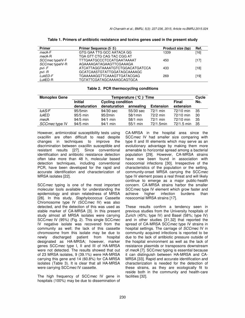
Table 1. Primers of antibiotic resistance and toxins genes used in the present study
Primer Primer Sequence (5'_3
') Product size (bp) Ref.
mecA-F GTG GAA TTG GCC AATACA GG TGA GTT CTG CAG TAC CGG AT
1339 [16] mecA-R SCCmec typeIV-F TTTGAATGCCCTCCATGAATAAAAT
AGAAAAGATAGAAGTTCGAAAGA 450 [17]
SCCmec typeIV-R pvl- F ATCATTAGGTAAAATGTCTGGACATGATCCA 433 [18] pvl- R GCATCAASTGTATTGGATAGCAAAAGC LukED-F TGAAAAAGGTTCAAAGTTGATACGAG 269 [19] LukED-R TGTATTCGATAGCAAAAGCAGTGCA
Table 2. PCR thermocycling conditions
Monoplex Gene Temperature (°C )/ Time Cycle No. Initial
denaturation Cycling condition Final
extension denaturation annealing Extension
lukS/F 95/5min 94/30 sec 55/30 sec 72/1 min 72/10 min 35 lukED 95/5 min 95/2min 58/1min 72/2 min 72/10 min 30 mecA 94/5 min 94/1 min 58/1 min 72/1 min 72/10 min 35 SCCmec type IV 94/5 min 94/1 min 55/1 min 72/1.5min 72/1.5 min 35
However, antimicrobial susceptibility
tests using
oxacillin are often difficult to read despite changes
in techniques to improve the
discrimination between oxacillin susceptible and
resistant results [27]. Since conventional identification and antibiotic resistance detection often take more than 48 h, molecular based detection techniques, including conventional PCR, have been developed for the rapid and accurate identification and characterization of MRSA isolates [22]. SCCmec typing is one of the most important molecular tools available for understanding the epidemiology and strain relatedness of MRSA [28]. In this study, Staphylococcus Cassette Chromosome type IV (SCCmec IV) was also detected, and the detection of this was used as stable marker of CA-MRSA [3]. In this present study almost all MRSA isolates were carrying SCCmec IV (95%) (Fig. 2). This single SCCmec IV negative isolate was recovered from the community as well; the lack of this cassette chromosome from this isolate may be due to newly discharged patient from hospital designated as HA-MRSA; however, marker genes SCCmec type I, II and III of HA-MRSA were not detected. The results showed that out of 23 MRSA isolates, 9 (39.1%) were HA-MRSA carrying this gene and 14 (60.8%) for CA-MRSA isolates (Table 3). It is clear that all HA-MRSA were carrying SCCmec IV cassette. The high frequency of SCCmec IV gene in hospitals (100%) may be due to dissemination of
CA-MRSA in the hospital area since the SCCmec IV had smaller size comparing with type II and III elements which may serve as an evolutionary advantage by making them more amenable to horizontal spread among a bacterial population [29]. However, CA-MRSA strains have now been found in association with nosocomial infections [30]. Irrespective of the characteristics of the population or the setting, community-onset MRSA carrying the SCCmec type IV element poses a real threat and will likely continue to emerge as a major public-health concern. CA-MRSA strains harbor the smaller SCCmec type IV element which grow faster and achieve higher infection burdens than nosocomial MRSA strains [17]. These results confirm a tendency seen in previous studies from the University hospitals of Zurich (45%; type IV) and Basel (58%; type IV) and in other studies [31,32] that reported the spread of CA-MRSA SCCmec type IV strains in hospital settings. The carriage of SCCmec IV in community acquired infections is reported to be due to the lack of antibiotic pressure outside of the hospital environment as well as the lack of resistance plasmids or transposons downstream of mecA [7]. SCCmec typing is essential because it can distinguish between HA-MRSA and CA-MRSA [33]. Rapid and accurate identification and characterization is needed for the detection of these strains, as they are ecologically fit to reside both in the community and health-care facilities [32].

Al-Charrakh et al.; BMRJ, 5(3): 227-236, 2015; Article no.BMRJ.2015.024
231
Fig. 1. Gel electrophoresis of PCR of mecA amplicon product Lane L: Ladder (3000-bp ladder), Lanes (S22, S23, S26, S28, S29, S31, and S32) represent isolates from skin.
Negative control without amplicon and positive control with the expected band were not shown
Fig. 2. Gel electrophoresis of PCR of SCCmec IV amplicon product Lane L: Ladder (3000-bp ladder), Lanes (S22, S23, S26, S28, S29, and S31) represent isolates from skin. Lane
S32 shows negative result
3.2 Molecular Detection of Virulence Genes by PCR
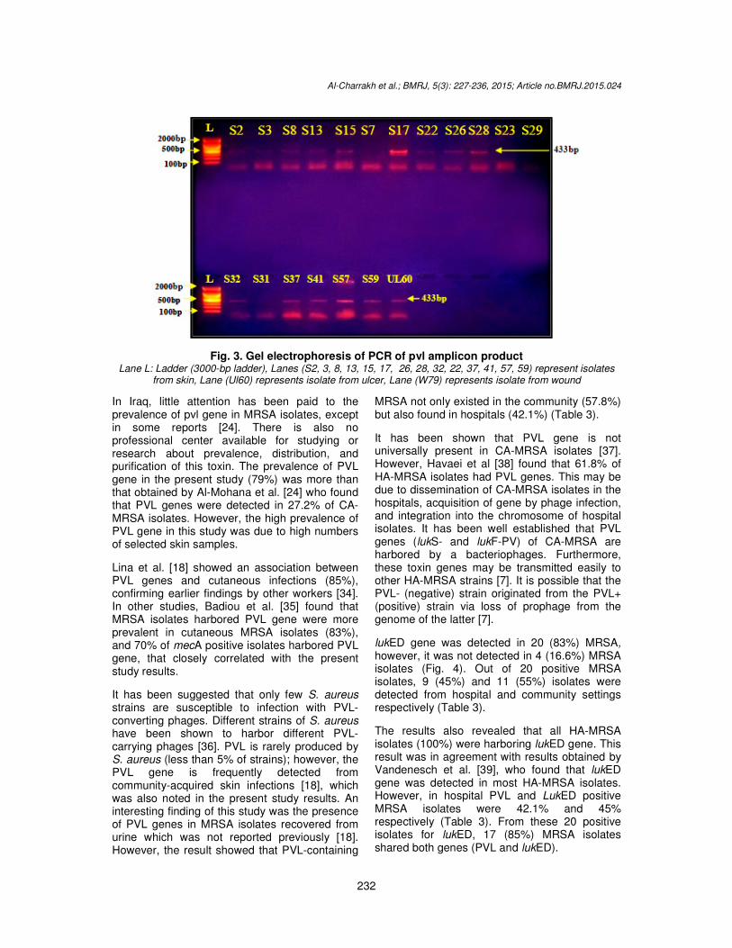
PCR was used to determine the bi-component leukocidin PVL gene in all 24 MRSA isolates. However, PVL gene by PCR was seen in 19 (79.1%) isolates. PVL gene was not detected in 5 (20.8%) isolates (Fig. 3). All these isolates were mecA gene positive and identified as MRSA by genotypic test (Fig. 3). The results also showed that mecA: PVL ratio was 24:19 and mecA is a peptidoglycan transpeptidase and this protein is expressed at the external surface of cytoplasmic membrane, where it could interact with the
extracellular protein pvl. In this case it can be given the name CA-MRSA to this MRSA isolates, as these isolates harbored PVL and SCCmec IV genes [3]. The high rate of pvl gene in the present study was due to involvement of purulent skin infections (boils, abscesses) because there is a close correlation between this gene and suppurative infections [18]. Out of these 19 positive isolates, 8 (42.1%) isolates were found in HA-MRSA while 11 (57.8%) isolates were found in CA-MRSA isolates.
Al-Charrakh et al.; BMRJ, 5(3): 227-236, 2015; Article no.BMRJ.2015.024
232
Fig. 3. Gel electrophoresis of PCR of pvl amplicon product Lane L: Ladder (3000-bp ladder), Lanes (S2, 3, 8, 13, 15, 17, 26, 28, 32, 22, 37, 41, 57, 59) represent isolates
from skin, Lane (Ul60) represents isolate from ulcer, Lane (W79) represents isolate from wound
In Iraq, little attention has been paid to the prevalence of pvl gene in MRSA isolates, except in some reports [24]. There is also no professional center available for studying or research about prevalence, distribution, and purification of this toxin. The prevalence of PVL gene in the present study (79%) was more than that obtained by Al-Mohana et al. [24] who found that PVL genes were detected in 27.2% of CA-MRSA isolates. However, the high prevalence of PVL gene in this study was due to high numbers of selected skin samples.
Lina et al. [18] showed an association between PVL genes and cutaneous infections (85%), confirming earlier findings by other workers [34]. In other studies, Badiou et al. [35] found that MRSA isolates harbored PVL gene were more prevalent in cutaneous MRSA isolates (83%), and 70% of mecA positive isolates harbored PVL gene, that closely correlated with the present study results.
It has been suggested that only few S. aureus strains are susceptible to infection with PVL-converting phages. Different strains of S. aureus have been shown to harbor different PVL-carrying phages [36]. PVL is rarely produced by S. aureus (less than 5% of strains); however, the PVL gene is frequently detected from community-acquired skin infections [18], which was also noted in the present study results. An interesting finding of this study was the presence of PVL genes in MRSA isolates recovered from urine which was not reported previously [18]. However, the result showed that PVL-containing
MRSA not only existed in the community (57.8%) but also found in hospitals (42.1%) (Table 3).
It has been shown that PVL gene is not universally present in CA-MRSA isolates [37]. However, Havaei et al [38] found that 61.8% of HA-MRSA isolates had PVL genes. This may be due to dissemination of CA-MRSA isolates in the hospitals, acquisition of gene by phage infection, and integration into the chromosome of hospital isolates. It has been well established that PVL genes (lukS- and lukF-PV) of CA-MRSA are harbored by a bacteriophages. Furthermore, these toxin genes may be transmitted easily to other HA-MRSA strains [7]. It is possible that the PVL- (negative) strain originated from the PVL+ (positive) strain via loss of prophage from the genome of the latter [7].
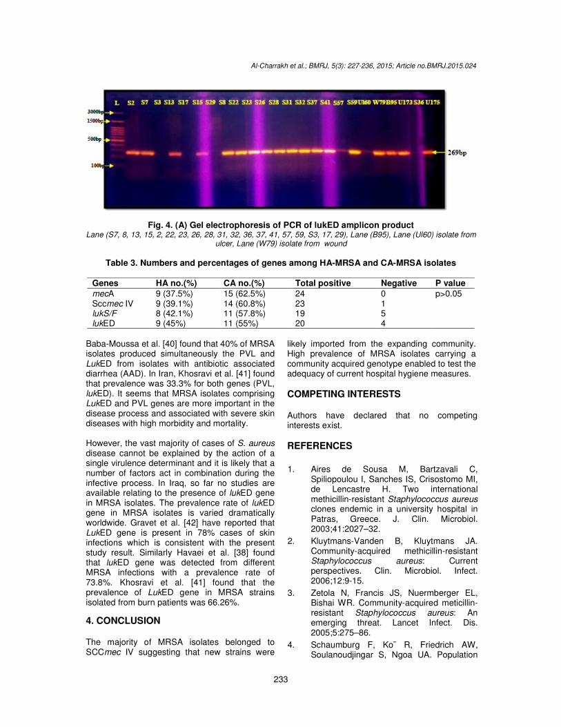
lukED gene was detected in 20 (83%) MRSA, however, it was not detected in 4 (16.6%) MRSA isolates (Fig. 4). Out of 20 positive MRSA isolates, 9 (45%) and 11 (55%) isolates were detected from hospital and community settings respectively (Table 3).
The results also revealed that all HA-MRSA isolates (100%) were harboring lukED gene. This result was in agreement with results obtained by Vandenesch et al. [39], who found that lukED gene was detected in most HA-MRSA isolates. However, in hospital PVL and LukED positive MRSA isolates were 42.1% and 45% respectively (Table 3). From these 20 positive isolates for lukED, 17 (85%) MRSA isolates shared both genes (PVL and lukED).
Al-Charrakh et al.; BMRJ, 5(3): 227-236, 2015; Article no.BMRJ.2015.024
233
Fig. 4. (A) Gel electrophoresis of PCR of lukED amplicon product Lane (S7, 8, 13, 15, 2, 22, 23, 26, 28, 31, 32, 36, 37, 41, 57, 59, S3, 17, 29), Lane (B95), Lane (Ul60) isolate from
ulcer, Lane (W79) isolate from wound
Table 3. Numbers and percentages of genes among HA-MRSA and CA-MRSA isolates
Genes HA no.(%) CA no.(%) Total positive Negative P value
mecA 9 (37.5%) 15 (62.5%) 24 0 p>0.05 Sccmec IV 9 (39.1%) 14 (60.8%) 23 1 lukS/F 8 (42.1%) 11 (57.8%) 19 5 lukED 9 (45%) 11 (55%) 20 4
Baba-Moussa et al. [40] found that 40% of MRSA isolates produced simultaneously the PVL and LukED from isolates with antibiotic associated diarrhea (AAD). In Iran, Khosravi et al. [41] found that prevalence was 33.3% for both genes (PVL, lukED). It seems that MRSA isolates comprising LukED and PVL genes are more important in the disease process and associated with severe skin diseases with high morbidity and mortality. However, the vast majority of cases of S. aureus disease cannot be explained by the action of a single virulence determinant and it is likely that a number of factors act in combination during the infective process. In Iraq, so far no studies are available relating to the presence of lukED gene in MRSA isolates. The prevalence rate of lukED gene in MRSA isolates is varied dramatically worldwide. Gravet et al. [42] have reported that LukED gene is present in 78% cases of skin infections which is consistent with the present study result. Similarly Havaei et al. [38] found that lukED gene was detected from different MRSA infections with a prevalence rate of 73.8%. Khosravi et al. [41] found that the prevalence of LukED gene in MRSA strains isolated from burn patients was 66.26%.
4. CONCLUSION The majority of MRSA isolates belonged to SCCmec IV suggesting that new strains were
likely imported from the expanding community. High prevalence of MRSA isolates carrying a community acquired genotype enabled to test the adequacy of current hospital hygiene measures.
COMPETING INTERESTS Authors have declared that no competing interests exist.
REFERENCES
1. Aires de Sousa M, Bartzavali C, Spiliopoulou I, Sanches IS, Crisostomo MI, de Lencastre H. Two international methicillin-resistant Staphylococcus aureus clones endemic in a university hospital in Patras, Greece. J. Clin. Microbiol. 2003;41:2027–32.
2. Kluytmans-Vanden B, Kluytmans JA. Community-acquired methicillin-resistant Staphylococcus aureus: Current perspectives. Clin. Microbiol. Infect. 2006;12:9-15.
3. Zetola N, Francis JS, Nuermberger EL, Bishai WR. Community-acquired meticillin-resistant Staphylococcus aureus: An emerging threat. Lancet Infect. Dis. 2005;5:275–86.
4. Schaumburg F, Ko¨ R, Friedrich AW, Soulanoudjingar S, Ngoa UA. Population

Al-Charrakh et al.; BMRJ, 5(3): 227-236, 2015; Article no.BMRJ.2015.024
234
structure of Staphylococcus aureus from remote African Babongo pygmies. PLoS Negl. Trop. Dis. 2011;5(5):1150.
5. Ito T, Katayama Y, Asanda K. Structural comparison of three types of staphylococcal cassette chromosome mec integrated in the chromosome in methicillin-resistant Staphylococcus aureus. Antimicrob Agents Chemother. 2001;45:1323–26.
6. Shukla SK, Stemper ME, Ramaswamy S, Conradt JM, Reich R, et al. Molecular characteristics of nosocomial and Native American community-associated methicillin-resistant Staphylococcus aureus clones from rural Wisconsin. J. Clin. Microbiol. 2004;42:3752-57.
7. David MZ, Daum RS. Community-associated methicillin-resistant Staphylococcus aureus: Epidemiology and clinical consequences of an emerging epidemic. Clin. Microbiol. Rev. 2010;23(3):616.
8. McClure J, Conly JM, Lau G. Novel multiplex PCR assay for detection of the staphylococcal virulence marker Panton-Valentine leukocidin genes and simultaneous discrimination of methicillin-susceptible from resistant staphylococci. J. Clin. Microbiol. 2006;44:1141-44.
9. Naimi TS, LeDell KH, Como-Sabetti K. Comparison of community and health care-associated methicillin-resistant Staphylococcus aureus infection. J. Am. Med. Assoc. 2003;290:2976–84.
10. Prevost G, Couppie P, Prevost P. Epidemiological data on Staphylococcus aureus strains producing synergohymenotropic toxins. J. Med. Microbiol. 1995;42:237–45.
11. Deresinski S. Review of MRSA. Clin. Infect. Dis. 2005;40:562-73.
12. Stranden A, Frei R, Widmer AF. Molecular typing of methicillin-resistant Staphylococcus aureus: Can PCR replace pulsed-field gel electrophoresis? J. Clin. Microbiol. 2003;41:3181–86.
13. Diep BA, Sensabaugh GF, Somboona NS, Carleton HA, Perdreau-Remington F. Widespread skin and soft-tissue infections due to two methicillin-resistant Staphylococcus aureus strains harboring the genes for Panton-Valentine leucocidin. J. Clin. Microbiol. 2004;42:2080–84.
14. MacFaddin JF. Biochemical tests for identification of medical bacteria. 3rd ed. Baltimore: Williams and Wilkins; 2000.
15. Al-Hassnawi HH, Al-Charrakh AH, Al-Khafaji JK. Prevalence of Antimicrobial susceptibility and virulence factors of community-acquired methicillin resistant Staphylococcus aureus (CA-MRSA) from Hilla/Iraq. Karbala J. Pharmaceut Sci. 2013;4:91-02.
16. Weller TMA. The distribution of mecA, mecR1 and mec1 and sequence analysis of mec1and the mec promoter region in staphylococci expressing resistance to methicillin. J. Antimicrob. Chemother. 1999;43:15-22.
17. Okuma K, Iwakawa K, Turnidge JD. Dissemination of new methicillin-resistant Staphylococcus aureus clones in the community. J. Clin Microbiol. 2002;40:4289–94.
18. Lina G, Piemont Y, Godail-Gamot F. Involvement of Panton-Valenntie leukocidin–producing Staphylococcus aureus in primary skin infections and pneumonia. Clin Infect Dis. 1999;29:1128–32.
19. Jarraud S, Mougel C, Thioulouse J, Lina G, Meugnier H, Forey F, et al. Relationships between Staphylococcus aureus genetic background, virulence factors, agr groups (alleles), and human disease. Infect. Immun. 2002;70:631–41.
20. Centers for Disease Control and Prevention (CDC). Active Bacterial Core Surveillance Report, Emerging Infections Program Network, Methicillin-Resistant Staphylococcus aureus; 2008. Accessed 20 September 2012.
Available: http://www.cdc.gov/abcs/reports-findings/survreports/mrsa08.html
21. Al-Charrakh AH, Naher HS, Al-Fu'adi AH. Methicillin resistant Staphylococcus aureus. 1st ed. Germany: Lambert academic publishing; 2012.
22. Fluit AD, Visser MR, Schmitz FJ. Molecular detection of antimicrobial resistance. Clin. Microbiol. Rev. 2001;4:836-71.
23. Corkill JE, Anson JJ, Griffiths P, Hart CA. Detection of elements of the staphylococcal cassette chromosome (SCC) in a methicillin susceptible (mecA gene negative) homologue of a fucidin-resistant MRSA. J. Antimicrob. Chemother. 2004;54:229–31.
Al-Charrakh et al.; BMRJ, 5(3): 227-236, 2015; Article no.BMRJ.2015.024
235
24. Al-Mohana AM, Al-Kudhieri MK, Al-Charrakh AH. Community-acquired methicillin-resistant Staphylococcus aureus carrying mecA and Panton-Valentine lukocidin PVL genes that isolated from holy shrine in Najaf, Iraq. J. Bacteriol. Res. 2012;4(2):15-23.
25. Del Giudice BP, Durupt F, Bes M, Martinez JP, Counillon E, Lina G, et al. Emergence of two populations of methicillin-resistant Staphylococcus aureus with distinct epidemiological, clinical and biological features, isolated from patients with community-acquired skin infections. Br. J. Dermatol. 2006;154:118–24.
26. Centers for Disease Control and Prevention (CDC). Community associated MRSA information for clinicians. Infection control topics. Centers for Disease Control and Prevention, Atlanta, GA; 2005.
27. Skov R, Smyth R, Clausen M. Evaluation of a cefoxitin 30 µg disc on Iso-Sensitest agar for detection of methicillin-resistant Staphylococcus aureus. J. Antimicrobiol. Chemother. 2003;52:204-07.
28. Conceição T, Tavares A, Miragaia M, Hyde K, Aires-de-Sousa M, de Lencastre H. Prevalence and clonality of methicillin-resistant Staphylococcus aureus (MRSA) in the Atlantic Azores islands: Predominance of SCCmec types IV, V and VI. Eur. J. Clin. Microbiol. Infect. Dis. 2010;29:543–50.
29. Daum RS, Ito T, Hiramatsu K, Hussain F, Mongkolrattanothai K. A novel methicillin resistance cassette in community-acquired methicillin-resistant Staphylococcus aureus isolates of diverse genetic backgrounds. J. Infect. Dis. 2002;186:1344–47.
30. Gonzalez BE, Rueda AM, Shelburne, SA, Musher DM, Hamill RJ, Hulten KG. Community-associated strains of methicillin-resistant Staphylococccus aureus as the cause of healthcare-associated infection. Infect. Control Hosp. Epidemiol. 2006;27:1051–56.
31. Strande´n, A, Frei R, Adler H, Flu¨ckiger U, Widmer A. Emergence of SCCmec type IV as the most common type of methicillin-resistant Staphylococcus aureus in a University hospital. Infect. 2009; 37:44–48.
32. Klevens RM, Edwards JR, Tenover FC, McDonald LC, Horan T, Gaynes R. Changes in the epidemiology of methicillin-resistant Staphylococcus aureus in intensive care units in US hospitals, 1992–2003. Clin. Infect. Dis. 2006;42:389–91.
33. Kilic A, Guclu AU, Senses Z. Staphylococcal cassette chromosome mec (SCCmec) characterization and Panton-Valentine leukocidin gene occurrence for methicillin-resistant Staphylococcus aureus in Turkey from 2003-2006. Antonie van Leeuwenhoek. 2008;94:607-14.
34. Holmes A, Ganner M, McGuane S, Pitt TL, Cookson BD, Kearns AM. Staphylococcus aureus isolates carrying panton-valentine leucocidin genes in England and Wales: frequency, characterization, and association with clinical disease, J. Clin. Microbiol. 2005;43(5):2384–90.
35. Badiou C, Dumitrescu O, George N, Forbes A, Drougka E. Rapid detection of Staphylococcus aureus Panton-Valentine leukocidin in clinical specimens by enzyme-linked immunosorbent assay and immunochromatographgic tests. J. Clin. Microbiol. 2010;48(4):1384-90.
36. Narita S, Kaneko J, Chiba J, Piemont Y, Jarraud S, Etienne J, et al. Phage conversion of Panton-Valentine leukocidin in Staphylococcus aureus: Molecular analysis of a PVL-converting phage, phiSLT. Gene. 2001;268:195-06.
37. Diep B, Carlton H, Chang B, Senabaugh G, Predreau-Remington F. Roles of 34 virulence genes in the evolution of hospital- and community-associated strains of methicillin-resistant Staphylococcus aureus. J. Infect. Dis. 2011;193:1495-03.
38. Havaei SA, Moghadam SO, Pourmand MR, Faghri J. Prevalence of genes encoding bi-component leukocidins among clinical isolates of methicillin–resistant Staphylococcus aureus. Iran J. Publ. Health 2010; 39: 18-14.
39. Vandenesch F, Naimi T, Enright MC, Lina G, Nimmo GR, Heffernan H, et al. Community acquired methicillin-resistant Staphylococcus aureus carrying Panton-Valentine leukocidin genes: Worldwide emergence. Emerg. Infect. Dis. 2003;9:978–84.
40. Baba-Moussa L, Ahissou H, Azokpota P, Assogba B, Atindéhou MM, Anagonou S. Toxins and adhesion factors associated with Staphylococcus aureus strains isolated from diarrhoeal patients in Benin. Afr. J. Biotech. 2009;9(5):604-11.
41. Khosravi AD, Hoveizavi H, Farshadzadeh Z. The prevalence of genes encoding leukocidins in Staphylococcus aureus strains resistant and sensitive to methicillin
Al-Charrakh et al.; BMRJ, 5(3): 227-236, 2015; Article no.BMRJ.2015.024
236
isolated from burn patients in Taleghani hospital, Ahvaz, Iran. Infect. Trop. Dis. Res. 2011;14:219-45.
42. Gravet A, Rondeau M, Harf-Monteil C. Predominant Staphylococcus aureus
isolated from antibiotic-associated diarrhea is clinically relevant and produces enterotoxin A and the bicomponent toxin LukED. J. Clin. Microbiol. 1999;37:4012-19.
_________________________________________________________________________________ © 2015 Al-Charrakh et al.; This is an Open Access article distributed under the terms of the Creative Commons Attribution License (http://creativecommons.org/licenses/by/4.0), which permits unrestricted use, distribution, and reproduction in any medium, provided the original work is properly cited.
Peer-review history: The peer review history for this paper can be accessed here:
http://www.sciencedomain.org/review-history.php?iid=703&id=8&aid=6514