specific conditions for resveratrol neuroprotection against ethanol-induced toxicity
TRANSCRIPT

Hindawi Publishing CorporationJournal of ToxicologyVolume 2012, Article ID 973134, 11 pagesdoi:10.1155/2012/973134
Research Article
Specific Conditions for Resveratrol Neuroprotection againstEthanol-Induced Toxicity
Brigitte Gonthier,1, 2 Nathalie Allibe,2, 3 Cecile Cottet-Rousselle,1 Frederic Lamarche,1, 2
Laurence Nuiry,2 and Luc Barret2, 3
1 INSERM, U1055 (LBFA), Universite Joseph Fourier, F-38041 Grenoble, France2 CHRU Grenoble, Hopital Michallon, Service de Medecine Legale et Toxicologie, F-38043 Grenoble, France3 Laboratoire de Medecine Legale et Toxicologie, Universite Joseph Fourier, F-38041 Grenoble, France
Correspondence should be addressed to Brigitte Gonthier, [email protected]
Received 6 March 2012; Revised 27 April 2012; Accepted 1 May 2012
Academic Editor: Wei Zheng
Copyright © 2012 Brigitte Gonthier et al. This is an open access article distributed under the Creative Commons AttributionLicense, which permits unrestricted use, distribution, and reproduction in any medium, provided the original work is properlycited.
Aims. 3,5,4′-Trihydroxy-trans-stilbene, a natural polyphenolic compound present in wine and grapes and better known asresveratrol, has free radical scavenging properties and is a potent protector against oxidative stress induced by alcohol metabolism.Today, the mechanism by which ethanol exerts its toxicity is still not well understood, but it is generally considered that freeradical generation plays an important role in the appearance of structural and functional alterations in cells. The aim of thisstudy was to evaluate the protective action of resveratrol against ethanol-induced brain cell injury. Methods. Primary culturesof rat astrocytes were exposed to ethanol, with or without a pretreatment with resveratrol. We examined the dose-dependenteffects of this resveratrol pretreatment on cytotoxicity and genotoxicity induced by ethanol. Cytotoxicity was assessed using theMTT reduction test. Genotoxicity was evidenced using single cell gel electrophoresis. In addition, DNA staining with fluorescentdyes allowed visualization of nuclear damage using confocal microscopy. Results. Cell pretreatment with low concentrations oftrans-resveratrol (0.1–10 μM) slowed down cell death and DNA damage induced by ethanol exposure, while higher concentrations(50–100 μM) enhanced these same effects. No protection by cis-resveratrol was observed. Conclusion. Protection offered by trans-resveratrol against ethanol-induced neurotoxicity was only effective for low concentrations of this polyphenol.
1. Introduction
The brain is particularly susceptible to oxidative stress due toits high rate of oxygen consumption, its high proportion ofpolyunsaturated fatty acids and its low levels of antioxidantdefense enzymes [1]. Consequently, the brain is a first-classtarget in situations in which free radicals are implicatedsuch as ageing, neurodegenerative diseases, and xenobioticmetabolism. This organ is thus a major target for ethanol,and its consumption has long been associated with severedamage. The adverse effect of ethanol on different functionsof the central nervous system has been well documented.Numerous experimental studies and necropsy examinationshave shown a wide range of structural and functionalalterations in neurons as well as in astrocytes. Astrocyteswere chosen as a model for cerebral intoxication because
their deleterious implication has considerable potential con-sequences. First, they represent the most abundant cell typein the central nervous system. Besides providing nutrientsand neurotrophic factors to neurons, these cells are immune-active and actively participate in host defense mechanisms[2, 3]. In addition, specific enzyme systems allow astrocytesto metabolize xenobiotics, free radicals, and metals, thusprotecting the brain from the toxicity of these agents [4, 5].
The advantage of primary cultures lies in their closerresemblance to cells found in vivo. The drawbacks of thischoice are their limited survival time and the continual needto prepare new cells in culture. This can explain why very fewreports are found in the literature on this subject.
There is strong evidence showing that chronic or exces-sive ethanol consumption enhances oxidative damage tobrain. This is due to the ability of ethanol to increase

2 Journal of Toxicology
oxidative stress, leading to enhanced production of oxidativespecies including reactive oxygen species (ROS) and in theformation of lipid peroxidation products [6, 7].
Using the spin-trapping technique, we previouslydetected hydroxyethyl-free radical formation followingethanol exposure of various biological systems such as ratliver or brain microsomes [8] and primary culture of ratastrocytes or C6 glioma cell line [9].
ROS are produced during normal cell metabolism aswell as after exposure to various xenobiotics includingalcohol. Although GSH and various antioxidant enzymes(SOD, GPX, catalase, etc.) prevent cells from attack bythese ROS, massive or chronic exposure to a toxic substancemay induce an oxidant/antioxidant imbalance, leading tocellular oxidative damage. The literature has reported thatantioxidant micronutrients such as vitamin C, vitamin E,and β-carotene in fruits and vegetables contribute in part toprotecting cells against the damaging effects of ROS.
3,5,4′-Trihydroxy-trans-stilbene, so called resveratrol, isa naturally occurring stilbene found in the skin of redgrapes and certain medicinal plants, where it is believed toprovide protection against various infections and stresses[10]. In addition, this red wine constituent may be implicatedin the so-called French paradox, a phenomenon relatingthe low incidence of coronary heart disease in southernFrance despite a high dietary intake of saturated fats [11].Numerous beneficial health effects have been reported suchas anticancer, antiviral, antiinflammatory, antiageing andneuroprotective effects [12–15]. Indeed, neuroprotectiveeffects have been related in cerebral ischemia models aswell as in neurodegenerative diseases such as Alzheimer’sdisease, Huntington’s disease, and Parkinson’s disease [16,17]. Moreover, most of the protective biological actionsassociated with resveratrol have been related to its intrinsicradical scavenger properties [18–21]. De Almeida et al. [22]demonstrated the protective effect of resveratrol againstacute H2O2-induced oxidative stress in astrocyte cultures.
Since a free radical pathway is implicated in the cellulardamage observed after ethanol exposure, we studied resver-atrol pretreatment of astrocytes because this antioxidantmight be able to promote the survival of brain cells exposedto ethanol stress.
The aim of this study was to evaluate the ability ofresveratrol to protect primary culture of rat astrocytes againstethanol-induced cellular damage in a single model of long-term exposed cells through in vitro experiments.
2. Materials and Methods
2.1. Culture of Primary Astrocytes. Pregnant Sprague-Dawleyrats were obtained from Janvier (L’Arbresle, France). Animalcare and use and all procedures involving animals werecarried out in accordance with French national regulations.
The primary astrocyte cultures were prepared asepti-cally from cerebral hemispheres of 1- or 2-day-old pups,according to previously described methods [23] with afew modifications [9]. The dissociated cells were platedeither in poly-L-lysine-coated 35-mm-diameter Petri dishes
or in 75 cm2 plastic flasks at a density of 500 000 viablecells/mL in the usual D-MEM medium containing 10%FCS, 100 U/mL penicillin, 0.1 mg/mL streptomycin, and2.5 μg/mL amphotericin B. The cultures were maintained at37◦C in a 5% CO2-humidified atmosphere. The mediumwas changed every 3 days. After 12 days in vitro (DIV), themonolayers were confluent and composed of 95% astrocytes,as demonstrated by positive immunostaining with antiserumto α-GFAP, an astrocyte marker [24].
For confocal microscopy experiments, ten days afterplating, cells in flasks were trypsinized and reseeded in thesame medium at a density of 250 000 viable cells/mL, ontopoly-L-lysine-coated 40-mm diameter glass coverslips. Cellswere used 48 hours after seeding when culture reached 80–90% confluence.
All experiments were conducted in culture medium andin an air/CO2 incubator between 12 and 15 days in vitro.
2.2. Formation and Identification of Cis- and Trans-Isomersof Resveratrol. Trans-resveratrol was purchased from Sigma-Aldrich Chemical (Saint Louis, MO, USA).
Two 100-mM solutions of trans-resveratrol were pre-pared in H2O/DMSO (50/50). One aliquot was kept inthe dark and the other placed to laboratory light, inorder to induce the trans-cis isomerization. Then sampleswere derivatized for 30 min at 65◦C using BSTFA (N,N-bis(trimethylsilyl)trifluoroacetamide). Finally, samples wereanalyzed by gas chromatography coupled to mass spectrom-etry (GC/MS).
2.3. Conditions for Exposure to Resveratrol and/or Ethanol.Trans-resveratrol was dissolved in H2O/DMSO (50/50) soas to obtain a 100-mM stock solution which was filtered,then aliquoted and stored at −20◦C until use. Just beforethe beginning of experiments, the trans-cis isomerizationwas realized by exposing an aliquot to laboratory light.Different intermediate dilutions of these two isomers werethen prepared in the same mixture in order to preservethe same final H2O/DMSO/culture medium ratio for eachresveratrol concentration tested.
Cells were preincubated for 1 hour in the culture mediumcontaining various concentrations of cis- or trans-resveratrolranging from 0.1 to 100 μM. Then, medium was replacedby a new one containing the same resveratrol concentrationand 20 mM ethanol for three days. Controls with resveratrolalone or ethanol alone were also realized.
To avoid alcohol evaporation, we used a previouslydescribed compensating system, which provided a constantconcentration of alcohol in the culture medium for 3 days[25].
2.4. Gas Chromatography/Mass Spectrometry Analysis. Theanalyses were performed on an Agilent Technologies 6890NNetwork GC System combined with an Agilent Technologies5975 network mass selective detector and an Agilent Tech-nologies 7683 series injector. The entire process, includingdata collection, was controlled by the Agilent TechnologiesChem Station Version Rev.D.02.00.275.

Journal of Toxicology 3
We injected 2 μL of each sample on to a DB-5 ms column(30 × 0.25 mm i.d.; 0.25-μm film thickness) using pulsedsplitless injection with an injector temperature of 250◦C.Temperature conditions were as follows: initial temperatureof 100◦C for 1 min, increased to 300◦C at 20◦C/min, andheld for 11 min. The flow of the carrier gas (helium) wasmaintained at 1 mL/min in constant flow mode. The gaschromatograph interface temperature was held at 315◦C.Electron impact ionization was performed at 70 eV, with anion source temperature of 230◦C and mass spectra collectedfrom 40 to 600 m/z.
2.5. Determination of Cell Viability. After each treatment,cell viability was determined using the MTT reduc-tion test [26]. Growth medium was replaced by D-MEM containing 0.5 mg/mL 3-(4,5-dimethylthiazol-2-yl)-2,5-diphenyltetrazolium bromide (MTT). After 2 h of incu-bation, cell medium was removed and replaced by 1 mL ofdimethylsulfoxide to solubilise the precipitated formazan.
Cell viability was quantified spectrophotometrically at540 nm, and 100% viability was assigned to the absorbance ofcontrol. At least five dishes were tested for each intoxicationcondition. The MTT assay, measuring the mitochondrialdehydrogenase activity, reflects the metabolic activity of thecells and is a helpful indicator of cell viability.
2.6. Evaluation of Nuclear Damage
2.6.1. Comet Assay. DNA damage was evaluated using singlecell gel electrophoresis, also called the comet assay [27]. Thistechnique was realized using an electrophoresis power supply(Consort EV265) and a submarine horizontal electrophoresissystem (Model HU25). This assay was performed eitherimmediately after the stress to evidence damage generated byethanol or after a 1- or 3-h recovery period with the aim ofstudying repair mechanisms. Therefore, the effects on DNAobserved in these conditions reflect the initial DNA damagein terms of strand breaks and oxidatively damaged basesgenerating alkali-labile sites in DNA.
The procedure used was a modification of the protocoldescribed by Singh et al. [28]. Frosted microscope slideswere first covered with 150 μL of 1% normal agarose inCa2+- and Mg2+-free phosphate-buffered saline (PBS) andimmediately covered with a 22 × 50 mm coverslip and keptat room temperature to allow the agarose to solidify. Thecoverslip was then gently slid off. Approximately 20 000cells were suspended in 80 μL of 0.8% low-melting-pointagarose in PBS kept at 37◦C and transferred onto the firstagarose layer. After having been covered with a coverslip, theslides were left on ice for 5 min. Then the coverslips wereremoved and the slides were placed in freshly prepared lysingsolution at 4◦C for 1 h in the dark (2.5 M NaCl, 100 mMNa2EDTA, 10 mM Tris, 1% sodium sarcosinate, pH set to 10with NaOH, supplemented with 10% DMSO and 1% TritonX-100 just before use). After lysis, the slides were gentlytransferred to a horizontal gel electrophoresis tank filledwith freshly prepared electrophoresis solution (0.3 M NaOH,1 mM Na2EDTA) at room temperature in the dark. The DNA
was allowed to unwind for 20 min, and electrophoresis wascarried out by adjusting the voltage to 25 V and the currentto 300 mA (∼0.7 V/cm) for 15 min. After electrophoresis, theslides were washed gently to remove any alkali and detergentsthat would interfere with ethidium bromide staining, usingneutralisation buffer (0.4 M Tris-HCl, pH 7.4) three times for5 min. After neutralization, the slides were stained with 50 μLof 3.3 μg/mL ethidium bromide in distilled water and coveredwith a coverslip. The slides were placed in a humidified air-tight container, to keep the gel from drying, until analysis.Three slides were prepared per assay, and 50 nuclei werecounted per slide.
Slides were examined using an epifluorescence micro-scope, Zeiss Axioskop 20 (Carl Zeiss, Microscope Division,Oberkochen, Germany), equipped with a short arc mercurylamp HBO (50 W, 516–560 nm, Zeiss), using a 20X dryobjective. Fifty randomly selected comets on each triplicateslide were scored with a Pulmix TM 765 camera (KineticImaging, Liverpool, UK) connected to a Komet 3.0 imageanalysis system (Kinetic Imaging). This software defineddifferent parameters for image processing. Among theseparameters, we chose the percentage of DNA in the tail forthe evaluation of DNA damage. The percentage of DNA inthe tail is linearly related to DNA break frequency [29].
2.6.2. Confocal Microscopy. Apoptotic cell death was assessedby evidencing nuclear morphology alterations. At the end ofvarious cell treatments, growth medium was supplied with1.6 μM Hoechst 33258 (final concentration) for 15 min at37◦C. Coverslips were then rinsed twice with PBS, fixed for15 min with 70% ethanol, and stored at 4◦C in PBS untilanalysis. Cells were imaged using a confocal laser-scanningmicroscope Leica TCS SP2 AOBS (Leica, Heidelberg, Ger-many). For fluorescence excitation, an UV laser at 351–364 nm was used. Optical sections were recorded using a 63xoil immersion objective. Images were collected in the 512 ×512 pixel format and processed by Leica confocal software.
2.7. Statistics. Three independent experimental series wereconducted for each exposure condition, unless otherwiseindicated. Controls corresponded to astrocytes not exposedto ethanol and grown in culture under the same conditionsas those in the experimental series.
The results were expressed as mean ± SEM. The Mann-Whitney U test was used for statistical analysis. Findingswith P < 0.05 were considered significant. The Kolgomorov-Smirnov test was employed to compare the distribution ofthe percentages of DNA in the tail. Values of P < 0.05 wereconsidered to be significant.
3. Results
In the literature, several authors dissolved resveratrol inDMSO and did not observe any cytotoxicity with thissolvent [15, 20, 21, 30]. Unfortunately, with our experimentalconditions, that is, long-term exposure and the fragility ofcerebral cells in primary culture, a moderate decrease in cellviability (15%) was observed when astrocytes were exposed
4 Journal of Toxicology
to this vehicle. Finally, we tested an H2O/DMSO (50/50, V/V)mixture to solubilize resveratrol, as described by Olas et al.[31]. In these conditions, the ratio DMSO/culture mediumwas 0.0005%, and no toxicity was detected for astrocytes inprimary culture (data not shown).
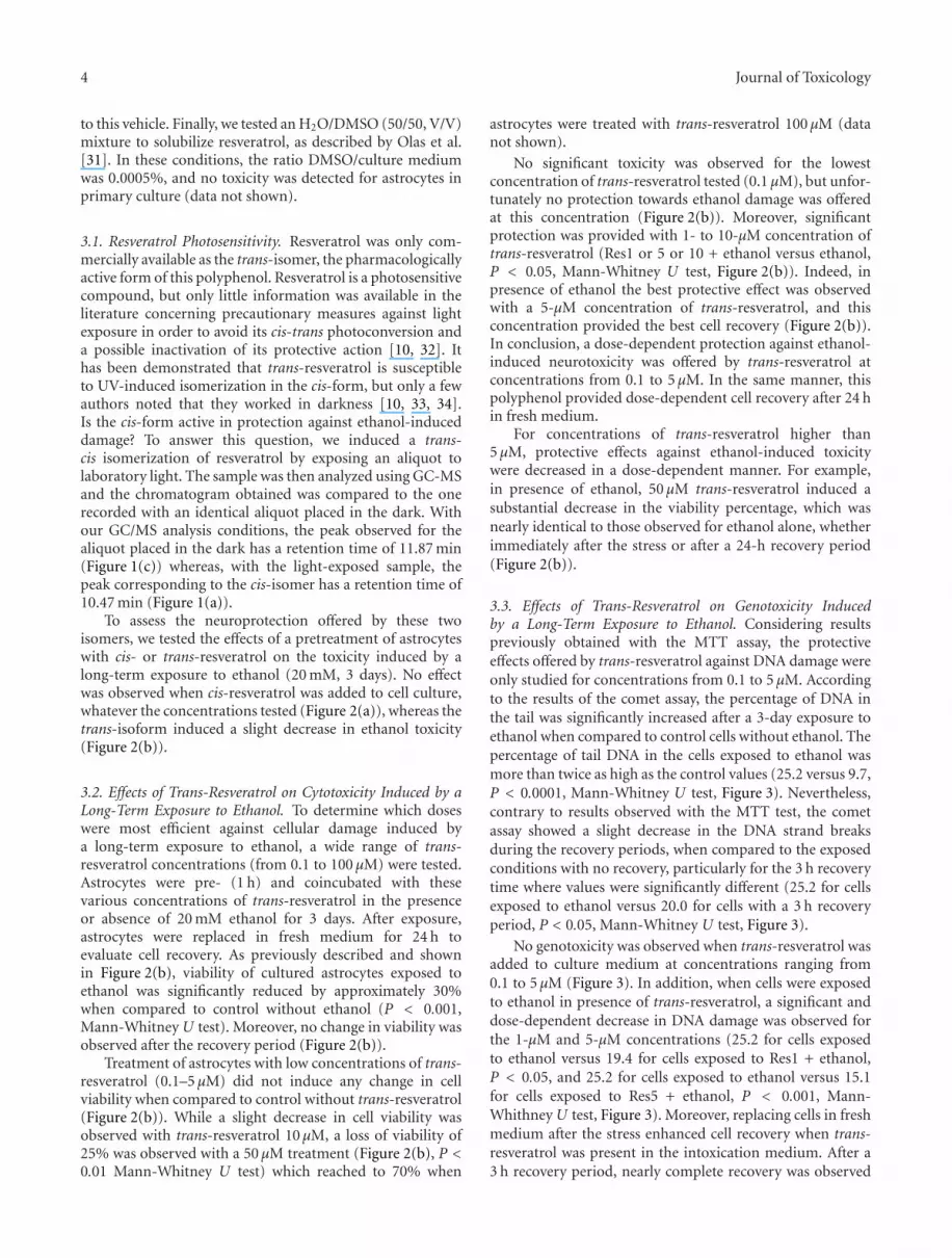
3.1. Resveratrol Photosensitivity. Resveratrol was only com-mercially available as the trans-isomer, the pharmacologicallyactive form of this polyphenol. Resveratrol is a photosensitivecompound, but only little information was available in theliterature concerning precautionary measures against lightexposure in order to avoid its cis-trans photoconversion anda possible inactivation of its protective action [10, 32]. Ithas been demonstrated that trans-resveratrol is susceptibleto UV-induced isomerization in the cis-form, but only a fewauthors noted that they worked in darkness [10, 33, 34].Is the cis-form active in protection against ethanol-induceddamage? To answer this question, we induced a trans-cis isomerization of resveratrol by exposing an aliquot tolaboratory light. The sample was then analyzed using GC-MSand the chromatogram obtained was compared to the onerecorded with an identical aliquot placed in the dark. Withour GC/MS analysis conditions, the peak observed for thealiquot placed in the dark has a retention time of 11.87 min(Figure 1(c)) whereas, with the light-exposed sample, thepeak corresponding to the cis-isomer has a retention time of10.47 min (Figure 1(a)).
To assess the neuroprotection offered by these twoisomers, we tested the effects of a pretreatment of astrocyteswith cis- or trans-resveratrol on the toxicity induced by along-term exposure to ethanol (20 mM, 3 days). No effectwas observed when cis-resveratrol was added to cell culture,whatever the concentrations tested (Figure 2(a)), whereas thetrans-isoform induced a slight decrease in ethanol toxicity(Figure 2(b)).
3.2. Effects of Trans-Resveratrol on Cytotoxicity Induced by aLong-Term Exposure to Ethanol. To determine which doseswere most efficient against cellular damage induced bya long-term exposure to ethanol, a wide range of trans-resveratrol concentrations (from 0.1 to 100 μM) were tested.Astrocytes were pre- (1 h) and coincubated with thesevarious concentrations of trans-resveratrol in the presenceor absence of 20 mM ethanol for 3 days. After exposure,astrocytes were replaced in fresh medium for 24 h toevaluate cell recovery. As previously described and shownin Figure 2(b), viability of cultured astrocytes exposed toethanol was significantly reduced by approximately 30%when compared to control without ethanol (P < 0.001,Mann-Whitney U test). Moreover, no change in viability wasobserved after the recovery period (Figure 2(b)).
Treatment of astrocytes with low concentrations of trans-resveratrol (0.1–5 μM) did not induce any change in cellviability when compared to control without trans-resveratrol(Figure 2(b)). While a slight decrease in cell viability wasobserved with trans-resveratrol 10 μM, a loss of viability of25% was observed with a 50 μM treatment (Figure 2(b), P <0.01 Mann-Whitney U test) which reached to 70% when
astrocytes were treated with trans-resveratrol 100 μM (datanot shown).
No significant toxicity was observed for the lowestconcentration of trans-resveratrol tested (0.1 μM), but unfor-tunately no protection towards ethanol damage was offeredat this concentration (Figure 2(b)). Moreover, significantprotection was provided with 1- to 10-μM concentration oftrans-resveratrol (Res1 or 5 or 10 + ethanol versus ethanol,P < 0.05, Mann-Whitney U test, Figure 2(b)). Indeed, inpresence of ethanol the best protective effect was observedwith a 5-μM concentration of trans-resveratrol, and thisconcentration provided the best cell recovery (Figure 2(b)).In conclusion, a dose-dependent protection against ethanol-induced neurotoxicity was offered by trans-resveratrol atconcentrations from 0.1 to 5 μM. In the same manner, thispolyphenol provided dose-dependent cell recovery after 24 hin fresh medium.
For concentrations of trans-resveratrol higher than5 μM, protective effects against ethanol-induced toxicitywere decreased in a dose-dependent manner. For example,in presence of ethanol, 50 μM trans-resveratrol induced asubstantial decrease in the viability percentage, which wasnearly identical to those observed for ethanol alone, whetherimmediately after the stress or after a 24-h recovery period(Figure 2(b)).
3.3. Effects of Trans-Resveratrol on Genotoxicity Inducedby a Long-Term Exposure to Ethanol. Considering resultspreviously obtained with the MTT assay, the protectiveeffects offered by trans-resveratrol against DNA damage wereonly studied for concentrations from 0.1 to 5 μM. Accordingto the results of the comet assay, the percentage of DNA inthe tail was significantly increased after a 3-day exposure toethanol when compared to control cells without ethanol. Thepercentage of tail DNA in the cells exposed to ethanol wasmore than twice as high as the control values (25.2 versus 9.7,P < 0.0001, Mann-Whitney U test, Figure 3). Nevertheless,contrary to results observed with the MTT test, the cometassay showed a slight decrease in the DNA strand breaksduring the recovery periods, when compared to the exposedconditions with no recovery, particularly for the 3 h recoverytime where values were significantly different (25.2 for cellsexposed to ethanol versus 20.0 for cells with a 3 h recoveryperiod, P < 0.05, Mann-Whitney U test, Figure 3).
No genotoxicity was observed when trans-resveratrol wasadded to culture medium at concentrations ranging from0.1 to 5 μM (Figure 3). In addition, when cells were exposedto ethanol in presence of trans-resveratrol, a significant anddose-dependent decrease in DNA damage was observed forthe 1-μM and 5-μM concentrations (25.2 for cells exposedto ethanol versus 19.4 for cells exposed to Res1 + ethanol,P < 0.05, and 25.2 for cells exposed to ethanol versus 15.1for cells exposed to Res5 + ethanol, P < 0.001, Mann-Whithney U test, Figure 3). Moreover, replacing cells in freshmedium after the stress enhanced cell recovery when trans-resveratrol was present in the intoxication medium. After a3 h recovery period, nearly complete recovery was observed

Journal of Toxicology 5
6 7 8 9 10 11 12 13 14 15 16 17 18 19 20
Time (min)
0
20
40
60
80
100
Rel
ativ
e ab
un
dan
ce
10.47
11.858.89 9.81 15.511.615.55 16.16.55 16.827.42 8.19 18.6417.67 19.71
H
H
15.03 19.3
HO
HO
HO
cis
13.2 13.92
(a)
50 100 150 200 250 300 350 400 450 500 550 600 650
0
20
40
60
80
100444.2
445.2
443.172.9 446.2207 429.1 447.2146.9 341.1133 355.232374.1 223189.145.2 251.1 309 372.2 517.4490.1 644.5
Rel
ativ
e ab
un
dan
ce
615.6593.7557.3
m/z
(b)
6 7 8 9 10 11 12 13 14 15 16 17 18 19 20
Time (min)
0
20
40
60
80
100
Rel
ativ
e ab
un
dan
ce
11.87
8.89 9.81 10.445.55 7.32 15.036.55 13.0311.34 13.3912.378.19 17.8415.75 16.24 18.37 19.35 20.25
H
HHO
HO
OH
trans
(c)
50 100 150 200 250 300 350 400 450 500 550 600 650
m/z
0
20
40
60
80
100444.2
445.3
446.2443.373.172.3 207.2 356.1341.1 371.374.1 448.3221.1133 4.824309.1 503.1R
elat
ive
abu
nda
nce
188.1147.1 267 489.3 554.3 641.7585.1 598.2
(d)
Figure 1: GC/MS analysis of resveratrol solubilized in an H2O/DMSO mixture. (a, c) show chromatograms obtained with samples placedeither in light (a) or in darkness (c). (b, d) represent mass spectrum of cis- and trans-resveratrol, respectively. The analysis conditions aredetailed in Section 2.
for cells pretreated with 1 μM and 5 μM trans-resveratrolwhen compared to control (Figure 3).
Figure 4 showed interesting changes in the distributionof tail DNA: in control conditions, more than 85% of cellshad tail DNA between 0% and 10% (Figure 4(a)), whereas
significant modifications in the distribution of tail DNA wereobserved after a 3-day exposure to ethanol (Figure 4(c)), witha progressive distribution towards higher percentages of tailDNA (control versus ethanol, P < 0.0001, Kolgomorov-Smirnov test). When cells were replaced in fresh medium

6 Journal of Toxicology
0
20
40
60
80
100
120
Control 5 10 50 100
Resveratrol (µM)
Without EtOHWith EtOHRecovery 24 H
Perc
enta
ge o
f vi
abili
ty
(a)
0
20
40
60
80
100
120
Control 0.1 1 5 10 50
Resveratrol (µM)
Without EtOHWith EtOHRecovery 24 H
∗•
∗∗
••
Perc
enta
ge o
f vi
abili
ty
(b)
Figure 2: Astrocyte variation in viability after 1 h of pretreatment with various concentrations of cis-resveratrol (a) or trans-resveratrol (b)followed by a 3-day exposure to 20 mM ethanol in presence of resveratrol. Determinations were made directly after the exposure or after 24 hof recovery in fresh culture medium. Cytotoxicity was determined by MTT assay, and control without resveratrol and ethanol represents100% viability. Data shown were obtained in a typical experiment representative of three independent experiments and are expressed asmean ± SEM, n = 5 (•P < 0.01 resveratrol-treated cells versus control, ••P < 0.001 ethanol exposed cells versus control, ∗P < 0.05, Res +ethanol treated cells versus control ethanol, Mann-Whitney U test). Cell exposure conditions are detailed in Section 2.
0
5
10
15
20
25
30
∗∗∗
∗∗
•••
Control 0.1 1 5
Without EtOHWith EtOH
1 H recovery3 H recovery
Resveratrol concentrations (µM)
Perc
enta
ge o
f ta
il D
NA
(a.
u.)
Figure 3: Influence of a 1 h pretreatment with various concentra-tions of trans-resveratrol on the level of DNA damage observedin astrocytes exposed to a 3-day treatment with 20 mM ethanol,followed or not by a 1 h or 3 h recovery period in fresh culturemedium. DNA damage was evaluated using the comet assay andis expressed as percentage of DNA in the tail. The figure showsthe mean results of three independent experiments. Fifty cells wererandomly examined in triplicate for each condition, and results areexpressed as mean ± SEM (•••P < 0.0001 ethanol exposed cellsversus control, ∗∗P < 0.05, ∗∗∗P < 0.001 Res + ethanol-treatedcells versus control ethanol, Mann-Whitney U test). Experimentalconditions are detailed in Section 2. a.u.: arbitrary units.
without ethanol for a 1 or 3 h recovery period, the previouslydescribed decrease in DNA strand breaks was confirmed.This recovery process was time dependant but incompletesince only 69.1% of cells had tail DNA between 0% and10% after 3 h of recovery (Figure 4(g)) compared to 88.2%of cells in control conditions (ethanol + 3 h recovery versuscontrol, P < 0.0001, Kolgomorov-Smirnov test, Figure 4(a)).When cells were pretreated with 5 μM trans-resveratrol,
no toxicity was observed for these astrocytes (Figure 4(b))when compared to control (Figure 4(a)). Moreover, therewas less DNA damage after 3 day exposure to ethanol andin presence of trans-resveratrol (Figure 4(d)) since 78.2%of cells had tail DNA between 0% and 10% compared to50.4% for cells exposed to ethanol alone (Res5 + ethanolversus ethanol, P < 0.0001, Kolgomorov-Smirnov test,Figure 4(c)). In addition, the recovery process was alsoimproved; after 1h of recovery, 80% of cells had tail DNAbetween 0% and 10% (Figure 4(f)), whereas without trans-resveratrol only 65% of cells had tail DNA between 0% and10% (Figure 4(e)), (Res5 + ethanol + 1 h recovery versusethanol + 1 h recovery, P < 0.0001, Kolgomorov-Smirnovtest). After 3 h of recovery, it could be considered that therecovery was complete when cells were pretreated with 5 μMtrans-resveratrol (88.2% and 85.3% of cells with tail DNAbetween 0% and 10%, for control (Figure 4(a)) and Res5+ ethanol + 3 h recovery (Figure 4(h)), respectively. Indeedthe histograms of distribution are not only not statisticallydifferent, but also nearly identical for these two conditions.
Nuclear damage was also evidenced based on nuclearmorphology. Astrocyte staining with nucleic acid dyeHoechst 33342 was performed after various treatments.Cells were then analyzed using laser confocal microscopy.In contrast to regular, blue nuclei observed in viableastrocytes of the control group (Figure 5(a)), nuclei withmembrane blebbings, irregular shapes, and apoptotic bodieswere evidenced in cells exposed to 20 mM ethanol for3 days (Figure 5(c)). These deleterious effects lasted afterthe recovery period since blebs of the nuclear membraneswere always detectable (Figure 5(e)). Astrocytes pretreat-ment with 5 μM trans-resveratrol did not induce any toxicity(Figure 5(b)). Moreover, the treatment with this trans-resveratrol concentration decreased damaged nuclei inducedby ethanol, particularly after the recovery period where no

Journal of Toxicology 7
908070605040302010
010 20 30 40 50 60 70 >80
Perc
enta
ge o
f ce
lls
Percentage of tail DNA
(a)
908070605040302010
010 20 30 40 50 60 70 >80
Perc
enta
ge o
f ce
lls
Percentage of tail DNA
(b)
908070605040302010
010 20 30 40 50 60 70 >80
Perc
enta
ge o
f ce
lls
Percentage of tail DNA
(c)
908070605040302010
010 20 30 40 50 60 70 >80
Perc
enta
ge o
f ce
llsPercentage of tail DNA
(d)
908070605040302010
010 20 30 40 50 60 70 >80
Perc
enta
ge o
f ce
lls
Percentage of tail DNA
(e)
908070605040302010
010 20 30 40 50 60 70 >80
Perc
enta
ge o
f ce
lls
Percentage of tail DNA
(f)
908070605040302010
010 20 30 40 50 60 70 >80
Perc
enta
ge o
f ce
lls
Percentage of tail DNA
(g)
908070605040302010
010 20 30 40 50 60 70 >80
Perc
enta
ge o
f ce
lls
Percentage of tail DNA
(h)
Figure 4: Histograms of the distribution of comet tail DNA obtained after a 3 day exposure of astrocytes to 20 mM ethanol, preceded ornot by a 1 h pretreatment with resveratrol (5 μM) and followed or not by a 1 h or 3 h recovery period in fresh culture medium. (a) Control;(b) pretreatment with resveratrol; (c) 3-day exposure to ethanol; (d) pretreatment with resveratrol followed by exposure to ethanol; 1 hrecovery (e) and 3 h recovery (g) of astrocytes after ethanol exposure; 1-h recovery (f) and 3 h recovery (h) of astrocytes after ethanolexposure when cells were first pretreated with resveratrol. (a) versus (c), (d) versus (c), (f) versus (e), P < 0.0001, Kolmogorov-Smirnovtest. Experimental conditions are detailed in Section 2. Insets show representative micrographs of “comet,” corresponding to the various celltreatments described in the legend.
abnormal nuclear morphology was evidenced (Figures 5(d)and 5(f), resp.).
4. Discussion
Normal cell metabolism results in a continuous generationof reactive oxygen species (ROS) that is strictly controlled by
antioxidant mechanisms. However, in some circumstances,oxidative stress can occur as a result of increased exposure tonormal metabolites of oxidative metabolism. This can hap-pen when ROS production is stimulated by the metabolismof certain toxicants and/or when the production or thebioavailability of antioxidants is affected by such agents.Consequently, the physiological balance between oxidants

8 Journal of Toxicology
(a) (b)
(d)
(e) (f)
(c)
Figure 5: Effect of treatment with 5 μM resveratrol on ethanol-induced nuclear damage in primary cultures of astrocytes. Nuclei werelabeled with Hoechst 33342 as described in Section 2. Figure 5 shows representative fluorescent microscope images evidencing the nuclearmorphology of astrocytes in control conditions (a), exposed to 5 μM resveratrol for 3 days (b), exposed to 20 mM ethanol for 3 dayswithout (c) or with a recovery period (e), or exposed to 20 mM ethanol for 3 days, with pre- and cotreatment with 5 μM resveratrol (d)and followed by a recovery period (f). In zoom sections of micrographs, arrows point nuclear blebs and nuclear fragmentation. All imagesare representative fields of at least three independent experiments carried out in duplicate.
and antioxidants can be directly or indirectly modifiedby a toxicant. Among these toxicants, ethanol is knownto produce free radicals during its metabolism [4, 9, 35].Particularly, chronic exposure to ethanol is known to inducecellular and nuclear damage in cerebral cells, mediated by afree radical pathway [5, 6, 36, 37].
The role of polyphenol obtained from diet in protectionagainst oxidative stress is a topic of continuing interest andsome controversy.
Several in vivo and in vitro studies have reported measur-able concentrations of trans-resveratrol after administrationto animals or exposure to cells. For instance, Bertelli et al.[38] showed that single or prolonged administration torats of red wine with a known trans-resveratrol contentled to its accumulation in blood and various organs. Inaddition, Vitrac et al. [39] demonstrated that 14C-labeledtrans-resveratrol is absorbed, metabolized and distributed inthe whole body of mice orally treated with this polyphenol.

Journal of Toxicology 9
Moreover, because of its high lipid solubility, resveratrolmight be deposited in tissues with high lipid content such asbrain and the nervous system [40], making this polyphenol afirst-class compound for neuroprotective studies. Resveratroltransport from plasma to intracellular targets seems toinvolve both passive diffusion and a carrier-mediated process[41, 42].
Concerning cerebral cells, Guo et al. [43] describeduseful protection offered in vivo by a large range ofresveratrol concentrations against genotoxicity induced byacute and chronic ethanol exposure. On the contrary, severalauthors pointed the importance of resveratrol concentra-tions towards cellular protection. Thus, they described aneuroprotective effect for low concentrations of resveratrolwhen high doses of this polyphenol induced cell toxicity[44–46]. Recently, Quincozes-Santos et al. [47] evidencedthat the nature of the stress could be more important thanthe resveratrol concentration. They demonstrated that underintense but short oxidative conditions resveratrol was ableto protect C6 glioma cells against H2O2 insult while underless intense but lasting oxidative insult, resveratrol had anopposite effect, potentiating the H2O2-induced damage andresulting in a prooxidant effect.
In this study, brain protection offered by trans-resveratrol, against ethanol-induced toxicity, was investi-gated. In order to avoid its photoisomerization leading toan inactive isomer (Figure 2(a)), trans-resveratrol has to becarefully kept in the dark throughout the experiments, fromsample preparation to cell treatment.
Contradictory information concerning the effects ofresveratrol treatment has been reported in the literature.Large variations in experimental systems such as cellularmodels, resveratrol concentrations, incubation durations,and so forth could explain these differences. Therefore,it seems important to test a large range of resveratrolconcentrations to define the optimal concentration offeringcellular protection with our own experimental system. Theconcentrations used for evidencing the protective effectsof resveratrol against cellular damage induced by ethanolhave been the subject of preliminary investigations. In ourexperimental conditions, treatment of astrocytes for 3 dayswith low concentrations of resveratrol (from 0.1 to 10 μM)did not induce any change in viability, whereas a toxic effectwas observed when the highest concentrations of resveratrol(50 and 100 μM) were added to cells, characterized by a dose-dependent cell mortality rate higher than 30%. These resultsare in accordance with those reported by Quincozes-Santoset al. [47].
As previously described [5], a chronic exposure ofastrocytes to ethanol led to a significant loss of viability,as evidenced by the MTT assay. Moreover, when cells werereplaced in fresh medium for a 24 h poststress period,no recovery was observed. These results revealed durablealterations in the normal functioning of astrocytes, partic-ularly for the respiratory and energetic process measured bythe MTT assay. The protection offered by trans-resveratrol
against ethanol cytotoxicity was dose-dependent and onlyobserved for the lowest concentrations, but the most valuableresult concerned the effect of this polyphenol on the recoveryof astrocytes; cell recovery was dose dependent and completefor 5 μM resveratrol.
The potential genotoxicity of long-term ethanol adminis-tration to astrocytes was investigated using comet assay. Thismethod provides fast results and requires only a few cells,so it seems suitable for analysis in primary culture, whichrequires continual new preparations.
The percentage of tail DNA was increased 2.5-fold afterlong-term exposure to ethanol. Moreover, after a 1 or 3 hrecovery period in fresh medium, these DNA strand breakswere only partially self-repaired. These results demonstratedthe genotoxicity of ethanol. In addition, it is generallyconsidered that DNA damage caused by chemicals increasedthe risk of mutation and cancer, even though the DNAdamage may be self-repaired [48].
When astrocytes were exposed to ethanol for 3 days,trans-resveratrol treatment induced both a decrease in DNAdamage and an enhancement of cell recovery in a dose-dependent manner. The effective concentration of trans-resveratrol that both reduced DNA strand break formationand enhanced DNA repair was 5 μM.
Consistent with these results, confocal laser microscopyimages of astrocytes stained with Hoechst 33342 allowedvisualization of apoptotic nuclei when cells are exposedfor 3 days to 20 mM ethanol. Interestingly, treatment with5 μM resveratrol completely prevented nuclear morphologyalterations induced by ethanol treatment since neithermembrane blebbings nor apoptotic bodies were evidencedin these conditions.
Several mechanisms may underlie trans-resveratrol-induced protection of astrocytes against ethanol neurotoxic-ity. Trans-resveratrol has been shown to possess helpful free-radical scavenging properties [19, 20, 49–51], and ethanolexposure is known to induce the generation of reactivefree radicals in vitro [8, 9, 52] as well as in vivo [53, 54].Therefore, it is reasonable to assume that trans-resveratrolmight have protective effects on ethanol-induced oxidativeDNA damage, by quenching free radicals generated duringits brain metabolism.
In conclusion, we clearly evidenced that trans-resveratrolcould markedly decrease cell mortality and levels of DNAstrand breaks induced by long-term ethanol exposure ofastrocytes in primary culture. Moreover, we demonstratedthat this polyphenol promoted poststress cell recovery.Nevertheless, this expected significant protection should beweighted against the restrictive conditions of resveratroltreatment since elevated levels and/or long-term exposurewith this compound could contribute to enhance cerebraldamage.
Although the relevance of our findings to in vivo clinicalsituations remains to be demonstrated, our results suggestthat caution is necessary with therapeutic use of trans-resveratrol since high level of this compound could lead tothe appearance of adverse effects on the brain.

10 Journal of Toxicology
References
[1] A. Y. Sun and G. Y. Sun, “Ethanol and oxidative mechanismsin the brain,” Journal of Biomedical Science, vol. 8, no. 1, pp.37–43, 2001.
[2] H. K. Kimelberg and M. D. Norenberg, “Astrocytes,” ScientificAmerican, vol. 260, no. 4, pp. 66–72, 1989.
[3] J. C. Copin, M. Ledig, and G. Tholey, “Free radical scavengingsystems of rat astroglial cells in primary culture: effects ofanoxia and drug treatment,” Neurochemical Research, vol. 17,no. 7, pp. 677–682, 1992.
[4] C. Montoliu, M. Sancho-Tello, I. Azorin et al., “Ethanolincreases cytochrome P4502E1 and induces oxidative stress inastrocytes,” Journal of Neurochemistry, vol. 65, no. 6, pp. 2561–2570, 1995.
[5] N. Signorini-Allibe, B. Gonthier, F. Lamarche, H. Eysseric,and L. Barret, “Chronic consumption of ethanol leads tosubstantial cell damage in cultured rat astrocytes in con-ditions promoting acetaldehyde accumulation,” Alcohol andAlcoholism, vol. 40, no. 3, pp. 163–171, 2005.
[6] C. Ribiere, I. Hininger, C. Saffar-Boccara, D. Sabourault,and R. Nordmann, “Mitochondrial respiratory activity andsuperoxide radical generation in the liver, brain and heart afterchronic ethanol intake,” Biochemical Pharmacology, vol. 47,no. 10, pp. 1827–1833, 1994.
[7] E. Agar, R. Amanvermez, M. Bosnak, S. Demir, M. Ayyildiz,and C. Celik, “The effect of ethanol on lipid peroxidation andglutathione level in the brain stem of rat,” NeuroReport, vol.10, no. 8, pp. 1799–1801, 1999.
[8] B. Gonthier, A. Jeunet, and L. Barret, “Electron spin resonancestudy of free radicals produced from ethanol and acetaldehydeafter exposure to a fenton system or to brain and livermicrosomes,” Alcohol, vol. 8, no. 5, pp. 369–375, 1991.
[9] B. Gonthier, H. Eysseric, A. Soubeyran, D. Daveloose, R.Saxod, and L. Barret, “Free radical production after exposureof astrocytes and astrocytic C6 glioma cells to ethanol.Preliminary results,” Free Radical Research, vol. 27, no. 6, pp.645–656, 1997.
[10] K. Schulze, L. Schreiber, and I. Szankowski, “Inhibiting effectsof resveratrol and its glucoside piceid against Venturia inae-qualis, the causal agent of apple scab,” Journal of Agriculturaland Food Chemistry, vol. 53, no. 2, pp. 356–362, 2005.
[11] J. M. Wu, Z. R. Wang, T. C. Hsieh, J. L. Bruder, J. G.Zou, and Y. Z. Huang, “Mechanism of cardioprotectionby resveratrol, a phenolic antioxidant present in red wine(review),” International Journal of Molecular Medicine, vol. 8,no. 1, pp. 3–17, 2001.
[12] C. A. de La Lastra and I. Villegas, “Resveratrol as an anti-inflammatory and anti-aging agent: mechanisms and clinicalimplications,” Molecular Nutrition and Food Research, vol. 49,no. 5, pp. 405–430, 2005.
[13] L. E. Donnelly, R. Newton, G. E. Kennedy et al., “Anti-inflammatory effects of resveratrol in lung epithelial cells:molecular mechanisms,” American Journal of Physiology, vol.287, no. 4, pp. L774–L783, 2004.
[14] J. Gusman, H. Malonne, and G. Atassi, “A reappraisal of thepotential chemopreventive and chemotherapeutic propertiesof resveratrol,” Carcinogenesis, vol. 22, no. 8, pp. 1111–1117,2001.
[15] Y. S. Han, W. H. Zheng, S. Bastianetto, J. G. Chabot, andR. Quirion, “Neuroprotective effects of resveratrol against β-amyloid-induced neurotoxicity in rat hippocampal neurons:involvement of protein kinase C,” British Journal of Pharma-cology, vol. 141, no. 6, pp. 997–1005, 2004.
[16] P. Marambaud, H. Zhao, and P. Davies, “Resveratrol promotesclearance of Alzheimer’s disease amyloid-β peptides,” TheJournal of Biological Chemistry, vol. 280, no. 45, pp. 37377–37382, 2005.
[17] V. Calabrese, C. Cornelius, C. Mancuso et al., “Cellular stressresponse: a novel target for chemoprevention and nutritionalneuroprotection in aging, neurodegenerative disorders andlongevity,” Neurochemical Research, vol. 33, no. 12, pp. 2444–2471, 2008.
[18] S. Burkhardt, R. J. Reiter, D. X. Tan, R. Hardeland, J.Cabrera, and M. Karbownik, “DNA oxidatively damaged bychromium(III) and H2O2 is protected by the antioxidantsmelatonin, N1-acetyl-N2-formyl-5-methoxykynuramine, res-veratrol and uric acid,” International Journal of Biochemistryand Cell Biology, vol. 33, no. 8, pp. 775–783, 2001.
[19] Z. Jia, H. Zhu, B. R. Misra, J. E. Mahaney, Y. Li, and H. P.Misra, “EPR studies on the superoxide-scavenging capacity ofthe nutraceutical resveratrol,” Molecular and Cellular Biochem-istry, vol. 313, no. 1-2, pp. 187–194, 2008.
[20] J. Karlsson, M. Emgard, P. Brundin, and M. J. Burkitt, “Trans-resveratrol protects embryonic mesencephalic cells from tert-butyl hydroperoxide: electron paramagnetic resonance spintrapping evidence for a radical scavenging mechanism,”Journal of Neurochemistry, vol. 75, no. 1, pp. 141–150, 2000.
[21] P. Lorenz, S. Roychowdhury, M. Engelmann, G. Wolf, andT. F. W. Horn, “Oxyresveratrol and resveratrol are potentantioxidants and free radical scavengers: effect on nitrosativeand oxidative stress derived from microglial cells,” NitricOxide, vol. 9, no. 2, pp. 64–76, 2003.
[22] L. M. V. de Almeida, C. C. Pineiro, M. C. Leite et al., “Pro-tective effects of resveratrol on hydrogen peroxide inducedtoxicity in primary cortical astrocyte cultures,” NeurochemicalResearch, vol. 33, no. 1, pp. 8–15, 2008.
[23] J. Booher and M. Sensenbrenner, “Growth and cultivation ofdissociated neurons and glial cells from embryonic chick, ratand human brain in flask cultures,” Neurobiology, vol. 2, no. 3,pp. 97–105, 1972.
[24] V. K. Vijayan, Y. L. Lee, and L. F. Eng, “Immunohistochemicallocalization of basic fibroblast growth factor in culturedrat astrocytes and oligodendrocytes,” International Journal ofDevelopmental Neuroscience, vol. 11, no. 2, pp. 257–267, 1993.
[25] H. Eysseric, B. Gonthier, A. Soubeyran, G. Bessard, R. Saxod,and L. Barret, “There is no simple method to maintain aconstant ethanol concentration in long-term cell culture: keysto a solution applied to the survey of astrocytic ethanolabsorption,” Alcohol, vol. 14, no. 2, pp. 111–115, 1997.
[26] M. Iselt, W. Holtei, and P. Hilgard, “The tetrazolium dye assayfor rapid in vitro assessment of cytotoxicity,” Arzneimittel-Forschung, vol. 39, no. 7, pp. 747–749, 1989.
[27] V. J. McKelvey-Martin, M. H. L. Green, P. Schmezer, B. L.Pool-Zobel, M. P. de Meo, and A. Collins, “The single cellgel electrophoresis assay (comet assay): a European review,”Mutation Research, vol. 288, no. 1, pp. 47–63, 1993.
[28] N. P. Singh, M. T. McCoy, R. R. Tice, and E. L. Schneider,“A simple technique for quantitation of low levels of DNAdamage in individual cells,” Experimental Cell Research, vol.175, no. 1, pp. 184–191, 1988.
[29] K. Wozniak and J. Blasiak, “In vitro genotoxicity of leadacetate: induction of single and double DNA strand breaks andDNA-protein cross-links,” Mutation Research, vol. 535, no. 2,pp. 127–139, 2003.
[30] B. Poussier, A. C. Cordova, J. P. Becquemin, and B. E. Sumpio,“Resveratrol inhibits vascular smooth muscle cell proliferation

Journal of Toxicology 11
and induces apoptosis,” Journal of Vascular Surgery, vol. 42, no.6, pp. 1190–1197, 2005.
[31] B. Olas, P. Nowak, and B. Wachowicz, “Resveratrol pro-tects against peroxynitrite-induced thiol oxidation in bloodplatelets,” Cellular and Molecular Biology Letters, vol. 9, no. 4A, pp. 577–587, 2004.
[32] C. H. Lin and Y. H. Chen, “On-line identification of trans-and cis-resveratrol by nonaqueous capillary electrophore-sis/fluorescence spectroscopy at 77 K,” Electrophoresis, vol. 22,no. 12, pp. 2574–2579, 2001.
[33] B. C. Trela and A. L. Waterhouse, “Resveratrol: isomeric molarabsorptivities and stability,” Journal of Agricultural and FoodChemistry, vol. 44, no. 5, pp. 1253–1257, 1996.
[34] J. F. Marier, P. Vachon, A. Gritsas, J. Zhang, J. P. Moreau, andM. P. Ducharme, “Metabolism and disposition of resveratrolin rats: extent of absorption, glucuronidation, and entero-hepatic recirculation evidenced by a linked-rat model,” TheJournal of Pharmacology and Experimental Therapeutics, vol.302, no. 1, pp. 369–373, 2002.
[35] S. C. Bondy and S. X. Guo, “Effect of ethanol treatment onindices of cumulative oxidative stress,” European Journal ofPharmacology, vol. 270, no. 4, pp. 349–355, 1994.
[36] V. Calabrese, M. Renis, A. Calderone et al., “Stress proteins andSH-groups in oxidant-induced cellular injury after chronicethanol administration in rat,” Free Radical Biology andMedicine, vol. 24, no. 7-8, pp. 1159–1167, 1998.
[37] F. Lamarche, N. Signorini-Allibe, B. Gonthier, and L. Barret,“Influence of vitamin E, sodium selenite, and astrocyte-conditioned medium on neuronal survival after chronicexposure to ethanol,” Alcohol, vol. 33, no. 2, pp. 127–138, 2004.
[38] A. Bertelli, A. A. E. Bertelli, A. Gozzini, and L. Giovannini,“Plasma and tissue resveratrol concentrations and pharma-cological activity,” Drugs under Experimental and ClinicalResearch, vol. 24, no. 3, pp. 133–138, 1998.
[39] X. Vitrac, A. Desmouliere, B. Brouillaud et al., “Distribution of[14C]-trans-resveratrol, a cancer chemopreventive polyphenol,in mouse tissues after oral administration,” Life Sciences, vol.72, no. 20, pp. 2219–2233, 2003.
[40] G. J. Soleas, M. Angelini, L. Grass, E. P. Diamandis, and D. M.Goldberg, “Absorption of trans-resveratrol in rats,” Methods inEnzymology, vol. 335, pp. 145–154, 2001.
[41] B. Jannin, M. Menzel, J. P. Berlot, D. Delmas, A. Lancon,and N. Latruffe, “Transport of resveratrol, a cancer chemo-preventive agent, to cellular targets: plasmatic protein bindingand cell uptake,” Biochemical Pharmacology, vol. 68, no. 6, pp.1113–1118, 2004.
[42] A. Lancon, D. Delma, H. Osman, J. P. Thenot, B. Jannin,and N. Latruffe, “Human hepatic cell uptake of resveratrol:involvement of both passive diffusion and carrier-mediatedprocess,” Biochemical and Biophysical Research Communica-tions, vol. 316, no. 4, pp. 1132–1137, 2004.
[43] L. Guo, L. H. Wang, B. Sun et al., “Direct in vivo evidenceof protective effects of grape seed procyanidin fractions andother antioxidants against ethanol-induced oxidative DNAdamage in mouse brain cells,” Journal of Agricultural and FoodChemistry, vol. 55, no. 14, pp. 5881–5891, 2007.
[44] A. Q. Dos Santos, P. Nardin, C. Funchal et al., “Resveratrolincreases glutamate uptake and glutamine synthetase activityin C6 glioma cells,” Archives of Biochemistry and Biophysics,vol. 453, no. 2, pp. 161–167, 2006.
[45] L. M. Vieira de Almeida, C. C. Pineiro, M. C. Leite et al.,“Resveratrol increases glutamate uptake, glutathione content,
and S100B secretion in cortical astrocyte cultures,” Cellularand Molecular Neurobiology, vol. 27, no. 5, pp. 661–668, 2007.
[46] A. M. Antonio and M. J. Druse, “Antioxidants preventethanol-associated apoptosis in fetal rhombencephalic neu-rons,” Brain Research, vol. 1204, pp. 16–23, 2008.
[47] A. Quincozes-Santos, P. Nardin, D. F. de Souza et al., “TheJanus face of resveratrol in astroglial cells,” NeurotoxicityResearch, vol. 16, no. 1, pp. 30–41, 2009.
[48] R. Kido, I. Sato, and S. Tsuda, “Detection of in vivo DNAdamage induced by ethanol in multiple organs of pregnantmice using the alkaline single cell gel electrophoresis (comet)assay,” The Journal of Veterinary Medical Science, vol. 68, no. 1,pp. 41–47, 2006.
[49] S. Lopez-Burillo, D. X. Tan, J. C. Mayo, R. M. Sainz, L. C.Manchester, and R. J. Reiter, “Melatonin, xanthurenic acid,resveratrol, EGCG, vitamin C and α-lipoic acid differentiallyreduce oxidative DNA damage induced by Fenton reagents:a study of their individual and synergistic actions,” Journal ofPineal Research, vol. 34, no. 4, pp. 269–277, 2003.
[50] K. T. Lu, R. Y. Y. Chiou, L. G. Chen et al., “Neuroprotectiveeffects of resveratrol on cerebral ischemia-induced neuron lossmediated by free radical scavenging and cerebral blood flowelevation,” Journal of Agricultural and Food Chemistry, vol. 54,no. 8, pp. 3126–3131, 2006.
[51] C. W. Chen, J. F. Chiou, C. H. Tsai et al., “Developmentof probe-based ultraweak chemiluminescence technique forthe detection of a panel of four oxygen-derived free radicalsand their applications in the assessment of radical-scavengingabilities of extracts and purified compounds from foodand herbal preparations,” Journal of Agricultural and FoodChemistry, vol. 54, no. 25, pp. 9297–9302, 2006.
[52] H. Eysseric, B. Gonthier, A. Soubeyran, M. J. Richard, D.Daveloose, and L. Barret, “Effects of chronic ethanol exposureon acetaldehyde and free radical production by astrocytes inculture,” Alcohol, vol. 21, no. 2, pp. 117–125, 2000.
[53] L. A. Reinke, E. K. Lai, C. M. DuBose, and P. B. McCay,“Reactive free radical generation in vivo in heart and liver ofethanol-fed rats: correlation with radical formation in vitro,”Proceedings of the National Academy of Sciences of the UnitedStates of America, vol. 84, no. 24, pp. 9223–9227, 1987.
[54] E. Albano, P. Clot, A. Tomasi, M. Morimoto, M. Ingelman-Sundberg, and S. W. French, “Role of cytochrome P4502E1-dependent formation of hydroxyethyl free radical in thedevelopment of liver damage in rats intragastrically fed withethanol,” Hepatology, vol. 23, no. 1, pp. 155–163, 1996.