sourui-39-04.pdf - 日本藻類学会
TRANSCRIPT
ISSN 0038-1578
¥-01. 39 No. 4- 10 Decel11b巴r1991
CONTENTS
Sung Min Boo, 1n Kyu Lcc, Jan Rucness and Tadao Yoshida: Aglaothamnion
callotll)'"ωcola (Yamada) cOlllb. nov. (Cerallliaceae, Rhodophyla)
K. A. Aitkcn, L. D. Melton and M. T. Brown: Seasonal prolcin varialion in lhe New
Zcaland scawccds Iもψhyra COlll/llbill1l Monl. and Porthyra sllbtll/llCnS .J. Ag
(1ミhodophyceac)
Hiroshi Kawai: Critical rcvicw of lhc laxonollly and lifc hislOry of Kjcll/llall問。町akii
(Diclyo叩honales,Phacophyccae)
Shigco Kawaguchi: Taxonolllic nOles on lhe HalYlllcniaccac (Rhodophyl九)frolll
J apan. 1. Haly/llcllia a印 川 illata(Hoh羽田).J.Agardh
Tatsuro Miyaji, Masahiro Masada and Goro Talllura: r白川loxin.川 tritcreducte附
frolll a cyanobacteriulll Stirlllil/G戸latclIsis
M. R. Vijayaraghavan and 1ndcrdccp Kaur: Hislochelllislry and ultrastruclurc 01
paraphyses in SaぜaSSl//11 ulIlgarc C. Agardh and S. jol/llstollii SClcheJl & Gardner
Yoshihiro Shiraiwa, Shin-ya Yokoyama and Akira Satoh: pH-depcndcnl rcglllalion
of carbonic anhydrase indllClion and changc in phOlosynthesis during adaplalion of
Chlorella cells lo low CO, Kazuhiro Katoh: Diatolll asselllblages in a high moor: an observcd correlalion
belween species composilion and pool size
+.・Notes
Ryozo Seto, R. N. Yadava and Shigcru Kumano: Developlllcnl nf shon spinous
branchlets of Com戸sot句071 acrugi1l0fUS var. catclIalum (Compsopogonaceae,
Rhodophyta)
Michio Masuda, Masao Ohno and Gavino C. Trono, Jr.: A laxonolllic asscssmenl of
1'0ψベyrasuborbic山 taKjellman, a food討peciesfrom thc Philippines
Hirotoshi Yamamoto: Observalions on lhe adelphoparasile COllgracilaria babac
Yalllalllolo (Gracilariaceae, Rhodophyla) of lhe Philippines
Yasushi Fujimori, Katsuhito Nakamura, Yuichi Maruoka, Tsunco Matsuhayashi and Goro Tamura: lllllllllnological cOlllparison of algal cysteinc synlhases
Mario Tsukidate, Shogo Arai and Junichi Narihara: Morphological fealures of
Eck/ollia caua Kjellman白om Kadogawa, nonhern pan of Miyazaki Prcfe口urc,
Kyushu
+.・Book Review
Miscellanea
八bSlraclsof thc 2nd Japan-Korea Sylllposium on Phycology
Annoullcemcnt
japan Sciencc Council New
Con川llen川1刊1臼sof Volu山11川Ille39
THE JAPANESE SOCIETY OF PHYCOLOGY
301
30i
319
:l29
33i
347
355
363
369
375
381
385
(in J apanesc) 389
(in .J apanese) 393
(in japanese) 394
399
(inJapanese) 415
(in japanese) 418
1-1V
日本藻類学会
日本藻類学会は1952年に設立され,藻学に関心をもち,本会の趣旨に賛同する個人及び団体の会員からなる。
本会は定期刊行物「藻類」を年4回刊行し,会員に無料で頒布する。普通会員は本年度の年会費7,000円(学生
は5,000円)を前納するものとする。団体会員の会費は12,000円,賛助会員の会費は 1口20,000円とする。
庶務および会計に関する通信は, 602京都市上京区下立売過小川東入 日本藻類学会宛に,また「藻類」
への原稿の送付は 184小金井市貫井北町4-1-1 東京学芸大学生物学教室内 日本藻類学会編集委員会宛にされたい。
The Japanese Society of Phycology
TheJapanese Society ofPhycology, founded in 1952, is open to all who are interested in any aspect ofphycology. Eitherindividuals or organizations may become members ofthe Society. TheJapaneseJournal ofPhycology (SORUI)
is published quarter1y and distributed to members free of charge.
Inquiries and other information regarding the society should be addressed to The Japanese Society of
Phycology, Shimotachiuri Ogawa Higashi, Ka皿 ikyoku,Kyoto, 602Japan. The annual dues (1990) for overseas
members are 7,000 Yen (Send the remittance to TheJapanese Society ofPhycology at the above address). Manuscript for publication should be submitted directIy to the Editor-in-Chief, Prof. I. Shihira-Ishikawa,
Department of Biology, Tokyo Gakugei University, Nukuikita・皿achi,Koganei-shi, Tokyo,184 Japan.
1991-1992年役員 0節 目 白 for1991-1992
会 長:有賀祐勝(東京水産大学 President:Yusho ARUGA (Tokyo Univers町 ofF凶 eries)庶務幹事:庵谷 晃(東京水産大学 Secretary:Teru IORIYA (Tokyo University of Fisheries)
会計幹事:能登谷正浩(東京水産大学 Treasurer:Masahiro NOTOYA (Tokyo University of Fisheries) 評議員 Membersof Executive Council:
榎本幸人(神戸大学 SachitoENOMOTO (Kobe Univers町)福島 博(東京女子体育大学 HiroshiFUKUSHIMA (Tokyo Women's College ofPhysical Education) 井上 勲(筑波大学IsaoINouE (University of Tsukuba)
石川依久子(東京学芸大学 IkukoSHIHlRA・ISHlKAWA(Tokyo Gakugei University) 岩崎英雄(三重大学 HideoIWASAKI (Mie University)
香村真徳(琉球大学 ShintokuKAMURA (University of the Ryukyus) 喜田和四郎(三重大学 WashiroKIDA (Mie University) 増田道夫(北海道大学 MichioMASUDA (Ho地aidoUniversity) 右田清治(長崎大学 SeijiMIGITA (Nagasaki University) 中原紘之(京都大学 HiroyukiNAKAHARA (Kyoto University) 大野正夫(高知大学 MasaoOHNO (Kochi University) 小河久朗(東北大学 HisaoOGAWA (Tohoku University) 舘脇正和(北海道大学 MasakazuTATEWAKI (Hokkaido University) 月舘潤ー(南西海区水産研究所)Jun-ichi TSUKIDATE (Nansei National Fisheries Research Institute) 渡辺 信(国立環境研究所 MakotoM. WATANABE (Nationa1 Institute for Environmenta1 Studies) 山岸高旺(日本大学 TakaakiYAMAGISHI (Nippon University)
編集委員会 EditorialBoard: 委員長:石川依久子(東京学芸大学 IkukoSHIHlRA-IsHlKAWA (Tokyo Gakugei University), Editor-in-Chief 幹 事:真山茂樹(東京学芸大学 ShigekiMAYAMA (Tokyo Gakugei University), Secretary 実行委員:原 慶明(筑波大学 YoshiakiHARA (University of Tsukuba), Associate Editor
岡崎 恵視(東京学芸大学 Meg肝u町mi凶iOK臥AZ払AK邸1(σT。休ky卯。 Ga紘kl暗1喝g伊炉eiUn凶iv町e町r官由s渡辺 信(国立環境研究所 Ma蜘蜘k恥蜘0凶tωoM.W.胤AT.川馴岨刷E(仰Na仰.ti巾ion叫aJI加n回51仙il加t
日本藻類学会第16回大会のお知らせ
日本藻類学会第15回大会を下記の要領で附催します。奮ってご参加下さL、。
会
会
場東京水産大学 千108東京都港区港南4-5-7
電話 03 (3471) 1251 (代)
期 :1992年 3月27日(金)1 |海苔栽培業見学会
28日 (土)J
29日 (日)編集委員会 ・評議員会
30日 (月) 口頭発表 ・総会 ・懇親会
31日(火) 口頭発表 ・展示発表
申込先 :
大会 ・懇親会 ・発表の申込票および発表要旨の送付,見学会の申込,その他連絡は下記宛にお願いします。
〒108東京都港区港南4-5-7
参加申込
東京水産大学資源育成学科大業英雄気付
日本藻類学会第16回大会準備委員会
電話 03 (3471) 1251 内線 437(大築)
FAX 03 (3474) 2165 (発表要旨の送付は受付けません)
1 )大会参加者は,発表の有無にかかわらず,本誌に綴込みの大会申込票に必要事項を記入して,上記の第16
回大会準備委員会宛にお送り下さ L、。
2)大会参加費4,000円 (学生3,000円),および懇親会費3,000円を同封の振替用紙でお送り下さい。
送金先 -郵便振替口座東京2-604139
日本藻類学会第16回大会準備委員会
3)大会参加申込,送金,下記の発表要旨送付の締切は1992年1月10日です。
発 表.発表を希望される方は,本誌に綴込みの発表申込票に必要事項を記入し,発表要旨の原稿を添えてお
申込み下さい (1992年 1月10日必着)。
1 )発表は,口頭発表と展示発表の 2種類があります。希望する方をOで囲んで下さ L、。
2) 口頭発表 発表時聞は,質疑応答のl時聞を含めて15分です。
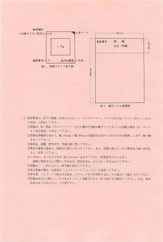
使用スライドは 35mm)坂,スライド枠には,図 lのように発表者氏名,発表番号(大会プログラムに記
されているもの),スライド総枚数, I決写順序,上辺マークをご記入下さL、。同じスライドを繰返し映写
する場合は,それに見合う枚数をご用意下さ L、。
3)展示発表 :パネノレの大きさは, 1 題につき,縦1.8m X横 0.9m の予定です。展示パネノレの上部には,図
2のように発表番号,演題,氏名,所属を明記して下さL、。その他のスベースは自由に利用して下さ L、。
表題には 5cm以上,説明文には lcm以上の文字を使用し,文章は必要最小限にとどめて下さL、。
展示物の貼出しは, 30日午前中にお願い致します。
映写順序
(10枚のうち l枚目)...11110 可←赤マー ク
MUnE
90 cm..
演題
氏名 (所属)
「ーーーーーーーーーーー
発表番号
EUD∞一:
|1 . ~ !d 品川由香里 1....氏名
使用スライド記入例
発表番号叶 15
図 l
展示パネル説明図
ワープロまたはノξ ソコン (24ドット以上
図 2
4)要旨原稿は,以下の要領 ・見本にしたがって,タイプライター,
の印字)で作成して下さい。
①原稿は, B5 用紙(オニオ ンベーパーなどの薄手の用紙は避けてくださL、)に明瞭な黒色(カーボン リ
ボン等を使用)で印字して下さい。
②原稿は演題等も含めて,縦 150mm X横 100mmの範囲内に24字 X 22行の印字を標準とします(枠で囲
まないで下さし、)。
③著者名,演題,要旨本文,所属の順に書いて下さい。
④著者が複数の場合は,演者名の前にOをつけて下さい。また,所属が異なるときは著者名の後に*印を
付し,区別して下さい。
⑤ l行目は,はじめの 3字分 (約 12.6mm)をあけて下さい (発表番号が入ります)。
演題と要旨本文との聞は l行分あけ,要旨本文は,はじめの 1字分をあけて下さい。
⑥所属は( )内に入れて,最下段に表示して下さL、。
⑦和文原稿の場合,句読点は r,J(コンマ)と roJ (マノレ)を使用して下さL、。
⑤学名等,イ タリックで表示する場合,イタリック文字を使用するか,その部分に下線をつけて下さい。
⑨原稿は約 2/3に縮小し,そのままオ フセット印刷されます。折り曲げずに郵送して下さい。なお,著者
校正はありませんので, ご注意下さ L、。
日本藻類学会主催 1毎苔栽培業見学会'のお知らせ
下記の要項により,海苔栽培業見学会を開催致します。
1 期日 :1992年 3月27日(金)~28 日 (土)
2. 日 程・ 3月27日(金)JR内房線上総湊駅前に集合
海苔栽培業見学
富津市「鈴孝荘」宿泊
3月28日 (土)海苔栽培業見学
JR内房線大賞駅前にて解散
3 見学先 天羽漁業協同組合湊のり生産組合
千葉県富津市湊1155
新富津漁業協同組合
千葉県富津市富津2430-1
(社)千葉県のり種苗センター
千葉県富津市小久保3090
4 参加費:交通費実費
昼食費実費
宿泊費約6,000円
懇親会費 2,000円
5. 定員 :30名
希望者多数の場合には先着順と致しますので,予めご了承下さい。
6 申 込 参加希望者は,往復葉書に 1)氏名, 2)連絡先,3)所属を明記の上, 1992年 1月10固までに,上
記の第16聞大会準備委員会宛にお申込下さい。
7 その他 雨具 (カッパ),防寒衣等をご持参下さい。
なお, 日程等の詳細につきま しては,追って参加決定者にご通知致します。
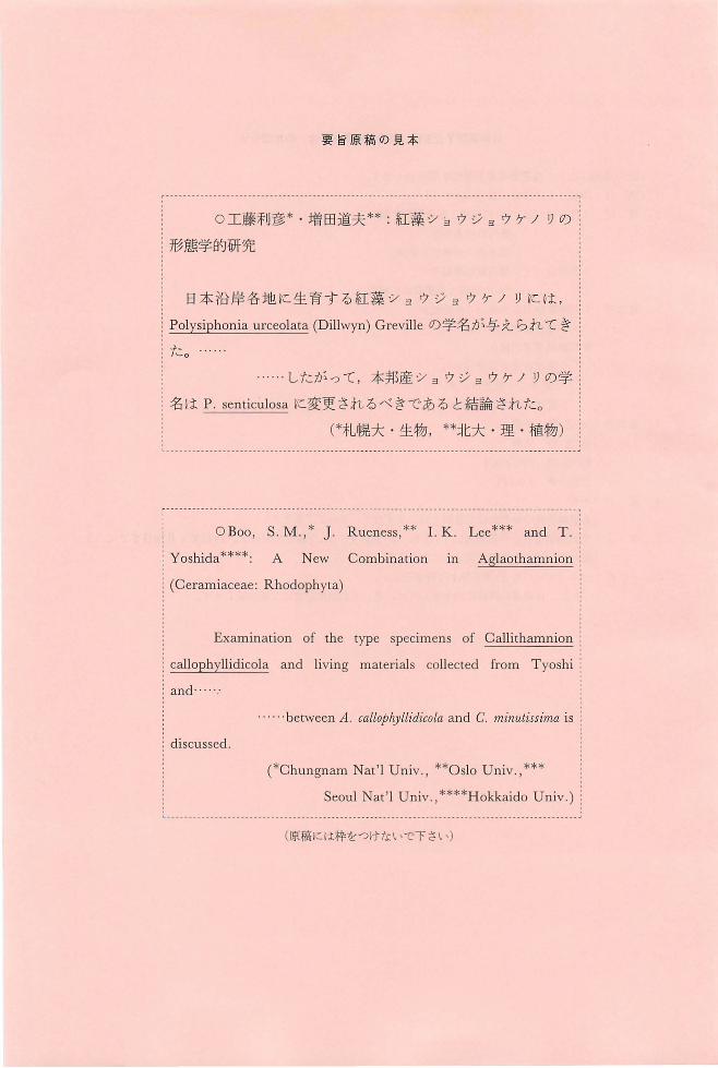
要旨原稿の見本
c-ーー一一ーー・--ーーーーーー ーーー・ーーーーー,ー可.-ーーーーーーーー,一...........ーーーー・ーー・ーーーーーーーーーーーーーーーーー'ーーー ー一ー ---ーー--一一ー-----ーー『
O工藤利彦*・増田道夫件:紅藻シ注ウジ ョウケノリの j
:形態学的研究
日本沿岸各地に生育する紅藻 ショウジ ョウケノ リには,
:Polysiphonia urceolat~ (Dillwy吋Grevilleの学名が与えられてき :
:1-こo
....したがって,本邦産ショ ウジョ ウケ ノリの学 i
:名は P senticulosaに変更されるべきである と結論された。
ゲ札幌大 ・生物, 料北大 ・理 ・植物)
「一ーー一一-一ー一一一一ーーーー 一---・ーーーーーーーーーーーーーー ー・ーーーーーーーー一一一 一一一一 一一一一一一一---ーーーーー一,一司.-・ー--一一一ー可.-ーー一一一ー-------ー『
OBoo, S.M.,* J. Rueness,** I.K. Lee*料 and T目 i
Yoshida****: A New Combination in Aglaothamnion
(Ceramiaceae: Rhodophyta)
Examination of the type specimens of Callithamnion
callophyllidicola and living materials ∞llected from Tyoshi:
and'"
-・betweenA. callophyllidicola and C. minutissima is :
discussed
(*Chungnam Nat'] Univ.,料OsloUniv.,*料
Seou1 Nat'l Univ.,料**HokkaidoU niv.) :
(原稿には枠をつけないで下さL、)
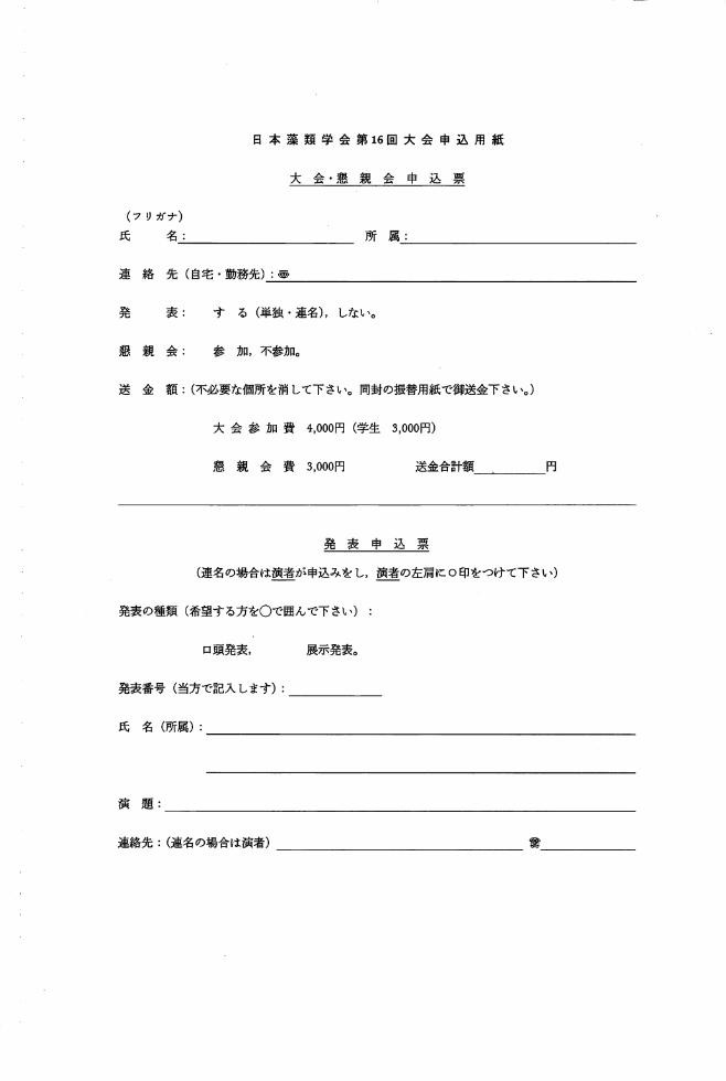
日本藻類学会第16回大会申込用紙
大会 ・懇 親会申込票
(フリガナ)
氏名:
連絡先(自宅・勤務先):~
所属:
発 表: する(単独・連名),しない。
懇親会: 参加,不参加。
送金額:(不必要な個所を消して下さい。同封の振替用紙で御送金下さい。)
大会参加費 4,000円(学生 3,000円)
懇親会費 3,000門 送金合計額 円
発表申込 票
(連名の場合は演者が申込みをし,演者の左肩に0印をつけて下さい)
発表の種類(希望する方をOで囲んで下さい)
口頭発表, 展示発表。
発表番号(当方で記入します):
氏名(所属):
演題:
連絡先:(連名の場合は演者) 重量

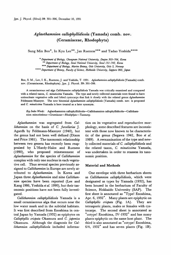
Jpn. J. Phycol. (Sorui) 39: 301-306, December 10, 1991
Aglaothamnion calloPhyllidicola (Yamada) comb. nov.
(Ceramiaceae, Rhodophyta)
Sung Min Boo*, In Kyu Lee**, Jan Rueness*** and Tadao Yoshida****
*n,ψartment of Biology, Chungnam National Univer.吻"Doejon 305-764, Korea ** n,ψ'artment 01 Biology, Seoul National University, Seoul151-742, Korea *** n,ψartment of Biology, Marine Bota司y,Oslo University, Oslo 3, Norway
**** D,ψa伽 ent01 Bota砂"Fa叫 lty01 Science, Holckai,ゐ University,Sapporo 060, Japan
Boo, S. M., L田,1.K., Rueness,J. and Yoshida, T. 1991. Aglaothamnion callop勿'lli・'dicola(Yamada) comb. nov. (Ceramiac伺 e,貼odophyta).Jpn. J. Phycol. 39: 301-306.
A ceramiaceous red alga Callithamnion callop,砂lli・d的laYamada was critically examined and compared with a related taxon, C. m印刷'SimaYamada. The type and newly collected materials were found to have uninucleate vegetative cells and lobed cystoαrps that link it closely with the related genus Aglaothamnion Feldmann-Mazoyer. The new binomial Aglaothamnion callophyllidicola (Yamada) comb. nov. is proposed 姐 dC. minutissil刷 Yamadais here treated as a later synon戸n.
Key lndex Wordr: Aglaothamnion callophyllidicola-Callithamnion callophyllidicola-Callitham-nion minutissima一Ceramiaceae-Rhodophyta-71国間omy.
Aglaothamnion was segregated from Cal-lithamnion on the basis of C. furcellan'ae J.
Agardh by Feldmann-Mazoyer (1940), but the genus had not been well defined (Dixon
and Price 1981). The taxonomic relationship between two genera has recently been reap-praised by L'Hardy-Halos and Rueness (1990), who proposed reinstatement of Aglaothamnion for也especies of Callithamnion complexwi也 onlyone nucleus in each vegeta-tive cell. Thus several species previously as-
signed to Callithamnion in Europe are newly at-
tributed to Aglaothamnion. In Korea and
J apan three Aglaothamnion and nine Callitham-nion species have been reported (Lee and
Kang 1986, Yoshida et al. 1990), but their tax-onomic positions have not been fully investi-
gated. Callithamnion calloPhyllidicola Yamada is a
small ceramiaceous alga that occurs near the low water mark and in the subtidal habitats. It was first described from Enoshima in cen-
tral Japan by Yamada (1932) as epiphytes on Callophyllis crispata Okamura and C. japonica Okamura. Although the diagnosis for Cal-lithamnion callop紗llidicolaincluded informa-
tion on its vegetative and reproductive mor-
phology, some described features are inconsis-tent with those now known to be characteris-
tic of the genus (Segawa 1942, Boo et al. 1989). A reexamination ofthe type and new-ly collected materials of C. callophyllidicola and the related taxon, C. minutissima Yamada, was undertaken in order to reassess its taxo-nomlc posltlon.
Material and Methods
One envelope with three herbarium sheets
as Callithamnion callophyllidicola, which were designated as types by Yamada (1932), has been housed in the herbarium of Faculty of Science, Hokkaido University (SAP). The first sheet is annotated as“Type! Enoshima, Apr. 6, 1932". Many pl組 ts紅 eepiphytic on Callophyllis cばrpata (Fig. 1A). They are tetrasporic plants, males or females wi也 cys-tocarps. The second sheet is annotated as “cotype! Enoshima, IV・1932"and has many plants epiphytic on the same host plant. The third is also annotated as“cotype! Enoshima, 6/4, 1932" and has seven plants (Fig. 1B).
302 Boo, S. M., Lee, 1. K., Ru巴ness,J. and Yoshida, T.
The annotations were made by Y. Yamada
himself
Small fragments of dry specimens from the
herbarium sheets were softened for a while in
distilled water and then prepared for
microscopic observation. When possible, the
preparations were stained with aniline blue/a-
cetic acid and washed for observation.
The live tetrasporic plants were collected
for staining nucleus at the low water mark in
Choshi (Jan. 28, 1990), Chiba Prefecture and
Kikonai (Feb. 15, 1990) of southern Hok-
乃/"岬01111111剛11叩11川1111111111111111111仰川11削1
D. -
cdill1
hVIl--
-
pbill-
-
l
守
Lal--E
-
-ー
erちr/ -
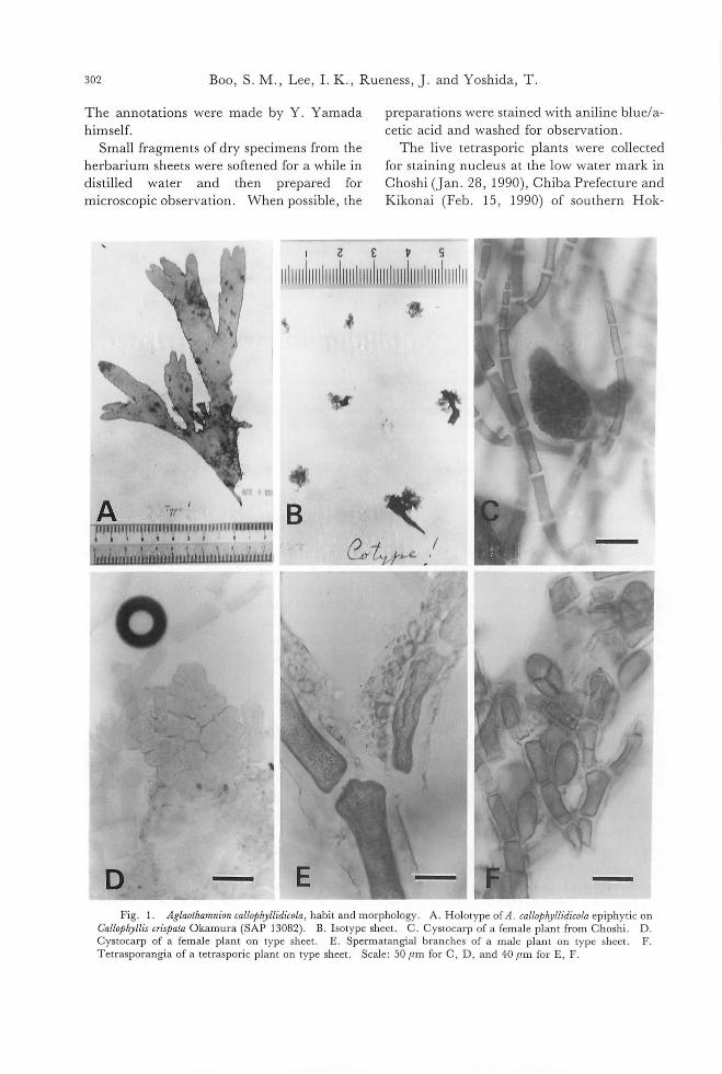
-Fig. 1. Aglaothamnion callo戸りllidicola,habit and morphology. A. Holotype of A. callo,μryll幼'colaepiphytic on
CalloT'ryllis cr坤ataOkamura (SAP 13082). B. Isotype sheet. C. Cystocarp of a female plant from Choshi. D Cystocarp of a female plant on type sheet. E. SpermatangiaJ branches of a maJe pJant on type sheet. F Tetrasporangia of a tetrasporic pJant on type sheet. Scale: 50μm for C, D, and 40μm for E, F.
Aglaothamnion callop勺必dicola(Yamada) comb. nov 303
kaido. They were fixed in 2% glutaralde-
hyde and stained with th巴臼uorochrome4'-6
diamidino-2-phenylindole (DAPI, Goff and Coleman 1984) for observation under pho-
tomicroscope equipped with epiftuorescence
filters.
The original materials of Callithamnion
minutissima Yamada are kept in SAP. They
are mounted on slide glass and are given col-
lection numbers of 368 and 1837
Results
Callithamnion calloPhyll必co必 Yamada:The
plants grow with a maximal height of about
8 mm and are attached to the host by thin
rhizoidal filaments which come out of the
basal cells of the frond. The rhizoids are
simply branched to d叩 tate(Fig. 3B). The
axial cells are formed from apical cells by ob-
lique division and 70-90μm broad and 200-
220μm long in the middle portion of plants,
thus th巴LlBratio being 2-3: 1. Th巴branch
ing pattern is alternate to subdichotomo-pin-
nate (Fig. 3A). The primary branches ar巴
derived alternately from every axial cell ex-
cept lower ones (Fig. 2A). The third or
fourth are usually formed similarly to the priー
mary branches. AII branches are distichous
and can grow ultimately. Only on巴 nucleus
is observed in every vegetative cell of the axis
and branches (Fig. 2A-B)
The gametophytes are dioecious. Sper-
matangial mother cells are cut off from the
adaxial portion of the branches and give rise
to spermatangia. Thus small spermatangial
patches are seen on the adaxial portion of
branches of male plants (Figs. 1E, 3D) and the branches with spermatangial patches are
often curved. Carpogonial branches are
formed on the axial vegetative cells in the up-
per portion of female plants. They are com-
posed of four cells and are arranged in zig-zag
(Fig. 3E-F). Two sterile cells are accompa-
nied with the carpogonial branches. After fer-
tilization young gonimolobes are form巴d(Fig.
3G-H), which become spherical to irregular
carposporophytes and 300-500μm long when
matur巴 (Fig.1C-D).
Tetrasporophytes are isomorphic to gameto-
phytes. Tetrasporangia are formed on th巴
adaxial portion of branches (Fig. 2A). They
are divided tetrah巴drallyand 40-55μm X 60-
70μm in size (Figs. 1 F, 3C). The gland
cells, which were described on the tetrasporo-
phytes by Yamada (1932) and Kawashima
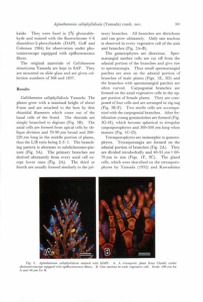
Fig. 2. Aglaothamnion callo戸hyllidωlastained with DAPI. A. A tetraspol'ic plant from Choshi under photomicroscope eqipped with epifluorescence制ters. B. One nucleus in each vegeta口、ecell. Scale: 100μm for A and 40μm for B
304 Boo, S. M., Lee, 1. K., Rue町民J.and Yoshida, T.
E H
200 )J町官
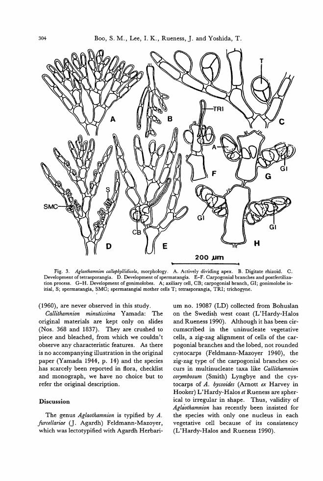
Fig. 3. Aglaothamnion callop勿Illidicola,morphology. A. Actively dividing apex. B. Digitate rhizoid. C. Development of tetrasporangia. D. Development of spermatangia. E-F. Carpogonia1 branches and postfertiliza-tion process. G-H. Development of gonimolobes. A; axiliary cell, CB; carpogonia1 branch, GI; gonimolobe in-itia1, S; spermatangia, SMC; spermatangia1 mother cel1s T; tetrasporangia, TRI; trichogyne.
(1960), are never observed in this study. Callithamnion minutissima Yamada: The
original materials are kept only on slides
(Nos. 368 and 1837). They are crushed to
piece and bleached, from which we couldn't observe any characteristic features. As there
is no accompanying illustration in the original
paper (Yamada 1944, p. 14) and the species has scarcely been reported in flora, checklist and monograph, we have no choice but to refer the original description.
Discussion
The genus Aglaothamnion is typified by A. furcellariae (J. Agardh) Feldmann-Mazoyer, which was lectotypified with Agardh Herbari-
um no. 19087 (LD) collected from Bohuslan
on the Swedish west coast (L'Hardy-Halos
and Rueness 1990). Although it has been cir-
cumscribed in the uninucleate vegetative
cells, a zig-zag alignment of cells of the car-
pogonial branches and the lobed, not rounded cystocarps (Feldman止 Mazoyer 1940), the zig-zag type of the carpogonial branches oc-curs in multinucleate taxa like Callithamnion coヮmbωum(Smith) Lyngbye and the cys-
tocarps of A.わ山oides(Arnott ex Harvey in
Ho∞okeぽrの)1ical t旬oirr問e思肝lla町ri泊nshape. Thus, validity of Aglaothamnion has recently been insisted for the species with only one nucleus in each vegetative cell because of its consistency (L'Hardy司 alosand Rueness 1990).
Aglaot.加mnionca[[,物 llidicola(Y抑制a)comb. nov. 305
Although there have been reports that Cal-lithamnion callophyllidicola Yamada has some di-agnostic characters ofthe genusAglaothamnion, a taxonomic combination has not been made
because the generic concept of Aglaothamnion was obscure (Segawa 1942) and type speci-mens had not been critically examined (Boo et al. 1989). Our observation on type materials of C. callophyllidicola agrees well to也eproto・
logue (Yamada 1932) and the previous obser-
vations (Segawa 1942, 1949, Kawashima 1960), but some features are inconsistent with them. One nucleus is found in each vegeta-tive cell of the live plants collected in Choshi, although we could not observe nucleus from dηtype materials. In addition, as our obser-vation confirms no gland. cells on type plants, Yamada must have mistaken small protuber-ances on branches for gland cells (Boo et al. 1989). The shape of the cystocarps in the proto・
logue is shown to be spherical (Yamada 1932, Pl. Vlb) , but they have been repo抗edto be
lobed (Segawa 1942, p. 208, Kawashima 1960, p. 107) 組 d irregularly spherical (Kawashima 1960, p. 107). In this study the cystocarps of type materials present lobed forms (Fig. 1D), that is 出 oincluded in the genus range of Aglaothamnion. Features on male and tetrasporic plants agree well to the
above reports. The foregoing sections mention that the
sheet (SAP 13082) designated as type by
Yamada (1932) has many individuals epip旬-ic on Callophyllis crispata. We confirmed that all specimens on the type sheet are not heter-ogeneous, so Y amada' s type of C. callophyllidi-cola is regarded as correct according to the Ar-ticle 9.1 of the Berlin code (Greuter et al. 1988).
It was reported that Callithamnion minutissi-ma resembled C. callop.紗lli・dicolabut was easily distinguished by its more slender frond, lon-ger axial cells and not tapered ultimate ramuli (Yamada 1944). As is pointed out in Europe-an Callithamnion species (Harris 1962), Yamada's diagnostic characters are quantita-
tive or unstable and subject to be changed in different environment (Boo et al. 1989).
However, since the same name was preoccu-pied for species of Adriatic Sea described by Zanardini (1842) and Kutzing (1843 p. 371), Yamada's C. minutissima is an illegitimate
name and should be replaced or rejected. The original materials in the herbarium of
Hokkaido University (SAP) are kept so crushed in piece and bleached that we could not observe any features from them and the protologue has no illustration . (Yamada
1944). The type locality, Hayama ofKanaga-wa Prefecture, is also situated near Enoshi-
ma, the type locality of ιcallopわIllidicola.Furthermore, as C. minutissima overlaps C. cal-lophyllidicola in protologue (Boo et al. 1989, Ta-ble 1), we conclude C. minutissima Yamada as a later synonym. According to Dawson (1962), Callithamnion
pωchale Borgesen is closely related to C. minutissima, but he did not state whether the alga had one or more nuclei in vegetative cells. Since both Dawson (1962) and Abbott
and Hollenberg (1976) distinguished Aglaothamnion from Callithamnion, it seems im-plicit that at least C. paschale is a multinucleate species. Aglaothamnion oosumiense Itono (1971) is another related species (Boo et al. 1989), that needs a further study.
There are still some questions to be an-swered about the genus Aglaothamnion and its species. However, from the view discussed above, it is obvious that Callithamnion callophyl・lidicola is a distinct and an endemic species to
Japan and the surrounding cClasts, and in-cludes the related taxon, C. minutissima. The following new binomial combination is there-
fore proposed: Aglaothamnion callophyllidicola (Yama-
da), comb. nov. Basionym: Callithamnion callo.仰 llidicola
Yamada 1932, p. 270. fig. 3a-b. pl. V, Vlb., Holotype SAP 13082“Enoshima, Sagami Province, Japan, Y. Yamada, April 6, 1932" Herbarium of Faculty of Science, Hokkaido U niversity .
Synonym: Callithamnion minutissima Yama-da 1944, p. 14., Types SAP, collection num-ber 368 and 1837 “Hayama, Kanagawa Prefecture, Japan" Herbarium of Faculty of
306 Boo, S. M., Lee, 1. K., Rueness,]. and Yoshida, T.
Science, Hokkaido University.
Acknowledgements
This work was supported in part by a
research grant from Korean Science and En-
gineering Foundation cooperated with
] apanese Society for Promotion of Science to
the first author (1989 2nd term). Many
thanks go to Drs Y. Hara ofTsukuba Univer-
sity, A. Miura of Tokyo Fishery University and Y. Saito of Hokkaido U niversity for their
kind helps to collection made by the first
author in ]apan. Mr. K. Kogame of Hok-
kaido University photographed the type speci-
mens and Mr. T. Kitayama made prepara-
tions with DAPI staining. The authors are
indebted to Dr. M. M. Watanabe for his help-
ful comments.
References
Abbott, 1. A. and Hollenberg, G.J. 1976. Marine algae of California. Stanford Univ. Press, Stanford.
Boo, S. M., Rueness,J. and Lee, 1. K. 1989. Lifehisto・
ry and taxonomy of Callithamnion callop勿,llidicolaYamada (Ceramiaceae,貼odophyta). Jpn. J. Phycol. 37: 28←290.
Dawson, E. Y. 1962. Marine red algae ofPacific Mexi-co. P紅 t.7. Ceramiales: Ceramiaceae, Delesseria-ceae. Univ. South Calif. Press, California.
Dixon, P. and Price, J. H. 1981. The genus Callitham-nion (Rhodophyta: Ceramiaceae) in the British Is1es. Bull. Br. Museum (Nat. HistふBot.ser. 9: 99-141.
Fe1dmann-Mazoyer, G. 1940. Recherches sur 1es Cer-amiacees de 1a Mediterranee Occidentale. Minerva, A1ger.
Goff, L.J.組 dCo1eman, A. W. 1984. E1ucidation offer-ti1ization and development in a red alga by qu組 tita・
tive DNA microspectrofluorometry. Deve10p. Biol. 102: 173-194.
Greuter, W., Burdet, H. M., Chaloner, W. G., Demou-
1in, V., Grolle, R., Hawks-Worth, D. L., Nico1son,
D. H., Si1va, P. C., Stafleu, F. A., Voss, E. G.,
McNeill, J. 1988. International code of botanical nomenclature. Reg. veg. 118: 1-328.
Harris, R. E. 1962. Contribution to the taxonomy of Callithamnion Lyngbye emend. Naegeli. Bot. Notiser 115: 18-28.
Itono, H. 1971. The genera Callithamnion, Aglaotham-nion, Seiro,ゆora,Pleonoゆoriumand Mesothamnion (Cer-amiaceae, Rhodophyta) in southern Japan. Mem. Fac. Fish., Kagoshima Univ. 20: 217-237.
Kawashima, S. 1960. Notes on some marine algae from the Northeastern Honshu, Japan (4). Jpn. J.
Phycol. 8: 100-107 (in Japanes斗Kutzing, F. T. 1843. Phyco10giaGeneralis. F. A. Brock-
haus, Leipzig. Lee, 1. K. and Kang,J. W. 1986. A check 1ist ofmarine
algae in Ko民 a.Korean J. Phycol. 1: 311-325 (in Ko問 an).
L'}王ardy-Halos,M. Th. and Rueness, J. 1990. Com-
Pぽ ativemorpho10gy and crossabi1ity of related spe・cies of Aglaothamnion (貼 odophyta).Phyco10gia 29:
351-366. Segawa, S. 1942. Cystocarp of Callithamnion callop.勿,llidi-
cola. Igaku to Seibutsugaku 1: 206-209 (in Japanese).
Segawa, S. 1949. The gonimob1ast deve10pment in cer-amiaceous algae of Japan. 1. J. Fac. Agr. Kyushu Univ. 9: 143-147.
Yamada, Y. 1932. Notes on someJapanese algae IV.J. Fac. Sci. Hokkaido Imp. Univ. Ser. V. 2: 267-276.
Yamada, Y. 1944. Notes on some Japanese algae X. Sci. Pap. Inst. A1g. Res. Hokkaido Imp. Univ. 3: 11-25.
Yoshida, T., Nakajima, Y. and Nakata, Y. 1990. Check-1ist of marine algae of Japan (re'由 edin 1990). Jpn. J. Phy∞1. 38: 269-329 (in Japanese).
Z阻むdini,G. 1842. Synopsis algarum in marin adriati-co hucusque collectarum. Mem. R. Acad. Sci., Tori-no, Ser. 2, 4: 105-258.
Boo, Sung Min* • Lee, In Kyu糾・ JanRueness*** .吉田忠生****.
キヌイトグサ(紅藻.イギス科)について
キヌイトグサのタイプ標本および新たに採集した生材料の詳しい観察から,この種の栄養細胞は単核であり,
嚢果の外形が浅裂することを確認した。これらの特徴は近縁属であるアグラオタムニオ γAglaothamnion属と
一致するので,キヌイトグサに対してAglaothamnioncallophyllid,的必の組合せを提案する。またヒナノキヌイトグ
サ Callithamnionminutissima Yamadaは種のレベルで区別することができないので,キヌイトグサの異名であると
結論される。 (*DepartmentofBio10gy, Chungnam National University, Daejon 305-764, Korea. 判 Departmentof
Bio10gy, Seou1 National University, SeouI151-742, Korea. 判 *DepartmentofBio10gy, Marine Botany, Oslo Univer-
sity, Oslo 3 Norway, *榊*060札幌市北区北10条西8丁目 北海道大学理学部植物学教室)
Jpn. J. Phycol. (Sorui) 39: 307-317, December 10, 1991
Seasonal protein variation in the N ew Zealand seaweeds POゆめm
columbina Mont. and POゆめIrasubtumens J. Ag.
(Rhodophyceae)
K. A. Aitken*, L. D. Melton失+and M. T. Brown**
* Food Science Dψartment, Universi,砂01Otago, P. O. Box 56, Dunedin, New Zealand ** Botany Depar加 ent,Unive口ityof Otago, P. O. Box 56, Dunedin, New Zealand.
Aitken, K. A., Melton, L. D. and Brown, M. T. 1991. Seasonal protein variation in the New Zealand seaweeds POψ紗racolumbina Mont. and Porphyra subtumens J. Ag. (Rhodophyceae). Jpn. J. Phycol. 39: 307-
317.
Seasonal variation in tissue protein was measured in the red a1gae POψhyra columbina Mont. and Por-P紗rasubtumens J. Ag. by three methods (total nitrogen multiplied by 6.25, biuret protein, and sum of a巾 ydroaminoacids). The protein levels in both species showed a seasonal trend, w凶 maximumlevels oc-curring in the winter. The measured protein content varied between the methods used, with the nitrogen value multiplied by 6.25 giving the highest value,おlIowedby the sum of anhydroamino acids and then the biuret method. The sum of amino acids appe訂 edto be the most accurate me出odof determination. Based on the sum of anhydroamino acids, a new multiplication factor of 5.0 for the conversion of nitrogen to pro-tein has been proposed. The predominant protein-bound amino acids were A1a, Glu, Asp and Leu, follow-ed by VaI, Lys and Arg. Ala was the main amino acid in P. columbina, but Glu was the main amino acid in P. subtumens. Similarly, A1a and Glu were the main free amino acids in P. columbina and P田川tumens,respec-tively. The protein-bound amino acids and the m司jorfree amino acids showed specific seasonality.
Key lndex Uノords: amino acids-biuret-nitrogen-nori-Porphyra columbina-Porphyra sub-tumens-protein-Rhodophyceae-se,町 'onal.
Mariculture of the red algae Porphyra and
its processing into thin, purple-black sheets, called “hoshi nori" , is a prominent food indus-
t町 in]apan. P. yezoensis Ueda is now the
main species used although P. tenera Kjellman
was important in the past (Miura 1975;
Nisizawa et al. 1987).
POψ紗rais used as food in other parts of the
world also. It is farmed in China, where it is known as“zicai" (Tseng 1981), and in Korea, where it is known as “kim" (Mumford釦 d
Miura 1984). It is consumed in smaller quan-tities in Wales and New Zealand, where it is
known as “laver" and “karengo" , respectively (Chapman and Chapman 1980). The most
common species of Porphyra found in New
Zealand are P. columbina Mont. and P. subtu・
mens]. Ag. (Chapman 1969). P. columbina, a
+ Author to whom co町田pondenceis to be addressed.
traditional food of the Maori, grows on rocky substrate in the intertidal zone on most of the
coastline around New Zealand (Nelson
1984). Mariculture of P. coluT1山・'nais being in-
vestigated (Brown et al. 1990). P. subtumens is
an endemic epiphyte on Durvillaea antarctica
(Chamisso) Hariot and D. willana Lindauer
(Chapman 1969). Recent studies Oll New
ZealandPoψ紗racolumbina and P. subtumens in-
clude the determination of their ascorbic acid
contents (Friedlander et al. 1989). The cell
culture (Xue-Wu and Gordon 1987) and
resistance to desiccation (Brown 1987) of P. columbina have also been investigated.
The protein content determined by differ-
ent investigators for the same P01灼 raspecles
show considerable variation. Protein in P.
tenera is reported to range from 14.9 %, dry
weight, (Mukai et al. 1981) to 56.1% (Noda and Horiguchi 1975). The only available
308 Aitken, K. A., Melton, L. D. and Brown, M. T.
literature on the protein content of P. columbi-
na Mont. was 5.5%, dry weight, as reported by Quilhot (1970). Ji et al. (1981) studied the
seasonal variation of amino acids in P. yezoen-sis in three locations differing in dissolved
nitrogen content (30μg to 300μg NHcN/l
seawater). The total amino acids ranged
from approximately 10 to 30%, air dry
weight. Lower protein contents occurred in
regions with lower seawater nitrogen.
8everal studies have been carried out on
seasonal tissue nitrogen variation. Brown et al. (1990) found higher values in P. columbina Mont. (New Zealand) during the peak growth period (winte吟 Thesame trend was ob-
served byJi et al. (1981) in P. yezoensむ, andby
Takagi (1951) in several POψ砂raspp. Various methods have been used to study
protein in POψわIra. Mukai et al. (1981) com-pared protein in由ecell walls of P. tenera ob-tained by multiplying the nitrogen content by
6.25, summation ofamino acids and a biuret-
Folin method. They found the protein con-
tent calculated from nitrogen gave the same
value as the sum of anhydroamino acids.
This suggests that all nitrogen was present as
amino acids, and the protein molecules con-
tained 16% nitrogen. However, protein de-termined by the Lowry-biuret method gave
considerably higher results than the other two
methods. The nitrogen conversion factor
6.25 indicates 16% protein nitrogen, and as-sumes no non-protem mtrogen IS present.
This factor is applicable to egg, meat and le-gumes. The factor for refined flour is 5.70
and for milk and milk products is 6.38. The
6.25 conversion factor is often employed
when the protein nitrogen content is not
known. Protein calculated by this method
should be referred to as“crude protein" (FAO/WHO 1973; FAO/WHO/UNU 1985). The nitrogen content of proteins can
vaηfrom 12 to 30% (Lillevik 1970).
Arasaki and Mino (1973) found the nitrogen content of alkali soluble seaweed proteins,
which they reported to be the major type of protein in POψ紗ra,ranged from approximate-1 y 12 to 14%. Determination of the prot巴m
nitrogen content usually involves extraction
of the protein, and assumes the extraction is
complete. However, Coulson (1955) and
8mith and Young (1953) found polysaccha-
rides can interfere with the extraction of pro-
tein from Phaeophyta. Three or four types of
soluble proteins have been found in seaweeds
(Takagi 1950; Arasaki and Mino 1973; Ama-
no and Noda 1990). 8eaweeds often contain
non-protein nitrogenous compounds. These
may include ammonia compounds, free ami-no acids, peptides, nitrates and pigments
(Rosell and 8rivastava 1985), and seaweeds produce non-protein amino acids (Impelliz-
zeri et al. 1975; Fattorusso and Piattelli 1980). Amino acids in algae may occur in com-
bined (protein-bound) or free form (Young
and 8mith 1958). Ala, Glu and Asp have been found to be the predominant amino
acids in Porphyra (Munda and Gubensek
1976; Ji et al. 1981; Amano and Noda 1990), while Harada et al. (1990) found Ala, Glu and Tau were the predominant free amino acids, with Asp also present. Tau has been found
to be extensively distributed in the seaweeds
of Rhodophyta (Impellizzeri et al. 1975; Fat-
torusso and Piattelli 1980). The biuret
method, a commonly used colorimetric
method, does not measure free amino acids or dipeptides. It is based on the solubility ofpro-
tein in alkali, which varies for different pro-teins. Arasaki and Mino (1973) reported
that P. tenera contained 65% alkali soluble pro-
tem.
The present study investigates the seasonal
protein variation in P. columbina and P. subtu-mens measured by three methods, and looks at the possibility of using a conversion factor
other than 6.25 for the nitrogen value. The
study looks briefly at the quality of the protein
present, and at the free amino acids, some of which have been reported to contribute to the
flavour of nori.
Materials and Methods
Porp再)lracolumbina was collected from rocks in the littoral zone of the rocky shore at 8t.
Clair, Dunedin. Porp均)lrasubtumens, an epi-phyte on Durvillaea antarctica and D.ωillana,
Protein variation in N ew Zealand P01拘Iraspecies 309
was collected from the sublittoral zone at
Brighton, 14 km south west ofDunedin. Col-
lections of P. subtumens were restricted to
periods of low tide (0.1 to 0.2 m below mean
tide level). Whole plants were washed with
seawater at the time of collection, to remove sand and epiphytes. Samples were pooled
and drying at 300C in an air circulating oven
commenced within three hours of collection.
Dried samples were ground in a Wiley Mill, to pass through mesh size 0.5 mm. The sam-
ples were then stored at room temperature (ap-
proximately 200C) in air tight containers un-
til analyzed. Each sample analyzed con-
tained portions from at least 50 plants. Total nitrogen was determined by the Du-
mas method, using a Coleman Model 29 Nitrogen Analyzer, and had an error of ::!::0.3%, dry weight. The measured nitrogen
.j..J
.r:. cl
40
35
30
.~ 25 玄
〉、
L
百 20量哩
c
~ 15 .j..J
O L a
10
5
O
content was multiplied by 6.25 to obtain the “crude protein content". The biuret method
(Goa 1953) as modified by Bergersen (1980) was used to determine the total protein con-
tent of the samples. Dialyzed bovine serum
albumin (2.5 mg/ml distilled water) was used as a standard solution. Preliminary studies
showed a green pigment, formed or un-masked by the addition of NaOH to the sam-
ples, caused interference. Consequently, de-terminations were carried out by simultane-
ously subtracting the pigment absorbance of
samples that did not have Benedicts reagent
added. Analyses were done in triplicate.
Amino acid analysis was used to determine
the amino acids: aspa口icacid, threonine, ser-ine, glutamic acid, proline, glycine, alanine, valine, isoleucine, leucine, tyrosine, phenylalanine, histidine, lysine, arginine,
Aug Oct Dec Feb Apr Jun Aug Oct 1985 1986
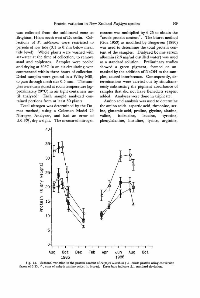
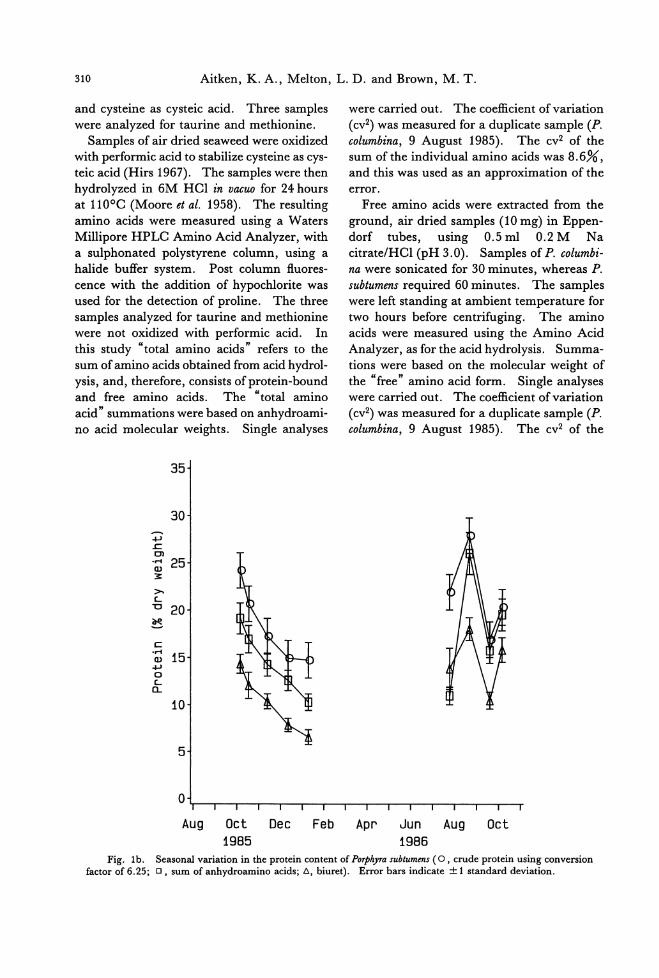
Fig. la. Seasonal variation in the protein content of POψりracolumbina (0 , crude protein using conversion factor of 6.25;ロ, sum of anhydroamino acids; d., biuret). Error bars indicate土1standard deviation.
310 Aitken, K. A., MeIton, L. D. and Brown, M. T.
and cysteine as cysteic acid. Three samples
were analyzed for taurine and methionine. Samples of air dried seaweed were oxidized
with performic acid to stabilize cysteine as cys-
teic acid (Hirs 1967). The samples were then hydrolyzed in 6M HCl in vacuo for 24 hours at 1100C (Moore et al. 1958). The resuIting
amino acids were measured using a Waters Millipore HPLC Amino Acid Analyzer, with a sulphonated polystyrene column, using a halide buffer system. Post column fl.uores-cence with the addition of hypochlorite was
used for the detection of proline. The three samples analyzed for taurine and methionine were not oxidized with performic acid. In this study “total amino acids" refers to the sum of amino acids obtained from acid hydrol-
ysis, and, therefore, consists ofprotein-bound and free amino acids. The “total amino acid" summations were based on anhydroami-no acid molecular weights. Single analyses
.j...I
.c. ロ3
35
30
・.~ 25 玄
〉、
L
"c 20 語哩
c .~ 15 キJ
o c.. a..
10
5
o
were carried out. The coefficient ofvariation
(cv2) was measured for a duplicate sample (P. columbina, 9 August 1985). The cv2 of the sum ofthe individual amino acids was 8.6%, and this was used as an approx凶 ationofthe
error. Free amino acids were extracted from the
ground, air dried samples (10 mg) in Eppen-dorf tubes, using 0.5 ml 0.2 M Na citrate/HCl (pH 3.0). Samples of P. columbi-na were sonicated for 30 minutes, whereas P. subtumens required 60 minutes. The samples were left standing at ambient temperature for two hours before centrifuging. The amino acids were measured using the Amino Acid Analyzer, as for the acid hydrolysis. Summa-
tions were based on the molecular weight of the “free" amino acid form. Single analyses were carried out. The coe伍cientof variation (cv2) was measured for a duplicate sample (P. columbina, 9 August 1985). The cv2 of the
Aug Oct Dec Feb Apr Jun Aug Oct
1985 1986
Fig. 1b. Seasonal variation in the protein content of POゆ'hyrasubtumens ( 0 , crude protein using conversion factor of 6.25;ロ, sum of anhydroamino acids; 6, biu附). Error bars indicate土1standard deviation.
Protein variation in N ew Zealand POψ砂raspecies 311
sum of the invididual amino acids was 5.0%, and this was used as an approximation of the
error.
The water content ofthe air dried (at 3000)
samples, was determined by drying duplicate portions under vacuum at 11000 until con-
stant weight was achieved (“oven dried").
“Dry weight" in this study refers to“oven dry weight".
Results
Seasonal variation in protein content was
observed in P. columbina and P. subtumens by
three methods (Fig. 1a, b). The highest lev-
els occurred in the winter months in both spe-
cies. Using the same method, the protein contents of the two species were comp町 able
for samples collected at similar times. The
protein levels in P. columbina samples ana-
lyzed ranged from 10.6 to 33.8%, dry weight, when measured by nitrogen multiplied by
6.25; 8.2 to 24.5% by sum of anhydroamino
acids (not including Met, Trp, Tau); 6.2 to 21.1% by the biuret method. For P. subtu-
mens the ranges were 14.8 to 28.2%, dry weight; 10.2 to 26.0%叩 d6.5 to 18.0%, respectively. The results of the three
methods were correlated (Pe訂 son'scorrela-
tion, P<0.05). However, the average pro-tein content varied significandy between the
three methods (Duncan's multiple range test, P<O.Ol). For both seaweeds multiplying
nitrogen by 6.25 gave the highest average
value (P. columbina: 25.5%:t 7.9%, dry weight and P. subtumens: 20.0% :t4.5%), fol-lowed by sum of anhydroamino acids
(17.6%:t5.2% and 16.1%:t5.0%, respec-tively (not including Met, Trp and Tau)) and the. biuret method (15.3%士4.8% and
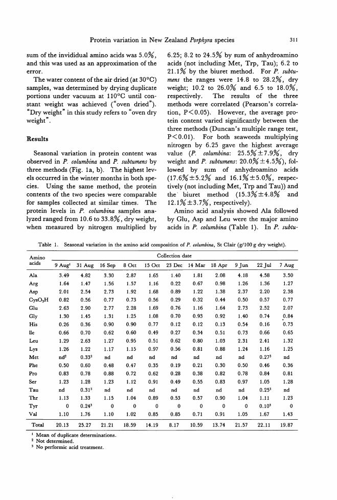
12.1% :t3. 7%, respectively). Amino acid analysis showed Ala followed
by Glu, Asp and Leu were也emajor amino
acids in P. columbina (Table 1). In P. subtu-
Table 1. Seasonal variation in the amino acid composition of P. columbina, 8t Clair (g/100 g dry weight).
Amino Collection date acids 9 Aug1 31 Aug 16 8ep 80ct 15 Oct 23 Dec 14 Mar 18 Apr 9Jun 22Jul 7 Aug
A1a 3.49 4.82 3.30 2.87 1.65 1.40 1.81 2.08 4.18 4.58 3.50
Arg 1.64 1.47 1.56 1.57 1.16 0.22 0.67 0.98 1.26 1.36 1.27
Asp 2.01 2.54 2.73 1.92 1.68 0.89 1.22 1.38 2.37 2.20 2.38
CyS03H 0.82 0.56 0.77 0.73 0.56 0.29 0.32 0.44 0.50 0.57 0.77
Glu 2.63 2.90 2.77 2.28 1.69 0.76 1.16 1.64 2.73 2.52 2.07
Gly 1.30 1.45 1.31 1.25 1.08 0.70 0.93 0.92 1.40 0.74 0.84
His 0.26 0.36 0.90 0.90 0.77 0.12 0.12 0.13 0.54 0.16 0.73
lle 0.66 0.70 0.62 0.60 0.49 0.27 0.34 0.51 0.73 0.66 0.65
Leu 1.29 2.63 1.27 0.95 0.51 0.62 0.80 1.03 2.31 2.41 1.32
Lys 1.26 1.22 1.17 1.15 0.97 0.56 0.81 0.88 1.24 1.16 1.25
Met nd2 0.333 nd nd nd nd nd nd nd 0.273 nd
Phe 0.50 0.60 0.48 0.47 0.35 0.19 0.21 0.30 0.50 0.46 0.36
Pro 0.83 0.78 0.88 0.72 0.62 0.28 0.38 0.82 0.78 0.84 0.81
Ser 1.23 1.28 1.23 1.12 0.91 0.49 0.55 0.83 0.97 1.05 1.28
Tau nd 0.3P nd nd nd nd nd nd nd 0.253 nd τ'hr 1.13 1.33 1.15 1.04 0.89 0.53 0.57 0.90 1.04 1.11 1.23 Tyr 。0.243 。 。 。 。 。 。 。0.103 。Val 1.10 1.76 1.10 1.02 0.85 0.85 0.71 0.91 1.05 1.67 1.43
Total 20.13 25.27 21.21 18.59 14.19 8.17 10.59 13.74 21.57 22.11 19.87
1 Mean of duplicate determinations. 2 Not determined. 3 No performic acid treatment.
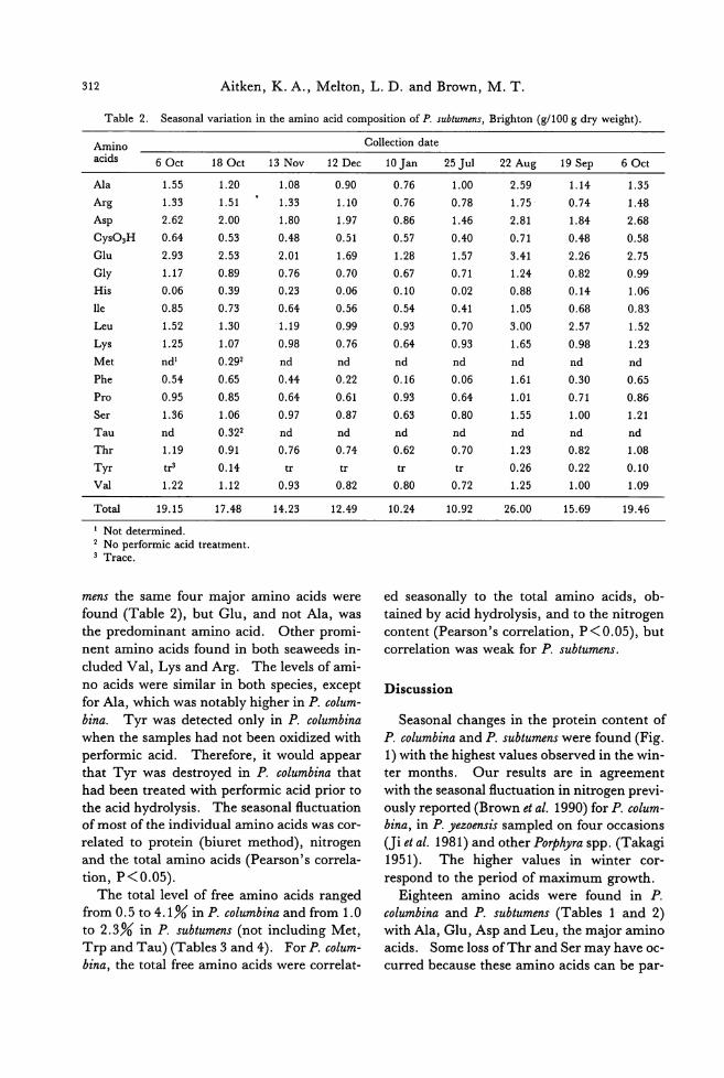
312 Aitken, K. A., Melton, L. D. and Brown, M. T.
Table 2. Seasonal variation in出eamino acid composition of P. subtumens, Brighton (g/100 g dry weight)・
Amino acids 60ct 180ct
Ala 1.55 1.20
Arg 1.33 1.51
Asp 2.62 2.00
CyS03H 0.64 0.53
Glu 2.93 2.53
Gly 1.17 0.89
His 0.06 0.39
lIe 0.85 0.73
Leu 1.52 1.30
Lys 1.25 1.07
Met nd1 0.292
Phe 0.54 0.65
Pro 0.95 0.85
Ser 1.36 1.06
Tau nd 0.322
Thr 1.19 0.91
Tyr tr3 0.14
VaI 1.22 1.12
Total 19.15 17.48
1 Not determined. 2 No performic acid treatment. 3 Trace.
13 Nov 12 Dec
1.08 0.90
1.33 1.10
1.80 1.97
0.48 0.51
2.01 1.69
0.76 0.70
0.23 0.06
0.64 0.56
1.19 0.99
0.98 0.76
nd nd
0.44 0.22
0.64 0.61
0.97 0.87
nd nd
0.76 0.74
tr tr
0.93 0.82
14.23 12.49
mens the same four major amino acids were found (Tab1e 2), but Glu, and not Ala, was the predominant amino acid. Other promi-
nent amino acids found in both seaweeds in-
cluded Val, Lys and Arg. The leve1s of ami-
no acids were similar in both species, except for Ala, which was notably higher in P. colum-bina. Tyr was detected only in P. columbina when the samples had not been oxidized with
performic acid. Therefore, it would appear that Tyr was destroyed in P. columbina that had been treated with performic acid prior to
the acid hydrolysis. The seasonal fl.uctuation of most of the individual amino acids was cor-
related to protein (biu問 tmethod), nitrogen and the total amino acids (Pearson's corre1a-tion, P<0.05).
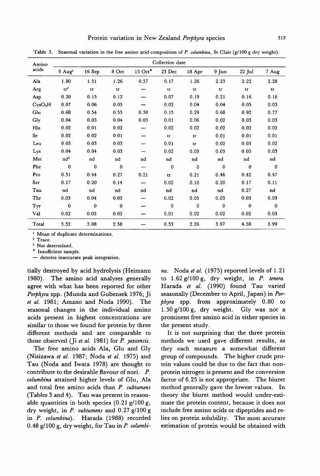
The total leve1 of free amino acids ranged from 0.5 to 4.1% in P. columbina and from 1.0
to 2.3% in P. subtumens (not including Met, Trp and Tau) (Tables 3 and 4). For P. colum-bina, the total free amino acids were corre1at-
Collection date
10Jan 25Jul 22 Aug 19 Sep 60ct
0.76 1.00 2.59 1.14 1.35
0.76 0.78 1.75 0.74 1.48
0.86 1.46 2.81 1.84 2.68
0.57 0.40 0.71 0.48 0.58
1.28 1.57 3.41 2.26 2.75
0.67 0.71 1.24 0.82 0.99
0.10 0.02 0.88 0.14 1.06
0.54 0.41 1.05 0.68 0.83
0.93 0.70 3.00 2.57 1.52
0.64 0.93 1.65 0.98 1.23
nd nd nd nd nd
0.16 0.06 1.61 0.30 0.65
0.93 0.64 1.01 0.71 0.86
0.63 0.80 1.55 1.00 1.21
nd nd nd nd nd
0.62 0.70 1.23 0.82 1.08
tr tr 0.26 0.22 0.10
0.80 0.72 1.25 1.00 1.09
10.24 10.92 26.00 15.69 19.46
ed seasonally to the total amino acids, ob-tained by acid hydr01ysis, and to the nitrogen content (Pearson's correlation, P<0.05), but corre1ation was weak for P. subtumens.
Discussion
Seasonal changes in the protein content of
P. columbina and P. subtumens were found (Fig. 1) with the highest values observed in the win-
ter months. Our results are in agreement
with the seasonal fl.uctuation in nitrogen previ-ously reported (Brown et al. 1990) for P. colum-bina, in P. yezoensis sampled on four occasions (Ji et al. 1981) and other P07舛yraspp. (Takagi
1951). The higher values in winter cor-
respond to the period of maximum growth.
Eighteen amino acids were found in P.
columbina and P. subtumens (Tables 1 and 2)
with Ala, Glu, Asp and Leu, the m司joramino
acids. Some 10ss ofThr and Ser may have oc・
curred because these amino acids can be par-
Protein variation in N ew Zealand P07舛jlraspecies 313
Table 3. Seasonal variation in the free amino acid composition of P. columbina, St Clair (g/100 g dry weight).
Amino acids 9 Aug' 16 Sep 80ct
Ala 1.80 1.51
Arg tr tr
Asp 0.30 0.15
CyS03H 0.07 0.06
Glu 0.68 0.54
Gly 0.04 0.03
His 0.02 0.01
lle 0.02 0.02
Leu 0.03 0.03
Lys 0.04 0.04
Met nd3 nd
Phe 。 。Pro 0.31 0.44
Ser 0.17 0.20
Tau nd nd τ'hr 0.03 0.04
Tyr 。 。Val 0.02 0.03
Total 3.52 3.08
, Mean of duplicate determinations. 2 Trace. 3 Not determined. * Insu伍cientsample.
1.26
tr
0.12
0.05
0.55
0.04
0.02
0.01
0.03
0.03
nd 。0.27
0.14
nd
0.03 。0.03
2.58
- denotes inaccurate peak integration.
150ct*
0.57
0.30
0.03
nd
0.21
nd
tially destroyed by acid hydrolysis (Heimann
1980). The amino acid analyses generally
agree with what has been reported for other
POψかaspp. (Munda and Gubensek 1976; Ji
et al. 1981; Amano and Noda 1990). The
seasonal changes in the individual amino
acids present in highest concentrations are
simil訂 tothose we found for protein by three
different methods and are comparable to
those observed (Ji et al. 1981) for P. yezoensis.
The free amino acids Ala, Glu and Gly
(Nisizawa et al. 1987; Noda et al. 1975) and
Tau (Noda and Iwata 1978) are thought to
contribute to the desirable flavour of nori. P. columbina attained higher levels of Glu, Ala
and total free amino acids th佃 P.subtumens (Tables 3 and 4). Tau was present in reason-
able quantities in both species (0.21 g/100 g, dry weight, in P. subtumens and 0.27 g/100 g
in P. columbina). Harada (1988) recorded
0.48 g/100 g, dry weight, for Tau in P. columbi-
Collection date
23 Dec 18 Apr 9Jun 22Jul 7 Aug
0.17 1.26 2.23 2.22 2.28
tr tr tr tr tr
0.07 0.19 0.21 0.16 0.16
0.02 0.04 0.04 0.05 0.03
0.15 0.29 0.68 0.92 0.77
0.01 0.06 0.02 0.03 0.03
0.02 0.02 0.02 0.02 0.02
tr tr 0.01 0.01 0.01
0.01 tr 0.02 0.03 0.02
0.02 0.03 0.03 0.05 0.03
nd nd nd nd nd 。 。 。 。 。tr 0.21 0.46 0.42 0.47
0.02 0.10 0.20 0.17 0.11
nd nd nd 0.27 nd
0.02 0.05 0.03 0.03 0.03 。 。 。 。 。0.01 0.02 0.02 0.02 0.03
0.53 2.26 3.97 4.38 3.99
na. Noda et al. (1975) reported levels of 1.21 to 1.62 g/100 g, dry weight, in P. tenera. Harada et al. (1990) found Tau varied
seasonally (December to April, Japan) in Por-phyra spp. from approximately 0.80 to
1.50 g/100 g, dry weight. Gly was not a
prominent free amino acid in either species in
the present study.
It is not surprising that the three protein
methods we used gave different results, as they each measure a somewhat different
group of compounds. The higher crude pro-
tein values could be due to the fact that non-
protein nitrogen is present and the conversion
factor of 6.25 is not appropriate. The biuret
method generally gave the lowest values. In
theory the biuret method would under-esti-
mate the protein content, because it does not include free amino acids or dipeptides and re-
lies on protein solubility. The most accurate
estimation of protein would be obtained with
314 Aitken, K. A., Melton, L. D. and Brown, M. T.
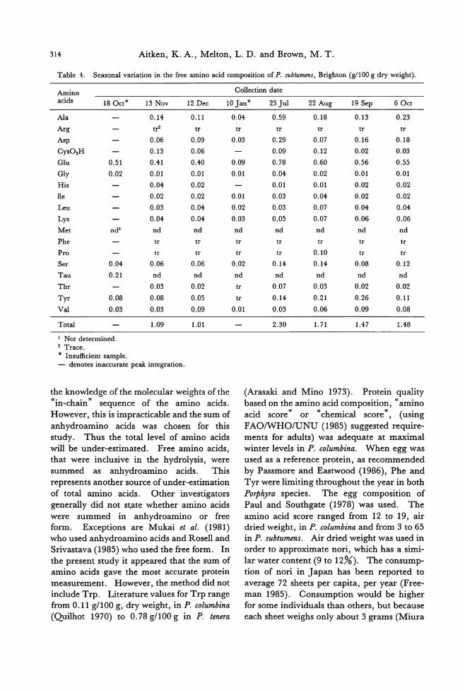
Table 4. Seasonal variation in the free amino acid composition of P. subtumens, Brighton (g/100 g dry weight).
Amino Collection date acids 180ct* 13 Nov 12 Dec 10 Jan* 25Jul 22 Aug 19 Sep 60ct
Ala
Arg
Asp
CyS03H Glu 0.51
Gly 0.02 His
lle
Leu Lys
Met nd1
Phe
Pro Ser 0.04
Tau 0.21
Thr
Tyr 0.08
Val 0.03
Total
1 Not determined. 2 Trace. * Insufficient sample.
0.14
tr
0.06
0.13
0.41
0.01
0.04
0.02
0.03
0.04
nd
tr
tr
0.06 nd
0.03
0.08
0.03
1.09
-denotes inaccurate peak integration.
0.11
tr
0.09
0.06
0.40
0.01
0.02
0.02
0.04
0.04
nd
tr
tr
0.06 nd
0.02
0.05
0.09
1.01
the knowledge of the molecular weights of the
“in-chain" sequence of the amino acids.
However, this is impracticable and the sum of anhydroamino acids was chosen for this
study. Thus the total level of amino acids
wi1l be under-estimated. Free amino acids, that were inclusive in the hydrolysis, were summed as anhydroamino acids. This
represents another source of under可 stimation
of total amino acids. Other investigators
generally did not s~ate whether amino acids
were summed in anhydroamino or free form. Exceptions are Mukai et al. (1981) who used anhydroamino acids and Rosell and Srivastava (1985) who used the free form. In
the present study it appe釘 edthat the sum of amino acids gave the most accurate protein
measurement. However, the method did not include Trp. Literature values for Trp range
from 0.11 g/100 g, dry weight, in P. columbina (Quilhot 1970) to 0.78 g/100 g in P. tenera
0.04 0.59 0.18 0.13 0.23
tr tr tr tr tr
0.03 0.29 0.07 0.16 0.18
0.09 0.12 0.02 0.03
0.09 0.78 0.60 0.56 0.55
0.01 0.04 0.02 0.01 0.01
0.01 0.01 0.02 0.02
0.01 0.03 0.04 0.02 0.02
0.02 0.03 0.07 0.04 0.04
0.03 0.05 0.07 0.06 0.06
nd nd nd nd nd
tr tr tr tr tr
tr tr 0.10 tr tr 0.02 0.14 0.14 0.08 0.12 nd nd nd nd nd
tr 0.07 0.03 0.02 0.02
tr 0.14 0.21 0.26 0.11
0.01 0.03 0.06 0.09 0.08
2.30 1.71 1.47 1.48
(Arasaki and Mino 1973). Protein quality
based on the amino acid composition,“amino
acid score" or “chemical score", (using FAO/WHO/UNU (1985) suggested require-ments for adults) was adequate at maximal
winter levels in P. columbina. When egg Was used as a reference protein, as recommended by Passmore and Eastwood (1986), Phe and Tyr were limiting throughout the ye町 inboth
POψ砂raspecies. The egg composition of
Paul and Southgate (1978) was used. The
amino acid score rang吋 from12 to 19, air dried weight, in P. columbina and from 3 to 65
in P. subtumens. Air dried weight was used in order to approximate nori, which has a simi-
lar water content (9 to 12%). The consump-
tion of nori in J apan has been reported to average 72 sheets per capita, per year (Free-m姐 1985). Consumption would be higher for some individuals than others, but because each sheet weighs only about 3 grams (Miura
Protein variation in New Zealand Porphyra species 315
1975), nori would not appear to be a m司jor
source of protein.
The conversion factors for nitrogen to pro-
tein obtained by summation of amino acids, ranged from 3.08 to 5.96 for P. columbina and
P. subtumens (not including Tau, Met and Trp). When the measurements for Tau and
Met were included (Tables 1 and 2), as well as an average literature data of
0.43:t:0.34 g/100 g, dry weight, for Trp (data from Quilhot 1970; Rao and Polacchi 1972;
Arasaki and Mino 1973), a conversion factor
of 5.0 was obtained. This estimated conver-
sion factor was lower than the factor 6.25, fre-quently used by researchers studying sea-
weed, but compares favourably with conver-sion factors that we have calculated from liter-
ature data, using nitrogen in Porphyra spp. and corresponding sum of amino acids.
These ranged from 3.47 for P. tenera (Arasaki
and Mino 1973) to 6.17 (Mukai et al. 1981)
for P. tenera, with an average of 4.98. These
conversion factors do not contain Tau, and often do not contain Met or Trp.
Rosell and Srivastava (1985) found that
multiplying the nitrogen content of the sea-
weed residue by 6.25, after alcohol extraction (80% ethanol), gave results comparable to those obtained by summation of amino
acids. Ito and Hori (1989) noted that 70%
ethanol extraction removes soluble nitrogen
compounds, including free amino acids, amines and pigments. These usually ac-
count for 10 to 20% ofthe total nitrogen com-
pounds. Leu時 etal. (1972) reported that
several seaweeds contained approximately
20% non-protein nitrogen. This indicates
80% of the nitrogen in seaweed is protein
nitrogen. Therefore, once again, a factor of
5.0 multiplied by total nitrogen would appear
to give a satisfactory estimate of protein con-
tent. We conclude, the best method for deter-mining protein levels would be the summa-
tion of amino acids, followed by using the con-version factor of 5.0 for the nitrogen value.
Acknowledgements
The authors are indebted to the Chemistry
Department Microanalytical Laboratorγ, University of Otago, for nitrogen determina-tions, and Alan Carne and Ingrid Emersen, Biochemistry Department, for performing the amino acid analyses.
References
Amano, H. and Noda, H. 1990. Protein protoplasts from red alga POψhyra yezoensis. Nippon Suisan Gak-kaishi. 56: 1859-1864.
Arasaki, T. and Mino, N. 1973. The alkali soluble pro・teins in marine algae. J. Jpn. Soc. Food Nutr. 26: 129-133.
Bergersen, F. J. 1980. Measurement of nitrogen fixa-tion by direct means. p. 65-110. In F.J. Bergersen [ed.] Methods for Evaluating Biological Nitrogen Fixation. John Wiley and Sons, New York.
Brown, M. T. 1987. Effects of desiccation on photos戸1・
thesis of intertidal algae from a southern New Zealand shore. Bot. Mar. 30: 121-127
Brown, M. T., Frazer, A. W.J., Brasch, D.J. and Mel-ton, L. D. 1990. Growth and reproduction of Por-仲間 columbina Mont. (Bangiales, Rhodophyceae) from southern New Zealand.J. Appl. Phycol. 2: 35-44.
Chapman, V.J. 1969. The Marine Algae of New Zealand. Part III. Rhodophyceae. 1. Bangiophyci-dae and Florideophycidae. Cramer, Germany. p 19-25.
Chapman, V.J. and Chapman, D.J. 1980. Seaweeds and their Uses. 3rd ed. Chapman and Hall, Lon-don. p. 88-89, 98-147.
Coulson, C. B. 1955. Plant proteins. V. Proteins and amino acids of marine algae. J. Sci. Food Agric. 6: 67←682.
F.A.O.lW.H.O. 1973. Energy and protein require-ments. Report of a joint FAOIWHO ad hoc expert committee目 WldHlth Org. Techn. Rep. Ser. 522: 14, 53-65, 105.
F.A.O.lW.H.O.lU.N.U. 1985. Energy and protein re・quirements. Report ofajoint FAOIWHO/UNU ex-pert consultation. Wld Hlth Org. Techn. Rep. Ser. 724: 113-129.
Fattorusso, E.回 dPiattelli, M. 1980. Amino acids from marine algae. p. 95-140. In P.J. Scheuer [ed.] Ma-rine Natural Products: Chemical and Biological Per-spectives. Acad. Press, New York.
Freeman, K. 1985. Fish ofthe month, nori. Pacific Fish-ing. 7: 21-27.
Friedlander, S. F., Melton, L. D. and Brown,恥ιT.1989. Ascorbic acid in the New Zealand seaweeds POψhyra columbina Mont. and Porp再yrasublumens J Ag. (貼odophyceae).Jpn. J. Phycol. 37: 295-301.
Goa, J. 1953. A micro biuret method for protein deter-mination. Determination of total protein in cerebro-spinal fluid. Scand. J. Clin. Lab. Invest. 5: 218-
316 Aitken, K. A., Melton, L. D. and Brown, M. T.
222.
Harada, K. 1988. Free amino acid compositions in
Graci加 iasordida and POψhyra columbina. Personal
Communication
Harada, K., Osumi, Y., Fukuda, N., Amano, H.阻 d
Noda, H. 1990. Changes of amino acid composi・
tions of‘Nori', POゆhhyraspp. during storage. Nip-pon Suisan Gakkaishi. 56: 607-612.
Heimann, W. 1980. Fundamentals of Food Chemistry.
Ellis Horwood Ltd, England. p. 46. Hirs, C. H. W. 1967. De,termination of cystine as cys-
teic acid. p. 59-63. In C. H. W. Hirs [ed.) Methods
in Enzymology. Volume XI. Enzyme Structure.
Acad. Press, New York. 1mpeIIizzeri, G., Mangiafico, S., Oriente, G., Piattelli,
M., Sciuto, S., Fattorusso, E., Magno, S., Santa-croce, C. and Sica, D. 1975. Amino acids and low-
molecular-weight carbohydrates of some marine red
a1gae. Phytochemistry. 14: 1549-1557.
1to, K. and Hori, K. 1989. Seaweed: chemical composi-
tion and potential food uses. Food Reviews 1nterna-
tional. 5: 101-144.
Ji, M. H., Pu, S. Z. and Niu, Z. Q. 1981. The varia-
tion in contents of various states of amino acids in
P岬加 yezoen血 Proc.1nt. Seaweed Symp. 10:
431-435.
Leung, W. W., Butrum, R. R. and Chang, F. H. 1972. Proximate composition, mineral and vitamin con-tents ofEast Asian foods. p. 2, 64. In Food Composi-tion Table for Use in East Asia. F.A.O. and
D.H.E.W., United States. LiIIevik, H. A. 1970. The determination oftotal organic
nitrogen. p. 601-616. In M. A. Joslyn [ed.)
Methods in Food Analysis. Physical, Chemical and 1nstrumental Methods of Analysis. 2nd ed. Acad.
Press, New York. Miura, A. 1975. POψhyra cuItivation in Japan. p. 273-
304. InJ. Tokida and H. Hirose [eds.) Advance of
Phycology inJapan. W. Junk, The Hague. Moore, S., Spackman, D. H. and Stein, W. H. 1958.
Chromatography of amino acids on sulfonated poly-
styrene resins. Anal. Chem. 30: 1185-1190.
Mukai, L. S., Craigie, J. S. and Brown, R. G目 1981.
Chemical composition and structure of the ceIl waIls
of the conchoceIis and thallus phases of Porphyra
tenera (Rhodophyceae). J. Phycol. 17: 192-198
Mumford, T. F. and Miura, A. 1984目 POψhyraas Food
Tokyo University of Fisheries, Japan. p. 3-4. Munda, 1. M.叩 dGubensek, F. 1976. The amino acid
composition of some common marine algae from
1ceIand. Bot. Mar. 19: 85-92. NeIson, W. A. 1984. Porphyra (Karengo). Biology,
Uses and Harvesting. M.A.F. 1nformation Services,
New Zealand. 2pp. Nisizawa, K., Noda, H., Kikuchi, R. and Watanabe, T.
1987. The main seaweed foods in Japan.
Hydrobiologia. 1511152: 5-29 Noda, H. and Horiguchi, Y. 1975. Studies on the savor
substances of ‘nori', the dried laver POゆhyratenera-1.
Dimethyl sulfide and dimethyl-s-propiothetin. BuIl.
Jpn. Soc. Sci. Fish. 41: 481-486
Noda, H., Horiguchi, Y. and Araki, S. 1975. Studies
on the savor substannces of 'nori', the dried laver POψ'hyra spp.-II. Free amino acids and 5'-nucleo-tides. BuIl. Jpn. Soc. Sci. Fish. 41: 1299-1303.
Noda, H. and 1wata, S. 1978. A Guide to the 1mprove-
ment of Nori Products. National Federation of Nori
and SheIlfish Fisheries Cooperative Associations, Japan. p. 9, 27-35.
Passmore, R. and Eastwood, M. A. 1986. Davidson
and Passmore Human Nutrition and Dietetics.
ChurchiII Livingstone, Edinburgh. 8th ed. p. 42-43, 49
Paul, A. A. and Southgate, D. A. T. 1978. McCance and Widdowson's The Composition ofFoods. Elsev-
ier/North司 HoIlandBiomedical Press, The Nether-lands. 4th ed. p. 8, 82, 280.
Quilhot, W. 1970. Estudio de los aminoacidos Iibres y
de las proteins de a1gunas a1gas marinas. Rev. Biol.
Mar. Valparaiso. 14: 55-61.
Rao, M. N. and Polacchi, W. 1972. Amino acid, fatty acid, certain B-vitamin and trace mineral content of some asian foods. p. 220. In Food Composition
Table for Use in East Asia. F.A.O. and D.H.E.W., United States.
RoseIl, K. and Srivastava, L. M. 1985. Seasonal varia-
tions in total nitrogen, carbon and amino acids in Macrocystis integrijolia and Nereo明 tIs luetkeana
(Phaeophyta). J. Phycol. 21: 30←309. Smith, D. G. and Young, E. G. 1953. On the
nitrogenous constituents of Fucus vesiculosus. J. Biol.
Chem. 205: 849-858
Takagi, M. 1950. Chemical studies on the seaweeds. 1.
Studies on the protein of the sea lettuce. BuIl. Fac.
Fish. Hokkaido Univ. 1: 35-43.
Takagi, M. 1951. Chemical studies on the seaweeds.
The nitrogen distribution of Porphyra by kinds and
seasons. BuIl. Fac. Fish. Hokkaido Univ. 2: 31-4l.
Tseng, C. K. 1981. Marine phytocuIture in China.
Proc. 1nt. Seaweed Symp. 10: 123-152.
Xue-Wu, L. and Gordon, M. E. 1987. Tissue and ceIl cuIture of New Zealand Pterocladia佃 dPOψ伊aspe-
cies. Hydrobiologia. 151/152: 147-154. Young, E. G. and Smi出,D.G. 1958. Amino acids, pep-
tides and proteins of 1rish moss, Chondrus crispus. J. Biol. Chem. 233: 406-410.
Protein variation in N ew Zealand POψ砂raspecies
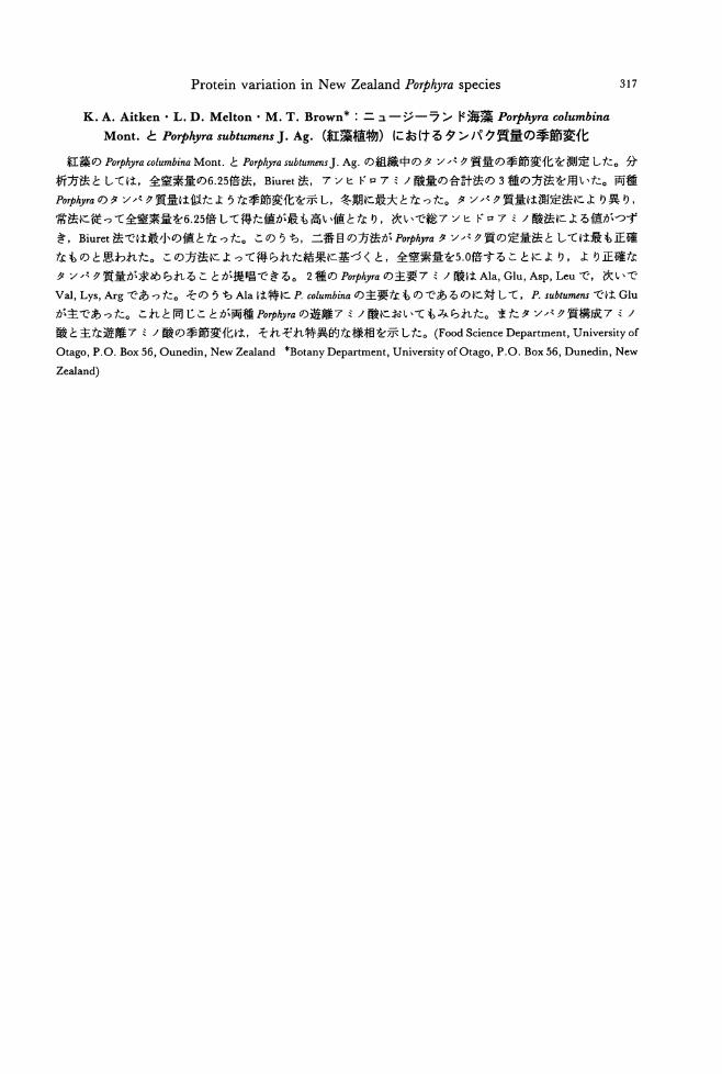
K. A. Aitken • L. D. Melton • M. T. Brown* :ニュージーランド海藻 POψ砂racolumbina
Mont.と POゆ砂rasubtumens J. Ag. (紅藻植物)におけるタンパク質量の季節変化
317
紅藻の POゆ'hyracolumbina Mont.とPOψhyrasubtumens J. Ag.の組織中のタンパタ質量の季節変化を測定した。分
析方法としては,全窒素量の6.25倍法, Biuret法,アンヒドロアミノ酸量の合計法の 3種の方法を用いた。両種
POψめ閣のタンパク質量は似たような季節変化を示し,冬期に最大となった。タンパタ質量は測定法により異り,
常法に従って全窒素量を6.25倍して得た値が最も高い値となり,次いで総アンヒドロアミノ酸法による値がつず
き, Biuret法では最小の値となった。このうち,二番目の方法がPOゅhyraタンパク質の定量法としては最も正確
なものと思われた。この方法によって得られた結果に基づくと,全窒素量を5.0倍することにより,より正確な
タンパタ質量が求められることが提唱できる。 2種の POゆ伊aの主要アミノ酸はA1a,Glu, Asp, Leuで,次いで
Val, Lys, Argであった。そのうちA1aは特に P.columbinaの主要なものであるのに対して,P. subtumensでは Glu
が主であった。これと同じことが両種POゅhyraの遊離アミノ酸においてもみられた。またタンパク質構成アミノ
敵と主な遊離アミノ酸の季節変化は,それぞれ特異的な様相を示した。 (FoodScience Department, University of
Otago, P.O. Box 56, Ounedin, New Zealand *Botany Department, University ofOtago, P.O. Box 56, Dunedin, New
Zealand)

Jpn. J. Phycol. (Sorui) 39: 319-328, December 10, 1991
Critical review of the taxonomy and life history of Kjellman白 arasakii
(Dictyosiphonales, Phaeophyceae)
Hiroshi Kawai
Dψar,伽 entof Botany, Fo出ultyof S.白ience,Hokkaido University, Sapporo, 060 Japan
Kaw必, H. 1991. Critical review of the taxonomy and life history of JVellmania arasakii (Dictyosiphonales,
Phaeophyceae). Jpn. J. Phycol. 39: 319-328.
A taxonomic review of JVellmania arasakii based on the materials newly collected at Yashiro Island, Seto Inl姐 dSea, J apan revealed也at也especies is identical wi也Stic砂市中'lwnsoriferus, which is distributed widely inNo地 AtIanticOcean. At Yashiro Island, Stictyosiplwn soriferus grows on subtidal副 ificialsubstrata at a depth of 3-4 m. The thallus is 3-4 times irregularly branched, delicate, attaining about 20 cm in height and forms abundant plurilocul釘叩or姐 gia. In culture the 抑制nersfrom plurilocular sporangia developed into protonemata on which erect thalli directIy釘 ose.The polystichous, branched erect thalli grew well叩 dformed plurilocular sporangia between 5-20oC, although best between 10 and 150C. Unilocular sporangia were formed only in 50C sho此 dayconditions.
Keyln伽陥rds: Dictyosiphonales一向山naniaarasakii-Life hist町一P加 'ophyc,雌ーStictyo討phonsoriferus-71酎 'onomy.
Yamada (1953) described勾iellmaniaarasakii based on some drift materials collected at
Fukagawa in Tokyo Bay by S. Arasaki during 1951姐 d1952. However, this report was preliminary, lacking Latin descriptions and detailed descriptions of the species. Yamada
mentioned that he intended to publish a sec-
ond report including a detailed description, but it has not been published. Arasaki and
Nozawa (1953) cultured the same material as
Yamada studied and reported the occurrence
of erect filamentous gametophytes from the
swarmers of the field plant; however, they could not complete the life history. They
reported a Laminaria-type of germination of the swarmers in which most cellular contents
migrated into the germination tube forming a
emptied embryospore. However, such a ger-
mination pattern is rather uncommon in the Dictyosiphonales in which the genus is placed, and does not agree with previous reports on the life history of Kjellmania and its closely related genus Stictyosiphon (Sauvageau 1929, Rosenvinge 1935, South and Hooper 1976). The genus符'ellmaniawas e蜘 blishedby
Reinke (1889) based on砂 llmaniasorifera Reinke collected at Kiel, Baltic Sea.
However, the distinction between ~jellmania and Stおりosiphon has been disputed, 叩 d
布'ellmaniais generally now recognized as a
taxonomic synonym of Stたりosip加n(Rosen-vinge 1935, Rosenvinge and Lund 1947, Nay-lor 1958, South and Hooper 1976). On the
other hand, the characters used to distinguish 符'ellmania ar,ω'akii from 符:ellmania sorifera
(=Stictyos糾onsoriferus), such as the manner of branching,組dpresence or absence ofinterca-
1訂 yplurilocular sporangia, seem to be rather variable and need revision. In addition, the life history of K. arasakii is not yet fully clear. Accordingly, this paper aims to reexamine the morphology and life history of K. arasakii using the type specimen and newly collected materials referable to出especies, and to com-pare it with specimens of Atlantic Stictyosiphon sorijもrus,including authentic materials, to clarify its systematic status.
Materials and Methods
Collections of specimens were made at
320 Kawai, H.
"...
匂・
130"
SEAOFJAPAN 喝。
帆O電~S'(弘、3
q
Fukagawa, Tokyo
PACIFIC OCEAN •
100 krn 140"
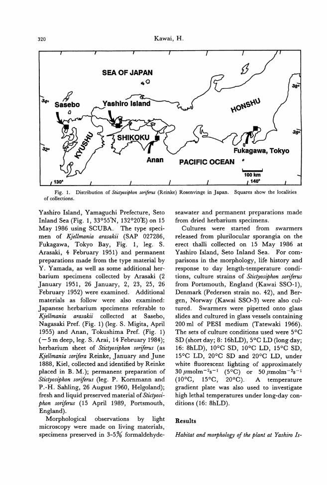
:I6o
Fig. 1. Distribution of Stictyosiphon sorifer山 (Reinke)Rosenvinge in japan. Squares show the localities of collections.
Yashiro lsland, Yamaguchi Prefecture, Seto lnland Sea (Fig. 1, 33055'N, 132020'E) on 15 May 1986 using SCUBA. The type speci-men of Kjellmania arasakii (SAP 027286, Fukagawa, Tokyo Bay, Fig. 1, leg. S. Arasaki, 4 February 1951) and permanent
preparations made from the type material by Y. Yamada, as well as some additional her-barium specimens collected by Arasaki (2
January 1951, 26 Janu町, 2, 23, 25, 26 February 1952) were examined. Additional materials as follow were also examined:
J apanese herbarium specimens referable to Kjellmania arasakii collected at Sasebo, Nagasaki Pref. (Fig. 1) (leg. S. Migita, April 1955) and Anan, Tokushima Pref. (Fig. 1) (-5 m deep, leg. S. Arai, 14 February 1984); herbarium sheet of Stictyosiphon sorijもrus(as
l(jellmania sorijもraReinke, January and June 1888, Kiel, collected and identified by Reinke placed in B. M.); permanent preparation of Stた砂ω似onsorijもrus(leg. P. Kornmann and P.-H. Sahling, 26 August 1960, Helgoland); fresh and liquid preserved material of Stictyosi-phon soriferus (15 April 1989, Portsmouth, England).
Morphological observations by light microscopy were made on living materials, specimens preserved in 3-5% formaldehyde-
seawater and permanent preparations made
from dried herbarium specimens. Cultures were started from swarmers
released from plurilocular sporangia on the erect thalli collected on 15 May 1986 at Yashiro lsland, Seto lnland Sea. For com-parisons in the morphology, life history and response to day length-temperature condi-tions, culture strains of Stictyos似onsorifer町
from Portsmouth, England (Kawai SSO・1), Denmark (Pedersen strain no. 42), and Ber-gen, Norway (Kawai SSO・3)were also cul-tured. Swarmers were pipetted onto glass slides and cultured in glass vessels containing 200 ml of PESI medium (Tatewaki 1966). The sets of culture conditions used were 50C
SD (short day; 8: 16hLD), 50C LD (long day; 16: 8hLD) , lOoC SD, lOoC LD, 150C SD, 150C LD, 200C SD and 200C LD, under white fluorescent lighting of approximately 30μmolm-2s-1 (50C) or 50μmolm-2s-1
(100C, 150C, 200C). A temperature gradient plate was also used to investigate high lethal temperatures under long-day con-ditions (16: 8hLD).
Results
Habitat and morphology of the plant at Yashiro ls-
321 Taxonomy of KJellmania arasakii
50.,.m -10mm
n,ι
器-4
盟国一一官官』留守』督官=せ』長時ロ官四日・-M
ロ
--
a
sB何回守同随時一ヨ一
322 Kawai, H
land and morPhology 01 other Ja戸anesematerials
The plants grow densely on concrete ex-
perimental substrata for algae at a depth of 3-4 m below Mean Low Water Level, together
with Punctaria sp. The erect thallus is 3-4
times irregular1y branched, without an obvi-
ous main axis, pale yellow in color, de!icate and attains 20 cm in height (Fig. 2). In cross
section the thallus consists of four large color-
less central cells surrounded by 1-2 layers of
Taxonomy of K.jellmania arasakii 323
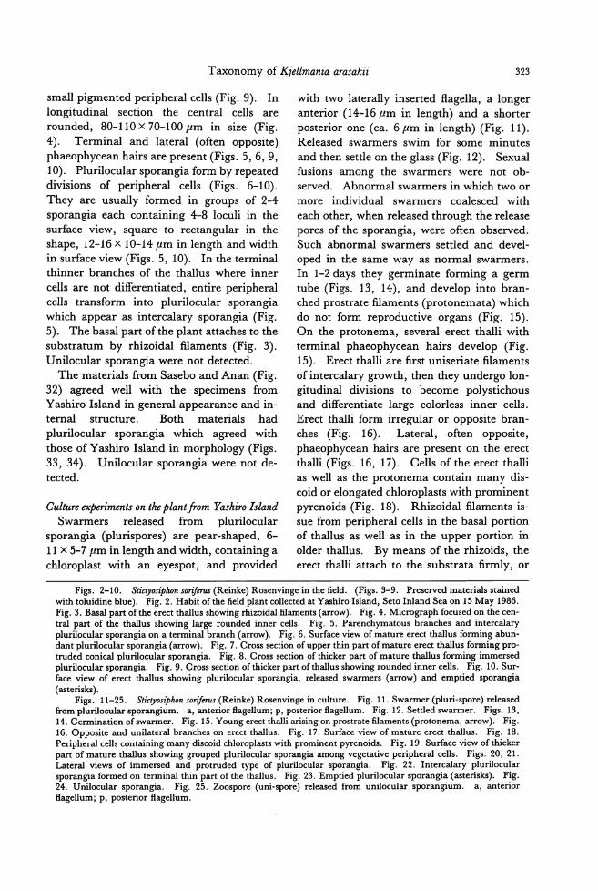
small pigmented peripheral cells (Fig. 9). In
longitudinal section the central cells are
rounded, 80-110 x 70-100μm in size (Fig.
4). Terminal and lateral (often opposite)
phaeophycean hairs are present (Figs. 5,6,9, 10). Plurilocular sporangia form by repeated
divisions of peripheral cells (Figs. 6-10).
They are usually formed in groups of 2-4
sporangia each containing←8 loculi in the
surface view, square to rectangular in the shape, 12-16 x 10-14μm in length and width
in surface view (Figs. 5, 10). In the terminal
thinner branches of the thallus where inner
cells are not differentiated, entire peripheral cells transform into plurilocular sporangia
which appear剖 intercalarysporangia (Fig.
5). The basal part ofthe plant attaches to the
substratum by rhizoidal filaments (Fig. 3).
Unilocular sporangia were not detected.
The materials from Sasebo and Anan (Fig.
32) agreed well with也especimens from
Yashiro Island in general appearance and in-
ternal structure. Both materials had
plurilocular sporangia which agreed wi血
those of Yashiro Island in morphology (Figs. 33, 34). Unilocular sporangia were not de-
tected.
Culture experiments on the plant j示。mYiωhiroIsland Swarmers released from plurilocular
sporangia (plurispores) are pear-shaped, 6-11 x 5-7μm in length and width, containing a
chloroplast with an eyespot, and provided
with two laterally inserted flagella, a longer
anterior (14・16μmin length) and a shorter
posterior one (ca. 6μm in length) (Fig. 11).
Released swarmers swim for some minutes
and then settle on the glass (Fig. 12). Sexual
fusions among the swarmers were not ob-
served. Abnormal swarmers in which two or
more individual swarmers coalesced with
each other, when released through the release pores of the sporangia, were often observed. Such abnormal swarmers settled and devel-
oped in the same way as normal swarmers.
In 1-2 days they germinate forming a germ
tube (Figs. 13, 14), and develop into bran-ched prostrate filaments (protonemata) which
do not form reproductive organs (Fig. 15).
On the protonema, several erect thalli with terminal phaeophycean hairs develop (Fig.
15). Erect thalli are first uniseriate filaments
of intercalary growth, then they undergo lon-gitudinal divisions to become polystichous
and differentiate large colorless inner cells.
Erect thalli form irregular or opposite bran-
ches (Fig. 16). Lateral, often opposite, phaeophycean hairs are present on the erect
thalli (Figs. 16, 17). Cells of the erect thalli
as well as the protonema contain many dis-
coid or elongated chloroplasts with prominent
pyrenoids (Fig. 18). Rhizoidal filaments is-sue from peripheral cells in the basal portion
of thallus as well as in the upper po口ionin
older thallus. By means of the rhizoids, the erect thalli attach to the substrata firmly, or
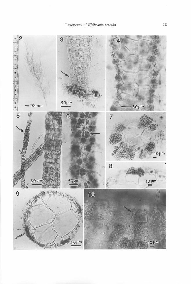
Figs. 2-10. Stiゆ'osiphonsoriferus (Reinke) Rosenvinge in the field. (Figs.3-9. Preserved materials stained with toluidine blue). Fig. 2. Habit ofthe field pl制 coll問 dat Yashiro Island, Seto Inland Sea on 15 May 1986. Fig. 3. Basal part ofthe erect thallus showing rhizoidal filame山 (arrow).Fig. 4. Micrograph focused on血ecen-tral part of the thallus showing large rounded inner cells. Fig. 5. Parenchymatous branches and intercalary pluriloc叫arsporangia on a terminal branch (arrow). Fig. 6. Surface view of mature erect thallus for酒 ingabun-d卸 tplurilocul釘 sporangia(釘TOW). Fig. 7. Cross section of upper thin part of mature erect thallus forming pro-truded conical plurilocular sporangia. Fig. 8. Cross section of thicker part of mature thallus forming immersed plurilocular sporangia. Fig. 9. Cross section of thicker p釘 tofthallus showing rounded inner cells. Fig. 10. Sur-face view of erect thallusぬowingplurilocular sporangia, rele蹴 dsw釘 mers(抑制)叩demptied sporangia (白terisks).
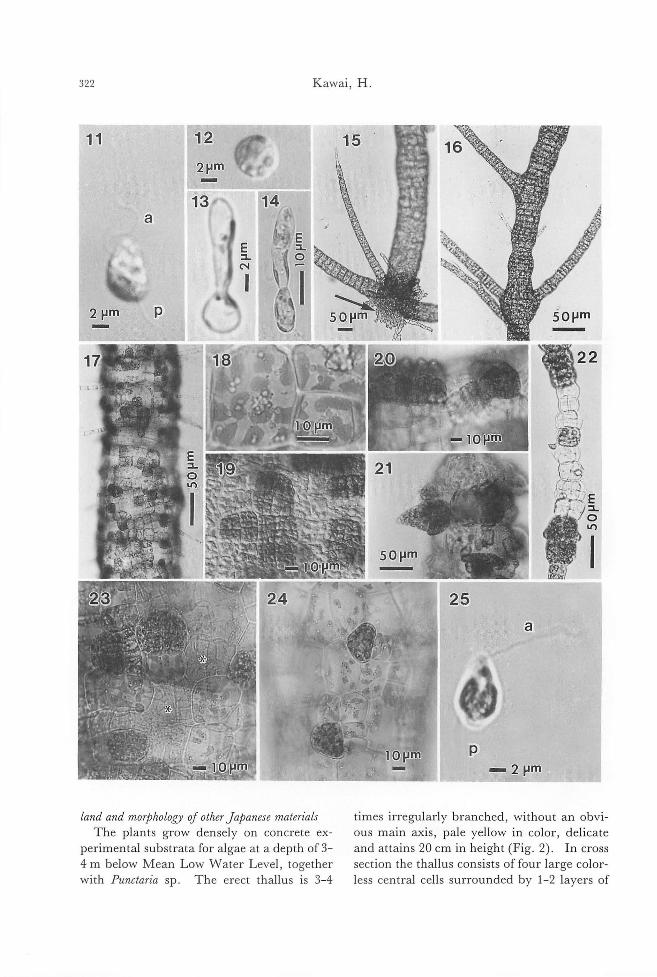
Figs. 11-25. Stictyosiphon sorifi町出 (Reinke)Rosenvinge in culture. Fig. 11. Swarmer (pluri-spore) released 企'omplurilocular sporangium. a, anterior ftagellum; p, posteri町 ftagellum.Fig. 12. Settled swarmer. Figs. 13, 14. Germination ofswarmer. F時i泡g.1β5. Youn時g悶 ctt由h祖叫叫allia叩d町 g 。叩'npros蜘t甘ra蹴t旬e量創la翻men町阻n附21“6. Opposite and unilateral branches on er陀宅匂ctthallus. Fig. 17. Surface view of mature erect thallus. Fig. 18. Peripheral cells containing m阻 ydiscoid chloroplasts wi出 prominentpyrenoids. Fig. 19. Surface view ofthicker part of mature thallus sho泊nggrouped plurilo叫訂 sporangiaamong vegetative peripheral cells. Figs. 20, 21. Lateral views of immersed and protruded type of plurilocular spor阻 gia. Fig. 22. Intercalary pluriloc叫釘
sporangia formed on terminal thin p制 ofthethallus. Fig. 23. Emptied plurilocular sporangia (酪terisks).Fig. 24. Unilocular sporangia. Fig. 25. Zoospore (uni叩 O問)released from unilocular sporangium. a, anterior ftagellum; p, posterior ftagellum.
324 Kawai, H.
entangle with each other.
The erect thalli developed and form巴d
plurilocular sporangia in all culture condi-
tions examined, but they grew faster, and at-tained their maximum length of 15-20 cm in
100C and 150C. In 50C short day conditions
they formed unilocular sporangia, sometimes
mixed with plurilocular ones. Plurilocular
sporangia are formed by repeated divisions of
the peripheral cells. The shapes of them are
rather variable depending on their position on
the thallus, age and culture conditions (Figs. 17, 19-23). Almost ftat plurilocular sporan-
gia as well as protruded ectocarpoid ones are
26 z
R
I!{
,、‘
., E r~
。
observed. In the terminal part of the thallus
where inner cells do not develop, the whole
surface of the thallus transforms into
plurilocular sporangia, which appear as inter-
calary sporangia (Fig. 22). Unilocular
sporangia are conical or irregularly spherical, sessile, formed from the peripheral cells, 28-30 x 20-28μm in length and width (Fig. 24)
Zoospores (uni-spores) released from unilocu-
lar sporangia are about 7 x 5μm in length
and width provided with two, longer ant巴nor
and shorter posterior, ftagella (Fig. 25)
Swarmers from plurilocular sporangia as well
as zoospores from unilocular sporangia germi-
',28
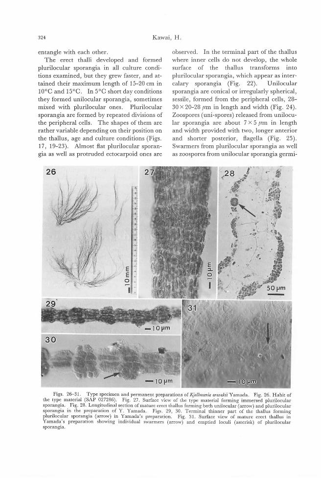
Figs. 26-31. Type specimen and permanent preparations of KJeilmania arasakii Yamada. Fig. 26. Habit of the type material (SAP 027286). Fig. 27. Surface view of the type material forming immersed plurilocular sporangia. . Fig. 28. Longitudina! section of mature erect thallus forming both unilocular-(arrow) and plurilocular sporangia in the preparation of Y. Yamada. Figs. 29, 30. Terminal thinner part of the thallus forming plurilocular sporangia (arrow) in Yamada's preparation. Fig. 31. Surface view of mature erect thallus in Yamada's preparation showing individual swarmers (arrowY and emptied loculi (asterisk) of plurilocular sporangJa.
Taxonomy of勾'ellmaniaarasakii 325
32
ー・lOmm
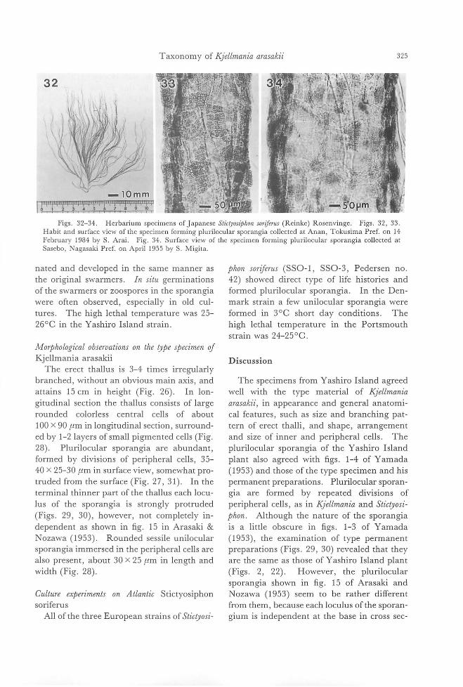
rr寸ーで:77?と1つつ~γFigs. 32-34. Herbarium specimens of japanese Sticりosψh01lsoriんrus(Reinke) Rosenvinge. Figs. 32, 33.
Habit and surface view of the specimen forming plurilocular sporangia collected at Anan, Tokusima Pref. on 14 February 1984 by S. Arai. Fig. 34. Surface view of the specimen forming plurilocular sporangia collected at Sasebo, Nagasaki Pref. on April 1955 by S. Migita.
nated and developed in the same manner as
the original swarmers. In situ germinations
of the swarmers or zoospores in the sporangia
were often observed, especially in old cul-
tures. The high lethal temperature was 25-
260C in the Yashiro Island strain.
Morthological observations on the tyte stecimen 01
Kjellmania arasakii
Th巴 erectthallus is 3-4 times irregularly
branched, without an obvious main axis, and attains 15 cm in height (Fig. 26). In lon
gitudinal section the thallus consists of large
rounded colorless central cells of about
100 x 90μm in longitudinal section, surround-ed by 1-2 la yers of small p甲田nted cells (Fig
28). Plurilocular sporangia are abundant,
formed by divisions of peripheral cells, 35-40 x 25-30μm in surface view, somewhat pro-truded from the surface (Fig. 27, 31). In the
terminal thinner part of th巴 thalluseach locu-
lus of the sporangia is strongly protruded
(Figs. 29, 30), however, not completely in-
dependent as shown in fig. 15 in Arasaki &
Nozawa (1953). Rounded sessile unilocular
sporangia immersed in the peripheral cells are
also present, about 30 x 25μm in length and
width (Fig. 28)ー
Culture exteriments on Atlantic Stictyosiphon
soriferus
All of the three European strains of Stictyosi-
thon sor許rus(SSO-l, SSO-3, Pedersen no 42) showed direct type of life histories and
formed plurilocular sporangia. In the Den-
mark strain a few unilocular sporangia were
formed in 30C short day conditions. The
high lethal temperature in the Portsmouth
strain was 24-250C.
Discussion
The specimens from Yashiro Island agreed
well with the type material of勾ellmania
arasakii, in appearance and general anatomi-cal features, such as size and branching pat-tern of erect thalli, and shape, arrangement
and size of inner and peripheral cells. The
plurilocular sporangia of the Yashiro Island
plant also agreed with figs. 1-4 of Yamada
(1953) and those of the type specimen and his
permanent preparations. Plurilocular sporan-
gia are formed by repeated divisions of
peripheral cells, as in時iellmaniaand SticりOSI-
thon. Although the nature of the sporangia
is a little obscure in figs. 1-3 of Yamada
(1953), the examination of type permanent
preparations (Figs. 29, 30) revealed that they
are the same as those of Yashiro Island plan t
(Figs. 2, 22). However, the plurilocular
sporangia shown in fig. 15 of Arasaki and
Nozawa (1953) seem to be rather different
from them, because each loculus ofthe sporan-gium is independent at the base in cross sec-
326 Kawai, H.
tion. Such kinds of plurilocular sporangia
are reminiscent of those of Coelocladia arctica Rosenvinge and Litosiphon subωntinuω(Rosen-vinge) Lund (Rosenvinge 1898, Pedersen 1976). Naylor (1958) reviewed the genus Stた砂ωz-
phon and its four European species (e.g.在 tor-tilis (Ruprecht) Reinke, S. adriaticωKutzing, S. sorijもrus,and S. griffithsianus (Le J olis) Hol-mes et Batters). According to her, S. adriati-ωand広 soriferushave rounded inner cells in
longitudinal sections, while S. tortilis and S. gr拶thsianus have longer on低 Further-more,志村mtsis distinguished from 丘adriaticωin having a solid thallus and more uniformly sized and arranged inner cells. In Europe the former has a more northern distri-bution than the latter. Compared with these species, KJellmania arasakii agrees well with Stic-抑糾onsoゆ即 inits anatomical features.
Yamada (1953) distinguished JVellmania sorijもraand K. arasakii in the manner of branching and absence of intercalary sporan-gia in the latter. However, the differences in the branching manner are apparently within the range of variations caused by age and growing conditions. As mentioned above, in this species the intercalary sporangia are es-sentially the same as those formed on the sur-face of the thicker thallus. Furthermore, since the specimens Yamada (1953) examined
were all dri丘materials,terminal thinner ma-ture portions could have been already lost. Detailed comparisons with the herbarium specimens and live or liquid preserved materi-
als of European specimens of Sti,ゆos紗,honsor排rus( cited in Materials and Methods) Wl出 thoseof砂 llmaniaarasakii in the present study also showed no distinctive differences between the two taxa.
In culture, the plants from Yashiro lsland showed a direct type of life history and formed unilocular and plurilocular sporangia. This life history pattern agreed well with the cul-ture results of Rosenvinge (1935) and South
and Hooper (1976) on St均'os桝onsorifi仰 s,as well as the results on the Atlantic culture strains in the present study. Although unilocular sporangia were not reported in the
culture of South and Hooper (1976), this difference may be due to the limited day
length conditions (12: 12hLD) examined in their experiments. The fact that the field material of符'ellmaniaarasakii collected at
Fukagawa in Tokyo Bay on February had some unilocular sporangia agree with the present culture results in which unilocular
sporangia were formed in lower temperature (50C) short day conditions corresponding to winter at Tokyo Bay.
On the contrary, the culture results of Arasaki and Nozawa (1953) differ from those in the previous reports and present work on S. s吋ωinthe following points; 1) Laminaria-type of germination in which most cellular contents migrate into germ tube and the em-bryospores become almost empty; 2) the出alli
derived from swarmers developed into erect (gametophytic) 創aments and formed clustered plurilocul町 sporangiaresembling
those of Botヮtella(= Soroca7抑);3) swarmers from the plurilocular sporangia were sup-
posed to be gametes, (actual copulations were not observed but presumptive zygotes with two stigmata were found) and the presump-tive zygotes developed into parenchymatous
sporophytes. With regard to 1), Laminaria-type (mediate filamentous type) germination is rather rare in the order Dictyosiphonales and not reported in Stictyosiphon. The rare known exception in the order is the case in Coe-locladia arctica (Pedersen 1976). About 2), as Arasaki and Nozawa (1953) also mentioned, such plurilocular sporangia differ from those known in Stictyωiphon, and are rather similar to those of Coelocladia arctica. Concerning 3), since they did not observe actual copulations
nor complete the life cycle, their explanation of the erect filaments derived from the origi-nal swarmer as gametophytes is question-able. In fact, the presumptive gametophytic filament illustrated in fig. 7 of Arasaki姐 d
Nozawa (1953) has some longitudinal walls, resembling those in the sporophytic thallus in their fig. 13. Settled swarmers provided with two (or more) stigmata can also be caused by abnormal coalescences of the swarmers as observed in the present culture. This
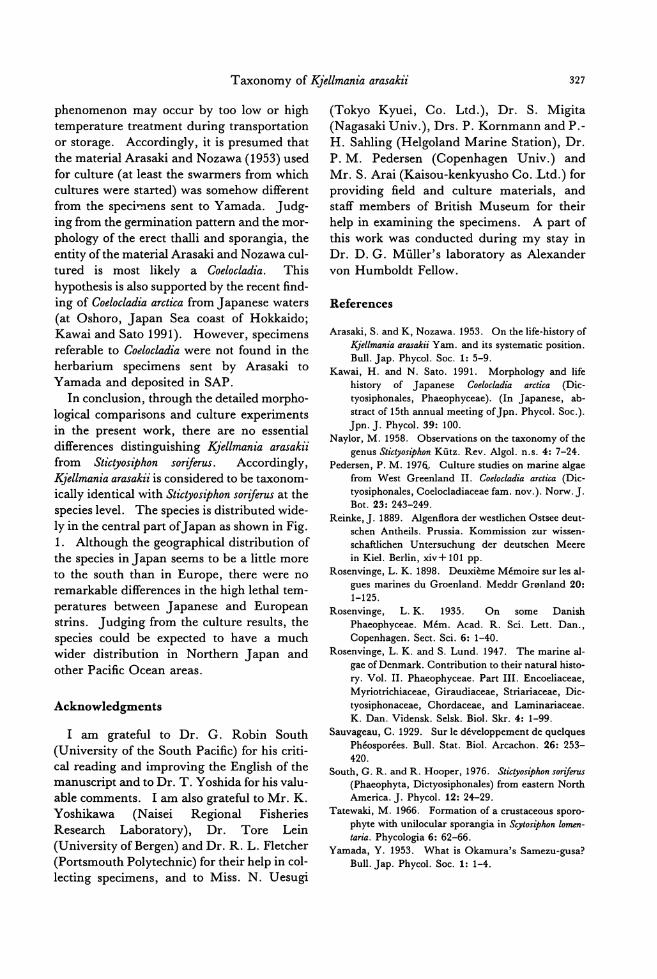
Taxonomy of勾iellmaniaarasakii 327
phenomenon may occur by too low or high
temperature treatment during transportation
or storage. Accordingly, it is presumed that the material Arasaki and Nozawa (1953) used for culture (at least the swarmers from which
cultures were started) was somehow different
from the speciηens sent to Yamada. J udg-
ing from the germination pattern and the mor-
phology of the erect thalli and sporangia, the entity ofthe material Arasaki and Nozawa cul-
tured is most likely a Coelocladia. This
hypothesis is also supported by the recent find-
ing of Coelocladia arctica from J apanese waters
(at Oshoro, Japan Sea coast of Hokkaido; Kawai and Sato 1991). However, specimens referable to Codocladia were not found in the
herbarium specimens sent by Arasaki to
Yamada and deposited in SAP.
In conclusion, through the detai!ed mo中ho・
logical comparisons and culture experiments
in the present work, there are no essential differences distinguishing K.fellmania arasakii from Stたりosiphon soriferus. Accordingly, K.fellmania arasakii is considered to be taxonom-ically identical with Stたりosiphonsoriferωatthe
species level. The species is distributed wide-
ly in the central pa口ofJapanas shown in Fig.
1. Although the geographical distribution of the species in J apan seems to be a little more
to the south than in Europe, there were no remarkable differences in the high lethal tem-
peratures between J apanese and European
strins. Judging from the culture results, the species could be expected to have a much
wider distribution in Northern J apan and
other Pacific Ocean areas.
Acknowledgments
1 am grateful to Dr. G. Robin South
(University of the South Pacific) for his criti-
cal reading and improving the English of the
manuscript and to Dr. T. Yoshida for his valu-
able comments. 1 am also grateful to Mr. K.
Y oshikawa (N aisei Regional Fisheries
Research Laboratory), Dr. Tore Lein
(University ofBergen) and Dr. R. L. Fletcher (Portsmouth Polytechnic) for their help in col司
lecting specimens, and to Miss. N. Uesugi
(Tokyo Kyuei, Co. Ltd.), Dr. S. Migita
(Nagasaki Univ.), Drs. P. Kornmann and P.-H. Sahling (Helgoland Marine Station), Dr. P. M. Pedersen (Copenhagen Univ.) and
Mr. S. Arai (Kaisou-kenkyusho Co. Ltd.) for
providing field and culture materials, and staff members of British Museum for their
help in examining the specimens. A part of
this work was conducted during my stay in
Dr. D. G. Muller's laboratory as Alexander
von Humboldt Fellow.
References
Arasaki, S. and K, Nozawa. 1953. On the life-history of Kjellmania arasakii Yam. and its systematic position. Bull. Jap. Phycol. Soc. 1: 5-9.
Kawai, H.釦 dN. Sato. 1991. Morphology and life history of J apanese Coelocladia arctica (Dic-tyosiphonales, Phaeophyceae). (In Japanese, ab-stract of 15th annual meeting of Jpn. Phycol. Soc.). Jpn. J. Phycol. 39: 100
Naylor, M. 1958. Observations on the taxonomy ofthe genus Stictyosiphon Kutz. Rev. Algol. n.s. 4: 7-24
Pedersen, P. M. 1976,. Culture studies on marine algae 仕omWest Greenland 11. Coelocladia a町tica(Dic-tyosiphonales, Coelocladiaceae fam. nov.). Norw. J Bot. 23: 243-249
Reinke, J. 1889. Algensora der westlichen Ostsee deut-schen Antheils. Prussia. Kommission zur wissen-schaftlichen Untersuchung der deutschen Meere in Kiel. Berlin, xiv+ 101 pp
Rosenvinge, L. K. 1898. DeuxiきmeMemoire sur les al-gues marines du Groenland. Meddr Gronland 20: 1-125.
Rosenvinge, L. K. 1935. On some Danish Phaeophyceae. Mem. Acad. R. Sci. Lett. Dan., Copenhagen. Sect. Sci. 6: 1-40
Rosenvinge, L. K.釦 dS. Lund. 1947. The marine al-gae of Denmark. Contribution to their natural histo-ry. Vol. II. Phaeophyceae. Part II1. Encoeliaceae, Myriotrichiaceae, Giraudiaceae, Striariaceae, Dic-tyosiphonaceae, Chordaceae, and Laminariaceae K.D佃 .Vidensk. Selsk. Biol. Skr. 4: 1-99.
Sauvageau, C. 1929. Sur le developpement de quelques Pheosporees. Bull. Stat目 Biol.Arcachon. 26: 253-420目
South, G. R. and R. Hooper, 1976. Stictyosiphon sori伽山
(Phaeophyta, Dictyosiphonales) from eastern North America. J. Phycol. 12: 24-29.
Tatewaki, M. 1966. Formation of a crustaceous sporo・phyte wi也 unilocularsporangia in Scytosiphon lomen-taria. Phycologia 6: 62-66.
Yamada, Y. 1953. What is Okamura's Samezu-gusa? Bull. Jap. Phycol. Soc. 1: 1-4.
328 Kawai, H.
川井浩史:褐藻サメズゲサ (K.jellmaniaarasakiz~ ウイキョウモ目)の分類と生活史の再検討
褐藻サメズグサの分類について瀬戸内海・屋代島において新たに採集した材料などにもとづき再検討した結
果,本種は北大西洋に広く生育する Stictyos自bhonsoriρ聞と同一種であると結論した。本種は屋代島においては水
深 3-4mの亜潮間帯の人工的な基物の上に生育しており,藻体は高さ 20cmに達し.3-4回不規則に枝分かれし,
柔らかく,表面に複子嚢を生ずる。培養下では複子獲に由来する遊走細胞はプロトネマに発達し,その上に直緩
直立藻体を形成した。直立藻体は多列形成的となり,分校しながらよく成長し 5-200Cで複子獲を生じたが,
100Cと150Cで特によく成長した。単子漢は 50C短目条件でのみ形成された。 (060札幌市北区北10条西8丁目
北海道大学理学部植物学教室)
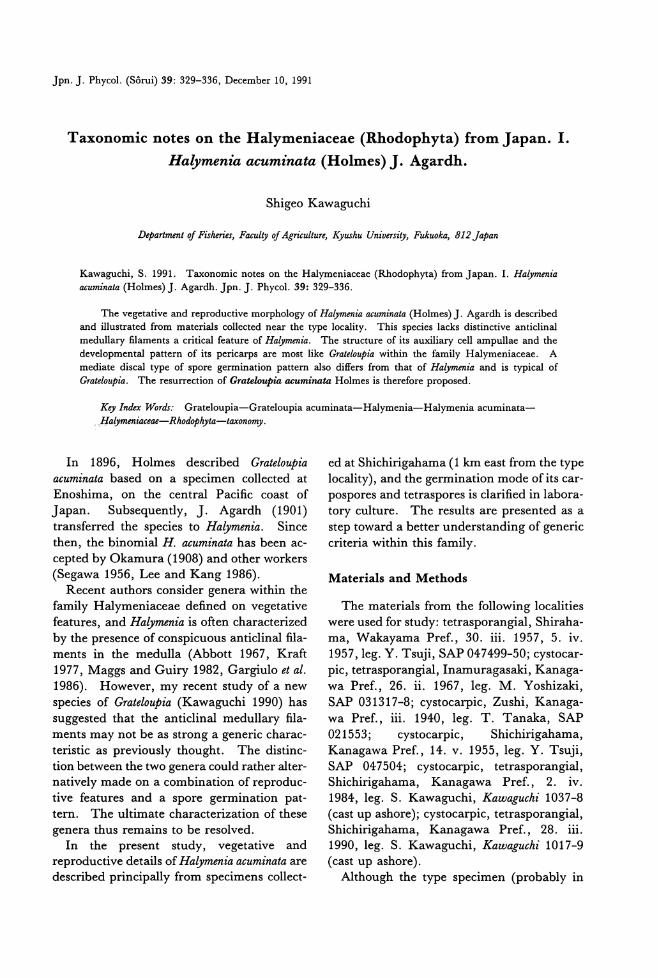
Jpn. J. Phycol. (Sorui) 39: 329-336, Decemher 10, 1991
Taxonomic notes on the Halymeniaceae (Rhodophyta) from Japan. I.
Halymenia acum
Shigeo Kawaguchi
Dφartment of Fi.市 n・'es, F aculty of Agriculture,母国huUniversity, Fukuoka, 812 Japan
Kawag吋lI, S. 1991. Taxonomic notes on出eHalymeniaceae (Rhodophyta) from Japan. 1. Ha砂抑制acuminata (Holmes) J. Agardh. Jpn. J. Phycol. 39: 329-336
The vegetative and reproductive morphology of Ha砂開niaacuminata (Holmes) J. Agardh is described and iIIustrated from materials collected near the type locality. This species lacks distinctive anticlinal medullary filaments a critical feature of R仲間enia. The structure of its auxiliary cell ampullae and出edevelopmental pattern of its pericarps are most like Grateloupia within the family Halymeniaceae. A mediate discal type of spore germination pattern a1so differs from伽 tof Halymenia印 dis typical of Grateloupia. The resurrection of Grateloupia acumi;間 taHolmes is therefore proposed.
KりlndexWora台・ Grateloupia-Grateloupia acuminata一Halymenia-Halymenia acuminata-. Halymeniaceae-Rhodophyta-taxono均P
In 1896, Holmes described GratelouPia
acuminata based on a specimen collected at
Enoshima, on the central Pacific coast of
]apan. Subsequently,]. Agardh (1901) transferred the species to Halymenia. Since
then, the binomial H. acuminata has been ac-cepted by Okamura (1908) and other workers (Segawa 1956, Lee and Kang 1986).
Recent authors consider genera within the
family Halymeniaceae defined on vegetative
features, and Halymenia is often characterized by the presence of conspicuous anticlinal臼a-
ments in the medulla (Abbott 1967, Kraft 1977, Maggs and Guiry 1982, Gargiulo et al. 1986). However, my recent study of a new
species of GratelouPia (Kawaguchi 1990) has suggested that the anticlinal medullary fila-
ments may not be as strong a generic charac-
teristic as previously thought. The distinc-
tion between the two genera could rather alter-
natively made on a combination of reproduc圃
tive features and a spore germination pat-
tern. The ultimate characterization of these
genera thus remains to be resolved.
In the present study, vegetative and
reproductive details of Halymenia acuminata are
described principally from specimens collect-
ed at Shichirigahama (1 km east from the type locality), and the germination mode ofits car-pospores and tetraspores is clarified in labora-
tory culture. The results are presented as a
step toward a better understanding of generic
criteria within this family.
Materials and Methods
The materials from the following localities
were used for study: tetrasporangial, Shiraha-ma, Wakayama Pref., 30. iii. 1957, 5. iv.
1957, leg. Y. Tsuji, SAP 047499-50; cystocar-pic, tetrasporangial, Inamuragasaki, Kanaga-wa Pref., 26. ii. 1967, leg. M. Yoshizaki, SAP 031317-8; cystocarpic, Zushi, Kanaga-wa Pref., iii. 1940, leg. T. Tanaka, SAP
021553; cystocarpic, Shichirigahama, Kanagawa Pref., 14. v. 1955, leg. Y. Ts吋i,SAP 047504; cystocarpic, tetrasporangial, Shichirigahama, Kanagawa Pref. , 2. iv.
1984, leg. S. Kawaguchi, Kawaguchi 1037-8 (cast up ashore); cystoca叩ic,tetrasporangial, Shichirigahama, Kanagawa Pref., 28. iii.
1990, leg. S. Kawaguchi, Kawaguchi 1017-9 (cast up ashore).
Although the type specimen (probably in
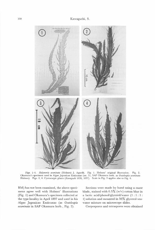
330 Kawaguchi, S
1,."....Soc.JOUI!"U'IT.Vgt.,瑚品加
の ①
①
F伊 . 1凶4. H.品alymer.山 a仰日印仰4ρLmOkamur日a'、ss叩pec口imenused in Algae J aponicae Exsiccatae (いno.3引1,SAP Oka町mur日aherb. a出sGr.悶ateωu戸悶 a叩C日u山mη問7
Holmesの). Figs. 3, 4. Cystocarpic plants (Kau勾 uchi1038, 1037). Scale in Fig. 3 applies also to Fig. 4
B恥1)has not been examined, the above speci-mens agree well with Holmes' illustrations
(Fig. 1) and Okamura's specimen col!ected at
the type locality in April 1897 and used in his
Algae J aponicae Exsiccatae (as Grateloupia
acuminata in SAP Okamura herb., Fig. 2)
S巴ctionswere made by hand using a razor
blade, stained with 0.5% (w/v) cotton blue in
a lactic acid/phenol/glycerol/water (1 : 1 : 1 :
1) solution and mounted in 50% glycerol-sea-
water mixture on microscope slides.
Carpospores and tetraspores were obtained
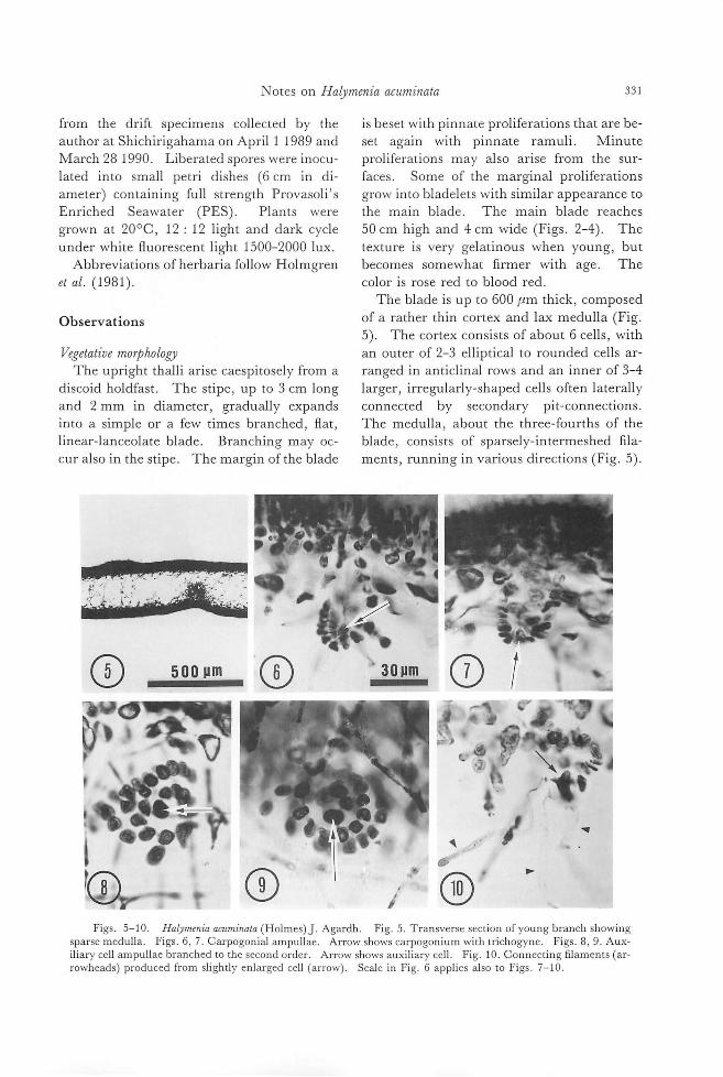
Not巴son Haの砂抑men仰 1ωaaCU77け叩7 331
from the drift specimens collected by the
author at Shichirigahama on April 1 1989 and
March 281990. Liberated spores were inocu-
lated into small petri dishes (6 cm in di-
ameter) containing full strength Provaso!i's
Enriched Seawater (PES)ー Plants were
grown at 20oC, 12: 12 light and dark cycle
under white fluorescent !ight 1500-2000 lux
Abbreviations of herbaria follow Holmgren
et al. (1981).
Observations
Vegetative moψhology
The upright thal!i arise caespitosely from a
discoid holdfast. The stipe, up to 3 cm long
and 2 mm in diameter, gradually expands into a simple or a few times branched, flat, !in巴ar-lanceolateblade. Branching may oc-
cur also in the stipe. The margin ofth巴blade
①
is beset with pinnate pro!iferations that are be-
set again with pinnate ramu!i. Minute
pro!iferations may also arise from the sur-
faces. Some of th巴 marginalpro!iferations
grow into bladelets with similar appearance to
the main blade. The main blade reaches
50 cm high and 4 cm wide (Figs. 2-4). The
texture is very gelatinous when young, but becomes somewhat firmer with age. The
color is rose red to blood red
The blade is up to 600μm thick, composed
of a rather thin cortex and lax medulla (Fig
5). The cortex consists of about 6 cells, with an outer of 2-3 elliptical to rounded cells ar-
ranged in anticlinal rows and an inner of 3-4
larg巴r,irregularly-shaped cells 0仕enlaterally
connected by s巴condary pit-connections
The medulla, about the thre巴ーfourthsof th巴
blade, consists of sparsely-intermeshed fila-
ments, running in various directions (Fig. 5).
司.
J ① ~
Figs. 5ー10. Halymenia acuminala (Holmes) J. Agardh. Fig. 5. Transverse section of young branch showing sparse medulla. Figs. 6, 7. Carpogonial ampullae. Arrow shows carpogonium with trichogyne. Figs. 8, 9. Aux-iliary cell ampullae branched to the second order. Arrow shows auxil町 ycell. Fig. 10. Connecting filaments (ar-rowheads) produced from slightly enlarged cell (arrow). Scale in Fig. 6 applies also to Figs. 7ー10
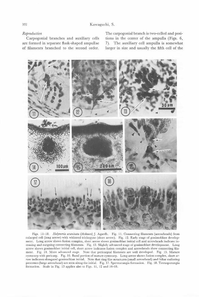
332 Kawaguchi, S
Reproduction
Carpogonial branches and auxi1iary cells are formed in separate flask-shaped ampullae
of filaments branched to the second order.
i①
①
The carpogonial branch is two-celled and posi-
tions in the center of the ampulla (Figs. 6,
7). The auxil即 ycell ampulla is somewhat
larger in size and usually th巴自fthcell of the
Figs. 11-18. Halymenia acuminata (Holmes) J. Agardh. Fig. 11. Con町r問 ctmg 白削lame町en川】t臼s(伊ar汀rowhe児eads吟)from enlarged cell (long a町rrQW吋)w凶 W削 lt出he町re吋dt凶richogyne(short a町rrow吋). Fig. 12. Early stage of gonimoblast develop-ment. Long arrow shows fusion complex, short arrow shows gonimoblast initial cell and arrowheads indicate in-coming and outgoing connecting filaments. Fig. 13. Slightly advanced stage of gonimoblast development. Long arrow shows gonimoblast initial cell, short arrow indicates fusion complex and arrowheads show connecting fila ment. Fig. 14. More advanced stage. Note that pericarpial自lamentsare well developed. Fig. 15. Mature cystocarp with pericarp. Fig. 16. Basal portion ofmature cystocarp. Long arrow shows fusion complex, short ar-row indicates elongated gonimoblast initial. Note that ring-like structures (small arrowhead) and foliar radiating processes (large arrowhead) are seen along the init凶 Fig.17. Spermatangia formation. Fig. 18. Tetrasporangia 五ormation. Scale in Fig. 13 applies also to Figs. 11, 12 and 16-18
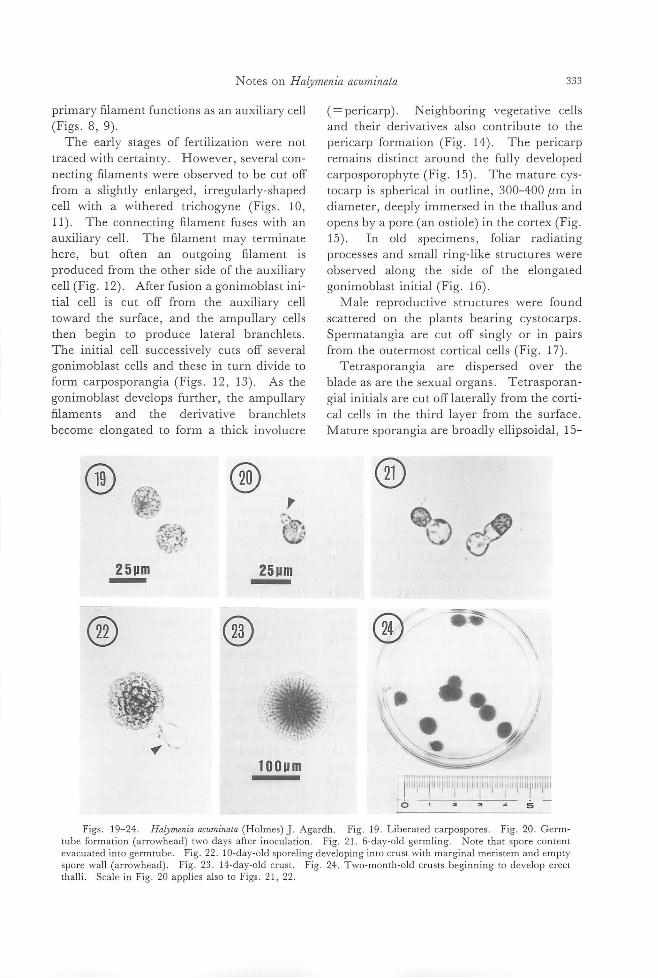
Notes on Halymenia acuminala
primary filament functions as an auxiliary cell
(Figs. 8, 9) The early stages of fertilization were not
traced with certainty. However, several con-necting filam巴ntswere observ巴dto be cut off
from a slightly巴nlarged,irregularly寸1aped
cell with a withered trichogyne (Figs. 10, 11). The conn巴cti時五lamentfuses with an
auxiliary cell. The filament may terminate
here, but often an outgoing filament is
produced from the other side of the auxiliary
cell (Fig. 12). After fusion a gonimoblast ini-
tial cell is cut off from the auxiliary cell
toward the surface, and the ampullary cells
then begin to produce lateral branchlets
The initial cell successiv巴lycuts off several
gonimoblast cells and these in turn divide to
form carposporangia (Figs. 12, 13). As the
gonimoblast d巴velopsfurther, the ampullary
filaments and the derivative branchlets
become elongated to form a thick involucre
① ⑪ 4M守 P
除、
G4・...'-';;"
ijgJ立:25um - 25um -
② ①
J会~~.与え 鞠芭正1
~
100um
333
(= pericarp). N eighbori月 vegetativecells
and their derivatives also contribute to the
pericarp formation (Fig. 14). The pericarp
remains distinct around the fully developed
carposporophyte (Fig. 15). The matur巴 cys-
tocarp is spherical in outlin巴, 300-400μm III
diameter, deeply immers巴din the thallus and
opens by a pore (an ostiole) in th巴 cortex(Fig.
15). In old specimens, foliar radiating
processes and small ring-like structures were
observed along the side of the elongated
gonimoblast initial (Fig. 16)
恥1alereproductive structur巴swere found
scattered on the plants bearing cystocarps
Spermatangia are cut off singly or in pairs
from the outermost cortical cells (Fig. 17)
Tetrasporangia are dispersed over the
blade as are the sexual organs. Tetrasporan-
gial initials are cut offlaterally from the corti-
cal cells in the third layer from the surface
Mature sporangia are broadly ellipsoidal, 15-
①
bグ
①
山
γ1・5
しい』
E'一a
HH11』
t
一3
,|『
t
!
E
f-
-一mi-
-
-
1
μドl-
。Figs. 19-24. Haかmeniaacuminata (Holmes) J. Agardh. Fig. 19. Liberated carpospores. Fig. 20. Germ
tube formation (arrowhead) two days after in】oc印ulatio叩n. F日ig.21. 6ι-day-old g伊errr目Y山evacuated in to ge町r口rm】tt凶ube. F抗ig.2η2. 10-day一olds叩po町relingde肝velop戸mgmto c口rus坑tw削 lt出hma訂rg伊m】u叫alrπm】e町n目st臼e町mand empty y
spore wall (arrowhead). Fig. 23. 14-day-old crust. Fig. 24. Two-month-old crusts beginning to develop erect thalli. Scale in Fig. 20 applies also to Figs. 21, 22
334 Kawa思lchi,S.
18μm wide by 33-40μm long, submerged in 也eco口ex,組dcruciately or decussately divid-ed (Fig. 18).
Development 01 spores Liberated carpospores are 18-22μm in di-
ameter (Fig. 19). One or two days after制・
tling, the spore developed a germ tube (Fig.
20) into which the spore content migrated (Fig. 21). A septum was then formed, leav-ing only the original spore wall behind. Ir-regular divisions of the germ tube cell gave rise to a multicellular, discoid sporeling (Fig. 22). The disc grew by divisions ofthe margin-al meristem and after one week reached 50μm in diameter. In two weeks, the disc reached 100μm in diameter (Fig. 23), and in five months, 3-5 mm (Fig. 24). Upright thal-li arose from near the center of the disc after two months. Tetraspores developed in a simi-lar manner to carpospores. The developed discs reached 2 mm in diameter in two
months and gave rise to upright thalli two weeks later.
Discussion
The genus Halyme仰 isoften characterized
by a single vegetative feature, I.e., the occur-rence of conspicuous anticlinal filaments in the medulla (Kra氏 1977,Maggs and Guiry 1982, Gargiulo, et al. 1986). In H. acuminata, the anticlinal medullary filaments are detecta-
ble, but dispersed among obliquely or pericli-nally directed filaments and are not as con-
spicuous as those reported in the type species H. .floresia (Clemente) C. Agardh (Codomier 1974, p. 27, Fig. 4). While all known species of Halymenia appear to po悶 ssdistinctive組-
ticlinal medullary filaments, the presence of such filaments are not exclusive to this ge-nus. 1 have recently repo口edtheir presence in GratelouPia kurogii (Kawaguchi 1990), as did Scott et al. (1982) for C.ヮIttonemia加llymenioidesfrom Australia, although they were not present as regularly nor predominantly as is usual in the genus Ha砂menza.
Chiang (1970) proposed that the mo中holo-gy of auxiliary cell ampullae is generically di-
agnostic within this family. The ampulla of
Halymenia acuminata is sparingly branched up to the second order and conical in outline. This type of ampulla corresponds well with the Grateloupia-type in Chiang's scheme, and in contrast to the Halymenia-type which is pro-fusely branched to the fourth order and tends
to be wide across the top (Chiang 1970). Although Kraft (1977) and Guiry and Maggs (1982) have shown that the ampullar types are
not invariably a consistent generic characteris-tic, the present alga is allied more to the genus Grateloupia than to Halymenia in this regard. The similarity of this alga to Grateloψza IS
also found in its comparatively thick pericarp constructed not only of ampullary filaments
but also ofneighboringvegetative cells. This differs to Halymenia where the pericarp is reported to be derived only from ampullarγ 日aments(Balakrishnan 1961, Chiang 1970, Maggs and Guiry 1982, Gargiulo et al. 1叩986町).
The most decisive feature tωo s関epa紅ra幻.teH. ac四仰uωt1叩7Cφira仰u帥lゐouψp仰z'ais a sp卯or児egermination pa幻紺t抗t句ern. In Halymenia, spores initially develop into bran-ched uniseriate filaments (Hoek and Cortel・
Breeman 1970, Codomier 1974, Maggs and Guiry 1982), whereas in Grateloupz'a spores
give rise to discoid sporelings as is the case with those of H. acuminata (Chemin 1937, Inoh 1947, Hayashida and Chihara 1967, Kawaguchi 1990). This difference is consis-tent between the two genera.
1 have discussed in my previous paper (Kawaguchi 1990) that the genus Grateloupia could well be distinguished from Halymenia on a combination of a spore germination pattern and certain reproductive features rather than
solely on vegetative features as the presence of anticlinal medullary filaments. The discus-sion applies equally well in this case with the present alga best allied to Grateloupia. The resemblance of the alga in texture or color to
Halymenia, which had been foreshadowed by Holmes (1896, p. 254), is considered superfi-cial. Grateloupia turuturu Yamada is a good ex-ample that supports this consideration. G.
turuturu has lubricous texture and bright red color, but its reproductive features and spore
Notes on Haly仰 niaacuminata 335
germination pattern are typical of GratelouPia
(Kawabata 1962, Hayashida and Chihara 1967).
On these grounds, 1 conclude that H. acuminata does not belong in the genus
Hゆmeniabut in GratelouPia. 1 therefore pro-
pose the resurrection ofthe following binomi-
al:
Grateloupia acuminata Holmes, Linn. Soc. J. Bot. 31: 254, fig. 2a-c. 1896.
Synonym: Halymenia acuminata (Holmes)J.
Agardh, Sp. Alg. 111(4): 130-131. 1901.
Acknowledgments
This study was undertaken mainly in the
laboratory of Department of Botany, Faculty of Science, Hokkaido University. 1 wish to
express my special thanks to Professor T.
Yoshida, Hokkaido University, for his con-tinual support and valuable comments on the
manuscript. 1 also wish to thank Professor
Y. M. Chiang, Taiwan National University, for critically reading the manuscript and in-
structive suggestions. My thanks are extend-
ed to Mr.J. A. Lewis, The University ofMel-
bourne, for his assistance in improving the manuscript, to Mr. K. Kogame, Hokkaido University, for providing me the photograph
of Okamura's specimen and finally to the
anonymous reviewers for critical comments
on the manuscript.
References
Abbott, 1. A. 1967. Studies in some red algae of the
Pacific coast. 1. Cryptonemiaceae. ]. Phycol. 3:
139-149. Ag町 dh,]. G. 1901. Species genera et ordines al-
garum,...III (4). Lund Balakrishnan, M. S. 1961. Studies on Indian Crγp-
tonemiales. 111. Halymenia C. A. Ag. J. Madras
Univ. 31B: 183-217. Chemin, E. 1937. Le developpement des spores chez les
Rhodophycees. Rev. Gen. Bot. 49: 205-234, 300-327, 353-374, 42←448, 478-536.
Chiang, Y. M. 1970. Morphological studies ofred algae of the family Cryptonemiaceae. Univ. Calif. Publs.
Bot. 58: 1-95.
Codomier, L. 1974. Recherches sur la structure et le
developpement des Halymenia C. Agardh
(Rhodophycees, Cryptonemiales) des cotes de
France et de la Mediterrane. Vie Millieu 24A: 1-24.
Gargiulo, G. M., De Masi, F. and Tripodi, G. 1986. Structure and reproduction of Haly加eniaasymmetrica sp. nov. (Rhodophyta) from山 Mediterraneansea.
Phycologia 25: 144-151.
Guiry, M.D.叩 dMaggs, C. A. 1982. The morphology
and life historγof D,昨摘。coヮ'nusmontagnei Crouan frat. (Halymeniaceae, Rhodophyta) from Ireland. Br. Phycol.]. 17: 215-228.
Hayashida, F. and Chihara, M. 1967. Studies on the germination of spores in the members of the Cηrp-tonemiaceae. 11. Germination of carpospores in cer-
tain species of Grateloupia. Bull. Nat. Sci. Mus. Tokyo 19: 19-30.
Hoek, C. van den and Cortel-Breeman, A. M. 1970. Life-history studies on Rhodophyceae. 11. Halymenia s.oresia (Clem.) Ag. Acta Bot. Neerl. 19: 341-362.
Holmes, E. M. 1896. New marine algae仕om]apan.
Linn. Soc.]. Bot. 31: 248-260.
Holmgren, P. K., Keuken, W. and Schofield, E. K. 1981. lndex herbariorum. P訂 t1. The herbaria of
the world. Ed. 7. Regnum Veg. 106 lnoh, S. 1947. Kaiso no hassei (Development ofmarine
algae) (in ]apanese). Hokuryukan, Tokyo Kawabata, S. 1962. A contribution to the systematic
study of Grateloupiaceae 合om]apan(1).]. of Hok-
kaido Gakugei Univ. 13: 22-51 Kawaguchi, S. 1990. Grateloupia kurogii, a new species of
the Halymeniaceae (Rhodophyta) from] apan. ] pn.
]. Phycol. 38: 135-146.
Kraft, G. T. 1977. The morphology of Grateloupia inl目'
tinalis from New Zealand, with some thoughts on generic criteria within the family Cryptonemiaceae
(Rhodophyta). Phycologia 16: 43-51. Lee, 1. K. and Kang,]. W. 1986. A check list ofmarine
algae in Korea. Korean]目 Phycol.1: 311-325目
Maggs, C. A. and Guiry, M. D. 1982. Morphology, phenology and photoperiodism in Haly抑制 latifoliaKutz. (Rhodophyta) from lreland. Botanica Marina
25: 589-599.
Okamura, K. 1908. lcones of ]apanese Algae, Vol. 1
(7)ー Tokyo.Scott, F.]., Wetherbee, R. and Kraft, G. T. 1982. The
morphology and development of some prominently
stalked southern Australian Halymeniaceae (Cryp-
tonemiales, Rhodophyta). 1. Cryptonemia kallymenioides (Harvey) Kraft comb. nov. and C.
undulata Sonder.]. Phycol. 18: 245-257. Segawa, S. 1956. Colored illustrations of the seaweeds
of ]apan (in ]apanese). Hoikusha, Os北a.
336 Kawa伊lchi,S.
川口栄男:日本産紅藻ムカデノリ科に関する分類ノート
1. Halymenia acuminata (Holmes) J. Agardh.
Holmes (1896)が江ノ島から Gratelou抑制minataの名で記載し, J.Ag町品(1901)によってイソノハナ属Halyme-
niaに移されたオオムカデノリ H.acuminataの栄養体,生殖器官を精査した。本藻にはイソノハナ属に特徴的と
される垂直な髄糸が顕著には認められず,その助細胞ampulla,嚢果を包むperic紅 pの構造はむしろムカデノリ
属 Grateloψiaに最も近い特徴を示した。さらに,間袋盤状型の胞子発芽様式はこれまでイソノハナ属で報告され
た様式とは明らかに異なり,ムカデノリ属に典型的である。従って,本藻はイソノハナ属ではなくムカデノリ属
の種として扱うのが妥当であると結論した。元の名である Grateloupia仰 minataHolmesを復活することを提案し
た。 (812福岡市東区箱崎6-10-1 九州大学農学部水産学第二教室)
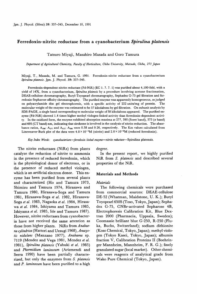
Jpn. J. Phycol. (Sorui) 39: 337寸 45,December 10, 1991
Ferredoxin-nitrite reductase from a cyanobacterium争irulinaplatensis
Tatsuro Miyaji, Masahiro Masada and Goro Tamura
Dψar,抑制 ofAgricultural C加問tヮ1,Fj刷砂 ojHorticulture, Chiba University, Matsudo, Chiba, 271 Japan
Miyaji, T., Masada, M. and Tamura, G. 1991. Ferredoxin-nitrite reductase from a cyanobacterium 尋問linap加'ensis.Jpn. J. Phycol. 39: 337-345.
Ferredoxin-dependent nitrite reductase (Fd-NiR) (EC 1. 7. 7. 1) was purified about 4,100-fold, with a yield of 14%,合'oma cy阻 ob紅白rium,争irulina片品開isby a procedure involving acetone fractionation, DEAE-cellulose chromatography, Butyl-Toyopearl chromatography, Sephadex G・75gel liltration and fer-redoxin-Sepharose affinity chromatography. The purified enzyme was apparendy homogeneous,判吋gedon polyacrylamide disc gel electrophoresis, with a specific activity of 222 units/mg of protein. The molecular weight of the en勾mewas estimated to be 57 kilodaltons by gel liltration. On subunit analysis by SDS-PAGE, a single band corresponding to molecular weight of 58 kilodaltons appeared. The purified en-勾me(Fd-NiR) showed 1.4 times higher methyl viologen-Iinked activity白血 ferredoxin-dependentactivi-ty. In the oxidized form, the enzyme exhibited absorption ma泊 naat 277, 390 (Soret b血 d),573 (a b阻 d)組 d695 (CT band) nm, indicating d出 sirol悶 neis involved in the catalysis of nitrite reduction. The absor-bance ratios, A390: A277組 dA573: A蜘 were0.58 and 0.26, respectively. The Km values calculated from Lineweaver-Burk plot of the data were 4.8 x 1O-4M (nitrite) and 2.0 x 1O-6M (reduced ferredoxin).
Key /ndex Wo地・ り伊P曜'anob,伽a邸:ct.幼伽te防e宵eTlu
The nitrite reductases (NiRs) from plants
catalyze the reduction of nitrite to ammonia
in the presence of reduced ferredoxin, which is the physiological donor of electrons, or in the presence of reduced methyl viologen, which is an artificial electron donor. This en-
Z戸nehas been purified from several plants
and characterized (Ho and Tamura 1973, Shimizu and Tamura 1974, Hirasawa and Tamura 1980, Hirasawa-Soga and Tamura 1981, Hirasawa-Soga et al. 1982, Hirasawa-Soga et al. 1983, Nagaoka et al. 1984, Hirasa-wa et al. 1984, Ishiyama and Tamura 1985, Ishiyama et al. 1985, Ide and Tamura 1987). However, nitrite reductases from cyanobacter-
ia have not received the same attention as
those from higher plants. NiRs from Anabae-
na cylindrica (Hattori and Uesugi 1968), Ana伊-
tis nidulans (Manz釦 o 1977), Anabaena sp. 7119 (Mendez and Vega 1981, Mendez et al. 1981), Spirulina platensむ(Yabukiet al. 1985) and Phormidium laminosum (Arizmendi and Sex・ra1990) have been partially characte-
rized, but only the enzymes from S. platensis and P. laminosum have been purified to a high
degree.
In the present repoロ, we highly purified
NiR from S. platensis and described several
properties of the NiR.
Materials and Methods
Materials The following chemicals were purchased
from commercial sources: DEAE-cellulose
DE・52(Whatman, Maidstone, U. K.); Butyl Toyopearl650S (Toso, Tokyo, Japan); Sepha-dex G・75,CNBr-activated Sepharose 4B, Electrophoresis Calibration Kit, Blue Dex-tran 2000 (Ph町 macia,Uppsala, Sweden); Coomassie brilliant blue G-250, R・250(Flu-
ka, Buchs, Switzerland); sodium dithionite (Koso Chemical, Tokyo, Japan); methyl violo・
gen (Tokyo Kasei, Tokyo, Japan); albumin fraction V, Calibration Proteins II (Boehrin-ger Mannheim, Mannheim, F. R. G.); finely granulated sugar (local market). Other chemi-
cals were reagents of analytical grade from
Wako Pure Chemical (Tokyo, Japan).
338 Miyaji, T., Masada, M. and Tamura, G.
Cell culture Spirulina platensis strain OU -1, kindly sup-
plied by professor K. Wada of Kanazawa University, was grown photoautotrophically at 3500 in inorganic artificial medium (SOT
medium; Ogawa and Terui 1970) in a 100 liter plastic container (AL・LLReactor, sup-plied by Mitsubishi kakoki, Tokyo, ]apan). The culture medium was agitated by a stream of air from a pump and continuous illumina-tion was provided by four white fluorescent lamps (FLR40SW/M40W; Mitsubishi) at 10,0001ux. About 300g (wet wt.) of cells were harvested by suction filtration at the sta-tionary phase, which was reached after one week. The harvested cells were immediately placed in a freezer at -3000 and stored until
use.
Assay of e四ymaticactivi,汐The method for the assay of nitrite reduc-
tase (NiR) activity w邸 側entiallythe same as that described in our previous paper (Yabuki
and Tamura 1985). The reaction mixture contained, in a total volume of 1 ml, 20μmol ofTris-HOl buffer, pH 7.5, 2μmolofNaN02, 3μmol of methyl viologen or 2 mg of ferredox-
in from S. Platensis, 3.75 mg of sodium dithionite (freshly dissolved in 0.3 M
NaH003) and an aliquot of enzyme prepara-tion. In the enzymatic reaction, reduced methyl viologen was used as electron donor unless otherwise stated.
The reaction was started by the addition of sodium dithionite after a 2 min of preincuba-
tion at 3500 and was incubated for 4 min at the same temperature. The reaction was ter-minated by vigorous shaking in a cyclomixer. The decrease in the level of nitrite was measured by the method of Snell and Snell with sulfanilamide and N・1-naphthylethy-
lenediamine dihydrochloride. In the inhibitorγexperiments, the effect of
inhibitors on the reduced form of NiR was measured by addition of sodium nitrite after the preincubation of the enzyme in a solution containing Tris-HOl, methyl viologen, sodi-um dithionite and each inhibitor for 7 min at 3500. The inhibitory effects on the oxidized
form was measured by addition of sodium
dithionite. One unit of NiR activity was defined as the
amount of enzyme that reduced 1μmolofni-trite per min under the conditions of the as-
say.
Puri.fication 0] ]erredoxin Ferredoxin from S. platensis were purified
by acetone fractionation, chromatography twice on DEAE-cellulose and once on Sepha-
dex G-75. In this study, an absorbance ratio for ferredoxin (A423/A277) of more也an0.45 was used. Amounts of ferredoxin were esti-mated using a molar absorption coe伍cientof 9.2 mM-lcm-l at 423 nm (Hall et al. 1972).
Preparation of ]erredoxin-SePharose Ferredoxin-Sepharose 4B was prepared by
the method of Shin and Oshino (1978).
Puri.fication of NiR All the purification procedures were per-
formed in a cold room at 400. Dialysis was performed overnight against the indicated buffer, and centrifugation was carried out at 10,000 x g for 15 min.
(1) Extraction 0] the Enzyme Frozen cells of S. platensis (300 g) were
thawed and mixed in the four volumes
(1,200 ml) of extraction buffer (20 m M Tris-HOl buffer, pH7.5, containing 200mM NaOl). The suspension was sonicated (Soni・cator; Otake seisakusho, Tokyo, ]apan) at 15 kHz for 4 min, and a crude extract was ob-tained after removal of cell debris by centrifu-
gatlOn.
(2) Acetone斤'actionationThe precipitate formed from the crude ex-
tract (1340 ml) after the addition of cold ace-tone (-3000) to a final concentration of 35%
(v/v) was removed by centrifugation and dis-carded. Oold acetone (-3000) was further added to the supernatant to give a final con-centration of 75%, then the resultant precipitate was collected by centrifugation, resuspended in 200 ml of 20 m M Tris-HOl
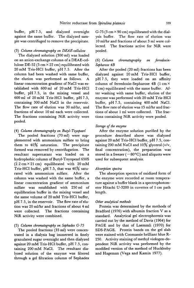
Nitrite reductase from Spirulina platensis 339
buffer, pH 7.5, and dialyzed overnight against the same buffer. The dialyzed sam-ple was centrifuged to remove the precipitate.
(3) Column chromatography on DEAE-cellulose The dialyzed solution (300 ml) was loaded
on an anion-exchange column of a DEAE-cel-luloseDE・52(5 cm X 22 cm) equilibrated with 20 m M Tris-HCl buffer, pH 7.5. After the column had been washed with same buffer, 也eelution was performed as follows. A linear concentration gradient of NaCl was es-tablished with 600 ml of 20 mM Tris-HCl
buffer, pH 7.5, in the mixing vessel and 600 ml of 20 mM Tris-HCl buffer, pH 7.5, containing 300 mM NaCl in the reservoir. The fl.ow rate of elution was 30 ml/hr, and fractions of about 10 ml each were collected.
The fractions containing NiR activity were pooled.
(4) Column chromatography on Bu砂1-Toyopearl The pooled fractions (70 ml) were sup-
plemented with ammonium sulfate to bring them to 40% saturation. The precipitate formed was removed by centrifugation. The resultant supernatant was loaded on a
hydrophobic column ofButyl-Toyopear1650S (2.2 cm X 21 cm) equilibr百 edwith 20mM Tris-HCl buffer, pH 7.5, that was 40% satu-
rated with ammonium sulfate. After the
column was washed with the same buffer, a linear concentration gradient of ammonium sulfate was established with 250 m1 of equilibration buffer in the mixing vessel and
the same volume of 20 mM Tris-HCl buffer, pH 7.5, in the reservoir. The fl.ow rate of elu-
tion was 20 m1/hr and fractions of about 4 ml were collected. The fractions containing NiR activity were combined.
(5) Column chromatography on S.ψhadex G・75The pooled fractions (35 ml) were concen-
trated in a dialysis bag immersed in finely granulated sugar overnight and then dialyzed against 20 mM Tris-HCl buffer, pH 7.5, con-taining 200 mM NaCl. The resultant dia-lyzed solution of the enzyme was filtered through a gel創trationcolumn of Sephadex
G-75 (3 cm X 90 cm) equilibrated with the dial-ysis buffer. The fl.ow rate of elution was 10 ml/hr and fractions of about 3 ml were col-
lected. The fractions active for NiR were pooled.
(6) Column chromatograp砂 on jもrredoxin-Sep加rose4B
After the pooled (20 ml) fractions has been
dialyzed against 20 m M Tris-HCl buffer, pH7.5,出eywere loaded on an a伍nitycolumn of ferredoxin-Sepharose 4B (1 cm X
2 cm) equilibrated with the same buffer. Aι
ter washing with same buffer, elution of the enzyme was performed with 20 m M Tris-HCl
buffer, pH 7.5, containing 400 m M NaCl. The fl.ow rate of elution was 15 ml/hr and frac-tions of about 1 ml were collected. The frac-tions containing NiR activity were pooled.
Storage 01 the e四ymeAfter the enzyme solution purified by the
procedure described above was dialyzed
against 20 mM Tris-HCl buffer, pH 7.5, con-taining 200 m M NaCl and 10% glycerol (v/v, final concentration), the preparation was stored in a freezer ( -800C) and aliquots were
used for subsequent analysis.
Absoψtぬnspectra The absorption spectra of oxidized form of
the enzyme were recorded at room tempera-ture against a buffer blank in a spectrophotom-eter Hitachi U・3200in cuvettes of 1 cm path
len供.
Other analytical methods Protein was determined by the methods of
Bradford (1976) with albumin fraction V as a st組 dard. Anal戸icalgel electrophoresis was carried out by the method ofDavis (1964) for
PAGE and by that of Laemmli (1970) for SDS-PAGE. Protein bands on the gel slab were stained with Coomassie brilliant blue R・
250. Activity staining ofmethyl viologen-de-pendent NiR activity was performed by the modified version of the method of Hucklesby and Hageman (Vega and Kamin 1977).
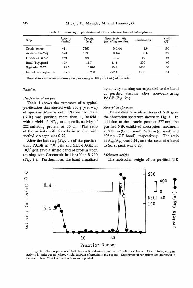
Miyaji, T., Masada, M. and Tamura, G. 340
Summary of purification of nitrite reductase from やirulinaplal四sisTable 1.
Yield (%)
Specific Activity (units/mg protein)
Protein (mω
Activity (units) Purification
411
528
230
163
83.5
55.6
100
129
56
40
20
14
1.0
8.6
19
200
1600
4100
Step
Crude extract
Acetone 35-75%
DEAE-Cellulose
Butyl Toyopearl
Sephadex G・75
Ferredoxin Sepharose
Th四 edata we問 obtainedduring the processing of 300 g (wet wt.) of the cells.
0.0544
0.467
1.03
11.1
85.2
222.4
7560
1130
224
14.7
0.980
0.250
by activity staining corresponded to the band
of purified enzyme a仕er non-denaturing
PAGE (Fig. 2a).
AbsoψtionゆectrumThe solution of oxidized form of NiR gave
the absorption spectrum shown in Fig. 3. In
addition to the protein peak at 277 nm, the purified NiR exhibited absorption maximum
at 390 nm (Soret band), 573 nm (αband)and
695 nm (CT band), respectively. The ratio
of A390/A277 was 0.58, and the ratio of αband to Soret peak was 0.26.
Molecular weight
The molecular weight of the purified NiR
Puriωion 01仰 meTable 1 shows the summary of a typical
purification that started with 300 g (wet wt.)
of Spirul仇aplatensis cell. Nitrite reductase
(NiR) was purified more than 4,100・fold,with a yield of 14%, to a specific activity of
222 units/mg protein at 350C. The ratio
of the activity with ferredoxin to that with
methyl viologen was 0.72.
After the last step (Fig. 1.) of the purifica-tion, PAGE in 7% gels and SDS-PAGE in
10% gels gave a single band of protein upon
staining with Coomassie brilliant blue R・250
(Fig. 2.). Furthermore, the band visualized
Results
?@(冨¥凶冨)ロHOパFolHQ
200
nu nU
8且E
1・lJ O
NaCl mM
100
0.4
o. 2
(HE¥凶パエロヨ)』
内
科
吋
〉
吋
#04
20
Fraction Number
Fig. 1. Elution pattern of NiR from a ferredoxin-Sepharose 4 B affinity column. Open circle, enzyme activity in units per ml; closed circle, amount of protein in mg per ml. Experimental conditions are described in the text. Nos. 23-24 of出e仕actionswere pooled.
10
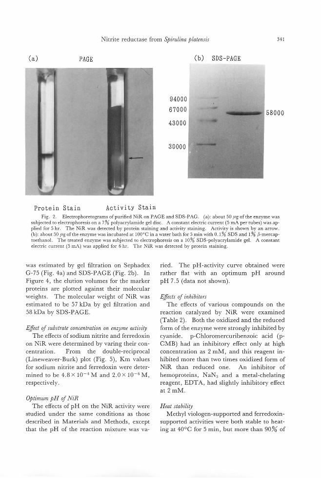
Nitrite reductase from Spirulina platensis 341
(a) PAGE
Protein Stain Activity Stain
94000
67000
43000
30000
(b) SDS-PAGE
58000
Fig. 2. Electrophoretograms ofpuri自edNiR on PAGE and SDS-PAG. (a): about 50μg ofthe enzyme was subjected to e1ectrophoresis on a 7% polyacrylamide gel disc. A constant electric current (5 mA per tubes) was ap-plied for 5 hr. The NiR was detected by protein staining and activity staining. Activity is shown by an arrow (b): about 50μg ofthe enzyme was incubated at 1000C in a water bath for 5 min with 0.1% SDS and 1% s-mercap-toethanol. The treated enzyme was subjected to electrophoresis on a 10% SDS-polyacrylamide gel. A constant electric current (5 mA) was appl陀 dfor 6 hr. The NiR was detected by pro日 instaining
was estimated by gel filtration on Sephadex
G-75 (Fig. 4a) and SDS-PAGE (Fig. 2b). In
Figure 4, the elution volumes for the marker
proteins are plotted against their molecular
weights. The molecular weight of NiR was
estimated to be 57 kDa by gel filtration and
58 kDa by SDS-PAGE
旦ffect0] substrate concentration on enそymeactiviり
The effects of sodium nitrite and ferredoxin
on NiR were determined by varing their con-
centration. From the double-reciprocal
(Lineweaver-Burk) plot (Fig. 5), Km values
for sodium nitrite and ferredoxin were deter
mined to be 4.8 x 10⁻⁴ M and 2.0 x 10⁻⁶ M, respectively.
Optimum pH 0] NiR
ried. The pH-activity curve obtained were
rather flat with an optimum pH around
pH 7.5 (data not shown)
旦ffects0] inhibitors The effects of various compounds on the
reaction catalyzed by NiR were examined
(Table 2). Both the oxidized and the reduced
form of the enzyme were strongly inhibi白dby
cyanide. p-Chloromercuribenzoic acid (p-
CMB) had an inhibitory effect only at high
concentration as 2 mM, and this reagent in-hibited more than two times oxidized form of
NiR than reduced one. An inhibitor of
hemoproteins, NaN3 and a metal-chelating
reagent, EDTA, had slightly inhibitory effect
at 2 mM.
The巴ffectsof pH on the NiR activity were Heat stability
studied under the same conditions as those Methyl viologen引 lpportedand ferredoxin-
described in Materials and Methods, except supported activities were both stable to heat-
that the pH of the reaction mixture was va- ing at 400C for 5 min, but mor巴than90% of
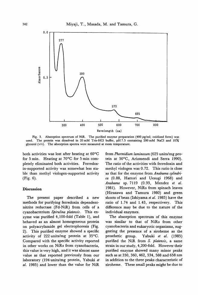
342 Miyaji, T., Masada, M. and Tamura, G.
0.6
由。回
z su 』..:
277
300
390
400 500 600 700 800
Wavelength (nm)
Fig. 3. Absorption spectrum of NiR. The purified enzyme preparation (400μg/m1; oxidized form) was used. The protein was dissolved in 20 mM T出-HCIbuffer, pH 7.5 containing 200 mM NaCI and 10% glycerol (v/v). The absorption spectra were measured at room temperature.
both activities was lost after heating at 600C for 5 min. Heating at 700C for 5 min com-
pletely eliminated both activities. Ferz吋 ox-in-supported activity was somewhat less sta-ble than methyl viologen-supported activity
(Fig. 6).
Discussion
The present paper described a new methods for purifying ferredoxin dependent-nitrite reductase (Fd-NiR) from cells of a cyanobacterium尋問linaplatensis. This en-zyme was purified 4,100-ゐld(Table 1), and behaved as an almost homogeneous protein on polyacrylamide gel electrophoresis (Fig. 2). This purified enzyme showed a specific activity of 222 units/mg protein at 350C.
Compared with the specific activity reported in other works on NiRs from cyanobacteria, this value is very high, and it was almost same value as that reported previously from our laboratory (194 units/mg protein, Yabuki et al. 1985) and lower than the value for NiR
from Phormidium laminosum (625 units/mg pro-tein at 50oC, Arizmendi and Serra 1990). The ratio of the activities wi也 ferredoxinand methyl viologen was 0.72. This ratio is close as that for the enzyme from Anabaena cylindri-ca (0.88, Hattori and Uesugi 1968) and Anabaena sp. 7119 (0.93, Mendez et al. 1981). However, NiRs from spinach leaves (Hirasawa and Tamura 1980) and green
shoots ofbean (Ishiyama et al. 1985) have the ratio of 1. 74 and 1.43, respectivery. This difference may be due to the nature of the individual enzymes.
The absorption spectrum of this enzyme
was similar to出atof NiRs from other cyanobacteria and eukaryotic organisms, sug-gesting the presence of a siroheme as出e
prosthetic group. Yabuki et al. (1985) purified the NiR from S. platensis, a same strain in our study, 4,200・fold. However their purified enzyme showed many minor peaks such as at 350,360,462,534,588 and 658 nm in addition to the three peaks characteristic of siroheme. These small peaks might be due to
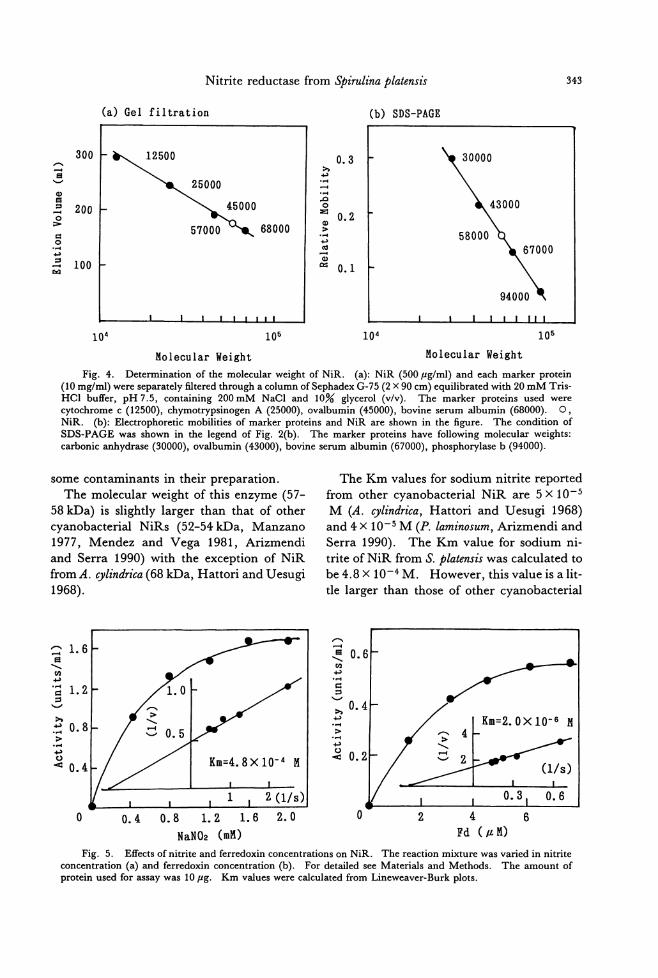
343 Nitrite reductase from Spirulina platensis
(b) SDS-PAGE
0.1
0.3 h +' ・同
..... ・同
..0 o
t仏> ' .-吋. +' ' 咽-也α=
(a) Gel filtration
68000
300
200
100
(-国)曲目=円。〉ロロ吋JVロ【同
105
Molecular Weight
Fig. 4. Determination of山 molecularweight of NiR. (a): NiR (500μg/ml) and each ma巾 rprotein (10 mg/ml) were separately filtered through a column ofSephadex G-75 (2 X 90 cm) equilibrated with 20 mM Tris-HCl buffer, pH 7.5, containing 200 mM NaCl and 10% glycerol (v/v). The marker proteins used were cytochrome c (12500), chymotrypsinogen A (25000), ovalbumin (45000), bovine serum albumin (68000). 0, NiR. (b): Electrophoretic mobilities of marker proteins and NiR are shown in the figure. The condition of SDS-PAGE was shown in the legend of Fig. 2(b). The marker proteins have following molecular weights: carbonic anhydrase (30000), ovalbumi
Molecular Weight
104 105 104
The Km values for sodium nitrite reported
from other cyanobacterial NiR are 5 x 10⁻⁵
M (A. cylindrica, Hattori and Uesugi 1968) and 4 X 10⁻⁵ M (P. laminosum, Arizmendi and Serra 1990). The Km value for sodium ni-
trite of NiR from S.ρlatensis was calculated to
be4.8x 1O-4M. However, thisvalueisalit-tle larger than those of other cyanobacterial
some contaminants in their preparation.
The molecular weight of this enzyme (57-
58回 a)is slightly larger th却 thatof other
cyanobacterial NiRs (52-54 kDa, Manzano 1977, Mendez and Vega 1981, Arizmendi and Serra 1990) with the exception of NiR
from A. cylindrica (68 kDa, Hattori and Uesugi 1968).
〆〆...-.- .
f司、...... ξ0.6 同...., ・... ロロ、--'>. 0.4 ...., .... 〉.... ....,
~ 0.2
1.6 回、、、凶
+占
'= 1. 2 口、--'
h ...., 0.8 〉.... ...., O
< 0.4
0.6
0.8 1.2 1.6
NaN02 (mM)
Fig. 5. Effects of nitrite and ferredoxin concentrations on NiR. The reaction mixture was varied in nitrite concentration (a)回 dferredoxin concentration (b). For detailed see Materials and Methods. The阻 ountof protein used for出 saywas 10μg. Km values were calculated from Lineweaver-Burk plots.
6 4
Fd (μM)
2 。0.4 。
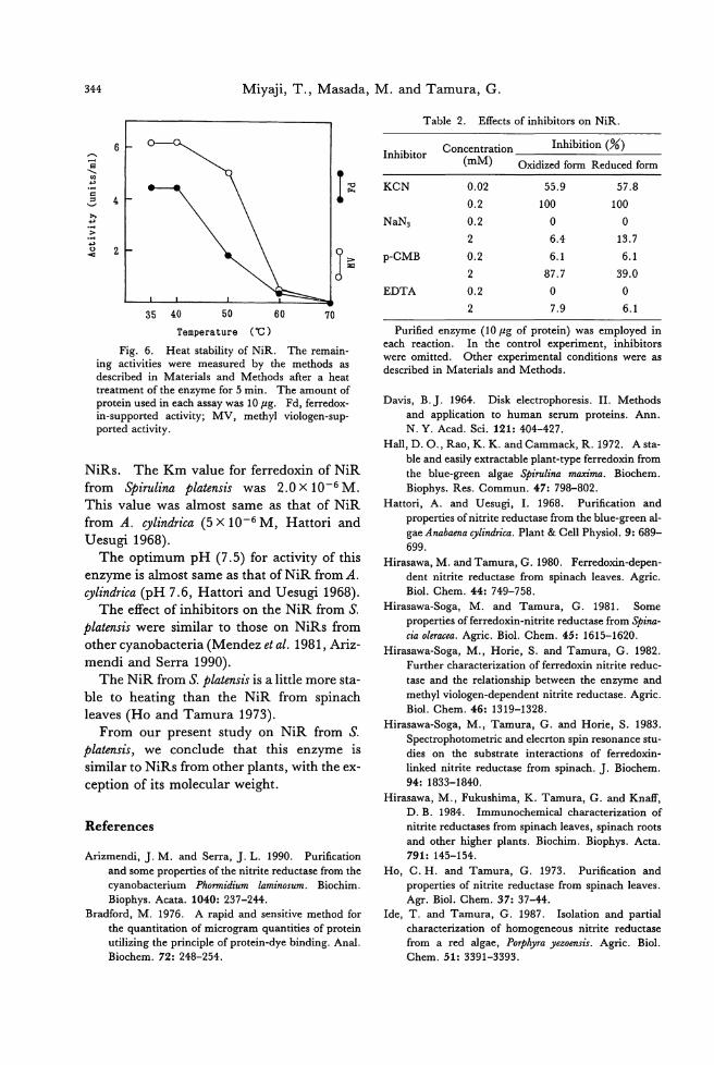
344 Miyaji, T., Masada, M. and Tamura, G.
Table 2. Effects of inhibitors on NiR.
6
唱』同・TIll企'
局
悼
の
'u
(
司
自
¥ω拍同国司
)
hパ戸司、
=JH04
PE
O--d
35 40 50
Temperature
60
('C )
70
Fig. 6. Heat stability of NiR. The remain-ing activities were measured by the methods as described in Materials and Methods after a heat treatment of the enzyme for 5 min. The amount of protein used in each assay was 10μg. Fd, ferredox-in-supported activity; MV, methyl viologen-sup-ported activity.
NiRs. The Km value for ferredoxin of NiR
from Spirulina platensis was 2.0 X 10⁻⁶ M.
This value was almost same as that of NiR
from A. cylindrica (5 X 10⁻⁶ M , Hattori and Uesugi 1968).
The optimum pH (7.5) for activity of this
enzyme is almost same as that of NiR from A.
cylindrica (pH 7.6, Hattori and Uesugi 1968). The effect of inhibitors on the NiR from S.
platensis were similar to those on NiRs from
other cyanobacter匂 (Mendezet al. 1981, Ariz-
mendi and Serra 1990).
The NiR from S. platensis is a little more sta-
ble to heating than the NiR from spinach
leaves (Ho and Tamura 1973).
From our present study on NiR from S. platensis, we conclude that this enzyme is
similar to NiRs from other plants, with the ex-ception of its molecular weight.
References
Arizmendi, J. M. and Serra, J. L. 1990. Purification
and some properties of the nitrite reductase仕omthe
cyanobacterium Phormidium laminosum. Biochim.
Biophys. Acata. 1040: 237-244
Bradford, M. 1976. A rapid and sensitive method for
the quantitation of microgram quantities of protein
utilizing the principle of protein-dye binding. Anal.
Biochem. 72: 248-254.
1nhibitor Concentrationnhibition (%)
(mM) Oxidized form Reduced form
KCN 。目02 55.9 57.8
。目2 100 100
NaN3 0.2 。 。
2 6.4 13.7
p-CMB 0.2 6.1 6.1
2 87.7 39.0
EDTA 0.2 。 。
2 7.9 6.1
Purified enzyme (10μg of protein) was employed in each reaction. 1n the control experiment, inhibitors were omitted. Other experimental conditions were as described in Materials and Methods.
Davis, B.J. 1964. Disk electrophoresis. 11. Methods
and application to human serum proteins. Ann.
N. Y. Acad. Sci. 121: 404-427.
Hall, D. 0., Rao, K. K. and Cammack, R. 1972. A sta-
ble and easily extractable plant-type ferredoxin from
the blue-green algae Spirulina maxima. Biochem.
Biophys. Res. Commun. 47: 798-802.
Hattori, A. and Uesugi, 1. 1968. Purification and
properties ofnitrite reductase from the blue-green al-
gae Anabaena cylindrica. Plant & Cell Physiol. 9: 689-
699.
Hirasawa, M. and Tamura, G. 1980. Ferredoxin-depen-
dent nitrite reductase from spinach leaves. Agric.
Biol. Chem. 44: 749-758.
Hirasawa-Soga, M. and Tamura, G. 1981. Some
prope口iesofferredoxin-nitrite reductase from 司pina-cia oleracea. Agric. Biol. Chem. 45: 1615-1620.
Hirasawa-Soga, M., Horie, S. and Tamura, G. 1982. Further characterization of ferredoxin nitrite reduc-
tase and the relationship between the enzyme and
methyl viologen-dependent nitrite reductase. Agric.
Biol. Chem. 46: 1319-1328.
Hirasawa-Soga, M., Tamura, G. and Horie, S. 1983
Spectrophotometric and e1ecrton spin resonance stu-dies on the substrate interactions of ferredoxin-
linked nitrite reductase from spinach. J. Biochem.
94: 1833-1840.
Hirasawa, M., Fukushima, K. Tamura, G.叩 dKnaff, D. B. 1984. 1mmunochemical characterization of
nitrite reductases from spinach leaves, spinach roots and other higher plants. Biochim. Biophys. Acta.
791: 145-154.
Ho, C. H. and Tamura, G. 1973. Purification and
properties of nitrite reductase from spinach leaves. Agr. Biol. Chem. 37: 37-44.
1de, T. and Tamura, G. 1987. Isolation and partial
characterization of homogeneous nitrite reductase
from a red algae, POψhyra y四oensis.Agric. Biol. Chem. 51: 3391-3393.
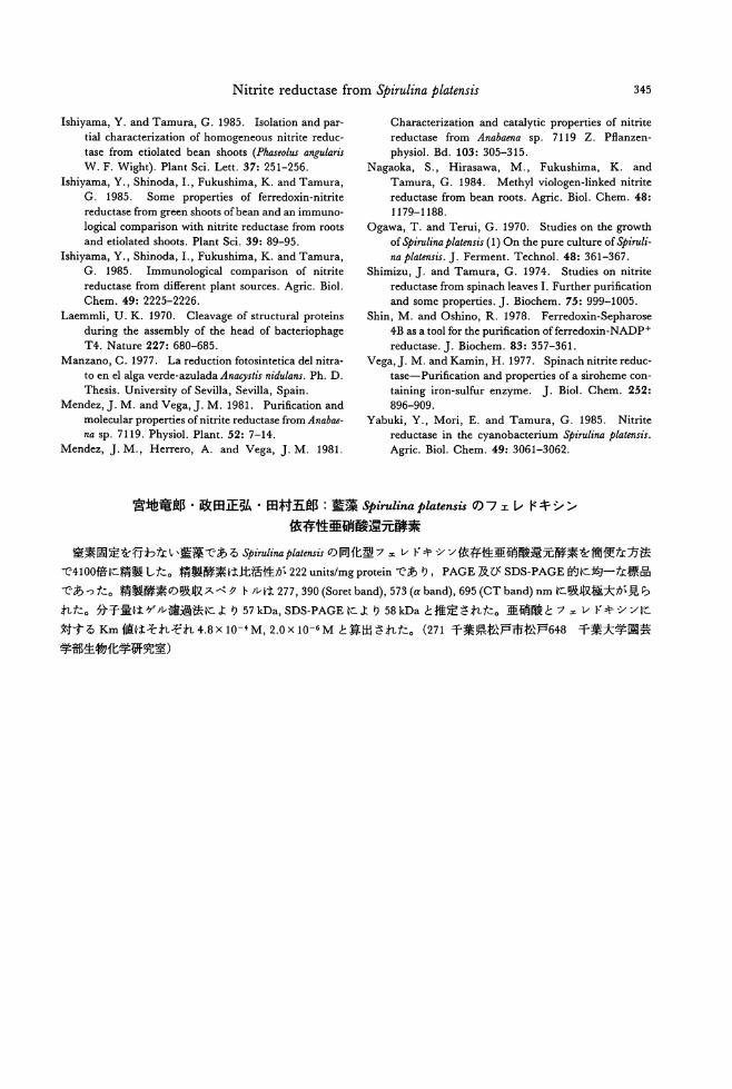
Nitrite reductase from Spirulina platensis 345
Ishiyama, Y. and Tamura, G. 1985. Isolation and par-
tial characterization of homogeneous nitrite reduc-
tase from etiolated bean shoots (Phaseolus angularis
W. F. Wight). Plant Sci. Lett. 37: 251-256.
Ishiyama, Y., Shinoda, 1., Fukushima, K. and Tamura,
G. 1985. Some properties of ferredoxin-nitrite
reductase from gr田 nshoots ofbean and an immuno-
logical comparison with nitrite reductase from roots
and etiolated shoots. Plant Sci. 39: 89-95.
Ishiyama, Y., Shinoda, 1., Fukushima, K. and Tamura,
G. 1985. Immunological comp訂 isonof nitrite
reductase from different plant sources. Agric. Biol
Chem. 49: 2225-2226.
Laemmli, U. K. 1970. Cleavage of structural proteins
during the assembly of the head of bacteriophage
T4. Nature 227: 680-685
Manzano, C. 1977. La reduction fotosintetica del nitra-
to en el alga verde-azulada An処ystisnidulans. Ph. D.
Thesis. University of Sevilla, Sevilla, Spain. Mendez,J. M. and Vega,J. M. 1981. Purification and
molecular propeロiesof nitrite reductase仕omAnabae-
na sp. 7119. Physiol. Plant. 52: 7-14
Mendez, J. M., Herrero, A. and Vega, J. M. 1981.
Characterization and catalytic propeロiesof nitrite
reductase from Anabaena sp. 7119 Z. Psanzen-
physiol. Bd. 103: 305-315.
Nagaoka, S., Hirasawa, M., Fukushima, K. and
Tamura, G. 1984. Methyl viologen-linked nitrite
reductase from bean roots. Agric. Biol. Chem. 48:
1179-1188.
Ogawa, T. and Terui, G. 1970. Studies on the growth
of争irulinaplatensis (1) On the pure culture ofやiruli-
na plat.間お.J. Ferment. Technol. 48: 361-367.
Shimizu, J. and Tamura, G. 1974. Studies on nitrite
reductase from spinach leaves 1. Further purification
and some properties. J. Biochem. 75: 999-1005.
Shin, M. and Oshino, R. 1978. Ferredoxin-Sepharose
4B as a tool for the purification offerredoxin-NADP+
reductase. J. Biochem. 83: 357-361.
Vega, J. M. and Kamin, H. 1977. Spinach nitrite reduc-
t田 e-Purification and properties of a siroheme con-
taining iron-sulfur enzyme. J. Biol. Chem. 252:
896-909.
Yabuki, Y., Mori, E. and Tamura, G. 1985. Nitrite reductase in the cyanobacterium Spirulina plat,四川.
Agric. Biol. Chem. 49: 3061-3062.
宮地竜郎・政田正弘・田村五郎:藍藻 Spirulinaρlatensisのフェレドキシン
依存性亜硝酸還元酵素
窒素固定を行わない藍藻である争irulinapla帥問の同化型フェレドキシン依存性亜硝酸還元酵素を簡便な方法
で4100倍に精製した。精製酵素は比活性が 222units/mg proteinであり, PAGE及び SDS-PAGE的に均一な標晶
であった。精製酵素の吸収スベクトルは 277,390 (Soret band), 573 (αband), 695 (CT band) nmに吸収極大が見ら
れた。分子量はゲノレ潟過法により 57kDa, SDS-PAGEにより 58kDaと推定された。亜硝酸とフェレドキシンに
対する Km値はそれぞれ 4.8X 10⁻⁴ M, 2.0 X 10⁻⁶ M と算出された。 (271千葉県松戸市松戸648 千葉大学園芸
学部生物化学研究室)

Jpn. J. Phycol. (Sorui) 39: 347-353, December 10, 1991
Histochemistry and ultrastructure of paraphyses in Sargassum vulgare
C. Agardh and S. johnstonii Setchell & Gardner
M. R. Vijayaraghavan and 1nderdeep Kaur
Dψartment of Botaり Univ宵:ri,紗 ofDelhi, Delhi 11000えlndia
Vijayaraghavan, M. R.組 dKaur, I. 1990. Histochemistry and ultrastructure ofparaphyses in Sarg,叩 umvulgare C. Ag訂 dhand S. johnstonii Setchell & G紅白er.Jpn. J. Phycol. 39: 347-353.
sa町E面 sum四 19areand S. johnstonii bear androgynous問 ceptacleswi出 unisexualconceptacles. The fully developed conceptacles訂 e佃 edwith secretions and communicate with the external environment through the ostiole plugged wi也polysaccharidicmaterial. The conceptacles are lined by flat cells-the conceptacle-wal1 ce1ls, which are the progenitors of either副ltheridiaor oogonia or paraphyses. In a young conceptacle, 出ep釘 aphysialand the conceptacle-cell walls contain a1ginic acid and sulphated polysacぬarides.During the early stages of conceptacle development, the cytoplasm is organelle-rich but in the old conceptacles,出eorganelles Iyse and the paraphysial and conceptacle-wall cell cytoplasm becomes vacuolate.
Key ln伽 陥rds: 咋inicacid-histochemistry-paraphysisーPhaeophyceae-Sargassum-secretions-sulphated polysacchari,ゐ's.
1n Sargassaceae (Fucales), the histochemi-cal and ultrastructural information on the
paraphyses and the conceptacle-wall cells is
meagre. The mucilaginous secretions have
been reported from the conceptacles of Fucus e伽 tatω(McCully1968) whereas elaborate
wall projections have been observed in the
conceptacle wall cells of Durvillaea totatorum
(Clayton et al. 1987). The present study on
Sargassum vulgare C. Agardh and S. johnstonii
Setchell & Gardner is undertaken to correlate
the histochemical, developmental and ultra-structural aspects of the paraphyses and the
conceptacle-wall cells.
Materials and Methods
The plants of Sargassum vulgare and S.
johnstonii were collected during the low tide
period from Port Okha, Gujarat, through the months ofJanuary, February and November of 1987-89. The selected parts of the plants
thus collected were processed for light, trans-mission and scanning electron microscopy.
For light microscopy, the plant pa此swere
fixed in 10% acrolein, washed in distilled water and postfixed in 1% HgG12・ Thiswas
followed by dehydration, and infiltration. The infiltrated materials were embedded in
glycol methacrylate (Vijayaraghavan & Shuk-
la 1990), and sectioned on a Spencer (AO) ro-
tary microtome fitted with a locally made
adaptor to hold glass knives. Two micron
thick sections were serially transferred to
small drops of distilled water and later stained
with PAS reagent for insoluble polysaccha-
rides (Vijayaraghavan and Shukla 1990); with Alcian Blue for sulphated polysaccha-
rides (Parker and Diboll1966); with TBO for sulphated and carboxylated polysaccharides
(McCully 1966); with Coomassie Brilliant
Blue for proteins (Weber and Osborn 1975)
and with Feulgen reagent for DNA
(Vijayaraghavan and Shukla 1990). For
SEM, the plant parts were fixed in 4% forma-
lin, dehydrated in graded acetone series, criti-cal point dried and scanned for topographical details. For transmission electron micro-
scopy, the desired stages were cut into
small pieces, fixed in 6% glutaraldehyde pre-
pared in phosphate buffer, dehydrated, infiltrated and embedded in Epon-Araldite
mixture (Mollenhauer 1964). Ultrathin sec-
tions were stained and then observed through
348 Vijayaraghavan, M. R., and Kaur, 1.
Philips EM 300.
Results
Light microscopic Studies
The reproductive phases are seasonal in Sar-
g仰 umv吟areand 広johnstonii. The axillary
receptacles occur in clusters (Figs. 1, 2). Unisexual conceptacles are embedded in the
receptacul町 tissues. The antheridial and 00・
gonial conceptacles are borne in the same
receptacle but in the conceptacles, either the antheridia or oogonia cooccur with para-
physes. Thus, the plants are neither monoecious nor dioecious in the generally ac-
cepted sense as both types of reproductive or-
gans are present in the same receptacle but al-
ways in separate conceptacles. Androgynous
condition thus prevails in these two species of
Sargassum.
Early in the ontogeny, the conceptacle-wall cells enlarge and develop into papillae which
through repeated transverse divisions develop
into multicellular paraphyses whose terminal
cells are globose (Fig. 3). Along with these
changes either the oogonia or出eantheridia
also codevelop inside the conceptacle.
The young paraphysial cells show walls which stain reddish-violet with TBO 組 d
moderate magenta with P AS reagent and are
hence rich in alginic acid. The cytoplasm
stains well for proteins and contains moderate amount of polysaccharides. Also enclosed in
the cytoplasm are a few small vacuoles and
phenolic compounds which occur in large
amounts. In the developing paraphyses, the cell walls reveal identical staining reactions.
The cell walls, in addition, reveal small
thread-like structures出atstain intense violet
with TBO (Figs. 3, 4). The paraphysial
cells, in transverse sections, reveal core創led
with phenolic materials (Fig. 4). The
cytoplasm gradually becomes vacuolate and reveals a low amount of proteins and moder-
ate polysaccharides. The cells are uninucle-
ate and the nucleus stains well for DNA.
Hence, the paraphyses which codevelop with sex organs, remain rich in cytoplasmic con-tents during the early stages of oogonial/an-
theridial development. Later as the sex or-
gans are ready for gamete release, the paraphysial cytoplasm shows vacuolation.
The conceptacle-wall cells when young con-
tain abundant proteins, moderate polysaccha-rides and phenolic materials. At maturity, the wall cells become more vacuolate and the
proteins are meagre. The conceptacular cavi-
ty reveals polysaccharidic secretions which
stain turquoise with Alcian Blue, magenta with PAS reagent and violet with TBO-thus
indicating a mixture of sulphated and carboxy-
lated polysaccharides (Fig. 4). This polysac-
charidic material also acts as a plug in出efe-
male conceptacle where it keeps the ostiole
closed while the oogonia are developing.
Electron Microscopic Studies
The paraphysial cell when viewed under scanning electron microscope reveals many
strands that radiate from the wall into the con-
ceptacle cavity (Fig. 5). At ultrastructural
level, this wall is well differentiated, thick, lacks an orderly arrangement of microfibrils
and is overlaid by material that is differentiat-
ed into an inner light and an outer dark
zones. The latter radiates a few electron
dense strands into the conceptacle cavity (Fig.
7). Adjacent to the wall lie many vacuoles that contain electron dense materials. The
material is released from the vacuoles and
Figs. 1-5. Fig. 1. Sargassum vu伝'are,a mature plant showing forked branches, leaves, bladders and recep-tacles (紅rows). x 0.47. Fig. 2. Sargassumj訪問tonii,whole mount of a plant bearing dense leaves and receptacles (釘rows). x 0.47. Fig. 3.ぶvulgare,Mature female conceptacle showing paraphyses (pa) ne紅白eostiol釘問gion.The terminal paraphysial cells are globular and veneered by polysaccharidic materials.τ'he paraphysis cytoplasm reveals phenolic materials and carboxylated polysaccharides. The p釘 aphysialand the conceptacle-cell wal1s 問 vealsmall polysaccharidic pr吋ections(arrows). x 1474 (TBO stained). Fig. 4. S.山信'are,Transverse section of a conceptacle, showing paraphyses surrounded by metachromatic material. The core of the paraphysis encloses phenolic materials and polysaccharides. From the core radiate thin strands into the conceptacle cavity. X 1474 (TBO stained). Fig. 5. S.joh即 '1onii.Scanning electron micrograph. The paraphyses白紙occludethe ostiole show blunt tips from which emerge many. small, thread-like structures (arrows). X 1180.
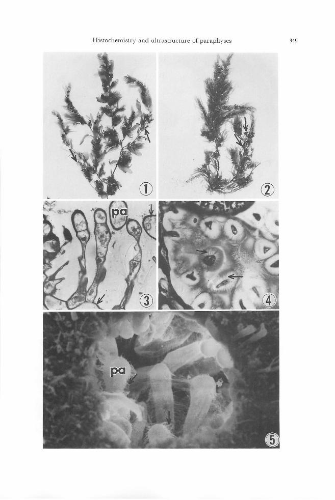
Histochemistry and ultrastructure of paraphyses 349
⑦ ⑧
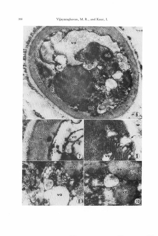
350 Vijayaraghavan, M. R., and Kaur, 1
、!f.
日輸
、
'⑥4
• iW :
'噌ゐ1':
仇~
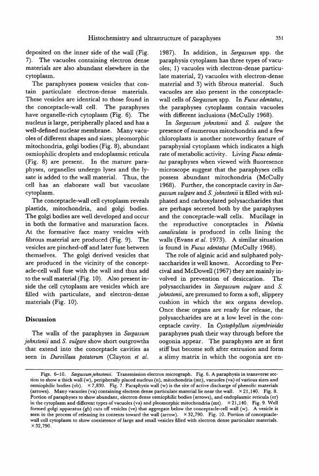
Histochemistry and ultrastructure of paraphyses 351
deposited on the inner side of the wall (Fig.
7). The vacuoles containing electron dense
materials are also abundant elsewhere in the
cytoplasm.
The paraphyses possess vesicles that con-tain pa此iculate electron-dense materials.
These vesicles are identical to those found in
the conceptacle-wall cell. The paraphyses
have organelle-rich cytoplasm (Fig. 6). The
nucleus is large, perヤherallyplaced and has a
well-defined nuclear membrane. Many vacu-
oles of different shapes and sizes; pleomorphic
mitochondria, golgi bodies (Fig. 8), abundant osmiophilic droplets and endoplasmic reticula
(Fig. 8) are present. 1n the mature para-
physes, organelles undergo lyses and the ly-sate is added to the wall material. Thus, the cell has an elaborate wall but vacuolate
cytoplasm.
The conceptacle-wall cell cytoplasm reveals
plastids, mitochondria, and golgi bodies.
The golgi bodies are well developed and occur
in both the formative and maturation faces.
At the formative face many vesicles with
fibrous material訂 eproduced (Fig. 9). The
vesicles are pinched-off and later fuse between
themselves. The golgi derived vesicles that
are produced in the vicinity of the concept-
acle-cell wall fuse with the wall and thus add
to the wall material (Fig. 10). Also present in-
side the cell cytoplasm are vesicles which are
filled with particulate, and electron-dense materials (Fig. 10).
Discussion
The walls of the paraphyses in Sargassum
johnstonii and忍 vu伊reshow short outgrowths that extend into the conceptacle cavities as
seen in Durvillaea potatorum (Clayton et al.
1987). 1n addition, 泊 S品a1管ま酔&ω仰rs捌sparaphys由iおscy戸to叩pla酪smhas three type回sofv刊acα印u-
ole回s;1り)va配cαuole白swith electron-dense particu-
late material, 2) vacuoles with electron-dense material and 3) with fibrous material. Such
vacuoles are also present in the conceptacle-
wall cells of Sargassum spp. 1n Fucus edentatus, the paraphyses cytoplasm contain vacuoles
with different inclusions (McCully 1968).
1n Sargassum johnstonii and S. vulgare也e
presence of numerous mitochondria and a few
chloroplasts is another noteworthy feature of
paraphysial cytoplasm which indicates a high rate of metabolic activity. Living Fucus ed初ta-
tus paraphyses when viewed with fl.uoresence
microscope suggest that the paraphyses cells
possess abundant mitochondria (McCully
1968). Further, the conceptacle cavity in Sar-ga;ωωf配“捌's
t phat飽edand ca訂rboxy升la剖tedpolysaccharide白st由ha説are p戸er出ha叩pssecreted b加ot出hby the p訂 a却ph匂ysesand the conceptacle-wall cells. Mucilage in
the reproductive conceptacles in Pelvetia
canaliculata is produced in cells lining the
walls (Evans et al. 1973). A similar situation
is found in Fucus ed抑制us(McCully 1968).
The role of alginic acid and sulphated poly-
saccharides is well known. According to Per-
cival and McDowell (1967) they are mainly in-volved in prevention of desiccation. The
polysacch町 idesin Sargassum vulgare and S.
johnstonii, are presumed to form a soft, slippery cushion in which the sex organs develop.
Once these organs are ready for release,也e
polysaccharides are at a low level in the con-
ceptacle cavity. 1n Cystoph)仇 msisymbrioi,ゐs
paraphyses push their way through before the
oogonia appear. The paraphyses are at first
stiff but become soft after extrusion and form
a slimy matrix in which the oogonia are en-
Figs. 6-10. Sargassumjohnstonii. Transmission electron miα'ograph. Fig. 6. A paraphysis in transverse sec-tion to show a thick wall (w), peripl町 allyplaced nuc1eus (n), mitochondria (mt), vacuoles (va) ofvarious sizes and osmiophilic bodies (ob). X 7,830. Fig. 7. Paraphysis wall (w) is也esite of active discharge of phenolic materials (釘rows).Many vacuoles (va) containing electron dense pa民iculatemateriallie near the wall. x 21,140. Fig.8. Portion of paraphyses to show abundant, electron dense osmiophilic bodies (釘rows),and endoplasmic reticula (er) in the cytopl踊 m佃 ddifferent types ofvacuoles (va)血 dpleomorphic mitochondria (mt). x 21,140. Fig. 9. Wel1 formed golgi apparatus (gb) cuts offvesic1es (ve) that aggregate below出econceptac1e吋 1wall (w). A vesic1e is seen in the process of releasing its contents toward the wall (arrow). x 32,790. Fig. 10. Portion of conceptac1e-wall cel1 cytoplasm to show coexistence oflarge and small vesic1es filled wi血 electrondense particulate materials. X32,790.
352 Vijayaraghavan, M. R., and Kaur, 1.
closed (T泊町a1913). 1n Bifurcaria br,仙台ifor-
mis, the oogonia could be seen carried up on stalks which were themselves embedded in a
tenacious common jelly, presumably derived from the walls of the paraphyses (Delf 1935).
The presence of phenolic materials in the
conceptacular tissues is of great ecological sig-
nificance. The ostioles in Sargassum spp. of
the old receptacle are devoid ofpolysaccharid-
ic plug-materials. The disappearance of
plug-material upon release of oogonia/sper-
matozoids makes conceptacles susceptible to
the attack by epiphytic and endophytic mi-
crobes. The abundance of the phenolic
deposits in the persistent tissues of old concep-
tacles help to deter the attack of the microbes
through invasion route. Phenolic materials
also help to prevent attack by the herbivores
(Clayton and Shankly, 1987). The paraphy-
sial cytoplasm is packed with phenolic materi-
als in Sarg,ω'sum spp. throughout the concepta-
cle development. 1n Scytos桝onsp. the para-
physes have a protective role during gamete
discharge, perhaps they help in preventing the excessive spread of lytic enz戸nesto the
walls of immature gametangia (Clayton, 1984).
The conceptacles, during the last phase of reproductive season (January end to Febru-ary) are totally devoid of contents. 1n Sargas-sum johnstonii and丘 vu伝'arethe conceptacle-
cell wall, during this stage, is elaborate and possesses prominent, thread-like, projections as seen in Durvillaea potatorum (Clayton et al.
1987), where the conceptacle cells immediate-ly adjacent to the conceptacle contents
produce short filamentous outgrowths which
extend into the central cavity.
The conceptacle-wall cells also reveal perinuclear golgi bodies which produce many vacuoles. The fibrous materials are frequent-ly incorporated into the vacuoles at the forma-
tive region, undergo condensation and are released at the cell surface (present work).
Such vacuoles have been termed as secretory vacuoles and have been shown to be released
at the cell surface (Bouck 1962; Mollenhauer
and Whaley 1963). The production of sul-
phated material by the meristoderm cells of
Pelvetia canaliculata and secretoη, cells of
Laminaria saccharina occurs in the golgi-rich, perinuclear areas (Evans et al., 1973). 1n
pancreatic acinar cells, sulphation of material occurs in golgi bodies (Berg and Y oung
1971). Discharge of fibrous materials into
the wall and cavity in Sargassum johnstonii and
S. vulgare presumably occurs by the activity of golgi bodies present in the conceptacle-wall
cells and the p訂 aphysesand the energy re-
quired for the secretory process is provided by
numerous mitochondria in the cell cyto・
plasm. These two cell populations, therefore, may be designated at least functionally as
secretory cells.
References
Berg, N. B. and Young, R. W. 1971. Sulphate metabolism in pancreatic acinar cel1s. J. Cel1 Biol. 50: 469-483.
Bouck, G. B. 1962. Chromatophore development, pits and other fine structure in the red alga, Lomentaria baileyana (Harv.) Farlow. J. Cel1 Biol. 12: 553-569.
Clayton, M. N. 1984. An electron microscope study of gamete release and setding in血ecomplanate form of S.りtoszμon(Scytosiphonaceae, Phaeophyta). J. Phycol. 20: 276-285.
Clayton, M. N., Hallam, N. D.回 dShankly, C. M. 1987. The seasonal pattem of conceptacle develop-ment and gamete maturation in Durvillaea totatorum (Durvillaeales, Phaeophyta). Phycologia 26: 35-45.
Clayton, M. N. and Shankly, C. M. 1987. The apical meristem ofやlachnidiumTI管制m(Phaeophyta). J. Phycol. 23: 296-307.
Delf, E. M. 1935. Liberation of oogonia in Bifurcaria and other members of the Fucaceae. New Phytol. 34: 245-259.
Evans, L. V., Simpson, M. and Callow, M. E. 1973. Sulphated polysaccharide synthesis in brown algae. Planta 110: 237-252.
McCul1y, M. E. 1966. Histological studies on the genus Fuc出.1. Light microscopy of the mature vegetative plant. Protoplasma 62: 287-305.
McCul1y, M. E. 1968. Histological studies on the genus F官山.II. Histology ofthe reproductive tissues. Pro-toplasma 66: 205-230.
Mol1enhauer, H. H. 1964. Plastic embedding mixture for use in electron microscopy. Stain Technol. 39: 111-114.
Mol1e曲 auer,H. H. and Whaley, W. G. 1963. An ob-servation of the functioning of the golgi apparatus. J. Cel1 Biol. 17: 222-226.
Parker B. C.叩 dDibol1, A. G. 1966. Alcian stains for histochemicallocalisation of acid and sulphated poly-
Histochemistry and ultrastructure of paraphyses 353
saccharides in a1gae. Phycologia 6: 37-46. Perciva1, E. and McDowell, H. 1967. Chemistry and
Enzymology of Marine Alga1 Polysaccharides. Aca-
demic Press, London. Tahara, M. 1913. Oogonium liberation組 d血eembryo-
geny of some Fucaceous a1gae. J. Cell Sci. Imp. Univ. Tokyo. 32: 1-13.
Vijayaraghavan, M. R. and Shukla, A. K. 1990.
Histochemistry: Theory and Practice. Bishen Singh Mahendra Pa1 Singh. Dehradun.
Weber, K. and Osborn, M. 1975. Proteins and sodium
dodecyl sulphate: Molecular weight determination on polyacrylamide gel 叩 d related procedure:
p. 179-223. In NEURATH, H. and HILL, R. L. [eds.] The Proteins Vol. 1. 3rd ed. U.S.A.
M. R. Vijayaraghavan・InderdeepKaur: Sargassum叩 19areC. Agardhと Sargassumjoh悶 tonii
Setchell & Gardnerの側糸の組織化学と微細構造
Sa需gassumvu伝'areC. AgordhとS・johnstoniiSeu;hell &白 rdnerは雌雄異巣の生殖器托をもっ。十分発達した生殖器巣
は分泌液で満たされており,巣口にある多糖物質を介して外界と連絡している。生殖器巣には,平板状の生殖器
巣壁細胞が並んでおり,それらは造精器か造卵器のいづれか,または側糸となる。若い生殖器巣では,側糸およ
び生殖器巣壁細胞は,アルギン酸と硫酸多糖を含んでL、る。生殖器巣の発達初期過程では,細胞内小器官が多く
みられるが,老成した細胞では,それらは溶解し,側糸や生殖器巣壁細胞は液胞化する。 (Departmentof Bota-
ny, University of Delhi, Delhi 110007, India)

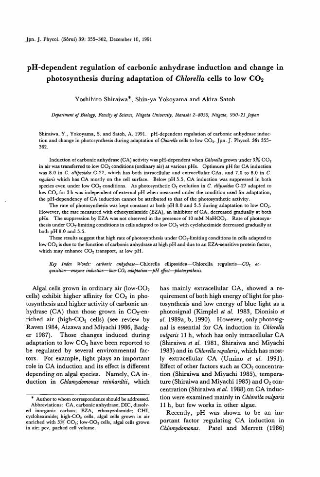
Jpn. J. Phycol. (Sdrui) 39: 355-362, December 10, 1991
pH-dependent regulation of carbonic anhydrase induction and change in
photosynthesis during adaptation of Chlorella cells to low CO2
Y oshihiro Shiraiwa *, Shin-ya Y okoyama and Akira Satoh
Depart:脚 'nt(グBiology,Facull汐OJScience, Niigata Universil仇 lkar,ι.hi2-8050, Niigata, 950-21 Japan
Shiraiwa, Y., Yokoyama, S. and Satoh, A. 1991. pH-dependent regulation of carbonic anhydrase induc-tion and change in photosyn出国isduring adaptation of Chlorella cells to low CO2・Jpn.J. Phycol. 39: 355-362.
Induction of carbonic副 lhydr酪 e(CA) activity was pH-dependent when Chlorella grown under 3% CO2
in air was transferred to low CO2 conditions (ordinary air) at various pHs. Optimum pH for CA induction W剖 8.0in C. el,ゆsoifkaC-27, which has bo也 intraαllularand extracellular CAs, and 7.0 to 8.0 in C. regularis which has CA mostly on the cell surface. Below pH 5.5, CA induction was suppressed in bo也species even under low CO2 conditions. As photosyn出eticO2 evolution in C. ell:伊o油aC-27 adapted to low CO2 for 3 h was independent of external pH when measured under the condition used for adaptation, the pH-dependency of CA induction cannot be attributed to白紙 ofthe photosynthetic activity.
τ'he rate of photosyn出esiswas kept constant at bo出 pH8.0 and 5.5 during adaptation to low CO2•
However, the rate measured with ethoxyzolamide (EZA),組 inhibitorof CA, decreased gradual1y at bo也pHs. The suppression by EZA was not observed in the presence of 10 mM NaHC03・ Rateof photos戸1・
th田 isunder CO2-limiting conditions in cells adapted to low CO2 with cycloheximide decreased gradually at both pH 8.0 and 5.5.
These results suggest that high rate ofphotosynthesis under CO2-1imiting conditions in cells adapted to low CO2 is due to也efunction of carbonic anhydrase at high pH and due to阻 EZA-sensitiveprotein factor, which may enhance CO2 transport, at low pH.
Key Infkx 陥 rds: ca伽 nIc anhydl間 'e-Chlorella ellipsoidea.ーChlorella regularis-C02 ac-quisition-enzyme induction-l,仰ぐO2a仰 ation-pHe.ffect-photosynthesis.
Algal cells grown in ordinary air (10w-C02
cells) exhibit higher a伍nityfor CO2 in pho・
tosynthesis and higher activity of carbonic an-
hydrase (CA) than those grown in CO2・en-
riched air (high-C02 cells) (see review by
Raven 1984, Aizawa and Miyachi 1986, sadg-
er 1987). Those changes induced during
adaptation to low CO2 have been reported to
be regulated by several environmental fac-
tors. For example, light plays an important role in CA induction and its effect is different
depending on algal species. Namely, CA in-
duction in Chlamydomonas reinhardtii, which
* Au出.orto whom correspondence should be addressed. Abbreviations: CA, c釘 bonicanhydrase; DIC, dissolv-
ed inorganic carbon; EZA, ethoxyzolamide; CHI, cycloheximide; high-C02 cells, algal cells grown in air enriched with 3% CO2; low-C02 cells, algal cells grown in air; pcv, packed cell volume.
has mainly extracellular CA, showed a re-
quirement ofboth high energy oflight for pho・
tosynthesis and low energy of blue light as a
photosignal (Kimplel et al. 1983, Dionisio et al. 1989a, b, 1990). However, only photosig-
nal is essential for CA induction in Chlorella
vulgaris 11 h, which has only intracellular CA
(Shiraiwa et al. 1981, Shiraiwa叩 dMiyachi
1983) and in Chlorella regularis, which has most-ly extracellular CA (Umino et al. 1991).
Effect of other factors such as CO2 concentra-
tion (Shiraiwa and Miyachi 1985), tempera-ture (Shiraiwa and Miyachi 1985)姐 d02con-
centration (Shiraiwa et al. 1988) on CA induc-
tion were examined mainly in Chlorella vulgaris
11 h, but few works in other algae. Recently, pH was shown to be an im-
po目antfactor regulating CA induction in
Chlamydomonas. Patel and Merrett (1986)
356 Shiraiwa, Y., Yokoyama, S. and Satoh, A.
showed that CA induction during adaptation
of high-C02 cells to air was enhanced con-
comitantly with pH, although how the induc-tion of CA is controlled by pH is not elucidat-
ed yet. As dissociation of D1C is strongly de-
pendent on pH ofthe medium, phenomena in-duced by a change in D1C concentration, such as CA induction and change in an a伍m-
ty of photosynthesis, may be probable to be affected directly and/or indirect1y by change
in pH. Response of the induction of CAs
with different locations in a cell to external
pH is also interesting to be compared.
1n the present study, we therefore investi-gated the effect of pH on CA induction within
a wide range of pH, from 3 to 9, in two spe-cies of Chlorella which have different CA locali-
zation. Effect of pH on CO2 acquisition in
photosynthesis during adaptation ofhigh-C02 cells to low CO2 conditions was also tested.
Materials and Methods
A佐almaterials and culture-Chlorella ell争m・dea
Gerneck (1AM C-27) was obtained by cour-
tesy ofProf. T. Hirokawa ofNiigata Universi-
ty. Chlorella r,そgularis(Endo et al. 1974) was a
kind gift of Prof. S. Miyachi of U niversity of Tokyo. These algae were grown autotrophi-
cally in a flat oblong glass vessel containing
ca. 1.31iter of the inorganic MC medium
(Watanabe 1960). The suspensions were con-
tinuously aerated with ordinary air enriched
with 3% CO2 to obtain cells adapted to high
CO2 (high-C02 cells). To obtain air-adapted
cells (low-C02 cells) , the cells harvested by centrifugation were suepended in an appropri-
ate buffer at a density of 3 ml pcv・liter-1,and then transferred to air. The algal suspension
was continuously illuminated by 200 W-incan-descent reflector lamp (Toshiba, Tokyo) at 1.2 kW .m-2 (16 klux). The temperature dur-
ing the growth and the adaptation to air was
kept under the optimum conditions for pho-
tosynthesis and growth in each alga, namely at 250C in Chlorella ellipsoidea and at 300C in
Chlorella regularis. Determination 01 photosynthetic O2 evolution-Al-
gal suspension (5 ml) of Chlorella ell伊oideahar-
vested from the culture was immediately
transferred into a water-jacketed transparent
glass-cylinder equipped with a Clark-type oxy-
gen probe (Rank Brothers, London). After
1-min incubation in the dark, photosynthesis was initiated by illumination by a tungsten
projector lamp at 1.4 kW・m-2(18 klux) , and change in O2 concentration in the medium
was continuously measured by the 02-elec-
trode. The temperature was kept at 250C.
Carbonic anhydraseωsり -Enzyme assay was
carried out according to the method ofWilbur
and Anderson (1948). To measure CA activi-
ty localized on the cell surface of intact cells
(E), 2 ml of CO2・saturatedwater was added
to 3 ml of 20 mM (final concentration, 12 mM) sodium veronal buffer (pH 8.3) con-
taining 100μl of the suspension of intact cells
suspended in 100 mM Tris-H2S04 buffer
(pH 8.3). The time required for pH change
from 8.3 to 7.3 was measured at 20C. The
reaction mixture was continuously stirred by
a magnetic stirrer. To determine CA activity
in cell homogenates (H), 100μl of homo-
genates obtained by disruption of algal cells
suspended in 100 m M Tris-H2S04 buffer
(pH 8.3) with a French Pressure Cell (Ohtake
Seisakusho, Tokyo) at 147 MPa (1,500 kg. cm -2) was used for the CA ass可・ Enzyme
units were calculated using the following
equatlOn:
units=tb・te-1-1
where tb and te represent the time (seconds)
needed for the pH change with or without
sample, respectively. 1nternal CA activity (1) was calculated by H -E.
Results
Effect of pH on the increase in CA activity
during adaptation of high-C02 cells to low
CO2 for 3 h was tested in Chlorella elliPsoidea c・27and Chlorella regularis (Fig. 1). CA activ-
ity detected both inside the cells and on the
cell surface was almost equal in Chlorella ell伊soidea C・27,whereas more than 97% of CA was located on the cell surface in Chlorella regularis. Both intra-and extracellular CA ac-
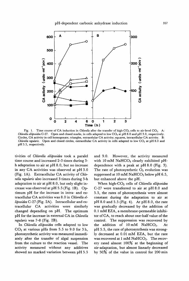
pH -dependent carbonic anhydrase induction 357
600 ~ A
500
『
2400
ε
~3∞ }
g 100
o o
B 300
言
a冨ョ
2 3 4 6 0 1 Tin栂 (h)
2
Fig. 1. Time course of CA induction in Chlorella after the transfer of high-C02 cells to air-level CO2. A: Chlorella eU争'soideaC・27. Open and closed marks, in cells adapted to low CO2 at pH 8.0 and pH 3.0, respectively. Circles, CA activity in cell homogenate; triangles, extracelular CA activity; squares, intracellular CA activity. B: Chlorella regularis. Open and closed circles, extracellular CA activity in cells adapted to low CO2 at pH 8.0組 dpH 5.5, respectively.
tivities of Chlorella ell砂soideatook a paralel
time course and increased 2-3 times during 3・
h adaptation to air at pH 8.0, but no increase in any CA activities was observed at pH 3.0
(Fig. 1A). Extracellular CA activity of Chlo-
rella r,唱gularisalso increased 3 times during 3・h
adaptation to air at pH 8.0, but only slight in-crease was observed at pH 5.5 (Fig. 1B). Op-
timum pH for the increase in intra-and ex-
tracellular CA activities was 8.0 in Chlorella el-
lipsoidea C・27(Fig. 2A). Intracellular and ex-
tracellular CA activities were similarly
changed depending on pH. The optimum
pH for the increase in external CA in Chlorella
r唱gulariswas 7-8 (Fig. 2B). In Chlorella ellipsoidea cells adapted to low
CO2 at various pHs from 5.5 to 9.0 for 3 h, photosynthetic activity was measured immedi-
ately a仕erthe transfer of algal suspension
from the culture to the reaction vessel. The activity measured without any additives
showed no marked variation between pH 5.5
and 9.0. However, the activity measured with 10 m M NaHC03 clearly exhibited pH-
dependence with a peak at pH 8.0 (Fig. 3).
The rate of photosynthetic O2 evolution was
suppressed at 10 mM NaHC03 below pH 6.5, but enhanced above the pH.
When high-C02 cells of Chlorella eU伊oidea
C-27 were transferred to air at pH 8.0 and
5.5, the rates of photosynthesis were almost constant during the adaptation to air at
pH 8.0 and 5.5 (Fig. 4). At pH 8.0, the rate was gradually decreased by the addition of
0.1 mM EZA, a membrane-permeable inhibi-
tor ofCA, to reach about one-halfvalue ofthe control. The suppression was recovered by the addition of 10 m M NaHC03 • At
pH 5.5, the rate ofphotosynthesis was strong-ly decreased at 0.01 m M EZA, but the rate was recovered at 1 m M NaHC03・Therecov-
e可 ratedalmost 100% at the beginning of
air-adaptation, but almost linearly decreased by 50% of the value in control for 200 min
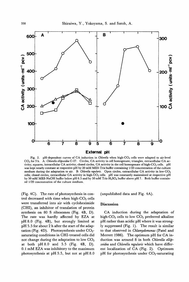
358 Shiraiwa, Y., Yokoyama, S. and Satoh, A.
600~ A 300
B
_ 500 、 ¥ -
盆'i F
200 ~ E 400 . 苦ω ヨさ
、旬圃, 、.圃,
f300 詰~ 200 υ 100g
1001 • o
6 7 8 9 5 6 7 8 9 o
External pH Fig. 2. pH-dependent curves of CA induction in Chlorella when high-C02 cells were adapted to air-level
CO2 for 3 h. A: Chlorella ellipsoidea C-27. Circles, CA activity in cell homogenate; triangles, extracellular CA ac-tivity; squares, intracellular CA activity; closed circles, CA activity in the cell homogenate of high-C02 cells. pH was kept nearly constant at respective pH by 20 mM MES-Tris buffer containing 1120 concentration of the culture medium during the adaptation to air. B: Chlorella regularis. Open circles, extracellular CA activity in low-C02
cells; closed circles, extracellular CA activity in high-C02 cells. pH was constantly maintained at respective pH by 50 mM MES-NaOH buffer below pH 6.5 and by 50 mM Tris-H2SO. buffer above pH 7. Both buffer contain-ed 1120 concentration of the culture medium
(Fig.4C). The rate ofphotosynthesis in con-
trol decreased with time when high-C02 cells
were transferred into air with cycloheximide
(CHI), an inhibitor of translation of protein synthesis on 80 S ribosomes (Fig. 4B, D). The rate was hardly affected by EZA at
pH 8.0 (Fig. 4B), but strongly limited at pH 5.5 for about 2 h after the start ofthe adap-
tation (Fig. 4D). Photosynthesis under CO2・
saturating conditions in CHI-treated cells did
not change during the adaptation to low CO2
at both pH 8.0 and 5.5 (Fig. 4B, D). 0.1 mM EZA was inhibitory to the maximum
photosynthesis at pH 5.5, but not at pH 8.0
(unpublished data and Fig. 4A).
Discussion
CA induction during the adaptation of
high-C02 cells to low CO2 preferred alkaline
pH rather than acidic pH where it was strong-
ly suppressed (Fig. 1). The result is similar
to that observed in Chla吻 domonas(Patel and
Merrett 1986). The optimum pH for CA in-
duction was around 8 in both Chlorella ell伊soidea and Chlorella regularis which have diffeト
ent localization of CA (Fig. 2). Optimum
pH for photosynthesis under CO2-saturating
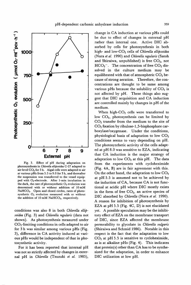
pH -dependent carbonic anhydrase induction 359
..... 、T z . E を1000. 6
E ::J. 750 、-
。制
o 678 External pH
9
Fig. 3. Effect of pH during adaptation on photosynthesis in Chlorella ellipsoidea C-27 adapted to air-level CO2 for 3 h. Algal cells were adapted to air at v訂 iouspHs from 5.5 to 9.0 for 3 h, and thereafter the suspension was住ansferredto the vessel equip-ped with 02-electrode. After l-min incubation in the dark, the rate ofphotosynthetic O2 evolution was determined wi出 orwithout addition of 10 mM NaHC03• Open and closed circles, rates of photo-synthetic O2 evolution measured with or without the addition of 10 mM NaHC03, respectively.
conditions was also 8 in both Chlorella ell争・so地'a(Fig. 3) and Chlorella regularis (data not shown). As photosynthesis measured under
CO2・limitingconditions in cells adapted to air for 3 h was similar among various pHs (Fig.
3), difference in CA activity induced at v訂 i-
ous pHs would be independent of that in pho-tosynthetic activity.
For it has been reported that internal pH
was not so stricdy affected by changes in exter-nal pH in Chlorella (Tsuzuki et al. 1985),
change in CA induction at various pHs could
be due to effect of changes in external pH rather than internal one. Active DIC ab-
sorbed by cells for photosyn曲目isin both
high-and low-C02 cells of Chlorella ell争soidea(Nara et al. 1990) and Chlorella regularis (Satoh and Shiraiwa, unpublished) is free CO2, not HC03 -. The concentration of free CO2 dis-
solved in the culture medium may be
equilibrated with that of atmospheric CO2 be-
cause of strong aeration. Therefore, the con-centrations are thought to be same among
various pHs because the solubility of CO2 is
not affected by pH. These things also sug-
gest that DIC acquisition and CA induction
are controlled mainly by changes in pH of the
medium. When high-C02 cells were transferred to
low CO2, photosynthesis can be limited by CO2 transfer from the medium to the site of
CO2 fixation by ribulose-1 ,5-bisphosphate car-boxylase/oxygenase. Under the conditions, physiological basis of adaptation to low CO2 conditions seems to vary depending on pH.
The photosynthetic activity of the cells adapt-
ed at pH 8.0 was sensitive to EZA, indicating that CA induction is the major strategy of
adaptation to low CO2 at this pH. The data
from the experiments with cycloheximide
(Fig. 4A, B) are in fair agreement with this. On the other hand, the adaptation to low CO2 at pH 5.5 is assumed not to be achieved by
the induction of CA, because CA is not func-
tional at acidic pH where DIC mosdy exists
in the form of free CO2, an active species of DIC absorbed by Chlorella (Nara et al. 1990). A reason for inhibition of photosynthesis by
EZA at pH 5.5 (Fig. 4C, D) is not elucidated yet. A possible speculation may be the inhibi-
tory effect of EZA on the membrane transport
of DIC, since EZA affected the membrane permeability to glycolate in Chlorella vu駒市
(Shiraiwa and Schmid 1986). Notable白血isrespect is the fact that the adaptation to low
CO2 at pH 5.5 is sensitive to cycloheximide, as is at alkaline pHs (Fig. 4). This indicates
d四 protein(s) other than CA has to be s戸 the-sized for the adaptation, in order to enhance DIC utilization at low pH.
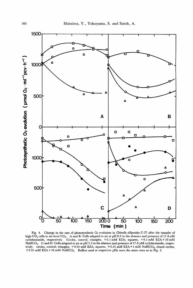
Shiraiwa, Y., Yokoyama, S. and Satoh, A. 360
A
1500
1000 (τz・〉
ωaτ-E・NO-。EE)
B A
。宣言-g@NOozo£-zgHSE
ω (min)
Fig. 4. Change in the rate of photosynthetic O2 evolution in Chlorella ell争'soideaC・27after the transfer of high-C02 cells to air-Ievel CO2・ Aand B: Cells adapted to air at pH 8.0 in the absence and presence of 17.8μM cycloheximide, respectively. Circles, control; triangles, +O.lmM EZA; squares, +O.lmM EZA+10mM NaHC03・Cand D: Cells adapted to air at pH 5.5 in the absence and presence of 17.8μM cycloheximide, respec-tively. circles, control; triangles, +0.01 m M EZA; squares, +0.01 mM EZA + 1 mM NaHC03; closed circles, +0.01 mM EZA + 10 mM NaHC03• Buffers used at respective pHs were the same ones as in Fig. 2
100 2000 Time
150 100
pH -dependent carbonic anhydrase induction 361
As shown in Fig. 3, photosynthesis in low-CO2 cells of Chlorella ell争soideaunder CO2・
saturated conditions was several times higher
at pH 8.0 than pH 5.5, although that under CO2-1imiting conditions was almost same at
various pHs. Photosynthesis at pH 5.5 was
diminished by the addition of high concentra-
tion of DIC. The reason for the suppression
is still unclear. One possibility, as assumed by Hogetsu and Miyachi (1979), is a strong
drop of internal pH caused by absorbing huge
amount of CO2 and the subsequent conver-
sion to HC03 - and H+. These results in
Chlorella are inconsistent with those, reported in Chlamydomonas by Patel 組 d Merrett
(1986), showing that photosynthesis under CO2-saturated conditions at pH 5.5 was three
times higher than that at pH 7.5 in both high-
and low-C02 cells. As the concentration of
DIC accumulated in the cells was two times
higher at pH 7.5 than pH 5.5 in low-C02
cells, but was independent ofpH in high-C02
cells, it is considered that photos戸ltheticactiv-
ity is dependent on external pH proper, but independent of internal DIC accumulated.
The mechanism how external pH affects the
CA induction and the change in photosynthe-
tic CO2 fixation during adaptation to low CO2, and the reason why pH -dependency of pho-
tosynthesis is different between Chlorella and
Chlamydomonas remains to be elucidated.
Acknowledgement
The authors are grateful to Dr. T. Suzuki
for correcting english and to Mrs. J. Nagashi-
ma for wordprocessing the manuscript.
References
Aizawa, K. and Miyachi, S. 1986. Carbonic anhycirase and CO2 concentrating mechanisms in microalgae 姐 dcyanobacteria. FEBS Microbiology Reviews 39: 215-233.
Badger, M. R. 1987. The CO2-concentrating mecha-nism in aquatic phototrophs. p. 219-274. In M. D. Hatch and N. K. Bo紅也nan[eds.) The Biochemis-try of Plants. Vol. 10. Photosynthesis. Acad. Press, San Diego.
Dionisio, M. L., Tsuzuki, M. and Miyachi, S. 1989a. Light requirement for carbonic anhycirase induction
in Chlamydomonas reinhardtii. Plant Cell Physiol. 30: 207-213.
Dionisio, M. L., Tsuzuki, M. and Miyachi, S. 1989b. Blue light induction of carbonic anhydrase activity in Chlamydomonas reinhardtii. Plant Cell Physiol. 30: 215-219.
Dionisio-Sese, M. L., Fukuzawa, H.加 dMiyachi, S. 1990. Light-induced carbonic anhydrase expres-sion in Chlamydomonas reinhardtii. Plant Physiol. 94: 1103-1110.
Endo, H., Nakajima, M., Chino, R. and Shirota, M. 1974. Growth characteristics and cellular compo-nents of Chlorella 符官laris,heterotropic fast growing strain. Agr. Biol. Chem. 38: 9-18.
Hogetsu, D. and Miyachi, S. 1979. Role ofcarbonic組 ・
hydrase in photosynthetic CO2 fixation in Chlorella. Plant Cell Physiol. 20: 747-756.
Kimpel, D. L., Togasaki, R. K. and Miyachi, S. 1983. Carbonic anhydrase in Chlamydomonas reinhardtii 1. Localization. Plant Cell Physiol. 24: 255-259.
Nara, M., Shiraiwa, Y. and Hirokawa, T. 1990. En-zymatic inactivation of extracellular carbonic anhy-drase and its effect on KlI2 (C02) for photosynthesis in Chlorella eU伊'oideaC・27.Plant Cell Physiol. 31: 961-967.
Patel, B. N. and Merrett, M.J. 1986. Regulation ofcar-bonic anhydrase activity, inorganic-carbon uptake and Photosynthetic biomass yield in Chla町'domon副
reinhardtii. Planta 169: 81-86.
Raven, J. A. 1984. Energetics and Transport in Aquat-ic Plants. Alan R. Liss, Inc. New York.
Shiraiwa, Y. and Miyachi, S. 1983. Factors controlling induction of carbonic anhydrase and efficiency of photosynthesis in Chlorella v咋'arIs11h cells. Plant Cell Physiol. 24: 919-923.
Shiraiwa, Y. and Miyachi, S. 1985. Effects oftempera-ture and CO2 concentration on induction of carbon-
ic anhydrase and changes in efficiency of photosyn-thesis in Chlorella山 19aris11h. Plant Cell Physiol. 26: 543-549.
Shiraiwa, Y., Fakler,J. and Miyachi, S. 1981. Factors controlling carbonic anhydrase activity in Chlorella 四台'arisllh. p. 493-499. In G. Ako刊 noglou[ed.) Photosynthesis IV, Regulation of Carbon Metabolism. Balaban Intemational Science Service, Philadelphia.
Shiraiwa, Y., Satoh, H. and Hirokawa, T. 1988. Fac-tors controlling induction of carbonic anhydrase in Chlorella叩 19aris11h: effects of CO2阻 dO2・PlantCell Physiol. 29: 731-734.
Shiraiwa, Y.阻 dSchmid, G. H. 1986. Stimulation of photor四 pirationby the carbonic anhydrase inhibi-tor ethoxyzolamide in Chlorella四 19aris.Z. Natur-forsch. 41c: 56←570.
Tsuzuki, M., Miyachi, S. and Berry, J. A. 1985. In-tracellular accumulation of inorgani
362 Shiraiwa, Y., Yokoyama, S. and Satoh, A.
ganic Carbon Uptake by Aquatic Photosynthetic
Organisms. The American Society of Plant Phys-
iologists, Rockville Umino, Y., Satoh, A. and Shiraiwa, Y. 1991. Factors
controlling induction of external carbonic anhydrase
and change in K1I2 (C02) of photosynthesis in Chlorel-
la regularis. Plant Cell Physiol. 32: 379-384.
もへTatanabe,A. 1960. List of algal strains in collection at
the Institute of Applied Microbiology, University of Tokyo. J. Gen. Appl. Microbiol. 6: 283-292.
Wilbur, K. M. and Anderson, N. G. 1948. Electromet-
ric and colorimetric determination of carbonic anhy-
drase, J. Biol. Chem. 176: 147-154.
白岩善博・横山真也・佐藤朗:クロレラの低 CO2条件への適応に伴う
力ルボニックアンヒドラーゼ誘導と光合成活性変動の pHによる調節
カノレボニッタアンヒドラーゼ (CA)の局在性が異なるこ種の単細胞緑漠 Chlorellaell伊oideaC-27および Chlorella
regula仰を用いて低 CO2条件への適応に伴う CA誘導および光合成の CO2に対する親和性の変動に及ぼす pHの
影響を調べた。 CA誘導の至適pHは藻種および CAの局在性に関わらずpH8付近であり, pH5.5以下では CA
の誘導は認められなかった。高 (3%)C02条件に適応した C.ellitsoideaを低 (0.03%)C02条件に移した場合,
CO2律速条件における光合成活性は pH5.5および8.0のいずれの pHでも変動しなかった。しかし, CA阻害剤
であるエトキシゾルアミド添加条件下では CO2律速条件下での光合成は時間と共に減少した。また,いづれの
pHでも,低CO2への適応時にシクロヘキシミドを添加すると,光合成の CO2に対する親和性の増大が阻害さ
れた。以上の結果より,これらの藻種が,アルカリ域では CAを誘導することにより,また,酸性域では CO2
利用を促進する CA以外のタンパク性因子を誘導することにより CO2律速条件下での光合成活性を維持してい
ることが示唆された。 (950-21新潟市五十嵐二の町8050 新潟大学理学部生物学科)
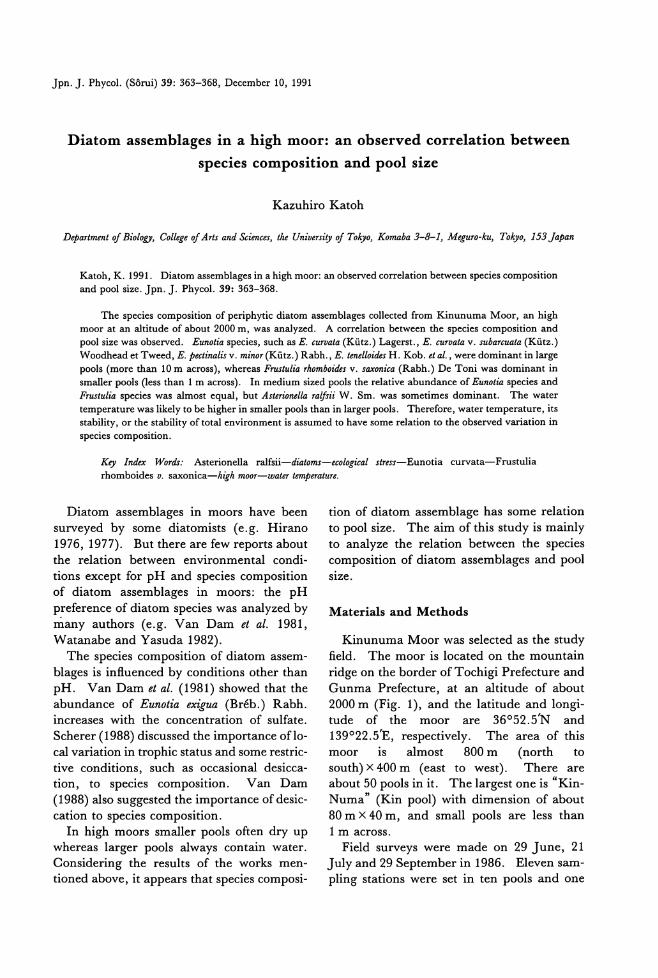
Jpn. J. Phycol. (Sorui) 39: 363-368, December 10, 1991
Diatom assemblages in a high moor: an observed correlation between
species composition and pool size
Kazuhiro Katoh
D駒市間toj Biol,切ら Collegeoj Arts and Sciences, the University oj Tokyo, Komaba 3-8-1, Meguro-ku, To.砂'0,153 Japan
Katoh, K. 1991. Diatom assemblages in a high moor: an observed correlation between species composition 阻 dpool size. Jpn. J. Phycol. 39: 363-368.
τ'he species composition of periphytic diatom assemblages collected from Kinunuma Moor,佃 highmoor at an a1titude of about 2000 m, was analyzed. A correlation between the species composition and pool size was observed. Eunotia species, such as E. curvata (Kutz.) Lagerst., E. curv仰 v.subarcuata (Kutz.) Woodhead et Tweed, E. pectinalis v. minor(Kutz.) Rabh., E. t四elloidesH. Kob. et al., were dominant in large pools (more出回 10m across), whereas Frustulia幼omboidesv. saxonica (Rabh.) De Toni was dominant in smal1er pools (less由an1 m across). In medium sized pools the relative abundance of Eunotia species and FrustulUl species was a1most equal, but Asterionella raljsii W. Sm. was sometimes dominant. The water temperature was likely to be higher in smaller pools也anin larger pools. Therefore, water temperature, its stability, or the stability of total environment is assumed to have some relation to the observed variation in specles composltlOn.
Key lndex Words: Asterionella ralfsii-diato即 -ecologicalstress-Eunotia curvata-Frustulia rhomboides v. saxonica-h併 moor-脚 tertempera的拡
Diatom assemblages in moors have been
surveyed by some diatomists (e.g. Hirano
1976, 1977). But there are few reports about
the relation between environmental condi-
tions except for pH and species composition
of diatom assemblages in moors: the pH
preference of diatom species was analyzed by
many authors (e.g. Van Dam et al. 1981, Watanabe and Yasuda 1982).
The species composition of diatom assem-
blages is insuenced by conditions other than
pH. Van Dam et al. (1981) showed that the
abundance of Eunotia exなua(Breb.) Rabh.
increases with the concentration of sulfate.
Scherer (1988) discussed the importance oflo・
cal variation in trophic status and some restric-tive conditions, such as occasional desicca-tion, to species composition. Van Dam
(1988) also suggested the importance of desic-
catlOn to specles composltlOn.
In high moors smaller pools often dry up
whereas larger pools always contain water.
Considering the results of the works men-
tioned above, it appears that species composi-
tion of diatom assemblage has some relation
to pool size. The aim of this study is mainly
to analyze the relation between the species
composition of diatom assemblages and pool
slze.
Materials and Methods
Kinunuma Moor was selected as the study
fi.eld. The moor is located on the mountain
ridge on the border of Tochigi Prefecture and
Gunma Prefecture, at an altitude of about 2000 m (Fig. 1), and the latitude and longi-tude of the moor are 36052.5'N and
139022.5'E, respectively. The area of this
moor is almost 800 m (nor出 to
south) x 400 m (east to west). There are
about 50 pools in it. The largest one is “Kin-Numa" (Kin pool) with dimension of about
80 m X 40 m, and small pools are less than 1 m across.
Field surveys were made on 29 J une, 21 July and 29 September in 1986. Eleven sam-
pling stations were set in ten pools and one
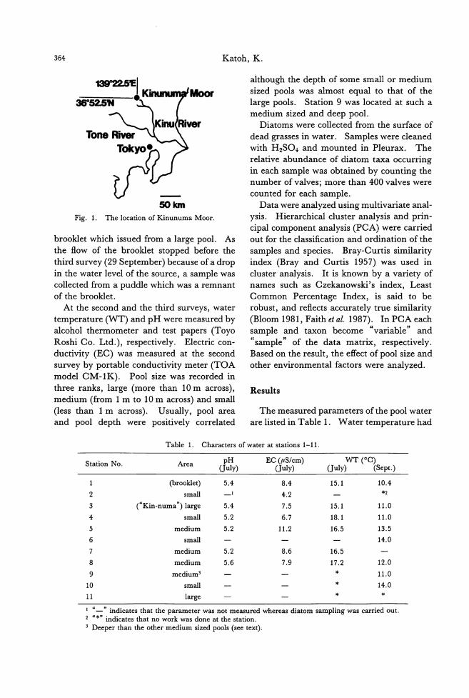
364 Katoh, K.
although the depth of some small or medium
刷Ioor sized pools was almost equal to that of the large pools. Station 9 was located at such a
50胞n
Fig. 1. The location of Kinunuma Moor.
brooklet which issued from a large pool. As
the flow of the brooklet stopped before the
third survey (29 September) because of a drop
in the water level ofthe source, a sample was
collected from a puddle which was a remnant of the brooklet.
At the second and the third surveys, water temperature (WT) and pH were measured by
alcohol thermometer and t回 tpapers (Toyo
Roshi Co. Ltd.), respectively. Electric con-
ductivity (EC) was measured at the second
survey by portable conductivity meter (TOA
model CM-1K). Pool size was recorded in
three ranks, large (mo問 than10m across), medium (仕om1 m to 10m across) and small
(less than 1 m across). Usually, pool area and pool depth were positively correlated
medium sized and deep pool. Diatoms were collected from the surface of
dead grasses in water. Samples were cleaned
with H2S04, and mounted in Pleurax. The
relative abundance of diatom taxa occurring
in each sample was obtained by counting the number of valves; more than 400 valves were
counted for each sample.
Data were analyzed using multivariate anal-
ysis. Hierarchical cluster analysis and prin-
cipal component analysis (PCA) were carried
out for the classification and ordination of the
samples and species. Bray-Curtis similarity
index (Bray and Curtis 1957) was used in
cluster analysis. It is known by a variety of
names such as Czekanowski's index, Least Common Percentage Index, is said to be
robust, and reflects accurately true similarity (Bloom 1981, Faith et al. 1987). In PCA each
sample and taxon become “variable" and
“sample" of the data matrix, respectively. Based on the result, the effect of pool size and other environmental factors were analyzed.
Results
The measured parameters of the pool water
are listed in Table 1. Water temperature had
Table 1. Characters of water at stations 1-11
Station No. Area J品) ECddJ;叫WT (OC) 。uly) (Sept.)
(brook1et) 5.4 8.4 15.1 10.4
2 sma11 4.2 *2
3 (“Kin-numa") large 5.4 7.5 15.1 11.0
4 sma11 5.2 6.7 18.1 11.0
5 medium 5.2 11.2 16.5 13.5
6 sma11 14.0
7 medium 5.2 8.6 16.5
8 medium 5.6 7.9 17.2 12.0
9 medium3 * 11.0
10 sma11 * 14.0
11 large * *
1“一"indicates that the parameter was not measured whereas diatom sampling was carried out. 2“*" indicates that no work was done at the station. 3 Deeper than the 0出ermedium sized pools (see text).
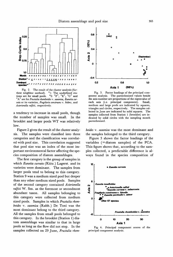
Diatom assemblage and pool size
島
崎
n
n
u
n
M
M@明HEst20,hmwr白血
9
7
F
9
8
F
9
9
E
7
8
A
,,,,
Aun
aB
・A-F
M
7
3
E
S
S
E
7
1
E
9
回-
F
9
6
-
F
7
6
-
F
7
4
-
F
7
2
-
F
6
7
F
9
1
F
7
5
F
a
a
a
d
E
r
e
,F
e
S
F
9
4
-
F
e
H
F
"
l
A
・
叩
向
山
…
…
Fig. 2. The result of the cluster analysis (fur-thest neighbor method)ー *1田 Theunderlined sta-tions are for small pools. *2:“F'¥“Eぺ“c"and “A" are for Frustulia rhomboides v. saxonica, Eunotia cur-vata or its varieties, Fragilaria construens v. bidens, and Asterionella問。'sii,respectively.
a tendency to increase in small pools, though the number of samples was small. In the
brooklet and larger pools WT was relatively
low.
Figure 2 gives the result ofthe cluster analy-sis. The samples were classified into three
categories and the classification was correlat-
ed with pool size. This correlation suggest巴d
that pool size was an index of the most im-
po口antenvironmental factor affecting the spe-
cies composition of diatom assemblages.
The first category is the group of samples in
which Eunotia curvata (Kutz.) Lagerst. and its
varieties were dominant. The samples from
larger pools tend to belong to this category.
Station 9 was a medium sized pool but deeper
than any other medium sized pools. Samples
of the second category contained Asterionella
ralfsii W. Sm. as the foremost or secondmost
abundant taxon. Al1 samples belonging to
this category were col1ected from medium sized pools. Samples in which Frustulia rhom-
boides v. saxonica (Rabh.) De Toni was the
most dominant belong to the third category.
All the samples from small pools belonged to
this catego町・ Inthe brooklet (Station 1) dia-tom assemblage was similar to that in large
pools as long as the ftow did not stop. In the
samples collected on 29 June, F.ηlstulia rhom-
365
• t占avBs
。
.. 企
(8却 包 噛 叫。 .. ,‘.
• .. . 2
-0.4
0.2 0.6 1.0
Axis 1 (68%)
Fig. 3. Factor loadings of the principal com-ponent analysis. The paren出esizedvalues beside the axis number are proportions of the eigenvalue of each axis (i.e. principal component). Small, medium and large pools are indicated by squares, triangles and circles, respectively. The samples col-lected in June are indicated by solid squares. The samples collected from Station 1 (brooklet) are in-dicated by solid circles with the sampling month p紅白血esized.
boides v. saxonica was the most dominant and
the samples belonged to the third category.
Figure 3 shows the factor loadings of the
variables (=diatom samples) of the PCA.
This figure shows that, according to the sam-ples col1ected, a predictable difference is al-
ways found in the species composition of
包
• Eunotla curva白
6 I Eunotla蜘 e//old.田
2| ・.As,制 'one//aral1sJJ 同 l ・'Eunotlacurva加 .suban:ua臼
ot IFn郁#larlacon,府 U四 sv.bJnodJお・・Eu.国泌ap副油価.alJsv.m,旬。roト ...
ー-0世聞 67ap副お8
-FrustulJa助副obokl.田 v.割自由曲理
-6
o 却 30 10
Axis 1 Fig. 4. Principal component scores of the
principal component analysis.
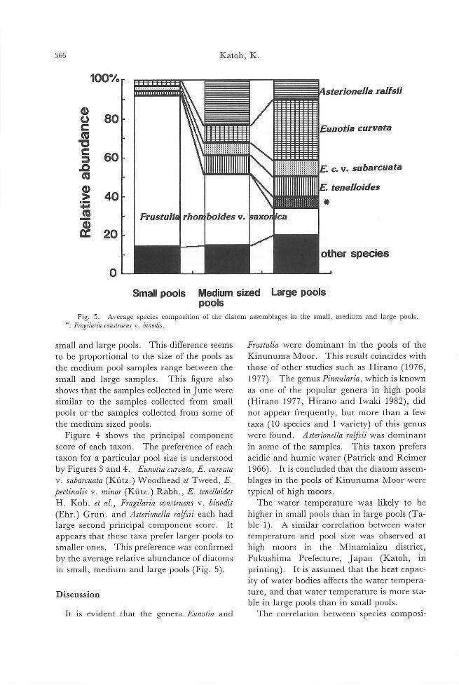
366 Katoh, K
100% 1111111111111111111 l~恥 raJfsii
。80 ω
c 1 1 111111111111Eunotia curvata 伺司3z
60 = a 四11川11川1111111¥ c. v. subarcuata 何
。>40
.. ・4伺
巴。20
o Small pools Medium sized Large pools
pools Fig. 5. Average species composition of the diatom assembJages in the smaJl, medium and Jarge pooJs
*: Fragilaria conslruens v. binodis
small and large pools. This difference s巴ems
to be proportional to the size of the pools as
the medium pool samples range between the
small and large samples. This figure also
shows that th巴samplescollected in June were
similar to the samples collected from small
pools or the samples collected from some of
the medium sized pools
Figure 4 shows the principal component
score of each taxon. The prefl巴renceof巴ach
taxon for a particular pool size is understood
by Figures 3 and 4. Eunotia curvata, E. curvata v. subarcωta (Kutz.) Woodhead et Tweed, E
pectinalis v. minor (K凸tz.)Rabh., E. tenelloides H. Kob. et al.) Fragilaria construens v. binodis
(Ehr.) Grun. and Asterionella ralfsii巴achhad
large second principal component score. It
appears that these taxa prefer larger pools to
smaller ones. This preference was confirmed
by the average relative abundance of diatoms
in small, medium and large pools (Fig. 5).
Discussion
It is evident that the genera Eunotia and
Frustulia were dominant in the pools of the
Kinunuma Moor. This result coincides with
those of other studies such as Hirano (1976, 1977). The genus Pinnular肌 whichis known
as one of the popular genera in high pools
(Hirano 1977, Hirano and I waki 1982), did
not appear frequently, but more than a few
taxa (10 species and 1 variety) of this genus
were found. Asterionella rafβii was dominant
in some of the samples. This taxon prefers
acidic and humic water (Patrick and Reimer
1966). It is concluded that the diatom assem-
blages in the pools of Kinunuma Moor were
typical of high moors.
The water temperature was likely to be
higher in small pools than in large pools (Ta-
ble 1). A similar correlation b巴tweenwater
temperature and pool size was observed at
high moors in the Minamiaizu district, Fukushima Prefecture, J apan (Katoh, in printing). It is assumed that the heat capac-
ity of water bodies affects the water tempera-
ture, and that water temperature is more sta-
ble in large pools than in small pools.
Th巴 corr巴lationbetween species composi-
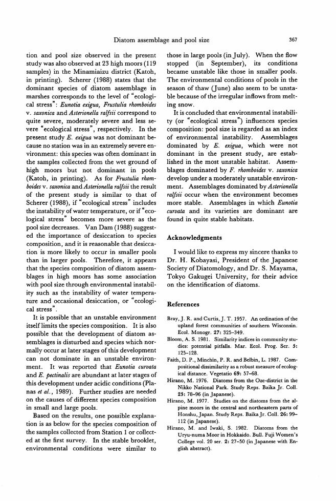
Diatom assemblage and pool size 367
tion and pool size observed in the present study was also observed at 23 high moors (119 samples) in the Minamiaizu district (Katoh, in printing). Scherer (1988) states that the dominant species of diatom assemblage in
marshes corresponds to the level of “ecologi-cal stress": Eunotia ex忽叫 Frustuliarhomboides v. saxonica and Asterionella ralfsii correspond to quite severe, moderately severe and less se-vere“ecological stressぺrespectively. In the present study E. exigua was not dominant be-cause no station was in an extremely severe en-vironment: this species was often dominant in the samples collected from the wet ground of high moors but not dominant in pools (Katoh, in printing). As for Frustulia rhom-boides v. saxonica and Asterionella ralfsii the result of the present study is similar to出atof
Scherer (1988), if“ecological stress" includes the instability ofwater temperature, or if“eco-logical stress" becomes more severe as the
pool size decreases. V姐 Dam(1988) suggest-ed the import組問 ofdesiccation to species composition, and it is reasonable也atdesicca-tion is more likely to occur in smaller pools th叩 inlarger pools. Therefore, it appears that the species composition of diatom assem-blages in high moors has some association with pool size through environmental instabil-ity such as the instability of water tempera-ture and occasional desiccation, or“ecologi-cal stress".
It is possible that an unstable environment
itselflimits the species composition. It is also possible that the development of diatom as-semblages is disturbed and species which nor-
mally occur at later stages of this development can not dominate in an unstable environ-ment. It was reported that Eunotia curvata
and E. pectinalis are abundant at later stages of
this development under acidic conditions (Pla-nas et al., 1989). Further studies are needed on the causes of different species composition in small and large pools.
Based on the results, one possible explana-tion is as below for the species composition of the samples collected from Station 1 or collect-ed at the first survey. In the stable brooklet, environmental conditions were similar to
those in large pools (inJuly). When the sow stopped (in September), its conditions became unstable like those in smaller pools.
The environmental conditions of pools in the
season of thaw (June) also seem to be unsta-ble because of the irregular insows from melt-
mg snow. It is concluded出atenvironmental instabili-
ty (or“ecological stress") insuences species composition: pool size is regarded as an index of environmental instability. Assemblages dominated by E. exなω,which were not dominant in the present study, are estab-lished in the most unstable habitat. Assem-blages dominated by F. r.加mboid旨'sv. saxonzca develop under a moderately unstable environ-ment. Assemblages dominated by Asterionella ralfsii occur when the environment becomes more stable. Assemblages in which Eunotia curvata and its varieties are dominant are
found in quite stable habitats.
Acknowledgments
1 would like to express my sincere thanks to
Dr. H. Kobayasi, President of the Japanese Society ofDiatomology, and Dr. S. Mayama, Tokyo Gakugei University, for their advice on the identification of diatoms.
References
Bray,J. R.副 dCurtis,J.T.1957. Anordinationofthe upland forest communities of southern Wisconsin. Ecol. Monogr. 27: 325-349.
Bloom, A. S. 1981. Similarity indices in community stu-dies: potentia1 pitfalls. Mar. Ecol. Prog. Ser. 5:
125-128. Fai也,D.P., Minchin, P. R. andBelbin, L. 1987. Com-
positiona1 dissimilarity as a robust measure of ecolog-ica1 distance. Vegetatio 69: 57-68.
Hirano, M. 1976. Diatoms from the Oze-district in the Nikko Nationa1 Park. Study Reps. Baika Jr. Coll. 25: 78-96 (in Japanese).
Hirano, M. 1977. Studies on也ediatoms from the a1-pine moors in the centra1 and northeastern parts of Honshu,Japan. Study Reps. BaikaJr. Coll. 26: 99-112 (inJapanese).
Hirano, M. and Iwaki, S. 1982. Diatoms from the Uryu・numaMoor in Hokkaido. Bull. Fuji Women's College vol. 20 ser. 2: 27-50 (inJapanese with En-glish abstract).
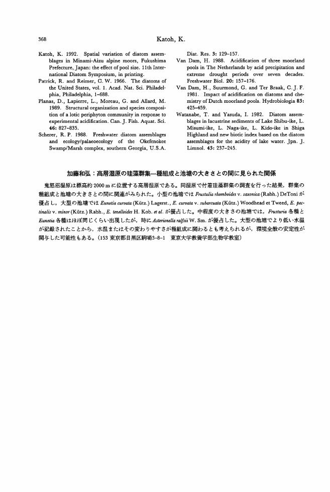
368 Katoh, K.
Katoh, K. 1992. Spatial variation of diatom assem-blages in Minami-Aizu alpine moors, Fukushima Prefecture, Japan: the effect ofpool size. 11th Inter-national Diatom Symposium, in printing.
Patrick, R. and Reimer, C. W. 1966. The diatoms of
the United States, vo1. 1. Acad. Nat. Sci. Phi1adel-phia, Phi1adelphia, 1-688.
Planas, D., Lapierre, L., Moreau, G.釦 dA11釘d,M. 1989. Structural organization組 dspecies composi-
tion of a lotic periphyton community in response to
experimental acidification. Can.J. Fish. Aquat. Sci.
46: 827-835. Scherer, R. P. 1988. Freshwater diatom assemblages
and ecology/palaeoecology of the Okefenokee
Swamp/Marsh complex, southern Georgia, U.S.A.
Diat. Res. 3: 129-157. V田 1Dam, H. 1988. Acidification of three moorland
pools in The Nether1ands by acid precipitation and
extreme drought periods over seven decades. Freshwater Bio1. 20: 157-176.
Van Dam, H., Suurmond, G. and Ter Braak, C.J. F. 1981. Impact of acidification on diatoms田ldche-
mistry of Dutch moorland pools. Hydrobiologia 83:
425-459.
Watanabe, T. and Yasuda, 1. 1982. Diatom assem-blages in lacustrine sediments ofLake Shibu-ike, L. Misumi-ike, L. Naga-ike, L. Kido・ikein Shiga
High1and and new biotic index based on the diatom
assemblages for the acidity of lake water. Jpn. J.
Limno1. 43: 237-245.
加藤和弘:高層湿原の珪藻群集一種組成と池塘の大きさとの聞に見られた関係
鬼怒沼湿原は標高約2000mに位置する高層湿原である。同湿原で付着珪藻群集の調査を行った結果,群集の
種組成と池塘の大きさとの聞に関連がみられた。小型の池纏ではF間tuliarlwmboides v. s,副 'onica(Rabh.) DeToniが
優占し,大型の池塘ではEunotia印刷ta(Kutz.) Lagerst., E. curvata v. subarcuata (Kutz.) Woodhead et Tweed, E. pec-
tinalis v. minor(Kutz.) Rabh., E.帥 elloi,釦 H.Kob. et al.が優占した。中程度の大きさの池塘では,Fl間'tu巾各種と
Eunotia各種はほぼ同じくらい出現したが,時にAsterionellaraifsii W. Sm.が優占した。大型の池塘でより低い水温
が記録されたことから,水温またはその変わりやすさが種組成に関わるとも考えられるが,環境全般の安定性が
関与した可能性もある。 (153東京都目黒区駒場3ーか1 東京大学教養学部生物学教室)

Jpn. J. Phycol. (Sorui) 39: 369-373, December 10, 1991
Ryozo Seto, R. N. Yadava and Shigeru Kumano: Development of short
spinous branch1ets of Compsopogon aeruginosus var. catenatum
(Compsopogonaceae, Rhodophyta)
Key ln伽陥油 central celZs一Compsopogon aeruginosus var. catenatum-fresh制御-
Rhodophyta-sp仇0山 branchlets.Ryozo Seto, Faculty of Domestic Science, Kobe College, Okado.-yama, Nishinomiya, 662 Japan R. N. Yadava, D.仰伽entof Botaり"Bhagalpur University, Bhagalpur 81200えlndiaShな'eruKumano, Department of Biol.ψ" Faculty of Science, Kobe Universi,仇 Rokko・dai,N.配お-ku,Kobe, 657 Japan
Krishnamurthy (1962) mentioned that Com-psopogon aeruginosa is very similar to C.
coerul叫ん butcan be distinguished by two sharp features: 1) the monospores are com-paratively small, and 2) the older thallus and the main axis bear short spinous branchlets. The latter feature, short spinous branchlets, is observed on the Indian specimens collected
from various parts of Gujrat by Patel and Francis (1969) and the Japanese specimens from Lake Shinji by Nakamura and Ch也m(1983).
Compsopogon aeruginosus var. catenatum, which has spinous branchlets in the nodulated
axis, was described as a new variety by YadavaandP阻止y(1980) based on the Indian specimens collected from Dorania River at Bareilly. A new form of Compsopogon aerugino・sωwith spinous branchlets was also repo凶edby Singh and Pandey (1986) from Nakatia River. This paper deals with the develop-ment of spinous branchlets based on the speci-
men of Compsopogon aeruginosus var. catenatum collected from Nakatia River in India.
Observations
1. Specimens e玄aminedin the present study: The specimens of Compsopogon
aeruginosus var. catenatum collected by R. N. Yadava from Nakatia River near Bareilly in India in August 1988 were deposited in the herbaria of Department of Botany, Universi-ty of Allahabad in India, and Department of Biology, Faculty of Science, Kobe University inJapan.
2. Development of filamentous branches prior to cortication: The younger portion ofthe thallus remains uniseriate. Prior to cor-tication, an erect thallus may give rise to sev-eral angular branches by oblique divisions of axial central cells. The cell destined to form a filamentous branch pushes out laterally組 ddistally and the protuberance thus formed is
delimited by a cross-wall as the initial cell of a filamentous branch. The initial cell then un-dergoes successive transverse divisions to give rise to a uniser泊tefilamentous branch (Figs. 1-3). This filamentous branch may eventual-
ly develop cortical cells in the same manner as themain砿 is. Filamentous branches general・
ly originate to form an angle of 30 to 60 degrees with reference to the main axis. Leteral filamentous branches of main thalli are also richly branched, arise alternatively, and are uniseriate.
Then, a uniseriate filamentous branch is
formed by a series of discoid cells developed by repeated transverse divisions of the dome-
shaped apical cell. A few cells below the api-cal cell of a branch do not divide while other
axial rows of cells divide periclinally and form a number of peripheral segments. These cells subsequently transform into a single lay-er of cortical cells. Further anticlinal and transverse divisions result in the formation of
one or two layers of cortical cells. 3. Development of short spinous branch-
lets: The old thallus bears many short spinous branchlets. They usually originate at right angles to the main axis from central cells of the old thalli, which have well-deve-
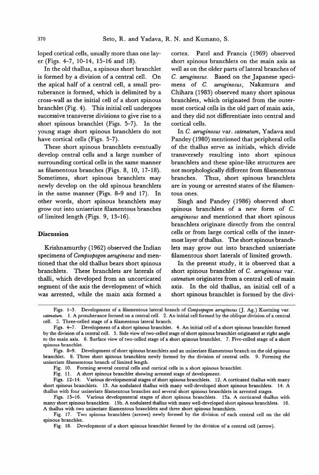
370 Seto, R. and Yadava, R. N. and Kumano, S.
loped cortical cells, usually more than one lay-er (Figs. 4-7, 10-14, 15-16 and 18).
In the old thallus, a spinous short branchlet
is formed by a division of a central cell. On
the apical half of a central cell, a small pro-
tuberance is formed, which is delimited by a
cross-wall as the initial cell of a short spinous
branchlet (Fig. 4). This initial cell undergoes
successive transverse divisions to give rise to a
short spinous branchlet (Figs. 5-7). In the
young stage short spinous branchlets do not
have cortical cells (Figs. 5-7). These short spinous branchlets eventually
develop central cells and a large number of
surrounding cortical cells in the same manner
as filamentous branches (Figs. 8, 10, 17-18). Sometimes, short spinous branchlets may
newly develop on the old spinous branchlets
in the same manner (Figs. 8-9 and 17). In
other words, short spinous branchlets may grow out into uniseriate filamentous branches
of limited length (Figs. 9, 13-16).
Discussion
Krishnamurthy (1962) observed the Indian specimens of Compsopogon aeruginosus and men-
tioned that the old thallus bears short spinous
branchlets. These branchlets are laterals of
thalli, which developed from an uncorticated segment of the axis the development of which
was arrested, while the main axis formed a
Figs. 1-3. Development of a fi創lam官m阻悶e捌E
cortex. Patel and Francis (1969) observed
short spinous branchlets on the main砿 ISas
well as on the older parts oflateral branches of
C. aeruginosus. sased on the Japanese speci-
mens of C. aeruginosus, Nakamura and
Cl曲 ara(1983) observed many short spinous branchlets, which originated from the outer-most cortical cells in the old p町 tofmain axis, and they did not differentiate into central and
cortical cells.
In C. aeruginosus v訂 .catenatum, Yadava and Pandey (1980) mentioned that peripheral cells of the thallus serve as initials, which divide transversely resulting into short spinous
branchlets and these spine-like structures are
not morphologically different from filamentous
branches. Thus, short spinous branchlets are in young or arrested states of the filamen-
tous ones.
Singh and P阻止y(1986) observed short spinous branchlets of a new form of C.
aer噌inosusand mentioned that short spinous
branchlets originate directly from the central
cells or from large cortical cells of the inner-
most layer of thallus.τ'he short spinous branch-
lets may grow out into branched uniser匂te
filamentous short laterals of limited growth.
In the present study, it is observed that a
short spinous branchlet of C. aeruginosωvar. catenatum originates from a central cell of main
axis. In the old thallus, an initial cell of a
short spinous branchlet is formed by the divi-
cate叩natum. 1. A protuberance formed on a centraJ cell. 2. An initiaJ cell formed by the oblique division of a centraJ cell. 3. Three-celled stage of a filamentous lateraJ branch.
Figs.←7. Development of a sho此 spinousbranchlet. 4. An initiaJ cell of a short spinous branchlet formed by the division ofa centraJ cell. 5. Side view oftwo-celled stage ofshort spinous branchlet originated at right angle to the main axis. 6. Surface view of two-celled stage of a short spinous branchlet. 7. Five-celled stage of a short spinous br佃 chlet.
Figs. 8-9. Development ofshort spinous br加 chletsand an uniseriate filamentous branch on the old spinous branchlet. 8. Three short spinous branchlets newly formed by the division of centraJ cells. 9. Forming the uniseriate filamemtous branch of limited length.
Fig. 10. Forming severaJ centraJ cells and corticaJ cells in a short spinous branchlet. Fig. 11. A short spinous branchlet showing arrested stage of development. Figs. 12-14. Various developmentaJ stages ofshort spinous branchlets. 12. A corticated thaJJus with many
sho此 spinousbranchlets. 13. An nodulated thaJlus wi也 manywell-developed sho目 spinousbranchlets. 14. A thaJJus with four uniseriate filamentous branches and severaJ sho目 spinousbranchlets in arrested stages.
Figs. 15-16. Various developmentaJ stages of short spinous branchlets. 15a. A corticated thaJlus wi出manysho抗 spinousbranchlets. 15b. A nodulated thaJlus with m佃 ywell-developed short spinous branchlets. 16. A thaJlus with two uniseriate filamentous branchlets and three sho口spinousbranchlets.
Fig. 17. Two spinous branchlets (arrows) newly formed by the division of each centraJ cell on the old spinous branchlet.
Fig. 18. Development of a sho目 spinousbranchlet formed by the division of a centraJ cell (aη'Ow).
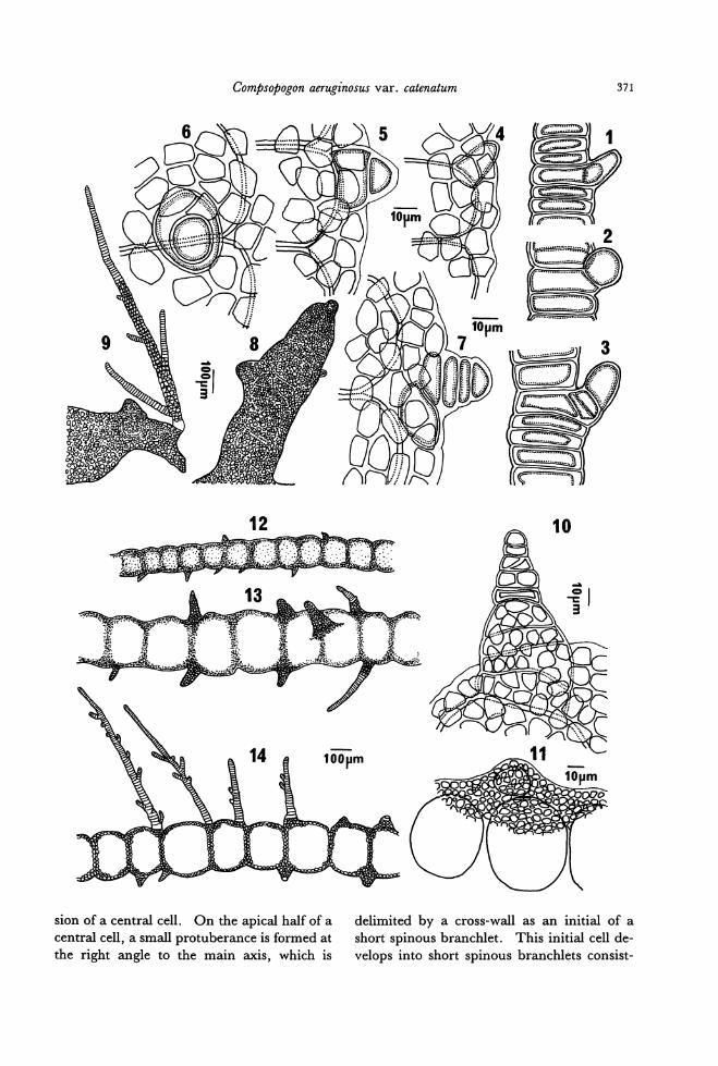
Compsopogon aeruginosus var. catenatum 371
12
sion of a central cell. On the apical half of a
central cell, a small protuberance is formed at
the right angle to the main axis, which is
10
喧
-tg
delimited by a cross-wall as an initial of a
short spinous branchlet. This initial cell de-
velops into short spinous branchlets consist-
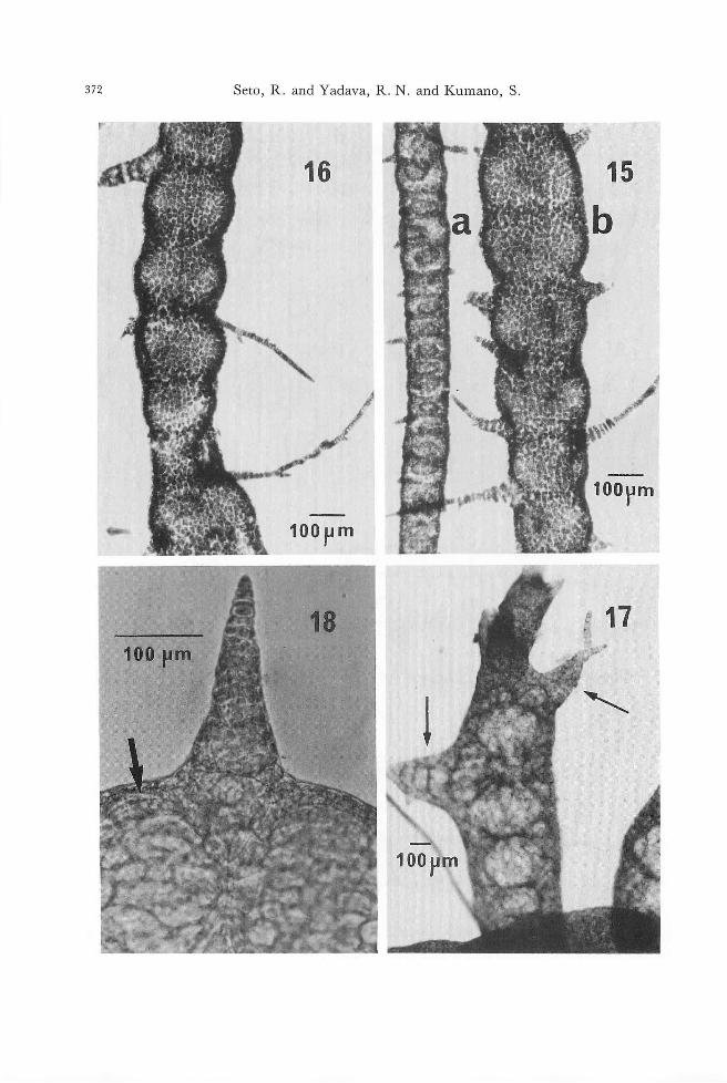
372 Seto, R. and Yadava, R. N. and Kumano, S.
100pm
18 100 pm
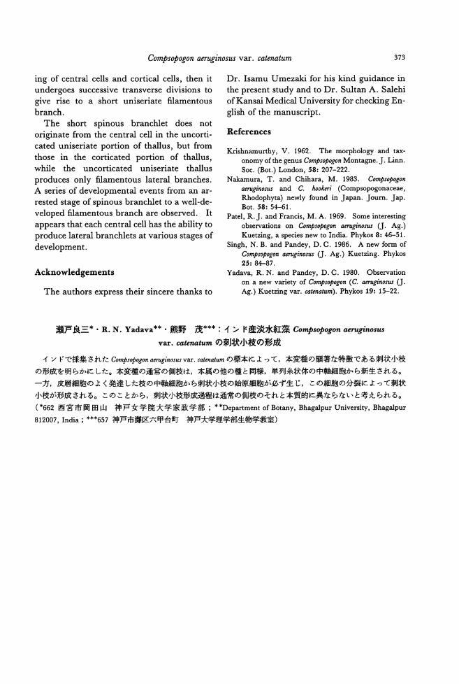
Compsopogon aeruginosus var. catenatum 373
ing of central cells and cortical cells, then it undergoes successive transverse divisions to
give rise to a short uniseriate filamentous
branch.
The short spinous branchlet does not
originate from the central cell in the uncorti-
cated uniseriate po口ionof thallus, but from those in the corticated portion of thallus, while the uncorticated uniseriate thallus
produces only filamentous lateral branches.
A series of developmental events from an ar-
rested stage of spinous branchlet to a well-de-
veloped filamentous branch are observed. It
appears that each central cell has the ability to
produce lateral branchlets at various stages of
development.
Acknowledgements
The authors express their sincere thanks to
Dr. Isamu Umezaki for his kind guidance in
the present study and to Dr. Sultan A. Salehi ofKansai Medical University for checking En-
glish of the manuscript.
References
Krishnamurthy, V. 1962. The morphology and tax-onomy of the genus Compsopogon Montagne. J. Linn. Soc. (Bot.) London, 58: 207-222.
Nakamura, T. and Chihara, M. 1983. Compsopogon flI!ruginos出阻d C. hoo.知 3・ (Compsopogonaceae,Rhodophyta) newly found in Japan. Joum. Jap. Bot. 58: 5←61.
Patel, R.J. and Francis, M. A. 1969. Some interesting observations on Compsopogon aerugino.凶 U.Ag.) Kuetzing, a species new to India. Phykos 8: 46-51.
Singh, N. B. and Pandey, D. C. 1986. A new form of Compsopogon側ぜinosusU. Ag.) Kuetzing. Phykos 25: 8←87.
Yadava, R. N. and Pandey, D. C. 1980. Observation on a new variety of Compsopogon (C. aerugin何回 U.Ag.) Kuetzing v釘 .&a伽 atum).Phykos 19: 15-22.
瀬戸良三*・ R.N. Yadava**'熊野茂*柿:インド産淡水紅葉 Compsopogonaen伊問削
V釘 .catenatumの刺状小枝の形成
インドで採集された Compsopogonaeruginos凶 V紅 .&atenatumの標本によって,本変種の顕著な特徴である刺状小枝
の形成を明らかにした。本変種の通常の側枝は,本属の他の種と同様,単列糸状体の中軸細胞から新生される。
一方,皮層細胞のよく発達した枝の中軸細胞から刺状小枝の始原細胞が必ず生じ,この細胞の分裂によって刺状
小枝が形成される。このことから,刺状小枝形成過程は通常の側枝のそれと本質的に異ならないと考えられる。
(*662西宮市岡田山神戸女学院大学家政学部;叫Departmentof Bot叩.y, Bhagalpur University, Bhagalpur
812007, India ; * 紳657神戸市灘区六甲台町神戸大学理学部生物学教室)

Jpn. J. Phycol. (Sorui) 39: 375-380, December 10, 1991
Michio Masuda, Masao Ohno and Gavino C. Trono, Jr.: A taxonomic
assessment of POゆ匂庁'asuborbiculata Kjellman, a food
species from the Philippines *
Key lndex 陥油 Nori・sheets-Phil.争'pines-Porphyra-Porphyracrispata-Porphyra subor-biculata-taxononザ・MichioM田 uda,Dψartment 01 Botaり"FaculiりofScience, Hokkaido Universi・'ty,Sapporo, 060 Japan Masao Ohno, Usa Marine Biological lnstitute, Kochi Universi机 Kochz;781-11 Japan Gavino C. Tro附o叩n叫tQuezon, PhilipPines
Five species of P01同yrahave been reported
form the Philippine Islands (Silva et al.
1987). These are mainly limited to the north-
ern coasts of Luzon Island. POψhyra thalli
are locally known as“gamet" and are collect-
ed along the coast of Ilocos Norte. Theyare
sold in the form of dried sheets called “ped-
azo" at the local market (Trono and Ganzon-
Fortes 1988). The materials for preparation
of the dried “gamet" are washed with
freshwater and are made into sheets, 100-150 x 100 cm in size and about 5 mm thick
and are dried under the sun. The dried
sheets are expensive in comparison wi也 other
seaweeds such as Enteromorpha, Codium, Cauler-pa, Halymenia, Gracilaria and Laurencia. “Gamet" is prepared in the form of salad or
used in soups. The utilization of Porphyra as
food is limited to the populations along coastal communities in northern Luzon.
This repo口presentsobservations on the ecolo-
gy, morphology and t砿 onomyof P01舛yram Ilocos Norte.
Materials and Methods: Plants were collected
at Ablan, Burgos, Ilocos Norte, Luzon Island on February 1 and 3, 1990, fixed and
preserved in 10% formalin in seawater. A
portion of the collections was dried for herbari-
um sheets, or was transported to ]apan. V oucher specimens are deposited in the her-
barium, Faculty ofScience, Hokkaido Univer-sityat Sapporo (SAP). Dried sheets were pur-
* This work was supported by the overseas research project wi白血egrant from the Ministry of Education, Science and Culture, Japan: No. 01041067 (1989)叩 dNo. 02041064 (1990).
chased from a local gamet gatherer.
Ecology: POψ砂raplants were found growing
on rocky, wave-exposed upper intertidal zone in Ilocos Norte from November to Februarγ. At the time the survey was done, the water temperature was 25.90C and salinity was 34.8耳元.
Two Porphyra entities have been noted previ-
ously (Trono unpublished observations): one
with roundish to reniform thalli and the other
with lanceolate thalli. The entity wi出 lanceo-
late也alliappears to be dominant during the
early period of the growing season in Novem-
ber, but was not seen or collected during this survey. The rocky habitat is exposed to very
rough water, and the gatherers were only able to collect the “gamet" for short periods during low tides for one or two calm days a week dur-
ing the winter months from November to
February.
Morphology: Specimens collected and materi-
als of dried sheets purchased have the follow-
ing features in common. Foliose thalli are
purplish red in color, round, reniform or fun-nel-shaped, 1-3 cm high, rolled toward one side (Fig. 1). Several thalli are deeply lobate
near the base (Fig. 2) and show a habit similar
to the green alga Ulva conglobata. Each thal-
lus possesses a discoid holdfast, 0.8-1.2 mm in diameter, and an obscure stipe. The thalli
are monostromatic, 35-45μm thick at the cen-ter (1 cm above the base), 22-25μm thick at the vegetative margin. Vegetative cells in
surface view are polygonal in shape, 20-30μm long X 12-22μm wide. The cells are ir-
regularly arranged, contain a single stellate
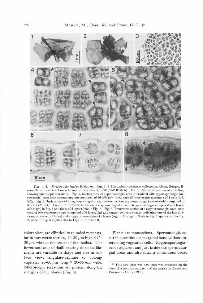
376 Masuda, M ., Ohno, M. and Trono, G目 c.Jr.
6
10um
Figs. 1-8. POゆhyrasuborbiculala Kjellman. Figs. 1, 2. Herbarium specimens collected at Ablan, Burgos, 1トocos Norte, northern Luzon Island on February 3, 1990 (SAP 054606). Fig. 3. Marginal portion of a thallus, showing microsopic serrations. Fig. 4. Surface view of a sperrr国 ang凶 area川 ermixedwith zygotosporangia (ar rowheads); note each spermatangium composed of 16 cells (a/4, b/4), each of three zygotosporangia of 4 cells (al2, b/2). Fig. 5. Surface view of a zygotosporang凶 area;note each offour zygotosporangia (arrowheads) composed of 8 cells (a/2, b/4). Figs. 6, 7. Transverse section ofa spermatangial area; note spermatangia composed of41ayers (c/4 stage) in Fig. 6 and those of 81ayers (c/8) in Fig. 7. Fig. 8. Transverse section of a zygotosporangial area; note each of two zygotosporangia composed of 4 layers (left and center, c/4; arrowheads indicati時 oneof the last divi-sions, others out offocus) and a zygotosporangium of21ayers (right, c/2 stage). Scale in Fig. 1 applies also to Fig 2; scale in Fig. 6 applies also to Figs. 4, 5, 7 and 8
chloroplast, are elliptical to rounded rectangu-
lar in transverse section, 20-30μm high X 15-
30μm wide at the center of the thallus. The
lowermost cells of thalli bearing rhizoidal fila-
ments are variable in shape and size in sur-
face view, angulate-capitate to oblong-
capitate, 20-60μm long X 20-35μm wide.
Microscopic serrations are present along the
margins of the blades (Fig. 3).
Plants are monoecious. Spermatangia oc-
cur in a continuous marginal band without in-
tervening vegetative cells. Zygotosporangia1)
occur adjacent and just inside the spermatan-
gial areas and also form a continuous broad
1) This new term was new term was proposed on the basis of a peculiar ontogeny of the zygote in Bangia and POゆりraby Guiry (1990)
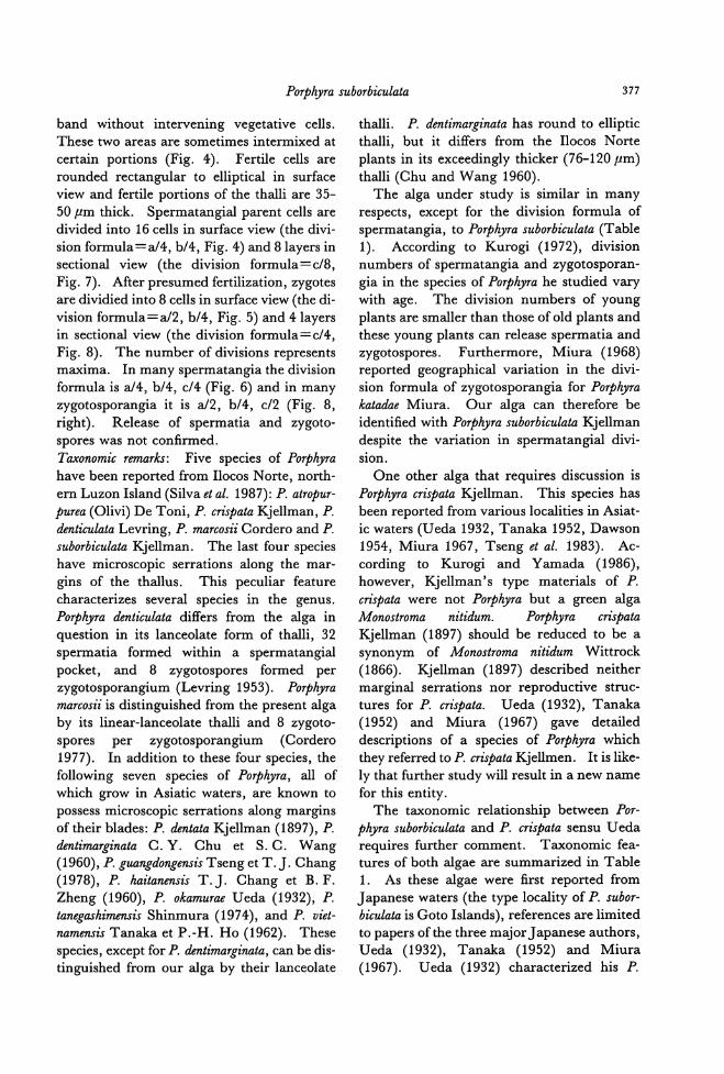
Porphyra suborbiculata 377
band without intervening vegetative cells. These two areas are sometimes intermixed at ce此ainpo口ions(Fig. 4). Fertile cells are rounded rectangular to elliptical in surface view and fertile po口ionsof the thalli are 35-50μm thick. Spermatangial p町 entcells are divided into 16 cells in surface view (由edivi-sion formula=a/4, b/4, Fig. 4) and 81ayers in sectional view (the division formula=c/8, Fig. 7). After presumed fertilization, zygotes 紅 edividied into 8 cells in surface view (the di-
vision formula=a/2, b/4, Fig. 5) and 41ayers in sectional view (the division formula=c/4, Fig. 8). The number of divisions represents maxima. In many spermatangia the division formula is a/4, b/4, c/4 (Fig. 6) and in m組 yzygotosporangia it is a/2, b/4, cl2 (Fig. 8, right). Release of spermatia and可goto-spores was not confi.rmed. T町 'onomicremarks: Five species of POψhyra have been reported from Ilocos No此e,no此h-
em Luzon Island (Silva et al. 1987): P. atropur-
purea (Olivi) De Toni, P. crispata Kjellman, P. denti,印 lataLevring, P. marcosii Cordero and P. suborbi・'culataKjellman. The last four species
have microscopic serrations along the mar-gins of the thallus. This peculiar feature characterizes several species in the genus. POψhyra denticulata differs from the alga in
question in its lanceolate form of thalli, 32 spermatia formed within a spermatangial
pocket, and 8 zygotospores formed per zygotosporangium (Levring 1953). POψhyra marcosii is distinguished from the present alga by its linear-lanceolate thalli and 8 zygoto-
spores per zygotosporangium (Cordero 1977). In addition to these four species, the following seven species of P01舛yra,all of which grow in Asiatic waters, are known to possess microscopic serrations along margins of their blades: P.伽 tataKjellman (1897), P. dentimarginata C. Y. Chu et S. C. Wang (1960), P. gzωngdongensis Tseng et T. J. Chang (1978), P. haitanensis T.J. Cha時 etB. F. Zheng (1960), P. okamurae Ueda (1932), P. ta7噌 ashimensisShinmura (1974), and P. viet-namensis Tanaka et P.-H. Ho (1962). These species, except for P.伽 timarginata,can be dis-tinguished from our alga by their lanceolate
thalli. P. dentimarginata has round to elliptic thalli, but it differs from the Ilocos Norte plants in its exceedingly thicker (76-120μm)
thalli (Chu and Wang 1960). The alga under study is similar in many
respects, except for the division formula of spermatangia, to POψ砂rasuborbiculata (Table 1). According to Kurogi (1972), division numbers of spermatangia and zygotosporan-
gia in the species of POゆ砂rahe studied vary with age. The division numbers of young plants are smaller than those of old plants and these young plants can release spermatia and zygotospores. Furthermore, Miura (1968) reported geographical variation in the divi-sion formula of zygotosporangia for P01灼 rakata由eMiura. Our alga can therefore be identifi.ed with POψめIrasuborbiculata Kjellman despite the variation in spermatangial divi-
slon. One other alga出atrequires discussion is
POψ砂racrispata Kjellman. This species has
been reported from various localities in Asiat-ic waters (Ueda 1932, Tanaka 1952, Dawson 1954, Miura 1967, Tseng et al. 1983). Ac-cording to Kurogi and Yamada (1986), however, Kjellman's type materials of P. m・'spatawere not POψ砂'rabut a green alga
Monostroma nitidum. P07沙めIra crispata Kjellman (1897) should be reduced to be a synonym of Monostroma nitidum Wittrock
(1866). 民jellman(1897) described neither marginal serrations nor reproductive struc-tures for P. crispata. Ueda (1932), Tanaka (1952) and Miura (1967) gave detailed
descriptions of a species of POψ砂rawhich they referred to P. cr坤ataKjellmen. It is like-ly that further study will result in a new name
for this entity. The taxonomic relationship between Por-
PめlTasuborbiculata and P. crispata sensu U eda requires further comment. Taxonomic fea-tures of both algae are summarized in Table 1. As these algae were fi.rst repo此edfrom J apanese waters (也etype locality of P. subor-bi,印 latais Goto Islands), references are limited to papers of the three m司jorJ apanese authors, Ueda (1932), Tanaka (1952) and Miura (1967). Ueda (1932) characterized his P.
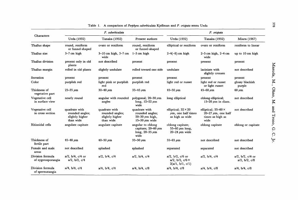
凶一可∞Acomp叩 sonof POゆ紗rasuborbiculata勾ell皿四組dP. cri.卵 tasensu U eda Table 1.
P. crおifJataP. suborbiculata
Miura (1967) T四 aka(1952) Ueda (1932) Present authors Tanaka (1952) Ueda (1932) Characters
reniform to linear ovate or reniform elliptical or reniform round, reniform or funnel-shaped
1-3 cm high
ovate or reniform round, reniform or funnel-shaped
3-7 cm high
Thallus shape
zsEP冨-wOEO-冨・自円二ザOロow。・0・V・
up to 10 cm high 2-5 cm high, 2-4 cm wide
present
2-4(-8) cm high 3-10 cm high, 3-7 cm wide
not described
Thallus size
present present present present only in old plants
rolled in old plants
Thallus division
not described
present
glossy blackish purple
60μm
laciniate with slightly crenate
present
light red or russet or light russet
45-68μm
undulate
present
light red or russet
rolled toward one side
present
purplish red
slightly undulate
present
light pink or purplish red
30-48μm
p回 sent
purplish red
Thallus margin
Serration
Color
45-50μm 35-45μm 25-35μm Thickness of vegetatlve part
Vegetative cell in surface view
not described
oblong or capitate
not described
oblong-elliptical; 15-20μm in diam.
elli ptical; 55-60 x 20-27μm, one half times as high as wide
oblong capitate
long elliptical
elliptical; 32 x 20 μm, one half times as high as wide
oblong capitate; 55-60μm long, 20-24μm wide
polygonal; 20-30μm long, 12-22μm wide
quadrate with rounded angles; 20-30μm high, 15-30μm wide
組伊larto oblong capitate; 20-60 f1m long, 20-35μm wide
35-50μm
angular with rounded angles
quadrate with rounded angles; slightly higher than wide
angulate capitate
nearly round
quadrate with rounded angles; slightly higher than wide
回伊latecapitate
n
o
uu
{色屯
ce
es
e
V
悶
・1u
ar
t
E
Fn
e--V
Rhizoidal cells
not described not described 55-65μm 40-50 f1m 45-48μm Thickness of fertile pa此
Female and male areas
Division formula of zygotosporangia
not described separated separated splashed splashed not described
a/2, b/2, c/4 or a/2, b/2, c/8
a/2, b/4, c/4 a/2, b/2, c/6 or a/2, b/2, c/6+ 2(a/1, b/1, c/1)
a/4, b/4, c/8
a/2, b/4, c/4 a/2, b/4, c/4 a/2, b/4, c/4 or a/2, b/2, c/4
a/4, b/4, c/8 a/4, b/4, c/8 a/4, b/4, c/8 a/4, b/4, c/4 a/4, b/4, c/4 Division formula of spermatangia
POψhyra suborbiculata 379
crispata as follows: 1) a peculiar division of
zygotosporangia, 2) a smaller thallus, 3) ex-ceedingly elongated capitate cells bearing rhizoidal filaments, and 4) a caespitose thallus
similar to Ulva conglobata. Tanaka (1952)組 d
Miura (1967) seemed to agree with the opin-
ion ofUeda. There is, however, a discrepan-
cy in the description of the division formula of
zygotosporangia by these authors. Accord-ing to U eda, the division formula of
zygosporangia for his P. crispata is a/2, b/2, cl6 or a/2, b/2, cl6+2 (a/1, b/1, cl1), but ac-
cording to Tanaka, it is a/2, b/4, c/4. The lat-ter is the same as that of P. suborbiculata given
by Ueda and Tanaka (Table 1). According
to Miura (1967), the division formula of zygotosporangia of P. crispata sensu U eda is
usually a/2, b/2, cl4 and rarely a/2, b/2, c/8. Miura (1967) states that the division mode of
zygotosporangia described by these three
authors is basically similar. The size ofthalli overlaps each other and is not clearly separat-
ed. The shape of lowermost cells bearing
rhizoidal filaments is very variable within an
individual, and so it is not a good diagnostic
character. “Caespitose" thalli (sensu Ueda and T阻止a)may be formed by divisions of
funnel-shaped thalli. Our observations on
specimens in the field showed that the Por-Phyra population was composed of variable shapes of foliose thalli. Divided “caespitose" thalli resembling Ulva conglobata in outer ap-pe訂 ancewere found among round, reniform or funnel-shaped也alli.
Ueda (1932) and Tanaka (1952) were in
agreement wi也 regardto the division formu-
lae of spermatangial parent cells for POψ砂m
suborbiculata and P. crispata sensu U eda: the
former species is a/4, b/4, c/4 and the latter is a/4, b/4, cl8. However, Kurogi (1972) described the division formula of P. suborbicula-ta as identical to that of P. crispata.
Tanaka (1952) emphasized the occurrence
of splashed patches of spermatangial and
zygotosporangial areas for POψhyra suborbたula-ta. We also found such intermixed areas in the Philippine materials. However, each area is widely distinct and intermixed areas
are very narrowly restricted. 1t is questiona-
ble whether this character has taxonomic sig-
nificance. Finally, thallus thickness in both algae
should be compared. POゆめlTacrispata has thicker thalli than P. suborbiculata (Table 1); however, it is hardly possible to distinguish P. crispata as described by Ueda (1932) from P.
subo巾・'culataas described by Tanaka (1952) on
the basis of thallus thickness. Thus, there is no clear distinction between P. suborbiculata
and P. cげ学atasensu U eda and it is suggested that the latter is “caespitose" and may be 01-der thalli of the former. 1n order to clari今further the status of POψ砂racnspata sensu Ueda it is necessary to analyze its seasonal
and geographical variations.
We are grateful to: Professor T. Yoshida, Hokkaido University and Professor M. D.
Guiry, National University of 1reland for their criticism of the manuscript; Drs. H.
Nakah町 aand T. Ajisaka, Kyoto University for their help in collecting materials.
References
Chang, T.J.阻 dZheng, B. F. 1960. POψhyra hailll脚 a
sis, a new species of POゆめ>rafrom Fukien. Acta Bot Sinica 9: 32-36. (in Chinese)
Chu, C. Y. and Wang, S. C. 1960. Study on POφ'hyra denti71Ulrginata sp. nov. Acta Bot. Sinica 9: 37-41. (in Chinese)
Cordero, P. A., J r. 1977. Studies on Philippine marine red algae. Special Publications from the Seto Marine Biological Lab. 4: 1-258.
Dawson, E. Y. 1954. Marine plants in the vicinity of the Institut Oceanographique de Nha Trang, Viet Nam. Pacific Sci. 8: 373-481.
Guiry, M. D. 1990. Sporangia and spores. 1n K. M. Cole and R. G. Shea血 [ed.]Biology ofthe red al-gae. pp. 347-376. Cambridge Univ. Press, Cam-bridge.
同elIman,F. R. 1897. Japanska arter af slagtet Por-phyra. Bihang TilI K. Svenska Vet.-Akad. H組 dl.23(4): 1-34.
Kurogi, M. 1972. Systematics of POψ砂惜 inJapan.1n1. A. Abbott and M. Kurogi [ed.] Contributions to the systematics of benthic marine algae of the north Pacific. pp. 167-192. Jap. Soc. Phycol. Kobe.
Kurogi, M. and Yamada, 1. 1986. New knowledges ob-tained by the observation on the original specimens ofF. R. KjelIman:JapanskaarterafslagtetPoψ,hyra. Jap. J. Phycol. 34: 62. (in Japanese)
Levring, T. 1953. The marine algae of Australia 1. Rhodophyta: Goniotrichales, Bangiales 阻 d
380 Masuda, M., Ohno, M. and Trono, G. C. Jr.
Nemalionales. Ark. Bot. ser. 2. 2: 457-530.
Miura, A. 1967. Two new species and a new record of
POψhyra from Enoshima, Sagami Bay. J. Tokyo
Univ. Fish. 54: 65-71.
Miura, A. 1968. POψわrakatadai, a new speci田 from
Japanese coast. J. Tokyo Univ. Fish. 54: 55-59. Shinmura, 1. 1974. POψhyra tanegashimensis, a new spe-
cies of Rhodophyceae from Tanegashima Island in
southern Japan. Bull. Jap. Soc. Sci. Fish. 40: 735-749.
Silva, P. C., Menez, E. G. and Moe, R. L. 1987. Cata-
log of出ebenthic marine algae of the Philippines.
Smithsonian Contributions to the Marine Sciences. 27: 1-179.
T釦 aka,T. 1952. The systematic study oftheJapanese
Protoflorideae. Mem. Fec. Fish. Kagoshima Univ.
2(2): 1-92.
Tanaka, T. and Ho, P.司H.1962. Notes on some marine
algae from Vietnam I. Mem. Fac. Fish. Kagoshima
Univ. 11: 24-40.
Trono, G. C., Jr. and Ganzon-Fortes, E. T. 1988.
Philippines Seaweed. National Book Store, Inc. Manila.
Tseng, C. K. (ed.) 1983. Common seaweeds of China
Science Press, Beijing. Tseng, C. K. and Chang, T.J. 1978. On two new Por-
phyra from China. Oceanologia et Limnologia Sinica
9: 76-83. (in Chinese)
Ueda, S. 1932. A systematic study ofthe genus POψhyra
found on the Japanese coast. J. Imp. Fish. Inst.
28(1): 1-45. (in Japanese)
Wittrock, V. B. 1866. Forsok till en monographi ofver
algslagtet Monostroma. Akad. Afh. Stockbolm.
増田道夫1・大野正夫2・GavinoC. Trono, Jr.3 :フィリピンで
食用にされているマルパアマノリ
フィリピンのルソン島北部の IIocosN orte 州では, 11月から 2月にかけて生育するアマノリ属から乾海苔を作
成して食用にしている。生育期の後期の乾海苔製品並びに現地で2月に採集した標本は,藻体の形,大きさ,厚
さ,鋸歯,及び生殖器官の分裂様式の特徴からマルパアマノリ (POゅhyrasuborbiculata Kjellman)であることが判明
した。ツクシアマノリ (P.crispata sensu U eda)と本種との関係についても論じた。 ('060札幌市北区北10条西 8丁
目 北海道大学理学部植物学教室;2781-11土佐市字佐町井尻194 高知大学海洋生物教育研究センター;3Ma-
rine Science Institute, College of Science, University of the Philippines, Diliman, Quezon, Philippines)
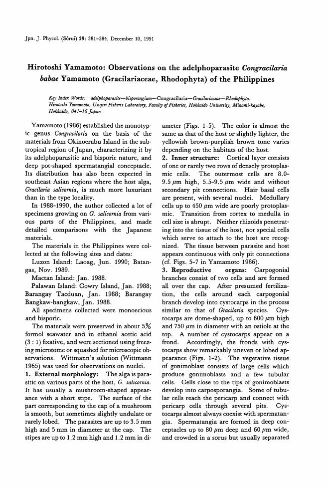
Jpn. J. Phycol. (Sorui) 39: 381-384, December 10, 1991
Hirotoshi Yamamoto: Observations on the adelphoparasite Congracila巾
babae Yamamoto (Gracilariaceae, Rhodophyta) of the Philippines
Key lndex陥油 adelthotarasite-bistorangium一Congracilaria-G間 ilariaceae-Rhodot加Hirotoshi Yama脚 Ito,日ujiriFisheris Laboraωry,F.仰 ltyofFis.伽九品>/,kaidoUniver.吻"Minami-句 abe,Hok/ca油" 041-16Jatan
Yamamoto (1986) established the monotyp-
ic genus Congracilaria on the basis of the
materials from Okinoerabu Island in the sub-
tropical region of Japan, characterizing it by its adelphoparasitic and bisporic nature, and deep pot-shaped spermatangial conceptacle. Its distribution has also been expected in
southeast Asian regions where the host alga, Gracilaria salicomia, is much more luxuriant than in the type locality.
In 1988-1990, the author collected a lot of
specimens growing on G. salicomia from vari-ous parts of the Philippines, and made detailed comparisons with the Japanese materials.
The materials in the Philippines were col-
lected at the following sites and dates: Luzon Island: Laoag, Jun. 1990; Bat組・
gas, Nov. 1989. Mactan Island: Jan. 1988. Palawan Island: Cowry Island, Jan. 1988;
Barangay Tacduan, Jan. 1988; Barangay
Bangkaw-bangkaw, Jan. 1988. All specimens collected were monoecious
and bisporic. The materials were preserved in about 5%
formol seawater and in ethanol acetic acid (3 : 1) fixative, and were民 ctionedusing freez-ing microtome or squashed for microscopic ob-servations. Wittmann's solution (Wittmann 1965) was used for observations on nuclei.
1. Externa1 morphology: The alga is p訂 a-sitic on various parts of the host, G. salicomia. It has usually a mushroom-shaped appe町-
ance with a short stipe. The surface of the part corresponding to the cap of a mushroom is smooth, but sometimes slightly undulate or rarely lobed. The parasites are up to 3.5 mm high and 5 mm in diameter at the cap. The stipes釘 eup to 1.2 mm high and 1.2 mm in di・
ameter (Figs. 1-5). The color is almost the
same踊 thatof the host or slightly lighter, the yellowish brown-purplish brown tone varies depending on the habitats of the host.
2. Inner structure: Corticallayer consists of one or rarely two rows of densely protoplas-mic cells. The outermost cells are 8.0-9.5μm high, 5.5-9.5μm wide and without secondary pit connections. Hair basal cells are present, wi出 severalnuclei. Medullary cells up to 450μm wide are poorly protoplas-mic. Transition from cortex to medulla in
cell size is abrupt. Neither rhizoids penetrat-
ing into the tissue of the host, nor special cells which serve to attach to the host are recog-
nized. The tissue between parasite and host appears continuous wi也 onlypit connections (cf. Figs. 5-7 in Yamamoto 1986).
3. Reproductive organs: Carpogonial branches consist of two cells and are formed all over the cap. After presumed fertiliza-
tion, the cells around each carpogonial
branch develop into cystoc訂 psin the process similar to that of Gracilaria species. Cys-toc訂 psare dome-shaped, up to 600μm high and 750μm in diameter with an ostiole at the top. A number of cystocarps appear on a frond. Accordingly, the fronds with cys-tocarps show remarkably uneven or lobed ap-
pearance (Figs. 1-2). The vegetative tissue
of gonimoblast consists of large cells which produce gonimoblasts and a few tubular cells. Cells close to the tips of gonimoblasts develop into carposporangia. Some of tubu-lar cells reach the pericarp and connect with peric紅 p cells through several pits. Cys-tocarps almost always coexist with spermatan-gia. Spermatangia are formed in deep con-ceptacles up to 80μm deep and 60μm wide, and crowded in a sorus but usually separated
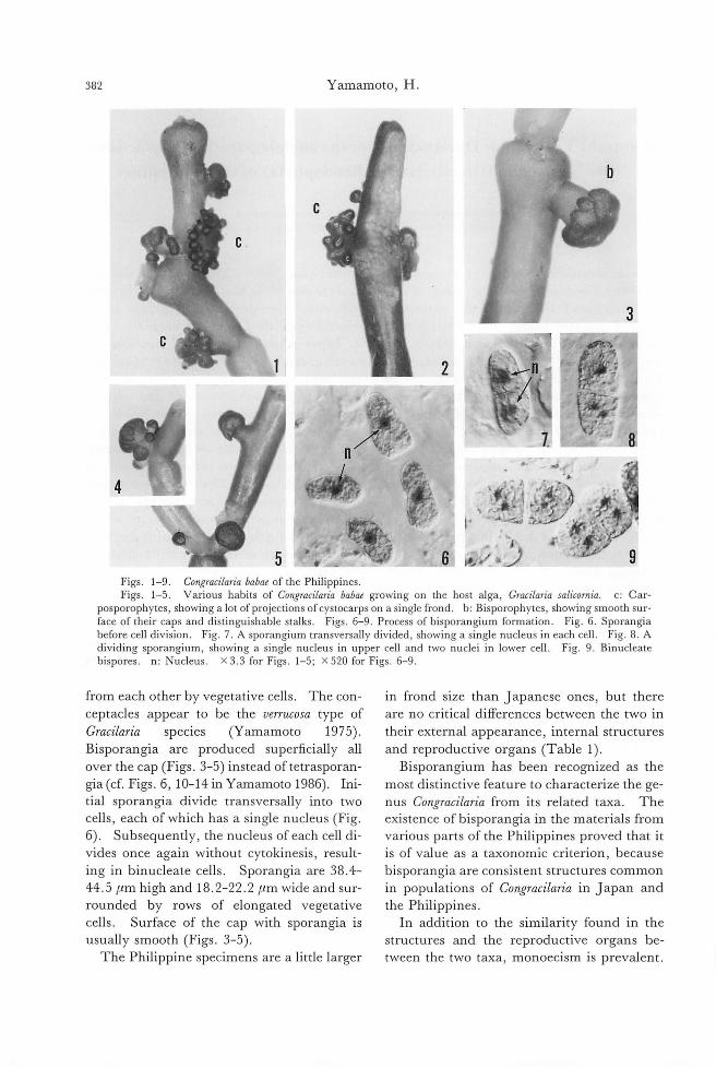
382 Yamamoto, H.
Figs. 1-9. Congracilaria babae of the Philippines. Figs. 1-5. Various habits of COllgracilaria babae growing on the host alga, Gracilaria salicornia. c: Car-
posporophytes, showing a lot of projections of cystocarps on a single frond. b: Bisporophytes, showing smooth sur-face of their caps and distinguishable stalks. Figs. 6-9. Process of bisporangium formation. Fig. 6. Sporangia before cell division. Fig. 7. A sporangium transversally divided, showing a single nucleus in each cell. Fig. 8. A
dividing sporangium, showing a single nucleus in upper cell and two nuclei in lower cell. Fig. 9. Binucleate bispores. n: Nucleus. X 3.3 for Figs. 1-5; X 520 for Figs. 6-9.
from each other by vegetative cells. The con-
ceptacles appear to be the verrucosa type of
Gracilaria species (Yamamoto 1975)
Bisporangia are produced superficially all
over the cap (Figs. 3-5) instead oftetrasporarト
gia (cf. Figs. 6, 10-14 in Yamamoto 1986). Ini-
tial sporangia divide transversally into two
cells, each of which has a single nucleus (Fig
6). Subsequently, the nucleus of each cell di-
vides once again without cytokinesis, result-ing in binucleate cells. Sporangia are 38.4-
44.5μm high and 18.2-22.2μm wide and sur-
rounded by rows of elongated vegetative
cells. Surface of the cap with sporangia is
usually smooth (Figs. 3-5)
The Philippine specimens are a Iittle larger
in frond size than ] apanese ones, but there
are no critical differences between the two in
their external appearance, internal structures
and reproductive organs (Tabl巴 1).
Bisporangium has been recognized as the
most distinctive feature to characterize the ge-
nus Congracilaria from its related taxa. The
existence of bisporangia in the materials from
various parts of the Philippines proved that it
is of value as a taxonomic criterion, because
bisporangia are consistent structures common
in populations of Congracilaria in ]apan and
the Philippines.
In addition to the similarity found in the
structures and the reproductive organs be-
tween the two taxa, monoecIsm is prevalent
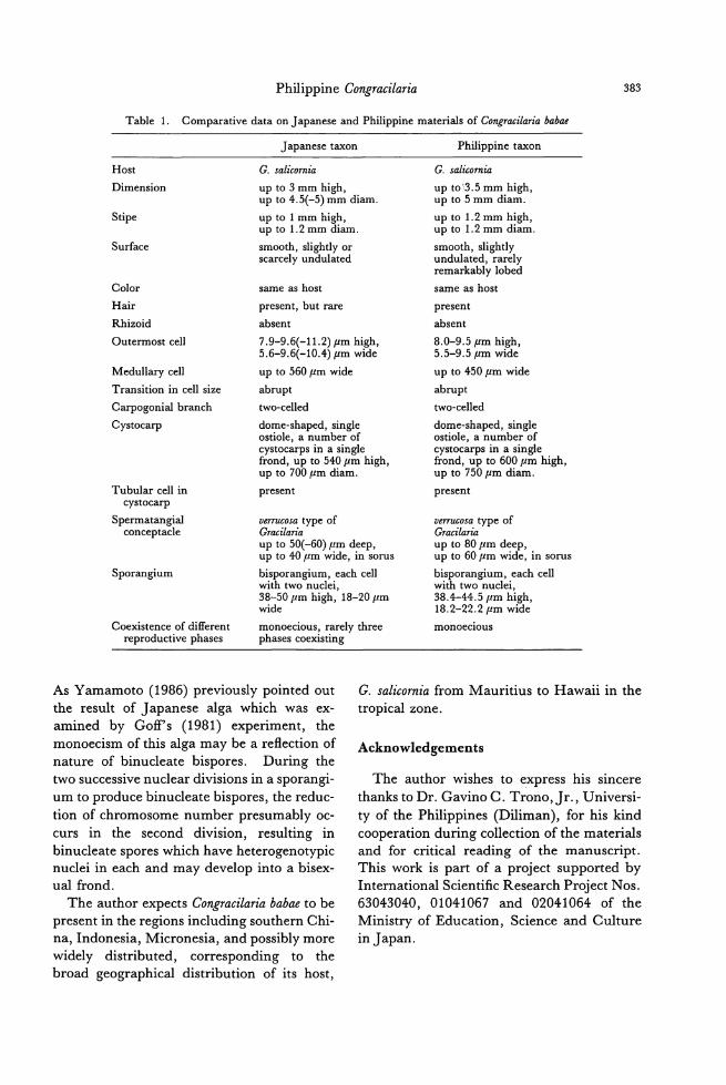
Philippine Congracilaria 383
Table 1. Comparative data on J apanese and Philippine materials of Congracilaria babae
Host
Dimension
Japanese taxon
G. salicornia
up to 3 mm high, up to 4.5(-5) mm diam.
up to 1 mm high, up to 1.2 mm diam
smooth, slightIy or scarcely undulated
Stipe
Surface
Color
Hair
Rhizoid
Outermost ceIl
same as host
present, but rare
absent
7.9-9.6(ー11.2)μmhigh, 5.6-9.6(ー10.4)μmwide
up to 560μm wide
abrupt
MeduIlary ceIl
Transition in ceIl size LU
FL
佃r
'b
d
m
m・
同
町
迅
旧
凹E
t
r
s
av'
cc
two-ceIled
dome-shaped, single ostiole, a number of cystocarps in a single frond, up to 540μm high, up to 700μmdiam.
present
verrucosa type of Gracilaria up to 50(-60)μm deep, up to 40μm wide, in sorus
bisporangium, each ceIl with two nuclei, 38-50μm high, 18-20μm wide
monoecious, rarely three phases coexisting
Tubular ceIl in cystocarp
Spermatangial conceptacle
Sporangium
Coexistence of different reproductive phases
Philippine taxon
G. salicornia
up to'3.5 mm high, up to 5 mm diam
up to 1.2 mm high, up to 1.2 mm diam.
smooth, slightIy undulated, rarely remarkably lobed
same as host
present
absent
8.0-9.5μm high, 5.5-9.5μm wide
up to 450μm wide
abrupt
two-ceIled
dome-shaped, single ostiole, a number of cystocarps in a single frond, up to 600μm high, up to 750μm diam.
present
verrucosa type of Gracilaria up to 80μm deep, up to 60μm wide, in sorus
bisporangium, each ceIl with two nuclei, 38.4-44.5μm high, 18.2-22.2μm wide
monoeclOus
As Yamamoto (1986) previou向 pointedout
the result of ] apanese alga which was ex司
amined by Goff's (1981) experiment, the monoecism of this alga may be a reflection of
nature of binucleate bispores. During the
two successive nuclear divisions in a sporangi-
um to produce binucleate bispores, the reduc-tion of chromosome number presumably oc-
curs in the second division, resulting in
binucleate spores which have heterogenotypic
nuclei in each and may develop into a bisex-
ual frond.
The author expects Congracilaria babae to be
present in the regions including southern Chi-
na, lndonesia, Micronesia, and possibly more widely distributed, corresponding to the
broad geographical distribution of its host,
G. salicornia from Mauritius to Hawaii in the
tropical zone.
Acknowledgements
The author wishes to express his sincere
thanks to Dr. Gavino C. Trono,]r., Universi-
ty of the Philippines (Diliman), for his kind cooperation during collection of the materials
and for critical reading of the manuscript.
This work is part of a project supported by
lnternational Scientific Research Project Nos.
63043040, 01041067 and 02041064 of the お1inistryof Education, Science and Culture in ]apan.
384 Yamamoto, H.
References
Goff, L.]. 1981. The role of bispores in the life history
of the p町田itic red alga, Gardneriella tuberグm(Solieriaceae, Gigartinales). Phycologia 20: 397-
406.
Wittmann, W. 1965. Ac回et蜘tωo仔何-寸lror凹叩nν叶凶-1七ha旧aem立ma旧a剖to凹x均yl凶linト日吋-ch叫由lor凶al l
hydrate for chromosome staining. Stain Tech. 40:
161-164.
Yamamoto, H. 1975. The relationship between
Gracilariopsis and Gracilaria from ] apan. Bull. Fac
Fish. Hokkaido Univ. 26: 217-222.
Yamamoto, H. 1986. Congracilaria babae gen. et sp. nov.
(Gracilariaceae), an adelphoparas巾 growingon
Gracilaria salicomia of ]apan. Bull. Fac. Fish. Hok-
kaido Univ. 37: 281-290.
山本弘敏:フィリピン産の寄生種フシクレタケの観察
現在まで鹿児島県沖永良部島からのみ知られていた寄生種フシクレタケ (Congracilariababae)が,フィリピンに
も広く分布することを確認した。フィリピン産の外部形態,内部組織,生殖器官はともに日本産のものとほぼ同
じであり,この種の特徴とされている二分胞子のうと雌雄同体性は全ての採集地域からの標本で認められた。こ
の結果,この特性は一地域(日本)の標本にのみ見られた例外的な形質ではなく,本種の特徴的な形質であるこ
とを確認した。 (041-16北海道南茅部町字臼尻152 北海道大学水産学部臼尻水産実験所)
Jpn. J. Phycol. (Sorui) 39: 385-387, December 10, 1991
Yasushi Fujimori, Katsuhito Nakamura, Yuichi Maruoka, Tsuneo Matsubayashi and Goro Tamura: Immunological comparison
of algal cysteine synthases
Key lndex Uゐrds: Chlorella-cysteine synthase-immunological comparison-N叩 nochloropsis-Por-phyra-Scenedesmus-Spirulina. Y山山.hiFujimori, Goro Tamura, Department oj Agricultural Chemistry, Facul砂 ojHorticulture, Chiba University, Matsudo, Chiba, 271 Japan Katsuhito Nakamura, (Present Adress) D.φ'artment 01 Biology, Faculty 01 General Education, Gifu University, Giju, 501-11 Ja.μn Yuichi Maruoka, (Present Adress) The Calpis Food lndustry Coリ LTD,Fuchinobe 5-11-10, Sagamihara, Kanaga出a,229JapanTsuneo Matsubayashi, Chlorella lndustψCo., LTD, Hisatomi 1343, Chikugo, Fukuoka, 833 Japan
Cysteine synthase (CSase, EC 4.2.99.8)
catalyzes the synthesis of L-cysteine from 0・
acetyl-L-serine and sulfide. CSases from a
variety of higher plants have been extensively purified and characterized (Masada et al.
1975, Tamura et al. 1976, Bertagnolli and Wedding 1977, Murakoshi et al. 1985, Ikega-mi et al. 1988, Nakamura and Tamura 1989), however relatively little is known about the
prope口iesof algal CSases (Schmidt 1977, Leon et al. 1987, Diessner and Schmidt 1981, Fujimori et al. 1991). In the present experi-ment, the antiserum raised against Chlorella and Porp紗raCSases have been prepared and, using these antiserum, the immunological comp町 isonof CSases from five species of algae [Chlorella and Scenedesmus (Chlo-
rophyceae) ; 地nnochloropsis (Eustig-
matophyceae); PorpわlTa(Rodophyceae);争Iru-
lina (Cyanophyceae)] was examined.
For the development of anti-Chlorella CSase
serum, the purification ofthe enzyme was per-formed by the procedures described in the previous paper (Nakamura 如 d Tamura 1989). The purified Chlorella CSase had a specific activity of270μmol L-cysteine formed per min per mg of protein and also showed a
single band in PAGE. The purified enzyme was emulsified with F・reund'scomplete ad-juvant and injected subcutaneously into a previously unimmunized rabbit. Each rab-bit was injected 4 times in the same manner. After 8 weeks, whole blood was collected by
cardiac puncture and the serum was obtained
after centrifugation. As for POゆめIraCSase, the methods for purification and preparation
of the antiserum were described in the previ-ous paper (Fujimori et al. 1991). The Por-
phyra CSase had a specific activity of 408μmol
cysteine formed per min per mg protein and
showed a single band in PAGE. Table 1 shows the presence of CSase in
several algae of different families. Each alga
in the table was macerated in 30 mM Tris-
HCl buffer (pH 8.0) containing 10 m M 2・mer-
captoethanol and 0.5 m M EDT A and
homogenized by sonication except POψhyra
that was extracted by autolysis, and the homogenate was subjected to enzyme assay.
Specific activity is expressed as cysteine for-
mation in 1μmol per min per mg protein of
crude extract. On a wet weight basis, 16 to 30・foldamount of activities compared with
that from POψ紗raand Spirulina were detected
in Chlorella and Nannochloropsis, respectively. When expressed on a protein basis, however, algal preparations exhibited approximately the same value except for Spirulina.
As shown in Fig. 1, the anti-ChloreLLa CSase thoroughly inactivated the CSase from Chlorel-
la, Scene.ゐ'smusand Nannochloropsis. However, this anti CSase serum was almost ineffective in inactivating the POψ砂raand Spirulina en-zymes (Fig. 1A). The amount of anti-Chlorel-la serum, which caused 50% inhibition of Chlorella, Scenedesmus 組 d Nannochloropsis
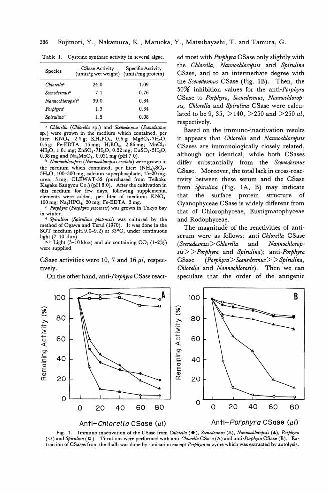
386 Fujimori, Y., Nakamura, K., Maruoka, Y., Matsubayashi, T. and Tamura, G.
Table 1. Cysteine syn也aseactivity in several algae.
Species CSase Activity Specific Activity (units/g wet weight) (u出 s/mgprotein)
Chlorellaa 24.0 1.09
Scenedesmus" 7.1 0.76
Nannochlorotsisb 39.0 0.84
POψhyraC 1.3 0.34
砂irulinad 1.5 0.08
a Chlorella (Chlorella sp.) and Scenedesmus (Scenedesmω sp.) were grown in the medium which contained, per liter: KN03, 2.5 g; KH2PO., 0.6 g; MgSO..7H20, 0.6g; F・e-EDTA,15mg; H3B03, 2.86mg; MnCl2・4H20, 1.81 mg; ZnSO..7H20, 0.22 mg; CuSO..5H20, 0.08 mg and N~MoO., 0.021 mg (pH 7.0).
b Nannochlorotsis (M朗 nochlorotsis0印 lata)were grown in the medium which contained, per liter: (NHみSO.・5H20, 100-300 mg; calcium superphosphate, 15-20 mg; urea, 5 mg; CLEWAT・32(purchased from Teikoku Kagaku Sangyou Co.) (pH 8.0). After the cultivation in 出ismedium for few days, following supplemental elements were added, per liter of medium: KN03, 100 mg; Na2HPO., 20 mg; F・e-EDTA,3 mg.
c POψ'hyra (Poψhyra yezoensis) was grown in Tokyo bay m wmter.
d Stirulina (砂irulinatlatensis) was cultured by the method of Ogawa and Terui (1970). It was done in the SOT medium (pH 9.0-9.2) at 330C, under continuous light (7-10 klux). a. b Light (5-10 klux) and air containing CO2 (1-2%)
were supplied.
CSase activities were 10, 7 and 16μ1, respec-tively.
On the other hand,組ti-PoψhyraCSase問 act-
100 1-.=::こ.n..二g- 0...ーか~ー _A,..旬、吉良、ー" 80 〉、・4ー-〉← 60 ι2 〈
。、c
40 c 。E Q)
区 20
O
O 20 40 60 80
ed most with P01舛yraCSase only slightly with
the Chlorella, Nannochloropsis and Spirulina CSase, and to an intermediate degree with 出eScenedesmωCSase (Fig. 1B). Then, the 50% inhibition values for the anti-Poψ砂ra
CSase to POψhyra, Scenedesmus, Nannochlorop-sis, Chlorella and争irulinaCSase were calcu-
lated to be 9, 35, > 140, >250 and >250μ1, respectively.
Based on the immuno・inactivationresults
it appe町 sthat Chlorella and Nannochloropsむ
CSases are immunologically closely related, although not identical, while both CSases differ substantially from the Scenedesmus CSase. Moreover, the totallack in cross-reac-
tivity between these serum and the CSase
from Spirulina (Fig. 1A, B) may indicate
that the surface protein structure of
Cyanophyceae CSase is widely different from
that of Chlorophyceae, Eustigmatophyceae and Rodophyceae.
The magnitude of the reactivities of anti-
serum were as follows: anti-Chlorella CSase
(Scenedesmus> Chlorella and Nannochlorop-
sis> > Porphyra and Spirulina); anti-Porphyra CSase (POψhyra> Scenedesmus > > Spirulina, Chlorella and Nannochlorosis). Then we can
speculate that the order of the antigenic
100
"、言史、・d
〉、 80 +ー〉+ー
60 ι』〈
01 c c 40 。E Q)
区 20
O O 20 40 60 80
Anti-Chlorella CSase (μl) Anti-Porphyra CSase (μl)
Fig. 1. Immuno-inactivation of the CSase from Chlorella (・), sc,朗edesm凶 (d),泊nnochlorotsis(A), POψ伊a(0) and争irulina(ロ). Titrations were performed with anti-Chlorella CSase (A) and anti-POI拘IraCSase (B). Ex-traction of CSases from the thalli was done by sonication except POψめ四回zymewhich was extracted by autolysis.
Algal cysteine synthases 387
reactivity between Chlorella and other four
species are; Chlorella-Nannochloropsis-Scenedes-mus---Poφhyra---Spirulina_
Nannochloropsis had been belonging to Chlo・
rophyceae group (Droop 1955)_ However, it was reclassified to Eustigmatophyceae (a kind
of Xanthophyceae) for the particular differ-
ence of the chloroplast pigment composition
and cell structures (Antia et aL 1975, Hibberd 1981). It is so curious也atthe Chlorella
CSase is immunologically more closely relat-
ed to the enzyme from Nannochloropsゐthan
that from Scenedesmus. On the other hand, Scenedesmus CSase had some common antigen-
ic determinants ofthe enzyme not only in Chlo・
rella but also in POψわIra.
Acknowledgements
We are grateful to Mr. Shigeru Araki of
Yamamoto Nori Research Laboratory for
providing us with POψ砂rathalli, and Dr. Keishiro Wada of Kanazawa University for
donation of Spirulina platensis OU・1strain.
We also thank Dr. Kazuo Fukushima of Ni-
hon University School of Dentistry at Matsu-
do, for his guidence in prep町 ationof the an-
tlserum.
References
Antia, N.J., Bisalputra, T., Cheng, J. Y.加 dKalley,
J. P. 1975. Pi伊 ent組 dcytological evidence for reclassification of Nannochloris oculata組 dMonallantas salina in the Eustigmatophyceae_J. Phycol. 11: 339-343_
Bertagnolli, B. L. and Wedding, R. T. 1977. Purifica-
tion and initial kinetic characterization of different forms 0・Acetylserinesulfhydrylase from seedlings of two species of Phaseolus. Plant Physiol. 60: 115-121.
Diessener, W. and Schmidt, A. 1981. Isoenzymes ofcys-teine synthase in出ecyanobacterium Synechococc凶
6301. z. Pllanzenphysiol. 102: 57-68. Droop, M. R. 1955. Some new supra-littoral Protista. J.
Marine Biol. Asc. of the U. K. 34: 233-245. 同 imori,Y., Nakamura, K. and Tamura, G. 1991.
Cysteine synthase仕'Oma red alga PO桝yrayezo即日:
purification and properties. Jpn. J. Phycol. 39: 7-14.
Hibberd, D.J. 1981. Notes on the t鉱 onomyand nomenclature ofthe algal classes Eustigmatophyceae and Tribophyceae (synonym Xanthophyceae). Bot. J. of the Linnean Soci. 82: 93-119.
Ikegami, F., Kaneko, M., Kamiyama, H. and Murakoshi,1. 1988. Purification and characteriza-tion of cysteine synthases from Citrullus vulga市.
Phytochemistry 27: 697-701. Leon, J., Romero, F., Galvan, F. and Vega, J. M.
1987. Purification and physicochemical characteri-zation of 0・acetyl-L-serine sulfhydrylase from Chla同)ldomonasreinhardtii. Plant Science 53: 93-99.
Masada, M., Fukushima, K. and Tamura, G. 1975. Cysteine synthase from rape leaves. J. Biochem. 77: 1107-1115.
Murakoshi, 1., Ikegami, F. and Kaneko, M. 1985. Purification and properties of cysteine s戸出ase仕'om争inacia0紛'acea.Phytochemistry 24: 1907-1911.
Nakamura, K.叩 dTamura, G. 1989. Five isoforms of cysteine syn伽 se in rape leaves. Agric. Biol. Chem. 53: 2537-2538.
Ogawa, T.叩 dTerui, G. 1970. Studies on the growth of争irulinaplat,酎1.Sis(1) On the pu問叫tureof砂iruli-naplatensis. J. Ferment. Technol. 48: 361-367.
Schmidt, A. 1977. Exchange of cysteine-bound sulfide with free sulfide and cysteine syn也aseactivity in Chlorella紗renoidosa.Z. Pllanzenphysiol. 84: 435-
449.
Tamura, G., Iwasawa, T., Masada, M. andFukushima, K. 1976. Some prope目iesof cysteine synthase from radish roots. Agric. Biol. Chem. 40: 637-638.
藤森 泰・中村勝人・丸岡裕一・松林恒夫*・田村五郎:藻類システイン合成酵素の免症学的比較
緑藻 Chlorella及び紅藻POψ伊aからそれぞれ精製したシステイン合成酵素(CS踊 e)を抗原として用いて,それ
ぞれの酵素に対する抗CSase血清を作製し 5種類の藻類(Chlorella,Scene,伽 m山,Nannochloropsi,ιPOゆ伊.a,伽 rulina)
のCSaseについて,免疫滴定を行ない比較を試みた。その結果,Chlorellaと真正眼点藻Nan田 'chloropsisのCSase
は共に2種の抗CSase血清に対し類似した反応を示し,免疫学的に近い関係が見られた。緑藻Scenedesm出の
CSaseは,上記のものとは異なる滴定曲線を示し,Chlorella及びNannochloropsisのCSaseと免疫学的にかなり異な
ることが示唆された。また,藍藻やirulinaのCSaseは,両抗CSase血滑にほとんど反応を示さなかった。 (271
千葉県松戸市松戸648 千葉大学園芸学部生物化学研究室, *833福岡県筑後市久富1343 クロレラ工業株式会社)

藻類 ]pn.]. Phycol. 39: 389-391, December 10, 1991
月舘真理雄*・新井章吾料.成原淳一***宮崎県門川地先のカジメ群落の観察
M訂 ioTsukidate, Shogo Arai and Junichi Narihara: Morphological features of
Ecklonia cava Kjel1man from Kadogawa, northern pa口 of
Miyazaki Prefecture, Kyushu.
Key lndex Words: Ecklonia cava-Laminariales-Morphology-Distribution Mario Tsuki・'date,Fuyo Ocean Development Engineeri・'ngCo., Ltd., Bakurocho 1-10-6, Nihonbasi, Chuo-ku,
To.砂'0,Japan. Shogo Arai, Ma悶ineAlgl出 Resea町'hCo・,Ltd., Green Heights 8-3-301, Tsukui 500, Yokosuka City,
Kanaga即aPrefecture, Japan Junichi Nari・hara,Miyazaki Prefectural Fisheries Experimental Station, Aosima 6-16-3, Miyazakiαty,
Miyazaki Prefecture, Japan.
岡村(1927)はカジメ Ecklonia幅四 Kjellm.の産地に
ついて「本州中部,太平洋沿岸ニ生ジ,専ラ東海道沿
岸(三重茨城間)ニ産スj,i分布ハ極メテ狭!l, 三重
県度合郡宿,回曽辺ヨリ専ラ東海道中部ニ普P茨域県
多賀郡大津辺ニ及ピ金華山迄ハ達セズ」とした。
その後,カジメの分布については,岡田 (1933)
は「三重県より茨城県に至る太平洋沿岸に分布するj,
瀬JII(1956)は「表日本中部j,新崎 (1964)は「中
南部・三重~茨城の間j,千原 (1970)は「太平洋沿
岸中部,九州,朝鮮j,川嶋 (1989)は「太平洋沿岸一
茨城県大洗以南の関東,東海地方,志摩半島沿岸,四
国南岸,瀬戸内海沿岸;九州一天草地方,唐津湾;日
本海沿岸ー隠岐島」としており,千原,川嶋は九州沿
岸にもカジメの分布することを記述した。
九州におけるカジメの生育地に関する具体的な指摘
は,山内ら (1979)が大分県沿岸について,百合野
ら (1979)が宮崎県沿岸について,中村ら (1981)
が熊本県沿岸について,また,松井ら (1984)は山口
県の日本海沿岸にカジメの分布することを,報告した。
今回,筆者らは1989年6月22日および1990年7月
12日の 2聞にわたって,宮崎県門川町地先の門川湾
内の,カジメと同定される植物の群落とその藻体の形
態を観察する機会を得たので報告する。
観察した場所は,宮崎県北部の東臼杵郡門川町乙島
南側周辺の海域で(Fig.1),水深 3~7m の範囲の天然
岩盤および防波堤基部斜面(コンクリート)にカジメ
群落が形成されていた。当該海域は地形的にはやや閉
鎖的で内湾性と考えられ,海域の透視度は数m 以下
で水中懸濁物が多く,業部には浮泥がかなり付着して
いた(Fig.2)。着生密度は32~44個体1m2,年齢組成
は幼体(初年体)から数令の成体にいたる複数の年齢
群によって構成されていた(Figs.3, 4)。平均的な成体
(Fig.4)の形態的特徴は以下の通りであった。
全長は 74cm,湿重量は840gで,茎は円柱状,実
質でやや偏圧し茎長 17cm,茎径(茎部最下部)は
1.5 cm,中央部はやや太くなっている。中央業は幅
llcm,厚さは下部(生長点付近)で4mm,頂部で
2mm,側葉数は左30枚,右31枚,最大側葉長は
45cmであった。中央葉および側葉は平滑な革質でし
わは無く,側葉は明瞭な鋸歯を有し単条で2次側葉
は認められなかった。
25個体の成体について茎部断商の年輸を観察した
結果では, 2輸の個体が16個体と多く,このことか
ら乙島南側周辺の海域に生育するカジメの年齢は比較
的若く,およそ3年以下ではないかと推測される。
成体には茎長の様々な個体が観察され,ほぽ
15~30cm の範囲にあるが,群落中では茎長
15~20cm の比較的茎の短い個体が優占していた。い
ずれの個体も中央葉の大きさに対し側葉数が多く,か
つ,かなり伸長しており, 2次側葉は認められなか
っTこ。
門川地先のカジメ成体の茎長は,岩橋 (1968)が
示した伊豆半島下田町須崎地先のカジメ成体の平均的
な茎長 110cm,および林田 (1984)の示した南伊豆
町下流地先のカジメ成体の茎長 100~200αn,とい
う値に比絞し,明らかに短い茎長値となっている。
百合野ら (1979)によれば,宮崎県沿岸のカジメ
の分布は北部の門川地先および隣接する日向市平岩,
幸脇地先とされ,これらの海域以外では認められてい
ない。宮崎県沿岸にはカジメ以外の多年生コンプ科藻
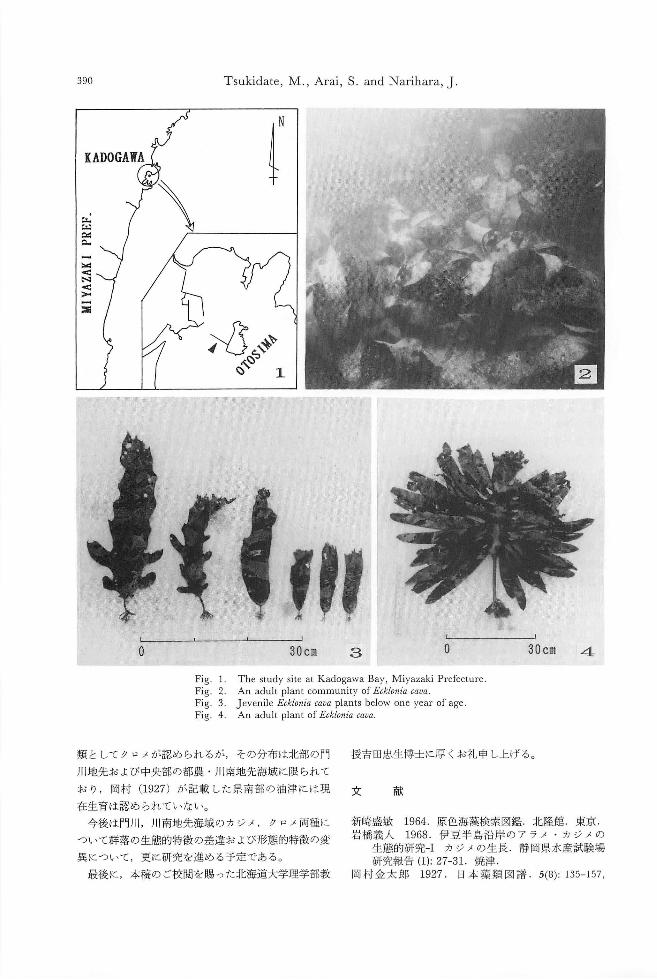
Tsukidat巴, M., Arai, S. and Narihara, J 390
バダ
.hHMZT回目
凶
〈
N吋」戸】酒
4 30cm O
The study site at Kadogawa Bay, Miyazaki Prefecture An adult plant community of Ecklonia cava Jevenile Ecklonia cava plants below one year of age An adult plant of Ecklonia cava
3 30cm
Fig. 1 Fig. 2 Fig. 3 Fig. 4
O
授吉田忠生博士に厚くお礼申し上げる。
新崎議敏 1964 原色海藻検索図鑑.北隆館 東京.
岩橋義人 1968. 伊豆半島沿岸のアラメ ・カジメの
生態的研究ーI カジメの生長.静岡県水産試験場
研究報告 (1):27-31.焼津.岡村金太郎 1927. 日本藻類図譜 .5(8)ー 135-157,
献文
類と してクロメが認められるが,その分布は北部の門
川地先および中央部の都農 ・川南地先海域に限られて
おり ,岡村 (1927)が記載した県南部の油津には現
在生育は認められていない。
今後は門)11,川南地先海域のカジメ,クロメ同種に
ついて群落の生態的特徴の差違および形態的特徴の変
異について,更に研究を進める予定である。
最後に,本稿のこe校閲をn易った北海道大学理学部教
Morphological features of Ecklonia cava Kjellman 391
PIs. 236-24-0. 岡村金太郎 1936.日本海藻誌.内田老鶴園.東京.岡田喜一 1933.日本海藻図鑑.風間書房.東京.川嶋昭二 1989. 日本産コンブ類図鑑.北日本海洋
センター.札幌.
千原光雄 1970.標準原色図鑑全集第15巻海藻海浜植物.保育社.大阪.
千原光雄 1983.学研生物図鑑海藻.学習研究社.東京.
中村郁也・南部豊揮・板崎 清・手島仁夫・吉田好一郎・河辺博 1981.熊本県沿岸海域の藻場・干潟分布調査.沿岸海域藻場調査九州西岸海成藻場・干潟分布調査報告. 175-241.水産庁西海区水産研究所.長崎.
林田文郎 1984.カジメの群落生態学的研究-II ヵジメの生長について.東海大学紀要海洋学部第18号.275-280.清水.
松井敏夫・大貝政治・大内俊彦・角田信孝・中村達夫1984.山口県日本海沿岸中部域における海藻群落.水産大学校研究報告.32(3): 91-113.下関.
瀬川宗吉 1956.原色日本海藻図鑑.保育社.大阪.山内正一・吉本幸雄・石川佑司・堀 隆喜・小原俊行
・荻田征男・井口弘章・伊島時郎・米田 寛・木谷益那・金枝豊広・高野傑・工藤勝宏・回染博章・篠田慶弘 1979.大分県沿岸海域の藻場調査沿岸海緩藻場調査瀬戸内海関係海繊藻場調査報告ー藻場の分布ー.179-209.水産庁南西海区水産研究所.広島.
百合野定・内田為彦・黒木 勝・工藤基善・緒方得生1979.宮崎県沿岸海繊の藻場調査.沿岸海域藻場調査瀬戸内海関係海域藻場分布調査報告ー藻場の分布一.211-231.水産庁南西海区水産研究所.広島.
*103東京都中央区日本橋馬喰町1-10-6芙蓉海洋
開発勝
日 239横須賀市津久井500グリーンハイツ8-3-
301側海藻研究所
神 *889-22宮崎市青島6-16-3宮崎県水産試験場

393
新刊紹介
Akats曲 a,I. (ed.): Introduction to applied phycology.
vi+683pp. 1990. SPB Academic Publishing bv, The
Hague. Price: DH. 240 (邦貨約 2万円)
我が園でも藻類学のこれまでの研究をまとめた綜説
論文集の英文著作が見られるが,その内容は主として
圏内に限られたものである。本書は日本人で初めて国
際的視野に立って編集した応用藻類学の本で昨年オラ
ンダから出版された。掲載する論文数は30編と多く,
それぞれの論文の長さは5頁から55頁と異なるが,全
体として683頁からなる大著である。執筆者は共著も
含めて51名.11名の日本人の方々のお名前も見られ,
いずれもその方面で菌際的に活躍している研究者ばか
りである。本書の特徴の一つは,現在話題になってL、
るものから将来その可能性が十分あるものを含めてこ
れまで余り紹介されていない色4な分野の総説を収め
ていることで,藻類が我々の周辺で広い範囲に関連し
ていることを改めて認識させられる。それぞれの論文
は藻類の応用の分野について極めて興味深い最先端の
知識を提供し,各トピックの概略を知ることが出来て
題名通り入門書としての役割も果たすが,いずれも豊
富に列挙している関係文献により最近の詳細な背景を
知ることが出来,むしろ専門書として研究者・学生の
いずれにも勧めることが出来る。本書は次ぎの様に 3
つの部分に大別され, トピックスごとにこれまでの研
究の経過,問題点更に将来展望などを述べてL、る。
大略を示すと,
パート 1は224頁.11編の論文からなり,細菌学,
菌類学,薬学,理化学などほかの基礎的な学問分野に
関連するものを含み,海藻の抗菌性;薬理作用;毒成
分とその取り込み機構;解毒作用;特殊含有成分(赤
血球凝集素金属イオン除去能力;海藻粘質物の物
理化学的性質;植物プランクトンの餌料としての価値
及びノリの病害と食品的価値(味と香り)について述
べている。
ミート 2では157頁にわたって海藻と生息環境との
関わりについての 5編の論文からなる。即ち植物プラ
ンクトンと富栄養(湖沼では現在量の変化,海洋では
赤潮動物の食庄;着生生物との相互関係及び海で
の汚染源(船底及び構築物の汚れ)について総説して
L 、る。
パート 3では幅広い利用や応用面,技術関係,バイ
オテクノロジーなどについての論文14編.277頁から
なる。即ち海藻粘質物の食品,医科・歯科への利
用;製紙・製塩技術;微細藻類の大量栽培技法;排水
処理やビタミン定量測定法での藻類の役割;ノリ・オ
ゴノリ・ツノマタの養殖についての海外事情;メタン
ガス生産の将来性;農耕・園芸への徴量成分の効用;
組織培養(細胞,原形質体,組織)などによる遺伝子
工学と予想出来る今後の成果など広く紹介している。
以上掲載論文の数々は藻類の各方面への利用・応用
についての知識を増し,理解を深め,より一層の興味
を覚えさせるものであり是非一読をお勧めする。尚,
本書は国際的にも評価が高く,逸早く Journalof Ap-
plied Phycology 3巻 (1991年)が好意ある書評を掲載
していることを付け加えておきます。
(226横浜市緑区東本郷6-27-2-306 正置富太郎)
藻類Jpn.J. Phycol. 39・394,December 10, 1991
吉田忠生:第4回国際藻類学会議報告
T. Yoshida: Report on the Fourth International Phycological Congress in North Carolina
第4回国際藻類学会議は1991年 8月4日から10日ま Cell and molecular biology: The cytoskeleton
での 7日間アメリカ NorthCarolina 州IDurhamの Algal systematics: Morphological and systematic data
Duke Universityにおいて行なわれた。松林に囲まれ Applied phycology: Developments in large-scale
たキャンパスのなかで BryanUniversity Centerを主会 culture
場とし , Biological Science Building, Chemistry Ecology: Terrestrial algae
Laboratoryの建物も使用した。この期間中, Durham Physiology: Nutrition
は東京の真夏と同じくらいの蒸し暑い気候だった。今 Cell and molecular biology: Molecular organization of
回はアメリカ藻類学会との共催で開催された。このた the chloroplast
め講演要旨はJournal of Phycology 27巻 3号のサプリ Algal systematics: Algal typification
メントとして印刷され,雑誌購読者には前以て配布さ Applied phycology: Algae pollution assessment and
れていたので,すでにご存じの方も多いはずである。 remediation
会議の参加者は600名以上あり,アメリカ合衆国は地 Ecology: Algal propagules and recruitment
元ということもあって,約250名で113以上を占めてい Physiology: Algae-atmosphere interactions
た。日本からは同伴者を含めて30名が出席した。 Celland molecular biology: Developmental molecular
8月4日(日)には参加登録とともにいくつかの biology
Workshopが開催された。夕方には野外で Barbeque SystematicsやEvolutionのSessionでは DAN,RNA
Dinnerを楽しんだ。 5日朝から簡単な開会式とそれ を用いた研究が多く発表され,これも一つの傾向であ
につづく PlenarySessionで会議が開始された。 ることが感じられた。
Plenary Sessionでは 5日J.Ramus: Photon capture: 6日(火)の夕方はアメリカ藻類学会主催の Ban-
Beyond the notion of color, 6日G.M. Hallegraeff: On quetがあり 7日(水)はエクスカーションと
the global increase of toxic algal bloom, 8日 G.O. Workshopが実施された。 8日(木)の夜にはアメリ
Kirst: Salinity tolerance in algae: Organic osmolyties- カ藻類学会の auctionが行なわれた。これは参加者が
necessary luxures for extreme conditions, 9日 R.S. 持ち寄った本や別刷り,その他の品物を競売して売り
Quatrano: Establishment of cell polarity in Fucusの4題 上げを学会の運営にあてるもののようで,この園の習
の講演が行なわれた。 債のようであった。
Congressの会期中に15のSymposiaと,多くの Con- 9日(金)の夕方からは市内のホテルでCongress
tributed papersのセッションが行なわれた。 Symposia Banquetと閉会式が行なわれた。このとき PosterSes-
の主題はつぎのとうりであった。 sionの表彰もあり,石田・中山・原グループと畠山・
Algal systematics: Modern concepts of algal phylogeny 佐々・渡辺・高市グループが 1,2位となった。次回
Applied phycology: Recent advances in algal は中国の青島市で開催されることが発表され,開催国
biotechnology を代表して曽呈歪氏のスピーチがあった。
Ecology: Macroalgae in sowing water (060札幌市北区北10条西 8丁目 北海道大学理学部)
Physiology: Algae and light
藻類Jpn.J. Phycol. 39: 395-396, December 10, 1991
原慶明・有賀祐勝:第2回日韓藻類学シンポジウム (1991年 9月8-11日)
Y. Hara and Y. Aruga: The 2nd ]apan-Korea Symposium on Phycology,
8-11 September 1991, in Tsukuba, ]apan
日本藻類学会と韓国藻類学会の協力で,日韓藻類学 InKyu教授の挨拶があり,三浦昭雄教授の乾杯の音
シンポジウム組織委員会の主催により標記シンポジウ 頭で開宴し,英語,韓国語,日本語を交えたにぎやか
ムが筑波大学国際会議場を主会場として開催された. な交歓会となった.また, Lee教授からは,本シンポ
これは日本藻類学会が毎年開催している秋季シンポジ ジウムのプロシーディングスを KoreanJ ournal of
ウムを兼ねたものである.参加者は,韓国からの31名 Phycologyの特別号として出版することについて検討
タイからの 1名,ベルーからの 1名を含め,合計約 してみたい,次回のシンポジウムは西太平洋地域の他
110名であった. の国々にも呼びかけて1993年にソウルで開催できるよ
9月8日夕方から登録をすませた人々が集まり, 18 う努力したい旨の発言があった.
時から筑波大学内のキャフェテリアで歓迎レセプシ!I 9月11日は, ワークショップ“Introductionto the
γが聞かれ,日韓両国の藻類研究者の交流がなごやか Phycoflagellates ..が筑波大学第二学群棟の実験室で行
に始まった. われ35名が参加した.午前は講義,午後は実習が行わ
9月9日には9時半から開会式が行われ,日本藻類 れた.その内容は次のとおりである.
学会の有賀祐勝会長および韓国藻類学会の KohNam 講義
Pyo会長の挨拶があった.引続き学術研究発表に移りr.Inouye: General Introduction: Microanatomy and
まず LeeIn Kyu教授の特別講演“Lifehistory of Taxonomy of Microalgae. Prasinophytes.
Ceramiales specially referring to the mixed-phase H. Nozaki: Ch1orophytes.
reproduction ..が行われた.その後, Session 1 Tax- M. Idei: Diatoms.
onomy: Red Algae 1, Session 2 Cultivation and Utiliza- r. Inouye: Chromophyte Algae Including Haptophytes.
tion of Algae 1, Session 3 Taxonomy: Diatoms, Session 4 T. Hori伊ぬi: Taxonomy and Identification of
Ecology: Macroalgaeが休憩と昼食をはさんで行われ Freshwater and Marine Dinoflagellates.
た.また,夜には Young phycologists と Senior M. Erata: Cryptomonads and Other Miscelaneous
phycologistsとに分かれて学外でナイトセッション Microalgae.
(、つくばの夜n 懇親会)が聞かれ,にぎやかな意見 実習
交換の機会がもたれた CultureTechniques and Light Microscopy
9月10日は川井浩史博士の特別講演“Aperspective 1) Inoculation, isolation and cultu問。fmicroalgae.
on the phylogeny of the Phaeophyceae"に始まり, Ses- 2) Identification of microalgae.
sion 5 Taxonomy: Brown and Green Algae, Session 6 Electron Microscopy
Molecular Taxonomy, Session 7 Physiology: Micro- 1) Sc叩 ningelectron microscopy (SEM): Techniques
algae, Session 8 Physiology and Genetics: Macroalgae, for preparing algal cells.
Session 9 Taxonomy: Red Algae II, Session 10 2) Transmission electron microscopy (TEM): Coating
Dinoflagellates and Environmental Sciencesが休憩と昼 grids wi也 Formvar,whole mount preparations and
食をはさんで行われた.日本藻類学会有賀会長の閉会 for cutting sections and staining.
の辞があり,学術研究発表の部は終了した.結局,こ 3) View wi也 TEMandSEM.
の2日聞に2題の特別講演, 18題の招待講演, 14題の また,講義・実習修了後も18時から21時まで,スラ
応募講演が行われた. (講演要旨については本誌39: イドやビデオを見ながら熱心な討論が行われた.
399-414を参照されたい.) なお,本シンポジウムの開催に対して次の諸国体等
同日18時半から筑波大学管理棟の食堂で晩餐会が聞 から協賛または協力を戴いた.ここに記して感謝いた
催
396 Hara, Y. and Aruga, Y.
筑波大学園際研究集会基金,角谷商庖,悌三洋,紛
高問屋,日本海苔生産機械協会,全国海苔貝類漁業協
同組合連合会,フルタ電気掛,輔山本海苔腐,理研食
品脚,渡議機関工業側,脚ユニカフェ,側白子,エチ
モウ輔,つくば市観光課,国立科学博物館筑波実験植
物園
参加者(登録者のみ)
Boo Sung-Min, Chae Seung-Moon, Choi Joong Ki,
Chung Ik-Kyo, Chung S組合Oak,Ee Yoo Kyung,
Hwang Mi Sook, Jung Yoon Hee, Keum Yeon Shim,
Kim Gwang Hoon, Kim Myung-Sook, Kim Young
Hwan, Koh Chul-Hwan, Koh Nam Pyo, Lee Hae Bok,
Lee Hae Il, Lee In Kyu, LeeJin Ae, LeeJin Hwan, Lee
Jung Ho, Lee WookJae, Nam Ki Wan, Oh Byong Gun,
Oh Yoon Sik, Park Ju Yong, Park Mi-Kyoung,
Seo Gyoung-Seok, Shin Jong-Ahm, Sohn Chul Hyun,
Yoo Soon-Ae, Yoon Mi-young, Patricia L. G. Kodaka,
Pornsilp Pholopumthin
有賀祐勝,千原光雄,土井孝爾,恵良国民由美,藤伊
正,藤田大介,福代康夫,後藤敏一,原慶明,畠山
典子,本多大輔,堀志保美,堀輝三,堀口健雄,市
村輝宜,出井雅彦,猪川倫好,井上勲,石川依久子,
石田健一郎,海部英一郎,神谷充伸,片桐正幸,片山
智康,加藤辰巳,河地正伸,川井浩史,北山太樹,菊
池則雄,小林弘,工藤利彦,前川行幸,真山茂樹,
峰一郎,三浦昭雄,宮村新一,中村良子,中山剛,
長嶋美香子,能登谷正浩,新山優子,野崎久義,岡崎
恵視,大房副,佐藤征矢,佐藤忍,斉藤譲,関
本弘之,須田彰一郎,田中次郎,綱川亜紀子,都筑幹
夫,和田雅人,渡辺信,山中良一,山田明子,吉崎
誠,横浜康継
(305つくば市天王台1-1-1 筑波大学生物科学系・
108港区港南4-5-7 東京水産大学藻類)
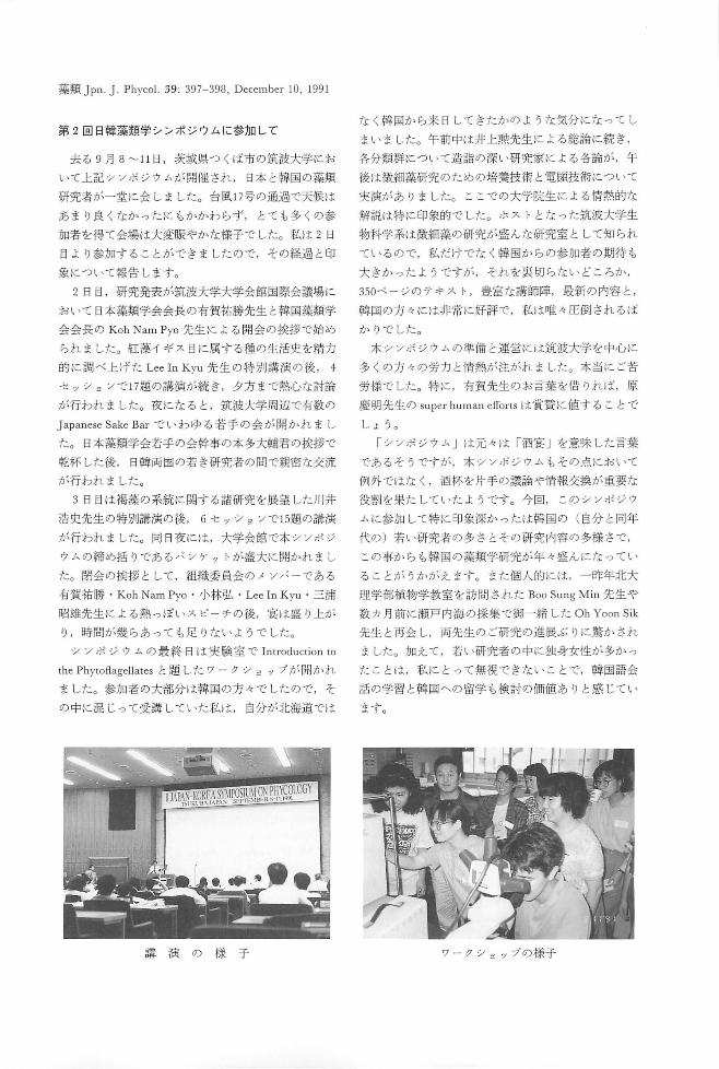
藻類 Jpn.J. Phycol. 39: 397-398, December 10, 1991
第 2回日韓藻類学シンポジウムに参加して
去る 9 月 8~ 11 日 ,茨城県つくば市の筑波大学にお
いて上記シンポジウムが開催され, 日本と韓国の藻類
研究者が一堂に会しました。台風17号の通過で天候は
あまり良くなかったにもかかわらず,とても多くの参
加者を得て会場は大変賑やかな様子でした。私は 2日
目より参加することができましたので,その経過と印
象について報告します。
2日目,研究発表が筑波大学大学会館国際会議場に
おいて日本藻類学会会長の有賀祐勝先生と斡国藻類学
会会長の KohNam Pyo先生による開会の挨拶で始め
られました。紅藻イギス自に属する種の生活史を精力
的に調べ上げた LeeIn Kyu先生の特別講演の後, 4
セッションで17題の講演が続き,夕方まで熱心な討論
が行われました。夜になると ,筑波大学周辺で有数の
J apanese Sake Barでいわゆる若手の会が聞かれまし
た。日本務類学会若手の会幹事の本多大輔君の挨拶で
乾杯した後, 日韓両国の若き研究者の間で親密な交流
が行われました。
3日目は褐藻の系統に関する諸研究を展望した川井
浩史先生の特別講演の後, 6セッションで 15題の講演
が行われました。向 日夜には,大学会館で本シンポジ
ウムの締め括りであるバンケットが盛大に聞かれまし
た。閉会の挨拶として,組織委員会のメ ンパーである
有賀祐勝 ・KohNam Pyo・小林弘 ・LeeIn Kyu .三浦
昭雄先生による熱っぽいスピーチの後,宴は盛り上が
り,時聞が幾らあっても足りないようでした。
シンポジウムの最終日は実験室で Introductionto
the Phytoftagellatesと題したワークショップが開かれ
ました。参加者の大部分は韓国の方々でしたので,そ
の中に混じって受講していた私は,自分が北海道では
講演の様子
なく韓国から来日してきたかのような気分になってし
まいました。午前中は井上勲先生による総論に続き,
各分類群について造詣の深い研究家による各論が,午
後は微細藻研究のための培養技術と電顕技術について
実演がありました。ここでの大学院生による情熱的な
解説は特に印象的でした。ホストとなった筑波大学生
物科学系は微細藻の研究が盛んな研究室として知られ
ているので,私だけでなく韓国からの参加者の期待も
大きかったようですが,それを裏切らないどころか,
350ベージのテキスト,豊富な講師陣,最新の内容と ,
韓国の方々には非常に好評で,私は日佐々圧倒されるば
かりでした。
本シンポジウムの準備と運営には筑波大学を中心に
多くの方々の労力と情熱が注がれました。本当にご苦
労様でした。特に,有賀先生のお言築を借りれば,原
慶明先生の s印upe町rhu山u山lIT
しよう。
「シンポジウム」は元々は「酒宴」を意味した言葉
であるそうですが,本シンポジウムもその点において
例外ではなく,酒杯を片手の議論や情報交換が重要な
役割を果たしていたようです。今回,このシンポジウ
ムに参加して特に印象深かったは韓国の (自分と同年
代の)若い研究者の多さとその研究内容の多様さで,
この事からも韓国の藻類学研究が年4盛んにな ってい
ることがうかがえます。また個人的には,一昨年北大
理学部植物学教室を訪問された BooSung Min先生や
数カ月前に瀬戸内海の採集で御一緒した OhYoon Sik
先生と再会し,両先生のご研究の進展ぶりに驚かされ
ました。加えて,若い研究者の中に独身女性が多かっ
たことは,私にとって無視できないことで,韓国語会
話の学習と隣国への留学も検討の価値ありと感じてい
ます。
ワークシ sップの様子
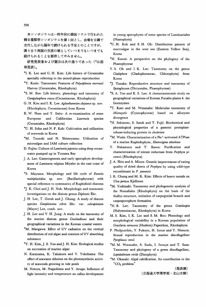
398
本シンポジウムは一昨年秋に韓国ソウルで行われた
韓日藻類学シンポジウムを第 1固とし,会場を日韓で
交代しながら隔年で続けられる予定とのことですが,
隣り合う両国の交流の場としていつまでもいつまでも
続けられることを期待してやみません。
研究発表者および題目は次の通りであった(*は招
待発表)。
*I. K. Lee and G. H. Kim: Life history of Ceramiales
specially referring to出elIIixed-phase reproduction
*T. Kudo: Taxonomic Features of Polys争'honiamo"owii
Harvey (Ceramiales,貼odophyta)
*S. M. Boo: Life history, phenology and taxonomy of
Campylaψhora crassa (Ceramiaceae, Rhodoph戸a)
G.H.Kim叩 dI. K. Lee: Aglaothamnion chりiuensesp. nov.
(貼odophyta,Ceramiaceae) from Korea K. W. Nam and Y. Saito: A re-examination of some
European and Californian Laurencia species
(Ceramiales, Rhodophyta)
*C. H. Sohn and N. P. Koh: Cultivation and utilization
of seaweeds in Korea
*M. Tsuzuki and N. Shimoyama: Utilization of
microalgae and IAM culture col1ection
D. Fujita: Culture of Laminariajaponica using deep-ocean-
water pumped up in Toyama Bay
J. A. Lee: Gametogenesis and early sporophyte develop-
ment of Laminaria reli抑'saMiyabe in the east coast of
Korea
*S. Mayama: Morphology and life cycle of Eunotia
mult争l回 'lidica sp. nov. (Bacillariophyceae) with
special reference to systematics of Raphidioid diatoms
勺.K. Choi andJ. H. Noh: Morphologic and taxonomic
investigations on the diatom genus D争loneisEhr.
J. H. Lee, T. Gotoh佃 dJ. Chung: A study of diatom
species Gomphonema vibrio Ehr. var. subcapitatum
(Mayer) Lee, comb. nov.
J. H. Lee and Y. H. Jung: A study on也etaxonomy of
也emarine diatom genus Coscinodiscus and their
geographical variations in the Korean coastal waters
市M.Maegawa: Effect of UV radiation on the vertical
distribution of red algae and contents of UV absorbing
substance
*Y. H. Kim,J. S. Yoo andJ. H. Kim: Ecological studies on succession of marine algae
N. Katayama, K. Takakura and Y. Y okohama: The
effect of seawater dilution on the photosynthetic activi-
ty of seaweeds growing in tide pools
M. Notoya, M. Nagashima and Y. Aruga: Influence of
light intensity and temperature an callus development
in young sporophytes of some species of Laminariales
(Phaeophyta)
*C. H. Koh and S. H. Oh: Distribution pattern of
macroalgae in the west sea (Eastern Yel10w Sea), Korea
*H. Kawai: A perspective on the phylogeny of the
Phaeophyceae
Y. S. Oh and I. K. Lee: Taxonomy on the genus
Cladophora (Cladophoraceae, Chloropl明 a) from
Ko問 a
勺 Tanaka:Reproductive stn叫 ure阻 dtaxonomy of
やatoglossum(Dictyotales, Phaeophyceae)
*S. A. Yoo and K. S. Lee: A chemotaxonomic study on
geographical variations ofKorean Fucales plants 4.血e
Isoenzymes
*T. Kato and M. Watanabe: Molecular taxonomy of
Microcy'山 (Cyanophyceae) based on allozyme
divergence
*H. Sekimoto, S. Satoh and T. Fujii: Biochemical and
physiological properties of a gametic protoplast-
release-inducing protein in c/osterium
*M. Wada: Characterization of a N a +・activatedATPase
of a marine Raphidophyte, Heteros忽maakashiwo
Y. Nakamura and T. Ikawa: Purification and
characterization of nitrate reductase from POψIhyra ye-
zoensis (Rhodophyta)
J. A. Shin and A. Miura: Genetic improvement of eating
quality of dried sheets of Po.同)lraby using wild-type
recombinant in P. yezoensis
I. K. Chung and M. K. Kim: Effects of heavy metals on
Ulva pert出aKjel1man
*M. Yoshizaki: Taxonomy and phylogenetic analysis of
the Nemaliales (貼odophy叫 onthe basis of the
thallus structure, initiation of ca中ogonialbranch and
carposporophyte formation
*H. B. Lee: Taxonomy of the genus Grateloupia
(Halymeniaceae, Rhodophyta) in Korea
M. S. Kim, I. K. Lee and S. M. Boo: Phenology and
morphological variability in a Korean population of
Gracilaria verrucosa (Hudson) Papenfuss,貼odophyta
P. Pholpunthin, Y. Fukuyo, H. Inoue叩 dY. Nimura:
Sexual reproduction in the marine dinoflagel1ate
乃rophacusstenii
*M. M. Watanabe, S. Suda, I. Inouye and T. Sasa:
T蹴 onomyand phylogeny of a green dinoflagel1ate, Lψ油 ,din白mviri・ゐ (Dinophyta)
*M. Okazaki: Algal calcification. Its contribution to the
“CO2 problem n
(発表順)
(北海道大学理学部・北山太樹)
Abstracts of the 2nd Japan-Korea Symposium on Phycology
第 2回日韓藻類学シンポジウム(日本藻類学会秋季シンポジウム)
講演要旨
Sept.8・11,1991, Tsukuba, Jap卸 1
399
400
PLENARY LECTURES
LIFE HISTORY OF CE臥MIALESSPECIALLY
REFERRING TO THE MIXED-PHASE REPRODUCTI0N.
field. ln
Platythamn10n
Antlthamn10n sparsum, yezoense, Dasyslphonla
l止血旦ム盟 andGwang Hoon Ki田.De凹rt田 nt chejuensls and Polys1phonla morrovll, mixed of Bioiogy Seoul National Unlversi ty, phases and bisexual i ty often occurred on
Seoul, Korea
Most studies on life history of
Cera回iaiescarried out in culture have not
incorporated either cytological observa-
tions or genetic investigations of key
features, and the unusual phenomena seen in
Cera田iales, 園ucha圃 bisexualityand mixed-
phase reproduction have been largely
unexplained. This study, based on labora-
tory culture of about sixty Korean specles
of Cera田iales under the controlled
environ回ent,曹as aimed to elucidate the
essential life history of ceramialecean
species as wel1 as the cause and meaning of
the mixed凶asesby chromo回目 analysisand
crossing experiment among isolat曲。feach
species.
The members of four familie圃 of the
Ceramiales investigated characteristically
showed a Polyslphonla-type l1fe hlstory
曹ith isomorphic gametophytlc and tetra-
sporophytic generations. Most of the
variations ln the 1 i fe history that have
been encountered both in nature and under
laboratory condi tions invol ve only secon-
dary modiflcations of the Polys1phon1a-type
life history. Among them, Antlthamnlon
nlpponlcum Yamada et lnagakl,且nt1thamnlon
sparsum Tokida, Antlthamnlon secundum
ltono, Platythamnlon yezoense lnagaki,
Aglaothamn10n oosumlense ltono (Ceramia
-ceae), Dasyslphonla chejuensls Lee et 'est
(Dasyaceae), Polys1ph01na morrovll Harvey
and Symphyoclad1a pennata Oka田ura
{臨odom哩laceae)showed mixed凶asesand/or
bisexual i ty in laboratory cul ture. Mixed-
ph闘 e plants of Ant1thamnlon sparsum, A. nlpponlcum, A. secundum and Platythamnlon
yezoense were col1ected also from the
the same plants. Most species 曹ere
regularly dioecious. Ho曹ever, dloecious
species sometimes producing both sexes on
the sa回 plant as an aberrant behaviour.
Most common is the occurrence of mixed
phases 冒ith both sexual stages and
tetrasporangia occurring on the same plant.
The occurring percentage of mixed-phase
plants varied greatiy according to the
species and popuiations, and it also varied
according to environmental conditions of
each isolates in Ant1thamnlon nlpponlcum, A. sparsum, A. secundum, Platythamnlon yezoense, Aglaothamnlon oosumlense and
Dasyslphon1a chejuensls. Al1 of the
exa田ined populations oC above species
showed a same pattern of liCe history.
ThereCore, mixed-phase reproduction was
found to be one of the natural property and
could be used as good character of each
species.
The cause of mlxed phases and/or
bisexuality must be closely related to the
sex determining mechanlsm of each species.
The田aleand female sex ratio examlned most
of the species胃asalway目 1:1regardless of
environmental condition, so that the sex of
these species was thought to be determined
by sex chro皿osome. Some of the species
曹hichhave only asexual 1 Ue history were
also observed: Honosporus lnd1cus Boergesen
and Honosporus keomundoensls Kim et Lee
(no18. lnval.) have monosporangiulD and
Call1thamnlon chejuense Kim et Lee (nom.
1nval.) have only perasporagiu回 for the
reproductive structure, respectively.
Sexual reproduction see回sto be degenerated
in these species.
A PBRSPBCI1VB ON百田町四LOGBNYOP古 田 PHBOPHYCEAB. Hiroshi Kawai, Department of BotaDY. Paculty of Sciencc. Hokkaido UDiversity. Sapporo. 060 JapaD
τlIe cl舗sPhaeoph,唱aeis a mcmhcr oC the 50・ωlled Chromophyta∞ntaina喝 chlorophyllA and .1: as pho旬明血叫cpi担問偽品血g帥吋 fromother cla鑓鴎 in havi曙 m雌叫lu1ar 白a1U wi血pl鰯modesmata, dilJcrentiation be同阻 vegeta助国個dreproductivc 回:lls 岨 d abund回t a単回同 and Oorotannins. The Phacophy切 aeis gencrally be6eved ωbe derived 針。m血.eChrysop匂'ceae,舗 lupportedby lome 6ne s出回世d 阻 dmole叩 lard蜘(Limet aI. l:弼5,O'KcIly &. Floyd 19邸. Bhattach間 'aet Druehl羽田:).
Conl関 :ming血.ephyl,噌畑yin the Pha回 'phy'ωae,Kylin (1933) propωed a phylog阻 ctictree、inwhich he regarded 血e 盤Ce hisωry patte岨 阻d basic ∞凶位uctionof血e白allusas血.emωt important charactcristics in品開ssingthe phylogeny. Since白阻
his basic ∞ 脚:pt 加 been g岡町拘 a帥 pt叫d血ou,晦hlome modified phylogenetic tr醐 havebeeoproposed (Scagel 19民 Wynnc&. Loiseaux 1仰の.Howe市er,so拘 :ra1basic ωntradictioDS havc bc∞me apparent踊 furtherknowledge accumulated especially from recent cu1turc studies. For邸 amplc,白corders Laminaria1凶阻d D凶 marestia1es which 鴨 re∞DSid町吋 tobelo.曙 todiffer阻 tphylog閲 eticlines, nowap戸arto have much clωer systematic r百:latioDS.Therefore, a revision of our ∞且切:pt of the fundam個 .ta1ぬaracte出tics阻 d phylogeny of the Pha聞 Iphyceaeis nccded. In出epresent paper, 1柑ureview 10.鵬 taxo岨omic chara~町凶cs 釦d reωnt biologica1 6ndi峨 payIDaspccla1 atteotion to血.cirsystematic imp6ca山
百 .0n阻加r阻 dぬa伊 of血echIoroplasts阻 dp防総nceand abseoce of tho pyr阻 oidsarc曲cmlωt pro.凶回岨tand important cytologica1加.tures.They aro g阻 crally∞凶t阻 tamo.曙 variousspccies in阻
ordcr irrespective of the Ufe-stage or part of曲a由allus, and are regarded as g四 d systematic charact,釘lsti岱.A single chIoroplast wi也 a py官官noidin a cell is ∞凶ideredto be the most primitivc ∞ndition in the Phaeophyceae,岨d m阻 ychIoropl箇箇叫血outD帽 阻.oidsin aωU to be血emost dcrived.
Life history patterns aro st出 oneof the mo嘩timport阻 tsystematic characteristics. In S町 inllodermao卸ious variatiゆDS in 血o morphology of thc 伊meωphyt偶{針。m branched filamcnts to only 2 伊 鵬t叩h界iccells) are found, despite the rathcr uniform morphology of也esporophyte (He町 y1984). Mon田 cism岨 ddioecism seem to occur叫delyin ω,mo o.吋町ssuch as Laminaria1es. Desmar鎚 tia1儲岨 dFu叫ω.
401
Sex皿1phoromoD筒 aremown to have儲腿ntialroles in thc I閣JaIrcproduction of血.cPha旬'p~戸cac(Maicr &. MOIIcr 印刷 About 10 I捌a1phcromon鎚 (often0関町d曙 asmixt町es)havc be個ideotified to date 阻 d 血eirdis凶bution among pha回 p抑制ntaxa appar阻.t1yhas systcmatic imp6-ωtiODS. For example, lamomene is∞lUumed to b司
自efunctiona1 pheromone in more白個 30spccies oC 伽 dcrivedLaminaria1略 ho鴨 鳩 町 itis not Cound in the p血 itivc1aminariaI,剛 f細部esnor in岨 Iy0血町orders. D儲 mar腿 tene, Yiridi阻e, fuα)SCrratcne阻d明 tophore田町a1soknOWD only in one order鎚
actua1 pheromon叫a1白ough0白crs(e.g.叫∞町個邑阻 dhormωirene) arc Cound in le'鴨川 ordc~ιIna凪温四S, a quantitativcly minor substance (hormcト位聞の issu昆estedωactω 血.eact:回 1phcromonc 例凶cret a1. 1990).
The structure oC swarmcrl (i.c.血e∞nditionoC theぬloropl腿匂阻dOagc1la) is ∞DSideredωhave important systcmatic imp6関白血s.Mωtpha悶 Iph:戸eanzoospores havc a chloroplωtwi血 a lti伊 aand OageUar Iwelling on thc basa1 part of posterior sageUum.問句.eUarb脳血1bodies are∞nnected 漏出the nuclcus岨 d are Ipatially associated 漏出血echloroplasts. However, those oC dcrivcd Laminaria1es lack a stigma and sageUar swelling. Green sageUar 醐 toOuorescen関 脇 田iated wi白血e ltigma 阻 dOageUar swelli略 whichis IU鴎槌tedto be involvcd i目白c photoreception of photot副S, is noted to O側 rin phototactic Iwarmerl Q叫 lcret a1. 19,町,Kawai 19邸:).The It抱ma is ShOWD to havc a rcOectivc function阻 dCI回回目白ereOected 6ght on 血.eOagcllar swc出ng(Kawai et aI.旬開1,Kreimer et a1. l:卯1). Some s.伊 rm (e.g. Laminaria1叫 havelongcr posterior Oagella 阻 d 鈎糊a1 fragmeoted chloroplasts which are not assoaated 漏 出 血B
Oagcllar base, while 0血闘(時 Fucal凶:)share the 10噌;crposterior OageUum but have a chIoroplast wi血 orwithout a stii戸a. Rcmnant OageUa鴨 reCound in lome immotilc cells IUch凶 laminar泊lc岨E鴎皿ddict)ota1ea且 spor.ω (Motomura1989, Philli伊1991), which su鴎鑓t白紙血B曙伊阻dtetraspores in 白ese orders ori酔ate from Oagellate cells. 百 S
Oagellar rootlet system is in principle s凶 laramong most phal回 phy,切回 swarmers, composed of bypassing rootlets which p儲 S白rough曲eOagcUar b鵬 s 10噸tudiDally,and Co町 kinds of rootlets 例AR,mar, MPR, mpr) which issue from 出血喝:eUarba醐 (0・Kelly1989). Laminaria1回nIpcrm 1ack MPR. In fucalc岨 lperma bundlc oC the MAR fo~ a proboscis.τlIcre are variatioDS in the numbcr of tub叫鎚 inth蹴 Z∞由民 ho鴨,vcr,自由
basic number in phaeophy,切阻 swarmersis re伊 dedto bc MAR!mar/MPR/mpr/BR = 7/1s11f7.
402
1 NV ITED PAPERS
TAXONmnc FEATURES OF POLY-SIPHONIA MORROft'II HARVEY (q:RAMlALES, RHODOPHYTA) . Tos弘主辿QA旦do',Michio ~Iasuda". ~Biol. Lab., Sapporo Univ., Toyohira-ku, Sapporo, Japan. ゐDept.of Botany, Fac. of Science, Hokkaido Univ., Kita目 ku,Sapporo, Japan
Taxonomic features of Polysip/lOnia morrOld i I~ere investigated through i ts life history stages by laboratory cullure experiments and periodic field observations. The taxonomic cri teria wllich had been used for this species were evaluated by comparing ontogenetic data of field-collected and cultured plants. Some of taxonomic features auopteu previously show phenotypic variation according to environmental condilions or growth slages: lhe lenglh/diameter ratio of segment日 the external appearance of branches, Ilumber of axillary tetra-sporangial branches. The significant spp.cific feat.ures of P. mOl"rowii were confirmed in this investigation.
This species is characterized by Lhe following feat.ures: (1) ecorti-cated thalli with 4 pericentral cells, (2) lhick and setaceous thall i in ful1y matured sLage, (3) adventitious rhizoids without sepLations, (4) tightly intricated prostrating branches, (5) sh札rplypointed ulti-mate uranchlets, (6) endogenous axillary branchlets formed from the cenlral axial cells, (7) a few tl'ichoblasts in matured stage, (8) Letrasporangia formed both on the ul timate and axillary branchlets, (9) ax i llnry te t rasporallg i al branchlets tuftly (7-8 in number) formed on the axils of fu11y目laturedplants, (10) spermatangial branches replacing the whole trichoblasts, (11) urceolate cystocarps.
The occurrence of endogenous axillary branches is a peculiar featm'e characterizing some species in the genus Polysipbollia. The t.axonomic relationship uetween P. morrowii and closely related species sharing the above-mentioned feature is discussed.
LIFE IUSTORY, PHENOLOGY AND TAXONO附 OF CAHPYLAEP,仰 '&4 C&4SSA <CERAMIACAE,附ODOPHYTA).B00. Sune:-Mln. Dept. of Blology. Chungnam National Univ., DaeJon, 305-764, Korea.
The llfe hlstory of an eplphytlc specles, Camp:ylαephora crassa (Okamura) Nakamura, was investigated ln the wild on the central eastern coast of Korea and under varlous laboratory conditlons. Korean C. crass,αplants showed no obvlous seasonal dlfferences ln the occurrence of reproductlve organs, whereas thelr overall hablt was varlable. In culture the specles completed a Polysipl1ollia type of llfe history wlthout the baslphyte, but parasporangia occurred at a frequency of about 1 ~ on young tetrasporophytes. Paraspores grew lnto tetrasporophytes, which later cyoled a Polysiphonia type of llfe hlstory. Crossabll i ty tests a1so documented that paraspores had the sa岡崎2
nuclear phase as normal tetrasphorophytes. Thls fact lndlcates that paraspores of C. crassa may be mltotlc diploids which show a temporary modificatlon ln the 11fe history strategy. Maxlmal growth ln the field occurred ln ~Iay when the seawater temperature ranged from 14.9 to 18.2 .C, whlle ln cul ture the optlmum temperature for growth was bet同 en 16 and 20 .C under each photon flux density. Plants in the field had tetrasporangla and cystocarps throughout year and the tetrasporophytes ln culture formed tetrasporangla at most comblnatlons of photon flux densitles and temperatures.
CULTIVATION AND UTILIZATION OF SEAWEE凶 lNKOREA. Chul Hyun Sohnl and Nam Pyo Koh2 • IDept. of Aquacultur・e, Nat・lFish. Univ. of Pusan, Pusan, Korea. 2Dept. of Aquaculture, Yeosu Flsh. College, Yeosu, Korea
Seaweed flora of the Korean coast Is qui te di verse, and more than 600 species has been reported. ln Korea uti 1 ization of the seaweeds dates back to the early Korean history, and they have been cultivated since 17 C. The total annual production of the seawe哩dfrom the natural algal beds and by cultivatio明 wasrecently estimated to be 483.000 M/T. The production by cultivatlon occupies more than 90" of the total algal production. ln 1988. 34.619 M/T of seaweeds were exported, whi le 3,090 MlT were i皿ported,and most of the皿曹eredr i ed and sal ted. The major algae of economic importance include Porphyra, Gelldlum, Gracl1arla, Pachymenlopsls, Undarla, Laminarla, Sargassum, Hlzlkla, Enteromorpha and Codlum etc. Of these Porphyra, Undarla, Lamlnarla, Hlzlkla, Enteromorpha have been cul tivated. Porphyra is very popular. and its annual production is amounting to US $
300 million which is more than half of the total aquacul ture fisheries production in
Korea. Traditionally the sea胃eedshave been consu田ed by various ways such as fresh salads, dried snacks, roasting lavers and seaweed soup. However industrial processing has been undeveloped. ln Korea the agar productlon is made mainly by agar extraction fro回 Gelidlum amansii, whlle production of carrageenan/alginic acid is very few.
403
UTILIZATION OF MICROALGAE AND IAM CULTURE COLLECTION. 盟主主主立Ts旦五且主よ and Naomi Shimoya国a.Institute of Applied Microbiology, Univ. of Tokyo, Yayoi, Bunkyo-ku, Tokyo 113 Japan
Microalgae has often been used as materials for research of plant science. It is because they can grow very fast under a certain condition and be taken out quantitatively with pipettes, Axenic culture and mutants have also been useful, However, the species usually used have been rather restricted so far, and the strain nu阻berof the species are sometimes important for physiological and bio-chemical researches, for each strain shows different characteristics at times, The same is the case in bio-technology. Though some chemicals such as βーcarotene and poly-saccharides are extracted from some strains, microalgae are still rela-tively untapped resources.
It is important for a researcher to obtain and then to keep strains of algae when he wants to use them. Since most of the algal species must be maintained by serial transfer at regular intervals, there is a great risk of disappearance. There needs the special center where algal strains are maintained and supplied by demand,
Algal Culture Collection in the Institute of Applied Microbiology (IAM), University of Tokyo was commenced in 1957. Many strains of microalgae were collected and identified there and were maintained to be distributed for scientific and technological studies and education. These works, however, were carried out in a research laboratory, In 1989 Microbial and Microalgal Research Center was established in the same insti tute and i t took over the collection for the purpose of substantial culture center of micro-organims including microalgae. As the first step of the center, we have been checking the strains we have and collecting information about them for a catalogue. Other activities of our collection will be also reported in this symposium.
404
即mね以沼Y AND L1回 CYCLE OF R脱班、TA MC.広.TIPLASTIDICA. SP. 関~.(1弛.C1LLARIO田町 臥E) W1叩 SP.配IALREFERENCEτo SYS1宮l'IAT1CS OF RAPI江-
DI01D D1A:官》偲 Shigeki 地.yaJDa.
Deparb時 ntof Biology, Tokyo Galu砥eiUniv., Koganei-shi, Tokyo, Japan.
Valve shapes of BUnotia sp担 ies町 e四 u叫 lysimple and the taxonomic characteristics are sc回"ce under light microscopy. 1n addition,油evalve shape varies during the life cycle. However, there has been no observation thro唱:houtthe life cycle using SEM in any sp回 ies.
E. multiplastidica 禍 soriginally found on wet moss in Sainokawa, Ehio旭町-ef.,Japan. Unialgar cul tures were established frαn the original materi-a1.伽e to two months later, sexual reproduction 禍 S observed in these cultures. 1n 油 is pr∞ess, one auxos戸 rewas formed from two game-tangia. 加 initial cell developed within each auxospore was followed by a forn叫 ion of post-initial cells. 百 einitial valves were 45-47凹 1001and about three times longer than those of g:回 et&ngialcel1s.
Observations of the valves and plastids were carried out thro唱 houtthe life cycle using these cultured cells. 切10ughthe shape and leng出of the valves varied extre胞 11',certain characteristics were stable thrO¥唱houtthe life cycle, 1.e., the numerousness of chloroplasts, the Iα現 .tion of pattern center and raphe, areola morphology, striation d四国ty,the number of labiate proc-ess and epitheca depth. 官1esechar-acteristics we阿国ed 回 taxonomiccriteria of油畑町田ies.
1n the pennate dia旬咽,油ere have been few re戸 rtson the fine struc-ture of tlle perizonium. 1n this st叫1', the structure of the perizo-nium was examined in detail using SEM. 旬1etradi tional syst回I8tiCSof the raphidioid diatoms w田∞閑truct-ed based on only the p四 senceof the labiate process and the raphe. However, the particular structure in the perizonIlDD, nar臨 11', the areolated pril臨 rytransverse band wi th以)th a circular and lin回 r pattern 四 nter,suggests a probable 悼'1'logeneticaffinity with the centric diatα阻邸時
the araphid diat叩回.
MORPHOLOGIC AND TAXONOMIC INVESTIGA-TIONS ON THE DIATOM GENUS DIPLOHEIS EHR. Chol. Joona K! and Jae Hoon Noh. Department of Oceanography. lnha Unlversl ty. lnchon 402・751.KOREA
A large number of Dlplone1s specles collected from the Kor・-側、 coastal waters have been investigated by light and scann-ing electron 皿icroscope. 27 species. 8
varieties and 1 for田a are identified. ho開 ver, 7 species are unidentified and photographed in this study. Among these species, 16 species, 5 varieties and 1 forma are newly recorded ln Korea. These species are D. bomboldes, D. adon1s var. oamaruensls, D. dlplostlcta, D. noraesleen-
dae, D. bombus var. bomblformls, D. hospes, D. dalmat1ca, D. campylod1scus, D. n1tes-cense, D. gemmata var. prlstophora, D. respa, D. notabl11s, D. suborblcularls, D. crabro var. dlrhombus, D. schmldtl1, D. contlgua, D. graefl1, D. racl11ans, D. parca, D. vacillans var. ren1tens, D. smlthll forma rho叩blcaand D. lacrlmans.
EFFECT OF UV RADIATION ON THE VERTICAL DISTRIBUTION OF RED AL-GAE AND CONTENTS OF UV ABSORBING SUBSTANCE. MiYuki Maes:aw!l, Masayo Kunleda and Washirou Kida. Faculty ot Bioresources, Mie Univ., Tsu, Mie, Jap-an.
UV band occupies 9 " ot solar rad-iation in the outer space and 1-4 " on the sea surtace depending on weather conditions. UV radiation, dose not penetrate so much as PAR in the sea, decreased to l.;β" at a depth ot 5 m, and below 1 " at a depth ot 10 m. We tested the intluence ot UV radiation, tull solar radlation and PAR for seve-ral red algae collected trom shallow water, intertidal zone to upper sub-tidal zone, and from deep water, 25・30 m depth. UV radi此,10n,which oc-cupied only a few ratios ot solar radia-tion, depressed photosynthetic activity ot d鵠 pwater specles slgnlticantly as well as full solar radiation and PAR did. Shallow water species were not affected so much by UV radlation, tull solar radia土lonand PAR.
We have a hypothesis that UV ab-sorbing substance ∞nta1ning much ln red and blu-green algae protect the thallus from strong UV radiatlon. Shallow water specles may have an ab1lity to reslst lnjurious UV radlatlon, and d鵠 p water species may have no such an ab1lity. However, there have been a little information tor e∞,logical role of UV absorbing substance. Then, we measured the contents ot UV ab-sorbing substance for 33 species ∞トlected from shallow and deep waters. Shallow water species conta1ned much UV absorbing substance, and deep water species contained little or no UV absorbing substance. Particularly, intertidal species had more UV absorb-ing substance than other species.
In our previous paper, deep w此erspecies had more phycoerythrin, which absorbed green to blue light, than shallow water species. So, deep water species adapted to deep water light condition. In this study, shallow water species have more UV absorbing sub-stance than deep water species. So, shallow water species are protec同dfrom injurious UV radiation by high contents ot UV absorbing substance. UV radiation in the sea is one ot the most important factors to control the vertlcal distribution ot red alg回, with reference to the ∞ntent levels of UV absorbing substan回 between shallow and deep water species.
405
ECOLOGICAL STUDIES ON SUCCESSION OF MARINE ALGAE. Younlf Hwan Ki!!h Jong Su Yoo and Jee Hwan Kim. Dept. of 8iology, Chungbuk Nat. Univ., Chongju 360-763, Korea.
As the process of ecological succession occurs more rapidly in benthic marine habitats than in terrestrial communities, benthic 回arine communities provide a particularly cゅnvenienttesting-ground for theories about ecosystem development and ecological succession. Since 1985 we have examined the variations of colonization, growth pattern and succession of benthic marine algae on various artificial Substl'ata and also on cleared natural surfaces.
Firstly, seven kinds of artificial substrata, i.e., concrete, slate, glass, wood, rubber, aluminium and PVC plates, were placed at upper, middle and lower intertida1 zones of Poryong and Sochon, western coast of Korea, durillg the period of January November 1985. As a whole, coccoid blue-green algae and diatoms were observed as pioneer algae settled over newly placed substrata, and then filamentous green and crustose coralline algae were gradually luxuriant, whereas diatoms decreased in abundance. Colonization and growth of marine algae were significantly illfluenced by differences between tidal levels or the kind of artificial substrala.
Sludies have also been made of the recolonization of cleared natural surfaces over a 21-mo period (July 1986 lo April 1988) at intertidal zones of Muchallgpo and Maryangri, western coast of Korea. Surfaces were sterilized by burning after clearing. ln general, the successive stages found in the permanellt quadrats were blue-greens or filamentous algae membranous algae 国 perennial algae. However, the nature and posi tion of the surface and length of tiDle of exposure have been shown to inCluence the population which develops. It was concluded that a climax communi ty can be attained aCter 18 mOllths since the substrata were cleared.
406
DISTRIBUTION PATTERN OF MACRO-ALGAE IN THE WEST SEA (EASTERN YELLOW SEA), KOREA. Chu1-Hwan Koh, Sang-Hee Oh, Dept. of Oceanography, Seou1 Nat. Univ. Seou1, Korea
Koh and Lee (1982) tried to differentiate the f1ora1 composition of benthic a1gae between coasta1 and open waters in the Kyunggi Bay, West Sea of Korea. The tota1 number of species tends to increase from the coasta1 waters to the open sea. Brown a1gae are more sensitive in terms of species number. Song (1984), Sohn (1987) and Park & Kim (1990) reported a1so the same tendency in the distribution pattern of macroa1gae in the who1e area of West Sea.
Koh and Lee (1982) insisted that the floristic differences that are observed between the coasta1 waters and open sea area appears to be re1ated to water turbidity rather than to temperature as reported by Kang (1966). Severa1 authers have agreed with the importance of turbidity since 1982. The re1ationship between the distribution and environments are reviewed in this presentation.
REPRODUCTIVE STRUCTURE AND TAXONOMY OF SPATOGLOSSUN (DI町 YOTALES,PIlAEOPIIYCEAE). Jiro Tanaka. Dept. of Botany, National Science Museum, Shinjuku-ku, Tokyo,
Japan Tbree species of SpatoglossulII, S.
pacificulII YENDO, S." crassulII sp. nov. and S. latulII sp. nov. occur along the Pacific coasts of the central Japan. They for圃 threekinds of reproductive organs, 1.e. sporangia, oogonia and antheridia on separate plants. Sporangia can be distinguished fro圃 oogoniaby bigger sIze and so皿etimesby possession of four spores. As tbe result of the 回orpho1ogicalco圃parison a皿ong the Japanese species and the other established species of the world, tbe genus SpatoglossulII can be separated into two groups based on tbe position of reproductive organs.
In tbe first group including S. pacificulII: spor岨 gia and oogonia aggregated in sori and project above the thal1us surface; antberidial sori stand up above tbe general thallus surface. These characters are well known in the other genera of the Dictyotales, i.e. D1ctrota, Dilophus, Pachrdictron and Dictyopteris. This group comprises S. areschoug11 J. AG. from Barbados in the Caribbean Sea and S. chaplllanii LINDAUER fro田 NewZealand. In the second group including S.
crassulII and S. latulII: sporangia scatter throughout the thal1us and not in a sorusj sporangia and antberidial sori are 17ing co凪pletely within the cortex; oogonia scatter throughout tbe tbal1us and project both above and below the cortex. These features, particularly the buried sporangia inl;o the cortex, have never been knowll in the otber genera of the Dictyotales. This group comprises S. crispata HOWE from Peru and S. lIIacrodontulII J. AG. from Australia.
It can be concluded that these two groups are separated into two genera based on the above-mentioned re田arkable differences 00
reproductive structures.
AOIE即r~例制IC SI1IDY側 G臨掘削PHlCALVARIA110NS OF KO賠訴Nfl臥LaPLAN'悶. 4.百EI回国也:YM回. Soon-Ae Yoo and K1-Sung Lee. Department o( B101ogy, Pa1-Cha1 Un1rers1ty, DaeJon 302・735,Korea.
10 obtain chemotaxonomic characteristics of Korean Fu岨 lesplants. 同 hadalready compared the c個 lposition of pigl鴎 nts,凶os凶olipids, neutral fats, inorganic poly凶os凶ate,and haema回lutlnin l . 2 • 31 . In this凹.per,陶 com開 re the patterns of 13 kinds of is田町zymes extracted f何回 10 species of Korean Fucales plants.
百1eelectrophoretic zy田昌r岨 ssho官官dthat most of the c創・bohydrate-回 tabolizinsenz)'ll闘(G-6-P聞, M聞, A聞 1聞,1'<訓,刷y)and catalase had mono- and/or di・田rphicpatterns. It seems that the genetic variations are 園田11 i n those enzy血esyste田. Those enzyme systems see皿sto be g串netically stable. They have the 回 in制官:ymeactivi ties in isoenzymatic Z)'ll悶'gr阻 S
回 bilizedto the cathode. On the other hand, in the case of
haydolase(AKP, ACP, EST), isomerase(GPl),
oxidoreductase, they all showed polymorphic isoenzymatic zymogra田s. We beiieve that these enzymatic systems are genetically rather unstabie. Most of these e回 y田 sare involved in凶osphatemetabolism, and the activlty of these e間四回 variesaccordins to the 凶os凶ate concentration of the environment. So these enzym鶴田咽 to be important in studying genetic variations in Fucal田 plants growins ln different geographlcal habitat.
1) Y,∞, S. A. 凱 d K. S. Lee. 1988. A chemotaxono皿lc study on g'伺 'graphi伺 lvariations of Korean Fucales plants. 1. The com凹rison on pi脚 nts, 前回陣。lipids,neutral fats and inorganic 凶os同18tes.Korean J. Phycol. 3(1):1・8.2) Y,∞, S. A., K. S. Lee, and K. S. Park. 1988. A che回 taxonomicstudy on geographical variations of Korean Fucales plants. 2. Lectin. Korean J. Phycol. 3(1):9-12.
3). Y,∞, S. A. and K. S. Lee. 19朗. A d羽田taxonomic study on 8回 'graphi個 lvariations of Korean Fucales piants. 3. The 3・di醐 lsionalanaiysis of total lipids. Kor. Jour. Bot. 31(2):83・89.
407
HOLECULAR TAXONOHY OF MICROCYSTI~ (CYANOPHYCEAE) BASED ON ALLOZYHE DIVERGENCE. MIQ I皐主呈旦mi and WATANABE Hasayuki. National Science Huseum, Tsukuba 305, JAPAN.
Microcysti~ is a limnoplank-tic, bloom-forming blue-green alga. As a part of molecular taxonomic revision, allozyme divergence of Japanese 旦主Q!:QヱCYS主主呈 wasstudied electropho-retical1y.
We investigated a110zyme genotypes at four enzyme 10ci (IDH, 6PGD, PGI and PGH) on a total of 168 strains co11ected at fourty-three waters. The obtained data were statistical-1y ana1yzed according to KATO & DOI's Hinimum Replacement Hethod. As a result, the strains studied were classified into four genetic types: 且主主主皇ヱ,C"I:呈主主呈 aerusrinosA S-type, 且L
aerusrinosA L-type, 且ι 主主主主昼主墨and 且.!...!!esenbersri1. Each of the four types was found to exhibit characteristic morphol-ogy. 且ム aerusrinosAS-type was characterized by its sma11 size cel1s (3.0-4.2 pm). The other three types that have 1arger cel1s (4.5-6.5pm) were dis-criminated by the co1ony forma-tion: 且ム aerusrinosAL-type, 且ι主主主主昼主呈 and 且ム Wesenbero:ii formed c1athrate, cubic and pouched colonies, respective1y. According1y, the four genetic types cou1d be viewed as sepa-rate taxonomic entities.
The above achievements wi11 provide a new and re1iable taxonomic system of Microcvs-主主呈・ As clear1y i11ustrated here, the a110zyme study (i.e. mo1ecu1ar taxonomic approach based on allozyme divergence) is considered as one of the most effective approaches that would lead to the solution of many taxonomic problems on cyanophytes, especial1y at the species and/or genus 1evel.
408
BIOCHEMICAL AND PHYStOLOGICAL PROPERTIES OF A GAMEl'IC PROTOPLAST-RELEASE-INDUCING PRO-TEIN IN CLOSTERIUM. Hi皿ヱ盛;iS盛国Q包,Shinobu Satoh, and Tadashl Fujii. Institute of Biol. Sci., Univ. of Tsukuba, Tsukuba-shi, Ibar位i305, Japan. HeterothalIic strains of Closterium oroliferate
asexually in nitrogen-sufficient medium while conjugation is induced in nitrogen-deficient mat-ing medium (MI m吋ium).百leconjugation pro-cess consists of cell division, pauing, formation of conjugation papillas, release and fusion of gametic protoplasts, and formation of zygotes. Some ?ells fo.r~ed a p.apilla ~n? ~el~ased .th~ir protoplasts without pairing.百lisindicated that the pr'∞ess of protoplast-releasing was indepen-dent upon p瓜尚19加 dthere rnight be a subs切にe,which was responsible for protoplast release, in the medium. 1). Detectioll 01 a substallce that illduces the
gametic protoplasts. When mt+創tdmt" cells were separately incubated in the MI medium obtained from55h・old-凶xedcultw'es of mt+ and mt" cells, release of protoplasts was observed only in mt"
cells. These data indicated that a substance re-sponsible for protoplast release was released into the medium. 2).Pw折catioll01 a protoplast-release-illducillg
proteill (PR-IP). The substance with the ability to induce the release of protoplasts was pur出edfrom the medium by sequential column-chromatographic steps, and named PR-IP. The PR・IPhad an apparent molecular mass of 95k on gel filtration and could be separated into sev-eral isoforms by anion-exchange chromatogra-phy. Each isoform consisted of two glyco-polypeptides ofM,s of 42k and 19k, 'Y~lile the deglytosylated polypeptides had M,s of 34k佃 dik, respectively. 1). Physiological properties 01 PR・Has indispensable for the protoplast叶om an analysis of dose-response cur可
Incentrationrequired for 50% of the m! sponse (EDso) was calculated as ~.1 ・oreover, thereduced protoplast-releasil
tion was observed by the treatment of high con-centration of PR-IP (5.2μg/2ml). These data indicated that PR-IP dose not act as cell wall Iytic enzyme, which is weII-known in conjuga・山nof Chlamydomollas. We propose the PR-IP is a biologicaIly active glycopolypeptide that in-duces the release of protoplasts by binding to 問ceptorson the cell surface, as do animal peptide hormones.
CHARACTERIZATION OF A NA七ACTIV A TED A TPASE OF A MAR町ERAPHI-DOPHYTE, HETEROSIGMA AKASHIWO. Masato Wada. Institute of Biol. Sci., Univ. of Tsukuba, Tsukuba-shi, Ibaraki, Japan. A marine raphidophycean unicellular biflagel-
late, H. akashiwo, is a naturally occurring, wall-less organism which is useful for bo自由eprep-aration of plasma membrane and the investiga-tions of its physiological functions. One of he major functions of plasma membranes is the regulation of ion transport at the cell surface. High purified plasma membranes were isolated from H. akashiwo WiUl silica rnicrobead meUlOd and the novel membrane associated Na七activated ATPase activity were characterized. The ionic requirements and spectra of effective inhibitors on Ule ATPase activity showed a close sirnilarity to the animal Na¥K九ATPase. This kind of A TPases which are sensitive to vanadate forms phosphorylated intermediate in their en-zyme cycJe. The phosphorylated interm巴diateof this ATPase were detected as 140 kDa polypeptide with acid SDS-polyacrylamide gel electrophoresis; this molecular weight was con・
siderably bigger than theαsubunit of animal Nぶ,K+-A TPase. However, the antiserum to animal Na¥K七ATPasereacted to H. akashiwo 140 kDa ATPase. lt was suggested 山atboth Na + -activated A TPases have a common epitope. The cDNA sequences of animal Cal+-ATPase andfungal H七ATPase has been 油田dyanalyzedwith biotechnological methods.τhe obtained infonnations on Uleir functions have facillitated to understand the ion transport, ion selectivity and ATP hydrolysis mechanisms of these AT-Pases. The primary structure of the A TPase of H. akashiwo con佐ibutethe understanding of Na ion transport or Na ion selectivity mecha-nism. A cDNA cJoning of the ATPase from H. akashiwo was achieved with PCR method and successed, the homology between those A TPases was discussed.
TAXONOMY AND PHYLOGENETIC ANALYSIS OF THE NEMALIALES (RHODOPHYTA) ON THE BASIS OF THE THALLUS STRUCTURE, ITIATION OF CARPOGONIAL BRANCH AND CARPO-SPOROPHYTE FORMATION. MakoもoYo豆且主互主主主 Depも. of Bio1ogy, Toho Uni versi七y,Funabashi-shi, Chiba, Japan
The order Nema1ia1es wa目
esもab1ished by Schmitz (1892), on 七he basis of the 1ack of auxi1iary ce11 in もhe formaもionof carposporopy七e, and 七hefo11owing 3 fami1ies a日七axacomposing七he order Nema1ia1es are proposed by Puescha1 and Co1e (1982): The Acrochaeもia-ceae, He1mi七hoc1adiaceae and Ga1axauraceae.
T h e d aもa avai1ab1e aも
present,七he fo11owing keys can be made in connec七ion wi七h 七he七axonomy and phy10gene七icre1aもionships of 七he Nema1ia1es on 七he basis of 七he 七ha11usBもruc七ure,carpogonia1 branch and carposporophyte formation: 1. Carpogonia1 branch is direc七ー
1y formed on the fi1amen七ous七ha11us 2
1. Carpogonia1 branch is formed on 七he assimi1atory fi1amen七
ーー-- 3 2. Compac七 carposporophy七日 is
formed ー-Acrochae七iaceae2. Diffused carposporophyもe is
formed ---- Woe1ker1ingiaceae fam. nov.
3. Carpogonia1 branch is formed 七ermina11y on a ce11 of assimi1atory fi1amen七 ーーー- 4
3. Carpogonia1 branch is formed 1a七era11y on a ce11 of assimi1aもoryfi1ament 6
4. Hypogynous ce11 gives rise 1aもera1 ce11s wiもh dense con七en七日 ーー--Ga1axauraceae
4. Hypogynous ce11 no七 produce1a七era1ce11s 5
5. Compac七 carpo目porophyもe is formed ---- Nema1iaceae
5. Diffused carposporophyもe is formed ---- Dotyophycaceae
fam. nov. 6. Compact carposporophyもe is
formed -- He 1 minもhoc1adiaceae6. Diffused carposporophyもe is
formed ---- Dermonamataceae
409
TAXONOMY OF THE GENUS GRATELOUPIA (HALYMENIACEAE, RHODOPHYTA) IN KOREA Hae Bok Lee. Department of Bio10gy, Chongju University. Chongju City 360-764, KOREA
The externa1 morpho10gy, interna1 struc-ture and reproductive organ of nine species of Grateloupia (Ha1y皿eniaceae,Rhodophyta)
analyzed and re-evaluated taxono田ically.The species of Grateloup1a reported untll now in Korea is Grateloupla dlvarlcata, G. fil1clna, G. fl11clna var. porracea, G. 1mbricata, G. llvlda, G. okamurae, G. prolongata, G. sparsa and G. turuturu. As a resu1t of comparison of the taxonomic
characters between G. fl11clna and G. f1l1c1na var. porracea. the latter is regarded as a independent species from the
former. And the morphological characters of G. okamurae is co皿pared to other species
and re-evaluated taxonomically. The taxono-田iccharacters of the plants be10ng to the
genus Grateloup1a are (1) lubricous tex-ture, (2) 5-10 cell layers of cortex, (3) lobed inner cortica1 ce11, (4) longitudina1 arrangement of medullary filaments, (5) reproductive organs on frond surfaces, and
(6) conical auxiliary cel1 a田pu11awith one pri皿aryand on to four secondary ampullar
fi laments.
410
TAXONOMY AND PHYI正氾ENYOFAGRFEN DINOFLAG阻止..A1E,LEPIDODINIUM VlRIDE.(D町OPHγTA).MakotoM. Wa包n油~l Shoichiro Suda1, lsa.o Inouye2 and Tsutomu Sasal. lNational Institute for白lvironmenta1Studies, Tsukuba,肋釘ヨki,Japan. 2Institute of Bio1.Sci., Univ. Tsukuba, Tsukuba, Ibara1d, Japan. A 民n-cゅ10悶dmarine dinoflagellate,
Z伊 idodiuniwnviride wi出achlorophyll a-創ldb-∞n凶iningvestigial endosymbiont is given with叩ecialemphasis on the mo甲加logy回 dthepigment∞moosition.
官lehost dinoflagellate cell is unarmored and hasagymn吋inoidover叫lap戸arance.官le曲目aor創nphi回 mabasically consists of the outer membrane and flat刷剖由民alvesicles in which no thecal pla飽sare developed. Unusual hand basket-shaped scales cover the entire cell surfa偲 ω酔山崎witha layer of mucilaginous material.百lesefindings led usωconclude出鉱
山eorganism was a new member of the Gymnodi凶郎国.eand to propo叩 theabove new genus and species name.百leultrョstructureof thehost田 sis typical of the dinoflagellates; ho~eyer, ~he or~~~sm~白 1) 卸 unusualcyωp恒smicprojection白紙maybeahomologue of the peduncle, 2) a single membrane-bounded body ∞ntaining membranous sh田 ts,closely situated nextω血eendosymbiont, and 3)叩 el配 tronop叫uenetwork-forming appen也.gesurrounding出etransverse flagellum. None of these featu.res have been found in other dinoflagellates.
官levestigial endosymbiont is unlike anything山athas been found in血sdinoflagellates before. The cyωIplasm of the endosvmbiont is seoarated from the host cytopfasm by a double membrane卸 dneitheranucleus or mitochon批iao∞urwithinil官leendosymbiont∞n凶nschlorophylls a and b and the~~ual ~~loropl}~te car~tenoids! ~hat are n即 X印刷n,violax卸白in,antheraxanthin, Z回xanthinand beta-carotene. In addition to 曲目ecarotenoids, some unknown peaks were de肱 ted.One peak is situated at the identical retention time to that of lutein, but the absorption s戸ctrumis slightly different from 白紙oflutein.百leother関誌sare und配 ided,al血.oughthere is a small peak which seemsω show the identical retention time and absorotion spe住umω山田eof siphonaxanthin. Bωedon lese隠 sults,it is 宅配ulatedthat a pras加。phyte
would s田 mωbe出.elikeliest candidate for a progenitor of血沼田dosymbiont
ALGAL CALCIFICATIOH. ITS COHTRIBUTIOH TO THE “CO2 PROBLEK" . Kegumi Okazaki. Department of Biology, Tokyo Gakugei University, Hukuikita-国achi,Koganei-shi, Tokyo 184, Japan.
The recent increase in atmospheric CO2 (350ppm at present) is a matter of anxiety because of rapid climatic changes from its greenhouse effect. However, the primitive atmosphere contained much more CO2 (about 97%) and it has been suggested that photo-synthesis and biological CaC03 depo-sition in ocean played an important role to obtain the present low level of CO2 on the earth. In fact, nearly 10,000 times as田uchcarbon of atmos-phere is in undecayed organic matter in sedi田entsand indeed 100,000 times as much in limestone (CaC03).
The certain corallinaean algae form algal limestone and M呈lim主主主 is im-portant as a CaC03-sand former and as a sediment-consolidator in coral reefs.
Coccolithophorid such as Emil且旦旦h旦主leyiis regarded as the chief pro-ducer of CaC03 in ocean.
CaC03 deposition in ocean takes place by a following reaction: 2 HC03- + Ca2+ → CO2 + CaC03 + H20. Therefore, it should be noticed that CaC03 formation accompanies CO2
evolution from dissolved HC03- in sea-water. Algal calcification, however, is coupled with their photosynthesis and the rate of photosynthesis is several times higher than calcifi-cation rate. Thus, CO2 release from seawater never takes place during their calcification process. However, their photosynthetic products in ocean might be completely oxidized sooner or later. Therefore, contri-bution of calcification to fixing atmospheric CO2 is dependent on what proportion of organic matter produced in ocean is not oxidized back to CO 2 •
CONTRIBUTED PAPERS
AGL且OTHAHIIIOIICHEJUEIISE SP. NOV.
RHO回 PHYTA,CERAMIACEAE) FR側聞REA.
G曹8JlJllIoon Kl回 andln Kyu Lee. Depart田ent
。f Blolo8Y, Seoul Nat10nal Unlversl ty,
Seoul 151-742, Korea
AglaothamnJon chejuense, a new red alga
bearlng parasporangla, Is descrlbed fro圃
CheJu Island of Korea. oAl though, the plants
sho曹edalternate branchlng pattern and had
only one nucleus In each vegetatlve cell,
1t was clearly dlst1n嘩川Ishedfro田 other
阻e田bersof the genus AglaothamnJon as冒ell
as A. hookerJ, the only other specles
bearlng perasporangla,旬 thecharacters of
branchlng pattern. aplcal parasporangla and
lack of sexual atages. Aaexual llfe hlatory
胃hlch comprlsed of perasporophyte and
tetraaporophytー曹as repeated slx t1皿es
叫 thoutany aexual reproduct1ve atructure.
The parasporangla developed from an aplcal
cell of branch. whereas the tetrasporangla
developed serlately on the adaxlal alde of
lateral branches.
CULTURE OF砧世出型A品回且CAUSING
DEEP-OCEAN-WATER PUHP回日PIN TOYAHA BAY.
Daisuke Fujita. Toyama Pref. Fish. Exp. Stn.,
Nameri-kawa-shi, Toyama, Japan.
Using flowing deep-ocean-water(DOW, 6~)
pumped up fro皿 thedepth <200m of Toyama Bay,
one year old plants of 1盟組E且且胆叫旦
were cultured in a tank set on the artificial
upwelling experimental facility for about one
month in summer. They grew well at the rate
of 9.6mm/day, while those cultured in the sea
(25~ , 5m depth) deteriorated. Iron level was
high at the old part of DOW-cultured kelp. In
the laboratory culture(10~ , 3,000 lux) for
one month, sporelings of the kelp grew the
best in PEDOWI, and in PESI, in DOW and in SW
in turn. 出血且皇叫盟並立組畠 spore1 ings also
grew well in the order above.
411
A RE-EXAMINATION OF SOME EU-ROPEAN AND CALIFORNIAN LAURENC-IA SPECIES(CERAMIALES, IUIODOPHY-TA) . Ki笠型A且昼m and Yuzuru 8aiLo*. Dept. of Mar. 8iol., NaLional Fish. Unlv. of Pusan. 章l.ab. Mar. 80t., Fac. Flsh., 1I0kkaldo Un.lv.
Morphology of some European and Californian species, L. pl-nnatifida, L. spectabilis, L. crispa, L. platycephalll and L. hybrida, was re-examined. 8per-matangia and tetrasporangia es-sentially differ from those of L. obtusa, the type of the gen-us, as produced at alternately branched filaments derived from epidermal cells rather than at dichotomously branched trichob-lasts done from axial cells and at epidermal cells rather than at pericentral cells, respectl-vely. Those spec.les probably occupy a different position in evolutionary l1ne from the gen-us.Of those specles, L. hybrida is distinctlve in with six peri-central cel1s insLead of five at fertile segment of female trichoblas t.
GAMEltxi副回18AND EARLY SPORO開YrEU町EWP~I副T OF L1II1JNARIA RELIGIOSA ttIYABE 1 N THE EAST COAST OF KOI也A. Jl且A立Le豆,
Dept. Environ. Sc1., InJe Unlv., Xlmhae 621-749, Ko同 a.
Ecotypic卯 pulatlons of 印刷nariarel igiω'(J ln the east coast of Korea嶋 mmonltor凶 fromOctober 1989 to October 1990 to lnvestlgate growth and同 pro-ductive phenology at near southern 11mlt of lts dlstrlbution in East Sea of Korea. Pl岨ntsexhlblted an annual growth pattern wlth maxlmu悶 lnJu田. tlost of the morp加ー
l唱 lcalparameters measured sho同 dthe slmllar pattern and問削除dlIIaxlmu回 valuesln June. Blade dlslntegratlon ∞curred durl噌 thesum回ermonths. Reproductlve sporophytes∞curred from Octo胎rto Uecem胎r1989. Y,側ngs凹lrophytepo凹ー
lat.ioll was obse円 'edln March, whlch was assumed to be the result of the late fall spon:耳~enesls actlvlty. Reproductlon alm gr'O¥olth ln gametophyt.es and growth ln Juvenlle sporophytes鳩 restudied in 同 181:Ionto temper叫 ureand irradlan田.
AI thnugh no seasonal varlatlon was found, hlgher lrradlance (80μE'III-2・s回ーり,and temper叫 ureof .1 -12 -c resulted ln g同 ateract.lvlties.
412
A STUDY OF DIATOM SPECIES GOMPHONETlA VIBRIO EHR. VAR. SUBCAPITATUM1百五百R)LEE, COMB. NOV. Jung Ho Leel , Toshikazu GotOh2 and Jun Chung l • I Dept. Biol., Kyungpook Univ., Taegu, Korea. Dept. Biol., 2Kinki Univ., Osaka, Japan.
This taxon was described as GoruDhonema intricatum var. vibrio f. subcaDitat~ by Mayer (1928). Micromorphology of this taxon and g. vibrio were studied Nith ornamentation of girdle can be devided this taxon from G・型遁盟主盟 (Syn.G.ntricatum) • There is no basical difference
between G. vibrio and V.ntricatum var. vibrio f. subcapitat~ except for size and outline 01' valve and striae density. It therefore is proposed that the new combination, G. vibrio var. subcapitatu!!1.. And a180,τττE elucidate that g・旦主m2旦旦主主呈 Skv. is a synonym of this taxon.
THE EFFECT OF SEAWATER DILUTION ON THE PHOTOSYNTHETIC ACTIVITY OF SEA-WEEDS GROWING IN TIDE POOLS. Nobuvasu 皇旦yama1,Kl;lmi Takakura 1 and Yasutsugu Yoko扇面82. lDept. of Biology, Tokyo ~akugei Univ., Koganei, Tokyo, Japan. LShimoda Marine Research Center, Univ. of Tsukuba, Shimoda, Shizuoka, Japan.
The effect of a stepwise seawater concentration fall, diluted with dis-tilled water or bufferred isotonic NaCl solution(pH 8.2), on the photosynthetic rate of ULvαpel'tu8α, Entel'omol'pha cl'i-nitαand Gl'lαteLoupiαfiUcinα, growing in tide pools, was examined. E. cl'initα was most tolerant of the seawater dilu-tion among them. The photosynthetic activity of G. fiLicina was affected mainly by the change in C02 concentra-tion, while of U. pel'tu8αthe activity was affected by the changes in both pH and salinity. These seaweeds retained their photosynthetic activities even in freshwater in several hours. The result obtained indicates that the tolerance for the seawater dilution by a rainfal1 must be one of the important properties of seaweeds growing in tide pools.
A STUDY ON THE TAXONOMY OF THE MARINE DIATOM GENUS COSCINODISCUS AND THEIR GEOGRAPHICAL VARIATIONS IN THE KOREAN COASTAL WATERS. Jin Hwan Lee and Yoon Hee Jung. Sangmyung Women's University, Seoul 110-743, Korea
In order to identify the RADIATI group of the diatom genus Coscino-discus from the eastern, western, and southern Korean coastal waters, it has been studied both light microscope ! LTI) and scanning electron microscope (SEM) observations of a fine structure and morphological patterns.
As a reslll t, the genlls Coscillo-discus was recorded six species and one unidentified species C. gigas, C. grallii, C. asteromphalus and C. sp.1.
Most of Coscinodiscus taxa have showed the insignificant morphological (the valve diameter, number of areolae, the number of marginal processes, etc.) variations in three coastal waters of Korea. As compared between Korean specimens and other waters, it showed geographical differences valve diameters, areolae number, feature of labiate processes, perforations of the cribra, etc.
INFLUENCE OF LIGHT INTENSITY AND TEMPERATURE ON CALLUS DEVELOPMENT IN YOUNG SPOROPHYTES OF SOME SPECIES OF LAMINARIALES (PHAEOPHYTA). Masahiro Notoya. Mikako Nagashima and Yusho Aruga. Lab. of Phycol.. Tokyo Univ. of Fish.. Konan-4. Minato-ku. Tokyo. 108 JBpan
Excised rectangular blade piec回 (0.5-1mm) in young sporophytes (3-5 岡田 blade length) of four Laminariales species
(色邑主己主盟邑邑2・邑豆盟主主且旦三よis担且盟且豆担胆且旦・出血且豆 PinnBtifida)were cultured under various light intensities (1-8 klux) Bnd temperatures (10・25"C). and callus development and di fferentia t.ion冒ereobserved. CBllus development from pieces of sporophyte explant 冒asalmost the sa回ein each species. After 2-4 days in culture. callus cel1s were produced from the cut edge. In about 3-4胃eeks.blade-like plantlets were differentiated from gro胃ingcBllus cells. Favorable conditions for callus development from explBnts were different with species: 15"C and 2-4 klux in C. costata. 20 "C and 1 klux in ~.単旦且語. 10-15"C and 1 klux in k.担胆且旦 and15-20"C and 2 klux in y. 且型車且旦虫.
TAXONOMY ON THE GENUS CLADOPHORA (CLADOPHORACEAE, CHLOROPH汀 A) FROM KOREA
Yoon Sik 0b and ln Kyu Lee. Depart皿entof
Biology, Seoul National Universi ty, Seoul
151-742, Korea
A taxono田icstudy on the members of Cladophora commonly collected fro皿 the
coasts of Korea was carried out to re-
appralse the morphゅloglcalcharacters and
elucidate the interspeciflc dellmitations
among species. For the distlnction of
Cladophora specles, the followln哩 taxonomlc
cri terla along wl th tradl tional ones 胃'ere
used: architecture, color, texture,
rhizoidal morphology of plants, denslty,
basal fusion, and ramiflcatlon wlth phyl10taxis of branches, and cell shape and di田enslon. On the basis of these criteria,
seve叩teenspe氾iesincluding a new on直 were
described in this study. They also sho咽ed
distributional pattern closely related
to environmental factors and were divlded
into several groups wlth ecologlcal
characteristics in relatlon to the
皿orphologicalvariations.
GENETIC IMPROVEMENT OF EATING QUALITY OF DRIED SHEETS OF PORPHYRA BY USING WILD-TYPE RECOMBINANT IN P. YEZOENSIS, Jong-Ahm Shin and Akio Miura, Lab, Algae Cultivation, Tokyo Univ. Fish., Minato-ku, Tokyo, Japan.
Growing test of the gametophytes of ZGRW was performed for determining of characteristics pertaining to ZGRW. Crispness and free amino acids con-tents of dried sheets of Porp担旦 arerelated in eating quality. They were determined to compare ZGRW with grow-ers' one. ZGRW showed better quality in crispness than growers' one. Con-cerning free amino acids contents, alanine, glutamic acid, aspartic acid and taurine in ZGRW were more abun-dant than those of growers' one. Ala-nine and glutamic acid are concerned in taste substances in dried sheets of Porohvra. Alanine was 522.86-2625.57m喧/100gand g1utamic acid was 592.87-1574.02m喧/100gin ZGRW to that a1anine 485.69-2525.89mg/100g and g1utamic acid 476.70-1472.59mg/100g in growers' one. ZGRW presented more sweet f1avor than growers' one.
413
PURIFICATION AND CHARACTERIZATION OF NITRATE REDUCTA8E FROM PORPHYRA YEZOENSIS (RIIO∞PHYTA). 12且辿a且AklI.皿且l:Aand To田oyoshi1 kawa. Inst1tute of Biol. 8c1., Univ. of Tsukuba, Tsukuba-shi, Ibaraki, Japan.
Assimilatory nitrate reductase (NR) catalyzes the first step in the reduc-tion of nitrate to ammonia. We develop-ped an effective method to isolate the homogenious NR from Porphyra yezoensis using PEG treatment, ammonium sulfate fractionation, chromatography on butyl Toyopearl 650-M, blue 8epharose CL-6B, DEAE Cellulose, hydroxyapatite colums and 8ephacryl 8-400 gel filteration. The best enzyme preparation was puri-fied to 5,700-fold and had a specific activity of 12.5 pmol NOz-'min-1・mg・1
protein. A molecular weight for the native enzy回ewas estimated to be 380,000 and for the subunit to be 100,000. Deduced from the native and subunit molecular weights, NR from P. yezoensis is a tetramer. The UV/visible absorption spectra of the oxidized and reduced NR indicated typical features of b-type cytochromes. The enzyme was NADH specific and had an optimal pH at pH 8.5.
EFFE<了ISOF HEAVY MF:rALS 側 ULYAPEKfllSA KJ乱凶AN.1k ){yo Chung & Ml Kyu唱Klm. 1除pl.of Har1ne Sclence. Pusan Natlonal Unlv., Pusan 609-735, KOREA.
The effects of seγeral metals (Cd. Cu, Zn, Hg) on Ul凶戸封.tusa嶋同 examlned.Aocllmaled dlsc samples問問 co叫 lnuゅuslyex凹 sedf町 amaxlmum perlod of 2同 eksto a serles of concentratlon of metals supplemented to enrlclad関 awater.
Toxlclty of metals assessed ln 旬 rmsof g代lWthrate, photosynthetlc plgment G叩 tentsand chlorophyl1 fluorescence, 同問 hlghestln Hg t問 atedsamples, severe ln Cu, m吋eratein Zn and 10l0I ln Cd ln U. pertusa. Valu四 ofF..ax/F .. ratlo 111 phot∞hemlcal quenchlng鳩問
問 markablydecreased and growth rates 問問 slgnlflcantlyreduced at hlgher concentrations of metals. These lndlcate t.hat those are effectlve and rellable means of assesslng to叫 cltyof ∞ntaml-nants 1/1 relatively short term studies wit.h thls g問 enalga.
414
PHENOLOGY AND MORPHOLOGICAL VARIA81-LITY IN A KOREAN POPULATI0N OF GRACILARIA YERRUCOSA (HUDSON) PAPENFUSS, RHODOPHYTA. k1m. Mvunll-Soo~, ln Kyu Lee and Sun圃-Min800*. Dept. oC 8iology, Seoul Natl. Unlv., Seoul 151・・742 and ・Dept. oC 81010gy, Chungnam Natl. Univ., DaeJon 305・764,Korea
The.phenologyand morphologlcal varlabl-Ilty oC-Grac11ar1a verrucosa (Huds) Papen-Cuss曹ereanalysed at the Ilttoral habltat oC Daechon, west coast oC Korea Cro皿 July1988 to June 1989. Fifty or more plants were sampled haphazardly 1n C1eld Cor.phen-ology. Twenty-Clve tlants were rando皿Iyselected and回easuredfor the length, axis diameter, medullary cell diameter-and cons-triction in basal portlon of branches. Cystocarpic plants occurred at maxi回U皿 fro固
June_to July, while tetrasporic ones do回1-nated fro田 Augustto Septe田ber.This impli-ed that ecologlcal cond1tions related wlth summer were Important for reproduction of our plants. Plant length, axls dlameter, medullary cell dla田eterand constricton 1n basal portion of branches varled slgnlfi-cantly. throughout the year. The correlation coefflcient between axis diameter and皿edu-llary cell diameter was posltively corre-lated, whereas between plant length and constriction of basal portion of branches it曹asnegatively correlated. In standardi-zation of four investigated characters, the monthly variability of basal constriction oC branches contrasted wlth those of other three features
SEXUAL REPRODUCTION IN百也胤RINEDINOFl.AGELlATE P'YROPHACUS STEINII. P.Q.皿illpf担lp血血in,Yasuwo Fukuyo, Hiroaki Inoue and Yoshihachiro Nimura. De戸 rbnentof Fisheries, Faculty of Agriculture, University of Tokyo, 1-1-1 Yayoi, Bunkyo-ku~ Tokyo, Ja戸 nlbe sexual reproduction in _~vroohacus
呈足担iiis anisogamous and heterothal-lic. Male gβmetes differ fr叩 femalegametes and vegetative cells. The fe-male gametes can not be differenciated fr叩 thevegetative cells. Cell fusion between the male and female伊 netesocεurs in a few hours to several days after inoculation of the male gametes into a culture of non-male clone. Zy-gotes are similar to the vegetative cells in shape except possessing two longitudinal flagella. The transfor-mation fr∞1 the planozygote to the hヴpnozygote(resting cyst) requires five to eight days for completion.
一会員移動一
新入会
住所変更
415
416
退会
黒荻尚(北海道),篠原尚文(栃木県),後藤将夫(茨城県),高野克夫(東京都), SRIVASTAVA,
MADHA VI (INDlA).
Acknowledgements to reviewers for V 01. 39
The Editorial Board is grateful to the following persons for their cooperation in reviewing the
m叩 uscriptssubmitted to the J apanese J ournal of Phycology Volume 39.
Yusho Aruga
Mitsuo Chihara
Sachito Enomoto
Yasuo Fukuyo
Y oshiaki Hara
Terumitsu Hori
Terunobu Ichimura
Isao Inouye
Tetzuya Katoh
Hitoshi Kito
Hiromu Kobayasi
Michio Masuda
Akio恥iiura
Kazutosi Nisizawa
Masahiro N otoya
Masa戸lkiOhmori
Masao Ohno
Megumi Okazaki
Jiro Tanaka
Masakazu Tatewaki
Mikio Tsuzuki
Makoto M. Watanabe
Iemasa Yamada
Yukuya Yamaguchi
Yasutsugu Y okohama
Tadao Yoshida
Makoto Y oshizaki
417
418
一B本学術会議だより eNo.22
一一一一 一 一 一 一
第15期最初の総会開催される
平成 3年 8月 日本学術会議広報委員会
日本学術会識の第15期が7月22日から発足し 7月22日一24日の 3日間,第四期最初の総会が開催されましたので,その
総会等についてお知らせします。
日本学術会議第112回総会報告
7月22日の第15期の発足に伴い,内閑総理大臣による日
本学術会議会員の辞令交付が行われた。第15期の会員は.
選出制度が学術研究団体を基礎とする推薦方式になって,
3回目の会員である。この第15期会員による最初の総会で
ある,第 112回総会が7月22日から24日までの 3日間,本
会議講堂で開催された。
第1日目 (22日)は.午前は新会員への辞令交付式があ
り,午後総会が開会され,直ちに,会長及び耐副会長の選
挙が行われた。会員による互選の結果,会長には近藤次郎
第5部会員が13期. 14期に引き続き三選された。人文科学
部門の副会長には.川図侃第2部会員.自然科学部門の副
会長には,渡逸格第 4部会員が選出された(波遁副会長は
再選L選挙終了後,近藤会長から「新人の方が半数以上お
られ,大きな飽負をもっておられると思う。挫折感を持つ
ことのないようできるだけの努力をしたい。皆様にも御協
力をお願いしたいu との就任のあいさつがあり,又. 111回,i度溢11句扇Ij会長からもそれぞれ就任のあいさつがあった。
会長,副会長選出後は.直ちに各部会が開催され.各部
の青11長,副部長,幹事の選出が行われた。(第15期の役員
については,別掲を参照)
第2日目は10時に総会が開催さtL.近藤会長が14期lの会
長と L寸資絡で第14期の総括的な活動報告を行った。その
報告の折々には,国際交流とか.将来計画I委員会.学術会
議の予算等,会長の感慨,または感恕をも交えてその所感
を述べた。続いて,会員推薦管理会報告として,久保亮五
委員長の代理として事務総長が.第15期会員の推薦を決定
するまで'の経過報告を行った。
引き続き,会長から 3日自の総会で従業・審議する予定
の『第15期活動計副委員会の設置について(申合せ業)J'ニ関する各部での事前討議について,並びに各常置委員会の
各部での委員の選出について,それぞれ各部へ依頼した。
総会終了後.各部会が開催され,自ijiliの1¥I{tせ案の討議
及び会常置委員会委員の選出等が行われた。
第3日目 (24日), 10時に総会がl調会され,会長から「第
15lUli8動計画i委員会の設置について」の提案が行われた。
これは,第15矧の活動の基本計画の立案を目的とする臨時
の委員会を次の定例f総会までの問,設置するということを
内容としている。そしてこの提案は原案どおり可決された。
総会総了後,直ちに各部会が開会され,設置が決定され
た第15期活動計画委員会委員の選出等が行われた。
なお,この第15期活動計画委員会は,総会期間中に第 1回の会識を聞き,全会員を対象にした第15期の学術会議の
活動に関するアンケートの実施を決めるなど,早速その活
動を開始した。
また.運営審議会附置委員会,常置委員会,国際対応委
員会等も活動を開始した。
第15期日本学術会議の辞令交付式等について
第 112回総会に先立ち.第15期日本学術会談会員の降令
交付式が7月22日(月 )11時から、総理大臣官邸ホールで
行われた。辞令交付式は.海部内閣総理大臣.坂本内閣官
房長官.大島. {i原凶1官房副長官,稲橋総理府次長等の出
席を得て執リ行われた。
第1部から第 7部までの会員 1人ずつの名前が読み上げ
られた後全会tiの紋年長である液型量格第 4音11会員が代表し
て海部総理から僻令を手j度された。この後,海部総理大臣
から「会員の侍様には.創造性豊かな科学!t術の発展.総
合的観点に立勺た学術研'先に係る諸活動に御尽力いただき
たい。」とのあいさつがあり.これに応えて第15JtJI会員を代
表してj度遁絡会員が「微力ながら全力を尽くし.重要な責
務を全うし.国民の矧待に応えたい。」とあいさつがあり,
式は終了した。式には192名の会員が出席した。
また,総会 2H日の夕方には.学術会議ホールで.坂本
宵房長宵主催の第15期会員就任パーティーが開催された。パ
ーティーは坂本官房長浜・のあいさつで開会し.日本学士院院
長代壊の謀長問良雄幹事の祝併があリ,これに対する近藤会
長の~礼のあいさつ,沢田敏男日本学術保興会会長の発声による乾杯の後.悠談に入った。ホールには溢れんばかり
の人々で歓談が続き磁会であった。
第15期日本学術会議役員
会長
副会長
近藤次郎(第 5部・経営工学)
川田 侃(第 2部・政治学)
j度進 格(第 4部・生物科学)副会長
<各部役員>
第 1部 部長
副部長
幹事
肥田野直(心理学)
弓削 達(歴史学)
一番ケ瀬康子(社会学)
山本 信(哲学)
第 2部部長 西原 道雄 (民事法学)
副 部 長 細 谷 千 博 (政治学)
幹 事正田 彬(社会法学)
山下健次(公法学)
第 3部部長 大石 泰彦 (経済政策)
副 部 長 島 袋 嘉 昌 (経営学)
幹事岡本康雄( 経営学)
藤井 隆(経済政策)
第4部部長中嶋貞雄(物理科学)
副部長田中元治(化学)
幹事竹内郁夫(生物科学)
樋口 敬二(地球物理学)
第5部部 長岡村総吾(電子工学)
副部長 市川 i享信(計測・制御工学)
幹事内田盛也(応用化学)
増子 完(金属工学)
第6音11 部長 中川昭一郎(農業総合科学)
副部長水間 豊(畜産学)
幹事志村博康(農業工学)
平田 照(農芸化学)
岡田 晃(社会医学)
伊藤正男(生理科学)
渥美和彦(内科系科学)
金岡祐一(薬科学)
(注)カ yコ内は.所属部・専門
第7部 部 長
副部長
幹事
平成4年(1992年)度共同主催国際会議
本会議は,昭和28年以降,学術関係国際会議を関係学術
研究団体と共同主催してきたが,平成4年 (1992年)度に
は,次の 6国際会議を開催することが 6月7日の閣議で
了解された。(カソコ内は,各国際会議の開催期間と開催地)
.第 9回国際光合成会議
(平成 4年 8月30日ー 9月5日.名古屋市)
共催団体:日本植物生理学会
-国際地質科学連合評議会及び第29回万国地質学会議
(平成4年 8月24日-9月3日,京都市)
共催団体:(社)東京地学協会外5学会
-第 5回世界臨床薬理学会議
(平成 4年 7月26日-31日,横浜市)
共催団体:日本臨床薬理学会
419
第15期日本学術会議会員の概要について
この度任命された 210人の第15期日本学術会談会員の概
要を以下に紹介する。(カッコ内は前期)
1 性別 男子207人(207人) 女子 3人(3人)
2 年齢層Ij 50-54歳 3人 55-59歳 29人
60-64歳 105人 65-69歳 58人
70-74歳 15人
最年長 74歳 (76歳)
最年少 54歳 (51歳)
平均年齢 63.5歳 (63.1歳)
3 勤務機関及び職名別
(1) 大学関係 国
公私
そ
)
)
)
照
)
)
)
)
人
)
)
)
)
)
)
人
)
)
)
)
人
)
)
)
)
家
人
人
人
人
2人
人
人
人
人
人
9人
人
人
人
O人
人
人
人
d
m
4何回
2U9m6uomm4幻
36UUU48引
人
人
人
人
人
人
人
人
人
人
人
人
人
人
人
人
人
人
人
人
人
月
九
2目
3伺
U99M2ωω3ω48お
mM567
e
l
l
l
報日付
学
学
学
他
等
係
社
職
他
国
貝
員
員
道
北
東
部
畿
国
縄
議
院
関
日
ha会
大
大
大
病
体
会
Eド
術
の
計
・
団
の
計
会
会
会
海
・
・
学
立
立
立
関
・
間
2
4本
機
人
E州
日
究
法
民
無
そ
前
元
新
北
東
関
中
近
中
九
研
)
ま
験
地
て
試
別
住
い
立
新
居
つ
私
他
・
(
公
の
一
冗
別
細
国
そ
・
方
詳
前
地
)
似
ぼ
注
a
-
q
u
j
{
、
-第11回国際光生物学会議
(平成 4年 9月7日-12日,京都市)
共催団体:日本光生物学協会
-第14回国際平和研究学会総会
(平成4年 7月27日-31日,京都市)
共催団体:日本平和学会
・第 8回国際バイオレオロジー会議
(平成 4年 8月3日-8日,横浜市)
共催団体:日本バイオレオロジー学会
御意見・お問い合わせ等がありましたら,下記ま
でお寄せください o
干106東京都港区六本木 7-22-34
日本学術会議広報委員会電話03(3403)6291

The Japanese Journal of PHYCOLOGY
Volume 39, 1991
Contents
No. 1 (10 March)
Shigeru Ogawa, K担 u}'1虫iHamada and Shunji Wada: Appearance of heterogeneity
in morphology and nucleoid distribution among chloroplasts in germling cell of Bヮψ-sis plu脚~a (Hudson) C. Ag. (Chlorophyceae) (小川茂・浜田和行・和田俊司:
緑藻ハネモ発芽細胞における葉緑体の形態および核様体の分布における不均質
性の出現)....・ H ・...............…..…....・H ・.................................................................... 1- 6
Yasushi F吋泊。ri,Kats曲 itoNakamura and Goro Tamura: Cysteine syn血asefrom
a red algaPoψ伊i1yezoensis: purific剖ionand properties (藤森泰・中村勝人・田
村五郎:紅藻スサピノリのシステイン合成酵素の精製とその性質)...・ H ・..…...・H ・..………… 7-14
Masaki Matsumoto and Tadao Y oshida: L制 iella 仰がca Kugrens
(Choreocolacaceae, Rhodophyceae), new toJapan (松本正喜・吉田忠生:日本新
産寄生紅吾、ネグサヤ川(新称)Leac肱'llapacifoa Kugrens (コレオコラック
ス科,カグレイト目)エ......................................................................................... 15-20
Hiroshi Yabu: Nuclear divisions in也etetrasporangia組 dtetr,踊po四 germlingsof
Gelidi,附 amansiiLamouroux (飯 燕:マタサの四分胞子嚢と四分胞子発芽体
における核分裂〕…....・H ・-……・……H ・H ・.....・ H ・H ・H ・......………・・...・ H ・........・.....…ー… 21-25
Fusayuki Kanda: The observation of specimens of Clo.dophora sauteTI from Toyama, Japan (神田房行:富山で発見されたマリモの観察〕・・H ・H ・...・ H ・....・ H ・.......・ H ・...………・…・ 27-30
Robert J. King, Christopher F. Puttock and Edison J. Pa由 Themorphology of
Bosり"hia pilulifera Montagne (Rhodomelaceae, Rhodophyta). (Robert J.
King.Christopher F. Puttock・EdisonJ. Paula: 紅藻コケモドキ属の 1種
Bosゅhiapiluliferaの形態)..........・H ・-…H ・H ・-…H ・H ・..…・…H ・H ・-一…….................・H ・-…・・・・ 31-36
大石英明・矢野洋・伊藤裕之・中原正展:兵庫県内の池に発生したヒカリモ(責
金藻)の観察 (HideakiOhishi • Hiro出 Yano• Hiroyuki Ito • Masanobu Nakahara: Observations on a chrysophyte“Hikarimo. in a pond in H yogo Prefec・
ture, Japan) ....・ H ・....・H ・-…・…・・……・・…....・H ・-…..................................................... 37-42
谷口和也・磯上孝太郎・小島博:アラメの 2-4歳個体の生長および成熟につい
ての観察〔区間yaTaniguchi • Kotaro Isogami • Hiroshi Kojima: Observa-
tions on也egrowth and maturation of 2-4 ye釘 oldplants of Eisenia bicyclis
(Laminariaceae, Phaeophyta)) ……..........・H ・-…H ・H ・-一……H ・H ・-・・ H ・H ・.......・H ・....・ H ・-・・・ 43-47 高知滋:防lvo%globatorの性誘起物質と生理活性特性 (SigeruKochi: A sex-induc-
ing substance in均 的xglobator and its physiological properties) ...・H ・.....・H ・-…….........・H ・... 49-54
Hirotoshi Yamam蜘 Life history of Gracilaria salicornia (C. Ag.) Dawson
(Gracilariaceae, Rhodophyta) in vitro (山本弘敏:紅藻トキダフシタレノリ
(Gracilo.ria salicomia)の生活史)....・H ・・……・・…....・H ・....・H ・....….....・ H ・....・H ・.....…・・H
No. 2 (20Ju且,e)
Wook Jae Lee and In Kyu Lee: Mixed phases reproduction of Polys例。抑制"owii
Harvey (Rhodomelaceae, Rhodophyta) in culture (WookJae Lee.In Kyu Lee:
11
モロイトグサ(紅藻フジマツモ科)の培養における混合相生殖〕…...・ H ・..…………...・ H ・....115-121
Tadao Y oshida and Hideo Mikami: So開llaμlchra(Yamada) comb. nov., based on Eヮ幼roglossumpu帥 rumYamada (Delesseriaceae, Rhodophyta) (吉田忠生・三上日
出夫:紅藻コノハノリ科クシノハウスベニ Sorellapulchra (Yamada) comb. nov.J 123-129
Shigeki Mayama姐 dHiromu Kobay剖 i: Observations of Eunotia arcus Ehr., type species of the genus Eunotia (Bacillariophyceae) (真山茂樹・小林弘:羽状珪藻
Eunotiaの属のタイプ種である Eunotiaar.ωEhr. var. arcusの観察〕…… H ・H ・...・ H ・.........・H ・.131-141
Robert J. King and Christopher F. Puttock: A comparative study of spermatangia in
Bosり'Chia Montagne (Rhodomelaceae,貼odoph戸a) (Robert J. King
Christopher F. Puttock: 紅藻フジマツモ科コケモドキ属の不動精子器に関す
る比較研究〕・….....................................................................................................143-150
Dieter G. Muller and Hiroshi Kawai: Sexual reproduction of Ectocaゆussiliculosus
(Ectocarpales, Phaeophyceae) inJapan (Dieter G. M組 er・111井浩史:日本産褐
藻シオミドロ (Ecωcarp制 siliculosus(Dillw.) Lyngbye)の有性生殖) ...・ H ・.....・ H ・....・H ・....・ H ・.151-155
Keigo Osada and Hiromu Kobayasi: Entomoneis aeq雌 bilis sp. nov.
(Bacillariophyceae), a brackish species wi也.outjunction-lines (長田敬五・小林
弘:縫合線をもたない汽水産羽状珪藻の新種E醐 moneisaequabilis)・…….....・ H ・-…….....・ H ・.157-166
Shigeru Ogawa: Karyogamy in 紗 ψ averruculosa J ao (Chlorophyceae) (小川| 茂:
緑藻アオミドロ{争Mψrave抑制losaJao)の核融合〕 …・…H ・H ・....・H ・.....・H ・...・ H ・-…H ・H ・-… 167-172
Miyuki Maegawa and Washirou Kida: Distribution pattem of Ecklonia cava
(Phaeophyta) marine forest in the coast of Shima Peninsula, centralJapan (前111行幸・喜田和四郎:三重県志摩半島沿岸におけるカジメ海中林の分散構造〕….....・ H ・....・ H ・....173-178
加藤季夫:淡水産ラフィド藻の日本における分布とその生育に及ぼすpHの影響
(Sueo Kato: Geographic distribution offreshwater raphidophycean algae inJapan
and也.eeffect of pH on their growth) ....・H ・....・H ・....・ H ・........・ H ・....・ H ・.......・H ・...・H ・...・H ・...179-183
Hiroshi Yabu and Hajime Ya阻む Chromosome number in four species of L制 inaria
(Phaeophyta) (簸 烈・安井肇:コンプ科植物4種についての染色体数)....・H ・....…・・・ 185-187
Taiju Kitayama, Hiroshi Kawai姐 dTadao Y oshida: Morphological observations
on Sp.加地げaca併問'ca Sauvageau ex Setchell et Gardner (Sphacelariales, Phaeophyceae), newtoJapan (北山太樹・川井浩史・吉田忠生:日本新産褐藻
やha'Celariacalifornica Sauvageau ex Setchell et Gardner (ハネグンセ γクロガシラ:
新称,クロガシラ目)の形態観察)....・H ・...・ H ・..…….....・H ・....….....・ H ・....…....・H ・.....……・ 189-193
川崎昭二:外国産コンプ目植物の漂着記録 (6).ゴヘイコンプについて (Shoji
区aw副 :hima: Dri氏ingrecords of alien species of the Lamina
ponds in southem Taiwan (蘇恵美・江永棉:台湾南部の養殖池から採集し
た渦鞭毛藻J.……・…・-ー・….....・H ・....…・…ー……・…・………H ・H ・.....・H ・...・H ・...............227-237 Yoshihiko Sakanishi, Yasutsugu Yokohama and Yusho Aruga: Photosynthetic
capacity ofvarious parts ofthe blade of Laminaria longissima Miyabe (Phaeophyta) (坂
西芳彦・横浜康継・有賀祐勝:褐藻ナガコンブ藻体の種々の部位における光合
成活性〕・・ー ー・・ ・ ・・・・・・・・ -…....・H ・.• • • • • • • • • • • • • .…・… ...............…......….... 239-243
Kunshan Gao: Comparative photosyn出eticcapacities of different parts of Sargassum
horneri (Phaeophyta) (高 坤山:褐藻アカモタの藻体部位別の光合成能J....・H ・....ー・・・・ 245-252
Hiro戸止iIto: Chrysophytes in the southem part of Hyogo Prefecture, ]apan (II). Mallomonas (伊藤裕之:兵庫県南部産黄金藻(II ). Mallomonas J….....・H ・.....・H ・-………・・・・ 253-262
Su-Fang Huang: Additions to the marine algal sora of Taiwan (Su-Fang Hu姐 g:
台湾の海藻フロラへの追加〕・……・…・……………………..........……………………………… 263-269
山本鎗子・菅野徳彦・林秀剛:木崎湖および諏訪湖底泥中の溶藻性微生物の分布
(Yoko Yamamoto • Naruhiko Kanno • Hidetake Hayashi: The distribution of
alg伊a-匂singmicrobes of sediments in Lake Kizaki and Lake SuwaJ …......・H ・...・H ・-一……・ 271-276
P. Ampili, M. V. N. Panikkar and V. D. Chauhan: Studies on spore-germination
and ea市 growthin Sarcone問メl仲間 (Sonder)Kylin (貼odophyta,Gigartinales)
(P. Ampili . M. V. N. Panikkar • V. D. Chauhan: Sarconemajiliforme (紅藻ス
ギノリ目)の胞子発芽と初期生長〕・・…....................…・.......................................277-279
Hiroshi Yabu: Occurr四回 ofassociated four minute chromosomes at meiotic
metaphase 1 in山 tetrasporangiaof Laurencia intricata Lamouroux (Rhodophyta) (簸
燕:紅藻モツレソゾの四分胞子袋内減数第一分裂に集合して出現する 4個の徴
小染色体〕………・・……・…....・H ・・・…H ・H ・-………………………・・H ・H ・...・H ・.....…………… 281-284 藤田大介:無節サンゴモの属レベルの同定について-Woelkerlingの検索表の紹介ー
(Daisuke Fujita: For the identification of genera of nongeniculate Coral-
linaceaeーIntroductionof Woelkerling' s dichotomous keyーJ...…・・・・・・・…一一・…・…・・....285-290
高坤山:海水の流速が褐藻ホンダワラの光合成による酸素発生に及ぼす影響
(Kunshan Gao: Effects of seawater current speed on the photosynthetic oxygen
evolution of Sarg.出血mthunbergii (Phaeophyta)J …………………...・ H ・..…...・H ・..........…・・ 291-293
新刊紹介 (BookReview J…….........・H ・.....……・……・・……・・………H ・H ・-…・・…・…............. 295
ニュース (NewsJ ………...……...・H ・..………………………………...・H ・.....・H ・H ・H ・............... 296 学会録事 (AnnouncementJ…・.....................……・・……………・・・・・・…・…・・………… 297-298
日本学術会議だより (Japan Science Council NewsJー…・・・…・…・・-…・・・…………・…・・……・ 299-300
No. 4 (10 December)
Sung Min Boo, In Kyu Lee, Jan Rueness and Tadao Yoshida: Aglaothamnion
callophylli伽 ola(Yamada) comb. nov. (Ceramiaceae, Rhodophyta) (Sung Min Boo
・InKyu Lee • Jan Rueness・吉田忠生:キヌイトグサ(紅藻,イギス科)に
ついてJ..…・ー.• . • • • • • • • • • • • • • • • • • • • • • • • • • • • .・……一・・・・・…・・ ・・・・・・…・…・…・・ ・・ ・・・ 301-306
K. A. Aitken, L. D. Melton and M. T. Brown: Seasonal protein variation in the New
Zealand seaweeds POφhyra columbina Mont. and POψわlTa subtumens ]. Ag.
(即lOdophyceae)(K. A. Aitken • L. D. Melton • M. T. Brown: ニュージーラ
ンド海藻POψhyracolumbina Mont.と POψhyrasubtumens].Ag. (紅藻植物)におけ
るタンパク質量の季節変化〕…...・H ・...・H ・-………………………...……ー………・…・....307-317
Hiroshi Kawai: Critical review of the taxonomy and life history of KJellmania a即 akii
(Dictyosiphonales, Phaeophyceae) (川井浩史:褐藻サメズグサ (KJellmania
arasakii,ウイキョウモ目)の分類と生活史の再検討〕…………-……....・H ・H ・H ・-………….319-328
Shigeo Kawaguchi: Taxonomic notes on the Halymeniaceae (貼odophyta)from
]a勾P組.I. Halym脚en仰z
ノり科に関する分類ノ一ト.I.H.仲間niaacuminata (Holmes) ]. AgardhJ ・一-……ー… 329-336Tatsuro Miy吋i,Masahiro Masada and Goro Tamura: Ferredoxin-nitrite reductase
仕'oma cyanobacterium今irulinaplatensis (宮地竜郎・政田正弘・田村五郎:藍藻
III
lV
Spirulina platensisのフェレドキシン依存性亜硝酸還元酵素〕 …..............・ H ・...・ H ・・・・ー…・・ 337-345
M. R. Vijayaraghavan and Inderdeep Kaur: Histochemistry and ultrastructure of
paraphyses in Sargassum vulgare C. Agardh and S. johnstonii Setchell & Gardner
CM. R. Vijayaraghavan・InderdeepKaur: Sargassum vulgare C. Agargh と
Sargassum joh町 toniiSetchell & Gardnerの側糸の組織化学と微細構造〕・H ・H ・-………………・・ 347-353
Yoshihiro Shiraiwa, Shin-ya Yokoyama and Akira Satoh: pH-dependent re思llation
of carbonic anhydrase induction and change in photosynthesis during adaptation of
Chlorella cells to low CO2 C白岩善博・横山真也・佐藤朗:クロレラの低 CO2
条件への適応に伴うカノレボニックアンヒドラーゼ誘導と光合成活性変動の pH
による調節〕…….....・ H ・...........・ H ・...・H ・....... ・ー…・・・・・・・…….........・ H ・...・ H ・.....……・ 355-362
Kazuhiro Katoh: Diatom assemblages in a high moor: an observed correlation
between species composition and pool size C加藤和弘:高層湿原の珪藻群集一種
組成と池塘の大きさとの関にみられた関係〕・・…-……………….. . . . . . . . .… H ・H ・...…ー・・・ 363-368
Ryozo Seto, R. N. Yadava and Shigeru Kumano: Development of short spinous
branchlets of Com戸sopogon aerug仇os山 var目 catenatum (Compsopogonaceae, Rhodophyta) C瀬戸良三.R. N. Yadava・熊野茂:インド産淡水紅藻
Compsopogon ae叩<gin町出 var.catenatumの刺状小枝の形成〕 ……………………...・ H ・-………・・ 369-373Michio Masuda, Masao Ohno and Gavino C. Trono,Jr.: A taxonomic assessment of
P07舛Jlrasuborbiculata Kjellman, a food species from the Philippines C増田道夫・大
野正夫・ GavinoC.Trono,Jr.: フィリピンで食用にされているマルバアマノ
リ)..........................................................…・・・・・・・・・…...・H ・.......…...・ H ・...・・… 375-380
Hirotoshi Yama皿 oto: Observations on the adelphoparasite Congracilaria babae
Yamamotωo(ρGra低叩叫cilariacea札e久, R貼ho吋do叩ph旬巧W明yぺ崎叫ta吋)口ft耐he】児ePh出il均li均1甲pp凶i訂泊n児esC山本弘敏:フィ リピ
ン産の寄生種フシクレタケの観察).…・・……・・・・・・…...・ a・"....・ H ・........………………… 381-384
Yasushi Fujimori, Katsuhito Nakamura, Yuichi Maruoka, Tsuneo Matsubayashi and Goro Tamura: Immunological comparison of algal cysteine synthases C藤森
泰・中村勝人・丸岡裕一・松林恒夫・閏村五郎:藻類システイン合成酵素の免
疫学的比較).....・・・・・・・・・・・・・・・・・・・・・・・・・・・・・・・・・・・・・・・・・・・・・・・・・・・・・・・・・ー・・ー・ー・・・・・・・・・・・・・・・・・・・・・・・・・・・・ー・・・・ー ー385-387 Mario Tsukidate, Shogo Arai and Junichi Narihara: Morphological features of
Ecklonia cava Kjellman from Kadogawa, northern pa此 ofMiyazaki Prefecture,
Kyushu C月舘真理雄・新井章吾・成原淳一:宮崎県門川地先のカジメ群落の観
察〕ー・・・ ・・・・・・・…...............…・・……・…....………・…………………………………… 389-391新刊紹介 CBookReview) .....・ a・-・・ H ・H ・..
v
AUTHORINDEX
Aitken, K. A. …・・・…......・H ・........……・・・……......307 Masuda, M.…・・・・・…・・・・・・・…・・・………・・…・・・・……・・・375
Ampili, P. ・・・…......・H ・・・・・・……・・・・・・・・・・・・・・・・・・・・・・・・277 Matsubayashi, T. …・・・・…・…ー…・ー…・・…………・・・385
Arai, S....・H ・.....…・ ・……・・・・・………・・・・・・・・・・・・・・…..389 Matsumoto, M.・・・…・・・…・・・一一・…・・…ー…・・・・…ー・…ー15
Aruga, Y....・H ・・ …… ……・・・・・…...・H ・・・H ・H ・.....…・239 Mayama, S.…・・・…・・・・…・・…・・・ー・…・・・…・…・・・・……・131
Boo, S.M. .…・・・・…・・・・…・・・・・・・・・・……….....・H ・-・・301 Me1ton, L. D. ・・・…・・・…・・…・・・……・・・・…・…・・・・・…・・307
Brown, M. T. …・……・・……・・・・・・・・……………...・...307 Mik副首i,H.…・・・・…・・・…・・…・・・…...・H ・…・・・・…・・…・・123
Chauhan, V. D・・…・・・ H ・H ・....・ H ・...・H ・....……H ・H ・277 Miyaji, T. … H ・H ・....・ H ・...・H ・......・H ・....・ H ・...・ H ・-・337
Chiang, Y.-M. ・・・・・…・・・・・・…..........…・・・・…・・・・・・・・227 Nakah釘 a,M. ・…・・・・…・・…・・・・…・・…・…・・…・・…・ー…37
F吋泊lOri,Y. ……...・H ・.....・H ・..…………………7,385 Nakamura, K. ・H ・H ・H ・..…...・H ・..……………7,385
Fujita, D.…………...・ H ・.....・ H ・....・ H ・...・ H ・.....…・・285 Narih釘 a,J.…...・H ・..…...・H ・.....・H ・..……...・H ・..…389
Gao, K. ...・・…・・・・・・……・・・…・・・・・・・・…...・・・・・…245,291 Ogawa, S. ・……・・…・・・…・・…...・H ・…・…・・・・・・…・.1,167 G帥町, D.J. ................................................223 Ohishi, H. .......................................……...・H ・.37
Hamada, K….......・H ・・……一.......・H ・H ・H ・..………… Ohno,M.・……・・…・・…・・・ …・ …・…・・・…・・…......・...375
Hayashi, H.…......・H ・...…… 一・ ・………...・H ・..…・271 Osada, K. …・・・・……・・…-…・・………・……・…-・・…・157
Huang, S.-F. …・・・・・…・・・・・・・…・・・・・・…… ・・……・・・・・・263 Panikk釘, M.V.N. ....…・…・・・…・・・…・・…・・・…・・…277
Isog細 i,K. …ー……H ・H ・-…H ・H ・...……H ・H ・-・…・.43 Paula, E.J・ … H ・H ・-…H ・H ・-… H ・H ・...……H ・H ・..…・・31
Ito, H. ……......・H ・・・…........・H ・.........…・・・・・・37,253 Puttock, C. F. …・…・・・ …... …・… ・… ・………31, 143
Kamura, S. ・H ・….....・H ・-….....・H ・-…....・H ・-…57 Rueness, J・….....・ H ・....・H ・....・ H ・-…・・H ・H ・.....……301
Kanda, F. ......・H ・・・・ …-・…… -…....・H ・....・H ・....…27 S紘佃ishi,Y. …・ー…・・・・…・・・…・・・…・・…・・….....・H ・.239
Kanno, N. ...・H ・-……・・……・・……..…・・・・・・・・・・・・…271 Satoh, A. ……・・・…・・・・…・…・・・・……ー・・・…・…・・・…・・・355
Kato, S. .......・H ・・…....…・…・・・・…........・H ・・・H ・H ・.179 Seto, R. ・・…ー・・…・・・・・・……・・…・・…・・・・…・・…・・…ー・369
Katoh, K.・・・・・・…-……ー・ ・・・….........・H ・.....・H ・...…363 Shiraiwa, Y.…ー・・…・・・・・・・・…・・…・……・…・・….....・...355
Kaur, I. ・・…・・… ……......・H ・.......・H ・H ・H ・.....・H ・.347 Silva, P. C. …ー・・………・・…・・…・…・・・…・・….....・H ・.217
Kaw
11川11川11川11川1111川11川11聞111川11川11聞l目川11川l日川11聞11肌11川11川11川11川11川11川1111川11川11川111川11川111川11川11川11川11川11川11川11川11川11川11川11川11川111川11川111川111川111川11川11川11刷11聞11川11川11山11川11川11川11川11川11川11川11川111川11川111川11川11川l川11川1川11聞l目叩11川11川l川11川11川111川11川11聞11川11川11川11刷11川111川11川11川111川11川11川11川川11川l川11川11川11川111川11川11川11川川11川川l川11川11川I川11川11川111川11川11川111川111川11川11川11川111川11川川l目川川l日川11川11剛刷11叩111川l日聞l叩l川11川11川I日川11川11刷1111川11川11川11聞11川11川11川11附11川11川11聞111川11川11川11川11附111川11川11川11川11川11川11川11川11川11川11川11川I川11山11川11川11川l叩l川11川11川11川11川11川11川11川11川11川11川11川11111川111川11川111川11川11川11川111附1111川11川111川11川11川11川11聞11川11川11川111川11川111川11川11聞11附11川11川11附11川I川11川11川11川1111川11川111川11川11川11川11川1111川11聞l目山11川I日川11肌11川11川11 l
賛助会員 北海道栽培漁業振興公社 060札幌市中央区北 3条西 7丁目
北海道第二水産ピル 4階
阿寒観光汽船株式会社 085-04北海道阿寒郡阿寒町字阿寒湖畔
有限会社 シロク商会 260千葉市春日 1-12-9-103
協和醸酵工業株式会社研究開発本部商品開発部センター
100東京都千代田区大手町1-6-1 大手町ビル
全国海苔貝類漁業協同組合連合会 108東京都港区高輪2-16-5
有限会社浜野顕微鏡 113東京都文京区本郷5-25-18
株式会社ヤクルト本社研究所 189東京都国立市谷保1769
弘学出版株式会社森田悦郎 214JI!崎市多摩区南生田6-16-12
田崎真珠株式会社田崎海洋生物研究所 779-23徳島県海部郡日和佐町外ノ牟井
神協産業株式会社 742-15山口県熊毛郡田布施町波野962-1
理研食品株式会社 985宮城県多賀城市宮内 2丁目 5番60号
日本ロシュ株式会社徴生物部 247鎌倉市梶原2001111川11川11川l川11叩11叩11川11川11川11川11川11川11川111川11川11川11川11川11川11川11川11川11川11川11川11川11川11川111川I日川11川11剛11川11川11川11叩111川11川111川l川11川11川11川11川11川11川11川11川11川11川11川11川11川11川11川11川11川11川11川11川11聞111川11川11山11川11川11川11川11川11川11川11川11川11川11川11川11川11川11川11川l川11川11川11川11川1111川11川11聞111川11川11川11川11川11川11川11川11川11川111川11川11附11川11川11川11川11川111川11川11川l川11川11川11川11川l川11川11川11川11川11川11川11川11附11川111川11川11川11川11川l川11川11川l川l川11刷11川11川11川11川11剛11剛11川11川11川11川11川11川I川11刷11川11川11川11川l川11刷111川11川11川111川11川1111川I川11川111川11川11剛111川l川11刷111川11川11川11川11川11川11川11川11刷11川11川11川11川11川11川11川11川11川11川11川11川11川11川11川11削11川11川11川l刷11川11川11川11川11川11川11川11川11川11川111川11川111川11川111川111川11川11川l川l川11川11川11川11川11川11聞11川11川11川11川11川11川11川11川11川11川111川11川11川11川11川11削11 l
Seaweed Ecology & Algal Farming
図鑑海藻の生態と藻礁編者=穏田 鹿・川嶋昭二・大野正夫・小河久朗
本書は、天然の海で海藻がどのような姿で生えている
のかをつぶさに見てとることの出来る溜漂生態図鑑であ
ると同時に、人為的に投入しだ藻礁に如何にして海藻を
生やすか、を紹介しだ世界に例のない図鑑でもある。
生態編では、緑藻42種、褐藻72種、 紅藻80種、)ffi草6種の総計200種をオール力ラーで紹介。藻礁編では、藻
礁、すなわち藻場造成田人工礁の構造や沈設位置を図示
し、 海中で、の藻礁上の海藻の生育状態、あるいは動物の
惜集状態を経時的に撮影しだ82点に及13i力ラー写真で示
しだ。
藻場造成にかかわる方々はもちろんのこと、海洋環境
の保全に意欲と関山をお持ちの一般の方々にも、本書は
幅広く受け入れられるであろう。
.85判上製総ページ 1980カラ一ページ 1790
定価 14800円(税込/送サービス)
緑書房干171東京都豊島区池袋2-14-4
fi03-3590-4441
情報処理印刷I川|目l川|目山l目111目山l目川|目11川1引川川|目山l目111目聞t目川11川|目11川i引川川|目山l目目川|目山I目川川11聞l目川|目11川I引川|目l川|目山川川l目川川|目山l目111目11川11剛l目川川l目山川l目M川|目山l目111目11川11刷川i目聞川l目川|目111目剛11刊l目M附11山l町川|目山l目川川|目山剛l目111目剛11馴剛I引川|目l川|日山I目川川|目山l目111目剛11刷1引|山l目111目111川|目1111山川川l目M川|目山l目111幅川側11附l目|山111川川|目山l目11目111山川川l目川川|目山l目川|目1111川l目川川|目山l目1111山l副|川11川111山l目川|目111111川川l目川川l目111目111111川|目山l目111目111111目山l目川11山川州l目川川|目山l目川|目l目1川川|目山川脚l目川山11山l目M川l目山l引川川|目川11川川|目山川川l目川川|目山l町川川11山l目111目1111目l川川l目山川l目111目l川|目川11川l目川|目聞1引川1111目剛l目叫11111川|目山I目川川|目山l削111目111目11川川l目山川l目111目山川l町川川|目山l目111目剛l目聞川1引川|目剛l目川川l目山l目111目111
ワ一プ口.テデ‘一夕べ一ス フ口ツピ一.磁気テ一プ力が1印刷へ直結
中西印刷株式会社取締役社長中西 売
京都本社 602京都市上京区下立売通小川東入ル
tel.075-441-3155 fax.075-441-315S 東京連絡所 113東京都文京区本郷一丁目21-5
tel東京03-815-7465
111川川川111川川川111川川1111川111川川111111川川川1111川川川111川川川111川川111川11111川111川川川111川川111川川川川111川川川川111川川川111川川111川111111川川川111川川川川111川川川111川川111川111111川川川111川川川川111川川川111川川111川川111川川川111川川111川11111川1111川川111川川111川11111川111川11111川川川川111川川川111川川111川111川川川川111川川川111川川川川111川川川川111川川川111川川111川11111川川川111川川川川111川川川111川川111川111新製品ご案内.グ 1111川川川川111川川川川111川川111川川111川111111111川川川111川川川111川川111川11111111111111111111111川川川川111川川川111川川111川1111111111111111111111川川川州111川川川111川川111川1111川川川111川川川川1111川111川川111川川川川1111川川111川1111111|
レタリンク夕‘シ一ト(フラツクアンドホワイト)
EMI NO.82014 EMI NO.82016 EMI NO.86626
ー- ~m ~m ~m • • • • ., • • ‘ .. • • • • • • .' . . -・・・・・・・・'. . ~m ~m ~m • • • • • • • • • -z
‘ • • • • • • • • • . a ~m ~m ~m ~m ー. . . . 11 ~m ~m ~m~m . .
EMI NO.86627 EMI NO.86902 EMI NO.86916
i口口口口口 A町mp…~ " ロロ口口口口口
ABCDドm ドmnmnm ‘ .. A B C D E F G H
EコEコ仁コ ドm ドm ドm ドm ドmnm nm nm nm n円、
,幅
口口口亡コ A B C D E ABCD
口口口仁コドm ドm ドm ドm ドm ドm ドm
nm nm nm nm nm nm nm
※レタリングcシートの総合カタロク、が11'1米ました。下記の住所へカタログをご請求ドさし、。
酉独製精密ピンセット EMク.リッドボックス
主主問旬 、 ① Eャーー一
③
-
ee-
-
4
4
3
e・e・-a-a-a-・』
F
ea--4嘩
湖
e-eee喧
ee4ee
。
e・e-ava--e-
a-e嘩
a・40
a-e4
・4
Z
ea--ea-z
-44 .. ‘μ
i;l
,、14
‘1Je
、t
,‘,d
‘1ti吋4
・
a.
,-a・la--a・4Z
e
・e・-u
a-a守
a..
,.a--
a-a-a--a-a--M
a-----c
e-a--a,a-a-u
a-e
・・a.‘
と
-a・a・4
・e,‘
古
田
、
円
A
m
J二ご:::::;=---=;:-~ 司 !l!"'11! ④
①時計ピンセッ ト ③5型変形ピンセット
②3Cピンセット ④55型ピンセッ ト
各 1本.¥2.200 1個:¥ 1,800 1O{回:¥ 15,000
扇 EM資材直販センター ZZL
匝f~J歪沼~ØJ・・・Eヨヨ彊翠語圏・・・
福代康夫・高野秀昭一 85判(上製函入)424頁
干原光雄・松岡数充柵 定価13.390円(干360円)
赤潮の発生を防除するためには,赤潮の発生原因となる穫をできるだけ正確に分類,同定する,
ことが必要である。本書は,主に日本近海および日本の活水域に出現する200穫の赤潮生物を収
録したものであり,その貴重な顕微鏡写真,録画, 解説,文献等と共|こ,赤潮生物の分類・ 同
定にl必携の書である。本書のえとなった「赤潮生物シート...J(水産庁1979~1984) は 6年間にわた
って集めだものを,今回改めて分類群別に編集し,近年の新知見を加えて現状にあう書とした。
〔特色〕収録種は,藍藻8種, -?Ijブト藻2種,i局鞭毛藻70種, 1王藻80種, ラフィド藻9種,
黄金色藻6種,ハプト藻4種,ユーヴレナ藻8種,ブラシノ藻5橿,緑藻1種原生動物2種の
言十200種。女オ種男聞き2頁|こまとめられており,まず写真 ・図ガあり,続いて写真説明,利
文記載,英文記載,文献が記述されている。*写真は研究者秘蔵のもの,および本書のために
新しく製作しだ。女写真 ・図1eJ:A.8.C'" と記号ガ聞けられ,和文説明ガ記されている。宵和
文記載は以下の特徴ガ記されている。@細胞の性状,外形と大きさ @細胞構造 @生殖法,
生活史 O生態と分布@類似種との比較,分類学的位置,学名の変遷@その他(皇内容見本〉
♂
藻類の生態出:;錦繍 A5判(上製函入)640頁定価13.184円(干410円)
オ水界生態系における藻類の役割ー育賀桁勝*2水界環境と藻類の生理一藤田善彦牢 3藻類の
生活圏一秋山優 *4海洋植物ブランクトンの生産生態一育賀祐勝本 5湖沼における植物ブラン
クトンの生産と動態一垣本充 *6自然界における藻類の窒素代謝一和田英太郎牢ア植物ブラン
7トンの異常増殖 飯塚昭二 本8海藻の分布と環境要因一横浜康継 *9河川底生藻類の生態
小称弘牢10汽水域の藻類の生態一大野正夫*竹工燦藻類の生態一秋山優牢12泡氷中の藻類の生
態一星合孝男牢13藻類と水界動物の相互作用一成田哲也 *14藻のパソジーン一山本路子判5藻類の細胞外代謝生産物とその生態的役割一大和田徳一本16藻類の生活史と生態 中原紘之判7藻類群集の構造と多様性一宝月限二
各章末に渇載の多数の文献は読者lことって貴重な資料となろう。
す、ゆF言、~々、、~々、、~々、、レ今、.v子、占〆守、、占バ\レ~、ー,々、""""、.:,.-":'、叫:,.."7、~今、~"':"、会守、,々、..:,.."':'、.:.~、ニノF、、,守、:,.-:"、,守、必~、"子、よ/ト、"守、~守、、,守、..:,..--:-、台市、.:..--:-、..:,..'":"、~、~'字、ニ-"、..:..--:-、..;..--:'、~守、、ユ,~、~号、~
シートでみ各種の周定・分類
淡水藻類写真集山岸高旺・秋山優編集
Photomicrographs of the Fresh-water AJgae
B 5半IJ・各100シート・ルーズリーフ式
第 1巻・第 2巻各4.120円送料360円
第 3巻~第10巻各5.150円
'τ、..;..-"':、、品/子、占〆号、、品,~、aノト、品,~、ュ~~守、二,~、.:,.-:"'、.:.--=、ー守、ニV守、,守、ψ守、、レ小、ム,守、、:,.'7'、~今、~々、、&守、ー,~、.:..~日守、ニヨ守、..:.,..--:--、ュ.~、~守、、ょ,~、~守、:,..-":'、、:.,"":'、" 子、ー, ~、:,."':"、~々、,守、,守、ー,守、~守、ー~、品,
日本淡水藻図鑑贋瀬弘幸 ・山岸高日王編 日本ではじめて倉IJられだ本格的な図書草。淡水藻類の研究者や水に関係する
万々にとっては貴重な文献である。定価37.080円
古宮 墾百 .-;:. t亀井圭首,末刃ミザー i旬、ロ見庚瀬弘幸著藻類の分類と形態を重点に置いて,
克明な図により丁寧に解説する。 定価10.300円
内田老鶴圃
植物組識学猪野俊平著 檀物組識字の定義・内容・発達史ガら研究方法を幅広く詳述した唯 の書。
定価15.450円
ゥ「酬
と誼コ時
間マ雨時
ナ培
東京・文京区大塚3-34-3TeI03-945-6781 FAX 03-945-6782 (価格は税込)
学会出版物
下記の出版物をご希望の方に頒布致しますので,学会事務局までお申し込み下さい。(価格は送料を含む)
1. r藻類」パ '1クナンバー価格,会員各号 1,750円,非会員各号 3,000円, 30巻 4号(創立30周年記念
増大号, 1-30巻索引付)のみ会員 5,000円,非会員 7,000円,欠号:1-2号 4巻 1,3号, 5巻 1ー2号,かー9
巻全号。
2. r藻類」索引 1-10巻,価格,会員 1,500円,非会員 2,000門, 11-20巻,会員 2,000円,非会員 3,000
円,創立30周年記念「藻類J索引, 1-30巻,会員 3,000円,非会員 4,000円。
3. 山田幸男先生追悼号 藻類25巻増補. 1977. A 5版, xxviii + 418頁.山田先生の遺影・経歴・業績一覧・
追悼文及び内外の藻類学者より寄稿された論文50編(英文26,和文24)を掲載,価格 7,000円。
4. 日米科学セミナー記録 Contributions to the systematics of the benthic marine algae of the North Pacific.
1. A. Abbott・黒木宗尚共編. 1972. B 5版, xiv+280頁 6図版.昭和46年 8月に札幌で開催された北太平洋産
海藻に関する日米科学セミナーの記録で, 20編の研究報告(英文)を掲載。価格 4,000円。
5. 北海道周辺のコンブ類と最近の増養殖学的研究. 1977. B 5版, 65頁。昭和49年 9月に札幌で行なわれ
た日本藻類学会主催「コンプに関する講演会」の記録。 4論文と討論の要旨。価格 1,000円。
Publications of the Society
Inquiries concerning copies of the following publications should be sent to the japanese Society of Phycology, Shimotachiuri Ogawa Higashi, Kamikyoku, Kyoto, 602 japan.
1. Back numbers of the japanese journal of Phycology (Vols. 1-28, Bulletin of japanese Society of Phycology). Price, 2,000 Yen per issue for member, or 3,500 Yen per issue for nonmember; price ofVol. 30, No. 4 (30th Anniversary Issue), with cumulative index (Vols. 1-30),6,000 Yen for member, or 7,500 Yen for nonmember (incl. postage, surface mail). Lack: Vol. 1, Nos. 1-2; Vol. 4, Nos. 1, 3; Vol. 5, Nos. 1-2; Vol. 6-Vol. 9, Nos. 1-3.
2. Index ofthe Bulletin ofjapanese Society ofPhycology. Vol. 1 (1953)-Vol. 10 (1962), Price 2,000 Yen for member, or 2,500 Yen for nonmember; Vol. 11 (1963)-Vol. 20 (1972), Price 3,000 Yen for member, or 4,000 Yen for nonlJ悶nber. Vol. 1 (1953)-Vol. 30 (1982), Price 4,000 Yen for member, or 5,000 Yen for nonm自由er(incl. postage, surface mail).
3. A Memorial Issue Honouring the late Professor Yukio Yamada (Supplement to Volume 25, the Bulletin of japanese Society of Phycology). 1977. xxviii+418 pages. This issue includes 50 articles (26 in English, 24 in japanese with English summary) on phycology, with photographs and list of publications of the late Professor Yukio YAMADA. 8,500 Yen (incl. postage, surface mail).
4. Contribution to the Systematics of the Benthic Marine Algae of the North Pacific. Edited by 1. A. ABOOTT 卸 dM. KUROCI, 1972. xiv + 280 pages, 6 plates. Twenty papers followed by discussions are included, which were presented in the U.S.-japan Seminar on the North Pacific Benthic Marine Algae, held in Sapporo, japan, August 13-16, 1971. 5,000 Yen (incl. postage, surface mail).
5. Recent Studies on the Cultivation of Laminaria in Hokkaido (injapanese). 1977. 65 pages. Four papers followed by discussion are included, which were presented in a symposium on Laminaria, sponsored by the Society, held in Sapporo, September 1977. 1,200 Yen (incl. postage, surface mail).
1991年 12月 5日印 制1991年 12月 10日発行
@1991 japanese Society of Phycology
「一ーーーーーーーーーー一寸
|禁転載 ii不 許 複 製 iL一一一一一一一一一一一一」
Printed by Nakanishi Printing Co., Ltd.
編集兼発行
印 刷所
発行所
石川依久子
〒184 小金井市貫井北町←1-1
東京学芸大学生物学教室内
Te1. 0423-2ト.2111 内線 2師
中 西 印 刷 株 式 会 社
干602 京都市上京区下立売通小川東入
Te1. 075-441-3155
日本藻類学会
干602 京都市上京区下立売通小JII東入
Te1. 075-441-3155
鋸替口座:京都 1-504朗
本誌の出版費の一部は文部省科学研究費補助金「研究成果公開促進費Jによる。
Publication of The ]apanese ]ourna1 of Phyco!ogy has been supported in part by a Grant-in-Aid for Publication of Scientific Research Result from the Ministry of Education, Science and Culture, japan.
第39巻 第 4号 1991年12月10円
目次
Sung Min Boo . In Kyu Lec . Jan Ruencss・吉田忠生 キヌ イト グ サ (紅涙,イ ギ
ス千、1)について
K. A. Aitkcn, L. D. Mclton and M. T. Brown: ニュ ジ ラ ン 1:海藻 PortllJ"G
CO/II川 billalV!ont と んψ/ザ叩川btumel/s.J.Ag. (制}忘れ出物)における タンパク 質i立の
季節変化
川井浩史 .尚N~ " r メ ズグ 'r (Kjellmal/ia arasakii,ウイキ ニI '/モ 11)の分類と とし前史の
再検日討、JI 川口栄男 : l'刊こ司lオ本、t産E主約紅1:泌 ムカ7デ"/ リ利tにこ|限見刻j汁寸す-る分顛 /一1,. 1. H一'Ia与伊1/川川a σ町叩C山11山dυF川 叩t臼0
(Holmcs) J. Agardh
宮地竜郎-政田正弘-田村五郎:~笠 N~ .5,Jirlllilla t/atellsiJ 0)ソェ レ 1,'キ・ンンikμ担1:llIiiit'j
敵還元両手素
M. R, Vija勾yaraghavan. Ind配cr吋dc凹cpKa川ur: Sふarなgc削1S山SJU川 U 咋ar叩eC. Aga剖r、て廿ζdhとSふa引F伊4
j戸"011似I/S巾tωo川叩iScα1代叶chcll& Car吋dnc引lの側糸の組織化t学学と 倣調紺制11陥名椛持j造生
白岩善1専・横山真也 ・佐藤 朗 クロ レラ の低 CO2条件への適応に1=1'うカノレボ ニ y
クアン ヒ| ラーゼ誘導と光合成活性変動の pHに よる制i沿
加藤和弘 高層湿原の法務群集 一 種組成と池以ljの大 きさと の|甘iにみ られた関係
..・ノー ト
瀬戸良三.R. N. Yaιlava .熊野茂 イン ドl主淡水車j泌 ComtsotogOTIaerugulOsus val
cal.clla{UT11の中IJ状小紋の形成
増田道夫・大野正夫・ GavinoC. Trono, J r.: ソィリピ ンで食用にされているマノレ
パアマ ノリ
山本弘敏 ソィリピ ン庄の容とI.f¥f;フνクレタケの観察
藤森 泰-中村勝人 ・丸岡裕一・松林恒夫 ・田村五郎 総類 γ λ テイ ン合成階素の
免疫学的比較
月舘真理雄 ・新井章吾・成原淳一 宮崎県門川地先のカジメl伴務の観祭
..・新刊紹介
事!E 録
?X~ 2凹1'1~i町長類学: ン ンポジ ウ ム講演虫jH
t半:会録事
|司本学術会議だより
おi139:{j;総111久
(英文) 301
(英文) 307
(英文) :119
(英文 ) 329
(英文) 337
(英文) 347
(英文) 355
(英文) 363
(英文) 369
(英文) 375
(英文) 381
(英文) 385
389
393
394
(英文) 399
'fl5
418
日 本藻類学 4E
J-¥V