sonic hedgehog modulates egfr dependent proliferation of neural stem cells during late mouse...
TRANSCRIPT
ORIGINAL RESEARCH ARTICLEpublished: 26 September 2013doi: 10.3389/fncel.2013.00166
Sonic Hedgehog modulates EGFR dependent proliferationof neural stem cells during late mouse embryogenesisthrough EGFR transactivationGisela Reinchisi1,2†‡, Margarita Parada1,2‡, Pablo Lois1, Claudia Oyanadel2, Ronan Shaughnessy2,
Alfonso Gonzalez2 and Verónica Palma1*
1 Centro FONDAP de Regulación del Genoma, Facultad de Ciencias, Universidad de Chile, Santiago, Chile2 Departamento de Inmunología Clínica y Reumatología, Departamento de Biología Celular y Molecular, Facultad de Medicina, Facultad de Ciencias Biológicas,
Centro de Envejecimiento y Regeneración, Pontificia Universidad Católica de Chile, Santiago, Chile
Edited by:
Juan Pablo Henríquez, Universidadde Concepcion, Chile
Reviewed by:
Hans Georg Kuhn, University ofGothenburg, SwedenYasushi Nakagawa, University ofMinnesota, USA
*Correspondence:
Verónica Palma, Centro FONDAP deRegulación del Genoma,Universidad de Chile, LasPalmeras#3425, Ñuñoa, Santiago7800024, Chilee-mail: [email protected]†Present address:
Gisela Reinchisi Department ofNeuroscience, University ofConnecticut Health Center,Farmington, USA‡These authors have contributedequally to this work.
Sonic Hedgehog (Shh/GLI) and EGFR signaling pathways modulate Neural Stem Cell(NSC) proliferation. How these signals cooperate is therefore critical for understandingnormal brain development and function. Here we report a novel acute effect of Shhsignaling on EGFR function. We show that during late neocortex development, Shhmediates the activation of the ERK1/2 signaling pathway in Radial Glial cells (RGC)through EGFR transactivation. This process is dependent on metalloprotease activity andaccounts for almost 50% of the EGFR-dependent mitogenic response of late NSCs.Furthermore, in HeLa cancer cells, a well-known model for studying the EGFR receptorfunction, Shh also induces cell proliferation involving EGFR activation, as reflected byEGFR internalization and ERK1/2 phosphorylation. These findings may have importantimplications for understanding the mechanisms that regulate NSC proliferation duringneurogenesis and may lead to novel approaches to the treatment of tumors.
Keywords: Shh, EGFR, Radial Glial Cells, transactivation, proliferation, cancer, neural progenitors
INTRODUCTIONThe mammalian neocortex develops from neural stem/progenitorcells (NSCs) that generate neurons, astrocytes and oligodendro-cytes under the control of a complex array of environmentalfactors (Caviness et al., 1995). The maintenance of these cellsplays a crucial role in normal brain development and home-ostasis, and its misbehavior has been related to the origin oftumors (McCarthy, 2012). Among the molecules that regulateNSC proliferation and maintenance, Sonic Hedgehog (Shh) hasbeen addressed as an important mitogenic factor that regulatesNSC proliferation, both in the embryonic and adult brain, act-ing together with the Epidermal Growth Factor (EGF) (Caricet al., 2001; Palma and Ruiz i Altaba, 2004; Palma et al.,2005). Shh and EGF synergic action is necessary to inducelate NSC proliferation and cross talk between the Shh andEGF signaling pathways has been reported based on canonicalShh/Gli activity and modulation of EGFR expression (Palmaand Ruiz i Altaba, 2004; Bigelow et al., 2005; Palma et al.,2005; Ruiz i Altaba et al., 2007). Importantly, recent evidencehas also involved Shh/Gli and EGFR cooperative interaction inoncogenic transformation (Schnidar et al., 2009). Therefore, a
Abbreviations: NSC, neural stem cells; nsps, neurospheres; Shh, Sonic Hedgehog;CNS, Central Nervous System; Cyc, cyclopamine; Pur, purmorphamine.
detailed understanding of the downstream processes and molec-ular players involved in this cooperative growth factor interactionare important not only for brain development but also for theidentification of novel drug targets and rational-based combina-tion therapies (Mimeault and Batra, 2010; Mangelberger et al.,2012).
Canonical Hedgehog signaling involves the binding of Shhto its receptor, Patched1 (Ptch1), relieving a repression of Ptch1upon the co-receptor Smoothened (Smo), which is a GPCRpresumably coupled to an inhibitory G-protein (Gαi) (Ogdenet al., 2008; Lappano and Maggiolini, 2011). Activation of Smodecreases the production of cAMP via Gαi, thus PKA is inhib-ited and no longer phosphorylates the Gli transcription factors,the only well-known mediators of Shh, ultimately leading to theirstabilization and activation (Huangfu and Anderson, 2006).
The EGF receptor of the tyrosine-kinase ERbB family(ERbB1-4) is one of the most widely distributed control systemsof cell proliferation and differentiation, not only by responding toits own ligands but also serving as a nodal element for a varietyof other stimuli (Carpenter, 1999; Yarden and Sliwkowski, 2001).Upon ligand binding, the receptor dimerizes and its intracellulartyrosine kinase domain becomes activated leading to phosphory-lation of the receptor itself and several intracellular proteins withsignaling or vesicular trafficking functions (Schlessinger, 2000;
Frontiers in Cellular Neuroscience www.frontiersin.org September 2013 | Volume 7 | Article 166 | 1
CELLULAR NEUROSCIENCE
Reinchisi et al. Sonic hedgehog transactivates EGFR in NSC
Sorkin and Goh, 2008). Activated EGFR, as other tyrosine-kinasereceptors, signals via the Ras/Raf/MEK/MAPK, STAT, PI3K/AKT,and PLC-γ pathways and changes its broadcasting location dur-ing endocytosis and recycling, remaining active during vari-able periods of intracellular trafficking before entering thelysosomal-degradation route that finally ends its signaling activ-ity (Schlessinger, 2000; Piper and Luzio, 2007; Sorkin and Goh,2008).
It is well-known that besides its own stimulus, the EGFR canalso be indirectly transactivated by signals emerging from a vari-ety of other receptors, most notably by GPCRs coupled to Gαior Gαq (Daub et al., 1996; Carpenter, 1999; Gschwind et al.,2001; Buvinic et al., 2007). Transactivation usually involves met-alloprotease (MMPs)-mediated release of soluble EGFR ligandsby cleavage of transmembrane ligand precursors at the cell sur-face (Izumi et al., 1998; Prenzel et al., 1999). Transactivation ofEGFR has not been explored during late central nervous system(CNS) embryogenesis. It remains unknown whether EGFR func-tion in NSCs is controlled by Shh via transactivation as recentlydescribed in embryonic stem cells (Heo et al., 2007). Here westudied the acute effect of Shh signaling over EGFR function inNSCs during late brain development. These cells are biologicallycharacterized by the in vitro double requirement for Shh andEGF for cell proliferation. We show for the first time that Shhis capable of modulating EGFR-dependent proliferation of latecortex NSCs through EGFR mediated transactivation and endo-cytosis. We identified a subpopulation of NSCs constituted byRadial Glial Cells (RGC) as the main target of Shh. Moreover, weextended our results providing evidence that Shh also inducedEGFR to mitogenic signaling, and to become endocytosed butnot degraded, in HeLa cells, a well-characterized cancerous cellmodel for the study of EGFR function (Salazar and González,2002; Buvinic et al., 2007; Sigismund et al., 2008; Norambuenaet al., 2009). Thus, Shh can modulate EGFR signaling in differentcell contexts. Such kind of control likely contributes to regulatethe function of stem and progenitor cells during brain develop-ment and also the pathogenic arising and progression of severalcancers.
MATERIALS AND METHODSREAGENTS AND ANTIBODIESCyclopamine (Infinity Pharmaceuticals, Inc.), recombinantoctyl-modified Shh-N protein (R&D Systems), Purmorphamine(Calbiochem), EGF (human recombinant, Invitrogen),Tyrphostin (Calbiochem), Shh specific blocking antibody (5E-1),Shh-N plus the Gli inhibitor Gant61 (ALEXIS). Anti-phosphoERK, anti-total ERK, anti-β-actin and anti-β-tubulin, rabbitanti-GFAP, PD98059 were all from Sigma. Sheep anti-EGFR(Upstate), guinea pig polyclonal anti-GLAST, rabbit anti-Sox2,Ilomastat were from Chemicon, mouse anti-PKCλ (TransductionLabs), rabbit anti-caspase3 (Cell Signaling), polyclonal antibodyEGFR984 (Biosonda Biotechnology), monoclonal antibodyHB8506 (American Type Culture Collection), anti-phospho-tyrosine 4G10 monoclonal antibody (gift kindly provided byDr. Maria Rosa Bono, Universidad de Chile, Santiago, Chile).Fluorescent secondary antibodies used were anti-rabbit Alexa488and anti-mouse Alexa555 (Invitrogen).
HeLa CELL CULTURE AND TREATMENTSAn in-house population of HeLa cells previously character-ized for EGFR internalization and transmodulation (Salazar andGonzález, 2002; Buvinic et al., 2007; Norambuena et al., 2009)were cultured in DMEM supplemented with 10% FBS and antibi-otics (100 U/ml penicillin and 100 μg/ml streptomycin), main-tained at 37◦C in a humidified atmosphere (95% air, 5% CO2).HeLa cells permanently expressing EGFR-GFP were obtained bytransfection with pEGFP-N1-EGFR plasmid (kindly provided byDr. Alexander Sorkin, University or Pittsburgh, USA) using theLipofectamine 2000 method (Invitrogen). Selection was made in1 mg/ml geneticin sulfate (G418) to obtain stable transfectantsand the cells were then maintained in 0.8 mg/ml G418. Beforethe experiments, the cells were cultured to ∼80% confluence andserum-starved for 24 h in media supplemented with 0.3% fetalbovine serum (FBS), unless otherwise indicated. Treatments wereperformed at 3.3 μg/ml recombinant Shh, hedgehog inhibitorCyclopamine (Cyc) at 10 μM, Shh specific blocking antibody(5E-1) at 5 mg/ml, Gli inhibitor Gant61 at 10 μM, Hedgehogagonist Purmorphamine (Pur) at 10 μM and EGF at 1 and50 ng/ml.
HeLa CELL RT-PCR AND IMMUNOBLOTHeLa RNA preparation and RT-PCR specific reaction con-ditions and sequences for the human hprt,ptc1, and gli1primer pairs were as described (Palma and Ruiz i Altaba,2004). For HeLa cells immunoblot assays, 60 mg proteinfrom total cell extracts prepared as described (Salazar andGonzález, 2002) were resolved on 10% polyacrylamide SDSgels and transferred onto nitrocellulose (Schleicher and Schuell,Germany). When required, EGFR was immunoprecipitatedwith the monoclonal antibody HB8506 and resolved by SDS-PAGE and immunoblotted with anti-ubiquitin P4D1 antibody,as described (Salazar and González, 2002). For total EGFRdetection, membranes were stripped and incubated with thepolyclonal antibody EGFR984 (Salazar and González, 2002).Immunoblots were revealed with ECL (Amersham Biosciences)and the bands were digitalized in a VISTA-T630 UMax scan-ner driven by Adobe Photoshop CS (Adobe Systems, MountainView, CA).
HeLa CELL IMMUNOFLUORESCENCE AND BrdU ASSAYTo analyze EGFR internalization, HeLa EGFR-GFP stable cellclones were grown on glass coverslips, treated and fixed for30 min at room temperature with 4% paraformaldehyde in PBSsupplemented with 0.1 mM CaCl2 and 1 mM MgCl2 (PBS-CM). For BrdU incorporation assays, cells were grown incover slips to ∼50% confluence and serum-starved for 24 hin media supplemented with 0.3% FBS. Cells were treatedwith 3 mM of BrdU and BrdU detection was performed aspreviously described (Palma and Ruiz i Altaba, 2004) andmarker-positive cells were assessed as percentage of DAPI-positive cells (5 random areas per experiment). All digitalfluorescence images were acquired at the time indicated ona Zeiss Axiophot microscope with a Plan-APOCHROMAT63X/1.4 oil immersion objective and the 14-bit Zeiss Axiocamcamera.
Frontiers in Cellular Neuroscience www.frontiersin.org September 2013 | Volume 7 | Article 166 | 2
Reinchisi et al. Sonic hedgehog transactivates EGFR in NSC
NEOCORTICAL EXPLANTS AND PRIMARY NEUROSPHERE CULTUREAND TREATMENTSE18.5 cortical explants and Neurospheres (nsps) were obtainedfrom outbred strains BalbC and C57/BL6 mice, respectively,and used for proliferation and differentiation and as described(Reynolds and Weiss, 1996; Dahmane et al., 2001), nsps for nomore than three passages. Shh-N protein was used at 3.3 μg/ml.Other treatments included hedgehog inhibitor Cyclopamine(Cyc) at 10 μM, Hedgehog agonist Purmorphamine (Pur) at10 μM and EGF 1 and 10 ng/ml. For acute stimulation of nspsShh only was used. For proliferation assays nsps were plated oncoated coverslips and cultured for 48 h in 10 ng/ml of EGF in thepresence of Cyc or 1 ng/ml of EGF plus Shh (Palma and Ruiz iAltaba, 2004). To evaluate a possible role for the Shh pathway inRG cell maintenance nsps were deprived of EFG and cultured inthe presence or absence of Shh alone to permit cell differentia-tion for 7 days, exchanging media every 3 days. Pharmacologicalinhibition with Cyc was performed similarly.
All animal procedures were in accordance with the Chilean leg-islation and were approved by Institutional Animal Care and UseCommittees.
NEUROSPHERE BrdU INCORPORATION AND IMMUNOFLUORESCENCEIncorporation of BrdU (3 mM, 2 h prior to culture fixation) andimmunofluorescence detection on NSCs was performed as pre-viously described (Dahmane et al., 2001) and marker-positivecells were assessed as percentage of DAPI-positive cells (5 randomareas per experiment, from at least three independent experi-ments). Cells undergoing apoptosis were identified by caspase-3immunodetection.
NEUROSPHERE IMMUNOBLOT AND IMMUNOPRECIPITATIONFor immunoblot and immunoprecipitation, extracts of nsps orexplants were prepared in lysis buffer (10mM Tris-Hcl, 5mMEDTA, 150 mM NaCl buffer and 1% Triton X-100) containingproteases and phosphatase inhibitors as described (Salazar andGonzález, 2002). Proteins in immunoblots were visualized byECL (Pierce) using horseradish peroxidase-conjugated secondaryantibodies (Jackson Immuno Research).
FLOW CYTOMETRY ON NEOCORTICAL EXPLANTSSingle cells dissociated from E18.5 neocortical explants preparedfollowing protocol described in (Dahmane et al., 2001) were fixedin 2% paraformaldehyde for 15 min, permeabilized with 0.05%saponin and incubated for 30 min on ice in 1% PBS-BSA withanti-GFAP or anti-EGFR followed by secondary FITC-conjugatedantibodies were analyzed in a flow cytometer (FACSort; BDPharmingen) with CellQuest software. Statistical analysis.
Results were analyzed using Student’s t-test and One-WayANOVA. Values were expressed as mean ± the standard error ofthe mean (SEM). Significance was set as p < 0.05.
RESULTSShh and EGF synergic action has recently been involved in cellproliferation in embryonic stem cells (ES), and can occur viaShh induced EGFR transactivation (Heo et al., 2007), thus ris-ing the question of how extensive this control system might be
to other progenitor cell types or to other kind of cells, includ-ing cancerous cells. EGFR can be transactivated by a variety ofstimuli. Depending on the kind and concentration of the ligand(e.g., EGF or TGF-a), the activated EGFR displays variations insignaling and cellular responses (Alwan et al., 2003; Sigismundet al., 2008; Madshus and Stang, 2009; Roepstorff et al., 2009). Itis therefore important to analyze the contribution of Shh to themodulation of EGFR function according to cell type and context.
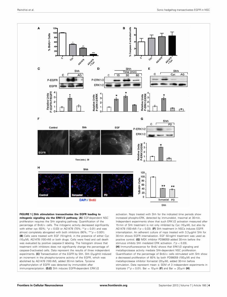
Shh MEDIATES NSC PROLIFERATION TROUGH EGFRMODULATIONDuring development, NSCs exhibit different requirements forgrowth factors. This is reflected in the growth of nsps, whichrequire basic fibroblast growth factor (bFGF) at earlier embry-onic stages and both Shh and EGF at late stages (Palma and Ruizi Altaba, 2004). Considering that EGFR-mediated proliferationof cortical NSCs specifically requires EGF and Shh during latebrain development, we first examined the effect of the specificinhibitors, Cyc (Hedgehog antagonist) and tyrphostin (EGFRinhibitor, AG1478) (Ward et al., 1994; Chen et al., 2002), on cellproliferation of E18.5 EGF-responsive nsps. Dissociated nsps wereplated on substrate-coated dishes and grown in the presence ofEGF (10 ng/ml) for 48 h. The cells were deprived of any growthfactor for 2 h to cause cell growth arrest and maximal exposal ofthe EGFR to the cell surface, and were then stimulated with EGFfor 2 h in the absence or presence of Cyc or AG1478, added 30 minbefore ligand addition. EGF-induced cell proliferation, mea-sured by BrdU incorporation, was reduced by 70% with AG1478(100 nM) and 50% with Cyc (10 μM) treatment, in agreementwith previous reports confirming basal Shh activity (Dahmaneet al., 2001; Palma and Ruiz i Altaba, 2004). Interestingly, simul-taneous treatment with AG1478 and Cyc almost completelyabrogated cell proliferation (about 96%) (Figure 1A), withoutsignificantly affecting apoptosis (Figure 1B). These results cor-roborate that the EGFR and Shh signaling pathways collaboratein the regulation of NSC proliferation during late embryogenesis.Interestingly, these observations also suggest that besides regulat-ing the level of EGFR expression, Shh substantially contributes tothe mitogenic response elicited by EGF signaling.
NSC PROLIFERATION INDUCED BY Shh REQUIRES EGFRTRANSACTIVATIONThe collaborative action of EGF and Shh can be exerted by theactivation of independent signaling pathways that might con-verge upon yet unknown downstream elements. Alternatively,the EGFR might constitute an upstream element shared byboth EGF and Shh. An obvious possibility is that Shh acutelytransactivates the EGFR and as a consequence activates theRas/Raf/MEK/ERK1/2 pathway involved in cell proliferation(Traverse et al., 1994; Seger and Krebs, 1995). We first studiedthe effects of Shh treatment over EGFR tyrosine-phosphorylation.Strikingly, the EGFR immunoprecipitated from nsps treated withShh (3 μg/ml) for 15 min showed an increased phosphotyro-sine content in immunoblots, which was abrogated by AG1478(Figure 1C), thus reflecting EGFR activation. This Shh-inducedEGFR activation led to ERK1/2 activation, detectable within15 min and reaching a maximum by 30 min (Figure 1D), which
Frontiers in Cellular Neuroscience www.frontiersin.org September 2013 | Volume 7 | Article 166 | 3
Reinchisi et al. Sonic hedgehog transactivates EGFR in NSC
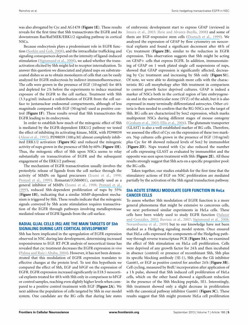
FIGURE 1 | Shh stimulation transactivates the EGFR leading to
mitogenic signaling via the ERK1/2 pathway. (A) EGF-dependent NSCproliferation requires the Shh signaling pathway. Quantification of thepercentage of BrdU+ cells. The mitogenic activity decreased significantlywith either cyc (50%; ∗p < 0.03) or AG1478 (70%; ∗∗p < 0.01) and wasalmost completely abrogated with both inhibitors (90%; ∗∗∗p < 0.001).(B) Cells were treated with EGF (10 ng/ml), in the presence of either Cyc(10 μM), AG1478 (100 nM) or both drugs. Cells were fixed and cell deathwas evaluated by positive caspase-3 labeling. The histogram shows thattreatment with inhibitors does not significantly change the percentage ofcaspase-3-activated cells. Data represent the results of three independentexperiments. (C) Transactivation of the EGFR by Shh. Shh (3 μg/ml) inducedan increment in the phospho-tyrosine activity of the EGFR, which wasabolished by AG1478 (100 nM), added 30 min before. Tyrosinephosphorylation of EGFR was detected by immunoblot afterimmunoprecipitation. (D,E) Shh induces EGFR-dependent ERK1/2
activation. Nsps treated with Shh for the indicated time periods showincreased phospho-ERK, detected by immunoblot, maximal at 30 min.Independent experiments show that such ERK1/2 activation measured after15 min of Shh treatment is not only inhibited by Cyc (10 μM), but also byAG1478 (100 nM) (∗p < 0.03). (F) Shh treatment in NSCs induces EGFRinternalization. An adherent culture of nsps treated with 3.3 μg/ml Shh for30 min shows EGFR internalization. EGF 50 ng/ml treatment was used aspositive control. (G) MEK inhibitor PD98059 added 30 min before thestimulus inhibits Shh mediated ERK activation. (∗p < 0.03).(H) Immunofluorescence for BrdU shows that ERK1/2 signaling andmetalloprotease activity mediate Shh-dependent NSC proliferation.Quantification of the percentage of BrdU+ cells stimulated with Shh showa decreased proliferation of 60% by both PD98059 (100 μM) and themetalloprotease inhibitor Ilomastat (20 μM), added 30 min beforestimulation. Data represent mean ± SEM of 3 independent experiments intriplicate (∗∗p < 0.01). Bar = 10 μm (F) and Bar = 20 μm (H).
Frontiers in Cellular Neuroscience www.frontiersin.org September 2013 | Volume 7 | Article 166 | 4
Reinchisi et al. Sonic hedgehog transactivates EGFR in NSC
was also abrogated by Cyc and AG1478 (Figure 1E). These resultsreveals for the first time that Shh transactivates the EGFR and itsdownstream Ras/Raf/MEK/ERK1/2 signaling pathway in corticalNSCs.
Because endocytosis plays a predominant role in EGFR func-tion (Sorkin and Goh, 2008), and the intracellular trafficking andsignaling consequences depend on the intensity of ligand-inducedstimulation (Sigismund et al., 2008), we asked whether the trans-activation elicited by Shh might led to receptor internalization. Toanswer this question we dissociated and plated nsps on substrate-coated dishes so as to obtain monolayers of cells that can be easilyanalyzed for EGFR endocytosis by indirect immunofluorescence.The cells were grown in the presence of EGF (10 ng/ml) for 48 hand depleted for 2 h before the experiments to induce maximalexposure of the EGFR to the cell surface. Treatment with Shh(3.3 μg/ml) induced a redistribution of EGFR from the cell sur-face to juxtanuclear endosomal compartments, although of lessmagnitude compared with EGF (50 ng/ml) used as positive con-trol (Figure 1F). These results reveal that Shh transactivates theEGFR leading to its endocytosis.
In order to establish how much of the mitogenic effect of Shhis mediated by the EGFR-dependent ERK1/2 pathway we testedthe effect of inhibiting its activating-kinase, MEK, with PD98059(Alessi et al., 1995). PD98059 (100 μM) almost completely inhib-ited ERK1/2 activation (Figure 1G) and reduced the mitogenicactivity of nsps grown in the presence of Shh by 60% (Figure 1H).Thus, the mitogenic effect of Shh upon NSCs seems to relaysubstantially on transactivation of EGFR and the subsequentengagement of the ERK1/2 pathway.
The mechanism of EGFR transactivation usually involves theproteolytic release of ligands from the cell surface through theactivity of MMPs on ligand precursors (Izumi et al., 1998;Prenzel et al., 1999). Ilomastat/(GM6001), currently used as ageneral inhibitor of MMPs (Izumi et al., 1998; Prenzel et al.,1999), reduced Shh-dependent proliferation of nsps by 55%(Figure 1H), indicating that a similar MMP-dependent mecha-nism is triggered by Shh. These results indicate that the mitogenicsignals conveyed by Shh acute stimulation requires transactiva-tion of the EGFR through pathways involving metalloproteasemediated release of EGFR ligands from the cell surface.
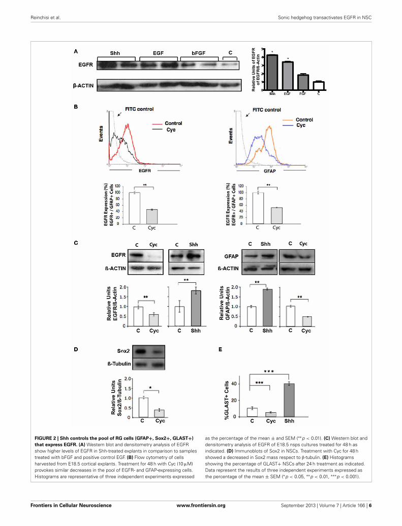
RADIAL GLIAL CELLS (RG) ARE THE MAIN TARGETS OF ShhSIGNALING DURING LATE CORTICAL DEVELOPMENTShh has been implicated in the upregulation of EGFR expressionobserved in NSC during late development, determining increasedresponsiveness to EGF. RT-PCR analysis of neocortical tissue hasrevealed that cyc treatment decreases the EGFR expression in vivo(Palma and Ruiz i Altaba, 2004). However, it has not been demon-strated that this modulation of EGFR expression translates toeffective changes at the protein level. To test this hypothesis wecompared the effect of Shh, EGF and bFGF on the expression ofEGFR. EGFR expression increased significantly in E18.5 neocorti-cal explants treated for 48 h with Shh only in comparison to bFGFor control samples, reaching even slightly higher levels when com-pared to a positive control treatment with EGF (Figure 2A). Wenext address the population of cells targeted by Shh in our modelsystem. One candidate are the RG cells that during late states
of embryonic development start to express GFAP (reviewed inImura et al., 2003; Ihrie and Alvarez-Buylla, 2008) and some ofthem are EGF-responsive stem cells (Doetsch et al., 1999). Weanalyzed the expression of GFAP by flow cytometry on neocor-tical explants and found a significant decrement after 48 h ofCyc treatment (Figure 2B), similar to the reduction in EGFRexpression. This observation suggests that Shh might be actingon GFAP+ cells that express EGFR. In addition, immunostain-ing of GFAP on 1 week plated single cell suspensions of nsps,showed that GFAP expression is significantly affected, decreas-ing by Cyc treatment and increasing by Shh only (Figure S1).Of note, we were able to distinguish more cells with the charac-teristic RG cell morphology after Shh treatment in comparisonto control growth factor deprived cultures. GFAP is indeed amarker of NSCs both in the cortical region of late embryogene-sis and in the subventricular zone (SVZ) of the adult, but it is alsoexpressed in many terminally differentiated astrocytes. Other cri-teria is then needed to confirm that the RG-NSCs are the target ofShh. RG cells are characterized by Sox2 expression, which marksmultipotent NSCs during different stages of mouse ontogeny(Graham et al., 2003; Ellis et al., 2004). The glutamate transporter(GLAST) is also a well-established marker of RG cells. Therefore,we assessed the effect of Cyc on the expression of these two mark-ers. Nsp cultures cells grown in the presence of EGF (10 ng/ml)plus Cyc for 48 showed reduced levels of Sox2 by immunoblot(Figure 2D). Nsps treated with Cyc also reduced the numberof cells expressing GLAST as evaluated by immunoblot and theopposite was seen upon treatment with Shh (Figure 2E). All theseresults strongly suggest that Shh acts on a specific progenitor pool:the RG cells.
Taken together, our studies establish for the first time that thestimulatory actions of EGF on NSC proliferation are mediatedpartially by the activation of the Shh signal transduction pathway.
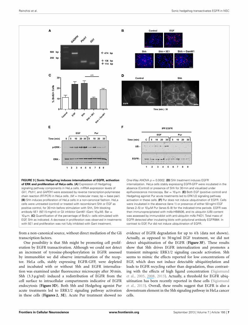
Shh ACUTE STIMULI MODULATE EGFR FUNCTION IN HeLaCANCER CELLSTo assess whether Shh modulation of EGFR function is a moregeneral phenomena that might be extensive to cancerous cells,we next performed similar experiments in HeLa cells. Thesecells have been widely used to study EGFR function (Salazarand González, 2002; Buvinic et al., 2007; Sigismund et al., 2008;Norambuena et al., 2009) but to our knowledge have not beenstudied as a Hedgehog signaling model system. Once ensuredthat HeLa cells expressed the components of the Hedgehog path-way through reverse transcriptase PCR (Figure 3A), we examinedthe effect of Shh stimulation on HeLa cell proliferation. Cellswere deprived of any growth factor for 24 h and then incubatedin absence (control) or presence of recombinant Shh, Shh plusits specific blocking antibody (5E-1), Shh plus the Gli inhibitorGant61, or EGF as positive control for another 24 h (Figure 3B).Cell cycling, measured by BrdU incorporation after application ofa 1 h pulse, showed that Shh induced cell proliferation of HeLacells, which on the other hand showed a significant reductionin the presence of the Shh blocking peptide, 5E1. Interestingly,Shh treatment showed only a slight decrease in proliferationwith the canonical pathway inhibitor Gant61 (Figure 3C). Theseresults suggest that Shh might promote HeLa cell proliferation
Frontiers in Cellular Neuroscience www.frontiersin.org September 2013 | Volume 7 | Article 166 | 5
Reinchisi et al. Sonic hedgehog transactivates EGFR in NSC
FIGURE 2 | Shh controls the pool of RG cells (GFAP+, Sox2+, GLAST+)
that express EGFR. (A) Western blot and densitometry analysis of EGFRshow higher levels of EGFR in Shh-treated explants in comparison to samplestreated with bFGF and positive control EGF. (B) Flow cytometry of cellsharvested from E18.5 cortical explants. Treatment for 48 h with Cyc (10 μM)provokes similar decreases in the pool of EGFR- and GFAP-expressing cells.Histograms are representative of three independent experiments expressed
as the percentage of the mean ± and SEM (∗∗p < 0.01). (C) Western blot anddensitometry analysis of EGFR of E18.5 nsps cultures treated for 48 h asindicated. (D) Immunoblots of Sox2 in NSCs. Treatment with Cyc for 48 hshowed a decreased in Sox2 mass respect to β-tubulin. (E) Histogramsshowing the percentage of GLAST+ NSCs after 24 h treatment as indicated.Data represent the results of three independent experiments expressed asthe percentage of the mean ± SEM (∗p < 0.05, ∗∗p < 0.01, ∗∗∗p < 0.001).
Frontiers in Cellular Neuroscience www.frontiersin.org September 2013 | Volume 7 | Article 166 | 6
Reinchisi et al. Sonic hedgehog transactivates EGFR in NSC
FIGURE 3 | Sonic Hedgehog induces internalization of EGFR, activation
of ERK and proliferation of HeLa cells. (A) Expression of Hedgehogsignaling pathway components in HeLa cells. mRNA expression levels ofGli1, Ptch1, and GAPDH were assessed by reverse transcription-polymerasechain reaction (RT-PCR) in HeLa cells. (M = molecular mass; bp = base pair).(B) Shh induces proliferation of HeLa cells in a non-canonical fashion. HeLacells were untreated (control) or treated with recombinant Shh or EGF aspositive control, for 30 min before stimulation with Shh, Shh blockingantibody 5E1 (5E1.5 mg/ml) or Gli inhibitor Gant61 (Gant 10 μM). Bar =10 μm. (C) Quantification of the percentage of BrdU+ cells stimulated withEGF, Shh as indicated. A decrease in proliferation was observed in treatmentswith 5E1 and proliferation was not fully inhibited with Gant treatment.
One-Way ANOVA p = 0.0002. (D) Shh treatment induces EGFRinternalization. HeLa cells stably expressing EGFR-GFP were incubated in theabsence (Control) or presence of Shh for 30 min and visualized underepifluorescence microscopy. Bar = 10 μm. (E) Both EGF (positive control) andHedgehog agonist Pur acute treatments led to ERK1/2 signaling pathwayactivation in these cells. (F) Pur does not induce ubiquitination of EGFR. Cellswere incubated in the absence (lane 1) or presence of either 50 ng/ml EGF(lanes 2–5) or 10 μM Pur (lanes 6–9) for the indicated time periods. EGFR wasthen immunoprecipitated with mAb-HB8506, and its ubiquitin (UB) contentwas assessed by immunoblot with anti-ubiquitin mAb P4D1. Total mass ofEGFR detected after incubating blots with polyclonal antibody EGFR984. Incontrast to EGF, Pur did not induce ubiquitination of EGFR.
from a non-canonical source, without direct mediation of the Glitranscription factors.
One possibility is that Shh might be promoting cell prolif-eration by EGFR transactivation. Although we could not detectan increment of tyrosine-phosphorylation in EGFR assessedby immunoblot we did observe internalization of the recep-tor. HeLa cells, stably expressing EGFR-GFP, were depletedand incubated with or without Shh and EGFR internaliza-tion was examined under fluorescence microscopy after 30 min.Shh (3.3 μg/ml) induced a redistribution of EGFR from thecell surface to intracellular compartments indicative of EGFRendocytosis (Figure 3D). Both Shh and Hedgehog agonist Puracute treatments led to ERK1/2 signaling pathway activationin these cells (Figures 2, 3E). Acute Pur treatment showed no
evidence of EGFR degradation for up to 4 h (data not shown).Actually, as opposed to 50 ng/ml EGF treatment, we did notdetect ubiquitination of the EGFR (Figure 3F). These resultsshow that Shh drives EGFR internalization and promotes atransient mitogenic ERK1/2 signaling cascade activation. Shhseems to mimic the effects reported for low concentrations ofEGF, which does not induce detectable ubiquitinylation andleads to EGFR recycling rather than degradation, thus contrast-ing with the effects of high ligand concentration (Sigismundet al., 2005, 2008, 2013). Actually, a threshold for EGFR ubiq-uitination has been recently reported in these cells (Sigismundet al., 2013). Overall, these results suggest that EGFR is also adownstream element in the Shh signaling pathway in HeLa cancercells.
Frontiers in Cellular Neuroscience www.frontiersin.org September 2013 | Volume 7 | Article 166 | 7
Reinchisi et al. Sonic hedgehog transactivates EGFR in NSC
DISCUSSIONAcquisition and modulation of EGF responsiveness is critical inthe regulation of several aspects of NSC development, such asself-renewal, differentiation and migration. Here we provide evi-dence that one of the main functions of Shh during late stages ofbrain embryogenesis is to promote and modulate EGF respon-siveness of NSCs. We demonstrate that Shh has the capabilityto transactivate the EGFR and as a consequence strengthens themitogenic intensity of the ERK1/2 signaling pathway triggered byEGF. This effect can explain the collaborative function of Shh andEGF exerted on the proliferation of late NSCs.
We identify RG (GFAP+, Sox2, and GLAST+) cells as themain target population of Shh, in which Shh induced transacti-vation, together with the reported EGFR expression (Dahmaneet al., 2001) can all contribute to increase the EGF responsive-ness. Our results are in line with a recent report by Dave et al.,which showed in vivo that Shh pathway activation in the condi-tional Ptc1Lox/Lox; NestinCre mutant cortex is mitogenic for cellsthat have a capacity to self-renew over an extended period of time.Authors identified RG cells- Nestin+/GLAST+ as the direct tar-gets in the developing neocortex by mid neurogenesis (E14.5).Importantly, they showed reduction of Trb2 basal progenitor pos-itive cells, suggesting that since RG cells can differentiate intobasal progenitors, an indirect effect on this cell population couldnot be ruled out (Dave et al., 2011). In contrast Komada et al.reported that in Shh-CKO and Smo-conditional mutant embryos,the number of Tbr2-positive basal progenitor cells is significantlydecreased in the SVZ/VZ of the developing neocortex (Komadaet al., 2008). Furthermore, no significant differences could beobserved in the Pax6 positive-RG cells among E15.5 wild-typeand mutant mice. Clearly, this matter will require future inves-tigation and we do not rule out that Shh signaling may be amechanism for the regulation of both the number of RG cells andbasal progenitors.
Our previous experiments in NSCs revealed synergism of theShh and EGF signaling pathways reflected by increments of pro-liferative responses and induction of gene expression (Palma andRuiz i Altaba, 2004). This up-regulation of EGFR expression lev-els induced by Shh indeed contributes to the collaborative actionof Shh and EGFR on NSCs. EGFR expression has been associatedwith changes in progenitor cell behavior as a limiting factor forboth proliferation and differentiation with response choice regu-lated in part by a concentration-dependent mechanism. Here weshow that the mitogenic mechanism employed by Shh stimulussubstantially depends on EGFR activity. Shh has been reportedto provoke acute transactivation of EGFR, leading to cell prolif-eration in mouse embryonic stem cells (Heo et al., 2007). Ourpresent results are the first to show that Shh transactivates theEGFR, leading to mitogenic ERK1/2 signaling in NSCs at lateembryonic stages. Treatment of E18.5 cortical nsps with Shhfor 15 min increases the tyrosine phosphorylation of the EGFR,a hallmark of its activation. Such Shh-induced EGFR phos-phorylation was abrogated with an inhibitor of EGFR tyrosinekinase activity, and had functional consequences, as it resultedin activation of mitogenic ERK1/2 signaling. Recent observationssuggest that the ability of cells to divide in response to EGFR acti-vation requires the expression of a high level of the receptor inNSC (Lillien and Gulacsi, 2006). Because Shh can provoke an
acute as well as a sustained status of EGFR activation (Bigelowet al., 2005; Heo et al., 2007), an initial event of EGFR transac-tivation can play a role in increasing the levels of the EGFR aswell as in establishing regulatory loops acting on the proliferationmachinery. Thus, Shh and EGF signaling pathways can function-ally interact at different levels and may even establish a long-termregulatory loop depending on the cell context. How the syner-gistic signaling system of Shh and EGFR are integrated in NCSsand how does cooperativity between the pathways lead to selec-tive activation of common response genes remains to be tested infuture studies.
Endocytic trafficking is a crucial element in the regulationof EGFR function control (Goh and Sorkin, 2013; Pennock andWang, 2003). The intensity, location and duration of recep-tor signaling can be modulated by the endocytic traffickingmachinery, which is tightly intertwined with the mechanismsthat control receptor signaling (Scita and Di Fiore, 2010; Gohand Sorkin, 2013). Activated EGFR undergoes transphospho-rylation and ubiquitylation and these modifications define itsendocytic trafficking/signaling behavior (Goh and Sorkin, 2013;Sigismund et al., 2013). Recent studies have shown that low ligandconcentrations drive EGFR toward clathrin-dependent endocy-tosis followed by recycling, whereas high ligand concentrationspromote EGFR internalization through a clathrin-independentroute directed to lysosomal degradation (Sigismund et al., 2008).These alternatives have functional consequences in specifyingthe responses (Sigismund et al., 2008) and involve a threshold-controlled ubiquitylation system (Sigismund et al., 2013), whichfinely tunes the balance between EGFR signaling and traffick-ing. The ubiquitin-dependent down regulation route involvesESCRT-mediated sorting into intraluminal vesicles of multi-vesicular bodies that then fuse with lysosomes (Wegner et al.,2011). Thus, activated EGFR can remain signaling-competentfor variable periods of time before degradation, determining dif-ferent response outcomes (Sigismund et al., 2008; Rush et al.,2012). Our results, both in NSC and in HeLa cells show thatHedgehog pathway activation leads to EGFR internalization andto transient activation of the MAPK/ERK signaling cascade(Figure 1D, Figure S2), without detectable ubiquitination. Thismimics the effect induced on EGFR by low EGF concentrations,which mainly leads to receptor recycling instead of degradation(Sigismund et al., 2008). In contrast, high EGF doses stimu-lation (50 ng/ml) induces EGFR ubiquitination and promotesits sorting to the lysosomal degradation pathway (Figure 3F).Besides mimicking EGFR stimulation by low EGF concentra-tion, which by itself can constitute a strong mitogenic stimulus(Sigismund et al., 2008), another possibility of Shh-mediatedregulation of EGFR function might occur through the reduc-tion of PKA activity produced by activation of Smo via Gαi(Huangfu and Anderson, 2006). A decreased PKA activity hasbeen shown to induce endocytosis and intracellular accumulationof empty/inactive EGFR, as well as an increment in the half-lifeof ligand-activated EGFR by delaying its sorting to the lysoso-mal degradation route (Salazar and González, 2002; Norambuenaet al., 2010).
Our findings provide evidence that Shh, constitutes a mito-genic stimulating system, activating the EGFR and its MAPK/ERKsignaling cascade. This potentiates the mitogenic effects of low
Frontiers in Cellular Neuroscience www.frontiersin.org September 2013 | Volume 7 | Article 166 | 8
Reinchisi et al. Sonic hedgehog transactivates EGFR in NSC
EGF concentrations. Taking into account the recently describedShh constitutive autocrine modulation in NSCs (Bigelow et al.,2005; Ruiz i Altaba et al., 2007; Martínez et al., 2013), ourresults suggest that downstram activation of EGFR might pro-vide the necessary strength to the intracellular pathways to mounta mitogenic response in NSCs. This might eventually increasethe expression of EGFR, as previously reported for other EGFRtransactivating receptors (Buvinic et al., 2007).
We suggest that during late development, neural precursor cellproliferation would be regulated by an interplay of mechanismsinvolving different signaling pathways, which would control thesize of the progenitor pool. Additional approaches addressing therelationship of Shh, EGFR and others signaling pathways will berequired order to obtain a better understanding of how, when andwhere these pathways interact to control NSC behavior.
Although at the moment specific mechanisms of EGFR trans-activation by Shh are not defined, we show the intermediaryaction of MMPs in Shh-induced proliferation in NSCs. Ourresults show that Shh elicits a mitogenic response through acti-vation of GM6001-sensitive MMPs. Inhibition of MMP activityattenuates the Shh-mediated cell proliferation indicating thatthe release of soluble ligands by induction of GM6001-sensitiveMMPs after Shh stimulation could be responsible for cell prolif-eration. Our findings complement those described recently in EScells by Heo et al. (2007). However, in NSCs, the identity of MMPsand their mechanism still remain to be defined.
Transactivation of the EGFR by agonist-activated GPCRs typ-ically requires growth factor cleavage mediated by MMPs. It isnoteworthy that the transactivation of the EGFR, mediated bymembers of the ADAM (a disintegrin and metalloproteinase)family of zinc-dependent proteases, is relevant in the devel-opment and progression of diverse types of human tumors.Furthermore, there is evidence suggesting that brain tumorsresemble stem cell niches (Oliver and Wechsler-Reya, 2004). Braintumors contain cancer stem cells, which are essential for bothdevelopment and recurrence of the tumors (Dahmane et al.,2001; Vescovi et al., 2006). Together, these studies raise the pos-sibility that the factors that regulate normal cell lineages fromneural precursors may serve similar functions in the devel-opment of brain cancers from stem-like cancer cells (Ruiz iAltaba et al., 2007). Integration of the Shh and EGFR path-ways could be a critical step in cancer initiation and/or tumorgrowth. Our data obtained in HeLa cells indeed suggest a con-served essential role of Shh on the regulation of EGFR function,which have therapeutic interest. The recognized importance ofthe EGFR in tumorigenesis suggests the possibility that abnor-mally increased Shh signaling contributes to carcinogenesis, thus
providing additional rational to Shh as a target for antitumortherapies.
In conclusion, the present work gives further evidence of theimportance of Shh in the regulation of the homeostasis of theEGF signaling pathways and proliferation, emphasizing cross-talkprocesses. Our findings shed new insight into the complex signaltransduction pathways that mediate the actions of growth factorsin the developing brain.
AUTHOR CONTRIBUTIONVerónica Palma and Alfonso Gonzalez designed research;Gisela Reinchisi, Margarita Parada, Claudia Oyanadel, RonanShaughnessy, Verónica Palma, and Pablo Lois performed research;Gisela Reinchisi, Verónica Palma, and Pablo Lois analyzeddata, Gisela Reinchisi, Margarita Parada, Alfonso Gonzalez, andVerónica Palma wrote the paper.
ACKNOWLEDGMENTSWe thank Dr. Pilar Sanchez for her advice and encourage-ment, Dr. Andrés Norambuena, Claudio Retamal and AriadnaMendoza for technical assistance and Dr. A. Ruiz i Altaba andDr. Luis Milla for comments on the manuscript. We are grate-ful to Infinity Pharmaceuticals, Inc. for providing Cyclopamine.Verónica Palma’s work is supported by FONDAP grant #15090007and FONDECYT grant #1110237. Alfonso Gonzalez’s work issupported by CONICYT grant PFB12/2007 and FONDECYTgrant #1100747.
SUPPLEMENTARY MATERIALThe Supplementary Material for this article can be found onlineat: http://www.frontiersin.org/Cellular_Neuroscience/10.3389/fncel.2013.00166/abstract
Figure S1 | Shh maintains a specific GFAP+ -RG cell pool. Nsps were
cultured for 48 h with EGF (10 ng/ml) and then EGF was removed and the
cells were cultured in the presence of only Shh (3 μg/ml) or Cyc (10 μM)
for 7 days. RG phenotype on treated nsps was assessed by
immunofluorescence staining for GFAP, counterstaining the cell nuclei
with DAPI Bar = 50 μm. The percentage of cells expressing GFAP is
depicted in the corresponding histogram. Shh treatment increases 2-fold
the number of GFAP+ cells, whereas Cyc treatment shows the opposite
effect, decreasing by 50% the number of GFAP+ cells. Values are the
mean ± SEM of three experiments per conditions. (∗∗p < 0.01 vs. control).
Figure S2 | Shh treatment in HeLa cells induces activation of the
mitogenic ERK1/2 transduction pathway. HeLa cells treated with
3.3 μg/ml Shh for the indicated time periods showed increased
phospho-ERK, detected by immunoblot, maximal at 30 min.
REFERENCESAlessi, D. R., Cuenda, A., Cohen,
P., Dudley, D. T., and Saltiel, A.R. (1995). PD 098059 is a spe-cific inhibitor of the activation ofmitogen-activated protein kinasekinase in vitro and in vivo. J. Biol.Chem. 270, 27489–27494. doi:10.1074/jbc.270.46.27489
Alwan, H. A. J., van Zoelen,E. J. J., and van Leeuwen,
J. E. M. (2003). Ligand-induced lysosomal epidermalgrowth factor receptor (EGFR)degradation is preceded byproteasome-dependent EGFRde-ubiquitination. J. Biol. Chem.278, 35781–35790. doi: 10.1074/jbc.M301326200
Bigelow, R. L. H., Jen, E. Y., Delehedde,M., Chari, N. S., and McDonnell, T.J. (2005). Sonic hedgehog induces
epidermal growth factor depen-dent matrix infiltration in HaCaTkeratinocytes. J. Invest. Dermatol.124, 457–465. doi: 10.1111/j.0022-202X.2004.23590.x
Buvinic, S., Bravo-Zehnder, M., Boyer,J. L., Huidobro-Toro, J. P., andGonzález, A. (2007). NucleotideP2Y1 receptor regulates EGFreceptor mitogenic signaling andexpression in epithelial cells.
J. Cell Sci. 120, 4289–4301. doi:10.1242/jcs.03490
Caric, D., Raphael, H., Viti, J., Feathers,A., Wancio, D., and Lillien, L.(2001). EGFRs mediate chemo-tactic migration in the developingtelencephalon. Development 128,4203–4216.
Carpenter, G. (1999). Employment ofthe epidermal growth factor recep-tor in growth factor-independent
Frontiers in Cellular Neuroscience www.frontiersin.org September 2013 | Volume 7 | Article 166 | 9
Reinchisi et al. Sonic hedgehog transactivates EGFR in NSC
signaling pathways. J. Cell Biol.146, 697–702. doi: 10.1083/jcb.146.4.697
Caviness, V. S. Jr., Takahashi, T.,and Nowakowski, R. S. (1995).Numbers, time and neocorticalneuronogenesis: a general devel-opmental and evolutionary model.Trends Neurosci. 18, 379–383. doi:10.1016/0166-2236(95)93933-O
Chen, J. K., Taipale, J., Cooper, M.K., and Beachy, P. A. (2002).Inhibition of hedgehog sig-naling by direct binding ofcyclopamine to smoothened.Genes Dev. 16, 2743–2748. doi:10.1101/gad.1025302
Dahmane, N., Sánchez, P., Gitton,Y., Palma, V., Sun, T., Beyna, M.,et al. (2001). The Sonic Hedgehog-Gli pathway regulates dorsalbrain growth and tumorigenesis.Development 128, 5201–5212.
Daub, H., Weiss, F. U., Wallasch, C.,and Ullrich, A. (1996). Role oftransactivation of the EGF receptorin signalling by G-protein-coupledreceptors. Nature 379, 557–560. doi:10.1038/379557a0
Dave, R. K., Ellis, T., Toumpas, M. C.,Robson, J. P., Julian, E., Adolphe,C., et al. (2011). Sonic hedgehogand notch signaling can cooper-ate to regulate neurogenic divisionsof neocortical progenitors. PLoSONE 6:e14680. doi: 10.1371/jour-nal.pone.0014680
Doetsch, F., Caillé, I., Lim, D. A.,García-Verdugo, J. M., and Alvarez-Buylla, A. (1999). Subventricularzone astrocytes are neural stem cellsin the adult mammalian brain. Cell97, 703–716. doi: 10.1016/S0092-8674(00)80783-7
Ellis, P., Fagan, B. M., Magness, S. T.,Hutton, S., Taranova, O., Hayashi,S., et al. (2004). SOX2, a persis-tent marker for multipotential neu-ral stem cells derived from embry-onic stem cells, the embryo or theadult. Dev. Neurosci. 26, 148–165.doi: 10.1159/000082134
Goh, L. K., and Sorkin, A. (2013).Endocytosis of receptor tyrosinekinases. Cold Spring Harb. Perspect.Biol. 5:a017459. doi: 10.1101/csh-perspect.a017459
Graham, V., Khudyakov, J., Ellis, P., andPevny, L. (2003). SOX2 functionsto maintain neural progenitoridentity. Neuron 39, 749–765.doi: 10.1016/S0896-6273(03)00497-5
Gschwind, A., Zwick, E., Prenzel, N.,Leserer, M., and Ullrich, A. (2001).Cell communication networks:epidermal growth factor receptortransactivation as the paradigm forinterreceptor signal transmission.
Oncogene 20, 1594–1600. doi:10.1038/sj.onc.1204192
Heo, J. S., Lee, M. Y., and Han, H.J. (2007). Sonic hedgehog stim-ulates mouse embryonic stemcell proliferation by cooperationof Ca2+/protein kinase C andepidermal growth factor recep-tor as well as Gli1 activation.Stem Cells 25, 3069–3080. doi:10.1634/stemcells.2007-0550
Huangfu, D., and Anderson, K. V.(2006). Signaling from Smo toCi/Gli: conservation and diver-gence of Hedgehog pathwaysfrom Drosophila to vertebrates.Development 133, 3–14. doi:10.1242/dev.02169
Ihrie, R. A., and Alvarez-Buylla, A.(2008). Cells in the astroglial lineageare neural stem cells. Cell Tissue Res.331, 179–191. doi: 10.1007/s00441-007-0461-z
Imura, T., Kornblum, H. I., andSofroniew, M. V. (2003). The pre-dominant neural stem cell isolatedfrom postnatal and adult forebrainbut not early embryonic forebrainexpresses GFAP. J. Neurosci. 23,2824–2832.
Izumi, Y., Hirata, M., Hasuwa,H., Iwamoto, R., Umata, T.,Miyado, K., et al. (1998). Ametalloprotease-disintegrin,MDC9/meltrin-gamma/ADAM9and PKCdelta are involved inTPA-induced ectodomain sheddingof membrane-anchored heparin-binding EGF-like growth factor.EMBO J. 17, 7260–7272. doi:10.1093/emboj/17.24.7260
Komada, M., Saitsu, H., Kinboshi,M., Miura, T., Shiota, K., andIshibashi, M. (2008). Hedgehogsignaling is involved in devel-opment of the neocortex.Development 135, 2717–2727.doi: 10.1242/dev.015891
Lappano, R., and Maggiolini, M.(2011). G protein-coupled recep-tors: novel targets for drug discoveryin cancer. Nat. Rev. Drug Discov. 10,47–60. doi: 10.1038/nrd3320
Lillien, L., and Gulacsi, A. (2006).Environmental signals elicitmultiple responses in dorsaltelencephalic progenitors bythreshold-dependent mechanisms.Cereb. Cortex 16(Suppl. 1), i74–i81.doi: 10.1093/cercor/bhj169
Madshus, I. H., and Stang, E. (2009).Internalization and intracellu-lar sorting of the EGF receptor:a model for understanding themechanisms of receptor trafficking.J. Cell Sci. 122, 3433–3439. doi:10.1242/jcs.050260
Mangelberger, D., Kern, D.,Loipetzberger, A., Eberl, M.,
and Aberger, F. (2012). CooperativeHedgehog-EGFR signaling. Front.Biosci. 17, 90–99. doi: 10.2741/3917
Martínez, C., Cornejo, V. H., Lois, P.,Ellis, T., Solis, N. P., Wainwright,B. J., et al. (2013). Proliferationof murine midbrain neural stemcells depends upon an endogenoussonic hedgehog (Shh) source. PLoSONE 8:e65818. doi: 10.1371/jour-nal.pone.0065818
McCarthy, N. (2012). Cancer stem cells:tracing clones. Nat. Rev. Cancer 12,579. doi: 10.1038/nrc3354
Mimeault, M., and Batra, S. K.(2010). Frequent deregulationsin the hedgehog signaling net-work and cross-talks with theepidermal growth factor receptorpathway involved in cancer pro-gression and targeted therapies.Pharmacol. Rev. 62, 497–524. doi:10.1124/pr.109.002329
Norambuena, A., Metz, C., Jung, J.E., Silva, A., Otero, C., Cancino,J., et al. (2010). Phosphatidic acidinduces ligand-independent epider-mal growth factor receptor endo-cytic traffic through PDE4 activa-tion. Mol. Biol. Cell 21, 2916–2929.doi: 10.1091/mbc.E10-02-0167
Norambuena, A., Metz, C., Vicuña, L.,Silva, A., Pardo, E., Oyanadel, C.,et al. (2009). Galectin-8 inducesapoptosis in Jurkat T cells by phos-phatidic acid-mediated ERK1/2activation supported by proteinkinase A down-regulation. J. Biol.Chem. 284, 12670–12679. doi:10.1074/jbc.M808949200
Ogden, S. K., Fei, D. L., Schilling, N. S.,Ahmed, Y. F., Hwa, J., and Robbins,D. J. (2008). G protein Galphaifunctions immediately downstreamof Smoothened in Hedgehog sig-nalling. Nature 456, 967–970. doi:10.1038/nature07459
Oliver, T. G., and Wechsler-Reya,R. J. (2004). Getting at theroot and stem of brain tumors.Neuron 42, 885–888. doi:10.1016/j.neuron.2004.06.011
Palma, V., Lim, D. A., Dahmane,N., Sánchez, P., Brionne, T. C.,Herzberg, C. D., et al. (2005).Sonic hedgehog controls stem cellbehavior in the postnatal and adultbrain. Development 132, 335–344.doi: 10.1242/dev.01567
Palma, V., and Ruiz i Altaba, A. (2004).Hedgehog-GLI signaling regulatesthe behavior of cells with stem cellproperties in the developing neocor-tex. Development 131, 337–345. doi:10.1242/dev.00930
Pennock, S., and Wang, Z. (2003).Stimulation of cell proliferationby endosomal epidermal growthfactor receptor as revealed through
two distinct phases of signaling.Mol. Cell. Biol. 23, 5803–5815.doi: 10.1128/MCB.23.16.5803-5815.2003
Piper, R. C., and Luzio, J. P. (2007).Ubiquitin-dependent sorting ofintegral membrane proteins fordegradation in lysosomes. Curr.Opin. Cell Biol. 19, 459–465. doi:10.1016/j.ceb.2007.07.002
Prenzel, N., Zwick, E., Daub, H.,Leserer, M., Abraham, R., Wallasch,C., et al. (1999). EGF receptortransactivation by G-protein-coupled receptors requiresmetalloproteinase cleavage ofproHB-EGF. Nature 402, 884–888.doi: 10.1038/47260
Reynolds, B. A., and Weiss, S.(1996). Clonal and populationanalyses demonstrate that anEGF-responsive mammalianembryonic CNS precursor is astem cell. Dev. Biol. 175, 1–13. doi:10.1006/dbio.1996.0090
Roepstorff, K., Grandal, M. V.,Henriksen, L., Knudsen, S. L. J.,Lerdrup, M., Grøvdal, L., et al.(2009). Differential effects of EGFRligands on endocytic sorting of thereceptor. Traffic 10, 1115–1127. doi:10.1111/j.1600-0854.2009.00943.x
Ruiz i Altaba, A., Mas, C., andStecca, B. (2007). The Gli code:an information nexus regulatingcell fate, stemness and cancer.Trends Cell Biol. 17, 438–447. doi:10.1016/j.tcb.2007.06.007
Rush, J. S., Quinalty, L. M., Engelman,L., Sherry, D. M., and Ceresa, B. P.(2012). Endosomal accumulationof the activated epidermal growthfactor receptor (EGFR) inducesapoptosis. J. Biol. Chem. 287,712–722. doi: 10.1074/jbc.M111.294470
Salazar, G., and González, A. (2002).Novel mechanism for regula-tion of epidermal growth factorreceptor endocytosis revealed byprotein kinase A inhibition. Mol.Biol. Cell 13, 1677–1693. doi:10.1091/mbc.01-08-0403
Schlessinger, J. (2000). Cell signalingby receptor tyrosine kinases. Cell103, 211–225. doi: 10.1016/S0092-8674(00)00114-8
Schnidar, H., Eberl, M., Klingler, S.,Mangelberger, D., Kasper, M.,Hauser-Kronberger, C., et al.(2009). Epidermal growth fac-tor receptor signaling synergizeswith Hedgehog/GLI in oncogenictransformation via activationof the MEK/ERK/JUN pathway.Cancer Res. 69, 1284–1292. doi:10.1158/0008-5472.CAN-08-2331
Scita, G., and Di Fiore, P. P. (2010).The endocytic matrix. Nature
Frontiers in Cellular Neuroscience www.frontiersin.org September 2013 | Volume 7 | Article 166 | 10
Reinchisi et al. Sonic hedgehog transactivates EGFR in NSC
463, 464–473. doi: 10.1038/nature08910
Seger, R., and Krebs, E. G. (1995). TheMAPK signaling cascade. FASEB J.9, 726–735.
Sigismund, S., Algisi, V., Nappo, G.,Conte, A., Pascolutti, R., Cuomo,A., et al. (2013). Threshold-controlled ubiquitination ofthe EGFR directs receptor fate.EMBO J. 32, 2140–2157. doi:10.1038/emboj.2013.149
Sigismund, S., Argenzio, E., Tosoni,D., Cavallaro, E., Polo, S., andDi Fiore, P. P. (2008). Clathrin-mediated internalization is essentialfor sustained EGFR signalingbut dispensable for degrada-tion. Dev. Cell 15, 209–219. doi:10.1016/j.devcel.2008.06.012
Sigismund, S., Woelk, T., Puri,C., Maspero, E., Tacchetti, C.,Transidico, P., et al. (2005).Clathrin-independent endocytosisof ubiquitinated cargos. Proc. Natl.
Acad. Sci. U.S.A. 102, 2760–2765.doi: 10.1073/pnas.0409817102
Sorkin, A., and Goh, L. K. (2008).Endocytosis and intracellu-lar trafficking of ErbBs. Exp.Cell Res. 314, 3093–3106. doi:10.1016/j.yexcr.2008.08.013
Traverse, S., Seedorf, K., Paterson, H.,Marshall, C. J., Cohen, P., andUllrich, A. (1994). EGF triggersneuronal differentiation of PC12cells that overexpress the EGF recep-tor. Curr. Biol. 4, 694–701. doi:10.1016/S0960-9822(00)00154-8
Vescovi, A. L., Galli, R., and Reynolds,B. A. (2006). Brain tumour stemcells. Nat. Rev. Cancer 6, 425–436.doi: 10.1038/nrc1889
Ward, W. H., Cook, P. N., Slater,A. M., Davies, D. H., Holdgate,G. A., and Green, L. R. (1994).Epidermal growth factor receptortyrosine kinase. Investigation of cat-alytic mechanism, structure-basedsearching and discovery of a potent
inhibitor.Biochem. Pharmacol. 48,659–666. doi: 10.1016/0006-2952(94)90042-6
Wegner, C. S., Rodahl, L. M. W.,and Stenmark, H. (2011). ESCRTproteins and cell signalling. Traffic12, 1291–1297. doi: 10.1111/j.1600-0854.2011.01210.x
Yarden, Y., and Sliwkowski, M. X.(2001). Untangling the ErbBsignalling network. Nat. Rev.Mol. Cell Biol. 2, 127–137. doi:10.1038/35052073
Conflict of Interest Statement: Theauthors declare that the researchwas conducted in the absence of anycommercial or financial relationshipsthat could be construed as a potentialconflict of interest.
Received: 24 May 2013; accepted: 06September 2013; published online: 26September 2013.
Citation: Reinchisi G, Parada M, Lois P,Oyanadel C, Shaughnessy R, GonzalezA and Palma V (2013) Sonic Hedgehogmodulates EGFR dependent proliferationof neural stem cells during late mouseembryogenesis through EGFR transacti-vation. Front. Cell. Neurosci. 7:166. doi:10.3389/fncel.2013.00166This article was submitted to the journalFrontiers in Cellular Neuroscience.Copyright © 2013 Reinchisi, Parada,Lois, Oyanadel, Shaughnessy, Gonzalezand Palma. This is an open-accessarticle distributed under the terms ofthe Creative Commons AttributionLicense (CC BY). The use, distribu-tion or reproduction in other forumsis permitted, provided the originalauthor(s) or licensor are credited andthat the original publication in thisjournal is cited, in accordance withaccepted academic practice. No use,distribution or reproduction is permit-ted which does not comply with theseterms.
Frontiers in Cellular Neuroscience www.frontiersin.org September 2013 | Volume 7 | Article 166 | 11