solid and soft nanostructured materials: fundamentals and applications
TRANSCRIPT
Solid and soft nanostructured materials: Fundamentals and applications
M. Willandera,*, O. Nura, Yu E. Lozovikb, S.M. Al-Hillia, Z. Chiragwandia, Q.-H. Hua,
Q.X. Zhaoa, P. Klasona
aPhysical Electronics and Photonic, Physics Department, Goteborg University, SE-412 96 Goteborg, SwedenbInstitute of Spectroscopy, Russian Academy of Sciences, 142190 Troitsk, Moscow District, Russia
Available online 3 June 2005
Abstract
The scientific work worldwide on nanostructured materials is extensive as well as the work on the applications of nanostructured materials.
We will review quasi two-, one- and zero-dimensional solid and soft materials and their applications. We will restrict ourselves to a few
examples from partly fundamental aspects and partly from application aspects. We will start with trapping of excitons in semiconductor
nanostructures. The subjects are: physical realizations, phase diagrams, traps, local density approximations, and mesoscopic condensates.
From these fundamental questions in solid nanomaterials we will move to trapping of molecules in water using nanostructured electrodes.
We will also discuss how to manipulate water (create vortices) by nanostructure materials.
The second part deals with nanorods (nano-wires). Particularly we will exemplify with ZnO nanorods. The reason for this is that ZnO has:
a very strong excitons binding energy (60 meV) and strong photon–excitons coupling energy, a strong tendency to create nanostructures, and
properties which make the material of interest for both optoelectronics and for medical applications. We start with the growth of crystalline
ZnO nanorods on different substrates, both crystalline (silicon, silicon carbide, sapphire, etc) and amorphous substrates (silicon dioxide,
plastic materials, etc) for temperatures from 50 8C up to 900 8C. The optical properties and crystalline properties of the nanorods will be
analyzed. Applications from optoelectronics (lasers, LEDs, lamps, and detectors) are analyzed and also medical applications like
photodynamic cancer therapy are taken up.
The third part deals with nano-particles in ZnO for sun screening. Skin cancer due to the exposure from the sun can be prevented by ZnO
particles in a paste put on the exposed skin.
q 2005 Elsevier Ltd. All rights reserved.
Keywords:: Nano-structures; Semiconductors; Soft materials; Trapping; Excitons
1. Introduction
At present the growth, processing and characterization of
nanorods and nano-particles is of global interest. There is an
intensive research for controlling and manipulating nano-
structured materials. This is of interest for both fundamental
understanding as well as for technical and medical
applications. This intense interest is true for both solid as
well as soft materials, or in some cases a combination of
both. The area of nanostructures and its applications is
rapidly expanding.
The quasi-two-dimensional system of spatially indirect
excitons in coupled quantum wells is perspective for the
0026-2692/$ - see front matter q 2005 Elsevier Ltd. All rights reserved.
doi:10.1016/j.mejo.2005.04.020
* Corresponding author.
E-mail address: [email protected] (M. Willander).
observation of coherent state and superfluidity [1–13]. The
last can manifest itself in the existence of persistent current
in each quantum well, quasi-Josephson phenomena in this
nonsuperconducting system, in statistics of fluorescence and
in variety of nonlinear optical phenomena (see [1–9] and
references therein). For extended 2D exciton system only
quasi-condensate with fluctuating phase and power-law
correlations is possible and the transition to superfluid state
is Kosterlitz–Thouless transition. However one can create
the trap for excitons analogeous to atom trap [14–16]. In this
case Bose-condensation can be observed in finite exciton
system even for two-dimensional case. In this connection
two main problems arose: the first is how to trap excitons.
There are several possibilities: the first is the trap (natural
quantum dot) originated from random fluctuations of the
potential connected with the roughness of interfaces in
quantum wells, impurities, defects, etc. (see interesting
experimental work [11] and analysis in [3] and references
herein). Other possibility is to create the mesa by nano-
Microelectronics Journal 36 (2005) 940–949
www.elsevier.com/locate/mejo

M. Willander et al. / Microelectronics Journal 36 (2005) 940–949 941
technology. Finally, there is the possibility to create
confining potential by nonhomogeneous pressure near the
surface created by a needle. Using last method interesting
experimental results were obtained recently by Snoke group
[12].
Moreover, the research on soft materials has been
growing and in most cases it is interdisciplanry. Among
soft materials, water is being an important member. The
presence and wide usage of water in many chemcial
reactions and biological expriements, lead to the critical
need for a nano-scale water based platform for the
utilization of a wide range of sophisticated experiments,
e.g. trapping single molecule, studying chemical reactions
on few molecules level. Such experiments are not only of
fundamental interest, but have a strong technological
impact. We have developed a nano-scale water based
transistor [17]. This nano-device is in fact a unique flexible
platform with up to sixteen nodes connected to different
micro- and nano-active parts of a specific configuration.
Zinc oxide is a direct bandgap wurtzite type semicon-
ductor with energy gap of 3.37 eV at room temperature. It is
one of the important members of the wurtzite family. The
two important characteristics of the wurtzite structure are:
the noncentral symmetry and polar surfaces. Due to its large
bandgap, ZnO is an excellent semiconductor material for
applications considered for other wide bandgap materials
such as GaN and SiC. In addition to this, due to the extreme
large exciton binding energy (about 60 meV), the excitons
in ZnO are thermally stable at room temperature, and thus
ZnO has significant advantages in optoelectronic appli-
cations such as for ultraviolet (UV) lasing. Optically
pumped stimulated emission has been demonstrated
recently in ZnO nano-wires [18] and ZnO films [19–20].
Due the low symmetry of ZnO crystal, three free exciton
(FE) transitions can be observed in high quality ZnO
0.00
0.20
0.40
0.60
0.80
1.00
1.20
1.40
3400 3500 3600 3700 3800
R
Wavelength (Å)
FEA
FEB
Excited exciton statesof FEA and FEB
E⊥ c
T=80 K
Fig. 1. ZnO reflectivity versus photon wavelength in the exciton region for
E perpendicular to the c-axis at 80 K.
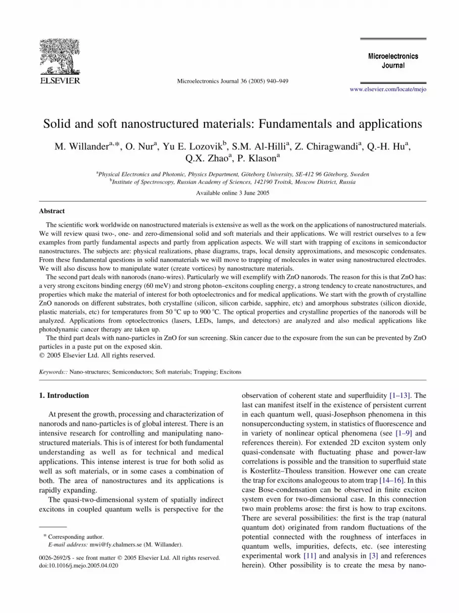
materials. Fig. 1 shows a typical reflectance spectrum of
bulk single crystal ZnO, measured at 80 K. The exciton
transitions are labeled as FEA, FEB and FEC. A number of
investigations on the synthesize of ZnO nano-wires have
been reported in the literature [21–24]. The traditional
problem in ZnO is to obtain p-type conductivity is still
challenging. Recent studies also indicate significant pro-
gress in the growth and understanding of p-type ZnO layers
[25–30]. These successes suggest the potential of ZnO for
applications in electronic and optoelectronic devices in the
near future. One-dimensional materials such as ZnO nano-
wires are of interest due to their importance in basic
scientific research and potential technological applications
[31]. These nano-wires can be grown at relatively low
temperatures and on wide choice of substrates, ranging from
Si to plastic (flexible substrates). ZnO nano-wires have
potential for applications in laser devices due to their
desirable optical properties. Therefore a detailed under-
standing of ZnO wires and the influence of impurities on
their properties are important. It may be worth noting that
ZnO nanorods as well as thin films can be grown or
deposited on various substrates including Si. We will show
our recent results for growth of nanorods in temperature
ranging from 900 8C down to 50 8C. In addition, ZnO nano-
particles are equally interesting and can be grown and
synthesized by different methods. These nano-particles are
of great interest for many technological as well as medical
appplications.
In addition, zinc oxide nano-particles have long been
recognized for its medicinal properties as an anti-irritant and
astringent as well as its UV blocking properties, in
sunscreens [32]. Scientists have shown that ultraviolet A
(UVA) radiation is a major culprit in photo-aging and skin
cancers [33]. Unfortunately, most sunscreens do not protect
against long-wave UVA. Ultraviolet radiation that reaches
the earth and damages skin can be divided into three key
wavelengths [33]: (1) Short-wave UVA (32–280 nm) or
UVC, (2) UVB (280–320 nm), and (3) Long-wave UVA
(320–400 nm) or UVA. We will present here our efforts to
characterize the influence of the particle shape (hexagonal),
and size (plate, equal ratio, column) on the optical properties
(scattering, absorption, and extinction efficiencies) for
moderate size diameter (DZ20–200 nm) in the UV region
(30–400 nm), comparable to the optically incident radiation
that dominant particle size range in sunscreens. We seek to
answer the following question: what is the effective
diameter of these hexagonal nano-particles that protect the
skin from the UVA, UVB, and UVC regions?
2. Excitons trapping
The width of a quantum well for excitons in the case to
create the confining potential by a needle the width of the
quantum well for excitons in this case is much larger than all
other scales in the problem [12]. In this case we can use

Fig. 3. Confinement potential for interwell excitons originated from non
homogeneous field created by STM for various forms of the tip: (1) conic
with the radii of the curving 20, 50, 100 and 200 nm, (2) parabolic with the
radii of the curvature (in lower point) 20 and 50 nm, (3) spherical electrode
with the radii of the curving 20 and 80 nm, (4) hyperbolic form of the tip.
M. Willander et al. / Microelectronics Journal 36 (2005) 940–949942
quasi-local approach. For rarified exciton system general-
ization of Gross–Pitaevskii equation can be used which
takes into account the summing of all ladder diagrams
(because the vertex for rarified Bose gas can be reduced to
amplitude of two-particle scattering but the last in 2D case
diverges at small energies—see, e.g. analysis in [2]):
KV2
2mCVextðrÞKm K
4pjfðrÞj2
m ln ðjfðrÞj2a2Þ
� �fðrÞ Z 0
The profile of the density for quadratic confinement
potential is slightly differs form Gaussian. For more
complex case, higher exciton concentration, we propose
another approach—quasi-local generalization of Koster-
litz–Thouless theory for the system. We also accept that
the width of quantum well for excitons is much larger
than all other scales in the problem (particularly,
separation between thermodynamically equilibrium vor-
texies in exciton system, etc.). The excitons have some
profile n (r) with maximum in the center of the (axially
symmetrical) trap and zero on the boundaries. Let
temperature T to increased. For any given temperature
T it can be equal to ‘local’ Kosterlitz–Thouless critical
temperature [34] which is proportional to the density
in the same point R. Due to axial symmetry all the
circle with radius R will be superfluid. At the boundary
R universal jump of superfluid density to zero takes
place.
So the ring at rOR will be normal (see Fig. 2). When
temperature rises the superfluid circle shrinks to zero at
critical temperature Tc which is proportional to the density
nmaxtot in the center of the trap.
The radius of the superfluid exciton system in the trap is
rs Z Lffiffiffiffiffiffiffiffiffiffiffiffiffiffiffiffiffiffi1 KT =Tc
p
where L is Thomas–Fermi radius.
Sufficiently narrow exciton traps can be created be higly
nonhomogeneous fields U(r) of the STM tip. For spatially
indirect excitons the confining potential is
Fig. 2. Superfluid circle and normal ring for exciton system.
Ueff Z dvU
vz
where d is dipole moment of indirect exciton due to charge
separation. The resulting confining potential calculated for
different forms of the tip is presented on Fig. 3.
The results on Fig. 3 were calculated for the system
presented on Fig. 4. If the doped layer is present on the
surface it can screen the electric field of the STM tip and
confining potential for excitons disappears. But if the
current is presented in tip–semiconductor system (due to
electron tunneling) the confining potential appears as the
current grows.
The exciton system confined by STM tip has small sizes,
of order of 1 m. So if the exciton density has the order of
Fig. 4. Radial distributions of condensed and noncondensed particles at
different number of particles N. Mesoscopic supersolid regime.

M. Willander et al. / Microelectronics Journal 36 (2005) 940–949 943
1010 cmK2 or smaller the number of excitons is smaller than
100, i.e. exciton system is mesoscopic. We studied the
Bose-condensation and strongly correlated regime in
mesoscopic exciton system. When the dipole moments of
the system grow (or confining potential diminishes) the
profile of the system changes essentially: it has Gaussian
form for weakly interacting system, then when interaction
grows it has the form of inverted parabola. For even more
strong interactions the profile of the total density and profile
of Bose–condensed particles have shell structure. By other
words in the mesoscopic Bose system beside superfluidity
some crystal-like order appears and the system become
mesoscopic supersolid (Details will be published elsewhere;
see also [35]).
Fig. 5. The water transistor, (a) A microscope photograph showing the large
area of the device with a water drop placed on top covering all active device
area, (b) the large pH electrodes (200!200 m2), and (c) the small pH
electrodes 0.6!0.6 mm2 together with the nano-gap (different configur-
ations with gaps of 20–200 nm) sensing electrodes [17].
3. The nano-scale water transistor and applications
We have developed a nano-scale water based Si
compatible water transistor [17]. The goal of the device is
to trap a single molecule in water and at the same time tune
the water pH. The physics behind the trapping is the same as
for excitons, i.e. using the gradient field. The active area of
the device composed of two (or more) nano-gap electrodes
(200 nm and down to 20 nm gaps have been processed) and
a third nearby electrode for controlling and manipulate the
pH of the water. The nano-gap electrodes play the role of the
emitter and collector, while the third electrode is considered
as the base in analogy to the bipolar transistor convention.
The operation of the device is described by a bipolar
transport. It is very sensitive to pH variations. In fact there is
a vast need for miniaturized pH sensors in many
biochemical, chemical or industrial applications, since
most of chemical or biological processes are pH dependant.
The ability of the developed device to detect local variation
of the pH, imply that, it is in fact a unique platform for a
many interesting experiments for both fundamental under-
standing as well as for the possible great technological
impact. Below we will briefly describe the principle of
operation and show some of the DC characteristics.
The nano-device was fabricated using silicon compatible
technology combined with electron beam lithography. A
low n-type doped Si wafer was used as the substrate for the
device. An 80 nm thick thermal oxide was grown on the
wafer to provide isolation. Different electron beam
lithography steps were performed to produce different
configurations. The final step was an insulting layer with
open windows on both the tip of the nano-electrodes as well
as for the pH sensing electrodes. Both the gap between the
nano-electrodes as well as the position of the third pH
electrode with respect to the sensing nano-electrodes and its
size were designed in different flexible combinations. Each
device contains 16 nodes. To investigate the pH response we
put a drop of de-ionized clean room water (resistively of
18 MU) on the sensing area of the device. The base
electrode, as well as the emitter and collector were biased in
the common emitter configuration. Fig. 5 shows different
micrographs and scanning electron micrograph of the
device. In Fig. 5(a) a microscope micrograph showing the
large pH electrodes (200 ! 200 mm2) with the water drop
covering all the active area of the device. Fig. 5(b) is an
SEM showing the pH electrodes of Fig. 5(a). While Fig. 1(c)
shows the small pH electrodes (0.6 ! 0.6 mm2) together
with four nano-gap sensing electrodes. Note that the small
electrode part of the device is located inside a small part of
the middle dark area (80 ! 80 mm2) of Fig. 1(b) as indicated
by the arrow. The three terminal device (transistor) basic
operation is based on the variation of the pH of water by
applying a voltage and independantly measure the current
variation between the two nano-spaced electrodes. From our

Fig. 6. The water transistor output characteristics (IECKVEC for different
VBE biases). This output characteristics were measured using the small pH
electrode configuration shown as insert [17].
M. Willander et al. / Microelectronics Journal 36 (2005) 940–949944
analysis of different configurations, we found that the
magnitude of the transistor current and sensitivity to pH
variation depends a lot on the configuration used with regard
to the positioning and distance of the base (pH control
electrode) from the sensing area (emitter–collector nano-
electrodes). When a voltage is applied between the emitter
and base (VBE), the water decomposes and the pH of the
water changes. In presence of an electric filed between the
emitter and collector (VEC), the OHK ions will be attracted
to the collector, and a potential drop is established between
the OHK and their images at the metal. Here the charge
neutrality is postulated for the whole system and, therefore,
OHK ions have to be compensated for by oppositely
charged ions, namely the HC ions in the water. The
dehydrated ions (OHK) are located in a plane adjacent to the
collector electrode called the inner Helmholtz plane (IHP).
The hydrated ions (H3OC) which diffuse from the bulk will
be located in the so called outer Helmholtz plane (OHP)
[36]. The H3OC ions are then surrounded by water dipoles
and will become nonconducting species. They cannot enter
the outer layer, and hence a potential drop is then
established between the OHP and the metal. The total
charge per unit area on the metal (s) is given by the
electronic charge multiplied by the difference between the
number of anions (OHK) and cations (HC) per unit area
[36]. Using the geometrical capacitance between the metal
and the OHP, and proceeding to establish a simple
expression to estimate the current between the emitter and
collector when a voltage of VEC is applied between them, an
expression for the emitter to collector current can be
obtained [17]. Quantitatively, this expression contains two
different dependencies that give the magnitude of the
current IEC. The first is a geometrical dependence. The
second is the effect of the third electrode. In deriving this
expression, we only considered decomposition of water, and
we have ignored other reactions that might exist [37–38].
These dependencies are in fact in consistency with our
experimental observations. In general, the main features of
the I–V output characteristics are: (1) asymmetric I–V with
regard when comparing the forward and reverse biasing (2)
variation of the emitter-collector current by changing the
emitter-base voltage, and (3) shift of the threshold voltage.
The shift of the threshold voltage is expected due to the pH
change. Fig. 6 depicts the output characteristics of the nano-
scale water transistor using the small pH electrode
configurations shown as insert in the figure. This pH
electrode configuration is special in the sense that the pH
electrodes electric field is lying in the same direction as the
field produced between the emitter and collector as can be
clearly seen. As clearly seen, the I–V is asymmetric in
behavior as expected. In addition, a shift in the threshold
voltage is observed in the reverse direction. In addition, a
plateau (saturation) is observed. The origin of this is
currently under investigation. While in the forward the
emitter–collector current response to the pH variation is a
weak response. This observed I–V behavior is mainly
attributed to the special location of the small H electrodes
with regard to the emitter–collector electric field. This
indicates the important role of the design of the pH electrode
with regard to its location and size. Although all other
analyzed configurations showed asymmetric I–V behavior,
all showed sensitivity to VEB on both polarities and a clear
shift of the threshold voltage was observed.
The developed nano-scale water transistor is a suitable
platform for many applications. The fact that local pH
variations can be monitored implies that the device can be
used to sense any type of hydrolytic enzyme reaction. The
presence and biocompatibility of water will open the door
for many applications. As an example we will emphasize
and briefly discuss trapping single molecule reactions.
Conversion of chemical energy into electrical signal in
hydrolytic media combined with addition of fluorescent
markers can lead to a platform of studying and analyzing
‘few’ trapped molecules. In fact trapping of small molecules
in aqueous media has become very interesting due to the
wealth of information possible to obtain from such
experiments. Spatial manipulation of objects by AC
electro-kinetics is a well established technique [39]. In an
inhomogeneous electric filed, polarizable particles experi-
ence a lateral force towards the regions highest filed
strength. This force is called dielectrophoretic force and it
depends on the dielectric properties of both the particle and
their surroundings. Employing alternating field, electro-
phoresis effects are avoided and permanent charges remain
unaffected. Objects that are smaller than about a micrometer
have come into focus only in recent years. This is because
the dielectrophoretic effect is proportional to the cube of the
particle’s radius, while the impact of the Brownian motion
increases with decreasing particles size. Therefore very high
electric field gradients are needed, which, on the other hand,
lead to disturbance by heating and electro hydrodynamic

M. Willander et al. / Microelectronics Journal 36 (2005) 940–949 945
effects. This dilemma is solved by working in low
conductive media and by using electrodes with gap widths
of only a few micrometers or less. Recently electric
properties of viruses and polystyrene micro spheres have
been determined by studying their edges or dielectrophore-
tic response at different field frequencies. Macromolecules
of some micrometers length like DNA has been concen-
trated at the electrode have been aligned in parallel to the
electric filed. In order to manipulate smaller molecules,
especially more compact ones like proteins, even higher
electric field strengths or higher field gradients, respect-
ively, are necessary. Interdigitated electrodes with thickness
of typically 100 nm exhibit a gradient almost only in the z-
direction. Thus sharp electrodes with a shape similar to
needles help to increase dielectrophoretic. Field strength
can be increased by using higher voltage or smaller
electrode distances. Side effects like heating can be reduced
by decreasing the volume of high fields. This is achieved by
shrinking the active volume by reducing electrode distances
and by confining the region of highest field strength to a
small region by, again, sharp electrode tips. The developed
platform, i.e. the nano-scale water based transistor, is an
ideal choice for trapping small molecules other biological or
chemical species. Indeed the presented platform was used
for such purpose. We have employed many nano-eletrodes
with different nano-gaps and different sharpness. Exper-
iments were performed on fluorescently labelled IgG
antibody molecules with a molecular weight of 150 kDa,
corresponding to a diameter of about 8 nm. The “north” and
“south” electrodes were activated by 3 V (RMA) at 100 kHz
(see [40]).
Moreover, we have also used the nano-scale water
platform to study and control vortex flow in a chemical cell.
We have analyzed and described the vortex phenomena. We
showed that in pure water and under external static electric
field we can reproduce and control the vortex flow
formation in a chemical cell (water transistor platform).
The origin of the phenomena is due to the electrochemical
decomposition of water. Due to the low conductivity of pure
water in absence of electrolyte (18 MU), the field driven
hydroxide ions at the anode becomes essential to the proton
release, which in turn is the result of the molecular O2(gas)
evolution. Water recombination processes, which have
proton flowing in hydroxide, background as a key ingredient
produce the phenomena of vortex flow [41].
4. ZnO nano-structures synthesis
As mentioned in the introduction, ZnO nano-structured
materials have attracted intense research communities
recently. The reason is that ZnO is a unique material.
Beside the physical properties mentioned in the abstract,
ZnO exhibits dual semi conducting as well as piezoelectric
properties. Nano-structures possibly to obtain from simply
ZnO powder, is by far more diverse than any known nano-
material including carbon nano-tubes. By solid state thermal
sublimation process with control of the growth kinetics,
local growth temperature, and the chemical composition of
the source material, many nano-materials can be obtained
with 100% reproducibility. Some of these are nanorods,
nano-belts, nano-rings, and more complicated nanostruc-
tures like nano-propellers with different equivalent multi
crystallographic directions, etc. It is important to mention
that all the possible ZnO nano-materials are single crystal-
line and defect free. We will restrict the material in this
paper to ZnO nanorods synthesized by a vapour phase
transport process using Au nano-particles as catalyst [18]. In
this growth procedure, thin films of Au were deposited on a
substrate (1–2 nm), which will then placed in a close
vicinity of a mixture of ZnO and graphite powder in a tube
furnace. The furnace is heated to about 900 8C for 30 min
and is then cooled to room temperature in the flow of argon.
In this process, ZnO is reduced by carbon; simultaneously
Au film de-wetted the substrate and formed nano-clusters.
When Zn atoms condensed on Au clusters, Zn formed alloy
with Au temporally and rendered AuZn in a liquid form.
Under the catalysis of Au, Zn is oxidized to form ZnO with
the Au clusters being elevated on top of the ZnO. Since the
lateral growth of ZnO was limited by the size of the Au
cluster, thin rods would result as time passed by. The
pronounced growth direction is !0001O. The orientation
of the rods with respect to the surface of the substrate was
determined by the orientation relationship of the ZnO!0001O and the crystallographic orientation of the substrate.
In addition, we have also developed a relatively low
temperature (around 50 8C) process based on chemical
reactions to synthesize high quality ZnO nanorods. The
main application for such low temperature process could be
nano-photonics devices on plastic (flexible substrates, e.g.
for electronic cards). In Section 5, some of our recent results
obtained from samples grown using this low temperature
techniques will be briefly described.
Fig. 7 displays different scanning electron micrograph
with different magnifications of a variety of ZnO nanorods
grown on Si, SiC, both at temperature around 900 8C and
finally on ITO at 50 8C. The hexagonal nature of the
nanorods is clearly seen. A variety of substrates have been
successfully employed to synthesize high quality ZnO
nanorods. Among them, oxidized Si (001), Si (001), Si
(111), sapphire (0001) and (1 1 K2 0) and amorphous SiN
membrane were used as substrates for the ZnO nanorods
growth. As mentioned above ITO (p-type) was also
successfully used for the growth of ZnO nanorods. The
choice of the Si substrates is made with the intention of
integrating the ZnO nanorods with silicon technology. The
choice of the sapphire substrate is to obtain vertical growth
of the nanorods. The choice of SiN membrane is made in
order to utilize TEM (transmission electron microscopy not
shown here) to study the structural properties of individual
rods on the substrate. We have been able to vary to diameter
of the ZnO nanorods in a typical growth procedure down to

Fig. 7. Different SEM of (a) nanorods grown on Si (001)-substrate at 920 8C. The length of ZnO nanorods is about 2 mm with diameter of about 0.3–0.8 mm, (b)
SEM viewing single hexagonal ZnO nanorods grown on 4H-SiC substrate at 900 8C, and (c) SEM image of ZnO nanorods grown on ITO-deposited glass
substrate at 50 8C in a solution.
5.00
10.00
15.00
3600 3700 3800 3900 4000 4100
PL in
nten
sity
(ar
b. u
nits
)
Wavelength (Å)
ZnO wireson
EVI
EVI
-1LO
FEA
D0 X T=80 K
Si
Sapphire
Fig. 8. PL spectra from ZnO wires grown on sapphire and Si substrates,
measured at 80 K.
M. Willander et al. / Microelectronics Journal 36 (2005) 940–949946
100 nm. Photoluminescence (PL) spectra were acquired for
all samples. We have reproduced most of the features of
the spectra reported in [18] except for the lack of the
appearance of the sharp peaks due to the lower power
density of our laser. In addition, some of our samples shows
strong white-bluish illumination witnessed by the naked eye
and signified by a broad peak starting at 4000 A and ending
at 6500 A with maxima located at about 4800 A. The PL
measurements were carried out in the temperature range of
80–300 K. A double grating monochromator and a photo-
multiplier detector were used to disperse and detect the ZnO
emission. The laser lines with a wavelength of 270 nm or
350 nm from an ArC laser were used as the excitation
sources. Fig. 8 shows the PL spectra of ZnO wires grown on
sapphire and Si substrates at a temperature of 80 K. The
dominant transitions are the free exciton (FEA) related to an
impurity bound exciton (BE) that is likely due to donor
bound excitons and a further intense transition labelled as
EVI. By carefully examining the wavelength range longer
than the EVI transition, we observed the LO-phonon replicas
of the EVI transition with up to three LO-phonons involved.
In the PL spectrum shown in Fig. 8, only the first LO-
phonon replica is clearly visible due to the large intensity
scale. The energy separation between the FEA and EVI is
about 60 meV at the temperature of 80 K, and this energy
separation decreases with increasing temperature. ZnO
wires grown on sapphire and Si substrates show a similar
spectrum, except that the FEA is relatively stronger in the
sample grown on Si substrates, which indicates that the
background doping concentration is relatively low. From

0
200
400
600
800
25 30 35 40 452 theta (deg.)
Inte
nsity
(cou
nts)
ITO
(22
2)
ZnO
(002
)
ZnO
(100
)
ZnO
(101
)IT
O (
400)
Fig. 9. Powder X-ray diffraction of ZnO nanorods in Fig. 1A (c).
M. Willander et al. / Microelectronics Journal 36 (2005) 940–949 947
the PL spectra measured at different excitation power, we
can state that at the excitation power used here, none of
the observed transitions are related to inelastic exciton–
exciton scattering. As ZnO is n-type in an un-intentional
growth process, the growth on the p-type ITO was for the
purpose of obtaining a heterostructure pn junction for
electrical characterization. Fig. 9 is a typical X-ray spectrum
obtained from the growth on ITO at low temperature.
Although this X-ray diffraction spectrum indicates a non-
axis growth, it shows the appearance of very clear and
strong peaks of single crystal ZnO rods. This implies that,
our newly developed process is very promising and more
optimization can lead to the growth of on-axis single crystal
ZnO nanorods of high quality. Fig. 10 displays the
Cathodoluminescence (CL) of the same sample. The spectra
consist of two features, a sharp exciton peak at 380 nm
reflecting the recombination of electron beam induced
electron-hole pairs in the crystalline ZnO nanorods, and a
Fig. 10. Cathodoluminescence of the ZnO nanorods shown in Fig. 8 (c).
broad visible peak at about 550 nm. The large difference in
the intensity of the two peaks seems to indicate the high
quality of the sample.
Beside the obvious application of ZnO nanorods for
nano-photonics, we will briefly describe here the possibility
of employing ZnO nano-materials for medical cancer
photodynamic therapy (PDT). The technique of the PDT
dates back to 1903, where the first trial was reported [42].
However, since then, the technique has been refined a lot.
Nevertheless, the main steps involved are still the same. The
PDT method relies on the coexistence of a photosensitive
compound (photosensitizer), oxygen and light. The photo-
sensitizer is administered to the patient, where it accumu-
lates in the cancerous tissue. The main features of the
technique are: it’s high selectivity, relatively fast healing
rates, and the ability to treat the same tissue several times if
needed. The mechanism of the PDT is based on the fact that
the therapeutic light, which must match absorption band of
the photosensitizer, excites the photosensitizer molecule
from its single ground state to its first excited singlet state.
The molecule can then, with high probability, be transferred
to its first excited triplet state. This transition although spin
forbidden, but its probability is rather large due to the small
separation between these two states. The final relaxation to
the ground state will also be spin forbidden, leading to long
life time (R100 ms). This long relaxation life time imply a
high probability for interaction with the surrounding
molecule. The excess energy of the photosensitizer may
be transferred to oxygen molecules, which thereby are
excited from their triplet ground state to one of the first
excited states that are biologically active. Singlet oxygen is
highly cytotoxic and its formation leads to degradation of
the cancerous cells by various mechanisms. The PDT is
selectivity leads to local treatment with low risk of damage
of healthy cells. Moreover, it is very efficient in also
treatment of precancerous cells. ZnO with its discussed
photonic properties is a very advantageous for the purpose
of PDT. In this connection, we will below also present our
recent work to optimize the use of ZnO nano-particles for
sun screening from the damaging part of the sun ultra-violet
radiation.
5. ZnO nano-particles UV absorption and scatteringefficiency
To describe light scattering from an arbitrarily shaped
nano-particle, we have adopted (DDA) method, as first
formulated by Purcell and Pennypacker [43] and modified
by Draine [44] and Goodman et al. [45]. In this method, the
particle of interest is consisting of array of N electro-
magnetically interacting dipoles on a cubic lattice grid.
Each dipole radiates a dipole field in response to the incident
radiation and the radiated fields of all other dipoles in the
ensemble. We have used DDSCAT6.1 program written by
Draine and Flatau [46] and we have modified their code to

Fig. 11. (a) Hexagonal sphere-packing of the ZnO particle model, (b) plate-
like shape, (c) column shape, and (d) real plate ZnO particle synthesized by
the vapor phase transport process as described in the text.
Fig. 12. Absorption and scattering efficiency factors versus wavelength for
plate, equal ratio, and column hexagonal ZnO particle with two constant
effective radii (40 and 80 nm).
M. Willander et al. / Microelectronics Journal 36 (2005) 940–949948
generate hexagonal particle geometry of identical spheres
from the planer generation matrix [47], then to generate
three-dimensional hexagonal shapes in a fundamental
region by extending the planar hexagonal lattice as a
building block for the first layer to any vertex–vertex
diameter of the hexagonal face and repeated this layer many
times fills the whole space of the final three-dimensional
hexagonal particle. This situation is pictured in Fig. 11.
Rather than direct methods for solving set of 3N complex
linear equations, in this study we fellow Flatau [48] in using
the stabilized version of Bi-Conjugate Gradients (Bi-
CGSTAB) method [49] with preconditioning. We adopted
the Draine and Goodman [45] ‘Lattice Dispersion Relation’
(LDR) method for prescribing the dipole polarizabilities.
The constraint for the validity of the DDA code is distance
between neighbouring dipoles; dZ(4p/3)1/3R, where R is
the radius of the dipole; must be small enough compared
with the wavelength l of light in the surrounding target
medium and for accurate calculations we need a more
conservative criterion kdjmj%0.5. In our study we specify
bZ0, fZ0, and QZ08, 608, 908 and we choose unpolarized
incident light for our study. Once the polarizations are
known, the extinction Qext, absorption Qabs, and scattering
Qsca efficiency factors may be evaluated from the optical
theorem. We take relative refractive index; which is the
ratio between the refractive index of the particles (ZnO) and
that to the matrix, such as the oil phase (in our study we take
it water for simplicity), we simulate the data for real and
imaginary parts of refractive index by adopting the work by
Dakhel [50] as approximate data and we used Segelstein
[51] data for the real refractive index of water in the UV
region; and wavelength in the medium which is the ratio
between the wavelength in vacuo to the real refractive index
of the medium.
Three different particle shapes are considered in this
study: hexagonal plates with aspect ratio (0.6650), hexagonal
particles with aspect ratio (1.0006), and hexagonal columns
with aspect ratio (1.4963) for values of the size range reffZ10 nm(10 nm)100 nm. The comparisons between absorption
and scattering efficiency factors for ZnO particles for three
hexagonal shapes with same effective radius are shown in
Fig. 12, the results for effective radiusZ40 and 80 nm. For
each size, both figures display two bands of similar position,
intensity and shape for the plate and equal lengths hexagonal
particles with small difference between the values for the
column shape. The similarity between the plate and equal
lengths hexagonal particles came from aspect ratio assumed
for the plate (0.6650), this value get less effective with
decreasing the effective radius as we can see from the curves
of effective radius 40 nm, we found it much similar and have
the same values in most regions than the curves of 80 nm. The
small difference in intensity between the plate and column
hexagonal particles arises obviously from the difference of
the target frame axes of the plate and column hexagonal ZnO
particle (see Fig. 1) and of course from the different in shape
of plate and column. When the particle size increases, its
shape being kept constant, the only increase the peak
intensity, which in this case is proportional to the hexagonal
volume. The location and the intensity of the first and second
peaks in the Fig. 12, which shifted are dependent on the
wavelength and particle size. To explain this case, the first
peak in the UVC region arises from the dipole excitations and
the relation between the polarizability and dielectric function
to extract 3(u) [52] and the second peak in the UVA
associated with the band gap at (380 nm) for the bulk ZnO
and this peak is shifted to the shorter wavelengths as the

M. Willander et al. / Microelectronics Journal 36 (2005) 940–949 949
particle size decreased because the wavelength dependence
of the absorption cross section seems to depend on the mean
optical thickness of the hexagonal nano-particle which is
function of the N and l [53].
6. Summary
In summary, we have presented some different important
issues related to nano-structured materials. Both funda-
mental as well as technological aspects are presented and
discussed. Some recent results of exciton trapping are
presented (e.g. the superfluid mesoscopic system). The so
called nano-scale water transistor platform developed
recently is presented. Applications of this unique pH local
sensor are discussed. We emphasized the use of the platform
to trap and study single or few molecules. Advantages,
growth, and characterization of ZnO nano-wires together
with our recent results are discussed. We have developed
relatively low temperature process (50 8C). Such a low
temperature process is of great technological impact due to
the possibility of growth on flexible substrates (e.g. plastic),
which is of interest for plastic electronics card. Photo-
dynamic therapy is suggested to benefit from ZnO nano-
materials due to the suitability. In addition, we have
developed a model, based upon light scattering by single
particle, which enables us to account satisfactorily for the
absorption, scattering, and extinction spectra of hexagonal
ZnO particles. We have studied the effect of the particle size
on scattering and absorption, with the aim to optimize the
processes for sun-screening against skin cancer.
References
[1] Y.E. Lozovik, V.I. Yudson, Pis’ma ZhETF 22 (1975); Y.E. Lozovik,
V.I. Yudson, ZhETF 71 (1976) 738; Y.E. Lozovik, V.I. Yudson, JETP
4 (1976) 389; Y.E. Lozovik, V.I. Yudson, Solid State Commun. 18
(1976) 628; Y.E. Lozovik, V.I. Yudson, Soid State Commun. 21
(1977) 211; Y.E. Lozovik, O.L. Berman, Pis’ma ZhETF 64 (1996)
526; Y.E. Lozovik, O.L. Berman, JETP Lett. 64 (1996) 573.
[2] Yu. E. Lozovik, V.I. Yudson, Physica A93 (1978) 493.
[3] Yu. E. Lozovik, M. Willander, Appl. Phys. A71 (2000) 379.
[4] Yu. E. Lozovik, O.L. Berman, M. Willander, J. Phys. C14 (2002) 12457.
[5] Yu. E. Lozovik, Uspekhi Fiz. Nauk 171 (2001) 1373.
[6] Yu. E. Lozovik, I.V. Ovchinnikov, Phys. Rev. B66 (2002) 075124.
[7] Xu. Zhu, P.B. Littlewood, M.S. Hybertsen, T.M. Rice, Phys. Rev.
Lett. 74 (1995) 1633.
[8] S. Conti, G. Vignale, A.H. MacDonald, Phys. Rev. B57 (1998) R6846.
[9] M.A. Olivares-Robles, S.E. Ulloa, Phys. Rev. B64 (2001) 115302.
[10] A.V. Larionov, V.B. Timofeev, I. Hvam, K. Soerensen, JETP Lett. 75
(2002) 233.
[11] L.V. Butov, L.S. Levitov, A.V. Mintsev, et al., Phys. Rev. Lett. 92
(2004) 117404; L.V. Butov, C.W. Lai, D.S. Chemla, Yu.E. Lozovik,
et al., Phys. Rev. Lett. 87 (2001) 216804; L.V. Butov, A. Zrenner,
G. Abstreiter, G. Bohm, G. Weimann, Phys. Rev. Lett. 73 (1994) 304;
L.V. Butov, C.W. Lai, A.L. Ivanov, A.C. Gossard, D.S. Chemla,
Nature 417 (2002) 47.
[12] D.W. Snoke, S. Denev, Y. Liu, et al., Nature 418 (2002) 754 D.W.
Snoke, et. al. (to bepublished)..
[13] V.V. Krivolapchuk, E.S. Moskalenko, A.L. Zhmodikov, Phys. Rev.
B64 (2001) 045313.
[14] M.H. Anderson, J.R. Ensher, Science 269 (1995) 198.
[15] J.R. Ensher, D.S. Jin, M.R. Matthews, C.E. Wieman, E.A. Cornell,
Phys. Rev. Lett. 77 (1996) 4984.
[16] W. Ketterle, N.J. Druten, Phys. Rev. A54 (1996) 656.
[17] Z. Chiragwandi, O. Nur, M. Willander, N. Calander, Appl. Phys. Lett.
83 (2003) 5310.
[18] M. Huang, S. Mao, H. Feick, H. Yan, Y. Wu, H. Kind, E. Weber,
R. Russo, P. Yang, Science 292 (2001) 1897.
[19] P. Zu, Z.K. Tang, G.K.L. Wong, M. Kawasakki, A. Ohtomo,
H. Koinuma, Y. Segawa, Solid State Commun. 103 (1997) 459.
[20] D.M. Bagnall, Y.F. Chen, Z. Zhu, T. Yao, S. Koyama, M.Y. Shen,
T. Goto, Appl. Phys. Lett. 70 (1997) 2230.
[21] H. Cao, et al., Phys. Rev. Lett. 84 (2000) 5584.
[22] W.I. Park, D.H. Kim, S.W. Jung, Gyu-Chui Yi, Appl. Phys. Lett. 80
(2002) 4232.
[23] H. Kim, W. Sigmund, Appl. Phys. Lett. 81 (2002) 2085.
[24] S.C. Lyu, Y. Zhang, H. Ruh, H.-J. Lee, H.-W. Shim, E.-K. Suh,
C.J. Lee, Chem. Phys. Lett. 363 (2002) 134.
[25] D.C. Look, et al., Appl. Phys. Lett. 81 (2002) 1830.
[26] T. Aoki, et al., Phys. Stat. S0ol. B229 (2002) 911X.
[27] Y. Yan, S.B. Zhang, S.T. Pantelides, Phys. Rev. Lett. 86 (2001) 5723.
[28] S.B. Zhang, S.H. Wei, Alex Zunger, Phys. Rev. B63 (2001) 075205.
[29] C.H. Park, S.B. Zhang, S.H. Wei, Phys. Rev. B66 (2001) 073202.
[30] A. Tuskazaki, A. Ohotomo, T. Onuma, M. Ohtani, T. Makino,
M. Sumiya, K. Ohtani, S.F. Chichibu, S. Fuke, Y. Segawa, H. Ohno,
H. Koinuma, M. Kawasaki, Nature 4 (2005) 42.
[31] J. Hu, T.W. Odom, C.M. Lieber, Acc. Chem. Res. 332 (1999) 435.
[32] R.E. Shore, International J. Radi. Bio. 57 (1990) 809.
[33] D. Fairhurst, M.A. Mitchnick, in: N.J. Lowe, N.A. Shaath,
M.A. Pathak (Eds.), Sunscreens; Development, Evaluation, and
Regulatory Aspects2nd ed., Marcel Dekker, New York, 1997, p. 313.
[34] J.M. Kosterlitz, D.J. Thouless, J. Phys. C6 (1973) 1181; D.R. Nelson,
J.M. Kosterlitz, Phys. Rev. Lett. 39 (1977) 1201; P. Minnhagen, Rev.
Mod. Phys. 59 (1987) 1001.
[35] Yu.E. Lozovik, S.Yu. Volkov, M. Willander, Pis’ma J. Exp. Theor.
Phys. 79 (2004) 585 Y.E. Lozovik, S.Y. Volkov, M. Willander, to be
published..
[36] D.E. Yates, S. Levine, T.W. Healy, J. Chem. Soc. Faraday Trans. 70
(1974) 1807.
[37] P. Atkins, J. de Paula, Atkin’s Physical Chemistry7th Edition, Oxford
University Press, New York, 2002, p. 271.
[38] D. Kek, N. Bonanos, M. Mogensen, S. Pejovink, Solid State Ion. 131
(2000) 249.
[39] R. Peting, G.H. Max, Wiley, Chichester, 1979.
[40] M. Willander, Z. Chiragwandi, E. Mamontov, O. Nur, N. Calander, R.
Holtz, and F. Bier, Theoretical and experimental aspects on trapping
and detections of single and few molecules by using nanoprobes,
Invited Talk ISCANA (2003), Virginia USA.
[41] Z. Chiragwandi, I. Panas, O. Nur, and M. Willander, submitted to
Appl. Phys. Lett. (2005).
[42] H. Tappeiner, A. Jesionek, Munich Med. Wachenschr 47 (1903) 2042.
[43] E.M. Purcell, C.R. Pennypacker, Astrophys. J. 186 (1973) 705.
[44] B.T. Draine, Astrophys. J. 333 (1988) 848.
[45] B.T. Draine, J. Goodman, Astrophys. J. 405 (1993) 685.
[46] Program DDSCAT6.1 by B.T. Draine, Princeton University Obser-
vatory, Princeton NJ, 08544-1001 and P.J. Flatau, Program
DDSCAT6.1, University of California, San Diego, Scripps Institution
of Oceanography, La Jolla, California 92093-0221, USA.
[47] J.H. Conway, N.J.A. Sloane,, 3rd ed., Springer-Verlag, New York,
1999. sections 1.2, 1.4, 4.6.1, and 4.6.2.
[48] P.J. Flatau, Opt. Lett. 22 (1997) 1205.
[49] H.A. van der Vosrt, SIAM, J. Sci. Statist. Comput. 13 (1992) 631.
[50] A.A. Dakhel, Mater. Chem. Phys. 81 (2003) 56.
[51] D. Segelstein, “The complex refractive index of water,” MSc thesis,
University of Missouri, Kansas city, USA, 1981.
[52] D.M. Wood, N.W. Ashcroft, Phys. Rev. B25 (1982) 6255.
[53] T. Kozasa, J. Blum, T. Mukai, Astron. Astrophys. 263 (1992) 423.