single-molecule junction spontaneously restored by dna zipper
TRANSCRIPT
ARTICLE
Single-molecule junction spontaneously restoredby DNA zipperTakanori Harashima1, Shintaro Fujii 1, Yuki Jono1, Tsuyoshi Terakawa 2, Noriyuki Kurita3, Satoshi Kaneko 1,
Manabu Kiguchi1 & Tomoaki Nishino 1✉
The electrical properties of DNA have been extensively investigated within the field of
molecular electronics. Previous studies on this topic primarily focused on the transport
phenomena in the static structure at thermodynamic equilibria. Consequently, the properties
of higher-order structures of DNA and their structural changes associated with the design of
single-molecule electronic devices have not been fully studied so far. This stems from the
limitation that only extremely short DNA is available for electrical measurements, since the
single-molecule conductance decreases sharply with the increase in the molecular length.
Here, we report a DNA zipper configuration to form a single-molecule junction. The duplex is
accommodated in a nanogap between metal electrodes in a configuration where the duplex is
perpendicular to the nanogap axis. Electrical measurements reveal that the single-molecule
junction of the 90-mer DNA zipper exhibits high conductance due to the delocalized πsystem. Moreover, we find an attractive self-restoring capability that the single-molecule
junction can be repeatedly formed without full structural breakdown even after electrical
failure. The DNA zipping strategy presented here provides a basis for novel designs of single-
molecule junctions.
https://doi.org/10.1038/s41467-021-25943-3 OPEN
1 Department of Chemistry, School of Science, Tokyo Institute of Technology, 2-12-1 W4-11 Ookayama, Meguro-ku, Tokyo 152-8551, Japan. 2 Department ofBiophysics, Graduate School of Science, Kyoto University, Kitashirakawa-Oiwakecho, Sakyo, Kyoto 606-8502, Japan. 3 Department of Computer Science andEngineering, Toyohashi University of Technology, Tempaku-cho, Toyohashi 441-8580, Japan. ✉email: [email protected]
NATURE COMMUNICATIONS | (2021) 12:5762 | https://doi.org/10.1038/s41467-021-25943-3 | www.nature.com/naturecommunications 1
1234
5678
90():,;
The structural, physical, and chemical properties of DNA atthe nanoscale have attracted attention to the prospect ofusing DNA as a building block in nanoscience. In the field
of molecular electronics, electron transport through a single DNAmolecule has been extensively investigated1–5. These studiesexploit single-molecule junctions, in which a DNA moleculebridges a nanogap between the metal electrodes. For example, itwas reported that A-form and B-form DNAs differ in the single-molecule conductance by an order of magnitude6. Many studieshave revealed that there is a change in the electron transportproperties of DNA after ligand binding7–9. These include anintercalator whose perturbation of base stacking leads toswitching or rectifying properties of DNA10–12. Moreover, athree-terminal DNA connected by a guanine quadruplex has beenexploited to construct a single-molecule junction, and it wasdemonstrated that the intricate junction structure shows excellentfunctionality for a charge splitter13. Significant advances havebeen made in understanding the transport phenomena through asingle DNA molecule, as exemplified above. However, theseprevious studies mainly focused on the transport phenomena instatic DNA structure at thermodynamic equilibria.
Recently, rational control of DNA structures at the single-molecule level has been achieved by modern techniques andemerging technologies. Capturing desired conformation andseparation of DNA are achieved by elaborate flow in nanofluidicsystems14–17. Sequencing analysis of DNA at the single-baseresolution can be performed with nanopore devices during theconstrained passage18–21. Also, precise control of DNA structuresby atomic force microscopy (AFM) has enabled accurate mappingof DNA unzipping dynamics and a free-energy landscape22–24.This combination of electrical measurements and structuralmodulation of DNA could lead to the realization of the sophis-ticated functionality of electronic devices based on a single DNAmolecule. For example, we expect that electron transport throughDNA under the deliberate control of its structure paves the wayfor DNA electronic devices with functional controllability in adynamic manner.
Here, we report the investigation of electron transport throughthe single-molecule junction of a zipper DNA that orthogonallyclamps a metal nanogap (see Fig. 1a). The present DNA single-molecule junction differs from conventional ones in the DNAconfiguration; the present and conventional junctions contain aDNA molecule oriented in perpendicular and parallel directionsto the axis of the nanogap, respectively. The DNA single-moleculejunction with the present zipper configuration exhibited highsingle-molecule conductance that cannot be achieved using DNAjunctions with the conventional configuration. The unzippingdynamics of the molecular junction were characterized by tun-nelling currents in break-junction (BJ) experiments, based onscanning tunnelling microscopy (STM). The STM measurementand the molecular dynamics (MD) simulations of the unzippingdynamics revealed that the DNA junction with zipper config-uration enables spontaneous restoration of the molecular junctionafter its electrical failure and thereby improves the reproducibilityof the junction formation. Our study demonstrated that a DNAdynamic structural change could be applied to a single-moleculejunction by using the zipper configuration. The findings pave wayfor novel functionality and superior properties of nanoscaleelectronic devices22,25.
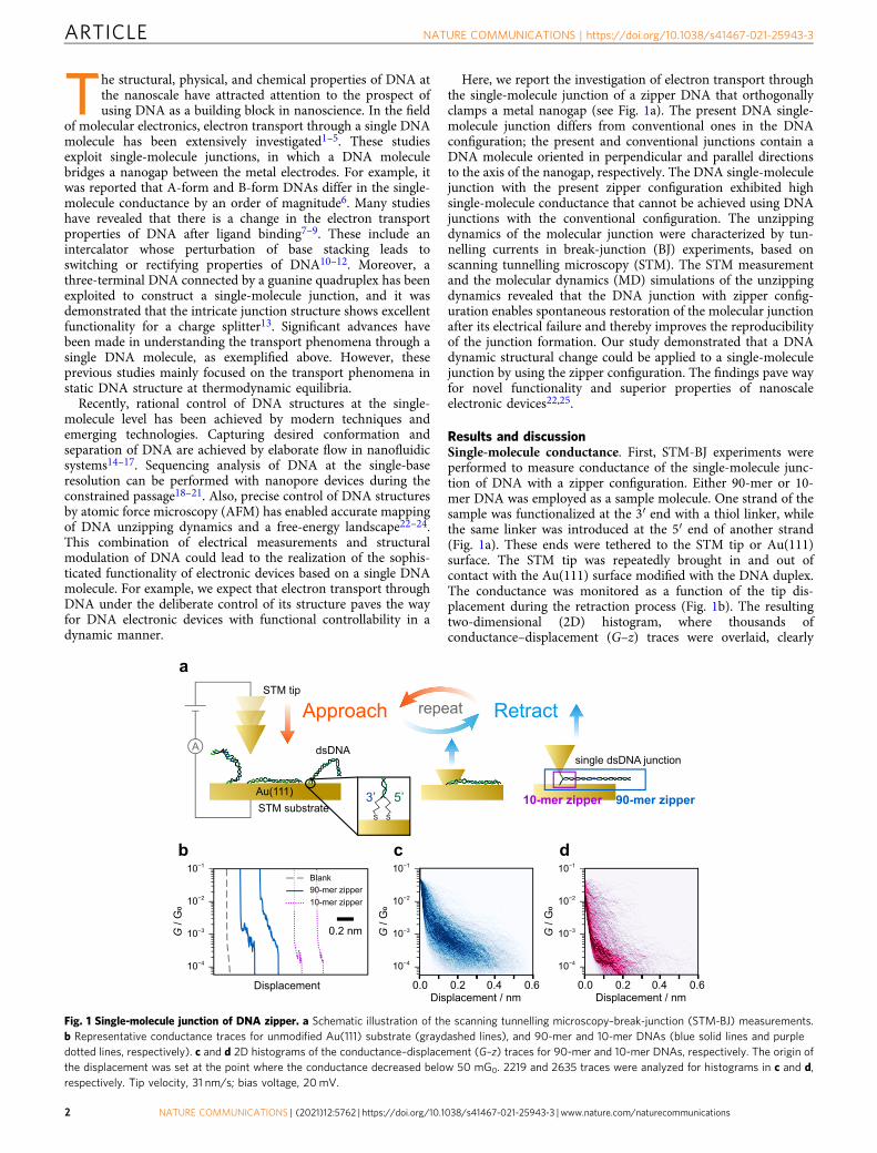
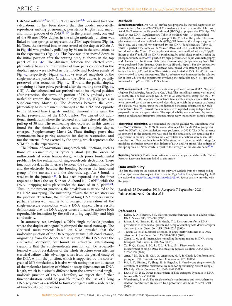
Results and discussionSingle-molecule conductance. First, STM-BJ experiments wereperformed to measure conductance of the single-molecule junc-tion of DNA with a zipper configuration. Either 90-mer or 10-mer DNA was employed as a sample molecule. One strand of thesample was functionalized at the 3′ end with a thiol linker, whilethe same linker was introduced at the 5′ end of another strand(Fig. 1a). These ends were tethered to the STM tip or Au(111)surface. The STM tip was repeatedly brought in and out ofcontact with the Au(111) surface modified with the DNA duplex.The conductance was monitored as a function of the tip dis-placement during the retraction process (Fig. 1b). The resultingtwo-dimensional (2D) histogram, where thousands ofconductance–displacement (G–z) traces were overlaid, clearly
Fig. 1 Single-molecule junction of DNA zipper. a Schematic illustration of the scanning tunnelling microscopy–break-junction (STM-BJ) measurements.b Representative conductance traces for unmodified Au(111) substrate (graydashed lines), and 90-mer and 10-mer DNAs (blue solid lines and purpledotted lines, respectively). c and d 2D histograms of the conductance–displacement (G–z) traces for 90-mer and 10-mer DNAs, respectively. The origin ofthe displacement was set at the point where the conductance decreased below 50 mG0. 2219 and 2635 traces were analyzed for histograms in c and d,respectively. Tip velocity, 31 nm/s; bias voltage, 20mV.
ARTICLE NATURE COMMUNICATIONS | https://doi.org/10.1038/s41467-021-25943-3
2 NATURE COMMUNICATIONS | (2021) 12:5762 | https://doi.org/10.1038/s41467-021-25943-3 | www.nature.com/naturecommunications
shows conductance plateaus at 1.9 and 0.15 mG0 for 90-mer and10-mer DNA, respectively (Fig. 1c, d). The difference in thesingle-molecule conductance is discussed later. The single plateauand subsequent conductance decay in the traces indicate thatthese plateaus are attributed to the single-molecule junction thatcontains DNA. The tunnelling decay constants during and afterthe plateau (β1 and β2, respectively) were analysed from eachconductance trace, and β1 and β2 for 90-mer DNA were deter-mined to be 0.27 and 2.0 Å−1, respectively (see SupplementaryNote 1 for the results of 10-mer DNA and detailed discussion ofSTM-BJ results). The decay constants are known to depend onthe energy gap between the Fermi level of the electrode and thatof the molecular orbital for the tunnelling transport26. The β2value found here is consistent with that for direct tunnellingbetween the tip and substrate without the molecular junction(2.2 Å−1, Supplementary Note 1). On the other hand, the β1 valueis smaller than the typical value for alkanedithiol, but similar tothe ones for π-conjugated molecules26, which indicates that theelectron transport involves the DNA. The DNA zipper junctiontransmits electrons in the transverse direction, and it is antici-pated that the base pairs, especially those located at the DNAterminal, mediate the electron transport (see Fig. 1a). In this case,the transport properties of the present junction can be comparedwith those of the single-molecule junction of DNA bases, whichhave been investigated toward the realization of single-moleculesequencing27–30. Indeed, the reduction in the decay constants asobserved for β1 value in the present experiments was reported forthe tunnelling through the DNA base pairs31,32. Further STM-BJexperiments showed that the conductance of the molecularjunction reflects the DNA sequence (Supplementary Note 2),which further supports that the electron transport is mediated bythe DNA.
The present DNA single-molecule junction possesses adifferent configuration from the conventional one. In the presentstudy, the DNA duplex bears the two linker groups at the sameend. Thus, the molecular junction contains DNA in a configura-tion orthogonal to the axis of the gap between the tip and
substrate (Fig. 1a). In contrast, the linker groups wereconventionally located at the opposite ends of the duplex, andthe junction accommodates the duplex aligned parallel to the gapaxis. Electron transport through the latter conventional junctionsteeply attenuates with increased DNA length, since the electronstravel through the whole duplex33. No such attenuation happensin the present single-molecule junction. Indeed, the conductanceof the 90-mer DNA zipper junction is larger than that of the 10-mer zipper junction. We measured the conductance of the zipperjunction of a variety of DNA lengths ranging from 10 to 90 basepairs and confirmed that the conductance value increased as theDNA length increased (Supplementary Note 3). This lengthdependence, together with the self-restoring capability describedlater, are the advantages of the present configuration.
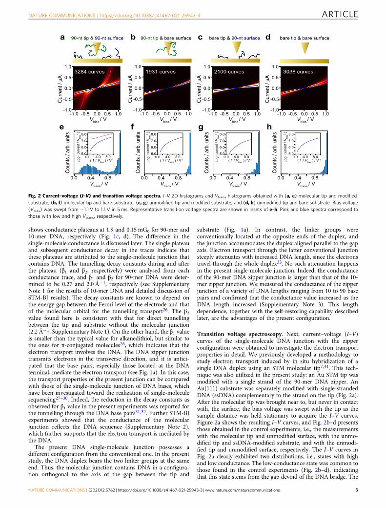
Transition voltage spectroscopy. Next, current–voltage (I–V)curves of the single-molecule DNA junction with the zipperconfiguration were obtained to investigate the electron transportproperties in detail. We previously developed a methodology tostudy electron transport induced by in situ hybridization of asingle DNA duplex using an STM molecular tip7,34. This tech-nique was also utilized in the present study: an Au STM tip wasmodified with a single strand of the 90-mer DNA zipper. AnAu(111) substrate was separately modified with single-strandedDNA (ssDNA) complementary to the strand on the tip (Fig. 2a).After the molecular tip was brought near to, but never in contactwith, the surface, the bias voltage was swept with the tip as thesample distance was held stationary to acquire the I–V curves.Figure 2a shows the resulting I–V curves, and Fig. 2b–d presentsthose obtained in the control experiments, i.e., the measurementswith the molecular tip and unmodified surface, with the unmo-dified tip and ssDNA-modified substrate, and with the unmodi-fied tip and unmodified surface, respectively. The I–V curves inFig. 2a clearly exhibited two distributions, i.e., states with highand low conductance. The low-conductance state was common tothose found in the control experiments (Fig. 2b–d), indicatingthat this state stems from the gap devoid of the DNA bridge. The
Curre
nt / �A
Vbias / V
0.0
0.5
1.0
-0.5
-1.00.0 0.5 1.0-0.5-1.0
3038 curves
bare tip & bare surface90-nt tip & bare surface90-nt tip & 90-nt surface
1931 curves3284 curves
Vtrans / V0.80.40.0
Coun
ts /
arb.
uni
ts
4.0 8.00.0
7.0
8.0
6.0
5.0
Log(
cur
rent
/ V b
ias2 )
( 1 / Vbias ) / V-1
e
a b d
f
bare tip & 90-nt surface
2100 curves
c
g hCu
rrent
/ �A
Vbias / V
0.0
0.5
1.0
-0.5
-1.00.0 0.5 1.0-0.5-1.0
Curre
nt / �A
Vbias / V
0.0
0.5
1.0
-0.5
-1.00.0 0.5 1.0-0.5-1.0
Curre
nt / �A
Vbias / V
0.0
0.5
1.0
-0.5
-1.00.0 0.5 1.0-0.5-1.0
4.0 8.00.0
7.0
8.0
6.0
5.0
( 1 / Vbias ) / V-1Log(
cur
rent
/ V b
ias2 )
4.0 8.00.0
7.0
8.0
6.0
5.0
( 1 / Vbias ) / V-1Log(
cur
rent
/ V b
ias2 )
4.0 8.00.0
7.0
8.0
6.0
5.0
( 1 / Vbias ) / V-1Log(
cur
rent
/ V b
ias2 )
Vtrans / V0.80.40.0
Coun
ts /
arb.
uni
ts
Coun
ts /
arb.
uni
ts
Vtrans / V0.80.40.0
Coun
ts /
arb.
uni
ts
Vtrans / V0.80.40.0
Fig. 2 Current–voltage (I–V) and transition voltage spectra. I–V 2D histograms and Vtrans histograms obtained with (a, e) molecular tip and modifiedsubstrate, (b, f) molecular tip and bare substrate, (c, g) unmodified tip and modified substrate, and (d, h) unmodified tip and bare substrate. Bias voltage(Vbias) was swept from −1.1 V to 1.1 V in 5 ms. Representative transition voltage spectra are shown in insets of e–h. Pink and blue spectra correspond tothose with low and high Vtrans, respectively.
NATURE COMMUNICATIONS | https://doi.org/10.1038/s41467-021-25943-3 ARTICLE
NATURE COMMUNICATIONS | (2021) 12:5762 | https://doi.org/10.1038/s41467-021-25943-3 | www.nature.com/naturecommunications 3
high-conductance state was thus ascribed to the molecularjunction of the DNA zipper. The conductance as estimated by theI–V properties agrees with the conductance as determined bythe BJ studies (Fig. 1c, d, see Supplementary Note 4), supportingthe assignment of a high-conductance state to the DNA junction.It has been reported that ssDNA can adsorb to a metal surface viaits bases35. However, no state that could be attributed to ssDNAmolecular junctions was found in the I–V curves in Fig. 2b, c.STM-BJ study was also conducted with the unmodified tip andthe ssDNA-modified substrate, and the conductance histogramswithout notable peaks were obtained (Supplementary Note 5).These results are most probably due to the significantly decreasedconductance of ssDNA as compared to that of dsDNA because ofbase stacking is less ordered in ssDNA36. The conductance of thessDNA junction would be almost indistinguishable from theconductance of the gap without the dsDNA bridge in the presentmeasurements.
For evaluation of the electronic structure of the molecularjunction, the transition voltage (Vtrans) was estimated using I–Vcurves of the high-conductance state (Fig. 2e–h). It has beenknown that Vtrans is proportional to the energy gap between theFermi level of the electrode (the tip or the substrate) and that ofthe conduction orbital of the molecule in the junction37–39. Intransition voltage spectra, log(I/V2) is plotted against V–1, andthe voltage at which the plot reaches a minimum corresponds toVtrans
37–39. The mean value of Vtrans for the molecular junctionof the 90-mer DNA zipper was found to be 0.4 V (Fig. 2e). In
our previous work, a Vtrans value of 0.8 V was found for a single-molecule junction of 8-mer DNA in the conventional config-uration. It was further observed that this value decreased to0.5 V upon binding of an intercalator to the DNA11. The Vtrans
value for the molecular junction of the DNA zipper is smallerthan in both these above-mentioned cases. This result suggeststhe decrease of the energy gap by delocalization of π stackingorbitals over its long base pairs, which agrees with previousresearch that used fragment molecular orbital and density-functional theory calculations40,41. We attribute the small Vtrans,that is, the decreased energy gap, to delocalization of the π-orbitals of DNA over its long base pairs. To prove this,molecular orbital calculations were performed based on density-functional theory (Supplementary Note 6). We indeed foundthat the energy gap between the highest occupied molecularorbital (HOMO) and the lowest unoccupied molecular orbital(LUMO) decreased with the increase in DNA length, in linewith previous theoretical studies40,41. The larger conductance ofthe 90-mer DNA junction compared to the 10-mer counterpart(Fig. 1c) is consistent with the length-dependent decrease in theHOMO–LUMO gap. Thus, we conclude that the single-molecule junction of the DNA zipper attains high conductancedue to the delocalized π system of the stacked DNA bases nearthe electrodes. The effect of this electron delocalization couldexplain the higher conductance of the 90-mer DNA zippercompared with that of the 10-mer counterpart (Fig. 1b), thoughthis behavior merits further investigation.
Approach & Stop
STM tip
STM substrate
ssDNAc-ssDNA
Retract
Con
duct
ance
Displacement
Con
duct
ance
Displacement
PlateaublankRetract
90-mer zipper10-mer zipperC
ondu
ctan
ce /
mG
0
4.0
1.02.0
0.0
3.0
Displacement
1 nm
400200 6000Trace number
Dw
ell l
engt
h / n
m 0.4
0.1
0.2
0.0
0.3
400200 6000Trace number
Dw
ell l
engt
h / n
m 0.4
0.1
0.2
0.0
0.3
ImmobilizationDiffusion
×
a
b
c
d 10-mer zipper 90-mer zipper
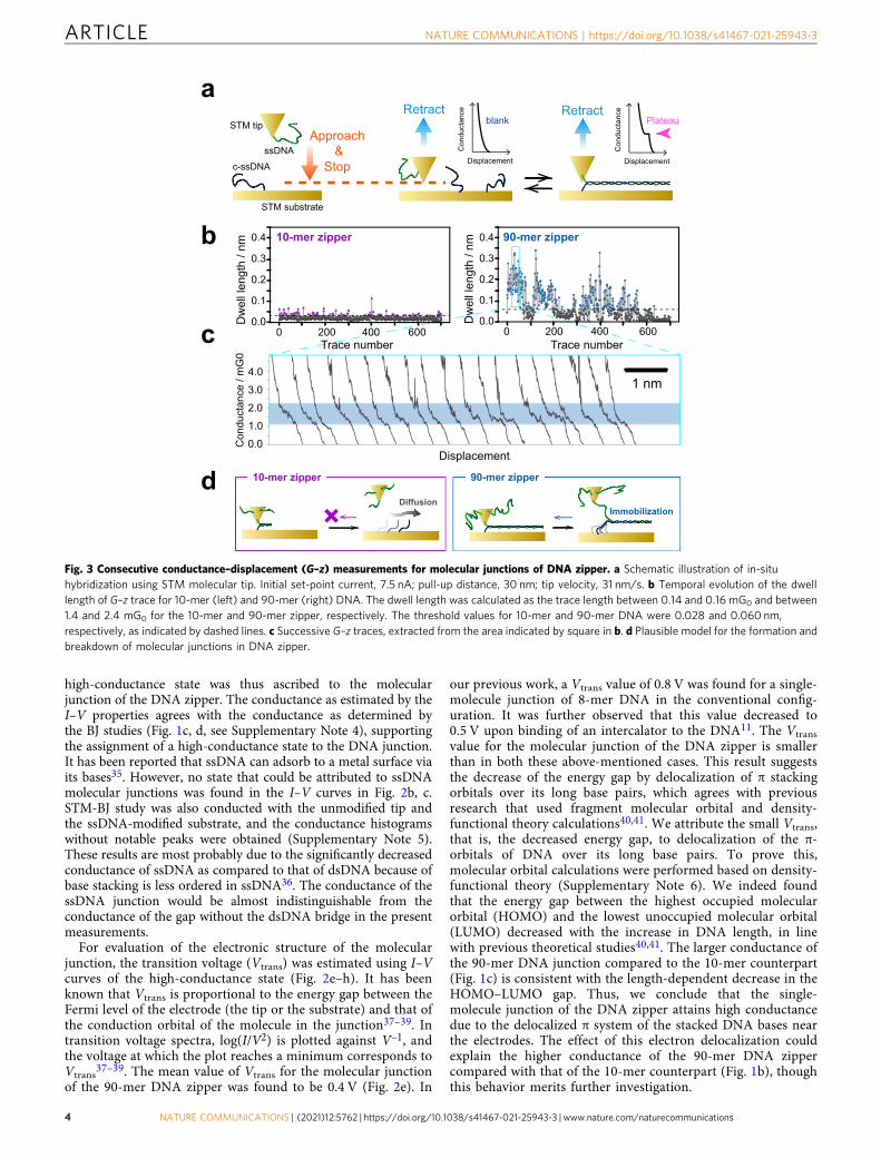
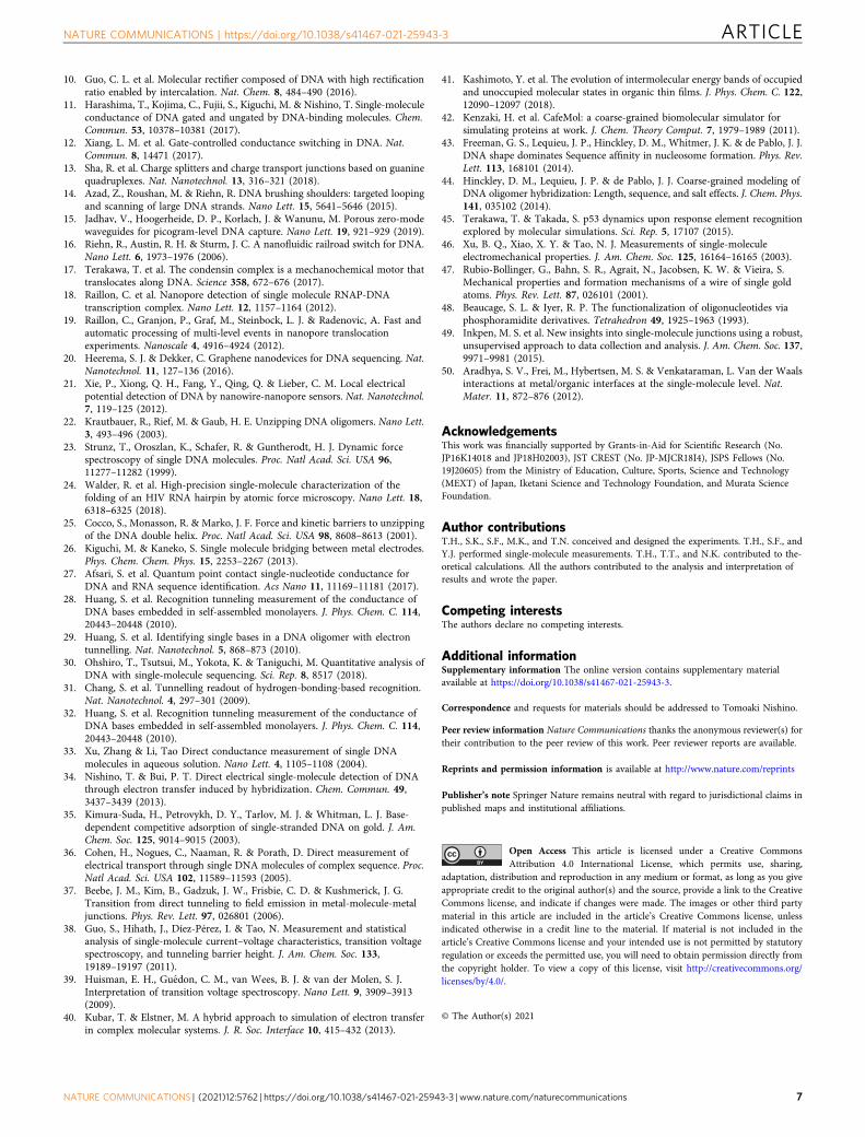
Fig. 3 Consecutive conductance–displacement (G–z) measurements for molecular junctions of DNA zipper. a Schematic illustration of in-situhybridization using STM molecular tip. Initial set-point current, 7.5 nA; pull-up distance, 30 nm; tip velocity, 31 nm/s. b Temporal evolution of the dwelllength of G–z trace for 10-mer (left) and 90-mer (right) DNA. The dwell length was calculated as the trace length between 0.14 and 0.16 mG0 and between1.4 and 2.4 mG0 for the 10-mer and 90-mer zipper, respectively. The threshold values for 10-mer and 90-mer DNA were 0.028 and 0.060 nm,respectively, as indicated by dashed lines. c Successive G–z traces, extracted from the area indicated by square in b. d Plausible model for the formation andbreakdown of molecular junctions in DNA zipper.
ARTICLE NATURE COMMUNICATIONS | https://doi.org/10.1038/s41467-021-25943-3
4 NATURE COMMUNICATIONS | (2021) 12:5762 | https://doi.org/10.1038/s41467-021-25943-3 | www.nature.com/naturecommunications
Self-restoring capability. Highly feasible and reproducible for-mation of a single-molecule junction, in addition to high con-ductivity, is a critical step toward the realization of electronicdevices using a single DNA molecule. The zipping configurationof the present molecular junction enabled us to employ a longerDNA duplex, which is known to improve the thermodynamicstability of the duplex in solution. Thus, we expect that the pre-sent strategy could improve the stability and/or the reproduci-bility of single-molecule junctions. To test this hypothesis, weinvestigated repeated formation of the single-molecule junction ofthe DNA zipper (Fig. 3a). An ssDNA molecule and its com-plementary strand were tethered to the STM tip and the Au(111)surface, similar to that in the I–V measurement. The moleculartip was first carefully brought in close proximity to the samplesurface under the STM feedback loop. After awaiting for 0.3 s tofacilitate hybridization and formation of the zipper structure, theSTM tip was pulled up by 30 nm to record the conductance trace.The whole process was repeated to record consecutive G–z traces.For each trace, the dwell length was determined as the tracelength between 1.4 and 2.4 mG0 and between 0.14 and 0.16 mG0
for the 90-mer and 10-mer zipper, respectively. These ranges weredetermined on the basis of the conductance values and theirstandard deviations from the STM-BJ measurements (see Fig. 1b).Figure 3b shows the time course of the dwell length obtained bysuccessive 700 G–z traces for 10-mer and 90-mer DNA. The dwelllength was then compared to the plateau length of the single-molecule junctions (dashed lines in Fig. 3b) of the 90-mer or 10-mer zipper DNA (Supplementary Note 1) to determine whetherthe molecular junction was successfully formed. It is clear that the90-mer DNA zipper structure significantly enhances the
repeatability of formation of the single-molecule junction com-pared with that of the 10-mer DNA zipper structure. The max-imum number of repeated junction formations of the 90-merDNA zipper reached 78; the single-molecule junction of the 90-mer DNA zipper was repeatedly reproduced for approximately100 s despite the repeated perturbation of the junction by the tipdisplacements (Fig. 3c). The time course of the dwell length wasquantitatively analyzed using joint probabilities (SupplementaryNote 7). The analysis led to the same conclusion: the repeated andrandom formation of the molecular junction for 90-mer and 10-mer DNAs, respectively. The repeated formation of the DNAzipper junction was also confirmed by measuring mechanicalforces exerted on the junction with AFM (SupplementaryNote 8). A plausible model of DNA dynamics in this experimentwas proposed in Fig. 3d. Repeated formation of the molecularjunction for the 90-mer DNA could be due to partial preservationof the DNA duplex during pull-up procedures in the currentmeasurements. This is not the case for the single-moleculejunction of the 10-mer DNA zipper, since the pull-up distance of30 nm is enough to break this junction considering the length ofthe duplex. The displacement dependence of the restorationbehavior demonstrates the participation of the partially hybri-dized duplex and thus corroborates this model (SupplementaryNote 9). The self-restoring behavior found for the present zipperjunction opens up a way for reliable operations of single-moleculedevices.
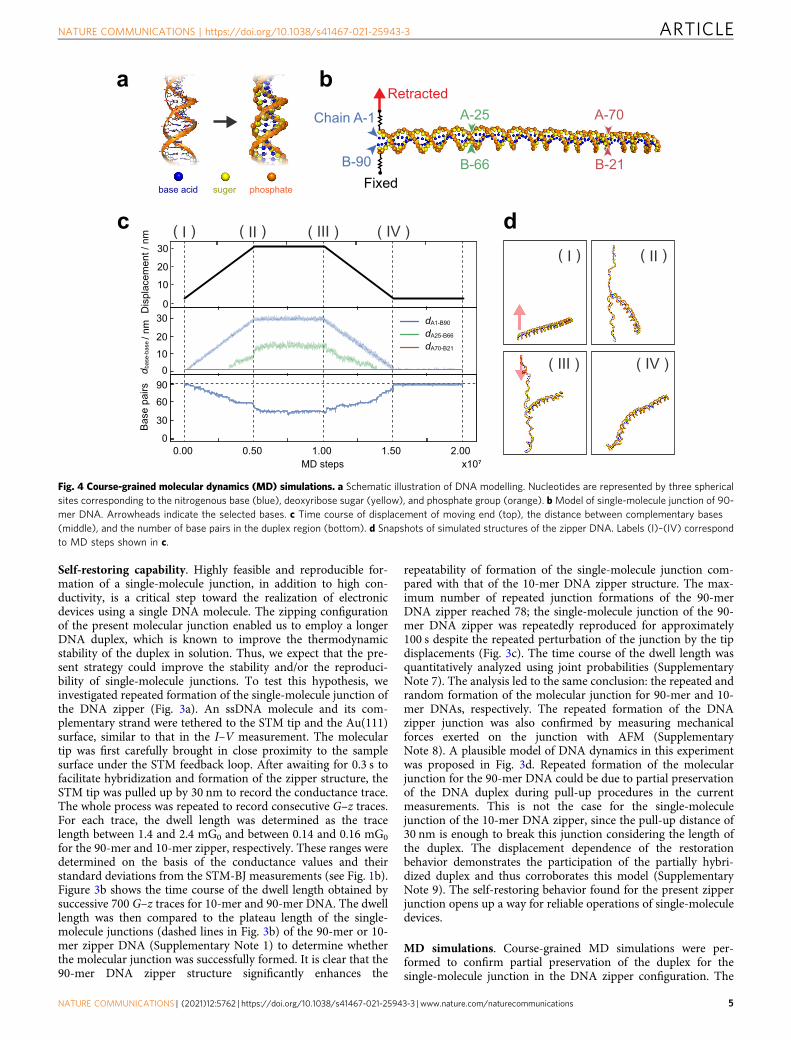
MD simulations. Course-grained MD simulations were per-formed to confirm partial preservation of the duplex for thesingle-molecule junction in the DNA zipper configuration. The
a b
c
Fixed
Retracted
Chain A-1
B-90
A-25
B-66
A-70
B-21base acid phosphatesuger
d( I ) ( II )
( IV )( III )
x10⁷0.00 0.50 1.00 1.50 2.00
MD steps
Base
pai
rsd b
ase-
base
/ nm
Dis
plac
emen
t / n
m
0
10
30
20
0
30
6090
1020
30
0
dA1-B90
dA25-B66
dA70-B21
( I ) ( II ) ( IV )( III )
Fig. 4 Course-grained molecular dynamics (MD) simulations. a Schematic illustration of DNA modelling. Nucleotides are represented by three sphericalsites corresponding to the nitrogenous base (blue), deoxyribose sugar (yellow), and phosphate group (orange). b Model of single-molecule junction of 90-mer DNA. Arrowheads indicate the selected bases. c Time course of displacement of moving end (top), the distance between complementary bases(middle), and the number of base pairs in the duplex region (bottom). d Snapshots of simulated structures of the zipper DNA. Labels (I)–(IV) correspondto MD steps shown in c.
NATURE COMMUNICATIONS | https://doi.org/10.1038/s41467-021-25943-3 ARTICLE
NATURE COMMUNICATIONS | (2021) 12:5762 | https://doi.org/10.1038/s41467-021-25943-3 | www.nature.com/naturecommunications 5
CafeMol software42 with 3SPN.2 C model43,44 was used for thesecalculations. It has been shown that this model successfullyreproduces melting phenomena, persistence lengths, and majorand minor grooves of dsDNA43–45. In the present work, one endof the 90-mer DNA duplex in the single-molecule junction waslinked to two springs to represent the STM experiments (Fig. 4a,b). Then, the terminal base in one strand of the duplex (Chain Ain Fig. 4b) was gradually pulled up by 30 nm in the simulation, asin the experiments (Fig. 3d). This base was then moved down tothe initial position after the waiting time, as shown in the toppanel of Fig. 4c. The distances between the selected com-plementary bases and the number of base pairs contained in theduplex region were plotted in the middle and bottom panels ofFig. 4c, respectively. Figure 4d shows selected snapshots of thesingle-molecule junction. Crucially, the DNA duplex is partiallypreserved after retraction (Fig. 4c, (II)), and the partial duplex,containing 44 base pairs, persisted after the waiting time (Fig. 4c,(III)). As the tethered end was pushed back to its original positionafter retraction, the unwound portion of DNA gradually rean-nealed to restore the complete duplex (Fig. 4c, (IV); see alsoSupplementary Movie 1). The distances between the com-plementary bases remained unchanged at the DNA end oppositeto the tethered base (Fig. 4c, middle), demonstrating again thepartial preservation of the DNA duplex. We carried out addi-tional simulations, where the tethered end was released after thepull-up of 30 nm. The reannealing also occurred in this case, asfound for the simulations above, and the complete duplexemerged (Supplementary Movie 2). These findings prove thatspontaneous base-pairing accounts for duplex restoration, andnot the external force exerted by the spring, which represents theSTM tip in the experiments.
The lifetime of conventional single-molecule junctions, such asthose of alkanedithiol, is typically short (in the order ofmilliseconds at room temperature), which poses fundamentalproblems for the realization of single-molecule electronics. Thesejunctions break at the interface between the constituent moleculeand the electrode, because the bonding between the functionalgroup of the molecule and the electrode, e.g., Au–S bond, isweakest in the junction46. It has been reported that the forcerequired to break the Au–S or Au–Au bond is 1–2 nN47, while theDNA unzipping takes place under the force of 10–50 pN22–24.Thus, in the present junctions, the breakdown is attributed to bevia DNA unzipping. The unzipping relaxes the tensile stress onthe junction. Therefore, the duplex of long DNA zippers can bepartially preserved, leading to prolonged preservation of thesingle-molecule connection with a DNA zipper. These resultsdemonstrate that the DNA zipper offers a means to achieve bothreproducible formation by the self-restoring capability and highconductivity.
In summary, we developed a DNA single-molecule junctionwhere the duplex orthogonally clamped the metal nanogap. Theelectrical measurements based on STM revealed that themolecular junction of the DNA zipper attains high conductance,originating from the delocalized π system of the DNA near theelectrodes. Moreover, we found an attractive self-restoringcapability that the single-molecule junction can be repeatedlyformed without breakdown of the whole structure even after anelectrical failure. This advantage arises from the partial unzip ofthe DNA within the junction, which is supported by the coarse-grained MD simulations. It is also worth noting that conductanceof the molecular junction exhibited no steep dependence on DNAlength, which is distinctly different from the conventional single-molecule junction of DNA. Therefore, we expect that furtherfunctionalization could be possible through the use of a longDNA sequence as a scaffold to form conjugates with a wide rangeof functional (bio)molecules.
MethodsSample preparation. An Au(111) surface was prepared by thermal evaporation ona mica surface. Au wires (99.999%, 0.25 mm diameter) were chemically etched with3.0 M NaCl solution in 1% perchloric acid (HClO4) to prepare the STM tips. Weused 90-mer DNA (Supplementary Table 1) modified with 1,3-propanethiol[–(CH2)3SH] linkers at the hydroxy group of the 3′ end as the probe. The com-plementary strand was modified with –(CH2)3SH linkers at the phosphate group ofthe 5′ end. As a control, we employed 10-mer DNA (Supplementary Table 1),which is partially the same as the 90-mer DNA, and –(CH2)3SH linkers wereintroduced at the 3′ end. The complement strand was modified with –(CH2)3SHlinkers at the 5′ end. All the DNAs, synthesized by solid-phase synthesis using thephosphoramidite method48, purified by high-performance liquid chromatography,and characterized by time-of-flight mass spectrometry (Supplementary Note 10),were purchased from Tsukuba Oligo Service (Ibaraki, Japan). For the preparationof the duplex, 1 μM solutions of ssDNAs were mixed with 10 mM phosphate-buffered saline (PBS) solution. This mixture was heated at 75 °C for 45 min. andslowly cooled to room temperature. The Au substrate was immersed in the solutionfor at least 2 h. For the experiments involving the molecular tip, STM tips wereimmersed in 1 μM ssDNA in PBS solution.
STM measurement. STM measurements were performed on an SPM 5100 system(Agilent Technologies, Santa Clara, CA, USA). The tunnelling current was sampledat 20,000 Hz. The bias voltage was 20 mV in all experiments, except for the I–Vmeasurement. In the analyses, conductance traces with a simple exponential decaywere removed based on an automated algorithm, in which the presence or absenceof a plateau was judged using the conductance histogram constructed for eachconductance trace49. Current measurements were repeated using independentlyprepared tips and sample surfaces. The reproducibility was confirmed by com-paring conductance histograms obtained using every independent sample surface.
Theoretical calculation. We conducted the course-grained MD simulation withCafeMol42 software. The 3SPN.2C model developed by the de Pablo group wasused for DNA44. All the simulations were performed at 300 K. The DNA sequenceas employed in the experiments was used for the simulation. For simulating theexperiment in ambient conditions, no electrostatic interactions were taken intoconsideration. One terminus in each DNA strand was connected with springs formodelling the bridge between thiol linkers of DNA and Au atoms. The stiffness ofthe spring was 8.5 N/m, which is equal to the strength of the Au–Au bond47,50.
Reporting Summary. Further information on research design is available in the NatureResearch Reporting Summary linked to this article.
Data availabilityThe data that support the findings of this study are available from the correspondingauthor upon reasonable request. Source data for Figs. 1–4 and Supplementary Fig. 1–12are archived at https://doi.org/10.5281/zenodo.5515109. Source data are provided withthis paper.
Received: 25 December 2019; Accepted: 7 September 2021;
References1. Kelley, S. O. & Barton, J. K. Electron transfer between bases in double helical
DNA. Science 283, 375–381 (1999).2. Risser, S. M., Beratan, D. N. & Meade, T. J. Electron-transfer in DNA -
predictions of exponential-growth and decay of coupling with donor-acceptordistance. J. Am. Chem. Soc. 115, 2508–2510 (1993).
3. Tsutsui, M. et al. Electrical detection of single methylcytosines in a DNAoligomer. J. Am. Chem. Soc. 133, 9124–9128 (2011).
4. Xiang, L. M. et al. Intermediate tunnelling-hopping regime in DNA chargetransport. Nat. Chem. 7, 221–226 (2015).
5. Xu, B. Q., Zhang, P. M., Li, X. L. & Tao, N. J. Direct conductancemeasurement of single DNA molecules in aqueous solution. Nano Lett. 4,1105–1108 (2004).
6. Artes, J. M., Li, Y. H., Qi, J. Q., Anantram, M. P. & Hihath, J. Conformationalgating of DNA conductance. Nat. Commun. 6, 8870 (2015).
7. Bui, P. T., Nishino, T., Shiigi, H. & Nagaoka, T. One-by-one single-moleculedetection of mutated nucleobases by monitoring tunneling current using aDNA tip. Chem. Commun. 51, 1666–1669 (2015).
8. Lewis, F. D. et al. Direct measurement of hole transport dynamics in DNA.Nature 406, 51–53 (2000).
9. Wierzbinski, E. et al. The single-molecule conductance and electrochemicalelectron-transfer rate are related by a power law. Acs Nano 7, 5391–5401(2013).
ARTICLE NATURE COMMUNICATIONS | https://doi.org/10.1038/s41467-021-25943-3
6 NATURE COMMUNICATIONS | (2021) 12:5762 | https://doi.org/10.1038/s41467-021-25943-3 | www.nature.com/naturecommunications
10. Guo, C. L. et al. Molecular rectifier composed of DNA with high rectificationratio enabled by intercalation. Nat. Chem. 8, 484–490 (2016).
11. Harashima, T., Kojima, C., Fujii, S., Kiguchi, M. & Nishino, T. Single-moleculeconductance of DNA gated and ungated by DNA-binding molecules. Chem.Commun. 53, 10378–10381 (2017).
12. Xiang, L. M. et al. Gate-controlled conductance switching in DNA. Nat.Commun. 8, 14471 (2017).
13. Sha, R. et al. Charge splitters and charge transport junctions based on guaninequadruplexes. Nat. Nanotechnol. 13, 316–321 (2018).
14. Azad, Z., Roushan, M. & Riehn, R. DNA brushing shoulders: targeted loopingand scanning of large DNA strands. Nano Lett. 15, 5641–5646 (2015).
15. Jadhav, V., Hoogerheide, D. P., Korlach, J. & Wanunu, M. Porous zero-modewaveguides for picogram-level DNA capture. Nano Lett. 19, 921–929 (2019).
16. Riehn, R., Austin, R. H. & Sturm, J. C. A nanofluidic railroad switch for DNA.Nano Lett. 6, 1973–1976 (2006).
17. Terakawa, T. et al. The condensin complex is a mechanochemical motor thattranslocates along DNA. Science 358, 672–676 (2017).
18. Raillon, C. et al. Nanopore detection of single molecule RNAP-DNAtranscription complex. Nano Lett. 12, 1157–1164 (2012).
19. Raillon, C., Granjon, P., Graf, M., Steinbock, L. J. & Radenovic, A. Fast andautomatic processing of multi-level events in nanopore translocationexperiments. Nanoscale 4, 4916–4924 (2012).
20. Heerema, S. J. & Dekker, C. Graphene nanodevices for DNA sequencing. Nat.Nanotechnol. 11, 127–136 (2016).
21. Xie, P., Xiong, Q. H., Fang, Y., Qing, Q. & Lieber, C. M. Local electricalpotential detection of DNA by nanowire-nanopore sensors. Nat. Nanotechnol.7, 119–125 (2012).
22. Krautbauer, R., Rief, M. & Gaub, H. E. Unzipping DNA oligomers. Nano Lett.3, 493–496 (2003).
23. Strunz, T., Oroszlan, K., Schafer, R. & Guntherodt, H. J. Dynamic forcespectroscopy of single DNA molecules. Proc. Natl Acad. Sci. USA 96,11277–11282 (1999).
24. Walder, R. et al. High-precision single-molecule characterization of thefolding of an HIV RNA hairpin by atomic force microscopy. Nano Lett. 18,6318–6325 (2018).
25. Cocco, S., Monasson, R. & Marko, J. F. Force and kinetic barriers to unzippingof the DNA double helix. Proc. Natl Acad. Sci. USA 98, 8608–8613 (2001).
26. Kiguchi, M. & Kaneko, S. Single molecule bridging between metal electrodes.Phys. Chem. Chem. Phys. 15, 2253–2267 (2013).
27. Afsari, S. et al. Quantum point contact single-nucleotide conductance forDNA and RNA sequence identification. Acs Nano 11, 11169–11181 (2017).
28. Huang, S. et al. Recognition tunneling measurement of the conductance ofDNA bases embedded in self-assembled monolayers. J. Phys. Chem. C. 114,20443–20448 (2010).
29. Huang, S. et al. Identifying single bases in a DNA oligomer with electrontunnelling. Nat. Nanotechnol. 5, 868–873 (2010).
30. Ohshiro, T., Tsutsui, M., Yokota, K. & Taniguchi, M. Quantitative analysis ofDNA with single-molecule sequencing. Sci. Rep. 8, 8517 (2018).
31. Chang, S. et al. Tunnelling readout of hydrogen-bonding-based recognition.Nat. Nanotechnol. 4, 297–301 (2009).
32. Huang, S. et al. Recognition tunneling measurement of the conductance ofDNA bases embedded in self-assembled monolayers. J. Phys. Chem. C. 114,20443–20448 (2010).
33. Xu, Zhang & Li, Tao Direct conductance measurement of single DNAmolecules in aqueous solution. Nano Lett. 4, 1105–1108 (2004).
34. Nishino, T. & Bui, P. T. Direct electrical single-molecule detection of DNAthrough electron transfer induced by hybridization. Chem. Commun. 49,3437–3439 (2013).
35. Kimura-Suda, H., Petrovykh, D. Y., Tarlov, M. J. & Whitman, L. J. Base-dependent competitive adsorption of single-stranded DNA on gold. J. Am.Chem. Soc. 125, 9014–9015 (2003).
36. Cohen, H., Nogues, C., Naaman, R. & Porath, D. Direct measurement ofelectrical transport through single DNA molecules of complex sequence. Proc.Natl Acad. Sci. USA 102, 11589–11593 (2005).
37. Beebe, J. M., Kim, B., Gadzuk, J. W., Frisbie, C. D. & Kushmerick, J. G.Transition from direct tunneling to field emission in metal-molecule-metaljunctions. Phys. Rev. Lett. 97, 026801 (2006).
38. Guo, S., Hihath, J., Díez-Pérez, I. & Tao, N. Measurement and statisticalanalysis of single-molecule current–voltage characteristics, transition voltagespectroscopy, and tunneling barrier height. J. Am. Chem. Soc. 133,19189–19197 (2011).
39. Huisman, E. H., Gued́on, C. M., van Wees, B. J. & van der Molen, S. J.Interpretation of transition voltage spectroscopy. Nano Lett. 9, 3909–3913(2009).
40. Kubar, T. & Elstner, M. A hybrid approach to simulation of electron transferin complex molecular systems. J. R. Soc. Interface 10, 415–432 (2013).
41. Kashimoto, Y. et al. The evolution of intermolecular energy bands of occupiedand unoccupied molecular states in organic thin films. J. Phys. Chem. C. 122,12090–12097 (2018).
42. Kenzaki, H. et al. CafeMol: a coarse-grained biomolecular simulator forsimulating proteins at work. J. Chem. Theory Comput. 7, 1979–1989 (2011).
43. Freeman, G. S., Lequieu, J. P., Hinckley, D. M., Whitmer, J. K. & de Pablo, J. J.DNA shape dominates Sequence affinity in nucleosome formation. Phys. Rev.Lett. 113, 168101 (2014).
44. Hinckley, D. M., Lequieu, J. P. & de Pablo, J. J. Coarse-grained modeling ofDNA oligomer hybridization: Length, sequence, and salt effects. J. Chem. Phys.141, 035102 (2014).
45. Terakawa, T. & Takada, S. p53 dynamics upon response element recognitionexplored by molecular simulations. Sci. Rep. 5, 17107 (2015).
46. Xu, B. Q., Xiao, X. Y. & Tao, N. J. Measurements of single-moleculeelectromechanical properties. J. Am. Chem. Soc. 125, 16164–16165 (2003).
47. Rubio-Bollinger, G., Bahn, S. R., Agrait, N., Jacobsen, K. W. & Vieira, S.Mechanical properties and formation mechanisms of a wire of single goldatoms. Phys. Rev. Lett. 87, 026101 (2001).
48. Beaucage, S. L. & Iyer, R. P. The functionalization of oligonucleotides viaphosphoramidite derivatives. Tetrahedron 49, 1925–1963 (1993).
49. Inkpen, M. S. et al. New insights into single-molecule junctions using a robust,unsupervised approach to data collection and analysis. J. Am. Chem. Soc. 137,9971–9981 (2015).
50. Aradhya, S. V., Frei, M., Hybertsen, M. S. & Venkataraman, L. Van der Waalsinteractions at metal/organic interfaces at the single-molecule level. Nat.Mater. 11, 872–876 (2012).
AcknowledgementsThis work was financially supported by Grants-in-Aid for Scientific Research (No.JP16K14018 and JP18H02003), JST CREST (No. JP-MJCR18I4), JSPS Fellows (No.19J20605) from the Ministry of Education, Culture, Sports, Science and Technology(MEXT) of Japan, Iketani Science and Technology Foundation, and Murata ScienceFoundation.
Author contributionsT.H., S.K., S.F., M.K., and T.N. conceived and designed the experiments. T.H., S.F., andY.J. performed single-molecule measurements. T.H., T.T., and N.K. contributed to the-oretical calculations. All the authors contributed to the analysis and interpretation ofresults and wrote the paper.
Competing interestsThe authors declare no competing interests.
Additional informationSupplementary information The online version contains supplementary materialavailable at https://doi.org/10.1038/s41467-021-25943-3.
Correspondence and requests for materials should be addressed to Tomoaki Nishino.
Peer review information Nature Communications thanks the anonymous reviewer(s) fortheir contribution to the peer review of this work. Peer reviewer reports are available.
Reprints and permission information is available at http://www.nature.com/reprints
Publisher’s note Springer Nature remains neutral with regard to jurisdictional claims inpublished maps and institutional affiliations.
Open Access This article is licensed under a Creative CommonsAttribution 4.0 International License, which permits use, sharing,
adaptation, distribution and reproduction in any medium or format, as long as you giveappropriate credit to the original author(s) and the source, provide a link to the CreativeCommons license, and indicate if changes were made. The images or other third partymaterial in this article are included in the article’s Creative Commons license, unlessindicated otherwise in a credit line to the material. If material is not included in thearticle’s Creative Commons license and your intended use is not permitted by statutoryregulation or exceeds the permitted use, you will need to obtain permission directly fromthe copyright holder. To view a copy of this license, visit http://creativecommons.org/licenses/by/4.0/.
© The Author(s) 2021
NATURE COMMUNICATIONS | https://doi.org/10.1038/s41467-021-25943-3 ARTICLE
NATURE COMMUNICATIONS | (2021) 12:5762 | https://doi.org/10.1038/s41467-021-25943-3 | www.nature.com/naturecommunications 7