signal processing by its coil zipper domain activates ikkgamma
TRANSCRIPT
Signal processing by its coil zipper domainactivates IKK�Stuart Bloor*, Grigory Ryzhakov*, Sebastian Wagner†, P. Jonathan G. Butler‡, David L. Smith*, Rebekka Krumbach*,Ivan Dikic†, and Felix Randow*§
*Division of Protein and Nucleic Acid Chemistry, ‡Structural Studies Division, Medical Research Council Laboratory of Molecular Biology, Hills Road,Cambridge CB2 2QH, United Kingdom; and †Goethe University Medical School, Institute for Biochemistry II, Theodor-Stern-Kai 7, 60590 Frankfurt,Germany
Edited by Douglas T. Fearon, University of Cambridge, Cambridge, United Kingdom, and approved December 10, 2007 (received for review July 12, 2007)
NF-�B activation occurs upon degradation of its inhibitor I-�B andrequires prior phosphorylation of the inhibitor by I-�B kinase (IKK).Activity of IKK is governed by its noncatalytic subunit IKK�.Signaling defects due to missense mutations in IKK� have beencorrelated to its inability to either become ubiquitylated or bindubiquitin noncovalently. Because the relative contribution of theseevents to signaling had remained unknown, we have studiedmutations in the coil-zipper (CoZi) domain of IKK� that eitherimpair signaling or cause constitutive NF-�B activity. Certainsignaling-deficient alleles neither bound ubiquitin nor were theyubiquitylated by TRAF6. Introducing an activating mutation intothose signaling-impaired alleles restored their ubiquitylation andcreated mutants constitutively activating NF-�B without repairingthe ubiquitin-binding defect. Constitutive activity therefore arisesdownstream of ubiquitin binding but upstream of ubiquitylation.Such constitutive activity reveals a signal-processing function forIKK� beyond that of a mere ubiquitin-binding adaptor. We proposethat this signal processing may involve homophilic CoZi interac-tions as suggested by the enhanced affinity of CoZi domains fromconstitutively active IKK�.
NF-�B � signaling � ubiquitin � Nemo
The transcription factor NF-�B plays an essential role incoordinating inflammation and immunity by controlling the
expression of proinflammatory and antiapoptotic genes (1, 2). Inresting cells, proteins of the I-�B family are bound to NF-�B tolimit its nuclear accumulation and transactivation potential.Agonists rapidly induce NF-�B activity by triggering the ubiq-uitylation and the degradation of I-�B proteins (3). The ubiq-uitylation of I-�Bs is tightly controlled and requires their priorphosphorylation by the I-�B kinase (IKK) complex (4–6). TheIKK complex contains two kinases, IKK� and IKK� (also calledCHUK/IKK1 and IKK2) (7–12). IKK��/� mice fail to degradeI-�B� (13–15), whereas mice deficient in IKK� are born withonly minor defects in I-�B�-controlled NF-�B activity (16–18).For historical reasons, the IKK�-controlled pathway has beentermed the ‘‘canonical pathway’’ (19). Signaling in this pathwayinduces catalytic activity of IKK� via phosphorylation of itsactivation loop (20, 21). Recent genetic evidence identifiedTAK1 as the IKK acting in the canonical pathway (22, 23). TAK1has been shown to become activated in the presence of lysine63-linked Ub conjugates, the formation of which required the Ubligase TRAF6 (24–26).
Besides IKK� and IKK�, the IKK complex also contains IKK�(also called NEMO, IKKAP, and FIP3) (27–30). IKK�-deficientcells cannot activate IKK�, which incapacitates the canonicalpathway (31–33). Hence, IKK� has been suggested to be theregulatory subunit of the IKK complex, but how exactly it performsthis function has remained unclear. A Ub-dependent control ofIKK� seems likely given the well established role of Ub conjugatesfor signaling upstream of IKK, and the enhanced IKK activity in theabsence of specific Ub hydrolases (34–37). Supporting this Ub-control hypothesis, exposure to NF-�B agonists causes ubiquityla-
tion of IKK�, and certain signaling-deficient point mutants of IKK�fail to become ubiquitylated (38–42). A different Ub-relatedcontrol mechanism has been proposed recently when it was dis-covered that IKK� mutants failing to signal in response to TNF�were unable to bind ubiquitylated RIP, a protein essential for TNF�signaling (43–45). Ub binding by IKK� was suggested to allow therecruitment of IKK�/� to specific complexes, where kinase activitymight be induced. We currently do not know the relationship, if any,between these two proposed Ub-dependent control mechanisms.
Using somatic cell genetics, we have isolated a series of IKK�alleles. We found that mutations in the coil-zipper (CoZi)domain of IKK� can cause signaling defects or constitutiveNF-�B activity. A constitutively activating IKK� allele unable tobind Ub suggests a role for IKK� beyond that of a Ub adaptormerely recruiting the IKK complex. Therefore, we propose thatduring signaling IKK� adopts an activated state. Genetic evi-dence indicates that this activated state occurs downstream ofUb binding and licences IKK� for ubiquitylation, which dem-onstrates a mechanistic link between IKK�’s role in sensingupstream and regulating downstream pathway activity.
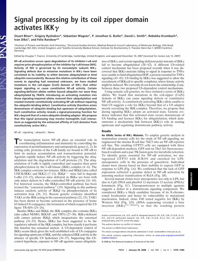
ResultsAn Allelic Series of IKK� Mutants. To employ genetic analysis inmammalian somatic cells for the study of NF-�B signaling, weengineered the murine B cell line 70Z/3 into an NF-�B reportercell line. The resulting GTPT3 cells are equipped with threeNF-�B-dependent markers: GFP and rat Thy1 for fluorescence-based readouts and a pac:TK fusion gene for metabolic selection.To identify genes essential for NF-�B activation, we mu-tagenized GTPT3 with ICR191 and enriched for LPS-unresponsive cells in the presence of ganciclovir. Individualclones were chosen based on their inability to express GFP inresponse to LPS (Fig. 1A). We confirmed that the lack of GFPexpression reflected a genuine defect in NF-�B activation byassessing nuclear translocation of RelA (Fig. 1B).
Several mutant clones were unresponsive not only to LPS, butalso to CpG DNA and phorbol 12-myristate 13-acetate (PMA)/Ionomycin (Fig. 1C). Unresponsiveness to multiple agonistssuggests a defect in a downstream signaling component. Weconsidered IKK� a likely candidate because of its X-chromo-somal localization and hence its vulnerability to mutationalinactivation. Indeed, clone F40 tested negative for IKK� byWestern blot (Fig. 1D). cDNA sequencing revealed a baseinsertion (IKK�137�138insG) so that the resulting protein
Author contributions: S.B., G.R., and F.R. designed research; S.B., G.R., S.W., P.J.G.B., D.L.S.,R.K., and F.R. performed research; S.B., G.R., S.W., P.J.G.B., D.L.S., R.K., I.D., and F.R.analyzed data; and F.R. wrote the paper.
The authors declare no conflict of interest.
This article is a PNAS Direct Submission.
§To whom correspondence should be addressed. E-mail: [email protected].
This article contains supporting information online at www.pnas.org/cgi/content/full/0706552105/DC1.
© 2008 by The National Academy of Sciences of the USA
www.pnas.org�cgi�doi�10.1073�pnas.0706552105 PNAS � January 29, 2008 � vol. 105 � no. 4 � 1279–1284
IMM
UN
OLO
GY
(IKK�T47fs), even if expressed and stable, would only contain 46amino acids (Fig. 1E). In contrast, clones F29 and J77 expressedwild-type-sized IKK� (Fig. 1D). Sequencing of their cDNArevealed point mutations in IKK� replacing glutamate 308 withvaline (IKK�E308V) in F29 and arginine 331 with proline(IKK�R331P) in J77 (Fig. 1E). Notably, the two mutationsoccurred in close proximity to each other and to a furthermutation exchanging aspartate with asparagine at position 311 inhuman IKK� (corresponding to murine IKK�D304N), seen in apatient suffering from anhidrotic ectodermal dysplasia withimmunodeficiency (EDA-ID) (46). The R331P mutation islocated within a leucine zipper (LZ) where the presence of ahelix-breaking proline may not be tolerated (Fig. 1E) (47). ThisLZ has been reported to bind the adjacent CC2 region (48),suggesting that residues D304 and E308 are part of a loopconnecting the two helical structures. We conclude that theregion spanning CC2 and LZ forms a functionally importantdomain in IKK�. We will refer to it as the CoZi domain.
To test whether the mutant IKK� alleles caused the observeddefect in NF-�B activation, we used IKK�-deficient F40 cells.Transduction with IKK�WT restored reporter induction (Fig.1F), which demonstrates that the absence of IKK� protein is theonly defect relevant to NF-�B signaling in F40. Transduction ofF40 with IKK�D304N, IKK�E308V, or IKK�R331P did not restoreNF-�B activation in response to LPS, thereby proving the allelesto be defective.
IKK� needs to assemble with IKK� to serve its signalingfunction. We wondered whether the assembly of this complexwas disturbed in the mutant clones. Immunoprecipitation ofIKK� resulted in equal amounts of IKK� and IKK� in precip-itates from wild-type GTPT3 and mutant F29 or J77 cells[supporting information (SI) Fig. 7A]. In contrast, neither IKK�
nor IKK� were precipitated from F40 cells. We also tested theassociation of IKK subunits into higher order complexes by gelfiltration (SI Fig. 7B). In lysates of wild-type GTPT3 cells, IKK�coeluted with IKK� in high-molecular-weight complexes,whereas in lysates of IKK�-deficient F40 cells, IKK� occurred inlater fractions. Importantly, complexes from wild-type cells andmutant clones F29 and J77 behaved indistinguishably. Theseresults indicate that IKK�E308V and IKK�R331P were incorpo-rated normally into IKK complexes. Therefore, we tested theirpotential as dominant-negative inhibitors of NF-�B signaling (SIFig. 7C). Similarly to dominant-negative I-�B�, the transductionof GTPT3 cells with the mutant IKK� alleles prevented LPS-induced NF-�B reporter expression. In contrast, transductionwith IKK�WT did not impair NF-�B activation. We conclude thatmutations in the CoZi domain of IKK� specifically interfere withits signaling function.
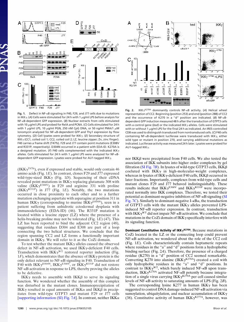
Dominant Constitutive Activity of IKK�K270A. Because mutations inCoZi located in the LZ or the connecting loop could preventNF-�B activation, we wondered about the role of the CC2 coil(Fig. 1E). Coils characteristically contain heptameric repeatswhere residues in the ‘‘a’’ and ‘‘d’’ positions form a hydrophobicbinding surface (Fig. 2A). Therefore, the presence of a chargedresidue (K270) in a ‘‘d’’ position of CC2 seemed remarkable.Converting K270 into alanine (IKK�K270A) created a coil withonly hydrophobic residues in the ‘‘a’’ and ‘‘d’’ positions. Incontrast to IKK�WT, which barely induced NF-�B upon trans-duction, IKK�K270A activated NF-�B potently because integra-tion of a single virus carrying IKK�K270A per cell caused similarlevels of NF-�B activity to saturating amounts of LPS (Fig. 2B).
The corresponding lysine K277 in human IKK� has beensuggested to control DNA damage-induced NF-�B activation viasumoylation, ubiquitylation, and nuclear accumulation of IKK�(38). Constitutive activity of human IKK�K277A, however, was
Fig. 1. Defect in NF-�B signaling in F40, F29, and J77 cells due to mutationsin IKK�. (A) Cells were stimulated for 24 h with 1 �g/ml LPS before analysis forNF-�B-dependent GFP expression. (B) Nuclear extracts from cells stimulatedwith 10 �g/ml LPS and probed for RelA and PCNA. (C) Cells stimulated for 24 hwith 1 �g/ml LPS, 10 �g/ml PGN, 250 nM CpG DNA, or 50 ng/ml PMA/1 �MIonomycin analyzed for NF-�B-dependent GFP and Thy1 expression by flowcytometry. (D) Cell lysates were probed for IKK�. (E) Secondary structure ofIKK� (CC1, coiled coil 1; CC2, coiled coil 2; LZ, leucine zipper; Zn, zinc finger).F40 carries a frame shift (T47fs). F29 and J77 contain point mutations (E308Vand R331P, respectively). D304N occurred in a patient with EDA-ID. K270A isa designed mutation. (F) F40 cells complemented with the indicated IKK�
alleles. Cells stimulated for 24 h with 1 �g/ml LPS were analyzed for NF-�B-dependent GFP expression. Lysates were probed for AU1-tagged IKK�.
Fig. 2. IKK�K270A dominantly controls NF-�B activity. (A) Helical wheelrepresentation of CC2. Beginning (position 253) and end (position 288) of CC2and the occurrence of K270 in a ‘‘d’’ position are indicated. (B) NF-�B-dependent GFP induction measured 48 h after the transduction of GTPT3 cellswith a control gene (bsd) or the indicated IKK� alleles. Cells were stimulatedwith or without 1 �g/ml LPS for the final 24 h as indicated. An IRES-controlledCD8 was used to distinguish transduced from nontransduced cells. (C) F40 cellscontaining NF-�B-dependent luciferase were transduced with IKK�, eitherwild type or mutant in position 270, and carrying additional mutations asindicated. Luciferase activity was measured 24 h later. Lysates were probed forAU1-tagged IKK�.
1280 � www.pnas.org�cgi�doi�10.1073�pnas.0706552105 Bloor et al.
not reported. We found that this difference between human andmurine IKK� is caused by the presence of seven additional aminoacids at the beginning of CC2 (V249–K255) in the humanprotein because human IKK�(�V249-K255)�K277A also activatedNF-�B strongly (SI Fig. 8A). We further observed that the con-stitutive activity of IKK�K270A is caused specifically by thepresence of alanine, rather than the absence of lysine, becauseIKK�K270R and IKK�K270Q were not constitutively activating, butsupported NF-�B activation by LPS (SI Fig. 8B). The phenotypeof IKK�K270A is therefore not related to the modification of K270with Ub or related proteins.
IKK�K270A is the only known constitutively active IKK�allele, and investigating its modus operandi may advance ourunderstanding of physiological IKK� activation. Deleting thebinding site for IKK� and IKK� (IKK�K270A�N102) preventedNF-�B activation (Fig. 2C; see also Fig. 1E). In contrast, theC-terminal Zn finger and the adjacent proline-rich region(IKK�K270A�C389 and IKK�K270A�C338, respectively) were notrequired for its activity. Deleting the LZ (IKK�K270A�C314),however, extinguished NF-�B activation. This finding indicatesthat the constitutive activity of IKK�K270A depends on theintegrity of its CoZi domain. We therefore investigated muta-tions in CoZi residues essential for LPS signaling. Importantly,double mutants (IKK�K270A�D304N, IKK�K270A�E308V, andIKK�K270A�R331P) constitutively activated NF-�B (Fig. 2C).Therefore, the constitutive activity of IKK�K270A appeared dom-inant over mutations blocking the signaling from LPS.
Constitutive Activity of IKK�K270A Occurs Independently of Ub Binding.Ub in the form of ubiquitylated RIP is a binding partner forIKK� (43, 44). Lack of Ub binding by human IKK�D311N
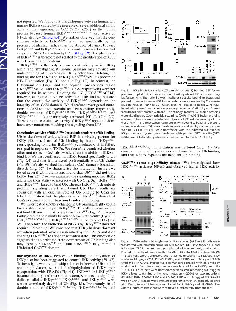
(corresponding to murine IKK�D304N) correlates with its failureto signal in response to TNF�. We therefore wondered whetherother mutations in CoZi also would affect the ability of IKK� tobind Ub. We first confirmed that IKK� bound specifically to Ub(Fig. 3A) and that it interacted preferentially with Ub chains(Fig. 3B). We also verified that isolated CoZi domains bound Ubdirectly (Fig. 3C). To characterize this interaction further, wetested several Ub mutants and found that UbI44A did not bindIKK� (Fig. 3D). Next we examined the signaling-impaired IKK�alleles for their ability to interact with Ub (Fig. 3E). IKK�D304N
and IKK�E308V failed to bind Ub, whereas IKK�R331P, despite itsprofound signaling defect, still bound Ub. These results areconsistent with an essential role of Ub binding to CoZi forNF-�B activation, but the phenotype of IKK�R331P shows thatCoZi performs another function besides Ub binding.
We investigated whether changes in Ub binding might explainthe constitutive activity of IKK�K270A. This allele, however, didnot bind Ub any more strongly than IKK�WT (Fig. 3E). Impor-tantly, despite their ability to induce NF-�B efficiently (Fig. 2C),IKK�K270A�D304N and IKK�K270A�E308V failed to bind Ub (Fig.3E). Therefore, the induction of NF-�B by IKK�K270A does notrequire Ub binding. We conclude that IKK� harbors dormantactivation potential, which is unleashed by the K270A mutationenabling IKK�K270A to adopt an activated state. This observationsuggests that an activated state downstream of Ub binding alsomay exist for IKK�WT and that CoZiK270A may mimic aUb-bound CoZiWT domain.
Ubiquitylation of IKK�. Besides Ub binding, ubiquitylation ofIKK� also has been suggested to control IKK activity (39–42).To investigate what relationship might exist between Ub bindingand ubiquitylation, we studied ubiquitylation of IKK� uponcoexpression with TRAF6 (Fig. 4A). IKK�WT and IKK�K270A
became ubiquitylated to a similar extent, whereas the signaling-deficient alleles IKK�D304N, IKK�E308V, and IKK�R331P werealmost completely devoid of Ub (Fig. 4B). Importantly, in alldouble mutants (IKK�D304N�K270A, IKK�E308V�K270A, and
IKK�R331P�K270A), ubiquitylation was restored (Fig. 4C). Weconclude that ubiquitylation occurs downstream of Ub bindingand that K270A bypasses the need for Ub binding.
CoZiK270A Forms High-Affinity Dimers. We investigated howIKK�K270A activates NF-�B and observed higher IKK activity
Fig. 3. IKK� binds Ub via its CoZi domain. (A and B) Purified GST fusionproteins coupled to beads were incubated with lysates of 293 cells expressingluciferase IKK�. The ratio between luciferase activity bound to beads andpresent in lysates is shown. GST fusion proteins were visualized by Coomassieblue staining. (C) Purified GST fusion proteins coupled to beads were incu-bated with lysate from bacteria expressing His-tagged CoZi. (Upper) Eluatesfrom beads were blotted with anti-His antibody. (Lower) GST fusion proteinswere visualized by Coomassie blue staining. (D) Purified GST fusion proteinscoupled to beads were incubated with lysates of 293 cells expressing a lucif-erase IKK�. The ratio between luciferase activity bound to beads and presentin lysates is shown. GST fusion proteins were visualized by Coomassie bluestaining. (E) The 293 cells were transfected with the indicated AU1-taggedIKK� constructs. Lysates were incubated with purified GST-tetra-Ub (GST-4xUb) bound to beads. Lysates and eluates were blotted for AU1-IKK�.
Fig. 4. Differential ubiquitylation of IKK� alleles. (A) The 293 cells weretransfected with plasmids encoding AU1-tagged IKK�, myc-tagged Ub, andHA-tagged TRAFs. Lysates were precipitated with an antibody against AU1.Precipitates and lysates were blotted for AU1-IKK�, HA-TRAFs, and myc-Ub. (B)The 293 cells were transfected with plasmids encoding AU1-tagged IKK�
alleles (wild type, K270A, D304N, E308V, and R331P) and HA-tagged TRAF6(wild type or C70A). Lysates were immunoprecipitated with an antibodyagainst AU1. Precipitates and lysates were blotted for AU1-IKK� and HA-TRAFs. (C) The 293 cells were transfected with plasmids encoding AU1-taggedIKK� alleles containing either one mutation (K270A) or two mutations(K270A/D304N, K270A/E308V, and K270A/R331P) and HA-tagged TRAF6 (wildtype or C70A). Lysates were immunoprecipitated with an antibody againstAU1. Precipitates and lysates were blotted for AU1-IKK� and HA-TRAFs. Theasterisk indicates lanes that were removed electronically from the blot.
Bloor et al. PNAS � January 29, 2008 � vol. 105 � no. 4 � 1281
IMM
UN
OLO
GY

associated with IKK�K270A than with IKK�WT (Fig. 5A). Theactivation of NF-�B by IKK�K270A required the presence of IKK�but not RIP (SI Fig. 9) and was inhibited by dominant-negativealleles of IKK� and I�B� (Fig. 5B). We conclude that cellsexpressing IKK�K270A harbor constitutively active IKK complexes.
Seeking the cause of IKK activity, we wondered whetherreplacing the positively charged lysine at the predicted bindingsurface of CC2 with a hydrophobic alanine had changed thebinding propensities of CoZi. To test this hypothesis, we inves-tigated the mass-action-driven oligomerization of CoZi by ana-lytical ultracentrifugation. Sedimentation equilibrium runsshowed fully reversible dimerization of CoZiWT (Fig. 5C). Thepresence of dimers and the absence of higher order oligomerswere confirmed by sedimentation velocity runs (SI Fig. 10).CoZiK270A also dimerized (Fig. 5C). However, in contrast toCoZiWT, which gave a Kd of 30 � 5 �M, CoZiK270A resisteddissociation even at 2 �M, the lowest concentration measurable.The activity of IKK�K270A may therefore result from the in-creased homophilic interactions of its CoZiK270A domain.
We next tested whether in vivo CoZiK270A and CoZiWT dif-fered in their ability to undergo homophilic interactions. Muchstronger binding occurred between CoZiK270A and IKK�K270A
than between the respective wild-type molecules (Fig. 5D).
Robust binding required both partners to contain the K270Amutation. Full-length IKK�WT oligomerized potently, and,therefore, no further increase was observed for IKK�K270A. Noneof the above interactions required the recruitment of IKK� andIKK� because removal of their binding site in IKK��N102 wasinconsequential (data not shown). We conclude that the in-creased affinity of CoZiK270A causes homophilic binding in vivo.
Such binding may have two consequences. If it occurredbetween CoZi domains of separate IKK complexes, the com-plexes would associate into larger clusters. Alternatively, becauseeach IKK complex already contains multiple IKK� subunits,binding could enforce conformational changes within a pre-formed complex. To distinguish these scenarios, we performedgel filtration and found that IKK complexes containing IKK�WT
or IKK�K270A were the same size (Fig. 5E). This result isconsistent with the occurrence of a conformational changewithin a preformed IKK complex induced by homophilicCoZiK270A interactions.
DiscussionWe have used somatic cell genetics to isolate a series of IKK�alleles. Here, we show that the CoZi domain controls signal f lowthrough IKK� and that mutations in this domain can cause eitherloss of signaling or constitutive NF-�B activity. We demonstratethat mutant IKK� can adopt an activated state based on aconstitutively activating allele that maintains its activity evenwhen unable to bind Ub. This finding suggests a function forIKK� beyond binding Ub and recruiting the IKK complex toupstream signaling components. We propose that during signal-ing IKK�WT also adopts an activated state that occurs upstreamof its ubiquitylation and could therefore link Ub binding toubiquitylation of IKK� and activation of NF-�B.
IKK� is essential for NF-�B activation in the canonical pathway(31–33). It has been suggested to control IKK activity in a Ub-dependent manner. Two distinct Ub-related signaling events occurat IKK�: noncovalent Ub binding and covalent ubiquitylation.Missense mutations that impair the signaling function of IKK� havebeen demonstrated either to disturb Ub binding (43, 44) or toprevent ubiquitylation of IKK� (38–42). Our identification ofmutations in CoZi, which either prevent NF-�B signaling orconstitutively activate it, indicate a crucial, possibly switch-likefunction for CoZi in regulating IKK� (Fig. 6A). In the followingsection, we attempt to gain further insight into this function of CoZiby analyzing the ability of single and double mutants to signal, tobind Ub, and to become ubiquitylated.
Binding of IKK� to Ub (in the form of ubiquitylated RIP) occursin TNF� signaling (43, 44). We extended this result by demonstrat-ing that CoZi, like most Ub-binding domains (49, 50), requires I44in Ub for binding. We also confirmed that IKK�D304N cannot bindUb, and we demonstrated a similar defect for IKK�E308V. Thesedata are consistent with LPS and CpG DNA requiring Ub bindingby IKK� to activate NF-�B. The constitutive activity of IKK�K270A,however, is not due to increased Ub binding. Notably, the intro-duction of K270A into alleles unable to bind Ub (IKK�K270A�D304N
and IKK�K270A�E308V) constitutively activated NF-�B, but did notrestore Ub binding. K270A therefore bypasses the need for Ubbinding, suggesting that it affects CoZi downstream of Ub binding.K270A also acts downstream of R331P because the transduction ofIKK�K270A�R331P caused NF-�B activity. Because IKK�R331P, incontrast to IKK�D304N and IKK�E308V, still bound tetra-Ub, CoZimust participate in two early signaling events. Consistent with thisconclusion would be a bipartite interaction of IKK� with a ubiq-uitylated ligand, in which D304N and E308V interfere with Ubbinding, while R331P prevents recognition of the non-Ub part ofthe ligand. The identity of the ubiquitylated ligand in LPS signalingis unknown because this pathway does not require RIP (51, 52).
Ubiquitylation of IKK� accompanies activation of the IKKcomplex, whereas impaired signaling due to missense mutations
Fig. 5. K270A causes high-affinity CoZi interactions. (A) GTPT3 cells weretransduced with the indicated IKK� alleles. AU1-tagged IKK� was precipi-tated, and a kinase assay was performed with GST-I�B� (amino acids 1–100) asa substrate. The expression level of AU1-tagged IKK� in lysates was analyzed.(B) NF-�B-dependent luciferase activity in 293 cells 48 h after transfection withthe indicated combinations of plasmids. IKK�DN corresponds to IKK�K44A andI�B�DN indicates to I�B�S32A�S36A. (C) (Left) CoZiWT and CoZiK270A were puri-fied from E. coli. (Right) Mass-action-driven association was analyzed byanalytical ultracentrifugation. Plots are from sedimentation equilibrium runsand indicate the formation of dimers. The Kd value for 30 � 5 �M CoZiWT andthe monomer size as determined from fitting the raw data are indicated. Thelatter is in excellent agreement with the theoretical value (10.9 kDa). CoZiK270A
did not dissociate detectably at concentrations as low as 2 �M and showedonly dimer (Mw, app, weight average apparent molecular weight). (D) The 293cells were transfected with the indicated combinations of luciferase-taggedIKK� (full length or CoZi) and FLAG-tagged IKK� (only full length) either wildtype or mutant in position 270. Proteins were precipitated with an antibodyagainst Flag and eluted with Flag peptide. The ratio between luciferaseactivity in eluates and lysates is shown. The expression of Flag-tagged proteinswas analyzed by Western blot. (E) Lysates from F40 cells transduced withIKK�WT or IKK�K270A were fractionated over Superdex 200. Fractions weretested for IKK�.
1282 � www.pnas.org�cgi�doi�10.1073�pnas.0706552105 Bloor et al.
in IKK� correlates with a lack of ubiquitylation (38–42). Con-cordant with these observations, TRAF6 failed to ubiquitylateIKK�D304N, IKK�E308V, and IKK�R331P. Introducing K270A intothese alleles restored TRAF6-induced ubiquitylation. Restoredubiquitylation, but sustained lack of Ub binding, inIKK�K270A�D304N and IKK�K270A�E308V identifies the ubiquity-lation defect as an indirect consequence of the D304N andE308V mutations. We conclude that Ub binding occurs up-stream of ubiquitylation and that K270A bypasses the need forUb binding. This order of events is entirely consistent withfunctions attributed previously to Ub binding and ubiquitylation,i.e., sensing and regulating pathway activity, respectively.
How the sensor and regulatory functions of IKK� are linkedremains largely unknown. Binding of IKK� to Ub was suggestedto recruit the IKK complex into the proximity of activatedupstream signaling components, leading to IKK activation (43,44). However, IKK� alleles that constitutively activate NF-�Beven when unable to bind Ub (IKK�K270A�D304N andIKK�K270A�E308V) challenge the notion of IKK� as a mere Ubadaptor, rather suggesting that the mutant protein has adoptedan activated state and IKK�WT harbors dormant activationpotential. Constitutive activity of IKK�K270A without Ub bindingis consistent with thr activation of IKK�WT occurring down-
stream of Ub binding. Therefore, Ub binding may serve a dualfunction: It may recruit the IKK complex into the proximity ofupstream signaling components (43, 44) and it also may inducean activated state in IKK�.
How could Ub binding activate IKK� (Fig. 6B)? The consti-tutive activity of IKK�K270A correlates with the high affinity ofits CoZiK270A domain for homophilic binding. If this interactiondoes cause activation, then Ub binding may serve to stabilize theweaker interaction of CoZiWT domains. IKK� binds preferen-tially to Ub chains, which, due to their multivalency, may drivecontacts between CoZiWT domains similar to the homophilicbinding of CoZiK270A domains in IKK�K270A.
Homophilic CoZiK270A interactions could occur betweenIKK� subunits of separate IKK complexes, thereby causing theirclustering. Alternatively, because each complex contains at leasttwo IKK� subunits, CoZiK270A interactions could cause a con-formational change within a preformed complex. IKK complexescontaining IKK�K270A were no larger than those containingIKK�WT. Therefore, CoZiK270A interactions seem to occur pref-erentially between IKK� subunits within one IKK complex. Thisresult supports the notion of a conformational change in IKKcomplexes containing IKK�K270A as the cause of constitutiveNF-�B activity. It is tempting to speculate that binding of a
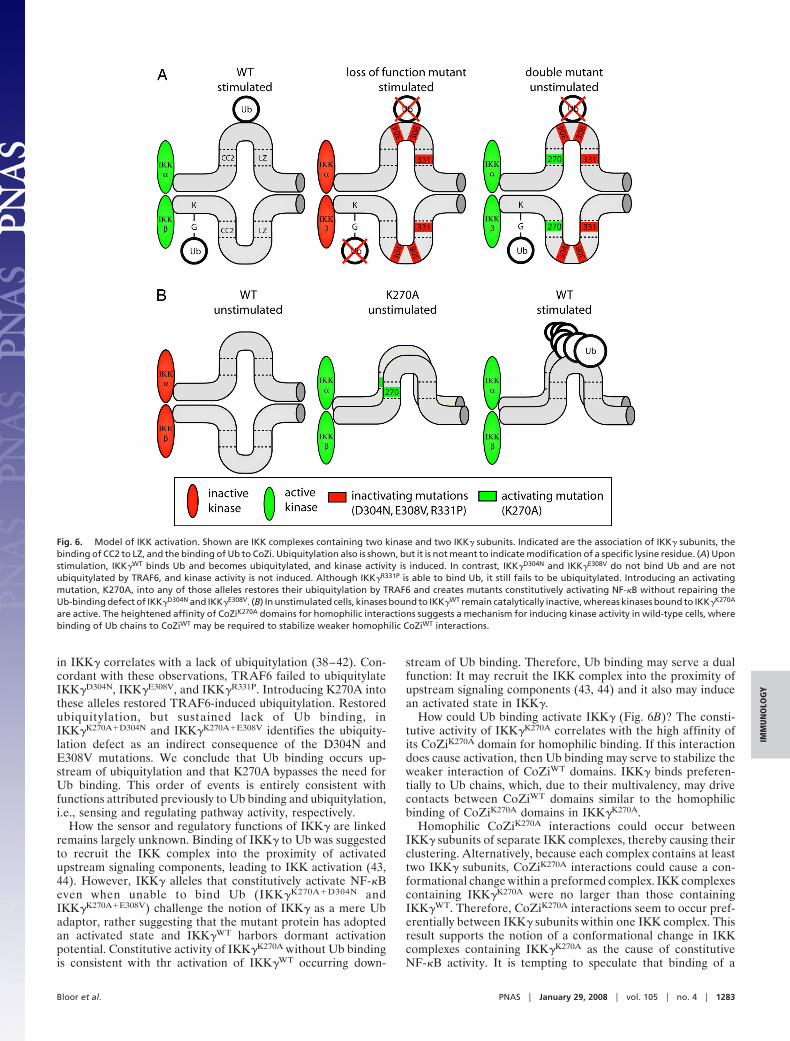
Fig. 6. Model of IKK activation. Shown are IKK complexes containing two kinase and two IKK� subunits. Indicated are the association of IKK� subunits, thebinding of CC2 to LZ, and the binding of Ub to CoZi. Ubiquitylation also is shown, but it is not meant to indicate modification of a specific lysine residue. (A) Uponstimulation, IKK�WT binds Ub and becomes ubiquitylated, and kinase activity is induced. In contrast, IKK�D304N and IKK�E308V do not bind Ub and are notubiquitylated by TRAF6, and kinase activity is not induced. Although IKK�R331P is able to bind Ub, it still fails to be ubiquitylated. Introducing an activatingmutation, K270A, into any of those alleles restores their ubiquitylation by TRAF6 and creates mutants constitutively activating NF-�B without repairing theUb-binding defect of IKK�D304N and IKK�E308V. (B) In unstimulated cells, kinases bound to IKK�WT remain catalytically inactive, whereas kinases bound to IKK�K270A
are active. The heightened affinity of CoZiK270A domains for homophilic interactions suggests a mechanism for inducing kinase activity in wild-type cells, wherebinding of Ub chains to CoZiWT may be required to stabilize weaker homophilic CoZiWT interactions.
Bloor et al. PNAS � January 29, 2008 � vol. 105 � no. 4 � 1283
IMM
UN
OLO
GY
ubiquitylated ligand causes a similar conformational change inwild-type IKK complexes during signaling. Ultimately, structuralwork may be required to test this hypothesis.
Materials and MethodsReagents. Antibodies were from BD PharMingen (IKK�, CD8, and Thy1.1),Imgenex (IKK� and IKK�), Abcam (rabbit HA11, AU1, and myc), Covance(murine AU1), Santa Cruz Biotechnology (Ub), and Dabco (HRP-conjugatedreagents). LPS (Escherichia coli O127:B8), PMA, and Ionomycin were fromSigma–Aldrich, peptidoglycan was from Fluka, and CpG DNA (ODN1668 TC-CATGACGTTCCTGATGCT) was from Operon.
Plasmids. TRAFs were expressed from pEAK8. All other genes were in M5P (53)or M6P8, an M5P derivative containing IRES-controlled CD8. Numbering inIKK� constructs refers to NM�178590. IKK� in GTPT3, from which the cDNA forthis study was derived, contains asparagine at position 285.
Cell Culture and Mutagenesis. An NF-�B-dependent promoter (45) was used tocontrol the expression of reporter genes (GFP, rat Thy1, nd puromycin acetyl-transferase fused to thymidine kinase). GTPT3, a clone derived from 70Z/3 cellsstably transfected with all three reporter genes, was chosen for its lowconstitutive- and high LPS-stimulated reporter expression. Mutagenesis wasperformed as described in ref. 54. After recovery, cells were stimulated with 1�g/ml LPS and selected with 0.5 �M ganciclovir (Sigma–Aldrich). Mutant cloneswere identified by the absence of GFP and Thy1 expression upon LPS stimulation.
M35 cells lack IKK� and were isolated from GTPT3 based on their unre-sponsiveness to CpG DNA after random mutagenesis as above. SVT35 Jurkatcells carry an NF-�B-controlled CD14 reporter. These cells and an RIP-deficientsubclone were provided by Adrian Ting (45).
Reporter Assays. NF-�B-dependent GFP activity was analyzed on a FACSCalibur(BD Biosciences). Luciferase activity was measured with Bright-Glo (Promega).
Chromatography. After swelling in hypotonic buffer [10 mM Tris�HCl (pH 7.4),1 mM KCl, and 10 mM MgCl2], cells were disrupted with a tight Dounce
homogenizer. NaCl was added to a final concentration of 150 mM, andinsoluble cell remnants were pelleted at 100,000 � g. Supernatants werefractionated on a Superdex 200 column (Amersham Pharmacia).
Immunoprecipitation. Postnuclear supernatants from cells lysed in 0.5% TritonX-100, 20 mM Tris�HCl (pH 7.4), 150 mM NaCl, and 1 mM EDTA were incubatedfor 2 h with 1–2 �g/ml primary antibody, followed by incubation for 2 h withprotein G Sepharose. After washing, samples were eluted with SDS buffer.
Kinase Assay. After immunoprecipitation in the presence of protease andphosphatase inhibitors, beads were washed in 20 mM Mops (pH 7.4), 1%Triton X-100, 0.1 mM EDTA, 1 mM EGTA, and 1 mM DTT. Immune complexeswere incubated for 20 min at 30°C in a 20-�l reaction mixture containing 25�M cold ATP, 3 �Ci [32]ATP, 12 mM MgCl2, and 3 �g of GST-I�B� 1–100. Thereaction was stopped with SDS buffer.
Analytical Ultracentrifugation. The CoZi domain of IKK� (amino acids 250–339)was expressed from pETM11. After purification on Ni-NTA agarose (Qiagen),the His tag was cleaved off with TEV protease, and the resulting material wasrepurified over Ni-NTA and Superdex 75 columns. Sedimentation equilibrium/velocity experiments were carried out as described in ref. 55.
Ub Binding. GSTfusionproteinsexpressed inE. coliwerecoupledontoGSHbeads.For LUMIER assays (56), Renilla luciferase fused to IKK� was expressed in 293ETcells. Binding was performed for 2 h in 20 mM Tris�HCl (pH 7.4), 150 mM NaCl, and0.1% Triton. Proteins were eluted with glutathione. The ratio between luciferaseactivity in eluates and lysates is presented as fold binding over a control reaction.
ACKNOWLEDGMENTS. We thank Olga Perisic and Allan Warren (MedicalResearch Council Laboratory of Molecular Biology) for TEV protease and helpwith chromatography, Aarie Geerlof (European Molecular Biology Labora-tory, Heidelberg) for pETM plasmids, Hiroyasu Nakano (Juntendo UniversityMedical School, Tokyo) for pGEX-I�B�, Adrian Ting (Mount Sinai MedicalCenter, New York) for RIP-deficient cells, and Alexander Betz and MariannBienz for reading the manuscript.
1. Ghosh S, Karin M (2002) Cell 109(Suppl):S81–S96.2. Hayden MS, Ghosh S (2004) Genes Dev 18:2195–2224.3. Karin M, Ben-Neriah Y (2000) Annu Rev Immunol 18:621–663.4. Brockman JA, Scherer DC, McKinsey TA, Hall SM, Qi X, Lee WY, Ballard DW (1995) Mol
Cell Biol 15:2809–2818.5. Brown K, Gerstberger S, Carlson L, Franzoso G, Siebenlist U (1995) Science 267:1485–1488.6. DiDonato J, Mercurio F, Rosette C, Wu-Li J, Suyang H, Ghosh S, Karin M (1996) Mol Cell
Biol 16:1295–1304.7. Connelly MA, Marcu KB (1995) Cell Mol Biol Res 41:537–549.8. DiDonato JA, Hayakawa M, Rothwarf DM, Zandi E, Karin M (1997) Nature 388:548–554.9. Mercurio F, Zhu H, Murray BW, Shevchenko A, Bennett BL, Li J, Young DB, Barbosa M,
Mann M, Manning A, Rao A (1997) Science 278:860–866.10. Woronicz JD, Gao X, Cao Z, Rothe M, Goeddel DV (1997) Science 278:866–869.11. Zandi E, Rothwarf DM, Delhase M, Hayakawa M, Karin M (1997) Cell 91:243–252.12. Regnier CH, Song HY, Gao X, Goeddel DV, Cao Z, Rothe M (1997) Cell 90:373–383.13. Tanaka M, Fuentes ME, Yamaguchi K, Durnin MH, Dalrymple SA, Hardy KL, Goeddel DV
(1999) Immunity 10:421–429.14. Li ZW, Chu W, Hu Y, Delhase M, Deerinck T, Ellisman M, Johnson R, Karin M (1999) J Exp
Med 189:1839–1845.15. Li Q, Van Antwerp D, Mercurio F, Lee KF, Verma IM (1999) Science 284:321–325.16. Takeda K, Takeuchi O, Tsujimura T, Itami S, Adachi O, Kawai T, Sanjo H, Yoshikawa K,
Terada N, Akira S (1999) Science 284:313–316.17. Hu Y, Baud V, Delhase M, Zhang P, Deerinck T, Ellisman M, Johnson R, Karin M (1999)
Science 284:316–320.18. Li Q, Lu Q, Hwang JY, Buscher D, Lee KF, Izpisua-Belmonte JC, Verma IM (1999) Genes
Dev 13:1322–1328.19. Senftleben U, Cao Y, Xiao G, Greten FR, Krahn G, Bonizzi G, Chen Y, Hu Y, Fong A, Sun
SC, Karin M (2001) Science 293:1495–1499.20. Ling L, Cao Z, Goeddel DV (1998) Proc Natl Acad Sci USA 95:3792–3797.21. Delhase M, Hayakawa M, Chen Y, Karin M (1999) Science 284:309–313.22. Sato S, Sanjo H, Takeda K, Ninomiya-Tsuji J, Yamamoto M, Kawai T, Matsumoto K,
Takeuchi O, Akira S (2005) Nat Immunol 6:1087–1095.23. Shim JH, Xiao C, Paschal AE, Bailey ST, Rao P, Hayden MS, Lee KY, Bussey C, Steckel M,
Tanaka N, et al. (2005) Genes Dev 19:2668–2681.24. Deng L, Wang C, Spencer E, Yang L, Braun A, You J, Slaughter C, Pickart C, Chen ZJ
(2000) Cell 103:351–361.25. Wang C, Deng L, Hong M, Akkaraju GR, Inoue J, Chen ZJ (2001) Nature 412:346–351.26. Kanayama A, Seth RB, Sun L, Ea CK, Hong M, Shaito A, Chiu YH, Deng L, Chen ZJ (2004)
Mol Cell 15:535–548.27. Yamaoka S, Courtois G, Bessia C, Whiteside ST, Weil R, Agou F, Kirk HE, Kay RJ, Israel
A (1998) Cell 93:1231–1240.28. Rothwarf DM, Zandi E, Natoli G, Karin M (1998) Nature 395:297–300.
29. Li Y, Kang J, Friedman J, Tarassishin L, Ye J, Kovalenko A, Wallach D, Horwitz MS (1999)Proc Natl Acad Sci USA 96:1042–1047.
30. Mercurio F, Murray BW, Shevchenko A, Bennett BL, Young DB, Li JW, Pascual G,Motiwala A, Zhu H, Mann M, Manning AM (1999) Mol Cell Biol 19:1526–1538.
31. Schmidt-Supprian M, Bloch W, Courtois G, Addicks K, Israel A, Rajewsky K, PasparakisM (2000) Mol Cell 5:981–992.
32. Makris C, Godfrey VL, Krahn-Senftleben G, Takahashi T, Roberts JL, Schwarz T, Feng L,Johnson RS, Karin M (2000) Mol Cell 5:969–979.
33. Rudolph D, Yeh WC, Wakeham A, Rudolph B, Nallainathan D, Potter J, Elia AJ, Mak TW(2000) Genes Dev 14:854–862.
34. Lee EG, Boone DL, Chai S, Libby SL, Chien M, Lodolce JP, Ma A (2000) Science 289:2350–2354.35. Brummelkamp TR, Nijman SM, Dirac AM, Bernards R (2003) Nature 424:797–801.36. Kovalenko A, Chable-Bessia C, Cantarella G, Israel A, Wallach D, Courtois G (2003)
Nature 424:801–805.37. Trompouki E, Hatzivassiliou E, Tsichritzis T, Farmer H, Ashworth A, Mosialos G (2003)
Nature 424:793–796.38. Huang TT, Wuerzberger-Davis SM, Wu ZH, Miyamoto S (2003) Cell 115:565–576.39. Tang ED, Wang CY, Xiong Y, Guan KL (2003) J Biol Chem 278:37297–37305.40. Zhou H, Wertz I, O’Rourke K, Ultsch M, Seshagiri S, Eby M, Xiao W, Dixit VM (2004)
Nature 427:167–171.41. Sun L, Deng L, Ea CK, Xia ZP, Chen ZJ (2004) Mol Cell 14:289–301.42. Abbott DW, Wilkins A, Asara JM, Cantley LC (2004) Curr Biol 14:2217–2227.43. Wu CJ, Conze DB, Li T, Srinivasula SM, Ashwell JD (2006) Nat Cell Biol 8:398–406.44. Ea CK, Deng L, Xia ZP, Pineda G, Chen ZJ (2006) Mol Cell 22:245–257.45. Ting AT, Pimentel-Muinos FX, Seed B (1996) EMBO J 15:6189–6196.46. Doffinger R, Smahi A, Bessia C, Geissmann F, Feinberg J, Durandy A, Bodemer C,
Kenwrick S, Dupuis-Girod S, Blanche S, et al. (2001) Nat Genet 27:277–285.47. Makris C, Roberts JL, Karin M (2002) Mol Cell Biol 22:6573–6581.48. Agou F, Traincard F, Vinolo E, Courtois G, Yamaoka S, Israel A, Veron M (2004) J Biol
Chem 279:27861–27869.49. Hicke L, Schubert HL, Hill CP (2005) Nat Rev Mol Cell Biol 6:610–621.50. Haglund K, Dikic I (2005) EMBO J 24:3353–3359.51. Meylan E, Burns K, Hofmann K, Blancheteau V, Martinon F, Kelliher M, Tschopp J (2004)
Nat Immunol 5:503–507.52. Cusson-Hermance N, Khurana S, Lee TH, Fitzgerald KA, Kelliher MA (2005) J Biol Chem
280:36560–36566.53. Randow F, Sale JE (2006) Subcell Biochem 40:383–386.54. Randow F, Seed B (2001) Nat Cell Biol 3:891–896.55. Chaillan-Huntington C, Butler PJ, Huntington JA, Akin D, Feldherr C, Stewart M (2001)
J Mol Biol 314:465–477.56. Barrios-Rodiles M, Brown KR, Ozdamar B, Bose R, Liu Z, Donovan RS, Shinjo F, Liu Y,
Dembowy J, Taylor IW, et al. (2005) Science 307:1621–1625.
1284 � www.pnas.org�cgi�doi�10.1073�pnas.0706552105 Bloor et al.