seven shades of tamoxifen resistance - helda
TRANSCRIPT

Susanne Hultsch
Seven shadesof tamoxifen resistance
Molecular mechanisms of drug resistance in breast cancer

Seven shades of tamoxifen resistance
Molecular mechanisms of drug resistance in breast cancer
Susanne Hultsch
Institute of Molecular Medicine Finland (FIMM)and
Doctoral Program in Biomedicine (DPBM) Faculty of Medicine,
University of Helsinki
Academic Dissertation
To be presented with the permission of the Faculty of Medicine, University of Helsinki, for public examination in Porthania, PIII
(119), Yliopistonkatu 3 on the 23rd of October 2018 at 12 noon.
Helsinki 2018

ii
Supervisors: Olli Kallioniemi, MD, PhD, Professor, DirectorInstitute for Molecular Medicine Finland, FIMM, HiLIFE, University of Helsinki, FinlandScience for Life Laboratory (SciLifeLab), Karolinska Institutet, Sweden
Vilja Pietiänen, PhDInstitute for Molecular Medicine Finland, FIMM, HiLIFE, University of Helsinki, Finland
Thesis Committee: Kaisa Lehti, PhDResearch Director and Senior Cancer ResearcherUniversity of Helsinki, Finland,Karolinska Institute, Sweden
Johanna Mattson, MD, Docent, Chief Medical Officer HYKS Cancer Center and University of Helsinki, Finland
Pre-examiners: Tuula Kallunki, PhD, Adjunct Professor Danish Cancer Society Research Center, Copenhagen, Denmark.
Katri Pylkäs, PhD, DocentLaboratory of Cancer Genetics and Tumor BiologyBiocenter Oulu, University of Oulu, Finland
Opponent: Leonie Young, PhD, Associate ProfessorRCSI Surgery, Royal College of Surgeons in Ireland
©Susanne HultschCover image and layout by Susanne Hultsch
ISBN 978-951-51-4491-1 (paperback)ISBN 978-951-51-4492-8 (PDF)
Unigrafia, Helsinki 2018

ABSTRACT iii
ABSTRACTTamoxifen treatment of estrogen receptor-positive breast cancer re-
duces breast cancer mortality. However, resistance to tamoxifen de-
velops frequently. A plethora of resistance mechanisms have been
described but their biological importance, clinical significance, and
possibilities for diagnostic or therapeutic intervention are poorly un-
derstood. Fusion genes, for example, have the potential as thera-
peutic targets or diagnostic tools as they are highly cancer-specific.
In order to determine the mechanisms underlying endocrine therapy
resistance and to identify new opportunities to defy resistance in
breast cancer, we created seven tamoxifen-resistant breast cancer
cell lines. We characterized the resistant cell lines by exome-se-
quencing to identify possible mutations or genomic rearrangements
involved in drug resistance. RNA-sequencing was applied to shed
light on the nature of fusion genes in the parental cell line as well as
their contribution to acquired drug resistance. RNA-sequencing also
exposed gene expression and pathway changes, which were followed
up in detail in one of the resistant cell lines. In addition to drug sen-
sitivity and resistance testing combined with high-content imaging,
network analysis determined the drug response profiles. We further
uncovered potential drug targets of tamoxifen resistance.
This intensive molecular profiling revealed that each tamoxifen-resis-
tant cell line developed its own resistance mechanism and acquired
individual drug vulnerabilities. However, we were able to detect a
common increased sensitivity towards an ERK1/2-inhibitor. On the
other hand, we discovered co-resistance to paclitaxel, which is
mostly driven by the slower growth rate of the resistant cells. Anal-
ysis of differentially expressed genes identified pathways which were
associated with cell cycle, protein modification, and metabolism, es-

iv ABSTRACT
pecially with the cholesterol pathway. After further investigation we
identified that the prevention of lysosomal membrane permeabilization
was associated with drug resistance in the T-47D tamoxifen-resistant
cell lines. Targeting this mechanism remains challenging. We further
revealed the complex nature of fusion genes, which include the high
prevalence of alternative splicing and the lack of recurrence across
different breast cancer cell lines. Additionally, we identified fusion
genes only present in the resistant cell lines. However, these were
mainly cell clone-specific or recurrent read-through fusions. Exome-se-
quencing revealed no known or common mutations or copy number
changes related to resistance development.
With the diversity of fusion genes, mutations, copy number changes,
differentially expressed genes, pathway changes, and drug responses
in tamoxifen-resistant cells, it is safe to say that tamoxifen resistance
cannot simply be explained by one common mechanism. Therefore, it
is likely that countering the resistance will require different thera-
peutic approaches.

TABLE OF CONTENTS v
TABLE OF CONTENTS
ABSTRACT ..........................................................................................iii
TABLE OF CONTENTS ........................................................................v
LIST OF ORIGINAL PUBLICATIONS ................................................vii
ABBREVIATIONS ..............................................................................viii
INTRODUCTION ...............................................................................1
REVIEW OF LITERATURE ................................................................2
1 The Breast ...................................................................................22 Estrogen Receptor – it's complicated .........................................33 Breast Cancer ..............................................................................6
3.1 Breast Cancer Subtypes ......................................................64 Fusion genes ...............................................................................95 Tamoxifen ..................................................................................11
5.1 A summary of tamoxifen's history .....................................115.2 Mode of Action ...................................................................12
6 Drug resistance in cancer .........................................................136.1 Resistance mechanisms .....................................................14
6.1.1 Drug uptake, efflux, and metabolism .........................156.1.2 Drug target alterations ...............................................166.1.3 Cell cycle alteration and DNA damage repair ...........176.1.4 Avoiding apoptosis ......................................................186.1.5 Resistance mediated by autophagy ............................196.1.6 Adaption of signaling ..................................................206.1.7 Epithelial-mesenchymal transition (EMT) .................206.1.8 Tumor microenvironment ...........................................216.1.9 Extracellular vesicles .................................................22
6.2 Resistance to Tamoxifen ....................................................24 AIM OF THE STUDY .......................................................................29
MATERIALS AND METHODS .........................................................30
1 Cell culture and generation of tamoxifen-resistant cell lines (I, II, III) .............................................................................................302 Characterization of tamoxifen-resistant cell lines (II, III) ........31
2.1 Growth analysis (II) ...........................................................312.2 Responsiveness to estrogens and downstream signaling (II) ...................................................................................................322.3 Measurements and drug induced changes in protein levels

vi TABLE OF CONTENTS
(II, III) ........................................................................................322.4 Immunofluorescence staining (III) ....................................332.5 Lysosomal membrane permeabilization (LMP) assay (III) 342.6 Measurement of triglycerides and cholesterol esters (III) 34
3 Paired-end RNA-sequencing (I, III) ...........................................344 RNA-sequencing analysis (I, III) ...............................................35
4.1 Fusion gene detection, characterization, and validation (I, III) .............................................................................................354.2 Integration of transcriptomic data and differential gene expression (III) ..........................................................................36
5 Genomic profiling by exome-sequencing (II) ............................376 Drug sensitivity and resistance testing (DSRT) (II, III) ............38
6.1 Cell viability measurement (II, III) ....................................386.2 High-content phenotypic drug profiling (III) .....................38
7 DSRT data analysis (II, III) ........................................................397.1 Analysis of cell viability (II, III) .........................................397.2 Construction of drug sensitivity and co-resistance networks (II) .............................................................................................397.3 Analysis of high-content phenotypic drug profiling (III) ...40
RESULTS & DISCUSSION ..............................................................41
1 Fusion genes .............................................................................411.1 Fusion gene characteristics in breast cancer ...................411.2 The role of fusion genes in tamoxifen resistance ..............43
2 Characterization of tamoxifen-resistant cell lines ....................442.1 Tamoxifen-resistant cells alter their cell cycle ..................452.2 Changes in ERα protein-levels and estrogen-responsiveness ...................................................................................................462.3 Genetic alterations in the genomes of tamoxifen-resistant cell lines ....................................................................................472.4 Transcriptional changes in tamoxifen-resistant cell lines and their resemblance to patients ...................................................482.5 Drug responses of tamoxifen-resistant cell lines ..............51
3 Lipid changes in T-47D and their consequences ......................53 CONCLUSION .................................................................................57
ACKNOWLEDGMENTS .......................................................................x
REFERENCES ..................................................................................xiii
INDEX OF FIGURES AND TABLES ..............................................xxxvi
ORIGINAL PUBLICATIONS ..........................................................xxxvii

LIST OF ORIGINAL PUBLICATIONS vii
LIST OF ORIGINAL PUBLICATIONS
I. Kangaspeska S, Hultsch S, Edgren H, Nicorici D, Murumägi
A, Kallioniemi O. Reanalysis of RNA-sequencing data reveals
several additional fusion genes with multiple isoforms. PLoS
One. 2012;7(10):e48745.
II. Kangaspeska S*, Hultsch S*, Jaiswal A, Edgren H, Mpindi JP,
Eldfors S, Brück O, Aittokallio T, Kallioniemi O. Comprehen-
sive drug sensitivity testing reveals specific vulnerabilities
and co-resistance patterns in endocrine-resistant breast
cancer. BMC Cancer. 2016 Jul 4;16:378.
III. Hultsch S, Kankainen M, Paavolainen L, Kovanen RM, Ikonen
E, Kangaspeska S, Pietiäinen V, Kallioniemi O. Lipid repro-
gramming in tamoxifen-resistant breast cancer cells and po-
tential therapeutic vulnerabilities (Manuscript accepted, BMC
Cancer
* Equal contribution

viii ABBREVIATIONS
ABBREVIATIONSABC ATP-binding cassette
Abl1 Abelson murine leukemia viral oncogene homolog 1
AEBS antiestrogen binding sites
AF-1 activation function domain 1
AR androgen receptor
BCL-2 B-cell lymphoma 2
BCR breakpoint cluster region protein
bp base pair
BRCA1/2 breast cancer 1/2 early onset
CAR chimeric antigen receptor
CML chronic myelogenous leukemia
CNV copy number variation
CP ceruloplasmin
CPM counts per million
CTG CellTiter-Glo
CTSD cathepsin D
DBD DNA-binding domain
DSRT drug sensitivity and resistance testing
DSS drug sensitivity score
ECM extracellular matrix
EGF epidermal growth factor
EGFR epidermal growth factor receptor
EMT epithelial-to-mesenchymal transition
ER estrogen receptor
ERα/ESR1 estrogen receptor α
ERβ/ESR2 estrogen receptor β
ERE estrogen response element
ETS E26 transformation-specific
EV extracellular vesicle
ABBREVIATIONS ix
FDA food and drug administration
GS-X glutathione S-conjugate export
HER2 human epidermal growth factor receptor 2
HGF hepatocyte growth factor
HIF-1 hypoxia-inducible factor 1
ICI Imperial Clinical Industries
IGF insulin growth-factor
LBD/AF-2 ligand-binding domain/activation function domain 2
LLOMe L-leucyl-L-leucine methyl ester
LMP lysosomal membrane permeabilization
lncRNA long non-coding RNA
MCL-1 Induced myeloid leukemia cell differentiation protein
MET Met proto-oncogene tyrosine kinase
miRNA microRNA
MRP1 multidrug resistance-associated protein 1
NCoR nuclear receptor co-repressor
NF-κBB nuclear factor kappa-light-chain-enhancer of activated B cells
NSCLC non-small-cell lung cancer
PI3K phosphatidylinositol-3-kinase
PKC protein kinase C
PR progesterone receptor
ROS reactive oxygen species
SERM selective estrogen receptor modulator
SOD1 superoxide dismutase 1
SRC steroid receptor co-activator
TGF-β Transforming growth factor beta
TMPRSS2 transmembrane protease, serine 2
VEGF vascular endothelial growth factor

1 INTRODUCTION
INTRODUCTIONImmediately, with the discovery of usefulness of cytotoxic agents in
cancer therapy, the phenomenon of cancer drug resistance was ob-
served in the 1940s (Goodman et al. 1946). To study drug resistance,
researchers applied lessons learned from the selection process of an-
tibiotic-resistant bacteria and cultivated patient-derived cell lines ex-
posed to the drug of interest and developed drug-resistant cancer
cell lines (McDermott et al. 2014). Samples were also taken from
drug-resistant tumors and investigated in animal models, for ex-
ample, as xenografts (Houghton, Houghton, and Green 1982;
Bertotti et al. 2011; S. Li et al. 2013). Results from cell line or animal
studies were not always easily translatable to the clinic. Hence, even
after 70 years of research, drug resistance is still a problem not only
in the case of chemotherapeutics but also of targeted therapy. Even
though cancer is a genetic disease, it became obvious that this might
not always be the case in terms of drug resistance. Drug resistance
can arise from genetic, epigenetic, as well as environmental factors
and those factors can act simultaneously. Another contributor to
drug resistance is the molecular diversity of cancer in which breast
cancer, with its multiple subtypes, is a poster child. In breast cancer,
commercial diagnostic tests, such as Oncotype DX (Carlson 2006),
MammaPrint (Glas et al. 2006), or EndoPredict (Filipits et al. 2011)
may provide prognostic information but also guide therapy. Given
that we can now detect circulating tumor cells as well as circulating
free DNA using so-called liquid biopsies from the blood (Friedlander,
Premasekharan, and Paris 2014), it would be very beneficial to iden-
tify drug resistance as soon as it occurs. Maybe even by imple-
menting such a test. However, we still need to learn more about drug
resistance mechanisms and vulnerabilities associated with these to
be able to treat drug-resistant cancers.

REVIEW OF LITERATURE 2
REVIEW OF LITERATURE
1 The BreastThe breast, or mammary gland, is not fully developed at birth. In its
post-natal development, it undergoes a course of transformations
such as cell proliferation, differentiation, and morphogenesis. Var-
ious hormones and growth factors regulate these transformations.
During puberty estrogens, together with progesterone, are respon-
sible for the expansion of the gland and the development of the
ductal epithelium, which give rise to its glandular tissue structure
(Brisken et al. 1998; Mallepell et al. 2006). The fully developed adult
mammary gland is composed of different cell types. The inner lumen
of the ductal and lobular structures are lined with a continuous layer
of epithelial cells that are surrounded by a layer of contractile my-
oepithelial cells. The myoepithelial cells are further enclosed by a
basement membrane which separates the epithelial from the stroma.
The stroma consists of adipocytes, lymphocytes, neurons, fibroblasts,
and endothelial cells (Anderson, Clarke, and Howell 2000). The hor-
mone-sensing cells such as estrogen- and progesterone receptor pos-
itive cells are located within the luminal layer ranging from 7-20% of
cells. Interestingly, those ER-positive cells do not divide in normal
tissue but are in close proximity to them (Clarke et al. 1997).
Overall, the female mammary gland experiences a lot of structural
and functional changes that are characterized by proliferation, dif-
ferentiation, and apoptosis of cells due to hormonal changes during
the menstrual cycle, pregnancy, lactation, and menopause (Macias
and Hinck 2012).

3 REVIEW OF LITERATURE
2 Estrogen Receptor – it's complicatedThe activities of estrogens are not only restricted to the development
and maintenance of sexual and reproductive functions, they are addi-
tionally involved in biological processes of the musculoskeletal, im-
mune, cardiovascular and central nervous system in men and women
(Gustafsson 2003). There are three types of estrogens: estrone (E1),
estriol (E3) and the predominant 17β-estradiol (E2), which binds to
the nuclear transcription factor receptors ERα and ERβ. The recep-
tors are encoded by ESR1 and ESR2 genes and have three functional
domains. The hormone-independent transcriptional activation func-
tion domain (AF-1) at the N-terminus harbors the biggest functional
differences between the two receptors. The DNA-binding domain
(DBD) shares about 55% homology and the ligand-binding domain
(LBD/AF-2) at the C-terminus is 95% homologous between the two
receptors (Kuiper et al. 1996). Nevertheless, primarily the activation
of ERα target genes is responsible for promoting cancer cell prolifer-
ation and a decrease in cancer cell apoptosis (Iwao et al. 2000;
Roger et al. 2001). There are several paths by which estrogens and
ER convey signaling (Figure 1). The ligand-dependent arm, as the
name implies, involves the binding of estrogen to the LBD/AF-2 of
ER, forming an estrogen-ER complex, which promotes different gene
activation scenarios. The complex can directly bind to estrogen re-
sponse elements (EREs) in the promoter region of a target gene
(Figure 1A). The ERE is defined by two inverted DNA-sequences sep-
arated by three random nucleotides (GGTCAnnnTGACC) (Klinge
2001). However, it is now clear that the complex also binds to en-
hancer regions in the genome (Carroll et al. 2006).

REVIEW OF LITERATURE 4
Alternatively, the estrogen-ER complex can interact with other tran-
scription factors such as activator protein 1 (AP1), specificity protein
1 (SP1), NF-κBB, or cAMP response element-binding protein (CREB)
to name just a few amongst many others (Galien and Garcia 1997;
Kushner et al. 2000; Lazennec, Thomas, and Katzenellenbogen 2001;
W. Porter et al. 1997). This co-activation facilitates the transcrip-
tional regulation of genes that do not harbor EREs (Figure 1B). In
order to fully facilitate transcriptional regulation the estrogen-ER
complex needs co-regulators that can modify the chromatin structure
or interact with the transcriptional machinery. Plenty of those co-reg-
Figure 1: Simplistic overview of ER signaling pathways. Ligand-dependent arm includes A the classical (direct) pathway, B the non-classical (tethered) pathway, and C the non-genomic pathway. The ligend-independent pathway is depicted in D. Grey ovals depict second messenger cascades; E2, Estradiol; ER, Estrogen receptor, ERE, Estrogen response element, TF, transcription factor; RE, Response elements to other transcription factors; P, phosphorylation; GF, Growth factor; RTK, tyrosine kinase receptor (modified from Le Romancer et al. 2011)
ERETF
Co-reg .
ER
E2
?
RE RE
P P
PP
GF
ERE
RTK
A B C D

5 REVIEW OF LITERATURE
ulators have been identified (Métivier et al. 2003). The most in-
tensely studied co-regulators belong to the p160 family proteins (or
SRCs). They can either directly or indirectly recruit co-activators and
so remodel chromatin and modify histone activity (Koh et al. 2001),
for example, by recruiting histone acetyltransferase (HAT), which is
known to play a role in the initiation of transcription (Brownell and
Allis 1996; Kraus and Kadonaga 1998). Cathepsin D (CTSD) and tre-
foil factor 1 (TFF-1, also known as pS2) have been among the first
identified target genes of the estrogen-ER and the p160 co-regula-
tors complex in vivo (Shang et al. 2000). As if this isn't already com-
plex enough, the estrogen-ER complex has been shown to activate
multiple signaling cascades through direct interactions with various
proteins including tyrosine kinase Src, phosphatidylinositol 3-kinase
(PI3K), and insulin-like growth factor 1 receptor (IGF-1R) (Castoria
et al. 2001; Kahlert et al. 2000; Simoncini et al. 2000). This so-called
non-genomic mode of action leads to an immediate response by the
estrogen-ER complex (Figure 1C).
Finally, there is a ligand-independent activation of ER signaling with
various effects on cellular processes (Cenni and Picard 1999). The
best-studied signals inducing ER activities are growth factors, for ex-
ample, EGF and even IGF-1, which induce signaling cascades that
lead to the phosphorylation and thereby activation of ER in an es-
trogen-independent manner (Kato et al. 1995, Figure 1D).
The different mechanisms of action demonstrate the complex nature
of ER signaling. However, it needs to be stated that most of the func-
tional studies have been conducted in cancer cell lines and little is
known about the interactions of ER in healthy human mammary
tissue.

REVIEW OF LITERATURE 6
3 Breast CancerDespite losing its first place in cancer mortality among young Eu-
ropean woman to lung cancer, breast cancer is still with around
460’000 cases, the most often diagnosed cancer among European
woman, and remains a major public health concern (Malvezzi et
al. 2015; Ervik et al. 2016). Fortunately, breast cancer is often
treatable if it represents one of the subtypes that have treatment
options available. If breast cancer is diagnosed late or belongs to
a subtype with limited treatment possibilities it is still deadly. An-
other major drawback is the development of therapy resistance,
which is followed by tumor progression and development of
metastasis.
3.1 Breast Cancer SubtypesBreast cancer is extremely heterogeneous. According to the
World Health Organization, the invasive ductal carcinoma, not
otherwise specified (ductal NOS) represents 40-75% of breast
cancer cases. These cases, as the NOS addition suggests, repre-
sent a heterogeneous group that does not exhibit sufficient histo-
logical properties in comparison to the invasive lobular carcinoma
(ILC, 5-15%) or tubular carcinoma (<2%) (Lakhani et al. 2012).
Breast cancer is routinely clinically characterized by tumor type,
size, grade, lymph node status as well as four immunohistochem-
istry markers ER, PR, Ki67, and HER2. Alternatively, the more
sensitive fluorescent in situ hybridization (FISH) is used to detect
HER2. However, a combination of IHC and FISH is recommended
(Lakhani et al. 2012; Wolff et al. 2018). Intensive molecular pro-
filing has shed light on this cancer's biology, which can also guide
clinical decisions.

7 REVIEW OF LITERATURE
More than 15 years ago, Perou, Sørlie, and colleagues divided
breast cancer into five different subtypes based on gene expres-
sion profiles (Perou et al. 2000; Sørlie et al. 2001). The most domi-
nant subtypes are the luminal A and luminal B types and as their
name suggests, they mainly express genes of the lumen of the
mammary duct (Table 1). Both subtypes are ER-positive. Addition-
ally, they can be either positive or negative for the progesterone
receptor (PR). These subgroups are usually responsive to en-
docrine- and chemotherapy (Early Breast Cancer Trialists’ Collab-
orative Group (EBCTCG) 2005). The luminal B type is also HER2-
positive and has high levels of Ki67 compared to luminal A and
has, therefore, a higher proliferation rate. Importantly, the HER2-
positivity makes it responsive to therapeutic approaches against
HER2, such as herceptin. Other subtypes do not express ER and
PR, including the HER2-positive subtype (Table 1), which is non-
responsive to endocrine therapy (Loibl and Gianni 2017). Finally,
the basal-like (or triple negative) subtype has a similar expression
profile to the myoepithelial cells. The treatment options are re-
stricted to surgery, radiation and/or chemotherapy, as this type
does not express ER, PR, and/or HER2 (Fulford et al. 2007; Holl-
iday and Speirs 2011). A controversial subtype is the normal
breast-like type. It has gene expression similar to normal breast
tissue and high expression of genes that reassemble the charac-
teristic for adipose tissue (Sørlie et al. 2001; Fan et al. 2006). How-
ever, the normal breast-like type has been regarded as an artifact
due to high contamination of “true” normal breast tissue in the
sample (Parker et al. 2009).

REVIEW OF LITERATURE 8
Table 1: Breast cancer subtypes (based on http://www.pathophys.org/ breast-cancer/ by Eric Wong)
Subtype Characteristics Prevalence Medical therapy
Luminal A ER PR HER2 Low Ki67
+++-
40 % Endocrine therapy
Luminal B ER PR HER2 high Ki67
++/-+/-
20 % Chemotherapy, Endocrine therapy, anti-HER2 targeted therapy
HER2-positive
ER PR HER2 high Ki67
--
++
10-15 % Chemotherapy with anti-HER2 targeted therapy
Triple negative
ER PR HER2 high Ki67
---
15-20 % Chemotherapy
This first classification (Perou et al. 2000; Sørlie et al. 2001) was
followed by several more suggestions. By additionally utilizing
copy number alterations, a study by Curtis et al. divided the ex-
isting classification in even 10 different subgroups (Curtis et al.
2012), whereas others suggested an extra claudin-low group
(Herschkowitz et al. 2007), or identified six TNBC subgroups of
which one is androgen receptor-positive (Lehmann et al. 2011).
For patients with the latter subtype there are currently several
clinical trials with anti-androgen-therapy ongoing (clinicaltrial-
s.gov: NCT01889238; NCT02689427; NCT03383679).
The examples of the different subgroups (Table 1) show that even
within the subtypes tumor-specific dissimilarities occur, which
likely contribute to tumor development and progression. It also

9 REVIEW OF LITERATURE
highlights that a “one-size-fits-all” therapy is not applicable in
breast cancer and a more personalized treatment is needed.
4 Fusion genesAs mentioned above, breast cancer is a heterogeneous disease
with frequent genetic alterations. These alterations can activate
cancer-promoting genes, so-called oncogenes, and inactivate
tumor suppressor genes thereby shifting the balance towards
cancer progression. Germ-line mutations, for example in
BRCA1/2, TP53 or CHEK2, account for about 25% of breast can-
cers (Olopade et al. 2008). More than 30 genes such as somatic
mutations in PIK3CA, GATA3, and TP53, as well as gene amplifi-
cations like the HER2 amplification, have been recurrently identi-
fied and are responsible for driving growth in a subset of breast
cancers (Cancer Genome Atlas Network 2012; Desmedt, Yates,
and Kulka 2016).
However, a potentially relevant but still overlooked class of
cancer mutations are fusion genes. A fusion gene is a chimeric
DNA molecule where either by chromosomal translocation, inver-
sion, deletion, insertion, or tandem duplication a part of a gene
shifts to a new genomic locus (Figure 2).
Figure 2: Main fusion gene types. A chromosomal translocation, B inversion, C deletion and D duplication (modified from Guy Leonard, 2012, Wikimedia Commons)
A B C D

REVIEW OF LITERATURE 10
This event might just inactivate a gene but can also result in a fu-
sion protein, which can drive neoplastic cell growth and cancer
progression (Edwards 2010). Mechanisms such as the proximity
between different chromosomes during the interphase or incor-
rect recombination due to shared sequence motifs at the chromo-
somal breakpoints have been suggested to give rise to fusion
genes (Mitelman, Johansson, and Mertens 2007).
The fusion of BCR and ABL1, for example, leads to an abnormal
tyrosine kinase in 90% of chronic myelogenous leukemia (CML)
cases. This fusion is formed by a reciprocal translocation of chro-
mosome 9 and 22 (Rowley 1973). The subsequent fusion protein,
encoded by the smaller so-called Philadelphia chromosome, is
based on its molecular weight associated with different leukemia
types. The 210kDa protein (p210BCR-ABL1) is detectable in CML, the
185/190kDa protein (p190BCR-ABL1) is linked to B-cell acute lym-
phoblastic leukemia (B-ALL) and the heavier 230kDA (p230BCR-
ABL1) has been found in a subtype of CML, which was once called
chronic neutrophilic leukemia (Clark et al. 1987; Hermans et al.
1987; Wada et al. 1995). The Philadelphia chromosome was al-
ready discovered in 1960 by Peter Nowell and David Hungerford
(Nowell and Hungerfood 1960) and several years later imatinib, a
drug that inhibits the fusion protein's enzyme activity, was devel-
oped (Thiesing et al. 2000).
Several decades after the discovery of the Philadelphia chromo-
some, TMPRSS2-ETS was the first recurrent fusion gene discov-
ered in solid tumors, namely prostate cancer (Tomlins et al. 2005).
It has been suggested that TMPRSS2-ETS can function as an ad-
ditional diagnostic marker, which is even detectable from urine
samples (Cornu et al. 2013; Rostad et al. 2009). Furthermore, in a

11 REVIEW OF LITERATURE
subgroup of non-small cell lung carcinoma patients, the EML4-
ALK fusion was detected of which the protein domains of the ALK
fusion partner can be targeted with crizotinib (Shaw et al. 2011).
In breast cancer, the ETV6–NTRK3 fusion, for example, has been
identified as the cause for about 90% of secretory breast cancers.
This fusion facilitates a ligand-independent activation of a sig-
naling cascade and thereby transforming mammary epithelial
cells into tumor-forming cells (Tognon et al. 2002). Larotrectinib,
a drug targeting NTRK1/2/3, is available and clinical trials with
patients harboring NTRK-fusions are ongoing and showing
promising results (Drilon et al. 2018).
As these few examples show, fusion genes have the potential to be
used as therapeutic targets or diagnostic tools as they are highly
cancer-specific. Additionally, they can be utilized as an indicators
of drug treatment success or failure as they are detectable in
urine (Cornu et al. 2013; Rostad et al. 2009) and blood (R. J.
Leary et al. 2010; McBride et al. 2010).
5 TamoxifenTamoxifen belongs to the drug class of selective estrogen receptor
modulators (SERMs). Due to its anti-estrogen behavior in breast
tissue, high efficiency and low price it is the preferred treatment of
ER-positive breast cancer in pre- and postmenopausal woman
(Jordan 2008; Senkus et al. 2015).
5.1 A summary of tamoxifen's historyTamoxifen failed to be a contraceptive, but got repurposed as an
anti-cancer drug and became helpful for millions of women with
breast cancer (Jordan 2003). In 1967 the non-steroidal compound

REVIEW OF LITERATURE 12
ICI46,474, the drug which became tamoxifen was synthesized by
Dora Richards and reported by Arthur Walpole and Michael Harper
from ICI Pharmaceuticals Division (today AstraZeneca). This team of
reproductive endocrinologists discovered that the drug was an es-
trogen agonist in mouse, but functions in the immature rat as an ag-
onist/antagonist (Harper and Walpole 1967). Further clinical studies
showed that tamoxifen could be used as a treatment for advanced
breast cancer because it caused fewer side effects and was as effec-
tive as contemporary treatments when given for 1 year (Cole, Jones,
and Todd 1971; Ward 1973). As the focus of the company was not
cancer research, it took Walpole a bit of persuasion to market the
drug as an advanced breast cancer treatment in the UK. In addition,
there were patent issues in the US (Jordan 2003).
Further, a lot of interpersonal connections helped to advance the re-
search on tamoxifen, which showed that 4-hydroxytamoxifen, the ac-
tive metabolite of tamoxifen, binds with high affinity to the estrogen
receptor (Jordan et al. 1977). These findings and further research
into the duration of tamoxifen treatment (Baum et al. 1983; Jordan
and Allen 1980) led to the more direct use of tamoxifen in ER-posi-
tive breast cancer, making tamoxifen the first targeted breast cancer
therapy and one of the widest used anticancer drug (Jordan 2003).
5.2 Mode of ActionAs complex as the molecular mechanism of ER is, so is the mecha-
nism of tamoxifen. The active metabolite 4-hydroxytamoxifen binds
to the LBD/AF-2 domain of ER and changes the protein structure of
the receptor, thereby preventing the binding of certain co-regulators
such as p160 (Norris et al. 1998; Shiau et al. 1998). However, this
conformation change enables the binding of nuclear receptor co-re-
pressors (NCoRs), which inhibit the gene transcription (Huang,

13 REVIEW OF LITERATURE
Norris, and McDonnell 2002; Lavinsky et al. 1998). The ER-depen-
dent mode of action already takes place at low concentrations of ta-
moxifen (< 0,1µM) (Coezy, Borgna, and Rochefort 1982; Murphy and
Sutherland 1985). However, in patients' plasma tamoxifen concentra-
tions of 1 µM and even higher within the tumor have been measured
(MacCallum et al. 2000). Therefore, other mechanisms by which ta-
moxifen induces cell death have been suggested.
Tamoxifen has been shown to inhibit protein kinase C (PKC), an en-
zyme that modulates cell growth regulating signals, among others
(Horgan et al. 1986; Lavie et al. 1998; O’Brian, Housey, and Wein-
stein 1988). Tamoxifen also induces transforming growth factor-β
(TGF-β), which is a hormonally regulated growth inhibitor (Butta et
al. 1992; Knabbe et al. 1987). Other signaling mediators influenced
by tamoxifen include calmodulin (Gulino et al. 1986; O’Brian,
Housey, and Weinstein 1988), the mitogen insulin-like growth factor I
(IGF-I) and the proto-oncogene c-myc (Kang, Cortina, and Perry
1996). Also, other proteins involved in apoptosis such as Bcl-2, Bax
or different caspases are impacted by tamoxifen (Thiantanawat,
Long, and Brodie 2003; G. J. Zhang et al. 1999). Additionally, tamox-
ifen has been shown to bind to so-called microsomal antiestrogen
binding sites (AEBS), which are associated with cholesterol metabo-
lism (Kedjouar et al. 2004). This mechanism, together with the induc-
tion of oxidative stress by tamoxifen, has been shown to induce apop-
tosis as well (Bekele et al. 2016; de Medina et al. 2011).
It now becomes more and more clear that tamoxifen has an array of
ways to induce cell death in breast cancer patients, and could even
provide benefits for ER-negative tumors.
6 Drug resistance in cancerDespite a patient's overall ability to absorb and metabolize a drug

REVIEW OF LITERATURE 14
as well as the drug's accessibility to the tumor, there are factors
within a tumor that cause drug resistance (Pluen et al. 2001). Sci-
entists differentiate between de-novo/primary and acquired resis-
tance (Giaccone and Pinedo 1996). Primary resistance is defined
as follows: a tumor consists of cells with diverse genetic back-
grounds and some of those cells are resistant to the given therapy
to begin with. In contrast, in acquired resistance, all cells within a
tumor, despite their genetic and epigenetic diversity, are initially
responsive to the therapy, but during the treatment, some cells
undergo changes that lead to drug resistance (Figure 3).
6.1 Resistance mechanismsDrug resistance can occur for just handful of drugs, whose functions
are similar to each other or to a plethora of drugs with no functional
relation. In the second case, scientists talk about multidrug resis-
tance (Gottesman et al. 1994).
Figure 3: Comparison of de-novo and acquired resistance. Pills indicate the start of drug treatment and color drug resistant cells (modified from Anna Azvolinsky 2017)
De-novo resistance
Acquired resistance

15 REVIEW OF LITERATURE
6.1.1 Drug uptake, efflux, and metabolism
Some drugs need to enter cancer cells via transporters. Resistance
to those drugs can occur if the transporters are downregulated or
mutated resulting in structural changes (Gottesman 2002). For in-
stance, expression changes in the folate transporters lead to resis-
tance of folate analogs, such as methotrexate (Gorlick et al. 1997).
Also, the uptake of cytarabine (AraC), a nucleoside drug, is impaired
due to alterations in the nucleoside transporter (Galmarini et al.
2002; Gati et al. 1998).
Another way to confer drug resistance is to increase the drug efflux,
which is mainly facilitated by the membrane-embedded transporters
of the ABC (ATP-binding cassette) superfamily (C. J. Chen et al.
1990). The efflux mechanism is important to clean out toxins within a
healthy cell. However, an overexpression of ABC-transporters has
been identified in multiple cancers (Goldstein et al. 1989; Nooter et
al. 1995). The activity of the ABC-transporter ABCB1 or P-glycopro-
tein, encoded by the MDR1 gene, was the first one to be identified to
be responsible for multidrug resistance. When a drug binds to the
transporter it activates an ATP-binding domain and further hydrol-
ysis of ATP, which in turn changes the conformation of ABCB1 and
the drug gets secreted. This cycle is repeated after the transporter
restores its original state by hydrolyzing another ATP (Sauna and
Ambudkar 2001).
Some drugs are administered as inactive prodrugs and need to be
activated in order to induce cell death. However, cancer cells have
found a way to decrease this activation. For example, the gene en-
coding for thymidine phosphorylase, an enzyme which converts the
prodrug capecitabine into the active form of 5-fluorouracil (Miwa et
al. 1998), can be downregulated by DNA methylation leading to drug
resistance (Kosuri et al. 2010). Further, drugs can get inactivated if

REVIEW OF LITERATURE 16
they get conjugated to glutathione, a mechanism which has been
found to inactivate platinum-based drugs (Meijer et al. 1992). In ad-
dition, the ABC-transporter MRP-1, sometimes referred as GS-X
pump, has been found to clear out glutathione-conjugated drugs
(Ishikawa and Ali-Osman 1993). Recently it has been shown that in
pancreatic cancer, even bacteria play a role in drug resistance. Bac-
teria with a longer version of the cytidine deaminase (CDD) gene
were able to inactivate gemcitabine and the effect was reversed
when antibiotics were added (Geller et al. 2017).
6.1.2 Drug target alterations
Mutations and expression alterations also occur as means of drug re-
sistance in targeted therapy, where the drugs affect oncogenes that
drive tumor growth. These targeted therapies have the general ad-
vantage of causing fewer side effects due to their cancer specificity.
Nevertheless, drug resistance is a major issue with these therapies
as well. One of the best examples of an oncogene treated with tar-
geted therapy is the activated EGFR-receptor, which is inhibited by
gefitinib or erlotinib (Shepherd et al. 2005; Lynch et al. 2004). How-
ever, resistance frequently occurs within a year and in 50% of the
cases, it is due to an EGFR-T790M mutation (Bell et al. 2005).
Fusion genes, more specifically the proteins they encode, are also
highly cancer-specific and therefore offer good targets for cancer
therapy. The activities of the BCR-ABL1 fusion protein are suc-
cessfully blocked by imatinib (Thiesing et al. 2000) until a mis-
sense mutation in the kinase domain at position T315 occurs and
imatinib cannot bind anymore (Gorre et al. 2001).
The efficacy of targeted therapy can be lowered by an increase in
drug target expression, as more molecules must be inhibited by the
drug. A good example is the genomically amplified androgen re-
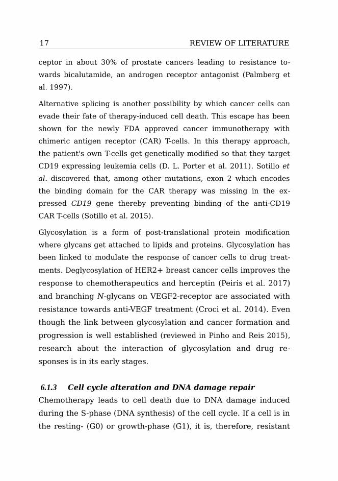
17 REVIEW OF LITERATURE
ceptor in about 30% of prostate cancers leading to resistance to-
wards bicalutamide, an androgen receptor antagonist (Palmberg et
al. 1997).
Alternative splicing is another possibility by which cancer cells can
evade their fate of therapy-induced cell death. This escape has been
shown for the newly FDA approved cancer immunotherapy with
chimeric antigen receptor (CAR) T-cells. In this therapy approach,
the patient's own T-cells get genetically modified so that they target
CD19 expressing leukemia cells (D. L. Porter et al. 2011). Sotillo et
al. discovered that, among other mutations, exon 2 which encodes
the binding domain for the CAR therapy was missing in the ex-
pressed CD19 gene thereby preventing binding of the anti-CD19
CAR T-cells (Sotillo et al. 2015).
Glycosylation is a form of post-translational protein modification
where glycans get attached to lipids and proteins. Glycosylation has
been linked to modulate the response of cancer cells to drug treat-
ments. Deglycosylation of HER2+ breast cancer cells improves the
response to chemotherapeutics and herceptin (Peiris et al. 2017)
and branching N-glycans on VEGF2-receptor are associated with
resistance towards anti-VEGF treatment (Croci et al. 2014). Even
though the link between glycosylation and cancer formation and
progression is well established (reviewed in Pinho and Reis 2015),
research about the interaction of glycosylation and drug re-
sponses is in its early stages.
6.1.3 Cell cycle alteration and DNA damage repair
Chemotherapy leads to cell death due to DNA damage induced
during the S-phase (DNA synthesis) of the cell cycle. If a cell is in
the resting- (G0) or growth-phase (G1), it is, therefore, resistant

REVIEW OF LITERATURE 18
to chemotherapy (Stewart et al. 2007). One of the cell cycle regu-
lating proteins is cyclin E, which associates with Cdk2 (cyclin-de-
pendent kinase 2) to regulate the transition into the S-phase,
where it gets degraded (Ohtsubo et al. 1995). If this mechanism is
out of balance, it can lead to reduced cell size, growth factor-in-
dependent proliferation, as well as lower cell growth rates (D. C.
Porter et al. 2001; Ohtsubo et al. 1995; Dulić et al. 1993). Addi-
tionally, a halt in the cell cycle gives cells time to repair therapy-
induced cell damage and therefore cancer cells can evade their
destiny of cell death. The DNA repair mechanisms nucleotide ex-
cision repair and homologous recombination reverse the effects of
DNA crosslinks of platinum-based drugs (Pennington et al. 2014;
Selvakumaran et al. 2003).
6.1.4 Avoiding apoptosis
The avoidance of apoptosis is a cancer hallmark (Hanahan and Wein-
berg 2000). Primary tumors present mechanisms that can lead to
failure of cancer therapy, especially if the drugs fail to induce other
forms of cell death, such as necroptosis (Krysko et al. 2017). For ex-
ample, it has been shown that an overexpression of the anti-apop-
totic protein Bcl-2 is present when resistance towards chemothera-
peutic drugs occurs (Ellis et al. 1998; Sartorius and Krammer 2002;
Miyashita and Reed 1993; Dole et al. 1994) and expression changes
in other anti- and pro-apoptotic family members were observed as
well (Kitada et al. 1998; G.-Q. Wang et al. 2001; Ni Chonghaile et al.
2011). However, whereas overexpression of MCL-1 can be connected
to poor patient survival after chemotherapy, there have been studies
that could not establish the link between overexpression of Bcl-2 and
poor treatment outcome (Kitada et al. 1998; Bosari et al. 1995). Nev-
ertheless, targeting mechanisms that lead to inhibition of apoptosis

19 REVIEW OF LITERATURE
are showing promising results. For example, preclinical data demon-
strate the effectiveness of navitoclax, a drug that promotes pro-apop-
totic and hinders anti-apoptotic signaling, when given as a single
agent or in combination with chemotherapy (van Delft et al. 2006;
Oltersdorf et al. 2005). However, overexpression of MCL-1 over-
writes the cytotoxicity of this drug (Konopleva et al. 2006). Therapy
that induces apoptosis by activating caspase-8 in combination with
chemotherapy has also shown promising results (Soria et al. 2010).
Even though not all changes in apoptotic signals are connected to
drug resistance, research shows that some are, meaning that
avoiding apoptosis is not only a cancer hallmark but also a hallmark
of drug resistance.
6.1.5 Resistance mediated by autophagy
Autophagy has an important role in intracellular homeostasis as it is
responsible for the recycling of unnecessary, damaged or aged cel-
lular components by lysosomal degradation. The removal of a cell's
damaged organelles, toxic proteins or oncogenic substrates reflects
the tumor suppressive character of autophagy, whereas intracellular
recycling provides food and energy for a cell and is tumor-promoting
(White 2012). Even though autophagy has been shown to promote
chemosensitization (Eum and Lee 2011; Zou et al. 2011) the latter
effect may facilitate drug resistance as it enables a tumor to respond
to environmental stress by increasing the breakdown of unnecessary
proteins and organelles. Targeting the p210BCR-ABL expressing
CML cells with imatinib can also induce autophagy. By inhibiting the
autophagous process with chloroquine an increase of imatinib-in-
duced cell death has been observed (Bellodi et al. 2009). Similar ef-
fects have been observed with the EGFR inhibitors gefitinib and er-
lotinib in human lung cancer cells (Han et al. 2011), in glioblastoma
multiforme where chloroquine was administered as an additional op-

REVIEW OF LITERATURE 20
tional adjuvant therapy (Briceño, Calderon, and Sotelo 2007), and in
colorectal cancer that was treated with the standard chemothera-
peutic 5-fluorouracil and 3-methyladenine, which is another au-
tophagy inhibitor (Jie Li et al. 2010; Sasaki et al. 2012).
6.1.6 Adaption of signaling
Cancer cells can also activate pro-survival signaling. For example,
the EGFR signaling cascade is activated to prevent chemothera-
peutic induced toxicity (Sumitomo et al. 2004; Van Schaeybroeck et
al. 2006, 2005). Even when EGFR is targeted cells can reprogram
their kinome and bypass the oncogene by activating HER3 (Sergina
et al. 2007; Wheeler et al. 2008). In addition, amplification of MET
has been observed to be the reason for drug resistance in 20% of
EGFR-driven lung cancers. MET bypasses the drug blocked EGFR by
inducing PI3K signaling via HER3 (Engelman et al. 2007).
6.1.7 Epithelial-mesenchymal transition (EMT)
During EMT, epithelial cells lose their polarization and cell-cell adhe-
sion and gain the more migratory phenotype of mesenchymal cells.
Several factors such as cytokines, chemokines, integrins, cadherins,
and metalloproteinases are involved in this phenotype change, which
has been undoubtedly linked to drug resistance (Singh and Set-
tleman 2010). To name a few examples, overexpression of Nanog, a
transcription factor linked to EMT, leads to cisplatin resistance in
ovarian cancer cells (Liu et al. 2016). An upregulation of TGF-β sig-
naling induces EMT in colon and triple-negative breast cancer cells,
which leads to doxorubicin resistance (W.-C. Chen et al. 2013; Jin-
peng Li et al. 2015). Also, EMT has been observed in patients with
non-small-cell lung carcinoma (NSCLC) treated with EGFR-inhibitors
(Uramoto et al. 2011; Sequist et al. 2011).

21 REVIEW OF LITERATURE
6.1.8 Tumor microenvironment
Not only can the tumor microenvironment contribute to tumor pro-
gression, invasion, and metastasis, it can also promote drug resis-
tance. The tumor microenvironment comprises the extracellular ma-
trix (ECM), blood and/or lymph vessels, cells associated to the im-
mune system as well as inflammation, and cancer-associated fibrob-
lasts (Bissell and Radisky 2001; Tlsty and Coussens 2006).
The heterodimeric integrin receptors are major connectors of cells to
the ECM. These interactions between integrins and ECM can render
different signaling pathways, such as the NF-κBB and PI3K pathways,
active (Danen 2005). High levels of β1-integrins have been associ-
ated with herceptin resistance in HER2-positive breast cancer (Les-
niak et al. 2009). In a cell line model of oral carcinoma, the adhe-
sion of β1-integrin even leads to cancer proliferation induced by
chemotherapeutic treatment with cisplatin (Eberle et al. 2011).
Furthermore, soluble factors released by the tumor itself or its
microenvironment have been shown to influence the outcome of
drug treatment. Interleukin-6 (IL-6), for example, confers mul-
tidrug resistance in breast cancer cells lines (Conze et al. 2001)
or cisplatin resistance in ovarian cancer (Cohen et al. 2013).
Other soluble factors such as fibroblast growth factor (FGF),
neuregulin (NRG1), hepatocyte growth factor (HGF) and epi-
dermal growth factor (EGF) to name just a few, have also been
identified to play a role in drug resistance (Wilson et al. 2012).
Some of these soluble factors are released by another important
component of the tumor microenvironment, the cancer-associated
fibroblasts (CAFs). HGF secretion by CAFs leads to resistance to-
wards RAF inhibition in BRAF-mutant melanoma (Straussman et
al. 2012). In addition, CAFs secrete platelet-derived growth factor C

REVIEW OF LITERATURE 22
and by this means induce resistance towards the inhibition of VEGF-
mediated angiogenesis (Crawford et al. 2009). Matrix metallopro-
teinase secretion leads to cetuximab resistance in head and neck
squamous cell carcinoma cell lines when co-cultured with CAFs (Jo-
hansson et al. 2012). Further, CAFs promote gemcitabine resistance
in pancreatic cancer by stimulating proliferation and survival path-
ways (Hwang et al. 2008).
The tumor vasculature is abnormal and defective compared to the
healthy tissue, leading to improper penetration of drugs, but also to
tissue areas that are nutrient-deprived and hypoxic (Tong et al.
2004). Hypoxic conditions induce HIF-1 transcriptions factor (con-
sisting of HIF-1α and HIF-1β, G. L. Wang et al. 1995), whose target
gene is MDR1 among others (see 5.1.1 Drug uptake, efflux, and me-
tabolism). Therefore, it does not come as a surprise that HIF-1α
overexpression has been observed in plenty of drug-resistant cancer
cell lines (Comerford et al. 2002; Nardinocchi et al. 2009; Zhu et al.
2005) as well as human colon and bladder cancer samples (Ding et
al. 2010; Sun et al. 2016). In addition, other resistance mechanisms
such as avoiding apoptosis (Chapter 5.1.4) and induction of au-
tophagy (Chapter 5.1.5) can involve HIF-1 (Rohwer and Cramer
2011).
6.1.9 Extracellular vesicles
Extracellular vesicles (EVs) facilitate the communication between
cells and can thereby influence the recipient cell's function. EVs can
simultaneously deliver all the different biomolecule categories
ranging from proteins to lipids, nucleic acids, and sugars (Yáñez-Mó
et al. 2015). Therefore, it is not surprising that EVs take part in the
communication between the tumor and its surrounding environment
(Milane et al. 2015) and are able to mediate drug resistance between

23 REVIEW OF LITERATURE
stromal and tumor cells. Examples such as the 5-fluorouracil resis-
tance in gastric cancer cells (Ji et al. 2015) or bortezomib resistance
in multiple myeloma cells (J. Wang et al. 2014) have been reported.
In addition, EVs are able to mediate the transfer of resistance from a
drug-resistant cell to a drug-sensitive cell. The best example is the
transfer of ABCB1 (P-glycoprotein) and even its protein-coding
mRNA MDR1 (see 5.1.1 Drug uptake, efflux, and metabolism) in
prostate (Corcoran et al. 2012), leukemia (Bebawy et al. 2009),
ovarian (F. Zhang et al. 2014), and breast cancer models (Pasquier et
al. 2012; Dong et al. 2014). Further, in leukemia, the MRP-1 trans-
porter (see 5.1.1 Drug uptake, efflux, and metabolism) has been
transferred from resistant to drug-sensitive cells (Lu et al. 2013).
Not only can EVs transfer drug efflux transporters, they can also se-
questrate drugs. The ABCG2 transporter, for example, localizes to
vesicles and the drug is then taken up into the EVs and released
from the cell (Ifergan, Scheffer, and Assaraf 2005).
Other molecules that are transferred by EVs and have been associ-
ated with promoting drug resistance and sensitivity are non-coding
RNAs such as miRNAs (19–25 nucleotides long) and lncRNAs (not
translated long RNA-transcripts). miRNAs from docetaxel-resistant
breast cancer cells target pathways related to drug therapy failure
and convey docetaxel resistance in a previous drug-sensitive cell line
(W.-X. Chen et al. 2014). The same has been observed with gemc-
itabine-resistant pancreatic cancer (Mikamori et al. 2017) or cis-
platin-resistant ovarian cancer (R. C. Pink et al. 2015) as well as lung
cancer cell lines (Wu et al. 2017). Even though drug resistance medi-
ated by long non-coding RNAs is a newly emerging research field,
several examples of how vesicles transfer lncRNAs exist. LincRNA-
ROR induces sorafenib or doxorubicin resistance and linc-VLDLR in-
duces the ABCG2 drug transporter (see above) in hepatocellular car-

REVIEW OF LITERATURE 24
cinoma cells (Takahashi, Yan, Kogure, et al. 2014; Takahashi, Yan,
Wood, et al. 2014).
It is obvious that drug resistance is a big complex research field as
there is not just one drug resistance mechanism. It is clear that
cancer cells can activate multiple mechanisms at the same time to
avoid drug-induced cancer cell death. Furthermore, one drug can be
inactivated by different resistance mechanisms as seen with cis-
platin, gemcitabine, imatinib, and 5-fluorouracil (see above). In addi-
tion, researchers continuously discover new mechanisms.
6.2 Resistance to TamoxifenEven 15 years post-treatment, tamoxifen has been shown to reduce
the death due to breast cancer by 30 %, compared to patients that
did not receive tamoxifen over a five-year period. However, the
cancer recurrence rate of 15 % five years after diagnosis of early-
stage ER-positive breast cancer has risen to 25 % after ten years
(Early Breast Cancer Trialists’ Collaborative Group (EBCTCG) 2005).
Almost all patients with metastatic breast cancer treated additionally
with tamoxifen eventually relapse. Various examples of tamoxifen re-
sistance have been proposed (Table 2), and more are to be discov-
ered.
Table 2: Possible mechanisms of tamoxifen resistance (modified from Tryfonidis et al. 2016)
Factors related to Examples
ER Loss of ER; ESR1 mutations; ER regulator modifications
Growth and survival Cell cycle regulation; deregulation of apoptosis; autophagy; cancer cell dormacy
Modification of signaling pathways
EGFR/HER2 signaling; AR signaling; PI3K/mTOR signaling

25 REVIEW OF LITERATURE
Factors related to Examples
Drug metabolism Decreased uptake; increased drug efflux/sequestration; metabolism of tamoxifen
Tumor microenvironment
CAFs; tumor stroma; extra cellular matrix; exosomes; immunomodulation
The most obvious mechanisms are related to the estrogen receptor,
tamoxifen's primary target. Mutations in ESR1 have been reported:
for example, a K303R gain-of-function mutation was detected in a
hinge region before the LB domain in hyperplastic lesions (Fuqua et
al. 2000). Cell line studies showed that if overexpressed and a
crosstalk with growth receptor pathways exists, this mutation can
lead to estrogen hypersensitivity and thus cells are less sensitive to
anti-estrogen treatment (Barone et al. 2010). However, several
studies failed to detect this mutation in primary and metastatic tu-
mors (Davies et al. 2005; Cancer Genome Atlas Network 2012; D. R.
Robinson et al. 2013; S. Li et al. 2013; Toy et al. 2013; Merenbakh-
Lamin et al. 2013; Jeselsohn et al. 2014). Less controversial is the
detection of mutations in the LBD of ESR1, which have been re-
ported in studies investigating metastatic ER-positive breast cancers.
The LBD ESR1 mutations were detected in 14%-54% of metastatic
ER+ tumor patients who received hormonal therapy including ta-
moxifen. The amino acid residues Y537 and D538 were most fre-
quently mutated. Further cell line studies showed that these muta-
tions lead to a ligand-independent activity as well as enhanced
ligand-stimulated activity, resulting in decreased tamoxifen sensi-
tivity (S. Li et al. 2013; D. R. Robinson et al. 2013; Toy et al. 2013;
Merenbakh-Lamin et al. 2013; Jeselsohn et al. 2014). In addition,
ESR1 amplification, as well as fusion genes, have been detected in
patients. However, their role in tamoxifen resistance is still unclear
(Jeselsohn et al. 2015).

REVIEW OF LITERATURE 26
Loss of ERα expression has been observed under tamoxifen treat-
ment (Johnston et al. 1995). Events such as CpG island methylation
and histone deacetylation (Parl 2003), hypoxia (Stoner et al. 2002),
EGFR and HER2 overexpression (Stoica et al. 2000; Creighton et al.
2006) as well as the interaction of p53 with ERα (Angeloni et al.
2004) have been proposed to be responsible for ERα downregula-
tion. However, roughly only 20% of patients with acquired tamoxifen
resistance lose ERα expression (Johnston et al. 1995; Kuukasjärvi et
al. 1996) indicating other means of tamoxifen resistance are at play.
Further, different expression levels of ERα co-regulatory proteins
have been linked to tamoxifen resistance. Well-studied examples in-
clude AIB1 (SRC3), SRC-1, NcoR and SMRT (Osborne et al. 2003;
Redmond et al. 2009; Girault et al. 2003; L. Zhang et al. 2013).
As mentioned above, administered tamoxifen is a prodrug that needs
to be metabolized in the liver to more potent metabolites, mainly en-
doxifen and 4-hydroxytamoxifen. One of the enzymes participating in
the conversion is cytochrome P450 2D6 (CYP2D6) (Klein et al. 2013).
Polymorphisms in this enzyme have been linked to a higher breast
cancer recurrence rate during tamoxifen only treatment (Province et
al. 2014). In addition, it has been observed that endoxifen is a sub-
strate of ABC-transporter ABCB1 (Teft, Mansell, and Kim 2011) and
a subsequent upregulation of the MDR1 gene could lead to tamox-
ifen resistance.
Probably the main mechanism by which tumor cells circumvent ta-
moxifen-induced death is the activation of alternative pathways and
factors. Especially HER2, EGFR, and IGFR play an important role,
but also the PI3K/AKT/mTOR signaling pathway, which is essential
for metabolism, cell survival and growth (García-Becerra et al.
2012). Results from the phase II TAMRAD trial or the phase III trial
BOLERO2 indicate that patients could benefit from additional

27 REVIEW OF LITERATURE
mTOR1 inhibition with everolimus. Nevertheless, these studies also
report a higher proportion of discontinuations due to adverse side
effects (Piccart et al. 2014; Treilleux et al. 2015). Further, the com-
plexity of the PI3K/AKT/mTOR pathway harbors the potential of de-
veloping drug resistance to the single inhibitors (Bihani et al. 2015;
Choi et al. 2016). As the targeting of this pathway is currently an ac-
tively studied field, time will tell whether this approach will lead to a
blockage of tumor progression.
As growth factors are known to induce EMT (Kalluri and Weinberg
2009), the upregulation of their receptors can be indirectly linked to
drug resistance. An upregulation of HER2 inhibits E-cadherin
leading to a reduced adherence of cells and then to EMT (D’souza
and Taylor-Papadimitriou 1994; Ingthorsson et al. 2016). Further,
other EMT promoting factors such as β-catenin, SLUG and SNAIL,
Notch and Wnt signaling have been associated with tamoxifen resis-
tance (Hiscox et al. 2006; Ye et al. 2010; Dhasarathy, Kajita, and
Wade 2007; Bui et al. 2017; Loh et al. 2013).
Immune cells within the tumor microenvironment have been known
to release growth factors that can lead to remodeling of ECM (see
above). Moreover, predictive gene-profiling of patients who were to
receive tamoxifen treatment linked an inflammation-related gene
cluster to therapy failure (Loi et al. 2008). In addition, cancer-associ-
ated fibroblasts, as well as their release of ILβ1, are associated with
tamoxifen resistance (Martinez-Outschoorn et al. 2011; Pontiggia et
al. 2012; Jiménez-Garduño et al. 2017).
Proteins involved in cell cycle regulation have also been detected to
impact tamoxifen resistance. Overexpression of ERα's direct target
cyclin D1, which promotes the progression through G1 to S phase,
has been linked to tamoxifen resistance in cell lines (Wilcken et al.
1997) and correlates with poor treatment outcome (Stendahl et al.

REVIEW OF LITERATURE 28
2004; Rudas et al. 2008). Also, cyclin E has been associated with ta-
moxifen resistance (Dhillon and Mudryj 2002; Hui et al. 2002).
The role of apoptosis avoidance in tamoxifen resistance is highly de-
batable, as changes in pro and antiapoptotic genes can be merely
due to activation of other resistance pathways (Butt, Sutherland, and
Musgrove 2007). However, increased expression of GRP78, a protein
that balances prodeath apoptosis and prosurvival autophagy, has
been reported to promote tamoxifen resistance (Cook et al. 2012).
Further, hydroxychloroquine, an autophagy inhibitor has been shown
to restore tamoxifen sensitivity (Cook et al. 2014).
In addition, it has been shown that miRNAs play a role in tamoxifen
resistance development and can function as a predictive marker (Xin
et al. 2009; Lyng et al. 2012; Huber-Keener et al. 2012). Interest-
ingly, miR-221/222, a well studied miRNA conveying tamoxifen resis-
tance (reviewed in Alamolhodaei et al. 2016), can be transferred via
exosomes to other cells and transform a tamoxifen sensitive cell to a
resistant one (Wei et al. 2014). The same phenomenon has been ob-
served for lncRNA UCA1 (Xu et al. 2016, 1).
As these examples show, it becomes more and more evident that a
cancer cell has a plethora of resistance mechanism towards tamox-
ifen at hand to prevent cell death. There is a need to define
biomarkers for each resistance mechanism and multiple therapeutic
options to combat them.

29 AIM OF THE STUDY
AIM OF THE STUDYSeveral therapies, such as surgery, radiation, chemo-, targeted, and
hormonal therapy, are used to treat breast cancer. Survival rates
have been improving as a result of therapies and earlier diagnoses.
However, cancer can develop drug resistances often leading to
therapy failure. Development of drug resistance and tumor progres-
sion are believed to be caused by a variety of mechanisms. The het-
erogeneity of resistance mechanisms most likely reflects the molec-
ular subtypes and intrinsic molecular properties of individual breast
cancers.
Therefore, the aims of my PhD work was to utilize breast cancer cell
line models to explore molecular pathogeneses and resistance mech-
anisms of breast cancer:
• Identify fusion genes in breast cancer, which could potentially
open new therapeutic approaches.
• Establish the molecular basis of tamoxifen resistance in breast
cancer by integrating exome-sequencing and drug profiling
using newly generated isogenic drug-resistant variants of
ER-positive breast cancers.
• Characterize the transcriptome of the tamoxifen-resistant cell
lines and define pathways as well as biomarkers of drug resis-
tance and identify ways to overcome resistance mechanisms.

MATERIALS AND METHODS 30
MATERIALS AND METHODSIn this chapter, materials and methods used in this thesis are briefly
portrayed. A detailed description can be found in the original publi-
cations, referred to by Roman numbers (I, II and III).
1 Cell culture and generation of tamoxifen-resistant cell lines (I, II, III)
BT-474 (HTB-20), MCF-7 (HTB-22), T-47D (HTB-133) and ZR-75-1
(CRL-1500) cells were obtained from American Type Culture Collec-
tion (Table 3). KPL-4 was a gift from Dr. Junichi Kurebayashi, D
(Tabuchi et al. 2009).
Table 3: Cell lines from human mammary gland
Cell line Description Used in
BT-474 Ductal carcinoma I, II, III
MCF-7 Adenocarcinoma derived by pleural effusion I, II, III
KPL-4 Inflammatory breast carcinoma derived by pleural effusion
I
T-47D Ductal carcinoma derived by pleural effusion II, III
ZR-75-1 Ductal carcinoma derived from ascites II, III
BT-474, MCF-7 and KPL-4 cells were grown in DMEM (PAN Biotech/
Euro Clone) supplemented with 10 % FCS (Gibco), 1 % penicillin/
streptomycin (Gibco) and 0,1 % bovine insulin (Sigma) except KPL-4.
ZR-75-1 and T-47D were grown in RPMI-1640 (PAN Biotech) supple-
mented with 10 % FCS (Gibco, Life Technologies, Carlsbad, CA), 1 %
penicillin/streptomycin (Gibco) and 0,1 % bovine insulin (Sigma) for
T-47D cell line.
Due to continuous exposure to 1 µM 4-OH-tamoxifen (Sigma) over a
time frame of 8-12 months seven tamoxifen-resistant cell lines (MCF-

31 MATERIALS AND METHODS
7 Tam1, T-47D Tam1 & Tam2, ZR-75-1 Tam1 & Tam2, BT-474 Tam1 &
Tam2) were derived from the parental cell lines (Figure 4). The cul-
ture media needed to be replaced every 2–3 days. Cells were pas-
saged when about 80 % confluent and incubated at 37 °C with 5 %
CO2. As the cell lines were grown in the presence of estrogen (FCS
and phenol), our studies mimic tamoxifen resistance in pre-
menopausal woman.
2 Characterization of tamoxifen-resistant cell lines (II, III)
2.1 Growth analysis (II)To measure the viability of the tamoxifen-resistant cells in study II,
CellTiter-Glo Cell Viability Assay (Promega) was performed with in-
creasing tamoxifen concentrations (0–1,8 μM). After 120M). After 120 h of incuba-
tion luminescence was measured with the PHERAstar plate reader
(Agilent Technologies). The active DNA synthesis and cell prolifera-
tion were measured with the Click-iT® EdU Alexa Fluor® 488 Flow
Figure 4: Schematic overview of tamoxifen-resistant cell lines. Tamoxifen resistant cell lines (color) were generated from parental cell line (no color) by exposure to 1µM 4-OH-tamoxifen (pills) for 8-12 months.
MCF-7
MCF-7 Tam1
T-47D
T-47D Tam1
ZR-75-1
ZR-75-1 Tam1
BT-474
BT-474 Tam1
T-47D Tam2
ZR-75-1 Tam2
BT-474 Tam2

MATERIALS AND METHODS 32
Cytometry Assay Kit (Life Technologies). The EdU Alexa Fluor® 488
incubation time was 4 h for T-47D and MCF-7 and 28 h for the BT-
474 and ZR-75-1 cells. The permeabilization of the ZR-75-1 cells had
to be adjusted to 0,1 % TritonX-100-PBS. After RibonucleaseA treat-
ment, the DNA content was detected using the FxCycle™ Far Red
(Life Technologies). The stained cells in suspension were analyzed
using Accuri C6 flow cytometer and its software (BD Biosciences).
2.2 Responsiveness to estrogens and downstream signaling (II)
The response to estrogen was measured by depriving all the cell
lines of estrogen. Therefore, phenol red-free medium (PAN Biotech)
was supplemented with 2,5 % dextran–charcoal-treated FCS (Sigma-
Aldrich). After 72 h, 17β-estradiol (Sigma, 10-8 M in ethanol) was
added for 4, 8 or 24 h. RNA was isolated and converted it into cDNA.
Quantitative-PCR with the DyNAmo ColorFlash SYBRGreen PCR kit
(Thermo Scientific) on the LightCycler 480 system (Roche) was per-
formed, to detect changes in pS2, PGR, and GREB1, the direct target
genes of ERα. Primer sequences can be found in Additional file 1 of
the original publication (Kangaspeska et al. 2016).
2.3 Measurements and drug induced changes in protein levels (II, III)
Changes in ERα in all the cell lines (Study II) and cathepsin D in the
T-47D cells (Study III) were investigated by Western blotting as de-
scribed earlier (Östling et al. 2011). Β-actin/-tubulin were used as
reference proteins. The effects of VX-11E (ERK inhibitor) and
selumetinib (MEK inhibitor) on several proteins were assessed with
increasing concentrations of VX-11E (50 nM, 100 nM, and 250 nM)
and with 100 nM VX-11E combined with 1 μM). After 120M selumetinib. Informa-
tion about all antibodies used for Western blotting is in Table 4.

33 MATERIALS AND METHODS
Table 4: Primary antibodies used for Western blotting (WB) and immunofluorescence (IF)
Antibody Product number and vendor
Application Study
ERα ab16660, Abcam WB II
EGFR CST4267, Cell Signaling Technologies (CST)
WB II
pEGFR CST3777, CST WB II
ERK1/2 CST9207, CST WB II
pERK1/2 CST4370, CST WB II
Cathepsin D 610800, BD Transduction Laboratories
WB III
β-actin A1978, Sigma WB II, III
β-tubulin ab6046, Abcam WB III
Ceruloplasmin ab110449, Abcam IF III
Lamp 1 H4A3, Developmental studies hybridoma bank (DSHB)
IF III
Lamp 2 H4B4, DSHB IF III
Galectin-3 556904, BD Biosciences IF III
Filipin F9765, Sigma IF III
2.4 Immunofluorescence staining (III)The parental and resistant T-47D cells were seeded on coverslips
with and without 1 µM 4-OH tamoxifen. Cells were fixed and
stained either directly with filipin for free cholesterol,
LipidTOXGreen neutral lipid stain, or primary antibodies (Table
4). Nuclei were detected with DRAQ5 (Biostatus) or Hoechst.
Images were acquired with a Nikon 90i microscope (Nikon) using
the 40x objective and processed with ImageJ (Schneider,
Rasband, and Eliceiri 2012) and the Adobe Photoshop software.

MATERIALS AND METHODS 34
2.5 Lysosomal membrane permeabilization (LMP) assay (III)
Galectin-3 translocation in the parental and resistant T-47D cells
was measured to investigate the lysosomal membrane integrity as
described earlier (Aits, Jäättelä, and Nylandsted 2015). The cells
were grown for 72 h +/- 1 µM 4-OH-tamoxifen and then incubated
with 1 mM LLMOe to induce LMP. Cells were fixed and stained
with galectin-3, ceruloplasmin as cell segmentation marker (Table
4) and Hoechst (405nm) to detect the nuclei. Confocal images
were taken with the PE Opera Phenix HCS system (PerkinElmer)
with the 40x water immersion objective (NA 1.1). Images were
analyzed with the Columbus Image Data Storage and Analysis
System (PerkinElmer). LMP was assessed by calculating the av-
erage spots per cell and the percentage of galectin-3 positive
cells within an image.
2.6 Measurement of triglycerides and cholesterol esters (III)
Triglycerides and cholesterol esters were extracted from the
parental and resistant T-47D cells and measured by thin layer
chromatography (TLC) as previously described (Bligh and Dyer
1959). The lipid bands were quantified using ImageJ (Schneider,
Rasband, and Eliceiri 2012).
3 Paired-end RNA-sequencing (I, III)For study I, data from our previously produced paired-end RNA li-
braries of the BT-474, MCF-7, and KPL-4 cell lines were used (Ed-
gren et al. 2011).
For study III, after RNA isolation with miRNeasy kit (Qiagen),

35 MATERIALS AND METHODS
RNA quality control with the Agilent Bioanalyzer using the
RNApico chip (Agilent) and determination of RNA amount with
Qubit RNA-kit (Life Technologies), a strand-specific paired-end
RNA-sequencing library was prepared with ScriptSeq™ Complete
kit for human/mouse/rat (Illumina). The mean fragment size was
ca 300-400 nucleotides, as fragments smaller than 200 bp were
removed by SPRI beads (Agencourt AMPure XP). The paired-end
library was sequenced with the Illumina HiSeq 2000 (Illumina) in-
strument. A detailed description can be found in Kumar et al.
(Kumar et al. 2017).
4 RNA-sequencing analysis (I, III)
4.1 Fusion gene detection, characterization, and validation (I, III)
Fusion genes were detected in study I and III with the
FusionCatcher algorithm using raw, unprocessed read files
(Edgren et al. 2011; Nicorici et al. 2014). Ensembl version 61
(Study I) and Ensembl version 80 (Study III) were used for se-
quence alignment to the human genome. A database containing
exon-exon fusion points and the junction sequences, as well as infor-
mation about both fusion partners, was generated. A fusion partner
was allowed to have multiple partner genes.
Fusion genes were further characterized and validated in study I.
Protein domains were annotated using SMART (Schultz et al. 1998;
Letunic, Doerks, and Bork 2012) and the 1M oligo Agilent aCGH data
(Edgren et al. 2011) was used to determine copy number changes
and gene coverage. Fusion genes were validated by Sanger-se-
quencing. Shortly, isolated RNA was reverse transcribed into cDNA.
With fusion gene-specific primers, the region of interest was ampli-

MATERIALS AND METHODS 36
fied and the gel-purified PCR-product was cloned into pCR2.1-TOPO
vector (Invitrogen). The vector was transferred into bacteria and
colony PCR was used to confirm a successful transfection. After
plasmid purification, the product was Sanger-sequenced and the ob-
tained sequences were compared with the predicted fusion se-
quence, using the multiple sequence alignment tool (Corpet 1988).
The chromosomal breakpoints of the fusion genes were determined
by Sanger-sequencing of the genomic DNA of the BT-474 cell line.
Primers for amplification of the region around the fusion breakpoint
were designed based on already existing exome-sequencing data.
The PCR product was directly Sanger-sequenced and the exact chro-
mosomal breakpoints were located using BLAT (Kent 2002) with the
“Near-exact matches” option. The primers of this study can be found
in Table S4 of the original publication (Kangaspeska et al. 2012).
4.2 Integration of transcriptomic data and differential gene expression (III)
Public patient transcriptome data (McBryan et al. 2015) was
downloaded from GEO database (Barrett et al. 2013). RNA-se-
quencing data analysis, which contained read pre-processing,
read alignment to the human reference genome (Ensembl
GRCh38) guided by the Ensembl reference gene models and fea-
tures (Ensembl v80), and count estimation are described in detail
in Kumar et al. (Kumar et al. 2017). The estimated expression
counts of cell line and patient data were combined, normalized
using the trimmed mean of M-values (TMM) method (M. D.
Robinson and Oshlack 2010), converted to counts per million
(CPM) estimates using edgeR (M. D. Robinson, McCarthy, and
Smyth 2010), corrected for the batch effect associated with the
study origin using limma (Smyth and Speed 2003), and assigned

37 MATERIALS AND METHODS
to Ensembl information using biomaRt (Smedley et al. 2015) with
default settings. Lists of differentially expressed genes, defined as
log2 ratio of >= |1| and CPM difference of >= |10| when com-
paring parental versus resistant cell line was generated and up-
loaded to Enricher (E. Y. Chen et al. 2013; Kuleshov et al. 2016)
to determine pathways involved in tamoxifen resistance develop-
ment. The data was visualized using principle component anal-
ysis, Venn diagrams as well as heat maps.
5 Genomic profiling by exome-sequencing (II)
We utilized exome-sequencing to detect point mutations or copy
number changes that arose during the development of tamoxifen re-
sistance by comparing parental with the isogenic resistant cell line.
The exomes were captured from genomic DNA with NimbleGen Se-
qCap EZ Human Exome v2.0 kit (Roche NimbleGen) and paired-end
sequenced on the Illumina HiSeq platform. The detection of the point
mutations has been previously described (Pemovska et al. 2013). The
parental cell lines were used as controls and Ensembl v66 was used
as annotation database. We excluded known false positive point mu-
tations and considered mutations with p < 0,05 and resistant variant
frequency >30 % with high confidence as present. Copy number
variants (CNVs) were detected with a variant calling pipeline (Su-
lonen et al. 2011). Copy number values were calculated as RPKM
(reads per kilobase length per million mapped reads) for exome
target regions and the log2 copy number ratios determined by using
the parental cell line as a reference. The values were then assigned
to the human genome based on Ensembl database v67 using the ex-
treme method option in GISTIC2 (Mermel et al. 2011). Thresholds to
identify CNVs were set at -0,4 (heterozygous deletion), -1,2 (homozy-

MATERIALS AND METHODS 38
gous deletion), +0,5 (gain) and +1,3 (amplification).
6 Drug sensitivity and resistance testing (DSRT) (II, III)
6.1 Cell viability measurement (II, III)The DSRT platform has been described in detail before (Pemovska et
al. 2013). For study II we used the FIMM FO2Baq library, which con-
tains 279 investigational and approved cancer drugs, on all the
parental and resistant cell lines. For study III we used 33 drugs tar-
geting the lipid and cholesterol metabolism, the lysosomal mem-
brane, and oxidation processes, in addition to drugs that have been
part of study II (Kangaspeska et al. 2016). We tested the 33 drugs
with the T-47D parental and resistant cell lines. In both studies, neg-
ative (0,1 % DMSO only) as well as positive (100 μM). After 120mol/l benzetho-
nium chloride) controls were included. Five different concentrations
of one drug covered a 10000-fold concentration range. The optimal
amount of cells was grown in the cell's normal media, and supple-
mented with 1 μM). After 120M 4-OH-tamoxifen. The incubation time was 72 h at
37 °C. CellTiter-Glo Cell Viability Assay (Promega) was used to mea-
sure the viability with the PHERAstar plate reader.
6.2 High-content phenotypic drug profiling (III)
In study III we additionally fixed plates from the DSRT with the
33 drugs with 4% PFA in PBS for image-based phenotyping. We
stained the plates with LipidTOXGreen neutral lipid stain
(ThermoFisher Scientific) and Hoechst for nuclei detection.
Confocal images were acquired with the PE Opera Phenix HCS
system (PerkinElmer) with the 40x water immersion objective (NA

39 MATERIALS AND METHODS
1.1). Laser power and exposure time were unchanged during the
imaging of the different plates with the same staining.
7 DSRT data analysis (II, III)
7.1 Analysis of cell viability (II, III)The raw ATP-measurements were analyzed with our in-house
bioinformatic pipeline Breeze, which gives dose response curves,
IC50 values as well as a Drug Sensitivity Score (DSS) for each in-
dividual drug treatment (Yadav et al. 2014). DSS scores (Study
III) and DSS differences between parental and resistant cell lines
(Study II) were visualized as a heat map. Rank product analysis
(Breitling et al. 2004) was used to identify drugs that significantly
changed their efficacy while tamoxifen resistance is obtained
(Study II).
7.2 Construction of drug sensitivity and co-resistance networks (II)
In study II, drug sensitivity and resistance networks were con-
structed based on drugs that showed a DSS difference of at least five
when comparing resistant with parental cell line DSS. Drug targets
were extracted from the ChEMBL database (https://www.ebi.ac.uk /
chembl) and assigned a Kinase Inhibitor BioActivity (KIBA)-score
(Tang et al. 2014). Based on the KIBA-score, point mutations, copy
number changes and canonical pathway for Estrogen Receptor Sig-
naling (www.qiagen.com) we used the Ingenuity Pathway Analysis
application (Ingenuity® Systems, Qiagen) (Krämer et al. 2014) to
construct sensitivity and co-resistance networks.

MATERIALS AND METHODS 40
7.3 Analysis of high-content phenotypic drug profiling (III)
Images were utilized to measure LipidTOXGreen signal and calcu-
late a DSS based on cell counts. Images were flatfield corrected
using CIDRE method (Smith et al. 2015), stitched to a single
multi-channel image per well, as well as segmented and analyzed
with CellProfiler 2.2.0 (Kamentsky et al. 2011). The LipidTOX-
Green signal was measured for each individual cell and a mean of
the intensities per well calculated. Dose-response curves were
drawn for the cell counts and used to calculate a DSS for each
drug in Breeze.

41 RESULTS & DISCUSSION
RESULTS & DISCUSSION
1 Fusion genes In study I we reanalyzed our already published RNA-sequencing data
(Edgren et al. 2011) by changing parameters such as the annotation
version of Ensembl to map the reads to the human genome, allowing
a fusion partner to be fused to more than one gene and having as
little as two reads to indicate the presence of a fusion gene. The re-
analysis predicted 16 additional fusion genes, which we validated
and characterized. Additionally, in study III we evaluated the
changes of fusion genes after tamoxifen resistance had developed.
1.1 Fusion gene characteristics in breast cancer
Out of the 16 newly predicted fusion genes, we were able to Sanger-
sequence 14 (Study I, Table 1 and S1). Our previously published and
validated 27 breast cancer fusion genes (Edgren et al. 2011), and the
results of others from solid tumors, show that fusion genes are not
just an event prevalent to hematological cancers. Indeed, in 2015,
9189 fusion genes have been found in solid tumors compared to 759
fusion genes in hematological diseases. However, only around 2 %
were found to be recurrent in solid tumors, compared to about 20 %
in hematological diseases (Mertens et al. 2015). The fusion genes we
identified were not recurrent. Nevertheless, one of the fusion part-
ners, for example, MED1 was found to be fused to different genes
(STXBP4, USP32, and ACSF2) in our study (Study I, Figure 3). Other
studies, for example, identified genes of the MAST- and NOTCH-gene
families to be fused to different partners in 4 or 6 % of breast cancer
patients, respectively (D. R. Robinson et al. 2011). The clinical
phase I trials with MK-0752, a γ-secretase inhibitor, which prevents

RESULTS & DISCUSSION 42
NOTCH activation, have shown promising results in advanced breast
cancer (Schott et al. 2013) and solid tumor patients (Krop et al.
2012). However, those studies were not selective for the NOTCH-fu-
sion genes and an evaluation of the fusion genes as predictive
markers or even therapy targets would be necessary. Even though
we did not detect NOTCH gene fusions, results like these show that
a disease-driving fusion partner could be targeted by therapy. In-
deed, FDA-approved drugs are already in use to treat NSCLC har-
boring ALK fusions and myeloproliferative neoplasms with PDGFR
fusions (Mertens et al. 2015).
Alternative splicing events such as cis- (intrachromosomal) and
trans-splicing (interchromosomal) or genomic rearrangements are
underlying fusion gene formation. The majority of fusion genes are
formed by genomic rearrangements due to genomic instability and
are frequently found at high-level amplifications (Study I, Table 1
and Figure 2, Stephens et al. 2009; Edgren et al. 2011; Kalyana-Sun-
daram et al. 2012). Most of the studied fusion genes are at a ge-
nomic breakpoint. We were able to sequence the exact chromosomal
breakpoints of three fusion genes (Study I, Figure 1b). The THRA-
AC090627.1 and MED1-ACSF2 fusion shared sequence microho-
mology of 1 bp at the breakpoint (Study I, Figure 1b). It is therefore
suggested that microhomology-mediated break-induced replication
(Lawson et al. 2011) is the mechanism by which these two genes
were joined.
We further identified the EEF1DP3-FRY fusion in the KPL-4 cell line
as a read-through event (Study I, Table S1). A read-through fusion,
also known as cis-transcription-induced gene fusion (cis-TIGF), oc-
curs when a large pre-mRNA, which contains two neighboring genes,
is transcribed and, due to splicing events, the two genes are fused
together (Akiva et al. 2006). Around 5 % of genes have been pro-

43 RESULTS & DISCUSSION
posed to form read-through fusions (Parra et al. 2006). However, the
majority of read-through fusions have little or no cellular impact and
may even be artifacts (Yu et al. 2014; He et al. 2018).
1.2 The role of fusion genes in tamoxifen resistance
In addition to the reanalysis of fusion genes in study I, we investi-
gated the fusion genes in the tamoxifen-resistant, as well as their iso-
genic parental cell lines (Study III). At first sight it appeared that the
cells acquire fusion genes during the resistance development as
some fusion genes were not found in the parental cell lines
(Study III, Additional file 6). After thorough investigation, all fusion
genes annotated in the Fusion Catcher results file as “known_fusion”
or “cell_lines” have been identified in the same parental cell line be-
fore (D. Kim and Salzberg 2011; Edgren et al. 2011; Kangaspeska et
al. 2012; Kalyana-Sundaram et al. 2012; Klijn et al. 2015). The elimi-
nation of all previously reported fusion genes and read-through fu-
sions, left only two acquired fusion genes (SMARCC1-EMCN in MCF-
7 Tam1 and RAD9A-FUT10 in ZR-75-1 Tam1), which have not been
detected in the parental cell line before. However, none are recur-
rent, suggesting that they do not play a major role in the develop-
ment of tamoxifen resistance. We were able to detect two shared
read-through fusions ABCC11-ABCC12 (BT-474 Tam1, T-47D Tam1
and ZR-75-1 Tam2) and TRIM3-HPX (T-47D Tam1 and ZR-75-1
Tam2, Study III, Additional file 6). The TRIM3-HPX has been de-
tected in prostate adenocarcinoma (Nacu et al. 2011), indicating
that it is neither breast cancer- and nor tamoxifen resistance-spe-
cific. ABCC11 and ABCC12 are highly homologous, which implies
that this read-through event is an artifact due to the mapping of
reads to similar genomes.

RESULTS & DISCUSSION 44
We were able to detect the ESR1-CCDC170 fusion in the MCF-7, as
well as in the tamoxifen-resistant MCF-7 Tam1, but not in the ZR-75-
1 as previously reported (Veeraraghavan et al. 2014) or in its iso-
genic tamoxifen-resistant clones (Study III, Additional file 6). The
ESR1-CCDC170 fusion has been identified in about 4 % of ER-posi-
tive breast tumors and tandem duplication has been proposed as the
underlying mechanism for the fusion formation (Veeraraghavan et al.
2014; Holst 2016). ESR1 is downstream of CCDC170 and a fusion of
the two genes should result in a CCDC170-ESR1 read-through fu-
sion, which we detected in MCF-7, but not in MCF-7 Tam1 (Study III,
Additional file 6). Whether the fusion contributes to endocrine resis-
tance is highly questionable. ESR1 contributes only with its pro-
moter region to form a fusion gene with CCDC170's coding regions
and a still fully functional ESR1 gene encodes for ERα. Indeed, dif-
ferential allele-specific expression (DASE) studies indicate that
CCDC170 alone is responsible for the breast cancer susceptibility
(Jiang et al. 2017), which has been earlier associated to the
CCDC170/C6orf97-ESR1 locus (6q25.1) by genome-wide association
studies (GWAS) (Turnbull et al. 2010; Dunning et al. 2016).
Nevertheless, true ESR1 fusion genes have been detected in ER-posi-
tive breast cancers. These include, ESR1-YAP1, which lacks the
ligand-binding domain (LBD/AF-2) and has therefore been shown to
drive endocrine resistance by promoting estrogen-independent cell
growth (S. Li et al. 2013).
2 Characterization of tamoxifen-resistant cell lines
We generated seven tamoxifen-resistant cell lines out of four
parental cell lines, by exposing them to 1 µM 4-OH-tamoxifen over a
period of 8-12 months (Figure 4, Study II, Figure 1A). The selected

45 RESULTS & DISCUSSION
cell lines reflect the molecular subtypes which would benefit from
endocrine-therapy. MCF-7, T-47D and ZR-75-1 were classified as lu-
minal A and the HER2-positive BT-474 cells as the luminal B-subtype
(Subik et al. 2010). The BT-474 cells functioned primarily as a con-
trol as HER2 overexpression is a known resistance mechanism to ta-
moxifen (García-Becerra et al. 2012). Before we obtained stable
growing tamoxifen-resistant cells, each of the clones underwent at
least one round of mass mortality, leaving just a few regrowing cells.
Once regrown, we assessed their tolerance towards tamoxifen and
found that the resistant cells were more tolerant, confirming that ta-
moxifen resistance has developed (Study II, Additional file 3A).
We further evaluated their growth, estrogen-responsiveness, ge-
nomic and transcriptional changes as well as their drug profiles.
2.1 Tamoxifen-resistant cells alter their cell cycle
Overall, we noticed during cell culturing that the resistant cells grew
slower than the parental cells and increased their doubling time dra-
matically. Further, FACS analysis revealed that all the resistant cells
accumulate in the G0/G1 phase (Study II, Additional file 3B). Studies
with another anti-estrogen resistant as well as a tamoxifen-resistant
model have also reported an increase in the doubling time and con-
sequently G0/G1 accumulation of the cells (Fog, Christensen, and
Lykkesfeldt 2005; H. S. Kim et al. 2015). Interestingly, the MCF-7
Tam1 cells accumulate less in G0/G1 (59 %), than the parental cells
under tamoxifen treatment (68 %). When ZR-75-1 Tam1 cells are ex-
posed to media without tamoxifen they dramatically halt in G0/G1
compared to tamoxifen treatment (86 % vs. 68 %), implying a tamox-
ifen addiction, which has been observed previously (van den Berg et
al. 1989). Withdrawal response to tamoxifen has also been reported

RESULTS & DISCUSSION 46
in breast cancer patients and can even lead to cancer remission
(Howell et al. 1992; Canney et al. 1987). On the other hand, non-ad-
herence to tamoxifen therapy is associated with a higher mortality
(McCowan et al. 2008). These results show that there would be a
need to define markers that could identify tamoxifen-addicted tu-
mors and help to stratify patients for therapy.
Interestingly, RNA-sequencing and subsequent pathway analysis of
differentially expressed genes showed that genes involved in cell
cycle were especially enriched in MCF-7 Tam1 & ZR-75-1 Tam1
(Study III, Table 2, Additional file 8). The results indicate that al-
tering the cell cycle properties is one way to overcome tamoxifen-in-
duced cell death.
2.2 Changes in ERα protein-levels and estrogen-responsiveness
Western blotting revealed that ERα was decreased in the ZR-75-1-
and T-47D-derived resistance models (Study II, Additional file 4C),
which is in line with previous research (van den Berg et al. 1989;
Graham et al. 1992). Acquired tamoxifen resistance has been associ-
ated with the loss of ERα expression (Oh et al. 2010) and epigenetic
mechanisms such as DNA methylation and histone deacetylation
have been proposed to underlay these changes (Sharma et al. 2005;
Y. Zhou et al. 2007). By contrast, in MCF-7 Tam1 resistant cells, the
levels of ERα remained almost unchanged, and in BT-474 resistant
clones, an increase was observed (Study II, Additional file 4C).
Therefore, depending on the molecular background, tamoxifen either
renders ERα stable (Lykkesfeldt, Madsen, and Briand 1994; J. J. Pink
and Jordan 1996; A. F. Leary et al. 2010) or promotes its destabiliza-
tion via proteasome-mediated degradation (Marsaud et al. 2003). At
the same time, we observed a decrease in ERα target gene transcrip-

47 RESULTS & DISCUSSION
tion (pS2, PGR, GREB1) as well as impaired estradiol-responsivity in
all resistant cell lines (Study II, Additional file 4A and 4B), sug-
gesting that even if the ERα is stabilized, its function may be im-
paired. Further, the loss of progesterone receptor frequently occurs
upon acquiring anti-estrogen resistance (Lykkesfeldt, Madsen, and
Briand 1994; Drury et al. 2011; Arnedos et al. 2014). Our results
suggest that different resistant clones may exhibit either steroid-de-
pendent or -independent growth and differ in their ER-mediated re-
sponses.
2.3 Genetic alterations in the genomes of tamoxifen-resistant cell lines
It has been reported that tamoxifen resistance is driven by genetic
adaptations such as point mutations or copy number changes (Karnik
et al. 1994; Borg et al. 1994; Dowsett et al. 2001; Moelans et al.
2010; Burandt et al. 2013; Lundgren et al. 2008; S. Li et al. 2013, 1;
D. R. Robinson et al. 2013; Toy et al. 2013; Merenbakh-Lamin et al.
2013; Jeselsohn et al. 2015). We, therefore, performed exome-se-
quencing on all the cell lines and determined resistance-specific
point mutations and copy number variations by comparing each ta-
moxifen-resistant cell line to its parental cell line. From around 250
to 350 acquired non-synonymous mutations per resistant cell, we
compared the high confident ones and found that no acquired point
mutation was shared across all the resistant cell lines (Study II, Addi-
tional file 8 and 9). Nevertheless, between the clones derived from
the same parental cell line, we found some shared mutations, which
involved TNS1, PTH2R, and NHLRC2 in BT-474 Tam1 and Tam2 and
TIMM23 and RP11-368 J21.2.1 in ZR-75-1 Tam1 and Tam2 (Study II,
Additional file 8 and 9). Copy number changes were predominantly
large heterozygous deletions, affecting many of the loci across the
genome, including chromosomes 7 and X. The ZR-75-1 Tam1 resis-

RESULTS & DISCUSSION 48
tant cell clone, on the other hand, acquired only copy number gains.
Whereas T-47D Tam1 and Tam2 had some shared copy number alter-
ations (mainly 7q), almost none were shared between the ZR-75-1
and BT-474 derived resistant clones (Study II, Additional file 8 and
10). Contrary to previous reports (Karnik et al. 1994; S. Li et al.
2013; Toy et al. 2013; D. R. Robinson et al. 2013; Merenbakh-Lamin
et al. 2013; Jeselsohn et al. 2015), we observed no acquired loss or
mutations of PTEN, PIK3CA or ESR1 in any of the drug-resistant
variants. However, unlike with ESR1 mutations, the causal relation-
ship between PTEN and PIK3CA mutations and tamoxifen resistance
has been questioned (López-Knowles et al. 2010; Beelen et al. 2014).
Thus, our results, and those from others (Block et al. 2012), suggest
that accumulation of numerous genomic aberrations can trigger re-
sistance development. In fact, antiestrogen resistance can be seen
even in the absence of any evident mutations (Brünner et al. 1997),
also without any in ESR1 (Martin et al. 2017). An absence of resis-
tance driving mutations has been observed in leukemic cells as well
(Pisco et al. 2013). The results imply that the emerged mutations and
copy number changes are rather passengers due to the selection
pressure than true drivers of tamoxifen resistance (Pisco and Huang
2015).
2.4 Transcriptional changes in tamoxifen-resistant cell lines and their resemblance to patients
Due to a lack of apparent genomic changes that could explain tamox-
ifen resistance, we investigated in study III the transcriptome of the
tamoxifen-resistant cell lines, to detect gene expression as well as
pathway changes. Further, we were able to compare our findings to
sequential tumor samples from patients that developed endocrine re-
sistance (McBryan et al. 2015).

49 RESULTS & DISCUSSION
In line with the exome-sequencing results (Study II), we did not iden-
tify a single common differentially expressed gene across all the re-
sistant cell lines (Study III, Figure 1B). Even within the clones de-
rived from the same parental cell line, a maximum of 38 % of genes
were commonly deregulated (Study III, Additional file 5 S1A). A
handful of common point mutations, shared copy number alterations
(Study II, Additional file 8 and 10) and diversified gene expression
changes (Study III, Figure 1A), indicate that each of the resistant
clones chooses its own way to acquire tamoxifen resistance.
Interestingly, the only study so far that has investigated global ex-
pression changes of sequential tumors from tamoxifen-treated pa-
tients identified less than 3 % of differentially expressed genes
(McBryan et al. 2015). Our reanalysis of their data set, where we
compared primary with the corresponding metastatic tumor, con-
firmed the results (Study III, Figure 1C). Further, principle compo-
nent analysis with the expressed genes of patients and cell lines clas-
sified as luminal A subtype (Subik et al. 2010) showed that the pa-
tient samples cluster together with the luminal A cell lines (Study III,
Figure 1D). In addition, we were able to identify SERPINA1,
PLXDC2, NAV1, DOCK10, and LRP1 as differentially expressed when
looking within the luminal A subtype. Almost nothing is known about
these genes and their relationship to tamoxifen resistance. Only
SERPINA1, which is upregulated in the patient samples as well, has
been shown to be upregulated by ligand-independent binding of ERα
(Chan et al. 2015). Indeed, when ERα protein expression is down
(Study II, Additional file 4C), we see a decrease of SERPINA1 in T-
47D Tam1, T-47D Tam2, ZR-75-1 Tam1 and ZR-75-1 Tam2 and vice
versa for the MCF-7 Tam1. In the patient samples, we see a slight
increase in ESR1 expression (Study III, Additional file 7). Another
interesting finding was the enormous increase in ceruloplasmin (CP)

RESULTS & DISCUSSION 50
expression in the resistant T-47D cell lines (240-290 fold-increase), in
MCF-7 Tam1 (26 fold-increase) and the three metastatic patient
samples (12 to 57 fold-increase; Study III, Additional file 7).
Ceruloplasmin has been suggested to be a marker for metastatic
breast cancer (Schapira and Schapira 1983) and it has been seen to
increase in the blood during tamoxifen treatment (Rössner and Wall-
gren 1984; Kailajärvi et al. 2000). Unfortunately, those studies did
not investigate the survival or recurrence rate of the patients with
increased ceruloplasmin-levels. In addition, it has been reported that
high levels of ceruloplasmin correlate with lower remission rate
(Ozyilkan et al. 1992), questioning the predictive value of
ceruloplasmin as a marker for metastatic disease.
In addition to the analysis of expression changes of single genes,
we used the differentially expressed genes and performed a
pathway analysis (Study III, Table 2, Additional file 8). Also, the
results of the pathway analysis supported the notion that every
cell line finds its own way to cope with the tamoxifen pressure. As
discussed earlier (2.1. Tamoxifen-resistant cells alter their cell
cycle), MCF-7 Tam1 & ZR-75-1 Tam1 mainly changed the
expression of genes involved in cell cycle. In the other cell lines,
altered genes were mainly involved in protein modification (BT-
474 Tam2), or extracellular matrix organization (ZR-75-1 Tam2 &
BT-474 Tam1), whereas T-47D Tam1 & Tam2 displayed changes in
metabolism associated genes, especially in those of the
cholesterol pathway (Study III, Table 2, Figure 2A). The results of
the ZR-75-1 Tam2 and BT-474 Tam1 needs to be viewed with
caution as the adjusted p-value is low due to a low amount of
input genes (Study III, Table 2).
Interestingly, we also observed an increase of cholesterol

51 RESULTS & DISCUSSION
pathway related genes in the metastatic patient samples
(Study III, Figure 2A). McBryan et al. did, however, remove all the
liver-related genes from their analysis as the breast cancer
metastasis samples were taken from the liver (McBryan et al.
2015). Nevertheless, our results raise the hen and egg question:
What came first, the changes in cholesterol pathway related
genes in breast cancer cells as a resistance mechanism or the
settlement of the metastatic breast cancer cells in the liver and
subsequent adjustment of cholesterol pathway related genes due
to the liver environment? The reprogramming of lipid metabolism,
however, is currently emerging as a player in metastatic cancer
(reviewed in Luo et al. 2017) and in drug resistance (C.-L. Chen et
al. 2016).
2.5 Drug responses of tamoxifen-resistant cell lines
When drug-resistance develops and signaling pathways in tumor
cells change, new therapy vulnerabilities or even co-resistance to
other drugs may arise. Therefore, we investigated in study II the
drug responses of the seven tamoxifen-resistant and their parental
cell lines towards 279 investigational and approved oncology drugs.
We identified common, cell type-, and cell clone-specific sensitivities
and co-resistances by calculating the DSS for each compound and
comparing parental with resistant cell line (Study II,
Figure 2, 3, 4, 5, 6, 7, Additional file 2, 5, 11, 12). We drew drug re-
sponse networks by integrating drug targets with copy number
changes (Study II, Figure 4, 5, 6, 7, Additional file 11 and 12). How-
ever, as the majority of the copy number changes are heterozygous
deletions, their role in the drug responses are potentially minor. The
networks are highly cell line specific, confirming once more that dif-

RESULTS & DISCUSSION 52
ferent resistance mechanisms develop in these cell lines.
We observed only a few common effector drugs (drugs with DSS dif-
ference < |5|) across the cell lines (Study II, Figure 2, 5A and 5B). As
the MCF-7 Tam 1 cell lines developed a rather overwhelming co-re-
sistance profile, there were no drugs to which all the resistant cell
lines became more sensitive (Study II, Figure 5A). However, one of
the drugs to which the other resistant cell lines were more sensitive,
was VX-11e (Study II, Figure 5A and 5B). It has been shown that VX-
11e induces ERK1/2 phosphorylation (Chaikuad et al. 2014, 1). The
resistant cells had reduced ERK1/2 phosphorylation compared with
the parental cell line, which was then increased under VX-11e treat-
ment (Study II, Figure 5C). Interestingly, a study by Zhou et al. ob-
served that tamoxifen induces cell death when ERK1/2 is continu-
ously activated (J.-H. Zhou et al. 2007). As VX-11e also induced cell
death in the parental cell line when supplemented with 1 µM tamox-
ifen (Study II, Additional File 5), the proposed mechanism by Zhou et
al. might explain the increased sensitivity to VX-11e.
On the other spectra, all of the tamoxifen-resistant cell lines devel-
oped a co-resistance towards paclitaxel (Study II, Figure 5A and 5B),
but also other chemotherapeutics were less effective in a more cell-
specific manner (Study II, Figure 3, 4, 6, Additional file 11 and 12).
As stated in study II, ER has been associated with chemoresistance
(Shi et al. 2014; Kurebayashi et al. 2010; Tabuchi et al. 2009), but in
the case of the tamoxifen-resistant cell lines we assume the lower
effectiveness of chemotherapeutics is rather due to the cells' slow
growth. Indeed, in study III we performed another DSRT, including
paclitaxel, with the T-47D cells (Study III, Figure 4A, Additional
file 9). Paclitaxel was more effective in the T-47D Tam2 cells in
this screen than in study II. However, we also observed an in-
creased proliferation in the T-47D Tam2 cells (Study III, unpub-

53 RESULTS & DISCUSSION
lished) compared to our first screen (Study II). Importantly, T-47D
Tam1, which grow at the same speed as in the first screen, con-
firmed the results in study II of reduced sensitivity towards pacli-
taxel (Study III, Figure 4A, Additional file 9).
In the HER2 overexpressing BT-474 cell lines, we saw that HER2/
EGFR inhibitors (gefitinib, ibrutinib, neratinib) were even more
effective in the tamoxifen-resistant cells compared to the already
sensitive parental cells (Study II, Figure 5, 6, Additional file 2). In-
terestingly, neratinib has been recently approved by the FDA for
treatment of early-stage HER2-positive breast cancer (Center for
Devices and Radiological Health, 2017). Analyzing the RNA-se-
quencing data, we detected changes in genes associated with pro-
tein modification (BT-474 Tam1 & Tam2), especially glycosylation
(Study III, Table 2). Glycosylation has been connected to oncogen-
esis (Ng et al. 1987; Vavasseur et al. 1994) and shown to modu-
late the response of HER2+ breast cancer cells to chemotherapy
and herceptin treatments (Peiris et al. 2017). Therefore, one
could speculate that changes in glycosylation of the EGFR re-
ceptor family members potentially make them more accessible to
their ligands, or in this case, drugs.
In addition to our results from the exome- and RNA-sequencing,
the drug profiling confirmed that clone-specific responses un-
derlay the tamoxifen-resistance and that different drugs are
needed to overcome each resistance mechanism.
3 Lipid changes in T-47D and their consequences
In study III, we investigated the T-47D cell lines more in depth. RNA-
sequencing indicated changes in cholesterol and lipid metabolism,

RESULTS & DISCUSSION 54
which were found in the patient samples as well (Study III, Table 2,
Figure 2A). We were able to observe an enormous increase in choles-
terol and neutral lipids by immunofluorescence as well as with thin
layer chromatography (Study III, Figure 2 and 4B). It has recently
been reported that the use of cholesterol-lowering drugs in en-
docrine-therapy prevents breast cancer recurrence. Even though this
study did not detect any benefits in the tamoxifen treatment only
arm, cholesterol levels were decreased (Borgquist et al. 2017). This
indicates that the increase of cholesterol does not seem to be a
normal drug response and might indeed be a resistance mechanism.
We further observed that the cholesterol accumulates in highly en-
larged lysosomes (Study III, Figure 3A and B). Lysosomes are in-
volved in mediating different types of cell death, such as apoptosis,
necroptosis, necrosis, and ferroptosis, through lysosomal mem-
brane permeabilization (LMP; Galluzzi, Bravo-San Pedro, and
Kroemer 2014). Therefore, one could say that preventing LMP can
lead to drug resistance. Indeed, when we treated the tamoxifen-re-
sistant T-47D cells with LLOMe, an LMP inducing agent, we ob-
served a decrease in the number of galectin-3 positive lysosomes
compared to the parental cells (Study III, Figure 3C and D). Fur-
thermore, it has been shown that accumulation of cholesterol (Ap-
pelqvist et al. 2011), an increase in lysosomal-associated mem-
brane protein 1 (Lamp1) and Lamp2 (Fehrenbacher et al. 2008),
as well as downregulation of cathepsins prevent LMP (Liaudet-
Coopman et al. 2006). All of these LMP inhibiting mechanisms
were seen in T-47D Tam1 and Tam2 (Study III, Figure 3).
We also measured an increase in triglycerides and lipid droplets
in these cells (Study III, Figure 2C, D and 4B). However, when
targeting the lipid phenotype with drugs affecting the lipid me-
tabolism, we did not observe a reversal of the lipid phenotype or a

55 RESULTS & DISCUSSION
major reduction in cell viability (Study III, Figure 4, Additional
file 9 and 10). We, therefore, suggest that the lipid droplets are
mainly functioning more as energy storage than a resistance
mechanism.
Compounds targeting pathways involved in regulating the reac-
tive oxygen species (ROS), such as SOD1 inhibitor LCS-1, were
able to induce cell death in all the T-47D cells. ROS are able to in-
duce LMP (Aits and Jäättelä 2013) and we did see an upregulation
of SOD1 expression in both of the tamoxifen-resistant cell clones
(Study III, Additional file 7). SOD1 encodes for a superoxide dis-
mutase, which turns superoxide into either O2 or H2O2, lowering
the ROS that could induce LMP. Also RSL-3, which inhibits the
glutathione peroxidase 4 that prevents lipid peroxidation, was
able to induce cell death in all the cell lines. In addition, we can
see an upregulation of ceruloplasmin, an enzyme that converts
Fe2+ to Fe3+ and thus prevents the Fenton-reaction, which would
produce superoxide that would then induce LMP. When putting all
these results in perspective, it seems that the tamoxifen-resistant
T-47D cells circumvent tamoxifen-induced cell death by pre-
venting LMP due to the reduction in superoxide, downregulation
of cathepsin D, and an increase in cholesterol and LAMPs (Figure
5).

RESULTS & DISCUSSION 56
These results also show that resistance mechanisms are very well
ingrained and targeting a particular resistance mechanism might
not always be successful or harbor only a small window of oppor-
tunity.
Figure 5: Proposed resistance mechanism of tamoxifen-resistant T-47D cells. The lysosomal membrane is stabilized by upregultation of Lamp1, Lamp2 and cholesterol. Upregulation of cerulopasmin (CP) and superoxide dismutase (SOD1) prevents the A Fenton as well as B Haber-Weiss reaction, respectively. Thereby ROS gets reduced and lysosomal membrane permeabilization (LMP) is not induced. LMP would, together with cathepsins, trigger various types of cell death. Lamp1/2, lysosomal-associated membrane protein 1/2; CAT, catalase; GPx, glutathion peroxidase
Cholesterol
Cathepsin D
Lamp1
Lamp2LMP
Necroptosis
Apoptosis
Necrosis
Ferroptosis
H2O2Fe2++
H2O2 + Fe2+ Fe3++ OH-
HO +
Fe3+O2-
+
CP
Fe2+ + O2
SOD1
H2O + O2
CAT/GPx
A
B

57 CONCLUSION
CONCLUSIONTamoxifen is one of the most widely used targeted therapies for ER-
positive breast cancer. However, patients that initially respond to the
therapy can acquire drug resistance and the cancer ultimately re-
lapses. A thorough understanding of the complex mechanisms of
drug resistance is needed in order to use tamoxifen more effectively.
This understanding can guide development of combinations with
other therapies to prevent drug resistance. It can also enable the de-
tection resistance when it occurs, and present an opportunity to
switch therapies or target the resistance mechanism.
In my thesis work I, therefore, investigated the genomic and tran-
scriptomic changes of seven tamoxifen-resistant cell lines, tried to
identify compounds that can target those changes, and explored the
possibility of fusion genes as possible prognostic or diagnostic
markers. It became evident relatively quickly that each of the resis-
tant cell lines had developed its own drug resistance profile, or as I
would say I discovered the “Seven shades of tamoxifen resistance”. I
observed the different molecular changes in the genetic-, transcrip-
tomic- and drug-profile with limited overlaps. I identified that the
MCF-7 resistant cell line mainly regulated their growth properties
and displayed multi-drug resistance. The resistant ZR-75-1s still
need a thorough investigation about their mechanisms, but there are
hints of cell cycle and adhesion protein modification. The resistant
BT-474s cells modified their protein glycosylation and adhesion ac-
cording to RNA-Sequencing analysis. And last but by no means least,
the T-47D cells prevented lysosomal membrane permeabilization by
upregulating their cholesterol metabolism. The example of the T-47D
cell lines additionally show how difficult it is to target the resistance
mechanisms, and one might need to look beyond the field of cancer

CONCLUSION 58
research in order to understand and combat them.
On one hand, it is confirming that the models represent already
known resistance mechanisms, as well as sharing similarities with
patient samples, making them appropriate models to study the
molecular mechanisms of resistance development and discover
possible drug vulnerabilities. On the other hand, seeing the diver-
sity of these mechanisms in only seven resistant cell lines enor-
mously highlights the need of personalized diagnosis and therapies
in treatment of tamoxifen-resistant breast cancer.
Whether fusion genes can be used as diagnostic markers or even
drug-targets as in hematological cancers such as CML, can be
doubted because the diversity of breast cancer is so high. Never-
theless, maybe one of the recurrent fusion gene partners could be
utilized as a therapeutic target and on a personalized therapy
level they could function as cancer progression markers and indi-
cate possible therapy success.
I was once asked by a scientist that actually works in this field if
this breast cancer field is highly competitive. I pointed to my re-
sults and said whoever develops another resistance model will
find other mechanisms and only when we have enough of the data
freely available we might be able to stratify patients. However,
more important than models would be the existence and access to
clinical data sets, such as the set published by McBryan et al.,
which not only contains the primary tumor information, but also
the information of diagnosed acquired tamoxifen resistance with
response/survival information. This would enable researchers to
compare the relevance of their models and prove that the resis-
tance mechanism of interest exists in human cancer.

x ACKNOWLEDGMENTS
ACKNOWLEDGMENTSFirst and foremost I want to thank Sara Kangaspeska. I am thankful
for bringing me onto the project. Even though you were not my official
supervisor, I enjoyed your excellent supervision with valuable com-
ments, guidance, encouragement and little pushes even after you left
the research group. I admire your drive to sit down and wrap things
up.
This study was carried out at the Institute for Molecular Medicine Fin-
land (FIMM) over quite some years. I would like to thank everyone at
the Institute for making it a wonderful and encouraging workplace.
Which brings me to one of my supervisors, Olli Kallioniemi who has
played an existential role in creating this excellent work environment.
I am highly grateful for letting me continue with this project even
though all the breast cancer expertise had supposedly left the group. I
am thankful for giving me the freedom and the trust to design the last
study. I also want to express my gratitude to Taija af Hällström, Päivi
Östling, and Vilja Pietiäinen for your interest to work with me. I espe-
cially want to thank Vilja, who became one of my supervisors, for ex-
ploring with me the lysosomes and the iron. I highly appreciate your
motivational enthusiasm and happiness about science. I hope you can
preserve this attitude in the years to come.
I would like to thank my thesis reviewers Tuula Kallunki and Katri
Pylkäs for their kind comments and my thesis committee members
Kaisa Lehti and Johanna Mattson for their encouragement to wrap
things up. In the end, it took longer than expected. Further, I would
like to thank Robert Mills for having a final glance over the thesis.
Of course, such a study could not have been done without the support
of the people in the lab, namely Anna, Mariliina, Katja, Sanna and Piia
who helped with culturing and generating the cells, the experiments,

ACKNOWLEDGMENTS xi
and the general lab stuff. The FIMM Tech Centers such as the se-
quencing unit with Pekka Ellonen and Pirkko Mattila, the HTB unit
with Ida Lindenschmidt, Laura Turunen, Maria Nurmi and most defi-
nitely Swapnil Podar, and finally the HCA unit with Antti Hassinen
and Peter Horvath, without these units such an extensive screening
and sequencing would not have been possible. I also want to thank
all my co-authors: Henrik Edgren, Daniel Nicorici, Astrid Murumägi,
Alok Jaiswal, John-Patrick Mpindi, Samuli Eldfors, Oscar Brück, Tero
Aittokallio, Matti Kankainen, Lassi Paavolainen, Ruusu-Maria Ko-
vanen and Elina Ikonen. And I want to give my sincerest apologies
for all the guilt that the bioinformaticians felt when they “acciden-
tally” met me in the aisle.
Many thanks to old and new lab mates that have not directly been in-
volved in my studies but participated in the group discussions and
were always good for a joke: Sami, Vesa, Teijo, Maija W., Anna A.,
Anna L., Dima, Riku, Tanja, Kalle, Minxia, Akira, Clement and Maija
P.. An extra big thanks go to Susanna Rosas for all the help and
making the impossible possible.
I spent a great deal of my FIMM time in the back office. I would like
to thank everyone, who shared the office with me, for being such
nice roomies. Of course, I want to highlight Disha, Khalid and Poo-
jitha for all the on- and off-topic discussions we had.
Along those lines, I would like to thank the not so secret club (you
know who you are) for having thoughtful and silly discussions over a
good and sometimes not so good whisky.
I want to sincerely thank Gretchen and Mari for encouraging and
supporting me to organize the science and art exhibitions.
Of course, a PhD would not have been possible without the (mental)
support of friends. A big thank you goes out to all my friends that

xii ACKNOWLEDGMENTS
kept me company during travel, hockey matches, lunch, beer and bar
explorations, parties, girls evenings, ravitolapäivät, issue discussions,
concerts, espresso and tea breaks, to the ones that let me be part of
their weddings and introduced me to their families and friends. A big
hug to Jenni M. & Vesa L., Annabrita, Pauliina, Jenni L, Manu, Daria,
Jennifer, Maren, Malu, Piia, Rita, Yuexi, Prson, Alok, Arjan, Kamila,
Mamun, Tini, Sami, Elsa, Stefano, Riikka, Dima, Alex, Poojitha &
Tuomas, Disha & Ashwini, the people from Kuopio, the Canadian and
the German hockey gang and all the others I have met over the past
years.
Finally, I want to thank my family for all their support during my life,
for letting me explore the world and for encouraging me no matter
what. Not to forget, thank you for accepting that I am sometimes not
reachable. DANKE für Alles!

REFERENCES xiii
REFERENCESAits, Sonja, and Marja Jäättelä. 2013. “Lysosomal Cell Death at a Glance.” Journal of Cell
Science 126 (Pt 9): 1905–12.
Aits, Sonja, Marja Jäättelä, and Jesper Nylandsted. 2015. “Methods for the Quantification of Lysosomal Membrane Permeabilization: A Hallmark of Lysosomal Cell Death.” Methods in Cell Biology 126: 261–85.
Akiva, Pinchas, Amir Toporik, Sarit Edelheit, Yifat Peretz, Alex Diber, Ronen Shemesh, Amit Novik, and Rotem Sorek. 2006. “Transcription-Mediated Gene Fusion in the Human Genome.” Genome Research 16 (1): 30–36.
Alamolhodaei, Nafiseh Sadat, Javad Behravan, Fatemeh Mosaffa, and Gholamreza Karimi. 2016. “MiR 221/222 as New Players in Tamoxifen Resistance.” Current Pharmaceutical Design 22 (46): 6946–55.
Anderson, Elizabeth, Robert B. Clarke, and Anthony Howell. 2000. “Estrogen Receptor in Mammary Gland Physiology.” In Endocrine Oncology, 1–16. Contemporary Endocrinology. Humana Press, Totowa, NJ. https://link.springer.com/chapter/10.1007/978-1-59259-223-4_1.
Angeloni, S. V., M. B. Martin, P. Garcia-Morales, M. D. Castro-Galache, J. A. Ferragut, and M. Saceda. 2004. “Regulation of Estrogen Receptor-Alpha Expression by the Tumor Suppressor Gene P53 in MCF-7 Cells.” The Journal of Endocrinology 180 (3): 497–504.
Anna Azvolinsky. 2017. “How Cancers Evolve Drug Resistance.” The Scientist. 2017. http://www.the-scientist.com/?articles.view/articleNo/48998/title/How-Cancers-Evolve-Drug-Resistance/.
Appelqvist, Hanna, Cathrine Nilsson, Brett Garner, Andrew J. Brown, Katarina Kågedal, and Karin Ollinger. 2011. “Attenuation of the Lysosomal Death Pathway by Lysosomal Cholesterol Accumulation.” The American Journal of Pathology 178 (2): 629–39.
Arnedos, M., S. Drury, M. Afentakis, R. A’Hern, M. Hills, J. Salter, I. E. Smith, J. S. Reis-Filho, and M. Dowsett. 2014. “Biomarker Changes Associated with the Development of Resistance to Aromatase Inhibitors (AIs) in Estrogen Receptor-Positive Breast Cancer.” Annals of Oncology: Official Journal of the European Society for Medical Oncology 25 (3): 605–10.
Barone, I., D. Iacopetta, K. R. Covington, Y. Cui, A. Tsimelzon, A. Beyer, S. Andò, and S. a. W. Fuqua. 2010. “Phosphorylation of the Mutant K303R Estrogen Receptor Alpha at Serine 305 Affects Aromatase Inhibitor Sensitivity.” Oncogene 29 (16): 2404–14.
Barrett, Tanya, Stephen E. Wilhite, Pierre Ledoux, Carlos Evangelista, Irene F. Kim, Maxim Tomashevsky, Kimberly A. Marshall, et al. 2013. “NCBI GEO: Archive for Functional Genomics Data Sets--Update.” Nucleic Acids Research 41 (Database issue): D991-995.
Baum, M., D. M. Brinkley, J. A. Dossett, K. McPherson, J. S. Patterson, R. D. Rubens, F. G. Smiddy, et al. 1983. “Improved Survival among Patients Treated with Adjuvant Tamoxifen after Mastectomy for Early Breast Cancer.” Lancet (London, England) 2 (8347): 450.
Bebawy, M., V. Combes, E. Lee, R. Jaiswal, J. Gong, A. Bonhoure, and G. E. R. Grau. 2009. “Membrane Microparticles Mediate Transfer of P-Glycoprotein to Drug Sensitive Cancer Cells.” Leukemia 23 (9): 1643–49.
Beelen, Karin, Mark Opdam, Tesa M. Severson, Rutger H. T. Koornstra, Andrew D. Vincent, Jelle Wesseling, Jettie J. Muris, et al. 2014. “PIK3CA Mutations, Phosphatase and Tensin Homolog, Human Epidermal Growth Factor Receptor 2, and Insulin-like Growth Factor 1 Receptor and Adjuvant Tamoxifen Resistance in Postmenopausal Breast Cancer Patients.” Breast Cancer Research: BCR 16 (1): R13.
Bekele, Raie T., Ganesh Venkatraman, Rong-Zong Liu, Xiaoyun Tang, Si Mi, Matthew G. K. Benesch, John R. Mackey, et al. 2016. “Oxidative Stress Contributes to the Tamoxifen-Induced Killing of Breast Cancer Cells: Implications for Tamoxifen Therapy and

xiv REFERENCES
Resistance.” Scientific Reports 6 (February).
Bell, Daphne W., Ira Gore, Ross A. Okimoto, Nadia Godin-Heymann, Raffaella Sordella, Roseann Mulloy, Sreenath V. Sharma, et al. 2005. “Inherited Susceptibility to Lung Cancer May Be Associated with the T790M Drug Resistance Mutation in EGFR.” Nature Genetics 37 (12): 1315–16.
Bellodi, Cristian, Maria Rosa Lidonnici, Ashley Hamilton, G. Vignir Helgason, Angela Rachele Soliera, Mattia Ronchetti, Sara Galavotti, et al. 2009. “Targeting Autophagy Potentiates Tyrosine Kinase Inhibitor-Induced Cell Death in Philadelphia Chromosome-Positive Cells, Including Primary CML Stem Cells.” The Journal of Clinical Investigation 119 (5): 1109–23.
Berg, H. W. van den, M. Lynch, J. Martin, J. Nelson, G. R. Dickson, and A. D. Crockard. 1989. “Characterisation of a Tamoxifen-Resistant Variant of the ZR-75-1 Human Breast Cancer Cell Line (ZR-75-9a1) and Ability of the Resistant Phenotype.” British Journal of Cancer 59 (4): 522–26.
Bertotti, Andrea, Giorgia Migliardi, Francesco Galimi, Francesco Sassi, Davide Torti, Claudio Isella, Davide Corà, et al. 2011. “A Molecularly Annotated Platform of Patient-Derived Xenografts (‘xenopatients’) Identifies HER2 as an Effective Therapeutic Target in Cetuximab-Resistant Colorectal Cancer.” Cancer Discovery 1 (6): 508–23.
Bihani, Teeru, Scott A. Ezell, Brendon Ladd, Shaun E. Grosskurth, Anne Marie Mazzola, Mark Pietras, Corinne Reimer, Michael Zinda, Stephen Fawell, and Celina M. D’Cruz. 2015. “Resistance to Everolimus Driven by Epigenetic Regulation of MYC in ER+ Breast Cancers.” Oncotarget 6 (4): 2407–20.
Bissell, M. J., and D. Radisky. 2001. “Putting Tumours in Context.” Nature Reviews. Cancer 1 (1): 46–54.
Bligh, E. G., and W. J. Dyer. 1959. “A Rapid Method of Total Lipid Extraction and Purification.” Canadian Journal of Biochemistry and Physiology 37 (8): 911–17.
Block, Martin, Carsten Gründker, Stefanie Fister, Julia Kubin, Ludwig Wilkens, Michael D. Mueller, Bernhard Hemmerlein, Günter Emons, and Andreas R. Günthert. 2012. “Inhibition of the AKT/MTOR and ErbB Pathways by Gefitinib, Perifosine and Analogs of Gonadotropin-Releasing Hormone I and II to Overcome Tamoxifen Resistance in Breast Cancer Cells.” International Journal of Oncology 41 (5): 1845–54.
Borg, A., B. Baldetorp, M. Fernö, D. Killander, H. Olsson, S. Rydén, and H. Sigurdsson. 1994. “ERBB2 Amplification Is Associated with Tamoxifen Resistance in Steroid-Receptor Positive Breast Cancer.” Cancer Letters 81 (2): 137–44.
Borgquist, Signe, Anita Giobbie-Hurder, Thomas P. Ahern, Judy E. Garber, Marco Colleoni, István Láng, Marc Debled, et al. 2017. “Cholesterol, Cholesterol-Lowering Medication Use, and Breast Cancer Outcome in the BIG 1-98 Study.” Journal of Clinical Oncology: Official Journal of the American Society of Clinical Oncology 35 (11): 1179–88.
Bosari, S., L. Moneghini, D. Graziani, A. K. Lee, J. J. Murray, G. Coggi, and G. Viale. 1995. “Bcl-2 Oncoprotein in Colorectal Hyperplastic Polyps, Adenomas, and Adenocarcinomas.” Human Pathology 26 (5): 534–40.
Breitling, Rainer, Patrick Armengaud, Anna Amtmann, and Pawel Herzyk. 2004. “Rank Products: A Simple, yet Powerful, New Method to Detect Differentially Regulated Genes in Replicated Microarray Experiments.” FEBS Letters 573 (1–3): 83–92.
Briceño, Eduardo, Alejandra Calderon, and Julio Sotelo. 2007. “Institutional Experience with Chloroquine as an Adjuvant to the Therapy for Glioblastoma Multiforme.” Surgical Neurology 67 (4): 388–91.
Brisken, C., S. Park, T. Vass, J. P. Lydon, B. W. O’Malley, and R. A. Weinberg. 1998. “A Paracrine Role for the Epithelial Progesterone Receptor in Mammary Gland Development.” Proceedings of the National Academy of Sciences of the United States of America 95 (9): 5076–81.
Brownell, J. E., and C. D. Allis. 1996. “Special HATs for Special Occasions: Linking Histone Acetylation to Chromatin Assembly and Gene Activation.” Current Opinion in Genetics & Development 6 (2): 176–84.
Brünner, N., B. Boysen, S. Jirus, T. C. Skaar, C. Holst-Hansen, J. Lippman, T. Frandsen, M.

REFERENCES xv
Spang-Thomsen, S. A. Fuqua, and R. Clarke. 1997. “MCF7/LCC9: An Antiestrogen-Resistant MCF-7 Variant in Which Acquired Resistance to the Steroidal Antiestrogen ICI 182,780 Confers an Early Cross-Resistance to the Nonsteroidal Antiestrogen Tamoxifen.” Cancer Research 57 (16): 3486–93.
Bui, Quyen Thu, Ji Hye Im, Sung Baek Jeong, Young-Mi Kim, Sung Chul Lim, Bumseok Kim, and Keon Wook Kang. 2017. “Essential Role of Notch4/STAT3 Signaling in Epithelial-Mesenchymal Transition of Tamoxifen-Resistant Human Breast Cancer.” Cancer Letters 390 (April): 115–25.
Burandt, E., G. Jens, F. Holst, F. Jänicke, V. Müller, A. Quaas, M. Choschzick, et al. 2013. “Prognostic Relevance of AIB1 (NCoA3) Amplification and Overexpression in Breast Cancer.” Breast Cancer Research and Treatment 137 (3): 745–53.
Butt, Alison J., Robert L. Sutherland, and Elizabeth A. Musgrove. 2007. “Live or Let Die: Oestrogen Regulation of Survival Signalling in Endocrine Response.” Breast Cancer Research: BCR 9 (5): 306.
Butta, A., K. MacLennan, K. C. Flanders, N. P. Sacks, I. Smith, A. McKinna, M. Dowsett, L. M. Wakefield, M. B. Sporn, and M. Baum. 1992. “Induction of Transforming Growth Factor Beta 1 in Human Breast Cancer in Vivo Following Tamoxifen Treatment.” Cancer Research 52 (15): 4261–64.
Cancer Genome Atlas Network. 2012. “Comprehensive Molecular Portraits of Human Breast Tumours.” Nature 490 (7418): 61–70.
Canney, P. A., T. Griffiths, T. N. Latief, and T. J. Priestman. 1987. “Clinical Significance of Tamoxifen Withdrawal Response.” Lancet (London, England) 1 (8523): 36.
Carlson, Bob. 2006. “Oncotype DX Test Offers Guidance For Women Debating Chemotherapy.” Biotechnology Healthcare 3 (4): 12–14.
Carroll, Jason S., Clifford A. Meyer, Jun Song, Wei Li, Timothy R. Geistlinger, Jérôme Eeckhoute, Alexander S. Brodsky, et al. 2006. “Genome-Wide Analysis of Estrogen Receptor Binding Sites.” Nature Genetics 38 (11): 1289–97.
Castoria, G., A. Migliaccio, A. Bilancio, M. Di Domenico, A. de Falco, M. Lombardi, R. Fiorentino, L. Varricchio, M. V. Barone, and F. Auricchio. 2001. “PI3-Kinase in Concert with Src Promotes the S-Phase Entry of Oestradiol-Stimulated MCF-7 Cells.” The EMBO Journal 20 (21): 6050–59.
Cenni, B, and D Picard. 1999. “Ligand-Independent Activation of Steroid Receptors: New Roles for Old Players.” Trends in Endocrinology and Metabolism: TEM 10 (2): 41–46.
Center for Devices and Radiological Health, Center for Drug Evaluation and Research. 2017. “Approved Drugs - FDA Approves Neratinib for Extended Adjuvant Treatment of Early Stage HER2-Positive Breast Cancer.” WebContent. 2017. https://www.fda.gov/Drugs/InformationOnDrugs/ApprovedDrugs/ucm567259.htm.
Chaikuad, Apirat, Eliana Tacconi, Jutta Zimmer, Yanke Liang, Nathanael S. Gray, Madalena Tarsounas, and Stefan Knapp. 2014. “A Unique Inhibitor Binding Site in ERK1/2 Is Associated with Slow Binding Kinetics.” Nature Chemical Biology 10 (10): 853–60.
Chan, Hei Jason, Haiqing Li, Zheng Liu, Yate-Ching Yuan, Joanne Mortimer, and Shiuan Chen. 2015. “SERPINA1 Is a Direct Estrogen Receptor Target Gene and a Predictor of Survival in Breast Cancer Patients.” Oncotarget 6 (28): 25815–27.
Chen, C. J., D. Clark, K. Ueda, I. Pastan, M. M. Gottesman, and I. B. Roninson. 1990. “Genomic Organization of the Human Multidrug Resistance (MDR1) Gene and Origin of P-Glycoproteins.” The Journal of Biological Chemistry 265 (1): 506–14.
Chen, Chia-Lin, Dinesh Babu Uthaya Kumar, Vasu Punj, Jun Xu, Linda Sher, Stanley M. Tahara, Sonja Hess, and Keigo Machida. 2016. “NANOG Metabolically Reprograms Tumor-Initiating Stem-like Cells through Tumorigenic Changes in Oxidative Phosphorylation and Fatty Acid Metabolism.” Cell Metabolism 23 (1): 206–19.
Chen, Edward Y., Christopher M. Tan, Yan Kou, Qiaonan Duan, Zichen Wang, Gabriela Vaz Meirelles, Neil R. Clark, and Avi Ma’ayan. 2013. “Enrichr: Interactive and Collaborative HTML5 Gene List Enrichment Analysis Tool.” BMC Bioinformatics 14 (April): 128.
Chen, Wei-Chih, Ying-An Lai, Ying-Chao Lin, Jui-Wen Ma, Li-Fen Huang, Ning-Sun Yang,

xvi REFERENCES
Chi-Tang Ho, Sheng-Chu Kuo, and Tzong-Der Way. 2013. “Curcumin Suppresses Doxorubicin-Induced Epithelial-Mesenchymal Transition via the Inhibition of TGF-β and PI3K/AKT Signaling Pathways in Triple-Negative Breast Cancer Cells.” Journal of Agricultural and Food Chemistry 61 (48): 11817–24.
Chen, Wei-Xian, Yan-Qin Cai, Meng-Meng Lv, Lin Chen, Shan-Liang Zhong, Teng-Fei Ma, Jian-Hua Zhao, and Jin-Hai Tang. 2014. “Exosomes from Docetaxel-Resistant Breast Cancer Cells Alter Chemosensitivity by Delivering MicroRNAs.” Tumour Biology: The Journal of the International Society for Oncodevelopmental Biology and Medicine 35 (10): 9649–59.
Choi, Ae-Ran, Ju-Hwa Kim, Yeon Hwa Woo, Ji Hyun Cheon, Hyung Sik Kim, and Sungpil Yoon. 2016. “Co-Treatment of LY294002 or MK-2206 with AZD5363 Attenuates AZD5363-Induced Increase in the Level of Phosphorylated AKT.” Anticancer Research 36 (11): 5849–58.
Clark, S. S., J. McLaughlin, W. M. Crist, R. Champlin, and O. N. Witte. 1987. “Unique Forms of the Abl Tyrosine Kinase Distinguish Ph1-Positive CML from Ph1-Positive ALL.” Science (New York, N.Y.) 235 (4784): 85–88.
Clarke, R. B., A. Howell, C. S. Potten, and E. Anderson. 1997. “Dissociation between Steroid Receptor Expression and Cell Proliferation in the Human Breast.” Cancer Research 57 (22): 4987–91.
Coezy, E., J. L. Borgna, and H. Rochefort. 1982. “Tamoxifen and Metabolites in MCF7 Cells: Correlation between Binding to Estrogen Receptor and Inhibition of Cell Growth.” Cancer Research 42 (1): 317–23.
Cohen, Sharon, Ilan Bruchim, Dror Graiver, Zoharia Evron, Varda Oron-Karni, Metsada Pasmanik-Chor, Ram Eitan, et al. 2013. “Platinum-Resistance in Ovarian Cancer Cells Is Mediated by IL-6 Secretion via the Increased Expression of Its Target CIAP-2.” Journal of Molecular Medicine (Berlin, Germany) 91 (3): 357–68.
Cole, M. P., C. T. Jones, and I. D. Todd. 1971. “A New Anti-Oestrogenic Agent in Late Breast Cancer. An Early Clinical Appraisal of ICI46474.” British Journal of Cancer 25 (2): 270–75.
Comerford, Katrina M., Timothy J. Wallace, Jörn Karhausen, Nancy A. Louis, Michael C. Montalto, and Sean P. Colgan. 2002. “Hypoxia-Inducible Factor-1-Dependent Regulation of the Multidrug Resistance (MDR1) Gene.” Cancer Research 62 (12): 3387–94.
Conze, D., L. Weiss, P. S. Regen, A. Bhushan, D. Weaver, P. Johnson, and M. Rincón. 2001. “Autocrine Production of Interleukin 6 Causes Multidrug Resistance in Breast Cancer Cells.” Cancer Research 61 (24): 8851–58.
Cook, Katherine L., Ayesha N. Shajahan, Anni Wärri, Lu Jin, Leena A. Hilakivi-Clarke, and Robert Clarke. 2012. “Glucose-Regulated Protein 78 Controls Cross-Talk between Apoptosis and Autophagy to Determine Antiestrogen Responsiveness.” Cancer Research 72 (13): 3337–49.
Cook, Katherine L., Anni Wärri, David R. Soto-Pantoja, Pamela Ag Clarke, M. Idalia Cruz, Alan Zwart, and Robert Clarke. 2014. “Hydroxychloroquine Inhibits Autophagy to Potentiate Antiestrogen Responsiveness in ER+ Breast Cancer.” Clinical Cancer Research: An Official Journal of the American Association for Cancer Research 20 (12): 3222–32.
Corcoran, Claire, Sweta Rani, Keith O’Brien, Amanda O’Neill, Maria Prencipe, Rizwan Sheikh, Glenn Webb, et al. 2012. “Docetaxel-Resistance in Prostate Cancer: Evaluating Associated Phenotypic Changes and Potential for Resistance Transfer via Exosomes.” PloS One 7 (12): e50999.
Cornu, Jean-Nicolas, Géraldine Cancel-Tassin, Christophe Egrot, Cécile Gaffory, François Haab, and Olivier Cussenot. 2013. “Urine TMPRSS2:ERG Fusion Transcript Integrated with PCA3 Score, Genotyping, and Biological Features Are Correlated to the Results of Prostatic Biopsies in Men at Risk of Prostate Cancer.” The Prostate 73 (3): 242–49.
Corpet, F. 1988. “Multiple Sequence Alignment with Hierarchical Clustering.” Nucleic Acids Research 16 (22): 10881–90.
Crawford, Yongping, Ian Kasman, Lanlan Yu, Cuiling Zhong, Xiumin Wu, Zora Modrusan, Josh Kaminker, and Napoleone Ferrara. 2009. “PDGF-C Mediates the Angiogenic and

REFERENCES xvii
Tumorigenic Properties of Fibroblasts Associated with Tumors Refractory to Anti-VEGF Treatment.” Cancer Cell 15 (1): 21–34.
Creighton, Chad J., Amy M. Hilger, Shalini Murthy, James M. Rae, Arul M. Chinnaiyan, and Dorraya El-Ashry. 2006. “Activation of Mitogen-Activated Protein Kinase in Estrogen Receptor Alpha-Positive Breast Cancer Cells in Vitro Induces an in Vivo Molecular Phenotype of Estrogen Receptor Alpha-Negative Human Breast Tumors.” Cancer Research 66 (7): 3903–11.
Croci, Diego O., Juan P. Cerliani, Tomas Dalotto-Moreno, Santiago P. Méndez-Huergo, Ivan D. Mascanfroni, Sebastián Dergan-Dylon, Marta A. Toscano, et al. 2014. “Glycosylation-Dependent Lectin-Receptor Interactions Preserve Angiogenesis in Anti-VEGF Refractory Tumors.” Cell 156 (4): 744–58.
Curtis, Christina, Sohrab P. Shah, Suet-Feung Chin, Gulisa Turashvili, Oscar M. Rueda, Mark J. Dunning, Doug Speed, et al. 2012. “The Genomic and Transcriptomic Architecture of 2,000 Breast Tumours Reveals Novel Subgroups.” Nature 486 (7403): 346–52.
Danen, Erik H. J. 2005. “Integrins: Regulators of Tissue Function and Cancer Progression.” Current Pharmaceutical Design 11 (7): 881–91.
Davies, Michael P. A., Penny A. O’Neill, Helen Innes, and D. Ross Sibson. 2005. “Hypersensitive K303R Oestrogen Receptor-Alpha Variant Not Found in Invasive Carcinomas.” Breast Cancer Research: BCR 7 (1): R113-118.
Delft, Mark F. van, Andrew H. Wei, Kylie D. Mason, Cassandra J. Vandenberg, Lin Chen, Peter E. Czabotar, Simon N. Willis, et al. 2006. “The BH3 Mimetic ABT-737 Targets Selective Bcl-2 Proteins and Efficiently Induces Apoptosis via Bak/Bax If Mcl-1 Is Neutralized.” Cancer Cell 10 (5): 389–99.
Desmedt, Christine, Lucy Yates, and Janina Kulka. 2016. “Catalog of Genetic Progression of Human Cancers: Breast Cancer.” Cancer Metastasis Reviews 35 (1): 49–62.
Dhasarathy, Archana, Masahiro Kajita, and Paul A. Wade. 2007. “The Transcription Factor Snail Mediates Epithelial to Mesenchymal Transitions by Repression of Estrogen Receptor-Alpha.” Molecular Endocrinology (Baltimore, Md.) 21 (12): 2907–18.
Dhillon, Navdeep K., and Maria Mudryj. 2002. “Ectopic Expression of Cyclin E in Estrogen Responsive Cells Abrogates Antiestrogen Mediated Growth Arrest.” Oncogene 21 (30): 4626–34.
Ding, Zhenyu, Li Yang, Xiaodong Xie, Fangwei Xie, Feng Pan, Jianjun Li, Jianming He, and Houjie Liang. 2010. “Expression and Significance of Hypoxia-Inducible Factor-1 Alpha and MDR1/P-Glycoprotein in Human Colon Carcinoma Tissue and Cells.” Journal of Cancer Research and Clinical Oncology 136 (11): 1697–1707.
Dole, M., G. Nuñez, A. K. Merchant, J. Maybaum, C. K. Rode, C. A. Bloch, and V. P. Castle. 1994. “Bcl-2 Inhibits Chemotherapy-Induced Apoptosis in Neuroblastoma.” Cancer Research 54 (12): 3253–59.
Dong, YePing, QiongXi Pan, Li Jiang, Zhen Chen, FangFang Zhang, YanJun Liu, Hui Xing, et al. 2014. “Tumor Endothelial Expression of P-Glycoprotein upon Microvesicular Transfer of TrpC5 Derived from Adriamycin-Resistant Breast Cancer Cells.” Biochemical and Biophysical Research Communications 446 (1): 85–90.
Dowsett, M., C. Harper-Wynne, I. Boeddinghaus, J. Salter, M. Hills, M. Dixon, S. Ebbs, G. Gui, N. Sacks, and I. Smith. 2001. “HER-2 Amplification Impedes the Antiproliferative Effects of Hormone Therapy in Estrogen Receptor-Positive Primary Breast Cancer.” Cancer Research 61 (23): 8452–58.
Drilon, Alexander, Theodore W. Laetsch, Shivaani Kummar, Steven G. DuBois, Ulrik N. Lassen, George D. Demetri, Michael Nathenson, et al. 2018. “Efficacy of Larotrectinib in TRK Fusion-Positive Cancers in Adults and Children.” The New England Journal of Medicine 378 (8): 731–39.
Drury, S. C., S. Detre, A. Leary, J. Salter, J. Reis-Filho, V. Barbashina, C. Marchio, et al. 2011. “Changes in Breast Cancer Biomarkers in the IGF1R/PI3K Pathway in Recurrent Breast Cancer after Tamoxifen Treatment.” Endocrine-Related Cancer 18 (5): 565–77.

xviii REFERENCES
D’souza, B., and J. Taylor-Papadimitriou. 1994. “Overexpression of ERBB2 in Human Mammary Epithelial Cells Signals Inhibition of Transcription of the E-Cadherin Gene.” Proceedings of the National Academy of Sciences of the United States of America 91 (15): 7202–6.
Dulić, V., L. F. Drullinger, E. Lees, S. I. Reed, and G. H. Stein. 1993. “Altered Regulation of G1 Cyclins in Senescent Human Diploid Fibroblasts: Accumulation of Inactive Cyclin E-Cdk2 and Cyclin D1-Cdk2 Complexes.” Proceedings of the National Academy of Sciences of the United States of America 90 (23): 11034–38.
Dunning, Alison M., Kyriaki Michailidou, Karoline B. Kuchenbaecker, Deborah Thompson, Juliet D. French, Jonathan Beesley, Catherine S. Healey, et al. 2016. “Breast Cancer Risk Variants at 6q25 Display Different Phenotype Associations and Regulate ESR1, RMND1 and CCDC170.” Nature Genetics 48 (4): 374–86.
Early Breast Cancer Trialists’ Collaborative Group (EBCTCG). 2005. “Effects of Chemotherapy and Hormonal Therapy for Early Breast Cancer on Recurrence and 15-Year Survival: An Overview of the Randomised Trials.” Lancet (London, England) 365 (9472): 1687–1717.
Eberle, Karen E., Hope A. Sansing, Peter Szaniszlo, Vicente A. Resto, and Allison L. Berrier. 2011. “Carcinoma Matrix Controls Resistance to Cisplatin through Talin Regulation of NF-KB.” PloS One 6 (6): e21496.
Edgren, Henrik, Astrid Murumagi, Sara Kangaspeska, Daniel Nicorici, Vesa Hongisto, Kristine Kleivi, Inga H. Rye, et al. 2011. “Identification of Fusion Genes in Breast Cancer by Paired-End RNA-Sequencing.” Genome Biology 12 (1): R6.
Edwards, Paul A. W. 2010. “Fusion Genes and Chromosome Translocations in the Common Epithelial Cancers.” The Journal of Pathology 220 (2): 244–54.
Ellis, P. A., I. E. Smith, S. Detre, S. A. Burton, J. Salter, R. A’Hern, G. Walsh, S. R. Johnston, and M. Dowsett. 1998. “Reduced Apoptosis and Proliferation and Increased Bcl-2 in Residual Breast Cancer Following Preoperative Chemotherapy.” Breast Cancer Research and Treatment 48 (2): 107–16.
Engelman, Jeffrey A., Kreshnik Zejnullahu, Tetsuya Mitsudomi, Youngchul Song, Courtney Hyland, Joon Oh Park, Neal Lindeman, et al. 2007. “MET Amplification Leads to Gefitinib Resistance in Lung Cancer by Activating ERBB3 Signaling.” Science (New York, N.Y.) 316 (5827): 1039–43.
Ervik, M., F. Lam, J. Ferlay, L Mery, I. Soerjomataram, and F. Bray. 2016. “Cancer Today. Lyon, France: International Agency for Research on Cancer. Cancer Today.” 2016. http://gco.iarc.fr/today/home.
Eum, Ki-Hwan, and Michael Lee. 2011. “Crosstalk between Autophagy and Apoptosis in the Regulation of Paclitaxel-Induced Cell Death in v-Ha-Ras-Transformed Fibroblasts.” Molecular and Cellular Biochemistry 348 (1–2): 61–68.
Fan, Cheng, Daniel S Oh, Lodewyk Wessels, Britta Weigelt, Dimitry S A Nuyten, Andrew B Nobel, Laura J van’t Veer, and Charles M Perou. 2006. “Concordance among Gene-Expression-Based Predictors for Breast Cancer.” The New England Journal of Medicine 355 (6): 560–69.
Fehrenbacher, Nicole, Lone Bastholm, Thomas Kirkegaard-Sørensen, Bo Rafn, Trine Bøttzauw, Christina Nielsen, Ekkehard Weber, Senji Shirasawa, Tuula Kallunki, and Marja Jäättelä. 2008. “Sensitization to the Lysosomal Cell Death Pathway by Oncogene-Induced down-Regulation of Lysosome-Associated Membrane Proteins 1 and 2.” Cancer Research 68 (16): 6623–33.
Filipits, Martin, Margaretha Rudas, Raimund Jakesz, Peter Dubsky, Florian Fitzal, Christian F. Singer, Otto Dietze, et al. 2011. “A New Molecular Predictor of Distant Recurrence in ER-Positive, HER2-Negative Breast Cancer Adds Independent Information to Conventional Clinical Risk Factors.” Clinical Cancer Research: An Official Journal of the American Association for Cancer Research 17 (18): 6012–20.
Fog, C. K., I. J. Christensen, and A. E. Lykkesfeldt. 2005. “Characterization of a Human Breast Cancer Cell Line, MCF-7/RU58R-1, Resistant to the Pure Antiestrogen RU 58,668.” Breast Cancer Research and Treatment 91 (2): 133–44.
Friedlander, Terence W., Gayatri Premasekharan, and Pamela L. Paris. 2014. “Looking Back,

REFERENCES xix
to the Future of Circulating Tumor Cells.” Pharmacology & Therapeutics 142 (3): 271–80.
Fulford, Laura G., Jorge S. Reis-Filho, Ken Ryder, Chris Jones, Cheryl E. Gillett, Andrew Hanby, Douglas Easton, and Sunil R. Lakhani. 2007. “Basal-like Grade III Invasive Ductal Carcinoma of the Breast: Patterns of Metastasis and Long-Term Survival.” Breast Cancer Research: BCR 9 (1): R4.
Fuqua, S. A., C. Wiltschke, Q. X. Zhang, A. Borg, C. G. Castles, W. E. Friedrichs, T. Hopp, et al. 2000. “A Hypersensitive Estrogen Receptor-Alpha Mutation in Premalignant Breast Lesions.” Cancer Research 60 (15): 4026–29.
Galien, R., and T. Garcia. 1997. “Estrogen Receptor Impairs Interleukin-6 Expression by Preventing Protein Binding on the NF-KappaB Site.” Nucleic Acids Research 25 (12): 2424–29.
Galluzzi, Lorenzo, José Manuel Bravo-San Pedro, and Guido Kroemer. 2014. “Organelle-Specific Initiation of Cell Death.” Nature Cell Biology 16 (8): 728–36.
Galmarini, Carlos M., Xavier Thomas, Fabien Calvo, Philippe Rousselot, Muriel Rabilloud, Assia El Jaffari, Emeline Cros, and Charles Dumontet. 2002. “In Vivo Mechanisms of Resistance to Cytarabine in Acute Myeloid Leukaemia.” British Journal of Haematology 117 (4): 860–68.
García-Becerra, Rocío, Nancy Santos, Lorenza Díaz, and Javier Camacho. 2012. “Mechanisms of Resistance to Endocrine Therapy in Breast Cancer: Focus on Signaling Pathways, MiRNAs and Genetically Based Resistance.” International Journal of Molecular Sciences 14 (1): 108–45.
Gati, W. P., A. R. Paterson, A. R. Belch, V. Chlumecky, L. M. Larratt, M. J. Mant, and A. R. Turner. 1998. “Es Nucleoside Transporter Content of Acute Leukemia Cells: Role in Cell Sensitivity to Cytarabine (AraC).” Leukemia & Lymphoma 32 (1–2): 45–54.
Geller, Leore T., Michal Barzily-Rokni, Tal Danino, Oliver H. Jonas, Noam Shental, Deborah Nejman, Nancy Gavert, et al. 2017. “Potential Role of Intratumor Bacteria in Mediating Tumor Resistance to the Chemotherapeutic Drug Gemcitabine.” Science (New York, N.Y.) 357 (6356): 1156–60.
Giaccone, null, and null Pinedo. 1996. “Drug Resistance.” The Oncologist 1 (1 & 2): 82–87.
Girault, Igor, Florence Lerebours, Samira Amarir, Sengül Tozlu, Michèle Tubiana-Hulin, Rosette Lidereau, and Ivan Bièche. 2003. “Expression Analysis of Estrogen Receptor Alpha Coregulators in Breast Carcinoma: Evidence That NCOR1 Expression Is Predictive of the Response to Tamoxifen.” Clinical Cancer Research: An Official Journal of the American Association for Cancer Research 9 (4): 1259–66.
Glas, Annuska M., Arno Floore, Leonie J. M. J. Delahaye, Anke T. Witteveen, Rob C. F. Pover, Niels Bakx, Jaana S. T. Lahti-Domenici, et al. 2006. “Converting a Breast Cancer Microarray Signature into a High-Throughput Diagnostic Test.” BMC Genomics 7 (October): 278.
Goldstein, L. J., H. Galski, A. Fojo, M. Willingham, S. L. Lai, A. Gazdar, R. Pirker, A. Green, W. Crist, and G. M. Brodeur. 1989. “Expression of a Multidrug Resistance Gene in Human Cancers.” Journal of the National Cancer Institute 81 (2): 116–24.
Goodman, L. S., M. M. Wintrobe, W. Dameshek, M. J. Goodman, A. Gilman, and M.T. McLennan. 1946. “Nitrogen Mustard Therapy; Use of Methyl-Bis (Beta-Chloroethyl) Amine Hydrochloride and Tris (Beta-Chloroethyl) Amine Hydrochloride for Hodgkin’s Disease, Lymphosarcoma, Leukemia and Certain Allied and Miscellaneous Disorders.” Journal of the American Medical Association 132 (September): 126–32.
Gorlick, R., E. Goker, T. Trippett, P. Steinherz, Y. Elisseyeff, M. Mazumdar, W. F. Flintoff, and J. R. Bertino. 1997. “Defective Transport Is a Common Mechanism of Acquired Methotrexate Resistance in Acute Lymphocytic Leukemia and Is Associated with Decreased Reduced Folate Carrier Expression.” Blood 89 (3): 1013–18.
Gorre, M. E., M. Mohammed, K. Ellwood, N. Hsu, R. Paquette, P. N. Rao, and C. L. Sawyers. 2001. “Clinical Resistance to STI-571 Cancer Therapy Caused by BCR-ABL Gene Mutation or Amplification.” Science (New York, N.Y.) 293 (5531): 876–80.
Gottesman, M. M. 2002. “Mechanisms of Cancer Drug Resistance.” Annual Review of

xx REFERENCES
Medicine 53: 615–27.
Gottesman, M. M., S. V. Ambudkar, B. Ni, J. M. Aran, Y. Sugimoto, C. O. Cardarelli, and I. Pastan. 1994. “Exploiting Multidrug Resistance to Treat Cancer.” Cold Spring Harbor Symposia on Quantitative Biology 59: 677–83.
Graham, M. L., J. A. Smith, P. B. Jewett, and K. B. Horwitz. 1992. “Heterogeneity of Progesterone Receptor Content and Remodeling by Tamoxifen Characterize Subpopulations of Cultured Human Breast Cancer Cells: Analysis by Quantitative Dual Parameter Flow Cytometry.” Cancer Research 52 (3): 593–602.
Gulino, A., G. Barrera, A. Vacca, A. Farina, C. Ferretti, I. Screpanti, M. U. Dianzani, and L. Frati. 1986. “Calmodulin Antagonism and Growth-Inhibiting Activity of Triphenylethylene Antiestrogens in MCF-7 Human Breast Cancer Cells.” Cancer Research 46 (12 Pt 1): 6274–78.
Gustafsson, Jan-Ake. 2003. “What Pharmacologists Can Learn from Recent Advances in Estrogen Signalling.” Trends in Pharmacological Sciences 24 (9): 479–85.
Han, Weidong, Hongming Pan, Yan Chen, Jie Sun, Yanshan Wang, Jing Li, Weiting Ge, et al. 2011. “EGFR Tyrosine Kinase Inhibitors Activate Autophagy as a Cytoprotective Response in Human Lung Cancer Cells.” PloS One 6 (6): e18691.
Hanahan, D., and R. A. Weinberg. 2000. “The Hallmarks of Cancer.” Cell 100 (1): 57–70.
Harper, M. J., and A. L. Walpole. 1967. “A New Derivative of Triphenylethylene: Effect on Implantation and Mode of Action in Rats.” Journal of Reproduction and Fertility 13 (1): 101–19.
He, Yan, Chengfu Yuan, Lichan Chen, Mingjuan Lei, Lucas Zellmer, Hai Huang, and Dezhong Joshua Liao. 2018. “Transcriptional-Readthrough RNAs Reflect the Phenomenon of ‘A Gene Contains Gene(s)’ or ‘Gene(s) within a Gene’ in the Human Genome, and Thus Are Not Chimeric RNAs.” Genes 9 (1).
Hermans, A., N. Heisterkamp, M. von Linden, S. van Baal, D. Meijer, D. van der Plas, L. M. Wiedemann, J. Groffen, D. Bootsma, and G. Grosveld. 1987. “Unique Fusion of Bcr and C-Abl Genes in Philadelphia Chromosome Positive Acute Lymphoblastic Leukemia.” Cell 51 (1): 33–40.
Herschkowitz, Jason I, Karl Simin, Victor J Weigman, Igor Mikaelian, Jerry Usary, Zhiyuan Hu, Karen E Rasmussen, et al. 2007. “Identification of Conserved Gene Expression Features between Murine Mammary Carcinoma Models and Human Breast Tumors.” Genome Biology 8 (5): R76.
Hiscox, Stephen, Wen Go Jiang, Kathrin Obermeier, Kathryn Taylor, Liam Morgan, Raj Burmi, Denise Barrow, and Robert I. Nicholson. 2006. “Tamoxifen Resistance in MCF7 Cells Promotes EMT-like Behaviour and Involves Modulation of Beta-Catenin Phosphorylation.” International Journal of Cancer 118 (2): 290–301.
Holliday, Deborah L, and Valerie Speirs. 2011. “Choosing the Right Cell Line for Breast Cancer Research.” Breast Cancer Research: BCR 13 (4): 215.
Holst, Frederik. 2016. “Estrogen Receptor Alpha Gene Amplification in Breast Cancer: 25 Years of Debate.” World Journal of Clinical Oncology 7 (2): 160–73.
Horgan, K., E. Cooke, M. B. Hallett, and R. E. Mansel. 1986. “Inhibition of Protein Kinase C Mediated Signal Transduction by Tamoxifen. Importance for Antitumour Activity.” Biochemical Pharmacology 35 (24): 4463–65.
Houghton, J. A., P. J. Houghton, and A. A. Green. 1982. “Chemotherapy of Childhood Rhabdomyosarcomas Growing as Xenografts in Immune-Deprived Mice.” Cancer Research 42 (2): 535–39.
Howell, A., D. J. Dodwell, H. Anderson, and J. Redford. 1992. “Response after Withdrawal of Tamoxifen and Progestogens in Advanced Breast Cancer.” Annals of Oncology: Official Journal of the European Society for Medical Oncology 3 (8): 611–17.
Huang, Huey-Jing, John D. Norris, and Donald P. McDonnell. 2002. “Identification of a Negative Regulatory Surface within Estrogen Receptor Alpha Provides Evidence in Support of a Role for Corepressors in Regulating Cellular Responses to Agonists and Antagonists.” Molecular Endocrinology (Baltimore, Md.) 16 (8): 1778–92.
Huber-Keener, Kathryn J., Xiuping Liu, Zhong Wang, Yaqun Wang, Willard Freeman, Song

REFERENCES xxi
Wu, Maricarmen D. Planas-Silva, et al. 2012. “Differential Gene Expression in Tamoxifen-Resistant Breast Cancer Cells Revealed by a New Analytical Model of RNA-Seq Data.” PloS One 7 (7): e41333.
Hui, Rina, Georgina L. Finney, Jason S. Carroll, Christine S. L. Lee, Elizabeth A. Musgrove, and Robert L. Sutherland. 2002. “Constitutive Overexpression of Cyclin D1 but Not Cyclin E Confers Acute Resistance to Antiestrogens in T-47D Breast Cancer Cells.” Cancer Research 62 (23): 6916–23.
Hwang, Rosa F., Todd Moore, Thiruvengadam Arumugam, Vijaya Ramachandran, Keith D. Amos, Armando Rivera, Baoan Ji, Douglas B. Evans, and Craig D. Logsdon. 2008. “Cancer-Associated Stromal Fibroblasts Promote Pancreatic Tumor Progression.” Cancer Research 68 (3): 918–26.
Ifergan, Ilan, George L. Scheffer, and Yehuda G. Assaraf. 2005. “Novel Extracellular Vesicles Mediate an ABCG2-Dependent Anticancer Drug Sequestration and Resistance.” Cancer Research 65 (23): 10952–58.
Ingthorsson, S., K. Andersen, B. Hilmarsdottir, G. M. Maelandsmo, M. K. Magnusson, and T. Gudjonsson. 2016. “HER2 Induced EMT and Tumorigenicity in Breast Epithelial Progenitor Cells Is Inhibited by Coexpression of EGFR.” Oncogene 35 (32): 4244–55.
Ishikawa, T., and F. Ali-Osman. 1993. “Glutathione-Associated Cis-Diamminedichloroplatinum(II) Metabolism and ATP-Dependent Efflux from Leukemia Cells. Molecular Characterization of Glutathione-Platinum Complex and Its Biological Significance.” The Journal of Biological Chemistry 268 (27): 20116–25.
Iwao, K., Y. Miyoshi, C. Egawa, N. Ikeda, and S. Noguchi. 2000. “Quantitative Analysis of Estrogen Receptor-Beta MRNA and Its Variants in Human Breast Cancers.” International Journal of Cancer 88 (5): 733–36.
Jeselsohn, Rinath, Gilles Buchwalter, Carmine De Angelis, Myles Brown, and Rachel Schiff. 2015. “ESR1 Mutations—a Mechanism for Acquired Endocrine Resistance in Breast Cancer.” Nature Reviews. Clinical Oncology 12 (10): 573–83.
Jeselsohn, Rinath, Roman Yelensky, Gilles Buchwalter, Garrett Frampton, Funda Meric-Bernstam, Ana Maria Gonzalez-Angulo, Jaime Ferrer-Lozano, et al. 2014. “Emergence of Constitutively Active Estrogen Receptor-α Mutations in Pretreated Advanced Estrogen Receptor-Positive Breast Cancer.” Clinical Cancer Research: An Official Journal of the American Association for Cancer Research 20 (7): 1757–67.
Ji, Runbi, Bin Zhang, Xu Zhang, Jianguo Xue, Xiao Yuan, Yongmin Yan, Mei Wang, Wei Zhu, Hui Qian, and Wenrong Xu. 2015. “Exosomes Derived from Human Mesenchymal Stem Cells Confer Drug Resistance in Gastric Cancer.” Cell Cycle (Georgetown, Tex.) 14 (15): 2473–83.
Jiang, Pengtao, Yueran Li, Andrey Poleshko, Valentina Medvedeva, Natalia Baulina, Yongchao Zhang, Yan Zhou, et al. 2017. “The Protein Encoded by the CCDC170 Breast Cancer Gene Functions to Organize the Golgi-Microtubule Network.” EBioMedicine 22 (August): 28–43.
Jiménez-Garduño, Aura M., Mónica G. Mendoza-Rodríguez, Daniel Urrutia-Cabrera, María C. Domínguez-Robles, Eloy A. Pérez-Yépez, Jorge Tonatiuh Ayala-Sumuano, and Isaura Meza. 2017. “IL-1β Induced Methylation of the Estrogen Receptor ERα Gene Correlates with EMT and Chemoresistance in Breast Cancer Cells.” Biochemical and Biophysical Research Communications 490 (3): 780–85.
Johansson, Ann-Charlotte, Anna Ansell, Fredrik Jerhammar, Maja Bradic Lindh, Reidar Grénman, Eva Munck-Wikland, Arne Östman, and Karin Roberg. 2012. “Cancer-Associated Fibroblasts Induce Matrix Metalloproteinase-Mediated Cetuximab Resistance in Head and Neck Squamous Cell Carcinoma Cells.” Molecular Cancer Research: MCR 10 (9): 1158–68.
Johnston, S. R., G. Saccani-Jotti, I. E. Smith, J. Salter, J. Newby, M. Coppen, S. R. Ebbs, and M. Dowsett. 1995. “Changes in Estrogen Receptor, Progesterone Receptor, and PS2 Expression in Tamoxifen-Resistant Human Breast Cancer.” Cancer Research 55 (15): 3331–38.
Jordan, V. C. 2003. “Tamoxifen: A Most Unlikely Pioneering Medicine.” Nature Reviews. Drug Discovery 2 (3): 205–13.

xxii REFERENCES
———. 2008. “Tamoxifen: Catalyst for the Change to Targeted Therapy.” European Journal of Cancer (Oxford, England: 1990) 44 (1): 30–38.
Jordan, V. C., and K. E. Allen. 1980. “Evaluation of the Antitumour Activity of the Non-Steroidal Antioestrogen Monohydroxytamoxifen in the DMBA-Induced Rat Mammary Carcinoma Model.” European Journal of Cancer 16 (2): 239–51.
Jordan, V. C., M. M. Collins, L. Rowsby, and G. Prestwich. 1977. “A Monohydroxylated Metabolite of Tamoxifen with Potent Antioestrogenic Activity.” The Journal of Endocrinology 75 (2): 305–16.
Kahlert, S., S. Nuedling, M. van Eickels, H. Vetter, R. Meyer, and C. Grohe. 2000. “Estrogen Receptor Alpha Rapidly Activates the IGF-1 Receptor Pathway.” The Journal of Biological Chemistry 275 (24): 18447–53.
Kailajärvi, M., O. Ahokoski, A. Virtanen, E. Salminen, and K. Irjala. 2000. “Early Effects of Adjuvant Tamoxifen Therapy on Serum Hormones, Proteins and Lipids.” Anticancer Research 20 (2B): 1323–27.
Kalluri, Raghu, and Robert A. Weinberg. 2009. “The Basics of Epithelial-Mesenchymal Transition.” The Journal of Clinical Investigation 119 (6): 1420–28.
Kalyana-Sundaram, Shanker, Sunita Shankar, Scott Deroo, Matthew K. Iyer, Nallasivam Palanisamy, Arul M. Chinnaiyan, and Chandan Kumar-Sinha. 2012. “Gene Fusions Associated with Recurrent Amplicons Represent a Class of Passenger Aberrations in Breast Cancer.” Neoplasia (New York, N.Y.) 14 (8): 702–8.
Kamentsky, Lee, Thouis R. Jones, Adam Fraser, Mark-Anthony Bray, David J. Logan, Katherine L. Madden, Vebjorn Ljosa, Curtis Rueden, Kevin W. Eliceiri, and Anne E. Carpenter. 2011. “Improved Structure, Function and Compatibility for CellProfiler: Modular High-Throughput Image Analysis Software.” Bioinformatics (Oxford, England) 27 (8): 1179–80.
Kang, Y., R. Cortina, and R. R. Perry. 1996. “Role of C-Myc in Tamoxifen-Induced Apoptosis Estrogen-Independent Breast Cancer Cells.” Journal of the National Cancer Institute 88 (5): 279–84.
Kangaspeska, Sara, Susanne Hultsch, Henrik Edgren, Daniel Nicorici, Astrid Murumägi, and Olli Kallioniemi. 2012. “Reanalysis of RNA-Sequencing Data Reveals Several Additional Fusion Genes with Multiple Isoforms.” PloS One 7 (10): e48745.
Kangaspeska, Sara, Susanne Hultsch, Alok Jaiswal, Henrik Edgren, John-Patrick Mpindi, Samuli Eldfors, Oscar Brück, Tero Aittokallio, and Olli Kallioniemi. 2016. “Systematic Drug Screening Reveals Specific Vulnerabilities and Co-Resistance Patterns in Endocrine-Resistant Breast Cancer.” BMC Cancer 16: 378.
Karnik, P. S., S. Kulkarni, X. P. Liu, G. T. Budd, and R. M. Bukowski. 1994. “Estrogen Receptor Mutations in Tamoxifen-Resistant Breast Cancer.” Cancer Research 54 (2): 349–53.
Kato, S., H. Endoh, Y. Masuhiro, T. Kitamoto, S. Uchiyama, H. Sasaki, S. Masushige, et al. 1995. “Activation of the Estrogen Receptor through Phosphorylation by Mitogen-Activated Protein Kinase.” Science (New York, N.Y.) 270 (5241): 1491–94.
Kedjouar, Blandine, Philippe de Médina, Mustapha Oulad-Abdelghani, Bruno Payré, Sandrine Silvente-Poirot, Gilles Favre, Jean-Charles Faye, and Marc Poirot. 2004. “Molecular Characterization of the Microsomal Tamoxifen Binding Site.” The Journal of Biological Chemistry 279 (32): 34048–61.
Kent, W. James. 2002. “BLAT--the BLAST-like Alignment Tool.” Genome Research 12 (4): 656–64.
Kim, Daehwan, and Steven L. Salzberg. 2011. “TopHat-Fusion: An Algorithm for Discovery of Novel Fusion Transcripts.” Genome Biology 12 (8): R72.
Kim, Hoe Suk, Lianji Tian, Minji Jung, Sul Ki Choi, Yujin Sun, Hyeonjin Kim, and Woo Kyung Moon. 2015. “Downregulation of Choline Kinase-Alpha Enhances Autophagy in Tamoxifen-Resistant Breast Cancer Cells.” PloS One 10 (10): e0141110.
Kitada, S., J. Andersen, S. Akar, J. M. Zapata, S. Takayama, S. Krajewski, H. G. Wang, et al. 1998. “Expression of Apoptosis-Regulating Proteins in Chronic Lymphocytic Leukemia: Correlations with In Vitro and In Vivo Chemoresponses.” Blood 91 (9): 3379–89.

REFERENCES xxiii
Klein, Daniel J., Caroline F. Thorn, Zeruesenay Desta, David A. Flockhart, Russ B. Altman, and Teri E. Klein. 2013. “PharmGKB Summary: Tamoxifen Pathway, Pharmacokinetics.” Pharmacogenetics and Genomics 23 (11): 643–47.
Klijn, Christiaan, Steffen Durinck, Eric W. Stawiski, Peter M. Haverty, Zhaoshi Jiang, Hanbin Liu, Jeremiah Degenhardt, et al. 2015. “A Comprehensive Transcriptional Portrait of Human Cancer Cell Lines.” Nature Biotechnology 33 (3): 306–12.
Klinge, C. M. 2001. “Estrogen Receptor Interaction with Estrogen Response Elements.” Nucleic Acids Research 29 (14): 2905–19.
Knabbe, C., M. E. Lippman, L. M. Wakefield, K. C. Flanders, A. Kasid, R. Derynck, and R. B. Dickson. 1987. “Evidence That Transforming Growth Factor-Beta Is a Hormonally Regulated Negative Growth Factor in Human Breast Cancer Cells.” Cell 48 (3): 417–28.
Koh, S. S., D. Chen, Y. H. Lee, and M. R. Stallcup. 2001. “Synergistic Enhancement of Nuclear Receptor Function by P160 Coactivators and Two Coactivators with Protein Methyltransferase Activities.” The Journal of Biological Chemistry 276 (2): 1089–98.
Konopleva, Marina, Rooha Contractor, Twee Tsao, Ismael Samudio, Peter P. Ruvolo, Shinichi Kitada, Xingming Deng, et al. 2006. “Mechanisms of Apoptosis Sensitivity and Resistance to the BH3 Mimetic ABT-737 in Acute Myeloid Leukemia.” Cancer Cell 10 (5): 375–88.
Kosuri, K. V., X. Wu, L. Wang, M. A. Villalona-Calero, and G. A. Otterson. 2010. “An Epigenetic Mechanism for Capecitabine Resistance in Mesothelioma.” Biochemical and Biophysical Research Communications 391 (3): 1465–70.
Krämer, Andreas, Jeff Green, Jack Pollard, and Stuart Tugendreich. 2014. “Causal Analysis Approaches in Ingenuity Pathway Analysis.” Bioinformatics (Oxford, England) 30 (4): 523–30.
Kraus, W. L., and J. T. Kadonaga. 1998. “P300 and Estrogen Receptor Cooperatively Activate Transcription via Differential Enhancement of Initiation and Reinitiation.” Genes & Development 12 (3): 331–42.
Krop, Ian, Tim Demuth, Tina Guthrie, Patrick Y. Wen, Warren P. Mason, Prakash Chinnaiyan, Nicholas Butowski, et al. 2012. “Phase I Pharmacologic and Pharmacodynamic Study of the Gamma Secretase (Notch) Inhibitor MK-0752 in Adult Patients with Advanced Solid Tumors.” Journal of Clinical Oncology: Official Journal of the American Society of Clinical Oncology 30 (19): 2307–13.
Krysko, Olga, Tania Løve Aaes, Valerian E. Kagan, Katharina D’Herde, Claus Bachert, Luc Leybaert, Peter Vandenabeele, and Dmitri V. Krysko. 2017. “Necroptotic Cell Death in Anti-Cancer Therapy.” Immunological Reviews 280 (1): 207–19.
Kuiper, G. G., E. Enmark, M. Pelto-Huikko, S. Nilsson, and J. A. Gustafsson. 1996. “Cloning of a Novel Receptor Expressed in Rat Prostate and Ovary.” Proceedings of the National Academy of Sciences of the United States of America 93 (12): 5925–30.
Kuleshov, Maxim V., Matthew R. Jones, Andrew D. Rouillard, Nicolas F. Fernandez, Qiaonan Duan, Zichen Wang, Simon Koplev, et al. 2016. “Enrichr: A Comprehensive Gene Set Enrichment Analysis Web Server 2016 Update.” Nucleic Acids Research 44 (W1): W90-97.
Kumar, Ashwini, Matti Kankainen, Alun Parsons, Olli Kallioniemi, Pirkko Mattila, and Caroline A. Heckman. 2017. “The Impact of RNA Sequence Library Construction Protocols on Transcriptomic Profiling of Leukemia.” BMC Genomics 18 (1): 629.
Kurebayashi, Junichi, Mamoru Nukatsuka, Hiroshi Sonoo, Junji Uchida, and Mamoru Kiniwa. 2010. “Preclinical Rationale for Combined Use of Endocrine Therapy and 5-Fluorouracil but Neither Doxorubicin nor Paclitaxel in the Treatment of Endocrine-Responsive Breast Cancer.” Cancer Chemotherapy and Pharmacology 65 (2): 219–25.
Kushner, P. J., D. A. Agard, G. L. Greene, T. S. Scanlan, A. K. Shiau, R. M. Uht, and P. Webb. 2000. “Estrogen Receptor Pathways to AP-1.” The Journal of Steroid Biochemistry and Molecular Biology 74 (5): 311–17.
Kuukasjärvi, T., J. Kononen, H. Helin, K. Holli, and J. Isola. 1996. “Loss of Estrogen Receptor in Recurrent Breast Cancer Is Associated with Poor Response to Endocrine

xxiv REFERENCES
Therapy.” Journal of Clinical Oncology: Official Journal of the American Society of Clinical Oncology 14 (9): 2584–89.
Lakhani, S.R., I O Ellis, S.J. Schnitt, P.H. Tan, and van de Vijver, M.J. 2012. WHO Classification of Tumours of the Breast, Fourth Edition - WHO - OMS - . Vol. 4. IARC. http://apps.who.int/bookorders/anglais/detart1.jsp.
Lavie, Y., Z. C. Zhang, H. T. Cao, T. Y. Han, R. C. Jones, Y. Y. Liu, M. Jarman, I. R. Hardcastle, A. E. Giuliano, and M. C. Cabot. 1998. “Tamoxifen Induces Selective Membrane Association of Protein Kinase C Epsilon in MCF-7 Human Breast Cancer Cells.” International Journal of Cancer 77 (6): 928–32.
Lavinsky, R. M., K. Jepsen, T. Heinzel, J. Torchia, T. M. Mullen, R. Schiff, A. L. Del-Rio, et al. 1998. “Diverse Signaling Pathways Modulate Nuclear Receptor Recruitment of N-CoR and SMRT Complexes.” Proceedings of the National Academy of Sciences of the United States of America 95 (6): 2920–25.
Lawson, Andrew R. J., Guy F. L. Hindley, Tim Forshew, Ruth G. Tatevossian, Gabriel A. Jamie, Gavin P. Kelly, Geoffrey A. Neale, et al. 2011. “RAF Gene Fusion Breakpoints in Pediatric Brain Tumors Are Characterized by Significant Enrichment of Sequence Microhomology.” Genome Research 21 (4): 505–14.
Lazennec, G., J. A. Thomas, and B. S. Katzenellenbogen. 2001. “Involvement of Cyclic AMP Response Element Binding Protein (CREB) and Estrogen Receptor Phosphorylation in the Synergistic Activation of the Estrogen Receptor by Estradiol and Protein Kinase Activators.” The Journal of Steroid Biochemistry and Molecular Biology 77 (4–5): 193–203.
Le Romancer, Muriel, Coralie Poulard, Pascale Cohen, Stéphanie Sentis, Jack-Michel Renoir, and Laura Corbo. 2011. “Cracking the Estrogen Receptor’s Posttranslational Code in Breast Tumors.” Endocrine Reviews 32 (5): 597–622.
Leary, Alexandra F., Suzanne Drury, Simone Detre, Sunil Pancholi, Anne E. Lykkesfeldt, Lesley-Ann Martin, Mitch Dowsett, and Stephen R. D. Johnston. 2010. “Lapatinib Restores Hormone Sensitivity with Differential Effects on Estrogen Receptor Signaling in Cell Models of Human Epidermal Growth Factor Receptor 2-Negative Breast Cancer with Acquired Endocrine Resistance.” Clinical Cancer Research: An Official Journal of the American Association for Cancer Research 16 (5): 1486–97.
Leary, Rebecca J., Isaac Kinde, Frank Diehl, Kerstin Schmidt, Chris Clouser, Cisilya Duncan, Alena Antipova, et al. 2010. “Development of Personalized Tumor Biomarkers Using Massively Parallel Sequencing.” Science Translational Medicine 2 (20): 20ra14.
Lehmann, Brian D., Joshua A. Bauer, Xi Chen, Melinda E. Sanders, A. Bapsi Chakravarthy, Yu Shyr, and Jennifer A. Pietenpol. 2011. “Identification of Human Triple-Negative Breast Cancer Subtypes and Preclinical Models for Selection of Targeted Therapies.” The Journal of Clinical Investigation 121 (7): 2750–67.
Lesniak, David, Yaoxian Xu, Jean Deschenes, Raymond Lai, John Thoms, David Murray, Sunita Gosh, John R. Mackey, Siham Sabri, and Bassam Abdulkarim. 2009. “Beta1-Integrin Circumvents the Antiproliferative Effects of Trastuzumab in Human Epidermal Growth Factor Receptor-2-Positive Breast Cancer.” Cancer Research 69 (22): 8620–28.
Letunic, Ivica, Tobias Doerks, and Peer Bork. 2012. “SMART 7: Recent Updates to the Protein Domain Annotation Resource.” Nucleic Acids Research 40 (Database issue): D302-305.
Li, Jie, Ni Hou, Ahmad Faried, Soichi Tsutsumi, and Hiroyuki Kuwano. 2010. “Inhibition of Autophagy Augments 5-Fluorouracil Chemotherapy in Human Colon Cancer in Vitro and in Vivo Model.” European Journal of Cancer (Oxford, England: 1990) 46 (10): 1900–1909.
Li, Jinpeng, Hao Liu, Jieping Yu, and Honggang Yu. 2015. “Chemoresistance to Doxorubicin Induces Epithelial-Mesenchymal Transition via Upregulation of Transforming Growth Factor β Signaling in HCT116 Colon Cancer Cells.” Molecular Medicine Reports 12 (1): 192–98.
Li, Shunqiang, Dong Shen, Jieya Shao, Robert Crowder, Wenbin Liu, Aleix Prat, Xiaping He, et al. 2013. “Endocrine-Therapy-Resistant ESR1 Variants Revealed by Genomic Characterization of Breast-Cancer-Derived Xenografts.” Cell Reports 4 (6): 1116–30.

REFERENCES xxv
Liaudet-Coopman, Emmanuelle, Mélanie Beaujouin, Danielle Derocq, Marcel Garcia, Murielle Glondu-Lassis, Valérie Laurent-Matha, Christine Prébois, Henri Rochefort, and Françoise Vignon. 2006. “Cathepsin D: Newly Discovered Functions of a Long-Standing Aspartic Protease in Cancer and Apoptosis.” Cancer Letters 237 (2): 167–79.
Liu, Suqing, Jing Sun, Bin Cai, Xiaowei Xi, Liu Yang, Zhenbo Zhang, Youji Feng, and Yunyan Sun. 2016. “NANOG Regulates Epithelial-Mesenchymal Transition and Chemoresistance through Activation of the STAT3 Pathway in Epithelial Ovarian Cancer.” Tumour Biology: The Journal of the International Society for Oncodevelopmental Biology and Medicine 37 (7): 9671–80.
Loh, Yan Ni, Ellen L. Hedditch, Laura A. Baker, Eve Jary, Robyn L. Ward, and Caroline E. Ford. 2013. “The Wnt Signalling Pathway Is Upregulated in an in Vitro Model of Acquired Tamoxifen Resistant Breast Cancer.” BMC Cancer 13 (April): 174.
Loi, Sherene, Benjamin Haibe-Kains, Christine Desmedt, Pratyaksha Wirapati, Françoise Lallemand, Andrew M. Tutt, Cheryl Gillet, et al. 2008. “Predicting Prognosis Using Molecular Profiling in Estrogen Receptor-Positive Breast Cancer Treated with Tamoxifen.” BMC Genomics 9 (May): 239.
Loibl, Sibylle, and Luca Gianni. 2017. “HER2-Positive Breast Cancer.” Lancet (London, England) 389 (10087): 2415–29.
López-Knowles, Elena, Sandra A. O’Toole, Catriona M. McNeil, Ewan K. A. Millar, Min R. Qiu, Paul Crea, Roger J. Daly, Elizabeth A. Musgrove, and Robert L. Sutherland. 2010. “PI3K Pathway Activation in Breast Cancer Is Associated with the Basal-like Phenotype and Cancer-Specific Mortality.” International Journal of Cancer 126 (5): 1121–31.
Lu, Jamie F., Frederick Luk, Joyce Gong, Ritu Jaiswal, Georges E. R. Grau, and Mary Bebawy. 2013. “Microparticles Mediate MRP1 Intercellular Transfer and the Re-Templating of Intrinsic Resistance Pathways.” Pharmacological Research 76 (October): 77–83.
Lundgren, Katja, Karolina Holm, Bo Nordenskjöld, Ake Borg, and Göran Landberg. 2008. “Gene Products of Chromosome 11q and Their Association with CCND1 Gene Amplification and Tamoxifen Resistance in Premenopausal Breast Cancer.” Breast Cancer Research: BCR 10 (5): R81.
Luo, Xiangjian, Can Cheng, Zheqiong Tan, Namei Li, Min Tang, Lifang Yang, and Ya Cao. 2017. “Emerging Roles of Lipid Metabolism in Cancer Metastasis.” Molecular Cancer 16 (1): 76.
Lykkesfeldt, A. E., M. W. Madsen, and P. Briand. 1994. “Altered Expression of Estrogen-Regulated Genes in a Tamoxifen-Resistant and ICI 164,384 and ICI 182,780 Sensitive Human Breast Cancer Cell Line, MCF-7/TAMR-1.” Cancer Research 54 (6): 1587–95.
Lynch, Thomas J., Daphne W. Bell, Raffaella Sordella, Sarada Gurubhagavatula, Ross A. Okimoto, Brian W. Brannigan, Patricia L. Harris, et al. 2004. “Activating Mutations in the Epidermal Growth Factor Receptor Underlying Responsiveness of Non-Small-Cell Lung Cancer to Gefitinib.” The New England Journal of Medicine 350 (21): 2129–39.
Lyng, Maria B., Anne-Vibeke Lænkholm, Rolf Søkilde, Karina H. Gravgaard, Thomas Litman, and Henrik J. Ditzel. 2012. “Global MicroRNA Expression Profiling of High-Risk ER+ Breast Cancers from Patients Receiving Adjuvant Tamoxifen Mono-Therapy: A DBCG Study.” PloS One 7 (5): e36170.
MacCallum, J., J. Cummings, J. M. Dixon, and W. R. Miller. 2000. “Concentrations of Tamoxifen and Its Major Metabolites in Hormone Responsive and Resistant Breast Tumours.” British Journal of Cancer 82 (10): 1629–35.
Macias, Hector, and Lindsay Hinck. 2012. “Mammary Gland Development.” Wiley Interdisciplinary Reviews. Developmental Biology 1 (4): 533–57.
Mallepell, Sonia, Andrée Krust, Pierre Chambon, and Cathrin Brisken. 2006. “Paracrine Signaling through the Epithelial Estrogen Receptor Alpha Is Required for Proliferation and Morphogenesis in the Mammary Gland.” Proceedings of the National Academy of Sciences of the United States of America 103 (7): 2196–2201.
Malvezzi, M., P. Bertuccio, T. Rosso, M. Rota, F. Levi, C. La Vecchia, and E. Negri. 2015.

xxvi REFERENCES
“European Cancer Mortality Predictions for the Year 2015: Does Lung Cancer Have the Highest Death Rate in EU Women?” Annals of Oncology: Official Journal of the European Society for Medical Oncology 26 (4): 779–86.
Marsaud, Véronique, Angélique Gougelet, Sébastien Maillard, and Jack-Michel Renoir. 2003. “Various Phosphorylation Pathways, Depending on Agonist and Antagonist Binding to Endogenous Estrogen Receptor Alpha (ERalpha), Differentially Affect ERalpha Extractability, Proteasome-Mediated Stability, and Transcriptional Activity in Human Breast Cancer Cells.” Molecular Endocrinology (Baltimore, Md.) 17 (10): 2013–27.
Martin, Lesley-Ann, Ricardo Ribas, Nikiana Simigdala, Eugene Schuster, Sunil Pancholi, Tencho Tenev, Pascal Gellert, et al. 2017. “Discovery of Naturally Occurring ESR1 Mutations in Breast Cancer Cell Lines Modelling Endocrine Resistance.” Nature Communications 8 (1): 1865.
Martinez-Outschoorn, Ubaldo E., Allison Goldberg, Zhao Lin, Ying-Hui Ko, Neal Flomenberg, Chenguang Wang, Stephanos Pavlides, et al. 2011. “Anti-Estrogen Resistance in Breast Cancer Is Induced by the Tumor Microenvironment and Can Be Overcome by Inhibiting Mitochondrial Function in Epithelial Cancer Cells.” Cancer Biology & Therapy 12 (10): 924–38.
McBride, David J., Arto K. Orpana, Christos Sotiriou, Heikki Joensuu, Philip J. Stephens, Laura J. Mudie, Eija Hämäläinen, et al. 2010. “Use of Cancer-Specific Genomic Rearrangements to Quantify Disease Burden in Plasma from Patients with Solid Tumors.” Genes, Chromosomes & Cancer 49 (11): 1062–69.
McBryan, Jean, Ailís Fagan, Damian McCartan, Fiona T. Bane, Damir Varešlija, Sinéad Cocchiglia, Christopher Byrne, et al. 2015. “Transcriptomic Profiling of Sequential Tumors from Breast Cancer Patients Provides a Global View of Metastatic Expression Changes Following Endocrine Therapy.” Clinical Cancer Research: An Official Journal of the American Association for Cancer Research 21 (23): 5371–79.
McCowan, C., J. Shearer, P. T. Donnan, J. A. Dewar, M. Crilly, A. M. Thompson, and T. P. Fahey. 2008. “Cohort Study Examining Tamoxifen Adherence and Its Relationship to Mortality in Women with Breast Cancer.” British Journal of Cancer 99 (11): 1763–68.
McDermott, Martina, Alex J. Eustace, Steven Busschots, Laura Breen, John Crown, Martin Clynes, Norma O’Donovan, and Britta Stordal. 2014. “In Vitro Development of Chemotherapy and Targeted Therapy Drug-Resistant Cancer Cell Lines: A Practical Guide with Case Studies.” Frontiers in Oncology 4 (March).
Medina, Philippe de, Michael R. Paillasse, Gregory Ségala, Farid Khallouki, Severine Brillouet, Florence Dalenc, Frederic Courbon, Michel Record, Marc Poirot, and Sandrine Silvente-Poirot. 2011. “Importance of Cholesterol and Oxysterols Metabolism in the Pharmacology of Tamoxifen and Other AEBS Ligands.” Chemistry and Physics of Lipids 164 (6): 432–37.
Meijer, C., N. H. Mulder, H. Timmer-Bosscha, W. J. Sluiter, G. J. Meersma, and E. G. de Vries. 1992. “Relationship of Cellular Glutathione to the Cytotoxicity and Resistance of Seven Platinum Compounds.” Cancer Research 52 (24): 6885–89.
Merenbakh-Lamin, Keren, Noa Ben-Baruch, Adva Yeheskel, Addie Dvir, Lior Soussan-Gutman, Rinath Jeselsohn, Roman Yelensky, et al. 2013. “D538G Mutation in Estrogen Receptor-α: A Novel Mechanism for Acquired Endocrine Resistance in Breast Cancer.” Cancer Research 73 (23): 6856–64.
Mermel, Craig H., Steven E. Schumacher, Barbara Hill, Matthew L. Meyerson, Rameen Beroukhim, and Gad Getz. 2011. “GISTIC2.0 Facilitates Sensitive and Confident Localization of the Targets of Focal Somatic Copy-Number Alteration in Human Cancers.” Genome Biology 12 (4): R41.
Mertens, Fredrik, Bertil Johansson, Thoas Fioretos, and Felix Mitelman. 2015. “The Emerging Complexity of Gene Fusions in Cancer.” Nature Reviews. Cancer 15 (6): 371–81.
Métivier, Raphaël, Graziella Penot, Michael R. Hübner, George Reid, Heike Brand, Martin Kos, and Frank Gannon. 2003. “Estrogen Receptor-Alpha Directs Ordered, Cyclical, and Combinatorial Recruitment of Cofactors on a Natural Target Promoter.” Cell 115 (6): 751–63.

REFERENCES xxvii
Mikamori, Manabu, Daisaku Yamada, Hidetoshi Eguchi, Shinichiro Hasegawa, Tomoya Kishimoto, Yoshito Tomimaru, Tadafumi Asaoka, et al. 2017. “MicroRNA-155 Controls Exosome Synthesis and Promotes Gemcitabine Resistance in Pancreatic Ductal Adenocarcinoma.” Scientific Reports 7 (February): 42339.
Milane, Lara, Amit Singh, George Mattheolabakis, Megha Suresh, and Mansoor M. Amiji. 2015. “Exosome Mediated Communication within the Tumor Microenvironment.” Journal of Controlled Release: Official Journal of the Controlled Release Society 219 (December): 278–94.
Mitelman, Felix, Bertil Johansson, and Fredrik Mertens. 2007. “The Impact of Translocations and Gene Fusions on Cancer Causation.” Nature Reviews. Cancer 7 (4): 233–45.
Miwa, M., M. Ura, M. Nishida, N. Sawada, T. Ishikawa, K. Mori, N. Shimma, I. Umeda, and H. Ishitsuka. 1998. “Design of a Novel Oral Fluoropyrimidine Carbamate, Capecitabine, Which Generates 5-Fluorouracil Selectively in Tumours by Enzymes Concentrated in Human Liver and Cancer Tissue.” European Journal of Cancer (Oxford, England: 1990) 34 (8): 1274–81.
Miyashita, T., and J. C. Reed. 1993. “Bcl-2 Oncoprotein Blocks Chemotherapy-Induced Apoptosis in a Human Leukemia Cell Line.” Blood 81 (1): 151–57.
Moelans, Cathy B., Hanneke N. Monsuur, Johannes H. de Pinth, Remco D. Radersma, Roel A. de Weger, and Paul J. van Diest. 2010. “ESR1 Amplification Is Rare in Breast Cancer and Is Associated with High Grade and High Proliferation: A Multiplex Ligation-Dependent Probe Amplification Study.” Analytical Cellular Pathology (Amsterdam) 33 (1): 13–18.
Murphy, L. C., and R. L. Sutherland. 1985. “Differential Effects of Tamoxifen and Analogs with Nonbasic Side Chains on Cell Proliferation in Vitro.” Endocrinology 116 (3): 1071–78.
Nacu, Serban, Wenlin Yuan, Zhengyan Kan, Deepali Bhatt, Celina Sanchez Rivers, Jeremy Stinson, Brock A. Peters, et al. 2011. “Deep RNA Sequencing Analysis of Readthrough Gene Fusions in Human Prostate Adenocarcinoma and Reference Samples.” BMC Medical Genomics 4 (January): 11.
Nardinocchi, Lavinia, Rosa Puca, Ada Sacchi, and Gabriella D’Orazi. 2009. “Inhibition of HIF-1alpha Activity by Homeodomain-Interacting Protein Kinase-2 Correlates with Sensitization of Chemoresistant Cells to Undergo Apoptosis.” Molecular Cancer 8 (January): 1.
NCT01889238. n.d. “Safety and Efficacy Study of Enzalutamide in Patients With Advanced, Androgen Receptor-Positive, Triple Negative Breast Cancer - Full Text View - ClinicalTrials.Gov.” Accessed July 8, 2018. https://clinicaltrials.gov/ct2/show/NCT01889238.
NCT02689427. n.d. “Phase IIB Neoadjuvant Enzalutamide (ZT) Plus Taxol for Androgen Receptor (AR)-Positive Triple-Negative Breast Cancer (AR+ TNBC) - Full Text View - ClinicalTrials.Gov.” Accessed July 8, 2018. https://clinicaltrials.gov/ct2/show/NCT02689427.
NCT03383679. n.d. “Study on Androgen Receptor and Triple Negative Breast Cancer - Full Text View - ClinicalTrials.Gov.” Accessed July 8, 2018. https://clinicaltrials.gov/ct2/show/NCT03383679.
Ng, R. C., A. N. Roberts, R. G. Wilson, A. L. Latner, and G. A. Turner. 1987. “Analyses of Protein Extracts of Human Breast Cancers: Changes in Glycoprotein Content Linked to the Malignant Phenotype.” British Journal of Cancer 55 (3): 249–54.
Ni Chonghaile, Triona, Kristopher A. Sarosiek, Thanh-Trang Vo, Jeremy A. Ryan, Anupama Tammareddi, Victoria Del Gaizo Moore, Jing Deng, et al. 2011. “Pretreatment Mitochondrial Priming Correlates with Clinical Response to Cytotoxic Chemotherapy.” Science (New York, N.Y.) 334 (6059): 1129–33.
Nicorici, Daniel, Mihaela Satalan, Henrik Edgren, Sara Kangaspeska, Astrid Murumagi, Olli Kallioniemi, Sami Virtanen, and Olavi Kilkku. 2014. “FusionCatcher - a Tool for Finding Somatic Fusion Genes in Paired-End RNA-Sequencing Data.” BioRxiv, November, 011650.

xxviii REFERENCES
Nooter, K., A. M. Westerman, M. J. Flens, G. J. Zaman, R. J. Scheper, K. E. van Wingerden, H. Burger, R. Oostrum, T. Boersma, and P. Sonneveld. 1995. “Expression of the Multidrug Resistance-Associated Protein (MRP) Gene in Human Cancers.” Clinical Cancer Research: An Official Journal of the American Association for Cancer Research 1 (11): 1301–10.
Norris, J. D., D. Fan, M. R. Stallcup, and D. P. McDonnell. 1998. “Enhancement of Estrogen Receptor Transcriptional Activity by the Coactivator GRIP-1 Highlights the Role of Activation Function 2 in Determining Estrogen Receptor Pharmacology.” The Journal of Biological Chemistry 273 (12): 6679–88.
Nowell, P. C., and D. Hungerfood. 1960. “A Minute Chromosome in Human Chronic Granulocytic Leukemia [Abstract].” Science 132: 1497.
O’Brian, C. A., G. M. Housey, and I. B. Weinstein. 1988. “Specific and Direct Binding of Protein Kinase C to an Immobilized Tamoxifen Analogue.” Cancer Research 48 (13): 3626–29.
Oh, Soo Jin, Ok Kim, Jong Suk Lee, Jung-Ae Kim, Mi Ra Kim, Hong Seok Choi, Jeong-Hyun Shim, Keon Wook Kang, and Young Chul Kim. 2010. “Inhibition of Angiogenesis by Quercetin in Tamoxifen-Resistant Breast Cancer Cells.” Food and Chemical Toxicology: An International Journal Published for the British Industrial Biological Research Association 48 (11): 3227–34.
Ohtsubo, M., A. M. Theodoras, J. Schumacher, J. M. Roberts, and M. Pagano. 1995. “Human Cyclin E, a Nuclear Protein Essential for the G1-to-S Phase Transition.” Molecular and Cellular Biology 15 (5): 2612–24.
Olopade, Olufunmilayo I., Tatyana A. Grushko, Rita Nanda, and Dezheng Huo. 2008. “Advances in Breast Cancer: Pathways to Personalized Medicine.” Clinical Cancer Research: An Official Journal of the American Association for Cancer Research 14 (24): 7988–99.
Oltersdorf, Tilman, Steven W. Elmore, Alexander R. Shoemaker, Robert C. Armstrong, David J. Augeri, Barbara A. Belli, Milan Bruncko, et al. 2005. “An Inhibitor of Bcl-2 Family Proteins Induces Regression of Solid Tumours.” Nature 435 (7042): 677–81.
Osborne, C. Kent, Valerie Bardou, Torsten A. Hopp, Gary C. Chamness, Susan G. Hilsenbeck, Suzanne A. W. Fuqua, Jiemin Wong, D. Craig Allred, Gary M. Clark, and Rachel Schiff. 2003. “Role of the Estrogen Receptor Coactivator AIB1 (SRC-3) and HER-2/Neu in Tamoxifen Resistance in Breast Cancer.” Journal of the National Cancer Institute 95 (5): 353–61.
Östling, Päivi, Suvi-Katri Leivonen, Anna Aakula, Pekka Kohonen, Rami Mäkelä, Zandra Hagman, Anders Edsjö, et al. 2011. “Systematic Analysis of MicroRNAs Targeting the Androgen Receptor in Prostate Cancer Cells.” Cancer Research 71 (5): 1956–67.
Ozyilkan, O., E. Baltali, E. Ozyilkan, G. Tekuzman, A. Kars, and D. Firat. 1992. “Ceruloplasmin Level in Women with Breast Disease. Preliminary Results.” Acta Oncologica (Stockholm, Sweden) 31 (8): 843–46.
Palmberg, C., P. Koivisto, E. Hyytinen, J. Isola, T. Visakorpi, O. P. Kallioniemi, and T. Tammela. 1997. “Androgen Receptor Gene Amplification in a Recurrent Prostate Cancer after Monotherapy with the Nonsteroidal Potent Antiandrogen Casodex (Bicalutamide) with a Subsequent Favorable Response to Maximal Androgen Blockade.” European Urology 31 (2): 216–19.
Parker, Joel S., Michael Mullins, Maggie C. U. Cheang, Samuel Leung, David Voduc, Tammi Vickery, Sherri Davies, et al. 2009. “Supervised Risk Predictor of Breast Cancer Based on Intrinsic Subtypes.” Journal of Clinical Oncology: Official Journal of the American Society of Clinical Oncology 27 (8): 1160–67.
Parl, F. F. 2003. “Multiple Mechanisms of Estrogen Receptor Gene Repression Contribute to ER-Negative Breast Cancer.” The Pharmacogenomics Journal 3 (5): 251–53.
Parra, Genís, Alexandre Reymond, Noura Dabbouseh, Emmanouil T. Dermitzakis, Robert Castelo, Timothy M. Thomson, Stylianos E. Antonarakis, and Roderic Guigó. 2006. “Tandem Chimerism as a Means to Increase Protein Complexity in the Human Genome.” Genome Research 16 (1): 37–44.
Pasquier, Jennifer, Ludovic Galas, Céline Boulangé-Lecomte, Damien Rioult, Florence

REFERENCES xxix
Bultelle, Pierre Magal, Glenn Webb, and Frank Le Foll. 2012. “Different Modalities of Intercellular Membrane Exchanges Mediate Cell-to-Cell p-Glycoprotein Transfers in MCF-7 Breast Cancer Cells.” The Journal of Biological Chemistry 287 (10): 7374–87.
Peiris, Diluka, Alexander F. Spector, Hannah Lomax-Browne, Tayebeh Azimi, Bala Ramesh, Marilena Loizidou, Hazel Welch, and Miriam V. Dwek. 2017. “Cellular Glycosylation Affects Herceptin Binding and Sensitivity of Breast Cancer Cells to Doxorubicin and Growth Factors.” Scientific Reports 7 (February): 43006.
Pemovska, Tea, Mika Kontro, Bhagwan Yadav, Henrik Edgren, Samuli Eldfors, Agnieszka Szwajda, Henrikki Almusa, et al. 2013. “Individualized Systems Medicine Strategy to Tailor Treatments for Patients with Chemorefractory Acute Myeloid Leukemia.” Cancer Discovery 3 (12): 1416–29.
Pennington, Kathryn P., Tom Walsh, Maria I. Harrell, Ming K. Lee, Christopher C. Pennil, Mara H. Rendi, Anne Thornton, et al. 2014. “Germline and Somatic Mutations in Homologous Recombination Genes Predict Platinum Response and Survival in Ovarian, Fallopian Tube, and Peritoneal Carcinomas.” Clinical Cancer Research: An Official Journal of the American Association for Cancer Research 20 (3): 764–75.
Perou, C M, T Sørlie, M B Eisen, M van de Rijn, S S Jeffrey, C A Rees, J R Pollack, et al. 2000. “Molecular Portraits of Human Breast Tumours.” Nature 406 (6797): 747–52.
Piccart, M., G. N. Hortobagyi, M. Campone, K. I. Pritchard, F. Lebrun, Y. Ito, S. Noguchi, et al. 2014. “Everolimus plus Exemestane for Hormone-Receptor-Positive, Human Epidermal Growth Factor Receptor-2-Negative Advanced Breast Cancer: Overall Survival Results from BOLERO-2†.” Annals of Oncology: Official Journal of the European Society for Medical Oncology 25 (12): 2357–62.
Pinho, Salomé S., and Celso A. Reis. 2015. “Glycosylation in Cancer: Mechanisms and Clinical Implications.” Nature Reviews. Cancer 15 (9): 540–55.
Pink, J. J., and V. C. Jordan. 1996. “Models of Estrogen Receptor Regulation by Estrogens and Antiestrogens in Breast Cancer Cell Lines.” Cancer Research 56 (10): 2321–30.
Pink, Ryan Charles, Priya Samuel, Davide Massa, Daniel Paul Caley, Susan Ann Brooks, and David Raul Francisco Carter. 2015. “The Passenger Strand, MiR-21-3p, Plays a Role in Mediating Cisplatin Resistance in Ovarian Cancer Cells.” Gynecologic Oncology 137 (1): 143–51.
Pisco, Angela Oliveira, Amy Brock, Joseph Zhou, Andreas Moor, Mitra Mojtahedi, Dean Jackson, and Sui Huang. 2013. “Non-Darwinian Dynamics in Therapy-Induced Cancer Drug Resistance.” Nature Communications 4: 2467.
Pisco, Angela Oliveira, and Sui Huang. 2015. “Non-Genetic Cancer Cell Plasticity and Therapy-Induced Stemness in Tumour Relapse: ‘What Does Not Kill Me Strengthens Me.’” British Journal of Cancer 112 (11): 1725–32.
Pluen, A., Y. Boucher, S. Ramanujan, T. D. McKee, T. Gohongi, E. di Tomaso, E. B. Brown, et al. 2001. “Role of Tumor-Host Interactions in Interstitial Diffusion of Macromolecules: Cranial vs. Subcutaneous Tumors.” Proceedings of the National Academy of Sciences of the United States of America 98 (8): 4628–33.
Pontiggia, Osvaldo, Rocio Sampayo, Diego Raffo, Andrea Motter, Ren Xu, Mina J. Bissell, Elisa Bal de Kier Joffé, and Marina Simian. 2012. “The Tumor Microenvironment Modulates Tamoxifen Resistance in Breast Cancer: A Role for Soluble Stromal Factors and Fibronectin through Β1 Integrin.” Breast Cancer Research and Treatment 133 (2): 459–71.
Porter, D. C., N. Zhang, C. Danes, M. J. McGahren, R. M. Harwell, S. Faruki, and K. Keyomarsi. 2001. “Tumor-Specific Proteolytic Processing of Cyclin E Generates Hyperactive Lower-Molecular-Weight Forms.” Molecular and Cellular Biology 21 (18): 6254–69.
Porter, David L., Bruce L. Levine, Michael Kalos, Adam Bagg, and Carl H. June. 2011. “Chimeric Antigen Receptor-Modified T Cells in Chronic Lymphoid Leukemia.” The New England Journal of Medicine 365 (8): 725–33.
Porter, W., B. Saville, D. Hoivik, and S. Safe. 1997. “Functional Synergy between the Transcription Factor Sp1 and the Estrogen Receptor.” Molecular Endocrinology (Baltimore, Md.) 11 (11): 1569–80.

xxx REFERENCES
Province, M. A., M. P. Goetz, H. Brauch, D. A. Flockhart, J. M. Hebert, R. Whaley, V. J. Suman, et al. 2014. “CYP2D6 Genotype and Adjuvant Tamoxifen: Meta-Analysis of Heterogeneous Study Populations.” Clinical Pharmacology and Therapeutics 95 (2): 216–27.
Redmond, Aisling M., Fiona T. Bane, Anthony T. Stafford, Marie McIlroy, Mary F. Dillon, Thomas B. Crotty, Arnold D. Hill, and Leonie S. Young. 2009. “Coassociation of Estrogen Receptor and P160 Proteins Predicts Resistance to Endocrine Treatment; SRC-1 Is an Independent Predictor of Breast Cancer Recurrence.” Clinical Cancer Research: An Official Journal of the American Association for Cancer Research 15 (6): 2098–2106.
Robinson, Dan R., Shanker Kalyana-Sundaram, Yi-Mi Wu, Sunita Shankar, Xuhong Cao, Bushra Ateeq, Irfan A. Asangani, et al. 2011. “Functionally Recurrent Rearrangements of the MAST Kinase and Notch Gene Families in Breast Cancer.” Nature Medicine 17 (12): 1646–51.
Robinson, Dan R., Yi-Mi Wu, Pankaj Vats, Fengyun Su, Robert J. Lonigro, Xuhong Cao, Shanker Kalyana-Sundaram, et al. 2013. “Activating ESR1 Mutations in Hormone-Resistant Metastatic Breast Cancer.” Nature Genetics 45 (12): 1446–51.
Robinson, Mark D., Davis J. McCarthy, and Gordon K. Smyth. 2010. “EdgeR: A Bioconductor Package for Differential Expression Analysis of Digital Gene Expression Data.” Bioinformatics (Oxford, England) 26 (1): 139–40.
Robinson, Mark D., and Alicia Oshlack. 2010. “A Scaling Normalization Method for Differential Expression Analysis of RNA-Seq Data.” Genome Biology 11 (3): R25.
Roger, P., M. E. Sahla, S. Mäkelä, J. A. Gustafsson, P. Baldet, and H. Rochefort. 2001. “Decreased Expression of Estrogen Receptor Beta Protein in Proliferative Preinvasive Mammary Tumors.” Cancer Research 61 (6): 2537–41.
Rohwer, Nadine, and Thorsten Cramer. 2011. “Hypoxia-Mediated Drug Resistance: Novel Insights on the Functional Interaction of HIFs and Cell Death Pathways.” Drug Resistance Updates: Reviews and Commentaries in Antimicrobial and Anticancer Chemotherapy 14 (3): 191–201.
Rössner, S., and A. Wallgren. 1984. “Serum Lipoproteins and Proteins after Breast Cancer Surgery and Effects of Tamoxifen.” Atherosclerosis 52 (3): 339–46.
Rostad, Kari, Olaf J. C. Hellwinkel, Svein A. Haukaas, Ole J. Halvorsen, Anne M. Øyan, Alexander Haese, Lars Budäus, et al. 2009. “TMPRSS2:ERG Fusion Transcripts in Urine from Prostate Cancer Patients Correlate with a Less Favorable Prognosis.” APMIS: Acta Pathologica, Microbiologica, et Immunologica Scandinavica 117 (8): 575–82.
Rowley, J. D. 1973. “Letter: A New Consistent Chromosomal Abnormality in Chronic Myelogenous Leukaemia Identified by Quinacrine Fluorescence and Giemsa Staining.” Nature 243 (5405): 290–93.
Rudas, Margaretha, Martina Lehnert, Anh Huynh, Raimund Jakesz, Christian Singer, Sigurd Lax, Walter Schippinger, et al. 2008. “Cyclin D1 Expression in Breast Cancer Patients Receiving Adjuvant Tamoxifen-Based Therapy.” Clinical Cancer Research: An Official Journal of the American Association for Cancer Research 14 (6): 1767–74.
Sartorius, Ute A., and Peter H. Krammer. 2002. “Upregulation of Bcl-2 Is Involved in the Mediation of Chemotherapy Resistance in Human Small Cell Lung Cancer Cell Lines.” International Journal of Cancer 97 (5): 584–92.
Sasaki, Kazuhito, Nelson H. Tsuno, Eiji Sunami, Kazushige Kawai, Kumiko Hongo, Masaya Hiyoshi, Manabu Kaneko, et al. 2012. “Resistance of Colon Cancer to 5-Fluorouracil May Be Overcome by Combination with Chloroquine, an in Vivo Study.” Anti-Cancer Drugs 23 (7): 675–82.
Sauna, Z. E., and S. V. Ambudkar. 2001. “Characterization of the Catalytic Cycle of ATP Hydrolysis by Human P-Glycoprotein. The Two ATP Hydrolysis Events in a Single Catalytic Cycle Are Kinetically Similar but Affect Different Functional Outcomes.” The Journal of Biological Chemistry 276 (15): 11653–61.
Schapira, D. V., and M. Schapira. 1983. “Use of Ceruloplasmin Levels to Monitor Response to Therapy and Predict Recurrence of Breast Cancer.” Breast Cancer Research and Treatment 3 (2): 221–24.

REFERENCES xxxi
Schneider, Caroline A., Wayne S. Rasband, and Kevin W. Eliceiri. 2012. “NIH Image to ImageJ: 25 Years of Image Analysis.” Nature Methods 9 (7): 671–75.
Schott, Anne F., Melissa D. Landis, Gabriela Dontu, Kent A. Griffith, Rachel M. Layman, Ian Krop, Lacey A. Paskett, et al. 2013. “Preclinical and Clinical Studies of Gamma Secretase Inhibitors with Docetaxel on Human Breast Tumors.” Clinical Cancer Research: An Official Journal of the American Association for Cancer Research 19 (6): 1512–24.
Schultz, J., F. Milpetz, P. Bork, and C. P. Ponting. 1998. “SMART, a Simple Modular Architecture Research Tool: Identification of Signaling Domains.” Proceedings of the National Academy of Sciences of the United States of America 95 (11): 5857–64.
Selvakumaran, Muthu, Debra A. Pisarcik, Rudi Bao, Anthony T. Yeung, and Thomas C. Hamilton. 2003. “Enhanced Cisplatin Cytotoxicity by Disturbing the Nucleotide Excision Repair Pathway in Ovarian Cancer Cell Lines.” Cancer Research 63 (6): 1311–16.
Senkus, E., S. Kyriakides, S. Ohno, F. Penault-Llorca, P. Poortmans, E. Rutgers, S. Zackrisson, F. Cardoso, and ESMO Guidelines Committee. 2015. “Primary Breast Cancer: ESMO Clinical Practice Guidelines for Diagnosis, Treatment and Follow-Up.” Annals of Oncology: Official Journal of the European Society for Medical Oncology 26 Suppl 5 (September): v8-30.
Sequist, Lecia V., Belinda A. Waltman, Dora Dias-Santagata, Subba Digumarthy, Alexa B. Turke, Panos Fidias, Kristin Bergethon, et al. 2011. “Genotypic and Histological Evolution of Lung Cancers Acquiring Resistance to EGFR Inhibitors.” Science Translational Medicine 3 (75): 75ra26.
Sergina, Natalia V., Megan Rausch, Donghui Wang, Jimmy Blair, Byron Hann, Kevan M. Shokat, and Mark M. Moasser. 2007. “Escape from HER-Family Tyrosine Kinase Inhibitor Therapy by the Kinase-Inactive HER3.” Nature 445 (7126): 437–41.
Shang, Y., X. Hu, J. DiRenzo, M. A. Lazar, and M. Brown. 2000. “Cofactor Dynamics and Sufficiency in Estrogen Receptor-Regulated Transcription.” Cell 103 (6): 843–52.
Sharma, Dipali, Julie Blum, Xiaowei Yang, Normand Beaulieu, A. Robert Macleod, and Nancy E. Davidson. 2005. “Release of Methyl CpG Binding Proteins and Histone Deacetylase 1 from the Estrogen Receptor Alpha (ER) Promoter upon Reactivation in ER-Negative Human Breast Cancer Cells.” Molecular Endocrinology (Baltimore, Md.) 19 (7): 1740–51.
Shaw, Alice T., Beow Y. Yeap, Benjamin J. Solomon, Gregory J. Riely, Justin Gainor, Jeffrey A. Engelman, Geoffrey I. Shapiro, et al. 2011. “Effect of Crizotinib on Overall Survival in Patients with Advanced Non-Small-Cell Lung Cancer Harbouring ALK Gene Rearrangement: A Retrospective Analysis.” The Lancet. Oncology 12 (11): 1004–12.
Shepherd, Frances A., José Rodrigues Pereira, Tudor Ciuleanu, Eng Huat Tan, Vera Hirsh, Sumitra Thongprasert, Daniel Campos, et al. 2005. “Erlotinib in Previously Treated Non-Small-Cell Lung Cancer.” The New England Journal of Medicine 353 (2): 123–32.
Shi, Jun-Feng, Nan Yang, Hai-Jian Ding, Jie-Xin Zhang, Mei-Ling Hu, Yan Leng, Xiao Han, and Yu-Jie Sun. 2014. “ERα Directly Activated the MDR1 Transcription to Increase Paclitaxel-Resistance of ERα-Positive Breast Cancer Cells in Vitro and in Vivo.” The International Journal of Biochemistry & Cell Biology 53 (August): 35–45.
Shiau, A. K., D. Barstad, P. M. Loria, L. Cheng, P. J. Kushner, D. A. Agard, and G. L. Greene. 1998. “The Structural Basis of Estrogen Receptor/Coactivator Recognition and the Antagonism of This Interaction by Tamoxifen.” Cell 95 (7): 927–37.
Simoncini, T., A. Hafezi-Moghadam, D. P. Brazil, K. Ley, W. W. Chin, and J. K. Liao. 2000. “Interaction of Oestrogen Receptor with the Regulatory Subunit of Phosphatidylinositol-3-OH Kinase.” Nature 407 (6803): 538–41.
Singh, A., and J. Settleman. 2010. “EMT, Cancer Stem Cells and Drug Resistance: An Emerging Axis of Evil in the War on Cancer.” Oncogene 29 (34): 4741–51.
Smedley, Damian, Syed Haider, Steffen Durinck, Luca Pandini, Paolo Provero, James Allen, Olivier Arnaiz, et al. 2015. “The BioMart Community Portal: An Innovative Alternative to Large, Centralized Data Repositories.” Nucleic Acids Research 43 (W1): W589-598.

xxxii REFERENCES
Smith, Kevin, Yunpeng Li, Filippo Piccinini, Gabor Csucs, Csaba Balazs, Alessandro Bevilacqua, and Peter Horvath. 2015. “CIDRE: An Illumination-Correction Method for Optical Microscopy.” Nature Methods 12 (5): 404–6.
Smyth, Gordon K., and Terry Speed. 2003. “Normalization of CDNA Microarray Data.” Methods (San Diego, Calif.) 31 (4): 265–73.
Soria, Jean-Charles, Egbert Smit, David Khayat, Benjamin Besse, Xinqun Yang, Cheng-Pang Hsu, David Reese, Jeffrey Wiezorek, and Fiona Blackhall. 2010. “Phase 1b Study of Dulanermin (Recombinant Human Apo2L/TRAIL) in Combination with Paclitaxel, Carboplatin, and Bevacizumab in Patients with Advanced Non-Squamous Non-Small-Cell Lung Cancer.” Journal of Clinical Oncology: Official Journal of the American Society of Clinical Oncology 28 (9): 1527–33.
Sørlie, T, C M Perou, R Tibshirani, T Aas, S Geisler, H Johnsen, T Hastie, et al. 2001. “Gene Expression Patterns of Breast Carcinomas Distinguish Tumor Subclasses with Clinical Implications.” Proceedings of the National Academy of Sciences of the United States of America 98 (19): 10869–74.
Sotillo, Elena, David M. Barrett, Kathryn L. Black, Asen Bagashev, Derek Oldridge, Glendon Wu, Robyn Sussman, et al. 2015. “Convergence of Acquired Mutations and Alternative Splicing of CD19 Enables Resistance to CART-19 Immunotherapy.” Cancer Discovery 5 (12): 1282–95.
Stendahl, M., A. Kronblad, L. Rydén, S. Emdin, N. O. Bengtsson, and G. Landberg. 2004. “Cyclin D1 Overexpression Is a Negative Predictive Factor for Tamoxifen Response in Postmenopausal Breast Cancer Patients.” British Journal of Cancer 90 (10): 1942–48.
Stephens, Philip J., David J. McBride, Meng-Lay Lin, Ignacio Varela, Erin D. Pleasance, Jared T. Simpson, Lucy A. Stebbings, et al. 2009. “Complex Landscapes of Somatic Rearrangement in Human Breast Cancer Genomes.” Nature 462 (7276): 1005–10.
Stewart, David J., Gabriela Chiritescu, Simone Dahrouge, Srabani Banerjee, and Eva M. Tomiak. 2007. “Chemotherapy Dose--Response Relationships in Non-Small Cell Lung Cancer and Implied Resistance Mechanisms.” Cancer Treatment Reviews 33 (2): 101–37.
Stoica, A., M. Saceda, V. L. Doraiswamy, C. Coleman, and M. B. Martin. 2000. “Regulation of Estrogen Receptor-Alpha Gene Expression by Epidermal Growth Factor.” The Journal of Endocrinology 165 (2): 371–78.
Stoner, Matthew, Bradley Saville, Mark Wormke, Dana Dean, Robert Burghardt, and Stephen Safe. 2002. “Hypoxia Induces Proteasome-Dependent Degradation of Estrogen Receptor Alpha in ZR-75 Breast Cancer Cells.” Molecular Endocrinology (Baltimore, Md.) 16 (10): 2231–42.
Straussman, Ravid, Teppei Morikawa, Kevin Shee, Michal Barzily-Rokni, Zhi Rong Qian, Jinyan Du, Ashli Davis, et al. 2012. “Tumour Micro-Environment Elicits Innate Resistance to RAF Inhibitors through HGF Secretion.” Nature 487 (7408): 500–504.
Subik, Kristina, Jin-Feng Lee, Laurie Baxter, Tamera Strzepek, Dawn Costello, Patti Crowley, Lianping Xing, et al. 2010. “The Expression Patterns of ER, PR, HER2, CK5/6, EGFR, Ki-67 and AR by Immunohistochemical Analysis in Breast Cancer Cell Lines.” Breast Cancer: Basic and Clinical Research 4 (May): 35–41.
Sulonen, Anna-Maija, Pekka Ellonen, Henrikki Almusa, Maija Lepistö, Samuli Eldfors, Sari Hannula, Timo Miettinen, et al. 2011. “Comparison of Solution-Based Exome Capture Methods for next Generation Sequencing.” Genome Biology 12 (9): R94.
Sumitomo, Makoto, Tomohiko Asano, Junichi Asakuma, Takako Asano, Akio Horiguchi, and Masamichi Hayakawa. 2004. “ZD1839 Modulates Paclitaxel Response in Renal Cancer by Blocking Paclitaxel-Induced Activation of the Epidermal Growth Factor Receptor-Extracellular Signal-Regulated Kinase Pathway.” Clinical Cancer Research: An Official Journal of the American Association for Cancer Research 10 (2): 794–801.
Sun, Yi, Zhenfeng Guan, Liang Liang, Yongyi Cheng, Jiancheng Zhou, Jing Li, and Yonggang Xu. 2016. “HIF-1α/MDR1 Pathway Confers Chemoresistance to Cisplatin in Bladder Cancer.” Oncology Reports 35 (3): 1549–56.
Tabuchi, Yoko, Junji Matsuoka, Mehmet Gunduz, Takako Imada, Ryoko Ono, Mitsuya Ito, Takayuki Motoki, et al. 2009. “Resistance to Paclitaxel Therapy Is Related with Bcl-2

REFERENCES xxxiii
Expression through an Estrogen Receptor Mediated Pathway in Breast Cancer.” International Journal of Oncology 34 (2): 313–19.
Takahashi, Kenji, Irene K. Yan, Takayuki Kogure, Hiroaki Haga, and Tushar Patel. 2014. “Extracellular Vesicle-Mediated Transfer of Long Non-Coding RNA ROR Modulates Chemosensitivity in Human Hepatocellular Cancer.” FEBS Open Bio 4: 458–67.
Takahashi, Kenji, Irene K. Yan, Joseph Wood, Hiroaki Haga, and Tushar Patel. 2014. “Involvement of Extracellular Vesicle Long Noncoding RNA (Linc-VLDLR) in Tumor Cell Responses to Chemotherapy.” Molecular Cancer Research: MCR 12 (10): 1377–87.
Tang, Jing, Agnieszka Szwajda, Sushil Shakyawar, Tao Xu, Petteri Hintsanen, Krister Wennerberg, and Tero Aittokallio. 2014. “Making Sense of Large-Scale Kinase Inhibitor Bioactivity Data Sets: A Comparative and Integrative Analysis.” Journal of Chemical Information and Modeling 54 (3): 735–43.
Teft, Wendy A., Sara E. Mansell, and Richard B. Kim. 2011. “Endoxifen, the Active Metabolite of Tamoxifen, Is a Substrate of the Efflux Transporter P-Glycoprotein (Multidrug Resistance 1).” Drug Metabolism and Disposition: The Biological Fate of Chemicals 39 (3): 558–62.
Thiantanawat, Apinya, Brian J. Long, and Angela M. Brodie. 2003. “Signaling Pathways of Apoptosis Activated by Aromatase Inhibitors and Antiestrogens.” Cancer Research 63 (22): 8037–50.
Thiesing, J. T., S. Ohno-Jones, K. S. Kolibaba, and B. J. Druker. 2000. “Efficacy of STI571, an Abl Tyrosine Kinase Inhibitor, in Conjunction with Other Antileukemic Agents against Bcr-Abl-Positive Cells.” Blood 96 (9): 3195–99.
Tlsty, Thea D., and Lisa M. Coussens. 2006. “Tumor Stroma and Regulation of Cancer Development.” Annual Review of Pathology 1: 119–50.
Tognon, Cristina, Stevan R. Knezevich, David Huntsman, Calvin D. Roskelley, Natalya Melnyk, Joan A. Mathers, Laurence Becker, et al. 2002. “Expression of the ETV6-NTRK3 Gene Fusion as a Primary Event in Human Secretory Breast Carcinoma.” Cancer Cell 2 (5): 367–76.
Tomlins, Scott A., Daniel R. Rhodes, Sven Perner, Saravana M. Dhanasekaran, Rohit Mehra, Xiao-Wei Sun, Sooryanarayana Varambally, et al. 2005. “Recurrent Fusion of TMPRSS2 and ETS Transcription Factor Genes in Prostate Cancer.” Science (New York, N.Y.) 310 (5748): 644–48.
Tong, Ricky T., Yves Boucher, Sergey V. Kozin, Frank Winkler, Daniel J. Hicklin, and Rakesh K. Jain. 2004. “Vascular Normalization by Vascular Endothelial Growth Factor Receptor 2 Blockade Induces a Pressure Gradient across the Vasculature and Improves Drug Penetration in Tumors.” Cancer Research 64 (11): 3731–36.
Toy, Weiyi, Yang Shen, Helen Won, Bradley Green, Rita A. Sakr, Marie Will, Zhiqiang Li, et al. 2013. “ESR1 Ligand-Binding Domain Mutations in Hormone-Resistant Breast Cancer.” Nature Genetics 45 (12): 1439–45.
Treilleux, I., M. Arnedos, C. Cropet, Q. Wang, J.-M. Ferrero, S. Abadie-Lacourtoisie, C. Levy, et al. 2015. “Translational Studies within the TAMRAD Randomized GINECO Trial: Evidence for MTORC1 Activation Marker as a Predictive Factor for Everolimus Efficacy in Advanced Breast Cancer.” Annals of Oncology: Official Journal of the European Society for Medical Oncology 26 (1): 120–25.
Tryfonidis, Konstantinos, Dimitrios Zardavas, Benita S. Katzenellenbogen, and Martine Piccart. 2016. “Endocrine Treatment in Breast Cancer: Cure, Resistance and Beyond.” Cancer Treatment Reviews 50 (November): 68–81.
Turnbull, Clare, Shahana Ahmed, Jonathan Morrison, David Pernet, Anthony Renwick, Mel Maranian, Sheila Seal, et al. 2010. “Genome-Wide Association Study Identifies Five New Breast Cancer Susceptibility Loci.” Nature Genetics 42 (6): 504–7.
Uramoto, Hidetaka, Hidehiko Shimokawa, Takeshi Hanagiri, Michihiko Kuwano, and Mayumi Ono. 2011. “Expression of Selected Gene for Acquired Drug Resistance to EGFR-TKI in Lung Adenocarcinoma.” Lung Cancer (Amsterdam, Netherlands) 73 (3): 361–65.

xxxiv REFERENCES
Van Schaeybroeck, Sandra, Anthi Karaiskou-McCaul, Donal Kelly, Daniel Longley, Leeona Galligan, Eric Van Cutsem, and Patrick Johnston. 2005. “Epidermal Growth Factor Receptor Activity Determines Response of Colorectal Cancer Cells to Gefitinib Alone and in Combination with Chemotherapy.” Clinical Cancer Research: An Official Journal of the American Association for Cancer Research 11 (20): 7480–89.
Van Schaeybroeck, Sandra, Joan Kyula, Donal M. Kelly, Anthi Karaiskou-McCaul, Susan A. Stokesberry, Eric Van Cutsem, Daniel B. Longley, and Patrick G. Johnston. 2006. “Chemotherapy-Induced Epidermal Growth Factor Receptor Activation Determines Response to Combined Gefitinib/Chemotherapy Treatment in Non-Small Cell Lung Cancer Cells.” Molecular Cancer Therapeutics 5 (5): 1154–65.
Vavasseur, F., K. Dole, J. Yang, K. L. Matta, N. Myerscough, A. Corfield, C. Paraskeva, and I. Brockhausen. 1994. “O-Glycan Biosynthesis in Human Colorectal Adenoma Cells during Progression to Cancer.” European Journal of Biochemistry 222 (2): 415–24.
Veeraraghavan, Jamunarani, Ying Tan, Xi-Xi Cao, Jin Ah Kim, Xian Wang, Gary C. Chamness, Sourindra N. Maiti, et al. 2014. “Recurrent ESR1-CCDC170 Rearrangements in an Aggressive Subset of Oestrogen Receptor-Positive Breast Cancers.” Nature Communications 5 (August): 4577.
Wada, H., S. Mizutani, J. Nishimura, Y. Usuki, M. Kohsaki, M. Komai, H. Kaneko, S. Sakamoto, D. Delia, and A. Kanamaru. 1995. “Establishment and Molecular Characterization of a Novel Leukemic Cell Line with Philadelphia Chromosome Expressing P230 BCR/ABL Fusion Protein.” Cancer Research 55 (14): 3192–96.
Wang, G. L., B. H. Jiang, E. A. Rue, and G. L. Semenza. 1995. “Hypoxia-Inducible Factor 1 Is a Basic-Helix-Loop-Helix-PAS Heterodimer Regulated by Cellular O2 Tension.” Proceedings of the National Academy of Sciences of the United States of America 92 (12): 5510–14.
Wang, Gui-Qiang, Brian R. Gastman, Eva Wieckowski, Leslie A. Goldstein, Andrea Gambotto, Tae-Hyoung Kim, Bingliang Fang, Asaf Rabinovitz, Xiao-Ming Yin, and Hannah Rabinowich. 2001. “A Role for Mitochondrial Bak in Apoptotic Response to Anticancer Drugs.” Journal of Biological Chemistry 276 (36): 34307–17.
Wang, Jinheng, An Hendrix, Sophie Hernot, Miguel Lemaire, Elke De Bruyne, Els Van Valckenborgh, Tony Lahoutte, Olivier De Wever, Karin Vanderkerken, and Eline Menu. 2014. “Bone Marrow Stromal Cell-Derived Exosomes as Communicators in Drug Resistance in Multiple Myeloma Cells.” Blood 124 (4): 555–66.
Ward, H. W. 1973. “Anti-Oestrogen Therapy for Breast Cancer: A Trial of Tamoxifen at Two Dose Levels.” British Medical Journal 1 (5844): 13–14.
Wei, Yifang, Xiaofeng Lai, Shentong Yu, Suning Chen, Yongzheng Ma, Yuan Zhang, Huichen Li, Xingmei Zhu, Libo Yao, and Jian Zhang. 2014. “Exosomal MiR-221/222 Enhances Tamoxifen Resistance in Recipient ER-Positive Breast Cancer Cells.” Breast Cancer Research and Treatment 147 (2): 423–31.
Wheeler, D. L., S. Huang, T. J. Kruser, M. M. Nechrebecki, E. A. Armstrong, S. Benavente, V. Gondi, K.-T. Hsu, and P. M. Harari. 2008. “Mechanisms of Acquired Resistance to Cetuximab: Role of HER (ErbB) Family Members.” Oncogene 27 (28): 3944–56.
White, Eileen. 2012. “Deconvoluting the Context-Dependent Role for Autophagy in Cancer.” Nature Reviews. Cancer 12 (6): 401–10.
Wilcken, N. R., O. W. Prall, E. A. Musgrove, and R. L. Sutherland. 1997. “Inducible Overexpression of Cyclin D1 in Breast Cancer Cells Reverses the Growth-Inhibitory Effects of Antiestrogens.” Clinical Cancer Research: An Official Journal of the American Association for Cancer Research 3 (6): 849–54.
Wilson, Timothy R., Jane Fridlyand, Yibing Yan, Elicia Penuel, Luciana Burton, Emily Chan, Jing Peng, et al. 2012. “Widespread Potential for Growth-Factor-Driven Resistance to Anticancer Kinase Inhibitors.” Nature 487 (7408): 505–9.
Wolff, Antonio C., M. Elizabeth Hale Hammond, Kimberly H. Allison, Brittany E. Harvey, Pamela B. Mangu, John M. S. Bartlett, Michael Bilous, et al. 2018. “Human Epidermal Growth Factor Receptor 2 Testing in Breast Cancer: American Society of Clinical Oncology/College of American Pathologists Clinical Practice Guideline Focused Update.” Journal of Clinical Oncology: Official Journal of the American Society of

REFERENCES xxxv
Clinical Oncology 36 (20): 2105–22.
Wu, Hao, Jingcheng Zhou, Shanshan Mei, Da Wu, Zhimin Mu, Baokun Chen, Yuancai Xie, Yiwang Ye, and Jixian Liu. 2017. “Circulating Exosomal MicroRNA-96 Promotes Cell Proliferation, Migration and Drug Resistance by Targeting LMO7.” Journal of Cellular and Molecular Medicine 21 (6): 1228–36.
Xin, Fuxiao, Meng Li, Curt Balch, Michael Thomson, Meiyun Fan, Yunlong Liu, Scott M. Hammond, Sun Kim, and Kenneth P. Nephew. 2009. “Computational Analysis of MicroRNA Profiles and Their Target Genes Suggests Significant Involvement in Breast Cancer Antiestrogen Resistance.” Bioinformatics (Oxford, England) 25 (4): 430–34.
Xu, C.-G., M.-F. Yang, Y.-Q. Ren, C.-H. Wu, and L.-Q. Wang. 2016. “Exosomes Mediated Transfer of LncRNA UCA1 Results in Increased Tamoxifen Resistance in Breast Cancer Cells.” European Review for Medical and Pharmacological Sciences 20 (20): 4362–68.
Yadav, Bhagwan, Tea Pemovska, Agnieszka Szwajda, Evgeny Kulesskiy, Mika Kontro, Riikka Karjalainen, Muntasir Mamun Majumder, et al. 2014. “Quantitative Scoring of Differential Drug Sensitivity for Individually Optimized Anticancer Therapies.” Scientific Reports 4 (June): 5193.
Yáñez-Mó, María, Pia R.-M. Siljander, Zoraida Andreu, Apolonija Bedina Zavec, Francesc E. Borràs, Edit I. Buzas, Krisztina Buzas, et al. 2015. “Biological Properties of Extracellular Vesicles and Their Physiological Functions.” Journal of Extracellular Vesicles 4: 27066.
Ye, Y., Y. Xiao, W. Wang, K. Yearsley, J. X. Gao, B. Shetuni, and S. H. Barsky. 2010. “ERalpha Signaling through Slug Regulates E-Cadherin and EMT.” Oncogene 29 (10): 1451–62.
Yu, Chun-Ying, Hsiao-Jung Liu, Li-Yuan Hung, Hung-Chih Kuo, and Trees-Juen Chuang. 2014. “Is an Observed Non-Co-Linear RNA Product Spliced in Trans, in Cis or Just in Vitro?” Nucleic Acids Research 42 (14): 9410–23.
Zhang, Fang-fang, Yi-fei Zhu, Qian-nan Zhao, Dan-tong Yang, Ye-ping Dong, Li Jiang, Wei-xing Xing, et al. 2014. “Microvesicles Mediate Transfer of P-Glycoprotein to Paclitaxel-Sensitive A2780 Human Ovarian Cancer Cells, Conferring Paclitaxel-Resistance.” European Journal of Pharmacology 738 (September): 83–90.
Zhang, G. J., I. Kimijima, M. Onda, M. Kanno, H. Sato, T. Watanabe, A. Tsuchiya, R. Abe, and S. Takenoshita. 1999. “Tamoxifen-Induced Apoptosis in Breast Cancer Cells Relates to down-Regulation of Bcl-2, but Not Bax and Bcl-X(L), without Alteration of P53 Protein Levels.” Clinical Cancer Research: An Official Journal of the American Association for Cancer Research 5 (10): 2971–77.
Zhang, Luduo, Chun Gong, Samantha L. Y. Lau, Nan Yang, Oscar G. W. Wong, Annie N. Y. Cheung, Janice W. H. Tsang, Kelvin Y. K. Chan, and Ui-Soon Khoo. 2013. “SpliceArray Profiling of Breast Cancer Reveals a Novel Variant of NCOR2/SMRT That Is Associated with Tamoxifen Resistance and Control of ERα Transcriptional Activity.” Cancer Research 73 (1): 246–55.
Zhou, Jian-Hua, David V. Yu, Jingwei Cheng, and David J. Shapiro. 2007. “Delayed and Persistent ERK1/2 Activation Is Required for 4-Hydroxytamoxifen-Induced Cell Death.” Steroids 72 (11–12): 765–77.
Zhou, Yamei, Christina Yau, Joe W. Gray, Karen Chew, Shanaz H. Dairkee, Dan H. Moore, Urs Eppenberger, Serenella Eppenberger-Castori, and Christopher C. Benz. 2007. “Enhanced NF Kappa B and AP-1 Transcriptional Activity Associated with Antiestrogen Resistant Breast Cancer.” BMC Cancer 7 (April): 59.
Zhu, H., X. P. Chen, S. F. Luo, J. Guan, W. G. Zhang, and B. X. Zhang. 2005. “Involvement of Hypoxia-Inducible Factor-1-Alpha in Multidrug Resistance Induced by Hypoxia in HepG2 Cells.” Journal of Experimental & Clinical Cancer Research: CR 24 (4): 565–74.
Zou, Chun-Fang, Luoqi Jia, Hongyan Jin, Ming Yao, Naiqing Zhao, Jin Huan, Zhen Lu, Robert C. Bast, Youji Feng, and Yinhua Yu. 2011. “Re-Expression of ARHI (DIRAS3) Induces Autophagy in Breast Cancer Cells and Enhances the Inhibitory Effect of Paclitaxel.” BMC Cancer 11 (January): 22.

xxxvi INDEX OF FIGURES AND TABLES
INDEX OF FIGURES AND TABLES
Index of figuresFigure 1: Simplistic overview of ER signaling pathways.(modified from Le Romancer et al. 2011) 3Figure 2: Main fusion gene types. (modified from Guy Leonard, 2012) 8Figure 3: Comparison of de-novo and acquired resistance.(modified from Anna Azvolinsky 2017) 12Figure 4: Schematic overview of tamoxifen-resistant cell lines. 28Figure 5: Proposed resistance mechanism of tamoxifen-resistant T-47D cells. 48
Index of tablesTable 1: Breast cancer subtypes (based on Eric Wong) 6Table 2: Possible mechanisms of tamoxifen resistance (modified from Tryfonidis et al. 2016) 19Table 3: Cell lines from human mammary gland 24Table 4: Primary antibodies used for Western blotting (WB) and immunofluorescence (IF) 26

ORIGINAL PUBLICATIONS xxxvii
ORIGINAL PUBLICATIONS
I. Kangaspeska S, Hultsch S, Edgren H, Nicorici D, Murumägi
A, Kallioniemi O. Reanalysis of RNA-sequencing data reveals
several additional fusion genes with multiple isoforms. PLoS
One. 2012;7(10):e48745.
https://doi.org/10.1371/journal.pone.0048745
II. Kangaspeska S*, Hultsch S*, Jaiswal A, Edgren H, Mpindi JP,
Eldfors S, Brück O, Aittokallio T, Kallioniemi O. Comprehen-
sive drug sensitivity testing reveals specific vulnerabilities
and co-resistance patterns in endocrine-resistant breast
cancer. BMC Cancer. 2016 Jul 4;16:378. * Equal contribution
https://doi.org/10.1186/s12885-016-2452-5
III. Hultsch S, Kankainen M, Paavolainen L, Kovanen RM, Ikonen
E, Kangaspeska S, Pietiäinen V, Kallioniemi O. Lipid repro-
gramming in tamoxifen-resistant breast cancer cells and po-
tential therapeutic vulnerabilities. BMC Cancer. 2018 Aug
24;18(1):850.
https://doi.org/10.1186/s12885-018-4757-z