second mafa variant causing a phosphorylation defect in
TRANSCRIPT
�����������������
Citation: Fottner, C.; Sollfrank, S.;
Ghiasi, M.; Adenaeuer, A.; Musholt,
T.; Schad, A.; Miederer, M.;
Schadmand-Fischer, S.; Weber, M.M.;
Lackner, K.J.; et al. Second MAFA
Variant Causing a Phosphorylation
Defect in the Transactivation Domain
and Familial Insulinomatosis. Cancers
2022, 14, 1798. https://doi.org/
10.3390/cancers14071798
Academic Editor: David Wong
Received: 18 February 2022
Accepted: 30 March 2022
Published: 1 April 2022
Publisher’s Note: MDPI stays neutral
with regard to jurisdictional claims in
published maps and institutional affil-
iations.
Copyright: © 2022 by the authors.
Licensee MDPI, Basel, Switzerland.
This article is an open access article
distributed under the terms and
conditions of the Creative Commons
Attribution (CC BY) license (https://
creativecommons.org/licenses/by/
4.0/).
cancers
Article
Second MAFA Variant Causing a Phosphorylation Defect in theTransactivation Domain and Familial InsulinomatosisChristian Fottner 1 , Stefanie Sollfrank 2, Mursal Ghiasi 2, Anke Adenaeuer 2, Thomas Musholt 3, Arno Schad 4,Matthias Miederer 5 , Simin Schadmand-Fischer 6, Matthias M. Weber 1, Karl J. Lackner 2
and Heidi Rossmann 2,*
1 Department of Endocrinology and Metabolism, I Medical Clinic, University Medical Center of the JohannesGutenberg University Mainz, 55131 Mainz, Germany; [email protected] (C.F.);[email protected] (M.M.W.)
2 Institute of Clinical Chemistry and Laboratory Medicine, University Medical Center of the JohannesGutenberg University Mainz, 55131 Mainz, Germany; [email protected] (S.S.);[email protected] (M.G.); [email protected] (A.A.);[email protected] (K.J.L.)
3 Clinic of General, Visceral- and Transplantation Surgery, Endocrine Surgery Section, University MedicalCenter of the Johannes Gutenberg University Mainz, 55131 Mainz, Germany;[email protected]
4 Institute of Pathology, University Medical Center of the Johannes Gutenberg University Mainz,55131 Mainz, Germany; [email protected]
5 Department of Nuclear Medicine, University Medical Center of the Johannes Gutenberg University Mainz,55131 Mainz, Germany; [email protected]
6 Department of Radiology, University Medical Center of the Johannes Gutenberg University Mainz,55131 Mainz, Germany; [email protected]
* Correspondence: [email protected]; Tel.: +49-6131-17-7297
Simple Summary: Adult-onset familial insulinomatosis is a rare disorder with recurrent, severehypoglycemia caused by multiple insulin-secreting pancreatic tumors. The etiology was unclearuntil the genetic variant p.Ser64Phe in the MAFA protein, a key coordinator of insulin secretion inpancreatic cells, was defined as the cause in two families. Based on the cases of two sisters withinsulinomatosis, we aimed to identify further disease causes. The sequencing of the complete codingregions of the patients’ genomes revealed a second genetic MAFA variant, p.Thr57Arg, as the cause offamilial insulinomatosis, linking genetic, clinical, and biochemical analyses from the patients’ familyto the pre-described cell culture data. Thus, we confirm a defect in a crucial regulatory region ofthe MAFA protein as an important cause of a specific hereditary syndrome, which is characterizedby insulinomatosis and/or mild hyperglycemia. This study extends the pathophysiological anddiagnostic disease concept and verifies the inheritance pattern of familial insulinomatosis.
Abstract: Adult-onset familial insulinomatosis is a rare disorder with recurrent, severe hypoglycemiacaused by multiple insulin-secreting pancreatic tumors. The etiology was unclear until the variantp.Ser64Phe in the transcription factor MAFA, a key coordinator of β-cell insulin secretion, was definedas the cause in two families. We here describe detailed genetic, clinical, and family analyses of twosisters with insulinomatosis, aiming to identify further disease causes. Using exome sequencing,we detected a novel, heterozygous missense variant, p.Thr57Arg, in MAFA’s highly conservedtransactivation domain. The impact of the affected region is so crucial that in vitro expressionstudies replacing Thr57 have already been performed, demonstrating a phosphorylation defectwith the impairment of transactivation activity and degradation. However, prior to our study,the link to human disease was missing. Furthermore, mild hyperglycemia was observed in sixadditional, heterozygote family members, indicating that not only insulinomatosis but also MODY-like symptoms co-segregate with p.Thr57Arg. The pre-described MAFA variant, p.Ser64Phe, islocated in the same domain, impairs the same phosphorylation cascade, and results in the samesymptoms. We confirm MAFA phosphorylation defects are important causes of a characteristicsyndrome, thus complementing the pathophysiological and diagnostic disease concept. Additionally,we verify the high penetrance and autosomal dominant inheritance pattern.
Cancers 2022, 14, 1798. https://doi.org/10.3390/cancers14071798 https://www.mdpi.com/journal/cancers
Cancers 2022, 14, 1798 2 of 20
Keywords: insulinomatosis; insulinoma; hyperinsulinemic hypoglycemia; MAFA; phosphorylationdefect
1. Introduction
Hypoglycemia due to endogenous hyperinsulinism is a rare clinical condition. Innewborn and infants, rare monogenetic diseases caused by variants in genes involved in theregulation of insulin secretion (ABCC8, KCNJ11, GCK, HADHSC, INSR, GLUD1, SLC16A1,HNF1A, HNF4A, and UCP2) present with severe persistent hypoglycemia [1]. In adults,however, the most common cause of this pathology is either sporadic insulinoma (80%)or insulinoma associated with multiple endocrine neoplasia type I (MEN1) (9–10%) [2,3].Although insulinomas can be small and sometimes challenging to detect, modern diag-nostic and surgical techniques usually allow localization and successful resection today.In the remaining 10% of cases, endogenous pancreatic hyperinsulinism is either due toinsulinomatosis or functional β-cell disorders, clinically termed as noninsulinoma pan-creatogenous hypoglycemia syndrome (NIPS) and adult nesidioblastosis [4,5]. They arehistopathologically characterized by β-cell hypertrophy, islet hyperplasia, and an increasein β-cell mass. Disease pathogenesis is largely unknown and may be different from con-genital hyperinsulinemic hypoglycemia, given that so far no distinct genetic causes havebeen identified in adult patients with NIPS/nesidioblastosis [1,6,7]. However, the observedassociation with gastric bypass surgery in obese patients suggests that a reactive processpossibly unmasks or induces a defect in β-cells, resulting in hyperfunction [8,9].
Insulinomatosis is another rare cause of hyperinsulinemic hypoglycemia in adultsand is characterized by the synchronous and metachronous occurrence of small insuli-nomas, multiple insulinoma precursor lesions, and small proliferative insulin-expressingmonohormonal endocrine cell clusters (IMECCs) [10]. Clinically, in parallel to few macro-tumors (usually 0.5–1 cm), multiple microtumors are found throughout the entire pancreas,expressing exclusively insulin. Due to the small size and the multicentric occurrence ofthe tumors, both diagnosis and therapy are clinically challenging. The clinical course ofthe disease is characterized by early recurrent hypoglycemia requiring repeated surgicalintervention. Already in 1977, Tragl et al. were the first to describe a family with multipleadenomas restricted to β-cells and the occurrence of diabetes mellitus in different membersof the same family, raising the possibility of a common genetic origin [11]. However, onlyrecently, a disease-causing variant in the V-MAF avian musculoaponeurotic fibrosarcomaoncogene homolog A (MAFA) basic leucine zipper-containing protein has been identifiedcausing insulinomatosis [12]. MAFA belongs to the family of large MAF transcriptionfactors and is expressed in islet β-cells. It is required for postnatal β-cell function andacts as a transactivator of insulin and several genes involved in glucose-stimulated insulinsecretion [13–17]. The only clinically relevant MAFA variant known, Ser64Phe, is associatedwith the autosomal dominant inheritance of either insulinomatosis or diabetes mellitus,documenting the physiological properties of MAFA, both as an oncogene and key isletβ-cell transcription factor [12]. In this study, we investigated two sisters with recurrenthyperinsulinemic hypoglycemia for genetic causes. Using whole exome sequencing, bioin-formatics, and family analysis, we identified a second, hitherto unreported MAFA variant(NM_201589.4: c.170C>G, p.(Thr57Arg)) as a cause of insulinomatosis and mild hyper-glycemia in other family members. The exchange of Thr57 results in a phosphorylationdefect as it is located in the functionally crucial part of the transactivator domain, and itsrelevance has already been demonstrated by others.
2. Materials and Methods2.1. Patients
Two German sisters suffering from insulinomatosis (detailed case reports for both areprovided in the Results section) and their family were evaluated (Figure 1: 17 relatives were
Cancers 2022, 14, 1798 3 of 20
genotyped; ten of them also underwent physical examination and blood sampling). Clinicalevaluation of the relatives included age, sex, body mass index (BMI), previous illnesses,hospitalizations, allergies, medication, use of nicotine and alcohol, an orienting physicalexamination and routine clinical chemistry, hematology, coagulation, and endocrine testing(see below). Special attention was paid to the report of symptoms of diabetes mellitus,hypoglycemia, eye diseases (especially hereditary cataract and glaucoma), and malignancy.
Cancers 2022, 14, x FOR PEER REVIEW 4 of 22
2. Materials and Methods 2.1. Patients
Two German sisters suffering from insulinomatosis (detailed case reports for both are provided in the Results section) and their family were evaluated (Figure 1: 17 relatives were genotyped; ten of them also underwent physical examination and blood sampling). Clinical evaluation of the relatives included age, sex, body mass index (BMI), previous illnesses, hospitalizations, allergies, medication, use of nicotine and alcohol, an orienting physical examination and routine clinical chemistry, hematology, coagulation, and endocrine testing (see below). Special attention was paid to the report of symptoms of diabetes mellitus, hypoglycemia, eye diseases (especially hereditary cataract and glaucoma), and malignancy.
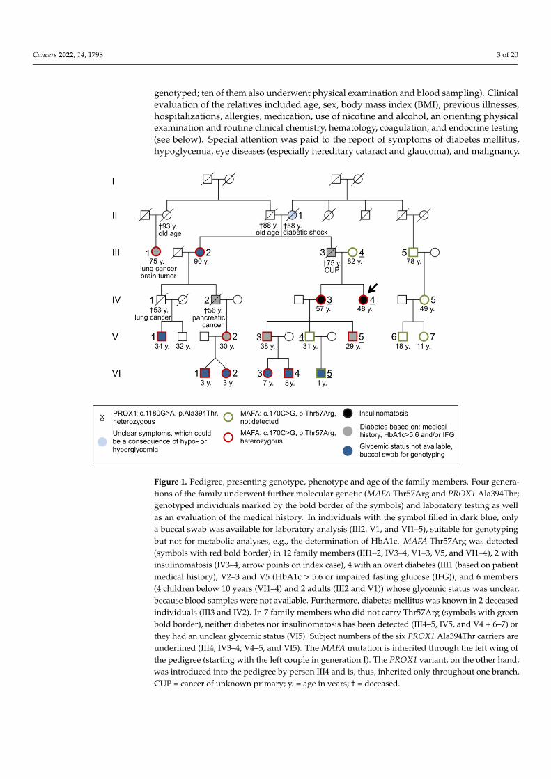
Figure 1. Pedigree, presenting genotype, phenotype and age of the family members. Four generations of the family underwent further molecular genetic (MAFA Thr57Arg and PROX1 Ala394Thr; genotyped individuals marked by the bold border of the symbols) and laboratory testing as well as an evaluation of the medical history. In individuals with the symbol filled in dark blue, only a buccal swab was available for laboratory analysis (III2, V1, and VI1–5), suitable for genotyping but not for metabolic analyses, e.g., the determination of HbA1c. MAFA Thr57Arg was detected (symbols with red bold border) in 12 family members (III1–2, IV3–4, V1–3, V5, and VI1–4), 2 with insulinomatosis (IV3–4, arrow points on index case), 4 with an overt diabetes (III1 (based on patient medical history), V2–3 and V5 (HbA1c > 5.6 or impaired fasting glucose (IFG)), and 6 members (4 children below 10 years (VI1–4) and 2 adults (III2 and V1)) whose glycemic status was unclear, because blood samples were not available. Furthermore, diabetes mellitus was known in 2 deceased individuals (III3 and IV2). In 7 family members who did not carry Thr57Arg (symbols with green bold border), neither diabetes nor insulinomatosis has been detected (III4–5, IV5, and V4 + 6–7) or they had an unclear glycemic status (VI5). Subject numbers of the six PROX1 Ala394Thr carriers are underlined (III4, IV3–4, V4–5, and VI5). The MAFA mutation is inherited through the left wing of the pedigree (starting with the left couple in generation I). The PROX1 variant, on the other hand, was introduced into the pedigree by person III4 and is, thus, inherited only throughout one branch. CUP = cancer of unknown primary; y. = age in years; † = deceased.
Figure 1. Pedigree, presenting genotype, phenotype and age of the family members. Four genera-tions of the family underwent further molecular genetic (MAFA Thr57Arg and PROX1 Ala394Thr;genotyped individuals marked by the bold border of the symbols) and laboratory testing as wellas an evaluation of the medical history. In individuals with the symbol filled in dark blue, onlya buccal swab was available for laboratory analysis (III2, V1, and VI1–5), suitable for genotypingbut not for metabolic analyses, e.g., the determination of HbA1c. MAFA Thr57Arg was detected(symbols with red bold border) in 12 family members (III1–2, IV3–4, V1–3, V5, and VI1–4), 2 withinsulinomatosis (IV3–4, arrow points on index case), 4 with an overt diabetes (III1 (based on patientmedical history), V2–3 and V5 (HbA1c > 5.6 or impaired fasting glucose (IFG)), and 6 members(4 children below 10 years (VI1–4) and 2 adults (III2 and V1)) whose glycemic status was unclear,because blood samples were not available. Furthermore, diabetes mellitus was known in 2 deceasedindividuals (III3 and IV2). In 7 family members who did not carry Thr57Arg (symbols with greenbold border), neither diabetes nor insulinomatosis has been detected (III4–5, IV5, and V4 + 6–7) orthey had an unclear glycemic status (VI5). Subject numbers of the six PROX1 Ala394Thr carriers areunderlined (III4, IV3–4, V4–5, and VI5). The MAFA mutation is inherited through the left wing ofthe pedigree (starting with the left couple in generation I). The PROX1 variant, on the other hand,was introduced into the pedigree by person III4 and is, thus, inherited only throughout one branch.CUP = cancer of unknown primary; y. = age in years; † = deceased.
Cancers 2022, 14, 1798 4 of 20
Ten samples from German healthy volunteers were analyzed in parallel with thegenomic DNA (gDNA) of the two sisters by Next Generation Sequencing to eliminatecommon local variants and sequencing artifacts.
All laboratory analyses in the patients were performed for diagnostic purposes at theInstitute of Clinical Chemistry and Laboratory Medicine and the Institute of Pathology,University Medical Center Mainz. Patients and healthy controls provided explicit consentto the use of their pseudonymized data for research purposes. The studies were designedand executed in accordance with all local legal and regulatory requirements, notablythe General Data Protection Regulation (EU 2016/679) and the Declaration of Helsinki,7th revision.
2.2. Routine Laboratory Analyses
Family members were subjected to a set of routine blood analyses: Clinical chemistryanalyses (Na+, K+, Ca2+, glucose, creatinine, alanine aminotransferase (ALAT), aspartateaminotransferase (ASAT), alkaline phosphatase, γ-glutamyl transferase, total bilirubin,lipase, α-amylase, C-reactive protein (CRP), albumin, urea-N, phosphate, triglyceride,and total and HDL-cholesterol) were performed on an Architect c8000 system (Abbott,Wiesbaden, Germany); hormone analyses (insulin, pro-insulin, C-peptide, and thyroid-stimulating hormone (TSH)) were performed on an Architect i2000 system (Abbott, Wies-baden, Germany); whole blood counts were performed on an Advia 2120i HematologySystem (Siemens Healthcare GmbH, Erlangen, Germany); coagulation testing (Quick, acti-vated partial thromboplastin time (aPTT), derived Fibrinogen) was performed on an ACLTOP 700 instrument (Instrumentation Laboratory (IL), Munich, Germany); and HbA1c ona Variant II Hemoglobin Testing System (BioRad, Munich, Germany), all using reagentsof the respective device manufacturers. The estimated glomerular filtration rate (eGFR)(according to the CKD-EPI formula), LDL-cholesterol (according to Friedewald), and theLDL/HDL ratio were calculated.
2.3. Histopathology and Immunohistochemistry
Serial sections from archival pancreatic tissue were subjected to MAFA immunohisto-chemistry as well as insulin and glucagon staining. The result was evaluated and comparedto that of normal human pancreas by an experienced pathologist. To achieve compa-rability, MAFA immunohistochemistry was performed using the same MAFA antibody(Anti-MafA antibody ab26405, Abcam, Cambridge, UK, RRID: AB_776146; dilution: 1:2000)as Iacovazzo et al. [12] following the manufacturer’s instructions. After deparaffinization,hydration, and heat-induced epitope retrieval, incubation steps were carried out in anAutostainer 480-A (Thermo Fisher Scientific, Dublin, Ireland) using the Dako EnVision™FLEX HRP/Dab (Agilent, Santa Clara, CA, USA) detection system with signal amplificationby EnVision™ FLEX + Rabbit (LINKER) (Agilent, Santa Clara, CA, USA).
Standard staining/immunohistochemisty protocols of the Institute of Pathology (Uni-versity Medical Center Mainz) were used to detect the neuroendocrine/proliferation mark-ers chromogranin, synaptophysin, insulin, glucagon, and KI-67 in formalin-fixed, paraffin-embedded pancreatic tissue sections from patients IV3 and IV4 following resection.
2.4. DNA Extraction and Sequencing
Genomic DNA was isolated from 200 µL whole blood or from buccal swabs OG-675(Oragene DNA, DNA Genotek, Ottawa, ON, Canada) using the blood/saliva protocolof the QIAamp DNA Mini kit. For primary evaluation of case 1 and 2, genetic variantswere detected by whole exome sequencing (WES) as described by Barco et al. [18], exceptfor the hybridization probe set (NimbleGen MedExome, Roche, Pleasanton, CA, USA).Sanger Sequencing (Beckman CEQ8000, Sciex, Darmstadt, Germany; Wenzel et al. [19]) andmultiplex ligation-dependent probe amplification (SALSA MLPA Probemix P017, MRCHolland, Amsterdam, The Netherlands) were used for MEN1 testing prior to WES andto confirm the variants detected by WES. Targeted analysis of family members was per-
Cancers 2022, 14, 1798 5 of 20
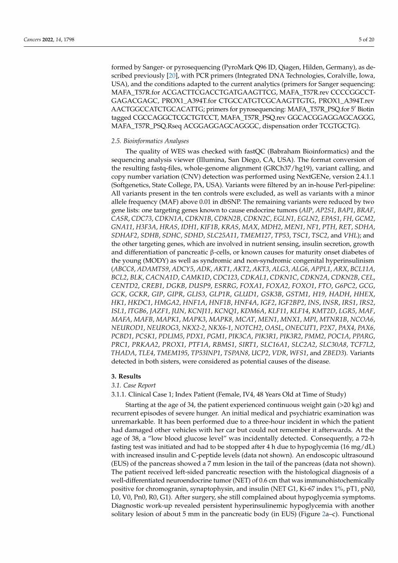
formed by Sanger- or pyrosequencing (PyroMark Q96 ID, Qiagen, Hilden, Germany), as de-scribed previously [20], with PCR primers (Integrated DNA Technologies, Coralville, Iowa,USA), and the conditions adapted to the current analytics (primers for Sanger sequencing:MAFA_T57R.for ACGACTTCGACCTGATGAAGTTCG, MAFA_T57R.rev CCCCGGCCT-GAGACGAGC, PROX1_A394T.for CTGCCATGTCGCAAGTTGTG, PROX1_A394T.revAACTGGCCATCTGCACATTG; primers for pyrosequencing: MAFA_T57R_PSQ.for 5′ Biotintagged CGCCAGGCTCGCTGTCCT, MAFA_T57R_PSQ.rev GGCACGGAGGAGCAGGG,MAFA_T57R_PSQ.Rseq ACGGAGGAGCAGGGC, dispensation order TCGTGCTG).
2.5. Bioinformatics Analyses
The quality of WES was checked with fastQC (Babraham Bioinformatics) and thesequencing analysis viewer (Illumina, San Diego, CA, USA). The format conversion ofthe resulting fastq-files, whole-genome alignment (GRCh37/hg19), variant calling, andcopy number variation (CNV) detection was performed using NextGENe, version 2.4.1.1(Softgenetics, State College, PA, USA). Variants were filtered by an in-house Perl-pipeline:All variants present in the ten controls were excluded, as well as variants with a minorallele frequency (MAF) above 0.01 in dbSNP. The remaining variants were reduced by twogene lists: one targeting genes known to cause endocrine tumors (AIP, AP2S1, BAP1, BRAF,CASR, CDC73, CDKN1A, CDKN1B, CDKN2B, CDKN2C, EGLN1, EGLN2, EPAS1, FH, GCM2,GNA11, H3F3A, HRAS, IDH1, KIF1B, KRAS, MAX, MDH2, MEN1, NF1, PTH, RET, SDHA,SDHAF2, SDHB, SDHC, SDHD, SLC25A11, TMEM127, TP53, TSC1, TSC2, and VHL); andthe other targeting genes, which are involved in nutrient sensing, insulin secretion, growthand differentiation of pancreatic β-cells, or known causes for maturity onset diabetes ofthe young (MODY) as well as syndromic and non-syndromic congenital hyperinsulinism(ABCC8, ADAMTS9, ADCY5, ADK, AKT1, AKT2, AKT3, ALG3, ALG6, APPL1, ARX, BCL11A,BCL2, BLK, CACNA1D, CAMK1D, CDC123, CDKAL1, CDKN1C, CDKN2A, CDKN2B, CEL,CENTD2, CREB1, DGKB, DUSP9, ESRRG, FOXA1, FOXA2, FOXO1, FTO, G6PC2, GCG,GCK, GCKR, GIP, GIPR, GLIS3, GLP1R, GLUD1, GSK3B, GSTM1, H19, HADH, HHEX,HK1, HKDC1, HMGA2, HNF1A, HNF1B, HNF4A, IGF2, IGF2BP2, INS, INSR, IRS1, IRS2,ISL1, ITGB6, JAZF1, JUN, KCNJ11, KCNQ1, KDM6A, KLF11, KLF14, KMT2D, LGR5, MAF,MAFA, MAFB, MAPK1, MAPK3, MAPK8, MCAT, MEN1, MNX1, MPI, MTNR1B, NCOA6,NEUROD1, NEUROG3, NKX2-2, NKX6-1, NOTCH2, OASL, ONECUT1, P2X7, PAX4, PAX6,PCBD1, PCSK1, PDLIM5, PDX1, PGM1, PIK3CA, PIK3R1, PIK3R2, PMM2, POC1A, PPARG,PRC1, PRKAA2, PROX1, PTF1A, RBMS1, SIRT1, SLC16A1, SLC2A2, SLC30A8, TCF7L2,THADA, TLE4, TMEM195, TP53INP1, TSPAN8, UCP2, VDR, WFS1, and ZBED3). Variantsdetected in both sisters, were considered as potential causes of the disease.
3. Results3.1. Case Report3.1.1. Clinical Case 1; Index Patient (Female, IV4, 48 Years Old at Time of Study)
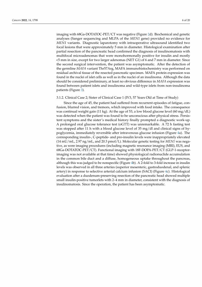
Starting at the age of 34, the patient experienced continuous weight gain (>20 kg) andrecurrent episodes of severe hunger. An initial medical and psychiatric examination wasunremarkable. It has been performed due to a three-hour incident in which the patienthad damaged other vehicles with her car but could not remember it afterwards. At theage of 38, a “low blood glucose level” was incidentally detected. Consequently, a 72-hfasting test was initiated and had to be stopped after 4 h due to hypoglycemia (16 mg/dL)with increased insulin and C-peptide levels (data not shown). An endoscopic ultrasound(EUS) of the pancreas showed a 7 mm lesion in the tail of the pancreas (data not shown).The patient received left-sided pancreatic resection with the histological diagnosis of awell-differentiated neuroendocrine tumor (NET) of 0.6 cm that was immunohistochemicallypositive for chromogranin, synaptophysin, and insulin (NET G1, Ki-67 index 1%, pT1, pN0,L0, V0, Pn0, R0, G1). After surgery, she still complained about hypoglycemia symptoms.Diagnostic work-up revealed persistent hyperinsulinemic hypoglycemia with anothersolitary lesion of about 5 mm in the pancreatic body (in EUS) (Figure 2a–c). Functional
Cancers 2022, 14, 1798 6 of 20
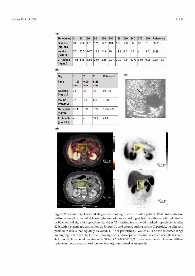
imaging with 68Ga-DOTATOC-PET/CT was negative (Figure 2d). Biochemical and geneticanalyses (Sanger sequencing and MLPA of the MEN1 gene) provided no evidence forMEN1 variants. Diagnostic laparotomy with intraoperative ultrasound identified twofocal lesions that were approximately 5 mm in diameter. Histological examination afterpartial resection of the pancreatic head confirmed the diagnosis of insulinomatosis withmultifocal microadenomas that were monohormonally positive for insulin and mostly<5 mm in size, except for two larger adenomas (NET G1) of 6 and 7 mm in diameter. Sincethe second surgical intervention, the patient was asymptomatic. After the detection ofthe germline MAFA variant Thr57Arg, MAFA immunohistochemistry was performed onresidual archival tissue of the resected pancreatic specimen. MAFA protein expression wasfound in the nuclei of islet cells as well as in the nuclei of an insulinoma. Although the datashould be considered preliminary, at least no obvious difference in MAFA expression wasfound between patient islets and insulinoma and wild-type islets from non-insulinomapatients (Figure 3).
3.1.2. Clinical Case 2; Sister of Clinical Case 1 (IV3, 57 Years Old at Time of Study)
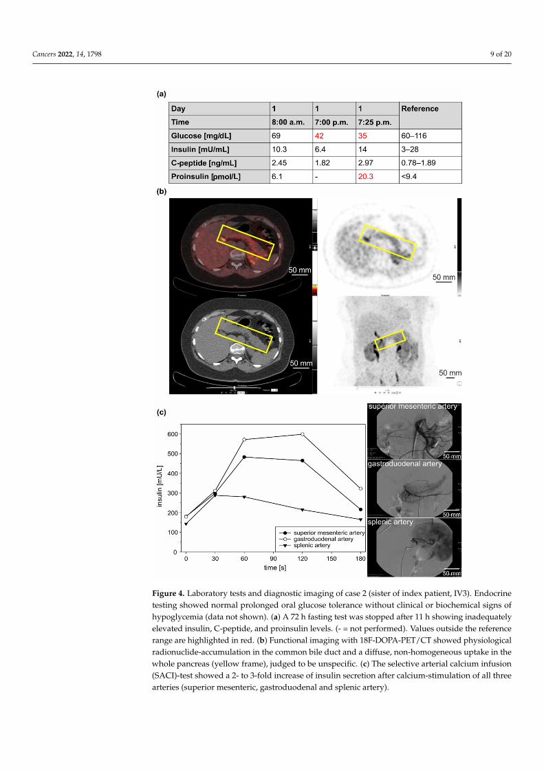
Since the age of 45, the patient had suffered from recurrent episodes of fatigue, con-fusion, blurred vision, and tremors, which improved with food intake. The consequencewas continual weight gain (11 kg). At the age of 53, a low blood glucose level (60 mg/dL)was detected when the patient was found to be unconscious after physical stress. Persis-tent symptoms and the sister’s medical history finally prompted a diagnostic work-up.A prolonged oral glucose tolerance test (oGTT) was unremarkable. A 72 h fasting testwas stopped after 11 h with a blood glucose level of 35 mg/dl and clinical signs of hy-poglycemia, immediately reversible after intravenous glucose infusion (Figure 4a). Thecorresponding insulin-, C-peptide- and pro-insulin levels were inappropriately elevated(14 mU/mL, 2.97 ng/mL, and 20.3 pmol/L). Molecular genetic testing for MEN1 was nega-tive, as were imaging procedures (including magnetic resonance imaging (MRI), EUS, and68Ga-DOTATOC-PET/CT). Functional imaging with 18F-DOPA-PET/CT (GLP-1-receptor-imaging was not available at that time) showed physiological radionuclide accumulationin the common bile duct and a diffuse, homogeneous uptake throughout the pancreas,although this was judged to be nonspecific (Figure 4b). A 2-fold to 3-fold increase in insulinlevels was observed in all three arteries (superior mesenteric, gastroduodenal, and splenicartery) in response to selective arterial calcium infusion (SACI) (Figure 4c). Histologicalevaluation after a duodenum-preserving resection of the pancreatic head showed multiplesmall insulin-positive tumorlets with 2–4 mm in diameter, consistent with the diagnosis ofinsulinomatosis. Since the operation, the patient has been asymptomatic.
Cancers 2022, 14, 1798 7 of 20Cancers 2022, 14, x FOR PEER REVIEW 8 of 22
Figure 2. Laboratory tests and diagnostic imaging of case 1 (index patient, IV4). (a) Endocrine testing showed unremarkable oral glucose tolerance (prolonged test conditions) without clinical or biochemical signs of hypoglycemia. (b) A 72 h fasting test showed marked hypoglycemia after 22 h with a plasma glucose as low as 33 mg/dL and corresponding serum C-peptide, insulin, and proinsulin levels inadequately elevated. (- = not performed). Values outside the reference range are highlighted in red. (c) Further imaging with endoscopic ultrasound revealed a single lesion of 4–5 mm. (d) Functional imaging with 68Ga-DOTATOC-PET/CT was negative with low and diffuse uptake in the pancreatic head (yellow frames), interpreted as unspecific.
Figure 2. Laboratory tests and diagnostic imaging of case 1 (index patient, IV4). (a) Endocrinetesting showed unremarkable oral glucose tolerance (prolonged test conditions) without clinicalor biochemical signs of hypoglycemia. (b) A 72 h fasting test showed marked hypoglycemia after22 h with a plasma glucose as low as 33 mg/dL and corresponding serum C-peptide, insulin, andproinsulin levels inadequately elevated. (- = not performed). Values outside the reference rangeare highlighted in red. (c) Further imaging with endoscopic ultrasound revealed a single lesion of4–5 mm. (d) Functional imaging with 68Ga-DOTATOC-PET/CT was negative with low and diffuseuptake in the pancreatic head (yellow frames), interpreted as unspecific.
Cancers 2022, 14, 1798 8 of 20Cancers 2022, 14, x FOR PEER REVIEW 9 of 22
Figure 3. Glucagon, insulin and MAFA immunohistochemistry of case 1 (IV4). Immunohistochemical staining of serial sections of the resected pancreatic specimen of case 1 showing a solitary insulinoma (left panels (a,c,e)) and normal pancreatic tissue with multiple pancreatic islets (right panels (b,d,f)).
3.1.2. Clinical Case 2; Sister of Clinical Case 1 (IV3, 57 Years Old at Time of Study) Since the age of 45, the patient had suffered from recurrent episodes of fatigue,
confusion, blurred vision, and tremors, which improved with food intake. The consequence was continual weight gain (11 kg). At the age of 53, a low blood glucose level (60 mg/dL) was detected when the patient was found to be unconscious after physical stress. Persistent symptoms and the sister’s medical history finally prompted a diagnostic work-up. A prolonged oral glucose tolerance test (oGTT) was unremarkable. A 72 h fasting test was stopped after 11 h with a blood glucose level of 35 mg/dl and clinical signs of hypoglycemia, immediately reversible after intravenous glucose infusion (Figure 4a). The corresponding insulin-, C-peptide- and pro-insulin levels were inappropriately elevated (14 mU/mL, 2.97 ng/mL, and 20.3 pmol/L). Molecular genetic testing for MEN1 was negative, as were imaging procedures (including magnetic resonance imaging (MRI), EUS, and 68Ga-DOTATOC-PET/CT). Functional imaging with 18F-DOPA-PET/CT (GLP-1-receptor-imaging was not available at that time) showed physiological radionuclide
Figure 3. Glucagon, insulin and MAFA immunohistochemistry of case 1 (IV4). Immunohistochemicalstaining of serial sections of the resected pancreatic specimen of case 1 showing a solitary insulinoma(left panels (a,c,e)) and normal pancreatic tissue with multiple pancreatic islets (right panels (b,d,f)).
Cancers 2022, 14, 1798 9 of 20
Cancers 2022, 14, x FOR PEER REVIEW 10 of 22
accumulation in the common bile duct and a diffuse, homogeneous uptake throughout the pancreas, although this was judged to be nonspecific (Figure 4b). A 2-fold to 3-fold increase in insulin levels was observed in all three arteries (superior mesenteric, gastroduodenal, and splenic artery) in response to selective arterial calcium infusion (SACI) (Figure 4c). Histological evaluation after a duodenum-preserving resection of the pancreatic head showed multiple small insulin-positive tumorlets with 2–4 mm in diameter, consistent with the diagnosis of insulinomatosis. Since the operation, the patient has been asymptomatic.
Figure 4. Laboratory tests and diagnostic imaging of case 2 (sister of index patient, IV3). Endocrine testing showed normal prolonged oral glucose tolerance without clinical or biochemical signs of hypoglycemia (data not shown). (a) A 72 h fasting test was stopped after 11 h showing inadequately elevated insulin, C-peptide, and proinsulin levels. (- = not performed). Values outside the reference range are highlighted in red. (b) Functional imaging with 18F-DOPA-PET/CT showed physiological radionuclide-accumulation in the common bile duct and a diffuse, non-homogeneous uptake in the
Figure 4. Laboratory tests and diagnostic imaging of case 2 (sister of index patient, IV3). Endocrinetesting showed normal prolonged oral glucose tolerance without clinical or biochemical signs ofhypoglycemia (data not shown). (a) A 72 h fasting test was stopped after 11 h showing inadequatelyelevated insulin, C-peptide, and proinsulin levels. (- = not performed). Values outside the referencerange are highlighted in red. (b) Functional imaging with 18F-DOPA-PET/CT showed physiologicalradionuclide-accumulation in the common bile duct and a diffuse, non-homogeneous uptake in thewhole pancreas (yellow frame), judged to be unspecific. (c) The selective arterial calcium infusion(SACI)-test showed a 2- to 3-fold increase of insulin secretion after calcium-stimulation of all threearteries (superior mesenteric, gastroduodenal and splenic artery).
Cancers 2022, 14, 1798 10 of 20
3.2. Molecular Genetic Testing3.2.1. Molecular Genetic Testing of the Two Patients and Classification of the DetectedVariants According to the ACMG Criteria
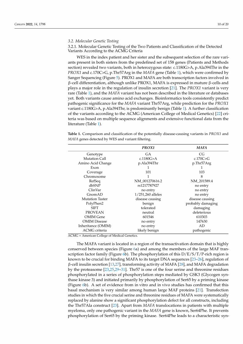
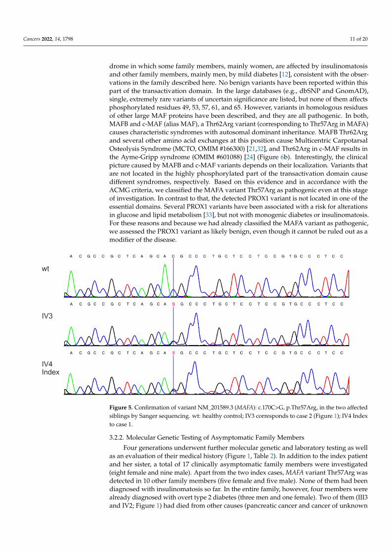
WES in the index patient and her sister and the subsequent selection of the rare vari-ants present in both sisters from the predefined set of 158 genes (Patients and Methodssection) revealed two variants, both in heterozygous state: c.1180G>A, p.Ala394Thr in thePROX1 and c.170C>G, p.Thr57Arg in the MAFA gene (Table 1), which were confirmed bySanger Sequencing (Figure 5). PROX1 and MAFA are both transcription factors involved inβ-cell differentiation, although unlike PROX1, MAFA is expressed in mature β-cells andplays a major role in the regulation of insulin secretion [21]. The PROX1 variant is veryrare (Table 1), and the MAFA variant has not been described in the literature or databasesyet. Both variants cause amino acid exchanges. Bioinformatics tools consistently predictpathogenic significance for the MAFA variant Thr57Arg, while prediction for the PROX1variant c.1180G>A, p.Ala394Thr, is predominantly benign (Table 1). A further classificationof the variants according to the ACMG (American College of Medical Genetics) [22] cri-teria was based on multiple sequence alignments and extensive functional data from theliterature (Table 1).
Table 1. Comparison and classification of the potentially disease-causing variants in PROX1 andMAFA genes detected by WES and variant filtering.
PROX1 MAFA
Genotype GA CGMutation Call c.1180G>A c.170C>G
Amino Acid Change p.Ala394Thr p.Thr57ArgExon 1 1
Coverage 101 103Chromosome 1 8
RefSeq NM_001270616.2 NM_201589.4dbSNP rs1217787927 no entryClinVar no entry no entry
GnomAD 1/251,260 alleles no entryMutation Taster disease causing disease causing
PolyPhen2 benign probably damagingSIFT tolerated damaging
PROVEAN neutral deleteriousOMIM Gene 601546 610303
OMIM Disease no entry 147630Inheritance (OMIM) no entry AD
ACMG criteria likely benign pathogenicACMG = American College of Medical Genetics.
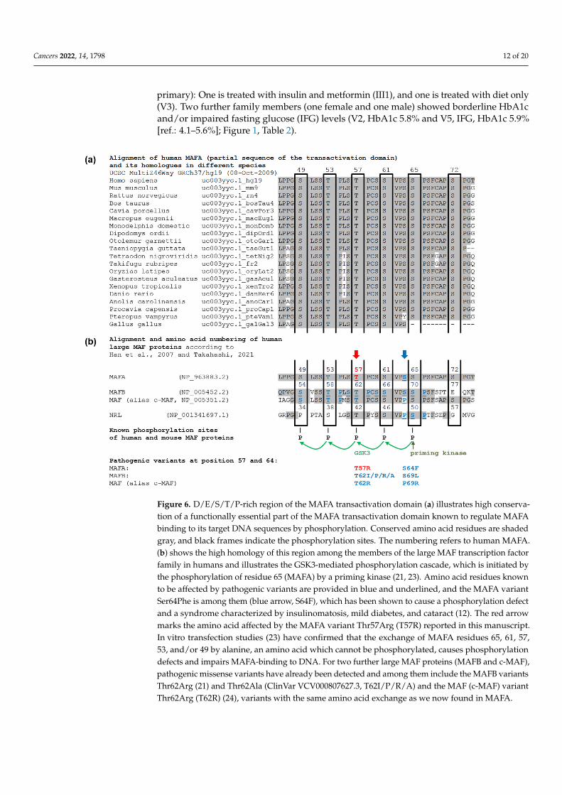
The MAFA variant is located in a region of the transactivation domain that is highlyconserved between species (Figure 6a) and among the members of the large MAF tran-scription factor family (Figure 6b). The phosphorylation of this D/E/S/T/P-rich region isknown to be crucial for binding MAFA to its target DNA sequences [23–26], regulation ofβ-cell insulin secretion [13,27], transforming activity of MAFA [28], and MAFA degradationby the proteasome [23,25,29–31]. Thr57 is one of the four serine and threonine residuesphosphorylated in a series of phosphorylation steps mediated by GSK3 (Glycogen syn-thase kinase 3) and initiated primarily by phosphorylation of Ser65 by a priming kinase(Figure 6b). A set of evidence from in vitro and in vivo studies has confirmed that thisbasal mechanism is very similar among human large MAF proteins [21]. Transfectionstudies in which the five crucial serine and threonine residues of MAFA were systematicallyreplaced by alanine show a significant phosphorylation defect for all constructs, includingthe Thr57Ala construct [23]. Apart from MAFA translocations in patients with multiplemyeloma, only one pathogenic variant in the MAFA gene is known, Ser64Phe. It preventsphosphorylation of Ser65 by the priming kinase. Ser64Phe leads to a characteristic syn-
Cancers 2022, 14, 1798 11 of 20
drome in which some family members, mainly women, are affected by insulinomatosisand other family members, mainly men, by mild diabetes [12], consistent with the obser-vations in the family described here. No benign variants have been reported within thispart of the transactivation domain. In the large databases (e.g., dbSNP and GnomAD),single, extremely rare variants of uncertain significance are listed, but none of them affectsphosphorylated residues 49, 53, 57, 61, and 65. However, variants in homologous residuesof other large MAF proteins have been described, and they are all pathogenic. In both,MAFB and c-MAF (alias MAF), a Thr62Arg variant (corresponding to Thr57Arg in MAFA)causes characteristic syndromes with autosomal dominant inheritance. MAFB Thr62Argand several other amino acid exchanges at this position cause Multicentric CarpotarsalOsteolysis Syndrome (MCTO, OMIM #166300) [21,32], and Thr62Arg in c-MAF results inthe Ayme-Gripp syndrome (OMIM #601088) [24] (Figure 6b). Interestingly, the clinicalpicture caused by MAFB and c-MAF variants depends on their localization. Variants thatare not located in the highly phosphorylated part of the transactivation domain causedifferent syndromes, respectively. Based on this evidence and in accordance with theACMG criteria, we classified the MAFA variant Thr57Arg as pathogenic even at this stageof investigation. In contrast to that, the detected PROX1 variant is not located in one of theessential domains. Several PROX1 variants have been associated with a risk for alterationsin glucose and lipid metabolism [33], but not with monogenic diabetes or insulinomatosis.For these reasons and because we had already classified the MAFA variant as pathogenic,we assessed the PROX1 variant as likely benign, even though it cannot be ruled out as amodifier of the disease.
Cancers 2022, 14, x FOR PEER REVIEW 12 of 22
Figure 5. Confirmation of variant NM_201589.3 (MAFA): c.170C>G, p.Thr57Arg, in the two affected siblings by Sanger sequencing. wt: healthy control; IV3 corresponds to case 2 (Figure 1); IV4 Index to case 1.
The MAFA variant is located in a region of the transactivation domain that is highly conserved between species (Figure 6a) and among the members of the large MAF transcription factor family (Figure 6b). The phosphorylation of this D/E/S/T/P-rich region is known to be crucial for binding MAFA to its target DNA sequences [23–26], regulation of β-cell insulin secretion [13,27], transforming activity of MAFA [28], and MAFA degradation by the proteasome [23,25,29–31]. Thr57 is one of the four serine and threonine residues phosphorylated in a series of phosphorylation steps mediated by GSK3 (Glycogen synthase kinase 3) and initiated primarily by phosphorylation of Ser65 by a priming kinase (Figure 6b). A set of evidence from in vitro and in vivo studies has confirmed that this basal mechanism is very similar among human large MAF proteins [21]. Transfection studies in which the five crucial serine and threonine residues of MAFA were systematically replaced by alanine show a significant phosphorylation defect for all constructs, including the Thr57Ala construct [23]. Apart from MAFA translocations in patients with multiple myeloma, only one pathogenic variant in the MAFA gene is known, Ser64Phe. It prevents phosphorylation of Ser65 by the priming kinase. Ser64Phe leads to a characteristic syndrome in which some family members, mainly women, are affected by insulinomatosis and other family members, mainly men, by mild diabetes [12], consistent with the observations in the family described here. No benign variants have been reported within this part of the transactivation domain. In the large databases (e.g., dbSNP and GnomAD), single, extremely rare variants of uncertain significance are listed, but none of them affects phosphorylated residues 49, 53, 57, 61, and 65. However, variants in homologous residues of other large MAF proteins have been described, and they are all pathogenic. In both, MAFB and c-MAF (alias MAF), a Thr62Arg variant (corresponding to Thr57Arg in MAFA) causes characteristic syndromes with autosomal dominant inheritance. MAFB Thr62Arg and several other amino acid exchanges at this position cause Multicentric Carpotarsal Osteolysis Syndrome (MCTO, OMIM #166300) [21,32], and Thr62Arg in c-MAF results in the Ayme-Gripp syndrome (OMIM #601088) [24] (Figure 6b). Interestingly, the clinical picture caused by MAFB and c-MAF variants depends on their localization. Variants that are not located in the highly phosphorylated part of the transactivation domain cause different syndromes, respectively. Based on this evidence and in accordance with the ACMG criteria, we classified the MAFA variant Thr57Arg as pathogenic even at this stage of investigation. In contrast to that, the detected PROX1 variant is not located in one of the essential domains. Several PROX1 variants have been associated with a risk for alterations in glucose and lipid metabolism [33], but not with monogenic diabetes or insulinomatosis. For these reasons and because we had already
Figure 5. Confirmation of variant NM_201589.3 (MAFA): c.170C>G, p.Thr57Arg, in the two affectedsiblings by Sanger sequencing. wt: healthy control; IV3 corresponds to case 2 (Figure 1); IV4 Indexto case 1.
3.2.2. Molecular Genetic Testing of Asymptomatic Family Members
Four generations underwent further molecular genetic and laboratory testing as wellas an evaluation of their medical history (Figure 1, Table 2). In addition to the index patientand her sister, a total of 17 clinically asymptomatic family members were investigated(eight female and nine male). Apart from the two index cases, MAFA variant Thr57Arg wasdetected in 10 other family members (five female and five male). None of them had beendiagnosed with insulinomatosis so far. In the entire family, however, four members werealready diagnosed with overt type 2 diabetes (three men and one female). Two of them (III3and IV2; Figure 1) had died from other causes (pancreatic cancer and cancer of unknown
Cancers 2022, 14, 1798 12 of 20
primary): One is treated with insulin and metformin (III1), and one is treated with diet only(V3). Two further family members (one female and one male) showed borderline HbA1cand/or impaired fasting glucose (IFG) levels (V2, HbA1c 5.8% and V5, IFG, HbA1c 5.9%[ref.: 4.1–5.6%]; Figure 1, Table 2).
Cancers 2022, 14, x FOR PEER REVIEW 13 of 22
classified the MAFA variant as pathogenic, we assessed the PROX1 variant as likely benign, even though it cannot be ruled out as a modifier of the disease.
Figure 6. D/E/S/T/P-rich region of the MAFA transactivation domain (a) illustrates high conservation of a functionally essential part of the MAFA transactivation domain known to regulate MAFA binding to its target DNA sequences by phosphorylation. Conserved amino acid residues are shaded gray, and black frames indicate the phosphorylation sites. The numbering refers to human MAFA. (b) shows the high homology of this region among the members of the large MAF transcription factor family in humans and illustrates the GSK3-mediated phosphorylation cascade, which is initiated by the phosphorylation of residue 65 (MAFA) by a priming kinase (21, 23). Amino acid residues known to be affected by pathogenic variants are provided in blue and underlined, and the MAFA variant Ser64Phe is among them (blue arrow, S64F), which has been shown to cause a phosphorylation defect and a syndrome characterized by insulinomatosis, mild diabetes, and cataract (12). The red arrow marks the amino acid affected by the MAFA variant Thr57Arg (T57R) reported in this manuscript. In vitro transfection studies (23) have confirmed that the exchange of MAFA residues 65, 61, 57, 53, and/or 49 by alanine, an amino acid which cannot be phosphorylated, causes phosphorylation defects and impairs MAFA-binding to DNA. For two further large MAF proteins (MAFB and c-MAF), pathogenic missense variants have already been detected and among them include the MAFB variants Thr62Arg (21) and Thr62Ala (ClinVar VCV000807627.3, T62I/P/R/A) and the MAF (c-MAF) variant Thr62Arg (T62R) (24), variants with the same amino acid exchange as we now found in MAFA.
3.2.2. Molecular Genetic Testing of Asymptomatic Family Members Four generations underwent further molecular genetic and laboratory testing as well
as an evaluation of their medical history (Figure 1, Table 2). In addition to the index patient and her sister, a total of 17 clinically asymptomatic family members were investigated (eight female and nine male). Apart from the two index cases, MAFA variant Thr57Arg was detected in 10 other family members (five female and five male). None of them had been diagnosed with insulinomatosis so far. In the entire family, however, four members were already diagnosed with overt type 2 diabetes (three men and one female). Two of
Figure 6. D/E/S/T/P-rich region of the MAFA transactivation domain (a) illustrates high conserva-tion of a functionally essential part of the MAFA transactivation domain known to regulate MAFAbinding to its target DNA sequences by phosphorylation. Conserved amino acid residues are shadedgray, and black frames indicate the phosphorylation sites. The numbering refers to human MAFA.(b) shows the high homology of this region among the members of the large MAF transcription factorfamily in humans and illustrates the GSK3-mediated phosphorylation cascade, which is initiated bythe phosphorylation of residue 65 (MAFA) by a priming kinase (21, 23). Amino acid residues knownto be affected by pathogenic variants are provided in blue and underlined, and the MAFA variantSer64Phe is among them (blue arrow, S64F), which has been shown to cause a phosphorylation defectand a syndrome characterized by insulinomatosis, mild diabetes, and cataract (12). The red arrowmarks the amino acid affected by the MAFA variant Thr57Arg (T57R) reported in this manuscript.In vitro transfection studies (23) have confirmed that the exchange of MAFA residues 65, 61, 57,53, and/or 49 by alanine, an amino acid which cannot be phosphorylated, causes phosphorylationdefects and impairs MAFA-binding to DNA. For two further large MAF proteins (MAFB and c-MAF),pathogenic missense variants have already been detected and among them include the MAFB variantsThr62Arg (21) and Thr62Ala (ClinVar VCV000807627.3, T62I/P/R/A) and the MAF (c-MAF) variantThr62Arg (T62R) (24), variants with the same amino acid exchange as we now found in MAFA.
Cancers 2022, 14, 1798 13 of 20
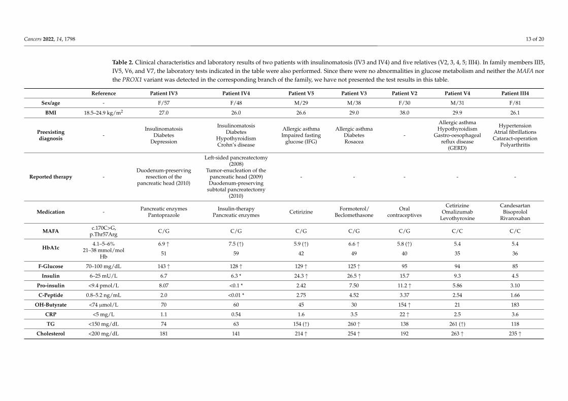
Table 2. Clinical characteristics and laboratory results of two patients with insulinomatosis (IV3 and IV4) and five relatives (V2, 3, 4, 5; III4). In family members III5,IV5, V6, and V7, the laboratory tests indicated in the table were also performed. Since there were no abnormalities in glucose metabolism and neither the MAFA northe PROX1 variant was detected in the corresponding branch of the family, we have not presented the test results in this table.
Reference Patient IV3 Patient IV4 Patient V5 Patient V3 Patient V2 Patient V4 Patient III4
Sex/age - F/57 F/48 M/29 M/38 F/30 M/31 F/81
BMI 18.5–24.9 kg/m2 27.0 26.0 26.6 29.0 38.0 29.9 26.1
Preexistingdiagnosis -
InsulinomatosisDiabetes
Depression
InsulinomatosisDiabetes
HypothyroidismCrohn’s disease
Allergic asthmaImpaired fasting
glucose (IFG)
Allergic asthmaDiabetesRosacea
-
Allergic asthmaHypothyroidism
Gastro-oesophagealreflux disease
(GERD)
HypertensionAtrial fibrillationsCataract-operation
Polyarthritis
Reported therapy -Duodenum-preserving
resection of thepancreatic head (2010)
Left-sided pancreatectomy(2008)
Tumor-enucleation of thepancreatic head (2009)Duodenum-preserving
subtotal pancreatectomy(2010)
- - - - -
Medication - Pancreatic enzymesPantoprazole
Insulin-therapyPancreatic enzymes Cetirizine Formoterol/
BeclomethasoneOral
contraceptives
CetirizineOmalizumab
Levothyroxine
CandesartanBisoprolol
Rivaroxaban
MAFA c.170C>G,p.Thr57Arg C/G C/G C/G C/G C/G C/C C/C
HbA1c4.1–5–6% 6.9 ↑ 7.5 (↑) 5.9 (↑) 6.6 ↑ 5.8 (↑) 5.4 5.4
21–38 mmol/molHb 51 59 42 49 40 35 36
F-Glucose 70–100 mg/dL 143 ↑ 128 ↑ 129 ↑ 125 ↑ 95 94 85
Insulin 6–25 mU/L 6.7 6.3 * 24.3 ↑ 26.5 ↑ 15.7 9.3 4.5
Pro-insulin <9.4 pmol/L 8.07 <0.1 * 2.42 7.50 11.2 ↑ 5.86 3.10
C-Peptide 0.8–5.2 ng/mL 2.0 <0.01 * 2.75 4.52 3.37 2.54 1.66
OH-Butyrate <74 µmol/L 70 60 45 30 154 ↑ 21 183
CRP <5 mg/L 1.1 0.54 1.6 3.5 22 ↑ 2.5 3.6
TG <150 mg/dL 74 63 154 (↑) 260 ↑ 138 261 (↑) 118
Cholesterol <200 mg/dL 181 141 214 ↑ 254 ↑ 192 263 ↑ 235 ↑
Cancers 2022, 14, 1798 14 of 20
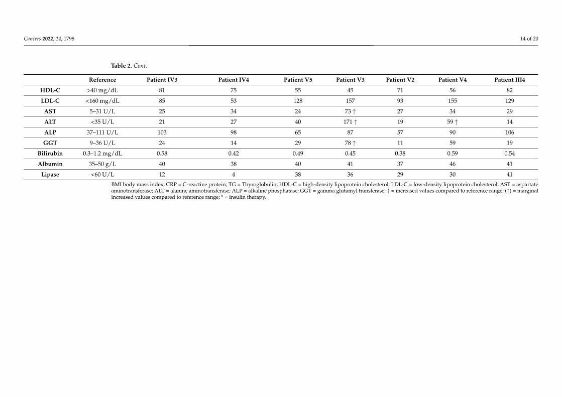
Table 2. Cont.
Reference Patient IV3 Patient IV4 Patient V5 Patient V3 Patient V2 Patient V4 Patient III4
HDL-C >40 mg/dL 81 75 55 45 71 56 82
LDL-C <160 mg/dL 85 53 128 157 93 155 129
AST 5–31 U/L 25 34 24 73 ↑ 27 34 29
ALT <35 U/L 21 27 40 171 ↑ 19 59 ↑ 14
ALP 37–111 U/L 103 98 65 87 57 90 106
GGT 9–36 U/L 24 14 29 78 ↑ 11 59 19
Bilirubin 0.3–1.2 mg/dL 0.58 0.42 0.49 0.45 0.38 0.59 0.54
Albumin 35–50 g/L 40 38 40 41 37 46 41
Lipase <60 U/L 12 4 38 36 29 30 41
BMI body mass index; CRP = C-reactive protein; TG = Thyroglobulin; HDL-C = high-density lipoprotein cholesterol; LDL-C = low-density lipoprotein cholesterol; AST = aspartateaminotransferase; ALT = alanine aminotransferase; ALP = alkaline phosphatase; GGT = gamma glutamyl transferase; ↑ = increased values compared to reference range; (↑) = marginalincreased values compared to reference range; * = insulin therapy.
Cancers 2022, 14, 1798 15 of 20
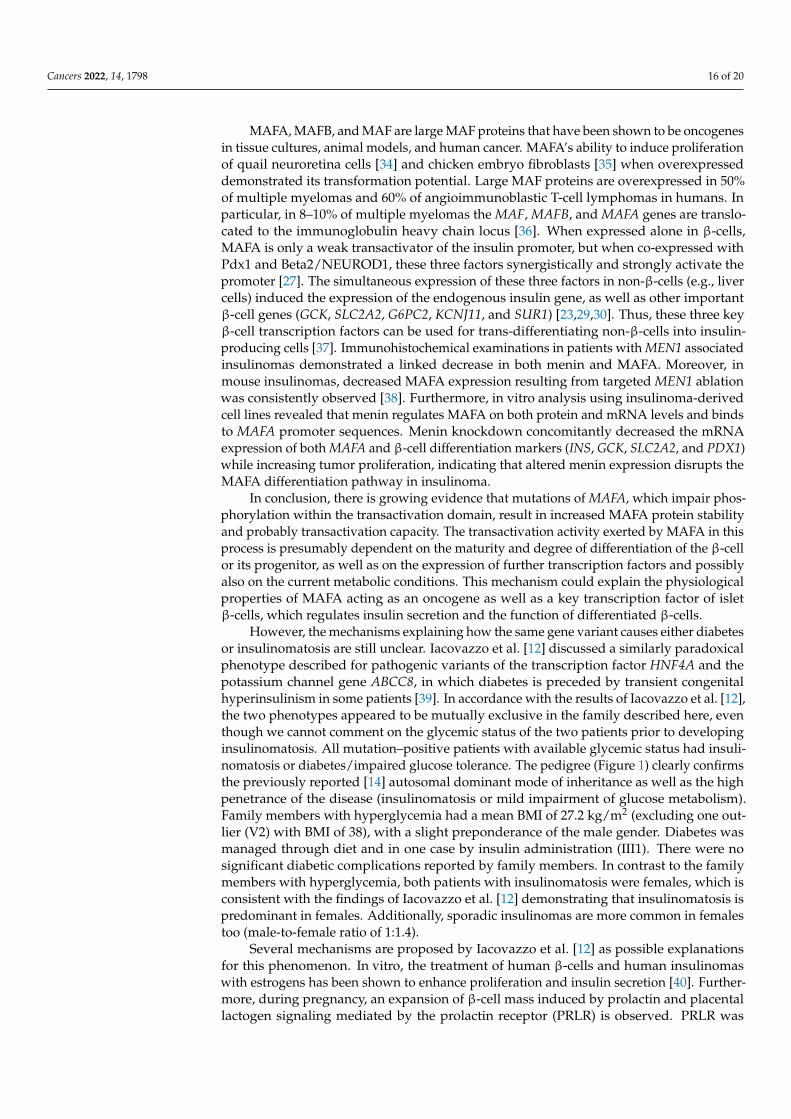
Four patients presented with a tumor disease (one with lung cancer (IV1), one withlung cancer and a brain tumor (III1), one with pancreatic cancer (IV2), and one with acancer of unknown primary (III3)), and one female with advanced age has dementia. Therewas no history of congenital eye disorders in this family.
Genetic testing revealed that the six symptomatic (insulinomatosis or mild diabetes;classification here after laboratory analyses) individuals studied from generations III-V(Figure 1, III1; IV3, 4; V2, 3, 5) all carried the MAFA variant, but only three of them hadthe PROX1 variant (IV3, 4; V5). Generation VI and individuals III2 and V1 could not beanalyzed in this regard because only a buccal swab was available for genotyping, but noEDTA blood was available for determining HbA1c. In the six healthy subjects studied, theMAFA variant was absent (III4, 5; IV5; V4, 6, 7), whereas the PROX1 variant was detected intwo of them (III4, V4). Thus, the segregation of these variants in the family clearly suggeststhe MAFA variant as the cause of the disease and further autosomal dominant inheritance.
4. Discussion
Using whole exome sequencing, we identified a so far unreported missense MAFAvariant (c.170C>G, p.Thr57Arg) as the cause of insulinomatosis in two sisters with recur-rent hyperinsulinemic hypoglycemia. Thr57Arg is located in MAFA’s highly conservedtransactivation domain, which is so crucial that extensive functional analyses have alreadybeen performed. These include expression studies confirming that the replacement ofthreonine at position 57 with an amino acid that cannot be phosphorylated by GSK3 resultsin a defect in the stepwise phosphorylation of MAFA, impairing the binding of MAFA toits DNA target and preventing proteasome-mediated degradation [23,25,29–31]. So far,only one other pathogenic MAFA variant (c.191C>T, p.Ser64Phe) has been described. It islocated in the same MAFA domain, impairs the same phosphorylation cascade, and resultsin the same characteristic symptoms in the two affected families as observed in the familydescribed here [12]. In summary, we describe a novel pathogenic MAFA missense variantthat is highly likely to cause impaired phosphorylation and, therefore, functional defects,cosegregating with both phenotypes, insulinomatosis and MODY-like diabetes (“MODY-like”: autosomal dominantly inherited, rather mild hyperglycemia (diabetes/IFG/HbA1c> 5.6) caused by a pathogenic variant in a transcription factor regulating insulin secretion).The detection of this second pathogenic MAFA variant confirms that a phosphorylationdefect in the transactivation domain is accompanied by a characteristic set of symptoms inthe affected family and confirms the autosomal dominant inheritance of the disease.
Iacovazzo et al. recently demonstrated for the first time that the Ser64Phe mutationof the MAFA gene was linked to insulinoma or mild, non-insulin dependent diabetes [12].Ser64Phe was discovered to impair phosphorylation within MAFA’s transactivation domainand to profoundly increase MAFA protein stability in β-cell-lines at high and low glucoseconcentrations. There was no significant change in the amount of wild-type and mutantMAFA mRNA in transfected cells, confirming that the effect on protein turnover wasposttranscriptional. These findings imply that the activity of Ser64Phe-MAFA is enhancedin β-cells/pancreas islets due to an increased transactivation capacity that is at least partlyrelated to decreased MAFA degradation [12,23,25,29–31]. However, the transactivationactivity of MAFA can be both decreased and increased depending on the cell type studiedand the expression of other transcription factors [12,23,25,28–31]. The Thr57Arg MAFA-variant found in our family, similar to that reported by Iacovazzo et al. [12], is locatedwithin the highly conserved D/E/S/T/P-rich domain, impairing phosphorylation withinMAFA’s transactivation domain, thus enhancing the protein stability and probably the totaltransactivation capacity of MAFA. Whether a phosphorylation defect in the D/E/S/T/P-rich domain of MAFA increases or decreases, net MAFA protein expression in β-cellscannot be conclusively answered at present. Both Iacovazzo et al. [12] and our groupfound no obvious difference by immunohistochemistry between MAFA expression inislets and insulinomas in insulinomatosis patients and islets from patients not affected byinsulinomatosis, but the data are based on very few cases so far.
Cancers 2022, 14, 1798 16 of 20
MAFA, MAFB, and MAF are large MAF proteins that have been shown to be oncogenesin tissue cultures, animal models, and human cancer. MAFA’s ability to induce proliferationof quail neuroretina cells [34] and chicken embryo fibroblasts [35] when overexpresseddemonstrated its transformation potential. Large MAF proteins are overexpressed in 50%of multiple myelomas and 60% of angioimmunoblastic T-cell lymphomas in humans. Inparticular, in 8–10% of multiple myelomas the MAF, MAFB, and MAFA genes are translo-cated to the immunoglobulin heavy chain locus [36]. When expressed alone in β-cells,MAFA is only a weak transactivator of the insulin promoter, but when co-expressed withPdx1 and Beta2/NEUROD1, these three factors synergistically and strongly activate thepromoter [27]. The simultaneous expression of these three factors in non-β-cells (e.g., livercells) induced the expression of the endogenous insulin gene, as well as other importantβ-cell genes (GCK, SLC2A2, G6PC2, KCNJ11, and SUR1) [23,29,30]. Thus, these three keyβ-cell transcription factors can be used for trans-differentiating non-β-cells into insulin-producing cells [37]. Immunohistochemical examinations in patients with MEN1 associatedinsulinomas demonstrated a linked decrease in both menin and MAFA. Moreover, inmouse insulinomas, decreased MAFA expression resulting from targeted MEN1 ablationwas consistently observed [38]. Furthermore, in vitro analysis using insulinoma-derivedcell lines revealed that menin regulates MAFA on both protein and mRNA levels and bindsto MAFA promoter sequences. Menin knockdown concomitantly decreased the mRNAexpression of both MAFA and β-cell differentiation markers (INS, GCK, SLC2A2, and PDX1)while increasing tumor proliferation, indicating that altered menin expression disrupts theMAFA differentiation pathway in insulinoma.
In conclusion, there is growing evidence that mutations of MAFA, which impair phos-phorylation within the transactivation domain, result in increased MAFA protein stabilityand probably transactivation capacity. The transactivation activity exerted by MAFA in thisprocess is presumably dependent on the maturity and degree of differentiation of the β-cellor its progenitor, as well as on the expression of further transcription factors and possiblyalso on the current metabolic conditions. This mechanism could explain the physiologicalproperties of MAFA acting as an oncogene as well as a key transcription factor of isletβ-cells, which regulates insulin secretion and the function of differentiated β-cells.
However, the mechanisms explaining how the same gene variant causes either diabetesor insulinomatosis are still unclear. Iacovazzo et al. [12] discussed a similarly paradoxicalphenotype described for pathogenic variants of the transcription factor HNF4A and thepotassium channel gene ABCC8, in which diabetes is preceded by transient congenitalhyperinsulinism in some patients [39]. In accordance with the results of Iacovazzo et al. [12],the two phenotypes appeared to be mutually exclusive in the family described here, eventhough we cannot comment on the glycemic status of the two patients prior to developinginsulinomatosis. All mutation–positive patients with available glycemic status had insuli-nomatosis or diabetes/impaired glucose tolerance. The pedigree (Figure 1) clearly confirmsthe previously reported [14] autosomal dominant mode of inheritance as well as the highpenetrance of the disease (insulinomatosis or mild impairment of glucose metabolism).Family members with hyperglycemia had a mean BMI of 27.2 kg/m2 (excluding one out-lier (V2) with BMI of 38), with a slight preponderance of the male gender. Diabetes wasmanaged through diet and in one case by insulin administration (III1). There were nosignificant diabetic complications reported by family members. In contrast to the familymembers with hyperglycemia, both patients with insulinomatosis were females, which isconsistent with the findings of Iacovazzo et al. [12] demonstrating that insulinomatosis ispredominant in females. Additionally, sporadic insulinomas are more common in femalestoo (male-to-female ratio of 1:1.4).
Several mechanisms are proposed by Iacovazzo et al. [12] as possible explanationsfor this phenomenon. In vitro, the treatment of human β-cells and human insulinomaswith estrogens has been shown to enhance proliferation and insulin secretion [40]. Further-more, during pregnancy, an expansion of β-cell mass induced by prolactin and placentallactogen signaling mediated by the prolactin receptor (PRLR) is observed. PRLR was
Cancers 2022, 14, 1798 17 of 20
significantly downregulated and the PRLR promoter was shown to be directly activatedby MAFA in luciferase reporter assays in both MAFA knockout islets and MIN6 β-cellswith siRNA-mediated knockdown of MAFA [41]. Therefore, estrogens and prolactin couldpotentially promote β-cell proliferation, predisposing female carriers of MAFA mutationsto insulinomatosis. Similarly to the family members with insulinomatosis reported byIacovazzo et al. [12], one of our female insulinomatosis patients experienced the first hypo-glycemic symptoms after pregnancy. She did not complain about any clinical problemsduring the pregnancy. The other affected sister, however, had never been pregnant anddeveloped the first symptoms after puberty. In summary, it is striking that there is afemale preponderance for developing insulinomatosis and a male preponderance for de-veloping diabetes/impaired glucose tolerance in the three families with MAFA mutationsreported so far. However, the precise mechanisms that determine the development ofeither insulinomatosis or diabetes remain unknown, and it is not ruled out that diabetesand insulinomatosis may develop consecutively. Furthermore, it is not excluded that thePROX1 variant detected in our family acts as a modifier in this regard.
In clinical practice, diagnosis and therapy of familial insulinomatosis remains chal-lenging. In large studies, insulinomatosis is responsible for less than 5% of all patientswith hyperinsulinemic hypoglycemia [2,10]. There are no distinct clinical or biochemicalcharacteristics that distinguish solitary insulinoma from multifocal insulinomatosis. Multi-ple insulin-secreting microtumors are present in affected patients, distributed throughoutthe entire pancreas and only detectable by histological examination. Even when utilizingmodern functional imaging modalities such as Glucagon-like peptide-1 receptor (GLP-1R)imaging, only few macrotumors with a mean size of 0.5–1 cm can be detected [42,43]. Thus,the clinical course of the disease is still characterized by early recurrent hypoglycemiarequiring repeated surgical intervention. All documented clinical cases had persistent orrecurrent disease after the initial surgical intervention. Only the histological examinationof a larger, representative pancreatic specimen confirmed the diagnosis of insulinomato-sis and distinguished it from multifocal NET with MEN1 mutations or nesidioblastosis.The SACI test should be considered in cases where noninvasive imaging modalities arenegative or when there is evidence of a multifocal disease, as it can distinguish solitaryinsulinoma from multifocal/diffuse diseases with sensitivity and specificity of 95–100%and 90%, respectively, and determine the extent of surgical intervention [44,45].
The mean age at the diagnosis of insulinomatosis in our family was 39.5 years, beingalmost exactly the same as reported in the families by Iacovazzo et al. [12] (39.4± 13.1 years).Unfortunately, we were unable to determine the exact age at the diagnosis of diabetes.However, the recorded past medical history of the living family members indicated thatthe initial presentation of diabetes in our family occurred between 29 (V5) and 50 yearsof age (compared to 38.4 ± 16.4 years as reported by Iacovazzo et al. [12]). Therefore,most patients with MAFA mutations show clinical features of insulinomatosis or diabetesalready in young adulthood. In contrast, the youngest patients with MAFA mutationsand diagnosed diabetes or insulinomatosis were 11 and 18 years old, respectively [12].We, therefore, recommend that asymptomatic mutation carriers should be monitored fordiabetes/impaired glucose tolerance beginning in adolescence and that these patientsshould receive regular clinical screening as well as information/counselling, especially forsigns and symptoms of neuroglycopenia.
The findings of this study have to be seen in light of some limitations. A moredetailed characterization of the mild hyperglycemia observed in four variant carriers(Figure 1) could provide further insights in the dynamics of the glucose metabolism in thesepatients. The affected family members consented to a one-time examination (includingblood glucose and HbA1c), but not to an oral glucose tolerance test. Furthermore, itwould be valuable to assess the glycemic status of the variant carriers in generation VI(genotyping by buccal swab). The parents were educated about potential symptoms ofhypoglycemia and hyperglycemia, but they declined blood sampling and functional testingof the asymptomatic children at that time.
Cancers 2022, 14, 1798 18 of 20
While it is clear that Thr57Arg causes a phosphorylation defect, which increasesprotein stability, and while Thr57Arg probably increases MAFA-mediated transactivationcapacity of the beta cell, the consequences for MAFA’s transactivation activity per moleculeremain largely unknown. Using the MAFA Thr57Ala construct, transactivation activityhas already been studied extensively by others. As the result was clearly dependent oncell type, differentiation, and embryonic or neoplastic origin of the cell model, it did notseem reasonable to repeat the expression studies on another (or the same) cell models onlyreplacing Thr57Ala by Thr57Arg. Extensive further studies are needed to elucidate thecomplex regulation of MAFA’s transactivation activity.
5. Conclusions
We define a second MAFA variant, p.Thr57Arg, as the cause of familial insulinomatosisby linking genetic, bioinformatic, clinical, biochemical, and family data to functionalin vitro analyses from the literature. We, thus, confirm MAFA phosphorylation defects asan important cause of a hereditary syndrome, which is characterized by insulinomatosisand/or mild, MODY-like hyperglycemia. This study extends the pathophysiological anddiagnostic disease concept and verifies the high penetrance and autosomal dominantinheritance pattern of familial insulinomatosis.
Author Contributions: Conceptualization, C.F., T.M., and H.R.; methodology, C.F., S.S., A.A., H.R.,M.G., A.S., M.M., and S.S.-F.; validation, C.F., A.A., and H.R.; formal analysis, S.S., A.A., M.G.,H.R., A.S., M.M., and S.S.-F.; investigation, C.F., S.S., A.A., H.R., M.G., A.S., M.M., S.S.-F., and H.R.;resources, M.M.W., T.M., and K.J.L.; data curation, A.A., M.G., and S.S.; writing—original draftpreparation, C.F., A.A., and H.R.; writing—review and editing, C.F., S.S., M.G., A.A., T.M., A.S., M.M.,S.S.-F., M.M.W., K.J.L., and H.R.; visualization, A.A., C.F., M.M., and H.R.; supervision, M.M.W., T.M.,and K.J.L.; project administration, C.F. and H.R.; funding acquisition, M.M.W., T.M., and K.J.L. Allauthors have read and agreed to the published version of the manuscript.
Funding: This research is supported by the German Federal Ministry of Education and Research(BMBF 01EO1003).
Institutional Review Board Statement: According to the guidelines of the local ethics committee(Ethics Committee of the State Medical Association of Rhineland-Palatinate; see http://www.laek-rlp.de/ausschuesse-kommissionen/ethikkommission/#w7e360e10191600092cdfdf4f0ea10c0 (accessedon 31 March 2022), topic 8), ethical review and approval were waived for this article.
Informed Consent Statement: Written informed consent has been obtained from the patients forpotential publication.
Data Availability Statement: All relevant data generated or analyzed during this study are includedin this published article. Restrictions apply to the availability of the complete next generationsequencing data of the patients to preserve patient confidentiality. The corresponding author will onrequest detail the restrictions and any conditions under which access to some data may be provided.
Acknowledgments: We would like to thank Carolin Neukirch for excellent technical assistance.This report includes experiments/work performed by Stefanie Sollfrank, Mursal Ghiasi, and AnkeAdenaeuer during the preparation of their doctoral theses.
Conflicts of Interest: The authors declare no conflict of interest. Outside of this research study,M.M.W. received advisory compensation from Novartis, GSK, and Roche.
References1. Nessa, A.; Rahman, S.A.; Hussain, K. Hyperinsulinemic Hypoglycemia-The Molecular Mechanisms. Front. Endocrinol. 2016, 7, 29.
[CrossRef]2. Yamada, Y.; Kitayama, K.; Oyachi, M.; Higuchi, S.; Kawakita, R.; Kanamori, Y.; Yorifuji, T. Nationwide survey of endogenous
hyperinsulinemic hypoglycemia in Japan (2017–2018): Congenital hyperinsulinism, insulinoma, non-insulinoma pancreatogenoushypoglycemia syndrome and insulin autoimmune syndrome (Hirata’s disease). J. Diabetes Investig. 2020, 11, 554–563. [CrossRef]
3. Anlauf, M.; Schlenger, R.; Perren, A.; Bauersfeld, J.; Koch, C.A.; Dralle, H.; Raffel, A.; Knoefel, W.T.; Weihe, E.; Ruszniewski,P.; et al. Microadenomatosis of the endocrine pancreas in patients with and without the multiple endocrine neoplasia type 1syndrome. Am. J. Surg. Pathol. 2006, 30, 560–574. [CrossRef] [PubMed]
Cancers 2022, 14, 1798 19 of 20
4. Service, F.J.; Natt, N.; Thompson, G.B.; Grant, C.S.; van Heerden, J.A.; Andrews, J.C.; Lorenz, E.; Terzic, A.; Lloyd, R.V.Noninsulinoma pancreatogenous hypoglycemia: A novel syndrome of hyperinsulinemic hypoglycemia in adults independent ofmutations in Kir6.2 and SUR1 genes. J. Clin. Endocrinol. Metab. 1999, 84, 1582–1589. [CrossRef] [PubMed]
5. Klöppel, G.; Anlauf, M.; Raffel, A.; Perren, A.; Knoefel, W.T. Adult diffuse nesidioblastosis: Genetically or environmentallyinduced? Hum. Pathol. 2008, 39, 3–8. [CrossRef] [PubMed]
6. Dravecka, I.; Lazurova, I. Nesidioblastosis in adults. Neoplasma 2014, 61, 252–256. [CrossRef]7. García-Santos, E.P.; del Carmen Manzanares-Campillo, M.; Padilla-Valverde, D.; Villarejo-Campos, P.; Gil-Rendo, A.; Muñoz-
Atienza, V.; Sánchez-García, S.; Puig-Rullán, A.M.; Rodríguez-Peralto, J.L.; Martín-Fernández, J. Nesidioblastosis. A case ofhyperplasia of the islets of Langerhans in the adult. Pancreatology 2013, 13, 544–548. [CrossRef] [PubMed]
8. Bernard, B.; Kline, G.A.; Service, F.J. Hypoglycaemia following upper gastrointestinal surgery: Case report and review of theliterature. BMC Gastroenterol. 2010, 10, 77. [CrossRef]
9. Mala, T. Postprandial hyperinsulinemic hypoglycemia after gastric bypass surgical treatment. Surg. Obes. Relat. Dis. 2014, 10,1220–1225. [CrossRef] [PubMed]
10. Anlauf, M.; Bauersfeld, J.; Raffel, A.; Koch, C.A.; Henopp, T.; Alkatout, I.; Schmitt, A.; Weber, A.; Kruse, M.L.; Braunstein, S.; et al.Insulinomatosis: A multicentric insulinoma disease that frequently causes early recurrent hyperinsulinemic hypoglycemia. Am. J.Surg. Pathol. 2009, 33, 339–346. [CrossRef] [PubMed]
11. Tragl, K.H.; Mayr, W.R. Familial islet-cell adenomatosis. Lancet 1977, 2, 426–428. [CrossRef]12. Iacovazzo, D.; Flanagan, S.E.; Walker, E.; Quezado, R.; de Sousa Barros, F.A.; Caswell, R.; Johnson, M.B.; Wakeling, M.; Brändle,
M.; Guo, M.; et al. MAFA missense mutation causes familial insulinomatosis and diabetes mellitus. Proc. Natl. Acad. Sci. USA2018, 115, 1027–1032. [CrossRef]
13. Zhang, C.; Moriguchi, T.; Kajihara, M.; Esaki, R.; Harada, A.; Shimohata, H.; Oishi, H.; Hamada, M.; Morito, N.; Hasegawa, K.;et al. MafA is a key regulator of glucose-stimulated insulin secretion. Mol. Cell. Biol. 2005, 25, 4969–4976. [CrossRef] [PubMed]
14. Nishimura, W.; Kondo, T.; Salameh, T.; El Khattabi, I.; Dodge, R.; Bonner-Weir, S.; Sharma, A. A switch from MafB to MafAexpression accompanies differentiation to pancreatic beta-cells. Dev. Biol. 2006, 293, 526–539. [CrossRef] [PubMed]
15. Artner, I.; Hang, Y.; Guo, M.; Gu, G.; Stein, R. MafA is a dedicated activator of the insulin gene in vivo. J. Endocrinol. 2008, 198,271–279. [CrossRef] [PubMed]
16. Artner, I.; Hang, Y.; Mazur, M.; Yamamoto, T.; Guo, M.; Lindner, J.; Magnuson, M.A.; Stein, R. MafA and MafB regulate genescritical to beta-cells in a unique temporal manner. Diabetes 2010, 59, 2530–2539. [CrossRef] [PubMed]
17. Hang, Y.; Stein, R. MafA and MafB activity in pancreatic β cells. Trends Endocrinol. Metab. 2011, 22, 364–373. [CrossRef] [PubMed]18. Barco, S.; Sollfrank, S.; Trinchero, A.; Adenaeuer, A.; Abolghasemi, H.; Conti, L.; Häuser, F.; Hovinga, J.A.K.; Lackner, K.J.;
Loewecke, F.; et al. Severe plasma prekallikrein deficiency: Clinical characteristics, novel KLKB1 mutations, and estimatedprevalence. J. Thromb. Haemost. 2020, 18, 1598–1617. [CrossRef] [PubMed]
19. Wenzel, J.J.; Rossmann, H.; Fottner, C.; Neuwirth, S.; Neukirch, C.; Lohse, P.; Bickmann, J.K.; Minnemann, T.; Musholt, T.J.;Schneider-Rätzke, B.; et al. Identification and prevention of genotyping errors caused by G-quadruplex- and i-motif-like sequences.Clin. Chem. 2009, 55, 1361–1371. [CrossRef] [PubMed]
20. Bickmann, J.K.; Kamin, W.; Wiebel, M.; Häuser, F.; Wenzel, J.J.; Neukirch, C.; Stuhrmann, M.; Lackner, K.J.; Rossmann, H. A novelapproach to CFTR mutation testing by pyrosequencing-based assay panels adapted to ethnicities. Clin. Chem. 2009, 55, 1083–1091.[CrossRef]
21. Takahashi, S. Functional analysis of large MAF transcription factors and elucidation of their relationships with human diseases.Exp. Anim. 2021, 70, 264–271. [CrossRef] [PubMed]
22. Richards, S.; Aziz, N.; Bale, S.; Bick, D.; Das, S.; Gastier-Foster, J.; Grody, W.W.; Hegde, M.; Lyon, E.; Spector, E.; et al. Standardsand guidelines for the interpretation of sequence variants: A joint consensus recommendation of the American College of MedicalGenetics and Genomics and the Association for Molecular Pathology. Genet. Med. 2015, 17, 405–424. [CrossRef]
23. Han, S.-I.; Aramata, S.; Yasuda, K.; Kataoka, K. MafA stability in pancreatic beta cells is regulated by glucose and is dependent onits constitutive phosphorylation at multiple sites by glycogen synthase kinase 3. Mol. Cell. Biol. 2007, 27, 6593–6605. [CrossRef][PubMed]
24. Niceta, M.; Stellacci, E.; Gripp, K.W.; Zampino, G.; Kousi, M.; Anselmi, M.; Traversa, A.; Ciolfi, A.; Stabley, D.; Bruselles, A.; et al.Mutations Impairing GSK3-Mediated MAF Phosphorylation Cause Cataract, Deafness, Intellectual Disability, Seizures, and aDown Syndrome-like Facies. Am. J. Hum. Genet. 2015, 96, 816–825. [CrossRef]
25. Han, S.-I.; Tsunekage, Y.; Kataoka, K. Phosphorylation of MafA enhances interaction with Beta2/NeuroD1. Acta Diabetol. 2016, 53,651–660. [CrossRef] [PubMed]
26. Lu, X.; Guanga, G.P.; Wan, C.; Rose, R.B. A novel DNA binding mechanism for maf basic region-leucine zipper factors inferredfrom a MafA-DNA complex structure and binding specificities. Biochemistry 2012, 51, 9706–9717. [CrossRef] [PubMed]
27. Kataoka, K.; Han, S.-I.; Shioda, S.; Hirai, M.; Nishizawa, M.; Handa, H. MafA is a glucose-regulated and pancreatic beta-cell-specific transcriptional activator for the insulin gene. J. Biol. Chem. 2002, 277, 49903–49910. [CrossRef] [PubMed]
28. Rocques, N.; Zeid, N.A.; Sii-Felice, K.; Lecoin, L.; Felder-Schmittbuhl, M.-P.; Eychène, A.; Pouponnot, C. GSK-3-mediatedphosphorylation enhances Maf-transforming activity. Mol. Cell 2007, 28, 584–597. [CrossRef] [PubMed]
29. Aramata, S.; Han, S.-I.; Kataoka, K. Roles and regulation of transcription factor MafA in islet beta-cells. Endocr. J. 2007, 54, 659–666.[CrossRef] [PubMed]
Cancers 2022, 14, 1798 20 of 20
30. Aramata, S.; Han, S.-I.; Yasuda, K.; Kataoka, K. Synergistic activation of the insulin gene promoter by the beta-cell enrichedtranscription factors MafA, Beta2, and Pdx1. Biochim. Biophys. Acta 2005, 1730, 41–46. [CrossRef] [PubMed]
31. Guo, S.; Burnette, R.; Zhao, L.; Vanderford, N.L.; Poitout, V.; Hagman, D.K.; Henderson, E.; Ozcan, S.; Wadzinski, B.E.; Stein, R.The stability and transactivation potential of the mammalian MafA transcription factor are regulated by serine 65 phosphorylation.J. Biol. Chem. 2009, 284, 759–765. [CrossRef] [PubMed]
32. Zankl, A.; Duncan, E.L.; Leo, P.J.; Clark, G.R.; Glazov, E.A.; Addor, M.-C.; Herlin, T.; Kim, C.A.; Leheup, B.P.; McGill, J.; et al.Multicentric carpotarsal osteolysis is caused by mutations clustering in the amino-terminal transcriptional activation domain ofMAFB. Am. J. Hum. Genet. 2012, 90, 494–501. [CrossRef]
33. Lecompte, S.; Pasquetti, G.; Hermant, X.; Grenier-Boley, B.; Gonzalez-Gross, M.; de Henauw, S.; Molnar, D.; Stehle, P.; Béghin, L.;Moreno, L.A.; et al. Genetic and molecular insights into the role of PROX1 in glucose metabolism. Diabetes 2013, 62, 1738–1745.[CrossRef]
34. Benkhelifa, S.; Provot, S.; Nabais, E.; Eychène, A.; Calothy, G.; Felder-Schmittbuhl, M.P. Phosphorylation of MafA is essential forits transcriptional and biological properties. Mol. Cell. Biol. 2001, 21, 4441–4452. [CrossRef] [PubMed]
35. Nishizawa, M.; Kataoka, K.; Vogt, P.K. MafA has strong cell transforming ability but is a weak transactivator. Oncogene 2003, 22,7882–7890. [CrossRef] [PubMed]
36. Eychène, A.; Rocques, N.; Pouponnot, C. A new MAFia in cancer. Nat. Rev. Cancer 2008, 8, 683–693. [CrossRef] [PubMed]37. Zhu, Y.; Liu, Q.; Zhou, Z.; Ikeda, Y. PDX1, Neurogenin-3, and MAFA: Critical transcription regulators for beta cell development
and regeneration. Stem. Cell Res. Ther. 2017, 8, 240. [CrossRef] [PubMed]38. Hamze, Z.; Vercherat, C.; Bernigaud-Lacheretz, A.; Bazzi, W.; Bonnavion, R.; Lu, J.; Calender, A.; Pouponnot, C.; Bertolino, P.;
Roche, C.; et al. Altered MENIN expression disrupts the MAFA differentiation pathway in insulinoma. Endocr. Relat. Cancer 2013,20, 833–848. [CrossRef] [PubMed]
39. Huopio, H.; Otonkoski, T.; Vauhkonen, I.; Reimann, F.; Ashcroft, F.M.; Laakso, M. A new subtype of autosomal dominant diabetesattributable to a mutation in the gene for sulfonylurea receptor 1. Lancet 2003, 361, 301–307. [CrossRef]
40. Yuchi, Y.; Cai, Y.; Legein, B.; de Groef, S.; Leuckx, G.; Coppens, V.; van Overmeire, E.; Staels, W.; de Leu, N.; Martens, G.;et al. Estrogen Receptor α Regulates β-Cell Formation During Pancreas Development and Following Injury. Diabetes 2015, 64,3218–3228. [CrossRef]
41. Eto, K.; Nishimura, W.; Oishi, H.; Udagawa, H.; Kawaguchi, M.; Hiramoto, M.; Fujiwara, T.; Takahashi, S.; Yasuda, K. MafA isrequired for postnatal proliferation of pancreatic β-cells. PLoS ONE 2014, 9, e104184. [CrossRef] [PubMed]
42. Antwi, K.; Hepprich, M.; Müller, N.A.; Reubi, J.C.; Fani, M.; Rottenburger, C.; Nicolas, G.; Kaul, F.; Christ, E.R.; Wild, D. Pitfalls inthe Detection of Insulinomas With Glucagon-Like Peptide-1 Receptor Imaging. Clin. Nucl. Med. 2020, 45, e386–e392. [CrossRef][PubMed]
43. Christ, E.; Antwi, K.; Fani, M.; Wild, D. Innovative imaging of insulinoma: The end of sampling? A review. Endocr. Relat. Cancer2020, 27, R79–R92. [CrossRef]
44. Kajiwara, K.; Yamagami, T.; Toyota, N.; Kakizawa, H.; Urashima, M.; Hieda, M.; Baba, Y.; Akita, T.; Tanaka, J.; Awai, K. NewDiagnostic Criteria for the Localization of Insulinomas with the Selective Arterial Calcium Injection Test: Decision Tree Analysis.J. Vasc. Interv. Radiol. 2018, 29, 1749–1753. [CrossRef]
45. Thompson, S.M.; Vella, A.; Thompson, G.B.; Rumilla, K.M.; Service, F.J.; Grant, C.S.; Andrews, J.C. Selective Arterial CalciumStimulation With Hepatic Venous Sampling Differentiates Insulinoma From Nesidioblastosis. J. Clin. Endocrinol. Metab. 2015, 100,4189–4197. [CrossRef] [PubMed]