satb1binding sequences and alu-like motifs define a unique chromatin context in the vicinity of...
TRANSCRIPT
Published Ahead of Print 21 March 2007. 2007, 81(11):5617. DOI: 10.1128/JVI.01405-06. J. Virol.
Debashis Mitra and Sanjeev GalandeDhananjay V. Raje, Dyavar S. Ravi, Ramesh R. Bhonde,Dimple Notani, Ranveer S. Jayani, Hemant J. Purohit, Pavan P. Kumar, Sameet Mehta, Prabhat Kumar Purbey, Virus Type 1 Integration Sites in the Vicinity of Human ImmunodeficiencyMotifs Define a Unique Chromatin Context
-LikeAluSATB1-Binding Sequences and
http://jvi.asm.org/content/81/11/5617Updated information and services can be found at:
These include:
REFERENCEShttp://jvi.asm.org/content/81/11/5617#ref-list-1at:
This article cites 55 articles, 26 of which can be accessed free
CONTENT ALERTS more»articles cite this article),
Receive: RSS Feeds, eTOCs, free email alerts (when new
http://journals.asm.org/site/misc/reprints.xhtmlInformation about commercial reprint orders: http://journals.asm.org/site/subscriptions/To subscribe to to another ASM Journal go to:
on August 31, 2014 by guest
http://jvi.asm.org/
Dow
nloaded from
on August 31, 2014 by guest
http://jvi.asm.org/
Dow
nloaded from
JOURNAL OF VIROLOGY, June 2007, p. 5617–5627 Vol. 81, No. 110022-538X/07/$08.00�0 doi:10.1128/JVI.01405-06Copyright © 2007, American Society for Microbiology. All Rights Reserved.
SATB1-Binding Sequences and Alu-Like Motifs Define a UniqueChromatin Context in the Vicinity of Human Immunodeficiency
Virus Type 1 Integration Sites�
Pavan P. Kumar,1† Sameet Mehta,1† Prabhat Kumar Purbey,1† Dimple Notani,1 Ranveer S. Jayani,1Hemant J. Purohit,2 Dhananjay V. Raje,2 Dyavar S. Ravi,1 Ramesh R. Bhonde,1
Debashis Mitra,1 and Sanjeev Galande1*National Centre for Cell Science, Ganeshkhind, Pune 411007, India,1 and Environmental Genomics Unit,
National Environmental Engineering Research Institute, Nagpur 440020, India2
Received 5 July 2006/Accepted 7 March 2007
Retroviral integration has recently been shown to be nonrandom, favoring transcriptionally active regionsof chromatin. However, the mechanism for integration site selection by retroviruses is not clear. We show herethe occurrence of Alu-like motifs in the sequences flanking the reported viral integration sites that aresignificantly different from those obtained from the randomly picked sequences from the human genome,suggesting that unique primary sequence features exist in the genomic regions targeted by human immuno-deficiency virus type 1 (HIV-1). Additionally, these sequences were preferentially bound by SATB1, the Tlineage-restricted chromatin organizer, in vitro and in vivo. Alu repeats make up nearly 10% of the humangenome and have been implicated in the regulation of transcription. To specifically isolate sequences flankingthe viral integration sites and also harboring both Alu-like repeats and SATB1-binding sites, we combinedchromatin immunoprecipitation with sequential PCRs. The cloned sequences flanking HIV-1 integration siteswere specifically immunoprecipitated and amplified from the pool of anti-SATB1-immunoprecipitated genomicDNA fragments isolated from HIV-1 NL4.3-infected Jurkat T-cell chromatin. Moreover, many of these se-quences were preferentially partitioned in the DNA associated tightly with the nuclear matrix and not in thechromatin loops. Strikingly, many of these regions were disfavored for integration when SATB1 was silenced,providing unequivocal evidence for its role in HIV-1 integration site selection. We propose that definitivesequence features such as the Alu-like motifs and SATB1-binding sites provide a unique chromatin context invivo which is preferentially targeted by the HIV-1 integration machinery.
SATB1 (special AT-rich sequence-binding protein 1) or-chestrates the maintenance of chromatin architecture in a celltype-specific manner by organizing it into domains via periodicanchoring of base-unpairing regions (BURs) to the nuclearmatrix (12). In thymocyte nuclei, SATB1 forms a cage-like“network” pattern circumscribing heterochromatin and selec-tively tethers BURs to its network, resulting in coordinatedregulation of distant genes (12). In SATB1-deficient thymo-cytes, multiple genes, including cytokine receptor genes, arederepressed at inappropriate stages of T-cell development in aspatiotemporal manner (2). SATB1 regulates large chromatindomains by acting as a “docking site” for several chromatin-remodeling enzymes in T cells (31, 54). SATB1 can act aseither an activator or a repressor of a large number of genes,depending upon its posttranslational modifications (33). Gene-profiling studies demonstrated that SATB1 dysregulates morethan 10% of genes and therefore acts as a global regulator ofgene expression (33). This prompted us to explore the regula-tory potential of SATB1 by isolating and characterizing all ofits genomic targets. Interestingly, sequence analysis of some of
the isolated targets revealed that they were similar, if notidentical, to the reported human immunodeficiency virus type1 (HIV-1) integration sequences, suggesting a role for SATB1,the T-cell-specific chromatin organizer, in target site selection.
An early obligatory event in HIV-1 pathogenesis is the in-tegration of cDNA into the human genome which is catalyzedby preintegration complexes (PICs) (22). These complexescontain viral DNA; several viral proteins, including integraseand matrix; and a few cellular proteins (22). The integrationreaction requires specific repeated sequences at the ends of theviral cDNA (22). Although the mechanisms of retroviral DNAintegration have been well established, the mechanism of tar-get site selection and the sequence requirements for integra-tion, if any, in the host genome are not well defined. The basecomposition of the regions surrounding integration sites hasbeen shown to affect retroviral target site selection (19, 56).Recently, Holman and Coffin reanalyzed the sequence data-bases generated by genome-wide studies of genomic sequencesat and around integrations of HIV, murine leukemia virus(MLV), and avian leukosis and sarcoma virus (ALSV). Theirstatistical analysis showed certain base preferences at and nearthe integration sites (23). The host genome is assembled into acompact but heterogeneous higher-order chromatin structure(52). Studies of in vitro integrations using naked templateDNA have indicated a preference for certain sequences (7, 10,27, 45); however, the body of evidence also suggests that theprimary sequence per se may not be the only requirement (39,
* Corresponding author. Mailing address: National Centre for CellScience, NCCS Complex, Ganeshkhind, Pune 411007, India. Phone:91-20-25708158. Fax: 91-20-25692259. E-mail: [email protected].
† P. P. Kumar, S. Mehta, and P. K. Purbey contributed equally tothis work.
� Published ahead of print on 21 March 2007.
5617
on August 31, 2014 by guest
http://jvi.asm.org/
Dow
nloaded from
41, 49, 51). Because of the heterogeneity of the chromatin, thesite of integration of HIV into the genome could have dramaticeffects on its transcriptional activation (26). Centromeric al-phoid repeats are disfavored for HIV integration (13). Variousfeatures of host DNA have been targeted by the retroviralintegration machinery, many of which are the characteristics ofnuclear-matrix attachment regions (MARs), and indeed theyhave been proposed to be targeted by retroviruses for integra-tion (39). Recent investigations of HIV-1 integrations into thehuman genome have indicated that it favors active genes andlocal hot spots (35, 47). Introns were preferred over exons forintegration, and all targeted genes were predicted to be tran-scribed by RNA polymerase II (47). Comparative analysis ofsets of DNA sequences from the integration sites of differentretroviruses also suggested a role for host chromatin proteins(40).
To better understand how HIV-1 selects integration siteswithin the T-cell genome, we analyzed motifs and patterns inthe genomic sequences directly surrounding the cloned inte-gration sites. Our analysis of the genomic sequences flankingknown integration sites revealed Alu-like motifs that may pro-mote chromatin organization favorable to the integration ma-chinery. Chromatin immunoprecipitation (ChIP)-PCR analysisof HIV-1-infected T cells demonstrated association of SATB1with the genomic regions flanking integration sites. Addition-ally, the cloned sequences flanking HIV-1 integration sitesfrom the studies of Schroder et al. (47) were found in SATB1-immunoprecipitated chromatin. Our studies suggest thatSATB1-mediated assembly of chromatin in T cells may play arole in integration site selection by HIV-1.
MATERIALS AND METHODS
HIV-1 infection. CEM-GFP (green fluorescent protein), a CD4� reporterT-cell line (21), was infected at a multiplicity of infection of 1 by incubation withan NL4.3 virus isolate (1) for 4 h at 37°C in the presence of 1 �g/ml Polybrene.The cells were then washed with phosphate-buffered saline (PBS), transferred tofresh RPMI 1640 with 10% fetal calf serum, and incubated at 37°C in a CO2
incubator. The progress of infection was visualized by GFP expression andmonitored by analysis of p24 antigen in the culture supernatant with a p24antigen enzyme-linked immunosorbent assay kit (Perkin-Elmer Life Science).The cells were harvested at 48 h postinfection for isolation of genomic DNA andfor ChIP.
EMSAs. Electrophoretic mobility shift assays (EMSAs) were performed asdescribed previously, under a condition of protein excess (28). Binding reactionswere performed with a 10-�l total volume containing 10 mM HEPES (pH 7.9),1 mM dithiothreitol, 100 mM KCl, 2.5 mM MgCl2, 10% glycerol, 0.5 �g ofdouble-stranded poly(dI-dC), 10 �g of bovine serum albumin, and 10 to 100 ngof recombinant SATB1. Samples were pre incubated at room temperature for 5min prior to the addition of a 32P-labeled probe. Gel-purified, 32P-labeled,PCR-amplified products of in vivo and in vitro integration sequences were usedas probes and correspond to approximately 5 ng of DNA. In competition assays,we also added a 10-fold or 100-fold amount of homologous and heterologousgel-purified PCR products, as well as a well-characterized MAR sequence con-taining seven copies of the 25-bp core of the immunoglobulin H (IgH) MAR(29). After 15 min of incubation at room temperature, the products of thesebinding reactions were resolved by 6 to 8% native polyacrylamide gel electro-phoresis. The gels were dried under vacuum and exposed to X-ray film. Thedifferences in the intensities of the probe bands reflect the differences in labelingefficiency due to the base composition of the sequences. Binding affinities wereestimated in the form of dissociation constants (Kds) by performing EMSAanalysis under a condition of protein excess as previously described (15, 17).
Genomic DNA isolation and PCR. Human peripheral blood mononuclear cells(PBMCs) were isolated from blood of normal seronegative donors by layering ona Ficoll gradient. Cells were harvested by centrifugation and washed in 1� PBS,and DNA was isolated with a genomic DNA isolation kit (QIAGEN). Diluted
DNA was PCR amplified in 100-�l reaction mixtures containing 50 mM KCl, 10mM Tris-HCl, 1.5 mM MgCl2, 0.1% Triton X-100, 1.0 U of Taq DNA polymer-ase (Promega), and 1 �M each primer pair with 1 cycle of 95°C for 5 min and 30cycles of 95°C for 1 min, 48°C for 1 min, and 72°C for 1 min. PCR products wereresolved by native polyacrylamide gel electrophoresis, stained with SYBR gold(Molecular Probes), and visualized under UV illumination. For preparation oflabeled probes, amplification reaction mixtures were supplemented with 1 �l of[32P]dCTP and labeled PCR products were purified by gel elution by standardprocedures. One nanogram of labeled DNA probe was used in each bindingreaction mixture. Reverse transcription (RT)-PCR analysis was performed byusing the kit according to the manufacturer’s instructions. Total RNA was ex-tracted from cultured cells with TRI Reagent (Sigma). Quantitative PCRs wereperformed with SYBR green IQ Supermix (Bio-Rad) and an ICycler IQ real-time thermal cycler (Bio-Rad). The n-fold changes in the level of SATB1 ex-pression were calculated from the threshold cycle (CT) values as follows: n-foldchange � 2�(�CT), where �CT � CT,SATB1 � CT,GAPDH.
Bioinformatic analyses. Multiple alignments were performed with a locallyinstalled Clustal program, ClustalX version 1.86 (42). For identification of con-sensus motifs, the integration sequences were analyzed by MEME, a bioinfor-matic tool that calculates consensus sequences from a given set of data (3).Details of the parameters used for determination of the consensus and genera-tion of random sequence database are available on request.
ChIP. Jurkat cells or control (uninfected) and CEM-GFP cells infected withthe HIV-1 NL4.3 isolate were cross-linked for 15 min at 37°C by adding form-aldehyde (to a final concentration of 1%) directly to the culture medium, andChIPs were carried out as previously described (32). Briefly, cells were cross-linked with 1% formaldehyde for 10 min, followed by subsequent washes withwash buffer 1 (0.25% Triton X-100, 10 mM EDTA, 0.5 mM EGTA, 10 mMHEPES [pH 7.5], 1 mM phenylmethylsulfonyl fluoride [PMSF], 10 mM sodiumbutyrate, 1 �g/ml each aprotinin, pepstatin, and leupeptin) and wash buffer 2 (0.2M NaCl, 1 mM EDTA, 0.5 mM EGTA, 10 mM HEPES [pH 7.5], 1 mM PMSF,10 mM sodium butyrate, 1 �g/ml each aprotinin, pepstatin, and leupeptin). Thecell pellet was resuspended in lysis buffer (150 mM NaCl, 25 mM Tris-HCl [pH7.5], 5 mM EDTA [pH 8.0], 1% Triton X-100, 0.1% sodium dodecyl sulfate,0.5% sodium deoxycholate, 1 mM PMSF, 10 mM sodium butyrate, 1 �g/ml eachaprotinin, pepstatin, and leupeptin) and lysed by sonication. The sonicated sam-ple was clarified by centrifugation at 20,000 � g in a microcentrifuge at 4°C for10 min. The clear supernatant containing soluble cross-linked chromatin wasused for immunoprecipitation with anti-SATB1 (16), anti-PARP, anti-p53, andanti-HMG-I(Y) (all from Santa Cruz Biotechnology). Control immunoprecipi-tations were performed with normal rabbit IgG and mouse monoclonal IgG1(Upstate Biotechnology). After immunoprecipitation, chromatin-antibody com-plexes were eluted by adding 2% sodium dodecyl sulfate, 0.1 M NaHCO3, and 10mM dithiothreitol and incubating the mixture for 10 min at room temperature.Reversal of cross-linking was performed by addition of 0.05 volume of 4 M NaCland incubation for 4 h at 65°C, followed by phenol-chloroform extraction andethanol precipitation. One-fiftieth of the DNA from each pool was PCR ampli-fied in 50-�l reaction mixtures containing 50 mM KCl, 10 mM Tris-HCl, 1.5 mMMgCl2, 0.1% Triton X-100, 1.0 U of Taq DNA polymerase (Promega), and 1 �Meach primer pair with 1 cycle of 95°C for 5 min and 30 cycles of 95°C for 1 min,48 to 62°C for 1 min, and 72°C for 1 min. PCR products were resolved by nativepolyacrylamide gel electrophoresis, stained with SYBR gold (Molecular Probes),and visualized under UV light.
Isolation of chromatin loop and nuclear-matrix-associated DNAs. DNA fromchromatin fractionated into loops and nuclear matrix was isolated as previouslydescribed (48). Briefly, Jurkat cells were washed with PBS, followed by gentlelysis of cells with CSK buffer 1 {0.5% Triton X-100, 10 mM PIPES [piperazine-N,N�-bis(2-ethanesulfonic acid), pH 6.8], 100 mM NaCl, 300 mM sucrose, 3 mMMgCl2, 1 mM EGTA, 1 mM PMSF, 1� protease inhibitor cocktail (SigmaChemical Co.)}. Nuclei were then resuspended in CSK buffer II (10 mM Tris-HCl [pH 7.4], 10 mM EDTA, 2 M NaCl, 1 mM dithiothreitol, 1� proteaseinhibitor cocktail), followed by DNase I digestion for 4 h at 37°C. The reactionmixture was then centrifuged at 12,000 rpm for 15 min. Supernatant containingchromatin loops and pellet containing undigested nuclear-matrix-associatedchromatin were deproteinized by proteinase K treatment for 2 h at 56°C. TheDNA was recovered by ethanol precipitation and is referred to as loop andmatrix fractions, respectively.
Localization of SATB1-binding sites by FISH. Amplified fluorescence in situhybridization (FISH) for detection of single-copy loci was performed by thetyramide signal amplification method as previously described (11). By thismethod, we monitored single-copy loci with short 300- to 600-bp DNA sequencesthat bind in vivo to SATB1 within T-cell nuclear matrices and histone-depletednuclei, generating “halos” due to distended chromatin loops. Briefly, nuclear
5618 KUMAR ET AL. J. VIROL.
on August 31, 2014 by guest
http://jvi.asm.org/
Dow
nloaded from
halos were prepared by high-salt treatment of isolated nuclei and nuclear ma-trices were further prepared by digesting the bulk of the extended chromatinloops with restriction enzymes as previously described (11). Specific probesagainst the integration sites or SATB1-binding sequences were generated bylabeling respective PCR-amplified DNAs with biotin-14-dCTP (Invitrogen). La-beled probes were used for hybridization and detection with the TSA BiotinSystem in accordance with the manufacturer’s (Perkin-Elmer) instructions.
Short hairpin RNA-mediated knockdown of SATB1. CEM-GFP cells (1 � 107)were transfected separately with 10 �g pSUPER vector or pSUPER-shSATB1construct DNA by using SiMPORTER transfection reagent (Upstate Biotech-nology). Cells were maintained for 48 h in RPMI 1640 with 10% fetal calf serumand incubated at 37°C in a CO2 incubator. An aliquot of 1 � 106 cells was usedto prepare RNA with the TRI Reagent (Sigma), followed by cDNA preparationwith 1 �g of total RNA. The relative expression of SATB1 was quantitated byreal-time PCR as described above, except that the n-fold changes in the expres-sion level of SATB1 were calculated from the threshold cycle (CT) values asfollows: n-fold change � 2��(�CT), where �CT � CT,SATB1 � CT,GAPDH, and�(�CT) � CT,siSATB1 � CT,control. The rest of the transfected CEM-GFP cellswere then used for infection with HIV-1 NL4.3 as described above. Cells wereharvested at 48 h postinfection for isolation of genomic DNA.
RESULTS AND DISCUSSION
Isolation of SATB1-binding sites. Since SATB1 organizesT-cell chromatin in a unique manner (12) that may reflectupon its regulatory potential, we wished to isolate genomicbinding sequences for SATB1. The cage-like manner in whichSATB1 occupies the nuclear volume actually suggests thatSATB1 may bind to a significant portion of the chromatin, andthis organization may dictate the regulation of chromatin do-mains in T cells. SATB1 preferentially binds to genomic se-quences with an ATC context (15); however, no consensus hasbeen defined yet. We therefore initiated a genome-wide anal-ysis of SATB1-binding sequences in a human lymphoblastoidJurkat T-cell line and PBMCs by the ChIP strategy and clonedthe isolated DNAs. The clones were then sequenced, and thesequences were used for BLAST analysis of the human ge-nome to map them. Surprisingly, one of the sequences mappedto the 11q13 locus, which is reported as the integration hot-spot region for HIV-1 (47). In fact, the sequence of the ChIPclone we obtained was virtually identical to a portion of the2.5-kb hot-spot region deposited as BH 609658 (47) (data notshown). The first genome-wide mapping and analysis of inte-gration target sites suggested that HIV-1 prefers to integratewithin intronic regions of transcriptionally active genes (47).Interestingly, this study also revealed the presence of regionswith clustering of integration sites termed integration hot spots(47). The integration site choice of MLV turned out to besimilar with respect to the activity status of genes; however, thetranscription start sites of active genes were preferred (35). Incontrast, ASLV integration sites were distributed more ran-domly throughout the genome, with a very weak bias towardtranscriptionally active genes and no bias for transcription startsites (40). If there are regional hot spots and a particularconformation of chromatin is favored, then we reasoned thatthere must be a common signature embedded within the DNAsequence itself that generates a chromatin conformation pre-ferred by the PIC. The analysis of chromosomal regions pre-ferred for integration also suggested a role for chromatin pro-teins (40).
SATB1 binds preferentially to the sequences flanking in vivointegration sites. Our initial observation that SATB1, a T lin-eage-restricted MAR-binding protein, bound to a sequence from
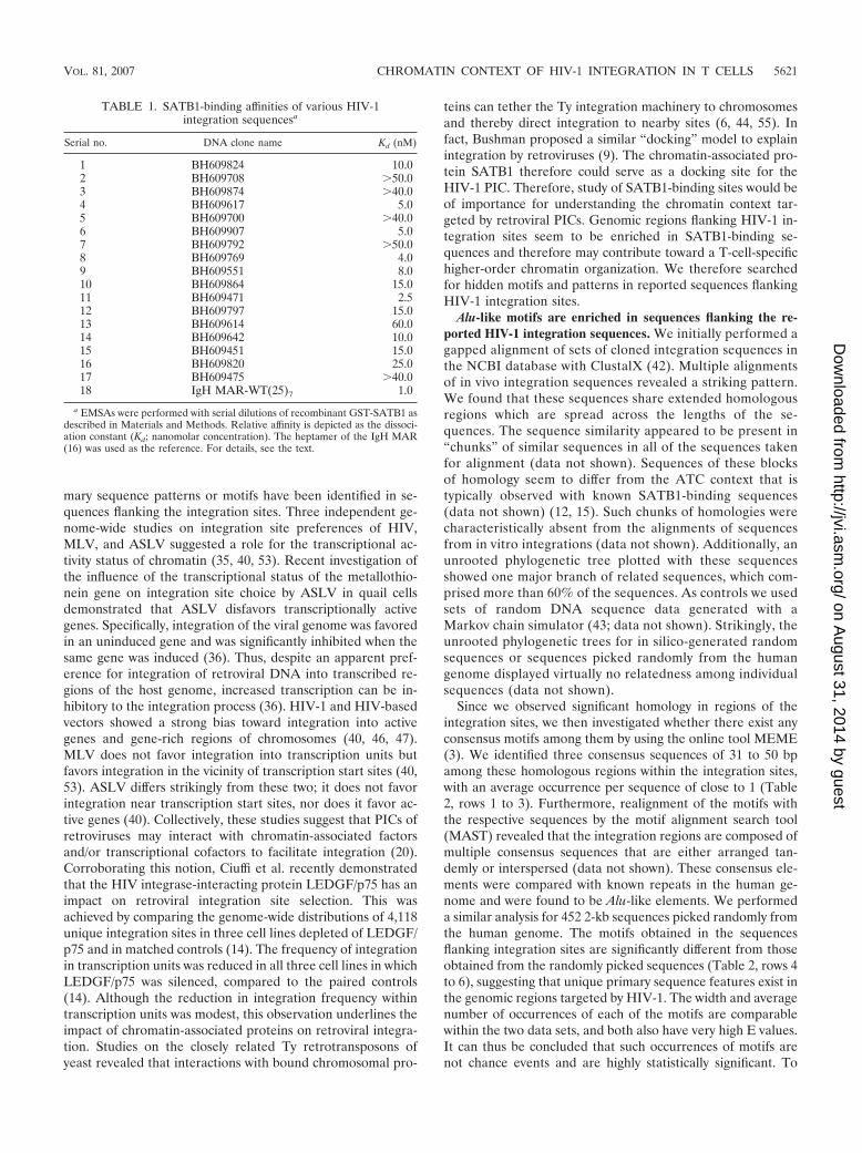
one of the proposed HIV-1 integration hot-spot regions in vivoprompted us to analyze the binding potential of other sequences.We essentially used the sequence information from the BH series(47) after separating the sequences from virus-host DNA junctionclones generated by integration reactions with naked cell-freeDNA as the template (in vitro) or a chromatin template in livecells (in vivo). From the sequences deposited by Bushman andcolleagues, we randomly selected a few in vivo and a few in vitrosequences and designed primers for their PCR amplification.PCR amplification of each of these sequences was performedwith genomic DNA isolated from Jurkat cells or PBMCs andspecific primer pairs. EMSA with labeled DNAs from two repre-sentative in vivo integration clones, BH609797 and BH609646,indicated that SATB1 bound them tightly in vitro (Fig. 1A and B,respectively, lanes 2 to 4). As controls, we used glutathione S-transferase (GST)-PARP, another DNA-binding protein (Fig.1A, lanes 5 to 7, and 1B, lanes 6 and 7), and GST alone (Fig. 1Aand B, lanes 8 and 9), both of which did not bind at all. AdditionalEMSA analysis with labeled DNAs corresponding to differentintegration clones from the BH series indicated that SATB1bound 80% (12 out of 15) in vivo sequences, as opposed to 20%(2 out of 10) in vitro sequences (data not shown). For accuratecomparison of binding affinities, we next estimated the dissocia-tion constants (Kds) with SATB1 for all in vivo integration clones(data not shown). The Kd values were in the range of 2.5 to 60nM, compared to the 1 nM of the IgH MAR heptamer (Table 1).Thus, SATB1 seems to bind preferentially to the in vivo integra-tion sequences and to at least some of them with an affinitycomparable to that of the IgH MAR, which contains the well-characterized BUR motif (29). It is reported that SATB1 doesnot bind to all of its genomic targets with the same affinity (12,15).
We next performed ChIP assays to monitor the binding ofSATB1 to these sequences in vivo. In vitro binding with nakedDNA substrates may not always reflect the in vivo occupancy ofSATB1 at the same site. Since we hypothesized a role forchromatin architecture in integration target choice by the PIC,it was essential to monitor binding of SATB1 to these sites invivo. As in the case of the 11q13 hot spot for integration, wefound that SATB1 bound both the BH609797 and BH609646in vivo integration sequences from the Bushman study in vivo(Fig. 1C, lane 1 in the top two parts). As controls for the ChIPassay, we used sequences from the upstream portions of IL-2and IL-2R� that were characterized with respect to their invivo occupancy by SATB1 (32). As expected under these con-ditions, the distal P1 region of the IL-2 promoter was bound bySATB1 in vivo but not the proximal P2 region and also not theP6 region of the IL-2R� promoter. We also performed ChIPanalysis for DNAs from two representative in vitro integrationclones, BH610076 and BH609954, and found that these sitesare not bound by SATB1 in vivo (Fig. 1D, lane 1 in the bottomtwo parts). To further verify the specificity of binding, weperformed a competition assay wherein binding was competedfor by homologous and heterologous unlabeled DNAs. Asprobes, we used in vivo integration sequence BH609471, whichbinds with high affinity, and BH609700, which binds with lowaffinity (data not shown). The binding of SATB1 to BH609471was not affected by addition of unlabeled DNA correspondingto heterologous in vitro integration clone BH609943 (Fig. 1E,lanes 3 and 4). A homologous in vivo integration clone,
VOL. 81, 2007 CHROMATIN CONTEXT OF HIV-1 INTEGRATION IN T CELLS 5619
on August 31, 2014 by guest
http://jvi.asm.org/
Dow
nloaded from
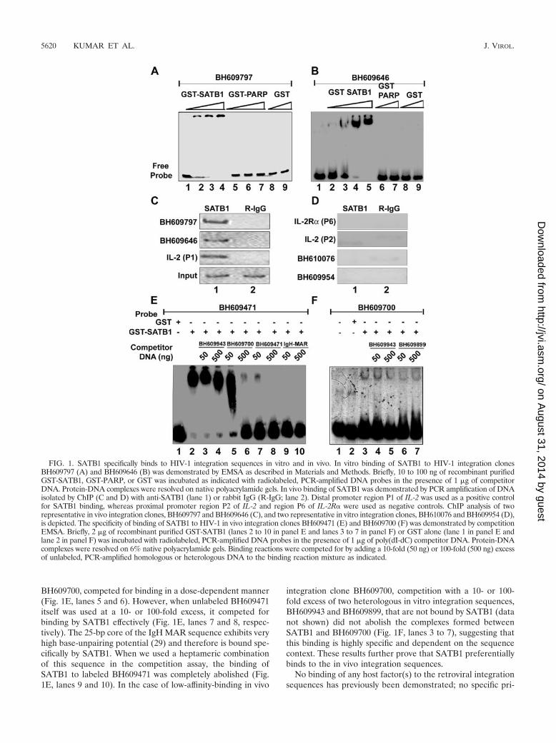
BH609700, competed for binding in a dose-dependent manner(Fig. 1E, lanes 5 and 6). However, when unlabeled BH609471itself was used at a 10- or 100-fold excess, it competed forbinding by SATB1 effectively (Fig. 1E, lanes 7 and 8, respec-tively). The 25-bp core of the IgH MAR sequence exhibits veryhigh base-unpairing potential (29) and therefore is bound spe-cifically by SATB1. When we used a heptameric combinationof this sequence in the competition assay, the binding ofSATB1 to labeled BH609471 was completely abolished (Fig.1E, lanes 9 and 10). In the case of low-affinity-binding in vivo
integration clone BH609700, competition with a 10- or 100-fold excess of two heterologous in vitro integration sequences,BH609943 and BH609899, that are not bound by SATB1 (datanot shown) did not abolish the complexes formed betweenSATB1 and BH609700 (Fig. 1F, lanes 3 to 7), suggesting thatthis binding is highly specific and dependent on the sequencecontext. These results further prove that SATB1 preferentiallybinds to the in vivo integration sequences.
No binding of any host factor(s) to the retroviral integrationsequences has previously been demonstrated; no specific pri-
FIG. 1. SATB1 specifically binds to HIV-1 integration sequences in vitro and in vivo. In vitro binding of SATB1 to HIV-1 integration clonesBH609797 (A) and BH609646 (B) was demonstrated by EMSA as described in Materials and Methods. Briefly, 10 to 100 ng of recombinant purifiedGST-SATB1, GST-PARP, or GST was incubated as indicated with radiolabeled, PCR-amplified DNA probes in the presence of 1 �g of competitorDNA. Protein-DNA complexes were resolved on native polyacrylamide gels. In vivo binding of SATB1 was demonstrated by PCR amplification of DNAisolated by ChIP (C and D) with anti-SATB1 (lane 1) or rabbit IgG (R-IgG; lane 2). Distal promoter region P1 of IL-2 was used as a positive controlfor SATB1 binding, whereas proximal promoter region P2 of IL-2 and region P6 of IL-2R� were used as negative controls. ChIP analysis of tworepresentative in vivo integration clones, BH609797 and BH609646 (C), and two representative in vitro integration clones, BH610076 and BH609954 (D),is depicted. The specificity of binding of SATB1 to HIV-1 in vivo integration clones BH609471 (E) and BH609700 (F) was demonstrated by competitionEMSA. Briefly, 2 �g of recombinant purified GST-SATB1 (lanes 2 to 10 in panel E and lanes 3 to 7 in panel F) or GST alone (lane 1 in panel E andlane 2 in panel F) was incubated with radiolabeled, PCR-amplified DNA probes in the presence of 1 �g of poly(dI-dC) competitor DNA. Protein-DNAcomplexes were resolved on 6% native polyacrylamide gels. Binding reactions were competed for by adding a 10-fold (50 ng) or 100-fold (500 ng) excessof unlabeled, PCR-amplified homologous or heterologous DNA to the binding reaction mixture as indicated.
5620 KUMAR ET AL. J. VIROL.
on August 31, 2014 by guest
http://jvi.asm.org/
Dow
nloaded from
mary sequence patterns or motifs have been identified in se-quences flanking the integration sites. Three independent ge-nome-wide studies on integration site preferences of HIV,MLV, and ASLV suggested a role for the transcriptional ac-tivity status of chromatin (35, 40, 53). Recent investigation ofthe influence of the transcriptional status of the metallothio-nein gene on integration site choice by ASLV in quail cellsdemonstrated that ASLV disfavors transcriptionally activegenes. Specifically, integration of the viral genome was favoredin an uninduced gene and was significantly inhibited when thesame gene was induced (36). Thus, despite an apparent pref-erence for integration of retroviral DNA into transcribed re-gions of the host genome, increased transcription can be in-hibitory to the integration process (36). HIV-1 and HIV-basedvectors showed a strong bias toward integration into activegenes and gene-rich regions of chromosomes (40, 46, 47).MLV does not favor integration into transcription units butfavors integration in the vicinity of transcription start sites (40,53). ASLV differs strikingly from these two; it does not favorintegration near transcription start sites, nor does it favor ac-tive genes (40). Collectively, these studies suggest that PICs ofretroviruses may interact with chromatin-associated factorsand/or transcriptional cofactors to facilitate integration (20).Corroborating this notion, Ciuffi et al. recently demonstratedthat the HIV integrase-interacting protein LEDGF/p75 has animpact on retroviral integration site selection. This wasachieved by comparing the genome-wide distributions of 4,118unique integration sites in three cell lines depleted of LEDGF/p75 and in matched controls (14). The frequency of integrationin transcription units was reduced in all three cell lines in whichLEDGF/p75 was silenced, compared to the paired controls(14). Although the reduction in integration frequency withintranscription units was modest, this observation underlines theimpact of chromatin-associated proteins on retroviral integra-tion. Studies on the closely related Ty retrotransposons ofyeast revealed that interactions with bound chromosomal pro-
teins can tether the Ty integration machinery to chromosomesand thereby direct integration to nearby sites (6, 44, 55). Infact, Bushman proposed a similar “docking” model to explainintegration by retroviruses (9). The chromatin-associated pro-tein SATB1 therefore could serve as a docking site for theHIV-1 PIC. Therefore, study of SATB1-binding sites would beof importance for understanding the chromatin context tar-geted by retroviral PICs. Genomic regions flanking HIV-1 in-tegration sites seem to be enriched in SATB1-binding se-quences and therefore may contribute toward a T-cell-specifichigher-order chromatin organization. We therefore searchedfor hidden motifs and patterns in reported sequences flankingHIV-1 integration sites.
Alu-like motifs are enriched in sequences flanking the re-ported HIV-1 integration sequences. We initially performed agapped alignment of sets of cloned integration sequences inthe NCBI database with ClustalX (42). Multiple alignmentsof in vivo integration sequences revealed a striking pattern.We found that these sequences share extended homologousregions which are spread across the lengths of the se-quences. The sequence similarity appeared to be present in“chunks” of similar sequences in all of the sequences takenfor alignment (data not shown). Sequences of these blocksof homology seem to differ from the ATC context that istypically observed with known SATB1-binding sequences(data not shown) (12, 15). Such chunks of homologies werecharacteristically absent from the alignments of sequencesfrom in vitro integrations (data not shown). Additionally, anunrooted phylogenetic tree plotted with these sequencesshowed one major branch of related sequences, which com-prised more than 60% of the sequences. As controls we usedsets of random DNA sequence data generated with aMarkov chain simulator (43; data not shown). Strikingly, theunrooted phylogenetic trees for in silico-generated randomsequences or sequences picked randomly from the humangenome displayed virtually no relatedness among individualsequences (data not shown).
Since we observed significant homology in regions of theintegration sites, we then investigated whether there exist anyconsensus motifs among them by using the online tool MEME(3). We identified three consensus sequences of 31 to 50 bpamong these homologous regions within the integration sites,with an average occurrence per sequence of close to 1 (Table2, rows 1 to 3). Furthermore, realignment of the motifs withthe respective sequences by the motif alignment search tool(MAST) revealed that the integration regions are composed ofmultiple consensus sequences that are either arranged tan-demly or interspersed (data not shown). These consensus ele-ments were compared with known repeats in the human ge-nome and were found to be Alu-like elements. We performeda similar analysis for 452 2-kb sequences picked randomly fromthe human genome. The motifs obtained in the sequencesflanking integration sites are significantly different from thoseobtained from the randomly picked sequences (Table 2, rows 4to 6), suggesting that unique primary sequence features exist inthe genomic regions targeted by HIV-1. The width and averagenumber of occurrences of each of the motifs are comparablewithin the two data sets, and both also have very high E values.It can thus be concluded that such occurrences of motifs arenot chance events and are highly statistically significant. To
TABLE 1. SATB1-binding affinities of various HIV-1integration sequencesa
Serial no. DNA clone name Kd (nM)
1 BH609824 10.02 BH609708 50.03 BH609874 40.04 BH609617 5.05 BH609700 40.06 BH609907 5.07 BH609792 50.08 BH609769 4.09 BH609551 8.010 BH609864 15.011 BH609471 2.512 BH609797 15.013 BH609614 60.014 BH609642 10.015 BH609451 15.016 BH609820 25.017 BH609475 40.018 IgH MAR-WT(25)7 1.0
a EMSAs were performed with serial dilutions of recombinant GST-SATB1 asdescribed in Materials and Methods. Relative affinity is depicted as the dissoci-ation constant (Kd; nanomolar concentration). The heptamer of the IgH MAR(16) was used as the reference. For details, see the text.
VOL. 81, 2007 CHROMATIN CONTEXT OF HIV-1 INTEGRATION IN T CELLS 5621
on August 31, 2014 by guest
http://jvi.asm.org/
Dow
nloaded from
perform a completely “unfiltered” scan for motifs within thegenomic sequences, repeat masking was not performed priorto a motif search. However, the derived motifs were thensearched for within the repeat-masked sequences with MASTand were undetectable.
The Alu repeats constitute about 5 to 10% of the humangenome (4). Alu elements affect the genome in several ways,causing insertion mutations, recombination between elements,gene conversion, and alterations in gene expression (4). TheAlu repeats have been implicated in transcription and tran-scription control (30, 34). In support of this, Alu repeats havebeen shown to be enriched in histone H3 lysine 9 methylation(30). Alu elements are each a dimer of similar, but not iden-tical, fragments with a total size of about 300 bp and originatefrom the 7SL RNA gene. Each element contains a bipartitepromoter for RNA polymerase III, a poly(A) tract locatedbetween the monomers, a 3�-terminal poly(A) tract, and nu-merous CpG islands and is flanked by short direct repeats. Thechromatin context of the Alu repeats is important for theirfunction (38), and the Alu elements themselves can play a rolein chromosomal rearrangement (37). Interestingly, analysis ofHIV-1 proviral integrations in isolates derived both from inte-grations in infected individuals and from cultured cells re-vealed a significant propensity of HIV-1 to integrate at or nearthe Alu repeats (50). Additionally, genome-wide analysis ofHIV integration sites by the Bushman group found 15.9% ofthe in vivo integration sites to be in Alu repeats (47). There-fore, it was not very surprising that our analysis picked upAlu-like motifs in the sequences flanking HIV-1 integrationsites. Additionally, mapping of genomic positions of integra-tion sites revealed that HIV-1 preferentially integrates withinthe transcribed and GC-rich regions of the human genome(19). The high GC content of Alu repeats may therefore con-stitute another feature facilitating their preferential targetingby the PIC.
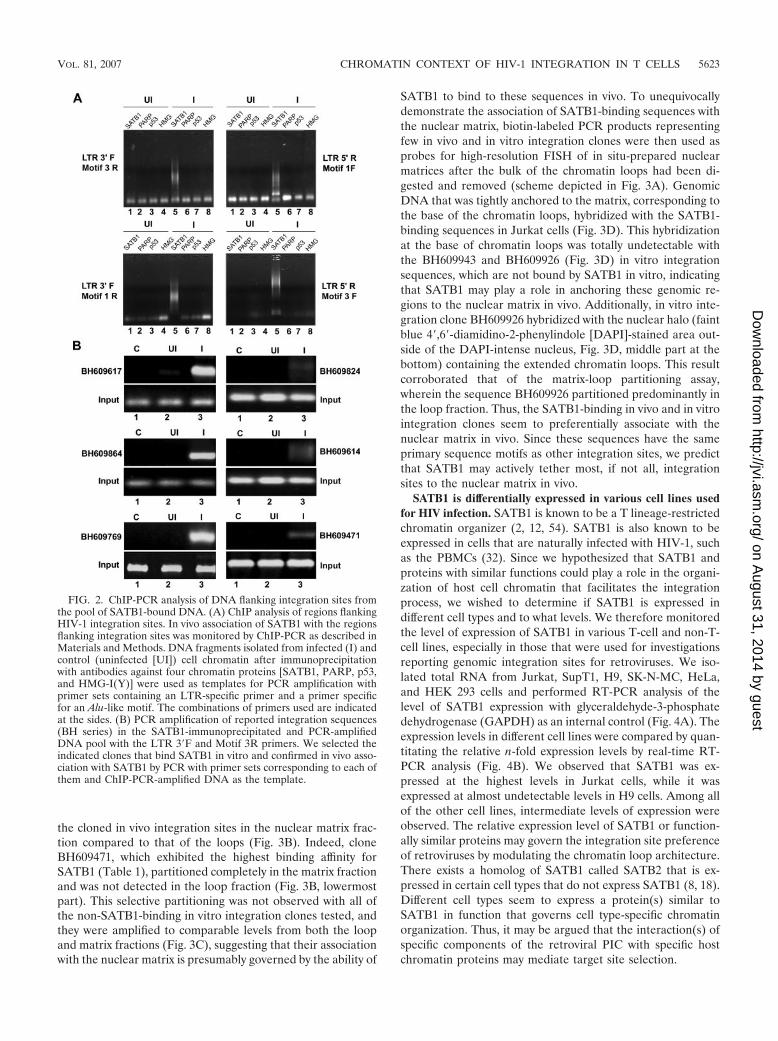
SATB1-associated chromatin contains cloned sequencesflanking HIV-1 integration sites. To test for the presence ofAlu-like motifs in HIV integration sites enriched in SATB1binding, we performed ChIP with SATB1-specific antibodiesand subsequently amplified the Alu repeats and HIV longterminal repeat (LTR) sequences in recovered DNA by PCR
with specifically designed primers corresponding to the Alu-like motifs (motifs 1 to 3, Table 2) and the LTRs of the NL4.3isolate of HIV-1. As controls we used antibodies specific top53, PARP, and HMG-I(Y). With combinations of Alu-likemotif-specific and LTR-specific primers, we observed thatPCR-amplified products were obtained specifically in the anti-SATB1-immunoprecipitated chromatin (Fig. 2A, lane 5 in allparts), suggesting that out of the four chromatin proteinstested, namely, SATB1, PARP, p53, and HMG-I(Y), onlySATB1 is specifically associated with the regions flankingHIV-1 integrations sites. We subsequently confirmed thatmany of the cloned integration sequences from the BH serieswere specifically amplified in PCRs with specific primers andpurified DNA template from anti-SATB1-immunoprecipitatedchromatin (Fig. 2B, lane 3 in all parts). Control reaction mix-tures with anti-SATB1-immunoprecipitated chromatin fromuninfected cells did not yield any amplification product, con-firming the specificity of the ChIP-PCRs. Collectively, theseresults demonstrated that the SATB1-associated chromatinharbors many regions of the genome that constitute HIV-1integration sites.
Regions flanking HIV-1 integration sites are associated withthe nuclear matrix in vivo. Chromatin is anchored to the nu-clear matrix by matrix/scaffold attachment regions (M/SARs),thereby organizing genomic DNA into topologically distinctloop domains that are important in replication and transcrip-tion (45). M/SARs are often closely associated with transcrip-tional promoters and enhancers of several genes and have beenshown to generate long-range chromatin accessibility (24).Juxtaposition with M/SARs correlates with transcriptional aug-mentation (5). SATB1 is a cell type-specific MAR-bindingprotein (16). We therefore reasoned that if genomic regionsflanking HIV-1 integration sites are enriched in SATB1-asso-ciated chromatin, then they should also be anchored to thenuclear matrix in vivo. To test this, we performed a partitioningassay designed to separately isolate (i) genomic DNA associ-ated with the nuclear matrix and (ii) that of chromatin loops.The DNA isolated from the loop and matrix fractions was thenused as the template for PCR amplification with primer setscorresponding to various cloned integration sites from the BHseries. This analysis revealed selective enrichment of most of
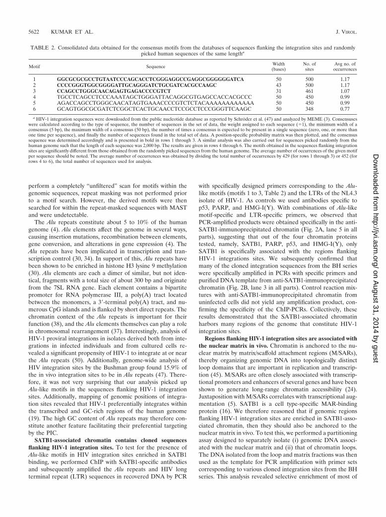
TABLE 2. Consolidated data obtained for the consensus motifs from the databases of sequences flanking the integration sites and randomlypicked human sequences of the same lengtha
Motif Sequence Width(bases)
No. ofsites
Avg no. ofoccurrences
1 GGCGCGCGCCTGTAATCCCAGCACCTCGGGAGGCCGAGGCGGGGGGATCA 50 500 1.172 CCCCGGGTGGCGGGGATTGCAGGGATCTGCGATCACGCCAAGC 43 500 1.173 CCAGCCTGGGCAACAGAGTGAGACCCCGTCT 31 461 1.074 TGCCTCAGCCTCCCAAATAGCTGGGATTACAGGCGTGAGCCACCACGCCC 50 450 0.995 AGACCAGCCTGGGCAACATAGTGAAACCCCGTCTCTACAAAAAAAAAAAA 50 450 0.996 GCAGTGGCGCGATCTCGGCTCACTGCAACCTCCGCCTCCCGGGTTCAAGC 50 348 0.77
a HIV-1 integration sequences were downloaded from the public nucleotide database as reported by Schroder et al. (47) and analyzed by MEME (3). Consensuseswere calculated according to the type of sequence, the number of sequences in the set of data, the weight assigned to each sequence (�1), the minimum width of aconsensus (5 bp), the maximum width of a consensus (50 bp), the number of times a consensus is expected to be present in a single sequence (zero, one, or more thanone time per sequence), and finally the number of sequences found in the total set of data. A position-specific probability matrix was then plotted, and the consensussequence was determined accordingly and is presented in bold in rows 1 through 3. A similar analysis was also carried out for sequences picked randomly from thehuman genome such that the length of each sequence was 2,000 bp. The results are given in rows 4 through 6. The motifs obtained in the sequences flanking integrationsites are significantly different from those obtained from the randomly picked sequences from the human genome. The average number of occurrences of the given motifper sequence should be noted. The average number of occurrences was obtained by dividing the total number of occurrences by 429 (for rows 1 through 3) or 452 (forrows 4 to 6), the total number of sequences used for analysis.
5622 KUMAR ET AL. J. VIROL.
on August 31, 2014 by guest
http://jvi.asm.org/
Dow
nloaded from
the cloned in vivo integration sites in the nuclear matrix frac-tion compared to that of the loops (Fig. 3B). Indeed, cloneBH609471, which exhibited the highest binding affinity forSATB1 (Table 1), partitioned completely in the matrix fractionand was not detected in the loop fraction (Fig. 3B, lowermostpart). This selective partitioning was not observed with all ofthe non-SATB1-binding in vitro integration clones tested, andthey were amplified to comparable levels from both the loopand matrix fractions (Fig. 3C), suggesting that their associationwith the nuclear matrix is presumably governed by the ability of
SATB1 to bind to these sequences in vivo. To unequivocallydemonstrate the association of SATB1-binding sequences withthe nuclear matrix, biotin-labeled PCR products representingfew in vivo and in vitro integration clones were then used asprobes for high-resolution FISH of in situ-prepared nuclearmatrices after the bulk of the chromatin loops had been di-gested and removed (scheme depicted in Fig. 3A). GenomicDNA that was tightly anchored to the matrix, corresponding tothe base of the chromatin loops, hybridized with the SATB1-binding sequences in Jurkat cells (Fig. 3D). This hybridizationat the base of chromatin loops was totally undetectable withthe BH609943 and BH609926 (Fig. 3D) in vitro integrationsequences, which are not bound by SATB1 in vitro, indicatingthat SATB1 may play a role in anchoring these genomic re-gions to the nuclear matrix in vivo. Additionally, in vitro inte-gration clone BH609926 hybridized with the nuclear halo (faintblue 4�,6�-diamidino-2-phenylindole [DAPI]-stained area out-side of the DAPI-intense nucleus, Fig. 3D, middle part at thebottom) containing the extended chromatin loops. This resultcorroborated that of the matrix-loop partitioning assay,wherein the sequence BH609926 partitioned predominantly inthe loop fraction. Thus, the SATB1-binding in vivo and in vitrointegration clones seem to preferentially associate with thenuclear matrix in vivo. Since these sequences have the sameprimary sequence motifs as other integration sites, we predictthat SATB1 may actively tether most, if not all, integrationsites to the nuclear matrix in vivo.
SATB1 is differentially expressed in various cell lines usedfor HIV infection. SATB1 is known to be a T lineage-restrictedchromatin organizer (2, 12, 54). SATB1 is also known to beexpressed in cells that are naturally infected with HIV-1, suchas the PBMCs (32). Since we hypothesized that SATB1 andproteins with similar functions could play a role in the organi-zation of host cell chromatin that facilitates the integrationprocess, we wished to determine if SATB1 is expressed indifferent cell types and to what levels. We therefore monitoredthe level of expression of SATB1 in various T-cell and non-T-cell lines, especially in those that were used for investigationsreporting genomic integration sites for retroviruses. We iso-lated total RNA from Jurkat, SupT1, H9, SK-N-MC, HeLa,and HEK 293 cells and performed RT-PCR analysis of thelevel of SATB1 expression with glyceraldehyde-3-phosphatedehydrogenase (GAPDH) as an internal control (Fig. 4A). Theexpression levels in different cell lines were compared by quan-titating the relative n-fold expression levels by real-time RT-PCR analysis (Fig. 4B). We observed that SATB1 was ex-pressed at the highest levels in Jurkat cells, while it wasexpressed at almost undetectable levels in H9 cells. Among allof the other cell lines, intermediate levels of expression wereobserved. The relative expression level of SATB1 or function-ally similar proteins may govern the integration site preferenceof retroviruses by modulating the chromatin loop architecture.There exists a homolog of SATB1 called SATB2 that is ex-pressed in certain cell types that do not express SATB1 (8, 18).Different cell types seem to express a protein(s) similar toSATB1 in function that governs cell type-specific chromatinorganization. Thus, it may be argued that the interaction(s) ofspecific components of the retroviral PIC with specific hostchromatin proteins may mediate target site selection.
FIG. 2. ChIP-PCR analysis of DNA flanking integration sites fromthe pool of SATB1-bound DNA. (A) ChIP analysis of regions flankingHIV-1 integration sites. In vivo association of SATB1 with the regionsflanking integration sites was monitored by ChIP-PCR as described inMaterials and Methods. DNA fragments isolated from infected (I) andcontrol (uninfected [UI]) cell chromatin after immunoprecipitationwith antibodies against four chromatin proteins [SATB1, PARP, p53,and HMG-I(Y)] were used as templates for PCR amplification withprimer sets containing an LTR-specific primer and a primer specificfor an Alu-like motif. The combinations of primers used are indicatedat the sides. (B) PCR amplification of reported integration sequences(BH series) in the SATB1-immunoprecipitated and PCR-amplifiedDNA pool with the LTR 3�F and Motif 3R primers. We selected theindicated clones that bind SATB1 in vitro and confirmed in vivo asso-ciation with SATB1 by PCR with primer sets corresponding to each ofthem and ChIP-PCR-amplified DNA as the template.
VOL. 81, 2007 CHROMATIN CONTEXT OF HIV-1 INTEGRATION IN T CELLS 5623
on August 31, 2014 by guest
http://jvi.asm.org/
Dow
nloaded from
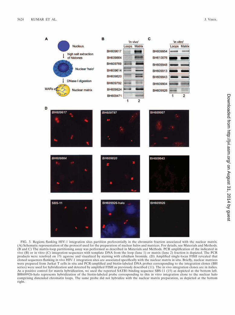
FIG. 3. Regions flanking HIV-1 integration sites partition preferentially in the chromatin fraction associated with the nuclear matrix.(A) Schematic representation of the protocol used for the preparation of nuclear halos and matrices. For details, see Materials and Methods.(B and C) The matrix-loop partitioning assay was performed as described in Materials and Methods. PCR amplification of the indicated invivo (B) or in vitro (C) integration sequences with template DNA from the loop (lane 1) or matrix (lane 2) fraction is depicted. The PCRproducts were resolved on 1% agarose and visualized by staining with ethidium bromide. (D) Amplified single-locus FISH revealed thatcloned sequences flanking in vivo HIV-1 integration sites are associated specifically with the nuclear matrix in situ. Briefly, nuclear matriceswere prepared from Jurkat T cells in situ and PCR-amplified and biotin-labeled DNA probes corresponding to the integration clones (BHseries) were used for hybridization and detected by amplified FISH as previously described (11). The in vivo integration clones are in italics.As a positive control for matrix hybridization, we used the reported SATB1-binding sequence SBS-11 (15) as depicted at the bottom left.BH609926-halo represents hybridization of the biotin-labeled probe corresponding to this in vitro integration clone to the nuclear halocomprising distended chromatin loops. The same probe did not hybridize with the nuclear matrix preparation, as depicted at the bottomright.
5624 KUMAR ET AL. J. VIROL.
on August 31, 2014 by guest
http://jvi.asm.org/
Dow
nloaded from
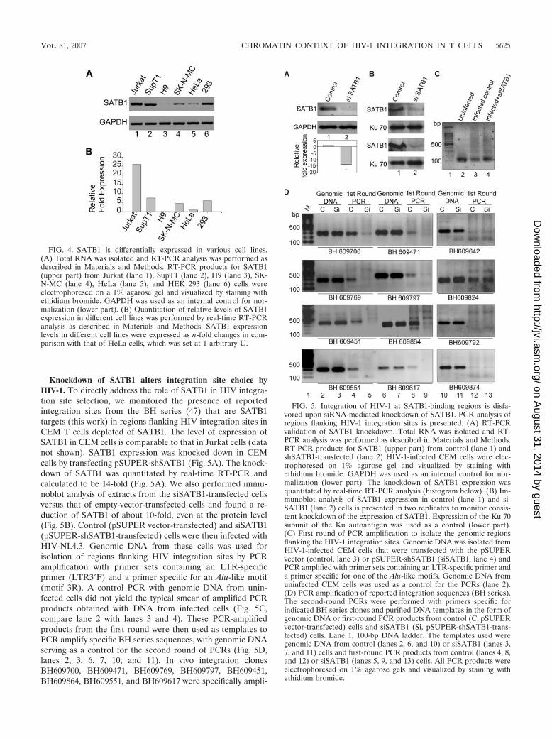
Knockdown of SATB1 alters integration site choice byHIV-1. To directly address the role of SATB1 in HIV integra-tion site selection, we monitored the presence of reportedintegration sites from the BH series (47) that are SATB1targets (this work) in regions flanking HIV integration sites inCEM T cells depleted of SATB1. The level of expression ofSATB1 in CEM cells is comparable to that in Jurkat cells (datanot shown). SATB1 expression was knocked down in CEMcells by transfecting pSUPER-shSATB1 (Fig. 5A). The knock-down of SATB1 was quantitated by real-time RT-PCR andcalculated to be 14-fold (Fig. 5A). We also performed immu-noblot analysis of extracts from the siSATB1-transfected cellsversus that of empty-vector-transfected cells and found a re-duction of SATB1 of about 10-fold, even at the protein level(Fig. 5B). Control (pSUPER vector-transfected) and siSATB1(pSUPER-shSATB1-transfected) cells were then infected withHIV-NL4.3. Genomic DNA from these cells was used forisolation of regions flanking HIV integration sites by PCRamplification with primer sets containing an LTR-specificprimer (LTR3�F) and a primer specific for an Alu-like motif(motif 3R). A control PCR with genomic DNA from unin-fected cells did not yield the typical smear of amplified PCRproducts obtained with DNA from infected cells (Fig. 5C,compare lane 2 with lanes 3 and 4). These PCR-amplifiedproducts from the first round were then used as templates toPCR amplify specific BH series sequences, with genomic DNAserving as a control for the second round of PCRs (Fig. 5D,lanes 2, 3, 6, 7, 10, and 11). In vivo integration clonesBH609700, BH609471, BH609769, BH609797, BH609451,BH609864, BH609551, and BH609617 were specifically ampli-
FIG. 5. Integration of HIV-1 at SATB1-binding regions is disfa-vored upon siRNA-mediated knockdown of SATB1. PCR analysis ofregions flanking HIV-1 integration sites is presented. (A) RT-PCRvalidation of SATB1 knockdown. Total RNA was isolated and RT-PCR analysis was performed as described in Materials and Methods.RT-PCR products for SATB1 (upper part) from control (lane 1) andshSATB1-transfected (lane 2) HIV-1-infected CEM cells were elec-trophoresed on 1% agarose gel and visualized by staining withethidium bromide. GAPDH was used as an internal control for nor-malization (lower part). The knockdown of SATB1 expression wasquantitated by real-time RT-PCR analysis (histogram below). (B) Im-munoblot analysis of SATB1 expression in control (lane 1) and si-SATB1 (lane 2) cells is presented in two replicates to monitor consis-tent knockdown of the expression of SATB1. Expression of the Ku 70subunit of the Ku autoantigen was used as a control (lower part).(C) First round of PCR amplification to isolate the genomic regionsflanking the HIV-1 integration sites. Genomic DNA was isolated fromHIV-1-infected CEM cells that were transfected with the pSUPERvector (control, lane 3) or pSUPER-shSATB1 (siSATB1, lane 4) andPCR amplified with primer sets containing an LTR-specific primer anda primer specific for one of the Alu-like motifs. Genomic DNA fromuninfected CEM cells was used as a control for the PCRs (lane 2).(D) PCR amplification of reported integration sequences (BH series).The second-round PCRs were performed with primers specific forindicated BH series clones and purified DNA templates in the form ofgenomic DNA or first-round PCR products from control (C, pSUPERvector-transfected) cells and siSATB1 (Si, pSUPER-shSATB1-trans-fected) cells. Lane 1, 100-bp DNA ladder. The templates used weregenomic DNA from control (lanes 2, 6, and 10) or siSATB1 (lanes 3,7, and 11) cells and first-round PCR products from control (lanes 4, 8,and 12) or siSATB1 (lanes 5, 9, and 13) cells. All PCR products wereelectrophoresed on 1% agarose gels and visualized by staining withethidium bromide.
FIG. 4. SATB1 is differentially expressed in various cell lines.(A) Total RNA was isolated and RT-PCR analysis was performed asdescribed in Materials and Methods. RT-PCR products for SATB1(upper part) from Jurkat (lane 1), SupT1 (lane 2), H9 (lane 3), SK-N-MC (lane 4), HeLa (lane 5), and HEK 293 (lane 6) cells wereelectrophoresed on a 1% agarose gel and visualized by staining withethidium bromide. GAPDH was used as an internal control for nor-malization (lower part). (B) Quantitation of relative levels of SATB1expression in different cell lines was performed by real-time RT-PCRanalysis as described in Materials and Methods. SATB1 expressionlevels in different cell lines were expressed as n-fold changes in com-parison with that of HeLa cells, which was set at 1 arbitrary U.
VOL. 81, 2007 CHROMATIN CONTEXT OF HIV-1 INTEGRATION IN T CELLS 5625
on August 31, 2014 by guest
http://jvi.asm.org/
Dow
nloaded from
fied in the reaction mixtures with DNA from the first-roundPCR of the control cells (Fig. 5D, lanes 4 and 8). There was amarked reduction in the PCR amplification of the BH seriessequences from the first-round PCR products in the siSATB1cells (Fig. 5D, lanes 5 and 9), suggesting that the integrationsite choice is altered when SATB1 is knocked down. We alsoobserved that many of the reported integration sites were ac-tually not targeted by the virus during infection, as deducedfrom the lack of PCR amplification product in the second-round PCR, even in the control cells (lane 12). In vivo inte-gration clones BH609642, BH609792, and BH609874 failed tobe amplified in the reaction mixtures with DNA from first-round PCRs. Since our data suggest that SATB1 binds theseregions in vitro, collectively these observations suggest thatHIV integration may not always occur at the same site(s)within the genome. However, in only one instance (BH609824)did we find that the second-round PCR product in controlinfected cells was not affected by SATB1 knockdown (Fig. 5D,second part from the top, lane 13), suggesting that a fewintegration events may occur in genomic regions that are notbound by SATB1 in vivo. It is unlikely that SATB1, or anyother protein that seems to be involved in the same process, isthe sole determinant of integration site selection by HIV. Ourdata nevertheless provide compelling evidence of a role forSATB1 in HIV-1 integration site selection.
Collectively, these results demonstrate that the SATB1-as-sociated chromatin harbors multiple regions of the genomethat constitute HIV-1 integration sites. In the absence ofSATB1, chromatin organization may be altered in such a man-ner that does not promote integration near SATB1-bindingsites. This could be due to a lack of interaction between acomponent(s) of the PIC and SATB1, to dynamic changes inchromatin loop domains, or both. We have, indeed, demon-strated that SATB1 collaborates with the nuclear-matrix-asso-ciated promyelocytic leukemia protein to organize the majorhistocompatibility complex class I locus into a distinct higher-order chromatin loop structure (31). Furthermore, gamma in-terferon treatment and silencing of either SATB1 or promy-elocytic leukemia protein dynamically alters the chromatinarchitecture, leading to an altered expression profile of a sub-set of major histocompatibility complex class I genes (31).Thus, the organization of the higher-order chromatin “loop-scape” by SATB1 and its interaction partners may be an im-portant determinant of the retroviral integration site selectionprocess. Here we show that silencing of SATB1 disfavors cer-tain regions of genome for HIV-1 integration. Elucidation ofthe molecular mechanism of this phenomenon requires furtherinvestigation of the interactions of the HIV-1 proteins withSATB1 and genome-wide comparative analysis of integrationsites in the presence or absence of SATB1.
Our analyses of the HIV-1 integration sites revealed uniquesignatures embedded within these sequences. First, they con-sist of multiple repeats of Alu-like consensus sequences andare specifically bound by SATB1. SATB1-binding sites consistof an AT-rich consensus element often flanked by GC-richsequences (P. K. Purbey and S. Galande, unpublished data).Since SATB1 organizes T-cell chromatin into a unique cage-like architecture that excludes heterochromatin, it is possiblethat the integration machinery of HIV-1 may specifically targetsuch regions for promoting its own replication and transcrip-
tion. If HIV integration occurs in a chromatin context of al-phoid repeats, it produces latent infection (25). Thus, it isevident that the chromatin context of the HIV integration siteis important for its own life cycle. Additionally, HIV integra-tions favor the entire length of the transcriptional regionswhereas MLV integrations are distributed evenly upstreamand downstream of the transcriptional start site (53). Interest-ingly, in contrast to the findings of Schroder et al. (47), inte-gration sites deposited by Wu et al. (53) did not show any kindof clustering (hot spots). This bias could be attributed to thedifferences in the expression levels of SATB1 in the cell linesused in these two studies; the SupT1 cells used by the Bushmangroup express higher levels of SATB1 compared to the HeLaor H9 cells used by the Burgess group (Fig. 4). The size ofchromosomal regions favorable for integration (100 kb) (40)also closely matches the average size of a chromosomal loop,which further argues for a role for higher-order chromatinorganization in retroviral integration. Different retrovirusesseem to have distinct patterns of integration site selectionwithin the human genome, suggesting that there may be localrecognition of chromosomal features and implying a role forchromosomal proteins (40).
No consensus sequences have been determined in the pri-mary flanking sequences of target site DNA in any of theretroviral integration site studies performed so far. The basepreferences reported by Holman and Coffin (23) actually cor-respond to the final step in proviral integration, when theintegrase recognizes and cleaves host DNA. However, it can begleaned that the PIC may have to first tether itself at specificsites within the chromatin and then integrase may actually beable to find preferred bases in the vicinity in a manner akin tothe Ty retrotransposons (9). Our results demonstrate thatHIV-1 prefers to integrate in T-cell chromatin specifically atsites that are enriched in specific consensus sequences andrepeating patterns. A primary sequence arrangement of thiskind may itself promote chromatin organization in a uniquearchitectural pattern in vivo that remains to be investigated.
ACKNOWLEDGMENTS
We are grateful to G. C. Mishra for support and encouragement andT. Kohwi-Shigematsu for the gift of the SATB1 antibody, pGEX-PARP-DBD, and the SBS-11 clone. We thank the CDAC supercom-puting facility at Pune and Bangalore, where part of the computationalanalysis was performed.
The molecular clone NL 4.3 and the CEM-GFP reporter cell linewere obtained through the AIDS Research and Reference ReagentProgram, Division of AIDS, NIAID, NIH. P.K., S.M., P.K.P., and D.N.are supported by fellowships from the Council of Scientific and Indus-trial Research, India. D. S. Ravi is supported by a fellowship from theUniversity Grants Commission, New Delhi, India. Work in the labo-ratory of S.G. is partly supported by a grant from the Department ofBiotechnology, Government of India. S. Galande is an InternationalSenior Research Fellow of the Wellcome Trust, United Kingdom.
REFERENCES
1. Adachi, A., H. E. Gendelman, S. Koeing, T. Folks, R. Willey, A. Rabson, andM. A. Martin. 1986. Production of acquired immunodeficiency syndrome-associated retrovirus in human and nonhuman cells transfected with aninfectious molecular clone. J. Virol. 59:284–291.
2. Alvarez, J. D., D. H. Yasui, H. Niida, T. Joh, D. Y. Loh, and T. Kohwi-Shigematsu. 2000. The MAR-binding protein SATB1 orchestrates temporaland spatial expression of multiple genes during T-cell development. GenesDev. 14:521–535.
3. Bailey, T. L., and C. Elkan. 1994. Fitting a mixture model by expectationmaximization to discover motifs in biopolymer, p. 28–36. In R. Altman, D.
5626 KUMAR ET AL. J. VIROL.
on August 31, 2014 by guest
http://jvi.asm.org/
Dow
nloaded from
Brutlag, P. Karp, R. Lathrop, and D. Searls (ed.), Proceedings of the SecondInternational Conference on Intelligent Systems for Molecular Biology.Press, Menlo Park, CA.
4. Batzer, M. A., and P. L. Deininger. 2002. Alu repeats and human genomicdiversity. Nat. Rev. Genet. 3:370–379.
5. Bode, J., C. Benham, A. Knopp, and C. Mielke. 2000. Transcriptional aug-mentation: modulation of gene expression by scaffold/matrix-attached re-gions (S/MAR elements). Crit. Rev. Eukaryot. Gene Expr. 10:73–90.
6. Boeke, J. D., and S. E. Devine. 1998. Yeast retrotransposons: finding a nicequiet neighborhood. Cell 93:1087–1089.
7. Bor, Y. C., M. D. Miller, F. D. Bushman, and L. E. Orgel. 1996. Target-sequence preferences of HIV-1 integration complexes in vitro. Virology222:283–288.
8. Britanova, O., S. Akopov, S. Lukyanov, P. Gruss, and V. Tarabykin. 2005.Novel transcription factor Satb2 interacts with matrix attachment regionDNA elements in a tissue-specific manner and demonstrates cell-type-de-pendent expression in the developing mouse CNS. Eur. J. Neurosci. 21:658–668.
9. Bushman, F. D. 2003. Targeting survival: integration site selection byretroviruses and LTR-retrotransposons. Cell 115:135–138.
10. Bushman, F. D., T. Fujiwara, and R. Craigie. 1990. Retroviral DNA inte-gration directed by HIV integration protein in vitro. Science 249:1555–1558.
11. Cai, S., and T. Kohwi-Shigematsu. 1999. Intranuclear relocalization of ma-trix binding sites during T cell activation detected by amplified fluorescencein situ hybridization. Methods 19:394–402.
12. Cai, S., H. J. Han, and T. Kohwi-Shigematsu. 2003. Tissue-specific nucleararchitecture and gene expression regulated by SATB1. Nat. Genet. 34:42–51.
13. Carteau, S., C. Hoffman, and F. Bushman. 1998. Chromosome structure andhuman immunodeficiency virus type 1 cDNA integration: centromeric al-phoid repeats are a disfavored target. J. Virol. 72:4005–4014.
14. Ciuffi, A., M. Llano, E. Poeschla, C. Hoffmann, J. Leipzig, P. Shinn, J. R.Ecker, and F. Bushman. 2005. A role for LEDGF/p75 in targeting HIVDNA integration. Nat. Med. 11:1287–1289.
15. de Belle, I., S. Cai, and T. Kohwi-Shigematsu. 1998. The genomic sequencesbound to special AT-rich sequence-binding protein 1 (SATB1) in vivo inJurkat T cells are tightly associated with the nuclear matrix at the bases of thechromatin loops. J. Cell Biol. 141:335–348.
16. Dickinson, L. A., T. Joh, Y. Kohwi, and T. Kohwi-Shigematsu. 1992. Atissue-specific MAR/SAR DNA-binding protein with unusual binding siterecognition. Cell 70:631–645.
17. Dickinson, L. A., C. D. Dickinson, and T. Kohwi-Shigematsu. 1997. Anatypical homeodomain in SATB1 promotes specific recognition of the keystructural element in a matrix attachment region. J. Biol. Chem. 272:11463–11470.
18. Dobreva, G., J. Dambacher, and R. Grosschedl. 2003. SUMO modificationof a novel MAR-binding protein, SATB2, modulates immunoglobulin mugene expression. Genes Dev. 17:3048–3061.
19. Elleder, D., A. Pavlicek, J. Paces, and J. Hejnar. 2002. Preferential integra-tion of human immunodeficiency virus type 1 into genes, cytogenetic R bandsand GC-rich DNA regions: insight from the human genome sequence. FEBSLett. 51:285–286.
20. Engelman, A. 2005. The ups and downs of gene expression and retroviralDNA integration. Proc. Natl. Acad. Sci. USA 102:1275–1276.
21. Gervaix, A., D. L. West, M. Leoni, D. D. Richman, F. Wong-Staal, and J.Corbeil. 1997. A new reporter cell line to monitor HIV infection and drugsusceptibility in vitro. Proc. Natl. Acad. Sci. USA 94:4653–4658.
22. Hindmarsh, P., and J. Leis. 1999. Retroviral DNA integration. Microbiol.Mol. Biol. Rev. 63:836–843.
23. Holman, A. G., and J. M. Coffin. 2005. Symmetrical base preferences sur-rounding HIV-1 and avian sarcoma/leukosis. Proc. Natl. Acad. Sci. USA102:6103–6107.
24. Jenuwein, T., W. C. Forrester, L. A. Fernandez-Herrero, G. Laible, M. Dull,and R. Grosschedl. 1997. Extension of chromatin accessibility by nuclearmatrix attachment regions. Nature 385:269–272.
25. Jordan, A., D. Bisgrove, and E. Verdin. 2003. HIV reproducibly establishesa latent infection after acute infection of T cells in vitro. EMBO J. 22:1868–1877.
26. Jordan, A., P. Defechereux, and E. Verdin. 2001. The site of HIV-1 integra-tion in the human genome determines basal transcriptional activity andresponse to Tat transactivation. EMBO J. 20:1726–1738.
27. Kitamura, Y., Y. M. H. Lee, and J. M. Coffin. 1992. Nonrandom integrationof retroviral DNA in vitro: effect of CpG methylation. Proc. Natl. Acad. Sci.USA 89:5532–5536.
28. Kohwi-Shigematsu, T., I. de Belle, L. A. Dickinson, S. Galande, and Y.Kohwi. 1998. Identification of base-unpairing region (BUR)-binding pro-teins and characterization of their in vivo binding sequences. Methods CellBiol. 53:323–354.
29. Kohwi-Shigematsu, T., and Y. Kohwi. 1990. Torsional stress stabilizes ex-tended base unpairing in suppressor sites flanking immunoglobulin heavychain enhancer. Biochemistry 29:9551–9560.
30. Kondo, Y., and J. P. Issa. 2003. Enrichment for histone H3 lysine 9 meth-ylation at Alu repeats in human cells. J. Biol. Chem. 278:27658–27662.
31. Kumar, P. P., O. Bischof, P. K. Purbey, D. Notani, H. Urlaub, A. Dejean, andS. Galande. 2007. Functional interaction between PML and SATB1 regu-lates chromatin loop architecture and transcription of the MHC class I locus.Nat. Cell Biol. 9:45–56.
32. Kumar, P. P., P. K. Purbey, D. S. Ravi, D. Mitra, and S. Galande. 2005.Displacement of SATB1-bound HDAC1 corepressor by HIV-1 transactiva-tor induces expression of interleukin-2 and its receptor in T cells. Mol. Cell.Biol. 25:1620–1633.
33. Kumar, P. P., P. K. Purbey, C. K. Sinha, D. Notani, A. Limaye, R. S. Jayani,and S. Galande. 2006. Phosphorylation of SATB1, a global gene regulator,acts as a molecular switch regulating its transcriptional activity in vivo. Mol.Cell 22:231–243.
34. Leem, S. H., N. Kouprina, J. Grimwood, J. H. Kim, M. Mullokandov, Y. H.Yoon, J. Y. Chae, J. Morgan, S. Lucas, P. Richardson, C. Detter, T. Glavina,E. Rubin, J. C. Barrett, and V. Larionov. 2004. Closing the gaps on humanchromosome 19 revealed genes with a high density of repetitive tandemlyarrayed elements. Genome Res. 14:239–246.
35. Mack, K. D., X. Jin, S. Yu, R. Wei, L. Kapp, C. Green, B. Herndier, N. W.Abbey, A. Elbaggari, Y. Liu, and M. S. McGrath. 2003. HIV insertions withinand proximal to host cell genes are a common finding in tissues containinghigh levels of HIV DNA and macrophage-associated p24 antigen expression.J. Acquir. Immune Defic. Syndr. 33:308–320.
36. Maxfield, L. F., C. D. Fraize, and J. M. Coffin. 2005. Relationship betweenretroviral DNA-integration-site selection and host cell transcription. Proc.Natl. Acad. Sci. USA 102:1436–1441.
37. McNeil, N. 2004. AluElements: repetitive DNA as facilitators of chromo-somal rearrangement. J. Assoc. Genet. Technol. 30:41–47.
38. Medstrand, P., L. N. van de Lagemaat, and D. L. Mager. 2002. Retroelementdistributions in the human genome: variations associated with age and prox-imity to genes. Genome Res. 12:1483–1495.
39. Mielke, C., K. Maass, M. Tummler, and J. Bode. 1996. Anatomy of highlyexpressing chromosomal sites targeted by retroviral vectors. Biochemistry35:2239–2252.
40. Mitchell, R. S., B. F. Beitzel, A. R. Schroder, P. Shinn, H. Chen, C. C. Berry,J. R. Ecker, and F. D. Bushman. 2004. Retroviral DNA integration: ASLV,HIV, and MLV show distinct target site preferences. PLoS Biol. 2:E234.
41. Mnller, H. P., and H. E. Varmus. 1994. DNA bending creates favored sitesfor retroviral integration: an explanation for preferred insertion sites innucleosomes. EMBO J. 13:4704–4714.
42. Pearson, W. R., and D. J. Lipman. 1988. Improved tools for biologicalsequence comparison. Proc. Natl. Acad. Sci. USA 85:2444–2448.
43. Pesole, G., S. Luini, and M. D’Souza. 2000. PatSearch: a pattern matchersoftware that finds functional elements in nucleotide and protein sequencesand assesses their statistical significance. Bioinformatics 16:450.
44. Pryciak, P. M., and H. E. Varmus. 1992. Nucleosomes, DNA-binding pro-teins, and DNA sequence modulate retroviral integration target site selec-tion. Cell 69:769–780.
45. Roberge, M., and S. M. Gasser. 1992. DNA loops: structural and functionalproperties of scaffold-attached regions. Mol. Microbiol. 6:419–423.
46. Sandmeyer, S. 2003. Integration by design. Proc. Natl. Acad. Sci. USA100:5586–5588.
47. Schroder, A. R., P. Shinn, H. Chen, C. Berry, J. R. Ecker, and F. Bushman.2002. HIV-1 integration in the human genome favors active genes and localhot spots. Cell 110:521–529.
48. Seo, J., M. M. Lozano, and J. P. Dudley. 2005. Nuclear matrix bindingregulates SATB1-mediated transcriptional repression. J. Biol. Chem. 280:24600–24609.
49. Shih, C. C., J. P. Stoye, and J. M. Coffin. 1988. Highly preferred targets forretrovirus integration. Cell 53:531–537.
50. Stevens, S. W., and J. D. Griffith. 1996. Sequence analysis of the humanDNA flanking sites of human immunodeficiency virus type 1 integration.J. Virol. 70:6459–6462.
51. Withers-Ward, E. S., Y. Kitamura, J. P. Barnes, and J. M. Coffin. 1994.Distribution of targets for avian retrovirus DNA integration in vivo. GenesDev. 8:1473–1787.
52. Wolffe, A. P., and D. Guschin. 2000. Chromatin structural features andtargets that regulate transcription. J. Struct. Biol. 129:102–122.
53. Wu, X., Y. Li, B. Crise, and S. M. Burgess. 2003. Transcription start regionsin the human genome are favored targets for MLV integration. Science300:1749–1751.
54. Yasui, D., M. Miyano, S. Cai, P. Varga-Weisz, and T. Kohwi-Shigematsu.2002. SATB1 targets chromatin remodelling to regulate genes over longdistances. Nature 419:641–645.
55. Zhu, Y., J. Dai, P. G. Fuerst, and D. F. Voytas. 2003. Controlling integrationspecificity of a yeast retrotransposon. Proc. Natl. Acad. Sci. USA 100:5891–5895.
56. Zoubak, S. 1994. Regional specificity of HTLV-1 proviral integration inhuman genome. Gene 143:155–163.
VOL. 81, 2007 CHROMATIN CONTEXT OF HIV-1 INTEGRATION IN T CELLS 5627
on August 31, 2014 by guest
http://jvi.asm.org/
Dow
nloaded from