saphenofemoral arteriovenous fistula as hemodialysis access
TRANSCRIPT

RESEARCH ARTICLE Open Access
Saphenofemoral arteriovenous fistula ashemodialysis accessJoão A Correa1*, Luiz Carlos de Abreu2, Adilson C Pires1, João R Breda1, Yumiko R Yamazaki1, Alexandre C Fioretti1,Vitor E Valenti2,3, Luiz Carlos M Vanderlei4, Hugo Macedo Junior2, Eduardo Colombari2, Fausto Miranda Junior5
Abstract
Background: An upper limb arteriovenous (AV) fistula is the access of choice for haemodialysis (HD). There havebeen few reports of saphenofemoral AV fistulas (SFAVF) over the last 10-20 years because of previous suggestionsof poor patencies and needling difficulties. Here, we describe our clinical experience with SFAVF.
Methods: SFAVFs were evaluated using the following variables: immediate results, early and late complications,intraoperative and postoperative complications (up to day 30), efficiency of the fistula after the onset of needlingand complications associated to its use.
Results: Fifty-six SFAVF fistulas were created in 48 patients. Eight patients had two fistulas: 8 patent (16%), 10transplanted (20%), 12 deaths (24%), 1 low flow (2%) and 20 thrombosis (39%) (first two months of preparation).One patient had severe hypotension during surgery, which caused thrombosis of the fistula, which was successfullythrombectomised, four thrombosed fistulae were successfully thrombectomised and revised on the firstpostoperative day. After 59 months of follow-up, primary patency was 44%.
Conclusion: SFAVF is an adequate alternative for patients without the possibility for other access in the upperlimbs, allowing efficient dialysis with good long-term patency with a low complication rate.
BackgroundThe establishment of vascular access for hemodialysis(HD) remains a challenge for vascular surgery. Due tothe improvement of chronic renal failure treatment andconsequently, increased survival, maintenance of vascu-lar access and long-term treatment of its complications,it has become an important cause of hospitalization,morbidity and patient costs [1,2].The access of choice for HD is a primary arteriove-
nous (AV) fistula in the upper limb, between the radialartery and the cephalic vein [3-6]. This techniqueincreases venous flow approximately 250-300 ml perminute, which is the minimum flow velocity needed toobtain the appropriate clearance of urea after 4 hours ofHD. The preference for its use is due to the longer per-iod of working of these AV communications, low rate ofcomplications and easy handling [7-9]. Prolonged use ofthese accesses may lead to complications such as
infection, puncture pseudoaneurysm, anastomotic pseu-doaneurysm, venous hypertension, distal ischemia andtheir own obstruction [10]. In this situation, generallycreation of new AV fistula is indicated.Some disadvantages of using AV fistula by transposi-
tion of the femoral vein are the necessity for a moreextensive and deeper dissection that uses larger vesselsand make it more susceptible to infection and steal syn-drome [11]. Such problems were also observed by us[12] and motivated us to seek a new alternative techni-que, using a saphenous vein in “bridge”; conformation,anastomosed to the superficial femoral artery near theadductor canal [12]. The initial benefits of this proce-dure encouraged us to expand the study with a largerpatient number, analyzing the results of clinical experi-ence and late complications. Therefore, in view of theabove considerations, we aimed to evaluate the resultsof saphenofemoral AV fistula (SFAVF) as access to HD.
MethodThis is a longitudinal prospective study conducted at theHospital of the Faculdade de Medicina do ABC, in São
* Correspondence: [email protected] de Cirugia da Faculdade de Medicina do ABC, Santo André,SP, BrasilFull list of author information is available at the end of the article
Correa et al. BMC Surgery 2010, 10:28http://www.biomedcentral.com/1471-2482/10/28
© 2010 Correa et al; licensee BioMed Central Ltd. This is an Open Access article distributed under the terms of the Creative CommonsAttribution License (http://creativecommons.org/licenses/by/2.0), which permits unrestricted use, distribution, and reproduction inany medium, provided the original work is properly cited.

Bernardo do Campo (SP). The patients were from theDepartment of Nephrology, Faculdade de Medicina doABC, São Bernardo do Campo (SP), and from theNephrology and Hypertension Center, Santo André(SP). We included patients in whom all access possibili-ties were exhausted in the upper limbs and could not betreated by peritoneal dialysis, while those with an absentor inadequate long saphenous vein, sequelae of deepvenous thrombosis or a femoropopliteal arterial occlu-sion were excluded.This research was explained to all subjects and it was
begun only after their consent according to the stan-dards of the Ethic Committee in Research of our Uni-versity (protocol number 021/2000). All patients gaveinformed consent. All procedures were in compliancewith the Helsinki Declaration.From August 1998 to May 2006 1183 fistulas for HD
access were performed. 1086 were native (91.8%) and 97(8.2%) were prosthetic polytetrafluoroethylene (PTFE).Among the 1086 native, 1030 (87.1%) were performedin upper limbs and 56 (4.7%) in lower limbs and usedthe saphenous vein. Among the fistulas which usedprostheses, 79 (6.7%) were performed in upper limbsand 18 (1.5%) in lower limbs.The 56 SFAVF were performed in 48 patients who
underwent HD, in which all access possibilities wereexhausted and limbs met the inclusion criteria. Datafrom each patient were recorded in a protocol forfurther statistical evaluation.The sample consisted of 27 female (56.2%) and 21
male (43.7%) patients. In eight patients (6 women and 2men) two SFAVF were performed, these were performedat the same time. Ages ranged from 10 to 82 years old(mean 44.4 years old). One 10-year old patient was ofsmall stature and had noOne 10-year old patient was of small stature and had
no vessels of an appropriate size. Among the 48patients, 42 (87.5%) presented one or more associateddiseases such as hypertension (HT), diabetes mellitus(DM), heart and arterial diseases.We conducted analysis of cumulative patency by
Kaplan-Meier method and other results were presentedas a simple percentage.The surgical technique consisted of making an AV fis-
tula by using only one anastomosis between the saphe-nous vein and the distal superficial femoral artery afterits implementation and superficial to the anterior thigh.Patients were discharged on the first day after surgery
after verification of clinical conditions and surgical out-comes. The follow-up was performed on an outpatientbasis, every seven days in the HD unit. Thirty days laterthe fistula was released to punctures.Fistulas were evaluated according to puncture, HD
flow, spontaneous venous pressure absence and dialysis
adequacy according to weekly Kt/v [13]. Puncture facil-ity was defined as catheterization of the fistula at itsfirst attempt, the minimal acceptable HD flow was con-sidered as values above 250 ml/min; absence of sponta-neous venous pressure was considered as pressure lowerthan 100/mm Hg at the end of devolution with optimalblood flow and urea clearance dialysis adequacy (invitro) was multiplied by the duration of dialysis in min-utes divided by the volume of urea distribution (weight× 0.6). We considered 1.2 as the ideal week value.We also evaluated the presence or absence of compli-
cations such as thrombosis, post-puncture hematoma,distal limb ischemia, venous hypertension, cardiacdecompensation, infection, puncture pseudoaneurysm,anastomotic pseudoaneurysm and aneurysmal dilatation.If a problem was found we used ultrasonography andEcho-color-Doppler 2P. In cases of stenosis we treatedwith an angioplasty and in cases of pseudoaneurysm weperformed a surgical revision.The immediate result was defined as thrill intensity
and presence or absence of postoperative complicationsafter surgery. Regarding fistula maturation we consid-ered early outcome as the fistula maturation from the1st to the 30th post-operative day and late result as thefistula maturation after punches onset (after the 30th
post-operative).
Technical StandardizationPatients were submitted to SFAVF creation according tothe following standardization: (1) Election of the mostsuitable limb for manufacture of AV fistula throughclinical examination based on arterial and venous condi-tions: presence of normal peripheral pulses and no signsof chronic venous hypertension or varicose vein disease.(2) Patient on horizontal dorsal supine position underspinal anesthesia. (3) Antisepsis with iodopovidine anddelimitation of the surgical area with sterile cloths (Fig-ure 1). (4) Longitudinal incision of approximately 6 cmin the inguinal region, dissection and ligation of tribu-tary veins of the proximal saphenous vein. (5) Stab inci-sions for dissection of the saphenous vein to its distalthird of the thigh (Figure 2). (6) Oblique incision ofapproximately 10 cm in the middle third of the distalinner thigh. (7) Dissection of the distal thigh followedby planes of dissection and isolation of the superficialfemoral artery above the hiatus of the adductor magnusmedially away from the sartorius muscle (Figure 3). (8)Distal ligation, removal of the bed with plastic tubecatheterization number six and heparinization of thesaphenous vein with 20 ml of heparin 1% (Figure 4). (9)Production of subcutaneous tunnel by blunt dissectionin the anterolateral thigh in order to place the saphe-nous vein in front of its new bed. (10) Proximal and dis-tal superficial femoral artery clamp and longitudinal
Correa et al. BMC Surgery 2010, 10:28http://www.biomedcentral.com/1471-2482/10/28
Page 2 of 7

arteriotomy of approximately 1 cm. (11) Proximalheparinization with 10 ml of heparin to 1%, end-to-sidesaphenous vein to the femoral artery, using runningsutures of 6-0 polypropylene. (12) Arterial and venousunclamp according to maneuvers and pulmonaryembolic protection. (13) Passage of the saphenous veinthrough the subcutaneous tunnel (Figure 5). (14) Verifi-cation of the final terms, as the conformation of thevein and the thrill of the proximal and distal AV fistula.Figure 6 represents the anastomosis between the saphe-nous vein and superficial femoral artery and Figure 7presents the final aspect of the surgical scars.
ResultsFifty-six fistulas were used in 48 patients, 27 femalesand 21 males. Six patients underwent construction oftwo saphenous vein fistulas (four females and twomales). The oldest is in operation for 59 months andthe youngest for three months. These fistulas were effi-cient, did not present difficulty to puncture, presentedgood HD flow (300 ml/min), presented no spontaneous
venous pressure and dialysis adequacy according to kt/v(within the ideal value) [13].Ten patients underwent renal transplantation with
cadaver kidney (20.8% - seven females and three males).The age ranged from 24 to 51 years old (mean 34.7years old). These transplants occurred from the 3rd tothe 37th month after fistulas creation (average of 9.6months) (Table 1).In this follow-up of 59 months, among the 56 fistulas
that were used in 48 patients we found four types ofcomplications: thrombosis, pseudoaneurysm, stenosis/low flow and puncture hematoma. Thrombosis and lowflow with functional loss occurred in 21 fistulas (41.2% -11 were male and 10 female patients). The workingperiod of the fistula ranged from two to 30 months(average of 9.5 months of use). The age ranged from 10to 80 years old (average of 43.3 years old). Pseudoaneur-ysm puncture occurred in five fistulas (9.8% - allpatients were female). The earliest occurred in fourmonths and the latest occurred after 30 months of use.Stenosis with low flow fistulas occurred in two cases(5.9% - one male and one female) with a mean time ofseven months. Puncture hematoma occurred in a fistula
Figure 1 Patient positioned for surgery with demarcation ofthe planning technique.
Figure 2 Dissection of the saphenous vein through stabincisions, starting from the inguinal region to the distal thirdof thigh.
Figure 3 Dissection of the superficial femoral artery above theadductor canal.
Figure 4 Saphenous vein dissected, it was removed from itsbed and was catheterized.
Correa et al. BMC Surgery 2010, 10:28http://www.biomedcentral.com/1471-2482/10/28
Page 3 of 7

in a female patient at the 3rd month of its maturationand its absorption occurred spontaneously without jeo-pardize the fistula patency. Other complications such as:ischemia, venous hypertension, infection, anastomoticpseudoaneurysm, aneurysmal dilatation and cardiacdecompensation were not observed.After treatment of complications, these fistulas could
be used for a varied period until the following outcomesoccured: maintenance of patency, transplantation, deathor thrombosis.In cases of patients with thrombosis or low flow (21
fistulas, eight subjects), it was decided to carry outSFAVF in the contralateral limb, and in two, both male,we decided to implant a PTFE in bridge, using thesuperficial femoral artery and common femoral vein ofthe contralateral limb. In another case, also male, wealso decided by the PTFE to implant in “loop” using thecommon femoral artery and vein in the inguinal region
in the same limb which the SFAVF was performed. Inthe same condition it was held biopsy of the saphenousvein thrombosis of the anterior fistula.In other cases of thrombosis due to the urgent neces-
sity for HD it was decided to install double-lumencatheter (perm-cath®) through the femoral vein in theinguinal region. In only two cases was it possible to per-form peritoneal dialysis.In 93 months of follow-up there were twelve deaths in
patients who had their SFAVF able (eight females andfour males), the youngest was 37 years old and the old-est 80 years old, with an average of 50 years. Thematuration period of these fistulas ranged from two to40 months, with an average of 16.6 months.At this time eight fistulas continue to be used in four
females and four males, the youngest aged 30 years oldand the oldest aged 53 years old (average of 42.9 yearsold). The most recent has three months of operationand the oldest 59 months (average of 16 months). Sincethe beginning of the investigation until now, we haveexperienced eight patent fistulas, 10 transplant patients,12 deaths, 20 thromboses and one low flow.We carried out the analysis of cumulative patency
[14], with a score of 44.04% patency likely at the end ofthe period of 59 months, which is the duration of the
Figure 5 Passage of the saphenous vein through thesubcutaneous tunnel on the anterior thigh.
Figure 6 Anastomosis between the saphenous vein andsuperficial femoral artery
Figure 7 Final aspect of the surgical scars.
Table 1 Distribution of SFAVF according to analysis after93 months of follow-up (N = 51)
Development Number of fistulas %
Functioning 8 15.7
Transplanted 10 19.8
Deaths 12 23.5
Thrombosis 20 39.2
Low flow 1 1.9
% = Percentage.
N = Number.
Correa et al. BMC Surgery 2010, 10:28http://www.biomedcentral.com/1471-2482/10/28
Page 4 of 7
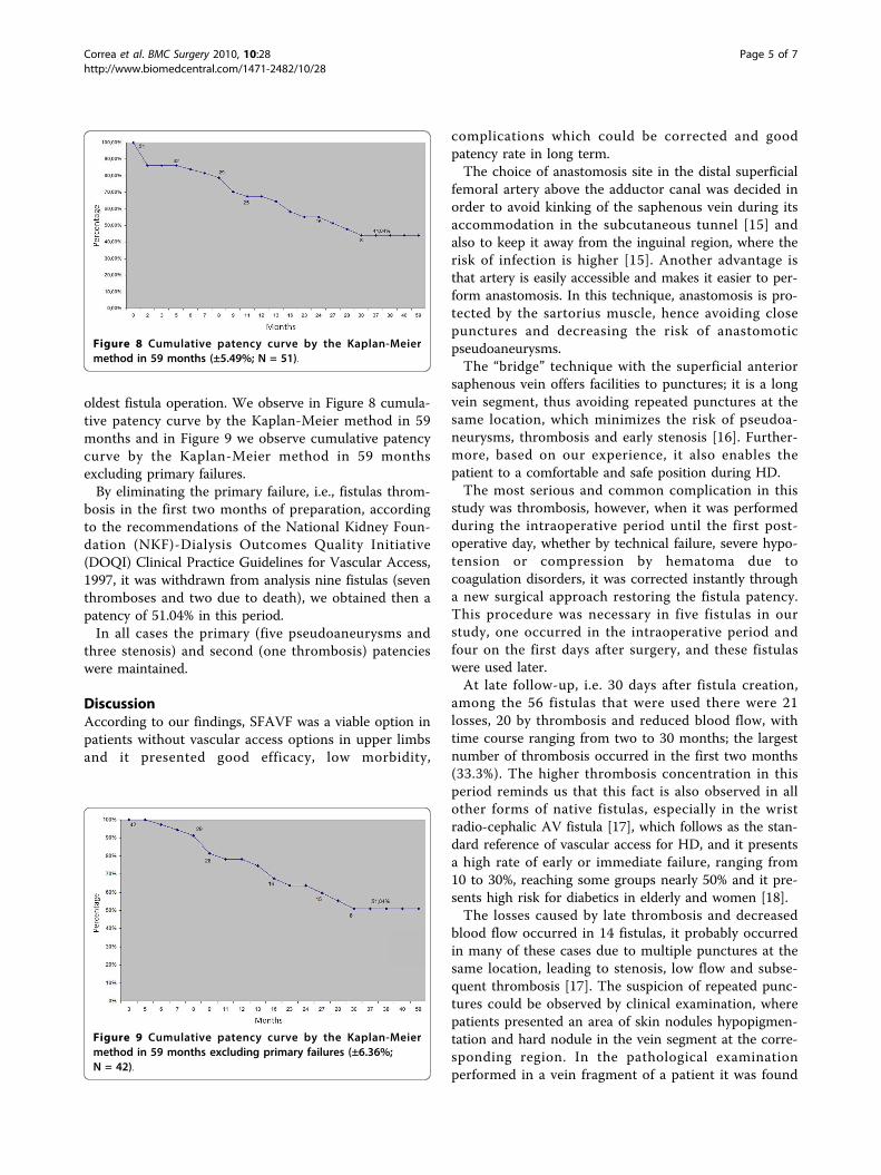
oldest fistula operation. We observe in Figure 8 cumula-tive patency curve by the Kaplan-Meier method in 59months and in Figure 9 we observe cumulative patencycurve by the Kaplan-Meier method in 59 monthsexcluding primary failures.By eliminating the primary failure, i.e., fistulas throm-
bosis in the first two months of preparation, accordingto the recommendations of the National Kidney Foun-dation (NKF)-Dialysis Outcomes Quality Initiative(DOQI) Clinical Practice Guidelines for Vascular Access,1997, it was withdrawn from analysis nine fistulas (seventhromboses and two due to death), we obtained then apatency of 51.04% in this period.In all cases the primary (five pseudoaneurysms and
three stenosis) and second (one thrombosis) patencieswere maintained.
DiscussionAccording to our findings, SFAVF was a viable option inpatients without vascular access options in upper limbsand it presented good efficacy, low morbidity,
complications which could be corrected and goodpatency rate in long term.The choice of anastomosis site in the distal superficial
femoral artery above the adductor canal was decided inorder to avoid kinking of the saphenous vein during itsaccommodation in the subcutaneous tunnel [15] andalso to keep it away from the inguinal region, where therisk of infection is higher [15]. Another advantage isthat artery is easily accessible and makes it easier to per-form anastomosis. In this technique, anastomosis is pro-tected by the sartorius muscle, hence avoiding closepunctures and decreasing the risk of anastomoticpseudoaneurysms.The “bridge” technique with the superficial anterior
saphenous vein offers facilities to punctures; it is a longvein segment, thus avoiding repeated punctures at thesame location, which minimizes the risk of pseudoa-neurysms, thrombosis and early stenosis [16]. Further-more, based on our experience, it also enables thepatient to a comfortable and safe position during HD.The most serious and common complication in this
study was thrombosis, however, when it was performedduring the intraoperative period until the first post-operative day, whether by technical failure, severe hypo-tension or compression by hematoma due tocoagulation disorders, it was corrected instantly througha new surgical approach restoring the fistula patency.This procedure was necessary in five fistulas in ourstudy, one occurred in the intraoperative period andfour on the first days after surgery, and these fistulaswere used later.At late follow-up, i.e. 30 days after fistula creation,
among the 56 fistulas that were used there were 21losses, 20 by thrombosis and reduced blood flow, withtime course ranging from two to 30 months; the largestnumber of thrombosis occurred in the first two months(33.3%). The higher thrombosis concentration in thisperiod reminds us that this fact is also observed in allother forms of native fistulas, especially in the wristradio-cephalic AV fistula [17], which follows as the stan-dard reference of vascular access for HD, and it presentsa high rate of early or immediate failure, ranging from10 to 30%, reaching some groups nearly 50% and it pre-sents high risk for diabetics in elderly and women [18].The losses caused by late thrombosis and decreased
blood flow occurred in 14 fistulas, it probably occurredin many of these cases due to multiple punctures at thesame location, leading to stenosis, low flow and subse-quent thrombosis [17]. The suspicion of repeated punc-tures could be observed by clinical examination, wherepatients presented an area of skin nodules hypopigmen-tation and hard nodule in the vein segment at the corre-sponding region. In the pathological examinationperformed in a vein fragment of a patient it was found
Figure 8 Cumulative patency curve by the Kaplan-Meiermethod in 59 months (±5.49%; N = 51).
Figure 9 Cumulative patency curve by the Kaplan-Meiermethod in 59 months excluding primary failures (±6.36%;N = 42).
Correa et al. BMC Surgery 2010, 10:28http://www.biomedcentral.com/1471-2482/10/28
Page 5 of 7

myointimal hyperplasia of the vein, which demonstratesthickening of venous wall.We observed some cases of thrombosis due to pro-
longed arterial hypotension and one case due to traumaof the fistula site at home environment. By studying thistype of complication, we alert to the importance ofmaintaining regular surveillance with periodic evaluationof these fistulas in order to detect early dysfunction sothat it may be corrected in time, reducing the risk ofthrombosis and increasing the usefulness period of theAV fistula [19].Puncture pseudoaneurysm occurred in five fistulas
(9.8%). It may be clinically diagnosed and confirmed byfistulography and ultrasound. This complication wasprobably due to inadequate compression of the puncturesite after dialysis sessions associated with repeated punc-tures in one place, leading to vein wall weakness. None-theless, this type of complication was possible to becorrected in three cases. In two cases it was correctedthrough its resection and interposition of a new veinsegment while in the third case it was corrected byresection and placement of a venous patch, maintainingthe primary patency. Vigilance with regard to this com-plication should be intense. When skin is affected therisk of rupture is too large and should therefore be per-formed surgical correction as early as possible in thosesituations [20].We observed one case of post-puncture hematoma
due to compression and inadequate care after dialysissession. This fistula may be preserved through clinicaltreatment and by spontaneous absorption of the hema-toma; however, the risk of an infection is always high insuch cases. It may be necessary in some cases to per-form surgical drainage.Stenoses were detected by flow decrease during dialy-
sis session. These situations could be confirmed by fistu-lography, through digital subtraction in three cases. Thiscomplication could be corrected in two cases by endo-vascular therapy through percutaneous transluminaldilatation, which maintained the fistula patency. More-over, its use may be retaken one day after this proce-dure. Therefore, to correct stenosis with hemodynamicrepercussion in this type of access as well as in othergrafts access types it should be made as early as possiblein order to keep an adequate dialysis to decrease throm-bosis rate and consequently increase the survival.When it is impossible to correct thrombosis, the crea-
tion of a new SFAVF in the contralateral limb may beindicated, since the saphenous vein and superficialfemoral artery allow it. This option was used in eightpatients in our investigation. In some cases of thrombo-sis, which the long saphenous vein of the contralaterallimb was already used, it was necessary to use PTFE inbridge by using the superficial femoral artery and distal
common femoral vein in the inguinal region. In case ofsaphenous vein absence, this was our first choicebecause there was facility to accommodate the prosthe-sis in subcutaneous tissue.The option to implant a PTFE “in loop” using com-
mon femoral artery and vein in the inguinal region, asperformed by Khadra et al [17] and Bhandari et al [16],was our last choice, we executed it in cases that it wasalready performed in the same SFAVF limb and thecontralateral limb was unable to use the saphenous veinand to implant PTFE in bridge. In some cases we usedthis technical option to prevent fibrosis in the anteriorregion of the anastomosis between the superficialfemoral vein and artery, thereby avoiding the creationof a new “bridge” fistula conformation. Notwithstanding,this technique presents high infection rate as reportedby Bhandari et al [16], who presented infection range of35% and Khadra et al [17], who showed infection rateof 16%.Other complications such as distal ischemia, venous
hypertension, cardiac decompensation, anastomoticpseudoaneurysm, aneurysmal dilatation, infection orother complications were not observed in our experi-ence. Conversely, Taylor et al [22] performed 45 grafts(“in loop” and “in thigh”), in whom PTFE prosthesiswere used in 39 cases and bovine carotid artery in sixcases. They observed high rate of non-thrombotic com-plications with 18% of infection and 16% of distal limbischemia.A new analysis was done after exclusion of primary
failure, i.e. the losses in the first two months of its crea-tion, as recommended by various conduct guidelines forvascular access (NKF-DOQI, Spanish Society ofNephrology). This analysis showed that the rateincreased to 51.04% in 60 months in 42 fistulas. On theother hand, analysis of results after 12 and 24 monthsrevealed a patency rate of 78.2% and 63.8% respectively,a rate close to researches published by Brescia andCimino [18], which primary patency at six months ran-ged between 65% and 81%.The limitation of this technique is given in cases when
the patient presents saphenous vein absence or whenthe saphenous vein is inadequate for this purpose andalso in patients with arterial occlusive disease in thefemoropopliteal territory. Another limitation of thistechnique is that the saphenous vein prevents the devel-opment of the fistula due to its developed muscle layer,similar to the cephalic vein in the internal forearm AVfistula. Although it prevents aneurysmal dilatation itincreases the risk of myointimal hyperplasia afterrepeated punctures of the AV fistula [20]. Nevertheless,because it is autologous material, presents low cost,higher infection resistance and it is easy to handling, theadvantages compensate its limitations even when
Correa et al. BMC Surgery 2010, 10:28http://www.biomedcentral.com/1471-2482/10/28
Page 6 of 7

compared to other access techniques in lower limbswhich also uses autologous material such as transposi-tion of the superficial femoral vein, first described byHuber et al [23], which reported two cases of use of thisvein, one in the thigh and one in the arm and alsoreported by Gradman et al [24], in a retrospective studyof 25 cases, which used this technique in lower limbs.This technique, which is an exception procedure,showed very good results in its long-term use accordingto our findings.
ConclusionSaphenofemoral arteriovenous fistula is a viable alterna-tive in patients which present no more options for vas-cular access in upper limbs. It presents good efficacy,low morbidity, complications that may be corrected andgood patency rate in long term.
AcknowledgementsThis study received financial support from Núcleo de Estudos, Pesquisas eAssessoria à Saúde da Faculdade de Medicina do ABC (NEPAS-FMABC). Wethank Dr. Eric Roger Wroclawski for the incentive and collaboration in thisstudy. We very thank Dr. Adam R. Wende for critical review of the grammar.We appreciate the reviewers for their highly constructive reviews, especiallyregarding grammar.
Author details1Departamento de Cirugia da Faculdade de Medicina do ABC, Santo André,SP, Brasil. 2Laboratório de Escrita Científica, Departamento de Morfologia eFisiologia, Faculdade de Medicina do ABC, Santo André, SP, Brasil.3Departamento de Medicina, Disciplina de Cardiologia, Universidade Federalde São Paulo (UNIFESP), São Paulo, SP, Brasil. 4Departamento de Fisioterapia,Universidade Estadual Paulista (UNESP), Presidente Prudente, SP, Brasil.5Disciplina de Cirurgia Cardiovascular, Universidade Federal de São Paulo(UNIFESP), São Paulo, SP, Brasil.
Authors’ contributionsAll authors participated in the acquisition of data and revision of themanuscript. JAC and FMJ conceived of the study, determined the design,performed the statistical analysis, interpreted the data and drafted themanuscript. All authors read and gave final approval for the versionsubmitted for publication.
Competing interestsThe authors declare that they have no competing interests.
Received: 16 March 2010 Accepted: 18 October 2010Published: 18 October 2010
References1. Vachharajani TJ, Atray NK: Invasive and innovative nephrology. Ren Fail
2005, 27(3):255-8.2. Vachharajani TJ, Vachharajani V: Obstacles for clinical monitoring in
hemodialysis patients because of multiple vascular accesses. Semin Dial2010, 23(1):114-6.
3. Fortunato JA Jr, Branco Filho AA, Branco A, Martins AL, Pereira ML,Ferraz JG, Paludo L: Standardization of video-assisted cardiac surgerytechnique: initial experience. Rev Bras Cir Cardiovasc 2008, 23(2):183-9.
4. Wolowczyk L, Williams AJ, Donovan KL, Gibbons CP: The snuffboxarteriovenous fistula for vascular access. Eur J Vasc Endovasc Surg 2000,19(1):70-6.
5. Gibbons CP: Primary vascular access. Eur J Vasc Endovasc Surg 2006,31(5):523-9.
6. Tannuri U, Tannuri AC, Watanabe A: Arteriovenous fistula for chronichemodialysis in pediatric candidates for renal transplantation: Technicaldetails and refinements. Pediatr Transplant 2009, 13(3):360-4.
7. Cura M, Elmerhi F, Suri R, Bugnone A, Dalsaso T: Vascular malformationsand arteriovenous fistulas of the kidney. Acta Radiol 2010, 51(2):144-9.
8. Lameire N, Van Biesen W, Vanholder R: Did 20 years of technologicalinnovations in hemodialysis contribute to better patient outcomes? ClinJ Am Soc Nephrol 2009, 4(Suppl 1):S30-40.
9. Shenoy S: Surgical anatomy of upper arm: what is needed for AVFplanning. J Vasc Access 2009, 10(4):223-32.
10. Rémy J, Rémy-Jardin M, Wallaert B, Lafitte JJ: Vaso-occlusion of thepulmonary artery. Rev Mal Respir 1988, 5(5):429-49.
11. Slater ND, Raftery AT: An evaluation of expanded polytetrafluorothylene(PTFE) loop grafs in the thigh as vascular access for hemodialysis inpatients with access problem. Ann of the Royal College of Surg of Engl1987, 70(3):244-5.
12. Hazinedaroglu SN, Tüzüner A, Ayli D, Demirer S, Dumon N, Yerdal MA:Femoral vein transposition versus femoral loop grafts for hemodialysis:A prospective evoluation. Transpl Proc 2004, 36(1):65-7.
13. Correa JA, Pires AC, Kafejian O, Miranda F Jr, Galego SJ, Yamazaki YR,Fujii EY, Fioretti AC: Superficial saphenofemoral arteriovenous fistula asaccess to hemodialysis - description of operative technique and initialclinical experience. J Vasc Br 2005, 4(4):341-8.
14. Barth RH: Direct calculation of K.T/V. A umplified approach to monitoringof hemodialysis. Nephron 1988, 50(3):191-5.
15. Rutherford RB, Baker JD, Ernst C, Joharston KW, Porter JM, Ahn S, Jones DN:Recommended Standards for reports dealing with lower extremityischemia: Revised Version. J Vasc Surg 1997, 26(3):517-38.
16. Bhandari S, Wilkinson A, Sellars L: Saphenous vein forearm grafts andgortex thigh grafts as alternative forms of vascular access. Clin Nephrol1995, 44(5):325-8.
17. Khadra MH, Dwyer AJ, Thompson JF: Advantages ofpolytetrafluoroethylene arteriovenous loops in the thigh forhemodialysis access. Am J Surg 1997, 173(4):280-3.
18. Brescia MJ, Cimino JE, Appel K, Hurwish BJ: Chronic hemodialysis usingvenipuncture and a surgically created arteriovenous fistula. N Engl J Med1966, 275(1):1089-92.
19. Neyra NR, Ikizler TA, May RE, Himmelforb J, Schulman G, Shyr Y, Hakim RM:Change in access blood flow overtime predicts access thrombosis. KidIntern 1998, 54(5):1714-9.
20. Safa AA, Valji K, Robert AC, Ziegler TM, Hye RJ, Oglevie SB: Detection andtreatment of dysfunctional hemodialysis access grafts: Effect of asurveillance program on graf patency and the incidence of thrombosis.Radiology 1996, 199(3):653-7.
21. Hansegger KA, Tiessenhansen K, Klipfinger M, Raith J, Hanser H, Tauss J:Aneurysmas of hemodialysis access grafts: treatment with coveredstents: a report of three cases. Cardiovasc Intervent Radiol 1998,21(4):334-7.
22. Taylor SM, Eaves GL, Weatherford DA, Mcalhany JC, Russel HC, Langan EM:Results of complications of arteriovenous access dialysis grafits in thelower extremity: A five year rewiew. The Am Surg 1996, 62(3):188-91.
23. Huber TS, Ozakic K, Flynn TC, Ross EA, Seeger JM: Use of superficialfemoral vein for hemodialysis arteriovenous access. J Vasc Surg 2000,31(5):1038-41.
24. Gradman WS, Cohen W, Haji-Aghari M: Arteriovenous fistulas constructionin the thigh with transposed superficial femoral vein: Our initialexperience. J Vasc Surg 2001, 33(5):968-75.
Pre-publication historyThe pre-publication history for this paper can be accessed here:http://www.biomedcentral.com/1471-2482/10/28/prepub
doi:10.1186/1471-2482-10-28Cite this article as: Correa et al.: Saphenofemoral arteriovenous fistulaas hemodialysis access. BMC Surgery 2010 10:28.
Correa et al. BMC Surgery 2010, 10:28http://www.biomedcentral.com/1471-2482/10/28
Page 7 of 7