ryanodine receptors, a family of intracellular calcium ion channels, are expressed throughout early...
TRANSCRIPT
SHORT REPORT Open Access
Ryanodine receptors, a family of intracellularcalcium ion channels, are expressed throughoutearly vertebrate developmentHoudini HT Wu, Caroline Brennan and Rachel Ashworth*
Abstract
Background: Calcium signals ([Ca2+]i) direct many aspects of embryo development but their regulation is not wellcharacterised. Ryanodine receptors (RyRs) are a family of intracellular Ca2+ release channels that control the flux ofCa2+ from internal stores into the cytosol. RyRs are primarily known for their role in excitation-contraction couplingin adult striated muscle and ryr gene mutations are implicated in several human diseases. Current evidencesuggests that RyRs do not have a major role to play prior to organogenesis but regulate tissue differentiation.
Findings: The sequences of the five zebrafish ryr genes were confirmed, their evolutionary relationship establishedand the primary sequences compared to other vertebrates, including humans. RyRs are differentially expressed inslow (ryr1a), fast (ryr3) and both types (ryr1b) of developing skeletal muscle. There are two ryr2 genes (ryr2a andryr2b) which are expressed exclusively in developing CNS and cardiac tissue, respectively. In addition, ryr3 and ryr2amRNA is detectable in the initial stages of development, prior to embryonic axis formation.
Conclusions: Our work reveals that zebrafish ryr genes are differentially expressed throughout the developingembryo from cleavage onwards. The data suggests that RyR-regulated Ca2+ signals are associated with severalaspects of embryonic development, from organogenesis through to the differentiation of the musculoskeletal,cardiovascular and nervous system. These studies will facilitate further work to explore the developmental functionof RyRs in each of these tissue types.
BackgroundTransient changes in the concentration of intracellularcalcium ions ([Ca2+]i) act as a powerful signal that iscrucial for the establishment of form and function inthe embryo. Detailed imaging studies have revealed thatthe spatial and temporal organisation of Ca2+ signalsduring embryogenesis are associated with many of themajor phases of development, from early cell division tothe differentiation of tissues. Despite their importancelittle is known about the generation and regulation ofembryonic Ca2+ signals. A comprehensive understandingof the pathways that regulate [Ca2+]i during develop-ment is essential to understand the functional relevanceof these signals in the embryo.Ryanodine receptors (RyR) are a family of intracellular
Ca2+ release channels that regulate the entry of Ca2+
into the cytosol from the intracellular organelles (theendoplasmic and sarcoplasmic reticulum). The RyR is alarge homotetrameric protein (approximately 2,200kDa), each subunit is comprised of a large N-terminalcytoplasmic domain which modulates the gating of thechannel, as well as luminal and transmembrane span-ning (TM) domains. In mammals, there are three dis-tinct ryr genes (ryr1, ryr2 and ryr3) that encode threedifferentially expressed RyR proteins. RyR1 and RyR2are expressed predominantly in skeletal and cardiacmuscle respectively, whilst RyR3 is found in many tis-sues at relatively low levels [1]. The primary role of theRyR is to increase [Ca2+]i during excitation-contractioncoupling (E-C coupling) in both skeletal and cardiacmuscle. In humans, mutations in the ryr1 and ryr2genes cause skeletal myopathies and cardiac diseaserespectively [2]. The generation of mouse knockout lineshas provided some insight into the role of the receptorsin the developing tissues of intact animals. Homozygous
* Correspondence: [email protected] of Biological and Chemical Sciences, Queen Mary University ofLondon, Mile End Road, London E1 4NS, UK
Wu et al. BMC Research Notes 2011, 4:541http://www.biomedcentral.com/1756-0500/4/541
© 2011 Wu et al; licensee BioMed Central Ltd. This is an Open Access article distributed under the terms of the Creative CommonsAttribution License (http://creativecommons.org/licenses/by/2.0), which permits unrestricted use, distribution, and reproduction inany medium, provided the original work is properly cited.
mice from ryr1(−/−) (skrrm1) and ryr2(−/−) knockout linesdisplay gross morphological defects in either the skeletalmuscle (ryr1) or heart tube (ryr2) and die at the perina-tal or embryonic day 10 (E10) stages respectively [3,4].In contrast, ryr3(−/−) knockout mice appear to have nogross developmental defects and evidence suggests thatRyR3 act to augment the [Ca2+]i response of the otherRyR isoforms in striated muscle [5,6]. The observationthat RyR expression does not occur until relatively latein mammalian development [7], coupled with the factthat knock out lines are not lethal at very early stageshas been interpreted as indicating that RyRs do notfunction during initial development.The zebrafish has been used extensively as a model
for vertebrate development. The rapid development exutero and embryonic transparency has proved advanta-geous for imaging the spatial and temporal organisationof Ca2+ signals. These signals are involved in many ofthe early embryonic events; the initiation of fertilisation(0 hours post fertilisation, hpf), the early cell divisionsassociated with the cleavage period (up to 2 hpf) andthe more extensive cellular rearrangements that occurin the blastula period (up to 5 hpf) (as reviewed in [8]).Evidence suggests that the release of Ca2+ from intracel-lular stores via the phosphatidylinositol (PI) signallingpathway is largely responsible for these early transientchanges in [Ca2+]i [9]. RyRs have not been implicated invery early developmental events and their expressionduring these stages has not been documented. Fluxes inthe levels of embryonic [Ca2+]i continue to occurthroughout gastrulation (up to 10 hpf) (as reviewed in[8]). Initially changes in [Ca2+]i occur as localised eventsbut, as gastrulation progresses, co-ordinated waves ofCa2+ signalling appear across the embryo. These latersignals are proposed to coordinate a wide range of cel-lular movements (epiboly, involution, convergence andextension) that give rise to the embryonic body plan.Release of Ca2+ from intracellular stores via the PI sig-nalling pathway is again implicated at this stage; how-ever, the contribution to gastrulation from other Ca2+
signalling pathways remains undefined. Finally, the seg-mentation period (from 10 up to 24 hpf) is charac-terised by organogenesis and the emergence of the bodysystems. The Ca2+ signals that occur during segmenta-tion are again more localised and typically associatedwith developing tissues. Transient changes in [Ca2+]ihave been recorded within the nervous system, somitesand cardiac tissue [10-12]. Several studies in zebrafishhave shown that inhibition of ryanodine receptor func-tion, using both pharmacological and genetic inhibitors,leads to impaired excitation-contraction coupling andgross morphological defects in the skeletal muscle, sug-gestive of a role in the development of this tissue[11,13,14].
This study set out to acquire a more comprehensiveunderstanding of ryanodine receptor expression in earlyvertebrate development, using the zebrafish as an in vivomodel. Our initial work confirmed the sequence of thefive zebrafish ryr genes, established their evolutionaryrelationship to those in other vertebrate species andprovided a direct comparison between the structural fea-tures of the primary protein sequences found in the zeb-rafish and mammals. An overview of ryr gene expressionduring zebrafish embryogenesis will inform work aimedat establishing the developmental significance of thisfamily of Ca2+-release channels. Therefore we conducteda comprehensive temporal and spatial analysis of ryrmRNA expression in the embryo using a combinationof semi-quantitative PCR and wholemount in situ hybri-disation. We observed strong maternal expression of ryrmRNA (ryr3 and ryr2a) during the cleavage and blastulaperiods suggestive of a novel role in early development.At 24 hours post fertilisation (hpf) ryr1a, ryr1b and ryr3are expressed in skeletal muscle, whereas ryr2a is loca-lized to the central nervous system (CNS) and ryr2b isfound exclusively in the cardiac muscle. Our study sug-gests that RyR channels have a role in early develop-ment prior to organogenesis as well as in thedifferentiation of different cell types.
MethodsAnimal proceduresWildtype (WT) zebrafish strains (Tubingen and Tupfellong fin) were bred and raised in-house at the zebrafishfacility of Queen Mary College, University of London,UK, as described previously [15]. Smoothened (smo)mutants were received as a gift from Prof. SimonHughes (King’s College London, UK). Embryos werecollected by natural spawning and staged according toKimmel and colleagues [16], given in the text as stan-dard developmental time at 28.5°C (hours post fertilisa-tion, hpf). Work on zebrafish embryos (prior toindependent feeding) is exempt under the U.K. Animals(Scientific Procedures) Act 1986 and does not requireethical approval.
Genomic analysis and gene prediction of ryr genesTo identify zebrafish ryr genes encoding RyRs asdescribed in Hirata and colleagues [13], proteinsequences corresponding to the human RyR family[http://www.ensembl.org/Homosapiens/familyview?famil-y=ENSF00000000736] were used as template for search-ing in GenBank and the zebrafish genome resources atEnsembl http://www.ensembl.org/Danio_rerio/; Zv7Ensembl assembly using a protein tBLASTn approach.Several hits were identified, which were then comparedto the 3’-UTR sequence of the EST to identify the corre-sponding genomic region. The gene predictions
Wu et al. BMC Research Notes 2011, 4:541http://www.biomedcentral.com/1756-0500/4/541
Page 2 of 17
program, Genwise (http://www.ebi.ac.uk/Wise2/) wasapplied to reveal genomic contigs and full sequencerelating to the ryr1a, ryr2a, ryr2b and ryr3 in zebrafish(Table 1). For ryr1a and ryr3, based on the tBLASTnresult, the Accession numbers (Table 2) and annotatedgene sequences for these isoforms were selected. Forryr2a, owing to the limitations to the resources availableon the database, 5 contigs (NA_1034, NA_1216,NA_3083, NA_1397 and NA_1713) were identified withnucleotide similarity to the human ryr2, based on theapproach described above. Bioinformatics databasesearch approaches using human ryr2 did not reveal anyfully annotated ryr2b gene sequences in zebrafish. How-ever, database blast results located a gene (EntrezGeneName LOC568506 on chromosome 17:18.7 m) encoding805 residues. The partial ryr2b gene located in a 19.98kb region in the Ensembl Zv7 assembly shared very lim-ited conserved symmetry with the human ryr2. Analignment approach revealed that the zebrafish ryr2bgene was reverse transcribed and the region of 19.98 kbencoding 805 residues at the C-terminus end showshigh peptide identity to the human RyR2 C-terminus.To help to find the missing large N-terminal cytoplas-mic domain of the RyR2b peptide, Genscan features onEnsembl Zv7 assembly were employed to reveal an extra1014 residues, giving a total of 1819 residues from theC-terminal end of the peptide. In order to search furtherfor the missing portion of the RyR2 peptide towards theN-terminus, a genomic region of 107,783 bp spanningthe C-terminal end of ryr2b gene to the start of neigh-bouring actn2 gene [Ensembl: ENSDARG00000071090],was reverse complemented using REQSEV programhttp://bioweb.pasteur.fr/seqanal/interfaces/revseq.htmlbefore performing a pairwise BLAST alignment withGasterosteus aculeatus (stickleback) RyR2 peptidesequence on NCBI (http://www.ncbi.nlm.nih.gov/blast/bl2seq/wblast2.cgi) to confirm its sequence identity.Based on the alignment result, the full length zebrafishRyR2b peptide was first constructed with reference toTakifugu rubripes (pufferfish) and stickleback RyR2 pep-tide sequences using the Genewise program (http://
www.ebi.ac.uk/Wise2/). The complete RyR2b peptidewas finalised by the alignment of the translated genomicregion using SHOWORF program http://bioweb.pasteur.fr/seqanal/interfaces/showorf.html. The sequence for theryr2b gene was deposited in EMBL Nucleotide SequenceDatabase (Accession no. FR822741). For ryr1b, sequencewas obtained via the NCBI CoreNucleotide search web-site [NCBI - AB247454] and isoform-specific PCR pri-mers (set 3) were used, as described previously [13].Isoform-specific PCR primers for ryr1a, ryr2a, ryr2b andryr3 were designed by retrieving the nucleotidesequences based on divergent regions of the RyRs iden-tified from peptide sequence (Figure 1). These regionswere re-confirmed by both BLAST against differentdatabases (NCBI nucleotide blast, Ensembl and ZFIN)and DNA sequencing.
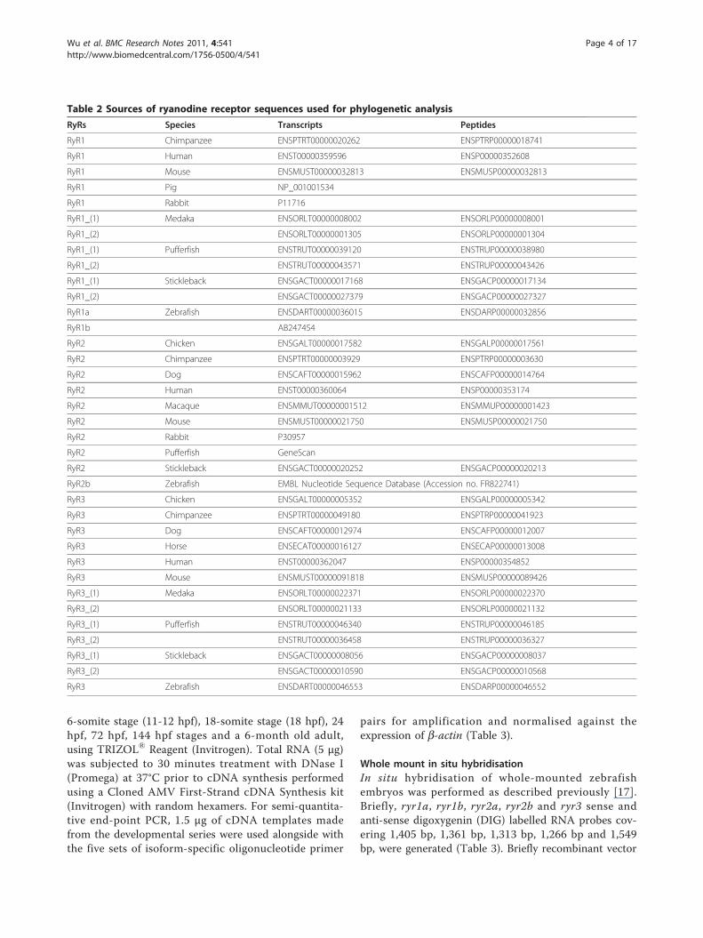
Phylogenetic AnalysisProtein sequences equivalent to the RIH_assoc(pfam08454) and RR_TM4-6 (pfam06459) domains inthe five zebrafish RyR protein sequences (RyR1a, RyR1b,partial RyR2a, RyR2b and RyR3) and other well charac-terised vertebrates RyR homologue sequences wereextracted from Ensembl and GenBank databases (Table2). The RyR sequences were pre-aligned using the Clus-talW alignment program to remove any gaps generatedwithin the sequences, followed by a multiple RyRsequence alignment using the T-coffee program avail-able from the European Bioinformatics Institute web-page (http://www.ebi.ac.uk/Tools/t-coffee/index.html)with the default parameters. The output from the multi-ple sequence alignment result obtained from T-coffeewas used subsequently as the template for the genera-tion of a Guide Tree using the equal angle methodavailable on the SplitsTree4 program with its defaultparameters (http://www.splitstree.org/for free download).
Semi-quantitative end-point PCRTotal RNA was isolated from nine stages of zebrafishdevelopment: 4- to 128-cell stage (1-2.25 hpf), 50%-epi-boly stage (5.3 hpf), 100%-epiboly stage (10 hpf), 3- to
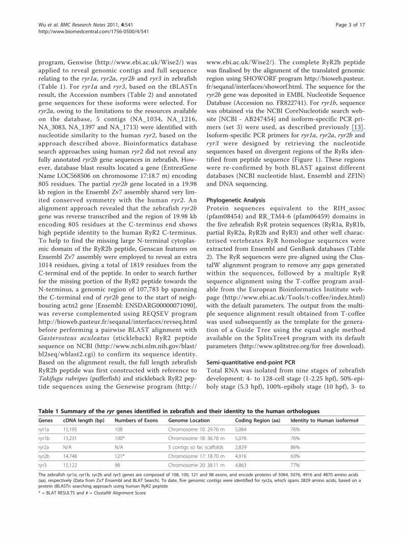
Table 1 Summary of the ryr genes identified in zebrafish and their identity to the human orthologues
Genes cDNA length (bp) Numbers of Exons Genome Location Coding Region (aa) Identity to Human isoforms#
ryr1a 15,195 108 Chromosome 10: 29.76 m 5,064 76%
ryr1b 15,231 100* Chromosome 18: 36.78 m 5,076 76%
ryr2a N/A N/A 5 contigs so far; scaffolds 2,829 86%
ryr2b 14,748 121* Chromosome 17: 18.70 m 4,916 63%
ryr3 15,122 98 Chromosome 20: 38.11 m 4,863 77%
The zebrafish ryr1a, ryr1b, ryr2b and ryr3 genes are composed of 108, 100, 121 and 98 exons, and encode proteins of 5064, 5076, 4916 and 4870 amino acids(aa), respectively (Data from Zv7 Ensembl and BLAT Search). To date, five genomic contigs were identified for ryr2a, which spans 2829 amino acids, based on aprotein tBLASTn searching approach using human RyR2 peptide
* = BLAT RESULTS and # = ClustalW Alignment Score
Wu et al. BMC Research Notes 2011, 4:541http://www.biomedcentral.com/1756-0500/4/541
Page 3 of 17
6-somite stage (11-12 hpf), 18-somite stage (18 hpf), 24hpf, 72 hpf, 144 hpf stages and a 6-month old adult,using TRIZOL® Reagent (Invitrogen). Total RNA (5 μg)was subjected to 30 minutes treatment with DNase I(Promega) at 37°C prior to cDNA synthesis performedusing a Cloned AMV First-Strand cDNA Synthesis kit(Invitrogen) with random hexamers. For semi-quantita-tive end-point PCR, 1.5 μg of cDNA templates madefrom the developmental series were used alongside withthe five sets of isoform-specific oligonucleotide primer
pairs for amplification and normalised against theexpression of b-actin (Table 3).
Whole mount in situ hybridisationIn situ hybridisation of whole-mounted zebrafishembryos was performed as described previously [17].Briefly, ryr1a, ryr1b, ryr2a, ryr2b and ryr3 sense andanti-sense digoxygenin (DIG) labelled RNA probes cov-ering 1,405 bp, 1,361 bp, 1,313 bp, 1,266 bp and 1,549bp, were generated (Table 3). Briefly recombinant vector
Table 2 Sources of ryanodine receptor sequences used for phylogenetic analysis
RyRs Species Transcripts Peptides
RyR1 Chimpanzee ENSPTRT00000020262 ENSPTRP00000018741
RyR1 Human ENST00000359596 ENSP00000352608
RyR1 Mouse ENSMUST00000032813 ENSMUSP00000032813
RyR1 Pig NP_001001534
RyR1 Rabbit P11716
RyR1_(1) Medaka ENSORLT00000008002 ENSORLP00000008001
RyR1_(2) ENSORLT00000001305 ENSORLP00000001304
RyR1_(1) Pufferfish ENSTRUT00000039120 ENSTRUP00000038980
RyR1_(2) ENSTRUT00000043571 ENSTRUP00000043426
RyR1_(1) Stickleback ENSGACT00000017168 ENSGACP00000017134
RyR1_(2) ENSGACT00000027379 ENSGACP00000027327
RyR1a Zebrafish ENSDART00000036015 ENSDARP00000032856
RyR1b AB247454
RyR2 Chicken ENSGALT00000017582 ENSGALP00000017561
RyR2 Chimpanzee ENSPTRT00000003929 ENSPTRP00000003630
RyR2 Dog ENSCAFT00000015962 ENSCAFP00000014764
RyR2 Human ENST00000360064 ENSP00000353174
RyR2 Macaque ENSMMUT00000001512 ENSMMUP00000001423
RyR2 Mouse ENSMUST00000021750 ENSMUSP00000021750
RyR2 Rabbit P30957
RyR2 Pufferfish GeneScan
RyR2 Stickleback ENSGACT00000020252 ENSGACP00000020213
RyR2b Zebrafish EMBL Nucleotide Sequence Database (Accession no. FR822741)
RyR3 Chicken ENSGALT00000005352 ENSGALP00000005342
RyR3 Chimpanzee ENSPTRT00000049180 ENSPTRP00000041923
RyR3 Dog ENSCAFT00000012974 ENSCAFP00000012007
RyR3 Horse ENSECAT00000016127 ENSECAP00000013008
RyR3 Human ENST00000362047 ENSP00000354852
RyR3 Mouse ENSMUST00000091818 ENSMUSP00000089426
RyR3_(1) Medaka ENSORLT00000022371 ENSORLP00000022370
RyR3_(2) ENSORLT00000021133 ENSORLP00000021132
RyR3_(1) Pufferfish ENSTRUT00000046340 ENSTRUP00000046185
RyR3_(2) ENSTRUT00000036458 ENSTRUP00000036327
RyR3_(1) Stickleback ENSGACT00000008056 ENSGACP00000008037
RyR3_(2) ENSGACT00000010590 ENSGACP00000010568
RyR3 Zebrafish ENSDART00000046553 ENSDARP00000046552
Wu et al. BMC Research Notes 2011, 4:541http://www.biomedcentral.com/1756-0500/4/541
Page 4 of 17
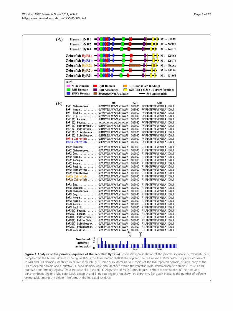
Figure 1 Analysis of the primary sequence of the zebrafish RyRs. (a) Schematic representation of the protein sequence of zebrafish RyRscompared to the human isoforms. The figure shows the three human RyRs at the top and the five zebrafish RyRs below. Sequence equivalentto MIR and RIH domains identified in all five zebrafish RyRs. Three SPRY domains, four copies of the RyR repeated domain, a single copy of theRIH associated domain and a putative EF hand domain were also identified within the zebrafish RyRs. Transmembrane domains (TM 4-6) andputative pore forming regions (TM 8-10) were also present. (b) Alignment of 36 RyR orthologues to show the sequences of the pore andtransmembrane regions (M8, pore, M10). Letters A and B indicate regions not shown in alignment. Bar graph indicates the number of differentamino acids among the different isoforms at the indicated residues.
Wu et al. BMC Research Notes 2011, 4:541http://www.biomedcentral.com/1756-0500/4/541
Page 5 of 17
templates (pGEM-T Easy-ryrs) were linearised with anappropriate restriction enzyme and then subjected tophenol/chloroform purification and alcohol precipita-tion. Purified, linear DNA (0.5 μg) was used for the invitro transcription reaction (a 20 μl reaction contained1X Transcription Buffer (Invitrogen), 2 μl DTT (0.1 M),2 μl nucleotide mix (1 mM GTP, 1 mM ATP, 1 mMCTP, 0.65 mM UTP and 0.35 mM DIG-11-UTP), 50Uplacental ribonuclease inhibitor and 10U of RNA poly-merase). The mixture was incubated at 37°C for 4 hoursbefore the removal of the original DNA template byincubating with 2U of DNase at 37°C for 1 hour. DIG-labelled cardiac myosin light chain 2 (cmlc2;[18]), myo-genic differentiation (myoD;[19]), nkx2.5 ([20]) andfluorescein-11-UTP labelled myosin heavy chain 1(myhz1;[21]) anti-sense probes were also prepared underthe same approach and used as positive markers.Zebrafish embryos were fixed in 4% paraformaldehyde
(PFA)/phosphate buffered Triton X100 (PBT) at 4°Covernight or at room temperature (RT) for 2 hours,then washed in 100% methanol and incubated at −20°Cfor at least 30 minutes. Embryos were rehydrated in amethanol gradient and treated with 10 μg/ml of Protei-nase K/PBT for 1 to 20 minutes at RT depending ontheir stages prior to fixation in 4% PFA/PBT for 20 min-utes at RT (embryos before or at 50% epiboly stage werenot treated with Protease K or re-fixed). Embryos werewashed with PBT and pre-hybridised in hybridisationmix (50% formamide, 5X SSC (0.75 M NaCl and 75mM triNa citrate) at pH 5.0, 500 μg/ml yeast RNA, 50μg/ml heparin, 0.1% Tween20) for 2 hours at 65°C. Themixture was replaced by fresh hybridisation mix con-taining DIG-labelled or Fluorescein-labelled RNA probeat the appropriate dilution and incubated at 65°C over-night. The post hybridisation washes were carried out at65°C. The excess probe was removed by sequentialwashing in 25% formamide in 2X SSC, 2X SSC, 0.2XSSC at 65°C and finally in PBT at RT. Embryos wereincubated in maleic acid buffer (0.1 M Maleic acid pH
7.5 and 0.15 M NaCl) containing 2% Blocking Reagent(Roche; MAB) at RT for a minimum of 1 hour. Stainingwas carried out by replacing the MAB solution with the1:5000 MAB diluted alkaline phosphatise (AP) conju-gated anti-DIG antibody (Roche) or 1:4000 MAB dilutedAP conjugated anti-Fluorescein antibody (Roche) at 4°Covernight. The embryos were washed with PBT andthen detection buffer (0.1 M Tris-HCl pH 9.5, 0.1 MNaCl and 50 mM MgCl2 and 0.1% Tween20) at RT.The AP substrates used for colour development wereBM purple (Roche), Fast Red tablets (Roche) and NBT/BCIP (Roche). The colour reaction was terminated bywashing in PBT and embryos were re-fixed in 4% PFA/PBT for 2 hours at RT.
ImmunocytochemistryWholemount immunocytochemistry (WICC) was con-ducted as described previously [22]. Briefly, embryoswere fixed overnight at 4°C and all subsequent stepswere performed at RT. Embryos were incubated inblocking buffer prior to incubation in primary antibodyin 1% goat serum in phosphate buffered saline supple-mented with 0.8% triton X100 (PBST) at room tempera-ture. The primary antibodies used were 34 C, F59 andMF20 at dilutions of 1:250, 1:10 and 1:100, respectively.Embryos were rinsed in phosphate buffer with 0.8% tri-ton (PBT) and incubated in a 1:1000 dilution of goatanti-mouse IgG Cy™-5 linked secondary antibody(Amersham) made up in 1% goat serum in PBT over-night. Embryos were rinsed in PBT and stored in 50%glycerol/50% PBS.For the double immuno-labelling (WISH/WICC), the
WISH labelled embryo was initially developed usingreagents as described above, followed by antibody label-ling using a 1:10 dilution of F59 and a 1:5000 dilution ofgoat anti-mouse horseradish peroxidase (HRP) conju-gated secondary antibody (Merck) carried out asdescribed in the Zebrafish Book [23]. Colour develop-ment by HRP was activated by the addition of 0.01% v/v
Table 3 Primer sequences used for PCR and WISH protocols
Genes (A) PCR Primers (5’ to 3’) (B) WISH Primers (5’ to 3’)
ryr1a F AAAGATCATGCCCTCAGCATGC
ryr1a R TTGGAATCTCCGCCTCCGAGTA
ryr1b F CCTCCTCCTGGTTATGCACCATGTTATGAG
ryr1b R TCAATGTGGTTGTGATCAGTGGGAACGGGA
ryr2a F GAAGATGGCGAGAAGAAACCTG ATGTCTGACGCCGGTGAACGAT
ryr2a R TGCAGCATCATTGTTGTCCGTTG CAGCACAAAGTGAACAGTGACGG
ryr2b F TCTAAATTCAGCCACCATGATGTGAGC
ryr2b R TGTTGCTCTCATGTACTCGCCC
ryr3 F AAGGAAAAGAATGAGTCGGGC
ryr3 R CACGCATCTTTTCTTTGGGA
Wu et al. BMC Research Notes 2011, 4:541http://www.biomedcentral.com/1756-0500/4/541
Page 6 of 17
DAB and 6% v/v H2O2. Reaction was terminated by theaddition of PBS and fixed overnight in 4% PFA at 4°C.For double-fluorescent labelling, the WISH labelledembryo was initially developed using Fast Red, followedby antibody labelling as described above.Embryos were mounted in 100% glycerol and for flat
mounting the yolk of the stained embryo was initiallyremoved. Brightfield and fluorescent single or Z-stackimages were collected using a X10, X20, X63 oil immer-sion or X100 water immersion objectives on a ZeissLSM 510 microscope and LSM Image Examiner soft-ware. LSM Image Browser (Version 4,2,0,121) softwarewas used for image post-processing such as preparingthree dimensional (3D) projected cross-sections fromthe acquired Z-stacks of images. For the observation ofthe Cy™5, Fast Red and Alexa Fluor® (Alexa Fluor®
488 nm) staining, argon lasers 633 nm, 543 nm and 488nm lines and LP 650, LP 560 and BP 505-550 emissionfilter(s) were used, respectively.
ResultsThe relationship between ryanodine receptor genes inzebrafish and those of other vertebratesWe conducted a tBLASTn search of the zebrafish geno-mic database (Zv7 Ensembl), using the protein
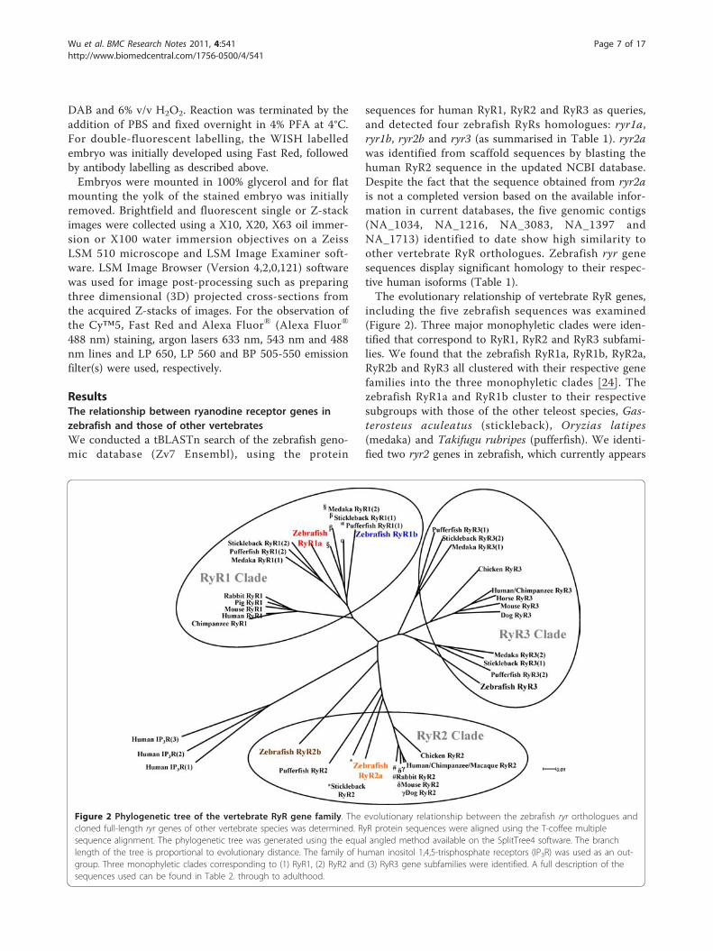
sequences for human RyR1, RyR2 and RyR3 as queries,and detected four zebrafish RyRs homologues: ryr1a,ryr1b, ryr2b and ryr3 (as summarised in Table 1). ryr2awas identified from scaffold sequences by blasting thehuman RyR2 sequence in the updated NCBI database.Despite the fact that the sequence obtained from ryr2ais not a completed version based on the available infor-mation in current databases, the five genomic contigs(NA_1034, NA_1216, NA_3083, NA_1397 andNA_1713) identified to date show high similarity toother vertebrate RyR orthologues. Zebrafish ryr genesequences display significant homology to their respec-tive human isoforms (Table 1).The evolutionary relationship of vertebrate RyR genes,
including the five zebrafish sequences was examined(Figure 2). Three major monophyletic clades were iden-tified that correspond to RyR1, RyR2 and RyR3 subfami-lies. We found that the zebrafish RyR1a, RyR1b, RyR2a,RyR2b and RyR3 all clustered with their respective genefamilies into the three monophyletic clades [24]. Thezebrafish RyR1a and RyR1b cluster to their respectivesubgroups with those of the other teleost species, Gas-terosteus aculeatus (stickleback), Oryzias latipes(medaka) and Takifugu rubripes (pufferfish). We identi-fied two ryr2 genes in zebrafish, which currently appears
Figure 2 Phylogenetic tree of the vertebrate RyR gene family. The evolutionary relationship between the zebrafish ryr orthologues andcloned full-length ryr genes of other vertebrate species was determined. RyR protein sequences were aligned using the T-coffee multiplesequence alignment. The phylogenetic tree was generated using the equal angled method available on the SplitTree4 software. The branchlength of the tree is proportional to evolutionary distance. The family of human inositol 1,4,5-trisphosphate receptors (IP3R) was used as an out-group. Three monophyletic clades corresponding to (1) RyR1, (2) RyR2 and (3) RyR3 gene subfamilies were identified. A full description of thesequences used can be found in Table 2. through to adulthood.
Wu et al. BMC Research Notes 2011, 4:541http://www.biomedcentral.com/1756-0500/4/541
Page 7 of 17
to be the only species in which this gene duplicationevent can be observed. The zebrafish ryr2a gene appearsto be more closely related to other teleosts (i.e. stickle-back and pufferfish) whereas ryr2b gene is morediverged. Although most teleosts appear to have tworyr3 genes, we confirmed previous studies which reporta single zebrafish ryr3 gene [11,13,24].
Zebrafish RyRs contain many of the conserved structuraldomains with similarities to other vertebratesThe molecular structure of the RyR protein has beenexplored extensively, partly in the drive to understandits regulation. Each monomeric RyR protein is approxi-mately 5,000 amino acids in length with a molecularmass of 565 kDa. The receptor has a large N-terminalcytoplasmic domain containing many regulatory bindingsites (as reviewed in [25,26]) that modulate the gating ofthe channel pore located in the C-terminus. The N-ter-minus cytoplamic domain of RyR interacts with a hostof regulatory proteins, such as calstabin and calmodulin.Physiological modulators of RyR function include ATP,Ca2+, Mg2+, cyclic ADP ribose, posttranslational modifi-cations (e.g. phosphorylation, oxidation) and pharmaco-logical substances (e.g. ryanodine, caffeine) [27].We conducted an analysis to explore whether the zeb-
rafish RyR protein sequences contain characterised con-served domains that may contribute to receptorregulation and their comparison to their human coun-terparts was analysed (Figure 1a). MIR (Mannosyltrans-ferase, Inositol 1,4,5-trisphophate receptor (IP3R) andRyR [pfam02815]) and RIH (RyR and IP3R Homology[pfam01365]) domains were identified within the N-terminal of all five zebrafish RyRs. The MIR and RIHdomains are common to all the members of the intra-cellular Ca2+-release channel super family [28,29]. TheMIR domain has been suggested to have a ligand trans-ferase function and the RIH domain may form a bindingsite for IP3; however, very little is known regarding theirrole in receptor regulation to date [29]. All of the zebra-fish RyRs contain three SPRY domains (SPla and theRyR [pfam00622]), which have been proposed to interactwith voltage gated channels [30,31]. Each zebrafish RyRalso contains four copies of the RyR domains (RyRrepeated domain [pfam02026]), a sequence unique tothese channels [29]. Furthermore, zebrafish RyRs con-tain the eukaryotic RIH associated (RyR and IP3RHomology associated [pfam08454]) domain, which cur-rently has no known function. EF-hand motifs may havea functional significance in activation of the channel byCa2+ themselves. Putative Ca2+ binding sequences, EF1and EF2 [32], have been identified in the receptor andare thought to be the major Ca2+ regulatory sites. Weidentified similar putative EF-hand motifs towards theC-terminal of the receptor and these appear to be
conserved in all zebrafish and human isoforms. The RyRpore determines the conductance and ion selectivity ofthe channel; however, the structure of the TM and poreforming region is still unresolved. There are eight pro-posed TM sequences, of which the last six are suggestedto form the Ca2+ release channel [33]. According to thetopological model by Du and colleagues six to eight TMsequences (i.e. M4a/M4b, M5, M6, M7a/M7b, M8 andM10) were identified, with the M9 (pore segment)inserted between the M8 and M10 TM segments. ThisM9 region is proposed to act as the selectivity filterallowing Ca2+ to transverse the membrane [34]. All fivezebrafish RyRs contain RR TM 4-6 domains (RyR TM4-6 [pfam06459]). Comparison of sequence in the corepore-forming region demonstrate that all five zebrafishRyR sequences are highly conserved with other verte-brate species, although it should be noted that there aresubtle changes at the single amino acid level (Figure 1b).
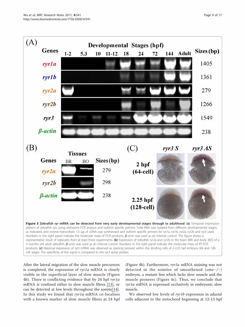
Zebrafish ryr mRNA expression can be detected from theearliest stages of development through to adulthoodWe examined the temporal expression of the ryr genesduring nine key stages of zebrafish development usingsemi-quantitative end-point PCR (Figure 3a). Expressionof ryr1a was detected from 11-12 hpf (3- to 6-somitestage) through to adulthood, whereas ryr1b expressionwas evident from 18 hpf (18-somite stage) onwards.Results also showed that levels of mRNA expression forboth ryr1a and ryr1b were at the highest in 144 hpf andadult stages. The ryr2a gene showed strong maternalexpression at 1-2 hpf (4- to 128-cell stage) and zygoticexpression was observed from 18 hpf through to adult-hood. Expression of ryr2b mRNA was detected from 18hpf through to adulthood with relatively weak expres-sion observed at 24 hpf and 144 hpf stages. Finally, ryr3maternal expression was observed at 1-2 hpf, followedby weak zygotic mRNA expression from 5.3 hpf (50%epiboly stage) through to 18 hpf (18-somite stage), afterwhich the levels became much stronger ryr1a, ryr1band ryr3 are expressed exclusively in skeletal muscleduring segmentation periodWe examined the spatial and temporal distribution
pattern of the zebrafish ryr mRNA in the developingembryo from 12 to 24 hpf of development using WISH.The expression of ryr1a, ryr1b and ryr3 in the develop-ing somites was compared to the myogenic determinant(myoD), one of the earliest markers of myogenic com-mitment with a key role in regulating muscle differentia-tion (Figure 4a) [19].We observed low levels of ryr1a expression in the
adaxial cells located on either side of the notochordbeginning at 11 hpf (Figure 4a). Our findings demon-strate that in slow muscle ryr1a appears at 11 hpf, 1 to2 hpf prior to fast muscle ryr1b mRNA expression.
Wu et al. BMC Research Notes 2011, 4:541http://www.biomedcentral.com/1756-0500/4/541
Page 8 of 17
After the lateral migration of the slow muscle precursorsis completed, the expression of ryr1a mRNA is clearlyvisible in the superficial layer of slow muscle (Figure4b). There is conflicting evidence that by 24 hpf ryr1amRNA is confined either to slow muscle fibres [13], orcan be detected at low levels throughout the somite[14].In this study we found that ryr1a mRNA co-localiseswith a known marker of slow muscle fibres at 24 hpf
(Figure 4b). Furthermore, ryr1a mRNA staining was notdetected in the somites of smoothened (smo−/−)embryos, a mutant line which lacks slow muscle and themuscle pioneers (Figure 4c). Thus, we conclude thatryr1a mRNA is expressed exclusively in embryonic slowmuscle.We observed low levels of ryr1b expression in adaxial
cells adjacent to the notochord beginning at 12-13 hpf
Figure 3 Zebrafish ryr mRNA can be detected from very early developmental stages through to adulthood. (a) Temporal expressionpattern of zebrafish ryrs using end-point PCR analysis and isoform specific primers. Total RNA was isolated from different developmental stages,as indicated, and reverse transcribed. 1.5 μg of cDNA was synthesised and isoform specific primers for ryr1a, ryr1b, ryr2a, ryr2b and ryr3 used.Numbers in the right panel indicate the molecular mass of PCR products. b-actin was used as an internal control. The figure shows arepresentative result of replicates from at least three experiments. (b) Expression of zebrafish ryr2a and ryr2b in the brain (BR) and body (BO) of a3 months old adult zebrafish. b-actin was used as an internal control. Numbers in the right panel indicate the molecular mass of RT-PCRproducts. (c) Maternal expression of ryr3 mRNA was observed as staining (arrows) within the dividing cells of 2-2.25 hpf embryos (64 and 128-cell stage). The specificity of the signal is compared to the ryr3 sense probes.
Wu et al. BMC Research Notes 2011, 4:541http://www.biomedcentral.com/1756-0500/4/541
Page 9 of 17
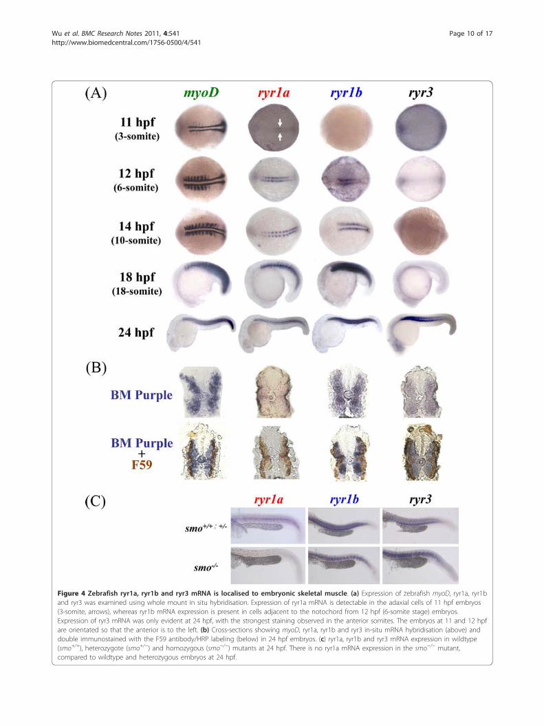
Figure 4 Zebrafish ryr1a, ryr1b and ryr3 mRNA is localised to embryonic skeletal muscle. (a) Expression of zebrafish myoD, ryr1a, ryr1band ryr3 was examined using whole mount in situ hybridisation. Expression of ryr1a mRNA is detectable in the adaxial cells of 11 hpf embryos(3-somite, arrows), whereas ryr1b mRNA expression is present in cells adjacent to the notochord from 12 hpf (6-somite stage) embryos.Expression of ryr3 mRNA was only evident at 24 hpf, with the strongest staining observed in the anterior somites. The embryos at 11 and 12 hpfare orientated so that the anterior is to the left. (b) Cross-sections showing myoD, ryr1a, ryr1b and ryr3 in-situ mRNA hybridisation (above) anddouble immunostained with the F59 antibody/HRP labeling (below) in 24 hpf embryos. (c) ryr1a, ryr1b and ryr3 mRNA expression in wildtype(smo+/+), heterozygote (smo+/−) and homozygous (smo−/−) mutants at 24 hpf. There is no ryr1a mRNA expression in the smo−/− mutant,compared to wildtype and heterozygous embryos at 24 hpf.
Wu et al. BMC Research Notes 2011, 4:541http://www.biomedcentral.com/1756-0500/4/541
Page 10 of 17
(Figure 4a). At 18 and 24 hpf, strong ryr1b expressionwas detected throughout the somites in a pattern analo-gous to that of myoD, suggestive of its presence in bothfast and slow muscle. Currently there is conflictingreports suggesting that ryr1b expression is confinedeither to the fast muscle [13] or appears in both fastand slow muscle [35]. In the later study the observationthat ryr1b mRNA expression is found in both muscletypes was attributed to an artefact arising as a conse-quence of contaminated tissue [35]. In order to clarifyour findings we performed double-fluorescent labelingexperiments, to observe ryr1b expression in the presenceof a slow muscle marker. In contrast to previous workwhich used a ryr1b in situ probe that targeted the rela-tively conserved pore forming region at the C-terminuswe generated a probe that recognised a more variableregion at the N-terminus of the receptor [13]. Our datarevealed that ryr1b mRNA is localised to both fast andslow developing skeletal muscle (Figure 4b, Additionalfile 1: Figure S1).Maternal expression of ryr3 in the dividing cells of 2
to 2.25 hpf (64- and 128-cell) embryos was confirmedby wholemount in situ hybridisation (WISH) (Figure3b); however, the low level expression of ryr3 from 5.3to 18 hpf detected in the PCR analysis was not con-firmed by WISH (data not shown). In this study ryr3mRNA expression was detected in the skeletal musclefrom 24 hpf using WISH (Figure 4a,b), with strongerexpression in the anterior compared to developing pos-terior somites (Figure 4a). There is conflicting evidencethat ryr3 mRNA in zebrafish is expressed exclusively inslow and fast skeletal muscle from 14 hpf [11] or inmany tissues including the CNS during the somitogen-esis [14,36]. The discrepancies in the expression patternsof ryr3 mRNA may be explained in part by the targetsequence used for probe synthesis in previous studies.The ryr3 clone (019-D04-2) used previously targeted theconserved pore-forming region at the C-terminus of thereceptor which shares high similarity to both the ryr1aand ryr1b sequence, this raises the possibility that crosshybridisation with ryr1 isoforms occurred [11]. In thecurrent work the ryr3 probe was designed to a moredivergent region located in the N-terminus of the recep-tor. In this study ryr3 mRNA expression was notdetected in the Kuppfer’s vesicles, adaxial cells at 12 hpfor in any region of the CNS at 24 hpf (data not shown).Double staining experiments revealed that ryr3 mRNAexpression was confined to the fast skeletal muscle at 24hpf (Figure 4b, Additional file 1: Figures S1 and Addi-tional file 2: Figure S2). Thus we conclude that ryr3mRNA is expressed exclusively in the fast skeletal mus-cle of embryos at 24 hpf.
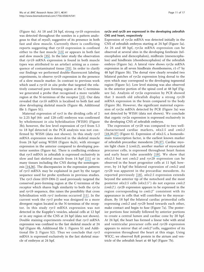
ryr2a and ryr2b are expressed in the developing zebrafishCNS and heart, respectivelyExpression of ryr2a mRNA was detected initially in theCNS of zebrafish embryos starting at 24 hpf (Figure 5a).At 24 and 48 hpf, ryr2a mRNA expression can beobserved at several sites in the developing forebrain (tel-encephalon and diencephalon), midbrain (mesencepha-lon) and hindbrain (rhombencephalon) of the zebrafishembryo (Figure 5a). A lateral view shows ryr2a mRNAexpression in all seven hindbrain rhombomeres, r1-r7 by48 hpf (Figure 5b). The dorsal view clearly revealed twobilateral patches of ryr2a expression lying dorsal to theeyes which may correspond to the developing tegmentalregion (Figure 5c). Low level staining was also observedin the anterior portion of the spinal cord at 48 hpf (Fig-ure 5a). Analysis of ryr2a expression by PCR showedthat 3 month old zebrafish display a strong ryr2bmRNA expression in the brain compared to the body(Figure 3b). However, the significant maternal expres-sion of ryr2a mRNA detected by PCR (Figure 3a) wasnot detected by WISH (data not shown). We concludethat zygotic ryr2a expression is expressed exclusively inthe developing CNS of zebrafish embryos.The expression of ryr2b was compared to two well-
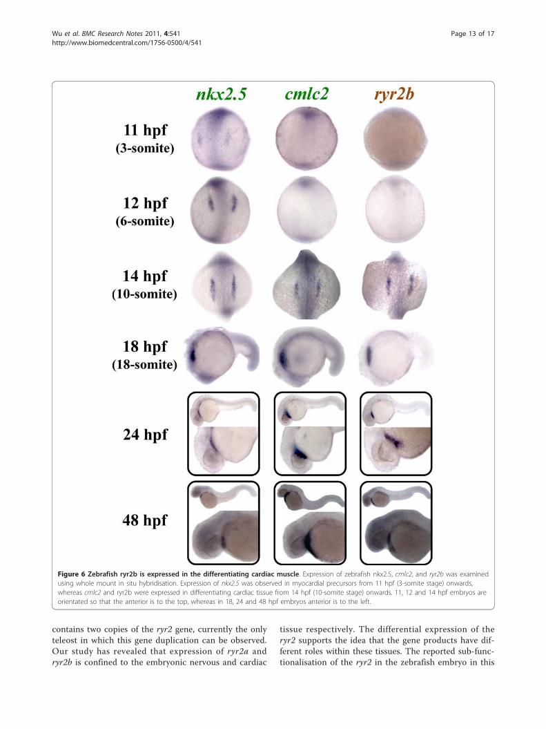
characterised cardiac markers, nkx2.5 and cmlc2[18,20,37] (Figure 6). Expression of nkx2.5, a homeodo-main transcription factor, is the earliest known markerof zebrafish precardiac mesoderm [20,37]. Cardiac myo-sin light chain 2 (cmlc2), another marker of myocardialprecursor cells, is expressed throughout heart fusionand early heart tube assembly [18]. We show thatnkx2.5 but not cmlc2 and ryr2b expression can beobserved in the heart progenitor cells at 11 hpf; how-ever, by 14 hpf the bilateral expression of cmlc2 andryr2b was apparent in the precardiac mesoderm. Asreported previously [18], nkx2.5 expression extendsbeyond the anterior tip of the notochord and the mostposterior nkx2.5 cells (nkx2.5+) do not express cmlc2(cmlc2-). ryr2b expression appears to be expressed in theregion corresponding to cmlc2+ consistent with itsappearance in cells that will contribute to the myocar-dium. By 18 hpf the bilateral cardiac primordial cellsexpressing cmlc2 and ryr2b bend towards each other,make contact and begin to fuse (Figure 7a). The poster-ior portions fuse initially followed by anterior portionsto create a central lumen and cardiac cone by 20 hpf.At 24 hpf, the heart has formed a linear tube with atrialand ventricular precursor cells and ryr2b expressionappears to mirror that of cmlc2+cells, suggestive of itsexpression throughout the heart at this stage. UsingWICC, we observed RyR protein in the atrium and ven-tricle of the zebrafish heart at 48 hpf (Figure 7b).
Wu et al. BMC Research Notes 2011, 4:541http://www.biomedcentral.com/1756-0500/4/541
Page 11 of 17
DiscussionStructure of the zebrafish ryr genes and their products:comparison to other speciesPrevious work identified a total of 14 genomic contigsfor ryr from the zebrafish genome assembly [13]. Classi-fication of RyR sequences by radiation hybrid mappingsuggested that there are at least five different zebrafishgenes: ryr1a, ryr1b, ryr2a, ryr2b and ryr3 [13]. Ourstudy has confirmed the sequences of the five zebrafish
ryr genes, although currently the ryr2a gene annotationis still incomplete. Zebrafish, like other teleosts, haveundergone a gene duplication event and appear to haveretained two distinct copies of ryr1 and ryr2. The teleostryr1 genes are differentially expressed in the skeletalmuscle tissue and the receptors have distinctive Ca2+
binding sensitivities, suggesting that the genes haveevolved to perform different physiological functions[13,38]. Here we report that the zebrafish genome
Figure 5 Zebrafish ryr2a is expressed in the central nervous system. The spatial distribution of ryr2a mRNA in the zebrafish embryo wasexamined using whole mount in situ hybridisation. (a) Specific staining was observed initially in localised regions of the brain, specifically thetelencephalon (T), diencephalon (D), mesencephalon (M), tegmentum (Tg) and rhomobomeres (rh) of whole mount embryos at 24 and 48 hpf.(b) Lateral and (c) dorsal views (eyes removed) of the brain at 48 hpf revealed that ryr2a expression was present in each of the sevenrhombomere segments, the tegmentum, trigeminal ganglia and anterior portion of the spinal cord. Embryos are orientated so that anterior is tothe left.
Wu et al. BMC Research Notes 2011, 4:541http://www.biomedcentral.com/1756-0500/4/541
Page 12 of 17
contains two copies of the ryr2 gene, currently the onlyteleost in which this gene duplication can be observed.Our study has revealed that expression of ryr2a andryr2b is confined to the embryonic nervous and cardiac
tissue respectively. The differential expression of theryr2 supports the idea that the gene products have dif-ferent roles within these tissues. The reported sub-func-tionalisation of the ryr2 in the zebrafish embryo in this
Figure 6 Zebrafish ryr2b is expressed in the differentiating cardiac muscle. Expression of zebrafish nkx2.5, cmlc2, and ryr2b was examinedusing whole mount in situ hybridisation. Expression of nkx2.5 was observed in myocardial precursors from 11 hpf (3-somite stage) onwards,whereas cmlc2 and ryr2b were expressed in differentiating cardiac tissue from 14 hpf (10-somite stage) onwards. 11, 12 and 14 hpf embryos areorientated so that the anterior is to the top, whereas in 18, 24 and 48 hpf embryos anterior is to the left.
Wu et al. BMC Research Notes 2011, 4:541http://www.biomedcentral.com/1756-0500/4/541
Page 13 of 17
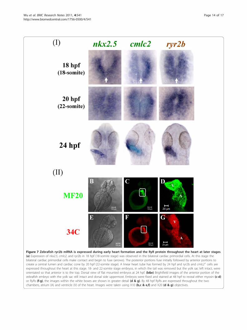
Figure 7 Zebrafish ryr2b mRNA is expressed during early heart formation and the RyR protein throughout the heart at later stages.(a) Expression of nkx2.5, cmlc2, and ryr2b in 18 hpf (18-somite stage) was observed in the bilateral cardiac primordial cells. At this stage thebilateral cardiac primordial cells make contact and begin to fuse (arrows). The posterior portions fuse initially followed by anterior portions tocreate a central lumen and cardiac cone by 20 hpf (22-somite stage). A linear heart tube has formed by 24 hpf and ryr2b and cmlc2+ cells areexpressed throughout the heart at this stage. 18- and 22-somite stage embryos, in which the tail was removed but the yolk sac left intact, wereorientated so that anterior is to the top. Dorsal view of flat mounted embryos at 24 hpf. (b&e) Brightfield images of the anterior portion of thezebrafish embryo with the yolk sac still intact and dorsal side uppermost. Embryos were fixed and stained at 48 hpf to reveal either myosin (c-d)or RyRs (f-g), the images within the white boxes are shown in greater detail (d & g). By 48 hpf RyRs are expressed throughout the twochambers, atrium (A) and ventricle (V) of the heart. Images were taken using X10 (b,c & e,f) and X20 (d & g) objectives.
Wu et al. BMC Research Notes 2011, 4:541http://www.biomedcentral.com/1756-0500/4/541
Page 14 of 17
study will facilitate the study of this receptor in thedevelopment of nervous and cardiac tissue.Our work has revealed that the primary sequence of
the zebrafish RyRs contains many of the conserveddomains associated with the regulation and function ofthis intracellular ion channel in other species, mostlynotably humans. The primary sequence of the Ca2+-con-ducting pore domain was found to be extensively con-served between zebrafish and other species examined.Our study of RyR primary structure supports previouswork to show that the biophysical properties and phar-macological regulation of the zebrafish RyR1 is similarto its mammalian homolog [39]. Taken together thisdata reveals that the zebrafish RyR functions in a similarmanner to those found in mammals, this information isof significant relevance to work using the zebrafish as amodel for human disease. However, differences in thepredicted primary sequence of the zebrafish ryanodinereceptor proteins and those of other species were alsorecorded. There are reports species-specific differencesin the Ca2+ regulation and single channel conductanceof the RyR1 channel [39]. Further comparative analysisof the RyR family will provide a better insight into thephysiological functions of the receptor at a tissue orwhole organism level.
The expression of ryr genes during early development,up to and including axis formationCalcium signalling is required throughout development;however the signal pathways have not been well defined.The prevailing view is that IP3R-driven Ca2+ signalshave a major role in axis formation prior to organogen-esis whereas RyR-induced Ca2+ signals are necessary forthe later aspects of tissue specific differentiation (e.g.muscle formation) [3-5,7,9,40]. Our current studyrevealed strong maternal expression of ryr2b and ryr3genes during cleavage, with low levels of ryr3 expressiondetectable throughout the blastula and gastrula periods.The significance of early ryr mRNA expression remainsto be determined; however, it raises the possibility thatRyR-generated Ca2+ signals act in development prior to10 hpf. The characterisation of expression in zebrafishestablishes a basis for future experimental work aimedat determining the action of RyR-induced Ca2+ signal-ling events in early embryonic patterning.
The expression of ryr genes in developing skeletal muscleSeveral studies have implicated RyR function in organo-genesis, particularly in striated muscle development (asreviewed in [9]). Our study has used both mutant linesand double staining to establish that RyRs are differen-tially expressed in slow (ryr1a), fast (ryr3) and both types(ryr1b) of developing skeletal muscle during the segmen-tation period. In E-C coupling within mammalian skeletal
muscle, RyR1 is directly coupled to a voltage gated-Ca2+
channel (VOC) on the sarcolemma. Activation of VOCsvia membrane depolarisation then triggers the opening ofthe ryanodine receptor (RyR1) and release of Ca2+ fromthe sarcoplasmic reticulum (SR) stores. Zebrafish havenon-Ca2+-conducting voltage-gated Ca channels thathave evolved solely as voltage sensors to trigger openingof the RyR [35]. The central subunit of the VOC(Cav1.1a1s) acts as the pore, selectivity filter and voltagesensor. In zebrafish two Cav1.1a1s genes (zf-a1s-a and zf-a1s-b) have been identified. The Cav1.1a1s gene productsare proposed to interact in a tissue specific manner withthe ryr1 genes; that is zf-a1s-a and ryr1a are expressed inslow muscle whilst zf-a1s-b and ryr1b are confined tofast muscle. However, our data suggest that the situationis not quite as clear cut as first proposed because ryr1b isnot expressed exclusively in fast muscle but is alsolocated in slow muscle. In addition, Ca2+ release can alsobe regulated by the ryr3 gene product which is alsolocated in the fast skeletal muscle. RyR3 is proposed toact as an uncoupled calcium-induced calcium release(CICR) channel to propagate the Ca2+ signal [41]. There-fore we propose that in the developing fast muscle zf-a1s-b and ryr1b act together to generate an increase in[Ca2+]i, with the ryr3 gene product acting to amplify thesignal. Our data in the embryonic fast muscle showedthat ryr1b expression occurs prior to ryr3 and suggeststhat the RyR-generated [Ca2+]i increase occurs initiallyvia ryr1b with the proposed amplification step via ryr3developing subsequently. In zebrafish the role of the RyRin E-C coupling within the developing slow muscleappears more complex. Our data reveals that both ryr1aand ryr1b are expressed in the developing slow muscle,but ryr3 is not. This presents the possibility that zf-a1s-acould couple to both ryr1a and ryr1b and raises the issueof whether amplification of the Ca2+ signals occurs inthis tissue and, if so, how is this achieved. Clearly there isstill much to understand about the maturation of depo-larization-induced Ca2+ signaling and its role during ske-letal muscle differentiation in vivo.
The expression of ryr genes in the developing nervousand cardiovascular systemsOur data has revealed that there are two ryr2 genes,ryr2a and ryr2b, which are exclusively expressed ineither the developing nervous system or cardiac tissue,respectively. Studies in mammals revealed that RyR areexpressed in the developing brain and that RyR-mediated Ca2+ signals may have a role in neuronal dif-ferentiation and neurite outgrowth [42-44]. All three ryrgenes are expressed within the embryonic mouse brain;however, from postnatal day 7 onwards ryr2 becomes inthe major isoform [45]. The postnatal changes in RyRexpression in mouse brain correlate with a period of
Wu et al. BMC Research Notes 2011, 4:541http://www.biomedcentral.com/1756-0500/4/541
Page 15 of 17
neuronal differentiation and may therefore be importantin establishing [Ca2+]i homeostasis in maturing neurons.In zebrafish ryr2a expression is localized to specificregions of the developing brains. In these regions ryr2ais likely to regulate neuronal Ca2+ signaling and there-fore play a role in CNS development.In mature cardiac muscle, Ca2+ signals are generated
by CICR via the activation of VOCs and the cardiac RyR(RyR2). Knockout mice which do not express ryr2 initi-ally display spontaneous rhythmic contractions of theheart at embryonic day 9 (E9) but no heart beat by day10 (E10) [4]. Furthermore, RyR-mediated Ca2+ releasedoes not play a significant role in the [Ca2+]i changesobserved within the heart of new born rats [46]. Thus inmammals it appears that the RyR2 does not contributeto the onset of contractile activity at very early embryo-nic stages, but is important for the subsequent matura-tion and development of the heart in vivo. The zebrafishcardiac ryr gene (ryr2b) is expressed exclusively in thedeveloping heart tissue (precardiac mesoderm) from 14hpf, 8 hours prior to the onset of cardiac contraction at22 hpf, and may well contribute to early cardiac devel-opment. Investigation of ryr2 function during mamma-lian development is complicated by the fact that a singleryr2 gene is expressed in several tissues [1]. The sub-functionalisation of the ryr2a and ryr2b genes in thezebrafish embryo provides an excellent system to studyindividual receptor function in neuronal and cardiac tis-sues during vertebrate development.
ConclusionsThis study has provided a comprehensive overview ofthe spatial and temporal expression of the ryr genefamily in developing zebrafish embryos. This family ofCa2+-release channels are expressed predominantly indeveloping skeletal, cardiac and neuronal tissue, suppor-tive of the view that RyRs function is relevant to laterdevelopment events, such as tissue differentiation. Inaddition, the study has also revealed that maternal ryrmRNA is present in the very early embryo, suggestive ofa function for this receptor prior to organogenesis. Rya-nodine receptors have been implicated in human diseaseand the zebrafish is an important vertebrate develop-mental model which will facilitate work in this area.Future work will explore the function of RyR-regulatedCa2+ signal pathways during zebrafish embryogenesis.
Additional material
Additional file 1: Figure S1. ryr1a, ryr1b and ryr3 mRNA isdifferentially expressed in the developing skeletal muscle. Doublelabelling in wholemount embryos at 24 hpf was performed using probesto myhz1, ryr1a, ryr1b and ryr3 and Fast Red as a substrate (red) followedby immunostaining using the F59 antibody and a fluorescent secondary(green). Images show Z-stacks of whole-mount double-labelled embryos
(top row) or sections (bottom row) from dissected embryos. Cross-sectional images revealed that ryr1a co-localised exclusively with slowmuscle staining, whereas myhz1, a marker of fast muscle, was notexpressed in the slow muscle. Furthermore ryr1b expression could beobserved throughout both muscle types whereas ryr3 did not co-localisewith the slow muscle staining and appeared to be expressed exclusivelyin the fast muscle. Scale bars = 20 μm, unless otherwise indicated.
Additional file 2: Figure S2. ryr3 mRNA expression is confined to thefast muscle fibres throughout the myotome at 24 hpf. Doublefluorescent cross-sections (1-3) were prepared by labelling a 24 hpfzebrafish embryo with a fluorescence substrate (i.e. FastRed; red) for ryr3in situ hybridisation and the F59 antibody (green) for immunostaining.The position of cross-sections 1-3 are illustrated in the 24 hpf ryr3 WISHlabelled embryo (top left) which has been stained with BM purple andlaterally orientated with anterior to the left. Scale bars = 20 μm, unlessotherwise indicated.
AcknowledgementsThe work was in part funded by a MRC grant (G0700216) to Dr Ashworth.Houdini Ho Tin Wu funded by QMUL college studentship. The antibodies 34C, F59 and MF20 were developed by J. Airey/J. Sutko, F.E. Stockdale and D.A.Fischman respectively and obtained from Developmental Studies HybridomaBank developed under the auspices of the NICHD and maintained by theUniversity of Iowa, Department of Biology, Iowa City, IA 52242. We thankDebbie Goode and Paul Piccinelli for their advice on the bioinformatics andHeather Callaway in the QMUL zebrafish facility. myhz1 and nkx2.5 plasmidDNA were received as a gift from Dr Yaniv Hintis (Prof. Simon Hughes Lab)and cmlc2 plasmid DNA was received as a gift from Ms Ana Filipa C. Simões(Prof. Roger Patient’s Lab).
Authors’ contributionsH.H.T.W performed all the experimental work and contributed to the writingof the manuscript. C.B. supervised and provided input on the design of theexperiments. R.A. designed and supervised the experiments and wrote themanuscript. All authors read and approved the final manuscript.
Competing interestsThe authors declare that they have no competing interests.
Received: 14 November 2011 Accepted: 14 December 2011Published: 14 December 2011
References1. Sutko JL, Airey JA: Ryanodine receptor Ca2+ release channels: does
diversity in form equal diversity in function? Physiol Rev 1996, 76:1027-71.2. Betzenhauser MJ, Marks AR: Ryanodine receptor channelopathies. Pflugers
Arch 460:467-80.3. Takeshima H, Iino M, Takekura H, Nishi M, Kuno J, Minowa O, Takano H,
Noda T: Excitation-contraction uncoupling and muscular degeneration inmice lacking functional skeletal muscle ryanodine-receptor gene. Nature1994, 369:556-9.
4. Takeshima H, Komazaki S, Hirose K, Nishi M, Noda T, Iino M: Embryoniclethality and abnormal cardiac myocytes in mice lacking ryanodinereceptor type 2. EMBO J 1998, 17:3309-16.
5. Takeshima H, Ikemoto T, Nishi M, Nishiyama N, Shimuta M, Sugitani Y,Kuno J, Saito I, Saito H, Endo M, et al: Generation and characterization ofmutant mice lacking ryanodine receptor type 3. J Biol Chem 1996,271:19649-52.
6. Bertocchini F, Ovitt CE, Conti A, Barone V, Scholer HR, Bottinelli R,Reggiani C, Sorrentino V: Requirement for the ryanodine receptor type 3for efficient contraction in neonatal skeletal muscles. EMBO Journal 1997,16:6956-6963.
7. Rosemblit N, Moschella MC, Ondriasa E, Gutstein DE, Ondrias K, Marks AR:Intracellular calcium release channel expression during embryogenesis.Dev Biol 1999, 206:163-77.
8. SE Webb, AL Miller: Calcium signalling during embryonic development.Nat Rev Mol Cell Biol 2003, 4:539-51.
Wu et al. BMC Research Notes 2011, 4:541http://www.biomedcentral.com/1756-0500/4/541
Page 16 of 17
9. Slusarski DC, Pelegri F: Calcium signaling in vertebrate embryonicpatterning and morphogenesis. Dev Biol 2007, 307:1-13.
10. Ashworth R: Approaches to measuring calcium in zebrafish: focus onneuronal development. Cell Calcium 2004, 35:393-402.
11. Brennan C, Mangoli M, Dyer CE, Ashworth R: Acetylcholine and calciumsignalling regulates muscle fibre formation in the zebrafish embryo. JCell Sci 2005, 118:5181-90.
12. Chi NC, Shaw RM, Jungblut B, Huisken J, Ferrer T, Arnaout R, Scott I, Beis D,Xiao T, Baier H, et al: Genetic and physiologic dissection of the vertebratecardiac conduction system. PLoS Biol 2008, 6:e109.
13. Hirata H, Watanabe T, Hatakeyama J, Sprague SM, Saint-Amant L,Nagashima A, Cui WW, Zhou W, Kuwada JY: Zebrafish relatively relaxedmutants have a ryanodine receptor defect, show slow swimming andprovide a model of multi-minicore disease. Development 2007,134:2771-81.
14. Jurynec MJ, Xia R, Mackrill JJ, Gunther D, Crawford T, Flanigan KM,Abramson JJ, MT Howard, DJ Grunwald: Selenoprotein N is required forryanodine receptor calcium release channel activity in human andzebrafish muscle. Proc Natl Acad Sci USA 2008, 105:12485-90.
15. Zimprich F, Ashworth R, Bolsover SR: Real-time measurements of calciumdynamics in neurons developing in situ within zebrafish embryos.Pflugers Arch-Eur J Physiol 1998, 436:489-493.
16. Kimmel CB, Ballard WW, Kimmel SR, Ullman B, Schilling TF: Stages ofEmbryonic Development of the Zebrafish. Dev Dyn 1995, 203:253-310.
17. Schulte-Merker S, Ho RK, Herrmann BG, C Nusslein-Volhard: The proteinproduct of the zebrafish homologue of the mouse T gene is expressedin nuclei of the germ ring and the notochord of the early embryo.Development 1992, 116:1021-32.
18. Yelon D, Horne SA, Stainier DY: Restricted expression of cardiac myosingenes reveals regulated aspects of heart tube assembly in zebrafish. DevBiol 1999, 214:23-37.
19. Weinberg ES, Allende ML, Kelly CS, Abdelhamid A, Murakami T,Andermann P, Doerre OG, Grunwald DJ, Riggleman B: Developmentalregulation of zebrafish MyoD in wild-type, no tail and spadetailembryos. Development 1996, 122:271-80.
20. Lee KH, Xu Q, Breitbart RE: A new tinman-related gene, nkx2.7,anticipates the expression of nkx2.5 and nkx2.3 in zebrafish heart andpharyngeal endoderm. Dev Biol 1996, 180:722-31.
21. Xu Y, He J, Wang X, Lim TM, Gong Z: Asynchronous activation of 10muscle-specific protein (MSP) genes during zebrafish somitogenesis. DevDyn 2000, 219:201-15.
22. Ashworth R, Zimprich F, Bolsover SR: Buffering intracellular calciumdisrupts motoneuron development in intact zebrafish embryos. Brain ResDev Brain Res 2001, 129:169-79.
23. Westerfield M: The Zebrafish Book: A guide for the laboratory use ofzebrafish (Brachydanio rerio). Univ of Oregon Press, Eugene;, 3 1995.
24. Darbandi S, Franck JP: A comparative study of ryanodine receptor (RyR)gene expression levels in a basal ray-finned fish, bichir (Polypterusornatipinnis) and the derived euteleost zebrafish (Danio rerio). CompBiochem Physiol B Biochem Mol Biol 2009, 154:443-8.
25. Hamilton SL: Ryanodine receptors. Cell Calcium 2005, 38:253-60.26. Hamilton SL, Serysheva I: Ryanodine receptor structure: Progress and
challenges. J Biol Chem 2008.27. Zalk R, Lehnart SE, Marks AR: Modulation of the ryanodine receptor and
intracellular calcium. Annu Rev Biochem 2007, 76:367-85.28. Sorrentino V, Barone V, Rossi D: Intracellular Ca(2+) release channels in
evolution. Curr Opin Genet Dev 2000, 10:662-7.29. Ponting CP: Novel repeats in ryanodine and IP3 receptors and protein O-
mannosyltransferases. Trends Biochem Sci 2000, 25:48-50.30. Ponting C, Schultz J, Bork P: SPRY domains in ryanodine receptors (Ca(2
+)-release channels). Trends Biochem Sci 1997, 22:193-4.31. Cui Y, Tae HS, Norris NC, Karunasekara Y, Pouliquin P, Board PG,
Dulhunty AF, Casarotto MG: A dihydropyridine receptor alpha(1s) loopregion critical for skeletal muscle contraction is intrinsically unstructuredand binds to a SPRY domain of the type 1 ryanodine receptor. Int JBiochem Cell Biol 2008.
32. Fessenden JD, Feng W, Pessah IN, Allen PD: Mutational analysis ofputative calcium binding motifs within the skeletal ryanodine receptorisoform, RyR1. J Biol Chem 2004, 279:53028-35.
33. Du GG, MacLennan DH: Topology and Transmembrane Organisation ofRyanodine Receptors. Ryanodine Receptors: structure, function and
dysfunction in clinical disease Edited by ZHT Wehrens, AR Marks: Springer;2005.
34. Du GG, Sandhu B, Khanna VK, Guo XH, MacLennan DH: Topology of theCa2+ release channel of skeletal muscle sarcoplasmic reticulum (RyR1).Proc Natl Acad Sci USA 2002, 99:16725-30.
35. Schredelseker J, Shrivastav M, Dayal A, Grabner M: Non-Ca2 + −conductingCa2+ channels in fish skeletal muscle excitation-contraction coupling.Proc Natl Acad Sci USA 2010, 107:5658-63.
36. Heyer V, Lux A, Alunni V, Degrave A, Seiliez I, Kirchner J, Parkhill JP, Thisse C:Spatial and temporal expression of the zebrafish genome by large-scalein situ hybridization screening. Methods Cell Biol 2004, 77:505-19.
37. Chen JN, Fishman MC: Zebrafish tinman homolog demarcates the heartfield and initiates myocardial differentiation. Development 1996,122:3809-16.
38. Franck JP, Morrissette J, Keen JE, Londraville RL, Beamsley M, Block BA:Cloning and characterization of fiber type-specific ryanodine receptorisoforms in skeletal muscles of fish. Am J Physiol 1998, 275:C401-15.
39. Koulen P, Janowitz T, Johenning FW, Ehrlich BE: Characterization of theCalcium-release Channel/Ryanodine Receptor from Zebrafish SkeletalMuscleb. Journal of Membrane Biology 2001, 183:155-163.
40. Ashworth R, Devogelaere B, Fabes J, Tunwell RE, Koh KR, De Smedt H,Patel S: Molecular and functional characterization of inositoltrisphosphate receptors during early zebrafish development. J Biol Chem2007, 282:13984-93.
41. Sorrentino V, Reggiani C: Expression of the ryanodine receptor type 3 inskeletal muscle. A new partner in excitation-contraction coupling? TrendsCardiovasc Med 1999, 9:54-61.
42. Faure AV, Grunwald D, Moutin MJ, Hilly M, Mauger JP, Marty I, De M,Waard M, Albrieux M: Developmental expression of the calcium releasechannels during early neurogenesis of the mouse cerebral cortex. Eur JNeurosci 2001, 14:1613-22.
43. Ooashi N, Futatsugi A, Yoshihara F, Mikoshiba K, Kamiguchi H: Celladhesion molecules regulate Ca2 + -mediated steering of growth conesvia cyclic AMP and ryanodine receptor type 3. J Cell Biol 2005,170:1159-67.
44. Lee SM, Lee JW, Song YS, Hwang DY, Kim YK, Nam SY, Kim DJ, YW Yun,Yoon DY, Hong JT: Ryanodine receptor-mediated interference ofneuronal cell differentiation by presenilin 2 mutation. J Neurosci Res 2005,82:542-50.
45. Mori F, Fukaya M, Abe H, Wakabayashi K, Watanabe M: Developmentalchanges in expression of the three ryanodine receptor mRNAs in themouse brain. Neurosci Lett 2000, 285:57-60.
46. Perez CG, Copello JA, Li Y, Karko KL, Gomez L, Ramos-Franco J, Fill M,Escobar AL, Mejia-Alvarez R: Ryanodine receptor function in newborn ratheart. Am J Physiol Heart Circ Physiol 2005, 288:H2527-40.
doi:10.1186/1756-0500-4-541Cite this article as: Wu et al.: Ryanodine receptors, a family ofintracellular calcium ion channels, are expressed throughout earlyvertebrate development. BMC Research Notes 2011 4:541.
Submit your next manuscript to BioMed Centraland take full advantage of:
• Convenient online submission
• Thorough peer review
• No space constraints or color figure charges
• Immediate publication on acceptance
• Inclusion in PubMed, CAS, Scopus and Google Scholar
• Research which is freely available for redistribution
Submit your manuscript at www.biomedcentral.com/submit
Wu et al. BMC Research Notes 2011, 4:541http://www.biomedcentral.com/1756-0500/4/541
Page 17 of 17