voltage-dependent modulation of cardiac ryanodine receptors (ryr2) by protamine
TRANSCRIPT

Voltage-Dependent Modulation of Cardiac RyanodineReceptors (RyR2) by ProtaminePaula L. Diaz-Sylvester, Julio A. Copello*
Department of Pharmacology, Southern Illinois University School of Medicine, Springfield, Illinois, United States of America
Abstract
It has been reported that protamine (.10 mg/ml) blocks single skeletal RyR1 channels and inhibits RyR1-mediated Ca2+
release from sarcoplasmic reticulum microsomes. We extended these studies to cardiac RyR2 reconstituted into planar lipidbilayers. We found that protamine (0.02–20 mg/ml) added to the cytosolic surface of fully activated RyR2 affected channelactivity in a voltage-dependent manner. At membrane voltage (Vm; SR lumen - cytosol) = 0 mV, protamine inducedconductance transitions to several intermediate states (substates) as well as full block of RyR2. At Vm.10 mV, the substatewith the highest level of conductance was predominant. Increasing Vm from 0 to +80 mV, decreased the number oftransitions and residence of the channel in this substate. The drop in current amplitude (full opening to substate) had thesame magnitude at 0 and +80 mV despite the ,3-fold increase in amplitude of the full opening. This is more similar torectification of channel conductance induced by other polycations than to the action of selective conductance modifiers(ryanoids, imperatoxin). A distinctive effect of protamine (which might be shared with polylysines and histones but not withnon-peptidic polycations) is the activation of RyR2 in the presence of nanomolar cytosolic Ca2+ and millimolar Mg2+ levels.Our results suggest that RyRs would be subject to dual modulation (activation and block) by polycationic domains ofneighboring proteins via electrostatic interactions. Understanding these interactions could be important as such anomaliesmay be associated with the increased RyR2-mediated Ca2+ leak observed in cardiac diseases.
Citation: Diaz-Sylvester PL, Copello JA (2009) Voltage-Dependent Modulation of Cardiac Ryanodine Receptors (RyR2) by Protamine. PLoS ONE 4(12): e8315.doi:10.1371/journal.pone.0008315
Editor: Edward Perez-Reyes, University of Virginia, United States of America
Received August 18, 2009; Accepted November 19, 2009; Published December 15, 2009
Copyright: � 2009 Diaz-Sylvester, Copello. This is an open-access article distributed under the terms of the Creative Commons Attribution License, whichpermits unrestricted use, distribution, and reproduction in any medium, provided the original author and source are credited.
Funding: This work was supported by the National Institutes of Health Grant R01 GM078665 to JAC. The funders had no role in study design, data collection andanalysis, decision to publish, or preparation of the manuscript.
Competing Interests: The authors have declared that no competing interests exist.
* E-mail: [email protected]
Introduction
In striated muscle, electrical excitation activates ryanodine
receptors (RyR) located in the sarcoplasmic reticulum (SR)
membrane which in turn mediate the massive release of
intracellular Ca2+ required for activating the contractile system
[1,2,3]. Electron microscopy studies indicate that cardiac (RyR2)
channels could interact among themselves as they are physically
connected in organized arrays at the terminal cisternae of SR [4].
Indeed, it has been shown that multiple RyRs synchronously
activate and deactivate during excitation-contraction (EC)-cou-
pling [1,5,6,7,8]. Moreover, under resting conditions, brief
elementary events of Ca2+ release (‘‘Ca2+ sparks’’) arise as a result
of the concerted activation and deactivation of six to twenty RyR2
in brief bursts lasting ,5–20 ms [9,10]. These functional channel-
channel interactions seem to survive isolation and reconstitution in
bilayers, where multiple RyRs often display synchronicity named
‘‘coupled gating’’ [11,12,13,14]. It is also apparent that, in the
cytosolic environment, RyR1 and RyR2 may be modulated via
physical interactions with other associated proteins, such as the L-
type Ca2+-channels [8,15]. The nature of the interactions between
neighboring RyRs and/or with associated proteins has not been
fully defined, but it is likely that electrostatic interactions may play
a role as the vestibule of RyRs contain negatively charged regions
that could be a target for cationic ligands [16,17]. Indeed, it is well
known that RyR channel function can be modulated by positively
charged moieties, including polycationic peptides such as prot-
amine, histone and polylysine, which seem to display a variety of
actions including activation and block of RyR-mediated Ca2+
release [18,19,20,21]. Furthermore, in failing heart as well as in
skeletal muscle pathologies, RyR-mediated SR Ca2+ release was
found to have increased sensitivity to activation by polylysine
[22,23].
Protamine is a mix of highly cationic (arginine rich) peptides
with molecular weight of ,5.1 kDa (major component) which has
been previously used as a tool to study how RyRs are modulated
through electrostatic interactions [24,25]. In this early study, large
doses of protamine (.20 mg/ml) were found to fully inhibit
skeletal (RyR1) channels regardless of the cytosolic Ca2+ levels
[24]. We extended these studies to cardiac RyR2 reconstituted
into planar lipid bilayers and tested a wider range of protamine
levels (0.02 to 20 mg/ml). Our results indicate that the action of
protamine added to the cytosolic surface of RyR2 is complex. It
includes voltage-dependent activation and block as well as
transitions to subconductance states (substates). Some of the
results have been presented in preliminary form [26].
Methods
Drugs and ChemicalsCaCl2 standard for calibration was from Word Precision
Instruments Inc. (Sarasota, FL). Phospholipids were obtained
from Avanti (Alabaster, AL). Ryanodine was from Calbiochem
(San Diego, CA). Imperatoxin A (IpTxA) was from Alomone Labs
PLoS ONE | www.plosone.org 1 December 2009 | Volume 4 | Issue 12 | e8315
(Jerusalem, Israel). Ryanodol was obtained from hydrolyzed
ryanodine as previously described [27]. Protamine and all other
drugs and chemicals were either from Sigma-Aldrich or were
reagent grade.
Sarcoplasmic Reticulum MicrosomesAll procedures with animals were designed to minimize pain
and suffering and conformed to the guidelines of the National
Institutes of Health. The committee on the Use and Care of
Laboratory Animals of Southern Illinois University School of
Medicine reviewed and approved the protocols for animal use.
Sarcoplasmic reticulum (SR) microsomes were obtained from pig
heart ventricle using heart homogenization and ultracentrifugation
steps that follow the procedures published by Chamberlain et al.
[28]. SR pellets obtained after high speed centrifugation were
resuspended in 290 mM sucrose - 5 mM Imidazole buffer (pH = 7)
and were aliquoted in cryovials (300 ml each) and kept in liquid
nitrogen (better and safer long-term storage). Every month, a few
cryovials are used to generate smaller aliquots of membranes
(15 ml each) which were stored at –80uC for easy access. For
experiments, aliquots were quickly thawed in water, kept on ice
and used within 3–5 hours.
Bilayer TechniqueReconstitution of RyR2 in planar lipid bilayers, was performed
as previously described [29]. Briefly, planar lipid bilayers were
formed on 80 to 100 mm-diameter circular holes in teflon septa,
separating two 1.3 ml compartments. The trans compartment was
filled with HEPES-Ca2+ solution containing HEPES 250 mM and
Ca(OH)2 53 mM, pH 7.4. The trans compartment was clamped at
0 mV using an Axopatch 200B patch-clamp amplifier (Axon
Instruments, Foster City, CA). The cis compartment (ground) was
filled with HEPES-Tris solution containing HEPES 250 mM and
TrisOH 118 mM, pH 7.4. Bilayers of a 5:4:1 mixture of bovine
brain phosphatidylethanolamine, phosphatidylserine and phos-
phatidylcholine (45–50 mg/ml in decane) were painted onto the
holes of teflon septa from the cis side. Sarcoplasmic reticulum
microsomes (5–15 mg) were then added to the cis solution followed
by 500–1000 mM CsCl and 1 mM CaCl2 to promote vesicle
fusion. After RyR currents (or Cl2 currents .100 pA at 0 mV)
were observed, the cis chamber was perfused with HEPES-TRIS
solution for 5 min at 4 ml/min. A mixture of BAPTA and
dibromo-BAPTA was used to buffer free [Ca2+] on the cytosolic
surface of the channel ([Ca2+]cyt) [29]. As previously done [29],
RyR channels were identified by current amplitudes (,3.5 pA at
0 mV), slope conductance (,100 pS), reversal potential
(,245 mV, trans - cis) and response to diagnostic ligands (e.g.,
ryanodine, Ca2+, ATP, caffeine and Ruthenium Red). RyR
channel currents are depicted as positive (upward deflections of the
current) in figures and reflect cation flux from the trans (lumenal) to
the cis (cytosolic) compartment. Membrane voltages always
represent the difference between trans - cis compartments (in mV).
Single Channel AnalysisChannel currents were first filtered through the Axopatch 200B
low-pass Bessel filter at 2 kHz, digitized at 20 kHz with an analog
to digital converter (Digidata 1320, Axon Instruments) and stored
on DVD. Recordings were analyzed using pClamp9 software
(Axon Instruments). Analysis with this program included open
times, closed times and open probabilities (Po), which were
determined by half-amplitude threshold analysis of single-channel
recordings as done before [29].
Recordings were digitally filtered at 500 Hz in order to estimate
the probabilities of substates using two different methods:
1) Manual analysis of the traces. Frame by frame analysis
(100 ms/frame) was performed using pClamp9 on each 4-min
recording. In most of our recordings, we were able to distinguish
different levels of current that included the baseline, full openings
and substates induced by protamine, ryanodol or imperatoxin A.
Events, at different levels of current lasting more than 3 ms, were
manually selected. The parameters (mean amplitude and duration)
of each event were collected and averaged. The probability of
substate occurrence, Psubstate, was estimated from the ratio: time
spent in substate/total recording time. Pfull open was estimated
as the fraction of time spent in the maximal current level (full
opening).
2) Current-amplitude distributions. All-points current-
amplitude histograms (band width = 0.01 pA) were obtained
from each 4-min recording. Histograms were normalized so that
total histogram area = 1. In most of our experiments, we were able
to detect peaks (components) corresponding to the baseline, full
opening and substate levels. Each component was fitted with a
Gaussian function using the Levenberg-Marquardt method. When
the fitting is good, the fitted area of each component can be used
as an estimation of its probability.
In some cases, the small signal-to-noise ratio precluded the
unequivocal resolution of all individual components from the
amplitude distribution histograms. In these cases, the Psubstate were
only estimated using frame by frame analysis. It is important to
point out that in previous studies [18] we have established that
frame by frame analysis of higher signal-to-noise recordings
yielded the same results as histograms (i.e. we can be confident
when comparing Po values calculated using different methods).
Statistical AnalysisData are shown as means6S.E.M. of n measurements.
Statistical comparisons between groups were performed with
Student’s t-test of paired differences. Differences were considered
statistically significant at P,0.05.
Results
In this work, we studied the action of protamine added to the
cytosolic surface of cardiac RyR2 reconstituted into planar lipid
bilayers. High concentrations of protamine (.20 mg/ml) have
been tested on partially active skeletal RyR1 channels [24]. Here,
we extended these studies testing the effect of lower levels of
protamine (0.02–20 mg/ml). As previous studies reported an
inhibitory effect of protamine [19,24], we conducted most of our
experiments using fully activated RyR2 (locked open by the
combined effect of high cytosolic Ca2+ and caffeine) in order to
minimize the interference of RyR2 intrinsic gating events
(closures) with the expected effect of protamine (full or partial
block). However, other studies have suggested that polycationic
peptides (histone, polylysine) could induce SR Ca2+ release under
cellular resting conditions [19]. Consequently, we also tested the
effect of protamine on RyR2 with low Po at low (resting) cytosolic
Ca2+ levels.
Protamine Induces Block and SubstatesAs shown in Fig. 1, at high levels of Ca2+ and caffeine, increasing
concentrations of protamine induced sub-conductance states
(substates) of several current levels (indicated by dashed lines).
With 0.02 mg/ml protamine, infrequent transitions to a substate of
,50% of the full open conductance were observed. With 0.2 mg/
ml protamine, the probability of this substate dramatically
increased. Addition of protamine to reach a concentration of
1 mg/ml resulted in the formation of an additional substate of
Protamine Modulates RyR2
PLoS ONE | www.plosone.org 2 December 2009 | Volume 4 | Issue 12 | e8315

,20% the full open conductance. Under these conditions, the
probability of full openings decreased to virtually zero and the
channel fluctuated between the two substates. With 2 mg/ml
protamine, the substate with the smaller conductance became
predominant. It is possible that even smaller substates arise at high
concentrations of protamine, but they cannot be unequivocally
resolved from the baseline current noise. As previously reported for
RyR1 [24], we observed full block of RyR2 in the presence of
20 mg/ml protamine. Addition of heparin (250 mg/ml), known to
bind protamine with very high affinity [30,31,32], reversed the
effect of protamine on RyR2. This reversibility suggests that the
action of protamine was not related to the dissociation of any
cofactor or modulatory subunit bound to the RyR2 channel.
At 0 mV, Fig. 1 showed that 1 mg/ml protamine induced
transitions between two subconductance levels. Figures 2A and 2B
show that at positive voltages (SR lumen - cytosol), 1 mg/ml
protamine induced the formation of substates of only one current
level. The probability of this substate decreased with increasing
positive holding voltages (Fig. 2C). This resulted in an increase of
the probability of the full open state at higher voltages, as the
channels were fully activated by the combined action of high
cytosolic Ca2+ and caffeine prior to protamine addition.
As previously done [18,33], we analyzed the voltage depen-
dence of protamine block using a modified version of the equation
derived by Woodhull [34] for block of a one-site, two-barrier
channel model:
Prelative~ 1= 1z Protamine½ �=KProð Þexp {dzVmF=RTð Þ½ �
where F, R, and T have their usual meanings, Prelative is the ratio
Figure 1. Effect of protamine on fully activated RyR2. Single channel recordings of a RyR2 channel fully activated by the combined action ofcytosolic Ca2+ (10 mM) and caffeine (5 mM). Lumenal (trans) Ca2+ (50 mM) was the current carrier. All recordings were performed at holding voltage(Vm) = 0 mV. Channel openings are observed as positives deflections of the current (o = full open; b = baseline). The frequency-current amplitudehistograms obtained from 4-min single-channel recordings are shown next to each trace. Representative traces recorded before (top trace) and afteraddition of increasing concentrations of protamine (0.02, 0.2, 1, 2 and 20 mg/ml). Current levels of protamine-induced substates are indicated bydashed lines. Subsequent addition of 250 mg/ml heparin reversed the effect of protamine (bottom trace). 4 min-recordings were performed in all theabove-mentioned conditions (n = 6 experiments).doi:10.1371/journal.pone.0008315.g001
Protamine Modulates RyR2
PLoS ONE | www.plosone.org 3 December 2009 | Volume 4 | Issue 12 | e8315

of open probabilities (presence of protamine/absence of prot-
amine) measured at each voltage and KPro is the protamine
concentration at which block is half-maximal at 0 mV. The
‘‘equivalent valence’’ of the blocker (dz) is influenced by z = 22
(average valence of protamine molecules) and d (fraction of the
membrane potential acting at the site). This term includes binding
site location within the membrane field, Ca2+ flow – protamine
interactions and the possibility of multi-sites. Fitting of Woodhull
equation to our data render a KPro = 50623 ng/ml and
d = 0.1060.03. According to the Woodhull model, a d ,0.1
would indicate that protamine interacts with a RyR2 region that
‘‘weakly senses the field’’. This resembles previously reported
observations for Imperatoxin A (IpTxA) [35] and neomycin [33].
Comparatively, peptide blockers [18] and even ryanodol, a neutral
ryanoid [36], have sharper voltage-dependence.
As shown in Fig. 2B and 3A, the difference in current amplitude
(full open – protamine substate) remains the same at all positive
voltages. This is not the case for other known conductance-
modifiers, where the substate amplitude changes proportionally
with the amplitude of the full open channel. As an example, we
show how a change in the holding voltage (from 0 to +40 mV)
affects the amplitude of the substates induced by ryanodol and
Figure 2. Effect of voltage changes on protamine-induced substates RyR2. Single-channel recordings of a RyR2 channel activated withCa2+/caffeine (10 mM/5 mM, respectively). Openings are shown as upward deflections (o = full open; b = baseline). The frequency-current amplitudehistograms obtained from 4-min single-channel recordings are shown next to each trace. Representative traces recorded at +30, +40, +50, +60 and+70 mV (trans - cis) are shown under control conditions (A) and after addition of 1 mg/ml protamine (B). p represents openings to the protamine-induced substate. (C) Fraction of time spent at the protamine-induced substate (PSubstate) as a function of holding voltage (Vm) ranging from +30 to+70 mV. Values are averages6SEM of PSubstate calculated from n = 5 recordings. Lines represent fits of the Woodhull equation to experimental data(see Results). (D) The drop in current amplitude from the full opening to the protamine-induced substate (DI) and the ratio: DI/Ifull opening werecalculated for 10 independent experiments and plotted as a function of holding voltage (open circles and filled circles, respectively). Averages 6 SEMof these values are shown as a function of Vm.doi:10.1371/journal.pone.0008315.g002
Protamine Modulates RyR2
PLoS ONE | www.plosone.org 4 December 2009 | Volume 4 | Issue 12 | e8315

imperatoxin A (IpTxA) (Fig. 3B and 3C, respectively). Notice that
at 0 mV ryanodol and IpTxA induced the formation of substates
which current levels of ,45% and ,33% the current through the
full open channel. The same proportions were observed when the
holding voltage was +40 mV. These results are reflected in the I-V
curves were the slopes for the full open state, the ryanodol-induced
substate and the IpTxA-induced substate are, respectively 12961,
5662 and 4363 pS (Fig. 3D, open circles, triangles and squares).
In contrast, the current levels of the protamine-induced substate at
0 mV and +40 mV were, respectively, ,50% and ,75% of the
full open current. Since the drop in current induced by protamine
is the same regardless of the holding voltage, the I-V curves for the
open state and the protamine-induced substate display very similar
slopes (12961, 12363 pS, respectively) (Fig. 3D, open circles vs.
filled circles).
To determine the reversal potential for the full open state and
the protamine-induced substate, experiments were conducted in
the presence of Cs+ and Ca2+ in the cis and trans chambers,
respectively. Figure 4 shows that the I-V curves for the full open
state and for the protamine-induced substate meet at the same
reversal potential. This indicates that the selectivity Ca2+/Cs+
during the full openings is the same as during protamine-modified
events.
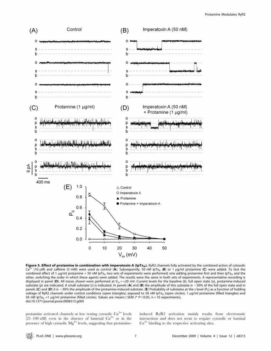
Protamine Affects the Probability of Imperatoxin A andRyanodol Substates
The peptide imperatoxin A (IpTxA) is an agent that has
similarities to the II-III loop of DHPR [37,38] and binds to a
specific site at the open RyR to induce the formation of long-
lasting, voltage dependent and reversible substates [35]. Here, we
tested possible interactions between the sites responsible for the
formation of protamine- and IpTxA-induced substates. We defined
‘‘s’’ as small substate that, in the absence of protamine, has ,30%
of the full opening current amplitude. In the presence of
protamine, s is defined as the ‘‘sub-substate’’ and its amplitude is
,30% of the protamine-induced highest conductance substate. As
shown in Fig. 5A, no transitions to any substate were observed
under control conditions (absence of IpTxA and/or protamine).
Figure 5B shows that at a holding voltage of +20 mV, IpTxA alone
induced frequent long-lived substates. The effect of 1 mg/ml
protamine alone is shown in Fig. 5C: the channel spent most of the
time in the protamine-induced substate with infrequent transitions
to short lived sub-substates. The combined action of protamine
and IpTxA was tested in two sets of experiments, switching the
order in which these agents were applied. The result was always
the same regardless of which agent was added in the first place: the
channel spent most of the time in the protamine-induced substate
Figure 3. Effect of protamine compared to other conductance-modifiers. Single-channel recordings of fully active RyR2 channels (10 mMcytosolic Ca2+/5 mM caffeine) in the presence of 1 mg/ml protamine, 10 mM ryanodol or 50 nM imperatoxin A (A, B and C, respectively). Channelactivity was recorded at Vm = 0 mV (top traces) and +40 mV (bottom traces). The current levels for the baseline, full open state and substates inducedby protamine, ryanodol and IpTxA are indicated (b, o, p, r and i, respectively). (D) Current amplitude as a function of voltage for the full open state(n = 28) and for substates induced by protamine (n = 10), ryanodol (n = 13) and IpTxA (n = 5). Slope conductances were, in pS, 12961, 12363, 5662and 4363, respectively.doi:10.1371/journal.pone.0008315.g003
Protamine Modulates RyR2
PLoS ONE | www.plosone.org 5 December 2009 | Volume 4 | Issue 12 | e8315

with infrequent transitions to sub-substates (Fig. 5D). In this case
the sub-substates were kinetically heterogeneous: there were short-
lived events, similar to those observed in absence of IpTxA (Fig. 5C)
and a few events of much longer duration that were only observed
upon addition of IpTxA. These events could represent either
IpTxA-induced substates in the protamine-modified channel or
IpTxA stabilization of the protamine-induced small-conductance
substates. Still, the global probability of sub-substates (which
includes both populations of events) was not significantly different
in absence versus presence of IpTxA (Fig. 5E, filled circles versus
filled triangles). Thus, IpTxA has little or no effect on RyR2
behavior when protamine is present.
Ryanodol binds to open RyRs at a specific ‘‘ryanoid site’’
inducing the formation of reversible substates of ,45% of the full
open channel conductance. Previous studies indicated that the
ryanoid site senses the electric field [36,39]. Here, we tested the
possibility that protamine induces substates by reversibly interact-
ing with the ‘‘ryanoid site’’. If this is the case, then we should not
observe protamine-induced substates in ryanodol-modified chan-
nels (the ‘‘ryanoid site’’ would be unavailable to protamine when it
is occupied by ryanodol). As shown in Fig. 6A, in the presence of
10 mM ryanodol, RyR2 reversibly fluctuated between the full
opening and the ryanodol-modified state. After addition of
protamine (Fig. 6B) the channel displayed virtually no full
openings and it alternated between the protamine-induced
substate and a long lasting (1163 seconds at Vm = 20 mV)
smaller substate which was never observed in the absence of
ryanodol. Thus, protamine and ryanodol are not likely to act at
the same binding site given that their effects do not exclude each
other. However, in the presence of protamine, the ryanodol effects
appear to be potentiated as events are longer and more frequent
than those observed in the absence of protamine (Fig. 6B, legend).
Therefore, addition of protamine increased the probability of
ryanodol-induced substates (PRyanodol) (Fig. 6D). As expected,
subsequent addition of heparin reversed this effect, the channel
turned back to fluctuate between the full open state and the
ryanodol substate (Fig. 6C), and PRyanodol decreased to control
values (Fig. 6D).
Protamine Increases the Activity of RyR2 at Low CytosolicCa2+
We tested the effect of protamine on RyR2 with low Po in the
presence of 100 nM cytosolic Ca2+ as this condition better mimics
the channel environment in a resting cell. In the control, at
Vm = 20 mV, a few short and infrequent openings were observed
(Fig. 7A) and the open probability (Po) was 0.04260.029 (Fig. 7D).
Upon addition of 1 mg/ml protamine, most of the openings were
to the protamine-induced substate (Fig. 7B) reaching a
Po = 0.94660.013 (Fig. 7D). As expected, upon addition of
heparin, the protamine-induced substate was no longer observed
(Fig. 7C) and channel activity returned to the levels observed
under control conditions, with a Po = 0.00960.006 (Fig. 7D).
Protamine activating effect on RyR2 was voltage-dependent and
Po only reached 0.58360.050 at Vm = 40 mV (Fig. 7D). The
activating effect of protamine was not affected by further
decreasing cytosolic Ca2+ levels to 25 nM (Po = 0.95060.012
and 0.90160.079, before and after decreasing Ca2+ levels,
respectively; Vm = 20 mV; n = 5). Addition of 5 mM cytosolic
Mg2+ decreased the amplitude of the full open state as well as the
substate by ,15% at 20 mV, which is expected from the decrease
in driving force for divalent flux [29]. However, Mg2+ did not
affect protamine induced activation (Po = 0.96160.006 and
0.95160.018, before and after addition of Mg2+, respectively;
Vm = 20 mV; n = 4). Notice that in the absence of protamine, at
low cytosolic Ca2+ and high Mg2+, RyR2 channels had Po,0,
even when exposed to 10 mM caffeine [40,41]. The activating
effect of protamine did not require lumen-to-cytosol Ca2+ flux as it
was also observed after replacing lumenal Ca2+ with Ba2+ (not
shown), a divalent cation that does not activate RyR2 [42].
Figure 8A shows a continuous recording of a RyR2 in the
presence of protamine at low cytosolic Ca2+. Notice that when the
RyR2 activates, it preferentially switches from the closed state to
the protamine-induced substate rather than to the full open state
(Fig. 8A, grey arrows). In contrast, when the channel closes, it is
more likely to do it from the full open state than from the
protamine-induced substate (Fig. 8A, black arrows). Estimations
(from 4 minutes of recordings at +40 mV) of the probability of full
openings, closures and protamine-induced substates are shown in
Fig. 8B. Analysis of these results suggests that there is a preferential
sequence of events during protamine-induced RyR2 activation.
First, protamine binds to the closed channel and induces
activation. Transitions from closed to the substate (1423) are 8.3
times more probable than transitions from closed to full openings
(171). Once activated, the channel fluctuates between the substate
and the full open state, probably as a result of rapid binding-
unbinding of protamine to its site. Relative rate of transition
(number of transitions ARB/probability A) for Full openRClosed
(9165) is nearly ten-fold that for Protamine substateRClosed (919).
If we assume that protamine is unbound during full openings, then
the RyR2 would be no longer under the activating influence of
protamine and it would be more likely to close.
Discussion
Protamine did not simply block RyR2 in an all-or-none fashion.
Rather, it affected the RyR2 channel conduction pathway by
producing substates and full block in a voltage-dependent and
dose-dependent manner. At low concentrations, protamine
induced a drop in the current amplitude to a large conductance
substate. The ratio between this amplitude drop and the amplitude
of the full opening decreases as the positive holding voltage (Vm)
increases. This is reminiscent of the rectification of some K+
channels observed in presence of polyamines. Furthermore,
Figure 4. Current voltage (I-V) curves for the full open stateand for the protamine-induced substate. Current amplitude as afunction of voltage was plotted based on single-channel recordings offully active RyR2 (10 mM cytosolic Ca2+/5 mM caffeine) performed in thepresence of cytosolic Cs+ and 0.2 mg/ml protamine. Values areaverages 6 SEM of 5 experiments.doi:10.1371/journal.pone.0008315.g004
Protamine Modulates RyR2
PLoS ONE | www.plosone.org 6 December 2009 | Volume 4 | Issue 12 | e8315
protamine activated channels at low resting cytosolic Ca2+ levels
(25–100 nM) even in the absence of lumenal Ca2+ or in the
presence of high cytosolic Mg2+ levels, suggesting that protamine-
induced RyR2 activation mainly results from electrostatic
interactions and does not seem to require cytosolic or luminal
Ca2+ binding to the respective activating sites.
Figure 5. Effect of protamine in combination with imperatoxin A (IpTxA). RyR2 channels fully activated by the combined action of cytosolicCa2+ (10 mM) and caffeine (5 mM) were used as control (A). Subsequently, 50 nM IpTxA (B) or 1 mg/ml protamine (C) were added. To test thecombined effect of 1 mg/ml protamine + 50 nM IpTxA two sets of experiments were performed: one adding protamine first and then IpTxA and theother, switching the order in which these agents were added. The results were the same in both sets of experiments. A representative recording isdisplayed in panel (D). All traces shown were performed at Vm = +20 mV. Current levels for the baseline (b), full open state (o), protamine-inducedsubstate (p) are indicated. A small substate (s) is indicated. In panels (A) and (B) the amplitude of this substate is ,30% of the full open state and inpanels (C) and (D) it is ,30% the amplitude of the protamine-induced substate. (E) Probability of substates at the s level (PS) as a function of holdingvoltage of RyR2 channels under control conditions (open triangles), exposed to 50 nM IpTxA (open circles), 1 mg/ml protamine (filled triangles) and50 nM IpTxA +1 mg/ml protamine (filled circles). Values are means6SEM (* P,0.05; n = 10 experiments).doi:10.1371/journal.pone.0008315.g005
Protamine Modulates RyR2
PLoS ONE | www.plosone.org 7 December 2009 | Volume 4 | Issue 12 | e8315

Comparison with Previous StudiesIn this work, protamine added to the cytosolic surface of RyR2
had a complex action as it affected RyR2 channel conduction
(multiple substates/block) and gating (activation). Our observation
of full block of RyR2 induced by high levels of protamine (20 mg/
ml; Vm = 0 mV) are consistent with previous findings in single
RyR1 channels [24]. However, lower protamine concentrations
that would have allowed to observe protamine-induced RyR1
activation have not been tested in this previous report [24].
Moreover, the substates and full block of fully activated RyR2
observed here at Vm = 0 mV would lead to a decrease in current
flow through the channels, which is in agreement with previous
reports were protamine was found to inhibit caffeine-induced
RyR1-mediated Ca2+ release from skeletal muscle SR microsomes
[19] (notice that the expected SR lumen voltage in microsomes is
,0 mV). Although this previous study did not test the effect of
protamine in conditions homologous to those used here to detect
activation, it provided evidence that RyRs can be activated by
another polyarginine (Histone IIS) and by a synthetic polylysine
[19]. These agents were found to activate Ca2+ release from
Figure 6. Effect of protamine in combination with ryanodol. Continuous trace of a RyR2 in the presence of 10 mM ryanodol before (A) andafter subsequent addition of 1 mg/ml protamine (B) and 250 mg/ml heparin (C). Recordings were performed at Vm = +20 mV (b = baseline; o = fullopen state; r = ryanodol-induced substate; p = protamine-induced substate; p+r = ryanodol-induced substate on the protamine-modified RyR2). (D)Probability of ryanodol-induced substates (PRyanodol) as a function of holding voltage of RyR2 channels exposed to 10 mM ryanodol (open circles);10 mM ryanodol+1 mg/ml protamine (filled circles) and 10 mM ryanodol+1 mg/ml protamine+250 mg/ml heparin (open triangles). Under these threeconditions, the dwell times of the ryanodol-induced events were (in seconds): 3.760.7; 11.362.5 and 3.460.5 respectively. Values are means6SEM(* P,0.05; n = 7 experiments).doi:10.1371/journal.pone.0008315.g006
Protamine Modulates RyR2
PLoS ONE | www.plosone.org 8 December 2009 | Volume 4 | Issue 12 | e8315

resting RyR1 and to induce block of drug-activated channels
[19,20]. Similarly, polylysine has been reported to activate RyR2-
mediated Ca2+ release in cardiac microsomes regardless of the
presence/absence of Mg2+ [43].
The literature suggests that the activation of RyR by
polycations is relatively restricted to peptides like polylysines
and polyarginines [19,20]. Protamine/histone/polylysine-in-
duced activation of SR Ca2+ release would result from Ca2+-
independent and Mg2+-insensitive transitions from closed to the
high subconductance state in resting RyRs. Protamine and
polylysine have been reported to produce diverse effects that do
not include activation on other ionic channels [44,45,46]. Thus,
protamine- and polylysine-induced activation seems to be specific
for RyRs. Inhibition of drug-stimulated RyR-mediated SR Ca2+
release would result from voltage-dependent transitions from the
full open state to multiple subconductance and block-like states. A
large variety of polycationic molecules were found to produce
these effects on RyRs [3,47,48,49] as well as on other channels
[50,51,52,53].
Effect of Protamine on RyR2 Channels ConductanceProtamine is essentially a poly-arginine molecule with a charge
of ,+22 [25]. Thus, it is reasonable to expect voltage-dependent
effects on the RyR channels as they contain rings of negatively
charged amino acids in the cytosolic vestibular regions that sense
the electrical field [16,17]. Indeed, at Vm#0 mV protamine
added to the RyR cytosolic surface produced several subconduc-
tance states as well as full block. However, at Vm ranging from
+10 to +80 mV, block events and smaller substates disappeared,
suggesting sharp voltage-dependence, while the substate with the
highest level of conductance remained with a significant
probability. This difference in the rate of decline of the probability
for the different substates could indicate that multiple protamine
molecules, despite their large charge and size, would bind to
multiple sites in the RyR vestibule that would have differential
sensitivity to the electric field. This would be in agreement with
the apparent multiple binding components that have been
suggested for polyamine-induced rectification of channels
[50,51,52,53].
Figure 7. Effect of protamine at low cytosolic Ca2+. Single channel recordings of partially active RyR2 in the presence of 100 nM cytosolic Ca2+
before (A) and after subsequent addition of 1 mg/ml protamine (B) and 250 mg/ml heparin (C). The frequency-current amplitude histograms obtainedfrom 4-min single-channel recordings are shown next to each trace. Channel activity was recorded at Vm = +20 mV (top traces) and +40 mV (bottomtraces). (D) Averages of open probabilities calculated for single RyR2 at Vm = +20 mV (black columns) and +40 mV (shaded columns). Here, the openprobability includes the probability of the full opening plus the probability of protamine-induced substates. Values are shown as means6SEM(* P,0.05 versus control; # P,0.05 versus protamine at +20 mV; n = 5 experiments).doi:10.1371/journal.pone.0008315.g007
Protamine Modulates RyR2
PLoS ONE | www.plosone.org 9 December 2009 | Volume 4 | Issue 12 | e8315

The nature and functional implication of substates in RyRs is
not known (discussed in [18]). Protamine substates are different
from those induced by known RyR conductance modifiers such as
ryanoids and Imperatoxin A (IpTxA) [3,18,35], which change
channel conductance by inducing a drop in the current amplitude
that is proportional to the amplitude of the full opening (i.e., these
agents produce a ‘‘rigid’’ conformational change in the RyR’s
conduction pathway). The nearly constant drop in current
amplitude observed at positive voltages for the protamine-induced
transition from full opening to substate is unusual and it implies
that the proportion by which the conductance is reduced (relative
to the size of the full open channel) is less as the holding voltage
increases. As the driving force of Ca2+ fluxes increases, it could
better counteract an electrostatic screening phenomenom pro-
duced by bound protamine and/or change the orientation of
protamine putative coiled-coil structure in a rigid vestibule [54]. It
is also possible that protamine binds to a flexible loop in the RyR
that modulates the ability of protamine to act as a plug within the
vestibule in a voltage-dependent manner.
The cationic peptide IpTxA, induces long-lived substates by
binding to sites putatively located at RyR regions that sense the
electrical field [35]. More recently, a 3D mapping study has located
the IpTxA binding to a RyR region between the clamp domain
and the central handle domain [55]. Here, protamine greatly
decreased the probability of IpTxA-induced substates suggesting
that protamine may bind to the same RyR region as IpTxA.
Protamine was effective at inducing substates in ryanodol-
modified channels, suggesting that the binding sites for protamine
and for ryanoids do not overlap. The ryanodol-protamine
interactions seem to be cooperative since protamine increased
the probability of ryanodol-induced substates. This result resem-
bles previously reported findings, where IpTxA, a segment of the
DHPR II-III loop (Peptide A) and other positively charged ‘‘ball
peptides’’ affected the action of ryanodol in a cooperative fashion
[18]. As ryanoids and IpTxA were shown to bind to different
regions in the RyR molecule [55,56], our finding that protamine
affects the action of both agents would suggest multiple sites of
action or allosteric effects. Increase in ryanodine binding induced
by polylysine has also been reported for skeletal and cardiac RyRs
[20,21,23].
In summary, protamine affects RyR2 channel conduction
(substates and block) and the binding affinity of agents that
Figure 8. Sequence of events for protamine-induced activation of RyR2. (A) Continuous trace of a RyR2 in the presence of 25 nM cytosolicCa2+ plus 1 mg/ml protamine recorded at Vm = +40 mV (b = baseline; o = full opening; p = protamine-induced substate). The beginning and end ofeach opening are indicated by grey and black arrows, respectively. (B) Diagram of states of RyR2 exposed to 1 mg/ml protamine with 25 nM cytosolicCa2+ (4 min recording at Vm = +40 mV). The following states were considered: full open, protamine substate, closed. State probabilities (P) are givenin parenthesis. Arrows represent observed unidirectional transitions from one state to another. Adjacent to each arrow is the respective absolutenumber of transitions.doi:10.1371/journal.pone.0008315.g008
Protamine Modulates RyR2
PLoS ONE | www.plosone.org 10 December 2009 | Volume 4 | Issue 12 | e8315

interact with vestibular regions of RyR2. This could be the result
of allosterically-induced flexible conformational changes, although
steric interference cannot be ruled out. This effect of protamine on
conduction does not appear to be highly specific as similar effects
were found for other polycationic molecules [3,18,47,48,49].
Protamine Activates RyR2Protamine (1 mg/ml) activated RyR2 at cytosolic Ca2+ levels
below those found in resting cells (when the RyR2 are normally
closed). Regarding the steps involved in protamine-induced
activation, our results show that there is a preferential sequence
of events. The channels first open to reach the protamine substate
level and then they fluctuate between the full open sate and the
substate. We also observed that the probability to close is higher
when the channel is fully open than during substates. Like
protamine, IpTxA- and ryanoids-induced substates rarely transi-
tion to the closed state [34,37]. Unlike protamine, IpTxA and
ryanoids do not activate RyR2 under resting Ca2+ conditions
[1,3,18,33,34,37].
The efficacy of protamine to activate RyR2 was not affected by
addition of up to 5 mM Mg2+, known to compete with Ca2+ at the
RyR2 cytosolic binding activating sites [1,3,42]. Moreover,
protamine remained effective after we replaced lumenal Ca2+ with
Ba2+, which would prevent ‘‘feed through’’ activation, as Ba2+ can
permeate through the channel but does not activate it [42]. The
results support the idea that protamine-induced activation of RyR2
is mostly mediated by interactions that are Ca2+- indepen-
dent within levels found in the cytosol and SR lumen of myocytes.
Although RyRs are highly sensitive to the activating action of
protamine and polylysine, they are not gated open by a variety of
other cationic molecules, including peptides, polyamines and
macrolide antibiotics, which only produce substates and/or block
[18,47,48,49]. The reason for this differential sensitivity is still
unclear but it is possible that the high efficacy of cationic
polypeptides is related to the large magnitude of their charge
density (e.g., ,+22 for protamine), which would allow them to
strongly interact with a negatively charged gating domain.
Previous studies have found that Ca2+ dependence of RyR gating
is insensitive to carbodiimide titration of negative surface charges
located either on the luminal or the cytosolic channel surface [57].
Likewise, [3H]ryanodine binding remained Ca2+ dependent even
in presence of very high salt levels (1 M NaCl or KCl) which
should greatly affect electrostatic interactions [58]. Thus, it is
possible that the action of protamine may be more than just a
surface-charge effect. Accordingly, previous reports with histones
and polylysines of different molecular weights and charges did not
clearly indicate whether the most charged molecules are better
RyR activating agents [19]. In principle, poly L-arginines would
be better activating agents than poly L-lysines [59]. Indeed, we
reach near maximal activation of RyR2 with ,250 nM
protamine, which is about one tenth of the concentration of a
larger molecular weight polysysine required to elicit the same
effect [19,52]. It is possible that the arginines present in protamine
are more suitable to form strong p-cation interactions with
aromatic aminoacids present at the RyR gating domain, resulting
in a higher binding affinity [60].
Is There a Role for Modulation of RyRs throughElectrostatic Interactions
Protamine is clinically utilized to reverse heparin overdose and
has been shown to display significant cardiotoxic effects, including
decreased cardiac output and electrocardiographic disturbances
[31]. Studies performed using isolated myocytes revealed multiple
effects of protamine, including changes in cardiomyocyte contrac-
tion (depressed contraction at 3 Hz but enhanced at 0.5 Hz or at
lower rates), partial membrane depolarization, rise in resting
tension and appearance of rested state rapid cooling contractures
[61]. The effects of protamine have been associated to calcium
overload and impairment of sarcoplasmic reticulum functions
[62]. Our results open the possibility that direct protamine bind-
ing to RyR2 could mediate, at least in part, the observed
abnormalities in SR Ca2+ homeostasis.
It is plausible that the strong effect of protamine on channel
conduction and gating results from its interaction with the rings of
charges in the vestibular region of RyRs [63]. However, the
physiological role for the existence of a RyR channel domain that
confers susceptibility to modulation by agents like protamine is still
unknown. A physiological implication of our findings is the
possibility that protamine mimics the action of positively charged
peptide motifs, present in the vicinity of the RyR2 channels in cells
which might modulate channel activity (activation and deactiva-
tion). In favor of this possibility, previous observations suggested
that RyRs hypersensitivity to polylysine is associated with cardiac
diseases [22] or with modifications of skeletal RyR1 interdomains
that trigger malignant hyperthermia [23].
Studies of multichannel behavior indicated that physical RyR-
RyR interactions between domains of neighboring channels could
shape their coupled gating [8,11,12,14]. It is possible that
protamine-sensitive domains are involved in this process. RyR
also interact with DHPRs. Indeed, during the action potential,
physical DHPR-RyR1 interactions activate RyR1-mediated Ca2+
release in skeletal fibers [2].
The very limited effects of IpTxA and DHPR peptides on sparks
suggest they have restricted access to the RyR2 [64]. An
interesting observation is that protamine also has very limited
effect on RyR1 in skeletal fibers, suggesting that the protamine-
sensitive domains of RyR1 are not accessible in the cellular
environment [65]. An interpretation is that there might be an
endogenous polycationic peptide, which could move when
propelled by a change in the electric field at the T-tubule during
excitation to induce activation of RyR1. The same moiety could
subsequently block the RyR1 when the T-tubule voltage returns to
resting values. In the heart, Ca2+ entry is known as an absolute
requirement for cardiac EC coupling [5] and the presence of a
voltage-dependent component in the activation is still an open
question [66]. Still, the observation of Ca2+-entry-independent
negative modulation of local events of Ca2+ sparks by specific
DHPR agonist and blockers suggests the existence of physical
RyR2-DHPR interactions of a still unknown nature that regulate
RyR2 function [5,15]. The protamine-binding domain in RyR2
might be involved in this modulation.
In summary, our studies indicate that protamine, can induce
Ca2+-independent RyR2 activation and deactivation. The possi-
bility of similar moieties shaping RyR-RyR interactions for
functional coupling or DHPR-RyR interactions for triggering
and/or terminating SR Ca2+ release opens the field for further
investigation.
Acknowledgments
We thank Prof. D.M. Caspary, Ph.D, SIU-SOM, for his critical input. We
also thank Mr. R. Herndon, Research Administrator and Mrs. J. Bryan,
Office System Specialist, SIU-SOM, for proofreading the manuscript.
Author Contributions
Conceived and designed the experiments: PLDS JAC. Performed the
experiments: PLDS JAC. Analyzed the data: PLDS JAC. Contributed
reagents/materials/analysis tools: PLDS JAC. Wrote the paper: PLDS
JAC.
Protamine Modulates RyR2
PLoS ONE | www.plosone.org 11 December 2009 | Volume 4 | Issue 12 | e8315

References
1. Fill M, Copello JA (2002) Ryanodine receptor calcium release channels. Physiol
Rev 82: 893–922.
2. Rios E, Stern MD (1997) Calcium in close quarters: microdomain feedback inexcitation-contraction coupling and other cell biological phenomena. Annu Rev
Biophys Biomol Struct 26: 47–82.
3. Sitsapesan R, Williams AJ (1998) The Structure and Function of Ryanodine
Receptors. London, UK: Imperial College Press.
4. Sun XH, Protasi F, Takahashi M, Takeshima H, Ferguson DG, et al. (1995)Molecular architecture of membranes involved in excitation-contraction
coupling of cardiac muscle. J Cell Biol 129: 659–671.
5. Bers DM (2001) Excitation-Contraction Coupling and Cardiac ContractileForce. Dordrecht, The Netherlands: Kluwer Academic Press.
6. Gonzalez A, Kirsch WG, Shirokova N, Pizarro G, Brum G, et al. (2000)
Involvement of multiple intracellular release channels in calcium sparks ofskeletal muscle. Proc Natl Acad Sci U S A 97: 4380–4385.
7. Stern MD, Song LS, Cheng H, Sham JS, Yang HT, et al. (1999) Local control
models of cardiac excitation-contraction coupling. A possible role for allosteric
interactions between ryanodine receptors. J Gen Physiol 113: 469–489.
8. Copello JA, Zima AV, Diaz-Sylvester PL, Porta M, Nani A, et al. (2006) Cardiacryanodine receptor (RyR) channels communicate among themselves and with
dyhidropyridine receptor L-type calcium channels (DHPR). Circulation 114:57–58.
9. Cheng H, Lederer WJ, Cannell MB (1993) Calcium sparks: elementary events
underlying excitation-contraction coupling in heart muscle. Science 262:740–744.
10. Stern MD, Cheng H (2004) Putting out the fire: what terminates calcium-
induced calcium release in cardiac muscle? Cell Calcium 35: 591–601.
11. Porta M, Diaz-Sylvester PL, Nani A, Fill M, Fleischer S, et al. (2004) Modulation
of coordinated gating of ryanodine receptor (RyR) channels in planar lipidbilayers. Biophys J 86: 241A.
12. Marx SO, Gaburjakova J, Gaburjakova M, Henrikson C, Ondrias K, et al.
(2001) Coupled gating between cardiac calcium release channels (ryanodinereceptors). Circ Res 88: 1151–1158.
13. Marx SO, Ondrias K, Marks AR (1998) Coupled gating between individual
skeletal muscle Ca2+ release channels (ryanodine receptors). Science 281:818–821.
14. Copello JA, Porta M, Diaz-Sylvester PL, Nani A, Escobar A, et al. (2003)
Coordinated gating of multiple ryanodine receptors (RyRs). Biophys J 84: 77pl.
15. Copello JA, Zima AV, Diaz-Sylvester PL, Fill M, Blatter LA (2007) Ca2+ entry-independent effects of L-type Ca2+ channel modulators on Ca2+ sparks in
ventricular myocytes. Am J Physiol Cell Physiol 292: C2129–2140.
16. Xu L, Wang Y, Gillespie D, Meissner G (2006) Two rings of negative charges in
the cytosolic vestibule of type-1 ryanodine receptor modulate ion fluxes.Biophys J 90: 443–453.
17. Williams AJ, West DJ, Sitsapesan R (2001) Light at the end of the Ca(2+)-release
channel tunnel: structures and mechanisms involved in ion translocation inryanodine receptor channels. Q Rev Biophys 34: 61–104.
18. Porta M, Diaz-Sylvester PL, Nani A, Ramos-Franco J, Copello JA (2008)
Ryanoids and imperatoxin affect the modulation of cardiac ryanodine receptorsby dihydropyridine receptor Peptide A. Biochim Biophys Acta 1778: 2469–
2479.
19. Palade P (1987) Drug-induced Ca2+ release from isolated sarcoplasmic
reticulum. III. Block of Ca2+-induced Ca2+ release by organic polyamines.J Biol Chem 262: 6149–6154.
20. Ohkusa T, Kang JJ, Morii M, Ikemoto N (1991) Conformational change of the
foot protein of sarcoplasmic reticulum as an initial event of calcium release.J Biochem 109: 609–615.
21. Lu X, Xu L, Meissner G (1994) Activation of the skeletal muscle calcium release
channel by a cytoplasmic loop of the dihydropyridine receptor. J Biol Chem 269:6511–6516.
22. Yamamoto T, Yano M, Kohno M, Hisaoka T, Ono K, et al. (1999) Abnormal
Ca2+ release from cardiac sarcoplasmic reticulum in tachycardia-induced heart
failure. Cardiovasc Res 44: 146–155.
23. Kobayashi S, Yamamoto T, Parness J, Ikemoto N (2004) Antibody probe studyof Ca2+ channel regulation by interdomain interaction within the ryanodine
receptor. Biochem J 380: 561–569.
24. Koulen P, Ehrlich BE (2000) Reversible block of the calcium release channel/ryanodine receptor by protamine, a heparin antidote. Mol Biol Cell 11: 2213–
2219.
25. Ando T, Watanabe S (1969) A new method for fractionation of protamines andthe amino acid sequences of salmine and three components of iridine.
Int J Protein Res 1: 221–224.
26. Diaz-Sylvester PL, Copello JA (2009) Voltage-Dependent Modulation of
Cardiac Ryanodine Receptors (RyR2) by Protamine. Biophysical Journal 96:113a.
27. Deslongchamps P, Belanger A, Berney DJF, Borschberg HJ, Brousseau R, et al.
(1990) The Total Synthesis of (+)-Ryanodol. Canadian Journal of Chemistry-Revue Canadienne De Chimie 68: 115–192.
28. Chamberlain BK, Levitsky DO, Fleischer S (1983) Isolation and characteriza-
tion of canine cardiac sarcoplasmic reticulum with improved Ca2+ transportproperties. J Biol Chem 258: 6602–6609.
29. Copello JA, Barg S, Onoue H, Fleischer S (1997) Heterogeneity of Ca2+ gatingof skeletal muscle and cardiac ryanodine receptors. Biophys J 73: 141–156.
30. Byun Y, Singh VK, Yang VC (1999) Low molecular weight protamine: a
potential nontoxic heparin antagonist. Thromb Res 94: 53–61.
31. Pugsley MK, Kalra V, Froebel-Wilson S (2002) Protamine is a low molecular
weight polycationic amine that produces actions on cardiac muscle. Life Sci 72:293–305.
32. Hobbhahn J, Conzen PF, Habazettl H, Gutmann R, Kellermann W, et al.
(1991) Heparin reversal by protamine in humans–complement, prostaglandins,blood cells, and hemodynamics. J Appl Physiol 71: 1415–1421.
33. Mead F, Williams AJ (2002) Block of the ryanodine receptor channel by
neomycin is relieved at high holding potentials. Biophys J 82: 1953–1963.
34. Woodhull AM (1973) Ionic blockage of sodium channels in nerve. J Gen Physiol
61: 687–708.
35. Tripathy A, Resch W, Xu L, Valdivia HH, Meissner G (1998) Imperatoxin Ainduces subconductance states in Ca2+ release channels (ryanodine receptors) of
cardiac and skeletal muscle. J Gen Physiol 111: 679–690.
36. Tanna B, Welch W, Ruest L, Sutko JL, Williams AJ (2000) The interaction of aneutral ryanoid with the ryanodine receptor channel provides insights into the
mechanisms by which ryanoid binding is modulated by voltage. J Gen Physiol116: 1–9.
37. Green D, Pace S, Curtis SM, Sakowska M, Lamb GD, et al. (2003) The three-
dimensional structural surface of two beta-sheet scorpion toxins mimics that ofan alpha-helical dihydropyridine receptor segment. Biochem J 370: 517–527.
38. Mosbah A, Kharrat R, Fajloun Z, Renisio JG, Blanc E, et al. (2000) A new fold
in the scorpion toxin family, associated with an activity on a ryanodine-sensitivecalcium channel. Proteins 40: 436–442.
39. Tinker A, Sutko JL, Ruest L, Deslongchamps P, Welch W, et al. (1996)Electrophysiological effects of ryanodine derivatives on the sheep cardiac
sarcoplasmic reticulum calcium-release channel. Biophys J 70: 2110–2119.
40. Sitsapesan R, Williams AJ (1990) Mechanisms of caffeine activation of singlecalcium-release channels of sheep cardiac sarcoplasmic reticulum. J Physiol 423:
425–439.
41. Porta M, Diaz-Sylvester PL, Nani A, Perez C, Mejia-Alvarez R, et al. (2004)Modulation of caffeine action on single ryanodine receptors (RyR2) channels by
endogenous agents. Biophys J 86: 62A.
42. Diaz-Sylvester P, Porta M, Nani A, Fill M, Copello JA (2006) Modulation ofcardiac ryanodine receptors (RyR2) by alkaline earth metal cations. Biophys J
89: 1990p.
43. Yano M, Yamamoto T, Kohno M, Hisaoka T, Ono K, et al. (1998) Polylysine-
induced rapid Ca2+ release from cardiac sarcoplasmic reticulum. J Cardiovasc
Pharmacol 32: 96–100.
44. Deutsch N, Matsuoka S, Weiss JN (1994) Surface charge and properties of
cardiac ATP-sensitive K+ channels. J Gen Physiol 104: 773–800.
45. Suh BC, Hille B (2007) Electrostatic interaction of internal Mg2+ withmembrane PIP2 Seen with KCNQ K+ channels. J Gen Physiol 130: 241–256.
46. Li L, Geng X, Yonkunas M, Su A, Densmore E, et al. (2005) Ligand-dependent
linkage of the ATP site to inhibition gate closure in the KATP channel. J GenPhysiol 126: 285–299.
47. Uehara A, Fill M, Velez P, Yasukochi M, Imanaga I (1996) Rectification ofrabbit cardiac ryanodine receptor current by endogenous polyamines. Biophys J
71: 769–777.
48. Xu L, Jones R, Meissner G (1993) Effects of local anesthetics on single channelbehavior of skeletal muscle calcium release channel. J Gen Physiol 101: 207–
233.
49. Mead FC, Williams AJ (2004) Electrostatic mechanisms underlie neomycin blockof the cardiac ryanodine receptor channel (RyR2). Biophys J 87: 3814–3825.
50. Lopatin AN, Makhina EN, Nichols CG (1994) Potassium channel block by
cytoplasmic polyamines as the mechanism of intrinsic rectification. Nature 372:366–369.
51. Williams K (1997) Interactions of polyamines with ion channels. Biochem J 325(Pt 2): 289–297.
52. Guo D, Lu Z (2003) Interaction mechanisms between polyamines and IRK1
inward rectifier K+ channels. J Gen Physiol 122: 485–500.
53. Hille B (2001) Ion Channels of Excitable Membranes. Sunderland MA, USA:Sinauer Associates, Inc.
54. Ebert G, Zolzer U, Nishi N (1990) Solubilization and conformation ofprotamines in reverse micelles. Progr Colloid Polym Sci 83: 181–187.
55. Samso M, Trujillo R, Gurrola GB, Valdivia HH, Wagenknecht T (1999) Three-
dimensional location of the imperatoxin A binding site on the ryanodinereceptor. J Cell Biol 146: 493–499.
56. Ranatunga KM, Chen SR, Ruest L, Welch W, Williams AJ (2007)
Quantification of the effects of a ryanodine receptor channel mutation oninteraction with a ryanoid. Mol Membr Biol 24: 185–193.
57. Tu Q, Velez P, Cortes-Gutierrez M, Fill M (1994) Surface charge potentiates
conduction through the cardiac ryanodine receptor channel. J Gen Physiol 103:853–867.
58. Ogawa Y (1994) Role of ryanodine receptors. Crit Rev Biochem Mol Biol 29:229–274.
59. Yamamoto T, Ikemoto N (2002) Peptide probe study of the critical regulatory
domain of the cardiac ryanodine receptor. Biochem Biophys Res Commun 291:1102–1108.
Protamine Modulates RyR2
PLoS ONE | www.plosone.org 12 December 2009 | Volume 4 | Issue 12 | e8315
60. Gallivan JP, Dougherty DA (1999) Cation-pi interactions in structural biology.
Proc Natl Acad Sci U S A 96: 9459–9464.61. Park WK, Pancrazio JJ, Lynch C, 3rd (1994) Mechanical and electrophysio-
logical effects of protamine on isolated ventricular myocardium: evidence for
calcium overload. Cardiovasc Res 28: 505–514.62. David JS, Vivien B, Lecarpentier Y, Coriat P, Riou B (2001) Extracellular
calcium modulates the effects of protamine on rat myocardium. Anesth Analg92: 817–823.
63. Gillespie D, Xu L, Wang Y, Meissner G (2005) (De)constructing the ryanodine
receptor: modeling ion permeation and selectivity of the calcium release channel.J Phys Chem B 109: 15598–15610.
64. Terentyev D, Viatchenko-Karpinski S, Valdivia HH, Escobar AL, Gyorke S
(2002) Luminal Ca2+ controls termination and refractory behavior of Ca2+-
induced Ca2+ release in cardiac myocytes. Circ Res 91: 414–420.
65. Brunder DG, Gyorke S, Dettbarn C, Palade P (1992) Involvement of
sarcoplasmic reticulum ‘Ca2+ release channels’ in excitation-contraction
coupling in vertebrate skeletal muscle. J Physiol 445: 759–778.
66. Balke CW, Goldman L (2003) Excitation contraction coupling in cardiac muscle:
is there a purely voltage-dependent component? J Gen Physiol 121: 349–352.
Protamine Modulates RyR2
PLoS ONE | www.plosone.org 13 December 2009 | Volume 4 | Issue 12 | e8315