role of plant lipid transfer proteins in plant cell physiology—a concise review
TRANSCRIPT
Review
Role of plant lipid transfer proteins in plant cellphysiology—A concise review
Andre de Oliveira Carvalho, Valdirene Moreira Gomes *
Laboratorio de Fisiologia e Bioquımica de Microrganismos, Centro de Biociencias e Biotecnologia, Universidade Estadual do
Norte Fluminense, Darcy Ribeiro, Av. Alberto Lamego, 2000 Campos dos Goytacazes, RJ CEP: 28013-600, Brazil
Contents
1. Introduction. . . . . . . . . . . . . . . . . . . . . . . . . . . . . . . . . . . . . . . . . . . . . . . . . . . . . . . . . . . . . . . . . . . . . . . . . . . . . . . . . 1145
1.1. The structure . . . . . . . . . . . . . . . . . . . . . . . . . . . . . . . . . . . . . . . . . . . . . . . . . . . . . . . . . . . . . . . . . . . . . . . . . . . 1145
1.1.1. The relationship between the structure and the capacity of transfer lipids . . . . . . . . . . . . . . . . . . . . 1147
1.2. Localization and gene expression . . . . . . . . . . . . . . . . . . . . . . . . . . . . . . . . . . . . . . . . . . . . . . . . . . . . . . . . . . . 1148
1.3. Biological activities . . . . . . . . . . . . . . . . . . . . . . . . . . . . . . . . . . . . . . . . . . . . . . . . . . . . . . . . . . . . . . . . . . . . . . 1149
1.3.1. Plant signalling . . . . . . . . . . . . . . . . . . . . . . . . . . . . . . . . . . . . . . . . . . . . . . . . . . . . . . . . . . . . . . . . . . . 1149
1.3.2. Antimicrobial activity . . . . . . . . . . . . . . . . . . . . . . . . . . . . . . . . . . . . . . . . . . . . . . . . . . . . . . . . . . . . . . 1150
1.3.3. As food allergens . . . . . . . . . . . . . . . . . . . . . . . . . . . . . . . . . . . . . . . . . . . . . . . . . . . . . . . . . . . . . . . . . 1150
2. Conclusion . . . . . . . . . . . . . . . . . . . . . . . . . . . . . . . . . . . . . . . . . . . . . . . . . . . . . . . . . . . . . . . . . . . . . . . . . . . . . . . . . . 1151
Acknowledgements . . . . . . . . . . . . . . . . . . . . . . . . . . . . . . . . . . . . . . . . . . . . . . . . . . . . . . . . . . . . . . . . . . . . . . . . . . . 1151
References . . . . . . . . . . . . . . . . . . . . . . . . . . . . . . . . . . . . . . . . . . . . . . . . . . . . . . . . . . . . . . . . . . . . . . . . . . . . . . . . . . 1151
p e p t i d e s 2 8 ( 2 0 0 7 ) 1 1 4 4 – 1 1 5 3
a r t i c l e i n f o
Article history:
Received 23 January 2007
Received in revised form
7 March 2007
Accepted 7 March 2007
Published on line 13 March 2007
Keywords:
Antimicrobial activity
Antimicrobial peptides
Gene expression
Lipid transfer protein
Plant signaling
a b s t r a c t
Plant lipid transfer proteins (LTP) are cationic peptides, subdivided into two families, which
present molecular masses of around 7 and 10 kDa. The peptides were, thus, denominated
due to their ability to reversibly bind and transport hydrophobic molecules in vitro. Both
subfamilies possess conserved patterns of eight cysteine residues and the three-dimen-
sional structure reveals an internal hydrophobic cavity that comprises the lipid binding site.
Based on the growing knowledge regarding structure, gene expression and regulation and in
vitro activity, LTPs are likely to play a role in key processes of plant physiology. Although the
roles of plant LTPs have not yet been fully determined. This review aims to present
comprehensive information of recent topics, cover new additional data, and present new
perspectives on these families of peptides.
# 2007 Elsevier Inc. All rights reserved.
avai lable at www.sc iencedi rec t .com
journal homepage: www.e lsev ier .com/ locate /pept ides
* Corresponding author. Tel.: +55 22 2726 1689; fax: +55 22 2726 1520.E-mail address: [email protected] (V.M. Gomes).
0196-9781/$ – see front matter # 2007 Elsevier Inc. All rights reserved.doi:10.1016/j.peptides.2007.03.004
p e p t i d e s 2 8 ( 2 0 0 7 ) 1 1 4 4 – 1 1 5 3 1145
1. Introduction
In living plant cells, lipids in the plasma membrane and in the
different membranes of organelles undergo anabolism,
catabolism and renewal. The glyoxysome membranes, for
example, possess phosphatidylcholine, phosphatidylglycerol
and phosphatidylethanolamine; however this organelle does
not have the biosynthetic enzymes to produce these phos-
pholipids. Thus, glyoxysomes must import these phospholi-
pids from the organelle that synthesizes them, the
endoplasmatic reticulum. A similar situation is also found
for the phospholipids that constitute the membranes of other
organelles, such as chloroplasts, mitochondria and the plasma
membrane [44] where a transport system for the intracellular
movement of lipids is necessary, due to the poor solubility of
lipids inside the aqueous milieu of cell cytoplasm.
Approximately 30 years ago, the lipid transfer proteins
(LTPs) were discovered [30] and thus denominated due to
their ability to facilitate the transfer of phospholipids
between a donor and an acceptor membrane, in vitro [31].
LTPs are small peptides that comprise two families. The
LTPs that form the first family, namely LTP1, have molecular
masses of approximately 10 kDa and are basic, presenting
isoeletric points (pI) of between 9 and 10. These LTPs have
90–95 amino acid residues, of which eight are cysteines
conserved in similar positions along the primary structure
of the already characterized LTP1 family. These eight
cysteines, bound to each other, form four disulfide bridges
that help the stabilization of the peptide tertiary structure
[31]. The LTP2 family is formed of peptides that have
molecular masses of approximately 7 kDa, possessing on
average 70 amino acids; their other characteristics, such as
a high pI, lipid transfer activity and another pattern of four
conserved disulfide bridges, are shared with the LTP1 family
[14,18,39]. Both families present a signal peptide at the
amino terminal region, which in general varies between 21
and 27 amino acids, for the LTP1 family [1,67,79], and from
27 to 35 amino acids, for the LTP2 family [20,32] (Fig. 1A and
B). This signal peptide is excised, rendering the mature
peptide and targeting the LTPs to cell secretory pathway
where they are exported to the apoplast. In keeping with
these discoveries and corroborating with the extracellular
location, LTP1 of various plants species are localized at the
cell wall, as demonstrated in Arabidopsis thaliana [69], in
Brassica oleracea var. italica leaves [56] and in Ricinus communis
and Vigna unguiculata seeds [12,72]. These findings were
inconsistent with the hypothesis of a biological role for the
LTPs in the plant cell cytoplasm.
Table 1 – Proposed biological activities of plant lipidtransfer proteins
Biological activity References
Cutin synthesis [24,56]
b-Oxidation [72]
Somatic embryogenesis [65]
Alergenics [75]
Plant signaling [4,7,40]
Plant defense against phytopathogens [33,42,57,61,77]
Pollen adherence [48]
Taking together these localizations with other findings,
different functions are suggested for the role of LTP in the
physiology of plants, such roles may include cutin synthesis
[24,56], b-oxidation [72], somatic embryogenesis [65], aler-
genics [75], plant signaling [4,7,40] and plant defense against
phytopathogens [33,42,57,61,77] (Table 1), but the true phy-
siological roles fulfilled by the LTPs have yet to be determined.
For the correct denomination of these peptides, biochem-
ical assays have been used to determine the transfer of
radioactivity or fluorescence labeled phospholipids from a
donor membrane to an aceptor membrane, in vitro [18,78,82].
These assays have the purpose of determining whether one
given protein belongs to the LTP class. In these assays, a
bidirectional movement of phospholipids between the donor
and aceptor membranes has been observed. Due to these
discoveries, these peptides were initially called phospholipid
exchange proteins; however, since this exchange does not
occur at a rate of 1:1 (donor:acceptor), these peptides were
renamed as phospholipid transfer proteins. Later the name
was changed once again, this time to the lipid transfer
proteins, due to these peptides can transport lipid molecules
other than phospholipids. Since the activity of the lipid
transfer is not specific, these peptides are also called non-
specific lipid transfer proteins [31,82].
1.1. The structure
The primary structures of the mature LTPs of both groups
comprise a unique polypeptide chain containing 90–95 amino
acid residues, in the case of the LTP1 family, and approxi-
mately 70 residues in the case of the LTP2 family [18,31,60].
Among these, there are eight strictly conserved cysteine
residues that form four intrachain disulfide bridges. Although
the eight cysteine residues are conserved between the two
groups, there is a mismatch in the cysteine paring motif. In the
LTP1 family, the Cys3 pares with Cys50 and Cys48 pares with
Cys87 and in the case of LTP2 family, the Cys3 pares with Cys35
and Cys37 pares with Cys68 [18,39,60] (Fig. 1A and B).
The secondary structure of the LTP1 family is composed of
four a-helices (helices H1 from Cys3 to Ala17, H2 from Ala25 to
Ala37, H3 from Thr41 to Ala56 and H4 from Ala63 to Cys73) and a
long carboxy terminal tail that is devoid of a defined secondary
structure, except for the presence of one turn of the 310 helix
(Fig. 2) [34,62]. The LTP2 family follows the same secondary
structural pattern as the LTP1 family, but presents three a-
helices (H1, from Cys3 to Ala16, H2 from Thr22 to Ala31 and H3
from Gln33 to Ala40) and a region containing two single-turn
helices (Tyr45 to Tyr48 and Ala54 to Val58). The carboxy terminal
tail also does not present any defined secondary structure
(Fig. 2) [18,60].
LTPs are abundant in charged residues, among the LTPs1
there are 12 in the Oryza sativa LTP [34], 11 in the Vigna radiata
var. radiate LTP [36], 11 in LTP1 and 13 in LTP2 of Sorghumvulgare
[54], 11 in the LTP1 of Brassica napus [34] and 11 in the EP2 of
Daucus carota [65]. The presence of such amino acids endows
the LTPs1 family with a high pI, varying between 9 and 10
[2,36,56]. Among those residues Asp43, Arg44 and Lys52 are
conserved in the LTPs1 that have been characterized (Fig. 1A).
Of the LTPs2, 10 amino acids are charged in the LTP(P) and
LTP(G) of Triticum aestivum [18], 9 in the LTP2 of Oryza sativa [39]
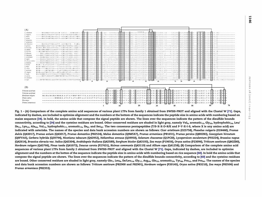
Fig. 1 – (A) Comparison of the complete amino acid sequences of various plant LTPs from family 1 obtained from SWISS-PROT and aligned with the Clustal W [71]. Gaps,
indicated by dashes, are included to optimize alignment and the numbers at the bottom of the sequences indicate the peptide size in amino acids with numbering based on
maize sequence [34]. In bold, the amino acids that compose the signal peptide are shown. The lines over the sequences indicate the pattern of the disulfide bounds
connectivity, according to [34] and the cysteine residues are boxed. Other conserved residues are shaded in light gray, namely Val6, aromatic13, Gly30, hydrophobic34, Leu/
Ile51, Lys52, Ala66, Val72, hydrophobic77, aromatic79, Ile81 and Ser82. The two consensus pentapeptides (T/S-X-X-D-R/K and P-Y-X-I-S, where X is any amino acid) are
indicated with asterisks. The names of the species and data bank accession numbers are shown as follows: Cicer arietinum (O23758), Phaselus vulgaris (O24440), Prunus
dulcis (Q43017), Prunus avium (Q43017), Prunus domestica (P82534), Malus domestica (Q9M5X7), Prunus armeniaca (P81651), Prunus persica (Q8H2B3), Gossypium hirsutum
(Q9FVA5), Gerbera hybrida (Q39794), Nicotiana tabacum (Q42952), Helianthus annuus (Q39950), Solanum chacoense (Q1PCI0), Lycopersicon esculentum (P93224), Brassica napus
(Q42614), Brassica oleracia var. italica (Q43304), Arabidopsis thaliana (Q42589), Sorghum bicolor (Q43193), Zea mays (P19656), Oryza sativa (P23096), Triticum aestivum (Q8GZB0),
Hordeum vulgare (Q43766), Pinus taeda (Q41073), Daucus carota (P27631), Ricinus communis (Q43119) and Allium cepa (Q41258); (B) Comparison of the complete amino acid
sequences of various plant LTPs from family 2 obtained from SWISS-PROT and aligned with the Clustal W [71]. Gaps, indicated by dashes, are included to optimize
alignment and the numbers at the botton of the sequence indicate the peptide size in amino acids with numbering based on rice sequence [60]. In bold the amino acids that
compose the signal peptide are shown. The lines over the sequences indicate the pattern of the disulfide bounds connectivity, according to [60] and the cysteine residues
are boxed. Other conserved residues are shaded in light gray, namely Gln7, Leu8, Ile/Leu14, Gly17, Arg30, Gln32, aromatic39, Tyr48, Pro52, and Pro66. The names of the species
and data bank accession numbers are shown as follows: Triticum aestivum (P82900 and P82901), Hordeum vulgare (P20145), Oryza sativa (P83210), Zea mays (P83506) and
Prunus armeniaca (P82353).
pe
pt
id
es
28
(2
00
7)
11
44
–1
15
31
14
6
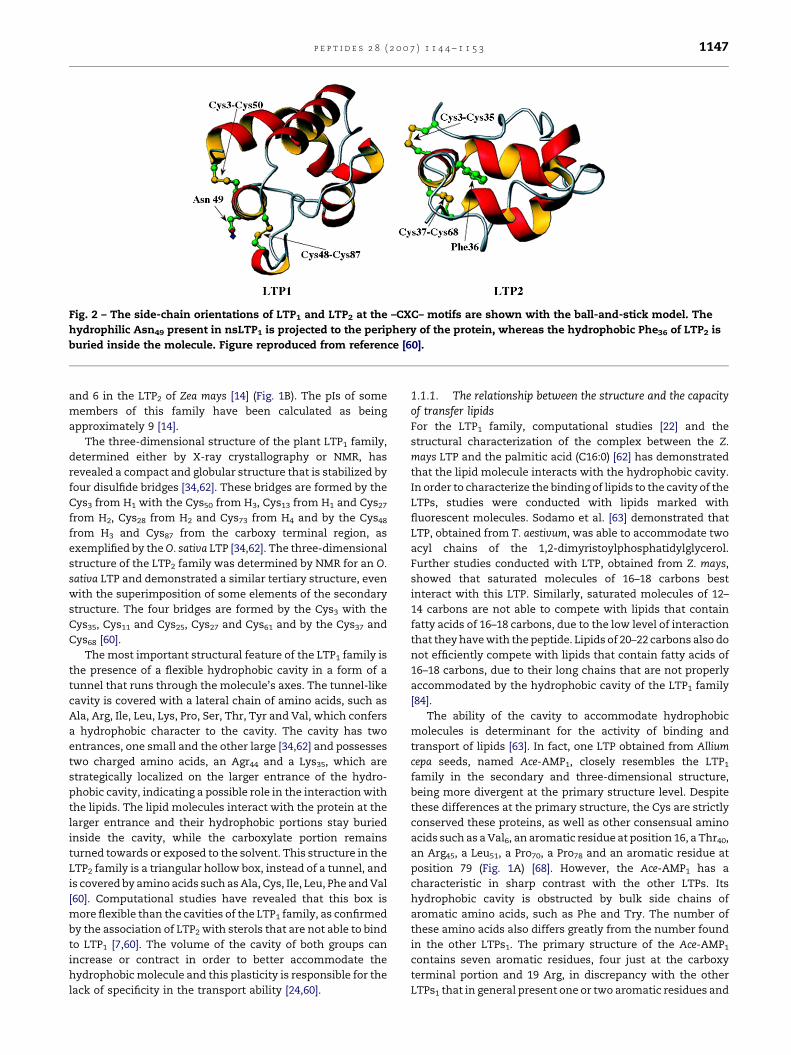
Fig. 2 – The side-chain orientations of LTP1 and LTP2 at the –CXC– motifs are shown with the ball-and-stick model. The
hydrophilic Asn49 present in nsLTP1 is projected to the periphery of the protein, whereas the hydrophobic Phe36 of LTP2 is
buried inside the molecule. Figure reproduced from reference [60].
p e p t i d e s 2 8 ( 2 0 0 7 ) 1 1 4 4 – 1 1 5 3 1147
and 6 in the LTP2 of Zea mays [14] (Fig. 1B). The pIs of some
members of this family have been calculated as being
approximately 9 [14].
The three-dimensional structure of the plant LTP1 family,
determined either by X-ray crystallography or NMR, has
revealed a compact and globular structure that is stabilized by
four disulfide bridges [34,62]. These bridges are formed by the
Cys3 from H1 with the Cys50 from H3, Cys13 from H1 and Cys27
from H2, Cys28 from H2 and Cys73 from H4 and by the Cys48
from H3 and Cys87 from the carboxy terminal region, as
exemplified by the O. sativa LTP [34,62]. The three-dimensional
structure of the LTP2 family was determined by NMR for an O.
sativa LTP and demonstrated a similar tertiary structure, even
with the superimposition of some elements of the secondary
structure. The four bridges are formed by the Cys3 with the
Cys35, Cys11 and Cys25, Cys27 and Cys61 and by the Cys37 and
Cys68 [60].
The most important structural feature of the LTP1 family is
the presence of a flexible hydrophobic cavity in a form of a
tunnel that runs through the molecule’s axes. The tunnel-like
cavity is covered with a lateral chain of amino acids, such as
Ala, Arg, Ile, Leu, Lys, Pro, Ser, Thr, Tyr and Val, which confers
a hydrophobic character to the cavity. The cavity has two
entrances, one small and the other large [34,62] and possesses
two charged amino acids, an Agr44 and a Lys35, which are
strategically localized on the larger entrance of the hydro-
phobic cavity, indicating a possible role in the interaction with
the lipids. The lipid molecules interact with the protein at the
larger entrance and their hydrophobic portions stay buried
inside the cavity, while the carboxylate portion remains
turned towards or exposed to the solvent. This structure in the
LTP2 family is a triangular hollow box, instead of a tunnel, and
is covered by amino acids such as Ala, Cys, Ile, Leu, Phe and Val
[60]. Computational studies have revealed that this box is
more flexible than the cavities of the LTP1 family, as confirmed
by the association of LTP2 with sterols that are not able to bind
to LTP1 [7,60]. The volume of the cavity of both groups can
increase or contract in order to better accommodate the
hydrophobic molecule and this plasticity is responsible for the
lack of specificity in the transport ability [24,60].
1.1.1. The relationship between the structure and the capacityof transfer lipidsFor the LTP1 family, computational studies [22] and the
structural characterization of the complex between the Z.
mays LTP and the palmitic acid (C16:0) [62] has demonstrated
that the lipid molecule interacts with the hydrophobic cavity.
In order to characterize the binding of lipids to the cavity of the
LTPs, studies were conducted with lipids marked with
fluorescent molecules. Sodamo et al. [63] demonstrated that
LTP, obtained from T. aestivum, was able to accommodate two
acyl chains of the 1,2-dimyristoylphosphatidylglycerol.
Further studies conducted with LTP, obtained from Z. mays,
showed that saturated molecules of 16–18 carbons best
interact with this LTP. Similarly, saturated molecules of 12–
14 carbons are not able to compete with lipids that contain
fatty acids of 16–18 carbons, due to the low level of interaction
that they have with the peptide. Lipids of 20–22 carbons also do
not efficiently compete with lipids that contain fatty acids of
16–18 carbons, due to their long chains that are not properly
accommodated by the hydrophobic cavity of the LTP1 family
[84].
The ability of the cavity to accommodate hydrophobic
molecules is determinant for the activity of binding and
transport of lipids [63]. In fact, one LTP obtained from Allium
cepa seeds, named Ace-AMP1, closely resembles the LTP1
family in the secondary and three-dimensional structure,
being more divergent at the primary structure level. Despite
these differences at the primary structure, the Cys are strictly
conserved these proteins, as well as other consensual amino
acids such as a Val6, an aromatic residue at position 16, a Thr40,
an Arg45, a Leu51, a Pro70, a Pro78 and an aromatic residue at
position 79 (Fig. 1A) [68]. However, the Ace-AMP1 has a
characteristic in sharp contrast with the other LTPs. Its
hydrophobic cavity is obstructed by bulk side chains of
aromatic amino acids, such as Phe and Try. The number of
these amino acids also differs greatly from the number found
in the other LTPs1. The primary structure of the Ace-AMP1
contains seven aromatic residues, four just at the carboxy
terminal portion and 19 Arg, in discrepancy with the other
LTPs1 that in general present one or two aromatic residues and
p e p t i d e s 2 8 ( 2 0 0 7 ) 1 1 4 4 – 1 1 5 31148
three or five Arg, respectively (Fig. 1A). This obstruction
explains why this peptide is not able to bind and transport free
lipids [10], but it is noteworthy that this peptide is still able to
interact with lipids in membranes [68].
The primary structures of LTP1 present some amino acids
that are relatively conserved. Of these an aromatic residue, a
Try at the carboxyl terminal region at approximately position
79, is of particular note (Fig. 1A). In the three-dimensional
structure, this residue is positioned at the larger entrance of
the hydrophobic cavity and it has been shown that it interacts
with fatty acids and stabilizes the binding between the peptide
and the hydrophobic molecule by a hydrogen bond that is
formed between the hydroxyl of the Try and the carboxyl
group of the polar head of the lipid [24]. The fatty acids must
have between 16 and 18 carbons to reach this Try residue,
possibly explaining the preference of binding in relation to the
stability of the molecules of such size and also why capric acid
(C10:0) binds to maize LTP in two orientations, with the
carboxyl group towards the interior of the protein or towards
the exterior. Since the molecule is small and remains
extremely concealed inside the cavity, it does not reach the
Try and, thus, does not form the hydrogen bond that stabilizes
the larger molecules with their polar head always turned
towards the exterior [24]. These specificities also explain why
fatty acids larger than 20 and 22 carbons do not interact so
strongly with LTPs1.
Another residue of note is the Ala that is relatively
conserved at position 66 in the primary structure of LTPs1
(Fig. 1A). A hydrogen bond between this Ala and the hydroxyl
group of fatty acids, such as ricinoleate (C18:1, 9, 12-OH)
stabilizes the binding of hydroxy-fatty acids. The relevance of
this fact is that it indicates that these proteins might be really
involved with the cutin synthesis that is composed of fatty
acids with hydroxyl and cetoxyl groups [24,25]. In the case of
the LTP2 family, these characterizations have not yet been
performed.
1.2. Localization and gene expression
Different possible functions have been proposed for plant
LTPs. The genetic structure of LTP1 indicates the presence of
different genes codifying LTPs that possess different expres-
sion patterns and possibly different functions as well. Assays
performed on O. sativa, for example, demonstrate that at least
three genes codify LTPs [79]. Similarly, in S. vulgare, the
presence of at least five genes codifying these peptides has
also been demonstrated; two of these genes have been
characterized and denominated as ltp1 and ltp2 [54]. In C.
annuum, another three genes have been identified [27], indeed
LTP is codified by several genes that belong to a multigene
family, as demonstrated in A. thaliana and O. sativa [2,80]. The
analysis of where, when and how the LTP genes are expressed
maybe of paramount importance to the understanding of their
function in vivo.
The extracellular location of LTP is not a general rule;
advances in the study of LTPs1 have revealed atypical
localizations, such as in R. communis seeds where a LTP
isoform has been found inside an organelle, which was
characterized as the glyoxosome. This LTP seems to increase
the activity of the acetyl-CoA oxidase enzyme in in vitro tests,
indicating a presumed involvement in b-oxidation, possibly in
the regulation of the catabolism of lipid storage [72]. In T.
aestivum seeds, the presence of a LTP has also been demon-
strated inside the alleurone granules that are rich in proteins,
and differently from other plants, LTP was not detected in the
seeds’ cell walls [19]. The presence of the LTP was also
demonstrated inside protein storage vacuoles in V. unguiculata
seeds [11,12] and the physiological role of these peptides in
these organelles requires further investigation.
In B. oleracea var. italica, LTP was found associated with the
waxy surface of the leaves. The pattern of expression of these
peptides demonstrated that they are expressed at high levels
in young leaves, constituting 50% of leafy proteins, and as the
leaves become older, the level of expression drops to 4%. This
expression pattern suggests a role of the LTP in the transport
of monomers of cutin, necessary during the expansion of the
leaf and the formation of cutin [56]. The ltp1 gene of A. thaliana
was shown to be highly expressed in young developing tissues
and its expression diminished in fully expanded tissues, this
pattern is consistent with a role in deposition of cuticular
material and reinforced by the observation of the this gene
expression in the petal and sepal abscission zone, where
additional structural materials are expected to be deposited to
seal off the abscission zone [70]. In embryonic cells of D. carota,
involvement of a LTP was also suggested in the deposition of
monomers of cutin, necessary for the formation of a lipophilic
layer around the embryo [65].
The expression of LTPs1 in flowers or flower organs of
different plant species is noteworthy, as demonstrated by Pyee
et al. [56], Soufleri et al. [64], Suelves and Puigdomenech [67],
Botton et al. [5], Jung et al. [27] and Yubero-Serrano et al. [83],
especially since the expression of flower LTP is related to a
possible facilitation in association with another uncharacter-
ized protein, of pollen adherence to the stigma during pollen
elongation in the Lilium longiflorum [48]. Despite these reports,
some exceptions to LTP1 expression in flowers have been
demonstrated by Vignols et al. [79], Liu et al. [38] and Carvalho
et al. [13] and further analyses will evaluate the relevance of
such a negative expression.
Differential transcription levels of LTP genes have been
shown in a variety of plants and plant tissues during diverse
developmental stages and physiological conditions [2,64,80].
LTP genes are also responsive to environmental changes such
as drought, cold, salt stress and also infection with bacterial
and fungal pathogen [26,27]. Signal molecules such as abscisic
acid, salicylic acid, ethylene and methyl jasmonate are
involved in the signaling pathway responsible for the
expression of LTP genes [20,27–29].
The role of LTPs in the defense mechanisms of plants has
been investigated either by studying the activity of purified
proteins [10,42,21,61], or through the expression pattern of
LTPs genes following the response to pathogen infection
[27,47]. Transgenic A. thaliana and Nicotiana tabacum plants,
expressing a barley LTP, demonstrate enhanced tolerance to
pathogen infection. In tobacco, the growth of the bacterium
Pseudomonas syringae pv. tabaci was retarded in comparison to
the non-transformed control plants and the percentage of
infection points that became necrotic lesions was reduced to
38%. The average size of those lesions was also reduced to 61–
81% in regard to the control. In A. thaliana, the transformed
p e p t i d e s 2 8 ( 2 0 0 7 ) 1 1 4 4 – 1 1 5 3 1149
plant demonstrated a reduction of 22–38% in the number of
infection points that became necrotic lesions in comparison to
the non-transformed plants and that the average lesion sizes
were 53–67% smaller than the control plants when infected
with the bacterium, P. syringae pv. tomato [43]. Transgenic A.
thaliana, over-expressing a CALTP1 from C. annuum, had an
enhanced resistance to P. syringae pv. tomato and the fungus
Botrytis cinerea, both with smaller lesions in comparison with
control plants. This transgenic plant also exhibited high levels
of tolerance to NaCl and drought stresses [28]. Transgenic
wheat plants, over-expressing Ace-AMP1, showed disease
resistance towards Blumeria graminis f. sp. tritici and Neovossia
indica [59]. Inspired on the work of Maldonado et al. [40], these
authors accessed the effect of Ace-AMP1 over-expression on
the induction of defense-related genes such as phenylalanine
ammonia lyase (PAL), PR-2 and PR-3. These genes were
induced, as well as salicylic acid, a product of the phenylpro-
panoid pathway in which PAL is a key enzyme [59]. These data
strongly corroborate the defense function of LTPs against
biotic and abiotic stresses.
Studies performed with in situ hybridization, promoter
fusion with b-glucoronidase (GUS) and Northern blotting with
different plant tissues, demonstrate that the genes that codify
LTPs present complex temporal and spatial control. In A.
thaliana, the localization of the expression of the ltp1 gene was
studied by promoter fusion with the GUS. This promoter was
shown to be active in protoderm cells of embryos at the heart
stage, in vascular tissues, shoot meristems and stipules during
early development. In emerged seedlings, its activity was
observed in cotyledons and hypocotyls near the root. In adult
plants, GUS activity was determined in epidermal cells of
young leaves, stem, inflorescence, in external layers of the
ovule, stigma, petals and sepals, demonstrating that the ltp1
gene is under tissue-specific developmental regulation.
Analysis of the promoter sequence showed that it contains
regions with high homology with conserved regions of genes
of the phenylpropanoid pathway, such as PAL and chalcone
synthase genes. This study indicates that the ltp1 gene might
be regulated by similar mechanisms involved in biotic or
abiotic stress stimuli [70].
Three cDNAs clones that codify LTPs in B. napus were
isolated and their expressions were detected only in
cotyledons and hypocotyl of seedlings. It has also been
demonstrated that the level of expression of these genes
increases in response to treatment with abscisic acid and
sodium chlorate [64]. Three LTP cDNAs, CALTPI, CALTPII and
CALTPIII, were identified from a pepper (C. annuum) cDNA
library prepared from hypersensitive lesions of leaves
infected with the bacterium Xanthomonas campestris pv.
vesicatoria. These are differentially expressed in leaves, stem
and fruit tissues in response to X. campestris pv. vesicatoria,
Phytophthora capsiciandColletotrichumgloeosporioides infection.
CALTPI and CALTPIII had a similar pattern of induction with
different pathogens and were also induced in a similar
manner by drought, high salinity, low temperature and
wounding, as well as by the hormones involved in biotic and
abiotic stresses, such as ethylene, methyl jasmonate and
abscisic acid. In contrast, CALTPII was not induced byP. capsici
and C. gloeosporioides and only high salinity induced its
expression [27].
Jung et al. [28] reported the presence of a regulatory
elements binding site in the promoter of the CALTPI gene from
C. annuum, among them were LTRE-1 (low temperature
responsive element), DPBF (drought responsive element), E-
box (involved in the response of plant–pathogen interaction),
W-box (pathogen-responsive element) and ERE box (ethylene-
responsive element). Jung et al. [29] also reported the presence
of an ERE box, a W box and MYB core elements (involved in
water stress) in the promoter region of the CALTPIII gene from
C. annuum. Both these studies corroborate with the initial
report of Jung et al. [27]. Yubero-Serrano et al. [83] also found
cis-regulatory elements in the promoter region of a LTP (Fxaltp)
gene in Fragaria ananassa, these include the ABRE, E-box, LTRE
and MYB-MYC responsive elements.
In the case of the LTP2 family, ltp2 gene transcripts were
demonstrated to be accumulated strongly in the dry seeds of
rice plants; however no transcripts were detected in roots or
shoots of seedlings. This gene also reacted to treatment with
abscisic acid and agents that provoke osmotic stress, such as
sodium chlorate and mannitol. The seedlings that received
treatment with these substances demonstrated increased ltp2
gene expression in roots compared to shoots. The analysis of
the gene’s promoter revealed the presence of cis-regulatory
elements, such as MYB, MYC, RY repeats and ABRE [20].
These reports also demonstrated the functionality of cis-
regulatory elements. Taken together, these results indicate
that LTPs may be responsible for the plant’s adaptation to
stress conditions, especially to those provoked by water stress.
1.3. Biological activities
1.3.1. Plant signallingRecently, the hypothesis that LTPs could have a role, or at least
be involved, in plant defense signaling emerged. Maldonado
et al. [40], working with an A. thaliana T-DNA tagged mutant,
screened one especially compromised mutant for the devel-
opment of systemic acquired resistance (SAR), denominated
dir1-1, for defective in induced resistance. This mutant
exhibited unaffected local resistance against virulent or
avirulent strains of Pseudomonas syringae, but was unable to
express the PR-1 gene in uninfected distant leaves and also to
develop SAR against virulent P. syringae and Perenospora
parasitica. The failure of this A. thaliana mutant to express
the PR-1 gene in uninoculated distant leaves, as well as SAR,
indicates that the product of the dir1-1 gene appears to be
required for long-distance signaling during SAR for signal
generation or transmission or even for signal perception level.
To try to differentiate between these hypotheses, this
group performed petiole exudate experiments and demon-
strated that the mutant is defective in the generation or
transmission of the signal. In addition, super-expression of
dir1-1 did not induce SAR, implying that dir1-1 is probably not
the mobile signal itself. The dir1-1 gene encodes a sequence of
102 amino acids, which shows homology with a putative LTP1.
The authors proposed that this putative LTP interacts with a
lipid-derived molecule to function as a long distance signal
complex [40].
Providing support to the involvement of LTPs in plant
signaling, it has been shown that a LTP from T. aestivum is able
to bind with a high affinity rate to the tobacco plasma
p e p t i d e s 2 8 ( 2 0 0 7 ) 1 1 4 4 – 1 1 5 31150
membrane [7]. The binding site was the same as that used by
elicitins, as demonstrated by binding and in vivo competition
experiments. Elicitins are peptides of approximately 10 kDa,
secreted by oomycetes and belonging to the gender Phy-
tophthora or Pythium; these peptides induce a hypersensitive
reaction and SAR in tobacco plants [55] and resemble plant
LTPs in some biochemical characteristics, such as small size
(98 amino acids and 10 kDa), are basic, possess three disulfide
bounds and have an a-helix secondary structure and also have
a hydrophobic pocket that endows them with the capacity to
bind hydrophobic molecules, mainly sterols. Despite these
similarities, the primary structure homology is low between
the two peptides, but at the tertiary level there are super-
impositions of some helixes [4,7,41].
Buhot et al. [8] revealed that a recombinant LTP1 from N.
tabacum is able to bind jasmonic acid (JA), and the complex
between LTP1 and JA is able to bind the elicitin receptor. The
authors also suggested that the formation of the complex
provokes a conformational change on LTP that facilitates its
recognition by the receptor. This LTP1-JA complex, when
applied to the N. tabacum plant, induces long distance
protection against P. parasitica. However, it has not been
demonstrated whether the complex is the mobile signal or
whether the binding of the complex on the plasma membrane
receptor is the requirement for the production of the mobile
signal [8].
1.3.2. Antimicrobial activityThe antimicrobial activity of the LTPs1 was discovered by the
screening of proteic extracts of plants, in order to find proteins
that could inhibit the growth of phytopathogens, in vitro [42].
Among the phytopathogens inhibited were bacteria and fungi,
however the activity was stronger against fungi [31]. Molina
et al. [42] isolated four LTPs from barley leaves and one from Z.
mays leaves and all of them presented biological activity
against the bacteria, Clavibacter michiganensis subsp. sepedoni-
cus and Rhalstonia (Pseudomonas) sonanacearum, and the
Fusarium solani fungus. Two other peptides that are homo-
logues of the LTPs, obtained from A. thaliana leaves, and two
others from Spinacia oleracea leaves, also demonstrated
antimicrobial activity against the aforementioned pathogens
[61]. Wang et al. [81] demonstrated the antimicrobial activity of
a LTP isolated from mung bean seeds against the fungi, F.
solani, F. oxysporum, Pythium aphanidermathum and Sclerotium
rolfsii and also against the Gram positive bacterium, Staphy-
lococcus aureus. In regard to human pathogens, two LTPs
isolated from Pandanus amaryllifolius did not inhibited S. aureus
as other Gram negative enteric bacteria, namely Escherichia coli,
Enterobacter aerugenes, Proteus vulgaris, Vibrio cholera, V. para-
haemolyticus and Salmonella typhimurium. The only Gram
negative bacterium inhibited was Pseudomonas aeruginosa
[45]. The human infectious yeast Candida albicans was also
not inhibited by LTPs isolated from P. amaryllifolius [45] and
Hordeum vulgare [23]. Albeit plant LTPs have been considered
an antimicrobial peptide it had been reported that some LTPs1
present low or did not present antifungal activity, among them
are examples from T. aestivum [19] and Z. mays [10].
The antimicrobial activity, sequence similarities and
induction upon pathogen attack have led to the inclusion of
these peptides in the family of pathogenesis-related proteins
that compose the family 14 [74]. The activity of the LTPs seems
to depend on the microorganism tested, for example, one LTP
from O. sativa leaves, expressed in E. coli, presented activity
against the Pyricularia oryzae fungus at concentrations of
27 mg mL�1. This LTP also inhibited the bacteria, Pseudomonas
syringae, at the same concentrations, but did not present
inhibitory activity agaisnt Xanthomonas oryzae, except a delay
in its growth [21].
The most potent peptide belonging to the LTP class was
obtained from onion seeds, the above mentioned Ace-AMP1
[10]. This peptide was able to inhibit all of the 12 fungi tested
and the Gram positives bacteria, Bacillus megateruim and
Sarcina lutea, at concentrations below of 10 mg mL�1. As already
demonstrated with other LTPs, this peptide did not present
activity against Gram-negative tested bacteria [10]. Despite its
strong antimicrobial activity, Ace-AMP did not presented
toxicity against mammal cells (fibroblasts) or cause hemolysis
of erythrocytes until concentrations of 200 mg mL�1 were
reached, the same lack of cytotoxicity against mammals was
demonstrated for LTPs from other plant species [10,21].
Differently to the other LTPs, this peptide was not able to
bind and transport hydrophobic molecules as mentioned
above [10]. The example of Ace-AMP1 demonstrates that the
binding and transport activities of lipids may not be directly
associated or correlated with the ability of interaction with
membranes and, in this case, with the antimicrobial activity.
This example also reflects that the interaction of LTPs with
membranes is not as well understood as the activity of binding
and transport hydrophobic molecules.
Since the discovery of the LTPs as peptides with the capacity
to inhibit phytopathogens, it has been speculated that this
effect could result from the interaction of the LTPs with
biological membranes, possibly leading to the permeabilization
due to loss of membrane integrity [31]. Indeed LTPs1 have been
shown to interact with model membranes, such as monolayers
composed of dipalmitoilphosphatidylglycerol [66] and large
unilamellar vesicles filled with fluorescent dyes [9]. It has been
recently demonstratedthata fraction enriched onLTP,obtained
from chilli pepper seeds, inhibited the growth of Saccharomyces
cerevisiae, C. albicans and Schysosaccharomyces pombe at concen-
trations of 9–150 mg mL�1. This same report demonstrated that
the fraction containing the chilli pepper LTP is able to
permeabilize yeast plasma membrane and allow the entrance
of the small dye (900 Da), SYTOX Green (Molecular Probes), a
high affinity nucleic acid stain that fluoresces upon binding to
nucleic acids and that only penetrates cells with compromised
plasma membranes [17]. Another LTP, isolated form sunflower
seeds [57], completely abrogated the growth of F. solani spores at
40 mg mL�1, decreasing the viability of these cells to a lethal
condition at this concentration. The Helianthus annuus LTP is
able to permeabilize the membranes of F. solani spores, as also
demonstrated by the SYTOX Green permeabilization assay [58].
Nevertheless, these results validate the information obtained
from artificial membranes and liposomes, the mechanism of
action on these target organisms as well the antimicrobial
properties of the LTP2 family have not yet been elucidated.
1.3.3. As food allergensSeveral reports have unambiguously suggested that the major
allergens of diverse plant species are proteins members of
p e p t i d e s 2 8 ( 2 0 0 7 ) 1 1 4 4 – 1 1 5 3 1151
LTPs1 family, such peptides have been reported in fruits of
Rosaceae [15,49,51] in fruits of Vitaceae [52] as well as in other
plant species such as Aspargus officinalis, B. oleracea var. capitata
and Z. mays [16,46,50,75]. It has been demonstrated that LTPs
are relatively stable, resisting thermal and chemical dena-
turation and enzymatic digestion [3,37,53,76].
These stable physical–chemical features allow these pep-
tides to reach the intestine of mammals in an immunenic form.
Although the ability of LTPs to sensibilize via the gastrointest-
inal tract is not fully understood yet, it is supposed that once
they are present in the gastrointestinal tract and in an
immunogenic form they are free to interact with the intestinal
immune system of sensitizing the individuals. Allergenic
proteins share some characteristics, such as the ability to bind
to ligands, as related in parvalbumin from fish and casein from
milk, both bind Ca+2 ions and seem to remain more stable after
this binding [6,73]. In the case of LTP, peptides bind to
phosphatidylcholine, a physiological surfactant that is secreted
by gastric mucosa and also occurs in bile. It has also shown that
this binding results in an additional enzymatic protection,
slowing down the breakdown of the grape LTP [76].
The understanding of the mechanism by which LTPs cause
allergy may allow the possibility understand the mechanisms
of other allergenic proteins, especially for assessing how
allergenic a given protein in new foods could be or when used
in the development of allergen variants with reduced side
effects that could then be used as vaccines and also for the
development of a reliable method for diagnosis [35].
2. Conclusion
Finally, LTPs are peptides that still do not possess a single
consensus in relation to their physiological role, in vivo.
Despite all the information related, herein, a definitive
biological function has not been conclusively provided for
these peptides. Investigations studying biological functions in
plants that bear antisense transcripts to the LTP genes should
yield further insights; however, studies to date have proved to
be particularly complicated, since these peptides comprehend
a multigenic family with different genes that are expressed in
different tissues, in different development stages of plants and
that also react differently to an array of stimuli [20,31,64].
Acknowledgements
This project was supported by the Brazilian agency CNPq,
FAPERJ, FENORTE/TECNORTE and International Foundation
for Science, Stockholm, Sweden, through a grant to C/2806–3F.
This work is part of fellowship of Andre O. Carvalho carried out
at the Universidade Estadual do Norte Fluminense through a
fellowship to FAPERJ (E-26/150-015/2006).
r e f e r e n c e s
[1] Arondel V, Tchang F, Baillet B, Vignols F, Grellet F, DelsenyM, et al. Multiple mRNA coding for phospholipid-transfer
protein form Zea mays arise from alternative splicing. Gene1991;99:133–6.
[2] Arondel V, Vergnolle C, Cantre C, Kader J-C. Lipid transferproteins are encoded by a small multigene family inArabidopsis thaliana. Plant Sci 2000;157:1–12.
[3] Asero R, Mistrello G, Roncarolo D, de Vries SC, Gautier M-F,Ciurana CLF, et al. Lipid transfer protein: a pan-allergen inplant-derived foods that is highly resistant to pepsindigestion. Int Arch Allergy Immunol 2000;122:20–32.
[4] Blein J-P, Coutos-Thevenot P, Marion D, Ponchet M. Fromelicitins to lipid-transfer proteins: a new insight in cellsignalling involved in plant defence mechanisms. TrendsPlant Sci 2002;7:293–6.
[5] Botton A, Begheldo M, Rasori A, Bonghi C, Tonutti P.Differential expression of two lipid transfer protein genesin reproductive organs of peach (Prunus persica L. Batsch).Plant Sci 2002;163:993–1000.
[6] Breiteneder H, Mills ENC. Molecular properties of foodallergens. J Allergy Clin Immunol 2005;115:14–23.
[7] Buhot N, Douliez J-P, Jacquemard A, Marion D, Tran V,Maume BF, et al. A lipid transfer protein binds to a receptorinvolved in the control of plant defence responses. FEBSLett 2001;509:27–30.
[8] Buhot N, Gomes E, Milat M-L, Ponchet M, Marion D, LequeuJ, et al. Modulation of the biological activity of a tobaccoLTP1 by lipid complexation. Mol Biol Cell 2004;15:5047–52.
[9] Caaveiro JMM, Molina A, Gonzalez-Manas JM, Rodrıguez-Palenzuela P, Garcıa-Olmedo F, Goni FM. Differential effectsof five types of antipathogenic plant peptides on modelmembranes. FEBS Lett 1997;410:338–42.
[10] Cammue BPA, Thevissen K, Hendriks M, Eggermont K,Goderis IJ, Proost P, et al. A potent antimicrobial proteinfrom onion seeds showing sequence homology to plantlipid transfer proteins. Plant Physiol 1995;109:445–55.
[11] Carvalho AO, Machado OLT, Da Cunha M, Santos IS, GomesVM. Antimicrobial peptides and immunolocalization of aLTP in Vigna unguiculata seeds. Plant Physiol Biochem2001;39:137–46.
[12] Carvalho AO, Teodoro CES, Da Cunha M, Okorokova-Facanha AL, Okorokov LA, Fernandes KVS, et al.Intracellular localization of a lipid transfer protein in Vignaunguiculata seeds. Physiol Plant 2004;122:328–36.
[13] Carvalho AO, Souza-Filho GA, Ferreira BS, Branco AT,Araujo IS, Fernandes KVS, Retamal CA, Gomes VM. Cloningand characterization of a cowpea seed lipid transfer proteincDNA: expression analysis during seed development andunder fungal and cold stresses in seedlings’ tissues. PlantPhysiol Biochem 2006;44:732–42.
[14] Castro MS, Gerhardt IR, Orru S, Pucci P, Bloch Jr C.Purification and characterization of a small (7.3 kDa)putative lipid transfer protein from maize seeds. JChromatogr B 2003;794:109–14.
[15] Dıaz-Perales A, Garcia-Casado G, Sanchez-Monge R, Garcia-Salles FJ, Barber D, Salcedo G. cDNA cloning andheterologous expression of the major allergens from peachand apple belonging to the lipid-transfer protein family.Clin Exp All 2002;32:87–92.
[16] Dıaz-Perales A, Tabar AI, Sanchez-Monge R, Garcıa BE,Gomez B, Barber D, et al. Characterization of asparagusallergens: a relevant role of lipid transfer proteins. J AllergyClin Immunol 2002;110:790–6.
[17] Diz MSS, Carvalho AO, Rodrigues R, Neves-Ferreira AGC, DaCunha M, Alves EW, et al. Antimicrobial peptides fromchilli pepper seeds causes yeast plasma membranepermeabilization and inhibits the acidification of themedium by yeast cells. Biochim Biophys Acta2006;1760:1323–32.
[18] Douliez J-P, Pato C, Rabesona H, Molle D, Marion D.Disulfide bond assignment, lipid transfer activity and
p e p t i d e s 2 8 ( 2 0 0 7 ) 1 1 4 4 – 1 1 5 31152
secondary structure of a 7-kDa plant lipid transfer protein,LTP2. Eur J Biochem 2001;268:1400–3.
[19] Dubreil L, Gaborit T, Bouchet B, Gallant DJ, Broekaert WF,Quillien L, et al. Spatial and temporal distribution of themajor isoforms of puroindolines (puroindoline-a andpuroindoline-b) and non-specific lipid transfer protein (ns-LTPe1) of Triticum aestivum seeds. Relationships with theirin vitro antifungal properties. Plant Sci 1998;138:121–35.
[20] Garcıa-Garrido JM, Menossi M, Puigdimenech P, Martınez-Izquierdo JA, Delseny M. Characterization of a geneencoding an abscisic acid-inducible type-2 lipid transferprotein from rice. FEBS Lett 1998;428:193–9.
[21] Ge X, Chen J, Li N, Lin Y, Sun C, Cao K. Resistance functionof rice lipid transfer protein LTP110. J Biochem Mol Biol2003;36:603–7.
[22] Gomar J, Sodamo P, Sy D, Shin DH, Lee JY, Shu SW, et al.Comparison of solution and crystal structures of maizenonspecific lipid transfer protein: a model for a potential invivo lipid carrier protein. Proteins 1998;31:160–71.
[23] Gorjanovic S, Spillner E, Beljanski MV, Gorjanovic R,Pavlovic M, Gojgic-Cvijanovic G. Malting barley grain non-specific lipid-transfer protein (ns-LTP): importance forgrain protection. J Inst Brew 2005;111(2):99–104.
[24] Han GW, Lee JY, Song HK, Chang C, Min K, Moon J, et al.Structural basis of non-specific lipid binding in maize lipid-transfer protein complexes revealed by high-resolution X-ray crystallography. J Mol Biol 2001;308:263–78.
[25] Heredia A. Biophysical and biochemical characteristics ofcutin, a plant barrier biopolymer. Biochim Biophys Acta2003;1620:1–7.
[26] Jang CS, Lee HJ, Chang SJ, Seo YW. Expression andpromoter analysis of the TaLTP1 gene induced by droughtand salt stress in wheat (Triticum aestivum L). Plant Sci2004;167:995–1001.
[27] Jung HW, Kim W, Hwang BK. Three pathogen-induciblegenes encoding lipid transfer protein from pepper aredifferentially activated by pathogens, abiotic andenvironmental stresses. Plant Cell Environ 2003;26:915–28.
[28] Jung HW, Kim KD, Hwang BK. Identification of pathogen-responsive regions in the promoter of a pepper lipidtransfer protein gene (CALTPI) and the enhanced resistanceof the CALTPI transgenic Arabidopsis against pathogenand environmental stresses. Planta 2005;221:361–73.
[29] Jung HW, Lim CW, Hwang BK. Isolation and functionalanalysis of a pepper lipid transfer protein III (CALTPIII) genepromoter during signaling to pathogen, abiotic andenvironmental stresses. Plant Sci 2006;170:258–66.
[30] Kader J-C. Proteins and the intracellular exchange of lipids:stimulation of phospholipid exchange betweenmitochondria and microssomal fractions by proteinsisolated from potato tuber. Biochim Biophys Acta1975;380:31–44.
[31] Kader J-C. Lipid-transfer proteins in plants. Annu Rev PlantPhysiol Plant Mol Biol 1996;47:627–54.
[32] Kalla R, Shimamoto K, Potter R, Nielsen PS, Linnestad C,Olsen O-A. The promoter of the barley aleurone-specificgene encoding a putative 7 kDa lipid transfer proteinconfers aleurone cell-specific expression in transgenic rice.Plant J 1994;6:849–60.
[33] Kristensen AK, Brunstedt J, Nielsen KK, Roepstorff P,Mikkelsen JD. Characterization of a new antifungal non-specific lipid transfer protein (nsLTP) from sugar beetleaves. Plant Sci 2000;155:31–40.
[34] Lee JY, Min K, Cha H, Hwang DHSKY, Suh SW. Rice non-specific lipid transfer protein: the 1.6 A crystal structure inthe unliganded state reveals a small hydrophobic cavity. JMol Biol 1998;276:437–48.
[35] Lehrer SB, Bannon GA. Risks of allergic reactions to biotechproteins in foods: perception and reality. Allergy2005;60:559–64.
[36] Lin K-F, Liu Y-N, Hsu S-TD, Samuel D, Cheng C-S, BonvinAMJJ, et al. Characterization and structural analyses ofnonspecific lipid transfer protein 1 from mung bean.Biochemistry 2005;44:5703–12.
[37] Lindorff-Larsen K, Winther JR. Surprisingly high stability ofbarley lipid transfer protein, LTP1, towards denaturant,heat and proteases. FEBS Lett 2001;488:145–8.
[38] Liu K, Jiang H, Moore SL, Watkins CB, Jahn MM. Isolationand characterization of a lipid transfer protein expressed inripening fruit of Capsicum chinense. Planta 2006;223:672–83.
[39] Liu Y-J, Samuel D, Lin C-H, Lyu P-C. Purification andcharacterization of a novel 7-kDa non-specific lipid transferprotein-2 from rice (Oryza sativa). Biochem Biophys ResCommun 2002;294:535–40.
[40] Maldonado AM, Doerner P, Dixonk RA, Lamb CJ, CameronRK. A putative lipid transfer protein involved in systemicresistance signalling in Arabidopsis. Nature 2002;419:399–403.
[41] Mikes V, Milat M-L, Ponchet M, Panabieres F, Ricci P, Blein J-P. Elicitins, proteinaceous elicitors of plant defense, are anew class of sterol carrier proteins. Biochem Biophys ResCommun 1998;245:133–9.
[42] Molina A, Segura A, Garcıa-Olmedo F. Lipid transferproteins (nsLTPs) from barley and maize leaves are potentinhibitors of bacterial and fungal plant pathogens. FEBS1993;316:119–22.
[43] Molina A, Garcıa-Olmedo F. Enhanced tolerance to bacterialpathogens caused by the transgenic expression of barleylipid transfer protein LTP2. Plant J 1997;12:669–75.
[44] Moreau P, Bessoule JJ, Mongrand S, Testet E, Vincent P,Cassagne C. Lipid trafficking in plant cells. Prog Lipid Res1998;37:371–91.
[45] Ooi LSM, Wong EYL, Sun SSM, Ooi VEC. Purification andcharacterization of non-specific lipid transfer proteins fromthe leaves of Pandanus maryllifolius (Pandanaceae). Peptides2006;27:626–32.
[46] Palacın A, Cumplido J, Figueroa J, Ahrazem O, Sanchez-Monge R, Carrillo T, et al. Cabbage lipid transfer protein Brao 3 is a major allergen responsible for cross-reactivitybetween plant foods and pollens food and pollen allergies. JAllergy Clin Immunol 2006;117:1423–9.
[47] Park C-J, Shin R, Park JM, Lee G-J, You J-S, Paek K-H.Induction of pepper cDNA encoding a lipid transfer proteinduring the resistance response to tobacco mosaic virus.Plant Mol Biol 2002;48:243–54.
[48] Park SY, Jauh GY, Mollet JC, Eckard KJ, Nothnagel EA,Walling LL, et al. A lipid transfer-like protein is necessaryfor lily pollen tube adhesion to an in vitro stylar matrix.Plant Cell 2000;12:151–64.
[49] Pastorello EA, Farioli L, Pravettoni V, Ortolani C, Ispano M,Monza M, et al. The major allergen of peach (Prunus persica)is a lipid transfer protein. J Allergy Clin Immunol1999;103:520–6.
[50] Pastorello EA, Farioli L, Pravettoni V, Ispano M, Scibola E,Trambaioli C, et al. The maize major allergen, which isresponsible for food-induced allergic reactions, is a lipidtransfer protein. J Allergy Clin Immunol 2000;106:744–51.
[51] Pastorello EA, Farioli L, Pravettoni V, Giuffrida MG, OrtolaniC, Fortunato D, et al. Characterization of the major allergenof plum as a lipid transfer protein. J Chromatogr B2001;756:95–103.
[52] Pastorello EA, Farioli L, Pravettoni V, Ortolani C, FortunatoD, Giuffrida MG, et al. Identification of grape and wineallergens as an endochitinase 4, a lipid-transfer protein,and a thaumatin LTP cross-reacting with the peach majorallergen. J Allergy Clin Immunol 2003;111:350–9.
p e p t i d e s 2 8 ( 2 0 0 7 ) 1 1 4 4 – 1 1 5 3 1153
[53] Pastorello EA, Pompei C, Pravettoni V, Farioli L, CalamariAM, Scibilia J, et al. Lipid-transfer protein is the majormaize allergen maintaining IgE-binding activity aftercooking at 100 degrees C, as demonstrated in anaphylacticpatients and patients with positive double blind, placebo-controlled food challenge results. J Allergy Clin Immunol2003;112:775–83.
[54] Pelese-Siebenbourg F, Caelles C, Kader J-C, Delseny M,Puigdomenech P. A pair of genes coding for lipid-transferproteins in Sorghum vulgare. Gene 1994;148:305–8.
[55] Ponchet M, Panabieres F, Milat M-L, Mikes V, Montillet J-L,Suty L, et al. Are elicitins cryptograms in plant–Oomycetecommunications? CMLS Cell Mol Life Sci 1999;56:1020–47.
[56] Pyee J, Yu H, Kolattukudy PE. Identification of a lipidtransfer protein as the major protein in the surface wax ofbroccoli (Brassica oleracea) leaves. Arch Biochem Biophys1994;311:460–8.
[57] Regente MC, de la Canal L. Purification, characterizationand antifungal properties of a lipid transfer protein fromsunflower (Heliantus annuns) seeds. Physiol Plant2000;110:158–63.
[58] Regente MC, Giudici AM, Villalaın J, de la Canal L. Thecytotoxic properties of a plant lipid transfer protein involvemembrane permeabilization of target cells. Lett ApplMicrobiol 2005;40:183–9.
[59] Roy-Barman S, Sautter C, Chattoo BB. Expression of thelipid transfer protein Ace-AMP1 in transgenic wheatenhances antifungal activity and defense responses.Transgenic Res 2006;15:435–46.
[60] Samuel D, Liu Y-J, Cheng C-S, Lyu P-C. Solution structure ofplant nonspecific lipid transfer protein-2 from rice (Oryzasativa). J Biol Chem 2002;277:35267–73.
[61] Segura A, Moreno M, Garcıa-Olmedo F. Purification andantipathogenic activity of lipid transfer proteins (LTPs)from the leaves of Arabidopsis and spinach. FEBS1993;332:243–6.
[62] Shin DH, Lee JY, Hwang KY, Kim KK, Suh SW. High-resolution crystal structure of the non-specific lipid-transfer protein from maize seedlings. Structure1995;3:189–99.
[63] Sodamo P, Caille A, Sy D, Person G, Marion D, Ptak M. 1HNMR and fluorescence studies of the complexation ofDMPG by wheat non-specific lipid transfer protein. Globalfold of the complex. FEBS Lett 1997;416:130–4.
[64] Soufleri I, Vergnolle C, Miginiac E, Kader J-C. Germination-specific lipid transfer protein cDNAs in Brassica napus L.Planta 1996;199:229–37.
[65] Sterk P, Booij H, Schellekens GA, Van Kammen A, De VriesSC. Cell-specific expression of the carrot EP2 lipid transferprotein gene. Plant Cell 1991;3:907–21.
[66] Subirade M, Marion D, Pezolet M. Interaction of two lipidbinding proteins with membrane lipids: comparative studyusing the monolayer technique and IR spectroscopy. ThinSolid Films 1996;284/285:326–9.
[67] Suelves M, Puigdomenech P. Different lipid transfer proteinmRNA accumulates in distinct parts of Prunus amygdalusflower. Plant Sci 1997;129:49–56.
[68] Tassin S, Broekaert WF, Marion D, Acland DP, Ptak M,Vovelle F, et al. Solution structure of Ace-AMP1, a potentantimicrobial protein extracted from onion seeds.
Structural analogies with plant nonspecific lipid transferproteins. Biochemistry 1998;37:3623–37.
[69] Thoma SL, Kaneko Y, Somerville C. An Arabidopsis lipidtransfer protein is a cell wall protein. Plant J 1993;3:427–37.
[70] Thoma S, Hecht U, Kippers A, Botella J, De Vries S,Somerville C. Tissue-specific expression of a gene encodinga cell wall-localized lipid transfer protein from Arabidopsis.Plant Physiol 1994;105:35–45.
[71] Thompson JD, Higgins DG, Gibson TJ. CLUSTAL W:improving the sensitivity of progressive multiple sequencealignment through sequence weighting, position-specificgap penalties and weight matrix choice. Nucl Acids Res1994;22:4673–80.
[72] Tsuboi S, Osafune T, Tsugeki R, Nishimura M, Yamada M.Nonspecific lipid transfer protein in castor bean cotyledonscells: subcellular localization and a possible role in lipidmetabolism. J Biochem 1992;111:500–8.
[73] Van Do T, Elsayed S, Florvaag E, Hordvik I, Endresen C.Allergy to fish parvalbumins: studies on the cross-reactivityof allergens from 9 commonly consumed fish by some ofthe tested patients. J Allergy Clin Immunol 2005;116:1314–20.
[74] Van Loon LC, Van Strien EA. The family of pathogenesis-related proteins, their activities, and comparative analysisof PR1-type proteins. Physiol Mol Plant Pathol 1999;55:85–97.
[75] van Ree R. Clinical importance of non-specific lipid transferproteins as food allergens. Biochem Soc Trans 2002;30:910–3.
[76] Vassilopoulou E, Rigby N, Moreno FJ, Zuidmeer L,Akkerdaas J, Tassios I, et al. Effect of in vitro gastric andduodenal digestion on the allergenicity of grape lipidtransfer protein. J Allergy Clin Immunol 2006;118:473–80.
[77] Velazhahan R, Radhajeyalakshmi R, Thangavelu R,Muthukrishnan S. An antifungal protein purified from pearlmillet seeds shows sequence homology to lipid transferproteins. Biol Plant 2001;44:417–21.
[78] Vergnolle C, Arondel V, Jolliot A, Kader J-C. Phospholipidtransfer proteins from higher plants. Methods Enzymol1992;209:522–30.
[79] Vignols F, Lund G, Pammi S, Tremousaygue D, Grellet F,Kader J-C, et al. Characterization of a rice gene coding for alipid transfer protein. Gene 1994;142:265–70.
[80] Vignols F, Wigger M, Garcıa-Garrido JM, Grellet F, Kader J-C,Delseny M. Rice lipid transfer protein (LTP) genes belong toa complex multigene family and are differently regulated.Gene 1997;195:177–86.
[81] Wang SY, Wu JH, Ng TB, Ye XY, Rao PF. A non-specific lipidtransfer protein with antifungal and antibacterial activitiesfrom the mung bean. Peptides 2004;25:1235–42.
[82] Yamada M. Lipid transfer proteins in plants andmicroorganisms. Plant Cell Physiol 1992;33(1):1–6.
[83] Yubero-Serrano E-M, Moyano E, Medina-Escobar N, Munoz-Blanco J, Caballero J-L. Identification of a strawberry geneencoding a non-specific lipid transfer protein that respondsto ABA, wounding and cold stress. J Exp Bot2003;54(389):1865–77.
[84] Zachowski A, Guerbette F, Grosbois M, Jolliot-Croquin A,Kader J-C. Characterization of acyl binding by a plant lipid-transfer protein. Eur J Biochem 1998;257:443–8.