revision of phaeosphaeriaceae
TRANSCRIPT
Revision of Phaeosphaeriaceae
Rungtiwa Phookamsak & Jian-Kui Liu & Eric H. C. McKenzie &
Dimuthu S. Manamgoda & Hiran Ariyawansa & Kasun M. Thambugala & Dong-Qin Dai &Erio Camporesi & Ekachai Chukeatirote & Nalin N. Wijayawardene & Ali H. Bahkali &Peter E. Mortimer & Jian-Chu Xu & Kevin D. Hyde
Received: 19 August 2014 /Accepted: 11 September 2014 /Published online: 7 October 2014# School of Science 2014
Abstract Phaeosphaeriaceae is a large and important familyin the order Pleosporales which includes economically im-portant plant pathogens. Species may also be endophytes orsaprobes on plant hosts, especially on monocotyledons (e.g.,Cannaceae, Cyperaceae, Juncaceae, Poaceae); some specieshave also been reported on dicotyledons. The family previ-ously accommodated 35 sexual and asexual genera and com-prised more than 300 species with a range of morphologicalcharacters. The morphological characters of taxa in this familyare often ambiguous and can be confused with other taxa inLeptosphaeriaceae andMontagnulaceae. Fourteen specimensof the type genera of Phaeosphaeriaceae were loaned fromherbaria worldwide and were re-examined and illustrated.Fresh collections were obtained from Italy and Thailand,
characterized, examined, isolated into pure culture and usedto obtain molecular data. The asexual state was induced wherepossible on sterile bamboo pieces placed on water agar.Multigene phylogenetic analyses of ITS, LSU, SSU, RPB2and TEF1 sequence datasets were carried out using maximumlikelihood, maximum parsimony and Bayesian analysis.Molecular analyses shows that 21 genera (Amarenomyces,Ampelomyces, Chaetosphaeronema, Dematiopleospora,En t o d e sm i um , Lo r a t o s p o r a , Neo s e t o p h oma ,Neostagonospora , Nodulosphaeria , Ophiobolus ,Ophiosphaerella , Paraphoma , Parastagonospora ,Phaeosphaeria, Phaeosphaeriopsis, Sclerostagonospora,Setomelanomma, Setophoma, Vrystaatia, Wojnowicia andXenoseptoria) belong in Phaeosphaeriaceae, while seven
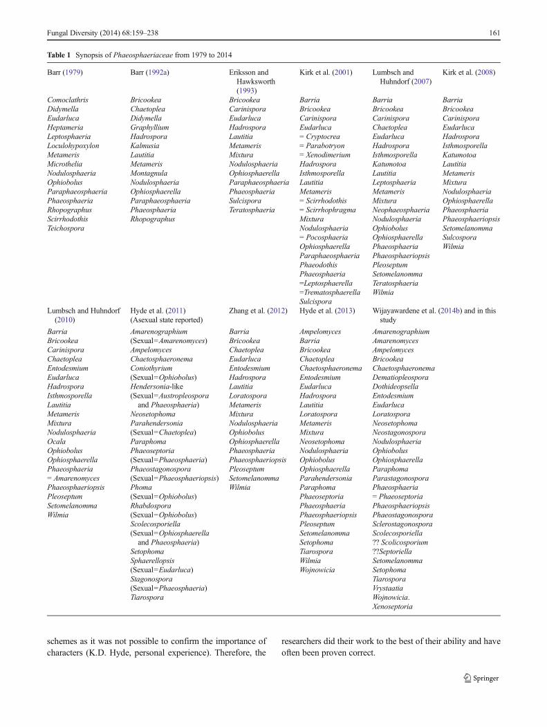
Electronic supplementary material The online version of this article(doi:10.1007/s13225-014-0308-3) contains supplementary material,which is available to authorized users.
R. Phookamsak :D.<Q. Dai : P. E. Mortimer : J.<C. Xu :K. D. Hyde (*)Key Laboratory for Plant Diversity and Biogeography of East Asia,Kunming Institute of Botany, Chinese Academy of Sciences,Kunming 650201, Yunnan, People’s Republic of Chinae-mail: [email protected]
R. Phookamsak :D.<Q. Dai : P. E. Mortimer : J.<C. Xu :K. D. HydeWorld Agroforestry Centre, East and Central Asia,Kunming 650201, Yunnan, People’s Republic of China
R. Phookamsak : J.<K. Liu :D. S. Manamgoda :H. Ariyawansa :K. M. Thambugala :D.<Q. Dai : E. Chukeatirote :N. N. Wijayawardene :K. D. HydeSchool of Science, Mae Fah Luang University, Chiang Rai 57100,Thailand
R. Phookamsak : J.<K. Liu :D. S. Manamgoda :H. Ariyawansa :K. M. Thambugala :D.<Q. Dai : E. Chukeatirote :N. N. Wijayawardene :K. D. HydeInstitute of Excellence in Fungal Research, Mae Fah LuangUniversity, Chiang Rai 57100, Thailand
A. H. Bahkali :K. D. HydeCollege of Science, Botany and Microbiology Department, KingSaud University, Riyadh 1145, Saudi Arabia
E. CamporesiA.M.B. Gruppo Micologico Forlivese “Antonio Cicognani”, ViaRoma 18, Forlì, Italy
E. CamporesiA.M.B. Circolo Micologico “Giovanni Carini”, C.P. 314, Brescia,Italy
E. CamporesiSocietà per gli Studi Naturalistici della Romagna, C.P. 144,Bagnacavallo, RA, Italy
E. H. C. McKenzieManaakiWhenua Landcare Research, Private Bag 92170, Auckland,New Zealand
Fungal Diversity (2014) 68:159–238DOI 10.1007/s13225-014-0308-3
genera (Amarenographium, Bricookea, Dothideopsella,Eudarluca, Phaeostagonospora, Scolecosporiella andTiarospora) are included based on morphological data.Amarenomyces is reinstated and Nodulosphaeria is confirmedin Phaeosphaeriaceae. Eudarluca is distinguished fromSphaerellopsis based on its morphological characters and istypical of Phaeosphaeriaceae. ITS gene phylogenetic analy-s i s i n d i c a t e s t h a t Spha e re l l o p s i s be l o ng s t oLeptosphaeriaceae. Ophiobolus species form a clade withinPhaeosphaeriaceae while Ophiosphaerella is shown to bepolyphyletic. Phaeosphaeria sensu stricto is redefined. Twonew species of Phaeosphaeria and one of Phaeosphaeriopsisare introduced while the asexual states of Phaeosphaeriachiangraina and Phaeosphaeriopsis dracaenicola are report-ed. Scolicosporium minkeviciusii forms a sister clade withNeo s t a g o n o s p o r a a n d Pa r a s t a g o n o s p o r a i nPhaeosphaeriaceae. However, Scolicosporium minkeviciusiiis not the type species. Thus, the placement of Scolicosporiumsensu stricto in Phaeosphaeriaceae is questionable.Phylogenetic analysis of combined ITS and LSU genes, con-firm the placement of Septoriella oudemansii inPhaeosphaeriaceae. However, it is not represented by thegeneric type, thus the placement of Septoriella is questionable.Setophaeosphaeria is excluded fromPhaeosphariaceae as thetype species, Sp. hemerocallidis forms a clade at the base ofCucurbitariaceae. Wilmia clusters in Didymosphaeriaceaeand is synonymized under Letendraea. Barria, Chaetoplea,Hadrospora, Lautitia, Metameris, Mixtura and Pleoseptumare excluded from Phaeosphaeriaceae based on their mor-phological characters. The asexual genera Mycopappus andXenostigmina are excluded from this family based on thephylogenetic evidence; these genera form a clade close toMelanommataceae.
Keywords Asexual state .Didymosphaeriaceae .
Montagnulaceae .Phaeosphaeria . Phylogeny .
Pleosporales . Taxonomy
Introduction
Phaeosphaeriaceae is a large and important family inPleosporales and was previously reported to include morethan 300 species in 35 genera (18 sexual and 17 asexualgenera; Kirk et al. 2008; Zhang et al. 2009; Hyde et al.2013; Wijayawardene et al . 2014b). Species ofPhaeosphaeriaceae, especially the asexual taxa, are importantplant pathogens, infecting major crops (Shoemaker andBabcock 1989b; Carson 2005; Stukenbrock et al. 2006;Zhang et al. 2009; Quaedvlieg et al. 2013; Hyde et al.2014). For example, Parastagonospora nodorum (Berk.)Quaedvlieg et al. causes leaf and glume blotch of cerealscrops, especially barley and wheat (Cunfer 2000;
Stukenbrock et al. 2006; Vergnes et al. 2006; Quaedvlieget al. 2013) while Parastagonospora avenae causes leafblotch of barley and rye (f.sp. tritici) (Cunfer 2000;Quaedvlieg et al. 2013). Furthermore, many genera have alsobeen reported as pathogens causing leaf spots on varioushos t s , such as Neose tophoma , Phaeosphaer ia ,Phaeosphaeriopsis , Setophoma , Wojnowicia andXenoseptoria (Cason 2005; Arzanlou and Crous 2006;Quaedvlieg et al. 2013; Wijayawardene et al. 2013b; Crouset al. 2014; Phookamsak et al. 2014). Some species are alsoendophytes (Wang et al. 2005; Sánchez Márquez et al. 2007;Lawrey et al. 2012), Lautitia danica (Berl.) S. Schatz isobligate parasite on algae (Schatz 1984; Barr 1987b; Zhanget al. 2012) and many species are saprobic on monocotyle-dons, especially poaceous hosts, while others may be saprobeson herbaceous dicotyledonous hosts (Shoemaker 1984;Shoemaker and Babcock 1989b; Schoch et al. 2006; Zhanget al. 2009, 2012; De Gruyter et al. 2010; Hyde et al. 2013;Quaedvlieg et al. 2013).
History of Phaeosphaeriaceae
Introduction to the family
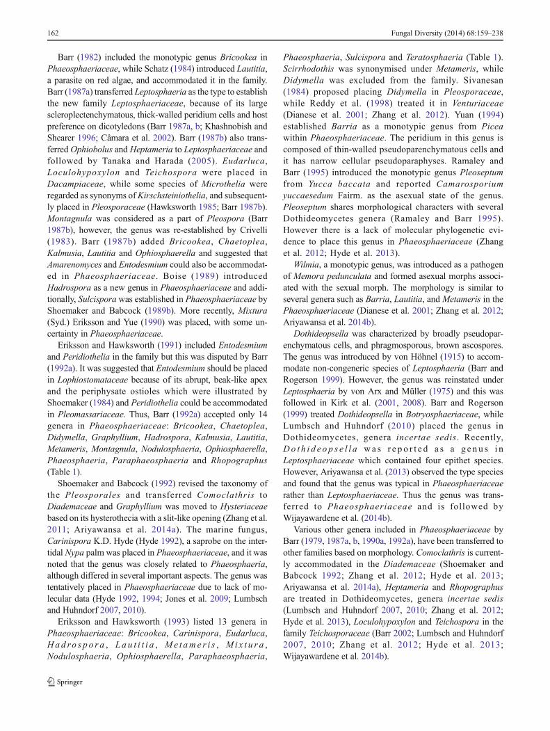
The family Phaeosphaeriaceae was introduced by Barr(1979) and typified by Phaeosphaeria with Ph. oryzae asthe type species. The important family characters were de-scribed by Barr (1979) as “saprobic, pathogenic or hyperpar-asitic fungi typically on herbaceous stems or monocotyledon-ous leaves, culms, or flowers, but also on woody substrates.Ascomata were described as immersed, erumpent or superfi-cial, globose or conical, short papillate, small to medium, ascias bitunicate, and ascospores as hyaline, yellowish or brown,narrowly or widely obovoid, aseptate or septate” Subsequentauthors are in agreement with this description (Barr 1987b,1992a, b; Zhang et al. 2012; Hyde et al. 2013). When Barr(1979) introduced the family Phaeosphaeriaceae, 15 generawere included (Table 1); i.e. Comoclathris, Didymella,Eudarluca, Heptameria, Leptosphaeria, Loculohypoxylon,Metameris, Microthelia, Nodulosphaeria, Ophiobolus,Paraphaeosphaeria, Phaeosphaeria, Rhopographus,Scirrhodothis and Teichospora (Barr 1979; Zhang et al.2009, 2012).
Arrangement of genera in Phaeosphaeriaceae basedon morphology (1979–1999)
Over the next 20 years the family was revised with additionsand exclusions of genera and this is detailed below (Table 1).These revisions were based on morphological characters andwere subjective with decisions made based on perceivedimportance of characters. During this period it was verydifficult for researchers to develop higher classification
160 Fungal Diversity (2014) 68:159–238
schemes as it was not possible to confirm the importance ofcharacters (K.D. Hyde, personal experience). Therefore, the
researchers did their work to the best of their ability and haveoften been proven correct.
Table 1 Synopsis of Phaeosphaeriaceae from 1979 to 2014
Barr (1979) Barr (1992a) Eriksson andHawksworth(1993)
Kirk et al. (2001) Lumbsch andHuhndorf (2007)
Kirk et al. (2008)
ComoclathrisDidymellaEudarlucaHeptameriaLeptosphaeriaLoculohypoxylonMetamerisMicrotheliaNodulosphaeriaOphiobolusParaphaeosphaeriaPhaeosphaeriaRhopographusScirrhodothisTeichospora
BricookeaChaetopleaDidymellaGraphylliumHadrosporaKalmusiaLautitiaMetamerisMontagnulaNodulosphaeriaOphiosphaerellaParaphaeosphaeriaPhaeosphaeriaRhopographus
BricookeaCarinisporaEudarlucaHadrosporaLautitiaMetamerisMixturaNodulosphaeriaOphiosphaerellaParaphaeosphaeriaPhaeosphaeriaSulcisporaTeratosphaeria
BarriaBricookeaCarinisporaEudarluca= Cryptocrea= Parabotryon= XenodimeriumHadrosporaIsthmosporellaLautitiaMetameris= Scirrhodothis= ScirrhophragmaMixturaNodulosphaeria= PocosphaeriaOphiosphaerellaParaphaeosphaeriaPhaeodothisPhaeosphaeria=Leptosphaerella=TrematosphaerellaSulcispora
BarriaBricookeaCarinisporaChaetopleaEudarlucaHadrosporaIsthmosporellaKatumotoaLautitiaLeptosphaeriaMetamerisMixturaNeophaeosphaeriaNodulosphaeriaOphiobolusOphiosphaerellaPhaeosphaeriaPhaeosphaeriopsisPleoseptumSetomelanommaTeratosphaeriaWilmia
BarriaBricookeaCarinisporaEudarlucaHadrosporaIsthmosporellaKatumotoaLautitiaMetamerisMixturaNodulosphaeriaOphiosphaerellaPhaeosphaeriaPhaeosphaeriopsisSetomelanommaSulcosporaWilmia
Lumbsch and Huhndorf(2010)
Hyde et al. (2011)(Asexual state reported)
Zhang et al. (2012) Hyde et al. (2013) Wijayawardene et al. (2014b) and in thisstudy
BarriaBricookeaCarinisporaChaetopleaEntodesmiumEudarlucaHadrosporaIsthmosporellaLautitiaMetamerisMixturaNodulosphaeriaOcalaOphiobolusOphiosphaerellaPhaeosphaeria= AmarenomycesPhaeosphaeriopsisPleoseptumSetomelanommaWilmia
Amarenographium(Sexual=Amarenomyces)AmpelomycesChaetosphaeronemaConiothyrium(Sexual=Ophiobolus)Hendersonia-like(Sexual=Austropleosporaand Phaeosphaeria)
NeosetophomaParahendersonia(Sexual=Chaetoplea)ParaphomaPhaeoseptoria(Sexual=Phaeosphaeria)Phaeostagonospora(Sexual=Phaeosphaeriopsis)Phoma(Sexual=Ophiobolus)Rhabdospora(Sexual=Ophiobolus)Scolecosporiella(Sexual=Ophiosphaerellaand Phaeosphaeria)
SetophomaSphaerellopsis(Sexual=Eudarluca)Stagonospora(Sexual=Phaeosphaeria)Tiarospora
BarriaBricookeaChaetopleaEudarlucaEntodesmiumHadrosporaLautitiaLoratosporaMetamerisMixturaNodulosphaeriaOphiobolusOphiosphaerellaPhaeosphaeriaPhaeosphaeriopsisPleoseptumSetomelanommaWilmia
AmpelomycesBarriaBricookeaChaetopleaChaetosphaeronemaEntodesmiumEudarlucaHadrosporaLautitiaLoratosporaMetamerisMixturaNeosetophomaNodulosphaeriaOphiobolusOphiosphaerellaParahendersoniaParaphomaPhaeoseptoriaPhaeosphaeriaPhaeosphaeriopsisPleoseptumSetomelanommaSetophomaTiarosporaWilmiaWojnowicia
AmarenographiumAmarenomycesAmpelomycesBricookeaChaetosphaeronemaDematiopleosporaDothideopsellaEntodesmiumEudarlucaLoratosporaNeosetophomaNeostagonosporaNodulosphaeriaOphiobolusOphiosphaerellaParaphomaParastagonosporaPhaeosphaeria= PhaeoseptoriaPhaeosphaeriopsisPhaeostagonosporaSclerostagonosporaScolecosporiella?? Scolicosporium??SeptoriellaSetomelanommaSetophomaTiarosporaVrystaatiaWojnowicia.Xenoseptoria
Fungal Diversity (2014) 68:159–238 161
Barr (1982) included the monotypic genus Bricookea inPhaeosphaeriaceae, while Schatz (1984) introduced Lautitia,a parasite on red algae, and accommodated it in the family.Barr (1987a) transferred Leptosphaeria as the type to establishthe new family Leptosphaeriaceae, because of its largescleroplectenchymatous, thick-walled peridium cells and hostpreference on dicotyledons (Barr 1987a, b; Khashnobish andShearer 1996; Câmara et al. 2002). Barr (1987b) also trans-ferred Ophiobolus and Heptameria to Leptosphaeriaceae andfollowed by Tanaka and Harada (2005). Eudarluca,Loculohypoxylon and Teichospora were placed inDacampiaceae, while some species of Microthelia wereregarded as synonyms ofKirschsteiniothelia, and subsequent-ly placed in Pleosporaceae (Hawksworth 1985; Barr 1987b).Montagnula was considered as a part of Pleospora (Barr1987b), however, the genus was re-established by Crivelli(1983). Barr (1987b) added Bricookea, Chaetoplea,Kalmusia, Lautitia and Ophiosphaerella and suggested thatAmarenomyces and Entodesmium could also be accommodat-ed in Phaeosphaeriaceae. Boise (1989) introducedHadrospora as a new genus in Phaeosphaeriaceae and addi-tionally, Sulcisporawas established in Phaeosphaeriaceae byShoemaker and Babcock (1989b). More recently, Mixtura(Syd.) Eriksson and Yue (1990) was placed, with some un-certainty in Phaeosphaeriaceae.
Eriksson and Hawksworth (1991) included Entodesmiumand Peridiothelia in the family but this was disputed by Barr(1992a). It was suggested that Entodesmium should be placedin Lophiostomataceae because of its abrupt, beak-like apexand the periphysate ostioles which were illustrated byShoemaker (1984) and Peridiothelia could be accommodatedin Pleomassariaceae. Thus, Barr (1992a) accepted only 14genera in Phaeosphaeriaceae: Bricookea, Chaetoplea,Didymella, Graphyllium, Hadrospora, Kalmusia, Lautitia,Metameris, Montagnula, Nodulosphaeria, Ophiosphaerella,Phaeosphaeria, Paraphaeosphaeria and Rhopographus(Table 1).
Shoemaker and Babcock (1992) revised the taxonomy ofthe Pleosporales and transferred Comoclathris toDiademaceae and Graphyllium was moved to Hysteriaceaebased on its hysterothecia with a slit-like opening (Zhang et al.2011; Ariyawansa et al. 2014a). The marine fungus,Carinispora K.D. Hyde (Hyde 1992), a saprobe on the inter-tidal Nypa palm was placed in Phaeosphaeriaceae, and it wasnoted that the genus was closely related to Phaeosphaeria,although differed in several important aspects. The genus wastentatively placed in Phaeosphaeriaceae due to lack of mo-lecular data (Hyde 1992, 1994; Jones et al. 2009; Lumbschand Huhndorf 2007, 2010).
Eriksson and Hawksworth (1993) listed 13 genera inPhaeosphaeriaceae: Bricookea, Carinispora, Eudarluca,Had ro s p o r a , Lau t i t i a , Me t am e r i s , Mi x t u r a ,Nodulosphaeria, Ophiosphaerella, Paraphaeosphaeria,
Phaeosphaeria, Sulcispora and Teratosphaeria (Table 1).Scirrhodothis was synonymised under Metameris, whileDidymella was excluded from the family. Sivanesan(1984) proposed placing Didymella in Pleosporaceae,while Reddy et al. (1998) treated it in Venturiaceae(Dianese et al. 2001; Zhang et al. 2012). Yuan (1994)established Barria as a monotypic genus from Piceawithin Phaeosphaeriaceae. The peridium in this genus iscomposed of thin-walled pseudoparenchymatous cells andit has narrow cellular pseudoparaphyses. Ramaley andBarr (1995) introduced the monotypic genus Pleoseptumfrom Yucca baccata and reported Camarosporiumyuccaesedum Fairm. as the asexual state of the genus.Pleoseptum shares morphological characters with severalDothideomycetes genera (Ramaley and Barr 1995).However there is a lack of molecular phylogenetic evi-dence to place this genus in Phaeosphaeriaceae (Zhanget al. 2012; Hyde et al. 2013).
Wilmia, a monotypic genus, was introduced as a pathogenof Memora pedunculata and formed asexual morphs associ-ated with the sexual morph. The morphology is similar toseveral genera such as Barria, Lautitia, and Metameris in thePhaeosphaeriaceae (Dianese et al. 2001; Zhang et al. 2012;Ariyawansa et al. 2014b).
Dothideopsella was characterized by broadly pseudopar-enchymatous cells, and phragmosporous, brown ascospores.The genus was introduced by von Höhnel (1915) to accom-modate non-congeneric species of Leptosphaeria (Barr andRogerson 1999). However, the genus was reinstated underLeptosphaeria by von Arx and Müller (1975) and this wasfollowed in Kirk et al. (2001, 2008). Barr and Rogerson(1999) treated Dothideopsella in Botryosphaeriaceae, whileLumbsch and Huhndorf (2010) placed the genus inDothideomycetes, genera incertae sedis. Recently,Do t h i d e o p s e l l a wa s r e p o r t e d a s a g e n u s i nLeptosphaeriaceae which contained four epithet species.However, Ariyawansa et al. (2013) observed the type speciesand found that the genus was typical in Phaeosphaeriaceaerather than Leptosphaeriaceae. Thus the genus was trans-ferred to Phaeosphaeriaceae and is followed byWijayawardene et al. (2014b).
Various other genera included in Phaeosphaeriaceae byBarr (1979, 1987a, b, 1990a, 1992a), have been transferred toother families based on morphology. Comoclathris is current-ly accommodated in the Diademaceae (Shoemaker andBabcock 1992; Zhang et al. 2012; Hyde et al. 2013;Ariyawansa et al. 2014a), Heptameria and Rhopographusare treated in Dothideomycetes, genera incertae sedis(Lumbsch and Huhndorf 2007, 2010; Zhang et al. 2012;Hyde et al. 2013), Loculohypoxylon and Teichospora in thefamily Teichosporaceae (Barr 2002; Lumbsch and Huhndorf2007, 2010; Zhang et al. 2012; Hyde et al. 2013;Wijayawardene et al. 2014b).
162 Fungal Diversity (2014) 68:159–238
Initial phylogenetics for resolving Phaeosphaeriaceae
In 1995 the first phylogenetic evidence for placement ofgenera in Phaeosphaeriaceae started to emerge. Moraleset al. (1995) attempted to determine the relationships betweenLeptosphaeria and Phaeosphaeria species using rRNA genesequences. The separation of Leptosphaeria andPhaeosphaeria was not strongly supported and the asexualstates were phylogenetically uninformative in their analysis.Phylogenetic analysis indicated that Phaeosphaeria was partof Leptosphaeriaceae due to the fact that Ph. nodorum andPh. microscopica formed a clade close to L. doliolum, the typespecies of Leptosphaeria (Morales et al. 1995; Câmara et al.2002.). Khashnobish and Shearer (1996) found thatPhaeosphaeria formed a distinct group separate fromLeptosphaeria based on a cladis t ic analys is ofmorphological and sequence data. Câmara et al. (2002) con-cluded that peridial characters, asexual state and hostpreferences are taxonomically and phylogeneticallysignificant at the generic level, with Leptosphaeria sensustricto and Phaeosphaeria sharing a more recent commonancestor in their analysis.
Kruys et al. (2006) investigated the relationships of fami-lies in Pleosporales and observed that Phaeosphaeriaceae isclosely related to Leptosphaeriaceae and formed an unre-solved monophyletic group. The main features distinguishingthe two families are the ascomata wall characters. InLeptosphaeriaceae taxa have scleroplectenchymatousascomata walls and in Phaeosphaeriaceae taxa have thin-walled, pseudoparenchymatous ascomata walls (Câmaraet al. 2002; Cheng et al. 2004; Schoch et al. 2006). Shiraia,an uncertain genus grouped with Leptosphaeriaceae andPhaeosphaeriaceae in the phylogenetic analysis of Kruyset al. (2006). Cheng et al. (2004) suggested that Shiraia shouldbe treated as a member of Phaeosphaeriaceae. The genus wasunofficially placed in Shiraiaceae, byOgawa et al. (2003) (seeGenBank AB105798), but was not validly published (Kruyset al. 2006; Liu et al. 2013). Liu et al. (2013) illustrated andepitypified the type species, Shiraia bambusicola from freshcollection in Zhejiang Province, China. Based on theirphylogenetic investigation, Shiraia formed a distinct cladeseparate from Phaeosphaeriaceae. Therefore, Liu et al.(2013) introduced a new valid family Shiraiaceae to accom-modate a monotypic genus Shiraia in Pleosporales and illus-trated a coelomycetous asexual state.
Zhang et al. (2009) incorporated multigene phylogeneticanalysis and included 15 families in Pleosporales. They in-cluded four families in the suborder Pleosporineae viz.Didymellaceae, Leptosphaeriaceae, Phaeosphaeriaceae andPleosporaceae. In addition, they introduced two new families,Amniculicolaceae and Lentitheciaceae to accommodate somefreshwater fungi (Zhang et al. 2009, 2012). Zhang et al. (2009)re-accommodated Ophiobolus in Phaeosphaeriaceae due to
its morphological characters and because Ophiobolus is oftenassociated with the asexual genus Chaetosphaeronema on thehost (Petrak 1944). However, this link has not been confirmed(Zhang et al. 2009).
Zhang et al. (2012) re-circumscribed the familiesPhaeosphaeriaceae in Pleosporales. These authors treated18 sexual genera base on morphology in Phaeosphaeriaceaeand accepted Entodesmium, Ophiosphaerella, Phaeosphaeriaand Setomelanomma based on phylogenetic analysis(Table 1). The freshwater genera Isthmosporella Shearer andCrane (1999) and Ocala Raja et al. (2009) were also accom-modated inPhaeosphaeriaceae. Zhang et al. (2012), however,removed these genera from Phaeosphaeriaceae based onmorphological characters and phylogenetic analysis; Ocalaformed a unique clade basal to Jahnulales (Shearer et al.2009) and Isthmosporellawas treated in Pleosporales, generaincertae sedis. Hyde et al. (2013) listed 27 sexual and asexualgenera in Phaeosphaeriaceae and later, Wanasinghe et al.(2014) introduced the monotypic genus, Dematiopleosporatypified by D. mariae in Phaeosphaeriaceae based on molec-ular data. Furthermore, Crous et al. (2014) includedSetophaeosphaeria Crous & Y. Zhang ter with Sp.badalingensis Crous & Y. Zhang ter, Sp. hemerocallidisCrous & Y. Zhang ter and Sp. setosa (Leuchtm.) Crous inPhaeosphaeriaceae. Setophaeosphaeria is morphologicallyand phylogenetically distinct from Phaeosphaeria oryzae,the type species of Phaeosphaeriaceae, but is similar to Ph.setosa. Thus the genus was established to accommodatephaeosphaeria-like species.
Recent molecular studies place Kirschsteiniothelia in anew family Kirschsteiniotheliaceae based on molecular data(Boonmee et al. 2012) and Microthelia species are treated insynonymy of various genera of Dothideomycetes (Hyde et al.2013). Kalmusia, Montagnula and Paraphaeosphaeria areaccommodated in Montagnulaceae (Boonmee et al. 2012;Hyde et al. 2013; Lumbsch and Huhndorf 2010; Zhang et al.2009, 2012; Ariyawansa et al. 2014b). Several genera inPhaeosphaeriaceae, such as Barria, Bricookea, Chaetoplea,Eudarluca, Hadrospora, Lautitia, Metameris, Mixtura,Nodulosphaeria, Ophiobolus, Pleoseptum, and Wilmia lackmolecular data.
History of genera
AmarenomycesErikssonwas introduced as a new genus basedon its multi-layered endotunica and large, thick-walled andsheathed ascospores (Eriksson 1981). Eriksson (1982) report-ed Amarenographium to be the asexual state ofAmarenomyces. Amarenomyces was synonymized underPhaeosphaeria by Zhang et al. (2009) based on phylogeneticevidence from Amarenomyces ammophilae (Lasch) O.E.Erikss. which formed a related clade with otherPhaeosphaeria species.
Fungal Diversity (2014) 68:159–238 163
Didymella was treated as a member of Phaeosphaeriaceaeby Barr (1979) when she introduced the new familyPhaeosphaeriaceae. However, Silva-Hanlin and Hanlin(1999) suggested that Didymella belongs in Pleosporaceaerather than Phaeosphaeriaceae based on phylogenetic rela-tionships, while Lumbsch and Huhndorf (2007) treatedDidymella in Pleosporales, genera incertae sedis. Later, DeGruyter et al. (2009) introduced a new family Didymellaceaebased on a multigene phylogenetic analysis withDidymella asthe type genus.
Entodesmium was introduced by Reiss (1854) as typifiedby E. rude Riess associated with legumes (Zhang et al. 2009).The genus was previously placed in Lophiostomataceae basedon its periphysate papilla (Eriksson and Hawksworth 1990;Barr 1992b; Zhang et al. 2009). Liew et al. (2000) sequenced astrain of E. rude and their phylogenetic analysis indicated thatEntodesmium belongs to Phaeosphariaceae which has beenupheld by subsequent authors (Schoch et al. 2009; Zhang et al.2009, 2012; Hyde et al. 2013, 2014). However, there is onlyone putative strain of Entodesmium rude (CBS 650.86) avail-able in GenBank (Liew et al. 2000), and this is not apparentlylinked to any herbarium material.
Eudarluca was introduced by Spegazzini (1908) as a hy-perparasite in the uredinia of rust fungi from Canna (Zhanget al. 2012). Sphaerellopsis was reported to be the asexualstate of this genus (Yuan et al. 1998; Nischwitz et al. 2005;Zhang et al. 2012). Many strains of Eudarluca caricis (Fr.)O.E. Erikss. are available in GenBank. Nischwitz et al. (2005)investigated host specialization and relationships ofEudarluca caricis with Ampelomyces. Based on the phyloge-netic analysis, their results have shown that Eudarluca caricisformed a clade with other pleosporalean taxa and appeared tobe host-specific (Nischwitz et al. 2005). The phylogeneticinvestigation was limited to variation between differentiatedisolates and could not resolve the current placement ofEudarluca in Phaeosphaeriaceae (Bayon et al. 2006; Zhanget al. 2012).
Katumotoa was introduced as a monotypic genus inPhaeosphaeriaceae and was tentatively placed inPhaeosphaeriaceae (Tanaka and Harada 2005). Later,Tanaka et a l . (2009) removed Katumotoa f romPhaeosphaeriaceae and accommodated this genus in the fam-ily Lentitheciaceae (Zhang et al. 2009), based on morpholog-ical and phylogenetic evidence.
Loratospora was introduced by Kohlmeyer andVolkmann-Kohlmeyer (1993) as a monotypic genus.The type species, Loratospora aestuarii Kohlm. &Volkm.-Kohlm. is an obligate and facultative marinetaxon (Kohlmeyer and Volkmann-Kohlmeyer 1993;Monkai et al. 2013). Suetrong et al. (2009) transferredLoratospora to Phaeosphaeriaceae based on phylogeneticdata, whereas based on morphology it had previously beenplaced in Planistromellaceae (Barr 1996).
Ophiosphaerella was introduced by Spegazzini (1909)with Op. graminicola Speg. as the type species. Barr(1987b) placed Ophiosphaerella in Phaeosphaeriaceae, dueto its morphological characters (Shoemaker and Babcock1989b ; Dong e t a l . 1998 ) . The p l a c emen t i nPhaeosphaeriaceae was confirmed by Zhang et al. (2009,2012) and Hyde et al. (2013) based on molecular phylogeny.
Phaeosphaeriopsis was introduced to accommodateParaphaeosphaeria species that were not congeneric basedon morphological characters and phylogenetic investigationand that had a Phaeostagonospora asexual state (Câmara et al.2003; Zhang et al. 2012). The placement of the genus inPhaeosphaeriaceae was confirmed by multi-gene analysis(Quaedvlieg et al. 2013; Thambugala et al. 2014).
Setomelanomma was introduced by Morelet (1980) as apathogen on blue and white spruce (Morelet 1980; Rossmanet al. 2002). The genus was considered morphologically sim-ilar to Phaeosphaeriaceae (Rossman et al. 2002; Zhang et al.2012). Rossman et al. (2002) confirmed the genus belongs inPhaeosphaeriaceae based on a phylogenetic analysis and thiswas verified with multi-gene analyses (Schoch et al. 2009;Zhang et al. 2009, 2012; De Gruyter et al. 2012; Hyde et al.2013; Wijayawardene et al. 2014b).
Placement of asexual genera in Phaeosphaeriaceae
Before the era of molecular phylogeny it was difficult toestablish if asexual genera belonged in Phaeosphaeriaceaeunless single ascospore cultures produced the asexual morphor where the asexual morph was associated with the sexualstate on the host (Eriksson 1982; Ramaley 1995; Ramaley andBarr 1995; Yuan et al. 1998; Câmara et al. 2003; Nischwitzet al. 2005). Previous to the compilation by Hyde et al. (2011)there were few attempts to incorporate asexual genera insexually defined families. Perhaps the first report of an asexualstate of Phaeosphaeriaceae is that of Eriksson (1982) whoreported Amarenographium to be the asexual state ofAmarenomyces. Barr (1987b) listed nine coelomycetous gen-era , Ascochyta , Chaetodiplodia , Coniothyr ium ,Microdiplodia, Microsphaeropsis, Phoma, Scolecosporiella,Stagonospora and Sphaerellopsis in Phaeosphaeriaceaebased on their holoblastic or enteroblastic conidiogenesisand aseptate or septate conidia.
Camarosporium yuccaesedum Fairm formed conidiomataon leaves of Yucca baccata that were associated withascomata of Pleoseptum yuccaesedum, the type species ofPleoseptum, (Ramaley and Barr 1995; Zhang et al. 2012).Based on its structurally similar and close association,Ramaley and Barr (1995) linked Camarosporiumyuccaesedum as an asexual state of Pleoseptum.Camarosporium species had been reported as asexual morphsin various genera of Botryosphaeriales and Cucurbitariaceaewith doubtful polyphyletic support (Crous et al. 2006; Kirk
164 Fungal Diversity (2014) 68:159–238
et al. 2008; Liu et al. 2012; Zhang et al. 2012; Doilom et al.2013). However, Wijayawardene et al. (2014a, c, d) recog-nized Camarosporium sensu stricto as a distinct phylogeneticlinage in Pleosporineae, Pleosporales hence introducedCamarosporiaceae, (Wijayawardene et al. 2014a, d)
Hyde et al. (2011) and Wijayawardene et al. (2012) alsolisted Hendersonia as an asexual state of Phaeosphaeriaceae.However, the genus was synonymized under Stagonosporaand is currently placed in Massarinaceae (Swart and Walker1988; Quaedvlieg et al. 2013).
Parahendersonia Ramaley (1995) was classified by itsampulliform conidiogenous cells producing thick-walled,brown, (1–)3-septate conidia. This monotypic genus is repre-sented by P. dasylirionis. The sexual state was reported asChaetoplea dasylirionis on Dasylirion (Agavaceae) inLeptosphaeriaceae (Ramaley 1995; Calatayud and Etayo2001). However, Barr (1987b) accommodated Chaetopleain Phaeosphaeriaceae and reported a microdiplodia-likeasexual state for Chaetoplea (Barr 1990b; Zhang et al.2012). Recently, Parahendersonia was treated inLeptosphaeriaceae (Index Fungorum 2014).
Rhabdosporawas originally established to accommodateseptoria-like species associated with stems of Neriumoleander L. (Priest 2006; Quaedvlieg et al. 2013). Thegenus was treated in the taxonomic studies of Sutton(1980) which 11 Rhabdospora species currently classifiedunder Septoria in Mycosphaerellaceae (Quaedvlieg et al.2013). Hyde et al. (2011) listed Rhabdospora as the asexualstate of Phaeosphaeriaceae, while Zhang et al. (2012)treated the genus as an asexual state of Ophiobolus.Wijayawardene et al. (2014b) treated the genus inDothideomycetes, genera incertae sedis. There are 648ep i the t s l i s t ed in Index Fungorum (2014 ) fo rR h a b d o s p o r a a n d i t w a s a c c ommo d a t e d i nMycosphaerellaceae. However, Rhabdospora is a poorlyunderstood genus and is in need of modern taxonomic andmolecular treatment (Quaedvlieg et al. 2013).
The type species of Scolecosporiella, Sc. typhae(Oudemans) Petr. was reported as the asexual state ofPhaeosphaeria typharum (Desm.) L. Holm (Nag Raj1989). The genus has been re-described taxonomic revi-sion by Sutton (1968, 1980) and Nag Raj (1989). Varioussexual genera, such as Ophiobolus, Ophiosphaerella, andPhaeosphaeria have been reported as Scolecosporiellasexual states (Farr et al . 1989; Nag Raj 1989;Shoemaker and Babcock 1989b; Hyde et al. 2011;Zhang et al. 2012).
Tiarospora was reported as an asexual member ofPhaeosphaeriaceae by Kirk et al. (2008). However, thereare no molecular data to prove that this genus belongs inPhaeosphaeriaceae. Tiarospora has four epithets in IndexFungorum (2014) with T. perforans (Sacc.) Höhn being listedas a synonym of Montagnula perforans (Aptroot 2006).
Molecular data has allowed linkage of asexual generain Phaeosphaeriaceae
Ascochy ta , Chaetodip lod ia , Microdip lodia andMicrosphaeropsis have been reported as asexual states ofPhaeosphaeriaceae (Barr 1987b); however, using phyloge-netic evidence Crous et al. (2006) treatedMicrodiplodia as theasexual state of Karstenula in Montagnulaceae (Ariyawansaet al. 2014b). Liu et al. (2012) and Hyde et al. (2013) reportedMicrodiplodia as an asexual state in Botryosphaeriaceae.Ascochyta and Microsphaeropsis were treated inDidymellaceae based on phylogenetic evidence with the sex-ual morph of these genera described asDidymella (De Gruyteret al. 2009; Aveskamp et al. 2010). Ascochyta was shown tobe polyphyletic in Pleosporales by subsequent researchers(De Gruyter et al. 2009, 2012; Aveskamp et al. 2010).Furthermore, allied Ascochyta species were determined tobelong in Pleosporaceae by De Gruyter et al. (2012). AChae tod i p l od i a s t r a i n f o rmed a c l ade w i t h i nLeptosphaeriaceae; however, the genus is polyphyletic inPleosporaceae (De Gruyter et al. 2009, 2012).
Based on phylogenetic evidence, Câmara et al. (2003)introduced the new genus Phaeosphaeriopsis inPhaeosphaeriaceae and transferred Paraphaeosphaeriano l inae A.W. Rama ley to Phaeosphaer iops i s .Phaeostagonospora was reported as the asexual state basedon its morphology (Ramaley 1997). Sexual and asexualmorph connections were studied by Quaedvlieg et al. (2013)and Thambugala et al. (2014).
De Gruyter et al. (2009) included Wojnowicia hirta (J.Schröt.) Sacc., the type species in their phylogenetic analysisand concluded that Wojnowicia hirta has an asexual state inPhaeosphaeriaceae and this phylogenetic placement was sup-ported by Wijayawardene et al. (2013b, 2014b).
Z h a n g e t a l . ( 2 0 0 9 ) l i s t e d Amp e l om y c e s ,Chaetosphaeronema, Coniothyrium, Phoma, Plenodomus,Stagonospora and Wojnowicia (asexual genera) inPhaeosphaeriaceae. Based on phylogenetic evidence,Chaetosphaeronema was placed in Phaeosphaeriaceae andoften formed a sister clade with the sexual genusEntodesmium (Zhang et al. 2009, 2012; Hyde et al. 2013;Wijayawardene et al. 2014b). Phoma radicina (McAlpine)Boerema, the type species of Phoma section Paraphomaformed a sister clade with the sexual genus Setomelanommaholmii M. Morelet as basal Phaeosphaeriaceae (Zhang et al.2009, 2012; Hyde et al. 2013; Wijayawardene et al. 2014b).
De Gruyter et al. (2010) introduced two new genera,Neosetophoma and Setophoma in Phaeosphaeriaceae andalso re-introduced Paraphoma in Phaeosphaeriaceae basedon phylogenetic evidence. Four Phoma species fromAv e s k amp e t a l . ( 2 0 1 0 ) wh i c h c l u s t e r e d i nPhaeosphaeriaceae were accommodated in these genera (DeGruyter et al. 2010). Hyde et al. (2011) reported 17 asexual
Fungal Diversity (2014) 68:159–238 165
genera in Phaeosphaer iaceae (Table 1) . Later,Wijayawardene et al. (2012) updated the checklist of asexuals t a tes of Phaeosphaer iaceae and inc luded thehyphomyceteous genera Harpophora and Mauginiella. Intheir compilation of families in Pleosporales, Zhang et al.(2012) accepted 17 asexual genera in Phaeosphaeriaceae.They excluded the hyphomycetous genera, based on a broadfamilial concept and phylogenetic support.
Hyde et al. (2013) excluded Amarenographium whichformed a clade outside Phaeosphaeriaceae and this is sup-ported by Hodhod et al. (2012). Wijayawardene et al. (2013a)accommodated Scolicosporium minkeviciusii Treigienė inPhaeosphaeriaceae as this species formed a clade with otherwell-established asexual genera. Furthermore, Crous et al.(2013) revealed that Xenostigmina and Mycopappus groupedin Phaeosphaeriaceae in their phylogenetic analysis.
Quaedvlieg et al. (2013) re-circumscribed the pathogenicfungi genera Septoria, and Stagonospora and related generasuch as Phaeosphaeria and Phaeoseptoria. Based on theirphylogenetic study, they introduced 14 new genera, 36 newspecies, and 19 new combinations. The new generaNeostagonospora, Parastagonospora, Xenoseptoria andVrystaatia were placed in Phaeosphaeriaceae. Furthermore,they epitypified Phaeosphaeria oryzae I. Miyake andPhaeoseptoria papayae Speg. and confirmed sexual asexualconnections. Phaeoseptoria was confirmed as the asexualmorph of Phaeosphaeria based on the sequenced strain ofP. papayae which formed a well-supported clade with Ph.oryzae.
Crous et al. (2014) included Septoriella oudemansiiOudem. in Phaeosphaeriaceae. Septoriella was comparedwith species of Phaeosphaeriaceae and a megablast searchof NCBI showed that Septoriella is similar to Phaeosphaeriacaricis, Ph. ammophilae and Ophiosphaerella herpotrichawith high percentage similarities (Crous et al. 2014).
I n a n ew ou t l i n e o f t h e Do t h i d eomyce t e s ,Wijayawardene et al. (2014b) proposed synonymies andsuggested names for 34 genera while incorporating asexualgenera into a natural classification. This publication result-ed from the new ruling in article 59.1 of International Codeof Nomenclature for Algae, Fungi, and Plants (ICN;Melbourne Code) that a species or genus can only haveone name. They listed 29 genera (17 only known as asexualgenera) in Phaeosphaeriaceae.
According to phylogenetic evidence, Ampelomyces speciesgrouped in Didymellaceae and Phaeosphaeriaceae (DeGruyter et al. 2009; Aveskamp et al. 2010; Hyde et al. 2013;Wijayawardene et al. 2013a, 2014b). The type species ofAmpelomyces, A. quisqualis Ces. formed a clade withinPhaeosphaeriaceae (De Gruyter et al. 2009; Aveskamp et al.2010; Wijayawardene et al. 2013a), while A. quercinus (Syd.)Rudakov grouped in Didymellaceae (De Gruyter et al. 2009).Therefore, Aveskamp et al. (2010) transferred all
Ampelomyces species belonging in Didymellaceae, toPhoma based on phylogenetic relationships.
Coniothyrium Corda (1840) is characterized by pycnidialconidiomata, reduced conidiophores , annel l idicconidiogenous cells, with brown, aseptate or septate, verru-cose conidia (De Gruyter et al. 2012). Based on the phyloge-netic analyses, Coniothyrium was revealed as polyphyletic inPleosporales (Verkley et al. 2004, 2014). The type species ofConiothyrium , C. palmarum Corda clustered inLeptosphaeriaceae (De Gruyter et al. 2009). In De Gruyteret al. (2012), C. palmarum formed a distinct clade with otherPhoma species in the reinstated Coniothyriaceae based onphylogenetic analysis. De Gruyter et al. (2012) indicated thatConiothyrium had convergent evolution with similar charac-ters found in other families in Pleosporales such asCucurbitariaceae, Didymellaceae, Phaeosphaeriaceae,Leptosphaeriaceae, Montagnulaceae and Sporormiaceae.However, Coniothyrium concentricum (Desm.) Sacc. isrelated to Phaeosphaeriaceae. A new genus should bedesignated to accommodate this coniothyrium-like species(Hyde et al. 2013).
Harpophorawas introduced by Gams (2000) to accommo-date phialophora-like species characterized by fast-growing,thin colonies and sickle-shaped phialoconidia (Gams 2000;Saleh and Leslie 2004). The genus contains the asexual stateof Gaeumannomyces and Magnaporthe inMagnaporthaceae(Gams 2000; Saleh and Leslie 2004; Kirk et al. 2008; Yuanet al. 2010).
Based on a homologous test using ITS sequence data,Abdullah et al. (2005) indicated that Mauginiella scaettaeCavara was closely related to species of Phaeosphaeriawhichformed a clade with Phaeosphaeria associated with non-grasshosts. It was concluded that Mauginiella may represent theasexual state of Phaeosphaeria following the suggestion ofCâmara et al. (2002). Câmara et al. (2002) mentioned that themorphological characters, asexual state and host preferencesare important to clarify Phaeosphaeria species. It has beensuggested that a great deal of convergent evolution had oc-curred in species of Leptosphaeria and Phaeosphaeria(Câmara et al. 2002; Abdullah et al. 2005).
Phoma species have been described as the asexual state ofvarious genera of Dothideomycetes i.e. Didymella,Mycosphaerella, Leptosphaeria and Pleospora (Boerema1997; De Gruyter et al. 2009). De Gruyter et al. (2009) carriedout a re-classification of the Phoma complex based on molec-ular phylogeny. Their results showed that Phoma was poly-phyletic and was subsequently classified in nine sections:Phoma , Heterospora , Paraphoma , Peyronellaea ,Phyllostictoides, Sclerophomella, Plenodomus, Macrospora,and Pilosa (Boerema 1997; Boerema et al. 2004; De Gruyteret al. 2009). Based on phylogenetic evidence, they concludedthat Phoma section Paraphoma, as typified by Ph. radicina(McAlpine) Boerema can be assigned in Phaeosphaeriaceae,
166 Fungal Diversity (2014) 68:159–238
while Ph. heteromorphospora, the type species of Phomasection Heterospora should be treated in Leptosphaeriaceae.Additionally, the family Didymellaceae was proposed to ac-commodate others Phoma sections viz. Macrospora,Peyronellaea, Phoma, Phyllostictoides and Sclerophomella(De Gruyter et al. 2009). Aveskamp et al. (2010) indicatedthat the current Boeremaean subdivision was incorrect asPhoma is polyphyletic comprising six distinct clades inPleosporales and appeared to be common in different families(Aveskamp et al. 2010). Therefore, De Gruyter et al. (2012)concluded that Phoma should be restricted only toDidymellaceae, while other Phoma sections were transferredto related families. However, Lawrey et al. (2012) treatedlichenicolous Phoma species in Phaeosphaeriaceae basedon their multigene phylogenetic analysis. Therefore, a newgenus should be introduced to accommodate all lichenicolousPhoma species in Phaeosphaeriaceae (Hyde et al. 2013).
Plenodomus Preuss is typified byPl. rabenhorstii Preuss toaccommodate the phoma-like species which Boerema (1982)synonymized under Phoma section Plenodomus (Boeremaet al. 2004; Torres et al. 2005). Torres et al. (2005) mentionedthat Reddy et al. (1998) treated phoma-like species with thesexual state known as Leptosphaeria in Plenodomus inLeptosphaeriaceae and concluded that Phoma should be re-str icted to the species relat ing to Didymella inPhaeosphaeriaceae. Zhang et al. (2009) listed Plenodomusin Phaeosphaeriaceae; however based on phylogenetic evi-dence, De Gruyter et al. (2012) reinstated the genusPlenodomus to accommodate Phoma section Plenodomuswithin Leptosphaeriaceae.
Sclerostagonospora was typified by S. heraclei (Sacc.)Höhn. (1917) and characterized with pycnidial conidiomatawith holoblastic, determinate, discrete, ampulliform to irreg-ular conidiogenous cells, and subcylindrical, 3-septate, palebrown, verruculose conidia (Sutton 1980; Quaedvlieg et al.2013). Sclerostagonospora differs from Stagonospora in itspigmented conidia (Quaedvlieg et al. 2013). Crous and Palm(1999) reported Sclerostagonospora leucadendri Crous &M.E. Palm as the asexual state of Leptosphaeria leucadendriCrous &M.E. Palm which occurs on leaves of Leucadendron.Based on phylogenetic analysis, Sclerostagonospora opuntiae(Ellis & Everh.) Huhndorf (CBS 118224) formed a clade withPhaeosphaeria and Phaeosphaeriopsis (Crous et al. 2008).Thus, Kirk et al. (2008) treated Sclerostagonospora as anasexual state in the Pleosporales. In Quaedvlieg et al.(2013), multigene phylogenetic analysis has shown thatSclerostagonospora phragmiticola Quaedvlieg et al. (CBS338.86) is related to Phaeosphaeriaceae.
Stagonospora is characterized by holoblastic or annellidicconidiogenesis and produces phragmoconidia (Schoch et al.2006; Quaedvlieg et al. 2013). The genus was reported as anasexual genus in Phaeosphaeriaceae, which often clusterswith Phaeosphaeria and Phoma in phylogenetic analyses
(Cunfer and Ueng 1999; Câmara et al. 2002; Solomon et al.2006; Zhang et al. 2009, 2012; Hyde et al. 2013).Stagonospora has often been confused with Septoria; howev-er, the genus was clarified by Cunfer and Ueng (1999) andSolomon et al. (2006). Based on phylogenetic evidence,Stagonospora is more closely related to other genera inP l e o s p o r a l e s , w h i l e S e p t o r i a c l u s t e r e d i nMycosphaerellaceae, Capnodiales (Quaedvlieg et al. 2013).Quaedvlieg et al. (2013) re-collected the type species ofStagonospora, St. paludosa (Sacc. & Speg.) Sacc. and theirphylogenetic analysis showed that this species formed a sep-arate clade with other Stagonospora species which occurredon Poaceae (i.e. St. avenae (A.B.. Frank) Bissett and St.nodorum (Berk.) E. Castell. & Germano). Therefore,Quaedvlieg et al. (2013) concluded that stagonospora-likespecies belonged in different genera and introduced the newgenera, Neostagonospora and Parastagonospora to accom-modate the stagonospora-like species in Phaeosphaeriaceae.The type species of Stagonospora, St. paludosa and otherStagonospora species grouped in Massarinaceae(Quaedvlieg et al. 2013).
The aim of this study is to revisit all genera ofPhaeosphaeriaceae and provide revised descriptions of thesexual and asexual morphs of the genera. Available generictype specimens were examined, described and illustrated. Wehave also collected fresh specimens from Thailand and Italy.Phylogenetic investigation of available sequence data revealsthe current placement of genera in Phaeosphaeriaceae.
Material and methods
Herbarium examination, fungal isolation, morphologyand asexual state study
Type specimens of each genus in Phaeosphaeriaceae wereobtained from BPI, BR, NY, S, and UB herbaria. Morphologyof generic types were examined and re-described. Genericdescriptions were provided based on the original descriptionsand non type herbarium specimens if generic type specimenscould not be located or loaned. Some types were redrawn fromiconotype. Ascomata on herbarium material were rehydratedby adding 5 % KOH. Free hand sections and squash mountswere prepared and morphological characters were observedunder stereo microscope and compound microscope.
Collections of Phaeosphaeriaceae were made from Italyand Thailand and returned to the laboratory for examination. Ifthe specimens were dry, they were incubated in a moistchamber for 2–3 days at 25 °C. Ascomata were sectioned byhand and examined under an Olympus SZH10 stereomicro-scope. Macro-morphological characters were photographedusing a Sony DSC-T110 digital camera. The micro-
Fungal Diversity (2014) 68:159–238 167
morphological characters were captured by a Cannon 550Ddigital camera, using a Nikon ECLIPSE 80i compound mi-croscope with DIC objectives. Indian ink was added to showmucilaginous sheaths surrounding ascospores. Cotton blueand congo red reagents were used as stains for checkingascospore septation and internal elements of the hamathecium.Permanent slides were prepared by mounting material onlactoglycerol and sealing with clear nail polish.Measurements were obtained using a Tarosoft (R) ImageFrameWork version 0.9.7. Photographic plates were preparedin Adobe Photoshop version CS3 (Adobe Systems Inc., TheUnited States).
Pure cultures were obtained from single spores follow-ing the methods described in Chomnunti et al. (2014) andPhookamsak et al. (2013). Ascomata were cut at the apexand sides walls by a razor blade. The contents insideascoma was picked and soaked in sterile water and brokenup further mechanically with the sterile needle until a sporesuspension was obtained. The ascospore suspension wasdropped on water agar (WA; 15.0 g/L sterile distilled water)and incubated at room temperature overnight. The germi-nating spores were transferred to malt extract agar (MEA;33.6 g/L sterile distilled water, Difco malt extract) andcultivated in corn meal agar (CMA, 17 g/l sterile distilledwater, Difco corn meal agar) and potato dextrose agar(PDA; 39 g/L distilled water, Difco potato dextrose) forrecording growth rates and culture characters. Sterile bam-boo pieces on water agar (WA; 15 g/l sterile distilled water)were used to encourage production of asexual morphs.Cultures are deposited in Mae Fah Luang UniversityCulture Collection (MFLUCC), and duplicated in theInternational Collection of Microorganisms from Plants(ICMP), Landcare Research, New Zealand. Herbariumspecimens were prepared by drying the specimens withsilica gel at room temperature for 7–10 days depending onthe humidity and kept in envelopes or small boxes. Theherbarium specimens are deposited in the herbarium of MaeFah Luang University (MFLU), Chiang Rai, Thailand andduplicates in the New Zealand Fungal Herbarium (PDD),Landcare Research, New Zealand or Université Catholiquede Louvain, Belgium (MUCL).
Molecular and phylogenetic analysis
Fungal DNA extraction, PCR amplification and sequencing
The fungal colonies were grown on MEA/PDA at 25–30 °C for 2–4 weeks. After 4 weeks, the fungal myceli-um was scraped and transferred to micro centrifuge tube.The fungal genomic DNA was extracted by BiospinFungus Genomic DNA Extraction Kit (BioFlux®,China) following the manufacturer ’s instructions(Hangzhou, P.R. China).
DNA amplifications were performed by Polymerase ChainReaction (PCR). The internal transcribed spacer (ITS) wasamplified by using primers ITS4 and ITS5 as defined byWhite et al. (1990). The partial small subunit nuclear rDNA(SSU) was amplified by using primers NS1 and NS4 (Whiteet al. 1990). The partial large subunit nuclear rDNA (LSU)was amplified by using primers LROR and LR5 (Vilgalys andHester 1990). The partial RNA polymerase second largestsubunit (RPB2) was amplified by using primers fRPB2-5Fand fRPB2-7cR (Liu et al. 1999). The translation elongationfactor 1-alpha gene (TEF1) was amplified by using primersEF1-983F and EF1-2218R (Rehner 2001). The amplificationreactionwere carried out following protocol: The final volumeof the PCR reaction was 50μl which contained 2.0μl of DNAtemplate, 1.5μl of each forward and reverse primers, 25μl of2× Easy Taq PCR SuperMix (mixture of EasyTaqTM DNAPolymerase, dNTPs, and optimized buffer, Beijing TransGenBiotech Co., Ltd., Chaoyang District, Beijing, PR China) and20μl sterilized water (Phookamsak et al. 2013).
The PCR thermal cycle program of ITS, nuSSU, nuLSUand TEF1 genes amplification were provided as: initially94 °C for 3 min, followed by 40 cycles of denaturation at94 °C for 30 s, annealing at 55 °C for 50 s, elongation at 72 °Cfor 1 min, and final extension at 72 °C for 10 min. The PCRthermal cycle program for the partial RNA polymerase secondlargest subunit (RPB2) was obtained by step as initially 95 °Cfor 5 min, followed by 40 cycles of denaturation at 95 °C for1 min, annealing at 52 °C for 2 min, elongation at 72 °C for90 s, and final extension at 72 °C for 10 min. The quality ofPCR products were checked on 1 % agarose gel electropho-resis strained with ethidium bromide. The PCR products weresend for sequencing at Shanghai Sangon BiologicalEngineering Technology & Services Co., Ltd (Shanghai,P.R. China) (Phookamsak et al. 2013).
Phylogenetic analyses
Combined ITS, LSU, SSU, RPB2 and TEF1 sequence datawere used for phylogenetic analysis in this study. The refer-ence nucleotide sequences of Phaeosphaeriaceae and otherselected families in Dothideomycetes were obtained from theGenBank database and included in the analysis (seeSupplementary table). The fungal sequence strains were com-bined and aligned in MEGA6 (Tamura et al. 2013) and im-proved manually where necessary in BioEdit v. 7.2 (Hall1999). The combined sequences alignment was analyzed byBayesian, maximum likelihood and maximum parsimonymethods.
Bayesian analyses was carried out using MrBayes v. 3.0b4(Ronquist and Huelsenbeck 2003). The best-fit model ofsequences evolution was estimated by using MrModeltest2.2 (Nylander 2004). Markov Chain Monte Carlo sampling(BMCMC) in MrBayes v. 3.0b4 (Huelsenbeck and Ronquist
168 Fungal Diversity (2014) 68:159–238
2001) were used to determine the Posterior probabilities (PP)(Rannala and Yang 1996; Zhaxybayeva and Gogarten 2002).Phylogenetic trees were sampled every 100th generation(resulting in 10,000 total trees) in 1,000,000 generations fromthe running of six simultaneous Markov chains. The first2,000 trees which contained the burn-in phase of the analyseswere discarded. The remaining 8,000 trees were used tocalculate the posterior probabilities (PP) in the majority ruleconsensus tree (Liu et al. 2011).
A maximum likelihood analysis (ML) was performed inRaxmlGUI v.1.0 (Silvestro and Michalak 2011). Alignmentsin PHYLIP format were loaded and automatically checkedtheir compatibility in raxmlGUI. The available substitutionmodels consisting of a generalized time reversible (GTR) wasapplied with four rate classes and a discrete gamma distribu-tion (Yang 1994) for nucleotides, symmetric, Markovian, andordered models for morphological characters (Liu et al. 2011;Silvestro and Michalak 2011). The best scoring tree from aseparate randomized tree under the same model was selected.Rapid bootstrap with nonparametric bootstrap iterations(Stamatakis et al. 2008) was run in 1,000 replicates with theGTR model and a discrete gamma distribution (Liu et al.2011).
A maximum parsimony (MP) analysis was conducted byusing PAUP v. 4.0b10 (Swofford 2002). The heuristic searchoption with 1,000 random sequences addition and tree-bisection reconnection (TBR) of branch-swapping algorithmwere performed. Maxtrees were setup at 1,000 and a zero ofmaximum branch length was collapsed. Gaps were treated asmissing data and all characters were unordered and of equalweight. All multiple, equally parsimonious trees were saved.Descriptive tree statistics for parsimony consistency index(CI), retention index (RI), rescaled consistency index (RC)and homoplasy index (HI) was calculated. The robustness ofthe best parsimonious tree was estimated by a bootstrap (BT)value with 1,000 replicates, each with 10 replicates of randomstepwise addition of taxa (Liu et al. 2011; Phookamsak et al.2013). The best scoring phylogram from Bayesian analysiswas selected for representing phylogenetic relationships anddrawn in Treeview (Page 1996) with bootstrap values aboveand below the branches. All the sequences generated in thisstudy have been deposited in GenBank (Liu et al. 2011;Phookamsak et al. 2013).
Results and discussion
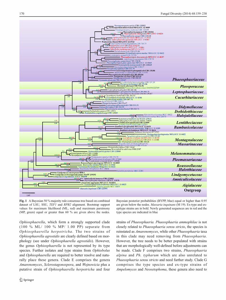
Three phylogenetic reconstructions were generated to clarifythe phylogenetic relationships of genera and species that havebeen accommodated in Phaeosphaeriaceae. Figure 1 dealswith the genus and family level placement within thePhaeosphaeriaceae and Pleosporales. The dataset includes
LSU, SSU, RPB2, and TEF1 sequences and were analyzedusing maximum likelihood (ML), maximum parsimony (MP)and Bayesian analyses. The dataset comprised 85 taxa, withMassaria inquinans (M 19) selected as the outgroup taxon.Twenty-nine Phaeosphaeriaceae taxa formed a well-supported clade (74 % ML/ 1.00 PP) within the suborderPleosporinae, while Mycopappus and Xenostigmina form asingle clade close to Melanommataceae. Therefore, we ex-clude these later genera fromPhaeosphaeriaceae in this study.Wilmia was synonymized under Letendraea by Ariyawansaet al. (2014b). Phylogenetic evidence shows Letendraeacordylinicola is related withDidymosphaeriaceaewhich forma strongly-supported clade (75 % ML/ 1.00 PP) withParaphaeosphaeria michotii (CBS 652.86), Deniquelatabarringtoniae (MFLUCC 11-0422) and Kalmusia brevispora(KT 2313) in Didymosphaeriaceae. Thus, Letendraea is ex-cluded fromPhaeosphariaceae in this study and transferred toD i d y m o s p h a e r i a c e a e . P h a e o s p h a e r i o p s i s ,Parastagonospora, Ophiosphaerella, Phaeosphaeria andSetophoma are well-resolved genera and formed distinctclades, while the other clades comprised groups of generawithout clear combining features or comprising more thanone genus (Fig. 2).
Figure 2 focuses on the phylogenetic relationships of gen-era in Phaeosphaeriaceae. The multigene combined dataset(ITS, LSU, SSU and RPB2) were obtained from maximumlikelihood (ML), maximum parsimony (MP) and Bayesiananalysis. Didymella exigua (CBS 183.55) was selected asthe outgroup taxon. The dataset consists of 58 taxa, with 57taxa belonging to the Phaeosphaeriaceae. Eleven clades (A–K) can be recognized in Fig. 2. Clade A comprises the generaChaetosphaeronema, Dermatiopleospora, Entodesmium,Loratospora . Nodulosphaer ia and Ophiobo lus .Dermatiopleospora, Entodesmium and Loratospora are rep-resented by their ex-type strains and type species. This cladeneeds better populating with many more species from therespective genera as it is presently not well-resolved.Ophiobolus cirsii (MFLUCC 13-0218) formed a robust clade(99 % ML/ 97 % MP/ 1.00 PP) with the asexual genusChaetosphaeronema, while O. erythrosporus (MFLU 12-2225) forms a weakly supported clade with Entodesmiumrude. However, Ophiobolus is not represented by its typespecies and the strains used have not been morphologicallyclearly defined (see Ariyawansa et al. 2014c). Clade B repre-sents the genus Phaeosphaeriopsis and is well resolved inPhaeosphaeriaceae (97 % ML/ 93 % MP/ 1.00 PP). It isrepresented by the type species Ps. glaucopunctata (ex-typestrain), plus other strains and a new species Ps. dracaenicolais also introduced here (see details in “Taxonomy” section).Clade C represents Phaeosphaeria sensu stricto with fivespecies, including the type species and two new species;detailed phylogenetic relationships among these species areshown in Fig. 3. Clade D represents two isolates of the genus
Fungal Diversity (2014) 68:159–238 169
Ophiosphaerella, which form a strongly supported clade(100 % ML/ 100 % MP/ 1.00 PP) separate fromOphiosphaerella herpotricha . The two strains ofOphiosphaerella agrostidis are clearly defined based on mor-phology (see under Ophiosphaerella agrostidis). However,the genus Ophiosphaerella is not represented by its typespecies. Further isolates and type strains from Ophiobolusand Ophiosphaerella are required to better resolve and natu-rally place these genera. Clade E comprises the generaAmarenomyces, Sclerostagonospora, and Wojnowicia plus aputative strain of Ophiosphaerella herpotricha and four
strains of Phaeosphaeria. Phaeosphaeria ammophilae is notclosely related to Phaeosphaeria sensu stricto, the species isreinstated as Amarenomyces, while other Phaeosphaeria taxain this clade may need removing from Phaeosphaeria.However, the tree needs to be better populated with strainsthat are morphologically well-defined before adjustments canbe made. Clade F comprises two strains, Phaeosphaeriaalpina and Ph. typharum which are also unrelated toPhaeosphaeria sensu stricto and need further study. Clade Gcomprises the type species and ex-type strains ofAmpelomyces and Neosetophoma, these genera also need to
Fig. 1 ABayesian 50%majority rule consensus tree based on combineddataset of LSU, SSU, TEF1 and RPB2 alignment. Bootstrap supportvalues for maximum likelihood (ML, red) and maximum parsimony(MP, green) equal or greater than 60 % are given above the nodes.
Bayesian posterior probabilities (BYPP, blue) equal or higher than 0.95are given below the nodes. Massaria inquinans (M 19). Ex-type and ex-epitype strains are in bold. Newly generated sequences are in red and thetype species are indicated in blue
170 Fungal Diversity (2014) 68:159–238
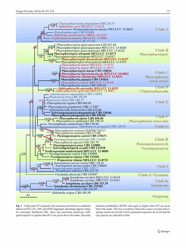
Fig. 2 ABayesian 50%majority rule consensus tree based on combineddataset of ITS, LSU, SSU and RPB2 alignment. Bootstrap support valuesfor maximum likelihood (ML, blue) and maximum parsimony (MP,green) equal to or greater than 60 % are given above the nodes. Bayesian
posterior probabilities (BYPP, red) equal or higher than 0.95 are givenbelow the nodes. The tree is rooted toDidymella exigua. Ex-type and ex-epitype strains are in bold. Newly generated sequences are in red and thetype species are indicated in blue
Fungal Diversity (2014) 68:159–238 171
be better populated with strains that are morphologicallywell-defined. Clade H comprises the ex-types strains andtype species of Parastagonospora and, Neostagonospora,and ex-type strains Sclerostagonospora cycadis ,Scolicosporium minkeviciusii and Wojnowicia viburni (thelatter three species are not generic types). ThreePhaeosphaeria sensu lato strains also cluster here indicat-ing the polyphyletic nature of the genus. Clade I comprisesXenoseptoria, Setomelanomma and Paraphoma, withXenoseptoria and Paraphoma represented by ex-typestrains of their type species. Setomelanomma is closelyrelated to Xenoseptoria which formed a robust clade(97 % ML/ 71 % MP) in our multigene phylogenetic anal-yses. Clade J represents Vrystaatia with single species,V. aloeicola. Clade K represents the asexual genusSetophoma with five strains, including the type species,S. terrestris. The genus is well-resolved in our phylogenetic
analyses which form a strongly supported clade (100 %ML/ 64 % MP/ 1.00 PP) at the basal of Phaeosphariaceae.
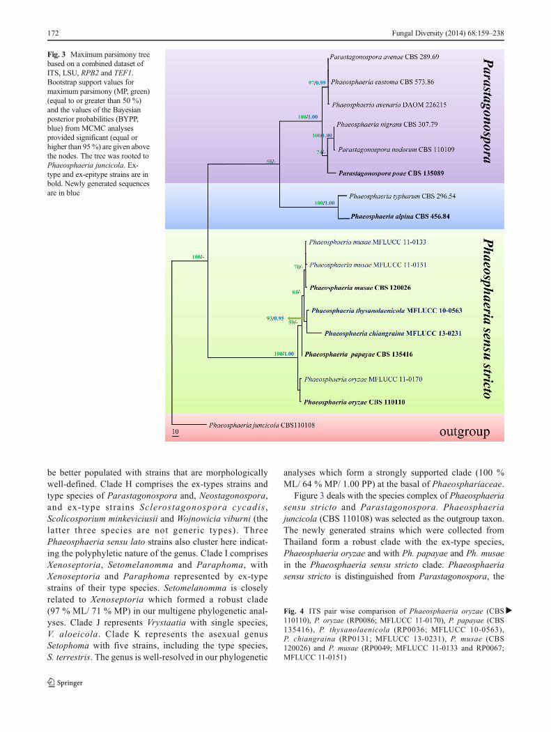
Figure 3 deals with the species complex of Phaeosphaeriasensu stricto and Parastagonospora. Phaeosphaeriajuncicola (CBS 110108) was selected as the outgroup taxon.The newly generated strains which were collected fromThailand form a robust clade with the ex-type species,Phaeosphaeria oryzae and with Ph. papayae and Ph. musaein the Phaeosphaeria sensu stricto clade. Phaeosphaeriasensu stricto is distinguished from Parastagonospora, the
Fig. 3 Maximum parsimony treebased on a combined dataset ofITS, LSU, RPB2 and TEF1.Bootstrap support values formaximum parsimony (MP, green)(equal to or greater than 50 %)and the values of the Bayesianposterior probabilities (BYPP,blue) from MCMC analysesprovided significant (equal orhigher than 95%) are given abovethe nodes. The tree was rooted toPhaeosphaeria juncicola. Ex-type and ex-epitype strains are inbold. Newly generated sequencesare in blue
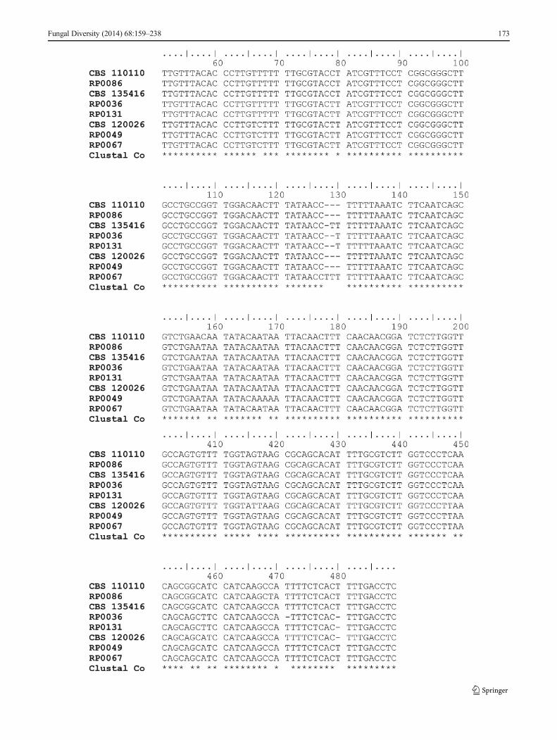
�Fig. 4 ITS pair wise comparison of Phaeosphaeria oryzae (CBS110110), P. oryzae (RP0086; MFLUCC 11-0170), P. papayae (CBS135416), P. thysanolaenicola (RP0036; MFLUCC 10-0563),P. chiangraina (RP0131; MFLUCC 13-0231), P. musae (CBS120026) and P. musae (RP0049; MFLUCC 11-0133 and RP0067;MFLUCC 11-0151)
172 Fungal Diversity (2014) 68:159–238
Fungal Diversity (2014) 68:159–238 173
species having been accommodated in Phaeosphaeria andPhaeosphaeria sensu lato (Phaeosphaeria alpina and Ph.typharum).
Phaeosphaeria sensu stricto taxa (MFLUCC 11-0133;MFLUCC 11-0151) cluster with Ph. musae (CBS 120026),a pathogen of banana. An ITS pair wise comparison showsthat these three isolates differ in seven base positions (Fig. 4)while there are only one to two base differentiated in TEF1gene comparisons. Isolate MFLUCC 11-0133 is slightly mor-phologically different from the other two isolates (MFLUCC11-0151 and CBS 120026) in its ascus and ascospore sizes.However, these three isolates are generally similar in mor-phology and phylogeny. Therefore, we treat the isolates caus-ing leaf spot disease on Calathea sp. and Cordyline sp. fromThailand as conspecific with Phaeosphaeria musae.Phaeosphaeria thysanolaenicola (MFLUCC 10-0563), apathogen on Thysanolaena maxima, forms a clade with. Ph.chiangraina, which is saprobic on rice. These two speciesform strongly-supported clades which are well-resolved fromthe ex-type species of Phaeosphaeria oryzae.
Phaeosphaeria avenaria, Ph. eustoma and Ph. nigransfo rm a c l ade wi th Paras tagonospora spec i e s .Phaeosphaeria avenaria was synonymized underParastagonospora avenae by Quaedvlieg et al. (2013), andthe other two species may need to be synonymized underParastagonospora based on the phylogenetic relationships.However, it was not possible to observe the types of thesespecies or confirm if they are correctly identified.
Phaeosphaeria alpina and Ph. typharum form a distinctclade from the Phaeosphaeria sensu stricto species which isseparate with other Phaeosphaeria sensu lato taxa. The typespecies need to re-visited and a new genus may need to beintroduced to accommodate these species.
Taxonomy
Phaeosphaeriaceae M.E. Barr Mycologia 71(5): 948 (1979).Faces of Fungi number: FoF00232Endophytic, hyperparasitic, pathogenic, or saprobic on
monocotyledonous and some dicotyledonous hosts. Sexualstate: Ascomata scattered, clustered, solitary or gregarious,immersed to semi-immersed or erumpent through host tissue,small to large, uniloculate, globose to subglobose, glabrous orsetose, brown to dark brown or black, ostiolate, with short tolong papilla. Peridium with thin to thick walls, composed ofbrown to dark brown or black, pseudoparenchymatous cells,mostly arranged in a textura angularis to textura prismatica.Hamathecium composed of sparse to numerous, filamentous,s ep t a t e , r a r e l y ana s t omos ing , mos t l y ce l l u l a rpseudoparaphyses, or pseudoparaphyses occasionally lack-ing. Asci 8-spored, bitunicate, fissitunicate, cylindrical to cy-lindric-clavate, or clavate, sessile to subsessile, or usually with
short pedicels, apically rounded with typically well-developedocular chamber. Ascospores overlapping muriform,phragmosporous, or scolecosporous, varying in shape fromellipsoidal, fusiform to broadly fusiform, clavate, and cylin-drical to filiform, mostly brown to dark brown or reddish-brown, sometimes hyaline, 1- to multi-septate, constricted ornot constricted at the septum, walls smooth or rough,echinulate, punctuate or verrucose, some with appendage ormucilaginous sheath. Asexual state: Coelomycetous.Conidiomata pycnidial, scattered, solitary or gregarious,immersed to superficial, uni- to multi-loculate, globoseto subglobose, glabrous or setose, light brown to darkbrown or black, ostiole central, papillate. Conidiomatawalls thin, composed of pseudoparenchymatous cells, ar-ranged in a textura angularis to textura prismatica.Conidiophores lining inner cavity of the conidioma, typ-ically reduced to conidiogenous cells, hyaline to brown,aseptate or septate. Conidiogenous cells enteroblastic orholoblastic, annellidic or phialidic, discrete, oblong toampulliform, hyaline to brown. Conidia varied in shape,hyaline to brown, aseptate or septate, walls smooth orrough, echinulate, punctuate or verrucose, some with ap-pendage or mucilaginous sheath.
Notes: Phaeosphaeriaceae is one of the largest familiesin Pleosporales, and demonstrates variability in morpho-logical characters. Previously, many genera with brownspores, associated with monocotyledonous hosts, having athin-walled peridium and producing coelomycetous asex-ual states were accommodated in Phaeosphaeriaceae(Barr 1979, 1987b, 1990a, 1992a; Crivelli 1983; Schatz1984; Shoemaker 1984; Shoemaker and Babcock 1989b;Yuan 1994; Ramaley and Barr 1995; Dianese et al. 2001;Zhang e t a l . 2009, 2012) . Mos t o f genera inPhaeosphaeriaceae lack molecular data or only limitedsequence data is available in some genera such asEntodesmium and Phaeosphaeria. Several genera inPhaeosphaeriaceae share morphological characters withgenera in other families (e.g. Didymosphaeriaceae,Leptosphaeriaceae and Pleosporaceae) and thus theirplacement is confused. Therefore, re-visiting all type gen-era of Phaeosphaeriaceae to establish their morphologyand to obtain molecular sequence data from fresh collec-tions are essential to provide modern descriptions and todetermine the placement of genera in this family.
Type: Phaeosphaeria I. Miyake, Bot. Mag., Tokyo 23: 93(1909).
Note s : The fo l l ow ing gene r a a r e p l a ced inPhaeosphaeriaceae. Genera are listed in alphabetical orderand provided with genus description and notes, descrip-tions of type or authentic species, notes on species, ma-terial examined and illustrations. If the genus is monotyp-ic, only a single description which also represents thegenus is provided.
174 Fungal Diversity (2014) 68:159–238
Key to genera of Phaeosphaeriaceae
1. Sexual state . . . . . . . . . . . . . . . . . . . . . . . . . . . . . . . . . . . . . . . . . 21. Asexual state . . . . . . . . . . . . . . . . . . . . . . . . . . . . . . . . . . . . . . 14
2. Ascospores phragmosporous or scolecosporous . . . . . . 32. Ascospores dictyosporous, muriform, on Ononis
spinosa (dicotyledon) . . . . . . . . . . . . .Dematiopleospora3. Ascospores phragmosporous . . . . . . . . . . . . . . . . . . . . . . . . . 43. Ascospores scolecosporous . . . . . . . . . . . . . . . . . . . . . . . . . 12
4. Ascospores 2–3-septate . . . . . . . . . . . . . . . . . . . . . . . . . . . 54. Ascospores mostlymore than 3-septate . . . . . . . . . . . . . 10
5. Ascospores mostly 2-septate, associated with rustfungi . . . . . . . . . . . . . . . . . . . . . . . . Eudarluca
5. Ascospores 3-septate, saprobic or pathogenic on monocotsor dicots . . . . . . . . . . . . . . . . . . . . . . . . . . . . . . . . . . . . . . . . . . . 66. Ascomata in groups or rows with slit-like ostioles;
ascospores hyaline . . . . . . . . . . . . . . . . . . . . . . .Bricookea6. Ascomata solitary, scattered, opening ostiolate to pa-
pillate; ascospores pale brown, yellowish brown tobrown . . . . . . . . . . . . . . . . . . . . . . . . . . . . . . . . . . . . .7
7. Asci ovoid to ampulliform, aparaphysate, on Juncusroemerianus . . . . . . . . . . . . . . . . . . . . . . . . . . . . . Loratospora
7. Asci broadly cylindrical, cellular pseudoparaphyses . . . . . 88. Ascomata typically glabrous . . . . . . . . . . . . . . . . . . . . . . . 98. Ascomata typically setose . . . . . . . . . . . Setomelanomma
9. Ascomata typically immersed, ostioles with minute papilla,asexual state:Phaeoseptoria . . . . . . . . . . . . . Phaeosphaeria
9. Ascomata semi-immersed to erumpent, near superfi-cial, ostiole with conical, knob-like, asexual:unknown . . . . . . . . . . . . . . . . . . . Dothideopsella10. Ascospores cylindrical, with typically one enlarged
cell . . . . . . . . . . . . . . . . . . . . . . . . . . . . . . . . . . . . . . . . . 1110. Ascospores broadly fusiform, widest at the middle cell,
saprobic on Ammophila . . . . . . . . . . . . .Amarenomyces11. Papilla, with numerous long, large setae in the
ostiole, ascospores with one enlarged cell nearapex . . . . . . . . . . . . . . . . . . . . Nodulosphaeria
11. Papilla absent, opening ostiolate, ascospores typicallyenlarged cell near the base . . . . . . . . . . Phaeosphaeriopsis
12. Ascospores with enlarged cells . . . . . . . . . . .Ophiobolus12. Ascospores lacking enlarged cells . . . . . . . . . . . . . . . . 13
13. Ascospores constricted at the septa, breaking into partspores, saprobic or parasitic on legumes . . . .Entodesmium
13. Ascospores non-constricted at the septum, notseparating into part spores, saprobic on variousmonocots . . . . . . . . . . . . . . . . Ophiosphaerella
14. Conidia amerosporous, didymosporous phragmosporousor scolecosporous . . . . . . . . . . . . . . . . . . . . . . . . . . . . . . . . 15
14. Conidia dictyosporous (muriform) . . .Amarenographium15. Conidia aseptate . . . . . . . . . . . . . . . . . . . . . . . . . . . . . . . . . . .1615. Conidia septate . . . . . . . . . . . . . . . . . . . . . . . . . . . . . . . . . . . 18
16. Saprobic or pathogenic, conidiomata setose . . . . . . . . 1716. Mycoparasitic on powdery mildew . . . . .Ampelomyces
17 . Con id ioma ta da rk b rown , no sexua l s t a t ereported . . . . . . . . . . . . . . . . . . . . . . . . . . . . . . . Paraphoma
17. Conidiomata pale brown, olivaceous to olivaceous-black,sexual state: ascospores cylindrical, hyaline, 3-septate,with enlarged in second cell from apex . . . . . . Setophoma18. Conidiomata setose or covered bymycelium . . . . . . . 1918. Conidiomata glabrous . . . . . . . . . . . . . . . . . . . . . . . . . . 21
19. Conidia 1–3-septate . . . . . . . . . . . . . . . . . . . . . . . . . . . . . . . 2019. Conidia more than 3-septate . . . . . . . . . . . . . . .Wojnowicia
20. Conidiophores present, hyaline, 1-septateconidia . . . . . . . . . . . . Chaetosphaeronema
20. Conidiophores absent, yellowish, typically 0–1(−3)-septate conidia . . . . . . . . . . . . . . . . . . . . .Neosetophoma
21. Conidia 1-septate . . . . . . . . . . . . . . . . . . . . . . . . . . . . . . . . . 2221. Conidia more than 1-septate . . . . . . . . . . . . . . . . . . . . . . . . 23
22. Conidia with appendage . . . . . . . . . . . . . . . . . Tiarospora22. Conidia without appendage . . . . . . . .Neostagonospora
23. Conidia hyaline . . . . . . . . . . . . . . . . . . . . . . . . . . . . . . . . . . . 2423. Conidia pigmented . . . . . . . . . . . . . . . . . . . . . . . . . . . . . . . . 26
24. One type of conidia, cylindrical to obclavate . . . . . . . 2524. Two types of conidia (macroconidia subcylindrical to
narrowly obclavate; microconidia aseptate, pear-shaped to globose or ellipsoid) . . . . . . . . . . . .Vrystaatia
25. Conidiomata exuding a creamy conidial mass, co-nidiophores absent; sexual state: phaeosphaeria-like . . . . . . . . . . . . . . . . . . . Parastagonospora
25. Conidiomata exuding a pink to orange conidialmass, conidiophores branched; sexual state: ??Setomelanomma . . . . . . . . . . . . . . Xenoseptoria
26. Conidia usually 3-septate . . . . . . . . . . . . . . . . . . . . . . . . 2726. Conidia usually more than 3-septate . . . . . . . . . . . . . . 29
27. Conidia produced one type of conidia . . . . . . . . . . . . . . . . 2827. Conidia produced two types of conidia (microconidia
globose to ellipsoidal, hyaline) . . . . . Phaeostagonospora28. Conidia subcylindrical, pale brown, minutely
verruculose . . . . . . . . . . . . . . . . . . . Sclerostagonospora28. Conidia subcylindrical, wider in the middle, yellowish-
brown, smooth-walled . . . . . . . Septoriella oudemansii29. Conidia fusiform, 5–6-septate, apical cell attenuated into a
short, beak-like, appendage . . . . . . . . . . . . Scolecosporiella29. Conidia curved to sigmoid, 6–7-septate, with hyaline end
cells . . . . . . . . . . . . . . . . . . . . ScolicosporiumminkeviciusiiAmarenographium O.E. Erikss., Mycotaxon 15: 199
(1982), Faces of Fungi number: FoF00237.Saprobic on marine grasses. Sexual state: see notes.
Asexual state: Conidiomata pycnidial, scattered, solitary,immersed to erumpent through host tissue by papilla,uniloculate, ellipsoidal to subglobose, glabrous, brown to darkbrown, ostiole central, with papilla. Conidiomata walls thin,composed of thick-walled, reddish-brown to dark brown cells,arranged in textura angularis to textura prismatica.Conidiophores mostly reduced to conidiogenous cells, hya-line to brown, septate, branched. Conidiogenous cells
Fungal Diversity (2014) 68:159–238 175
holoblastic, phialidic, discrete, determinate, ampulliform,rarely cylindrical, hyaline, smooth-walled, produced macro-and microconidia. Macroconidia dictyosporous, blastic-phialidic, broadly ellipsoidal to clavate or fusiform, brown todark brown, paler at the ends cells, muriform, septate, smooth-walled, with mucoid appendage at apex. Microconidiaamerosporous, blastic-phialidic, hyaline, smooth-walled(from Eriksson 1982; Nag Raj 1989).
Type species: Amarenographium metableticumPhylogenetic study: Hodhod et al. (2012)Notes: Amarenographium metableticum (Trail) O.E.
Erikss. is the type ofAmarenographiumwhich was introducedas the asexual state of Amarenomyces (Eriksson 1982; NagRaj 1989; Hodhod et al. 2012). Amarenographiummetablet icum had previously been identi f ied asCamarosporiummetableticumwhich was not congeneric withCamarosporium stephensi i , the gener ic type ofCamarosporium at that time (Sutton 1980; Eriksson 1982).Therefore, Eriksson (1982) transferred Camarosporiummetableticum to a new genus based on the different morpho-logical characters (Hodhod et al. 2012; Hyde et al. 2013).Amarenographium differs from Camarosporium by its conid-iophores and conidia. In Amarenographium conidiophores arelonger and branched and conidia have gelatinous appendages(Eriksson 1982; Hodhod et al. 2012). Hodhod et al. (2012)introduced a new species of Amarenographium, Am. soliumAbdel-Wahab et al. from mangroves and noted thatAmarenographium did not belong in Phaeosphaeriaceae butclusters with Pleosporales, genera incertae sedis, whileAmarenomyces formed a clade with Phaeosphaeria inPhaeosphaeriaceae (Zhang et al. 2009, 2012). However,Hodhod et al. (2012) did not study the type species and thetype species of Amarenographium lacks molecular data toconfirm the relationships between these genera. Thus wepresently maintain the genus in Phaeosphaeriaceae.
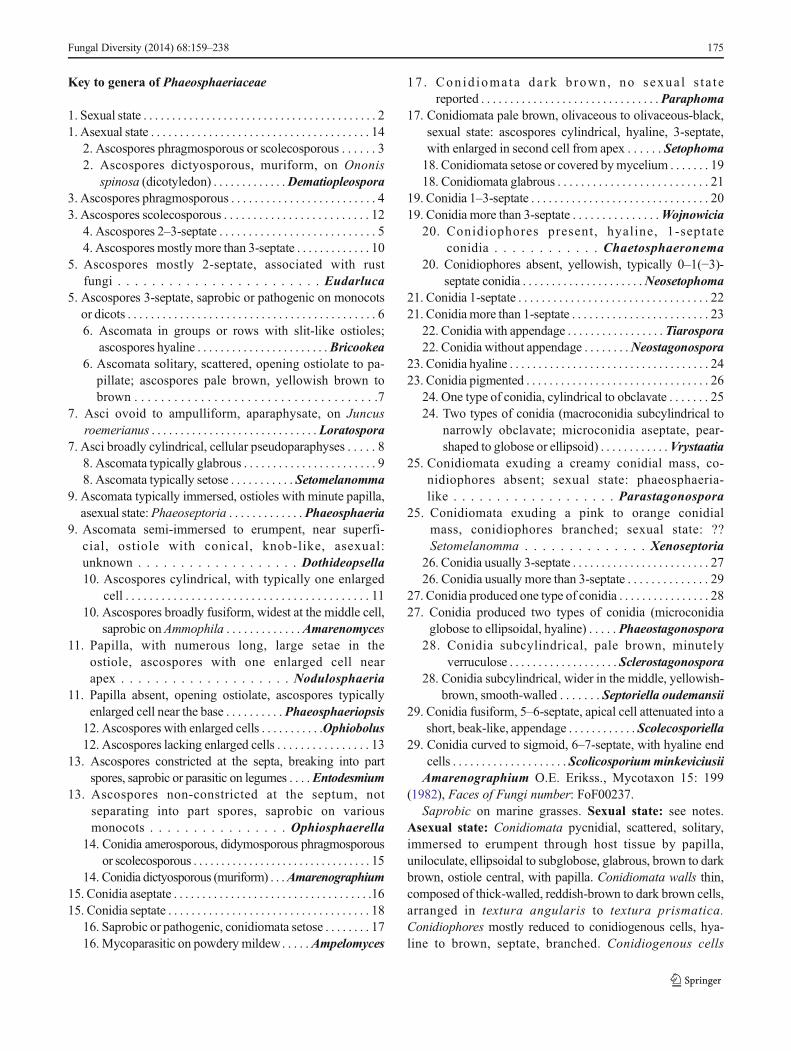
Amarenographium metableticum (Trail) O.E. Erikss.,Mycotaxon 15: 199 (1982), Faces of Fungi number:FoF00238, Fig. 5.
≡CamarosporiummetableticumTrail, Scott. Natural., N.S.2 (‘8’): 267 (1886) [1885–1886]
Saprobic on Ammophila. Sexual state: see notes. Asexualstate: Conidiomata 160–215.5μm high, 150–270μm diam.,pycnidial, scattered, solitary, immersed to erumpent, visible asblack spots on host surface, uniloculate, globose tosubglobose, glabrous, dark brown, ostiole central, with shortto long papilla. Conidiomata walls 13–19μm wide, thin-walled, of equal thickness, composed of 3–5 layers of flat-tened, brown to dark brown, pseudoparenchymatous cells,arranged in a textura angularis to textura prismatica.Conidiophores arising from basal cavity of conidioma, mostlyreduced to conidiogenous cells, hyaline to brown, septate.Conidiogenous cells (1.5–)3–4(−9)×1.5–2.5(−5) μm(x = 3.9×2.3μm, n=10), holoblastic, phialidic, discrete,
oblong to ampulliform, hyaline to brown, 1-septate at thebase, smooth-walled. Conidia (20–)24–27(−29)×12–15(−17) μm (x = 25.4 × 13.4μm , n= 30), solitary,dictyosporous, muriform, ellipsoidal, ovoid, or lageniform,with obtuse ends or sometimes truncate at the basal cell,initially hyaline, becoming brown to yellowish-brown at ma-turity, mostly 3–4 transverse septa, 1–4 longitudinal septa,with several segments, smooth and thick-walled, with indis-tinct mucoid appendage.
Material examined: SWEDEN, Öland, Böda parish,Lugnet beach, on Ammophila arenari (L.) Link (Poaceae), 2June 1963, B. Eriksson & O. Eriksson 2022c (S-F207509).
Amarenomyces O.E. Erikss., Op. bot. 60: 124 (1981),Faces of Fungi number: FoF00235.
Type species: Amarenomyces ammophilaePhylogenetic study: Zhang et al. (2009, 2012), Hyde et al.
(2013)Amarenomyces ammophilae (Lasch) O.E. Erikss., Op.
bot. 60: 124 (1981), Faces of Fungi number: FoF00236,Fig. 6.
≡ Sphaeria ammophilae Lasch Flora, Jena 33: 282 (1850)Saprobic on Ammophila. Sexual state: Ascomata 170–
290μm high, 170–285μm diam., scattered, immersed toerumpent through host tissue, visible as small black spots onhost surface, uniloculate, globose to subglobose, glabrous,dark brown, ostiole central, with short papilla (50–130μmlong). Peridium 7.5–25μm wide, thin-walled, of equal thick-ness, composed of 3–5 layers of flattened, dark brown,pseudoparenchymatous cells, arranged in textura angularisto textura prismatica. Hamathecium composed of numer-ous, 1.5–3μm wide, filiform, distinctly septate, frequentlyanastomosing, narrow cellular pseudoparaphyses, embed-ded in mucilaginous matrix. Asci (112–)140–160(−173)×(28–)30–35(−37) μm (x = 140×32.4μm, n=25), 8-spored,bitunicate, fissitunicate, broadly cylindrical, subsessilewith minute knob-like pedicel, apically rounded, with awell-developed ocular chamber. Ascospores 39–47(−50)×12–14μm (x = 43.4 × 13.2μm , n= 30), overlapping1–3-seriate, phragmosporous, broadly fusiform, rarelycylindric-clavate, widest at the middle cell, pale yellowishwhen immature, becoming yellowish-brown to brown atmaturity, 6-septate, slightly constrict at the septum, smoothand thick-walled.
Material examined: BELGIUM, West-Vlaanderen,Blankenbergh, on Ammophila arenaria L. (Poaceae),Westendorp (S-F77691); ITALY, Sardegna, Cagliari, LaMaddelena, March 1866, Marcucci (S-F77741).
Notes: Amarenomyces was introduced by Eriksson (1981)to accommodate the single species from a marine grass, whichtraditionally has been identified as Sphaeria ammophilaeLasch. Eriksson (1981) mentioned that the genus should be-long in Botryosphaeriaceae rather than Phaeosphaeriaceae asits morphological characters differed from Phaeosphaeria.
176 Fungal Diversity (2014) 68:159–238
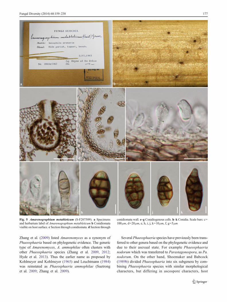
Zhang et al. (2009) listed Amarenomyces as a synonym ofPhaeosphaeria based on phylogenetic evidence. The generictype of Amarenomyces, A. ammophilae often clusters withother Phaeosphaeria species (Zhang et al. 2009, 2012;Hyde et al. 2013). Thus the earlier name as proposed byKohlmeyer and Kohlmeyer (1965) and Leuchtmann (1984)was reinstated as Phaeosphaeria ammophilae (Suetronget al. 2009; Zhang et al. 2009).
Several Phaeosphaeria species have previously been trans-ferred to other genera based on the phylogenetic evidence anddue to their asexual state. For example Phaeosphaerianodorum which was transferred to Parastagonospora, as Pa.nodorum. On the other hand, Shoemaker and Babcock(1989b) divided Phaeosphaeria into six subgenera by com-bining Phaeosphaeria species with similar morphologicalcharacters, but differing in ascospore characters, host
Fungal Diversity (2014) 68:159–238 177
Fig. 5 Amarenographium metableticum (S-F207509). a Specimensand herbarium label of Amarenographium metableticum b Conidiomatavisible on host surface. c Section through conidiomata. d Section through
conidiomata wall. e–g Conidiogenous cells. h–k Conidia. Scale bars: c=100μm, d=20μm, e, h, i, j, k=10μm, f, g=5μm
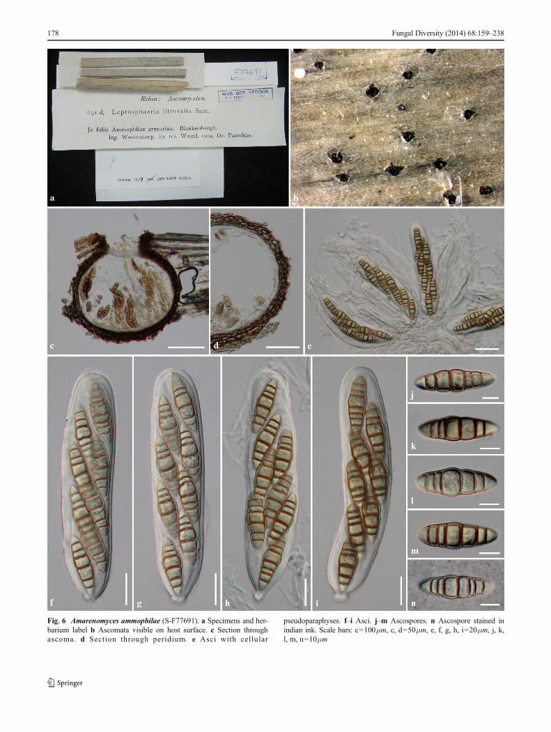
178 Fungal Diversity (2014) 68:159–238
Fig. 6 Amarenomyces ammophilae (S-F77691). a Specimens and her-barium label b Ascomata visible on host surface. c Section throughascoma. d Section through peridium. e Asci with cellular
pseudoparaphyses. f–i Asci. j–m Ascospores. n Ascospore stained inindian ink. Scale bars: c=100μm, c, d=50μm, e, f, g, h, i=20μm, j, k,l, m, n=10μm
preference and asexual state (Câmara et al. 2002; Zhang et al.2012; Quaedvlieg et al. 2013). The asexual state ofPhaeosphaeria has been reported as Phaeoseptoria and thisis supported by phylogenetic data (Quaedvlieg et al. 2013).Quaedvlieg et al. (2013) transferred Phaeosphaeria specieswhich did not produce Phaeoseptoria asexual states to othergenera. Therefore, the genus Amarenomyces is reinstated inthis study based on the phylogenetic analysis asA. ammophilae forms a separate clade to the type species,Phaeosphaeria oryzae. Although A. ammophilae has oftenbeen reported to be the sexual state of Amarenographiummetableticum (Trail) O.E. Erikss., they are not treated ascongeneric here. The presumed link was established as bothspecies occur on the same host in close proximity. There is,however, no sequence data for Amarenographiummetableticum and it might be predicted that if both specieswere host-specific then it would not be surprising that theyboth occur on the same host. We therefore refrain from com-bining the taxa until molecular data confirms the link.
Ampelomyces Ces. ex Schlecht., Bot. Zeit. 10: 302 (1852),Faces of Fungi number: FoF00291.
Hyperparasitic on Erysiphales or saprobic. Sexual state:unknown. Asexual state: Conidiomata pycnidial, scattered,solitary, superficial, uniloculate, globose or elongated to pyr-iform, pale brown, ostiolate, sometimes papillate.Conidiomata walls thick, composed of pale brown cells,arranged in textura angularis. Conidiophores mostly reducedto conidiogenous cells. Conidiogenous cells enteroblastic,phialidic, determinate, discrete, doliiform to ampulliform, hy-aline, smooth-walled, channel and collarette minute, periclinalwall towards apex thickened, formed directly from the pyc-nidial wall cells. Conidia amerosporous, cylindrical to fusi-form, straight to curved, very pale brown, aseptate, thin andsmooth-walled, with guttules. (Sutton 1980)
Type species: Ampelomyces quisqualis Ces., Bot. Ztg. 10:301 (1852).
Phylogenetic study: Kiss and Nakasone (1998),Szentiványi et al. (2005), Liang et al. (2007), De Gruyteret al. (2009), Aveskamp et al. (2010), Hyde et al. (2013),Wijayawardene et al.(2013a)
Notes: Ampelomyces species have been reported asmycoparasites on powdery mildews (Erysiphaceae) and areworldwide in nature (de Bary 1870; Griffith 1899; Kiss 1998;Kiss et al. 2004). The genus was introduced by Cesati (1852)to accommodate taxa that formed pycnidia as intracellularmycoparasites on powdery mildews (De Bary 1870; Kisset al. 2004). Previously, Ampelomyces had varied morpholog-ical and cultural characteristic with species differentiatedbased on pycnidia, cultures and conidial types in isolates ofpycnidial mycoparasites from powdery mildews (Clare 1964;Belsare et al. 1980; Kiss et al. 2004). In Kiss et al. (2004),Ampelomyces species could be distinguished into slow andfast growing types. Based on the rDNA ITS region
phylogenetic analysis, Kiss et al. (2004) concluded that strainsfrom the two types of differentiated growth rate were notcongeneric. The slow-growing isolates, obtained from intra-cellular pycnidia in powdery mildew mycelia wasAmpelomyces sensu stricto. While fast growing isolates fromsessile pycnidia on mildew-infected leaves, were closely re-lated to Phoma species. Ampelomyces quercinus (Syd.)Rudakov, A. humuli (Fautrey) Rudakov and A. heraclei(Dejeva) Rudakov are example of fast growing species(Rudakov 1979; Kiss and Nakasone 1998; Sullivan andWhite 2000). De Gruyter et al. (2009) included two isolatesof the type species;A. quisqualis in their phylogenetic analysisand showed that the type species of Ampelomyces can beaccommodated in Phaeosphaeriaceae . WhereasAmpelomyces quercinus grouped in Didymellaceae; thus,Aveskamp et al. (2010) introduced A. quercinus as new com-bination within Phoma in Didymellaceae.
BricookeaM.E. Barr, Mycotaxon 15: 346 (1982), Faces ofFungi number: FoF00239.
Saprobic on Juncaceae. Sexual state: Ascomata scatteredor clustered, immersed to semi-immersed, becomingerumpent through host tissue or superficial at maturity, uni-to multi-loculate, globose to subglobose, glabrous, brown todark brown, ostiole central, with slit-like opening. Peridiumthin-walled, composed of brown to dark brown, pseudopar-enchymatous cells, arranged in a textura angularis.Hamathecium composed of numerous, filamentous, distinctlyseptate, broad cellular pseudoparaphyses, anastomosing at theapex. Asci 8-spored, bitunicate, fissitunicate, cylindrical tocylindric-clavate, narrow towards apex, short pedicellate, api-cally rounded, with well-developed ocular chamber.Ascospores overlapping, 1–3-seriate, phragmosporous, ellip-soidal to broadly fusiform, hyaline, septate, slightly curved,constricted at the median septum, smooth and thick-walled.Asexual state: Unknown.
Type species: Bricookea sepalorumPhylogenetic study: NoneNotes: Barr (1982) introduced Bricookea to accommodate
B. sepalorum that was previously identified as Leptosphaeriasepalorum (Vleugel) Lind (Lind 1928). Holm (1957)observed the collections from Sweden and compared themorphological characters with specimens from NorthAmerica. He mentioned that the ascospores and asci ofSwedish collections were smaller than North Americanspecimens and regarded the species as synonym of Lizoniaemperigonia (Auersw.) de Not. (Barr 1982; Shoemaker andBabcock 1989a; Zhang et al. 2012). Barr (1982) did not agreewith Holm (1957) and designated the new genus,Bricookea toaccommodate Leptosphaeria sepalorum. Barr (1982)suggested placing Bricookea in Phaeosphaeriaceae with itsuniquely opening ostioles and lack of known asexual state.Barr (1982) mentioned that Sphaerulina inquinansRehmmaybe provided as earlier epithet of Bricookea sepalorum.
Fungal Diversity (2014) 68:159–238 179
However, Shoemaker and Babcock (1989a) studied the typespecimens of Bricookea from S herbaria and mentioned thatnomaterial was found under the name Sphaerulina inquinans.Subsequently, Shoemaker and Babcock (1989a) introducedthe new species, Bricookea barrae from a North Americancollection which had been examined by Barr (1982).Bricookea barrae [as Br. barriae] was synonymized underthe name Lophiostoma barriae in Lophiostomataceae byEriksson (2007) leaving the genus monotypic. Zhang et al.(2012) tentatively place Bricookea in Phaeosphaeriaceae.Cu r r e n t l y, Br i c oo k ea i s g en e r a l l y t r e a t e d i nLophiostomataceae based on its slit-like ostioles, but the typespecies, Br. sepalorum has been synonymized under the ear-lier name in Leptosphaeriaceae as Leptosphaeria sepalorum(Index Fungorum 2014).
We examined the collection from North America and typespecimen from S herbaria. Both collections have similar mor-phological characters. However, the specimens from NorthAmerica differ in having larger ascoma, asci and ascosporesthan the type specimen from Europe. In this study, Bricookeaforms a group in pseudostroma, with slit-like ostioles; char-acters typical of Lophiostomataceae. However, we tentativelyplace Bricookea in Pheaosphaeriaceae as its peridium is thin-walled, composed of pseudoparenchymatous cells, asci areclavate with a subsessile to short acute pedicel and ascosporesare phragmosporous. Fresh collection, epitypification andmolecular analysis are needed to confirm the naturalplacement.
Bricookea sepalorum (Vleugel) M.E. Barr, Mycotaxon 15:346 (1982), Faces of Fungi number: FoF00240, Fig. 7.
≡ Metasphaeria sepalorum Vleugel, Svensk bot. Tidskr.2(4): 369 (1908)
Saprobic on Juncus spp. Sexual state: Ascomata 130–210μm high, 170–250μm diam., scattered to clustered, gre-garious, immersed to semi-immersed under epidermis, be-coming erumpent through host tissue, to superficial at matu-rity, visible as black dots in rows or groups on host surface,uni- to multi-loculate, globose to subglobose, glabrous, darkbrown to black, ostioles central, with slit-like opening.Peridium 10–20μm wide, thin-walled, of equal thickness,composed of 2–3 layers of large, brown to dark brown,pseudoparenchymatous cells, arranged in a textura angularis.Hamathecium composed of numerous, 1.5–2μm wide, fila-mentous, distinctly septate, cellular pseudoparaphyses, em-bedded in mucilaginous matrix, anastomosing at the apex.Asci (60–)65–75(−80)×12–14(−16) μm (x = 71.5×13.5μm,n=30), 8-spored, bitunicate, fissitunicate, cylindric-clavate,narrowing towards the apex, with subsessile to shortly acutepedicel, apically rounded with well-developed ocular cham-ber. Ascospores 17.5–19(−21)×6–7.5μm (x = 19×6.4μm,n=30), overlapping 1–2-seriate, phragmosporous, ellipsoidalto broadly fusiform, hyaline to subhyaline, initially formingone median septate, becoming 3-septate at maturity, slightly
curved, constricted at the median septum, easily breaking intopart spores at the median septum, smooth and thin to thick-walled. Asexual state: Unknown.
Material examined: SWEDEN, Suecia. Holmön,Västerbotten, on dead sepals of Juncus filiformis Linn.(Juncaceae), July 1907, J. Vleugel (S-F49661, type); USA,California, Squaw Valley Creek Meadows, Mt. Shasta,Siskiyou Co. alt. 8,500 ft., on dead sepals of Juncus parryiEngelm. (Juncaceae), 10 July 1947, W. B. Cooke & V. G.Cooke (BPI 737398);Washington,Mt. Baker National Forest,Whatcom Co. Subalpine Meadows along Washington Route#1 near Baker Lodge, on dead sepals of Juncus parryiEngelm. (Juncaceae), 12 July 1948, W. B. Cooke & V. G.Cooke 24247 (BPI 737397); California, King Creek Trail,Lassen Volcanic National Park. alt. 8,000 ft., on dead sepalsof Juncus parryi Engelm. (Juncaceae), 1 July 1954 W. B.Cooke & V. G. Cooke 29530 (BPI 737400); California, KingCreek Trail, Lassen Volcanic National Park. alt. 8,000 ft., ondead sepals of Juncus parryi Engelm. (Juncaceae), 1 July1954, W. B. Cooke & V. G. Cooke 29530 (BPI 737399);California, Panther Creek Meadows, Mt. Shasta, SiskiyouCo. alt. 7,600 ft., on dead sepals of Juncus parryi Engelm.(Juncaceae), 4 July 1948, W. B. Cooke 24055 (BPI 737401).
Chaetosphaeronema Moesz, Bot. Közl. 14: 152 (1915),Faces of Fungi number: FoF00241.
Saprobic or pathogenic on Malus, Euphorbia, Anthyllisand Dorycnium spp. Asexual state: Unknown Conidiomatapycnidial, scattered, immersed to semi-immersed, uniloculate,globose, setose, brown to dark brown, ostiole central, withsetose papilla. Conidiomata walls thin, arranged in a texturaprismatica, pale brown, outer layers comprising dark brown,thick-walled cells. Conidiophores arising from basal cavity ofconidiomata mostly reduced to conidiogenous cells.Conidiogenous cells enteroblastic, phialidic, discrete or inte-grated, cylindrical, hyaline, smooth-walled, with minute chan-nel or collarette, hardly distinguishable from the inner wallcells. Conidia didymosporous, oblong to cylindrical, withobtuse ends, straight or slightly curved, hyaline, 1-septate,thin and smooth-walled, with guttules (from Sutton 1980;Quaedvlieg et al. 2013)
Type species: Chaetosphaeronema hispidulum (Corda)Moesz, Bot. Közl. 14: 152 (1915)
= Sphaeronaema hispidulumCorda, Icon. fung. (Prague) 4:39 (1840)
Phylogenetic study: Schoch et al. (2009), De Gruyter et al.(2009, 2010), Zhang et al. (2009, 2012), Hyde et al. (2013),Quaedvlieg et al. (2013).
Notes: Chaetosphaeronema was introduced by Moesz(1915) as typified by Ch. hispidulum to accommodate speciesof Sphaeronaema that were not congeneric. A partial morpho-logical treatment of the genus was provided by Sutton (1980).Petrak (1944) treated Ch. herbarum (Hollós) Moesz as asynonym of Ch. hispidulum and accepted three species in
180 Fungal Diversity (2014) 68:159–238
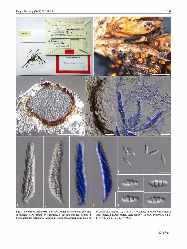
Fungal Diversity (2014) 68:159–238 181
Fig. 7 Bricookea sepalorum (S-F49661, type). a Herbarium label andspecimens b Ascomata on substrate. c Section through stroma dSection through peridium. eAsci with cellular pseudoparaphyses strained
in cotton blue reagent. f–g Asci. h–i Asci stained in cotton blue reagent. jAscospores. k–n Ascospores. Scale bars: b=1000μm, c=100μm, e, f, g,h, i, j=20μm, d, k, l, m, n=10μm
the genus; Ch. collivagum (Petr.) Petr., Ch. galii (Petr.) Petr.and Ch . h i sp idu lum (Su t t on 1980 ) . Howeve r,Chaetosphaeronema was subsequently limited to the typespecies (De Gruyter et al. 2009, 2010; Zhang et al. 2012;Hyde et al. 2013; Quaedvlieg et al. 2013). De Gruyter et al.( 2 0 0 9 ) s h ow e d , u s i n g m o l e c u l a r d a t a t h a tChaetosphaeronema was related to Phaeosphaeriaceae andPleosporaceae. De Gruyter et al. (2010) introduced a newspecies, Ch. coonsii (Boerema & Loer.) Gruyter et al., andt r e a t e d Chae t o s pha e ron ema s en s u s t r i c t o i nPhaeosphaeriaceae. This placement has been widely accept-ed by subsequent authors based on the phylogenetic evidence,while no sexual state is presently known (Schoch et al. 2009;Zhang et al. 2009, 2012; Hyde et al. 2013; Quaedvlieg et al.2013).
In this study, Chaetosphaeronema clusters with varioussexual genera and is closely related to Ophiobolus cirsii(MFLUCC13-0218) as propose by Petrak (1944) and Zhanget al. (2009) who suggested that Chaetosphaeronema may beasexual state of Ophiobolus. The link between these generahas not yet equivocally been proven. Epitypification withmolecular data is therefore needed to confirmed the naturalplacement of these genera and confirm asexual-sexualconnections.
Dematiopleospora Wanasinghe et al., Cryptog. Mycol. 35(2): 105-11(2014), Faces of Fungi number: FoF00242.
Saprobic on Ononis spinosa L (Fabaceae). Sexual state:Ascomata scattered, solitary, superficial, uniloculate, globoseto subglobose, glabrous, dark brown to black, coriaceous,cupulate when dry, ostiole central, minute papilla, with thick,brown, periphyses. Peridium thin-walled, composed of pseu-doparenchymatous cells, outer layer comprising heavilypigmented, thick-walled, reddish to dark brown cells, ar-ranged in textura angularis, inner layer comprising 2–3 celllayers of thin-walled, hyaline cells, arranged in a texturaangularis. Hamathecium composed of numerous, filamen-tous, distinctly septate, broad cellular pseudoparaphyses.Asci 8-spored, bitunicate, fissitunicate, cylindrical to cylin-dric-clavate, short pedicellate, apically rounded with minuteocular chamber. Ascospores overlapping 1–2-seriate,dictyosporous, muriform, ellipsoidal to subfusiform, widestin the upper part, slightly curved, 5–9 transverse septa, with 3–6 longitudinal septa, constricted at the central septum, initiallyhyaline, becoming yellowish-brown at maturity, smooth-walled. Asexual state: Unknown (from Wanasinghe et al.2014)
Type species:Dematiopleospora mariaeWanasinghe et al.,Cryptog. Mycol. 35 (2): 105-11(2014)
Phylogenetic study: Wanasinghe et al. (2014).Notes: Dematiopleospora was introduced by Wanasinghe
et al. (2014) to accommodate D. mariae. The genus is char-acterized by thick, brown, periphyses in the ostiole, superficialascomata and muriform ascospores. Dematiopleospora is
similar to Nodulosphaeria in its periphysate ostiole, butthey differ in peridium structure and ascospore morphol-ogy. Dematiopleospora has muriform ascospores similarto those characterized in Phaeosphaeria vagans (Niessl)O.E. Erikss., Ph. phragmiticola Leuchtm., Ph. phragmitis(Hollós) Leuchtm and Pleoseptum yuccaesedum A.W.Ramaley & M.E. Barr (Shoemaker and Babcock 1989b;Zhang e t a l . 2012 ; Wanas inghe e t a l . 2014) .Dematiopleospora is similar to Pleoseptum, but theydiffer in peridium structures and papillate ostioles.Pleoseptum lacks of molecular data to confirm its naturalplacement. Therefore, the genus is tentatively placed inPleosporaceae based on its muriform ascospores andthick-walled peridium that are dominant characters inPleosporaceae. Multigene phylogenetic analyses indicat-ed that Dematiopleospora belongs to Phaeosphaeriaceaewhich forms a separate clade from those Phaeosphaeriaspec ies and groups wi th Chae tosphaeronema ,Entodesmium, Loratospora and Nodulosphaeria. Noasexual morph is known for Dematiopleospora.
Dothideopsella Höhn., Sber. Akad. Wiss. Wien, Math.-naturw. Kl., Abt. 1 124: 70 (1915), Faces of Fungi number:FoF00295
Saprobic on dead wood and stems in terrestrial habitats.Sexual state: Ascomata scattered, gregarious, immersed tosemi-immersed, or erumpent, nearly superficial, globose,uniloculate, subglobose or obypyriform, glabrous, dark brownto black, ostiole central, broadly or narrowly conical, with adark brown to black papilla. Peridium thin to thick, of unequalthickness, slightly thick at the basal ascoma, composed of darkbrown to black, thin-walled, pseudoparenchymatous cells,arranged in a textura angularis. Hamathecium composed ofnumerous, filamentous, distinctly septate, cellularpseudoparaphyses, anastomosing without a gelatinous coat-ing. Asci 8-spored, bitunicate, fissitunicate, cylindrical to ob-long, with a knob like pedicel, apically rounded with well-developed ocular chamber Ascospores phragmosporous, el-lipsoidal to fusiform, brown or yellowish brown, septate,slightly constricted at the septum, smooth-walled. Asexualstate: Unknown (from Ariyawansa et al. 2013).
Type species: Dothideopsella agminalis (Sacc. &Morthier) Höhn., Sber. Akad. Wiss. Wien, Math.-naturw.Kl., Abt. 1 124: 70 (1915)
Phylogenetic study: NoneNotes: Dothideopsella was introduced by Höhnel (1915)
and is typified by Dothideopsella agminalis (Sacc. &Morthier) Höhn. The genus was established to accommodateDothideomycetes species characterized by a broad peridiumst ruc ture , wi th conical , papi l la te os t io les , andphragmosporous, brown ascospores (Ahn and Shearer 1998;Ariyawansa et al. 2013). Lumbsch and Huhndorf (2010)treated Dothideopsella in Dothideomycetes, genera incertaesedis, while Index Fungorum (2014) lists the genus in
182 Fungal Diversity (2014) 68:159–238
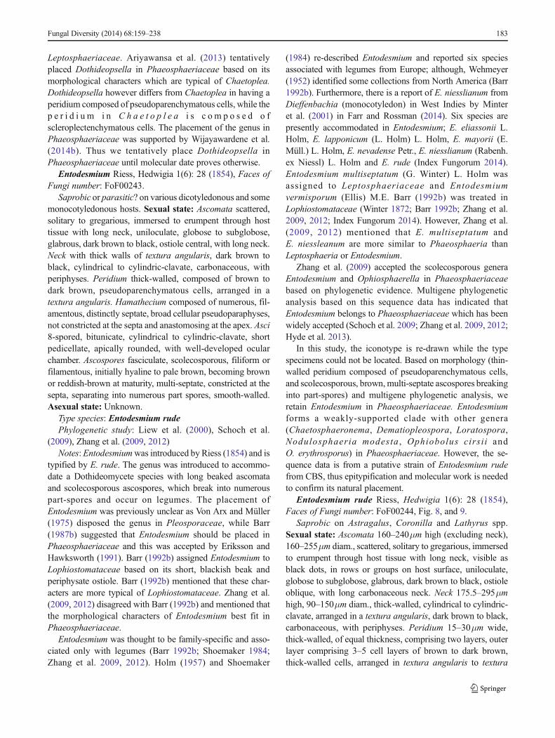
Leptosphaeriaceae. Ariyawansa et al. (2013) tentativelyplaced Dothideopsella in Phaeosphaeriaceae based on itsmorphological characters which are typical of Chaetoplea.Dothideopsella however differs from Chaetoplea in having aperidium composed of pseudoparenchymatous cells, while thep e r i d i u m i n C h a e t o p l e a i s c o m p o s e d o fscleroplectenchymatous cells. The placement of the genus inPhaeosphaeriaceae was supported by Wijayawardene et al.(2014b). Thus we tentatively place Dothideopsella inPhaeosphaeriaceae until molecular date proves otherwise.
Entodesmium Riess, Hedwigia 1(6): 28 (1854), Faces ofFungi number: FoF00243.
Saprobic or parasitic? on various dicotyledonous and somemonocotyledonous hosts. Sexual state: Ascomata scattered,solitary to gregarious, immersed to erumpent through hosttissue with long neck, uniloculate, globose to subglobose,glabrous, dark brown to black, ostiole central, with long neck.Neck with thick walls of textura angularis, dark brown toblack, cylindrical to cylindric-clavate, carbonaceous, withperiphyses. Peridium thick-walled, composed of brown todark brown, pseudoparenchymatous cells, arranged in atextura angularis. Hamathecium composed of numerous, fil-amentous, distinctly septate, broad cellular pseudoparaphyses,not constricted at the septa and anastomosing at the apex. Asci8-spored, bitunicate, cylindrical to cylindric-clavate, shortpedicellate, apically rounded, with well-developed ocularchamber. Ascospores fasciculate, scolecosporous, filiform orfilamentous, initially hyaline to pale brown, becoming brownor reddish-brown at maturity, multi-septate, constricted at thesepta, separating into numerous part spores, smooth-walled.Asexual state: Unknown.
Type species: Entodesmium rudePhylogenetic study: Liew et al. (2000), Schoch et al.
(2009), Zhang et al. (2009, 2012)Notes: Entodesmiumwas introduced by Riess (1854) and is
typified by E. rude. The genus was introduced to accommo-date a Dothideomycete species with long beaked ascomataand scolecosporous ascospores, which break into numerouspart-spores and occur on legumes. The placement ofEntodesmium was previously unclear as Von Arx and Müller(1975) disposed the genus in Pleosporaceae, while Barr(1987b) suggested that Entodesmium should be placed inPhaeosphaeriaceae and this was accepted by Eriksson andHawksworth (1991). Barr (1992b) assigned Entodesmium toLophiostomataceae based on its short, blackish beak andperiphysate ostiole. Barr (1992b) mentioned that these char-acters are more typical of Lophiostomataceae. Zhang et al.(2009, 2012) disagreed with Barr (1992b) and mentioned thatthe morphological characters of Entodesmium best fit inPhaeosphaeriaceae.
Entodesmium was thought to be family-specific and asso-ciated only with legumes (Barr 1992b; Shoemaker 1984;Zhang et al. 2009, 2012). Holm (1957) and Shoemaker
(1984) re-described Entodesmium and reported six speciesassociated with legumes from Europe; although, Wehmeyer(1952) identified some collections from North America (Barr1992b). Furthermore, there is a report of E. niesslianum fromDieffenbachia (monocotyledon) in West Indies by Minteret al. (2001) in Farr and Rossman (2014). Six species arepresently accommodated in Entodesmium; E. eliassonii L.Holm, E. lapponicum (L. Holm) L. Holm, E. mayorii (E.Müll.) L. Holm, E. nevadense Petr., E. niesslianum (Rabenh.ex Niessl) L. Holm and E. rude (Index Fungorum 2014).Entodesmium multiseptatum (G. Winter) L. Holm wasassigned to Leptosphaeriaceae and Entodesmiumvermisporum (Ellis) M.E. Barr (1992b) was treated inLophiostomataceae (Winter 1872; Barr 1992b; Zhang et al.2009, 2012; Index Fungorum 2014). However, Zhang et al.(2009, 2012) mentioned that E. multiseptatum andE. niessleanum are more similar to Phaeosphaeria thanLeptosphaeria or Entodesmium.
Zhang et al. (2009) accepted the scolecosporous generaEntodesmium and Ophiosphaerella in Phaeosphaeriaceaebased on phylogenetic evidence. Multigene phylogeneticanalysis based on this sequence data has indicated thatEntodesmium belongs to Phaeosphaeriaceae which has beenwidely accepted (Schoch et al. 2009; Zhang et al. 2009, 2012;Hyde et al. 2013).
In this study, the iconotype is re-drawn while the typespecimens could not be located. Based on morphology (thin-walled peridium composed of pseudoparenchymatous cells,and scolecosporous, brown,multi-septate ascospores breakinginto part-spores) and multigene phylogenetic analysis, weretain Entodesmium in Phaeosphaeriaceae. Entodesmiumforms a weakly-supported clade with other genera(Chaetosphaeronema, Dematiopleospora, Loratospora,Nodulosphaeria modesta , Ophiobolus cirsi i andO. erythrosporus) in Phaeosphaeriaceae. However, the se-quence data is from a putative strain of Entodesmium rudefrom CBS, thus epitypification and molecular work is neededto confirm its natural placement.
Entodesmium rude Riess, Hedwigia 1(6): 28 (1854),Faces of Fungi number: FoF00244, Fig. 8, and 9.
Saprobic on Astragalus, Coronilla and Lathyrus spp.Sexual state: Ascomata 160–240μm high (excluding neck),160–255μm diam., scattered, solitary to gregarious, immersedto erumpent through host tissue with long neck, visible asblack dots, in rows or groups on host surface, uniloculate,globose to subglobose, glabrous, dark brown to black, ostioleoblique, with long carbonaceous neck. Neck 175.5–295μmhigh, 90–150μm diam., thick-walled, cylindrical to cylindric-clavate, arranged in a textura angularis, dark brown to black,carbonaceous, with periphyses. Peridium 15–30μm wide,thick-walled, of equal thickness, comprising two layers, outerlayer comprising 3–5 cell layers of brown to dark brown,thick-walled cells, arranged in textura angularis to textura
Fungal Diversity (2014) 68:159–238 183
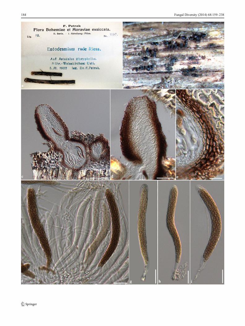
184 Fungal Diversity (2014) 68:159–238
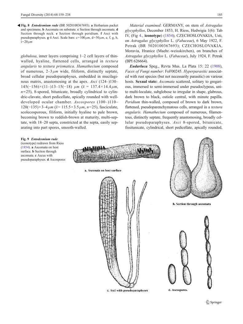
globulosa, inner layers comprising 1–2 cell layers of thin-walled, hyaline, flattened cells, arranged in texturaangularis to textura prismatica. Hamathecium composedof numerous, 2–3μm wide, filiform, distinctly septate,broad cellular pseudoparaphyses, embedded in mucilagi-nous matrix, anastomosing at the apex. Asci (124–)130–145(−156)×(11–)13–15(−18) μm (x = 137.4×14.4μm,n=25), 8-spored, bitunicate, broadly cylindrical to cylin-dric-clavate, short pedicellate, apically rounded with well-developed ocular chamber. Ascospores (100–)110–120(−135)×3–4μm (x= 115.5×3.5μm, n=25), fasciculate,scolecosporous, filiform, initially hyaline to pale brown,becoming brown to reddish-brown at maturity, multi-sep-tate, with 18–20 septa, constricted at the septa, easily sep-arating into part spores, smooth-walled.
Material examined: GERMANY, on stem of Astragalusglycyphyllos, December 1853, H. Riess, Hedwigia 1(6): TabIV, (Fig 4., iconotype) (1854); CZECHOSLOVAKIA, Usti,on Astragalus glycyphyllos L. (Fabaceae), 6 May 1922, F.Petrak (BR 5020100367693); CZECHOSLOVAKIA,Moravia, Hranice (Maehr.-weisskirchen), on branches ofAstragalus glycyphyllos L. (Fabaceae), July 1924, F. Petrak(BPI 626664).
Eudarluca Speg., Revta Mus. La Plata 15: 22 (1908),Faces of Fungi number: FoF00245. Hyperparasitic associat-ed with rust species (but not necessarily parasitic) on varioushosts. Sexual state: Ascomata scattered, solitary to gregari-ous, immersed to semi-immersed under pseudoclypeus, uni-to multi-loculate, subglobose to irregular in shape, glabrous,dark brown to black, ostiole central, with minute papilla.Peridium thin-walled, composed of brown to dark brown,flattened, pseudoparenchymatous cells, arranged in a texturaangularis. Hamathecium composed of numerous, filamen-tous, distinctly septate, frequently anastomosing, broadly cel-lular pseudoparaphyses. Asci 8-spored, bitunicate,fissitunicate, cylindrical, short pedicellate, apically rounded,
�Fig. 8 Entodesmium rude (BR 5020100367693). a Herbarium packetand specimens. b Ascomata on substrate. c Section through ascomata. dSection through neck. e Section through peridium. f Asci withpseudoparaphyses. g–i Asci. Scale bars: c=100μm, d=50μm, e, f, g, h,i=20μm
Fig. 9 Entodesmium rude(iconotype) redrawn from Riess(1854). a Ascomata on hostsurface. b Section throughascomata. c Ascus withpseudoparaphyses. d Ascospores
Fungal Diversity (2014) 68:159–238 185
with well-developed ocular chamber. Ascosporesphragmosporous, overlapping 2-seriate, fusiform or oblongwith rounded ends, pale brown, septate, smooth-walled.Asexual state: see notes.
Type species: Eudarluca australisPhylogenetic study: NoneNotes: Eudarlucawas introduced by Spegazzini (1908) and
typified by Eu. australis which is associated with a rust onleaves of Canna. Eriksson (1966) treated Eu. australis as asynonym of Eudarluca caricis (Fr.) O.E. Erikss. Eudarlucacaricis has been reported from various hosts, mostly in itspresumed asexual state, Sphaerellopsis (Yuan et al. 1998;Nischwitz et al. 2005; Zhang et al. 2012). The asexual andsexual state connection between Eudarluca caricis andSphaerellopsis filum was derived from ascospores growing onPuccinia extensicola-oenotherae (Mont.) Arthur on Carex sp.in Pennsylvania, USA (Keener 1951) and confirmed by Yuanet al. (1998). Yuan et al. (1998) grew ascospores of Eudarlucacaricis on PDA and derived Sphaerellopsis conidia andconidiomata after 12 days. Zhang et al. (2012) compared theITS sequence of Eudarluca caricis strainMullMK in GenBankwith Leptosphaeria species and mentioned that Eudarlucashould be treated in Leptosphaeriaceae due to comparablesequences and also morphological characters. However, boththe generic type of Eudarluca and Sphaerellopsis have neverbeen linked (Wijayawardene et al. 2014b).
Molecular studies on Eudarluca are limited to speciesvariation and are based on Sphaerellopsis strains (Nischwitzet al. 2005; Bayon et al. 2006; Zhang et al. 2012). The ITSgene phylogenetic analysis showed that Eudarluca ciricis(strains MosID6 and MullMK) cluster in Leptosphaeriaceae.However, the generic type of Sphaerellopsis has found onQuercus, while the type of Eudarluca is from Canna. Thesetwo generic types have not been linked by molecular data. Wesuggest that caution should be followed when treating thesetaxa, as Eu. australis is immersed in the Canna leaves andalthough associated with the rust fungi (Sphaerellopsis) onCanna, it does not seem to grow on the rust.
In this study, we tentatively place Eudarluca inPhaeosphaeriaceae based on its morphological characters whichare typical of Phaeosphaeria as the peridium is thin-walled, andcomposed of pseudoparenchymatous cells, asci are cylindrical,subsessile to short pedicellate and ascospores are fusiform andphragmosporous. Fresh collections of Eu. australis are neededfor epitypification and molecular study. Sphaerellopsis may be-long in Leptosphaeriaceae where it is tentatively assigned.
Eudarluca australis Speg., Revta Mus. La Plata 15: 22(1908), Faces of Fungi number: FoF00246, Fig. 10.
Hyperparasitic associated with rust fungi on leaves ofCanna indica L. (Cannaceae). Sexual state: Ascomata 110–180μm high, 170–280μm diam., scattered, solitary to gregar-ious, immersed to semi-immersed, visible as raised, black tinyspots in black pseudostromata on leaf lesions, uniloculate,
subglobose to irregular, glabrous, dark brown to black, ostiolecentral, with minute papilla. Peridium 5–13μm wide, thin-walled, of unequal thickness, slightly thickened at the apex,composed of 1–3 layers of brown to dark brown, flattened,pseudoparenchymatous cells, arranged in textura angularis.Hamathecium composed of numerous, 1.5–3μm wide, fili-form, distinctly septate, frequently anastomosing, broadly cel-lular pseudoparaphyses, embedded in mucilaginous matrix.Asci (44–)50–65×8–10μm (x = 57×9μm, n=25), 8-spored,bitunicate, fissitunicate, cylindrical, short pedicellate orsubsessile, apically rounded, with indistinct ocular chamber.Ascospores 15–17(−18)×3–4.5μm (x= 16.7×4.1μm, n=30),phragmosporous, overlapping or 2-seriate, fusiform, or ob-long with rounded ends, pale brown, 2-septate, rarely 1-sep-tate, slightly curved, not constricted at the septa, smooth-walled. Asexual state: see notes.
Material examined: BRAZIL, São Paulo, BotanicalGardens, associated with rust (Pucciniaceae) on leaves ofCanna indica L., 10 August 1905, A. Usteri (S-F11420,isotype).
Loratospora Kohlm. & Volkm.-Kohlm., Syst. Ascom.12(1–2): 10 (1993), Faces of Fungi number: FoF00247.
Type species: Loratospora aestuariiPhylogenetic study: Schoch et al. (2009), Suetrong et al.
(2009), Zhang et al. (2012), Hyde et al. (2013).Loratospora aestuarii Kohlm. & Volkm.-Kohlm., Syst.
Ascom. 12(1–2): 10 (1993), Faces of Fungi number:FoF00248, Fig. 11.
Saprobic on Juncus roemerianus Scheele. Sexual state:Ascomata 130–250μm high, 160–310μm diam., scattered toclustered, solitary, immersed to erumpent through host surface,visible as small black spots on host surface, uni to biloculate,globose to subglobose, glabrous, brown to dark brown, ostiolecentral, with minute papilla, with brown periphyses. Peridium18–42μmwide, thin-walled, of unequal thickness, thickened atthe apex, composed of several layers of brown to dark brown,pseudoparenchymatous cells, arranged in textura angularis totextura globulosa. Hamathecium absent, visible as mucilagi-nous matrix between asci. Asci (60–)70–85(−110)×(23–) 25–30(−35) μm (x = 80.9×28.2μm, n=20), 8-spored, bitunicate,fissitunicate, ovoid to ampulliform, thick-walled at the apex,sessile to subsessile, apically rounded with indistinct ocularchamber, sparse in mucilaginous matrix. Ascospores 40–50(−55)×5–7(−8) μm (x = 46.7×6.7μm, n=30), overlappingor parallel, 4–5-seriate, phragmosporous, cylindrical, or nar-rowly elongate fusiform with rounded ends, initially hyalineto subhyaline, pale yellowish at maturity, 3-septate, slightlycurved, not constricted at the septa, rough-walled, surroundedby a thin, distinct sheath. Asexual state: Unknown.
Material examined: USA, North Carolina, on dead culmsof Juncus roemerianus (Juncaceae), Kohlmeyer_5505(holotype), 5520, 5523, 5525, 5535 (NY_Slide box: 17,Tray no. 21); ibid. Broad Creek (Atlantic Ocean) 34° 43′ 00″
186 Fungal Diversity (2014) 68:159–238
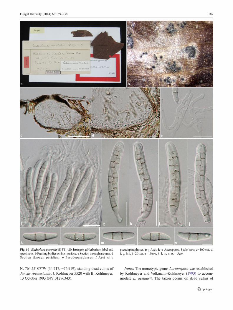
N, 76° 55′ 07″W (34.717, −76.919), standing dead culms ofJuncus roemerianus, J. Kohlmeyer 5520 with B. Kohlmeyer,13 October 1993 (NY 01276343).
Notes: The monotypic genus Loratospora was establishedby Kohlmeyer and Volkmann-Kohlmeyer (1993) to accom-modate L. aestuarii. The taxon occurs on dead culms of
Fungal Diversity (2014) 68:159–238 187
Fig. 10 Eudarluca australis (S-F11420, isotype). aHerbarium label andspecimens. b Fruiting bodies on host surface. c Section through ascoma. dSection through peridium. e Pseudoparaphyses. f Asci with
pseudoparaphyses. g–j Asci. k–o Ascospores. Scale bars: c=100μm, d,f, g, h, i, j=20μm, e=10μm, k, l, m, n, o, = 5μm
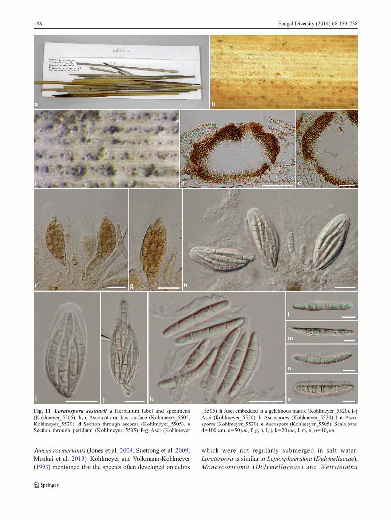
Juncus roemerianus (Jones et al. 2009; Suetrong et al. 2009;Monkai et al. 2013). Kohlmeyer and Volkmann-Kohlmeyer(1993) mentioned that the species often developed on culms
which were not regularly submerged in salt water.Loratospora is similar to Leptosphaerulina (Didymellaceae),Monascostroma (Didymellaceae) and Wettsteinina
Fig. 11 Loratospora aestuarii a Herbarium label and specimens(Kohlmeyer_5505). b, c Ascomata on host surface (Kohlmeyer_5505,Kohlmeyer_5520). d Section through ascoma (Kohlmeyer_5505). eSection through peridium (Kohlmeyer_5505) f–g Asci (Kohlmeyer
_5505). h Asci embedded in a gelatinous matrix (Kohlmeyer_5520). i–jAsci (Kohlmeyer_5520). k Ascospores (Kohlmeyer_5520) l–n Asco-spores (Kohlmeyer_5520). o Ascospore (Kohlmeyer_5505). Scale bars:d=100 μm, e=50μm, f, g, h, I, j, k=20μm, l, m, n, o=10μm
188 Fungal Diversity (2014) 68:159–238
(Dothideomycetes genera incertae sedis), but Leptosphaerulinaand Monascostroma have smaller ascomata, lack periphyses,and have a thin-walled peridium. Ascospores inLeptosphaerulina are muriform and didymosporous inMonascostroma (Kohlmeyer and Volkmann-Kohlmeyer 1993;Jones et al. 2009). Loratospora forms large ascomata withbrown ostiolar periphyses, a thick-walled peridium at the apexand phragmosporous, cylindrical ascospores (Kohlmeyer andVolkmann-Kohlmeyer 1993; Jones et al. 2009; Suetrong et al.2009).Wettsteinina differs from Loratospora in lacking ostiolarperiphyses, in having a thick-walled peridium of equal widthand ellipsoidal to broadly fusiform ascospores (Kohlmeyer andVolkmann-Kohlmeyer 1993)
Loratospora was re-circumscribed by Jones et al. (2009)and Monkai et al. (2013). The genus was accommodated inPlanistromellaceae by Barr (1996) due to its periphysateostiole (Jones et al. 2009; Suetrong et al. 2009; Monkai et al.2013). Based on multigene phylogenetic analysis,Loratospora was placed in Phaeosphaeriaceae (Schochet al. 2009; Zhang et al. 2012; Hyde et al. 2013).
In the multigene phylogenetic analyses (Fig. 2),Loratospora forms a strongly supported clade withChaetosphaeronema, Dematiopleospora, Entodesmium,Nodulosphaeria and Ophiobolus. However, the genus has adifferentiated centrum type as compared with other genera.Loratospora lacks of pseudoparaphyses but has an internalmucilaginous hamathecium, while most genera inPhaeosphaeriaceae have pseudoparaphyses. It seem like thecentrum-type is not significant to distinguish genera at thefamily level. There is only one species listed in Loratospora inIndex Fungorum (2014) and no asexual state is known. Thesequences data of Loratospora is limited to only one strain.
Neosetophoma Gruyter et al., in Gruyter et al. Mycologia102(5): 1075 (2010), Faces of Fungi number: FoF00249.
Pathogenic on the grasses, Phlox paniculata L. and Urticadioica L., or associated with soil. Sexual state: Unknown.Asexual state: Conidiomata pycnidial, scattered, solitary orgregarious, superficial on agar, uniloculate, globose or irreg-ular, setose, olivaceous to dark olivaceous, ostiole central,with papilla or long neck. Conidiomata walls composed ofpseudoparenchymatous cell. Conidiophores reduced toconidiogenous cells. Conidiogenous cells phialidic, discrete,hyaline. Conidia ellipsoidal to cylindrical, attenuate at oneend, yellowish, aseptate or septate, not constricted at theseptum, smooth-walled, with guttules (from De Gruyteret al. 2010; Quaedvlieg et al. 2013).
Type species:Neosetophoma samararum (Desm.) Gruyteret al. [as ‘samarorum’], in De Gruyter et al. Mycologia 102(5):1075 (2010).
= Phoma samararum Desm., Pl. Crypt. Nord France, Edn1 7: no. 349 (1828)
Phylogenetic study: De Gruyter et al. (2010, 2012), Zhanget al. (2012), Hyde et al. (2013), Quaedvlieg et al. (2013).
Notes: Neosetophoma was introduced by De Gruyter et al.(2010) to accommodate N. samararum. Neosetophoma oftenforms a single clade in Phaeosphaeriaceae. The genus hasbeen widely accepted in Phaeosphaeriaceae by various au-thors (De Gruyter et al. 2010, 2012; Zhang et al. 2012; Hydeet al. 2013; Quaedvlieg et al. 2013).
Neostagonospora Quaedvlieg et al., Stud. Mycol. 75: 364(2013), Faces of Fungi number: FoF00250.
Pathogenic on Carex acutiformis Ehrh and Elegiacuspidate Mast., or soil borne. Sexual state: Unknown.Asexual state: Conidiomata pycnidial, scattered, semi-immersed to superficial, uniloculate, globose, glabrous,producing pale luteous to creamy conidial mass on hostsurface. Conidiomata walls thin, composed of 2–3 layersof pale brown cells, arranged in a textura angularis.Conid iophores reduced to conid iogenous ce l ls .Conidiogenous cells phialidic, integrated, ampulliform todoliiform, hyaline, smooth-walled, lining the inner cavity,with prominent periclinal thickening. Conidia narrowfusoid-ellipsoidal to subcylindrical, apically subobtuse torounded, truncate at the base, hyaline, septate, initially non-constricted at the septum, becoming constricted at the septawhen mature, smooth-walled, with granules (fromQuaedvlieg et al. 2013).
Type species: Neostagonospora caricis Quaedvlieg et al.,Stud. Mycol. 75: 364 (2013).
Phylogenetic study: Quaedvlieg et al. (2013).Notes: The asexual genus Neostagonospora was intro-
duced by Quaedvlieg et al. (2013) to accommodate thestagonospora-like species in Phaeosphaeriaceae. Two specieswere placed in this genus; Neostagonospora caricis and Ne.elegiae Quaedvlieg et al. with Ne. caricis being the typespecies. Neostagonospora is similar to Stagonospora but dif-fers in its conidiogenous cells. Neostagonospora has determi-nate phialidic conidiogenous cells, with prominent periclinalthickening while Stagonospora has percurrent proliferation ofconidiogenous cells. Based on phylogenetic analysis,Neostagonospora is related to Phaeosphaeriaceae, while thetype species of Stagonospora, St. paludosa clusters inMassarinaceae (Quaedvlieg et al. 2013). In the phylogenyin Fig. 2, Neostagonospora forms a sister clade withParastagonospora for which no sexual state is known.Phaeosphaeria sensu lato taxa are polyphyletic and therefore,it is likely that Neostagonospora may be an asexual state ofphaeosphaeria-like species.
Nodulosphaeria Rabenh., Klotzschii Herb. Viv. Mycol.,Edn 2: no. 725 (in sched.) (1858), Faces of Fungi number:FoF00251.
Saprobic or hemibiotrophic mostly on herbaceous dicoty-ledons. Sexual state: Ascomata scattered, solitary to gregari-ous, immersed to semi-immersed, or erumpent through hostepidermis at maturity, visible as black dots on host surface,uniloculate, globose to subglobose, setose, covered by
Fungal Diversity (2014) 68:159–238 189
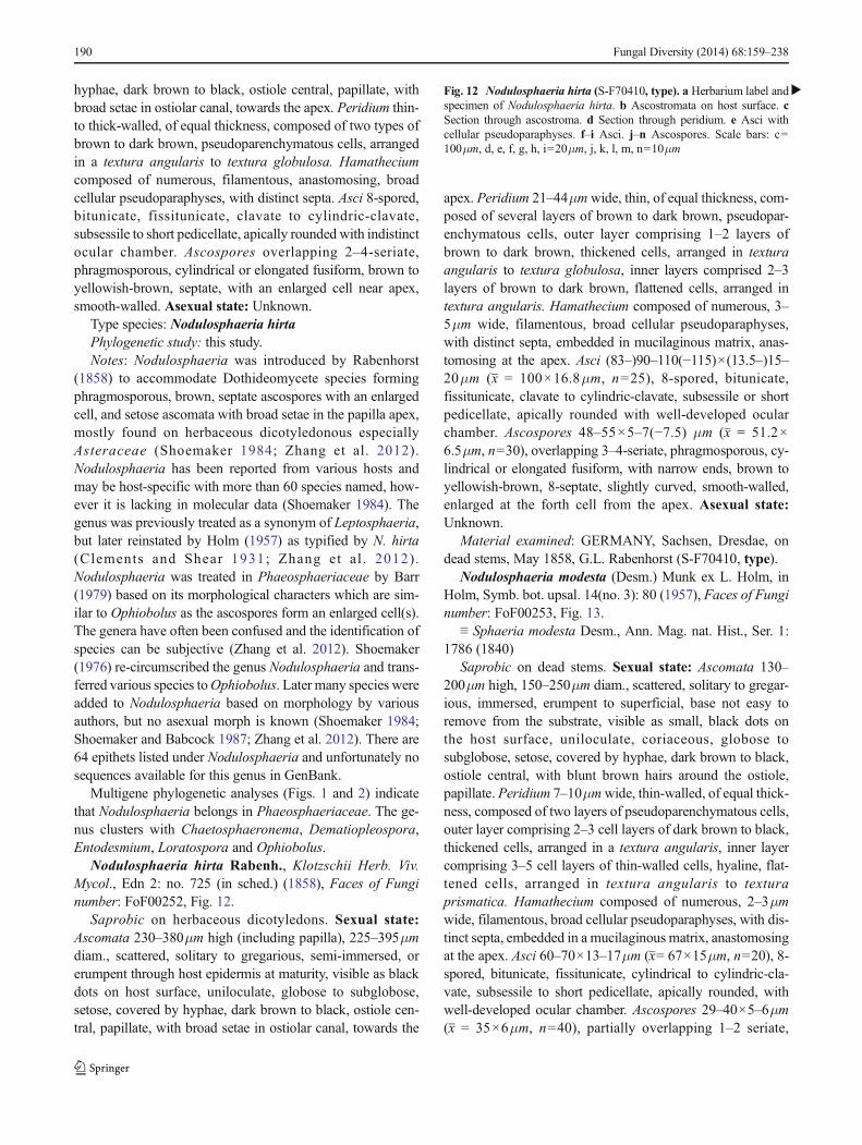
hyphae, dark brown to black, ostiole central, papillate, withbroad setae in ostiolar canal, towards the apex. Peridium thin-to thick-walled, of equal thickness, composed of two types ofbrown to dark brown, pseudoparenchymatous cells, arrangedin a textura angularis to textura globulosa. Hamatheciumcomposed of numerous, filamentous, anastomosing, broadcellular pseudoparaphyses, with distinct septa. Asci 8-spored,bitunicate, fissitunicate, clavate to cylindric-clavate,subsessile to short pedicellate, apically rounded with indistinctocular chamber. Ascospores overlapping 2–4-seriate,phragmosporous, cylindrical or elongated fusiform, brown toyellowish-brown, septate, with an enlarged cell near apex,smooth-walled. Asexual state: Unknown.
Type species: Nodulosphaeria hirtaPhylogenetic study: this study.Notes: Nodulosphaeria was introduced by Rabenhorst
(1858) to accommodate Dothideomycete species formingphragmosporous, brown, septate ascospores with an enlargedcell, and setose ascomata with broad setae in the papilla apex,mostly found on herbaceous dicotyledonous especiallyAsteraceae (Shoemaker 1984; Zhang et al. 2012).Nodulosphaeria has been reported from various hosts andmay be host-specific with more than 60 species named, how-ever it is lacking in molecular data (Shoemaker 1984). Thegenus was previously treated as a synonym of Leptosphaeria,but later reinstated by Holm (1957) as typified by N. hirta(Clements and Shear 1931; Zhang et al . 2012).Nodulosphaeria was treated in Phaeosphaeriaceae by Barr(1979) based on its morphological characters which are sim-ilar to Ophiobolus as the ascospores form an enlarged cell(s).The genera have often been confused and the identification ofspecies can be subjective (Zhang et al. 2012). Shoemaker(1976) re-circumscribed the genus Nodulosphaeria and trans-ferred various species toOphiobolus. Later many species wereadded to Nodulosphaeria based on morphology by variousauthors, but no asexual morph is known (Shoemaker 1984;Shoemaker and Babcock 1987; Zhang et al. 2012). There are64 epithets listed under Nodulosphaeria and unfortunately nosequences available for this genus in GenBank.
Multigene phylogenetic analyses (Figs. 1 and 2) indicatethat Nodulosphaeria belongs in Phaeosphaeriaceae. The ge-nus clusters with Chaetosphaeronema, Dematiopleospora,Entodesmium, Loratospora and Ophiobolus.
Nodulosphaeria hirta Rabenh., Klotzschii Herb. Viv.Mycol., Edn 2: no. 725 (in sched.) (1858), Faces of Funginumber: FoF00252, Fig. 12.
Saprobic on herbaceous dicotyledons. Sexual state:Ascomata 230–380μm high (including papilla), 225–395μmdiam., scattered, solitary to gregarious, semi-immersed, orerumpent through host epidermis at maturity, visible as blackdots on host surface, uniloculate, globose to subglobose,setose, covered by hyphae, dark brown to black, ostiole cen-tral, papillate, with broad setae in ostiolar canal, towards the
apex. Peridium 21–44μmwide, thin, of equal thickness, com-posed of several layers of brown to dark brown, pseudopar-enchymatous cells, outer layer comprising 1–2 layers ofbrown to dark brown, thickened cells, arranged in texturaangularis to textura globulosa, inner layers comprised 2–3layers of brown to dark brown, flattened cells, arranged intextura angularis. Hamathecium composed of numerous, 3–5μm wide, filamentous, broad cellular pseudoparaphyses,with distinct septa, embedded in mucilaginous matrix, anas-tomosing at the apex. Asci (83–)90–110(−115)×(13.5–)15–20μm (x = 100×16.8μm, n=25), 8-spored, bitunicate,fissitunicate, clavate to cylindric-clavate, subsessile or shortpedicellate, apically rounded with well-developed ocularchamber. Ascospores 48–55×5–7(−7.5) μm (x = 51.2×6.5μm, n=30), overlapping 3–4-seriate, phragmosporous, cy-lindrical or elongated fusiform, with narrow ends, brown toyellowish-brown, 8-septate, slightly curved, smooth-walled,enlarged at the forth cell from the apex. Asexual state:Unknown.
Material examined: GERMANY, Sachsen, Dresdae, ondead stems, May 1858, G.L. Rabenhorst (S-F70410, type).
Nodulosphaeria modesta (Desm.) Munk ex L. Holm, inHolm, Symb. bot. upsal. 14(no. 3): 80 (1957), Faces of Funginumber: FoF00253, Fig. 13.
≡ Sphaeria modesta Desm., Ann. Mag. nat. Hist., Ser. 1:1786 (1840)
Saprobic on dead stems. Sexual state: Ascomata 130–200μm high, 150–250μm diam., scattered, solitary to gregar-ious, immersed, erumpent to superficial, base not easy toremove from the substrate, visible as small, black dots onthe host surface, uniloculate, coriaceous, globose tosubglobose, setose, covered by hyphae, dark brown to black,ostiole central, with blunt brown hairs around the ostiole,papillate. Peridium 7–10μmwide, thin-walled, of equal thick-ness, composed of two layers of pseudoparenchymatous cells,outer layer comprising 2–3 cell layers of dark brown to black,thickened cells, arranged in a textura angularis, inner layercomprising 3–5 cell layers of thin-walled cells, hyaline, flat-tened cells, arranged in textura angularis to texturaprismatica. Hamathecium composed of numerous, 2–3μmwide, filamentous, broad cellular pseudoparaphyses, with dis-tinct septa, embedded in a mucilaginous matrix, anastomosingat the apex. Asci 60–70×13–17μm (x= 67×15μm, n=20), 8-spored, bitunicate, fissitunicate, cylindrical to cylindric-cla-vate, subsessile to short pedicellate, apically rounded, withwell-developed ocular chamber. Ascospores 29–40×5–6μm(x = 35×6μm, n=40), partially overlapping 1–2 seriate,
�Fig. 12 Nodulosphaeria hirta (S-F70410, type). a Herbarium label andspecimen of Nodulosphaeria hirta. b Ascostromata on host surface. cSection through ascostroma. d Section through peridium. e Asci withcellular pseudoparaphyses. f–i Asci. j–n Ascospores. Scale bars: c=100μm, d, e, f, g, h, i=20μm, j, k, l, m, n=10μm
190 Fungal Diversity (2014) 68:159–238
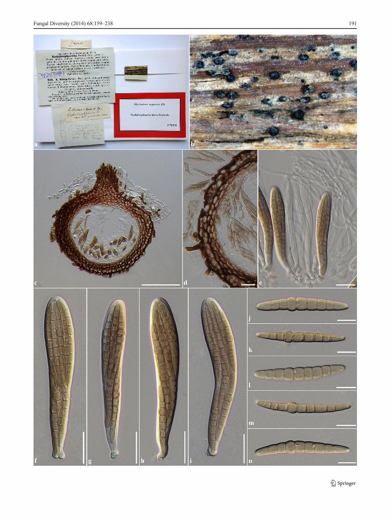
Fungal Diversity (2014) 68:159–238 191
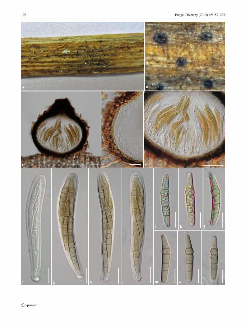
192 Fungal Diversity (2014) 68:159–238
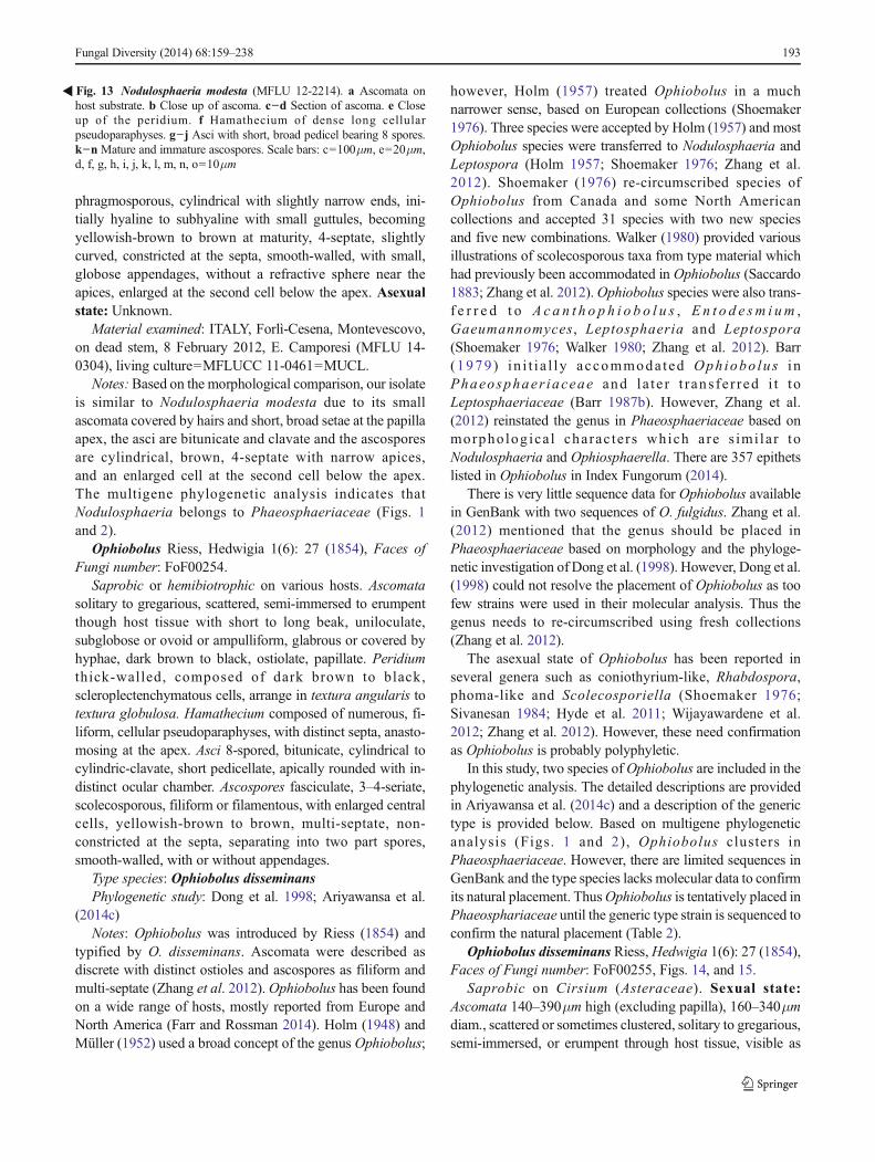
phragmosporous, cylindrical with slightly narrow ends, ini-tially hyaline to subhyaline with small guttules, becomingyellowish-brown to brown at maturity, 4-septate, slightlycurved, constricted at the septa, smooth-walled, with small,globose appendages, without a refractive sphere near theapices, enlarged at the second cell below the apex. Asexualstate: Unknown.
Material examined: ITALY, Forlì-Cesena, Montevescovo,on dead stem, 8 February 2012, E. Camporesi (MFLU 14-0304), living culture=MFLUCC 11-0461=MUCL.
Notes:Based on the morphological comparison, our isolateis similar to Nodulosphaeria modesta due to its smallascomata covered by hairs and short, broad setae at the papillaapex, the asci are bitunicate and clavate and the ascosporesare cylindrical, brown, 4-septate with narrow apices,and an enlarged cell at the second cell below the apex.The multigene phylogenetic analysis indicates thatNodulosphaeria belongs to Phaeosphaeriaceae (Figs. 1and 2).
Ophiobolus Riess, Hedwigia 1(6): 27 (1854), Faces ofFungi number: FoF00254.
Saprobic or hemibiotrophic on various hosts. Ascomatasolitary to gregarious, scattered, semi-immersed to erumpentthough host tissue with short to long beak, uniloculate,subglobose or ovoid or ampulliform, glabrous or covered byhyphae, dark brown to black, ostiolate, papillate. Peridiumthick-walled, composed of dark brown to black,scleroplectenchymatous cells, arrange in textura angularis totextura globulosa. Hamathecium composed of numerous, fi-liform, cellular pseudoparaphyses, with distinct septa, anasto-mosing at the apex. Asci 8-spored, bitunicate, cylindrical tocylindric-clavate, short pedicellate, apically rounded with in-distinct ocular chamber. Ascospores fasciculate, 3–4-seriate,scolecosporous, filiform or filamentous, with enlarged centralcells, yellowish-brown to brown, multi-septate, non-constricted at the septa, separating into two part spores,smooth-walled, with or without appendages.
Type species: Ophiobolus disseminansPhylogenetic study: Dong et al. 1998; Ariyawansa et al.
(2014c)Notes: Ophiobolus was introduced by Riess (1854) and
typified by O. disseminans. Ascomata were described asdiscrete with distinct ostioles and ascospores as filiform andmulti-septate (Zhang et al. 2012).Ophiobolus has been foundon a wide range of hosts, mostly reported from Europe andNorth America (Farr and Rossman 2014). Holm (1948) andMüller (1952) used a broad concept of the genus Ophiobolus;
however, Holm (1957) treated Ophiobolus in a muchnarrower sense, based on European collections (Shoemaker1976). Three species were accepted by Holm (1957) and mostOphiobolus species were transferred to Nodulosphaeria andLeptospora (Holm 1957; Shoemaker 1976; Zhang et al.2012). Shoemaker (1976) re-circumscribed species ofOphiobolus from Canada and some North Americancollections and accepted 31 species with two new speciesand five new combinations. Walker (1980) provided variousillustrations of scolecosporous taxa from type material whichhad previously been accommodated in Ophiobolus (Saccardo1883; Zhang et al. 2012).Ophiobolus species were also trans-f e r r e d t o A c a n t h o p h i o b o l u s , En t o d e sm i um ,Gaeumannomyces, Leptosphaeria and Leptospora(Shoemaker 1976; Walker 1980; Zhang et al. 2012). Barr(1979) in i t i a l l y accommoda ted Ophiobo lus i nPhaeosphaer iaceae and la te r t r ans f e r red i t toLeptosphaeriaceae (Barr 1987b). However, Zhang et al.(2012) reinstated the genus in Phaeosphaeriaceae based onmorpholog ica l charac te rs which are s imi la r toNodulosphaeria and Ophiosphaerella. There are 357 epithetslisted in Ophiobolus in Index Fungorum (2014).
There is very little sequence data for Ophiobolus availablein GenBank with two sequences of O. fulgidus. Zhang et al.(2012) mentioned that the genus should be placed inPhaeosphaeriaceae based on morphology and the phyloge-netic investigation of Dong et al. (1998). However, Dong et al.(1998) could not resolve the placement of Ophiobolus as toofew strains were used in their molecular analysis. Thus thegenus needs to re-circumscribed using fresh collections(Zhang et al. 2012).
The asexual state of Ophiobolus has been reported inseveral genera such as coniothyrium-like, Rhabdospora,phoma-like and Scolecosporiella (Shoemaker 1976;Sivanesan 1984; Hyde et al. 2011; Wijayawardene et al.2012; Zhang et al. 2012). However, these need confirmationas Ophiobolus is probably polyphyletic.
In this study, two species ofOphiobolus are included in thephylogenetic analysis. The detailed descriptions are providedin Ariyawansa et al. (2014c) and a description of the generictype is provided below. Based on multigene phylogeneticanalysis (Figs. 1 and 2), Ophiobolus clusters inPhaeosphaeriaceae. However, there are limited sequences inGenBank and the type species lacks molecular data to confirmits natural placement. ThusOphiobolus is tentatively placed inPhaeosphariaceae until the generic type strain is sequenced toconfirm the natural placement (Table 2).
Ophiobolus disseminans Riess,Hedwigia 1(6): 27 (1854),Faces of Fungi number: FoF00255, Figs. 14, and 15.
Saprobic on Cirsium (Asteraceae). Sexual state:Ascomata 140–390μm high (excluding papilla), 160–340μmdiam., scattered or sometimes clustered, solitary to gregarious,semi-immersed, or erumpent through host tissue, visible as
�Fig. 13 Nodulosphaeria modesta (MFLU 12-2214). a Ascomata onhost substrate. b Close up of ascoma. c−d Section of ascoma. e Closeup of the peridium. f Hamathecium of dense long cellularpseudoparaphyses. g−j Asci with short, broad pedicel bearing 8 spores.k−n Mature and immature ascospores. Scale bars: c=100μm, e=20μm,d, f, g, h, i, j, k, l, m, n, o=10μm
Fungal Diversity (2014) 68:159–238 193
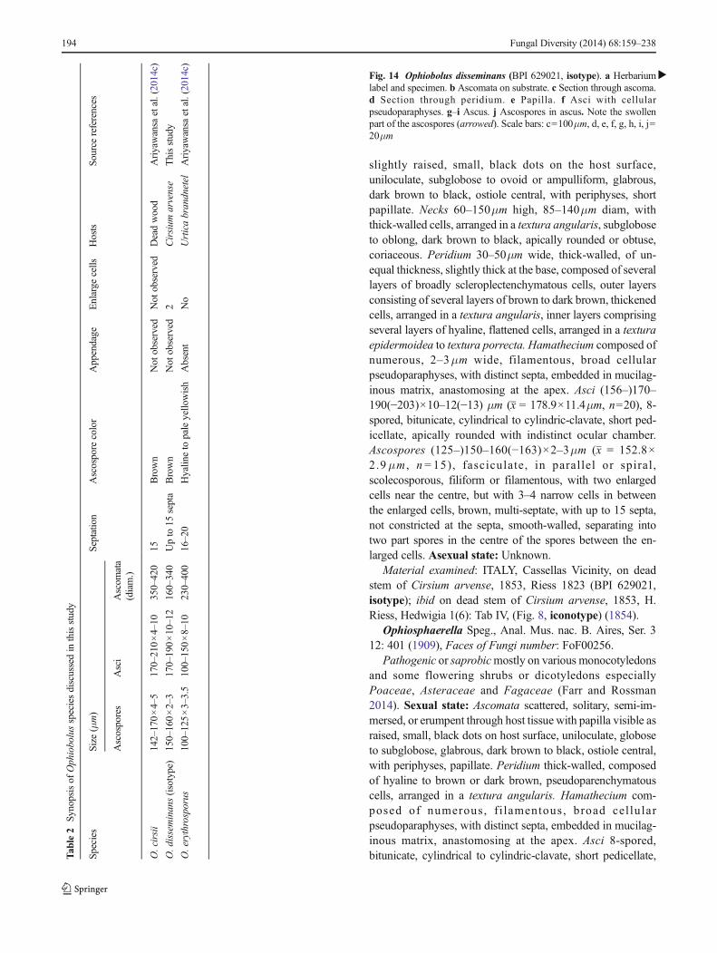
slightly raised, small, black dots on the host surface,uniloculate, subglobose to ovoid or ampulliform, glabrous,dark brown to black, ostiole central, with periphyses, shortpapillate. Necks 60–150μm high, 85–140μm diam, withthick-walled cells, arranged in a textura angularis, subgloboseto oblong, dark brown to black, apically rounded or obtuse,coriaceous. Peridium 30–50μm wide, thick-walled, of un-equal thickness, slightly thick at the base, composed of severallayers of broadly scleroplectenchymatous cells, outer layersconsisting of several layers of brown to dark brown, thickenedcells, arranged in a textura angularis, inner layers comprisingseveral layers of hyaline, flattened cells, arranged in a texturaepidermoidea to textura porrecta. Hamathecium composed ofnumerous, 2–3μm wide, filamentous, broad cellularpseudoparaphyses, with distinct septa, embedded in mucilag-inous matrix, anastomosing at the apex. Asci (156–)170–190(−203)×10–12(−13) μm (x = 178.9×11.4μm, n=20), 8-spored, bitunicate, cylindrical to cylindric-clavate, short ped-icellate, apically rounded with indistinct ocular chamber.Ascospores (125–)150–160(−163)×2–3μm (x = 152.8×2.9μm , n = 15), fasciculate, in parallel or spiral ,scolecosporous, filiform or filamentous, with two enlargedcells near the centre, but with 3–4 narrow cells in betweenthe enlarged cells, brown, multi-septate, with up to 15 septa,not constricted at the septa, smooth-walled, separating intotwo part spores in the centre of the spores between the en-larged cells. Asexual state: Unknown.
Material examined: ITALY, Cassellas Vicinity, on deadstem of Cirsium arvense, 1853, Riess 1823 (BPI 629021,isotype); ibid on dead stem of Cirsium arvense, 1853, H.Riess, Hedwigia 1(6): Tab IV, (Fig. 8, iconotype) (1854).
Ophiosphaerella Speg., Anal. Mus. nac. B. Aires, Ser. 312: 401 (1909), Faces of Fungi number: FoF00256.
Pathogenic or saprobicmostly on various monocotyledonsand some flowering shrubs or dicotyledons especiallyPoaceae, Asteraceae and Fagaceae (Farr and Rossman2014). Sexual state: Ascomata scattered, solitary, semi-im-mersed, or erumpent through host tissue with papilla visible asraised, small, black dots on host surface, uniloculate, globoseto subglobose, glabrous, dark brown to black, ostiole central,with periphyses, papillate. Peridium thick-walled, composedof hyaline to brown or dark brown, pseudoparenchymatouscells, arranged in a textura angularis. Hamathecium com-posed of numerous, f i lamentous, broad cel lularpseudoparaphyses, with distinct septa, embedded in mucilag-inous matrix, anastomosing at the apex. Asci 8-spored,bitunicate, cylindrical to cylindric-clavate, short pedicellate,T
able2
Synopsisof
Ophiobolusspeciesdiscussedin
thisstudy
Species
Size
(μm)
Septatio
nAscospore
color
Appendage
Enlarge
cells
Hosts
Source
references
Ascospores
Asci
Ascom
ata
(diam.)
O.cirsii
142–170×4–5
170–210×4–10
350–420
15Brown
Not
observed
Not
observed
Deadwood
Ariyawansa
etal.(2014c)
O.disseminans(isotype)
150–160×2–3
170–190×10–12
160–340
Upto
15septa
Brown
Not
observed
2Cirsium
arvense
Thisstudy
O.erythrosporus
100–125×3–3.5
100–150×8–10
230–400
16–20
Hyalin
eto
paleyello
wish
Absent
No
Urticabrandnetel
Ariyawansa
etal.(2014c)
�Fig. 14 Ophiobolus disseminans (BPI 629021, isotype). a Herbariumlabel and specimen. b Ascomata on substrate. c Section through ascoma.d Section through peridium. e Papilla. f Asci with cellularpseudoparaphyses. g–i Ascus. j Ascospores in ascus. Note the swollenpart of the ascospores (arrowed). Scale bars: c=100μm, d, e, f, g, h, i, j=20μm
194 Fungal Diversity (2014) 68:159–238
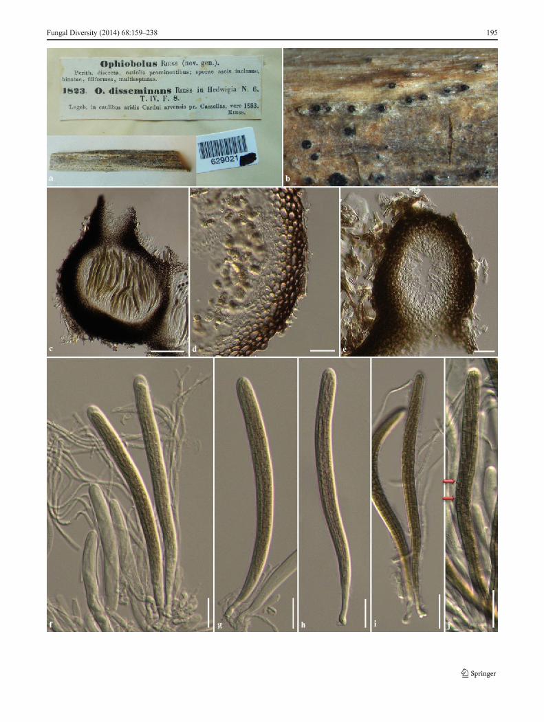
Fungal Diversity (2014) 68:159–238 195
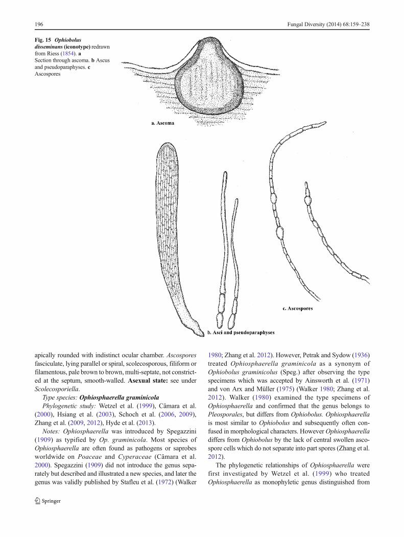
apically rounded with indistinct ocular chamber. Ascosporesfasciculate, lying parallel or spiral, scolecosporous, filiform orfilamentous, pale brown to brown,multi-septate, not constrict-ed at the septum, smooth-walled. Asexual state: see underScolecosporiella.
Type species: Ophiosphaerella graminicolaPhylogenetic study: Wetzel et al. (1999), Câmara et al.
(2000), Hsiang et al. (2003), Schoch et al. (2006, 2009),Zhang et al. (2009, 2012), Hyde et al. (2013).
Notes: Ophiosphaerella was introduced by Spegazzini(1909) as typified by Op. graminicola. Most species ofOphiosphaerella are often found as pathogens or saprobesworldwide on Poaceae and Cyperaceae (Câmara et al.2000). Spegazzini (1909) did not introduce the genus sepa-rately but described and illustrated a new species, and later thegenus was validly published by Stafleu et al. (1972) (Walker
1980; Zhang et al. 2012). However, Petrak and Sydow (1936)treated Ophiosphaerella graminicola as a synonym ofOphiobolus graminicolus (Speg.) after observing the typespecimens which was accepted by Ainsworth et al. (1971)and von Arx and Müller (1975) (Walker 1980; Zhang et al.2012). Walker (1980) examined the type specimens ofOphiosphaerella and confirmed that the genus belongs toPleosporales, but differs from Ophiobolus. Ophiosphaerellais most similar to Ophiobolus and subsequently often con-fused in morphological characters. However Ophiosphaerelladiffers from Ophiobolus by the lack of central swollen asco-spore cells which do not separate into part spores (Zhang et al.2012).
The phylogenetic relationships of Ophiosphaerella werefirst investigated by Wetzel et al. (1999) who treatedOphiosphaerella as monophyletic genus distinguished from
Fig. 15 Ophiobolusdisseminans (iconotype) redrawnfrom Riess (1854). aSection through ascoma. b Ascusand pseudoparaphyses. cAscospores
196 Fungal Diversity (2014) 68:159–238
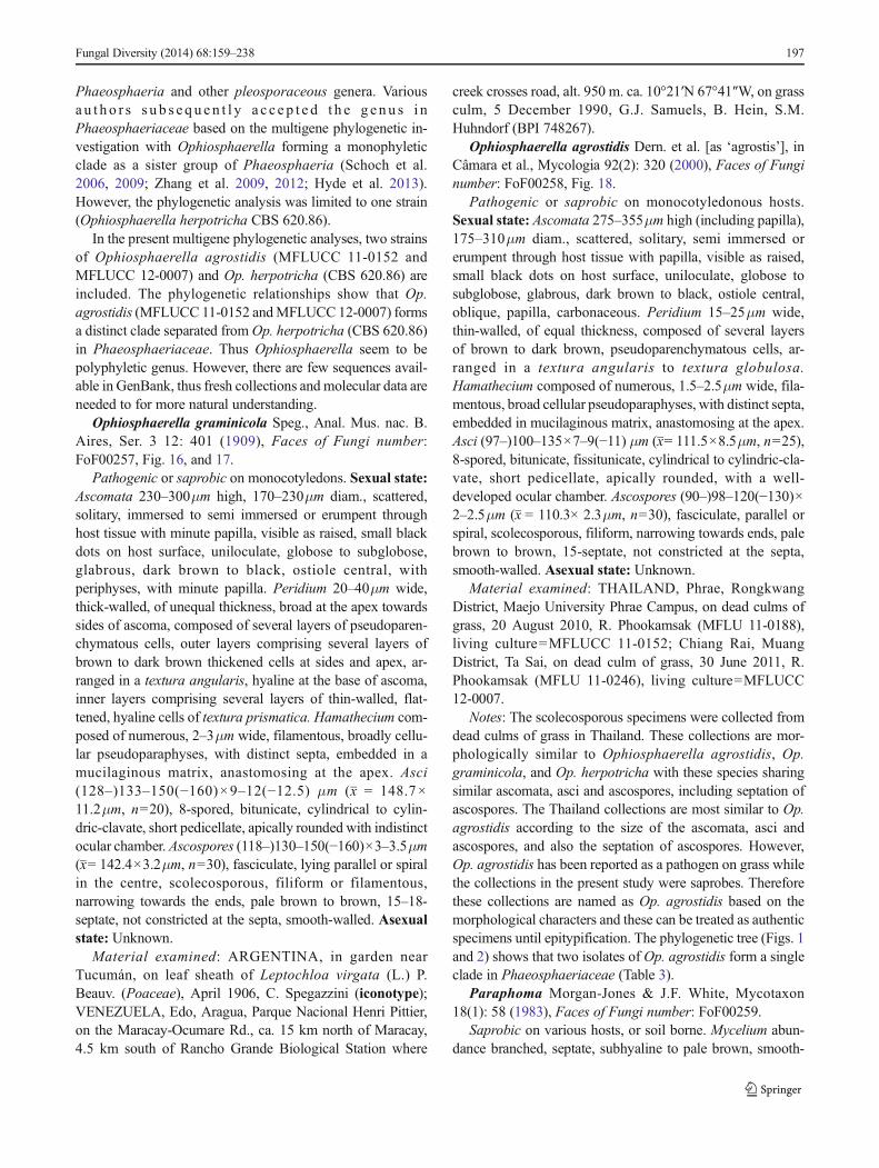
Phaeosphaeria and other pleosporaceous genera. Variousa u t h o r s s u b s e qu e n t l y a c c e p t e d t h e g e nu s i nPhaeosphaeriaceae based on the multigene phylogenetic in-vestigation with Ophiosphaerella forming a monophyleticclade as a sister group of Phaeosphaeria (Schoch et al.2006, 2009; Zhang et al. 2009, 2012; Hyde et al. 2013).However, the phylogenetic analysis was limited to one strain(Ophiosphaerella herpotricha CBS 620.86).
In the present multigene phylogenetic analyses, two strainsof Ophiosphaerella agrostidis (MFLUCC 11-0152 andMFLUCC 12-0007) and Op. herpotricha (CBS 620.86) areincluded. The phylogenetic relationships show that Op.agrostidis (MFLUCC 11-0152 andMFLUCC 12-0007) formsa distinct clade separated fromOp. herpotricha (CBS 620.86)in Phaeosphaeriaceae. Thus Ophiosphaerella seem to bepolyphyletic genus. However, there are few sequences avail-able in GenBank, thus fresh collections and molecular data areneeded to for more natural understanding.
Ophiosphaerella graminicola Speg., Anal. Mus. nac. B.Aires, Ser. 3 12: 401 (1909), Faces of Fungi number:FoF00257, Fig. 16, and 17.
Pathogenic or saprobic on monocotyledons. Sexual state:Ascomata 230–300μm high, 170–230μm diam., scattered,solitary, immersed to semi immersed or erumpent throughhost tissue with minute papilla, visible as raised, small blackdots on host surface, uniloculate, globose to subglobose,glabrous, dark brown to black, ostiole central, withperiphyses, with minute papilla. Peridium 20–40μm wide,thick-walled, of unequal thickness, broad at the apex towardssides of ascoma, composed of several layers of pseudoparen-chymatous cells, outer layers comprising several layers ofbrown to dark brown thickened cells at sides and apex, ar-ranged in a textura angularis, hyaline at the base of ascoma,inner layers comprising several layers of thin-walled, flat-tened, hyaline cells of textura prismatica. Hamathecium com-posed of numerous, 2–3μm wide, filamentous, broadly cellu-lar pseudoparaphyses, with distinct septa, embedded in amucilaginous matrix, anastomosing at the apex. Asci(128–)133–150(−160)×9–12(−12.5) μm (x = 148.7×11.2μm, n=20), 8-spored, bitunicate, cylindrical to cylin-dric-clavate, short pedicellate, apically rounded with indistinctocular chamber. Ascospores (118–)130–150(−160)×3–3.5μm(x= 142.4×3.2μm, n=30), fasciculate, lying parallel or spiralin the centre, scolecosporous, filiform or filamentous,narrowing towards the ends, pale brown to brown, 15–18-septate, not constricted at the septa, smooth-walled. Asexualstate: Unknown.
Material examined: ARGENTINA, in garden nearTucumán, on leaf sheath of Leptochloa virgata (L.) P.Beauv. (Poaceae), April 1906, C. Spegazzini (iconotype);VENEZUELA, Edo, Aragua, Parque Nacional Henri Pittier,on the Maracay-Ocumare Rd., ca. 15 km north of Maracay,4.5 km south of Rancho Grande Biological Station where
creek crosses road, alt. 950 m. ca. 10°21′N 67°41″W, on grassculm, 5 December 1990, G.J. Samuels, B. Hein, S.M.Huhndorf (BPI 748267).
Ophiosphaerella agrostidis Dern. et al. [as ‘agrostis’], inCâmara et al., Mycologia 92(2): 320 (2000), Faces of Funginumber: FoF00258, Fig. 18.
Pathogenic or saprobic on monocotyledonous hosts.Sexual state: Ascomata 275–355μm high (including papilla),175–310μm diam., scattered, solitary, semi immersed orerumpent through host tissue with papilla, visible as raised,small black dots on host surface, uniloculate, globose tosubglobose, glabrous, dark brown to black, ostiole central,oblique, papilla, carbonaceous. Peridium 15–25μm wide,thin-walled, of equal thickness, composed of several layersof brown to dark brown, pseudoparenchymatous cells, ar-ranged in a textura angularis to textura globulosa.Hamathecium composed of numerous, 1.5–2.5μm wide, fila-mentous, broad cellular pseudoparaphyses, with distinct septa,embedded in mucilaginous matrix, anastomosing at the apex.Asci (97–)100–135×7–9(−11) μm (x= 111.5×8.5μm, n=25),8-spored, bitunicate, fissitunicate, cylindrical to cylindric-cla-vate, short pedicellate, apically rounded, with a well-developed ocular chamber. Ascospores (90–)98–120(−130)×2–2.5μm (x = 110.3× 2.3μm, n=30), fasciculate, parallel orspiral, scolecosporous, filiform, narrowing towards ends, palebrown to brown, 15-septate, not constricted at the septa,smooth-walled. Asexual state: Unknown.
Material examined: THAILAND, Phrae, RongkwangDistrict, Maejo University Phrae Campus, on dead culms ofgrass, 20 August 2010, R. Phookamsak (MFLU 11-0188),living culture=MFLUCC 11-0152; Chiang Rai, MuangDistrict, Ta Sai, on dead culm of grass, 30 June 2011, R.Phookamsak (MFLU 11-0246), living culture=MFLUCC12-0007.
Notes: The scolecosporous specimens were collected fromdead culms of grass in Thailand. These collections are mor-phologically similar to Ophiosphaerella agrostidis, Op.graminicola, and Op. herpotricha with these species sharingsimilar ascomata, asci and ascospores, including septation ofascospores. The Thailand collections are most similar to Op.agrostidis according to the size of the ascomata, asci andascospores, and also the septation of ascospores. However,Op. agrostidis has been reported as a pathogen on grass whilethe collections in the present study were saprobes. Thereforethese collections are named as Op. agrostidis based on themorphological characters and these can be treated as authenticspecimens until epitypification. The phylogenetic tree (Figs. 1and 2) shows that two isolates of Op. agrostidis form a singleclade in Phaeosphaeriaceae (Table 3).
Paraphoma Morgan-Jones & J.F. White, Mycotaxon18(1): 58 (1983), Faces of Fungi number: FoF00259.
Saprobic on various hosts, or soil borne. Mycelium abun-dance branched, septate, subhyaline to pale brown, smooth-
Fungal Diversity (2014) 68:159–238 197
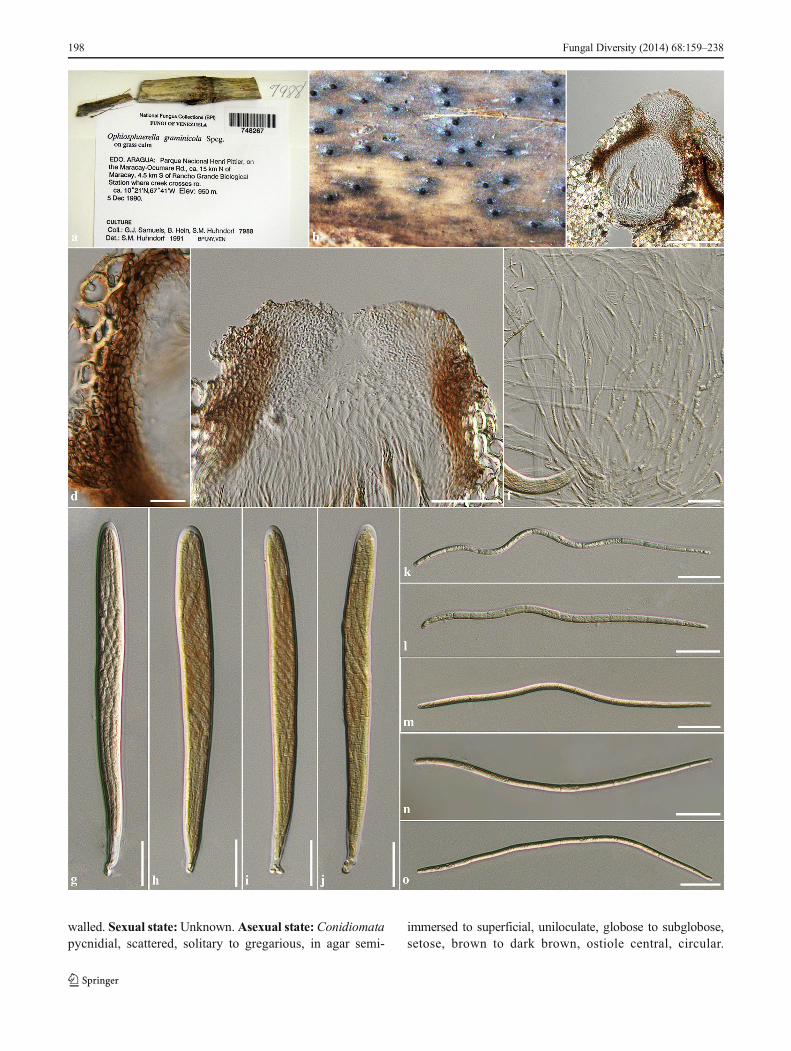
walled. Sexual state:Unknown.Asexual state: Conidiomatapycnidial, scattered, solitary to gregarious, in agar semi-
immersed to superficial, uniloculate, globose to subglobose,setose, brown to dark brown, ostiole central, circular.
198 Fungal Diversity (2014) 68:159–238
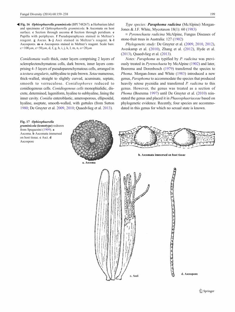
Conidiomata walls thick, outer layers comprising 2 layers ofscleroplectenchymatous cells, dark brown, inner layers com-prising 4–5 layers of pseudoparenchymatous cells, arranged ina textura angularis, subhyaline to pale brown. Setae numerous,thick-walled, straight to slightly curved, acuminate, septate,smooth to verruculose. Conidiophores reduced toconidiogenous cells. Conidiogenous cells monophialidic, dis-crete, determined, lageniform, hyaline to subhyaline, lining theinner cavity. Conidia enteroblastic, amerosporous, ellipsoidal,hyaline, aseptate, smooth-walled, with guttules (from Sutton1980; De Gruyter et al. 2009, 2010; Quaedvlieg et al. 2013).
Type species: Paraphoma radicina (McAlpine) Morgan-Jones & J.F. White, Mycotaxon 18(1): 60 (1983)
≡ Pyrenochaeta radicina McAlpine, Fungus Diseases ofstone-fruit trees in Australia: 127 (1902)
Phylogenetic study: De Gruyter et al. (2009, 2010, 2012),Aveskamp et al. (2010), Zhang et al. (2012), Hyde et al.(2013), Quaedvlieg et al. (2013).
Notes: Paraphoma as typified by P. radicina was previ-ously treated in Pyrenochaeta by McAlpine (1902) and later,Boerema and Dorenbosch (1979) transferred the species toPhoma. Morgan-Jones and White (1983) introduced a newgenus, Paraphoma to accommodate the species that producedheavily setose pycnidia and transferred P. radicina to thisgenus. However, the genus was treated as a section ofPhoma (Boerema 1997) until De Gruyter et al. (2010) rein-stated the genus and placed it in Phaeosphaeriaceae based onphylogenetic evidence. Recently, four species are accommo-dated in this genus for which no sexual state is known.
�Fig. 16 Ophiosphaerella graminicola (BPI 748267). a Herbarium labeland specimens of Ophiosphaerella graminicola. b Ascomata on hostsurface. c Section through ascoma d Section through peridium. ePapilla with periphyses. f Pseudoparaphyses stained in Meltzer’sreagent. g Ascus. h–j Asci stained in Meltzer’s reagent. k–lAscospores. m–o Ascospores stained in Meltzer’s reagent. Scale bars:c=100μm, e=50μm, d, f, g, h, i, j, k, l, m, n, o=20μm
Fig. 17 Ophiosphaerellagraminicola (iconotype) redrawnfrom Spegazzini (1909). aAscoma. b Ascomata immersedon host tissue. c Asci. dAscospore
Fungal Diversity (2014) 68:159–238 199
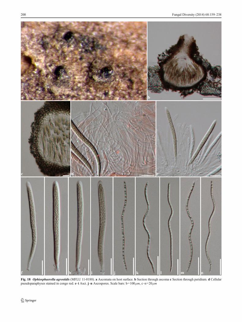
Fig. 18 Ophiosphaerella agrostidis (MFLU 11-0188). a Ascomata on host surface. b Section through ascoma c Section through peridium. d Cellularpseudoparaphyses stained in congo red. e–i Asci. j–n Ascospores. Scale bars: b=100μm, c–n=20μm
200 Fungal Diversity (2014) 68:159–238
Parastagonospora Stud. Mycol. 75: 362 (2013), Faces ofFungi number: FoF00260.
Pathogenic on various cereal crops (Poaceae). Sexualstate: Ascomata scattered, solitary to gregarious, immersed,uniloculate, globose, glabrous, brown to black, ostiole central,with papilla. Peridium thick-walled, composed of 3–6 layersof brown pseudoparenchymatous cells, arranged in a texturaangularis. Hamathecium composed of numerous, filiform,cellular pseudoparaphyses, with distinct septa. Asci 8-spored,bitunicate, cylindrical to clavate, short pedicellate. Ascosporesphragmosporous, fusiform, subhyaline to pale brown, septate(−3 septa), constricted at the septa, swollen at penultimate cell.Asexual state:Conidiomata pycnidial, scattered, immersed tosemi-immersed, uniloculate, subglobose, glabrous, black, ex-uding creamy conidial mass, ostiole central. Conidiomatawalls thin, composed of 2–3 layers of brown cells, arrangedin textura angularis. Conidiophores reduced to conidiogenouscells. Conidiogenous cells phialidic, integrate, ampulliform tosubcylindrical, hyaline, smooth-walled, lining the inner cavi-ty, with percurrent proliferation near apex. Conidiaphragmosporous, cylindrical, apically obtuse with truncatebase, hyaline, septate, thin and smooth-walled, with granulesor multiguttules (from Quaedvlieg et al. 2013).
Type species: Parastagonospora nodorum (Berk.)Quaedvlieg et al. Stud. Mycol. 75: 363 (2013)
≡ Depazea nodorum Berk., Gard. Chron., London: 601(1845)
Phylogenetic study: Quaedvlieg et al. (2013)Notes: Parastagonospora was introduced by Quaedvlieg
et al. (2013) to accommodate stagonospora-like species whichhave been reported as asexual state of phaeosphaeria-like taxa.The genus accommodated five species; Pa. avenae (A.B..
Frank) Quaedvlieg et al., Pa. caricis Quaedvlieg et al., Pa.nodorum (Berk.) Quaedvlieg et al., Pa. poaeQuaedvlieg et al.and Pa. poagena Crous & Quaedvlieg with Pa. nodorum asthe type (Index Fungorum, 2014). Parastagonospora is sim-ilar to Stagonospora; however, they differ in the morphologyof the conidiogenous cells (Quaedvlieg et al. 2013). Based onmultigene phylogenetic analysis, Parastagonospora clustersin Phaeosphaeriaceae which forms a clade withPhaeosphaer i a avena r ia ( s ynonymized unde rParastagonospora avenae), Ph. eustoma and Ph. nigrans.These Phaeosphaeria species may be the sexual states ofParastagonospora.
Phaeosphaeria I. Miyake, Bot. Mag., Tokyo 23: 93(1909), Faces of Fungi number: FoF00233.
= Phaeoseptoria Speg., Revta Mus. La Plata 15: 39. 1908.Saprobic or pathogenic mostly on monocotyledons.
Sexual state: Ascomata solitary to gregarious, immersed,visible as slightly raised, small, black dots on the host surface,uniloculate, globose to subglobose, glabrous, brown, ostiolecentral, with minute papilla. Peridium thin-walled, of equalthickness, composed of flattened, brown, pseudoparenchyma-tous cells, arranged in textura angularis. Hamathecium com-posed of sparse, filiform, broad cellular pseudoparaphyses,with distinct septa, slightly constricted at the septa, rarelyanastomosing at the apex. Asci 8-spored, bitunicate,fissitunicate, broadly cylindrical, short pedicellate orsubsessile, apically rounded, with an indistinct ocular cham-ber. Ascospores overlapping 2–3-seriate, phragmosporous,narrowly fusiform, pale brown to brown or yellowish-brown,septate, straight or slightly curved, constricted or not constrict-ed at the septa, walls smooth or rough, echinulate orverruculose, with or without mucilaginous sheaths. Asexual
Table 3 Synopsis of Ophiosphaerella species discussed in this study
Species Size (μm) Septation Hosts Source references
Ascospores Asci Ascomata(diam.)
Op. agrostidis 75–150×2–2.5 100–230×10–15 130–350 6–15 Agrostis andCynodon×dactylon
Câmara et al. (2000),Farr and Rossman(2014)
Op. agrostidis(MFLUCC11-0152)
98–130×2–2.5 98–130×7–9(−11) 175–310 15 Grass culm This study
Op. graminicola(holotype)
140×3 150×10 180–200 12–20 Leptochloa virgata Spegazzini (1909),Walker (1980)
Op. graminicola(BPI748267)
142.4×3.2 148.7×11.2 170–230 15–18 grass culm Farr and Rossman(2014)
Op. herpotricha 140–180×2–2.5(−3) 150–190×7–9 300–400 12–16 Arrhenatherum, Bromus,Buchloe, Cynodon,Dactylis, Puccinellia,Secale, Triticum,Vetiveria, Zoysia
Farr and Rossman(2014)Walker (1980)
Op. korrae 120–180×4–5(−5.5) 145–200×10–13(−15) 300–500 (3–6)7(−15) Axonopus, Cynodon,Poa, Zoysia
Farr and Rossman(2014)
Fungal Diversity (2014) 68:159–238 201
state: Conidiomata stromatic, pycnidial, scattered to gregari-ous, immersed, uni- to multi-loculate, globose to subglobose,brown. Conidiomata walls thin, composed of thin-walled, ofbrown pseudoparenchymatous cells, arranged in texturaangularis. Conidiophores reduced to conidiogenous cells.Conidiogenous cells holoblastic, phialidic, flattenedampulliform to doliiform, hyaline to pale brown, hardly dis-tinguishable from inner cells walls of conidioma. Conidiaphragmosporous, oblong to cylindrical, pale brown to brown,septate, straight or slightly curved with obtuse at the base,smooth-walled with guttules.
Type species: Phaeosphaeria oryzaePhylogenetic study: Câmara et al. (2002); Schoch et al.
(2006, 2009); Zhang et al. (2009, 2012); De Gruyter et al.(2010, 2012); Quaedvlieg et al. (2013); Hyde et al. (2013,2014); Wijayawardene et al. (2014b)
Notes: Phaeosphaeria was established by Miyake (1909).Miyake (1910) accommodated Phaeosphaeria inMycosphaerellaceae andmentioned that the genus differs fromSphaerulina and Leptosphaeria in ascospore colour and lackof pseudoparaphyses (Eriksson 1967). Phaeosphaeria wasconsidered as a synonym of Leptosphaeria for a long time(Müller 1950; Munk 1957; Zhang et al. 2009, 2012). Thegenus was however, reinstated by Holm (1957) who foundpseudoparaphyses in Phaeosphaeria oryzae and 17 mostlygraminicolous species were transferred to Phaeosphaeria(Eriksson 1967; Zhang et al. 2009). Barr (1979) establishedthe new family Phaeosphaeriaceae and designatedPhaeosphaeria as the generic type and later, Eriksson (1981)introduced a broad generic concept for Phaeosphaeria whichincluded relatively small ascomata, dictyospores, as well asascospores with several wall layers, and an occurrence onmonocotyledons (Câmara et al. 2002; Zhang et al. 2012).Subsequent authors also included Phaeosphaeria species fromdicotyledonous hosts (Dennis 1978; Sivanesan 1984; von Arxand Müller 1975; Eriksson 1967; Hedjaroude 1969;Shoemaker and Babcock 1989b; Zhang et al. 2012).Shoemaker and Babcock (1989b) re-circumscribedPhaeosphaeria and divided the genus into six subgenera basedon the differences in ascospore shape and septation viz.Fusispora, Ovispora, Phaeosphaeria, Sicispora, Spathisporaand Vagispora. Various authors subsequently indicated thatPhaeosphaeria was heterogeneous and shared several charac-ters with Leptosphaeria. Phaeosphaeria however, differs fromLeptosphaeria in morphology and these genera have beendistinguished in different families by phylogenetic analysis(Câmara et al. 2002; Schoch et al. 2009; Zhang et al. 2009,2012; Hyde et al. 2013). Presently, there are 200 epithets forPhaeosphaeria in Index Fungorum (2014) with 18,983 nucle-otide sequences available in GenBank. However, several spe-cies of Leptosphaeria sensu stricto and Phaeosphaeria areconfused in having similar morphology and lack phylogeneticinvestigation.
The asexual states of Phaeosphaeria have been reported ascoelomycetes and some as hyphomycetes namelyhendersonia-like, Mauginiella , Microsphaeropsis ,Neostagonospora, Parastagonospora, Phaeoseptoria,Scolecosporiella and stagonospora-like (Leuchtmann 1984;Shoemaker and Babcock 1989b; Câmara et al. 2002;Abdullah et al. 2005; Kirk et al. 2008; Hyde et al. 2011,2013; Wijayawardene et al. 2012; Zhang et al. 2012;Quaedvlieg et al. 2013). However, Leuchtmann (1984) treatedthe asexual state of Phaeosphaeria as species ofStagonospora and mentioned that the asexual state ofPhaeosphaeria often formed pycnidial conidiomata, pos-sessed holoblastic conidiogenous cells and had elongated tofiliform, hyaline to pale brown, multiseptate conidia, whichwas also mentioned by Câmara et al. (2002). Câmara et al.(2002) suggested that Phaeosphaeria shares common ances-tors with Leptosphaeria and that multigene phylogenetic anal-ysis was necessary to understand the boundaries of taxa. Theperidium type, asexual state and host preferences can distin-guish fungal taxa in these two genera (Câmara et al. 2002)
Quaedvlieg et al. (2013) studied Septoria, Stagonosporaand related genera such as Phaeosphaeria and Phaeoseptoriausing molecular analysis and concluded that Stagonospora(based on the type species, S. paludosa) belongs toMassarinaceae . They introduced two new genera(Neostagonospora and Parastagonospora) to accommodatestagonospora-like genera in Phaeosphaeriaceae (Quaedvlieget al. 2013). Additionally, Phaeosphaeria was linked to theasexual state Phaeoseptoria based on the phylogenetic analy-sis with the type species of Phaeosphaeria, Ph. oryzaeforming a well-supported clade with Phaeoseptoria papayae,the type species of Phaeoseptoria (Quaedvlieg et al. 2013).Thus Quaedvlieg et al. (2013) synonymized the earlier namePhaeoseptoria under Phaeosphaeria as the latter was well-studied and taxonomy less confused than Phaeoseptoriawhich have been confused with several septoria-like inpigmented conidia (Walker et al. 1992). Hendersonia, whichwas treated as a synonym of Stagonospora , andMicrosphaeropsis were placed in Didymellaceae (Sutton1977; Swart and Walker 1988; Kirk et al. 2008; De Gruyteret al. 2009; Aveskamp et al. 2010).Mauginiellawas treated asan asexual state of Phaeosphaeria by Abdullah et al. (2005);however, this has not been confirmed by phylogenetic inves-tigation. In this study Mauginiella could not be resolved inPhaeosphaeriaceae. The genus is currently placed in asexualPezizomycotina (Kirk et al. 2008). Therefore we excludeMauginiella from Phaeosphaeriaceae. Other asexual generaof Phaeosphaeria still lack phylogenetic investigation.Therefore we accept three asexual genera (NeostagonosporaQuaedvlieg et al., Parastagonospora Quaedvlieg et al., andPhaeoseptoria Speg.) for Phaeosphaeria sensu lato.
Phaeosphaeria oryzae was designated as typified ofPhaeosphaeria by Miyake (1909). However, Miyake (1909)
202 Fungal Diversity (2014) 68:159–238
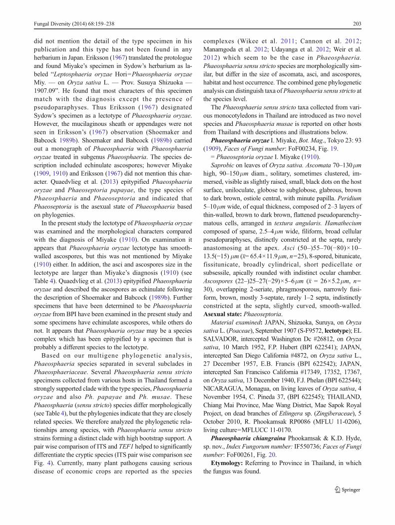
did not mention the detail of the type specimen in hispublication and this type has not been found in anyherbarium in Japan. Eriksson (1967) translated the protologueand found Miyake’s specimen in Sydow’s herbarium as la-beled “Leptosphaeria oryzae Hori=Phaeosphaeria oryzaeMiy. — on Oryza sativa L. — Prov. Susuya Shizuoka —1907.09”. He found that most characters of this specimenmatch with the diagnosis except the presence ofpseudoparaphyses. Thus Eriksson (1967) designatedSydow’s specimen as a lectotype of Phaeosphaeria oryzae.However, the mucilaginous sheath or appendages were notseen in Eriksson’s (1967) observation (Shoemaker andBabcock 1989b). Shoemaker and Babcock (1989b) carriedout a monograph of Phaeosphaeria with Phaeosphaeriaoryzae treated in subgenus Phaeosphaeria. The species de-scription included echinulate ascospores; however Miyake(1909, 1910) and Eriksson (1967) did not mention this char-acter. Quaedvlieg et al. (2013) epitypified Phaeosphaeriaoryzae and Phaeoseptoria papayae, the type species ofPhaeosphaeria and Phaeoseptoria and indicated thatPhaeoseptoria is the asexual state of Phaeosphaeria basedon phylogenies.
In the present study the lectotype of Phaeosphaeria oryzaewas examined and the morphological characters comparedwith the diagnosis of Miyake (1910). On examination itappears that Phaeosphaeria oryzae lectotype has smooth-walled ascospores, but this was not mentioned by Miyake(1910) either. In addition, the asci and ascospores size in thelectotype are larger than Miyake’s diagnosis (1910) (seeTable 4). Quaedvlieg et al. (2013) epitypified Phaeosphaeriaoryzae and described the ascospores as echinulate followingthe description of Shoemaker and Babcock (1989b). Furtherspecimens that have been determined to be Phaeosphaeriaoryzae from BPI have been examined in the present study andsome specimens have echinulate ascospores, while others donot. It appears that Phaeosphaeria oryzae may be a speciescomplex which has been epitypified by a specimen that isprobably a different species to the lectotype.
Based on our multigene phylogenetic analysis,Phaeosphaeria species separated in several subclades inPhaeosphaeriaceae. Several Phaeosphaeria sensu strictospecimens collected from various hosts in Thailand formed astrongly supported clade with the type species,Phaeosphaeriaoryzae and also Ph. papayae and Ph. musae. ThesePhaeosphaeria (sensu stricto) species differ morphologically(see Table 4), but the phylogenies indicate that they are closelyrelated species. We therefore analyzed the phylogenetic rela-tionships among species, with Phaeosphaeria sensu strictostrains forming a distinct clade with high bootstrap support. Apair wise comparison of ITS and TEF1 helped to significantlydifferentiate the cryptic species (ITS pair wise comparison seeFig. 4). Currently, many plant pathogens causing seriousdisease of economic crops are reported as the species
complexes (Wikee et al. 2011; Cannon et al. 2012;Manamgoda et al. 2012; Udayanga et al. 2012; Weir et al.2012) which seem to be the case in Phaeosphaeria.Phaeosphaeria sensu stricto species are morphologically sim-ilar, but differ in the size of ascomata, asci, and ascospores,habitat and host occurrence. The combined gene phylogeneticanalysis can distinguish taxa ofPhaeosphaeria sensu stricto atthe species level.
The Phaeosphaeria sensu stricto taxa collected from vari-ous monocotyledons in Thailand are introduced as two novelspecies and Phaeosphaeria musae is reported on other hostsfrom Thailand with descriptions and illustrations below.
Phaeosphaeria oryzae I. Miyake, Bot. Mag., Tokyo 23: 93(1909), Faces of Fungi number: FoF00234, Fig. 19.
= Phaeoseptoria oryzae I. Miyake (1910).Saprobic on leaves of Oryza sativa. Ascomata 70–130μm
high, 90–150μm diam., solitary, sometimes clustered, im-mersed, visible as slightly raised, small, black dots on the hostsurface, uniloculate, globose to subglobose, glabrous, brownto dark brown, ostiole central, with minute papilla. Peridium5–10μm wide, of equal thickness, composed of 2–3 layers ofthin-walled, brown to dark brown, flattened pseudoparenchy-matous cells, arranged in textura angularis. Hamatheciumcomposed of sparse, 2.5–4μm wide, filiform, broad cellularpseudoparaphyses, distinctly constricted at the septa, rarelyanastomosing at the apex. Asci (50–)55–70(−80)×10–13.5(−15) μm (x= 65.4×11.9μm, n=25), 8-spored, bitunicate,fissitunicate, broadly cylindrical, short pedicellate orsubsessile, apically rounded with indistinct ocular chamber.Ascospores (22–)25–27(−29)×5–6μm (x = 26×5.2μm, n=30), overlapping 2-seriate, phragmosporous, narrowly fusi-form, brown, mostly 3-septate, rarely 1–2 septa, indistinctlyconstricted at the septa, slightly curved, smooth-walled.Asexual state: Phaeoseptoria.
Material examined: JAPAN, Shizuoka, Suruya, on Oryzasativa L. (Poaceae), September 1907 (S-F9572, lectotype); ELSALVADOR, intercepted Washington Dc #26812, on Oryzasativa, 10 March 1952, F.P. Hubert (BPI 622541); JAPAN,intercepted San Diego California #4872, on Oryza sativa L.,27 December 1957, E.B. Francis (BPI 622542); JAPAN,intercepted San Francisco California #17349, 17352, 17367,onOryza sativa, 13 December 1940, F.J. Phelan (BPI 622544);NICARAGUA, Monagua, on living leaves of Oryza sativa, 4November 1954, C. Pineda 37, (BPI 622545); THAILAND,Chiang Mai Province, Mae Wang District, Mae Sapok RoyalProject, on dead branches of Etlingera sp. (Zingiberaceae), 5October 2010, R. Phookamsak RP0086 (MFLU 11-0206),living culture=MFLUCC 11-0170.
Phaeosphaeria chiangraina Phookamsak & K.D. Hyde,sp. nov., Index Fungorum number: IF550736; Faces of Funginumber: FoF00261, Fig. 20.
Etymology: Referring to Province in Thailand, in whichthe fungus was found.
Fungal Diversity (2014) 68:159–238 203
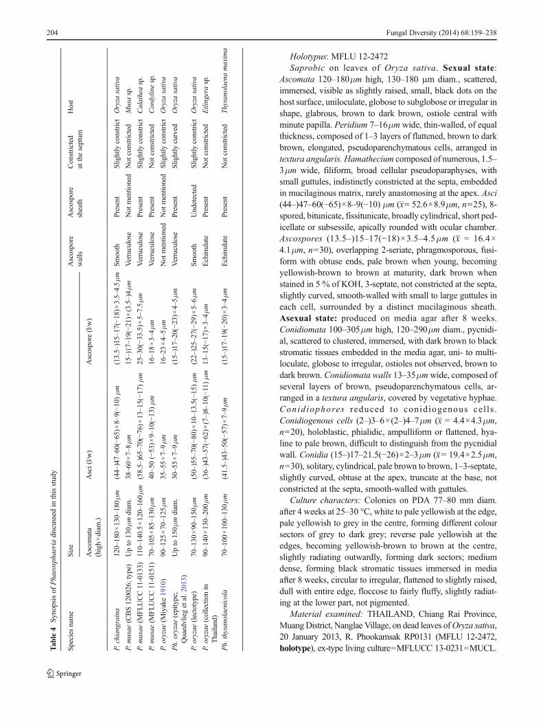
Holotypus: MFLU 12-2472Saprobic on leaves of Oryza sativa. Sexual state:
Ascomata 120–180μm high, 130–180 μm diam., scattered,immersed, visible as slightly raised, small, black dots on thehost surface, uniloculate, globose to subglobose or irregular inshape, glabrous, brown to dark brown, ostiole central withminute papilla. Peridium 7–16μm wide, thin-walled, of equalthickness, composed of 1–3 layers of flattened, brown to darkbrown, elongated, pseudoparenchymatous cells, arranged intextura angularis. Hamathecium composed of numerous, 1.5–3μm wide, filiform, broad cellular pseudoparaphyses, withsmall guttules, indistinctly constricted at the septa, embeddedin mucilaginous matrix, rarely anastomosing at the apex. Asci(44–)47–60(−65)×8–9(−10) μm (x= 52.6×8.9μm, n=25), 8-spored, bitunicate, fissitunicate, broadly cylindrical, short ped-icellate or subsessile, apically rounded with ocular chamber.Ascospores (13.5–)15–17(−18)×3.5–4.5μm (x = 16.4×4.1μm, n=30), overlapping 2-seriate, phragmosporous, fusi-form with obtuse ends, pale brown when young, becomingyellowish-brown to brown at maturity, dark brown whenstained in 5 % of KOH, 3-septate, not constricted at the septa,slightly curved, smooth-walled with small to large guttules ineach cell, surrounded by a distinct mucilaginous sheath.Asexual state: produced on media agar after 8 weeks.Conidiomata 100–305μm high, 120–290μm diam., pycnidi-al, scattered to clustered, immersed, with dark brown to blackstromatic tissues embedded in the media agar, uni- to multi-loculate, globose to irregular, ostioles not observed, brown todark brown. Conidiomata walls 13–35μmwide, composed ofseveral layers of brown, pseudoparenchymatous cells, ar-ranged in a textura angularis, covered by vegetative hyphae.Conidiophores reduced to conidiogenous cel ls .Conidiogenous cells (2–)3–6×(2–)4–7μm (x = 4.4×4.3μm,n=20), holoblastic, phialidic, ampulliform or flattened, hya-line to pale brown, difficult to distinguish from the pycnidialwall. Conidia (15–)17–21.5(−26)×2–3μm (x= 19.4×2.5μm,n=30), solitary, cylindrical, pale brown to brown, 1–3-septate,slightly curved, obtuse at the apex, truncate at the base, notconstricted at the septa, smooth-walled with guttules.
Culture characters: Colonies on PDA 77–80 mm diam.after 4 weeks at 25–30 °C, white to pale yellowish at the edge,pale yellowish to grey in the centre, forming different coloursectors of grey to dark grey; reverse pale yellowish at theedges, becoming yellowish-brown to brown at the centre,slightly radiating outwardly, forming dark sectors; mediumdense, forming black stromatic tissues immersed in mediaafter 8 weeks, circular to irregular, flattened to slightly raised,dull with entire edge, floccose to fairly fluffy, slightly radiat-ing at the lower part, not pigmented.
Material examined: THAILAND, Chiang Rai Province,MuangDistrict, Nanglae Village, on dead leaves ofOryza sativa,20 January 2013, R. Phookamsak RP0131 (MFLU 12-2472,holotype), ex-type living culture=MFLUCC 13-0231=MUCL.T
able4
Synopsisof
Phaeosphaeria
discussedin
thisstudy
Speciesname
Size
Ascospore
walls
Ascospore
sheath
Constricted
attheseptum
Host
Ascom
ata
(high×diam
.)Asci(l/w
)Ascospore
(l/w)
P.chiangraina
120–180×130–180μm
(44–)47–60(−65)×
8–9(−1
0)μm
(13.5–)15–17(−18)×
3.5–4.5μm
Smooth
Present
Slightly
constrict
Oryza
sativa
P.musae
(CBS120026;type)
Upto
130μmdiam
.38–60×7–8μm
15–)17–19(−2
1)×(3.5–)4μm
Verruculose
Not
mentio
ned
Not
constricted
Musasp.
P.musae
(MFL
UCC11-0133)
110–140.5×120–160μm
(58.5–)65–70(−76)×
13–15(−1
7)μm
25–30(−3
3.5)×5–7.5μm
Verruculose
Present
Slightly
constrict
Calatheasp.
P.musae
(MFL
UCC11-0151)
70–105
×85–130
μm
40–50(−53)×
9–10(−13)μm
16–18×3–4μm
Verruculose
Present
Not
constricted
Cordylin
esp.
P.oryzae
(Miyake1910)
90–125
×70–125
μm
35–55×7–9μm
16–23×4–5μm
Not
mentio
ned
Not
mentio
ned
Slightly
constrict
Oryza
sativa
Ph.oryzae
(epitype;
Quaedvliegetal.2013)
Upto
150μmdiam
.30–55×7–9μm
(15–)17–20(−23)×
4–5μm
Verruculose
Present
Slightly
curved
Oryza
sativa
P.oryzae
(lectotype)
70–130
×90–150
μm
(50–)55–70(−80)×
10–13.5(−1
5)μm
(22–)25–27(−29)×
5–6μm
Smooth
Undetected
Slightly
constrict
Oryza
sativa
P.oryzae
(collectionin
Thailand)
90–140
×130–200μm
(36–)43–57(−62)×
(7–)8–10(−11)μm
13–15(−1
7)×3–4μm
Echinulate
Present
Not
constricted
Etlingerasp.
Ph.thysanolaenicola
70–100
×100–130μm
(41.5–)43–50(−57)×
7–9μm
(15–)17–19(−20)×
3–4μm
Echinulate
Present
Not
constricted
Thysanolaena
maxima
204 Fungal Diversity (2014) 68:159–238
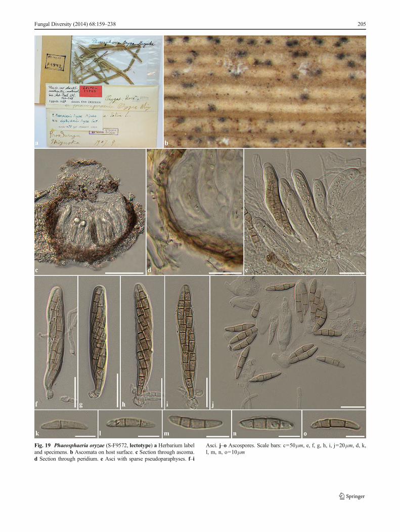
Fungal Diversity (2014) 68:159–238 205
Fig. 19 Phaeosphaeria oryzae (S-F9572, lectotype) a Herbarium labeland specimens. b Ascomata on host surface. c Section through ascoma.d Section through peridium. e Asci with sparse pseudoparaphyses. f–i
Asci. j–o Ascospores. Scale bars: c=50μm, e, f, g, h, i, j=20μm, d, k,l, m, n, o=10μm
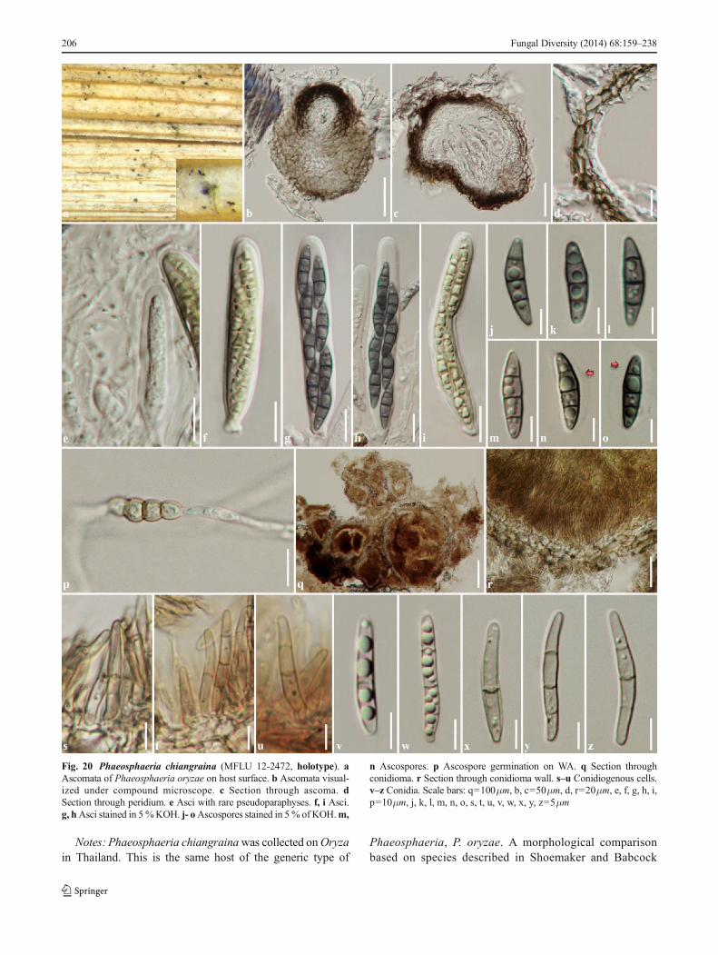
Notes: Phaeosphaeria chiangrainawas collected onOryzain Thailand. This is the same host of the generic type of
Phaeosphaeria, P. oryzae. A morphological comparisonbased on species described in Shoemaker and Babcock
Fig. 20 Phaeosphaeria chiangraina (MFLU 12-2472, holotype). aAscomata of Phaeosphaeria oryzae on host surface. b Ascomata visual-ized under compound microscope. c Section through ascoma. dSection through peridium. e Asci with rare pseudoparaphyses. f, i Asci.g, hAsci stained in 5%KOH. j- oAscospores stained in 5% of KOH.m,
n Ascospores. p Ascospore germination on WA. q Section throughconidioma. r Section through conidioma wall. s–u Conidiogenous cells.v–z Conidia. Scale bars: q=100μm, b, c=50μm, d, r=20μm, e, f, g, h, i,p=10μm, j, k, l, m, n, o, s, t, u, v, w, x, y, z=5μm
206 Fungal Diversity (2014) 68:159–238
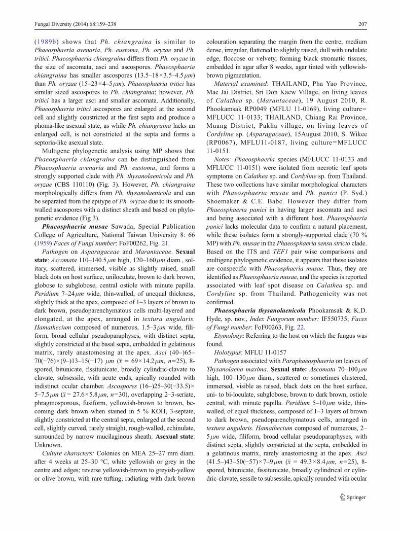
(1989b) shows that Ph. chiangraina is similar toPhaeosphaeria avenaria, Ph. eustoma, Ph. oryzae and Ph.tritici. Phaeosphaeria chiangraina differs from Ph. oryzae inthe size of ascomata, asci and ascospores. Phaeosphaeriachiangraina has smaller ascospores (13.5–18×3.5–4.5μm)than Ph. oryzae (15–23×4–5μm). Phaeosphaeria tritici hassimilar sized ascospores to Ph. chiangraina; however, Ph.tritici has a larger asci and smaller ascomata. Additionally,Phaeosphaeria tritici ascospores are enlarged at the secondcell and slightly constricted at the first septa and produce aphoma-like asexual state, as while Ph. chiangraina lacks anenlarged cell, is not constricted at the septa and forms aseptoria-like asexual state.
Multigene phylogenetic analysis using MP shows thatPhaeosphaeria chiangraina can be distinguished fromPhaeosphaeria avenaria and Ph. eustoma, and forms astrongly supported clade with Ph. thysanolaenicola and Ph.oryzae (CBS 110110) (Fig. 3). However, Ph. chiangrainamorphologically differs from Ph. thysanolaenicola and canbe separated from the epitype of Ph. oryzae due to its smooth-walled ascospores with a distinct sheath and based on phylo-genetic evidence (Fig 3).
Phaeosphaeria musae Sawada, Special PublicationCollege of Agriculture, National Taiwan University 8: 66(1959) Faces of Fungi number: FoF00262, Fig. 21.
Pathogen on Asparagaceae and Marantaceae. Sexualstate: Ascomata 110–140.5μm high, 120–160μm diam., sol-itary, scattered, immersed, visible as slightly raised, smallblack dots on host surface, uniloculate, brown to dark brown,globose to subglobose, central ostiole with minute papilla.Peridium 7–24μm wide, thin-walled, of unequal thickness,slightly thick at the apex, composed of 1–3 layers of brown todark brown, pseudoparenchymatous cells multi-layered andelongated, at the apex, arranged in textura angularis.Hamathecium composed of numerous, 1.5–3μm wide, fili-form, broad cellular pseudoparaphyses, with distinct septa,slightly constricted at the basal septa, embedded in gelatinousmatrix, rarely anastomosing at the apex. Asci (40–)65–70(−76)×(9–)13–15(−17) μm (x = 69×14.2μm, n=25), 8-spored, bitunicate, fissitunicate, broadly cylindric-clavate toclavate, subsessile, with acute ends, apically rounded withindistinct ocular chamber. Ascospores (16–)25–30(−33.5)×5–7.5μm (x = 27.6×5.8μm, n=30), overlapping 2–3-seriate,phragmosporous, fusiform, yellowish-brown to brown, be-coming dark brown when stained in 5 % KOH, 3-septate,slightly constricted at the central septa, enlarged at the secondcell, slightly curved, rarely straight, rough-walled, echinulate,surrounded by narrow mucilaginous sheath. Asexual state:Unknown.
Culture characters: Colonies on MEA 25–27 mm diam.after 4 weeks at 25–30 °C, white yellowish or grey in thecentre and edges; reverse yellowish-brown to greyish-yellowor olive brown, with rare tufting, radiating with dark brown
colouration separating the margin from the centre; mediumdense, irregular, flattened to slightly raised, dull with undulateedge, floccose or velvety, forming black stromatic tissues,embedded in agar after 8 weeks, agar tinted with yellowish-brown pigmentation.
Material examined: THAILAND, Pha Yao Province,Mae Jai District, Sri Don Kaew Village, on living leavesof Calathea sp. (Marantaceae), 19 August 2010, R.Phookamsak RP0049 (MFLU 11-0169), living culture=MFLUCC 11-0133; THAILAND, Chiang Rai Province,Muang District, Pakha village, on living leaves ofCordyline sp. (Asparagaceae), 15August 2010, S. Wikee(RP0067), MFLU11-0187, living culture=MFLUCC11-0151.
Notes: Phaeosphaeria species (MFLUCC 11-0133 andMFLUCC 11-0151) were isolated from necrotic leaf spotssymptoms on Calathea sp. and Cordyline sp. from Thailand.These two collections have similar morphological characterswith Phaeosphaeria musae and Ph. panici (P. Syd.)Shoemaker & C.E. Babc. However they differ fromPhaeosphaeria panici in having larger ascomata and asciand being associated with a different host. Phaeosphaeriapanici lacks molecular data to confirm a natural placement,while these isolates form a strongly-supported clade (70 %MP) with Ph. musae in the Phaeosphaeria sensu stricto clade.Based on the ITS and TEF1 pair wise comparisons andmultigene phylogenetic evidence, it appears that these isolatesare conspecific with Phaeosphaeria musae. Thus, they areidentified asPhaeosphaeria musae, and the species is reportedassociated with leaf spot disease on Calathea sp. andCordyline sp. from Thailand. Pathogenicity was notconfirmed.
Phaeosphaeria thysanolaenicola Phookamsak & K.D.Hyde, sp. nov., Index Fungorum number: IF550735; Facesof Fungi number: FoF00263, Fig. 22.
Etymology: Referring to the host on which the fungus wasfound.
Holotypus: MFLU 11-0157Pathogen associated with Paraphaeosphaeria on leaves of
Thysanolaena maxima. Sexual state: Ascomata 70–100μmhigh, 100–130μm diam., scattered or sometimes clustered,immersed, visible as raised, black dots on the host surface,uni- to bi-loculate, subglobose, brown to dark brown, ostiolecentral, with minute papilla. Peridium 5–10μm wide, thin-walled, of equal thickness, composed of 1–3 layers of brownto dark brown, pseudoparenchymatous cells, arranged intextura angularis. Hamathecium composed of numerous, 2–5μm wide, filiform, broad cellular pseudoparaphyses, withdistinct septa, slightly constricted at the septa, embedded ina gelatinous matrix, rarely anastomosing at the apex. Asci(41.5–)43–50(−57)×7–9μm (x = 49.3×8.4μm, n=25), 8-spored, bitunicate, fissitunicate, broadly cylindrical or cylin-dric-clavate, sessile to subsessile, apically rounded with ocular
Fungal Diversity (2014) 68:159–238 207
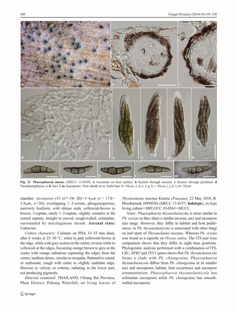
chamber. Ascospores (15–)17–19(−20)×3–4μm (x = 17.8×3.8μm, n=30), overlapping 1–3-seriate, phragmosporous,narrowly fusiform, with obtuse ends, yellowish-brown tobrown, 3-septate, rarely 1–2-septate, slightly constrict at thecentral septum, straight to curved, rough-walled, echinulate,surrounded by mucilaginous sheath. Asexual state:Unknown.
Culture characters: Colonies on PDA 33–35 mm diam.after 4 weeks at 25–30 °C, white to pale yellowish-brown atthe edge, white with grey sectors in the centre; reverse white toyellowish at the edges, becoming orange brown to grey at thecentre with orange radiations separating the edges from thecentre; medium dense, circular to irregular, flattened to raised,or umbonate, rough with entire to slightly undulate edge,floccose to velvety or cottony, radiating in the lower part,not producing pigments.
Material examined: THAILAND, Chiang Rai Province,Phan District, Pukang Waterfall, on living leaves of
Thysanolaena maxima Kuntze (Poaceae), 22 May 2010, R.Phookamsak (RP0036) (MFLU 11-0157, holotype), ex-typeliving culture=MFLUCC 10-0563=MUCL.
Notes: Phaeosphaeria thysanolaenicola is most similar toPh. oryzae as they share a similar ascoma, asci and ascosporesize range. However, they differ in habitat and host prefer-ences, as Ph. thysanolaenicola is associated with other fungion leaf spots of Thysanolaena maxima. Whereas Ph. oryzaewas found as a saprobe on Oryzae sativa. The ITS pair wisecomparison shows that they differ in eight base positions.Phylogenetic analysis performed with a combination of ITS,LSU, RPB2 and TEF1 genes shows that Ph. thysanolaenicolaforms a clade with Ph. chiangraina. Phaeosphaeriathysanolaenicola differs from Ph. chiangraina in its smallerasci and ascospores, habitat, host occurrence and ascosporeornamentation. Phaeosphaeria thysanolaenicola hasechinulate ascospores while Ph. chiangraina has smooth-walled ascospores.
Fig. 21 Phaeosphaeria musae. (MFLU 11-0169). a Ascomata on host surface. b Section through ascoma. c Section through peridium. dPseudoparaphyses. e–h Asci. i–m Ascospores. Note sheath in m. Scale bars: b=50μm, c, d, e, f, g, h, = 20μm, i, j, k, l, m=10μm
208 Fungal Diversity (2014) 68:159–238
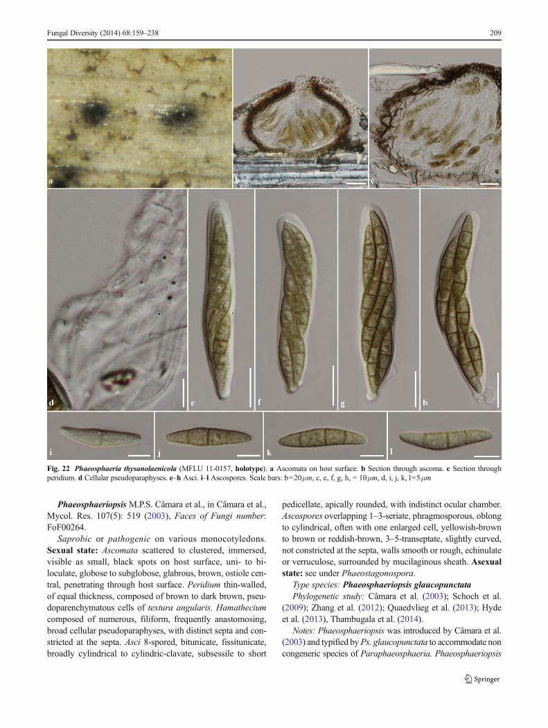
Phaeosphaeriopsis M.P.S. Câmara et al., in Câmara et al.,Mycol. Res. 107(5): 519 (2003), Faces of Fungi number:FoF00264.
Saprobic or pathogenic on various monocotyledons.Sexual state: Ascomata scattered to clustered, immersed,visible as small, black spots on host surface, uni- to bi-loculate, globose to subglobose, glabrous, brown, ostiole cen-tral, penetrating through host surface. Peridium thin-walled,of equal thickness, composed of brown to dark brown, pseu-doparenchymatous cells of textura angularis. Hamatheciumcomposed of numerous, filiform, frequently anastomosing,broad cellular pseudoparaphyses, with distinct septa and con-stricted at the septa. Asci 8-spored, bitunicate, fissitunicate,broadly cylindrical to cylindric-clavate, subsessile to short
pedicellate, apically rounded, with indistinct ocular chamber.Ascospores overlapping 1–3-seriate, phragmosporous, oblongto cylindrical, often with one enlarged cell, yellowish-brownto brown or reddish-brown, 3–5-transeptate, slightly curved,not constricted at the septa, walls smooth or rough, echinulateor verruculose, surrounded by mucilaginous sheath. Asexualstate: see under Phaeostagonospora.
Type species: Phaeosphaeriopsis glaucopunctataPhylogenetic study: Câmara et al. (2003); Schoch et al.
(2009); Zhang et al. (2012); Quaedvlieg et al. (2013); Hydeet al. (2013), Thambugala et al. (2014).
Notes: Phaeosphaeriopsis was introduced by Câmara et al.(2003) and typified byPs. glaucopunctata to accommodate noncongeneric species of Paraphaeosphaeria. Phaeosphaeriopsis
Fig. 22 Phaeosphaeria thysanolaenicola (MFLU 11-0157, holotype). a Ascomata on host surface. b Section through ascoma. c Section throughperidium. d Cellular pseudoparaphyses. e–h Asci. i–l Ascospores. Scale bars: b=20μm, c, e, f, g, h, = 10μm, d, i, j, k, l=5μm
Fungal Diversity (2014) 68:159–238 209
agavensis (A.W. Ramaley et al.) Câmara et al., Ps. amblysporaA.W. Ramaley, Ps. glaucopunctata (Grev.) Câmara et al., Ps.nolinae (A.W. Ramaley) Câmara et al., and Ps. obtusispora(Speg.) Câmara et al. were introduced in the genus basedon morphological characters and phylogenetic analyses ofPhaeosphaeriopsis glaucopunctata (CBS 653.86) (Câmaraet al. 2003; Zhang et al. 2012; Thambugala et al. 2014).Farr et al. (2006) transferred Sphaeria phacidiomorpha toPhaeosphaeriopsis phacidiomorpha based on the type mor-phological characters and microsphaeropsis-like asexual statewhich was most similar to Phaeosphaeriopsis. However,Sphaer ia phac id iomorpha d i f fe r s f rom otherPhaeosphaeriopsis species by its hyaline ascospores (Farret al. 2006; Thambugala et al. 2014). Phaeosphaeriopsisphacidiomorpha is currently synonymized under the nameGlomerella phacidiomorpha (Ces.) Petr. (Index Fungorum2014; Thambugala et al. 2014).
Phaeosphaeriopsis musae was introduced andaccommodated in Phaeosphaeriaceae by Arzanlou andCrous (2006) and is a pathogen causing leaf spots on banana.Subsequent phylogenetic analysis indicated that Ps. musaeclusters with Phaeosphaeria oryzae and separate toPhaeosphaeriopsis glaucopunctata (Zhang et al. 2012; Hydeet al. 2013; Phookamsak et al. 2013, 2014). Thus,Thambugala et al. (2014) synonymized Phaeosphaeriopsismusae under the older name Phaeosphaeria musae Sawada(1959) based on morphological characters and phylogeneticevidence. Additionally, Thambugala et al. (2014) epitypifiedPhaeosphaeriopsis glaucopunctata and described the newspecies Phaeosphaeriopsis triseptata K.M. Thambugala &K.D. Hyde with sexual and asexual morphs. Six species arelisted in Index Fungorum (2014) and 59 sequences are avail-able in GenBank. Molecular analysis confirms the placementof Phaeosphaeriopsis in Phaeosphaeriaceae (Zhang et al.2012; Hyde et al. 2013; Quaedvlieg et al. 2013; Thambugalaet al. 2014; Wijayawardene et al. 2014b).
A collection of Phaeosphaeriopsiswas made which causesleaf spots on leaves ofDracaena lourieriGagnep in Thailand.Based on morphological characters and multigene phyloge-netic analysis, a new species, Phaeosphaeriopsisdracaenicola, is introduced to accommodate this taxon inthe present paper.
Phaeosphaeriopsis glaucopunctata (Grev.) M.P.S.Câmara et al. [as ‘glauco-punctata’], in Câmara et al.,Mycol. Res. 107(5): 519 (2003), Faces of Fungi number:FoF00265, Fig. 23.
≡ Cryptosphaeria glaucopunctata Grev., Fl. Edin.: 1-478(1824)
Saprobic on Ruscus aculeatus (Asparagaceae). Sexualstate: Ascomata 110–145μm high, 130–220μm diam,scattered, solitary, immersed, visible as small, brown spotssurrounded by pale yellowish region, uniloculate, subglobose,glabrous, with central black ostiole penetrating through host
surface. Peridium 7–18μm wide, thin-walled, of unequalthickness, composed of 3–4 layers of thickened, brown, pseu-doparenchymatous cells, arranged in a textura angularis.Hamathecium composed of sparse, 1–3μm wide, filiform,frequently anastomosing, broad cellular pseudoparaphyses,with distinct septa, constricted at the septa. Asci (55–)60–75(−76)×(11–)12–14(−15) μm (x = 68.2×13.4μm, n=20),bitunicate, 8-spored, broadly cylindrical to cylindric-clavate,subsessile to short pedicellate, apically rounded with indistinctocular chamber. Ascospores 18–20(−22)×(3–)4–5.5μm(x = 20 × 4.8μm , n= 20), overlapping 1–3-seriate,phragmosporous, cylindrical, narrower and longer at the lowercell, brown to dark brown, 4-septate, often enlarged at theforth cell, rough-walled, echinulate, surrounded by mucilagi-nous sheath.Asexual state:Conidiomata pycnidial, scattered,solitary, immersed to superficial (in agar), uniloculate, glo-bose, glabrous, brown to black, ostiolate, with black droplets.Conidiomata walls thin, comprising 2–3 layers of yellowish-brown to brown, pseudoparenchymatous cells, arranged intextura angularis. Conidiophores reduced to conidiogenouscells. Conidiogenous cells holoblastic, phialidic, annellidic,with a short collarette, ampulliform to subcylindrical, hyaline,lining the inner cavity of conidioma. Conidia amerosporous,cylindrical to oblong, with rounded ends, hyaline to yellow,becoming yellowish-brown at maturity, aseptate, smooth tofinely verruculose, accumulating in a huge bluish-black slimymass at the tip of ostiole (from Thambugala et al. 2014).
Material examined: UK, near Edinburgh, Slateford, ondead leaves of Ruscus aculeatus L. (Asparagaceae), 15August 1824, R.K. Greville (E 00074283, syntype).
Phaeosphaeriopsis dracaenicola Phookamsak & K.D.Hyde, sp. nov., Index Fungorum number: IF550737; Facesof Fungi number: FoF00266, Fig. 24.
Etymology: referring to the host, of which the fungus wasfound.
Holotypus: MFLU 11-0193Pathogenic causing necrotic leaf spot on Dracaena
lourieri; lesions 3–6 cm, infecting mostly on leaf margin,oval-shaped, pale brown to yellowish-brown, separated fromhealthy tissue by a reddish-brown to orange-brown margin.Sexual state: Ascomata 88.5–170μm high, 140–200μmdiam., solitary to gregarious, immersed, visible as slightlyraised, small black spots on host surface, subglobose, brownto dark brown, ostiole central. Peridium 5–17.5μmwide, thin-walled, of equal thickness, composed of 3–4 layers of darkbrown, pseudoparenchymatous cells, arranged in texturaangularis, outer layer comprising 2–3 layers of thick-walledcells, inner layers comprising 1–2 layers of thin-walled hya-line cells. Hamathecium composed of numerous, 1.5–4μmwide, frequently septate, frequently anastomosing, branching,broad cellular pseudoparaphyses, embedded in mucilaginousmatrix. Asci 62–78×9–12μm (x = 71.5×10.8μm, n=25), 8-spored, bitunicate, fissitunicate, cylindrical to cylindric-
210 Fungal Diversity (2014) 68:159–238
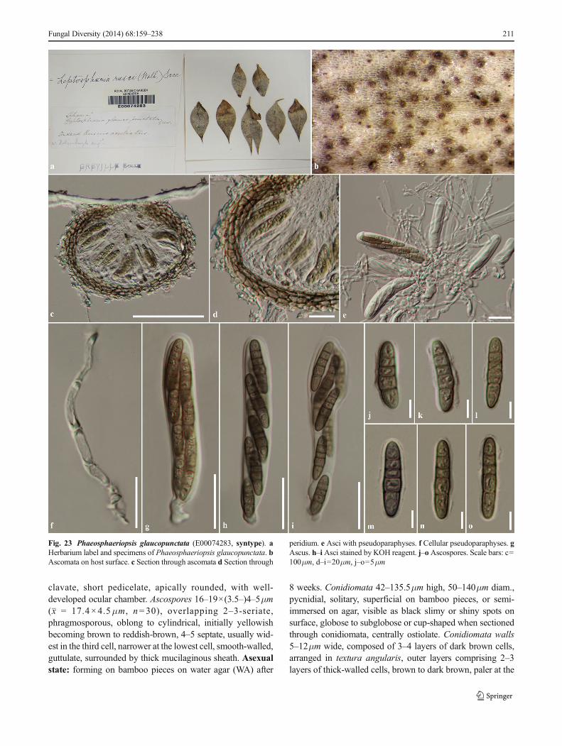
clavate, short pedicelate, apically rounded, with well-developed ocular chamber. Ascospores 16–19×(3.5–)4–5μm(x = 17.4×4.5μm, n=30), overlapping 2–3-seriate,phragmosporous, oblong to cylindrical, initially yellowishbecoming brown to reddish-brown, 4–5 septate, usually wid-est in the third cell, narrower at the lowest cell, smooth-walled,guttulate, surrounded by thick mucilaginous sheath. Asexualstate: forming on bamboo pieces on water agar (WA) after
8 weeks. Conidiomata 42–135.5μm high, 50–140μm diam.,pycnidial, solitary, superficial on bamboo pieces, or semi-immersed on agar, visible as black slimy or shiny spots onsurface, globose to subglobose or cup-shaped when sectionedthrough conidiomata, centrally ostiolate. Conidiomata walls5–12μm wide, composed of 3–4 layers of dark brown cells,arranged in textura angularis, outer layers comprising 2–3layers of thick-walled cells, brown to dark brown, paler at the
Fungal Diversity (2014) 68:159–238 211
Fig. 23 Phaeosphaeriopsis glaucopunctata (E00074283, syntype). aHerbarium label and specimens of Phaeosphaeriopsis glaucopunctata. bAscomata on host surface. c Section through ascomata d Section through
peridium. e Asci with pseudoparaphyses. f Cellular pseudoparaphyses. gAscus. h–i Asci stained by KOH reagent. j–oAscospores. Scale bars: c=100μm, d–i=20μm, j–o=5μm
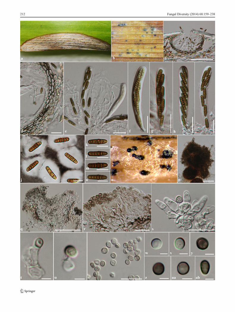
212 Fungal Diversity (2014) 68:159–238
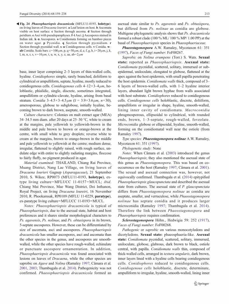
base, inner layer comprising 2–3 layers of thin-walled cells,hyaline. Conidiophores simple, rarely branched, doliiform tocylindrical or ampulliform, septate, hyaline, mostly reduced toconidiogenous cells. Conidiogenous cells 4–12×3–4μm, ho-loblastic, phialidic, single, discrete, sometimes integrated,ampulliform or cylindric-clavate, hyaline, arising from basalstratum. Conidia 3–4.5×3–4.5μm (x = 3.9×3.6μm, n=30),amerosporous, globose to subglobose, initially hyaline, be-coming brown to dark brown, aseptate, smooth-walled.
Culture characters: Colonies on malt extract agar (MEA)34–34.5 mm diam. after 20 days at 25–30 °C, white to creamat the margins, pale yellowish to yellowish-brown in themiddle and pale brown to brown or orange-brown at thecentre, with small white to grey droplets; reverse white tocream at the margins, brown to orange-brown in the middleand pale yellowish to yellowish at the centre; medium dense,irregular, flattened to slightly raised, with rough surface, un-dulate edge with entire to slightly radiating margins, floccoseto fairly fluffy, no pigment produced in agar.
Material examined: THAILAND, Chiang Rai Province,Muang District, Nang Lae Village, on living leaves ofDracaena lourieri Gagnep (Asparagaceae), 21 September2010, S. Wikee, RP0073 (MFLU11-0193, holotype), ex-type living culture=MFLUCC 11-0157=MUCL; ibid.Chiang Mai Province, Mae Wang District, Doi Inthanon,Royal Project, on living Dracaena loureiri, 16 November2010, R. Phookamsak, RP0109 (MFLU 11-0229, paratype),ex-paratype living culture=MFLUCC 11-0193=MUCL.
Notes: Phaeosphaeriopsis dracaenicola is typical ofPhaeosphaeriopsis, due to the asexual state, habitat and hostpreferences and it shares similar morphological characters toPs. agavensis, Ps. nolinae, and Ps. obtusispora in its brown,5-septate ascospores. However, these can be differentiated bysize of ascomata, asci and ascospores. Phaeosphaeriopsisdracaenicola has smaller ascospores, asci and ascomata thanthe other species in the genus, and ascospores are smooth-walled, while the other species have rough-walled, echinulateor punctuate ascospore ornamentation. In addition,Phaeosphaeriopsis dracaenicola was found associated withlesions on leaves of Dracaena, while the other species aresaprobic on Agave and Nolina (Ramaley 1997; Câmara et al.2001, 2003; Thambugala et al. 2014). Pathogenicity was notconfirmed. Phaeosphaeriopsis dracaenicola formed an
asexual state similar to Ps. agavensis and Ps. obtusispora,but differed from Ps. nolinae as conidia are globose.Multigene phylogenetic analysis shows that Ps. dracaenicolaformed a robust clade (100 %ML/ 100 %MP/ 1.00 PP) at thebasal of Phaeosphaeriopsis species in Phaeosphaeriaceae.
Phaeostagonospora A.W. Ramaley, Mycotaxon 61: 351(1997), Faces of Fungi number: FoF00267.
Saprobic on Nolina erumpens (Torr.) S. Wats. Sexualstate: reported as Phaeosphaeriopsis. Asexual state:Conidiomata pycnidial, scattered, solitary, immersed or sub-epidermal, uniloculate, elongated to globose, flattened at theapex against the host epidermis, with small papilla penetratingthe host epidermis. Conidiomata walls thick, composed of 3–6 layers of brown-walled cells, with 1–2 hyaline interiorlayers, abundant light brown hyphae from walls associatedwith host substrate. Conidiophores reduced to conidiogenouscells. Conidiogenous cells holoblastic, discrete, doliiform,ampulliform or irregular in shape, hyaline, smooth-walled,lining inner cavity of conidiomata. Macroconidiaphragmosporous, ellipsoidal to cylindrical, with roundedends, brown, 1–3-septate, rough-walled, foveolate.Microconidia globose or ellipsoidal, hyaline, smooth-walled,forming on the conidiomatal wall near the ostiole (fromRamaley 1997).
Type species: Phaeostagonospora nolinae A.W. Ramaley,Mycotaxon 61: 351 (1997).
Phylogenetic study: NoneNotes: When Câmara et al. (2003) introduced the genus
Phaeosphaeriopsis; they also mentioned the asexual state ofthis genus as Phaeostagonospora. This was based on co-occurrence on the host (Ramaley 1997; Câmara et al. 2001).The sexual and asexual connection was, however, notequivocally confirmed. Thambugala et al. (2014) epitypifiedPhaeosphaeriopsis glaucopunctata and described the asexualstate from cultures. The asexual state of P. glaucopunctatadiffers from Phaeostagonospora nolinae as conidia areaseptate, smaller, and verruculose, while Phaeostagonosporanolinae has septate conidia and it produces largermicroconidia (Ramaley 1997; Thambugala et al. 2014).Therefore the link between Phaeostagonospora andPhaeosphaeriopsis requires confirmation.
Sclerostagonospora Höhn., Hedwigia 59: 252 (1917),Faces of Fungi number: FoF00268.
Pathogenic or saprobic on various monocotyledons anddicotyledons. Sexual state: phaeosphaeria-like. Asexualstate: Conidiomata pycnidial, scattered, solitary, immersed,uniloculate, globose, glabrous, dark brown to black, ostiolecentral, with papilla. Conidiomata walls thin, composed ofthick-walled cells, arranged in textura angularis, dark brown,inner layers lined with a hyaline cells bearing conidiogenouscells. Conidiophores reduced to conidiogenous cells.Conidiogenous cells holoblastic, discrete, determinate,ampulliform to irregular, hyaline, smooth-walled, lining inner
�Fig. 24 Phaeosphaeriopsis dracaenicola (MFLU11-0193, holotype).on living leaves of Dracaena lourieri. a Leaf lesion on host. b Ascomatavisible on host surface. c Section through ascoma. d Section throughperidium. eAsci with pseudoparaphyses. f–iAsci. jAscospores stained inIndian ink. k–n Ascospores. o Conidiomata forming on bamboo pieceson water agar. p Pycnidia. q Section through pycnidium. rSection through pycnidial wall. s–u Conidiogenous cells. v Conidia. w–abConidia. Scale bars: c=100μm, p, q=50μm, d, e, f, g, h, i=20μm, j, k,l, m, n, r, s, v=10μm, t, u, w, x, y, z, aa, ab=2μm
Fungal Diversity (2014) 68:159–238 213
cavity of conidiomata. Conidia phragmosporous, cylindricalto tapered slightly to the base, obtuse at the apex, with truncatebase, pale brown, septate, slightly constricted at the septa, thinand rough-walled, verruculose (from Quaedvlieg et al. 2013).
Type species: Sclerostagonospora heraclei (Sacc.) Höhn.,Hedwigia 59: 252. 1917.
Phylogenetic study: Crous et al. (2008), Quaedvlieg et al.(2013).
Notes: Sclerostagonospora was introduced by Höhnel(1917) and typified by S. heraclei (Sacc.) Höhn. The genusis similar to Stagonospora but differs in having pigmentedconidia (Sutton 1980; Quaedvlieg et al. 2013). Crous et al.(2008) included Sclerostagonospora species in their phyloge-netic analysis and Sclerostagonospora form a clade with otherPhaeosphaeria species. Quaedvlieg et al. (2013) introduced anew species, S. phragmiticola from Phragmites australis(Poaceae) and confirmed that Sclerostagonospora is asexualgenus belongs to Phaeosphaeriaceae based on their phyloge-netic investigation. Quaedvlieg et al. (2013) mentioned thatthe sexual state of this genus is phaeosphaeria-like.
In this study, two species of Sclerostagonospora form adistinct clade in our multigene phylogenetic analyses (Fig. 2).Sclerostagonospora phragmiticola (CBS 338.86) groups withPhaeosphaeria phragmiticola (CBS 459.84) in clade E andother genera in Phaeosphaeriaceae with weak bootstrap sup-port. Whereas Sclerostagonospora cycadis (CBS 123538)clusters with Parastagonospora in clade H (Fig. 2).However, Sclerostagonospora is not represented by their typespecies. This genus needs better populating as it is presentlynot well-resolved.
Scolecosporiella Petr., Annls mycol. 19(1/2): 30 (1921),Faces of Fungi number: FoF00269.
Saprobic or pathogenic on various host plants. Sexualstate: reported as Ophiosphaerella and Phaeosphaeria.Asexual state: Conidiomata stromatic, pycnidial,amphigenous, scattered to gregarious, immersed to subepider-mal, uniloculate, glabrous, depressed-globose to globose orovoid, pale brown to brown, ostiole central, circular, non-papillate, with dark tendrils of conidia extruding throughostiole. Conidiomata walls thin, composed of thin-walled,subhyaline to pale brown cells, arranged in textura angularis.Conidiophores reduced to conidiogenous cells. Conidiogenouscells holoblastic, annellidic, discrete, ampulliform, globose orirregular, hyaline, smooth-walled. Conidia phragmosporous,fusiform, with acute apex and a truncate base, subhyaline topale brown, multi-septate, constricted at the septa, thick andsmooth-walled, apical cell attenuated into a short, beak-like,appendage (from Nag Raj 1989).
Type species: Scolecosporiella typhae (Oudem.) Petr.,Annls mycol. 19(1/2): 31 (1921).
≡ Hendersonia typhae Oudem., Ned. kruidk. Archf, 2 sér.1: 255 (1873)
Phylogenetic study: None
Notes: Scolecosporiella was introduced by Petrak (1921)with S. typhae (Oudemans) Petr. as the type species. Thegenus was treated by Sutton (1968, 1980) and Sutton andAlcorn (1974) with five species being accommodated. NagRaj (1989) re-examined the type species of Scolecosporiella,S. typhae and described the morphological characters in detail.Nag Raj (1989) distinguished Urohendersoniellamastigospora Petr. from Scolecosporiella typhae and reinstat-ed the genus Urohendersoniella as this monospecific genushad been synonymized as Scolecosporiella mastigospora(Petr.) B. Sutton in Sutton (1980). Additionally, a new genusOrphanocoela was established to accommodate non-congeneric species of Scolecosporiella (Nag Raj 1989). Sixspecies are listed in Index Fungorum (2014).
Scolecosporiella has been reported as the asexual state ofOphiosphaerella herpotricha (Farr et al. 1989; Câmara et al.2000; Zhang et al. 2012). However, there are no phylogenicevidence to confirm the connection between the sexual andasexual state, thus the generic connection is still questionable.
Scolicosporium Lib. ex Roum., Fungi Selecti GalliaeiExs.: no. 676 (1880)
Type species: Scolicosporium fagi Lib. ex Roum., inRoumeguère, Revue mycol., Toulouse 2: 22, 676 (1880)
Phylogenetic study: Wijayawardene et al. (2013a)Notes: Scolicosporium Lib. ex Roum. was introduced by
Roumeguère (1880) and typified by S. fagi Lib. ex Roum.Sutton (1980) treated Scolicosporium fagi, the type species ofScolicosporium as a synonym of S. macrosporium (Berk.) B.Sutton and designated S. macrosporium as the type species ofScolicosporium. However, Scolicosporium fagi is currentlyreported as the type species in Index Fungorum (2014).Scolicosporium was treated as a hyphomycete by Spoonerand Kirk (1982) and Seifert et al. (2011). However,Treigiene and Mel’nik (2002) introduced S. minkeviciusiiTreigiene as coelomycete, while Wijayawardene et al.(2013a) confirmed the species as a coelomycete.Furthermore, Wijayawardene et al. (2013a) showed thatS. minkeviciusi i belongs to Phaeosphaeriaceae ,Pleosporales, in their molecular analyses. However, the genuswas not represented by the type species, hence further collec-tions of the type species of Scolicosporium, S. fagi are essen-tial to confirm the familial placement of Scolicosporium sensustricto. Below we only discuss S. minkeviciusii.
Scolicosporium minkeviciusii Treigiene., in Treigiene.&Mel’nik, Mikol. Fitopatol. 36(6): 45 (2002), Faces of Funginumber: FoF00270.
Saprobic on bark of Quercus pubescens Willd. andQ. robur L. Sexual state: Unknown. Asexual state:Conidiomata 100–120μm high, 120–180μm diam., pycnidi-al, solitary to gregarious, uniloculate, elongate-globose, darkbrown. Ostiole central, sometimes towards one side, papillatewhen young, opening longitudinally at maturity.Conidiomatawalls comprising 3–4 layers, outer layers 8–10μm wide, dark
214 Fungal Diversity (2014) 68:159–238
brown, cells of textura angularis, inner layers hyaline, 5–40μm wide. Conidiophores 13–30×2–3μm, 1–2-septate,branched at the base, hyaline, cylindrical. Conidiogenouscells annellidic, hyaline, cylindrical. Conidia 60–65×7–9μm(x = 62.2×7.7μm, n=20), phragmosporous, curved to sig-moid, pale to moderately dark brown, with hyaline end cells,6–7-transverse eusepta, smooth-walled, tapered to the obtuseapex, base truncate (Wijayawardene et al. 2013a).
SeptoriellaOudem., Ned. kruidk. Archf, 2 sér. 5: 52 (1889)Type species: Septoriella phragmitis Oudem. [as
‘phragmitidis’], Ned. kruidk. Archf, 2 sér. 5: 54 (1889)Phylogenetic study: Crous et al. (2014)Notes: Septoriella was introduced by Oudemans (1889)
with the generic type Septoriella phragmitis. Crous et al.(2014) introduced a new species Septoriella oudemansii basedon a megablast nucleotide search in GenBank. The specieswas similar to Phaeosphaeria caricis, Ph. ammophilae andOphiosphaerella herpotricha (Crous et al. 2014). The taxonclusters with the asexual species, Wojnowicia hirta andAmarenomyces ammophilae in Phaeosphaeriaceae in LSUgene phylogenetic analysis (data is not shown). However,the type species of Septoriella is not represented inGenBank. Thus, we tentatively include Septoriella inPhaeosphaeriaceae. Below we only discuss S. oudemansii.
Septoriella oudemansii Crous & Quaedvl., Persoonia,Mol. Phyl. Evol. Fungi 32: 281 (2014), Faces of Funginumber: FoF000304.
Saprobic on Phragmites australis (Cav.) Trin. ex Steud(Poaceae). Sexual state: Unknown. Asexual state:Conidiomata up to 250μm diam., pycnidial, solitary, im-mersed, uniloculate, globose, glabrous, brown, ostiole central,with a brown, oozing, conidial cirrhus. Conidiomata wallscomposed of 3–4 layers, brown, arranged in textura angularis.Conidiophores reduced to conidiogenous cel ls .Conidiogenous cells 5–11×4–5μm, ampulliform, hyaline,smooth-walled, apex with minute periclinal thickening orpercurrent proliferation, lining the inner cavity. Conidia(17–)23–32(−35)×(3.5–)4(−6), solitary, subcylindrical, withobtuse ends, slightly wider in the middle, yellowish-brown,3(−5)-septate, becoming slightly constricted at the septa withage, smooth-walled, with guttules and flared mucoid append-ages at the ends (from Crous et al. 2014).
Setomelanomma M. Morelet, Bull. Soc. Sci. nat. Arch.Toulon et du Var 227: 15 (1980), Faces of Fungi number:FoF00271.
Type species: Setomelanomma holmii.Phylogenetic study: Rossman et al. (2002), Schoch et al.
(2009), Zhang et al. (2009, 2012), Hyde et al. (2013),Wijayawardene et al. (2014b).
Notes: Setomelanomma was introduced by Morelet (1980)as typified by S. holmii to accommodate the monotypic genusfrom France associated with branches of Picea. Morelet(1980) however, did not illustrate the fungus and did not
provide a detailed description (Rossman et al. 2002; Zhanget al. 2012). Rossman et al. (2002) re-circumscribed the typespecies and compared the morphological characters withNorth American collections. Detailed descriptions using mor-phology and molecular phylogeny were given by Zhang et al.(2012). Rossman et al. (2002) placed the genus inPhaeosphaeriaceae due to the morphology and phylogeneticdata and this was later accepted by various authors (Kodsuebet al. 2006, 2007; Schoch et al. 2009; Zhang et al. 2009, 2012;Hyde et al. 2013). The genus is morphologically typical ofPhaeosphaeriaceae as the ascomata are relatively small, theperidium is thin-walled comprising pseudoparenchymatouscells, pseudoparaphyses are cellular; and brown to yellow-ish-brown, and ascospores are phragmosporous (Rossmanet al. 2002; Zhang et al. 2012). Setomelanomma however,differs from other genera in Phaeosphaeriaceae by its setoseascomata. No asexual state is known for the genus. There aretwo species, S. holmii and S. prolata K.J. Leonard & Suggslisted in Index Fungorum (2014)
Based on the multigene phylogenetic analyses(Figs. 1 and 2), Setomelanomma forms a strongly sup-ported clade with the asexual species, Xenoseptorianeosaccardoi and Paraphoma radicina (CBS 111.79)in Phaeosphaeriaceae and these genera may be theasexual morphs of Setomelanomma.
Setomelanomma holmii M. Morelet, Bull. Soc. Sci. nat.Arch. Toulon et duVar 36(no. 227): 15 (1980),Faces of Funginumber: FoF00272, Fig. 25.
Pathogenic on branches of Picea. Sexual state: Ascomata100–150μm high, 140–175μm diam., scattered, gregarioussemi-immersed to erumpent through host tissue, becomingsuperficial, visible as abundant, small black dots on the hostsurface, uniloculate, globose to subglobose, covered by setae,dark brown to black, with central ostiole. Peridium 12.5–26μm wide, thin-walled, of equal thickness, composed of 5–7 layers of black, broadly pseudoparenchymatous cells, ar-ranged in a textura angularis to textura globulosa.Hamathecium composed of numerous, 1.5–3μm wide, fila-mentous, frequently anastomosing, broad cellularpseudoparaphyses, with distinct septa, embedded in mucilag-inous matrix. Asci (67–)68–77(−85)×(13–)15–17(−20) μm(x= 75.2×15.2μm, n=20), 8-spored, bitunicate, fissitunicate,broadly cylindrical to cylindric-clavate, subsessile to shortpedicellate, apically rounded with well-developed ocularchamber. Ascospores (18–)21–23(−24)×(6–)7–9(−11) μm(x = 21.3×8.2μm, n=20), overlapping 1–2-seriate,phragmosporous, ellipsoidal to broadly fusiform, initial-ly hyaline to pale yellowish, becoming light brown tobrown or yellowish-brown at maturity, initially formingone median septum, becoming 3-septate at maturity,slightly curved, often constricted at the median septum,smooth and thick-walled. Asexual state: see underXenoseptoria neosaccardoi Quaedvlieg et al. (2013)
Fungal Diversity (2014) 68:159–238 215
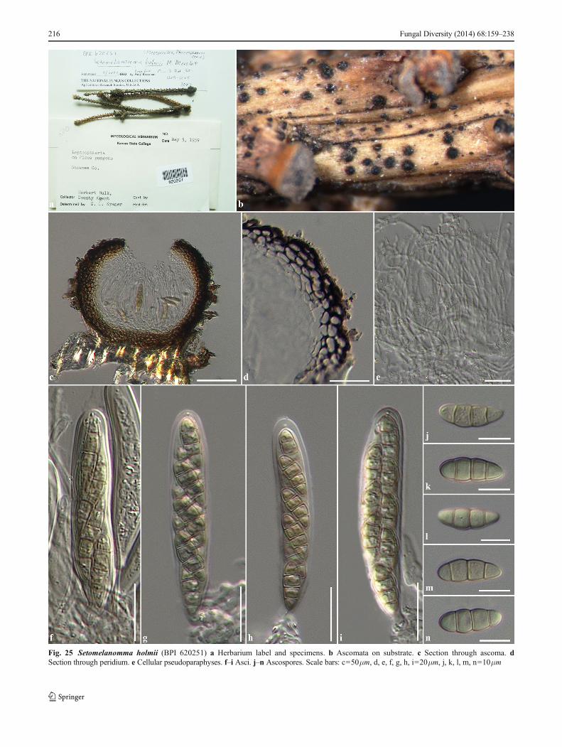
Fig. 25 Setomelanomma holmii (BPI 620251) a Herbarium label and specimens. b Ascomata on substrate. c Section through ascoma. dSection through peridium. e Cellular pseudoparaphyses. f–i Asci. j–n Ascospores. Scale bars: c=50μm, d, e, f, g, h, i=20μm, j, k, l, m, n=10μm
216 Fungal Diversity (2014) 68:159–238
Material examined: USA, Kansas, Shawnee Coounty, onbranches of Picea pungens Engelm (Pinaceae), 5 May 1959,B. Herbert (BPI 620251).
Setophoma Gruyter et al., in De Gruyter et al. Mycologia102(5): 1077 (2010), Faces of Fungi number: FoF00273,Fig. 26.
Pathogenic or saprobic on various hosts (Alliaceae,Asteraceae and Poaceae). Sexual state: Ascomata, scattered,gregarious, immersed, visible as raised, minute black dots onhost surface, uniloculate, globose to subglobose, glabrous,brown to dark brown, ostiole central, with minute papilla.Peridium thin-walled, of equal thickness, composed of pseu-doparenchymatous cells, arranged in flattened texturaangularis to textura prismatica. Hamathecium composed ofnumerous, filiform, broad cellular, pseudoparaphyses, withdistinct septa, anastomosing at the apex. Asci 8-spored,bitunicate, fissitunicate, cylindrical to cylindric-clavate,short pedicellate, apically rounded, with well-developednarrowly ocular chamber. Ascospores overlapping 2-seriate,phragmosporous, cylindrical to cylindric-clavate, hyaline,3-septate, usually enlarge at the second cell from apex,smooth-walled with large guttules. Asexual state: pro-duced on rice straw and bamboo pieces on water agarafter four weeks. Conidiomata scattered to clustered, gre-garious, superficial, globose to subglobose, setose, andsometimes glabrous when mature, initially light brown,becoming brown to dark brown. Conidiomata walls thin,composed of light brown to brown pseudoparenchymatouscells, arranged in a textura angularis. Conidiophores re-duced to conidiogenous cells. Conidiogenous cellsenteroblastic, phialidic, determinate, discrete, unbranched,oblong to cylindrical, or ampulliform, hyaline, slightlycurved, aseptate, lining the inner cavity of conidioma.Conidia amerosporous, oblong to ellipsoidal, rarely irreg-ular, with rounded or obtuse ends, hyaline, aseptate,smooth-walled (from De Gruyter et al . 2010;Phookamsak et al. 2014).
Type species: Setophoma terrestris (H.N. Hansen) Gruyteret al., in De Gruyter et al. Mycologia 102(5): 1077 (2010).
≡ Phoma terrestris H.N. Hansen, Phytopathology 19(8):699 (1929)
Phylogenetic study: De Gruyter et al. (2010), Quaedvlieget al. (2013), Phookamsak et al. (2014)
Notes: Setophoma was introduced by De Gruyter et al.(2010) to accommodate phoma-like species and is typifiedby S. terrestris. Two species, S. sacchari (Bitanc.) Gruyteret al. and S. terrestris (H.N. Hansen) Gruyter were accommo-dated in this genus. Quaedvlieg et al. (2013) introduced a newspecies Setophoma chromolaenae Quaedvlieg et al. on leavesof Chromolaena odorata from Brazil. No sexual state wasreported. Phookamsak et al. (2014) reported the sexual state ofSetophoma based on asexual-sexual connection of S. sacchariand phylogenetic data. The sexual state of Setophoma is
similar to Phaeosphaeria species, but differs in ascosporecolour and asexual state (Phookamsak et al. 2014).Setophoma chromolaenae, S. sacchari, S. terrestris andS. vernoniae Crous & Alfenas are presently accommodatedin the genus (Index Fungorum 2014; Crous et al. 2014;Phookamsak et al. 2014). Based on multigene phylogeneticanalysis, we accept Setophoma in Phaeosphaeriaceae whichforms a single clade at basal of the family (Fig. 2).
Tiarospora Sacc. & Marchal, Revue mycol., Toulouse7(no. 26): 148 (1885), Faces of Fungi number: FoF00274.
Saprobic or pathogenic on AmmophilaHost,DeschampsiaP. Beauv., Elymus L. (Poaceae) and Dryas grandis Jurtzev(Rosaceae). Sexual state: Unknown. Asexual state:Conidiomata scattered, gregarious, immersed to semi-im-mersed, uniloculate, globose, glabrous, brown, ostiole central,with short papilla. Conidiomata walls thick, composed onseveral layers, outer layers comprising thick-walled darkbrown, flattened cells of textura angularis, inner layers com-prising thin-walled paler cells. Conidiophores reduced toconidiogenous cells. Conidiogenous cells holoblastic, sympo-dial, discrete, doliiform, cylindrical or lageniform, hyaline,smooth-walled. Conidia didymosporous, broadly ellipsoid,with obtuse to flattened ends, hyaline to pale brown, 1-septate,continuous, thick and smooth-walled, with guttules or irregu-lar or cap-like gelatinous appendage (from Sutton 1980).
Type species: Tiarospora westendorpii Sacc. & Marchal,Revue mycol., Toulouse 7(no. 26): 148 (1885).
Phylogenetic study: NoneNotes: Tiarospora was introduced by Saccardo and
Marchal (1885) and is typified by T. westendorpii Sacc. &Marchal. Four species were accommodated under Tiarospora;T. deschampsiae Krusch., T. perforans (Sacc.) Höhn.,T. pirozynskii Chleb. and T. westendorpii Sacc. & Marchal.However, Tiarospora perforans was transferred toMontagnulaceae by Aptroot (2006). Tiarospora is similar toTiarosporella Höhn.; however, they differ in the morphologyof their conidia. Tiarospora has 1-septate conidia, whileTiarosporella has aseptate conidia (Abbas et al. 2012).Tiarospora was described in Sutton (1980); however, nofamily placement was given for the genus. Kirk et al. (2001,2008) treated the genus under Phaeosphaeriaceae but did notmention any literature to confirm this and this was accepted byWijayawardene et al. (2012). Oertel (2011) however, men-tioned that the type species of Tiarospora, T. westendorpiiis a synanamoph of Amarenographium metableticum,which is the asexual state of Amarenomyces, also inPhaeosphaeriaceae. Therefore the current placement ofthis genus is somewhat unclear. The genus is relativelypoorly known, lacks molecular data and no sexual state isknown. We tentatively place Tiarospora in Phaeosphaeriaceaeuntil molecular data resolves this issue.
Vrystaatia Quaedvlieg et al. Stud. Mycol. 75: 372 (2013),Faces of Fungi number: FoF00275, Fig. 27.
Fungal Diversity (2014) 68:159–238 217
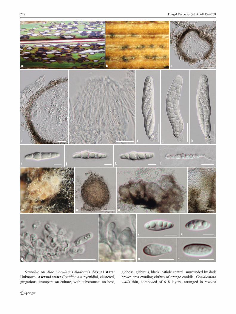
Saprobic on Aloe maculata (Aloaceae). Sexual state:Unknown. Asexual state: Conidiomata pycnidial, clustered,gregarious, erumpent on culture, with substromata on host,
globose, glabrous, black, ostiole central, surrounded by darkbrown area exuding cirrhus of orange conidia. Conidiomatawalls thin, composed of 6–8 layers, arranged in textura
218 Fungal Diversity (2014) 68:159–238
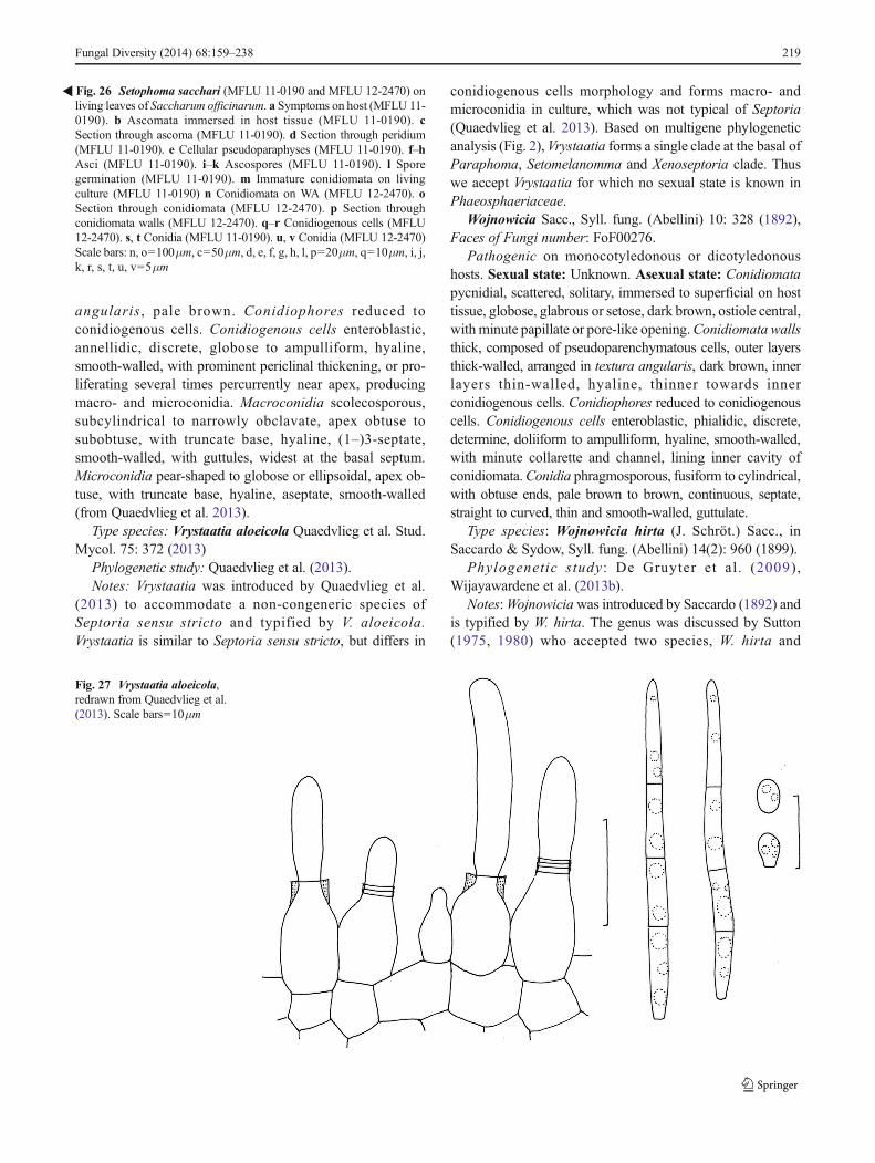
angularis, pale brown. Conidiophores reduced toconidiogenous cells. Conidiogenous cells enteroblastic,annellidic, discrete, globose to ampulliform, hyaline,smooth-walled, with prominent periclinal thickening, or pro-liferating several times percurrently near apex, producingmacro- and microconidia. Macroconidia scolecosporous,subcylindrical to narrowly obclavate, apex obtuse tosubobtuse, with truncate base, hyaline, (1–)3-septate,smooth-walled, with guttules, widest at the basal septum.Microconidia pear-shaped to globose or ellipsoidal, apex ob-tuse, with truncate base, hyaline, aseptate, smooth-walled(from Quaedvlieg et al. 2013).
Type species: Vrystaatia aloeicola Quaedvlieg et al. Stud.Mycol. 75: 372 (2013)
Phylogenetic study: Quaedvlieg et al. (2013).Notes: Vrystaatia was introduced by Quaedvlieg et al.
(2013) to accommodate a non-congeneric species ofSeptoria sensu stricto and typified by V. aloeicola.Vrystaatia is similar to Septoria sensu stricto, but differs in
conidiogenous cells morphology and forms macro- andmicroconidia in culture, which was not typical of Septoria(Quaedvlieg et al. 2013). Based on multigene phylogeneticanalysis (Fig. 2), Vrystaatia forms a single clade at the basal ofParaphoma, Setomelanomma and Xenoseptoria clade. Thuswe accept Vrystaatia for which no sexual state is known inPhaeosphaeriaceae.
Wojnowicia Sacc., Syll. fung. (Abellini) 10: 328 (1892),Faces of Fungi number: FoF00276.
Pathogenic on monocotyledonous or dicotyledonoushosts. Sexual state: Unknown. Asexual state: Conidiomatapycnidial, scattered, solitary, immersed to superficial on hosttissue, globose, glabrous or setose, dark brown, ostiole central,withminute papillate or pore-like opening.Conidiomata wallsthick, composed of pseudoparenchymatous cells, outer layersthick-walled, arranged in textura angularis, dark brown, innerlayers thin-walled, hyaline, thinner towards innerconidiogenous cells. Conidiophores reduced to conidiogenouscells. Conidiogenous cells enteroblastic, phialidic, discrete,determine, doliiform to ampulliform, hyaline, smooth-walled,with minute collarette and channel, lining inner cavity ofconidiomata.Conidia phragmosporous, fusiform to cylindrical,with obtuse ends, pale brown to brown, continuous, septate,straight to curved, thin and smooth-walled, guttulate.
Type species: Wojnowicia hirta (J. Schröt.) Sacc., inSaccardo & Sydow, Syll. fung. (Abellini) 14(2): 960 (1899).
Phylogenetic study : De Gruyter et al . (2009),Wijayawardene et al. (2013b).
Notes:Wojnowiciawas introduced by Saccardo (1892) andis typified by W. hirta. The genus was discussed by Sutton(1975, 1980) who accepted two species, W. hirta and
�Fig. 26 Setophoma sacchari (MFLU 11-0190 and MFLU 12-2470) onliving leaves of Saccharum officinarum. a Symptoms on host (MFLU 11-0190). b Ascomata immersed in host tissue (MFLU 11-0190). cSection through ascoma (MFLU 11-0190). d Section through peridium(MFLU 11-0190). e Cellular pseudoparaphyses (MFLU 11-0190). f–hAsci (MFLU 11-0190). i–k Ascospores (MFLU 11-0190). l Sporegermination (MFLU 11-0190). m Immature conidiomata on livingculture (MFLU 11-0190) n Conidiomata on WA (MFLU 12-2470). oSection through conidiomata (MFLU 12-2470). p Section throughconidiomata walls (MFLU 12-2470). q–r Conidiogenous cells (MFLU12-2470). s, t Conidia (MFLU 11-0190). u, v Conidia (MFLU 12-2470)Scale bars: n, o=100μm, c=50μm, d, e, f, g, h, l, p=20μm, q=10μm, i, j,k, r, s, t, u, v=5μm
Fig. 27 Vrystaatia aloeicola,redrawn from Quaedvlieg et al.(2013). Scale bars=10μm
Fungal Diversity (2014) 68:159–238 219
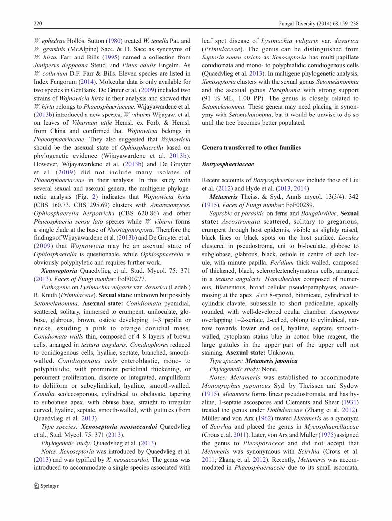
W. ephedrae Hollós. Sutton (1980) treatedW. tenella Pat. andW. graminis (McAlpine) Sacc. & D. Sacc as synonyms ofW. hirta. Farr and Bills (1995) named a collection fromJuniperus deppeana Steud. and Pinus edulis Engelm. AsW. colluvium D.F. Farr & Bills. Eleven species are listed inIndex Fungorum (2014). Molecular data is only available fortwo species in GenBank. De Gruter et al. (2009) included twostrains of Wojnowicia hirta in their analysis and showed thatW. hirta belongs to Phaeosphaeriaceae. Wijayawardene et al.(2013b) introduced a new species, W. viburni Wijayaw. et al.on leaves of Viburnum utile Hemsl. ex Forb. & Hemsl.from China and confirmed that Wojnowicia belongs inPhaeosphaeriaceae. They also suggested that Wojnowiciashould be the asexual state of Ophiosphaerella based onphylogenetic evidence (Wijayawardene et al. 2013b).However, Wijayawardene et al. (2013b) and De Gruyteret al. (2009) did not include many isolates ofPhaeosphaeriaceae in their analysis. In this study withseveral sexual and asexual genera, the multigene phyloge-netic analysis (Fig. 2) indicates that Wojnowicia hirta(CBS 160.73, CBS 295.69) clusters with Amarenomyces,Ophiosphaerella herpotricha (CBS 620.86) and otherPhaeosphaeria sensu lato species while W. viburni formsa single clade at the base of Neostagonospora. Therefore thefindings ofWijayawardene et al. (2013b) and DeGruyter et al.(2009) that Wojnowicia may be an asexual state ofOphiosphaerella is questionable, while Ophiosphaerella isobviously polyphyletic and requires further work.
Xenoseptoria Quaedvlieg et al. Stud. Mycol. 75: 371(2013), Faces of Fungi number: FoF00277.
Pathogenic on Lysimachia vulgaris var. davurica (Ledeb.)R. Knuth (Primulaceae). Sexual state: unknown but possiblySetomelanomma. Asexual state: Conidiomata pycnidial,scattered, solitary, immersed to erumpent, uniloculate, glo-bose, glabrous, brown, ostiole developing 1–3 papilla ornecks, exuding a pink to orange conidial mass.Conidiomata walls thin, composed of 4–8 layers of browncells, arranged in textura angularis. Conidiophores reducedto conidiogenous cells, hyaline, septate, branched, smooth-walled. Conidiogenous cells enteroblastic, mono- topolyphialidic, with prominent periclinal thickening, orpercurrent proliferation, discrete or integrated, ampulliformto doliiform or subcylindrical, hyaline, smooth-walled.Conidia scolecosporous, cylindrical to obclavate, taperingto subobtuse apex, with obtuse base, straight to irregularcurved, hyaline, septate, smooth-walled, with guttules (fromQuaedvlieg et al. 2013)
Type species: Xenoseptoria neosaccardoi Quaedvlieget al., Stud. Mycol. 75: 371 (2013).
Phylogenetic study: Quaedvlieg et al. (2013)Notes: Xenoseptoria was introduced by Quaedvlieg et al.
(2013) and was typified by X. neosaccardoi. The genus wasintroduced to accommodate a single species associated with
leaf spot disease of Lysimachia vulgaris var. davurica(Primulaceae). The genus can be distinguished fromSeptoria sensu stricto as Xenoseptoria has multi-papillateconidiomata and mono- to polyphialidic conidiogenous cells(Quaedvlieg et al. 2013). In multigene phylogenetic analysis,Xenoseptoria clusters with the sexual genus Setomelanommaand the asexual genus Paraphoma with strong support(91 % ML, 1.00 PP). The genus is closely related toSetomelanomma. These genera may need placing in synon-ymy with Setomelanomma, but it would be unwise to do sountil the tree becomes better populated.
Genera transferred to other families
Botryosphaeriaceae
Recent accounts of Botryosphaeriaceae include those of Liuet al. (2012) and Hyde et al. (2013, 2014)
Metameris Theiss. & Syd., Annls mycol. 13(3/4): 342(1915), Faces of Fungi number: FoF00289.
Saprobic or parasitic on ferns and Bougainvillea. Sexualstate: Ascostromata scattered, solitary to gregarious,erumpent through host epidermis, visible as slightly raised,black lines or black spots on the host surface. Loculesclustered in pseudostroma, uni to bi-loculate, globose tosubglobose, glabrous, black, ostiole in centre of each loc-ule, with minute papilla. Peridium thick-walled, composedof thickened, black, scleroplectenchymatous cells, arrangedin a textura angularis. Hamathecium composed of numer-ous, filamentous, broad cellular pseudoparaphyses, anasto-mosing at the apex. Asci 8-spored, bitunicate, cylindrical tocylindric-clavate, subsessile to short pedicellate, apicallyrounded, with well-developed ocular chamber. Ascosporesoverlapping 1–2-seriate, 2-celled, oblong to cylindrical, nar-row towards lower end cell, hyaline, septate, smooth-walled, cytoplasm stains blue in cotton blue reagent, thelarge guttules in the upper part of the upper cell notstaining. Asexual state: Unknown.
Type species: Metameris japonicaPhylogenetic study: None.Notes: Metameris was established to accommodate
Monographus japonicus Syd. by Theissen and Sydow(1915). Metameris forms linear pseudostromata, and has hy-aline, 1-septate ascospores and Clements and Shear (1931)treated the genus under Dothideaceae (Zhang et al. 2012).Müller and von Arx (1962) treated Metameris as a synonymof Scirrhia and placed the genus in Mycosphaerellaceae(Crous et al. 2011). Later, von Arx andMüller (1975) assignedthe genus to Pleosporaceae and did not accept thatMetameris was synonymous with Scirrhia (Crous et al.2011; Zhang et al. 2012). Recently, Metameris was accom-modated in Phaeosphaeriaceae due to its small ascomata,
220 Fungal Diversity (2014) 68:159–238
broad cylindrical to obclavate asci with a knob-like pediceland monocotyledons host preferences (Eriksson 2006;Lumbsch and Huhndorf 2007, 2010; Crous et al. 2011;Zhang et al. 2012; Hyde et al. 2013). However, the genuslacks molecular data to confirm its familial placement(Crous et al. 2011; Zhang et al. 2012).
There are five epithets listed for Metameris in IndexFungorum (2014); however, three species are synonymizedunder Scirrhia. In this study, we exclude Metameris fromPhaeosphaeriaceae. The large, elongated pseudostromata,with a peridium of thickened scleroplectenchymatous cells,broad, or irregularly cellular pseudoparaphyses, ascus mor-phology and didymosporous ascospores are characters typ-ical of genera in Botryosphaeriaceae where it is tentativelyreferred. Leptoguignardia is probably most similar (Liuet al. 2012) and should be compared in future studies offresh collections.
Metameris japonica (Syd.& P. Syd.) Syd., in Theissen &Sydow, Annls mycol. 13(3/4): 342 (1915), Faces of Funginumber: FoF00290, Fig. 28.
≡ Monographus japonicus Syd. & P. Syd., Annls mycol.10(4): 408 (1912)
Saprobic on Osmunda regalis var. japonica L. Sexualstate: Ascostromata 170–240μm high, 370–640μm diam.,clustered, solitary to gregarious, erumpent through hostepidermis, visible as slightly raised, black, elongatelining on host surface. Locules 160–240μm high, 200–250μm diam, clustered, immersed in pseudostroma, unito bi-loculate, globose to subglobose, glabrous, black,ostiole in centre of each locule, with minute papilla.Peridium 23–67μm wide, thick-walled, of unequalthickness, slightly thick at the base towards sides ofpseudostroma, composed of several layers of thickened,dark brown to black, scleroplectenchymatous cells, ar-ranged in textura angularis. Hamathecium composed ofnumerous, 3–7μm wide, filamentous, broad, irregularcellular pseudoparaphyses, with distinct septa whichare constricted at the septum, embedded in mucilaginousmatrix, anastomosing at the apex. Asci 80–110(−118)×(12.5–)13–18μm (x = 99.2×14.9μm, n=25), 8-spored,bitunicate, cylindrical to cylindric-clavate, narrow to-wards the apex, subsessile to short pedicellate, apicallyrounded with well-developed ocular chamber, arisingfrom the basal ascoma. Ascospores (22–)24–28×6–8μm(x = 26.7×6.8μm, n=30), overlapping 1–2 seriate, 2-celled,oblong to cylindrical, narrow towards lower end cell, hyaline,1-septate, constricted at the septum, smooth-walled, mostlyupper cell larger than lower cell, cytoplasm stains blue incotton blue reagent, the large guttules in the upper part ofthe upper cell not staining. Asexual state: Unknown.
Material examined: Japan, Mino, Kawauye-mura, onOsmunda regalis L. var. japonica (Osmundaceae), 10 May1912, K. Hara (S-F7166, holotype).
Didymellaceae
For a discussion of Didymellaceae see Hyde et al. (2013).Zhang et al. (2012) questionedwhether the placementMixturain Phaeosphaeriaceaewas natural. The genus is considered tobelong in Didymellaceae and is transferred herein.
Mixtura O.E. Erikss. & J.Z. Yue, Mycotaxon 38: 203(1990), Faces of Fungi number: FoF00278.
Type species: Mixtura saginataPhylogenetic study: None.Mixtura saginata (Syd.) O.E. Erikss. & J.Z. Yue,
Mycotaxon 38: 203 (1990), Faces of Fungi number:FoF00279, Fig. 29.
≡ Leptosphaeria saginata Syd., Annls mycol. 37(4/5): 377(1939).
Pathogen causing necrotic leaf spots on Chusqueaserrulata Pilger. Lesions up to 2–5 cm long, infecting mostlythe leaf margin, elongate cylindrical, pale brown to yellowish-brown, dry, separated from healthy tissue by a reddish-brownto purple-brown or black margin. Sexual state: Ascomata120–260μm high, 180–335μm diam., scattered, sometimesclustered, solitary, immersed, visible as raised, small blackdots on leaf lesions, uniloculate, globose to subglobose, gla-brous, dark brown to black, ostiole central with minute papilla.Peridium 12–40μm wide, thin-walled, of unequal thickness,thickened at apex at sides, composed of 3 or more layers ofbrown to dark brown, pseudoparenchymatous cells, arrangedin a textura angularis, outer layers comprising 2–5 layers ofthickened cells, brown to dark brown, inner layers comprisingseveral layers of flattened cells, hyaline to pale brown.Hamathecium composed of numerous, 3–8μmwide, filamen-tous , f r equen t ly anas tomos ing , b road ce l lu l a rpseudoparaphyses, with distinct septa which are constrictedat the septa, embedded in mucilaginous matrix. Asci(105–)112–135(−142)×(57–)60–100(−127) μm (x = 121.3×80.3μm, n=20), 8-spored, bitunicate, fissitunicate, globose tosubglobose, or ovoid, sessile to subsessile, apically roundedwith well-developed ocular chamber, producing a few asci,ar is ing from the basal ascoma. Ascospores 60–90(−95)×(19–)22–25(−27) μm (x = 81.2×23μm, n=30), ir-regular overlapping 4-seriate, phragmosporous, broadly fusi-form with rounded, germ pore at the ends, initially hyaline topale brown, becoming yellowish-brown or brown at maturity,paler at the end cells, distoseptate with 7-septa, slightlycurved, rough, thick-walled, surrounded by thin, distinctsheath. Asexual state: Unknown.
Material examined: ECUADOR, Tungurahua, HaciendaSan Antonio pr. Baños, on leaves of Chusquea serrulataPilger (Poaceae), 9 January 1938, H. Sydow (S-F8935,lectotype).
Notes: The monotypic genus Mixtura was introduced byEriksson and Yue (1990) to accommodateMi. saginatawhichcauses leaf spots on Chusquea serrulata. Mixtura is an
Fungal Diversity (2014) 68:159–238 221
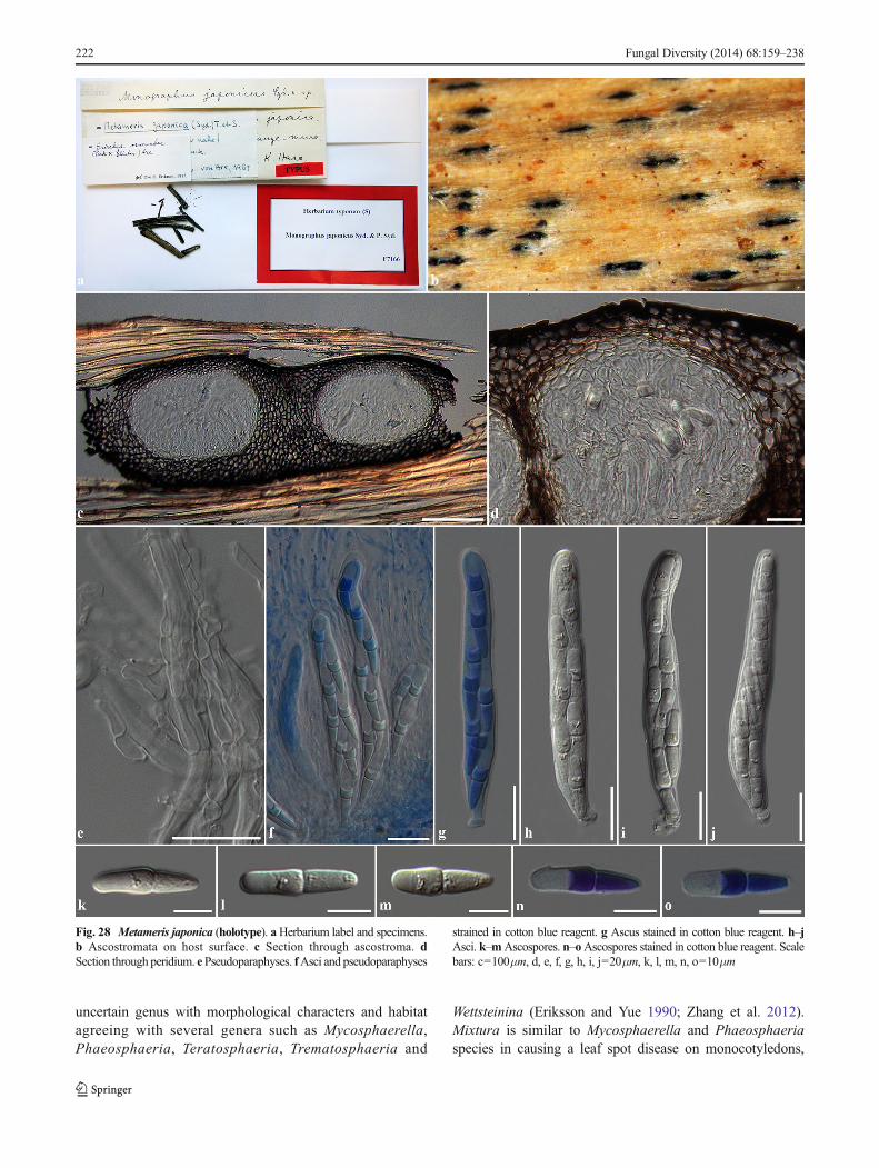
uncertain genus with morphological characters and habitatagreeing with several genera such as Mycosphaerella,Phaeosphaeria, Teratosphaeria, Trematosphaeria and
Wettsteinina (Eriksson and Yue 1990; Zhang et al. 2012).Mixtura is similar to Mycosphaerella and Phaeosphaeriaspecies in causing a leaf spot disease on monocotyledons,
222 Fungal Diversity (2014) 68:159–238
Fig. 28 Metameris japonica (holotype). aHerbarium label and specimens.b Ascostromata on host surface. c Section through ascostroma. dSection through peridium. ePseudoparaphyses. fAsci and pseudoparaphyses
strained in cotton blue reagent. g Ascus stained in cotton blue reagent. h–jAsci. k–mAscospores. n–oAscospores stained in cotton blue reagent. Scalebars: c=100μm, d, e, f, g, h, i, j=20μm, k, l, m, n, o=10μm
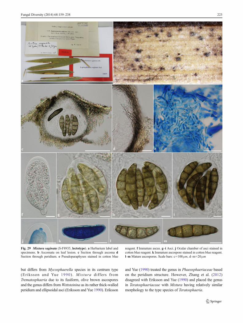
but differs from Mycosphaerella species in its centrum type(Eriksson and Yue 1990). Mixtura differs fromTrematosphaeria due to its fusiform, olive brown ascosporesand the genus differs fromWettsteinina as its rather thick-walledperidium and ellipsoidal asci (Eriksson and Yue 1990). Eriksson
and Yue (1990) treated the genus in Phaeosphaeriaceae basedon the peridium structure. However, Zhang et al. (2012)disagreed with Eriksson and Yue (1990) and placed the genusin Teratosphaeriaceae with Mixtura having relatively similarmorphology to the type species of Teratosphaeria.
Fungal Diversity (2014) 68:159–238 223
Fig. 29 Mixtura saginata (S-F8935, lectotype). a Herbarium label andspecimens. b Ascomata on leaf lesion. c Section through ascoma dSection through peridium. e Pseudoparaphyses stained in cotton blue
reagent. f Immature ascus. g–i Asci. j Ocular chamber of asci stained incotton blue reagent. k Immature ascospore stained in cotton blue reagent.l–m Mature ascospores. Scale bars: c=100μm, d–m=20μm
Mixtura is similar to other genera in Phaeosphaeriaceaesuch as Hadrospora and Loratospora in its saccate asci andlarge ascospores inHadrospora. However, these genera differin their centrum type. Mixtura forms dense, thick, cellularpseudoparaphyses, while Hadrospora forms trabeculatepseudoparaphyses, and pseudoparaphyses are lacking inLoratospora. Hadrospora is excluded as a member ofPhaeosphaeriaceae in this study, but Loratospora isconfirmed based on phylogenetic evidence. Mixtura issimilar to Leptosphaerulina in habitat on host and asci.However, they differ in the ascospores and centrum type asLeptosphaerulina lacks pseudoparaphyses and has muriformascospores. Mixtura has dense pseudoparaphyses andphragmosporous ascospores with distosepta. Therefore, weagree with Zhang et al. (2012) and exclude Mixtura fromPhaeosphaeriaceae. The genus is tentatively placed inDidymellaceae.
Didymosphaeriaceae
For an account of this family see Ariyawansa et al. (2014b).Barria and Wilmia were transferred from Phaeosphaeriaceaeto Didymosphaeriaceae in Ariyawansa et al. (2014b and areonly briefly discussed here.
Barria Z.Q. Yuan, Mycotaxon 51: 313 (1994), Faces offungi number: FoF00031.
Type species: Barria piceae Z.Q. Yuan, Mycotaxon 51:314 (1994).
Phylogenetic study: None.Notes: Barria is a monotypic genus introduced by Yuan
(1994) based on Barria piceae. The genus was reported aspathogenic on needles of Picea schrenkiana Fisch. & C.A.Mey. Barria is excluded from Phaeosphaeriaceae in thisstudy based on its morphology. Barria is associated withgymnosperms, asci are clavate and short pedicellate, andascospores are didymosporous with thick walls. Whereas thegeneric type, Phaeosphaeria is associated mostly with angio-sperms, asci are often broad cylindrical to cylindric-clavateand subsessile and ascospores are phragmosporous (Yuan1994; Zhang et al. 2012; Hyde et al. 2013; Quaedvlieg et al.2013). Additionally, most genera with confirmed phylogenet-ic placement in Phaeosphaeriaceae have phragmospores,scolecospores, or muriform ascospores, but there are nodidymosporous taxa (Zhang et al. 2012; Hyde et al. 2013;Quaedvlieg et al. 2013). Barria is associated in the same hostfamily as Setomelanomma which was confirmed asphaeosphaeriacous based on phylogenetic evidence. Barriahas didymosporous ascospores, while Setomelanomma hasphragmosporous ascospores. Most didymosporous genera inPhaeosphaeriaceae lack molecular phylogenetic data to con-firm their current placements. Furthermore, Barria is moresimilar to genera in Didymosphaeriaceae rather thanPhaeosphaeriaceae (Ariyawansa et al. 2014b), where is it
tentatively accommodated. Zhang et al. (2012) acceptedLautitia in Phaeosphaeriaceae; in contrast, all genera of ma-rine and freshwater fungi (Carinispora, Isthmosporella andOcala) were excluded from Phaeosphaeriaceae in Zhanget al. (2012). However, Lautitia differs from other genera inPhaeosphaeriaceae in having clavate asci with long pedicelsand forming a pseudostromata with the peridium arranged in atextura porrecta. Other genera in Phaeosphaeriaceae mostlyhave cylindrical asci with subsessile to short pedicels anduniloculate ascomata with the peridium arranged in a texturaangularis. Metameris forms large, elongated pseudostromata,with a thick-walled peridium of scleroplectenchymatous cellswhich is rarely found in other genera of Phaeosphaeriaceae.A new species of Letendraea (=Wilmia) was introduced basedon phylogenetic evidence and belongs inDidymosphaeriaceae(see Ariyawansa et al. 2014b).
Wilmia Dianese et al., Mycologia 93(5): 1014 (2001),Faces of fungi number: FoF00046.
Type species: Wilmia brasiliensis Dianese et al.,Mycologia 93(5): 1014 (2001).
Phylogenetic study: Ariyawansa et al. (2014a).Notes: Wilmia was introduced by Dianese et al. (2001) to
accommodate a monotypic genus, Wilmia brasiliensis thatcaused a leaf spot disease of Memora pedunculata (Vell.)Miers. in Brazil. Dianese et al. (2001) treated Wilmia inPhaeosphaeriaceae based on the morphological characterswhich fitted Barr’s concept (Barr 1987b, 1992a). Wilmia issimilar to other genera in Phaeosphaeriaceae viz. Barria,Lautitia and Metameris in having didymosporous ascosporesand a coelomycetous asexual morph (Dianese et al. 2001;Zhang et al. 2012). Zhang et al. (2012) tentatively placedWilmia in Phaeosphaeriaceae as the genus is poorly knownand lacks molecular data, although Zhang et al. (2012) men-tioned that the genus should be accommodated inLeptosphaeriaceae based on its dicotyledonous host habit.
A Dothideomycete species was collected from living anddead leaves of Cordyline sp. in Thailand. The species alsocaused leaf spot disease and had similar morphological char-acters to Wilmia brasiliensis. Ariyawansa et al. (2014b syn-onymized Wilmia under Letendraea based on morphologyand phylogeny. Thus, the new species collected fromCordyline sp. was accommodated in Didymosphaeriaceaeand named Letendraea cordylinicola Phookamsak et al.(Ariyawansa et al. 2014b).
Leptosphaeriaceae
For a discussion on Leptosphaeriaceae see Hyde et al. (2013);Chaetoplea is considered to belong in this family and istransferred herein.
Chaetoplea (Sacc.) Clem., in Clements & Shear, Gen.fung., Edn 2 (Minneapolis): 275 (1931), Faces of Funginumber: FoF00280.
224 Fungal Diversity (2014) 68:159–238
≡ Pyrenophora subgen. Chaetoplea Sacc., Syll. fung.(Abellini) 2: 279 (1883)
Saprobic on woody stems or herbaceous dicotyledonous.Sexual state: Ascomata solitary, gregarious, semi-immersedto superficial, or erumpent through host tissue, uniloculate,subglobose, glabrous, brown to dark brown, ostioles central,with short papilla. Peridium thick-walled, of equal thickness,composed of dark brown to black, scleroplectenchymatouscells of textura angularis or textura globulosa. Hamatheciumcomposed of numerous, filamentous, distinctly septate, broadcellular pseudoparaphyses, anastomosing at the apex. Asci 8-spored, bitunicate, clavate to cylindric-clavate, short pedicel-late, apically rounded, with an ocular chamber. Ascosporesoverlapping 1–2-seriate, phragmosporous, fusiform or broad-ly fusiform to ellipsoidal, brown to yellowish-brown, septate,slightly constricted at the septa, smooth-walled. Asexualstate: Conidiomata pycnidial, scattered, solitary to gregari-ous, semi-immersed to erumpent, uniloculate, ellipsoidal tobroadly fusiform with rounded apex, glabrous, dark brownto black, ostioles central, with pore-like opening.Conidiomata walls thin-walled, composed of flattened,scleroplectenchymatous cells, arranged in textura angularisto textura prismatica, brown to dark brown. Conidiophoresreduced to conidiogenous cells. Conidiogenous cells holo-blastic, phialidic, discrete, ampulliform, hyaline, smooth-walled, hard to distinguish from the inner wall cells.Conidia oblong to ellipsoidal, with obtuse ends, brown,1-septate, smooth-walled.
Type species: Chaetoplea calvescensPhylogenetic study: NoneNotes: Chaetoplea was introduced by Clements and Shear
(1931) to accommodate Dothideomycete species from woodysubstrates which included species transferred fromCucurbitaria, Leptosphaeria, Pleospora, Pyrenophora,Sphaeria and Teichospora (Shoemaker 1961; Wehmeyer1961; von Arx and Müller 1975; Barr 1990b; Yuan and Barr1994; Zhang et al. 2012). Barr (1987b, 1990a, b) treatedChaetoplea sensu lato in Phaeosphaeriaceae. The type spec-imen has not been recovered from any herbaria worldwide.Zhang et al. (2012) however observed the isotype which is aslide from the Farlow Herbarium (FH) and has only asci andascospores. Zhang et al. (2012) mentioned that Chaetopleamay be heterogeneous in nature and occurs in a wide range ofsubstrates such as herbaceous stalks, decorticated wood andperiderm tissues.
The asexual state of Chaetoplea (for Pyrenophoracalvescens (Fr.) Sacc.) was first reported as Dendryphiumcomosum Walk. (Saccardo 1883). Later Webster and Lucas(1959) treated Microdiplodia henningsii Staritz. as the asexualstate of Chaetoplea and this was accepted by Barr (1987b).However, Crous et al. (2006) treatedMicrodiplodia as an asex-ual state of Karstenula placed in the family Montagnulaceae.Ramaley (1995) reported Parahendersonia as the asexual state
of Chaetoplea dasylirionis being saprobic on Dasylirion(Agavaceae) and placed it in Leptosphaeriaceae. In this study,the asexual state of Chaetoplea was observed occurring on thesame host as the lectotype. The asexual state of Chaetopleaforms oblong to ellipsoidal, brown, 1-septate, smooth-walledconidia which are microdiplodia-like.
After examining Fries exsiccatus (Scler. Suecicaeexsiccatus no. 401) in the US National Fungus Collections(BPI) it was determined that this should be designated as alectotype in this study. Based on morphology, Chaetoplea ismore similar to Leptosphaeriaceae than Phaeosphaeriaceaeand this supports the current placement ofChaetoplea as listedin Index Fungorum (2014). Therefore Chaetoplea shouldcurrently be placed in Leptosphaeriaceae. Recollection,epitypification, and molecular data are needed to confirm thenatural placement of Chaetoplea.
Chaetoplea calvescens (Fr.) Clem., in Clements & Shear,Gen. fung., Edn 2 (Minneapolis): 275 (1931), Faces of Funginumber: FoF00281, Fig. 30.
≡ Sphaeria calvescens Fr., Sclerom. Suec.: no. 401 (1822)Saprobic on herbaceous dicotyledonous hosts. Sexual
state: Ascomata 140–220μm high, 280–350μm diam.,scattered, solitary to gregarious, semi-immersed to superficial,visible as small black dots on the host surface, uniloculate,subglobose, glabrous, dark brown to black, ostiole central,with short papilla. Peridium 15–40μm wide, thick-walled, ofequal thickness , composed of several layers ofscleroplectenchymatous cells, outer layers comprising 2–3cell layers of thick-walled, black, textura angularis to texturaglobulosa, inner layers comprising 4–5 cell layers of thin-walled, hyaline to pale brown, flattened cells of texturaangularis to textura prismatica. Hamathecium composed ofnumerous, 2–3μm wide, filamentous, broad cellularpseudoparaphyses, with distinct septa, not constricted at thesepta, anastomosing at the apex. Asci (80–)90–115(−120)×11–13(−15) μm (x = 100×12.1μm, n=20), 8-spored,bitunicate, fissitunicate, clavate, short pedicellate, apicallyrounded, with indistinct ocular chamber. Ascospores (14–)15–17(−119)×6–8.5μm (x = 16.7×7.4μm, n=30), overlap-ping 1–2-seriate, phragmosporous, broadly fusiform to ellip-soidal, enlarged at the second cell, brown to yellowish-brown,3-septate, rarely 2-septate, straight or curved, constricted at thesepta, smooth-walled. Asexual state: Conidiomata growingwith sexual state, 170–190μm high, 150–190μm diam., pyc-nidial, solitary to gregarious, semi-immersed to erumpent,visible as black dots on the host surface, uniloculate, ellipsoi-dal to broadly fusiform, with rounded apex, glabrous, darkbrown to black, ostiole central, with minute papilla.Conidiomata walls 13.5–25μm wide, thick-walled, of equalthickness, composed of several layers of flattened,scleroplectenchymatous cells, outer layers comprising 2–3cell layers of thick-walled, dark brown to black cells, arrangedin a textura angularis, inner layers comprising 3–5 layers of
Fungal Diversity (2014) 68:159–238 225
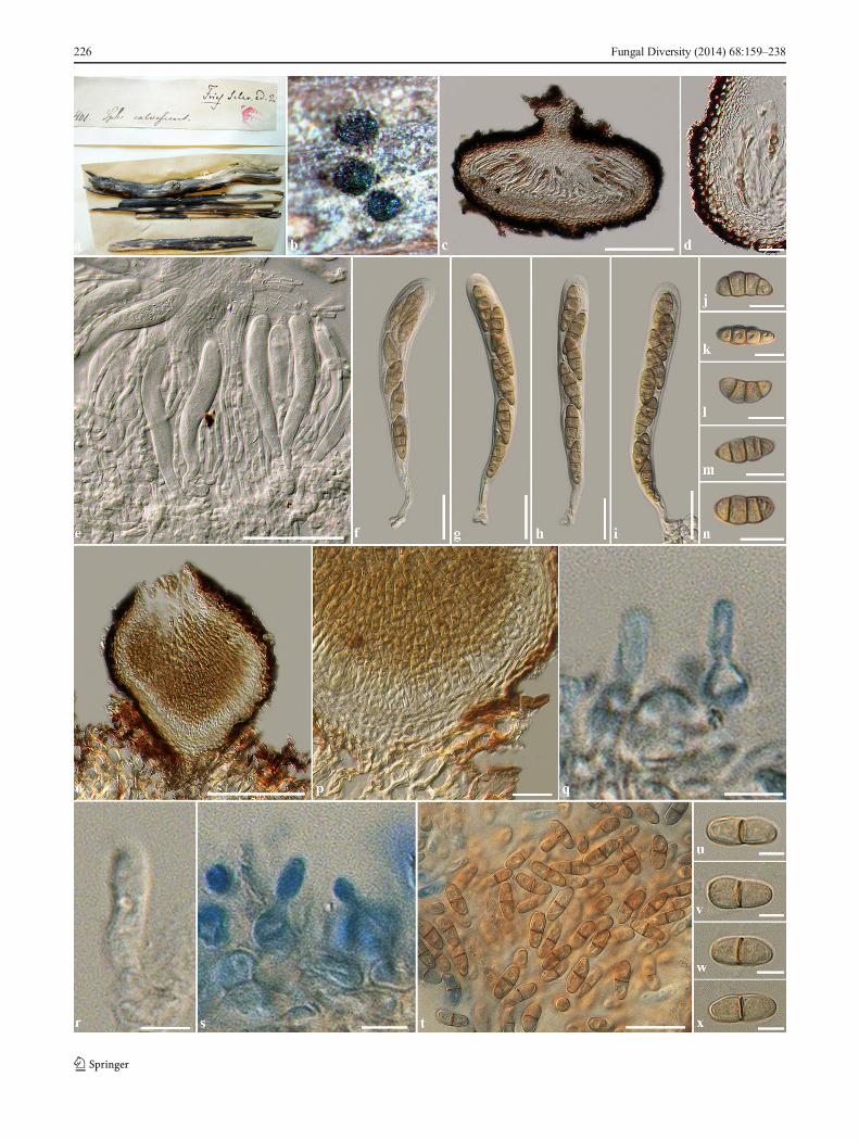
226 Fungal Diversity (2014) 68:159–238
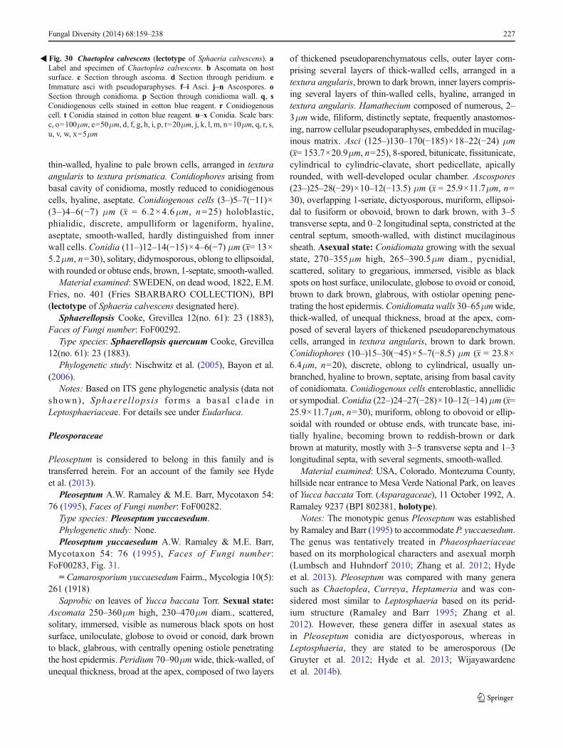
thin-walled, hyaline to pale brown cells, arranged in texturaangularis to textura prismatica. Conidiophores arising frombasal cavity of conidioma, mostly reduced to conidiogenouscells, hyaline, aseptate. Conidiogenous cells (3–)5–7(−11)×(3–)4–6(−7) μm (x = 6.2×4.6μm, n=25) holoblastic,phialidic, discrete, ampulliform or lageniform, hyaline,aseptate, smooth-walled, hardly distinguished from innerwall cells. Conidia (11–)12–14(−15)×4–6(−7) μm (x= 13×5.2μm, n=30), solitary, didymosporous, oblong to ellipsoidal,with rounded or obtuse ends, brown, 1-septate, smooth-walled.
Material examined: SWEDEN, on dead wood, 1822, E.M.Fries, no. 401 (Fries SBARBARO COLLECTION), BPI(lectotype of Sphaeria calvescens designated here).
Sphaerellopsis Cooke, Grevillea 12(no. 61): 23 (1883),Faces of Fungi number: FoF00292.
Type species: Sphaerellopsis quercuum Cooke, Grevillea12(no. 61): 23 (1883).
Phylogenetic study: Nischwitz et al. (2005), Bayon et al.(2006).
Notes: Based on ITS gene phylogenetic analysis (data notshown) , Sphaerel lops is forms a basa l c lade inLeptosphaeriaceae. For details see under Eudarluca.
Pleosporaceae
Pleoseptum is considered to belong in this family and istransferred herein. For an account of the family see Hydeet al. (2013).
Pleoseptum A.W. Ramaley & M.E. Barr, Mycotaxon 54:76 (1995), Faces of Fungi number: FoF00282.
Type species: Pleoseptum yuccaesedum.Phylogenetic study: None.Pleoseptum yuccaesedum A.W. Ramaley & M.E. Barr,
Mycotaxon 54: 76 (1995), Faces of Fungi number:FoF00283, Fig. 31.
= Camarosporium yuccaesedum Fairm., Mycologia 10(5):261 (1918)
Saprobic on leaves of Yucca baccata Torr. Sexual state:Ascomata 250–360μm high, 230–470μm diam., scattered,solitary, immersed, visible as numerous black spots on hostsurface, uniloculate, globose to ovoid or conoid, dark brownto black, glabrous, with centrally opening ostiole penetratingthe host epidermis. Peridium 70–90μmwide, thick-walled, ofunequal thickness, broad at the apex, composed of two layers
of thickened pseudoparenchymatous cells, outer layer com-prising several layers of thick-walled cells, arranged in atextura angularis, brown to dark brown, inner layers compris-ing several layers of thin-walled cells, hyaline, arranged intextura angularis. Hamathecium composed of numerous, 2–3μm wide, filiform, distinctly septate, frequently anastomos-ing, narrow cellular pseudoparaphyses, embedded in mucilag-inous matrix. Asci (125–)130–170(−185)×18–22(−24) μm(x= 153.7×20.9μm, n=25), 8-spored, bitunicate, fissitunicate,cylindrical to cylindric-clavate, short pedicellate, apicallyrounded, with well-developed ocular chamber. Ascospores(23–)25–28(−29)×10–12(−13.5) μm (x = 25.9×11.7μm, n=30), overlapping 1-seriate, dictyosporous, muriform, ellipsoi-dal to fusiform or obovoid, brown to dark brown, with 3–5transverse septa, and 0–2 longitudinal septa, constricted at thecentral septum, smooth-walled, with distinct mucilaginoussheath. Asexual state: Conidiomata growing with the sexualstate, 270–355μm high, 265–390.5μm diam., pycnidial,scattered, solitary to gregarious, immersed, visible as blackspots on host surface, uniloculate, globose to ovoid or conoid,brown to dark brown, glabrous, with ostiolar opening pene-trating the host epidermis.Conidiomata walls 30–65μmwide,thick-walled, of unequal thickness, broad at the apex, com-posed of several layers of thickened pseudoparenchymatouscells, arranged in textura angularis, brown to dark brown.Conidiophores (10–)15–30(−45)×5–7(−8.5) μm (x = 23.8×6.4μm, n=20), discrete, oblong to cylindrical, usually un-branched, hyaline to brown, septate, arising from basal cavityof conidiomata. Conidiogenous cells enteroblastic, annellidicor sympodial.Conidia (22–)24–27(−28)×10–12(−14) μm (x=25.9×11.7μm, n=30), muriform, oblong to obovoid or ellip-soidal with rounded or obtuse ends, with truncate base, ini-tially hyaline, becoming brown to reddish-brown or darkbrown at maturity, mostly with 3–5 transverse septa and 1–3longitudinal septa, with several segments, smooth-walled.
Material examined: USA, Colorado. Montezuma County,hillside near entrance to Mesa Verde National Park, on leavesof Yucca baccata Torr. (Asparagaceae), 11 October 1992, A.Ramaley 9237 (BPI 802381, holotype).
Notes: The monotypic genus Pleoseptum was establishedby Ramaley and Barr (1995) to accommodate P. yuccaesedum.The genus was tentatively treated in Phaeosphaeriaceaebased on its morphological characters and asexual morph(Lumbsch and Huhndorf 2010; Zhang et al. 2012; Hydeet al. 2013). Pleoseptum was compared with many generasuch as Chaetoplea, Curreya, Heptameria and was con-sidered most similar to Leptosphaeria based on its perid-ium structure (Ramaley and Barr 1995; Zhang et al.2012). However, these genera differ in asexual states asin Pleoseptum conidia are dictyosporous, whereas inLeptosphaeria, they are stated to be amerosporous (DeGruyter et al. 2012; Hyde et al. 2013; Wijayawardeneet al. 2014b).
�Fig. 30 Chaetoplea calvescens (lectotype of Sphaeria calvescens). aLabel and specimen of Chaetoplea calvescens. b Ascomata on hostsurface. c Section through ascoma. d Section through peridium. eImmature asci with pseudoparaphyses. f–i Asci. j–n Ascospores. oSection through conidioma. p Section through conidioma wall. q, sConidiogenous cells stained in cotton blue reagent. r Conidiogenouscell. t Conidia stained in cotton blue reagent. u–x Conidia. Scale bars:c, o=100μm, e=50μm, d, f, g, h, i, p, t=20μm, j, k, l, m, n=10μm, q, r, s,u, v, w, x=5μm
Fungal Diversity (2014) 68:159–238 227
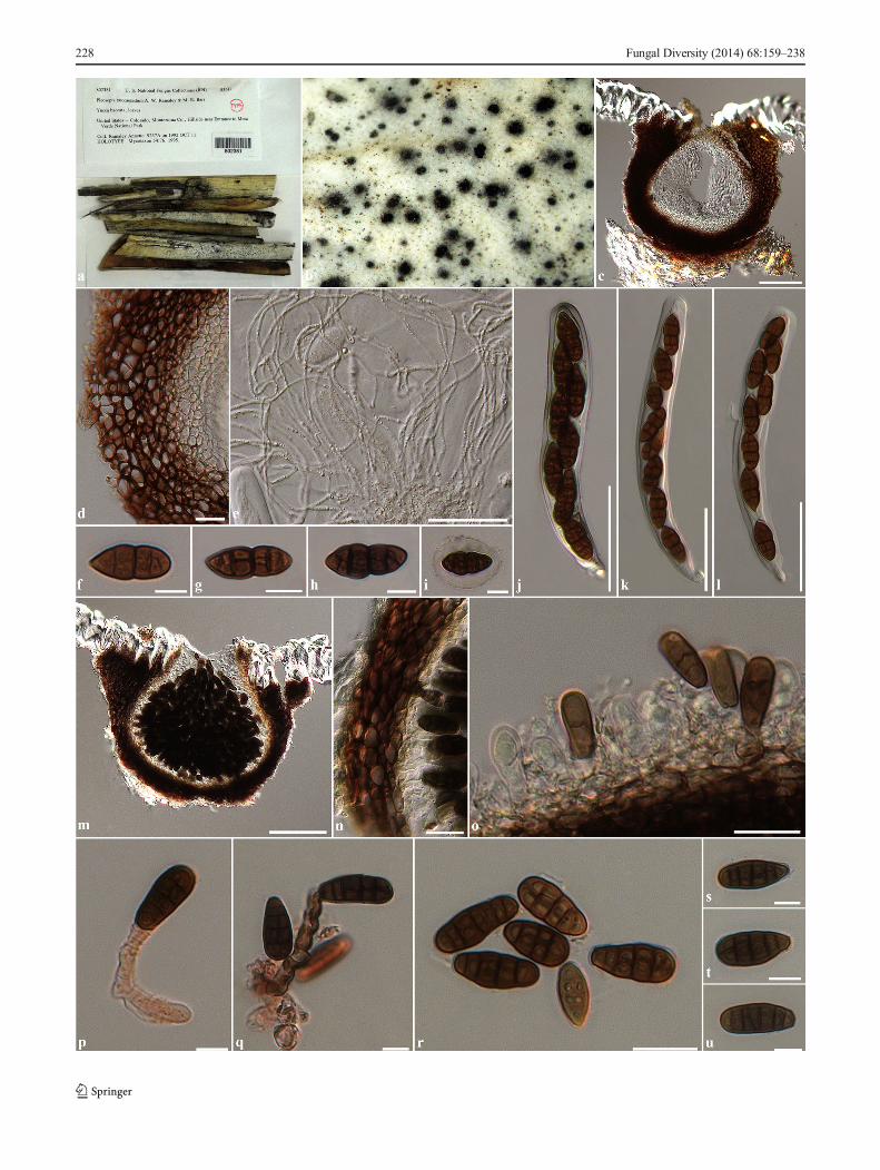
228 Fungal Diversity (2014) 68:159–238
Pleoseptum is a poorly known genus represented by onlyone species in Index Fungorum (2014) that lacks molecularsequence data in GenBank. The species has muriform sporeswhich are similar to Dematiopleospora (Wanasinghe et al.2014). Pleoseptum differs from Dematiopleospora by its pe-ridium structure which is very thick, composed of heavilypigmented, thick-walled cells with ostiole and typical ofPleosporaceae in the former. Whereas the peridium is thinner,composed of lighter pigmented, thin-walled cells, with thickperiphyses in the ostioles and typical of Phaeosphaeriaceae inDematiopleospora. In Pleoseptum pseudoparaphyses are nar-row, and additionally Pleoseptum forms a camarosporium-like asexual state. Pleoseptum is therefore excluded forPhaeosphaeriaceae whose genera typically have a thin-walled peridium and broad cellular pseudoparaphyses andtentatively placed in Pleosporaceae. However, Pleoseptumyuccaesedum needs recollecting and subjected to molecularstudy to confirm this placement is correct.
Camarosporium yuccaesedum was introduced as the asex-ual state of Pleoseptum yuccaesedum by Ramaley and Barr(1995) as both states co-inhabited the same host. Sexual-asexual links were connected by culture comparisons with co-nidia produced in culture from a sexual state (Ramaley and Barr(1995). Camarosporium however, has also been reported as theasexual state in various genera in Botryosphaeriales andCucurbitariaceae (Kirk et al. 2008; Zhang et al. 2012; Hydeet al. 2013). Recent molecular based research onCamarosporium by Crous et al. (2014) and Wijayawardeneet al. (2014a, c, d) showed that Camarosporium sensu lato ishighly polyphyletic in Pleosporales, while Camarosporiumsensu stricto is well-established genus in Pleosporineae,Pleosporales (Wijayawardene et al. 2014a). Thus,Neocamarosporium Crous & M.J. Wingf. (Pleosporalesincertae sedis fide Crous et al. 2014), ParacamarosporiumWijayaw. & K.D. Hyde (Montagnulaceae fide Wijayawardeneet al. 2014a), Pseudocamarosporium Wijayaw. & K.D. Hyde(Montagnulaceae fide Wijayawardene et al. 2014c) andEucamarosporium Wijayaw. & K.D. Hyde (Didymellaceaefide Wijayawardene et al. 2014d) have been introduced to ac-commodate several camarosporium-like taxa in Pleosporales.Significantly, Ramaley and Barr (1995) reported microconidiafromCamarosporium yuccaesedumwhich have not been report-ed from Camarosporium sensu stricto (Crous et al. 2013;Wijayawardene et al. 2014a). However, Crous et al. (2013)
reported macroconidia from Camarosporium psoraleaeCrous & M.J. Wingf. (current name Paracamarosporiumpsoraleae (Crous & M.J. Wingf.) Wijayaw. & K.D.Hyde fide Wijayawardene et al. 2014c), hence the statusof Camarospor ium yuccaesedum i s uncer ta in .Camarosporium yuccaesedum is synonymized underPleoseptum yuccaesedum herein.
Trematosphaeriaceae
For accounts of this family see Suetrong et al. (2011) andHyde et al. (2013).
Hadrospora Boise, Mem. N. Y. bot. Gdn 49: 310 (1989),Faces of Fungi number: FoF00285.
Saprobic on woody or monocotyledonous hosts. Sexualstate: Ascomata scattered, solitary, immersed to semi-im-mersed, or erumpent through host tissue, visible as tiny, small,black dots on the host surface, uniloculate, globose tosubglobose, glabrous, dark brown to black, ostiole centralwith a minute papilla. Peridium thin-walled, composed ofblack, broad pseudoparenchymatous cells, arranged in atextura angularis. Hamathecium composed of numerous, fil-amentous, distinctly septate, frequently anastomosing, narrowcellular pseudoparaphyses. Asci 8-spored, bitunicate,fissitunicate, ovoid to ampulliform, sessile to minutely pedi-cellate, apically rounded, with indistinct ocular chamber.Ascospores overlapping 1–3-seriate, phragmosporous, broad-ly fusiform, vermiform, brown to reddish-brown, paler at theend cells, septate, smooth-walled. Asexual state: Unknown.
Type species: Hadrospora fallaxPhylogenetic study: NoneNotes: Hadrospora was introduced to accommodate two
species, originally placed in Trematosphaeria by Boise(1989), and was typified by Hadrospora fallax. Hadrosporahas been reported from various hosts and habitats, both ter-restrial and freshwater, from Belgium, China, Italy, Japan,Switzerland, and the United States (Boise 1989; Shearer andCrane 1971; Fisher and Webster 1992; Webster 1993; Tanakaand Harada 2003; Zhang et al. 2012). The genus was tenta-tively placed in Phaeosphaeriaceae due to its small ascomata,thin-walled peridium and narrow pseudoparaphyses (Boise1989; Zhang et al. 2012). However, Hadrospora differs fromother genera in Phaeosphaeriaceae by its large vermiformascospores, ampulliform to saccate asci, trabeculatepseudoparaphyses and the hyphomycetous asexual morphwith the type species which was treated as an amphibiousspecies (Shearer 1993; Boise 1989; Tanaka and Harada 2003;Zhang et al. 2012). Other genera of Phaeosphaeriaceae aremostly terrestial and have cylindrical to cylindri-clavate asciwith broad cellular pseudoparaphyses and producecoelomycetous asexual morphs. Hadrospora is similar toMixtura in its large ascospores, and saccate asci; althoughHardrospora has been reported as saprobic on various hosts,
�Fig. 31 Pleoseptum yuccaesedum (BPI 802381, holotype). aHerbarium label and specimens of Pleoseptum yuccaesedum. bAscomata on host surface. c Section through ascoma. d Section throughperidium. e Pseudoparaphyses. f–h Ascospores. i Ascospore stained inIndian ink. j–l Asci. m Section through conidiomata. n Pycnidial wall. oConidiogenous cells. p–q Conidiogenous cells stained in congo redreagent (p=annellidic, q=sympodial proliferation). r–u Conidia.Scale bars: c, m=100μm, e, j, k, l=50μm, d, n, o, r, = 20μm, f, g,h, i, p, q, s, t, u=10μm
Fungal Diversity (2014) 68:159–238 229
while Mixtura was found as plant pathogen of bamboo. Thesecond species, Hadrospora clarkii (Sivan.) Boise is listed asTrematosphaeria clarkii Sivan. in Index fungorum (2014).
A zalerion-like asexual morph was reported forHadrospora with conidia being produced on PDA after4 months (Tanaka and Harada 2003). The genus is character-ized by holoblastic, helicoid, multiseptate, brown to darkbrown, smooth-walled conidia which are constricted at theseptum (Tanaka and Harada 2003). Jones et al. (2009), how-ever, treated Zalerion maritima as asexual morph of Lulwoana(Lulworthiales) and introduced a new genus, Halenospora toaccommodate a non-congeneric species of Zalerion maritima.
Hardrospora is a poorly known genus which lacks se-quences in GenBank. In this study, the proposal of Zhanget al. (2012) is followed excluding the genus fromPhaeosphaeriaceae, based on its morphological characters,and it is tentatively placed in Trematosphaeriaceae.
Hadrospora fallax (Mouton) Boise, Mem. N. Y. bot. Gdn49: 310 (1989), Faces of Fungi number: FoF00286, Fig. 32.
≡ Trematosphaeria fallaxMouton, Bull. Soc. R. Bot. Belg.25: 155 (1886)
Saprobic on woody hosts. Ascomata 200–350μm high,180–400μm diam., scattered, sometimes clustered, solitary,immersed to semi-immersed, becoming erumpent throughhost tissue, visible as tiny, black dots on the host surface,uniloculate, globose to subglobose, glabrous, dark brown toblack, ostiole central, with minute papilla, truncate at the apex.Peridium 20–50μm wide, thick-walled, of equal thickness,composed of several layers of black, broad pseudoparenchy-matous cells, arranged in a textura angularis. Hamatheciumcomposed of numerous, 1–1.7μm wide, filamentous, distinct-ly septate, anastomosing, narrow cellular pseudoparaphyses,embedded in mucilaginous matrix, with few asci. Asci 140–160(−186)×(54–)60–67μm (x = 157.3×61.8μm, n=10), 8-spored, bitunicate, fissitunicate, ovoid to ampulliform, sessileto minutely pedicellate, apically rounded with indistinct ocu-lar chamber, broad at the apex. Ascospores (50–)65–75(−80)×(18–)21–23(−28) μm (x = 72×21.3μm, n=25), irregularlyoverlapping 1–3-seriate, phragmosporous, broad fusiform,vermiform, widest at the median cells, reddish-brown to darkbrown, with lighter end cells, 8-septate, slightly constricted atthe median septum, smooth and thick-walled.
Material examined: BELGIUM, Beaufays, on recently cutwood, V. Mouton 245, (BR5 020009617141, holotype,microslide); SLOVAKIA, Kal’niste (East Slovakia), on stemsof over wintered Triticum aestivum L. (Poaceae), 20 February2009, M. Pastircak (BPI 878936).
Pleosporales, genera incertae sedis
For notes on Pleosporales, genera incertae sedis seeHyde et al. (2013)
Setophaeosphaeria Crous & Y. Zhang ter, Persoonia, Mol.Phyl. Evol. Fungi 32: 271 (2014), Faces of Fungi number:FOF000305.
Pathogenic on Hemerocallis fulva L. (Hemerocallidaceae).Sexual state: Ascomata scattered, immersed to subepidermal,uniloculate, globose, glabrous, ostiole central, somewhatpapillate. Peridium composed of 2–3 layers of brown cellsarranged in textura angularis. Hamathecium composed ofhyaline, hyphal pseudoparaphyses. Asci 8-spored, bitunicate,narrowly ellipsoid, rostrate, short pedicellate, apicallyrounded with inconspicuous ocular chamber. Ascospores2–3-seriate, fusoid to ellipsoidal, pale brown, 5-septate,enlarged at the second cell from apex, smooth-walled, withguttules and mucoid caps at each end. Asexual state:forming in culture. Conidiomata pycnidial, scattered,erumpent with papilla, uniloculate, globose, setose, brown,ostiole central, with round to ellipsoid papilla. Conidiomatawalls thin, composed of 2–3 layers, of pale brown cells,arranged in textura angularis. Conidiophores reduced toconidiogenous cells. Conidiogenous cells ampulliform, hya-line, smooth, percurrently proliferating at the apex. Conidiasubcylindrical, rounded or obtuse at the apex, base truncate,with marginal frill, hyaline, aseptate, smooth-walled, withguttules (from Crous et al. 2014).
Type species: Setophaeosphaeria hemerocallidis Crous &Y. Zhang ter, Persoonia, Mol. Phyl. Evol. Fungi 32: 271(2014).
Phylogenetic study: Crous et al. (2014).Notes: Setophaeosphaeria was introduced by Crous et al.
(2014). The genus was established to accommodate non-congeneric species of Phaeosphaeria. Crous et al. (2014) in-troduced two new species, Setophaeosphaeria badalingensisand Sp. hemerocallidis and synonymized Phaeosphaeriasetosa under the genus. Crous et al. (2014) mentioned thatSetophaeosphaeria differs from the type species ofPhaeosphaeria, Ph. oryzae, based on its asexual state.Phaeosphaeria oryzae forms a septoria-like asexual state,while Setophaeosphaeria species form phoma-like asexualstates (Quaedvlieg et al. 2013; Crous et al. 2014). Based onphylogenetic analysis, the type species, Setophaeosphaeriahemerocallidis is closely related to Phaeosphaeria setosa,but can be distinguished by dimensions of their spores(Crous et al. 2014). Thus, Crous et al. (2014) introduceda new genus to accommodate these three species inPhaeosphaeriaceae. Phylogenetic analysis of ITS andLSU combined genes (data is not shown) indicates thatthe type species, Setophaeosphaeria hemerocallidis forms aclade outside Phaeosphaeriaceae, close to Cucurbitariaceae,while Sp. badalingensis clusters with Phaeosphaeria sensustricto in Phaeosphaeriaceae. Thus, Setophaeosphaeria ispolyphyletic and excluded from Phaeosphaeriaceae and ten-tatively placed in Pleosporales, genera incertae sedis. Thesecond species, Setophaeosphaeria badalingensis may
230 Fungal Diversity (2014) 68:159–238
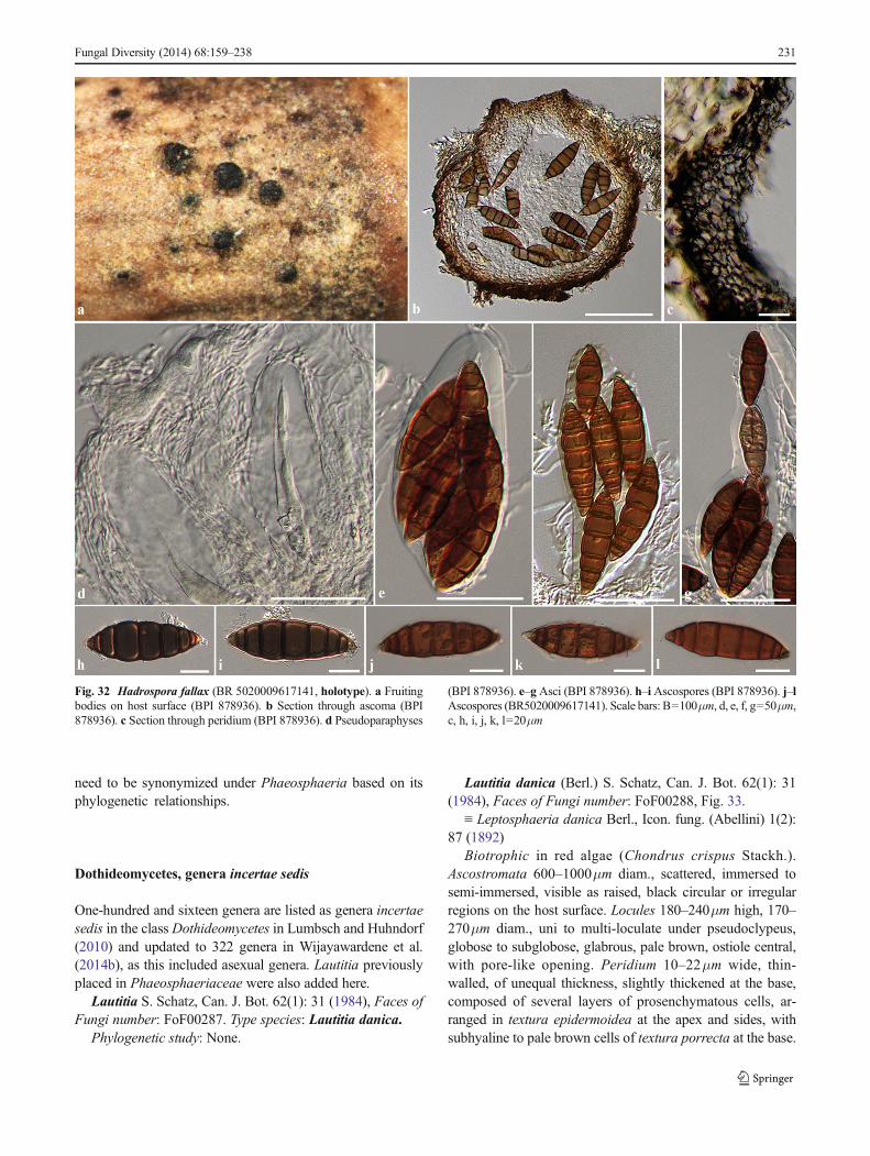
need to be synonymized under Phaeosphaeria based on itsphylogenetic relationships.
Dothideomycetes, genera incertae sedis
One-hundred and sixteen genera are listed as genera incertaesedis in the class Dothideomycetes in Lumbsch and Huhndorf(2010) and updated to 322 genera in Wijayawardene et al.(2014b), as this included asexual genera. Lautitia previouslyplaced in Phaeosphaeriaceae were also added here.
Lautitia S. Schatz, Can. J. Bot. 62(1): 31 (1984), Faces ofFungi number: FoF00287. Type species: Lautitia danica.
Phylogenetic study: None.
Lautitia danica (Berl.) S. Schatz, Can. J. Bot. 62(1): 31(1984), Faces of Fungi number: FoF00288, Fig. 33.
≡ Leptosphaeria danica Berl., Icon. fung. (Abellini) 1(2):87 (1892)
Biotrophic in red algae (Chondrus crispus Stackh.).Ascostromata 600–1000μm diam., scattered, immersed tosemi-immersed, visible as raised, black circular or irregularregions on the host surface. Locules 180–240μm high, 170–270μm diam., uni to multi-loculate under pseudoclypeus,globose to subglobose, glabrous, pale brown, ostiole central,with pore-like opening. Peridium 10–22μm wide, thin-walled, of unequal thickness, slightly thickened at the base,composed of several layers of prosenchymatous cells, ar-ranged in textura epidermoidea at the apex and sides, withsubhyaline to pale brown cells of textura porrecta at the base.
Fungal Diversity (2014) 68:159–238 231
Fig. 32 Hadrospora fallax (BR 5020009617141, holotype). a Fruitingbodies on host surface (BPI 878936). b Section through ascoma (BPI878936). c Section through peridium (BPI 878936). d Pseudoparaphyses
(BPI 878936). e–gAsci (BPI 878936). h–i Ascospores (BPI 878936). j–lAscospores (BR5020009617141). Scale bars: B=100μm, d, e, f, g=50μm,c, h, i, j, k, l=20μm
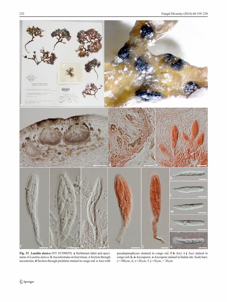
232 Fungal Diversity (2014) 68:159–238
Fig. 33 Lautitia danica (NY 01390859). a Herbarium label and speci-mens of Lautitia danica. bAscostromata on host tissue. c Section throughascostroma. d Section through peridium stained in congo red. eAsci with
pseudoparaphyses strained in congo red. f–h Asci. i–j Asci stained incongo red. k–nAscospores. oAscospore stained in Indian ink. Scale bars:c=100μm, d, e=20μm, f–j=10μm, = 20μm
Hamathecium composed of numerous, 1–1.5μm wide, fila-mentous, anastomosing, rough-walled, narrow cellularpseudoparaphyses, with distinct septa, embedded in a muci-laginous matrix. Asci (57–)70–90(−104)×(12–)15–18(−20)μm (x = 78.8×15.9μm, n=25), 8-spored, bitunicate, clavate,long pedicellate, apically rounded to truncate, with an indis-tinct ocular chamber. Ascospores (31–)35–40(−42)×5–7μm(x = 35.9×5.8μm, n=30), overlapping 1–3-seriate, 2-celled,obclavate with narrow upper cell, mostly the upper cell longerthan the lower cell, hyaline, 1-septate, slightly constricted atthe septum, rough-walled with appendage on upper cell.
Material examined: FRANCE, Belclem near Roscoff,Finistère (Atlantic Ocean), on Chondrus crispus(Gigartinaceae), 5 October 1971 J. Kohlmeyer no. 2920(NY 01390859); USA, Massachusetts, Gloucester, HalibusPoint, 2 m deep, on Chondrus crispus, 28 September1980, Kate van den Bosch; ibid. Rhode Island, WatchHill Lighthouse (Atlantic Ocean), on Laminaria, 20August 1966, J. Kohlmeyer_no. 1988 (NY 01390747);no. 1988a, (NY 01389139); Slide Box 15, Tray 20, S.Schatz (NY 01319510–NY 01319526).
Notes: The monotypic genus Lautitia was introduced bySchatz (1984) as typified by L. danica and placed inDothideomycetes (Schatz 1984). A detailed description andtaxonomic history were given by Wilson and Knoyle (1961)who treated the species then named Didymosphaeria danica(Schatz 1984). Berlese (1892) considered the species as aLeptosphaeria based on its hyaline, 1-septate, immature asco-spores. Lind (1913) transferred the species toDidymosphaeriaas D. marina (Rostrup) Lind, due to the ascospores turningbrown and being 1-septate at maturity.Wilson and Knoyle(1961) could not find brown ascospores, but retained thespecies in Didymosphaeria. Schatz (1984) disagreed withWilson and Knoyle (1961) as the species has cellularpseudoparaphyses, while genera in Didymosphaeria have tra-beculate pseudoparaphyses. Thus Schatz (1984) designated anew genus, Lautitia, to accommodate the taxon. Schatz (1984)treated Lautitia as a member Phaeosphaeriaceae due to itspseudoparenchymatous to prosenchymatous peridium, andelongate, hyaline asymmetric ascospores. The genus wasaccepted in Phaeosphaeriaceae by Zhang et al. (2012) andHyde et al. (2013).
In this study, we exclude Lautitia from Phaeaosphaeriaceaebased on its morphology. Lautitia can be distingiushedfrom other genera in Phaeosphaeriaceae which differ inperidium structure, asci and ascospores. Lautitia hasprosenchymatous perdial cells, clavate asci with long ped-icels, and asymmetric didymosporous ascospores, whileother genera in Phaeoaperiaceae have pseudoparenchyma-tous peridial cells, cylindrical to cylindric-clavate asci whichare subsessile to short pedicellate, and phragmosporous,scolecosporous or muriform ascospores (Zhang et al.2009, 2012; Hyde et al. 2013). It is difficult to place this
species in any family or order any thus we refer it toDothideomycetes, genera incertae sedis.
Acknowledgments The Royal Golden Jubilee Ph. D. Program (PHD/0090/2551) under Thailand Research Fund, Humidtropics, a CGIARResearch Program that aims to develop new opportunities for improvedlivelihoods in a sustainable environment and Mae Fah Luang University(grant for study Dothideomycetes No. 56101020032) are gratefullythanked for partially funding this work. KD Hyde acknowledges TheChinese Academy of Sciences, project number 2013T2S0030, for theaward of Visiting Professorship for Senior International Scientists atKunming Institute of Botany. Wen Jing Li and International FungalResearch & Development Centre, Research Institute of Resource Insects,Chinese Academy of Forestry; Ruvishika S. Jayawardena and Institute ofPlant and Environment Protection, Beijing Academy of Agriculture andForestry Sciences; Jun-Bo Yang and Plant Germplasm and GenomicsCenter in GermplasmBank ofWild Species, Kunming Institute of Botanyare gratefully thanked for the molecular laboratory support. The curatorsfrom BPI, BR, NY, S, and UB herbaria are gratefully thanked for loaningspecimens and type information. Amy Y. Rossman, ChatsacheeChatpapamon, Dhanushka N. Wanasinghe, Dhanushka Udayanga, E. B.Gareth Jones, Joanne E. Taylor, Saowanee Wikee, Saranyaphat Boonmeand Supalak Yacharoen are gratefully thanked for taxonomic literatureinformation, cultures preparation and general assistance.
References
Abbas SQ, Iftikhar T, Niaz M, Ali I, Iftikhar N, Abbas A (2012) A newspecies of Tiarosporella azadarrichta and new fungal records onAzadirachta indica from Pakistan. Pak J Bot 44(6):2093–2102
Abdullah SK, Asensio L, Monfort E, Gomez-Vidal S, Palma-Guerrero J,Salinas J, Lopez-Llorca LV, Jansson HB, Guarro J (2005)Occurrence in Elx, SE Spain of inflorescence rot disease of datepalms caused by Mauginiella scaettae. J Phytopathol 153:417–422
Ahn YM, Shearer CA (1998) Re-examination of taxa in Leptosphaeriaoriginally described on host species inRanunculaceae,Papaveraceae,andMagnoliaceae. Can J Bot 76:258–280
Ainsworth GC, James PW, Hawksworth DL (1971) Ainsworth & Bisby’sdictionary of the fungi, 6th edn. CAB, Kew
Aptroot A (2006) Mycosphaerella and its anamorphs 2. Conspectus ofMycosphaerella. CBS Biodivers Ser 5:1–231
Ariyawansa HA, Kang JC, Alias SA, Chukeatirote E, Hyde KD (2013)Towards a natural classification of Dothideomycetes: the generaDermatodothella, Dothideopsella, Grandigallia, Hysteropeltellaand Gloeodiscus (Dothideomycetes incertae sedis). Phytotaxa147(2):35–47
Ariyawansa HA, Phookamsak R, Tibpromma S, Kang JC, Hyde KD(2014a)Amolecular andmorphological reassessment ofDiademaceae.Sci World J. doi:10.1155/2014/675348
Ariyawansa HA, Tanaka K, Thambugala KM, Phookamsak R, Tian Q,Camporesi E, Hongsanan S, Monkai J, Wanasinghe DN,Chukeatirote E, Kang JC, Xu JC, McKenzie EHC, Jones EBG,Hyde KD (2014b) A molecular phylogenetic reappraisal of theDidymosphaeriaceae (Montagnulaceae). Fungal Divers. 68. doi:10.1007/s13225-014-0305-6
Ariyawansa HA, Thambugala KM, Maharachchikumbura SSN,Udayanga D, Manamgoda DS, Daranagama A, Shivas RG,Jayawardena R, Phookamsak R, Senanayake IC, Liu JK, Tian Q,Camporesi E, McKenzie EHC, Lücking R, Hawksworth DL, KangJC, Alias SA, Chukeatirote E, Mortimer PE, Xu JC, Jones EBG,Hyde KD (2014c) Epitypification and neotypification. FungalDivers (in press)
Fungal Diversity (2014) 68:159–238 233
Arzanlou M, Crous PW (2006) Phaeosphaeriopsis musae. Fungal Planet9. CBSKNAW Fungal Biodiversity Centre, Utrecht
Aveskamp MM, de Gruyter J, Woudenberg JHC, Verkley GJM, CrousPW (2010) Highlights of theDidymellaceae: a polyphasic approachto characterise Phoma and related pleosporalean genera. StudMycol65:1–60
Barr ME (1979) A classification of Loculoascomycetes. Mycologia 71:935–957
Barr (1982) Leptosphaeria sepalorum. Mycotaxon 15:345–348Barr ME (1987a) New taxa and combinations in the Loculoascomycetes.
Mycotaxon 29:501–505Barr ME (1987b) Prodromus to Class Loculoascomycetes. University of
Massachusetts, Massachusetts, AmherstBarr ME (1990a) Melanommatales (Loculoascomycetes). N Am Flora
13(II):1–129Barr ME (1990b) Some dictyosporous genera and species of
Pleosporales in North America. Mem N Y Bot Gard 62:1–92Barr ME (1992a) Additions to and notes on the Phaeosphaeriaceae
(Pleosporales, Loculoascomycetes). Mycotaxon 43:371–400Barr ME (1992b) Notes on the Lophiostomataceae (Pleosporales).
Mycotaxon 45:191–221Barr ME (1996) Planistromellaceae, a new family in the Dothideales.
Mycotaxon 60:433–442Barr ME (2002) Teichosporaceae, another family in the Pleosporales.
Mycotaxon 82:373–389Barr ME, Rogerson CT (1999) Some Loculoascomycete species form the
Great Basin, USA. Mycotaxon 71:473–480Bayon C, Yuan Z-W, Ruiz C, Liesebach M, Pei MH (2006) Genetic
diversity in the mycoparasite Sphaerellopsis filum inferred fromAFLP analysis and ITS–5.8S sequences. Mycol Res 110:1200–1206
Belsare SW, Moniz L, Deo VB (1980) The hyperparasite Ampelomycesquisqualis Ces. from Maharashtra State, India. Biovigyanam 6:173–176
Berlese AN (1892) Leptosphaeria danica. Icones Fungorum 1(2):67–90Boerema GH (1982) Phoma-soorten van de sectie Plenodomus.
Verslagen en Mededelingen Plantenziektenkundige DienstWageningen 158(Jaarboek 1981):28–30
Boerema GH (1997) Contributions towards a monograph of Phoma(Coelomycetes) - V. Subdivision of the genus in sections.Mycotaxon 64:321–333
Boerema GH, Dorenbosch MMJ (1979) Mycologisch-taxonomischonderzoek in Jaarboek 1978. Verslagen en MededelingenPlantenziektenkundige Dienst 153:17–21
Boerema GH, De Gruyter J, Noordeloos ME, Hamers MEC (2004)Phoma identification manual. Differentiation of specificand infraspecific taxa in culture. CABI Publishing, UnitedKingdom
Boise JR (1989) OnHadrospora, a new genus in the Phaeosphaeriaceaeand Byssothecium alpestris. Mem N Y Bot Gard 49:308–310
Boonmee S, Ko Ko TW, Chukeatirote E, Hyde KD, Chen H, Lei C,McKenzie EHC, Jones EBG, Rampai K, Bahkali AH (2012) Twonew Kirschsteiniothelia species with Dendryphiopsis anamorphscluster in Kirschsteiniotheliaceae fam. nov. Micologia 104(3):698–714
Calatayud V, Etayo J (2001) Five new species of lichenicolous conidialfungi from Spain. Can J Bot 79:223–230
Câmara MPS, O’Neill NR, van Berkum P, Dernoeden PH, Palm ME(2000)Ophiosphaerella agrostis sp. nov. and its relationship to otherspecies of Ophiosphaerella. Mycologia 92:317–325
Câmara MPS, Palm ME, van Berkum P, Stewart EL (2001) Systematicsof Paraphaeosphaeria: a molecular and morphological approach.Mycol Res 105:41–56
Câmara MPS, Palm ME, van Berkum P, O’Neill NR (2002) Molecularphylogeny of Leptosphaeria and Phaeosphaeria. Mycologia 94:630–640
Câmara MPS, Ramaley AW, Castlebury LA, Palm ME (2003)Neophaeosphaeria and Phaeosphaeriopsis, segregates ofParaphaeosphaeria. Mycol Res 107:516–522
Cannon PF, Damm U, Johnston PR, Weir BS (2012) Colletotrichum–current status and future directions. Stud Mycol 73:181–213
Carson ML (2005) Yield loss potential of Phaeospharia leaf spot ofmaize caused by Phaeosphaeria maydis in the United States. PlantDis 89:986–988
Cesati V (1852) Ampelomyces quisqualis Ces. Botanische Zeitung 10:301–302
Cheng TF, Jia XM,MaXH, Lin HP, Zhao YH (2004) Phylogenetic studyon Shiraia bambusicola by rDNA sequence analyses. J BasicMicrobiol 44:339–350
Chomnunti P, Hongsanan S, Aguirre-Hudson B, Tian Q, Peršoh D,Dhami MK, Alias AS, Xu JC, Liu X, Stadler M, Hyde KD (2014)The sooty moulds. Fungal Divers 66:1–36
Clare BG (1964) Ampelomyces quisqualis (Cicinnobolus cesatii) onQueensland Erysiphaceae. University of Queensland Papers 4:147–149
Clements FE, Shear CL (1931) Genera of fungi, 2nd edn. H.W. WilsonCompany, New York
Corda ACI (1840) Icones Fungorum hucusque Cognitorum 4: i–iii, 1–63.Prague; apud J.G. Calve [Fridericum Ehrlich]
Crivelli PG (1983) Über die heterogene Ascomycetengattung PleosporaRabh.: vorschlag für eine Aufteilung. Dissertation ETH Nr. 7318,Zürich, Germany
Crous PW, Palm ME (1999) Systematics of selected foliicolous fungiassociated with leaf spots ofProteaceae. Mycol Res 103:1299–1304
Crous PW, Slippers B, Wingfield MJ, Rheeder J, Marasas WFO, PhilipsAJL, Alves A, Burgess TI, Barber PA, Groenewald JZ (2006)Phylogenetic lineages in the Botryosphaeriaceae. Stud Mycol 55:235–253
Crous PW, Groenewald JZ, Lombard L, Wingfield MJ (2008)Homortomyces gen. nov., a new dothidealean pycnidial fungus fromthe Cradle of Humankind. IMA Fungus 3(2):109–115
Crous PW, Minnis AW, Pereira OL, Alfenas AC, Alfenas RF,Rossman AY, Groenewald JZ (2011) What is Scirrhia? IMAFungus 2:127–133
Crous PW, Wingfield MJ, Guarro J, Cheewangkoon R et al (2013)Molecular phylogeny and evolution of fungi – Fungal Planet de-scription sheets: 154–213. Persoonia 31:188–296
Crous PW, Shivas RG, Quaedvlieg W, van der Bank M, Zhang Y,Summerell BA, Guarro J, Wingfield MJ, Wood AR, Alfenas AC,Braun U, Cano-Lira JF, García D, Marin-Felix Y, Alvarado P,Andrade JP, Armengol J, Assefa A, den Breeÿen A, Camele I,Cheewangkoon R, De Souza JT, Duong TA, Esteve-Raventós F,Fournier J, Frisullo S, García-Jiménez J, Gardiennet A, Gené J,Hernández-Restrepo M, Hirooka Y, Hospenthal DR, King A,Lechat C, Lombard L, Mang SM, Marbach PAS, Marincowitz S,Marin-Felix Y, Montaño-Mata NJ, Moreno G, Perez CA, PérezSierra AM, Robertson JL, Roux J, Rubio E, Schumacher RK,Stchigel AM, Sutton DA, Tan YP, Thompson EH, van der LindeE, Walker AK, Walker DM, Wickes BL, Wong PTW, GroenewaldJZ (2014) Fungal Planet Description Sheets: 214–280. Persoonia 32:184–306
Cunfer BM (2000) Stagonospora and Septoria diseases of barley, oat, andrye. Can J Plant Pathol 22:332–348
Cunfer BM, Ueng PP (1999) Taxonomy and identification of Septoriaand Stagonospora species on small-grain cereals. Annu RevPhytopathol 37:267–284
De Bary A (1870) Eurotium, Erysiphe, Cicinnobolus, nebstBemerkungen über die Geschlechtsorgane der Ascomyceten. In:Beiträge zur Morphologie und Physiologie der Pilze (A. De Bary& M. Woronin, eds). Verlag von C. Winter, Frankfurt, pp 1–95
De Gruyter J, Aveskamp MM, Woudenberg JHC, Verkley GJM,Groenewald JZ, Crous PW (2009) Molecular phylogeny of Phoma
234 Fungal Diversity (2014) 68:159–238
and allied anamorph genera: towards a reclassification of the Phomacomplex. Mycol Res 113:508–519
De Gruyter J, Woudenberg JH, AveskampMM, Verkley GJ, GroenewaldJZ, Crous PW (2010) Systematic reappraisal of species in Phomasection Paraphoma, Pyrenochaeta and Pleurophoma. Mycologia102:1066–1081
De Gruyter J, Woudenberg JHC, Aveskamp AA, Verkley GJM,Groenewald JZ, Crous PW (2012) Redisposition of phoma-likeanamorphs in Pleosporales. Stud Mycol 75:1–36
Dennis RWG (1978) British Ascomycetes, 3rd edn. J. Cramer, VaduzDianese JC, Inácio CA, Dornelo-Silva D (2001)Wilmia, a new genus of
phaeosphaeriaceous ascomycetes on Memora pedunculata in cen-tral Brazil. Mycologia 93:1014–1018
Doilom M, Liu JK, Jaklitsch WM, Ariyawansa H, Wijayawardene NN,Chukeatirote E, Zhang M, McKenzie EHC, Geml J, Voglmayr H,Hyde KD (2013) An outline of the family Cucurbitariaceae.Sydowia 65(1):167–192
Dong JW, Chen WD, Crane JL (1998) Phylogenetic studies of theLeptosphaeriaceae , Pleosporaceae and some otherLoculoascomycetes based on nuclear ribosomal DNA sequences.Mycol Res 102:151–156
Eriksson OE (1966) On Eudarluca caricis (Fr.) O. Erikss., comb. nov., acosmopolitan uredinicolous pyrenomycete. Bot Not 119:33–69
Eriksson OE (1967) On graminicolous pyrenomycetes from Fennoscandia.I, II, III. Ark Bot Ser 26:339–466
Eriksson OE (1981) The families of bitunicate ascomycetes. Opera Bot60:1–220
Eriksson OE (1982) Notes on ascomycetes and coelomycetes from NW.Europe Mycotaxon 15:189–202
Eriksson OE (2006) Outline of Ascomycota–2006. Myconet 12:1–82Eriksson OE (2007) Ascomycet-Nytt 1. Svensk Mykologisk Tidskrift
28(2):38–41Eriksson OE, Hawksworth DL (1990) Outline of the Ascomycetes—1989.
Syst Ascomyc Reprint of Volumes 1–4 (1982–1985) 8 (2): 119–318Eriksson OE, Hawksworth DL (1991) Outline of the Ascomycetes–1990.
Syst Ascomyc Reprint of Volumes 1–4 (1982–1985) 9: 39–271Eriksson OE, Hawksworth DL (1993) Outline of the Ascomycetes –
1993. Syst Ascomyc 12(1–2):51–257Eriksson OE, Yue J-Z (1990) Notes on bambusicolous pyrenomycetes.
Nos. 1–10. Mycotaxon 38:201–220Farr DF, Bills GF (1995) Wojnowicia colluvium sp. nov. isolated from
conifer litter. Mycologia 87(4):518–524Farr DF, Rossman AY (2014) Fungal databases, systematic mycology
and microbiology laboratory, ARS, USDA. Retrieved September 1,2014, from http://nt.ars-grin.gov/fungaldatabases/
Farr DF, Bills GF, Chamuris GP, RossmanAY (1989) Fungi on plants andplant products in the United States. APS Press, St. Paul
Farr DF, Aime MC, Rossman AY, Palm ME (2006) Species ofcolletotrichum on agavaceae. Mycol Res 110(12):1395–1408
Fisher PJ,Webster J (1992) ATrematosphaeria endophyte from rice rootsand its Zalerion anamorph. Nova Hedw 54:77–81
Gams W (2000) Phialophora and some similar morphologically little-differentiated anamorphs of divergent ascomycetes. Stud Mycol 45:187–200
Griffith D (1899) The common parasite of the powdery mildews. BullTorrey Bot Club 26:184–188
Hall TA (1999) BioEdit: a user-friendly biological sequence alignmenteditor and analysis program for Windows 95/98/NT. In: NucleicAcids Symposium Series, pp 95–98
Hawksworth DL (1985)Kirschsteiniothelia a new genus for theMicrotheliaincrustans–group (Dothideales). Bot J Linn Soc 91:181–202
Hedjaroude A (1969) Études taxonomiques sur les PhaeosphaeriaMiyake et leurs formes voisines (ascomycètes). Sydowia 22:57–107
Hodhod MS, Abdel-Wahab MA, Bahkali AHA, Hyde KD (2012)Amarenographium solium sp nov from Yanbu mangroves in theKingdom of Saudi Arabia. Cryptogam Mycol 33(3):285–294
Holm L (1948) Taxonomical notes onAscomycetes. 1. The Swedishspecies of the genus Ophiobolus Riess sensu Sacc. Sven BotTidskr 42:337–347
Holm L (1957) Etudes taxonomiques sur les pléosporacées. Symb BotUpsaliens 14:1–188
Hsiang T, Chen F, Goodwin PH (2003) Detection and phylogeneticanalysis of mating type genes of Ophiosphaerella korrae. Can JBot 81(4):307–315
Huelsenbeck JP, Ronquist F (2001) MRBAYES: Bayesian inference ofphylogenetic trees. Bioinformatics 17(8):754–755
Hyde KD (1992) Fungi from decaying inter-tidal fronds of Nypafruticans, including three new genera and four new species. J LinnSoc Bot 110:95–110
Hyde KD (1994) Fungi from palms. XII. Three new intertidal ascomy-cetes from submerged palm fronds. Sydowia 46:257–264
Hyde KD, McKenzie EHC, KoKo TW (2011) Towards incorporatinganamorphic fungi in a natural classification – checklist and notes for2010. Mycosphere 2:1–88
Hyde KD, Jones EBG, Liu JK, Ariyawansa HA, Boehm E, Boonmee S,Braun U, Chomnunti P, Crous PW, Dai DQ, Diederich P,Dissanayake AJ, Doilom M, Doveri F, Hongsanan S, JayawardenaR, Lawrey JD, Li YM, Liu YX, Lücking R, Monkai J, Muggia L,Nelsen MP, Pang KL, Phookamsak R, Senanayake I, Shearer CA,Seutrong S, Tanaka K, Thambugala KM, Wijayawardene NN,Wikee S, Wu HX, Zhang Y, Aguirre-Hudson B, Alias SA, AptrootA, Bahkali AH, Bezerra JL, Bhat JD, Camporesi E, Chukeatirote E,Gueidan C, Hawksworth DL, Hirayama K, De Hoog S, Kang JC,Knudsen K, Li WJ, Li X, Liu ZY, Mapook A, McKenzie EHC,Miller AN, Mortimer PE, Phillips AJL, Raja HA, Scheuer C,Schumm F, Taylor JE, Tian Q, Tibpromma S, Wanasinghe DN,Wang Y, Xu JC, Yan J, Yacharoen S, Zhang M (2013) Families ofDothideomycetes. Fungal Divers 63:1–313
Hyde KD, Nilsson RH, Alias SA, Ariyawansa HA, Blair JE, Cai L, deCock AWAM, Dissanayake AJ, Glockling SL, Goonasekara ID,Gorczak M, Hahn M, Jayawardena RS, van Kan JAL, LaurenceMH, Lévesque CA, Li X, Liu JK, Maharachchikumbura SSN,Manamgoda DS, Martin FN, McKenzie EHC, McTaggart AR,Mortimer PE, Nair PVR, Pawłowska J, Rintoul TL, Shivas RG,Spies CFJ, Summerell BA, Taylor PWJ, Terhem RB, Udayanga D,Vaghefi N, Walther G, Wilk M, Wrzosek M, Xu JC, Yan JY,Zhou N (2014) One stop shop: backbones trees for importantphytopathogenic 5 genera: I (2014). Fungal Divers. doi:10.1007/s13225-014-0298-1
Index Fungorum (2014) http://www.indexfungorum.org/Names/Names.aspJones EBG, Sakayaroj J, Suetrong S, Somrithipol S, Pang KL (2009)
Classification of marine Ascomycota, anamorphic taxa andBasidiomycota. Fungal Divers 35:1–187
Keener PD (1951) An ascigerous stage ofDarluca filum (Biv.) Castagne.Plant Dis Rep 35:86–87
Khashnobish A, Shearer CA (1996) Phylogenetic relationships insome Leptosphaeria and Phaeosphaeria species. Mycol Res100:1355–1363
Kirk PM, Cannon PF, David JC, Stalpers JA (2001) Dictionary of theFungi 9th edn. CABI, Wallingford
Kirk PM, Cannon PF, Minter DW, Staplers JA (2008) Dictionary of theFungi 10th edn. CABI Bioscience, UK
Kiss L (1998) Natural occurrence of Ampelomyces intracellularmycoparasites in mycelia of powdery mildew fungi. New Phytol140:709–714
Kiss L, NakasoneKK (1998) Ribosomal DNA internal transcribed spacersequences do not support the species status of Ampelomycesquisqualis, a hyperparasite of powdery mildew fungi. Curr Genet33:362–367
Kiss L, Russell J, Szentiványi O, Xu X, Jeffries P (2004) Biology andbiocontrol potential of Ampelomyces mycoparasites, natural antag-onists of powdery mildew fungi. Biocontrol Sci Tech 14:635–651
Fungal Diversity (2014) 68:159–238 235
Kodsueb R, Dhanasekaran V, Aptroot A, Lumyong S, McKenzie EHC,Hyde KD, Jeewon R (2006) The family Pleosporaceae: intergenericrelationships and phylogenetic perspectives based on sequence anal-yses of partial 28S rDNA. Mycologia 98:571–583
Kodsueb R, Lumyong S, Ho WH, Hyde KD, McKenzie EHC,Jeewon R (2007) Morphological and molecular characterizationof Aquaticheirospora and phylogenetics of Massarinaceae(Pleosporales). Bot J Linn Soc 155:283–296
Kohlmeyer J, Kohlmeyer E (1965) Phaeosphaeria ammophilae. IconesFungorum Maris 3
Kohlmeyer J, Volkmann-Kohlmeyer B (1993) A trotorquata andLoratospora: new ascomycete genera on Juncus roemerianus.Syst Ascomyc 12:7–22
Kruys A, Eriksson OE, Wedin M (2006) Phylogenetic relationships ofcoprophilous Pleosporales (Dothideomycetes, Ascomycota), andthe classification of some bitunicate taxa of unknown position.Mycol Res 110:527–536
Lawrey JD, Diederich P, Nelsen MP, Freebury C, Broeck DVD, SikaroodiM, Ertz D (2012) Phylogenetic placement of lichenicolous Phomaspecies in the Phaeosphaeriaceae (Pleosporales, Dothideomycetes).Fungal Divers 55:195–213
Leuchtmann A (1984) Über Phaeosphaeria Miyake und anderebitunicate Ascomyceten mit mehrfach querseptierten Ascosporen.Sydowia 37:75–194
Liang C, Yang J, Kovács GM, Szentiványi O, Li B, Xu XM, KissL (2007) Genetic diversity of Ampelomyces mycoparasitesisolated from different powdery mildew species in Chinainferred from analyses of rDNA ITS sequences. FungalDivers 24:225–240
Liew ECY, Aptroot A, Hyde KD (2000) Phylogenetic significance of thepseudoparaphyses in Loculoascomycete taxonomy. Mol PhylogenetEvol 16:392–402
Lind J (1913) Danish fungi, as represented in the herbarium of E.Rostrup. 1–650. Denmark, Copenhagen
Liu YJ, Whelen S, Hall BD (1999) Phylogenetic relationships amongascomycetes: evidence from an RNA polymerase II subunit. MolBiol Evol 16:1799–1808
Liu JK, Phookamsak R, Jones EBG, Zhang Y, Ko-Ko TW, Hu HL,Boonmee S, Doilom M, Chukeatirote E, Bahkali AH, Wang Y,Hyde KD (2011) Astrosphaeriella is polyphyletic, with species inFissuroma gen. nov., and Neoastrosphaeriella gen. nov. FungalDivers 51:135–154
Liu JK, Phookamsak R, Doilom M, Wiki S, Mei LY, Ariyawansa HA,Boonmee S, Chomnunti P, Dai DQ, Bhat DJ, Romero AI, XhuangWY, Monkai J, Jones EBG, Chukeatirote E, KoKo TW, Zhoa YC,Wang Y, Hyde KD (2012) Towards a natural classification ofBotryosphaeriales. Fungal Divers 57:149–210
Liu YX, Hyde KD, Ariyawansa HA, Li WJ, Zhou DQ, Yang YL, ChenYM, Liu ZY (2013) Shiraiaceae, new family of Pleosporales(Dothideomycetes, Ascomycota). Phytotaxa 103(1):51–60
Lumbsch HT, Huhndorf SM (eds.) (2007) Outline of Ascomycota–2007.Myconet 13:1–58
Lumbsch HT, Huhndorf SM (2010) Outline ofAscomycota–2009.Fieldiana Life Earth Sci 1:1–60
Manamgoda DS, Cai L, McKenzie EHC, Crous PW, Madrid H,Chukeatirote E, Shivas RG, Tan YP, Hyde KD (2012) A phyloge-netic and taxonomic re–evaluation of the Bipolaris, Cochliobolus,Curvularia Complex. Fungal Divers 56:131–144
McAlpine D (1902) Fungus diseases of stone-fruit trees in Australia andtheir treatment, pp 1–165
Minter DW, Rodríguez Hernández M, Mena Portales J (2001) Fungi ofthe Caribbean: an annotated checklist. PDMS Publishing: 946 pp
Miyake I (1909) Studies on the parasitic fungi of rice in Japan. Bot MagTokyo 23:85–97
Miyake I (1910) Studien über die Pilze der Reispflanze in Japan. J CollAgric Tokyo 2:237–276
Moesz G (1915) Mykologiai közlemények. II. közlemény. BotanikaiKölzlemények 14(5–6):145–158
Monkai J, Liu JK, Boonmee S, Chomnunti P, Chukeatirote E, Jones EBG,Wang Y, Hyde KD (2013) Planistromellaceae (Botryosphaeriales).Cryptogam Mycol 34:45–77
Morales VM, Jasalavich CA, Pelcher LE, Petrie GA, Taylor JL (1995)Phylogenetic relationships among several Leptosphaeria spe-cies based on their ribosomal DNA sequences. Mycol Res99:593–603
Morelet M (1980) Sur quatre Dothideales. Bull Soc Sci Nat ArcheolToulon 227:14–15
Morgan-Jones G, White JF (1983) Studies on the genus Phoma. III.Paraphoma, a new genus to accommodate Phoma radicina.Mycotaxon 18(1):57–65
Müller E (1950) Die schweizerischen Arten der Gattung Leptosphaeriaund ihrer Verwandten. Sydowia 4:185–319
Müller E (1952) Die schweizerischen Arten der Gattung OphiobolusRiess. Ber Schweiz Bot Ges 62:307–339
Müller E, von Arx JA (1962) Die Gattungen der didymosporenPyrenomyceten. Beitr Krypt Fl Schweiz 11:1–92
Munk A (1957) Danish pyrenomycetes. A preliminary flora. Dansk BotArk 17:1–491
Nag Raj TR (1989) Genera coelomycetum. XXVI. Amarenographium,Callistospora, Hyalothyridium, Orphanocoela anam.-gen.nov.,Scolecosporiella, and Urohendersoniella. Can J Bot 67(3169–3):186
Nischwitz C, Newcombe G, Anderson LC (2005) Host specialization ofthe mycoparasite Eudarluca caricis and its evolutionary relationshipto Ampelomyces. Mycol Res 109(4):421–428
Nylander JAA (2004) MrModeltest 2.0. Program distributed by theauthor. Evolutionary Biology Centre, Uppsala University
Oertel B (2011) Pilzgattungen Europas-Liste 6: NotizbuchartigeAuswahlliste zur Bestimmungsliteratur für Coelomyceten
Page RDM (1996) TreeView: an application to display phyloge-netic trees on personal computers. Comput Appl Biosci 12:357–358
Petrak F (1921) Mykologische Notizen. II Ann Mycol 19(1–2):17–128Petrak F (1944) Über die Gattungen Chaetopyrena Pass., Sclerochaeta
v. Höhn., Sclerochaetella v. Höhn., Vermiculariella Oud.,Chaetosphaeronema Moesz und Pseudophoma v. Höhn. AnnMycol 42:58–71
Petrak F, Sydow H (1936) Kritisch-systematische Originaluntersuchungenüber Pyrenomyzeten, Sphaeropsideen und Melanconieen. VII. AnnalMycol 34(1–2):11–52
Phookamsak R, Liu JK, Chukeatirote E, McKenzie EHC, Hyde KD(2013) Phylogeny and morphology of Leptosphaerulinasaccharicola sp. nov. and Pleosphaerulina oryzae and relationshipswith Pithomyces. Cryptogam Mycol 34(4):303–319
Phookamsak R, Liu JK, Manamgoda DS, Chukeatirote E, Mortimer PE,McKenzie EHC, Hyde KD (2014) The sexual state of Setophoma.Phytotaxa 176:260–269
Priest MJ (2006) Fungi of Australia: Septoria. CSIRO publishing,Melbourne, Australia, ABRS, Canberra
QuaedvliegW, VerkleyGJM, ShinH-D, Barretto RW, Alfenas AC, SwartWJ, Groenewald JZ, Crous PW (2013) Sizing up Septoria. StudMycol 75:307–390
Rabenhorst (1858) Herb myc, ed. 2 no. 725 (in sched)Raja HA, Ferrer A, Shearer CA (2009) Freshwater ascomycetes: a new
genus, Ocala scalariformis gen. et sp. nov, and two new species,Ayria nubispora sp. nov. and Rivulicola cygnea sp. nov. FungalDivers 34:79–86
Ramaley AW (1995) New fungi from Dasylirion (Agavaceae). Aliso 14:147–153
Ramaley AW (1997) New Paraphaeosphaeria species and theiranamorphs. Mycotaxon 61:347–358
Ramaley AW, Barr ME (1995) New dictyosporous species from leaves ofAgavaceae. Mycotaxon 54:75–90
236 Fungal Diversity (2014) 68:159–238
Rannala B, Yang Z (1996) Probability distribution of molecular evolu-tionary trees: a new method of phylogenetic inference. J Mol Evol43(3):304–311
Reddy PV, Patel R, White JF Jr (1998) Phylogenetic and developmentalevidence supporting reclassification of cruciferous pathogensPhoma lingam and Phoma wasabiae in Plenodomus. Can J Bot76:1916–1922
Rehner S (2001) Primers for elongation factor 1-α (EF1-α). http://ocid.NACSE.ORG/research/deephyphae/EF1primer.pdf
Reiss MLC (1854) Neue Kernpilze. Hedwigia 1:23–28Ronquist F, Huelsenbeck JP (2003) MrBayes 3: Bayesian phylogenetic
inference under mixed models. Bioinformatics 19(12):1572–1574Rossman AY, Farr DF, Castlebury LA, Shoemaker R, Mengistu A (2002)
Setomelanomma holmii (Pleosporales, Phaeosphaeriaceae) onliving spruce twigs in Europe and North America. Can J Bot80:1209–1215
Roumeguère C (1880) Revisio Reliquiae Libertianae. – Pars 1(1). RevueMycologique 2:15–24
Rudakov OL (1979) Fungi of the genus Ampelomyces Ces. Ex Schlecht.Mikol Fitopatol 13:104–110 [In Russian.]
Saccardo PA (1883) Sylloge Fungorum 2, Italy, Pavia, p 815Saccardo PA (1892) Supplementum Universale, Pars II. Discomyceteae-
Hyphomyceteae. Syll Fun 10:1–964Saccardo PA, Marchal E (1885) Reliquiae mycologicae Westendorpianae.
Revue Mycol Toulouse 7:140–149Saleh AA, Leslie JF (2004) Cephalosporium maydis is a distinct species
in the Gaeumannomyces-Harpophora species complex. Mycologia96(6):1294–1305
Sánchez Márquez S, Bills GF, Zabalgogeazcoa I (2007) The endophyticmycobiota of the grassDactylis glomerata. Fungal Divers 27:171–195
SawadaK (1959) Descriptive catalogue of Taiwan (Formosan) fungi. PartXI. Spec Publ Coll Agric National Taiwan Univ 8:1–268
Schatz S (1984) The life history, developmental morphology, and taxon-omy of Lautitia danica gen. nov., comb. nov. Can J Bot 62:28–32
Schoch CL, Shoemaker RA, Seifert KA, Hambleton S, Spatafora JW,Crous PW (2006) A multigene phylogeny of the Dothideomycetesusing four nuclear loci. Mycologia 98:1041–1052
Schoch CL, Crous PW, Groenewald JZ, Boehm EWA, Burgess TI, deGruyter J, de Hoog GS, Dixon LJ, Grube M, Gueidan C, Harada Y,Hatakeyama S, Hirayama K, Hosoya T, Huhndorf SM, Hyde KD,Jones EBG, Kohlmeyer J, Kruys Å, Li YM, Lücking R, LumbschHT, Marvanová L, Mbatchou JS, McVay AH, Miller AN, MugambiGK, Muggia L, Nelsen MP, Nelson P, Owensby CA, Phillips AJL,Phongpaichit S, Pointing SB, Pujade-Renaud V, Raja HA, RivasPlata E, Robbertse B, Ruibal C, Sakayaroj J, Sano T, Selbmann L,Shearer CA, Shirouzu T, Slippers B, Suetrong S, Tanaka K,Volkmann-Kohlmeyer B, Wingfield MJ, Wood AR, WoudenbergJHC, Yonezawa H, Zhang Y, Spatafora JW (2009) A class–widephylogenetic assessment of Dothideomycetes. Stud Mycol 64:1–15
Seifert K, Morgan-Jones G, Gams W, Kendrick B (2011) The genera ofHyphomycetes. CBS-KNAW Fungal Biodiversity Centre, Utrecht
Shearer CA (1993) Reexamination of eight taxa originally described inLeptosphaeria onmembers of the Asteraceae. Mycologia 85:825–834
Shearer CA, Crane JL (1971) Fungi of the Chesapeake Bay and itstributaries. I. Patuxent River. Mycologia 63:237–260
Shearer CA, Crane JL (1999) Freshwater Ascomycetes: Isthmosporellapulchragen. and sp. nov. Mycologia 91:141–144
Shearer CA, Raja HA, Miller AN, Nelson P, Tanaka K, Hirayama K,Marvanová L, Hyde KD, Zhang Y (2009) The molecular phylogenyof freshwater Dothideomycetes. Stud Mycol 64:145–153
Shoemaker RA (1961) Pyrenophora phaeocomes (Reb. ex Fr.) Fr. Can JBot 39:901–908
Shoemaker RA (1976) Canadian and some extralimital Ophiobolus spe-cies. Can J Bot 54:2365–2404
Shoemaker RA (1984) Canadian and some extralimital Nodulosphaeriaand Entodesmium species. Can J Bot 62:2730–2753
Shoemaker RA, Babcock CE (1987)Wettsteinina. Can J Bot 65:373–405Shoemaker RA, Babcock CE (1989a) Bricookea barrae n. sp. compared
with Bricookea sepalorum. Stud Mycol 31:165–169Shoemaker RA, Babcock CE (1989b) Phaeosphaeria. Can J Bot 67:
1500–1599Shoemaker RA, Babcock CE (1992) Applanodictyosporous Pleosporales:
Clathrospora, Comoclathris, GraphyIlium, Macrospora, andPlatysporoides. Can J Bot 70(8):1617–1658
Silva-Hanlin DMW, Hanlin RT (1999) Small subunit ribosomal RNAgene phylogeny of several loculoascomycetes and its taxonomicimplications. Mycol Res 103:153–160
Silvestro D, Michalak I (2011) raxmlGUI: a graphical front-end forRAxML. Org Divers Evol 12:335–337
Sivanesan A (1984) The bitunicate ascomycetes and their anamorph. J.Cramer, Vaduz, 701 pp
Solomon PS, Lowe RGT, Tan KC, Waters ODC, Oliver RP (2006)Stagonospora nodorum: cause of Stagonospora nodorum blotchof wheat. Mol Plant Pathol 7:147–156
Spegazzini C (1908) Fungi aliquot paulistani. Revista del Museo de LaPlata 15:7–48
Spegazzini C (1909) Ophiosphaerella. Anales del Museo Nacional deHistoria Natural Buenos Aires 19(12):401 [ser. 3, 12]
Spooner BM, Kirk PM (1982) Taxonomic notes on Excipularia andScolicosporium. Trans Br Mycol Soc 78:247–257
Stafleu FA et al (eds) (1972) International Code of BotanicalNomenclature. International Association for Plant Taxonomy,Utrecht, p 426
Stamatakis A, Hoover P, Rougemont J (2008) A rapid bootstrap algo-rithm for the RAxMLWeb Servers. Syst Biol 57:758–771
Stukenbrock EH, Banke S, McDonald BA (2006) Global migrationpatterns in the fungal wheat pathogen Phaeosphaeria nodorum.Mol Ecol 15:2895–2904
Suetrong S, Schoch CL, Spatafora JW, Kohlmeyer J, VolkmannKohlmeyer B, Sakayaroj J, Phongpaichit S, Tanaka K, HirayamaK, Jones EBG (2009) Molecular systematics of the marineDothideomycetes. Stud Mycol 64:155–173
Sullivan RF, White JF Jr (2000) Phoma glomerata as a mycoparasite ofpowdery mildew. Appl Environ Microbiol 66:425–427
Sutton BC (1968) Kellermania and its generic segregates. Can J Bot 46:181–196
Sutton BC (1975)Wojnowicia and Angiopomopsis.ČeskaMykologie 29:97–104
Sutton BC (1977) Coelomycetes VI. Nomenclature of generic namesproposed for coleomycetes. Mycol Pap 141:1–253
Sutton BC (1980) The Coelomycetes: fungi Imperfecti with pycnidia,acervuli and stromata. Commonwealth Mycological Institute, Kew
Sutton BC, Alcorn JL (1974) Neottiosporina. Aust J Bot 22(3):517–530Swart HJ, Walker J (1988) Australian leaf-inhabiting fungi. XXVIII.
Hendersonia on Eucalyptus. Trans Br Mycol Soc 90(4):633–641Swofford DL (2002) PAUP: phylogenetic analysis using parsimony,
version 4.0 b10. Sinauer Associates, SunderlandSzentiványi O, Kiss L, Russell JC, Kovács GM, Varga K, Jankovics T,
Lesemann S, Xu XM, Jeffries P (2005) Ampelomycesmycoparasitesfrom apple powdery mildew identified as a distinct group based onsingle-stranded conformation polymorphism analysis of the rDNAITS region. Mycol Res 109:429–438
Tamura K, Stecher G, Peterson D, Filipski A, Kumar S (2013) MEGA6:Molecular evolutionary genetics analysis version 6.0. Mol Biol Evol30:2725–2729
Tanaka E, Harada Y (2003) Hadrospora fallax (Pleosporales) found inJapan. Mycoscience 44:245–248
Tanaka K, Harada Y (2005) Bambusicolous fungi in Japan (6):Katumotoa, a new genus of phaeosphaeriaceous ascomycetes.Mycoscience 46:313–318
Tanaka K, Hirayama K, Yonezawa H, Hatakeyama S, Harada Y, Sano T,Shirouzu T, Hosoya T (2009) Molecular taxonomy of bambusicolous
Fungal Diversity (2014) 68:159–238 237
fungi: Tetraplosphaeriaceae, a new pleosporalean family withtetraploa-like anamorphs, and notes on the phylogeny of selectedspecies from bamboo. Stud Mycol 64:175–209
Thambugala KM, Camporesi E, Ariyawansa HA, Phookamsak R, LiuZY, Hyde KD (2014) Phylogeny and morphology ofPhaeosphaeriopsis triseptata sp. nov., and Phaeosphaeriopsisglaucopunctata. Phytotaxa 176:238–250
Theissen F, Sydow H (1915) Die Dothideales. Kritisch-systematischOriginaluntersuchungen. Ann Mycol 13:147–74
Torres MS, Bergen M, Singh S, Bischoff J, Sullivan RF, White JF Jr(2005) Plenodomus morganjonesii sp. nov. and a discussion of thegenus Plenodomus. Mycotaxon 93:333–343
Treigiene A, Mel’nik VA (2002) Interesting records of deuteromycetesfrom Lithuania (in Russian). Mikologiyai Fitopatologiya 36:42–47
Udayanga D, Liu X, Crous PW, McKenzie EHC, Chukeatirote E, HydeKD (2012) A multi-locus phylogenetic evaluation of Diaporthe(Phomopsis). Fungal Divers 56(1):157–171
Vergnes DM, Zhanarbekova A, Renard M-E, Duveiller E, Maraite H(2006) Mating types of Phaeosphaeria nodorum (anamorphStagonospora nodorum) from Central Asia. Phytopathology 154:317–319
Verkley GJM, da Silva M, Wicklow DT, Crous PW (2004)Paraconiothyrium, a new genus to accommodate the mycoparasiteConiothyriumminitans, anamorphs ofParaphaeosphaeria, and fournew species. Stud Mycol 50:323–335
Verkley GJM, Renfurm R, Göker M, Stielow JB (2014) Novel genera andspecies of coniothyrium-like fungi inMontagnulaceae (Ascomycota).Persoonia 32:25–51. doi:10.3767/003158514X679191
Vilgalys R, Hester M (1990) Rapid genetic identification and mapping ofenzymatically amplified ribosomal DNA from severalCryptococcusspecies. J Bacteriol 172:4238–4246
von Arx JA, Müller E (1975) A re-evaluation of the bitunicate ascomy-cetes with keys to families and genera. Stud Mycol 9:1–159
von Höhnel FXR (1915) Fragmente zur Mykologie (XVII. Mitteilung,Nr. 876 bis 943). Sitzungsberichten der Kaiserliche Akademie derWissenschaften in Wien Mathematische-NaturwissenschaftlicheKlasse, Abt. 1 124: 49–159
von Höhnel FXR (1917) Fungi imperfecti. Beiträge zur Kenntnisderselben. Hedwigia 59:236–284
Walker JM (1980) Gaeumannomyces, Linocarpon, Ophiobolus and sev-eral other genera of scolecospored ascomycetes and Phialophoraconidial states, with a note on hyphopodia. Mycotaxon 11:1–129
Walker JM, Sutton BC, Pascoe IG (1992) Phaeoseptoria eucalypti andsimilar fungi on Eucalyptus, with description of Kirramyces gen.nov. (Coelomycetes). Mycol Res 96(11):911–924
Wanasinghe DN, Jones EBG, Camporesi E, Boonmee S, KarunarathnaSC, Thines M, Mortimer PE, Xu J, Hyde KD (2014)Dematiopleospora mariae gen. sp. nov., from Ononis spinosainItaly. Crypto Mycol 35(2):105–117
Wang Y, Guo L, Hyde KD (2005) Taxonomic placement of sterilemorphotypes of endophytic fungi from Pinus tabulaeformis(Pinaceae) in northeast China based on rDNA sequences. FungalDivers 20:235–260
Webster J (1993) A rice root endophyte identified as Hadrospora fallax.Nova Hedw 57:141–142
Webster J, Lucas MT (1959) Observations on British species ofPleospora. Trans Brit Mycol Soc 42:332–342
Wehmeyer LE (1952) The genera Leptosphaeria, Pleospora andClathrospora in Mt Rainier National Park. Mycologia 44:621–655
Wehmeyer LE (1961) Aworld monograph of the genus Pleospora and itssegregates. University of Michigan Press, Michigan
Weir BS, Johnston PR, Damm U (2012) The Colletotrichumgloeosporioides species complex. Stud Mycol 73:115–180
Wetzel HC, Hulbert SH, Tisserat NA (1999) Molecular evidence for thepresence ofOphiosphaerella narmarin. comb., a cause of spring deadspot of bermuda grass, in North America. Mycol Res 103:981–989
White TJ, Bruns T, Lee S, Taylor J (1990) Amplification and directsequencing of fungal ribosomal RNA genes for phylogenetics. In:Innis MA, Gelfand DH, Sninsky JJ, White TJ (eds) PCRprotocols: a guide to methods and applications. Academic, SanDiego, pp 315–322
Wijayawardene NN, McKenzie EHC, Hyde KD (2012) Towards incor-porating anamorphic fungi in a natural classification checklist andnotes for 2011. Mycosphere 3:157–228
Wijayawardene NN, Camporesi E, Song Y, Dai DQ, Bhat DJ, MckenzieEHC, Chukeatirote E,Mel’nikVA,WangY,HydeKD (2013a)Multi-gene analyses reveal taxonomic placement of Scolicosporiumminkeviciusiiin Phaeosphaeriaceae (Pleosporales). CryptogamMycol 34(4):357–366
Wijayawardene NN, Song Y, Bhat DJ, McKenzie EHC, Chukeatirote E,Wang Y, Hyde KD (2013b)Wojnowicia viburni sp. nov. from Chinaand its phylogenetic placement. Sydowia 65(1):113–128
Wijayawardene NN, Bhat DJ, Hyde KD, Camporesi E,Wikee S, ChethanaKWT, Tangthirasunun N, Wang Y (2014a) Camarosporium sensustricto in Pleosporinae, Pleosporales with two new species.Phytotaxa (Accepted)
Wijayawardene NN, Crous PW, Kirk PM, Hawksworth DL et al (2014b)Naming and outline of Dothideomycetes–2014. Fungal Divers 68(inpress)
Wijayawardene NN, Hyde KD, Bhat DJ, Camporesi E, Schumacher RK,Chethana KWT, Wikee S, Bahkali AH, Wang Y (2014c)Camarosporium-like species are polyphyletic in Pleosporales; in-troducing Paracamarosporium and Pseudocamarosporium gen.nov. in Montagnulaceae. Cryptogam Mycol 35(2):177–198
Wijayawardene NN, Hyde KD, Bhat DJ, Wanasinghe DN, Song Y,Camporesi E, Schumacher RK, Wang Y (2014d) Additions tocoelomycetous taxa in Pleosporinae; introducing Eucamarosporiumgen. sp. nov. (in prep)
Wikee S, Udayanga D, Crous PW, Chukeatirote E, Mckenzie EHC,Bahkali AH, Dai DQ, Hyde KD (2011) Phyllosticta—an overviewof current status of species recognition. Fungal Divers 51:43–61
Wilson IM, Knoyle JM (1961) Three species of Didymosphaeria onmarine algae: D. danica (Berlese) comb. nov., D. pelvetiana Suth.and D. fucicola Suth. Trans Br Mycol Soc 44:55–71
Winter G (1872) Diagnosen neuer Pilze. Hedwigia 11:147–148Yang Z (1994) Maximum likelihood phylogenetic estimation from DNA
sequences with variable rates over sites: approximate methods. JMol Evol 39:306–314
Yuan ZQ (1994) Barria, a new ascomycetous genus in thePhaeosphaeriaceae. Mycotaxon 51:313–316
Yuan ZQ, Barr ME (1994) Species of Chaetoplea on desert plants inChina. Mycotaxon 52:495–499
Yuan ZW, Pei MH, Hunter T, Royle DJ (1998) Eudarluca caricis, theteleomorph of the mycoparasite Sphaerellopsis filum, on blackberryrust Phragmidium violaceum. Mycol Res 102:866–868
Yuan ZL, Lin FC, Zhang CL, Kubicek CP (2010) A new species ofHarpophora (Magnaporthaceae) recovered from healthy wild rice(Oryza granulata) roots, representing a novel member of a bene¢cialdark septate endophyte. FEMS Microbiol Lett 307(1):94–101
Zhang Y, Schoch CL, Fournier J, Crous PW, De Gruyter J, WoudenbergJHC, Hirayama K, Tanaka K, Pointing SB, Hyde KD (2009) Multi-locus phylogeny of the Pleosporales: a taxonomic, ecological andevolutionary re-evaluation. Stud Mycol 64:85–102
Zhang Y, Crous PW, Schoch CL, Bahkali AH, Guo LD, Hyde KD (2011)A molecular, morphological and ecological re-appraisal ofVenturiales—a new order of Dothideomycetes. Fungal Divers 51:249–277
Zhang Y, Crous PW, Schoch CL, Hyde KD (2012) Pleosporales. FungalDivers 53:1–221
Zhaxybayeva O, Gogarten JP (2002) Bootstrap, Bayesian probability andmaximum likelihood mapping: exploring new tools for comparativegenome analyses. BMC Genomics 3(1):4
238 Fungal Diversity (2014) 68:159–238