reversal of dopamine d2 agonist-induced inhibition of ventral tegmental area neurons by gq-linked...
TRANSCRIPT
1521-0103/344/1/253–263$25.00 http://dx.doi.org/10.1124/jpet.112.199844THE JOURNAL OF PHARMACOLOGY AND EXPERIMENTAL THERAPEUTICS J Pharmacol Exp Ther 344:253–263, January 2013Copyright ª 2013 by The American Society for Pharmacology and Experimental Therapeutics
Reversal of Dopamine D2 Agonist-Induced Inhibition of VentralTegmental Area Neurons by Gq-Linked Neurotransmitters IsDependent on Protein Kinase C, G Protein-Coupled ReceptorKinase, and Dynamin
Sudarat Nimitvilai, Maureen A. McElvain, and Mark S. BrodieDepartment of Physiology and Biophysics, University of Illinois at Chicago, Chicago, Illinois
Received September 5, 2012; accepted September 25, 2012
ABSTRACTDopaminergic neurons of the ventral tegmental area areimportant components of brain pathways related to addiction.Prolonged exposure of these neurons to moderate concen-trations of dopamine (DA) decreases their sensitivity to inhibitionby DA, a process called DA-inhibition reversal (DIR). DIR ismediated by phospholipase C and conventional subtype ofprotein kinase C (cPKC) through concurrent stimulation of D2and D1-like DA receptors, or by D2 stimulation concurrent withactivation of 5-HT2 or neurotensin receptors. In the presentstudy, we further characterized this phenomenon by use ofextracellular recordings in brain slices to examine whether DIR islinked to G protein-coupled receptor kinase-2 (GRK2) ordynamin by assessing DIR in the presence of antagonists of
these enzymes. DIRwasblockedbyb-ARK1 inhibitor, which inhibitsGRK2, and by dynasore, which blocks dynamin. Reversal ofinhibition by D2 agonist quinpirole was produced by serotonin(50 mM) and by neurotensin (5–10 nM). Serotonin-induced orneurotensin-induced reversal was blocked by b-ARK1 inhibitor,dynasore,or cPKCantagonist 5,6,7,13-tetrahydro-13-methyl-5-oxo-12H-indolo[2,3-a]pyrrolo[3,4c]carbazole-12-propanenitrile (Gö6976).This further characterization of DIR indicates that cPKC, GRK2,and dynamin play important roles in the desensitization of D2receptors. As drugs of abuse produce persistent increases in DAconcentration in the ventral tegmental area, reduction of D2receptor sensitivity as a result of drug abuse may be a criticalfactor in the processes of addiction.
IntroductionDopaminergic (DAergic) neurons in the ventral tegmental
area (VTA) project to several regions of the mesocorticolimbicsystem including the nucleus accumbens, prefrontal cortex,and amygdala (Koob, 2003; Oades and Halliday, 1987).Increases in DAergic neurotransmission, which are causedby salient and motivational stimuli, are important for rewardand reinforcement by numerous drugs of abuse (Di Chiaraand Imperato, 1988; Wise, 1996; Mirenowicz and Schultz,1996). Prolonged increases in dopamine (DA) concentrationsin the VTA may affect the excitability of DAergic neurons ofthe VTA and may produce long-term changes in neurotrans-mission; for example, elevated DA can increase glutamatergicreceptor expression in the prefrontal cortex (Gao and Wolf,2008; Sun et al., 2008).Five classes of DA receptors have been identified: two “D1-
like” receptors (D1 and D5) and three “D2-like” receptors (D2,
D3, and D4) (Sibley et al., 1993; Neve et al., 2004; Ciliax et al.,2000; Khan et al., 2000). The DAergic neurons of the VTApossess high densities of D2 (Bouthenet et al., 1991; Sesacket al., 1994) and D5 receptors (Ciliax et al., 2000; Khan et al.,2000) but low levels of D3 receptors (Bouthenet et al., 1991;Diaz et al., 1995; Gurevich and Joyce, 1999). The D1 and D4receptors are quite sparse or are not detectable in the DAergicVTA neurons (Meador-Woodruff et al., 1992; Mengod et al.,1992; Rivera et al., 2008). However, the D1 receptors appearto be on presynaptic glutamatergic terminals projecting to theregion, not on the DAergic VTA neurons themselves (Cailléet al., 1996).The putative dopaminergic (pDAergic) VTA neurons fire
action potentials spontaneously in vivo (Bunney et al., 1973)and in vitro (Brodie and Dunwiddie, 1987). This spontaneousfiring is inhibited by the action of DA at D2 autoreceptors(Lacey et al., 1987; Brodie et al., 1990). However, we havedemonstrated that prolonged application of DA results ina time- and concentration-dependent decrease in the magni-tude of DA-induced inhibition, a phenomenon that we termeddopamine-inhibition reversal (DIR) (Nimitvilai and Brodie,2010). This DIR is mediated by concurrent stimulation of D2
This work was supported by the National Institutes of Health NationalInstitute on Alcohol Abuse and Alcoholism [Grant AA05846 and AA09125].
dx.doi.org/10.1124/jpet.112.199844.
ABBREVIATIONS: AC, adenylyl cyclase; aCSF, artificial cerebrospinal fluid; b-ARK1 inhibitor, methyl 5-[2-(5-nitro-2-furyl)vinyl]-2-furoate; cPKC,conventional protein kinase C; DA, dopamine; DAergic, dopaminergic; Gö6976, 5,6,7,13-tetrahydro-13-methyl-5-oxo-12H-indolo[2,3-a]pyrrolo[3,4c]carbazole-12-propanenitrile; DIR, dopamine-inhibition reversal; GRK2, G protein-coupled receptor kinase-2; 5-HT, serotonin; MiTMAB,tetradecyltrimethlammonium bromide; pDAergic, putative dopaminergic; PI, phosphatidylinositide; PKC, protein kinase C; PLC, phospholipase C;VTA, ventral tegmental area.
253
at ASPE
T Journals on N
ovember 30, 2014
jpet.aspetjournals.orgD
ownloaded from
and D1-like receptors, requires 10 to 40 minutes to develop,and persists for up to 90 minutes (Nimitvilai and Brodie,2010). Activation of the D1/D5 receptor linked to phosphati-dylinositide (PI) accumulation, but not those that causeadenylyl cyclase (AC) activation, produced a decrease insensitivity of the D2 receptor to its agonist (Nimitvilai et al.,2012c). DIR also requires the activation of phospholipase C(PLC) and conventional protein kinase C (cPKC), withoutinvolvement of AC, cAMP, or protein kinase A (Nimitvilaiet al., 2012a). Recently, we have demonstrated that some (e.g.,5-HT2 and neurotensin) but not all (e.g., a1-adrenergic andgroup I metabotropic glutamate) Gq-coupled receptors thatstimulate the PLC and PKC pathway can mediate thereversal of D2 agonist inhibition (Nimitvilai et al., 2012c).An involvement of PKC in the phosphorylation and in-
ternalization of D2 receptors has been reported in manysystems such as HEK293 cells, and striatal and hippocampalneurons (Namkung and Sibley, 2004; Bofill-Cardona et al.,2000; Thibault et al., 2011). Phosphorylation and internali-zation of D2 receptors may contribute to the reversal of DAinhibition found in the pDAergic VTA neurons. In the presentstudy, therefore, we extended our investigation of DIR toexamine elements shown to be involved in the phosphoryla-tion and internalization of the D2 receptors. We alsoexamined whether activation of either serotonin or neuro-tensin receptors requires similar phosphorylation and in-ternalization processes to mediate the reversal of D2 agonist-induced inhibition.
Materials and MethodsAnimals. Male Fischer 344 (F344; adult rats, 4–6 weeks old,
90–150 g) used in these studies were obtained from Harlan Sprague-Dawley (Indianapolis, IN). All rats were treated in strict accordancewith the National Institutes of Heath Guide for the Care and Use ofLaboratory Animals, and all experimental methods were approved bythe Animal Care Committee of the University of Illinois at Chicago.
Preparation of Brain Slices. Brain slices containing the VTAwere prepared from the subject animals as previously describedelsewhere (Brodie et al., 1999a). Briefly, after brief isofluraneanesthesia and rapid removal of the brain, the tissue was blockedcoronally to contain the VTA and substantia nigra; the cerebralcortices and a portion of the dorsal mesencephalon were removed. Thetissue block was mounted in the Vibratome and submerged in chilledcutting solution to cut the coronal sections (400 mm thick). Anindividual slice was placed onto a mesh platform in the recordingchamber and was totally submerged in artificial cerebrospinal fluid(aCSF) maintained at a flow rate of 2 ml/min; the temperature in therecording chamber was kept at 35°C. The composition of the aCSF inthese experiments was (in mM): NaCl 126, KCl 2.5, NaH2PO4 1.24,CaCl2 2.4, MgSO4 1.3, NaHCO3 26, and glucose 11. The composition ofthe cutting solution was (in mM): KCl 2.5, CaCl2 2.4, MgSO4 1.3,NaHCO3 26, glucose 11, and sucrose 220. Both solutions weresaturated with 95% O2/5% CO2 (pH 5 7.4). Equilibration time of atleast 1 hour was allowed after placement of tissue in the recordingchamber before the electrodes were placed in the tissue.
Cell Identification. The VTA was clearly visible in the freshtissue as a gray area medial to the darker substantia nigra, andseparated from the nigra by white matter. Recording electrodes wereplaced in the VTA under visual control. Putative DAergic (pDAergic)neurons have been shown to have distinctive electrophysiologiccharacteristics (Grace and Bunney, 1984; Lacey et al., 1989). Westudied only those neurons that were anatomically located within theVTA and that conformed to the criteria for pDAergic neuronsestablished in the literature and in this laboratory (Lacey et al.,
1989; Mueller and Brodie, 1989). These criteria include broad actionpotentials (2.5 msec or greater, measured as the width of the biphasicor triphasic waveform at the baseline), slow spontaneous firing rate(0.5–5.0 Hz), and a regular interspike interval. The cells were nottested with opiate agonists as has been done by other groups tofurther characterize and categorize VTA neurons (Margolis et al.,2006; Chieng et al., 2011).
Additional characterization, such as determining the projectiontarget of the cells we were studying (Margolis et al., 2008), would havebeen difficult as we have used extracellular recording to ensure high-quality, long-duration recordings. The long-duration, low-frequencyaction potentials that characterized the cells from which we recordedare associated with DA-sensitive, DA-containing neurons projectingto the nucleus accumbens, and DA sensitivity also is associated withDA VTA neurons projecting to the prefrontal cortex (Margolis et al.,2008). One consequence of differential initial sensitivity to DAinhibition among groups of neurons projecting to different brainareas (Margolis et al., 2008; Lammel et al., 2008) would be differentamounts of DIR (Nimitvilai and Brodie, 2010), resulting in a greaterrelative change in neurons more sensitive to DA inhibition.
Drug Administration. Drugs were added either to the aCSF or tothemicroelectrode filling solution (0.9%NaCl). Application of drugs tothe aCSF by means of a calibrated infusion pump from stock solutions100 to 1000 times the desired final concentrations was performed insuch a way as to permit the drug solution to mix completely withaCSF before this mixture reached the recording chamber. Finalconcentrations were calculated from the aCSF flow rate, pumpinfusion rate, and concentration of drug stock solution. The smallvolume chamber (∼300 ml) used in these studies permitted the rapidapplication and washout of drug solutions. Typically drugs reachequilibrium in the tissue after 2 to 3 minutes of application.
When drugs were added to the microelectrode filling solution (0.9%NaCl), a concentration about 10 times greater than that which wouldhave been used in the extracellular medium was needed. In all of ourprevious studies in which agonists and antagonists were delivered viathe recording pipette (Nimitvilai et al., 2012b), the effectiveconcentration of drugs were 10-fold higher than the effectiveconcentration used in the extracellular medium. The concentrationsof drugs used in the present study were likewise 10-fold higher thanthe concentrations reported in the literature for selective action. Toallow time for the drug to diffuse from the pipette to the cell, theeffects of bath-applied drugs were tested no less than 20minutes afterinitiating the recording; this pipette-applicationmethod has producedcomparable results to the administration of drugs through theextracellular medium in the cases in which both methods were tested(data not shown), with the advantage of more localized applicationand reduced expense. Such local delivery of drugs through recordingpipettes has been used by our laboratory and others (Pesavento et al.,2000; Nimitvilai et al., 2012a). One disadvantage of this method isthat the exact concentration of drug received by the neurons fromwhich we recorded is unknown.
DA hydrochloride, quinpirole, serotonin (5-HT), neurotensin, andmost of the salts used to prepare the extracellular media werepurchased from Sigma (St. Louis, MO). Gö6976 (5,6,7,13-tetrahydro-13-methyl-5-oxo-12H-indolo[2,3-a]pyrrolo[3,4c]carbozole-12-propaneni-trile) and dynasore were purchased from Tocris (Ellisville, MO).b-ARK1 inhibitor (methyl 5-[2-(5-nitro-2-furyl)vinyl]-2-furoate) waspurchased from Calbiochem (Gibbstown, NJ). MiTMAB (tetradecyltri-methlammonium bromide) was purchased from Abcam (Cambridge,MA).
Extracellular Recording. Extracellular recording was chosenfor these studies as this method permits the recordings to be of longduration and allows us to assess the effects of extended exposure (.60minutes) to drugs. The limitation of only measuring spontaneousaction potential frequency (rather than membrane potential or otherelectrophysiologic parameters) is counterbalanced by the advantageof being able to determine the time course of drug actions andinteractions without disrupting the internal milieu. Extracellular
254 Nimitvilai et al.
at ASPE
T Journals on N
ovember 30, 2014
jpet.aspetjournals.orgD
ownloaded from
recording electrodes were made from 1.5 mm diameter glass tubingwith filament and were filled with 0.9% NaCl. Tip resistance of themicroelectrodes ranged from 2 to 5 MV. A Fintronics amplifier wasused in conjunction with an IBM PC-based data-acquisition system(ADInstruments Inc., Colorado Springs, CO). Offline analysis wasused to calculate, display, and store the frequency of firing in 1-minute intervals.
Additional software was used to calculate the firing rate over 5-second intervals. The firing rate, which was determined before andduring drug application, was calculated over 1-minute intervalsbefore administration of drugs and during the drug effect. The peakdrug-induced changes in firing rate were expressed as the percentagechange from the control firing rate according to this formula: ((FRD2FRC) / FRC)� 100, where FRD is the firing rate during the peak drugeffect and FRC is the control firing rate. The change in firing rate thusis expressed as a percentage of the initial firing rate, which controlsfor small changes in firing rate thatmay occur over time. This formulawas used to calculate both excitatory and inhibitory drug effects. Peakexcitation produced by the drug (e.g., DA) was defined as the peakincrease in firing rate over the predrug baseline. Inhibition wasdefined as the lowest firing rate below the predrug baseline.Inhibition reversal was identified as a statistically significantreduction in the inhibition.
Data Collection. For comparison of the time course of effects onfiring rate, the data were normalized and averaged. Firing rates over1-minute intervals were calculated and normalized to the 1-minuteinterval immediately before DA administration. These normalizeddata were averaged by synchronizing the data to the DA administra-tion period, and graphs of the averaged data were made.
Statistical Analysis. Averaged numerical values were expressedas the mean 6 the standard error of the mean (S.E.M.). Meanresponse graphs are shown as the relative change in the firing ratenormalized to the inhibition observed in the first 5-minute interval; inthese cases, the mean percentage inhibition as a function of thebaseline firing rate is indicated in the text. The effect of inhibitorsalone on the firing rate was assessed using a paired t test. To addressthe question of whether there is a change in the magnitude ofinhibition by DA agonists over time, the differences among firing ratesduring the long drug administration intervals in these studies wereassessed with one-way repeated measures analysis of variance(ANOVA); degrees of freedom and statistical error terms are shownas subscripts to F in the text (Kenakin, 1987). Comparisons of degreeof reversal of inhibition were not made, as there are a variety offactors that may contribute to different degrees of reversal, includingthe concentration of the agonist (Nimitvilai and Brodie, 2010).Statistical analyses were performed with OriginPro 8.5 (OriginLabCorp., Northampton, MA).
ResultsVTA Neuron Characteristics. A total of 121 VTA
neurons were examined. Their firing rate in a normalextracellular medium ranged from 0.6 to 4.87 Hz, with ameanof 2.24 6 0.08 Hz. All neurons had regular firing rates andwere inhibited by DA agonists. Sensitivity to DA (0.5–5.0 mM)was initially assessed by administering the agonist for 5minutes, and then washing it out until the firing raterecovered to at least 70% of the baseline firing rate; quinpirole(25–150 nM) was administered for 5 minutes, and theconcentration was increased if inhibition greater than 50%was not achieved. The concentrations of agonist were adjustedfor each neuron so that inhibition exceeded 50%, as inhibitionthat was less than 50% was not reliably reversed (Nimitvilaiand Brodie, 2010). This method of adjusting the concentrationof DAergic agonist controlled for differences in sensitivitybetween neurons but also sometimes resulted in the mean
concentrations of DA or quinpirole slightly differing amonggroups. Overall, for pDAergic VTA neurons from adult rats,the concentration of DA used was 5.66 6 0.67 mM (n 5 31),which produced a mean change in firing rate of 267.55 62.28% after 5 minutes of exposure; the concentration ofquinpirole used was 84.196 5.99 nM (n5 80), which produceda mean change in firing rate of 264.65 6 1.59% after 5minutes of exposure. There were no statistically significantdifferences in the concentration of DAergic agonists or in thepercentage inhibition among the groups (Table 1; one-wayANOVA, P . 0.05).In the absence of DA transporter blockers, DA produces
inhibitory effects at concentrations ranging from 0.5 to 100mM, although in dissociated DA VTA neurons, concentrationsas low as 50 nM can completely inhibit spontaneous actionpotential firing (Brodie et al., 1999b). Cells that did not returnto at least 70% of their pre-DA firing rate during this washoutwere not used; 8 out of 129 cells did not return to 70% of theirpreagonist baseline firing rate. Table 1 also lists the effects onspontaneous firing rate of the various inhibitors used in theexperiments to be described; none of the inhibitors produceda statistically significant change in the firing rate (paired ttest, P . 0.05). One benefit of the extracellular recordingmethod used in these studies is that long-duration recordingscan be made reliably; the average recording duration was95.58 6 0.66 minutes, with a range of 90 to 105 minutes.Dopamine Inhibition Reversal Did Not Occur When
Either G Protein-Coupled Receptor Kinase-2 or Dyna-min GTPase Was Suppressed. Time-dependent reversal ofDA inhibition occurs with moderate concentrations of DAalone or the D2 agonist quinpirole in the presence of D1-likereceptor agonist (Nimitvilai and Brodie, 2010; Nimitvilaiet al., 2012a). This phenomenon is dependent on calcium andis mediated by activation of the PLC and cPKC pathway(Nimitvilai et al., 2012a). D1/D5 agonists linked to the PI/PLCbut not the AC/cAMP pathway also induce the reversal ofquinpirole-induced inhibition (Nimitvilai et al., 2012c). Thereis evidence that agonist-induced D2 receptor desensitizationand internalization is dependent on G protein-coupled re-ceptor kinase-2 (GRK2) and endocytotic GTPase dynamin (Itoet al., 1999; Iwata et al., 1999; Thibault et al., 2011).In the present study, therefore, we examined whether DIR
is inhibited by blockers of GRK2 or dynamin (Figs. 1 and 2).Figure 1, A–D, illustrates data from single neurons. Forclarity, the pooled data in Fig. 2 are presented normalized tothe firing rate 5 minutes after DA was superfused; increasesin the relative firing rate indicate reversal of inhibition, anddecreases in the relative firing rate indicate more inhibitionwith time. The selective inhibitor of GRK2 called b-ARK1inhibitor (300 mM) (Iino et al., 2002), the dynamin inhibitordynasore (800 mM) (Macia et al., 2006), or the dynamininhibitor MiTMAB (400 mM) (Quan et al., 2007) was dissolvedin saline. Saline alone or saline containing one of these drugswas used to fill the recording electrodes that were used tomake extracellular recordings of single pDAergic VTAneurons. After initiating recording of pDAergic neurons andallowing the drug in the pipettes to act locally for at least 20minutes, concentrations of DA were applied in the super-fusate in a stepwise fashion, in which each concentration wasadded for 5 minutes and increased until inhibition of firing of50% or greater was achieved; this concentration was appliedfor 40 minutes. As in our previous studies (Nimitvilai et al.,
D2 Agonist Inhibition Reversal Linked to PKC, GRK2, Dynamin 255
at ASPE
T Journals on N
ovember 30, 2014
jpet.aspetjournals.orgD
ownloaded from
2012c; Nimitvilai and Brodie, 2010), DA alone (Figs. 1A and 2)produced an inhibition in firing rate at 5 minutes of 72.78%64.83%, and this inhibition partially reversed with time so thatat 40 minutes there was a statistically significant reduction inthe DA-induced inhibition.In Fig. 2A, DIR is illustrated as a relative increase in firing
rate (%) compared with the 5-minute time point (j, [DA] 54.456 1.25 mM, n5 10) (one-way repeatedmeasures ANOVA,F(7,63) 5 5.96, P , 0.05). In the presence of b-ARK1 inhibitor(Figs. 1B and 2A), however, DA produced a statisticallysignificant reduction in firing rate with no reversal (d, [DA]55.2 6 1.2 mM, n 5 9) (one-way repeated measures ANOVA,F(7,56) 5 3.23, P , 0.05). In the presence of dynasore (Figs. 1Cand 2B), no statistically significant reversal of DA inhibitionwas observed (., [DA] 5 6.64 6 1.58 mM, n 5 7) (one-wayrepeated measures ANOVA, F(7,42) 5 1.29, P . 0.05). Like-wise, in the presence of dynamin inhibitor MiTMAB (Figs. 1Dand 2B), DA produced a statistically significant inhibition infiring rate with no reversal (d, [DA] 5 7.5 6 1.12 mM, n 5 5)(one-way repeatedmeasures ANOVA, F(7,28)5 5.46, P, 0.05).These results suggest that DIR is mediated by GRK-2phosphorylation and dynamin-dependent internalization ofD2 receptors.Activation of Neurotensin Receptors Reversed
Quinpirole-Induced Inhibition through ConventionalProtein Kinase C, G Protein-Coupled Receptor Kinase-2, and Dynamin-Dependent Processes. We have demon-strated previously elsewhere that some, but not all, Gq-coupled receptors produce a decrease in sensitivity of D2receptors to D2 agonist quinpirole (Nimitvilai et al., 2012c).Since activation of neurotensin receptors produces the re-versal of quinpirole-induced inhibition, we explored whetherthis phenomenon is also mediated by GRK2- and dynamin-dependent processes (Fig. 3). The saline alone or salinecontaining either the GRK2-inhibitor b-ARK1 inhibitor (300mM) or the dynamin-inhibitor dynasore (800 mM) was used tofill the recording pipettes, and these pipettes were used tomeasure changes in the firing rate of pDAergic VTA neuronsover time. After obtaining the recording of pDAergic VTAneurons, the firing rate was measured for at least 20 minutesto allow the drug in the pipettes to act locally on the neurons.Then neurotensin (10 nM) was added to the superfusate for 15
minutes, producing an increase in the firing rate of 72.0% 629.2%; this new firing rate was used as a new baseline formeasuring the effect of quinpirole over the 40-minute timecourse.Concentrations of quinpirole were applied in a stepwise
fashion, in which each concentration was added for 5 minutesand increased until inhibition of 50% or greater was achieved.This concentration of quinpirole was sustained for 40minutes. As shown previously elsewhere (Nimitvilai et al.,2012c), in the presence of neurotensin (s, n 5 9), quinpirole(100 6 19.54 nM) produced a statistically signifi-cant inhibition in the firing rate with a maximum inhibitionof 271.74% 6 5.85% at 10 minutes, and this inhibitionpartially reversed with time; there was a statistically signif-icant difference between the last four time points comparedwith the 10-minute time point (one-way repeated measuresANOVA, F(7,35) 5 4.0, P , 0.05). Without neurotensin,quinpirole (54 6 8.5 nM) alone statistically significantlyinhibited the firing rate, and this inhibition did not reversewith time (u, n 5 10) (one-way repeated measures ANOVA,F(7,63) 5 11.2, P , 0.05) (Fig. 3A). In control experiments,when neurotensin alone was applied for 60 minutes, the firingrate statistically significantly increased by 78.8 6 20.7%within 15 minutes; there was no statistically significantchange in the firing rate from 15 to 60 minutes of neurotensinadministration (one-way repeated measures ANOVA, F(11,55)
5 4.8, P , 0.05) (data not shown). This result indicates thatthe apparent reversal of inhibition was not due to a gradualincrease in neurotensin-mediated excitation over time butmore likely was due to reduction of the quinpirole-inducedinhibition.With b-ARK1 inhibitor (300 mM) in the recording pipettes,
when neurotensin was applied in the superfusate (d, n 5 6),no reversal of quinpirole inhibition was observed ([quinpirole]5 125 6 8.26 nM); however, unlike under control conditions,quinpirole did not produce a statistically significant addi-tional inhibition in the firing rate over time (one-way repeatedmeasures ANOVA, F(7,35)5 3.33, P . 0.05) (Fig. 3A). As theeffect of the b-ARK1 inhibitor could have been overwhelmedby the effect of the high neurotensin concentration, and as theb-ARK1 inhibitor has limited solubility, we tested whethera lower concentration of neurotensin could mediate the
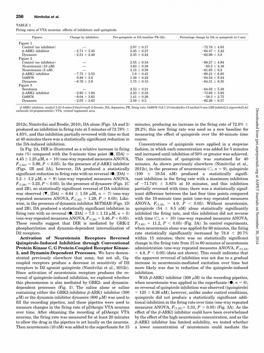
TABLE 1Firing rates of VTA neurons: effects of inhibitors and quinpirole
Figures Change by inhibitors Pre-quinpirole or DA baseline FR (Hz) Percentage change by DA or quinpirole at 5 min
Figure 1Control (no inhibitor) — 2.07 6 0.17 272.78 6 4.83b-ARK1 inhibitor 22.71 6 3.25 2.45 6 0.27 264.47 6 3.42Dynasore 22.51 6 2.49 2.35 6 0.24 262.99 6 3.0
Figure 2Control (no inhibitor) — 2.55 6 0.34 268.27 6 4.94Neurotensin (10 nM) — 3.63 6 0.38 265.5 6 4.16Neurotensin (5 nM) — 3.15 6 0.58 261.65 6 6.2b-ARK1 inhibitor 27.75 6 3.53 1.8 6 0.45 268.21 6 6.85Gö6976 0.88 6 3.0 1.38 6 0.22 264.54 6 6.24Dynasore 26.79 6 3.9 1.75 6 0.15 264.31 6 6.35
Figure 3Serotonin — 2.53 6 0.21 264.02 6 5.38b-ARK1 inhibitor 22.95 6 1.94 2.23 6 0.32 273.68 6 5.93Gö6976 29.04 6 3.63 1.41 6 0.28 258.3 6 2.75Dynasore 22.03 6 2.63 2.59 6 0.3 262.26 6 6.57
b-ARK1 inhibitor, methyl 5-[2-(5-nitro-2-furyl)vinyl]-2-furoate; DA, dopamine; FR, firing rate; Gö6976 (5,6,7,13-tetrahydro-13-methyl-5-oxo-12H-indolo[2,3-a]pyrrolo[3,4c]carbozole-12-propanenitrile); VTA, ventral tegmental area.
256 Nimitvilai et al.
at ASPE
T Journals on N
ovember 30, 2014
jpet.aspetjournals.orgD
ownloaded from

reversal of quinpirole inhibition, and whether the b-ARK1inhibitor would be able to block quinpirole inhibition reversalinduced by this lower neurotensin concentration.Neurotensin (1 and 5 nM) was added to the superfusate for
15 minutes. Then concentrations of quinpirole that hadproduced at least 50% inhibition during the first 5 minuteswere applied for 40 minutes with the continued administra-tion of neurotensin. In the presence of 5 nM neurotensin,quinpirole (60.76 9.2 nM, =, n5 7) inhibited the firing rate by61.65% 6 6.2% at 5 minutes. Unlike the effect of quinpirolealone, quinpirole in the presence of 5 nM neurotensin did notsignificantly produce more inhibition over time; partialreversal of quinpirole inhibition was observed (one-wayrepeated measures ANOVA, F(7,42) 5 1.25, P . 0.05) (Fig.3B). Neurotensin (1 nM) did not mediate quinpirole inhibitionreversal; there was a statistically significant increase ininhibition over the time course of quinpirole administration in
the presence of 1 nM neurotensin (one-way repeated meas-ures, F(7,21) 5 5.03, P , 0.05) (data not shown). Thus, 5 nMneurotensin was used to test whether b-ARK1 inhibitor couldblock neurotensin-induced quinpirole inhibition reversal.With b-ARK1 inhibitor (300 mM) in the recording pipettes
and neurotensin (5 nM) in the superfusate, quinpirole (45.863.7 nM, ., n 5 8) produced a statistically significantinhibition in the firing rate with no reversal (one-wayrepeated measures ANOVA, F(7,49)5 4.2, P , 0.05), and thisinhibition was similar to that produced by quinpirole alone(Fig. 3B). This result suggests that there is a competitive doseeffect in the interaction between neurotensin and b-ARK1inhibitor; greater activation of neurotensin receptors canovercome the interference by b-ARK1 inhibitor with themechanism of reversal of quinpirole inhibition.There is evidence that neurotensin activation of PKC can
phosphorylate D2 receptors in HEK293 cells, resulting in
Fig. 1. Mean ratemeter graphs of the effects of long-duration application of dopamine (DA) in the presence or absence of either G protein-coupledreceptor kinase-2 (GRK2) inhibitor or dynamin inhibitor in single neurons. Vertical bars indicate the firing rate over 5-second intervals; the dashedvertical line indicates the end of DA administration for clarity. Horizontal bars indicate the duration of drug application (concentrations indicated abovebar). (A) DA alone initially produced a decrease in the firing rate, which subsided over time in the continued presence of DA. (B) in the presence of 300 mMb-ARK1 inhibitor in the recording pipette, DA produced an inhibition in the firing rate, and this inhibition did not reverse with time. (C) in the presenceof 800 mMdynasore in the recording pipette, DA produced an inhibition in the firing rate, with no reversal during DA administration. (D) in the presenceof 400 mM MitMAB in the recording pipette, DA produced a statistically significant inhibition in the firing rate over the time course of administration,indicating blockade of reversal.
D2 Agonist Inhibition Reversal Linked to PKC, GRK2, Dynamin 257
at ASPE
T Journals on N
ovember 30, 2014
jpet.aspetjournals.orgD
ownloaded from
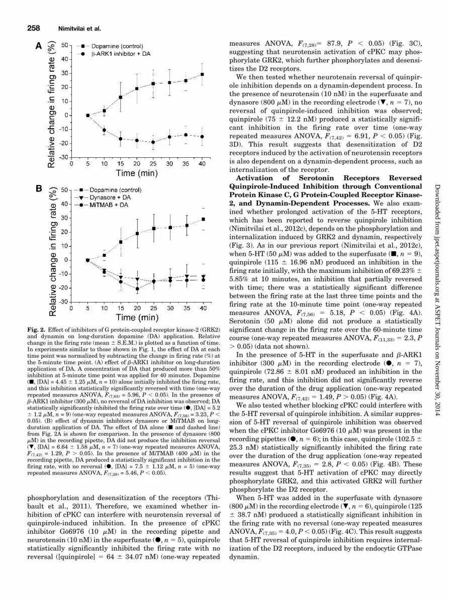
phosphorylation and desensitization of the receptors (Thi-bault et al., 2011). Therefore, we examined whether in-hibition of cPKC can interfere with neurotensin reversal ofquinpirole-induced inhibition. In the presence of cPKCinhibitor Gö6976 (10 mM) in the recording pipette andneurotensin (10 nM) in the superfusate (d, n5 5), quinpirolestatistically significantly inhibited the firing rate with noreversal ([quinpirole] 5 64 6 34.07 nM) (one-way repeated
measures ANOVA, F(7,28)5 87.9, P , 0.05) (Fig. 3C),suggesting that neurotensin activation of cPKC may phos-phorylate GRK2, which further phosphorylates and desensi-tizes the D2 receptors.We then tested whether neurotensin reversal of quinpir-
ole inhibition depends on a dynamin-dependent process. Inthe presence of neurotensin (10 nM) in the superfusate anddynasore (800 mM) in the recording electrode (., n 5 7), noreversal of quinpirole-induced inhibition was observed;quinpirole (75 6 12.2 nM) produced a statistically signifi-cant inhibition in the firing rate over time (one-wayrepeated measures ANOVA, F(7,42) 5 6.91, P , 0.05) (Fig.3D). This result suggests that desensitization of D2receptors induced by the activation of neurotensin receptorsis also dependent on a dynamin-dependent process, such asinternalization of the receptor.Activation of Serotonin Receptors Reversed
Quinpirole-Induced Inhibition through ConventionalProtein Kinase C, G Protein-Coupled Receptor Kinase-2, and Dynamin-Dependent Processes. We also exam-ined whether prolonged activation of the 5-HT receptors,which has been reported to reverse quinpirole inhibition(Nimitvilai et al., 2012c), depends on the phosphorylation andinternalization induced by GRK2 and dynamin, respectively(Fig. 3). As in our previous report (Nimitvilai et al., 2012c),when 5-HT (50 mM) was added to the superfusate (j, n 5 9),quinpirole (115 6 16.96 nM) produced an inhibition in thefiring rate initially, with the maximum inhibition of 69.23%65.85% at 10 minutes, an inhibition that partially reversedwith time; there was a statistically significant differencebetween the firing rate at the last three time points and thefiring rate at the 10-minute time point (one-way repeatedmeasures ANOVA, F(7,56) 5 5.18, P , 0.05) (Fig. 4A).Serotonin (50 mM) alone did not produce a statisticallysignificant change in the firing rate over the 60-minute timecourse (one-way repeated measures ANOVA, F(11,33) 5 2.3, P. 0.05) (data not shown).In the presence of 5-HT in the superfusate and b-ARK1
inhibitor (300 mM) in the recording electrode (d, n 5 7),quinpirole (72.86 6 8.01 nM) produced an inhibition in thefiring rate, and this inhibition did not significantly reverseover the duration of the drug application (one-way repeatedmeasures ANOVA, F(7,42) 5 1.49, P . 0.05) (Fig. 4A).We also tested whether blocking cPKC could interfere with
the 5-HT reversal of quinpirole inhibition. A similar suppres-sion of 5-HT reversal of quinpirole inhibition was observedwhen the cPKC inhibitor Gö6976 (10 mM) was present in therecording pipettes (d, n5 6); in this case, quinpirole (102.5625.3 nM) statistically significantly inhibited the firing rateover the duration of the drug application (one-way repeatedmeasures ANOVA, F(7,35) 5 2.8, P , 0.05) (Fig. 4B). Theseresults suggest that 5-HT activation of cPKC may directlyphosphorylate GRK2, and this activated GRK2 will furtherphosphorylate the D2 receptor.When 5-HT was added in the superfusate with dynasore
(800 mM) in the recording electrode (., n5 6), quinpirole (1256 38.7 nM) produced a statistically significant inhibition inthe firing rate with no reversal (one-way repeated measuresANOVA, F(7,35) 5 4.0, P, 0.05) (Fig. 4C). This result suggeststhat 5-HT reversal of quinpirole inhibition requires internal-ization of the D2 receptors, induced by the endocytic GTPasedynamin.
Fig. 2. Effect of inhibitors of G protein-coupled receptor kinase-2 (GRK2)and dynamin on long-duration dopamine (DA) application. Relativechange in the firing rate (mean 6 S.E.M.) is plotted as a function of time.In experiments similar to those shown in Fig. 1, the effect of DA at eachtime point was normalized by subtracting the change in firing rate (%) atthe 5-minute time point. (A) effect of b-ARK1 inhibitor on long-durationapplication of DA. A concentration of DA that produced more than 50%inhibition at 5-minute time point was applied for 40 minutes. Dopamine(j, [DA] = 4.45 6 1.25 mM, n = 10) alone initially inhibited the firing rate,and this inhibition statistically significantly reversed with time (one-wayrepeated measures ANOVA, F(7,63) = 5.96, P , 0.05). In the presence ofb-ARK1 inhibitor (300 mM), no reversal of DA inhibition was observed; DAstatistically significantly inhibited the firing rate over time (d, [DA] = 5.26 1.2 mM, n = 9) (one-way repeated measures ANOVA, F(7,56) = 3.23, P ,0.05). (B) effect of dynamin inhibitors dynasore or MiTMAB on long-duration application of DA. The effect of DA alone (j and dashed line)from Fig. 2A is shown for comparison. In the presence of dynasore (800mM) in the recording pipette, DA did not produce the inhibition reversal(., [DA] = 6.64 6 1.58 mM, n = 7) (one-way repeated measures ANOVA,F(7,42) = 1.29, P . 0.05). In the presence of MiTMAB (400 mM) in therecording pipette, DA produced a statistically significant inhibition in thefiring rate, with no reversal (d, [DA] = 7.5 6 1.12 mM, n = 5) (one-wayrepeated measures ANOVA, F(7,28) = 5.46, P , 0.05).
258 Nimitvilai et al.
at ASPE
T Journals on N
ovember 30, 2014
jpet.aspetjournals.orgD
ownloaded from
DiscussionWe have previously reported a phenomenon of DIR that is
induced by extended periods of exposure to moderateconcentrations of DA; DIR persists for up to 90 minutes, andrequires the concurrent stimulation of D1/D5 and D2 DAreceptors (Nimitvilai and Brodie, 2010). We have alsodemonstrated that DIR is mediated by PLC and cPKC, and
is dependent on both extracellular calcium influx and in-tracellular calcium release (Nimitvilai et al., 2012a). Inaddition, agonists of either 5-HT2 or neurotensin receptors,both of which are linked to PLC activation, produceda decrease in sensitivity of D2 receptor to the D2 agonistquinpirole (Nimitvilai et al., 2012c), suggesting that thisphenomenon is mediated through the Gq/PLC/PKC pathway.In the present study, we extended our examination of the
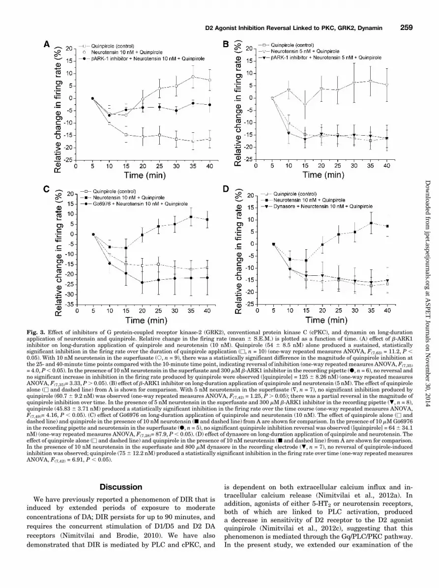
Fig. 3. Effect of inhibitors of G protein-coupled receptor kinase-2 (GRK2), conventional protein kinase C (cPKC), and dynamin on long-durationapplication of neurotensin and quinpirole. Relative change in the firing rate (mean 6 S.E.M.) is plotted as a function of time. (A) effect of b-ARK1inhibitor on long-duration application of quinpirole and neurotensin (10 nM). Quinpirole (54 6 8.5 nM) alone produced a sustained, statisticallysignificant inhibition in the firing rate over the duration of quinpirole application (u, n = 10) (one-way repeated measures ANOVA, F(7,63) = 11.2, P ,0.05). With 10 nM neurotensin in the superfusate (s, n = 9), there was a statistically significant difference in the magnitude of quinpirole inhibition atthe 25- and 40-minute time points compared with the 10-minute time point, indicating reversal of inhibition (one-way repeated measures ANOVA, F(7,35)= 4.0, P, 0.05). In the presence of 10 nM neurotensin in the superfusate and 300 mM b-ARK1 inhibitor in the recording pipette (d, n = 6), no reversal andno significant increase in inhibition in the firing rate produced by quinpirole were observed ([quinpirole] = 125 6 8.26 nM) (one-way repeated measuresANOVA, F(7,35)= 3.33, P. 0.05). (B) effect of b-ARK1 inhibitor on long-duration application of quinpirole and neurotensin (5 nM). The effect of quinpirolealone (u and dashed line) from A is shown for comparison. With 5 nM neurotensin in the superfusate (=, n = 7), no significant inhibition produced byquinpirole (60.76 9.2 nM) was observed (one-way repeated measures ANOVA, F(7,42) = 1.25, P. 0.05); there was a partial reversal in the magnitude ofquinpirole inhibition over time. In the presence of 5 nM neurotensin in the superfusate and 300 mM b-ARK1 inhibitor in the recording pipette (., n = 8),quinpirole (45.83 6 3.71 nM) produced a statistically significant inhibition in the firing rate over the time course (one-way repeated measures ANOVA,F(7,49)= 4.16, P , 0.05). (C) effect of Gö6976 on long-duration application of quinpirole and neurotensin (10 nM). The effect of quinpirole alone (u anddashed line) and quinpirole in the presence of 10 nM neurotensin (j and dashed line) from A are shown for comparison. In the presence of 10 mMGö6976in the recording pipette and neurotensin in the superfusate (d, n = 5), no significant quinpirole inhibition reversal was observed ([quinpirole] = 646 34.1nM) (one-way repeated measures ANOVA, F(7,28)= 87.9, P, 0.05). (D) effect of dynasore on long-duration application of quinpirole and neurotensin. Theeffect of quinpirole alone (u and dashed line) and quinpirole in the presence of 10 nM neurotensin (j and dashed line) from A are shown for comparison.In the presence of 10 nM neurotensin in the superfusate and 800 mM dynasore in the recording electrode (., n = 7), no reversal of quinpirole-inducedinhibition was observed; quinpirole (756 12.2 nM) produced a statistically significant inhibition in the firing rate over time (one-way repeated measuresANOVA, F(7,42) = 6.91, P , 0.05).
D2 Agonist Inhibition Reversal Linked to PKC, GRK2, Dynamin 259
at ASPE
T Journals on N
ovember 30, 2014
jpet.aspetjournals.orgD
ownloaded from

reversal of DA or quinpirole inhibition to show that enzymesinvolved in the phosphorylation of D2 receptors are required;phosphorylation as a result of DA, 5-HT, or neurotensinreceptor stimulation is mediated by GRK2, and internaliza-tion is mediated by the endocytic GTPase dynamin. Of course,we are basing our interpretation on our pharmacologicresults, but biochemical studies would be needed to confirmthis interpretation of our findings. As we did not specificallyexamine the phosphorylation of D2 receptors, we cannot becertain that it is indeed the D2 receptor that is thephosphorylation target.Functional efficiencies of G protein-coupled receptors
(GPCRs) are not static; rather, they are dynamic anddependent on a memory of prior receptor activation (Haus-dorff et al., 1990). Prolonged or repeated activation ofa receptor results in a reduced response to a subsequentreceptor stimulation, a process called desensitization. De-sensitization can be homologous or heterologous; homologousdesensitization is due to a decrease in response of a receptoras a result of binding its agonist, whereas heterologousdesensitization is due to a decrease in response to a receptoras a result of agonist binding to a different receptor. We foundthat DIR of DAergic VTA neurons is neither homologous norheterologous desensitization because it requires the concur-rent stimulation of D2 and D1/D5 receptors (Nimitvilai andBrodie, 2010). To our knowledge, the D2 receptor is unique inthat no other G protein-coupled receptor requires activation oftwo receptors to stimulate the processes necessary fordesensitization of that receptor.G protein-receptor kinases (GRKs) and b-arrestins are the
two major cytoplasmic components responsible for desensitiz-ing GPCR signaling (Premont et al., 1995; Sterne-Marr andBenovic, 1995). Binding of a ligand as well as the release of Gaand Gbg induce the conformational change of the GPCR,resulting in a recruitment of GRK to the serine/threoninephosphorylation site of the intracellular loops or the COOHterminus of the receptor (Pitcher et al., 1998). GRK phos-phorylation of the GPCR increases the affinity of that GPCRfor b-arrestin. Once b-arrestin binds to GPCR, it prevents thereformation of functional GPCR so that a ligand cannot bindand activate the receptor. The b-arrestin also recruits clathrinand the adaptor protein AP2 to the phosphorylated GPCR,resulting in the formation of clathrin-coated vesicle. DynaminGTPase then pinches off the clathrin-coated pit from the cell
Fig. 4. Effect of inhibitors of G protein-coupled receptor kinase-2 (GRK2),conventional protein kinase C (cPKC), and dynamin on long-durationapplication of serotonin (5-HT) and quinpirole. Relative change in thefiring rate (mean 6 S.E.M.) is plotted as a function of time. (A) effect ofb-ARK1 inhibitor on long-duration application of quinpirole and 5-HT.The effect of quinpirole alone (u and dashed line) from Fig. 3A is shown forcomparison. With 50 mM 5-HT in the superfusate (j, n = 9), there wasa statistically significant difference in the magnitude of quinpiroleinhibition at the 30- and 40-minute time points compared with the 10-minute time point, indicating reversal of inhibition. In the presence of 50mM 5-HT in the superfusate and 300 mM b-ARK1 inhibitor in the
recording pipette (d, n = 7), no reversal and no significant inhibition in thefiring rate produced by quinpirole were observed ([quinpirole] = 72.96 8.0nM) (one-way repeated measures ANOVA, F(7,42)= 1.49, P . 0.05). (B)effect of Gö6976 on long-duration application of quinpirole and serotonin.The effect of quinpirole alone (u and dashed line) from Fig. 3A andquinpirole in the presence of 50 mM 5-HT (j and dashed line) from A areshown for comparison. In the presence of 10 mM Gö6976 in the recordingpipette and 5-HT in the superfusate (d, n = 6), no significant quinpiroleinhibition reversal was observed ([quinpirole] = 102.56 25.3 nM) (one-wayrepeated measures ANOVA, F(7,35)= 92.8, P , 0.05). (C) effect of dynasoreon long-duration application of quinpirole and 5-HT. The effect ofquinpirole alone (u and dashed line) from Fig. 3A and quinpirole in thepresence of 5-HT (j and dashed line) from A are shown for comparison. Inthe presence of 50 mM5-HT in the superfusate and 800 mMdynasore in therecording electrode (., n = 6), no reversal of quinpirole-induced inhibitionwas observed; quinpirole (125 6 38.7 nM) produced a statisticallysignificant inhibition in the firing rate over time (one-way repeatedmeasures ANOVA, F(7,35) = 4.0, P , 0.05).
260 Nimitvilai et al.
at ASPE
T Journals on N
ovember 30, 2014
jpet.aspetjournals.orgD
ownloaded from
surface, internalizing the receptor (McMahon and Boucrot,2011).Dopamine receptors, like other GPCRs, can be regulated in
a number of ways. Dopamine receptors contain phosphoryla-tion sites for GRK, PKC, and Ca21/calmodulin-dependentprotein kinase II (CaMKII) on their third intracellular loopand their C-terminal region (Namkung and Sibley, 2004;Bofill-Cardona et al., 2000). Phosphorylation and desensiti-zation of D2 receptors by PKC and CaMKII has also beenstudied in HEK293 cells, and striatal and hippocampalneurons (Rogue et al., 1990; Bofill-Cardona et al., 2000;Namkung and Sibley, 2004; Thibault et al., 2011). Secondmessenger-dependent protein kinases can either directlyphosphorylate and desensitize the GPCR (Bofill-Cardonaet al., 2000; Namkung and Sibley, 2004) or phosphorylateGRKs (Pronin and Benovic, 1997; Pronin et al., 1997; Chuanget al., 1996; Chuang et al., 1995; Winstel et al., 1996). Second-messenger protein kinase phosphorylation of GRK can eitheractivate or inhibit GRK activity. For example, PKC phos-phorylation can activate GRK2 (Chuang et al., 1995; Winstelet al., 1996), but inhibit GRK5 (Pronin and Benovic, 1997)activity in b-adrenergic receptors. Calmodulin inhibits GRKactivity with a higher specificity of GRK5 (IC50 ∼50 nM) overGRK2 (IC50 ∼2 mM) (Chuang et al., 1996; Pronin et al.,1997). Phosphorylation and desensitization of DA D2receptors by GRK2 and GRK5 have been reported (Itoet al., 1999).At present, seven subtypes of GRKs (GRK1–7) have been
identified. GRK1 and GRK7 are expressed in the photorecep-tor rhodopsin, and GRK4 is found predominantly in the malegerm line. GRK2, 3, and 6 are widely distributed in the ratbrain, in which GRK2 is present in high levels in most brainareas and is also expressed in VTA neurons (Erdtmann-Vourliotis et al., 2001). In this study, we demonstrated thatdesensitization of the D2 receptor induced by DA, 5-HT, orneurotensin was not observed when GRK2 was suppressed,suggesting that GRK2 is involved in the desensitization of D2receptors induced by DA, 5-HT, or neurotensin. We only used
one antagonist (b-ARK1 inhibitor) to interfere with GRK2, asit is somewhat unique in having properties suitable for usewith the methods used in other experiments in this study;more complex studies using other methods (e.g., siRNA orknockout of GRK2) would be needed to more definitivelyestablish the importance of GRK2 in the desensitizationmechanism. As cPKC is required to produce D2 desensitiza-tion (Nimitvilai et al., 2012a) and our present study showsthat blocking of cPKC by Gö6976 also inhibited serotonin- orneurotensin-induced reversal of quinpirole inhibition, it ispossible that activation of cPKC by either DA, 5-HT, orneurotensin results in its binding to and phosphorylation ofGRK2.Of the seven subtypes of serotonin receptors, 5-HT2 is the G
protein-coupled receptor that is linked to Gq, and 5-HT2
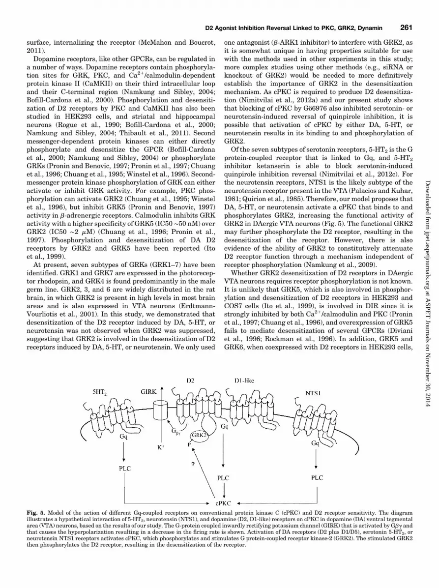
inhibitor ketanserin is able to block serotonin-inducedquinpirole inhibition reversal (Nimitvilai et al., 2012c). Forthe neurotensin receptors, NTS1 is the likely subtype of theneurotensin receptor present in the VTA (Palacios and Kuhar,1981; Quirion et al., 1985). Therefore, our model proposes thatDA, 5-HT, or neurotensin activate a cPKC that binds to andphosphorylates GRK2, increasing the functional activity ofGRK2 in DAergic VTA neurons (Fig. 5). The functional GRK2may further phosphorylate the D2 receptor, resulting in thedesensitization of the receptor. However, there is alsoevidence of the ability of GRK2 to constitutively attenuateD2 receptor function through a mechanism independent ofreceptor phosphorylation (Namkung et al., 2009).Whether GRK2 desensitization of D2 receptors in DAergic
VTA neurons requires receptor phosphorylation is not known.It is unlikely that GRK5, which is also involved in phosphor-ylation and desensitization of D2 receptors in HEK293 andCOS7 cells (Ito et al., 1999), is involved in DIR since it isstrongly inhibited by both Ca21/calmodulin and PKC (Proninet al., 1997; Chuang et al., 1996), and overexpression of GRK5fails to mediate desensitization of several GPCRs (Divianiet al., 1996; Rockman et al., 1996). In addition, GRK5 andGRK6, when coexpressed with D2 receptors in HEK293 cells,
Fig. 5. Model of the action of different Gq-coupled receptors on conventional protein kinase C (cPKC) and D2 receptor sensitivity. The diagramillustrates a hypothetical interaction of 5-HT2, neurotensin (NTS1), and dopamine (D2, D1-like) receptors on cPKC in dopamine (DA) ventral tegmentalarea (VTA) neurons, based on the results of our study. The G-protein coupled inwardly rectifying potassium channel (GIRK) that is activated by Gbg andthat causes the hyperpolarization resulting in a decrease in the firing rate is shown. Activation of DA receptors (D2 plus D1/D5), serotonin 5-HT2, orneurotensin NTS1 receptors activates cPKC, which phosphorylates and stimulates G protein-coupled receptor kinase-2 (GRK2). The stimulated GRK2then phosphorylates the D2 receptor, resulting in the desensitization of the receptor.
D2 Agonist Inhibition Reversal Linked to PKC, GRK2, Dynamin 261
at ASPE
T Journals on N
ovember 30, 2014
jpet.aspetjournals.orgD
ownloaded from
have no impact on agonist-induced D2 signaling (Namkunget al., 2009). Other possibilities, such as whether GRK3 orother proteins sensitive to b-ARK1 inhibitor are involved inthe desensitization of the D2 receptor in DAergic VTAneurons, will be a subject for future study.We also demonstrated that inhibition of dynamin GTPase
suppressed DIR and the quinpirole inhibition reversal pro-duced by either serotonin or neurotensin. These resultssuggest that once the D2 receptor has been desensitized, itwill be internalized into the endosome. The endocytosedreceptor may be dephosphorylated before returning to the cellsurface, or it may be degraded to lysosomes. We have shownpreviously that the effect of DIR is long lasting, persisting forup to 90 minutes after washout of DA (Nimitvilai and Brodie,2010). However, whether the reoccurrence of inhibition in thefiring rate produced by D2 agonist is the result of the recyclingof D2 receptors that had been initially desensitized or is theresult of activation of newly synthesized D2 receptors is notknown.Exposure to drugs of abuse causes a sustained increase in
DAergic neurotransmission in the reward system; desensiti-zation of the inhibitory D2 autoreceptors in the VTA neuronsthrough the PI/PLC/cPKC pathway may increase the excit-ability of these neurons. The present study further defines themechanism of that desensitization induced by DA, 5-HT, orneurotensin, with the involvement of phosphorylation andinternalization of D2 receptors. The action of drugs of abuseon DAergic VTA neurons to reduce D2 autoreceptor in-hibition, resulting in an increase in DAergic neurotransmis-sion in the reward/reinforcement system, may be a key eventin the development of addiction. Understanding molecularmechanisms underlying the reversal of DA inhibition in theVTA may contribute to medication discovery for moreeffective treatment of addiction disorders.
Authorship contributions
Participated in research design: Nimitvilai, Brodie.Conducted experiments: Nimitvilai, McElvain.Performed data analysis: Nimitvilai, Brodie.Wrote or contributed to the writing of the manuscript: Nimitvilai,
Brodie.
References
Bofill-Cardona E, Kudlacek O, Yang Q, Ahorn H, Freissmuth M, and Nanoff C (2000)Binding of calmodulin to the D2-dopamine receptor reduces receptor signaling byarresting the G protein activation switch. J Biol Chem 275:32672–32680.
Bouthenet ML, Souil E, Martres MP, Sokoloff P, Giros B, and Schwartz JC (1991)Localization of dopamine D3 receptor mRNA in the rat brain using in situ hy-bridization histochemistry: comparison with dopamine D2 receptor mRNA. BrainRes 564:203–219.
Brodie MS and Dunwiddie TV (1987) Cholecystokinin potentiates dopamine in-hibition of mesencephalic dopamine neurons in vitro. Brain Res 425:106–113.
Brodie MS, McElvain MA, Bunney EB, and Appel SB (1999a) Pharmacological re-duction of small conductance calcium-activated potassium current (SK) potentiatesthe excitatory effect of ethanol on ventral tegmental area dopamine neurons.J Pharmacol Exp Ther 290:325–333.
Brodie MS, Pesold C, and Appel SB (1999b) Ethanol directly excites dopaminergicventral tegmental area reward neurons. Alcohol Clin Exp Res 23:1848–1852.
Brodie MS, Shefner SA, and Dunwiddie TV (1990) Ethanol increases the firing rate ofdopamine neurons of the rat ventral tegmental area in vitro. Brain Res 508:65–69.
Bunney BS, Walters JR, Roth RH, and Aghajanian GK (1973) Dopaminergic neurons:effect of antipsychotic drugs and amphetamine on single cell activity. J PharmacolExp Ther 185:560–571.
Caillé I, Dumartin B, and Bloch B (1996) Ultrastructural localization of D1 dopaminereceptor immunoreactivity in rat striatonigral neurons and its relation with do-paminergic innervation. Brain Res 730:17–31.
Chieng B, Azriel Y, Mohammadi S, and Christie MJ (2011) Distinct cellular prop-erties of identified dopaminergic and GABAergic neurons in the mouse ventraltegmental area. J Physiol 589:3775–3787.
Chuang TT, LeVine H, 3rd, and De Blasi A (1995) Phosphorylation and activation ofbeta-adrenergic receptor kinase by protein kinase C. J Biol Chem 270:18660–18665.
Chuang TT, Paolucci L, and De Blasi A (1996) Inhibition of G protein-coupled re-ceptor kinase subtypes by Ca21/calmodulin. J Biol Chem 271:28691–28696.
Ciliax BJ, Nash N, Heilman C, Sunahara R, Hartney A, Tiberi M, Rye DB, CaronMG, Niznik HB, and Levey AI (2000) Dopamine D(5) receptor immunolocalizationin rat and monkey brain. Synapse 37:125–145.
Di Chiara G and Imperato A (1988) Drugs abused by humans preferentially increasesynaptic dopamine concentrations in the mesolimbic system of freely moving rats.Proc Natl Acad Sci USA 85:5274–5278.
Diaz J, Lévesque D, Lammers CH, Griffon N, Martres MP, Schwartz JC, and SokoloffP (1995) Phenotypical characterization of neurons expressing the dopamine D3receptor in the rat brain. Neuroscience 65:731–745.
Diviani D, Lattion AL, Larbi N, Kunapuli P, Pronin A, Benovic JL, and Cotecchia S(1996) Effect of different G protein-coupled receptor kinases on phosphorylation anddesensitization of the alpha1B-adrenergic receptor. J Biol Chem 271:5049–5058.
Erdtmann-Vourliotis M, Mayer P, Ammon S, Riechert U, and Höllt V (2001) Distri-bution of G-protein-coupled receptor kinase (GRK) isoforms 2, 3, 5 and 6 mRNA inthe rat brain. Brain Res Mol Brain Res 95:129–137.
Gao C and Wolf ME (2008) Dopamine receptors regulate NMDA receptor surfaceexpression in prefrontal cortex neurons. J Neurochem 106:2489–2501.
Grace AA and Bunney BS (1984) The control of firing pattern in nigral dopamineneurons: single spike firing. J Neurosci 4:2866–2876.
Gurevich EV and Joyce JN (1999) Distribution of dopamine D3 receptor expressingneurons in the human forebrain: comparison with D2 receptor expressing neurons.Neuropsychopharmacology 20:60–80.
Hausdorff WP, Caron MG, and Lefkowitz RJ (1990) Turning off the signal: de-sensitization of beta-adrenergic receptor function. FASEB J 4:2881–2889.
Iino M, Furugori T, Mori T, Moriyama S, Fukuzawa A, and Shibano T (2002) Rationaldesign and evaluation of new lead compound structures for selective betaARK1inhibitors. J Med Chem 45:2150–2159.
Ito K, Haga T, Lameh J, and Sadée W (1999) Sequestration of dopamine D2 receptorsdepends on coexpression of G-protein-coupled receptor kinases 2 or 5. Eur J Bio-chem 260:112–119.
Iwata K, Ito K, Fukuzaki A, Inaki K, and Haga T (1999) Dynamin and rab5regulate GRK2-dependent internalization of dopamine D2 receptors. Eur J Biochem 263:596–602.
Kenakin TP (1987) Analysis of dose-response data, in Pharmacologic Analysis ofDrug-Receptor Interaction, pp 129–162, Raven Press, New York.
Khan ZU, Gutiérrez A, Martín R, Peñafiel A, Rivera A, and de la Calle A (2000)Dopamine D5 receptors of rat and human brain. Neuroscience 100:689–699.
Koob GF (2003) Alcoholism: allostasis and beyond. Alcohol Clin Exp Res 27:232–243.Lacey MG, Mercuri NB, and North RA (1987) Dopamine acts on D2 receptors toincrease potassium conductance in neurones of the rat substantia nigra zonacompacta. J Physiol 392:397–416.
Lacey MG, Mercuri NB, and North RA (1989) Two cell types in rat substantia nigrazona compacta distinguished by membrane properties and the actions of dopamineand opioids. J Neurosci 9:1233–1241.
Lammel S, Hetzel A, Häckel O, Jones I, Liss B, and Roeper J (2008) Unique prop-erties of mesoprefrontal neurons within a dual mesocorticolimbic dopamine sys-tem. Neuron 57:760–773.
Macia E, Ehrlich M, Massol R, Boucrot E, Brunner C, and Kirchhausen T (2006)Dynasore, a cell-permeable inhibitor of dynamin. Dev Cell 10:839–850.
Margolis EB, Lock H, Hjelmstad GO, and Fields HL (2006) The ventral tegmentalarea revisited: is there an electrophysiological marker for dopaminergic neurons?J Physiol 577:907–924.
Margolis EB, Mitchell JM, Ishikawa J, Hjelmstad GO, and Fields HL (2008) Mid-brain dopamine neurons: projection target determines action potential durationand dopamine D(2) receptor inhibition. J Neurosci 28:8908–8913.
McMahon HT and Boucrot E (2011) Molecular mechanism and physiological func-tions of clathrin-mediated endocytosis. Nat Rev Mol Cell Biol 12:517–533.
Meador-Woodruff JH, Mansour A, Grandy DK, Damask SP, Civelli O, and WatsonSJ, Jr (1992) Distribution of D5 dopamine receptor mRNA in rat brain. NeurosciLett 145:209–212.
Mengod G, Villaró MT, Landwehrmeyer GB, Martinez-Mir MI, Niznik HB, SunaharaRK, Seeman P, O’Dowd BF, Probst A, and Palacios JM (1992) Visualization ofdopamine D1, D2 and D3 receptor mRNAs in human and rat brain. Neurochem Int20 (Suppl):33S–43S.
Mirenowicz J and Schultz W (1996) Preferential activation of midbrain dopamineneurons by appetitive rather than aversive stimuli. Nature 379:449–451.
Mueller AL and Brodie MS (1989) Intracellular recording from putative dopamine-containing neurons in the ventral tegmental area of Tsai in a brain slice prepa-ration. J Neurosci Methods 28:15–22.
Namkung Y, Dipace C, Urizar E, Javitch JA, and Sibley DR (2009) G protein-coupledreceptor kinase-2 constitutively regulates D2 dopamine receptor expression andsignaling independently of receptor phosphorylation. J Biol Chem 284:34103–34115.
Namkung Y and Sibley DR (2004) Protein kinase C mediates phosphorylation, de-sensitization, and trafficking of the D2 dopamine receptor. J Biol Chem 279:49533–49541.
Neve KA, Seamans JK, and Trantham-Davidson H (2004) Dopamine receptor sig-naling. J Recept Signal Transduct Res 24:165–205.
Nimitvilai S, Arora DS, and Brodie MS (2012a) Reversal of dopamine inhibition ofdopaminergic neurons of the ventral tegmental area is mediated by protein kinaseC. Neuropsychopharmacology 37:543–556.
Nimitvilai S, Arora DS, and Brodie MS (2012b) Reversal of dopamine inhibition ofdopaminergic neurons of the ventral tegmental area is mediated by protein kinaseC. Neuropsychopharmacology 37:543–556.
Nimitvilai S and Brodie MS (2010) Reversal of prolonged dopamine inhibition of dopa-minergic neurons of the ventral tegmental area. J Pharmacol Exp Ther 333:555–563.
Nimitvilai S, McElvain MA, Arora DS, and Brodie MS (2012c) Reversal ofquinpirole inhibition of ventral tegmental area neurons is linked to the
262 Nimitvilai et al.
at ASPE
T Journals on N
ovember 30, 2014
jpet.aspetjournals.orgD
ownloaded from
phosphatidylinositol system and is induced by agonists linked to G(q). J Neuro-physiol 108:263–274.
Oades RD and Halliday GM (1987) Ventral tegmental (A10) system: neurobiology. 1.Anatomy and connectivity. Brain Res 434:117–165.
Palacios JM and Kuhar MJ (1981) Neurotensin receptors are located on dopamine-containing neurones in rat midbrain. Nature 294:587–589.
Pesavento E, Margotti E, Righi M, Cattaneo A, and Domenici L (2000) Blocking theNGF-TrkA interaction rescues the developmental loss of LTP in the rat visualcortex: role of the cholinergic system. Neuron 25:165–175.
Pitcher JA, Freedman NJ, and Lefkowitz RJ (1998) G protein-coupled receptorkinases. Annu Rev Biochem 67:653–692.
Premont RT, Inglese J, and Lefkowitz RJ (1995) Protein kinases that phosphorylateactivated G protein-coupled receptors. FASEB J 9:175–182.
Pronin AN and Benovic JL (1997) Regulation of the G protein-coupled receptor ki-nase GRK5 by protein kinase C. J Biol Chem 272:3806–3812.
Pronin AN, Satpaev DK, Slepak VZ, and Benovic JL (1997) Regulation of G protein-coupled receptor kinases by calmodulin and localization of the calmodulin bindingdomain. J Biol Chem 272:18273–18280.
Quan A, McGeachie AB, and Keating DJ, et al. (2007) Myristyl trimethyl ammoniumbromide and octadecyl trimethyl ammonium bromide are surface-active smallmolecule dynamin inhibitors that block endocytosis mediated by dynamin I ordynamin II. Mol Pharmacol 72:1425–1439.
Quirion R, Chiueh CC, Everist HD, and Pert A (1985) Comparative localization ofneurotensin receptors on nigrostriatal and mesolimbic dopaminergic terminals.Brain Res 327:385–389.
Rivera A, Peñafiel A, Megías M, Agnati LF, López-Téllez JF, Gago B, Gutiérrez A, de laCalle A, and Fuxe K (2008) Cellular localization and distribution of dopamine D(4)receptors in the rat cerebral cortex and their relationship with the cortical dopami-nergic and noradrenergic nerve terminal networks. Neuroscience 155:997–1010.
Rockman HA, Choi DJ, Rahman NU, Akhter SA, Lefkowitz RJ, and Koch WJ (1996)Receptor-specific in vivo desensitization by the G protein-coupled receptor kinase-5in transgenic mice. Proc Natl Acad Sci USA 93:9954–9959.
Rogue P, Zwiller J, Malviya AN, and Vincendon G (1990) Phosphorylation by proteinkinase C modulates agonist binding to striatal dopamine D2 receptors. Biochem Int22:575–582.
Sesack SR, Aoki C, and Pickel VM (1994) Ultrastructural localization of D2 receptor-like immunoreactivity in midbrain dopamine neurons and their striatal targets.J Neurosci 14:88–106.
Sibley DR, Monsma FJ, Jr, and Shen Y (1993) Molecular neurobiology of dopami-nergic receptors. Int Rev Neurobiol 35:391–415.
Sterne-Marr R and Benovic JL (1995) Regulation of G protein-coupled receptors byreceptor kinases and arrestins. Vitam Horm 51:193–234.
Sun X, Milovanovic M, Zhao Y, and Wolf ME (2008) Acute and chronic dopaminereceptor stimulation modulates AMPA receptor trafficking in nucleus accumbensneurons cocultured with prefrontal cortex neurons. J Neurosci 28:4216–4230.
Thibault D, Albert PR, Pineyro G, and Trudeau LÉ (2011) Neurotensin triggersdopamine D2 receptor desensitization through a protein kinase C and beta-arrestin1-dependent mechanism. J Biol Chem 286:9174–9184.
Winstel R, Freund S, Krasel C, Hoppe E, and Lohse MJ (1996) Protein kinase cross-talk: membrane targeting of the beta-adrenergic receptor kinase by protein kinaseC. Proc Natl Acad Sci USA 93:2105–2109.
Wise RA (1996) Neurobiology of addiction. Curr Opin Neurobiol 6:243–251.
Address correspondence to: Mark S. Brodie, Department of Physiology andBiophysics, University of Illinois at Chicago, 835 S. Wolcott, Room E-202, M/C901, Chicago, IL 60612-7342. E-mail: [email protected]
D2 Agonist Inhibition Reversal Linked to PKC, GRK2, Dynamin 263
at ASPE
T Journals on N
ovember 30, 2014
jpet.aspetjournals.orgD
ownloaded from