resume oral medicine
TRANSCRIPT
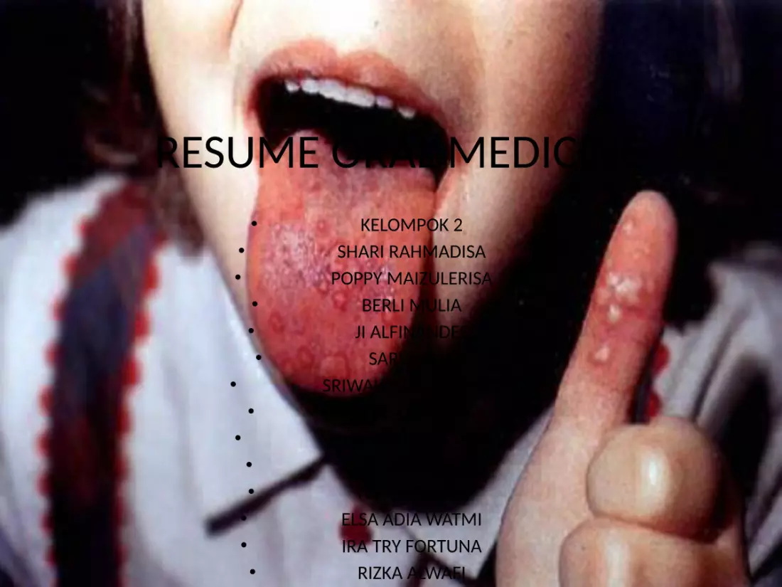
RESUME ORAL MEDICINE• KELOMPOK 2• SHARI RAHMADISA• POPPY MAIZULERISA• BERLI MULIA• JI ALFINANDES• SARI AULIA
• SRIWAHYUNI RITONGA• FITRI ELLANDA• NIKA PERMATA DELA• ARDIAN BISTOK• YERI AMRILLIA• ELSA ADIA WATMI• IRA TRY FORTUNA• RIZKA ALWAFI

Oral ulcer
Trauma
infection
idhiopatic
drug
Systemic disease
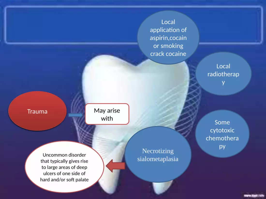
Trauma May arise with
Local application of aspirin,cocain
or smoking crack cocaine
Local radiotherap
y
Some cytotoxic
chemotherapy
Necrotizing sialometaplasia
Uncommon disorder that typically gives rise to large areas of deep ulcers of one side of
hard and/or soft palate
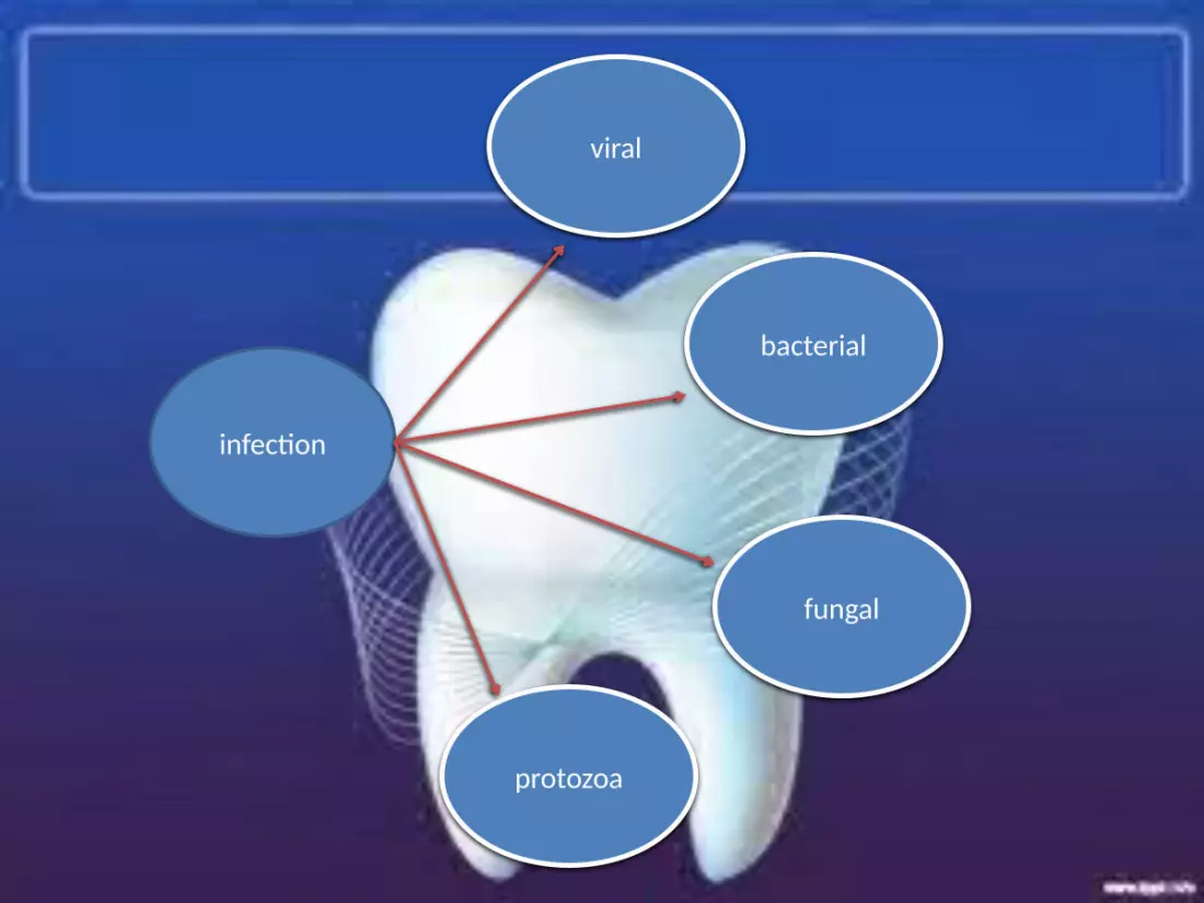
infection
viral
bacterial
fungal
protozoa

viral
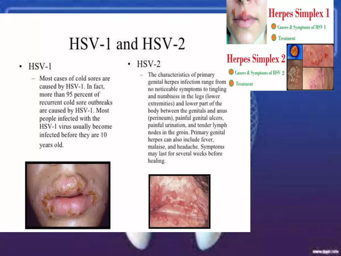
HSV 1
•Widespread,small,superficial ulcers of oral mucosa, the ginggiva are often swollen and ulcerated, giving rise to features akin to ANUG.•5% of patients who have pimary HSV-1 will develop reccurent episodes of herpes labialis.•While regarded as a disease of chilhood
HSV 2
The Oral ulcers can arises as a consequence of congenital
transmission of the causative virus.

•Rare•Feature of infection mononucleoisis•The ulcers comprises a few small superficial ulcers of the oral mucosa.
Epstein-barr virus
cytomegalovirus
•large•Chronic ulcers of the oral mucosa or ginggiva
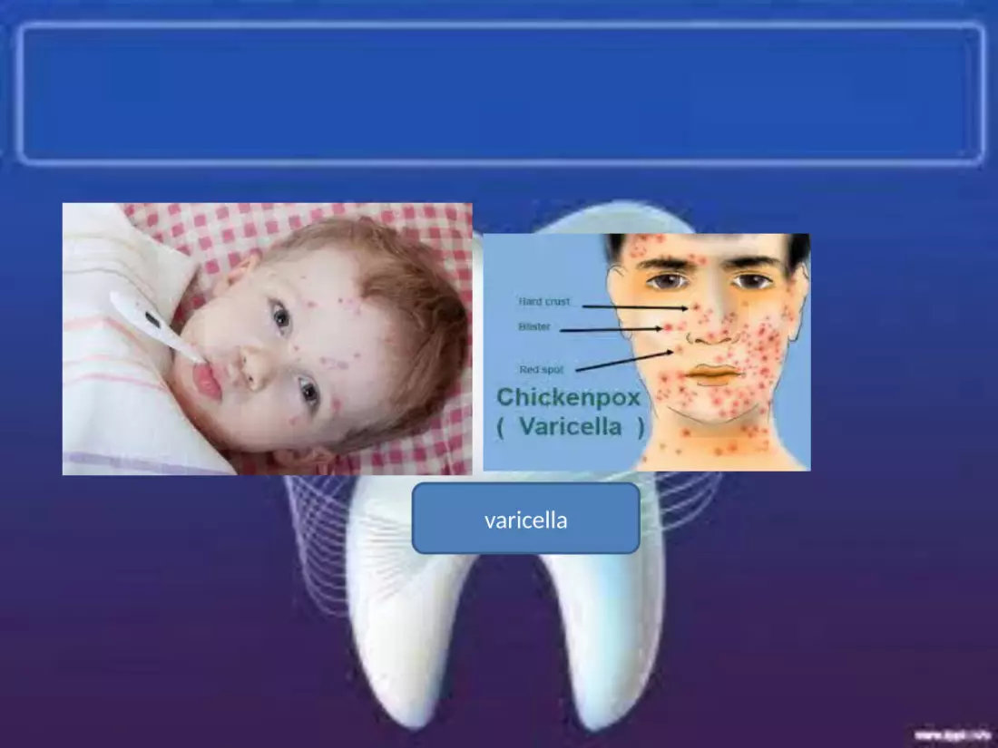
varicella
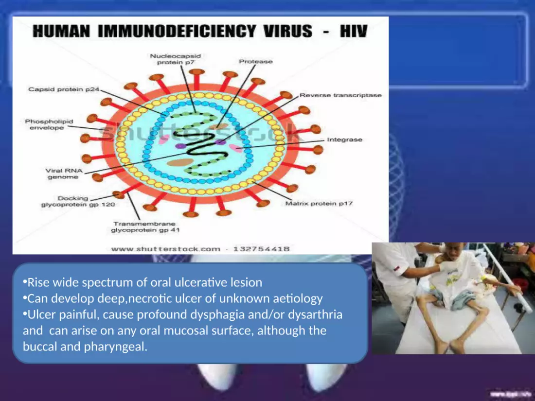
•Rise wide spectrum of oral ulcerative lesion•Can develop deep,necrotic ulcer of unknown aetiology•Ulcer painful, cause profound dysphagia and/or dysarthria and can arise on any oral mucosal surface, although the buccal and pharyngeal.

Human herpesvirus 8
•The cause of sarkoma kaposi•Lession commonly arrising within the mouth of patient HIV or a feature of profound iatrogenic immunosuppresion.•Oral KS affect the palate or ginggiva and manifest as red, blue or purple macules,papules, nodules or ulcers•KS of the anterior ginggiva may be unsightly•Rarely ginggival lesion will cause destruction of underlying periodontal tissues leading to loss of teeth and seuetration of bone.

bacterial
•Nonspecific ulcer•Associated contributing factors include poorly controlled DM, tobacco smoking, immunodefeciency and physicollogical stress.•Painful ulcer of the ginggival margins•Particulaarly the interdental areas•Ulcer may be localized or generalized and when severe will give rise to cervical lymphadenopathy•Very rarely pyrexia and malaise•Reccurent disease may lead destruction and loss of interdental pappilae
ANUG
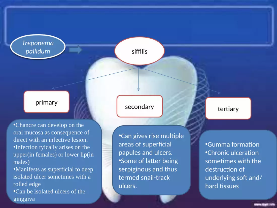
Treponema pallidum siffilis
primary secondary tertiary
•Chancre can develop on the oral mucosa as consequence of direct with an infective lesion.•Infection tyically arises on the upper(in females) or lower lip(in males)•Manifests as superficial to deep isolated ulcer sometimes with a rolled edge•Can be isolated ulcers of the ginggiva
•Can gives rise multiple areas of superficial papules and ulcers.•Some of latter being serpiginous and thus termed snail-track ulcers.
•Gumma formation•Chronic ulceration sometimes with the destruction of underlying soft and/ hard tissues
Mycobacterial infection
•Primary infection rarely•Tubercolossis infection of the oral mucosa arises secondary to pulmonary disease•Necrotic ulcers of the tongue•Infection by atypical mycobacteria is rare but may affect the oral mucosa or ginggiva ussually in HIV infected individuals.
•Usually candida albicans is the most fungal infection of the mouth•Rarely gives rise to oral ulcers.•CMC may give rise to ulcers of the dorsum of tongue
Fungal infection
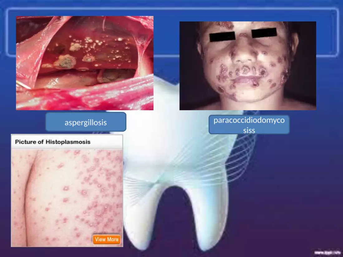
aspergillosis paracoccidiodomycosiss
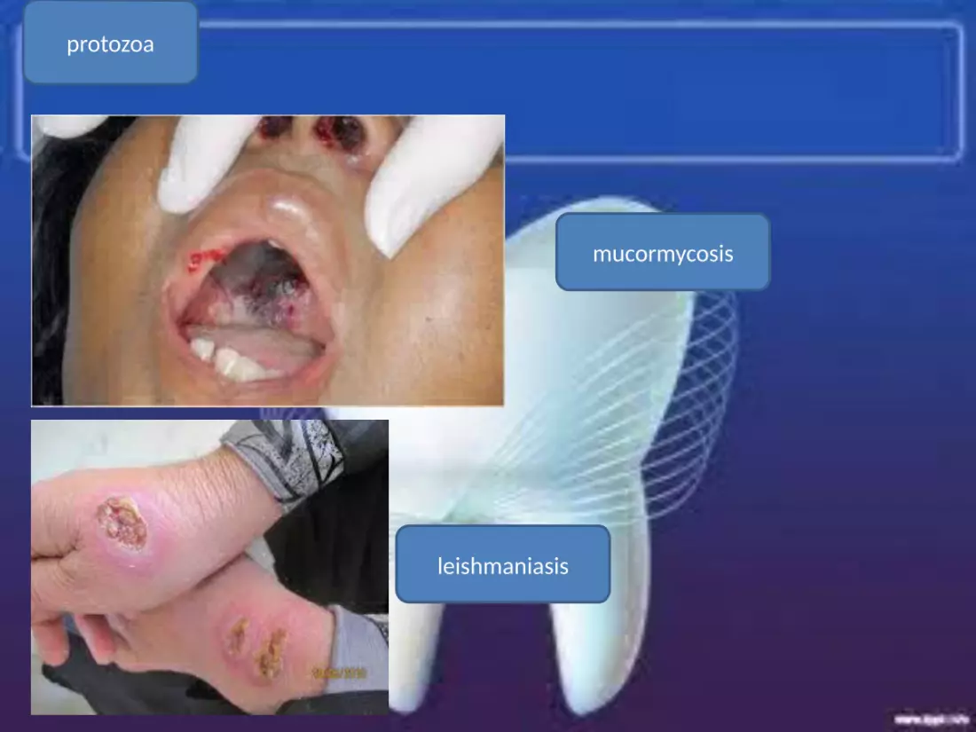
protozoa
mucormycosis
leishmaniasis
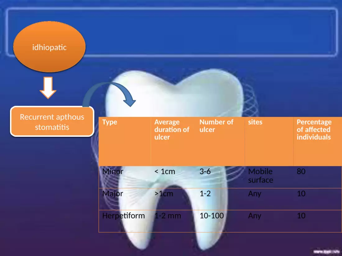
idhiopatic
Recurrent apthous stomatitis
Type Average duration of ulcer
Number of ulcer
sites Percentage of affected individuals
Minor < 1cm 3-6 Mobile surface
80
Major >1cm 1-2 Any 10
Herpetiform 1-2 mm 10-100 Any 10
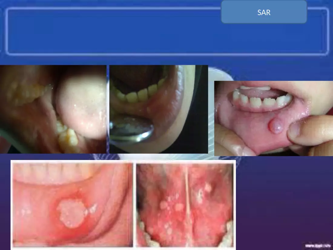
SAR
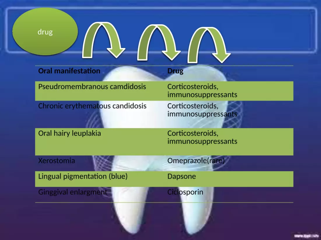
drug
Oral manifestation Drug
Pseudromembranous camdidosis Corticosteroids, immunosuppressants
Chronic erythematous candidosis Corticosteroids, immunosuppressants
Oral hairy leuplakia Corticosteroids, immunosuppressants
Xerostomia Omeprazole(rare)
Lingual pigmentation (blue) Dapsone
Ginggival enlargment Ciclosporin
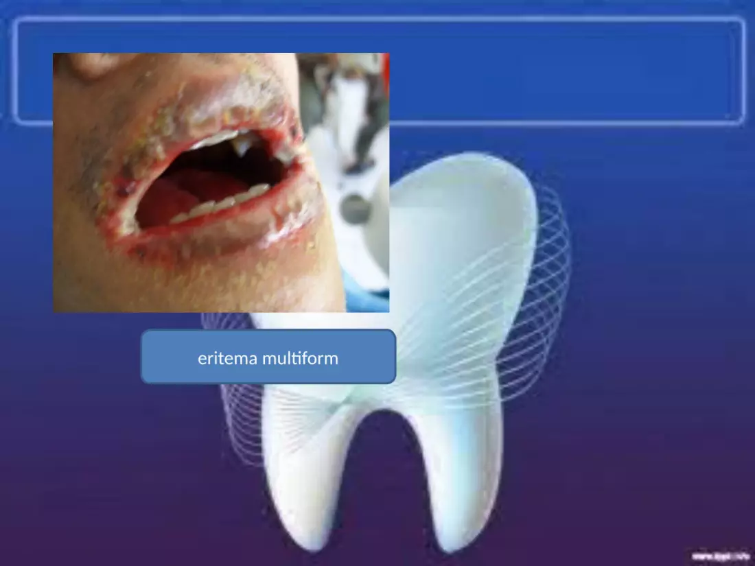
eritema multiform

Haematological AnemiasLymphoprolicerative diseaseLeukimiasNon-hodgkin’s lymphoma(almost all)Hodgkin’s lymphoma(rare)Myeloproliferative diseaseneutropenia
gastroenterological Gluten-sensitve enterophatyChron’s disease and related disoredersUlcerative colitisDermatitis herpetiformis
dermatological Lichen planusdPemphigus-usually vulgarisPemphigoidLinear IgAEpidermylosis bullosaothers
immunological Wegener’s granulomatosisSarcoidosisimmunodeficiency
malignancy Oral squamosa cell carcinomanon=-hodgkin’s lymphomaKaposi sarkomaSalivary gland malignancyMetastatic deposits
Drug induced Lichenoid drug reaction(NSAIDs, interferon,etc)Erythema multiforme(carbamazepine, sulphonamide, etc)Pemphigus( clonidine, psoralens)Drug induced neutropenia/anemia(carbamazepin)Drug induced mukositis( methotrexate)others
Systemic disease

Gastrointestinal disorder Oral manifestations
Bulimia nervosa Necrotizing sialometaplasiaSuperficial oral ulcersDental erosionBilateral parotid enlargment
Post-cricoid webbing Chronic mucocutaneouscandidosis
Gastro-ocsophagcal reflux disease Dental erosion
Gluten-sensitive enteropathy Superficial ulcersEnamell hypoplasia inchildren
Dermatitis herpetiformis Vesicles, bullaeDesquamative ginggivitisEnamel hypoplasia
Peutz-jegher’s syndrome Perilabial pigmented mavulesEnamel hypoplasiaTetracyline staining of teethSuperficial oral ulcers
Congenital hepatic disease Pigmentation of the ginggivae
Hepatitis C virus infection Xerostomia
Primary billiary cirrhosis telangiectasiaxerostomia
Chron’s disease Labial(and facial) enlargmentFissuring of the tongueLinear ulcers of the buccal and labial vestibulesSuperficial oral ulcersGinggival enlargmentFacial nerve palsy
Ulcerative colitis Pyostomatitis vegetansPyoderma gangrenosum
Colonic malignancy Superficial oral ulcersAcanthosis nigricans

Sarkoma kaposi
Karsinoma sel squomosa
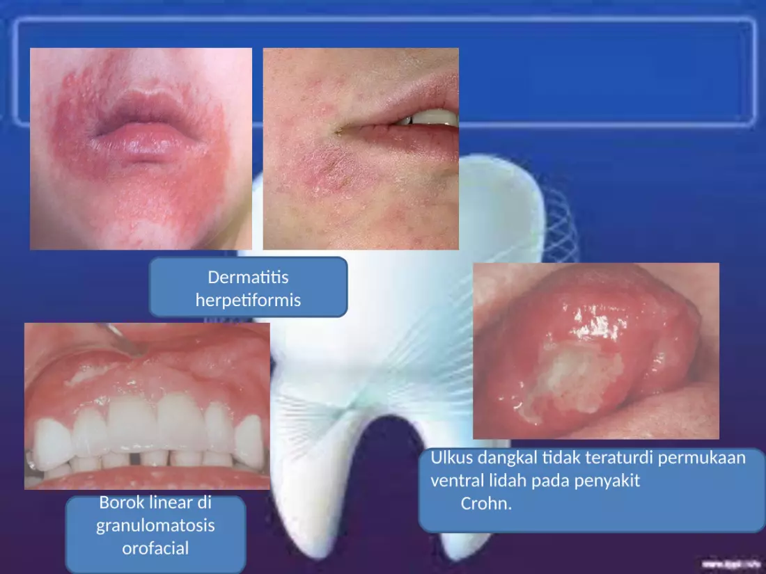
Dermatitis herpetiformis
Borok linear di granulomatosis
orofacial
Ulkus dangkal tidak teraturdi permukaan ventral lidah pada penyakit Crohn.

Gingiva pembesaran di sarkoidosis.
Peutz Jegher Syndrome

Atrophic glossitis in a patient with pernicious anemia. Mucosal atrophy
appears as smooth, bald areas devoid of lingual papillae on the dorsal tongue.
Diffuse ulceration of the buccal mucosa in a patient with pemphigus vulgaris.
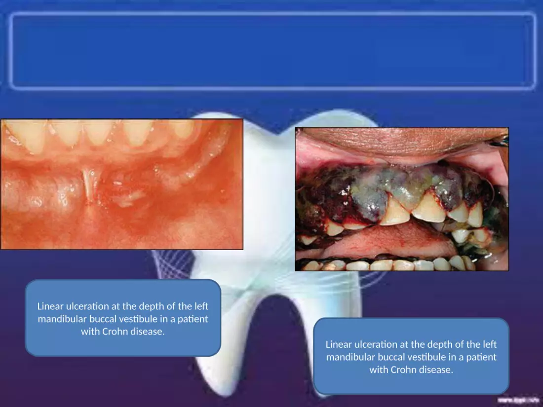
Linear ulceration at the depth of the left mandibular buccal vestibule in a patient
with Crohn disease.Linear ulceration at the depth of the left mandibular buccal vestibule in a patient
with Crohn disease.
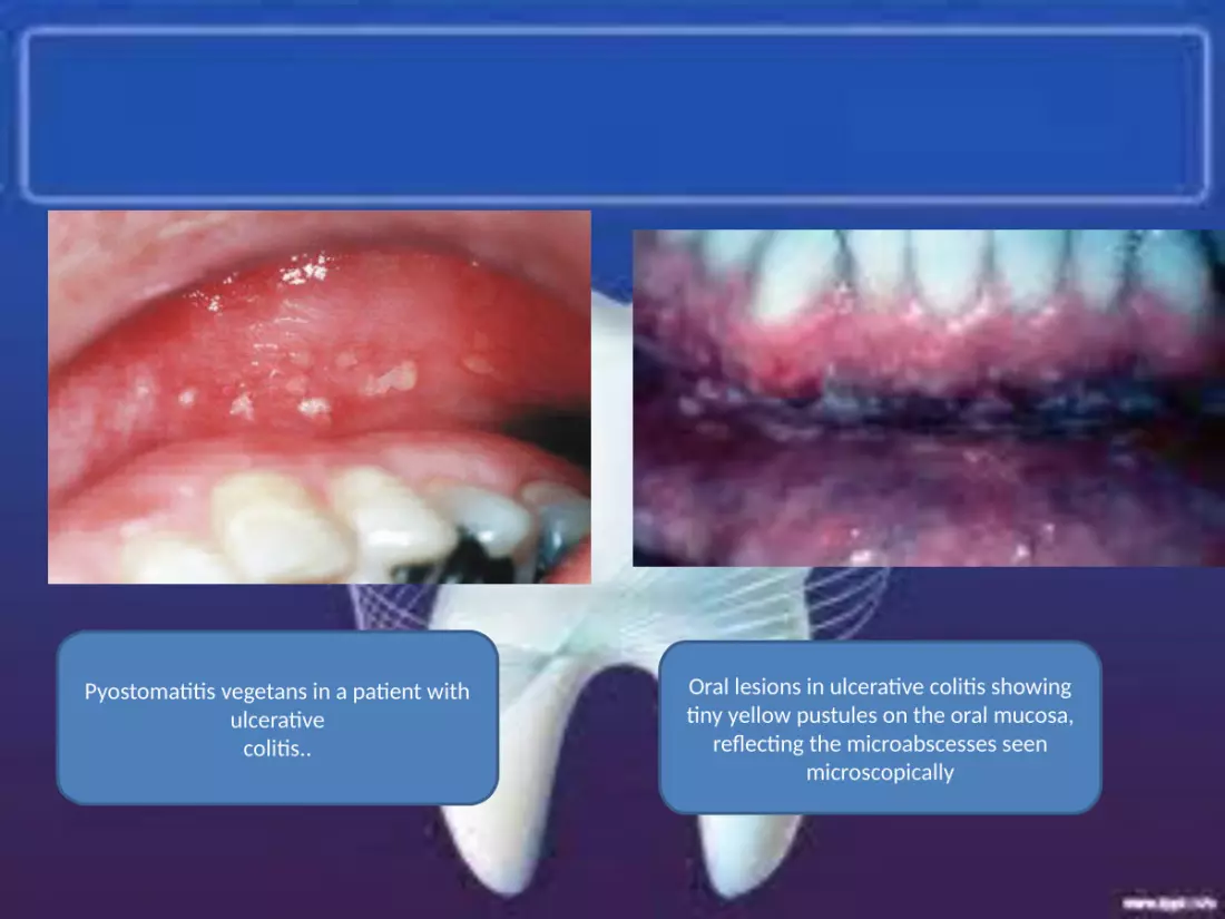
Pyostomatitis vegetans in a patient with ulcerative
colitis..
Oral lesions in ulcerative colitis showing tiny yellow pustules on the oral mucosa,
reflecting the microabscesses seen microscopically
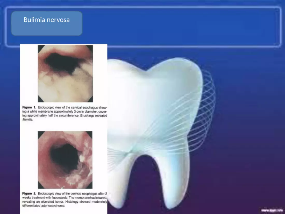
Bulimia nervosa

Gastrointestinal disease
TERIMA KASIH