response of vestibular-nerve afferents to active and passive rotations under normal conditions and...
TRANSCRIPT
Response of Vestibular-Nerve Afferents to Active and Passive RotationsUnder Normal Conditions and After Unilateral Labyrinthectomy
Soroush G. Sadeghi,1 Lloyd B. Minor,2 and Kathleen E. Cullen1
1Department of Physiology, McGill University, Montreal, Canada; and 2Department of Otolaryngology–Head and Neck Surgery,Johns Hopkins University School of Medicine, Baltimore, Maryland
Submitted 8 August 2006; accepted in final form 16 November 2006
Sadeghi SG, Minor LB, Cullen KE. Response of vestibular-nerveafferents to active and passive rotations under normal conditions and afterunilateral labyrinthectomy. J Neurophysiol 97: 1503–1514, 2007. Firstpublished November 22, 2006; doi:10.1152/jn.00829.2006. We investi-gated the possible contribution of signals carried by vestibular-nerveafferents to long-term processes of vestibular compensation after unilat-eral labyrinthectomy. Semicircular canal afferents were recorded fromthe contralesional nerve in three macaque monkeys before [horizontal(HC) � 67, anterior (AC) � 66, posterior (PC) � 50] and 1–12 mo after(HC � 192, AC � 86, PC � 57) lesion. Vestibular responses wereevaluated using passive sinusoidal rotations with frequencies of 0.5–15Hz (20–80°/s) and fast whole-body rotations reaching velocities of500°/s. Sensitivities to nonvestibular inputs were tested by: 1) comparingresponses during active and passive head movements, 2) rotating thebody with the head held stationary to activate neck proprioceptors, and 3)encouraging head-restrained animals to attempt to make head movementsthat resulted in the production of neck torques of �2 Nm. Mean restingdischarge rate before and after the lesion did not differ for the regular, D(dimorphic)-irregular, or C (calyx)-irregular afferents. In response topassive rotations, afferents showed no change in sensitivity and phase,inhibitory cutoff, and excitatory saturation after unilateral labyrinthec-tomy. Moreover, head sensitivities were similar during voluntary andpassive head rotations and responses were not altered by neck proprio-ceptive or efference copy signals before or after the lesion. The onlysignificant change was an increase in the proportion of C-irregular unitspostlesion, accompanied by a decrease in the proportion of regularafferents. Taken together, our findings show that changes in responseproperties of the vestibular afferent population are not likely to play amajor role in the long-term changes associated with compensation afterunilateral labyrinthectomy.
I N T R O D U C T I O N
In normal animals, the vestibuloocular reflex (VOR) effec-tively stabilizes gaze for head velocities and frequencies in therange of natural head movements (Huterer and Cullen 2002;Minor et al. 1999; Ramachandran and Lisberger 2005). More-over, the VOR is capable of remarkable adjustments in re-sponse to environmental challenges including the use of mag-nifying or minimizing optical lenses as well as lesions of thevestibular system. For example, immediately after unilaterallabyrinthectomy, there is a marked asymmetry in gain charac-terized by diminished responses to rotations that would beexcitatory for the lesioned side. Within a month, however, theVOR shows nearly complete functional recovery for headrotations at lower frequencies, velocities, and/or accelerations,in primates (human: Allum et al. 1988; Curthoys and Halmagyi
1995; squirrel monkey: Lasker et al. 2000; Paige 1983b;macaque: Fetter and Zee 1988).
A simple three-neuron arc (vestibular afferents, to neuronsin the vestibular nuclei, to extraocular motoneurons) mediatesthe most direct pathway of the VOR (Lorente De No’ 1933).Previous investigations used in vivo and in vitro approaches ina variety of species to characterize the mechanisms that under-lie compensation after labyrinthectomy within this pathway atthe level of single-neuron responses and intrinsic cellularproperties (reviewed in Curthoys and Halmagyi 1995; Straka etal. 2005). These studies focused on changes at the level of theneurons of the vestibular nuclei that constitute the middle legof the direct VOR pathway. There is general agreement acrossstudies that neuronal resting rates initially show significantchanges and then return to prelesion values within 30 days(Newlands and Perachio 1990a,b; Ris and Godaux 1998; Ris etal. 1995; Smith and Curthoys 1989). In addition, neuronalresponse gains decrease in both the ipsilesional and contrale-sional vestibular nuclei immediately after lesions and remainattenuated after compensation (Newlands and Perachio1990a,b; Ris and Godaux 1998; Ris et al. 1995; Smith andCurthoys 1989). The compensatory decrease in the sensitivityof neurons on the contralesional side prevents inhibitory cutoffand excitatory saturation in responses, thereby increasing thelinear response range.
Although many studies have considered the central changesthat occur after vestibular lesions, it remains to be determinedwhether corresponding changes at the level of vestibular nerveafferents might also contribute to the vestibular compensationprocess. Vestibular hair cells and afferent fibers receive bilat-eral inputs from the vestibular efferent system, which consistsof a group of neurons located in the brain stem neighboring theabducens nucleus (Gacek and Lyon 1974; Goldberg and Fer-nandez 1980; Rasmussen and Gacek 1958). The results ofprevious investigations are consistent with the hypothesis thatthe vestibular efferent system could be used to extend thedynamic range of the input that is available from the intact sideafter vestibular damage (see Cullen and Minor 2002). Electri-cal activation of the vestibular efferent pathway results in anincrease in resting discharge and a decrease in sensitivity ofvestibular afferents in toad fish and squirrel monkey (Goldbergand Fernandez 1980; Highstein and Baker 1985; Marlinski etal. 2004). Support for the idea that activation of the efferentsystem can induce long-term changes in afferent physiology isprovided by studies of the sensory periphery of the auditory
Address for reprint requests and other correspondence: K. E. Cullen,Department of Physiology, 3655 Sir William Osler, Montreal, Quebec H3G1Y6, Canada (E-mail: [email protected]).
The costs of publication of this article were defrayed in part by the paymentof page charges. The article must therefore be hereby marked “advertisement”in accordance with 18 U.S.C. Section 1734 solely to indicate this fact.
J Neurophysiol 97: 1503–1514, 2007.First published November 22, 2006; doi:10.1152/jn.00829.2006.
15030022-3077/07 $8.00 Copyright © 2007 The American Physiological Societywww.jn.org
by 10.220.33.6 on October 7, 2016
http://jn.physiology.org/D
ownloaded from
system (Liberman 1990; Walsh et al. 1998). Because thevestibular and auditory systems have a common phylogeneticorigin (reviewed in Fritzsch 1992), it is possible that thevestibular efferent system plays an analogous role in shapingvestibular afferent responses.
The functional role of the vestibular efferent system inprimates is not well understood. The findings of prior investi-gations in the frog led to the proposal that extravestibularinputs (e.g., somatosensory, proprioceptive, and motor effer-ence copy signals) transmitted through efferent neurons can beused to change the background discharge and sensitivity ofvestibular afferents under specific behavioral conditions (Bri-cout-Berthout et al. 1984; Caston and Bricout-Berthout 1984;Precht et al. 1971; Schmidt 1963). In toadfish, efferent inner-vation alters the background discharge and sensitivity to mo-tion of vestibular-nerve afferents as a component of the escapereaction (Boyle and Highstein 1990; Highstein and Baker1985). Such nonvestibular signals could be functionally useful.For example, after vestibular lesions they could in part substi-tute the missing vestibular information, which could provide anexplanation for the enhanced VOR gains for active as com-pared with passive head-on-body rotations (Della Santina et al.2001; Dichgans et al. 1973; Newlands et al. 2001).
The principal goal of the present study was to test whetherthe vestibular efferent system plays a role in the long-termchanges associated with compensation after unilateral laby-rinthectomy over the range of natural head movements. Toaddress this, afferents innervating the semicircular canals wererecorded in alert macaques before labyrinthectomy and then onthe contralesional side in response to head movements over thefrequency range where compensation is the most complete(i.e., �4 Hz), as well as during more challenging stimulationdesigned to probe the full range of physiologically relevanthead movements (Sadeghi et al. 2006). We also assessedwhether extravestibular inputs including inputs from neckproprioceptors and/or motor efference signals resulting frommotor commands to the neck altered afferent responses afterunilateral labyrinthectomy. First, recordings were made fromthe same afferents during passively applied head rotations andactive head movements. Additionally, two supplementary par-adigms were used in which we selectively activated neckproprioceptors or induced the production of motor commandsto the neck in the absence of vestibular stimulation.
M E T H O D S
Surgical preparation
Three macaque monkeys (Macaca fascicularis) were prepared forchronic behavioral experiments under aseptic conditions. All proce-dures were approved by the McGill University Animal Care Com-mittee and Johns Hopkins University Animal Care and Use Commit-tee and were in compliance with the guidelines of the CanadianCouncil on Animal Care and the National Institutes of Health.
The surgical preparation was previously described elsewhere (Syl-vestre and Cullen 1999). Briefly, using aseptic techniques and isoflu-orane anesthesia (2–3%, to effect), we implanted several stainlesssteel screws into the skull and attached a stainless steel recordingchamber and a post for head restraint to these screws with dentalcement. In the same procedure, a 17- to 18-mm-diameter eye coil,consisting of three loops of Teflon-coated stainless steel wire, wasimplanted in each eye beneath the conjunctiva (Fuchs and Robinson1966). After the surgery, the animals were administered buprenor-
phine [0.01 mg/kg, administered intramuscularly (im)] for postoper-ative analgesia and the antibiotic cephazolin (Ancef; 25 mg/kg im, for5 days). Animals were given at least 2 wk to recover from the surgerybefore experiments began.
Labyrinthectomy was performed as previously described (Lasker etal. 2000). A postauricular incision was made and the mastoid bonewas removed with an otologic drill and curettes to expose thehorizontal and posterior semicircular canals. The petrous bone wasremoved further anteriorly and superiorly to visualize the superiorcanal near its union with the common crus. Each of the semicircularcanals was then obliterated with removal of the ampulla. The vestibulewas entered and the utriculus and sacculus were removed. The internalauditory canal was opened next and the distal ends of the ampullarynerve branches were removed. The space created by the labyrinthec-tomy was packed with muscle and fascia and the postauricularincision was closed.
Data acquisition
The experimental setup, apparatus, and methods of data acquisitionwere similar to those described before (Cullen and Minor 2002;Huterer and Cullen 2002). We monitored gaze and head position usingthe magnetic search coil technique (1-m field coil system; CNCEngineering). Single-unit extracellular recordings were made usingglass electrodes with impedances of about 25 M�. Once a unit wasisolated, the semicircular canal innervated by that fiber was deter-mined based on the responses of the afferent to rotations deliveredwhile the head was restrained (see Cullen and Minor 2002). Gaze andhead positions, target position, and table velocity were recorded onDAT tape with unit activity for later playback. During playback headand gaze position signals were low-pass filtered at 250 Hz by aneight-pole Bessel filter and sampled at 1 kHz.
Experimental design
Afferents were recorded in each animal before labyrinthectomy toobtain prelesion data. A labyrinthectomy was then preformed on theside in which the recording had been done and postlesional data werecollected from the contralesional nerve. At the end of all experimentsand after sacrificing the animal, the location of the microelectrodewithin the vestibular nerve was confirmed by histology. Vestibularstimulation and data acquisition were controlled by a QNX-basedreal-time data-acquisition system (REX) (Hayes et al. 1982).
RECORDINGS MADE DURING PASSIVE HEAD ROTATIONS. Neuronalsensitivities to head velocity were measured using whole-body rota-tions (WBRs) applied at 0.5, 1, 2, and 4 Hz (peak velocity �20, �50,and �80°/s). For some afferents, responses to more dynamic stimuliwere also characterized. Their response were recorded during 1)higher frequency (5, 10, and 15 Hz at �20, �50, or �80°/s)horizontal head-on-body rotations that were applied by a torque motor(Animatics) securely coupled to the post implanted on the monkey’shead (Huterer and Cullen 2002) and/or 2) rapid position steps (about600 ms) with displacements of 20–100° and peak velocities of50–500°/s.
RECORDINGS MADE DURING VOLUNTARY HEAD ROTATIONS. Oncethe neuron’s sensitivity to head velocity during passive rotations wascharacterized, the monkey’s head was carefully released to allowfreedom of motion about the yaw (horizontal) axis (Roy and Cullen2001). If the neuron remained well isolated, its activity was thenrecorded during voluntary combined eye–head movements made toorient to laser and food targets.
RECORDINGS MADE TO DETERMINE THE INFLUENCE OF MOTOR COM-
MAND SIGNALS AND/OR NECK PROPRIOCEPTION ON NEURONAL AC-
TIVITY. We recorded from afferents during large saccades (�30°) totest whether the production of neck motor command alters their
1504 S. G. SADEGHI, L. B. MINOR, AND K. E. CULLEN
J Neurophysiol • VOL 97 • FEBRUARY 2007 • www.jn.org
by 10.220.33.6 on October 7, 2016
http://jn.physiology.org/D
ownloaded from
responses (see Roy and Cullen 2004). The concurrent neck torqueproduced against the head restraint (reaction torque transducer, Sen-sotec) was recorded and �10 intervals where torques reached highlevels (�1 Nm) were analyzed. In addition, the influence of passiveactivation of the neck proprioceptors was assessed by passivelyrotating the animal’s body under its stationary head (BUH paradigm)at frequencies of 0.5, 1.0, and 2.0 Hz (�20 or �50°/s).
Data analysis
Data were imported into the Matlab (The MathWorks, Natick MA)programming environment for analysis. Recorded gaze and headposition signals were digitally filtered with zero-phase at 125 Hz usinga 51st-order finite-impulse-response (FIR) filter with a Hammingwindow. Position signals were then differentiated to produce velocitysignals. The neural discharge was represented using a spike densityfunction in which a Gaussian was convolved with the spike train(Cullen et al. 1996).
The resting discharge of each unit and coefficient of variation (CV)of the interspike interval were determined. A normalized coefficient ofvariation (CV*) was calculated using the method described by Gold-berg et al. (1984). Afferents with a CV* �0.2 and sensitivity at 2-Hzstimulation of �1.5 (spikes/s)/(°/s) were assigned to the C (calyx)-irregular group as suggested by the morphophysiological results ofBaird et al. (1988) in the chinchilla. Regular afferents were identifiedas having a CV* �0.1. All other afferents were identified as D(dimorphic)-irregular units (Hullar et al. 2005). It should be noted thatsimilar to previous studies (Haque et al. 2004; Hullar et al. 2005;Marlinski et al. 2004; Ramachandran and Lisberger 2006), we did notexamine the morphology of the afferent endings directly, but insteadused this functional classification.
A least-squares regression analysis was used to determine the phaseshift of each unit relative to head velocity, resting discharge (bias,spikes/s), and head velocity sensitivity [(spikes/s)/(°/s)] (see Roy andCullen 2001; Sylvestre and Cullen 1999), using �10 cycles of thestimulus. Neuronal responses to body-under-head rotations (BUHparadigm) and active head rotations were similarly characterized.Afferents were typically driven into the nonlinear range (inhibitorycutoff) for large-velocity head movements in their off direction.Accordingly, fits to the passive and active rotations were made basedon responses that were �10 spikes/s. To compare the model’s abilityto predict the firing rate, the variance-accounted-for (VAF) value wasdetermined (Cullen et al. 1996). Because the head rotations for thevertical canal afferents were not in the plane of the canal, trigono-metric corrections were made based on the canal planes in this species(Reisine et al. 1988) for the sensitivities of the units as describedpreviously (Cullen and Minor 2002). A neuron’s response to fastrotations was described by least-squares optimization of a sigmoidalfunction.
Statistical analysis
Data are described as means � SE. A Student’s t-test was used todetermine whether the average of two measured parameters differedsignificantly from each other. We used a z-test to compare changes inpopulation percentages of regular and irregular afferents pre- andpostlabyrinthectomy.
R E S U L T S
We recorded from 639 vestibular nerve fibers in threeanimals. Of these, 241 (67 horizontal canal, 66 superior canal,50 posterior canal, and 58 otolith) were recorded in normalanimals. The remaining 398 afferents (192 horizontal canal, 86superior canal, 57 posterior canal, and 63 otolith) were re-corded from the contralesional nerve 1–12 mo after unilateral
labyrinthectomy. The following results are confined to thoseafferents that innervate the three semicircular canals. Forpurposes of this study, we combined the vertical and horizontalcanal afferents in the data analysis of resting rates and sinu-soidal WBRs �4 Hz because they encoded similar signals.
In one animal, recordings were also made from the ipsile-sional nerve under direct visualization 12 mo after the lesionand immediately before sacrificing the animal. From ninemicroelectrode passes along the nerve, only three units wererecorded. Two units had sporadic activity and one had a restingdischarge that varied between 50 and 80 spikes/s. This appre-ciable reduction in activity in the ipsilesional vestibular nerveis consistent with the evidence that resting discharge activity isdependent on transmitter release from hair cells (Guth et al.1998).
Resting discharge and regularity before and after lesion
Figure 1A shows the distribution of the resting rate and CV*of the entire population of canal afferents recorded in thisstudy, before (gray histograms) and after (black histograms)unilateral labyrinthectomy. Resting discharge distributions(Fig. 1A, left) were similar in both conditions (populationaverages: 107.4 � 2.6 vs. 101.1 � 2.0 spikes/s, before vs. afterlabyrinthectomy, respectively, P � 0.07). Moreover, both pre-and postlesion distributions were comparable to those reportedin previous studies of normal macaque monkeys (Bronte-Stewart and Lisberger 1994; Lisberger and Pavelko 1986).
Before lesion, the distribution of CV* was bimodal (Fig. 1A,right, gray histograms) as previously described (e.g., Baird etal. 1988). Although the distributions appeared qualitativelysimilar before and after labyrinthectomy, there was an overallincrease in the percentage of irregular afferents (CV* �0.1) inour postlesion sample compared with our prelesion sample (62vs. 55%). This difference was statistically significant (z statis-tics, P � 0.001) and was the result of a specific increase in thepercentage of irregular afferents that had CV* values �0.2 (26vs. 39% in our pre- vs. postlesion samples, respectively). Thischange was accompanied by a relative decrease in the propor-tion of afferents with 0.1 � CV* � 0.2 postlesion (29 vs.23%). The potential mechanisms meditating these changes andimplications of these findings are further addressed underDISCUSSION.
Categorization of afferents: responses at 2 Hz before andafter lesion
Afferents were further divided into three groups (regular,D-irregular, C-irregular) based on their CV* and sensitivitiesto 2-Hz rotations (Table 1 and Fig. 1B; see METHODS). The meanresting rate for each of the three groups of afferents wascomparable before and after labyrinthectomy (P � 0.3). Rest-ing discharges of regular and C-irregular afferents were com-parable, whereas those of the D-irregular afferents were sig-nificantly higher in both conditions (P � 0.001). Furthermore,response sensitivities and phases of each of the three groups ofafferents were comparable before (Fig. 1B, gray symbols) andafter (Fig. 1B, black symbols) labyrinthectomy (P � 0.4). Thusthe dynamic range of the input available from the intact nerveafter unilateral labyrinthectomy is not extended by changes inafferent resting discharge and/or sensitivity in response to 2-Hzsinusoidal rotations.
1505VESTIBULAR AFFERENT RESPONSES TO ACTIVE AND PASSIVE ROTATIONS
J Neurophysiol • VOL 97 • FEBRUARY 2007 • www.jn.org
by 10.220.33.6 on October 7, 2016
http://jn.physiology.org/D
ownloaded from
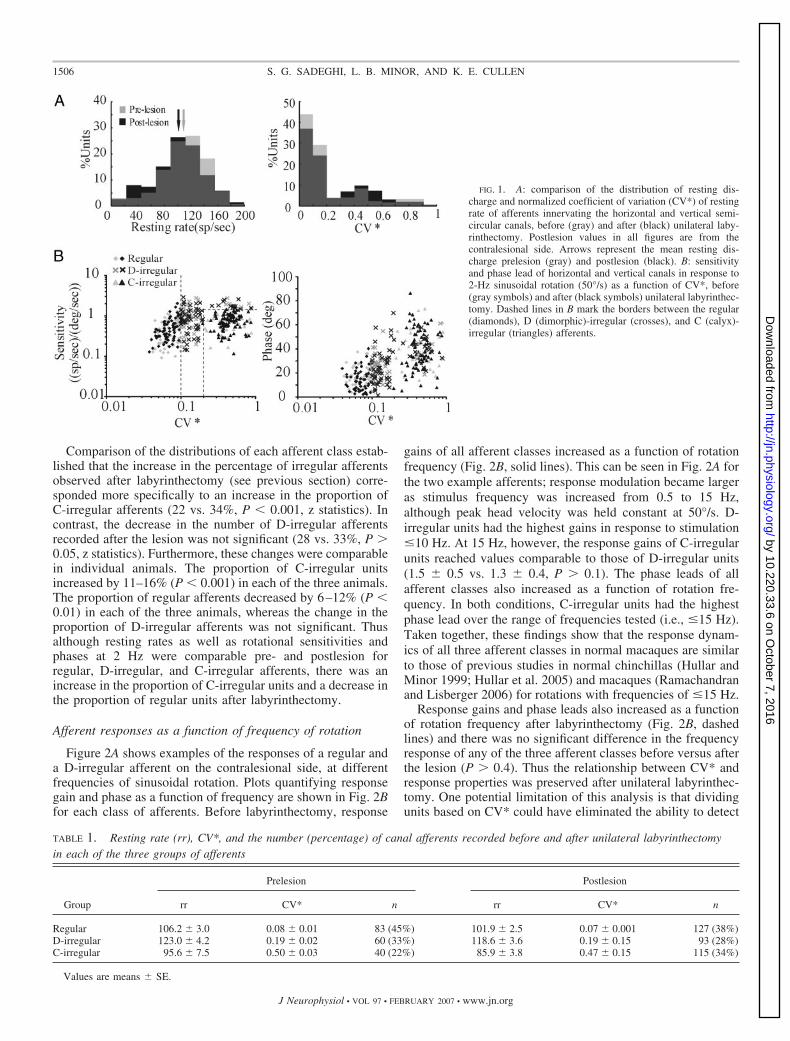
Comparison of the distributions of each afferent class estab-lished that the increase in the percentage of irregular afferentsobserved after labyrinthectomy (see previous section) corre-sponded more specifically to an increase in the proportion ofC-irregular afferents (22 vs. 34%, P � 0.001, z statistics). Incontrast, the decrease in the number of D-irregular afferentsrecorded after the lesion was not significant (28 vs. 33%, P �0.05, z statistics). Furthermore, these changes were comparablein individual animals. The proportion of C-irregular unitsincreased by 11–16% (P � 0.001) in each of the three animals.The proportion of regular afferents decreased by 6–12% (P �0.01) in each of the three animals, whereas the change in theproportion of D-irregular afferents was not significant. Thusalthough resting rates as well as rotational sensitivities andphases at 2 Hz were comparable pre- and postlesion forregular, D-irregular, and C-irregular afferents, there was anincrease in the proportion of C-irregular units and a decrease inthe proportion of regular units after labyrinthectomy.
Afferent responses as a function of frequency of rotation
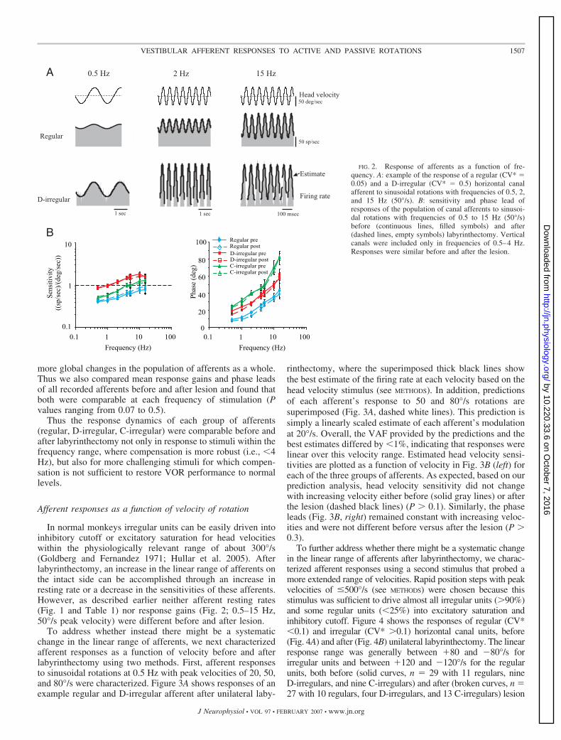
Figure 2A shows examples of the responses of a regular anda D-irregular afferent on the contralesional side, at differentfrequencies of sinusoidal rotation. Plots quantifying responsegain and phase as a function of frequency are shown in Fig. 2Bfor each class of afferents. Before labyrinthectomy, response
gains of all afferent classes increased as a function of rotationfrequency (Fig. 2B, solid lines). This can be seen in Fig. 2A forthe two example afferents; response modulation became largeras stimulus frequency was increased from 0.5 to 15 Hz,although peak head velocity was held constant at 50°/s. D-irregular units had the highest gains in response to stimulation�10 Hz. At 15 Hz, however, the response gains of C-irregularunits reached values comparable to those of D-irregular units(1.5 � 0.5 vs. 1.3 � 0.4, P � 0.1). The phase leads of allafferent classes also increased as a function of rotation fre-quency. In both conditions, C-irregular units had the highestphase lead over the range of frequencies tested (i.e., �15 Hz).Taken together, these findings show that the response dynam-ics of all three afferent classes in normal macaques are similarto those of previous studies in normal chinchillas (Hullar andMinor 1999; Hullar et al. 2005) and macaques (Ramachandranand Lisberger 2006) for rotations with frequencies of �15 Hz.
Response gains and phase leads also increased as a functionof rotation frequency after labyrinthectomy (Fig. 2B, dashedlines) and there was no significant difference in the frequencyresponse of any of the three afferent classes before versus afterthe lesion (P � 0.4). Thus the relationship between CV* andresponse properties was preserved after unilateral labyrinthec-tomy. One potential limitation of this analysis is that dividingunits based on CV* could have eliminated the ability to detect
FIG. 1. A: comparison of the distribution of resting dis-charge and normalized coefficient of variation (CV*) of restingrate of afferents innervating the horizontal and vertical semi-circular canals, before (gray) and after (black) unilateral laby-rinthectomy. Postlesion values in all figures are from thecontralesional side. Arrows represent the mean resting dis-charge prelesion (gray) and postlesion (black). B: sensitivityand phase lead of horizontal and vertical canals in response to2-Hz sinusoidal rotation (50°/s) as a function of CV*, before(gray symbols) and after (black symbols) unilateral labyrinthec-tomy. Dashed lines in B mark the borders between the regular(diamonds), D (dimorphic)-irregular (crosses), and C (calyx)-irregular (triangles) afferents.
TABLE 1. Resting rate (rr), CV*, and the number (percentage) of canal afferents recorded before and after unilateral labyrinthectomyin each of the three groups of afferents
Group
Prelesion Postlesion
rr CV* n rr CV* n
Regular 106.2 � 3.0 0.08 � 0.01 83 (45%) 101.9 � 2.5 0.07 � 0.001 127 (38%)D-irregular 123.0 � 4.2 0.19 � 0.02 60 (33%) 118.6 � 3.6 0.19 � 0.15 93 (28%)C-irregular 95.6 � 7.5 0.50 � 0.03 40 (22%) 85.9 � 3.8 0.47 � 0.15 115 (34%)
Values are means � SE.
1506 S. G. SADEGHI, L. B. MINOR, AND K. E. CULLEN
J Neurophysiol • VOL 97 • FEBRUARY 2007 • www.jn.org
by 10.220.33.6 on October 7, 2016
http://jn.physiology.org/D
ownloaded from
more global changes in the population of afferents as a whole.Thus we also compared mean response gains and phase leadsof all recorded afferents before and after lesion and found thatboth were comparable at each frequency of stimulation (Pvalues ranging from 0.07 to 0.5).
Thus the response dynamics of each group of afferents(regular, D-irregular, C-irregular) were comparable before andafter labyrinthectomy not only in response to stimuli within thefrequency range, where compensation is more robust (i.e., �4Hz), but also for more challenging stimuli for which compen-sation is not sufficient to restore VOR performance to normallevels.
Afferent responses as a function of velocity of rotation
In normal monkeys irregular units can be easily driven intoinhibitory cutoff or excitatory saturation for head velocitieswithin the physiologically relevant range of about 300°/s(Goldberg and Fernandez 1971; Hullar et al. 2005). Afterlabyrinthectomy, an increase in the linear range of afferents onthe intact side can be accomplished through an increase inresting rate or a decrease in the sensitivities of these afferents.However, as described earlier neither afferent resting rates(Fig. 1 and Table 1) nor response gains (Fig. 2; 0.5–15 Hz,50°/s peak velocity) were different before and after lesion.
To address whether instead there might be a systematicchange in the linear range of afferents, we next characterizedafferent responses as a function of velocity before and afterlabyrinthectomy using two methods. First, afferent responsesto sinusoidal rotations at 0.5 Hz with peak velocities of 20, 50,and 80°/s were characterized. Figure 3A shows responses of anexample regular and D-irregular afferent after unilateral laby-
rinthectomy, where the superimposed thick black lines showthe best estimate of the firing rate at each velocity based on thehead velocity stimulus (see METHODS). In addition, predictionsof each afferent’s response to 50 and 80°/s rotations aresuperimposed (Fig. 3A, dashed white lines). This prediction issimply a linearly scaled estimate of each afferent’s modulationat 20°/s. Overall, the VAF provided by the predictions and thebest estimates differed by �1%, indicating that responses werelinear over this velocity range. Estimated head velocity sensi-tivities are plotted as a function of velocity in Fig. 3B (left) foreach of the three groups of afferents. As expected, based on ourprediction analysis, head velocity sensitivity did not changewith increasing velocity either before (solid gray lines) or afterthe lesion (dashed black lines) (P � 0.1). Similarly, the phaseleads (Fig. 3B, right) remained constant with increasing veloc-ities and were not different before versus after the lesion (P �0.3).
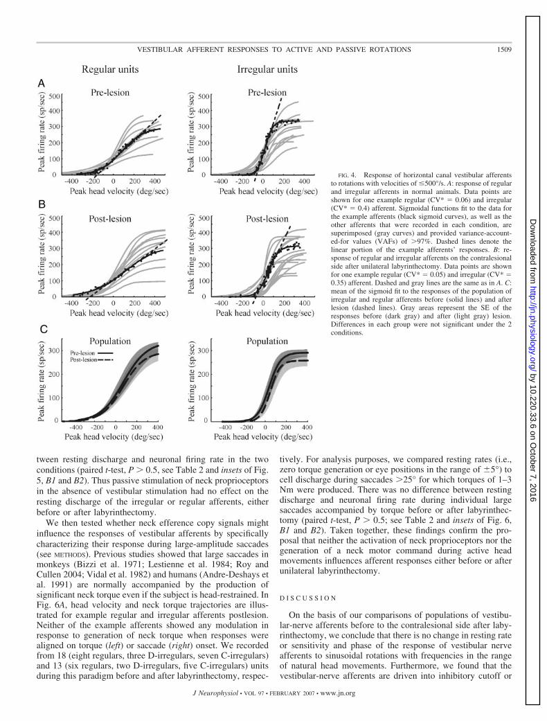
To further address whether there might be a systematic changein the linear range of afferents after labyrinthectomy, we charac-terized afferent responses using a second stimulus that probed amore extended range of velocities. Rapid position steps with peakvelocities of �500°/s (see METHODS) were chosen because thisstimulus was sufficient to drive almost all irregular units (�90%)and some regular units (�25%) into excitatory saturation andinhibitory cutoff. Figure 4 shows the responses of regular (CV*�0.1) and irregular (CV* �0.1) horizontal canal units, before(Fig. 4A) and after (Fig. 4B) unilateral labyrinthectomy. The linearresponse range was generally between �80 and �80°/s forirregular units and between �120 and �120°/s for the regularunits, both before (solid curves, n � 29 with 11 regulars, nineD-irregulars, and nine C-irregulars) and after (broken curves, n �27 with 10 regulars, four D-irregulars, and 13 C-irregulars) lesion
0.5 Hz 15 Hz2 Hz
1 sec1 sec 100 msec
Regular
D-irregular
Head velocity
50 sp/sec
Estimate
Firing rate
A
BRegular preRegular postD-irregular preD-irregular postC-irregular preC-irregular post
0
20
40
60
80
100
Phas
e(d
eg)
0.1 1 10 100
Frequency (Hz)
0.1 1 10 100
Frequency (Hz)
Sens
itivi
ty((
sp/s
ec)/
(deg
/sec
))
1
0.1
10
50 deg/sec
FIG. 2. Response of afferents as a function of fre-quency. A: example of the response of a regular (CV* �0.05) and a D-irregular (CV* � 0.5) horizontal canalafferent to sinusoidal rotations with frequencies of 0.5, 2,and 15 Hz (50°/s). B: sensitivity and phase lead ofresponses of the population of canal afferents to sinusoi-dal rotations with frequencies of 0.5 to 15 Hz (50°/s)before (continuous lines, filled symbols) and after(dashed lines, empty symbols) labyrinthectomy. Verticalcanals were included only in frequencies of 0.5–4 Hz.Responses were similar before and after the lesion.
1507VESTIBULAR AFFERENT RESPONSES TO ACTIVE AND PASSIVE ROTATIONS
J Neurophysiol • VOL 97 • FEBRUARY 2007 • www.jn.org
by 10.220.33.6 on October 7, 2016
http://jn.physiology.org/D
ownloaded from
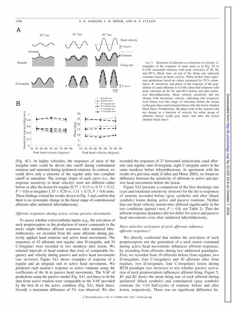
(Fig. 4C). At higher velocities, the responses of most of theirregular units could be driven into cutoff during contralateralrotations and saturated during ipsilateral rotations. In contrast, wecould drive only a minority of the regular units into completecutoff or saturation. The average slopes of each curve (i.e., theresponse sensitivity to head velocity) were not different eitherbefore or after the lesion for regular (0.77 � 0.13 vs. 0.73 � 0.12,P � 0.8) or irregular (1.35 � 0.20 vs. 1.31 � 0.15, P � 0.8) units.These findings extend the results shown in Fig. 3 and confirm thatthere is no systematic change in the linear range of contralesionalafferents after unilateral labyrinthectomy.
Afferent responses during active versus passive movements
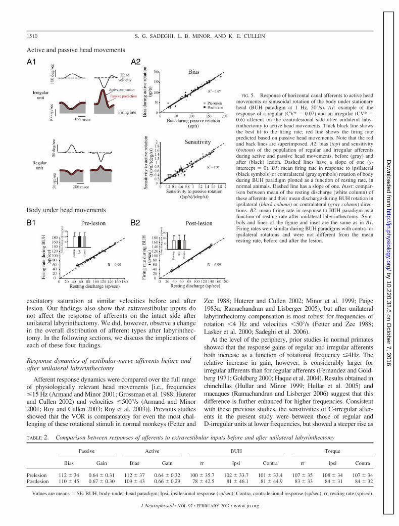
To assess whether extravestibular inputs (e.g., the activation ofneck proprioceptors or the production of motor commands to theneck) might influence afferent responses after unilateral laby-rinthectomy, we recorded from the same afferents during pas-sively applied head rotations and active head movements. Theresponses of 43 afferents (ten regular, nine D-irregular, and 24C-irregular) were recorded in two monkeys after lesion. Weselected intervals of head rotation that were of comparable fre-quency and velocity during passive and active head movements(see METHODS). Figure 5A1 shows examples of response of aregular and an irregular unit to active head movements. Wepredicted each neuron’s response to active rotations using thecoefficients of the fit to passive head movements. The VAF ofpredictions using the passive model (Fig. 5A1, red lines) to fit thedata from active rotation were comparable to the VAF providedby the best fit to the active condition (Fig. 5A1, black lines).Overall, a maximum difference of 3% was observed. We also
recorded the responses of 27 horizontal semicircular canal affer-ents (ten regular, nine D-irregular, eight C-irregular units) in thesame monkeys before labyrinthectomy. In agreement with theresults of a previous study (Cullen and Minor 2002), we found nodifference between the sensitivity of afferents to active and pas-sive head movements before the lesion.
Figure 5A2 presents a comparison of the bias discharge rate(top) and rotational sensitivity (bottom) for the fits to responsesof neurons recorded before (gray symbols) and after (blacksymbols) lesion during active and passive rotations. Neitherbias nor head velocity sensitivities differed significantly in thetwo conditions (paired t-test, P � 0.8; see Table 2). Thus theafferent response dynamics did not differ for active and passivehead movements even after unilateral labyrinthectomy.
Does selective activation of neck afferents influenceafferent responses?
We directly confirmed that neither the activation of neckproprioceptors nor the generation of a neck motor commandduring active head movements influences afferent responses,by recording from afferents during two additional paradigms.First, we recorded from 10 afferents before (four regulars, twoD-irregulars, four C-irregulars) and 10 afferents after (fourregulars, two D-irregulars, four C-irregulars) lesion duringBUH paradigm (see METHODS) to test whether passive activa-tion of neck proprioceptors influences afferent firing. Figure 5,B1 and B2 shows the mean firing rate of each afferent duringipsilateral (black symbols) and contralateral (gray symbols)rotations for �10 half-cycles of rotation, before and afterlesion, respectively. There was no significant difference be-
FIG. 3. Response of afferents as a function of velocity. A:examples of the response of same units as in Fig. 2A to0.5-Hz sinusoidal rotations with peak velocities of 20, 50,and 80°/s. Black lines on top of the firing rate representestimates based on head velocity. White broken lines repre-sent predictions based on values estimated for 20°/s stimu-lation. B: sensitivity and phase of the response of the pop-ulation of canal afferents to 0.5-Hz sinusoidal rotations withpeak velocities of 20, 50, and 80°/s before and after unilat-eral labyrinthectomy. Head velocity sensitivity did notchange with increasing velocity, indicating that responseswere linear over this range of velocities before the lesion(solid gray lines) and remained linear after the lesion (dashedblack lines). Furthermore, the phase lead of the response didnot change as a function of velocity for either group ofafferents before (solid gray lines) and after the lesion(dashed black lines).
1508 S. G. SADEGHI, L. B. MINOR, AND K. E. CULLEN
J Neurophysiol • VOL 97 • FEBRUARY 2007 • www.jn.org
by 10.220.33.6 on October 7, 2016
http://jn.physiology.org/D
ownloaded from
tween resting discharge and neuronal firing rate in the twoconditions (paired t-test, P � 0.5, see Table 2 and insets of Fig.5, B1 and B2). Thus passive stimulation of neck proprioceptorsin the absence of vestibular stimulation had no effect on theresting discharge of the irregular or regular afferents, eitherbefore or after labyrinthectomy.
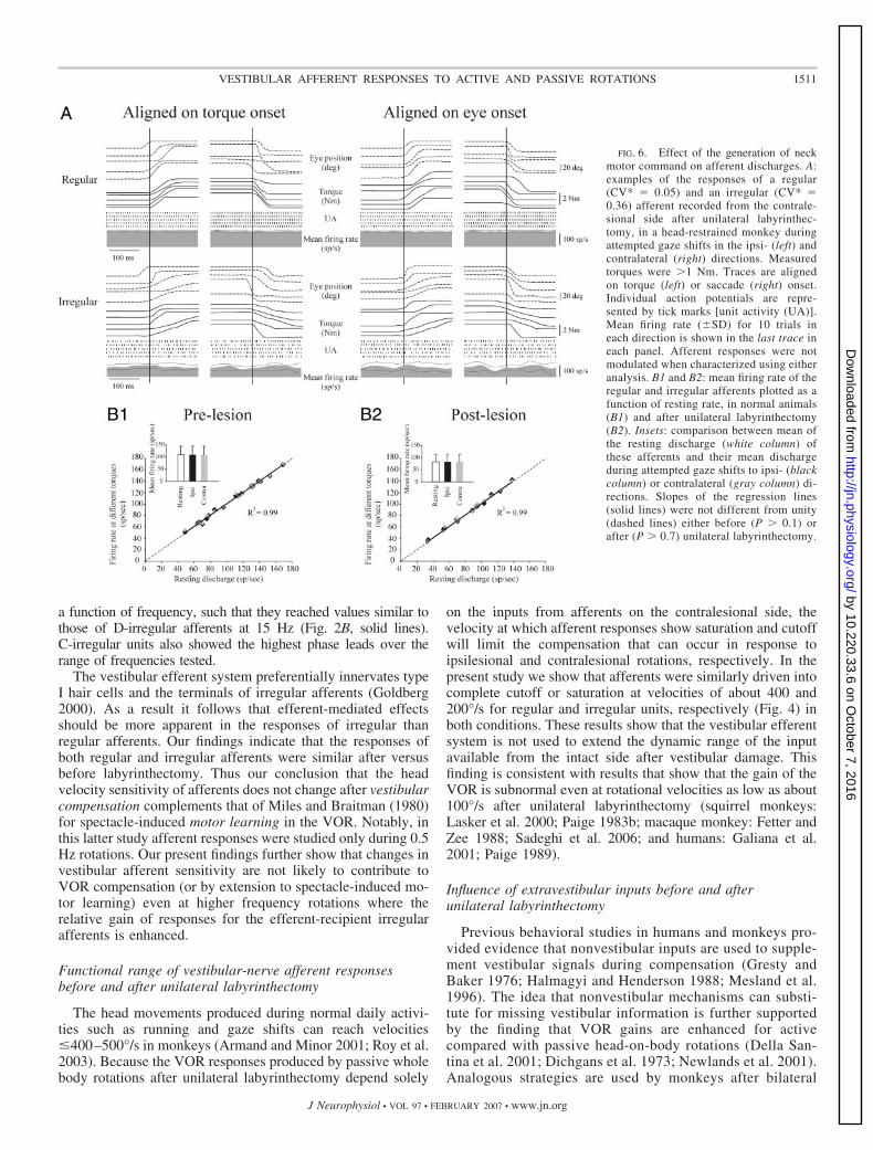
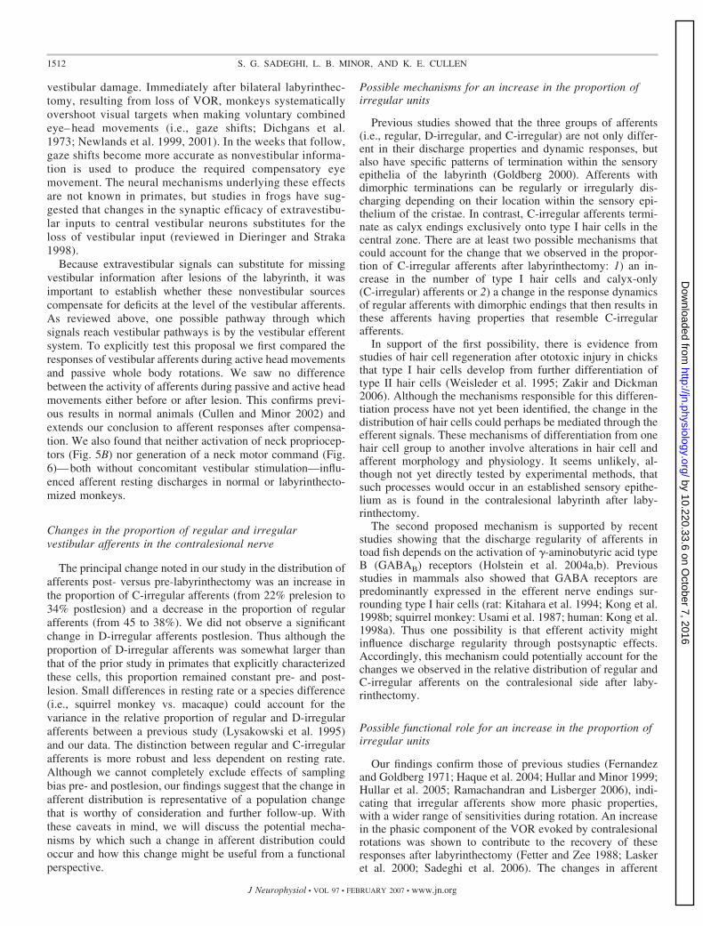
We then tested whether neck efference copy signals mightinfluence the responses of vestibular afferents by specificallycharacterizing their response during large-amplitude saccades(see METHODS). Previous studies showed that large saccades inmonkeys (Bizzi et al. 1971; Lestienne et al. 1984; Roy andCullen 2004; Vidal et al. 1982) and humans (Andre-Deshays etal. 1991) are normally accompanied by the production ofsignificant neck torque even if the subject is head-restrained. InFig. 6A, head velocity and neck torque trajectories are illus-trated for example regular and irregular afferents postlesion.Neither of the example afferents showed any modulation inresponse to generation of neck torque when responses werealigned on torque (left) or saccade (right) onset. We recordedfrom 18 (eight regulars, three D-irregulars, seven C-irregulars)and 13 (six regulars, two D-irregulars, five C-irregulars) unitsduring this paradigm before and after labyrinthectomy, respec-
tively. For analysis purposes, we compared resting rates (i.e.,zero torque generation or eye positions in the range of �5°) tocell discharge during saccades �25° for which torques of 1–3Nm were produced. There was no difference between restingdischarge and neuronal firing rate during individual largesaccades accompanied by torque before or after labyrinthec-tomy (paired t-test, P � 0.5; see Table 2 and insets of Fig. 6,B1 and B2). Taken together, these findings confirm the pro-posal that neither the activation of neck proprioceptors nor thegeneration of a neck motor command during active headmovements influences afferent responses either before or afterunilateral labyrinthectomy.
D I S C U S S I O N
On the basis of our comparisons of populations of vestibu-lar-nerve afferents before to the contralesional side after laby-rinthectomy, we conclude that there is no change in resting rateor sensitivity and phase of the response of vestibular nerveafferents to sinusoidal rotations with frequencies in the rangeof natural head movements. Furthermore, we found that thevestibular-nerve afferents are driven into inhibitory cutoff or
FIG. 4. Response of horizontal canal vestibular afferentsto rotations with velocities of �500°/s. A: response of regularand irregular afferents in normal animals. Data points areshown for one example regular (CV* � 0.06) and irregular(CV* � 0.4) afferent. Sigmoidal functions fit to the data forthe example afferents (black sigmoid curves), as well as theother afferents that were recorded in each condition, aresuperimposed (gray curves) and provided variance-account-ed-for values (VAFs) of �97%. Dashed lines denote thelinear portion of the example afferents’ responses. B: re-sponse of regular and irregular afferents on the contralesionalside after unilateral labyrinthectomy. Data points are shownfor one example regular (CV* � 0.05) and irregular (CV* �0.35) afferent. Dashed and gray lines are the same as in A. C:mean of the sigmoid fit to the responses of the population ofirregular and regular afferents before (solid lines) and afterlesion (dashed lines). Gray areas represent the SE of theresponses before (dark gray) and after (light gray) lesion.Differences in each group were not significant under the 2conditions.
1509VESTIBULAR AFFERENT RESPONSES TO ACTIVE AND PASSIVE ROTATIONS
J Neurophysiol • VOL 97 • FEBRUARY 2007 • www.jn.org
by 10.220.33.6 on October 7, 2016
http://jn.physiology.org/D
ownloaded from
excitatory saturation at similar velocities before and afterlesion. Our findings also show that extravestibular inputs donot affect the response of afferents on the intact side afterunilateral labyrinthectomy. We did, however, observe a changein the overall distribution of afferent types after labyrinthec-tomy. In the following sections, we discuss the implications ofeach of these four findings.
Response dynamics of vestibular-nerve afferents before andafter unilateral labyrinthectomy
Afferent response dynamics were compared over the full rangeof physiologically relevant head movements [i.e., frequencies�15 Hz (Armand and Minor 2001; Grossman et al. 1988; Hutererand Cullen 2002) and velocities �500°/s (Armand and Minor2001; Roy and Cullen 2003; Roy et al. 2003)]. Previous studiesshowed that the VOR is compensatory for even the most chal-lenging of these rotational stimuli in normal monkeys (Fetter and
Zee 1988; Huterer and Cullen 2002; Minor et al. 1999; Paige1983a; Ramachandran and Lisberger 2005), but after unilaterallabyrinthectomy compensation is most robust for frequencies ofrotation �4 Hz and velocities �50°/s (Fetter and Zee 1988;Lasker et al. 2000; Sadeghi et al. 2006).
At the level of the periphery, prior studies in normal primatesshowed that the response gains of regular and irregular afferentsboth increase as a function of rotational frequency �4Hz. Therelative increase in gain, however, is considerably larger forirregular afferents than for regular afferents (Fernandez and Gold-berg 1971; Goldberg 2000; Haque et al. 2004). Results obtained inchinchillas (Hullar and Minor 1999; Hullar et al. 2005) andmacaques (Ramachandran and Lisberger 2006) suggest that thisdifference is further enhanced for higher frequencies. Consistentwith these previous studies, the sensitivities of C-irregular affer-ents in the present study were between those of regular andD-irregular units at lower frequencies, but showed a steeper rise as
FIG. 5. Response of horizontal canal afferents to active headmovements or sinusoidal rotation of the body under stationaryhead (BUH paradigm at 1 Hz, 50°/s). A1: example of theresponse of a regular (CV* � 0.07) and an irregular (CV* �0.6) afferent on the contralesional side after unilateral laby-rinthectomy to active head movements. Thick black line showsthe best fit to the firing rate; red line shows the firing ratepredicted based on passive head movements. Note that the redand back lines are superimposed. A2: bias (top) and sensitivity(bottom) of the population of regular and irregular afferentsduring active and passive head movements, before (gray) andafter (black) lesion. Dashed lines have a slope of one (y-intercept � 0). B1: mean firing rate in response to ipsilateral(black symbols) or contralateral (gray symbols) rotation of bodyduring BUH paradigm plotted as a function of resting rate, innormal animals. Dashed line has a slope of one. Inset: compar-ison between mean of the resting discharge (white column) ofthese afferents and their mean discharge during BUH rotation inipsilateral (black column) or contralateral (gray column) direc-tions. B2: mean firing rate in response to BUH paradigm as afunction of resting rate after unilateral labyrinthectomy. Sym-bols and lines of the figure and inset are the same as in B1.Firing rates were similar during BUH paradigms with contra- oripsilateral rotations and were not different from the meanresting rate, before and after the lesion.
TABLE 2. Comparison between responses of afferents to extravestibular inputs before and after unilateral labyrinthectomy
Passive Active BUH Torque
Bias Gain Bias Gain rr Ipsi Contra rr Ipsi Contra
Prelesion 112 � 34 0.64 � 0.31 112 � 37 0.64 � 0.32 100 � 35.7 102 � 33.7 101 � 33.4 107 � 35 108 � 34 107 � 34Postlesion 110 � 45 0.67 � 0.30 109 � 43 0.66 � 0.29 78 � 42.5 81 � 46.1 81 � 44.9 83 � 33 84 � 31 84 � 32
Values are means � SE. BUH, body-under-head paradigm; Ipsi, ipsilesional response (sp/sec); Contra, contralesional response (sp/sec); rr, resting rate (sp/sec).
1510 S. G. SADEGHI, L. B. MINOR, AND K. E. CULLEN
J Neurophysiol • VOL 97 • FEBRUARY 2007 • www.jn.org
by 10.220.33.6 on October 7, 2016
http://jn.physiology.org/D
ownloaded from
a function of frequency, such that they reached values similar tothose of D-irregular afferents at 15 Hz (Fig. 2B, solid lines).C-irregular units also showed the highest phase leads over therange of frequencies tested.
The vestibular efferent system preferentially innervates typeI hair cells and the terminals of irregular afferents (Goldberg2000). As a result it follows that efferent-mediated effectsshould be more apparent in the responses of irregular thanregular afferents. Our findings indicate that the responses ofboth regular and irregular afferents were similar after versusbefore labyrinthectomy. Thus our conclusion that the headvelocity sensitivity of afferents does not change after vestibularcompensation complements that of Miles and Braitman (1980)for spectacle-induced motor learning in the VOR. Notably, inthis latter study afferent responses were studied only during 0.5Hz rotations. Our present findings further show that changes investibular afferent sensitivity are not likely to contribute toVOR compensation (or by extension to spectacle-induced mo-tor learning) even at higher frequency rotations where therelative gain of responses for the efferent-recipient irregularafferents is enhanced.
Functional range of vestibular-nerve afferent responsesbefore and after unilateral labyrinthectomy
The head movements produced during normal daily activi-ties such as running and gaze shifts can reach velocities�400–500°/s in monkeys (Armand and Minor 2001; Roy et al.2003). Because the VOR responses produced by passive wholebody rotations after unilateral labyrinthectomy depend solely
on the inputs from afferents on the contralesional side, thevelocity at which afferent responses show saturation and cutoffwill limit the compensation that can occur in response toipsilesional and contralesional rotations, respectively. In thepresent study we show that afferents were similarly driven intocomplete cutoff or saturation at velocities of about 400 and200°/s for regular and irregular units, respectively (Fig. 4) inboth conditions. These results show that the vestibular efferentsystem is not used to extend the dynamic range of the inputavailable from the intact side after vestibular damage. Thisfinding is consistent with results that show that the gain of theVOR is subnormal even at rotational velocities as low as about100°/s after unilateral labyrinthectomy (squirrel monkeys:Lasker et al. 2000; Paige 1983b; macaque monkey: Fetter andZee 1988; Sadeghi et al. 2006; and humans: Galiana et al.2001; Paige 1989).
Influence of extravestibular inputs before and afterunilateral labyrinthectomy
Previous behavioral studies in humans and monkeys pro-vided evidence that nonvestibular inputs are used to supple-ment vestibular signals during compensation (Gresty andBaker 1976; Halmagyi and Henderson 1988; Mesland et al.1996). The idea that nonvestibular mechanisms can substi-tute for missing vestibular information is further supportedby the finding that VOR gains are enhanced for activecompared with passive head-on-body rotations (Della San-tina et al. 2001; Dichgans et al. 1973; Newlands et al. 2001).Analogous strategies are used by monkeys after bilateral
FIG. 6. Effect of the generation of neckmotor command on afferent discharges. A:examples of the responses of a regular(CV* � 0.05) and an irregular (CV* �0.36) afferent recorded from the contrale-sional side after unilateral labyrinthec-tomy, in a head-restrained monkey duringattempted gaze shifts in the ipsi- (left) andcontralateral (right) directions. Measuredtorques were �1 Nm. Traces are alignedon torque (left) or saccade (right) onset.Individual action potentials are repre-sented by tick marks [unit activity (UA)].Mean firing rate (�SD) for 10 trials ineach direction is shown in the last trace ineach panel. Afferent responses were notmodulated when characterized using eitheranalysis. B1 and B2: mean firing rate of theregular and irregular afferents plotted as afunction of resting rate, in normal animals(B1) and after unilateral labyrinthectomy(B2). Insets: comparison between mean ofthe resting discharge (white column) ofthese afferents and their mean dischargeduring attempted gaze shifts to ipsi- (blackcolumn) or contralateral (gray column) di-rections. Slopes of the regression lines(solid lines) were not different from unity(dashed lines) either before (P � 0.1) orafter (P � 0.7) unilateral labyrinthectomy.
1511VESTIBULAR AFFERENT RESPONSES TO ACTIVE AND PASSIVE ROTATIONS
J Neurophysiol • VOL 97 • FEBRUARY 2007 • www.jn.org
by 10.220.33.6 on October 7, 2016
http://jn.physiology.org/D
ownloaded from
vestibular damage. Immediately after bilateral labyrinthec-tomy, resulting from loss of VOR, monkeys systematicallyovershoot visual targets when making voluntary combinedeye– head movements (i.e., gaze shifts; Dichgans et al.1973; Newlands et al. 1999, 2001). In the weeks that follow,gaze shifts become more accurate as nonvestibular informa-tion is used to produce the required compensatory eyemovement. The neural mechanisms underlying these effectsare not known in primates, but studies in frogs have sug-gested that changes in the synaptic efficacy of extravestibu-lar inputs to central vestibular neurons substitutes for theloss of vestibular input (reviewed in Dieringer and Straka1998).
Because extravestibular signals can substitute for missingvestibular information after lesions of the labyrinth, it wasimportant to establish whether these nonvestibular sourcescompensate for deficits at the level of the vestibular afferents.As reviewed above, one possible pathway through whichsignals reach vestibular pathways is by the vestibular efferentsystem. To explicitly test this proposal we first compared theresponses of vestibular afferents during active head movementsand passive whole body rotations. We saw no differencebetween the activity of afferents during passive and active headmovements either before or after lesion. This confirms previ-ous results in normal animals (Cullen and Minor 2002) andextends our conclusion to afferent responses after compensa-tion. We also found that neither activation of neck propriocep-tors (Fig. 5B) nor generation of a neck motor command (Fig.6)—both without concomitant vestibular stimulation—influ-enced afferent resting discharges in normal or labyrinthecto-mized monkeys.
Changes in the proportion of regular and irregularvestibular afferents in the contralesional nerve
The principal change noted in our study in the distribution ofafferents post- versus pre-labyrinthectomy was an increase inthe proportion of C-irregular afferents (from 22% prelesion to34% postlesion) and a decrease in the proportion of regularafferents (from 45 to 38%). We did not observe a significantchange in D-irregular afferents postlesion. Thus although theproportion of D-irregular afferents was somewhat larger thanthat of the prior study in primates that explicitly characterizedthese cells, this proportion remained constant pre- and post-lesion. Small differences in resting rate or a species difference(i.e., squirrel monkey vs. macaque) could account for thevariance in the relative proportion of regular and D-irregularafferents between a previous study (Lysakowski et al. 1995)and our data. The distinction between regular and C-irregularafferents is more robust and less dependent on resting rate.Although we cannot completely exclude effects of samplingbias pre- and postlesion, our findings suggest that the change inafferent distribution is representative of a population changethat is worthy of consideration and further follow-up. Withthese caveats in mind, we will discuss the potential mecha-nisms by which such a change in afferent distribution couldoccur and how this change might be useful from a functionalperspective.
Possible mechanisms for an increase in the proportion ofirregular units
Previous studies showed that the three groups of afferents(i.e., regular, D-irregular, and C-irregular) are not only differ-ent in their discharge properties and dynamic responses, butalso have specific patterns of termination within the sensoryepithelia of the labyrinth (Goldberg 2000). Afferents withdimorphic terminations can be regularly or irregularly dis-charging depending on their location within the sensory epi-thelium of the cristae. In contrast, C-irregular afferents termi-nate as calyx endings exclusively onto type I hair cells in thecentral zone. There are at least two possible mechanisms thatcould account for the change that we observed in the propor-tion of C-irregular afferents after labyrinthectomy: 1) an in-crease in the number of type I hair cells and calyx-only(C-irregular) afferents or 2) a change in the response dynamicsof regular afferents with dimorphic endings that then results inthese afferents having properties that resemble C-irregularafferents.
In support of the first possibility, there is evidence fromstudies of hair cell regeneration after ototoxic injury in chicksthat type I hair cells develop from further differentiation oftype II hair cells (Weisleder et al. 1995; Zakir and Dickman2006). Although the mechanisms responsible for this differen-tiation process have not yet been identified, the change in thedistribution of hair cells could perhaps be mediated through theefferent signals. These mechanisms of differentiation from onehair cell group to another involve alterations in hair cell andafferent morphology and physiology. It seems unlikely, al-though not yet directly tested by experimental methods, thatsuch processes would occur in an established sensory epithe-lium as is found in the contralesional labyrinth after laby-rinthectomy.
The second proposed mechanism is supported by recentstudies showing that the discharge regularity of afferents intoad fish depends on the activation of �-aminobutyric acid typeB (GABAB) receptors (Holstein et al. 2004a,b). Previousstudies in mammals also showed that GABA receptors arepredominantly expressed in the efferent nerve endings sur-rounding type I hair cells (rat: Kitahara et al. 1994; Kong et al.1998b; squirrel monkey: Usami et al. 1987; human: Kong et al.1998a). Thus one possibility is that efferent activity mightinfluence discharge regularity through postsynaptic effects.Accordingly, this mechanism could potentially account for thechanges we observed in the relative distribution of regular andC-irregular afferents on the contralesional side after laby-rinthectomy.
Possible functional role for an increase in the proportion ofirregular units
Our findings confirm those of previous studies (Fernandezand Goldberg 1971; Haque et al. 2004; Hullar and Minor 1999;Hullar et al. 2005; Ramachandran and Lisberger 2006), indi-cating that irregular afferents show more phasic properties,with a wider range of sensitivities during rotation. An increasein the phasic component of the VOR evoked by contralesionalrotations was shown to contribute to the recovery of theseresponses after labyrinthectomy (Fetter and Zee 1988; Laskeret al. 2000; Sadeghi et al. 2006). The changes in afferent
1512 S. G. SADEGHI, L. B. MINOR, AND K. E. CULLEN
J Neurophysiol • VOL 97 • FEBRUARY 2007 • www.jn.org
by 10.220.33.6 on October 7, 2016
http://jn.physiology.org/D
ownloaded from
distribution that we observed after labyrinthectomy would leadto an increase in the phasic signal from the intact labyrinth.Earlier studies of cellular mechanisms of vestibular compen-sation showed an increase in the proportion of cells with phasicproperties (i.e., type B cells) in the contralesional vestibularnuclei after unilateral lesions (Beraneck et al. 2004). Ourfindings could thus provide a plausible mechanism to explainthe change in the physiology of central vestibular neurons thatwould also be consistent with previous modeling results(Lasker et al. 2000). Accordingly, such an increase in irregularafferent inputs to neurons in the vestibular nuclei provides apossible signal for restoring the properties of these centralneurons. However, we note that our findings do not excludeother compensatory mechanisms such as changes in the syn-aptic strength of afferents or changes in intrinsic membraneproperties of central vestibular neurons (reviewed in Straka etal. 2005). The latter could be the result of homeostatic plas-ticity and/or changes in ionic conductances of vestibular neu-ron membranes. Under these conditions, modifications at thelevel of vestibular nuclei could influence the proportion ofirregular vestibular nerve afferent fibers, either by means of theefferent vestibular system (see above) or by retrograde signal-ing (e.g., see Zweifel et al. 2005).
Conclusion
In conclusion, our findings indicate that afferents from thesemicircular canals of the contralesional labyrinth show nochange in resting rate, head-velocity sensitivity, inhibitorycutoff, excitatory saturation, and/or sensitivity to nonvestibularinputs after unilateral labyrinthectomy. Our experiments wereperformed 1–12 mo after the lesion so acute changes that thenreverted to baseline physiological properties cannot be ex-cluded based on the data reported here. These results show thatmechanisms of compensation proposed on theoreticalgrounds—such as an increase in the resting discharge rate anddecrease in sensitivity of afferents on the intact side—do notoccur.
We did observe a statistically significant increase in theproportion of C-irregular afferents and decrease in regularafferents in our recordings postlabyrinthectomy. Although ar-tifacts based on sample bias cannot be excluded with certainty,this change in afferent distribution may provide a mechanismfor adjustments in the VOR and in central vestibular neuronsreported postlabyrinthectomy in other studies. Further investi-gations that explore potential morphological or cellularchanges in the contralesional labyrinth are needed to clarifythese issues.
A C K N O W L E D G M E N T S
We thank D. Lasker, N. Sadeghi, and M. Beraneck for critically reading themanuscript.
G R A N T S
This work was supported by the Canadian Institutes of Health Research andNational Institute on Deafness and Other Communication Disorders Grant R01DC-02390.
R E F E R E N C E S
Allum JH, Yamane M, Pfaltz CR. Long-term modifications of vertical andhorizontal vestibulo-ocular reflex dynamics in man. I. After acute unilateralperipheral vestibular paralysis. Acta Otolaryngol 105: 328–337, 1988.
Andre-Deshays C, Revel M, Berthoz A. Eye-head coupling in humans. II.Phasic components. Exp Brain Res 84: 359–366, 1991.
Armand M, Minor LB. Relationship between time- and frequency-domainanalyses of angular head movements in the squirrel monkey. J ComputNeurosci 11: 217–239, 2001.
Baird RA, Desmadryl G, Fernandez C, Goldberg JM. The vestibular nerveof the chinchilla. II. Relation between afferent response properties andperipheral innervation patterns in the semicircular canals. J Neurophysiol60: 182–203, 1988.
Beraneck M, Idoux E, Uno A, Vidal PP, Moore LE, Vibert N. Unilaterallabyrinthectomy modifies the membrane properties of contralesional vestib-ular neurons. J Neurophysiol 92: 1668–1684, 2004.
Bizzi E, Kalil RE, Tagliasco V. Eye-head coordination in monkeys: evidencefor centrally patterned organization. Science 173: 452–454, 1971.
Boyle R, Highstein SM. Efferent vestibular system in the toadfish: actionupon horizontal semicircular canal afferents. J Neurosci 10: 1570–1582,1990.
Bricout-Berthout A, Caston J, Reber A. Influence of stimulation of auditoryand somatosensory systems on the activity of vestibular nuclear neurons inthe frog. Brain Behav Evol 24: 21–34, 1984.
Bronte-Stewart HM, Lisberger SG. Physiological properties of vestibularprimary afferents that mediate motor learning and normal performance ofthe vestibulo-ocular reflex in monkeys. J Neurosci 14: 1290–1308, 1994.
Caston J, Bricout-Berthout A. Responses to somatosensory input by afferentand efferent neurons in the vestibular nerve of the frog. Brain Behav Evol24: 135–143, 1984.
Cullen KE, Minor LB. Semicircular canal afferents similarly encode activeand passive head-on-body rotations: implications for the role of vestibularefference. J Neurosci 22: RC226, 2002.
Cullen KE, Rey CG, Guitton D, Galiana HL. The use of system identifica-tion techniques in the analysis of oculomotor burst neuron spike traindynamics. J Comput Neurosci 3: 347–368, 1996.
Curthoys IS, Halmagyi GM. Vestibular compensation: a review of theoculomotor, neural, and clinical consequences of unilateral vestibular loss.J Vestib Res 5: 67–107, 1995.
Della Santina CC, Cremer PD, Carey JP, Minor LB. The vestibulo-ocularreflex during self-generated head movements by human subjects withunilateral vestibular hypofunction: impoved gain, latency, and alignmentprovide evidence for preprogramming. Ann NY Acad Sci 942: 465–466,2001.
Dichgans J, Bizzi E, Morasso P, Tagliasco V. Mechanisms underlyingrecovery of eye-head coordination following bilateral labyrinthectomy inmonkeys. Exp Brain Res 18: 548–562, 1973.
Dieringer N, Straka H. Steps toward recovery of function after hemilaby-rinthectomy in frogs. Otolaryngol Head Neck Surg 119: 27–33, 1998.
Fernandez C, Goldberg JM. Physiology of peripheral neurons innervatingsemicircular canals of the squirrel monkey. II. Response to sinusoidalstimulation and dynamics of peripheral vestibular system. J Neurophysiol34: 661–675, 1971.
Fetter M, Zee DS. Recovery from unilateral labyrinthectomy in rhesusmonkey. J Neurophysiol 59: 370–393, 1988.
Fritzsch B. The Water-to-Land Transition: Evolution of the Tetrapod BasilarPapilla, Middle Ear, and Auditory Nuclei. New York: Springer-Verlag,1992.
Fuchs AF, Robinson DA. A method for measuring horizontal and vertical eyemovement chronically in the monkey. J Appl Physiol 21: 1068–1070, 1966.
Gacek RR, Lyon M. The localization of vestibular efferent neurons in thekitten with horseradish peroxidase. Acta Otolaryngol 77: 92–101, 1974.
Galiana HL, Smith HL, Katsarkas A. Modelling non-linearities in thevestibulo-ocular reflex (VOR) after unilateral or bilateral loss of peripheralvestibular function. Exp Brain Res 137: 369–386, 2001.
Goldberg JM. Afferent diversity and the organization of central vestibularpathways. Exp Brain Res 130: 277–297, 2000.
Goldberg JM, Fernandez C. Physiology of peripheral neurons innervatingsemicircular canals of the squirrel monkey. I. Resting discharge and response toconstant angular accelerations. J Neurophysiol 34: 635–660, 1971.
Goldberg JM, Fernandez C. Efferent vestibular system in the squirrelmonkey: anatomical location and influence on afferent activity. J Neuro-physiol 43: 986–1025, 1980.
Goldberg JM, Smith CE, Fernandez C. Relation between discharge regularityand responses to externally applied galvanic currents in vestibular nerve affer-ents of the squirrel monkey. J Neurophysiol 51: 1236–1256, 1984.
1513VESTIBULAR AFFERENT RESPONSES TO ACTIVE AND PASSIVE ROTATIONS
J Neurophysiol • VOL 97 • FEBRUARY 2007 • www.jn.org
by 10.220.33.6 on October 7, 2016
http://jn.physiology.org/D
ownloaded from
Gresty M, Baker R. Neurons with visual receptive field, eye movement andneck displacement sensitivity within and around the nucleus prepositushypoglossi in the alert cat. Exp Brain Res 24: 429–433, 1976.
Grossman GE, Leigh RJ, Abel LA, Lanska DJ, Thurston SE. Frequencyand velocity of rotational head perturbations during locomotion. Exp BrainRes 70: 470–476, 1988.
Guth PS, Perin P, Norris CH, Valli P. The vestibular hair cells: post-transductional signal processing. Prog Neurobiol 54: 193–247, 1998.
Halmagyi GM, Henderson CJ. Visual symptoms of vestibular disease. AustN Z J Ophthalmol 16: 177–179, 1988.
Haque A, Angelaki DE, Dickman JD. Spatial tuning and dynamics ofvestibular semicircular canal afferents in rhesus monkeys. Exp Brain Res155: 81–90, 2004.
Hayes A, Richmond B, Optican L. A UNIX-based multiple process system forreal-time data acquisition and control. WESCON Conf Proc 2: 1–10, 1982.
Highstein SM, Baker R. Action of the efferent vestibular system on primaryafferents in the toadfish, Opsanus tau. J Neurophysiol 54: 370–384, 1985.
Holstein GR, Martinelli GP, Henderson SC, Friedrich VL Jr, Rabbitt RD,Highstein SM. Gamma-aminobutyric acid is present in a spatially discretesubpopulation of hair cells in the crista ampullaris of the toadfish Opsanustau. J Comp Neurol 471: 1–10, 2004a.
Holstein GR, Rabbitt RD, Martinelli GP, Friedrich VL Jr, Boyle RD,Highstein SM. Convergence of excitatory and inhibitory hair cell transmit-ters shapes vestibular afferent responses. Proc Natl Acad Sci USA 101:15766–15771, 2004b.
Hullar TE, Della Santina CC, Hirvonen T, Lasker DM, Carey JP, MinorLB. Responses of irregularly discharging chinchilla semicircular canalvestibular-nerve afferents during high-frequency head rotations. J Neuro-physiol 93: 2777–2786, 2005.
Hullar TE, Minor LB. High-frequency dynamics of regularly dischargingcanal afferents provide a linear signal for angular vestibuloocular reflexes.J Neurophysiol 82: 2000–2005, 1999.
Huterer M, Cullen KE. Vestibuloocular reflex dynamics during high-fre-quency and high-acceleration rotations of the head on body in rhesusmonkey. J Neurophysiol 88: 13–28, 2002.
Kitahara T, Takeda N, Ohno K, Araki T, Kubo T, Kiyama H. Expressionof GABAA receptor gamma 1 and gamma 2 subunits in the peripheralvestibular system of the rat. Brain Res 650: 157–160, 1994.
Kong WJ, Hussl B, Thumfart WF, Schrott-Fischer A. Ultrastructurallocalization of GABA-like immunoreactivity in the human utricular macula.Hear Res 119: 104–112, 1998a.
Kong WJ, Hussl B, Thumfart WF, Schrott-Fischer A. Ultrastructurallocalization of GABA-like immunoreactivity in the vestibular periphery ofthe rat. Acta Otolaryngol 118: 90–95, 1998b.
Lasker DM, Hullar TE, Minor LB. Horizontal vestibuloocular reflex evokedby high-acceleration rotations in the squirrel monkey. III. Responses afterlabyrinthectomy. J Neurophysiol 83: 2482–2496, 2000.
Lestienne F, Vidal PP, Berthoz A. Gaze changing behaviour in headrestrained monkey. Exp Brain Res 53: 349–356, 1984.
Liberman MC. Effects of chronic cochlear de-efferentation on auditory-nerveresponses. Hear Res 49: 209–223, 1990.
Lisberger SG, Pavelko TA. Vestibular signals carried by pathways subserv-ing plasticity of the vestibulo-ocular reflex in monkeys. J Neurosci 6:346–354, 1986.
Lorente De No’ R. Vestibular-ocular reflex arc. Arch Neurol Psychiatry 30:245–291, 1933.
Lysakowski A, Minor LB, Fernandez C, Goldberg JM. Physiological identi-fication of morphologically distinct afferent classes innervating the cristaeampullares of the squirrel monkey. J Neurophysiol 73: 1270–1281, 1995.
Marlinski V, Plotnik M, Goldberg JM. Efferent actions in the chinchillavestibular labyrinth. J Assoc Res Otolaryngol 5: 126–143, 2004.
Mesland BS, Finlay AL, Wertheim AH, Barnes GR, Morland AB, Bron-stein AM, Gresty MA. Object motion perception during ego-motion:patients with a complete loss of vestibular function vs. normals. Brain ResBull 40: 459–465, 1996.
Miles FA, Braitman DJ. Long-term adaptive changes in primate vestibu-loocular reflex. II. Electrophysiological observations on semicircular canalprimary afferents. J Neurophysiol 43: 1426–1436, 1980.
Minor LB, Lasker DM, Backous DD, Hullar TE. Horizontal vestibuloocularreflex evoked by high-acceleration rotations in the squirrel monkey. I.Normal responses. J Neurophysiol 82: 1254–1270, 1999.
Newlands SD, Hesse SV, Haque A, Angelaki DE. Head unrestrained hori-zontal gaze shifts after unilateral labyrinthectomy in the rhesus monkey. ExpBrain Res 140: 25–33, 2001.
Newlands SD, Ling L, Phillips JO, Siebold C, Duckert L, Fuchs AF. Short- andlong-term consequences of canal plugging on gaze shifts in the rhesus monkey.I. Effects on gaze stabilization. J Neurophysiol 81: 2119–2130, 1999.
Newlands SD, Perachio AA. Compensation of horizontal canal related activity inthe medial vestibular nucleus following unilateral labyrinth ablation in thedecerebrate gerbil. I. Type I neurons. Exp Brain Res 82: 359–372, 1990a.
Newlands SD, Perachio AA. Compensation of horizontal canal related activity inthe medial vestibular nucleus following unilateral labyrinth ablation in thedecerebrate gerbil. II. Type II neurons. Exp Brain Res 82: 373–383, 1990b.
Paige GD. Vestibuloocular reflex and its interactions with visual followingmechanisms in the squirrel monkey. I. Response characteristics in normalanimals. J Neurophysiol 49: 134–151, 1983a.
Paige GD. Vestibuloocular reflex and its interactions with visual followingmechanisms in the squirrel monkey. II. Response characteristics and plas-ticity following unilateral inactivation of horizontal canal. J Neurophysiol49: 152–168, 1983b.
Paige GD. Nonlinearity and asymmetry in the human vestibulo-ocular reflex.Acta Otolaryngol 108: 1–8, 1989.
Precht W, Llinas R, Clarke M. Physiological responses of frog vestibularfibers to horizontal angular rotation. Exp Brain Res 13: 378–407, 1971.
Ramachandran R, Lisberger SG. Normal performance and expression oflearning in the vestibulo-ocular reflex (VOR) at high frequencies. J Neuro-physiol 93: 2028–2038, 2005.
Ramachandran R, Lisberger SG. Transformation of vestibular signals intomotor commands in the vestibuloocular reflex pathways of monkeys. J Neu-rophysiol 96: 1061–1074, 2006.
Rasmussen GL, Gacek RR. Concerning the question of the efferent fibercomponent of the vestibular nerve of the cat. Anat Rec 130: 361–362, 1958.
Reisine H, Simpson JI, Henn V. A geometric analysis of semicircular canalsand induced activity in their peripheral afferents in the rhesus monkey. AnnNY Acad Sci 545: 10–20, 1988.
Ris L, de Waele C, Serafin M, Vidal PP, Godaux E. Neuronal activity in theipsilateral vestibular nucleus following unilateral labyrinthectomy in thealert guinea pig. J Neurophysiol 74: 2087–2099, 1995.
Ris L, Godaux E. Neuronal activity in the vestibular nuclei after contralateralor bilateral labyrinthectomy in the alert guinea pig. J Neurophysiol 80:2352–2367, 1998.
Roy JE, Cullen KE. Selective processing of vestibular reafference duringself-generated head motion. J Neurosci 21: 2131–2142, 2001.
Roy JE, Cullen KE. Brain stem pursuit pathways: dissociating visual, ves-tibular, and proprioceptive inputs during combined eye–head gaze tracking.J Neurophysiol 90: 271–290, 2003.
Roy JE, Cullen KE. Dissociating self-generated from passively applied headmotion: neural mechanisms in the vestibular nuclei. J Neurosci 24: 2102–2111, 2004.
Roy JE, Sadeghi SG, Cullen KE. Vestibuloocular reflex dynamics: neuronalcorrelates of behavioural responses during high frequency and velocity headrotations. Soc Neurosci Abstr 29: 593–595, 2003.
Sadeghi SG, Minor LB, Cullen KE. Dynamics of the horizontal vestibu-loocular reflex after unilateral labyrinthectomy: response to high frequency,high acceleration, and high velocity rotations. Exp Brain Res 175: 471–484,2006.
Schmidt RS. Frog labyrinthine efferent impulses. Acta Otolaryngol 56:51–64, 1963.
Smith PF, Curthoys IS. Mechanisms of recovery following unilateral laby-rinthectomy: a review. Brain Res Brain Res Rev 14: 155–180, 1989.
Straka H, Vibert N, Vidal PP, Moore LE, Dutia MB. Intrinsic membraneproperties of vertebrate vestibular neurons: function, development andplasticity. Prog Neurobiol 76: 349–392, 2005.
Sylvestre PA, Cullen KE. Quantitative analysis of abducens neuron dischargedynamics during saccadic and slow eye movements. J Neurophysiol 82:2612–2632, 1999.
Usami S, Igarashi M, Thompson GC. GABA-like immunoreactivity in thesquirrel monkey vestibular endorgans. Brain Res 417: 367–370, 1987.
Vidal PP, Roucoux A, Berthoz A. Horizontal eye position-related activity inneck muscles of the alert cat. Exp Brain Res 46: 448–453, 1982.
Walsh EJ, McGee J, McFadden SL, Liberman MC. Long-term effects of section-ing the olivocochlear bundle in neonatal cats. J Neurosci 18: 3859–3869, 1998.
Weisleder P, Tsue TT, Rubel EW. Hair cell replacement in avian vestibularepithelium: supporting cell to type I hair cell. Hear Res 82: 125–133, 1995.
Zakir M, Dickman JD. Regeneration of vestibular otolith afferents afterototoxic damage. J Neurosci 26: 2881–2893, 2006.
Zweifel LS, Kuruvilla R, Ginty DD. Functions and mechanisms of retrogradeneurotrophin signalling. Nat Rev Neurosci 6: 615–625, 2005.
1514 S. G. SADEGHI, L. B. MINOR, AND K. E. CULLEN
J Neurophysiol • VOL 97 • FEBRUARY 2007 • www.jn.org
by 10.220.33.6 on October 7, 2016
http://jn.physiology.org/D
ownloaded from