resolving the diagnostic and management dilemma: a case of giant sporadic mesenteric fibromatosis...
TRANSCRIPT
World Journal of Colorectal SurgeryVolume 4, Issue 4 2014 Article 5
Resolving The Diagnostic and ManagementDilemma: A Case of Giant Sporadic
Mesenteric Fibromatosis And Review Of TheLiterature
Ramesh Wijaya∗ Manraj Singh†
Adrian Jit Hin Koh‡ Andrew Siang Yih Wong∗∗
∗Department of General Surgery, Changi General Hospital, Singapore,Ramesh [email protected]†National University of Singapore, Yong Loo Lin School of Medicine, Singapore, up-
[email protected]‡Department of General Surgery, Changi General Hospital, Singapore,
adrian [email protected]∗∗Department of General Surgery, Changi General Hospital, Singapore, an-
drew [email protected]
Copyright c©2015 The Berkeley Electronic Press. All rights reserved.
Resolving The Diagnostic and ManagementDilemma: A Case of Giant Sporadic
Mesenteric Fibromatosis And Review Of TheLiterature
Ramesh Wijaya, Manraj Singh, Adrian Jit Hin Koh, and Andrew Siang Yih Wong
Abstract
Mesenteric fibromatosis (MF) is a benign intra-abdominal mesenchymal tumour and its radi-ological features are nonspecific leading to frequent misdiagnosis and current literature for man-agement consensus is lacking. We describe a case and review the literature.
A 26-year-old gentleman presented with symptomatic abdominal distension and an epigastric massto our institution. Imaging showed a large heterogenous mass arising from the root of the mesen-tery. En bloc resection including right hemicolectomy and small bowel resection was performedfor proper tumour clearance. Histology revealed mesenteric fibromatosis with clear margins. Thepatient has been disease free for 18 months.
Sporadic giant MF is a rare clinical entity that presents as both a difficult diagnostic and man-agement issue. Despite the use of imaging and pre-operative biopsy to improve diagnostic rates,treatment is complex and may result in radical resections associated with significant morbidity.Surgery with disease-free resection margins provides the best possibility of cure with no recur-rence in patients who are symptomatic or have complicated disease that are deemed resectable.
KEYWORDS: Mesenteric Fibromatosis, Desmoid Tumor

Introduction
Mesenteric Fibromatosis (MF) is a benign, intra-abdominal mesenchymal tumour
comprising spindle shaped and myofibroblast-like cells. Although non-metastatic,
it is locally invasive and prone to recurrence. (1–3) The radiological features of
MF are non-specific, and coupled with its location in the mesentery, results in
frequent misdiagnosis. (4) This may result in inappropriate therapeutic decisions.
Its large size, local growth and invasion to surrounding intra-abdominal structures
further add to its clinical complexity. We describe the case of a 26-year-old man
presenting with abdominal distension and ureteric obstruction secondary to giant
MF of the small bowel mesentery, who was treated with surgery. In addition, we
review the literature for the diagnostic and therapeutic dilemma surrounding MF
and describe this rare clinical entity of giant, sporadic MF.
Case Study
A 26-year-old gentleman presented to the emergency department with a 1-month
history of abdominal distension. There was no history of nausea, vomiting,
change in bowel habits, weight loss or loss of appetite. No previous abdominal
surgery or trauma was noted. On examination, there was a hard, non-tender
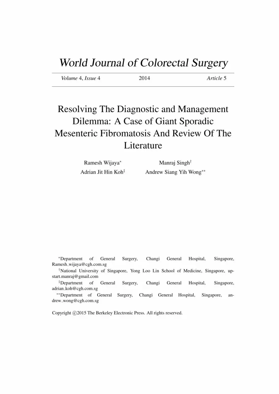
epigastric mass measuring 20 x 10cm. (Figure 1) The mass was non-pulsatile and
moved with respiration. No peripheral lymphadenopathy was found. Laboratory
examinations were not significant. Computed Tomography (CT) of the abdomen
and pelvis revealed a well-defined, heterogeneously enhancing soft-tissue mass
measuring 23cm x 19cm x 11cm in the root of the mesentery. The mass was seen
splaying bowel loops and mesenteric vessels. Fortunately, patency of the superior
mesenteric artery and gastroduodenal artery was preserved. Direct mass effect on
the right ureter resulted in mild right hydroureteronephrosis. There was no intra-
abdominal lymphadenopathy or evidence of bowel obstruction. (Figure 2)
Surgical Technique
In view of the clinical suspicion of a desmoid tumor, the patient was counseled
for and underwent an exploratory laparotomy and resection of the intra-abdominal
tumor. A double-J stent was inserted to decompress the enlarged right ureter prior
to the exploratory laparotomy. Intraoperative findings noted a large mesenteric
mass measuring 25cm x 18.5cm x 16cm and weighed 3.8kg. (Figure 3) The mass
was found originating from the junction of the ileocolic vessels from the superior
1Wijaya et al.: Resolving The Diagnostic and Management Dilemma: A Case of Giant
Produced by The Berkeley Electronic Press, 2014

mesenteric vessels and was closely adherent to the middle colic vessels. It was
also adherent to terminal ileum, caecum and ascending colon. (Figure 4) Bowel
reconstruction was a functional end-to-end ileocolic anastomosis after 100 cm of
small bowel resection and right hemicolectomy was performed for appropriate
tumor clearance. The patient’s post-operative recovery was uneventful and was
discharged well on post-operative day 10.
Pathology
Histological examination revealed interlacing bundles of spindle cells in a mixed
collagenous stroma. Tumour cells were hypochromatic with relatively few mitotic
figures observed. Immunohistochemistry showed positive peri-nuclear β-Catenin
granules but negative nuclear expression. Tumour cells were immunonegative for
Smooth Muscle Actin, Desmin, CD34, CD99, CD117, S-100, DOG-1 and
Epithelial Membrane Antigen. In the context of the given histology, the
immunohistochemical findings were suggestive of mesenteric fibromatosis,
confirming the diagnosis. The patient subsequently underwent gastroscopy and
colonoscopy performed which did not show any intestinal polyposis. Subsequent
imaging at 18 months post-resection showed no evidence of tumor recurrence.
Discussion
Although desmoid tumours may be found extra-abdominally in the soft tissues of
the limbs, mediastinum, head and neck, they are predominantly found intra-
abdominally. Intra-abdominal desmoids tumors are located within the mesentery,
retroperitoneum, pelvis and abdominal wall. These are also known as
aggressive/deep fibromatoses. Intra-abdominal desmoids tumors account for 28-
69% of all desmoids (2) and small bowel mesentery is the commonest site of
occurrence. MF has an incidence of 2-4 per million persons per year. (1) This
accounts for a mere 0.03% of all neoplasms, and 3% of mesenchymal tumours.
(1) The literature suggests a slight female preponderance in MF. However, a clear
gender association is only seen in desmoid tumours occurring in the abdominal
wall. (4) Proposed risk factors for MF include prior abdominal surgery,
abdominal trauma, and hyper-oestrogenaemic states including pregnancy and oral
contraceptive use. (2) Although a possible association with Crohn’s disease has
been reported, it has only been limited to one case report in literature thus far. (3)
Most desmoid tumours are sporadic, with 13-20% related to Gardner’s syndrome
and Familial Adenomatous Polyposis (FAP). (2) Hence, given this genetic
2 World Journal of Colorectal Surgery Vol. 4, Iss. 4 [2014], Art. 5
http://services.bepress.com/wjcs/vol4/iss4/art5
association, all patients with MF should have gastroscopy and colonoscopy to
identify polyposis, which was undertaken in our patient who had no other
documented risk factor of MF.
Of all deep fibromatoses, MF may have a more symptomatic clinical course due
to its location in the mesentery, proximity to surrounding structures and local
aggressiveness. MF may present with one of a myriad of symptoms, such as
abdominal distension (5,6), pain (7), nausea or secondary to local complications
including intestinal obstruction (7), perforation and haemorrhage. (8) Rare
presentations include ureteric obstruction (5), irreducible hernias (6) and fistulae
(9). Huss et al conducted a clinico-histopathlogical study on 56 MF tumours and
reported median tumour length was 9.4cm (range 2-30 cm). (4) Our patient has a
tumour length of 25cm, and based on tumour volume and gross weight of 3.8kg,
represents one of the largest reported in the literature, thus belonging to a clinical
entity described as giant MF. (10)
MF is especially challenging to diagnose due to its bulky, solid appearance,
localised origin in the mesentery and invasion of surrounding tissues on imaging,
akin to Gastro-Intestinal Stromal Tumours (GIST) and spindle cell sarcomas. In
contrast, extra-abdominal or abdominal wall type fibromatosis tend to spread in 2-
dimensional fibrous planes. (4) This peculiar difference in tumour behavior in MF
contributes to radiological misdiagnosis. Huss et al reports a 68% misdiagnosis
rate for MF in his series, with the majority misdiagnosed as GIST. (4) Computed
Tomography (CT) and Magnetic Resonance Imaging (MRI) are the main imaging
modalities used to aid diagnosis, assess pre-operative mass characteristics and
relation to surrounding structures. CT features of MF include a soft tissue
attenuation pattern that may be homogenous or heterogeneous, depending on the
relative amounts of collagenous and myxoid stroma comprising the tumour,
respectively. MRI, with its excellent soft tissue definition, shows up MF as a
heterogeneous intermediate on T1-weighted images and high signal intensities on
T2-weighted images. T2-weighted imaging may also prognosticate the
aggressiveness of MF; stronger signal intensity is associated with rapid tumour
growth. (2,4) Additionally, there has been reported usage of Contrast Enhanced
Ultrasound guided core-biopsy (CEUS) (10), Video-assisted laparoscopic biopsy
(10) with mutational analysis and immunohistochemistry of tissue samples
thereafter to obtain pre-operatively diagnosis of MF over other differentials,
especially GIST and retroperitoneal sarcomas. (4)
The histological diagnosis of MF presents a dilemma due to its similarities to
spindle cell neoplasms, dedifferentiated liposarcomas, GIST, and given its rarity,
the lack of exposure/knowledge in histopathologists. (4) Peculiar microscopic
findings suggestive of MF include: proliferation of uniform elongated spindle
3Wijaya et al.: Resolving The Diagnostic and Management Dilemma: A Case of Giant
Produced by The Berkeley Electronic Press, 2014

cells that infiltrate surrounding tissue, stellate cells with hypochromatic nuclei
within a variable collagenous stroma, thin walled dilated vessels, keloid-like
collagen deposits and paucicellular perivascular spaces. (4,7,11) Lower mitotic
rates and absence of haemorrhage and necrosis are observational differences in
distinguishing MF from GIST. (5) In addition to the abovementioned differentials,
such histology may be misdiagnosed as sclerosing mesenteritis and keloid-like
fibrosis. Immunohistochemistry has become commonplace in aiding diagnosis
and differentiation of MF from GIST. 92.7% of all MF’s had β-Catenin (cadherin-
associated protein) nuclear overexpression/staining, a diagnostic feature of MF in
the given histology, whereas GIST is classically β-Catenin negative. Furthermore,
immunohistochemical staining for CD34, CD117, smooth muscle actin and
desmin are usually negative, features consistent with deep fibromatoses. A high
nuclear positivity for β-Catenin has a specificity of 71% for desmoid type
fibromatosis, with few other mesenchymal tumours mimicking this (synovial
sarcoma, endometrial stromal sarcoma etc). (12) The CTNNB1 gene codes for the
nuclear expression of β- Catenin. Mutations in exon 3 of CTNNB1 shut down
nuclear over-expression of β- Catenin, thereby accounting for immunonegative
MF. (4,10) To account for the remaining 6.3% of MF that are β-Catenin
immunonegative, the corroboration with mutational analysis becomes valuable. In
these cases, identification of either mutations in exon 3 of CTNNB1 or presence
of the Adenomatous Polyposis Coli (APC) gene (found in FAP) inaugurates the
diagnosis.
Lack of a concrete consensus on treatment algorithms for MF is evident, with
varying results from limited case reports and case series available in literature.
Treatment may involve a trial of watchful waiting, surgical excision, systemic
therapy, radiation therapy or a combination of the above. Many authors advocate
a radical approach of surgical excision with negative margins. (5,8,12) However,
a radical excision results in high morbidity, including bleeding, short gut
syndrome and post-operative death. (13) Smith et al documented no survival
differences between resected and unresected patients (median duration of follow
up of 62 months), noting that some MF tumours have prolonged periods of
dormancy or even regression. (13) He attributes this to the biology of desmoids-
which grow rapidly in the initial phases followed by stability or regression.
Therefore, questioning the role of radical treatment, advocating instead for serial
observation or watchful waiting for asymptomatic patients. Nonetheless, surgery
is the definitive solution in symptomatic tumours presenting with intolerable
symptoms or complications such as intestinal obstruction, perforation or
mesenteric ischaemia. (10)
In patients who undergo surgery, a high rate of local recurrence following
surgery, as much as 19-77% has been reported. (2) Given the high risk of local
4 World Journal of Colorectal Surgery Vol. 4, Iss. 4 [2014], Art. 5
http://services.bepress.com/wjcs/vol4/iss4/art5

recurrence following excision, adjuvant therapy is on the rise. Studies have shown
that the combination of wide excision surgery with adjuvant radiotherapy reduced
local recurrence rates. (4,8,12) Gari et al reports a local recurrence of 20-40%
with the addition of adjuvant radiotherapy, compared to 40-70% with resection
alone. (11)
Due to the anatomical position of MF, some tumours lie in close proximity to
vital structures such as the superior mesenteric vessel root, or are excessively
adherent to small bowel (necessitating extensive bowel resection to achieve
negative margins). These are deemed inoperable, and associated with significant
morbidity and mortality if resected as mentioned earlier. Smith et al alludes to
cases of severe post-operative mesenteric ischaemia, dependence on total
parenteral nutrition and death in these patients. (13) In such cases, and where
surgery has failed, systemic therapy may be advocated. A broad range of
interventions, including NSAIDs (non-steroidal anti-inflammatory Drugs)
(Sulindac), anti-oestrogens (Tamoxifen, Raloxifene), Cytotoxics (Methotrexate,
Vinblastine, Doxorubin) and Tyrosine Kinase Inhibitors (Imatinib) have been
reported for inoperable and asymptomatic tumours. However, outcomes have
been unpredictable and sporadic. Janinis et al reports at least partial remission in
44% of patients with MF treated with Sulindac (n=9). (14) The rest had stable or
progressive disease. From her systematic review of pharmacological treatment in
desmoids, she recommends NSAIDs as a 1st
line, followed by Tamoxifen and
cytotoxics as 2nd
and 3rd
line respectively. Raloxifene has also been shown to
reduce tumour size without significant side effects as well. (12) Imatinib, widely
used in GIST therapy has no proven results, while use of Doxorubin and
Dacarbacine have achieved at least partial response in 84% of patients with
desmoids (n=25 across 4 studies). (15) However, side effects of cytotoxics
preclude their use as 1st
line agents. Moreover, most of these studies apply to
desmoids as a whole, with no specific treatment guideline available at present for
MF exclusively. In addition, reports were single-armed with small patient
numbers, thereby weakening the argument for medical therapy. (4,8)
Conclusion
Giant MF is no doubt a rare clinical entity that presents both as a difficult
diagnostic as well as management issue. Despite the use of CT and T2-weighted
MRI and the possible adjunctive use of pre-operative biopsy,
immunohistochemistry and mutational analyses to improve diagnostic rates of
5Wijaya et al.: Resolving The Diagnostic and Management Dilemma: A Case of Giant
Produced by The Berkeley Electronic Press, 2014

MF, the treatment complexity of the disease is still a burden for a clinician. These
tumors are often symptomatic and local tumor invasion may result in morbid
radical resections associated with significant morbidity. Nonetheless, surgery with
disease free resection margins provides the only possibility of cure with no
recurrence.
References
1. Shields CJ, Winter DC, Kirwan WO, Redmond HP. Desmoid tumours. Eur J
Surg Oncol J Eur Soc Surg Oncol Br Assoc Surg Oncol. 2001
Dec;27(8):701–6.
2. Shinagare AB, Ramaiya NH, Jagannathan JP, Krajewski KM, Giardino AA,
Butrynski JE, et al. A to Z of desmoid tumors. AJR Am J Roentgenol.
2011 Dec;197(6):W1008–1014.
3. Slater G, Greenstein AJ. Mesenteric fibromatosis in Crohn’s disease. J Clin
Gastroenterol. 1996 Mar;22(2):147–9.
4. Huss S, Nehles J, Binot E, Wardelmann E, Mittler J, Kleine MA, et al. β-
catenin (CTNNB1) mutations and clinicopathological features of
mesenteric desmoid- type fibromatosis. Histopathology. 2013
Jan;62(2):294–304.
5. Choi JY, Kang KM, Kim BS, Kim T-H. Mesenteric fibromatosis causing
ureteral stenosis. Korean J Urol. 2010 Jul;51(7):501–4.
6. Alsaif FA. Mesenteric fibromatosis presenting as an irreducible inguinal hernia.
Saudi J Gastroenterol Off J Saudi Gastroenterol Assoc. 2011
Oct;17(5):357–9.
7. Misiak P, Piskorz L, Wcislo S, Jablonski S, Brocki M. Giant mesentery
fibromatosis presenting as acute abdomen - case report. Contemp Oncol.
2013;17(5):468–9.
8. Georgiades C, Vallianou N, Argyrakos T, Aristodimou A, Kolovelonis G,
Sioula E. An unusual case of desmoid tumour presenting as haemorrhagic
shock. Ann R Coll Surg Engl. 2012 Mar;94(2):e81–82.
9. Vallabha T, Sindgikar V, Baloorkar R, Dhamangoankar M, Karjol U. Desmoid
6 World Journal of Colorectal Surgery Vol. 4, Iss. 4 [2014], Art. 5
http://services.bepress.com/wjcs/vol4/iss4/art5

infiltrating ileum, a rare complication. Indian J Surg. 2013 Jun;75(Suppl
1):192–4.
10. Wang Y, Cui N-Y, Li L, Zhang R, Hao Y-Z, Xue L-Y, et al. An
abdominal desmoid-type fibromatosis. Quant Imaging Med Surg. 2013
Aug;3(4):228–30.
11. Gari MKM, Guraya SY, Hussein AM, Hego MMN. Giant mesenteric
fibromatosis: Report of a case and review of the literature. World J
Gastrointest Surg. 2012 Mar 27;4(3):79–82.
12. Ng TL, Gown AM, Barry TS, Cheang MCU, Chan AKW, Turbin DA, et
al. Nuclear beta-catenin in mesenchymal tumors. Mod Pathol Off J U S
Can Acad Pathol Inc. 2005 Jan;18(1):68–74.
13. Smith AJ, Lewis JJ, Merchant NB, Leung DH, Woodruff JM, Brennan
MF. Surgical management of intra-abdominal desmoid tumours. Br J
Surg. 2000 May;87(5):608–13.
14. Janinis J, Patriki M, Vini L, Aravantinos G, Whelan JS. The
pharmacological treatment of aggressive fibromatosis: a systematic
review. Ann Oncol Off J Eur Soc Med Oncol ESMO. 2003
Feb;14(2):181–90.
15. Ezumi K, Yamamoto H, Takemasa I, Nomura M, Ikeda M, Sekimoto M,
et al. Dacarbazine-Doxorubicin therapy ameliorated an extremely
aggressive mesenteric desmoid tumor associated with familial
adenomatous polyposis: report of a case. Jpn J Clin Oncol. 2008
Mar;38(3):222–6.
7Wijaya et al.: Resolving The Diagnostic and Management Dilemma: A Case of Giant
Produced by The Berkeley Electronic Press, 2014
Figure 1. Mass in relation to anatomical landmarks
8 World Journal of Colorectal Surgery Vol. 4, Iss. 4 [2014], Art. 5
http://services.bepress.com/wjcs/vol4/iss4/art5

Figure 2. Computed Tomography (CT) of the Abdomen and Pelvis: Sagittal (A),
coronal (B) and axial (C) views
Figure 3. Intraoperative picture of the mass
9Wijaya et al.: Resolving The Diagnostic and Management Dilemma: A Case of Giant
Produced by The Berkeley Electronic Press, 2014

Figure 4. Front and back view of specimen
10 World Journal of Colorectal Surgery Vol. 4, Iss. 4 [2014], Art. 5
http://services.bepress.com/wjcs/vol4/iss4/art5