relationship between gene expression and hybrid vigor in primary root tips of young maize ( zea mays...
TRANSCRIPT

Theor Appl Genet (1990) 80:769-775
�9 Springer-Verlag 1990
Relationship between gene expression and hybrid vigor in primary root tips of young maize (Zea mays L.) plantlets
S. Romagnoli, M. Maddaloni, C. Livini and M. Motto Experimental Institute of Cereal Crops, Bergamo Section, Via Stezzano 24, 1-24100 Bergamo, Italy
Received April 30, 1990; Accepted May 15, 1990 Communicated by H.F. Linskens
Summary. To provide an insight into the molecular basis of heterosis, we investigated gene expression in primary root tips of a heterotic maize hybrid (B73 x Mo17) and its parental lines (B73 and Mo17). This analysis was carried out (i) by differential plaque hybridization of a recombinant cDNA library made to poly(A) RNA iso- lated from B73 x Mo17 primary root tips, and (ii) by comparing with two-dimensional gel electrophoresis proteins synthesized in vitro in the rabbit retieulocyte system by poly(A) RNA isolated, at different stages of development, from the three genotypes. The results showed that there are sets of proteins and mRNAs that are differentially synthesized and expressed in the F1 primary root tips in comparison to the parental lines. Moreover, results from the survey of 21 major in-vitro- synthesized polypeptide variants, from mRNAs of pri- mary root tips of the parental lines and their F~ hybrid, indicated that in seven instances hybrid proteins translat- ed in vitro were more abundant or possibly new. In most of the remaining cases, hybrid spots were similar in inten- sity to the same protein produced by one of the two parental lines.
Key words: Zea mays L. - Hybrid vigor - Gene expres- sion - Protein synthesis - mRNAs
Introduction
In crop plants, hybrid vigor has been recognized from many years (cf. Shull 1952), but its genetic and biochem- ical backgrounds are not yet understood. In maize, hy- brid vigor is particularly evident (Hallauer and Miranda 1981), and the species is a model system for the investiga- tion of the physiological and biochemical nature of this
phenomenon. Attempts to account for hybrid vigor at the physiological level have previously been focused on demonstrating that maize hybrids possess biochemical properties that exceed those of the parental lines, such as mitochondrial oxidation and phosphorylation, nucleic acid synthesis, nitrogen metabolism, plant hormone lev- els, and relative enzyme activities (for a review, see Mc- Daniel 1986).
F1 hybrid seeds of maize have a superior germination capacity in comparison with that of the parental lines (Sarkissian et al. 1964; Sinha and Khanna 1975; Mino 1980). Studies have also indicated that vigorous growth of the embryonic axis in germinating F 1 seed is related to a higher rate of RNA and protein synthesis (Cherry et al. 1961; Mino and Inoue 1980). Similarly, Nebiolo et al. (1983), in a study on RNA metabolism in protoplasts isolated from maize seedlings of a heterotic hybrid and its parents, suggested that the hybrid nucleus may have ad- vantages in the rate of DNA and RNA synthesis.
Because hybrid vigor in maize is detectable at early stages of germination (Sarkissian et al. 1964), the molec- ular analyses presented here focus on the hybrid and its parental inbreds during early seedling growth. The objec- tive was to gain an insight into the expression of genes involved in hybrid vigor by comparative analysis of mRNAs and proteins produced during this early stage of development.
Materials and methods
Plant materials
Two commonly used inbred lines of maize (B73 and Mo17) and their heterotic F 1 hybrid (B73 x Mo17) were included in this study. Self-pollinations and B73 (female) x Mo 17 (male) crosses were made in the summer of 1986 at the Maize Section of the Experimental Institute of Cereal Crops, Bergamo, Italy. The
770
ears were harvested and dried to 10-12% moisture at 35~ in a forced air dryer. Seeds were hand-shelled and stored at 5 ~ until planting.
For each genotype, batches of 100 seeds were surface-steril- ized with 70% ethanol for 90 s, then with 3% sodium hypochlo- rite for 10 rain, rinsed three times in sterile, distilled water, and placed on moistened filter papers in a 150-ram petri dish con- taining 14 seeds. Germination took place at 25~ in a dark incubator, and seeds were kept moistened throughout the exper- iments using sterile water.
Plant tissues were collected at various developmental stages of seedling growth, i.e., 72 h after germination and when the primary roots were 2-4, 8-10, 15-20, and 25-30 mm in length, respectively. At these stages, seedlings were collected, washed three times in distilled water, and tips of the primary roots were aseptically cut off and frozen in liquid nitrogen. Frozen root tips were stored at -80~ until used for RNA extraction.
RNA preparation and dot blots
The root tips were ground to a fine powder in liquid nitrogen using a mortar and pestle and the resulting powder was dis- persed in 10 ml of RNA extraction buffer (50 mM TRIS-HC1, pH 9.0, 10 mM EDTA, 2.0% SDS, 200 mM NaC1, 10 mM B- mercaptoethanol, and 0.2mg/ml proteinase K). Total RNA was isolated essentially as described by Dean et al. (1985) and subjected to two cycles of oligo(dT) cellulose chromatography to isolate poly(A) RNA (Aviv and Leder 1972). RNA was stored in 70% ethanol at - 8 0 ~
RNA dot blots
The RNA dot blot analyses were carried out by the procedure described in Cullimore et al. (1984), and hybridizations were done as described (Maniatis etal. 1982), using 32p-labeled probes (Rigby et al. 1977) derived from inserts selected from phages showing differential hybridization.
Construction and screening of a cDNA library
Total RNA, isolated 72 h after germination at 25 ~ from pri- mary root tips of the B73 x Mo17 hybrid seeds, was used for preparing the cDNA library. Double-stranded DNA copies of the total poly(A) RNA were synthesized according to Maniatis et ai. (1982) and cloned after linker addition into the EcoRI site of bacteriophage lambda NMl149.
The cDNA library (60,000 recombinants) was grown on Escheriehia coIi strain POP 13. Replica filters were differentially screened using radioactive (108 cpm/gg) single-stranded cDNA, synthesized on poly(A) RNAs from primary root tips of B73 and Mo17 inbreds and their F 1 hybrid, respectively, or from poly(A) RNA labeled at the 5'-end (up to 1.5 x 10⁸ cpm/gg), using 32p-ATP and T4 polynucleotide kinase essentially as de- scribed by Maniatis et al. (1982). The procedure for plaque and filter hybridization followed the methods outlined in Maniatis et al. (1982).
DNA preparation and Southern analysis
Phage and plasmid DNA purification and recombinant DNA work followed the procedure outlined in Maniatis et al. (1982). Southern blotting (Southern 1975) and hybridizations were per- formed according to published protocols (Motto et al. 1988).
Hybrid-selected translation
The method used was as previously described by Di Fonzo et al. (1988).
In vitro translation of poly(A) RNA
The poly(A) RNAs for B73, Mo17, and B73 x Mo17, extracted at two different stages of primary root growth (2-4 and 25- 30 mm primary root length) were translated in vitro in a rabbit reticulocyte lysate system, which was prepared and used accord- ing to Jackson and Hunt (1983). For 20-gl translation assays, saturating amounts ofpoly(A) RNA (0.5-1 gg) were incubated for 2 h at 30~ in the presence of [3sS]-methionine. The samples were precipitated with cold acetone and kept in ice for 1 h, then centrifuged for 5 rain at 10,000 9. The pellet, after drying in a vacuum, was dissolved in lysis buffer for two-dimensional elec- trophoretic analysis.
Two-dimensional electrophoresis
The procedure was essentially as described by Barrels et al. (1988). The isoelectric focusing gels contained a mixture of 1/5 Ampholine pH 5-10, 2/5 Ampholine pH4-6, 1/5 Ampholine pH 5 8, 1/5 Ampholine pH 7-9. In the second dimension the focused proteins were separated on a gradient (7.5-15%) poly- acrylamide gel overlaid with a 4% polyacrylamide stacking gel (Laemmli 1970). The gels were fixed in 6% (w/v) TCA, 5% (v/v) ethanol, and then prepared for fluorography according to Bon- net and Laskey (1974). Methylated 14C-proteins were used as molecular-weight markers for SDS gel electrophoresis.
Enzymes and chemicals
DNA restriction endonucleases, DNA polymerase I klenow fragment, reverse transcriptase, RNAse A, and T4-polynucle- otide kinase were purchased from Bethesda Research Laborato- ries and Boehringer. Acrylamide, bisacrylamide, and SDS were obtained from Bio-Rad Laboratories, ampholines from LKB. All other reagents were of the highest purity. Radioehemicals were obtained from Amersham.
Results
Isolation o f cDNAs differentially expressed in primary root tips o f the F 1 and parental lines
To ask whether hybr id vigor may be related to the expres- sion of a specific gene product(s), a e D N A l ibrary was prepared in phage vector l ambda NM1149, with R N A extracted from B73 x Mo17 pr imary root tips dissected 72 h after germination. We used a direct screening proce- dure to identify and isolate clones for genes with differen- tial expression in the F 1 hybrid relative to expression in the parental lines, c D N A clones from the l ibrary were plated at low density and triplicate nitrocellulose lifts were made from each plate. One filter was hybridized with 32P-labeled single-stranded cDNA; this was synthe- sized from the R N A extracted from the pr imary root tips of the hybr id that had been used to construct the library. The second and third filters were hybridized with a sim- ilar probe made from pr imary root tips dissected 72 h after germinat ion from the parental lines. The plaques showing differential hybr idizat ion were picked and plat- ed at 200 pfu/plate. The putat ive differential clones were plaque-purif ied and retested in two more rounds of screening until all plaques scored positively.

771
Fig. 1. Dot-blot analysis of total RNA from the F~ hybrid (B73 x Mo17) and parental lines (B73 and Mo/7) hybridized to 3;P-labeled cDNA inserts that are differentially expressed among the three genotypes. Several dilutions of each RNA sam- ple were applied; a 4.0 gg; b t.0 gg; e 0.25 gg
Fig. 3. [35S]-methionine labeled cell-free reticulocyte system translation products of poly(A) RNA hybrid selected with plas- mids 95,/52, and/81 after SDS-gel electrophoresis and autora- diography. Lane C, reticulocyte lysate without any added poly(A) RNA. Lane T, pattern of in vitro synthesis directed by total poly(A) RNA extracted from the hybrid primary root tips. Molecular weights are shown on the left side of the fluorograph
Fig. 2. Dot-blot analysis of poly(A) RNA from primary root tips isolated at different stages of development of the F 1 hybrid (F) and parental lines Mo17 (M) and B73 (B); 2.0 gg each of mRNA was dotted onto nitrocellulose and hybridized with the indicated 3ZP-labeled DNA probe
Final screening was performed using DNA dot blots of phage DNAs isolated from the putative differential clones. For each clone, 100 gg of DNA was applied to nitrocellulose membranes through the wells of a Hybrid- Dot manifold (Bethesda Research Laboratories). Tripli- cate DNA dot blots were hybridized with similar 3ZP-la- beled probes used in the original screening steps. Only 3 out the 72 putative differential clones were identified that hybridized more intensively to the parental or to the hybrid 32p-labeled single-stranded cDNA. The differen- tial hybridization of these three clones was further con- firmed (i) by repeating the hybridization of a similar experiment with Y-labeled mRNA (data not shown), and (it) by dot-blot analysis of varying amounts of poly(A) RNA for each of three genotypes hybridized to the 32p_ labeled cloned probes (Fig. 1). It was also evident from this last analysis that clone 95 displayed a hybridization intensity intermediate between the parental ones, while clones 152 and /81 showed hybridization intensities of
the F 1 root extracts that were similar to the more intense parental spots. Similar results were also obtained by hy- bridizing each cDNA clone with mRNA isolated at dif- ferent stages of development from primary root tips dis- sected from each of the three genotypes (Fig. 2).
The three cDNA clones differentially expressed in the three genotypes were subcloned into pUC9 plasmid. The insert size of these clones varied from ca. 300 bp to ca. 1,300 bp. They did not show cross-hybridization and rep- resented cDNA copies of mRNAs, as shown by hybrid selection of mRNAs and cell-free translation. Clones 95 and 152 hybridized with a specific mRNA, from primary root tips, that codes for a protein of 54 kDa, while clone /81 hybridized with an RNA that directs in vitro the synthesis of a polypeptide with an apparent molecular weight of 43 kDa (see arrows in Fig. 3).
Expression of proteins translated in vitro from poly(A) RNA derived from F 1 and parentaI lines in primary root tips of young plantlets
Polyadenylated RNA was isolated after germination from each parental line and its F1 hybrid at different stages of primary root growth, and was translated in vitro in a rabbit reticulocyte lysate system. The proteins synthesized were separated by two-dimensional elec- trophoresis. Figure 4 shows in-vitro-synthesized proteins derived from poly(A) RNA of the three genotypes con-
772
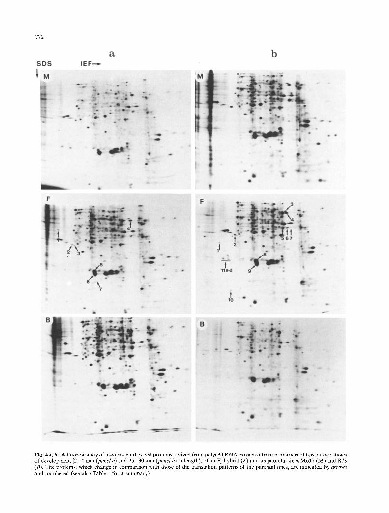
Fig. 4a, b. A fluorography of in-vitro-synthesized proteins derived ~u poly(A) RNA extracted from primary root tips, at two stages of development [2-4 mm (panel a) and 25-30 mm (panel b) in length], of an F i hybrid (F) and its parental lines Mo 17 (M) and B73 (B), The proteins, which change in comparison with those of the translation patterns of the parental lines, are indicated by arrows and numbered (see also Table I for a summary)
773
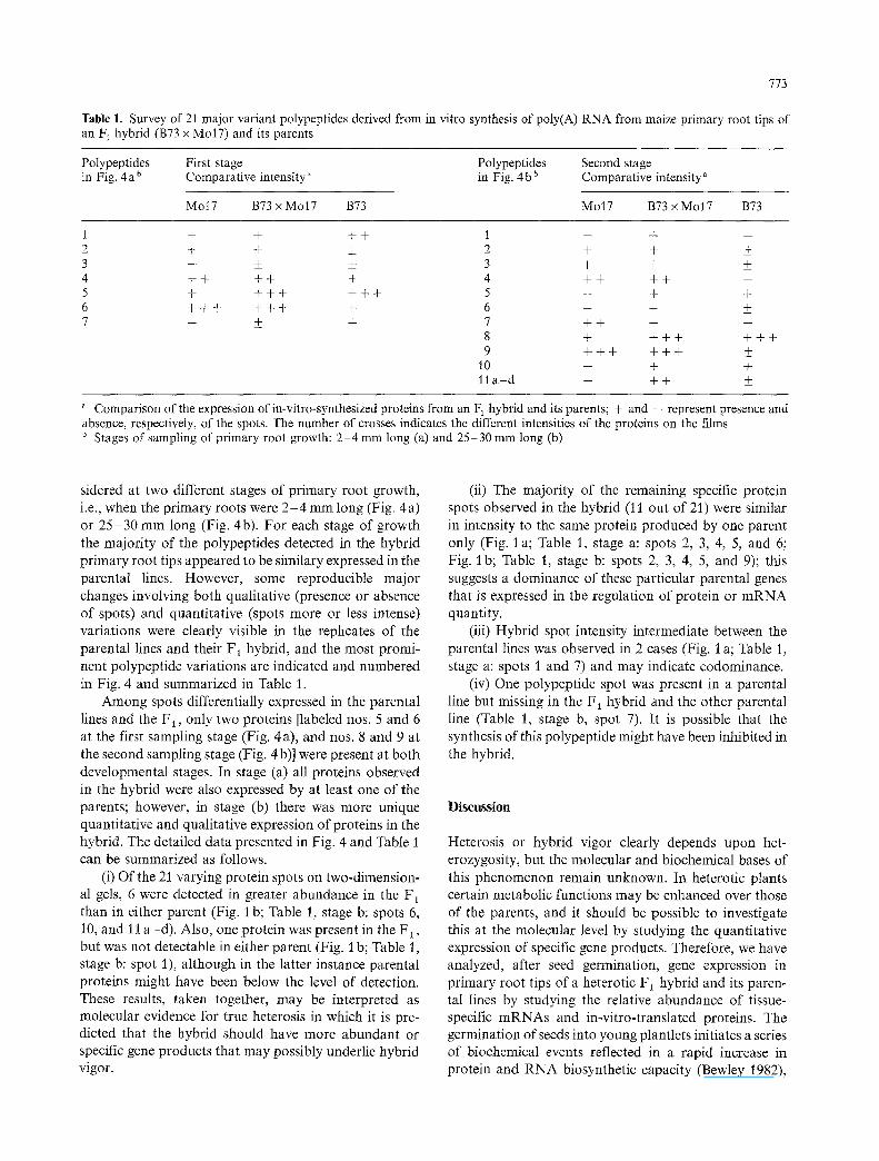
Table 1. Survey of 21 major variant polypeptides derived from in vitro synthesis of poly(A) RNA from maize primary root tips of an F~ hybrid (B73 x MolT) and its parents
Polypeptides First stage Polypeptides Second stage in Fig. 4 a b Comparative intensity ~ in Fig. 4 b b Comparative intensity a
Mo17 B73 x Mo17 B73 Mo17 B73 x Mo17 B73
1 - - + + + 2 + + + 3 -- +_ _+ 4 + + + + + 5 + + + + + + + 6 + + + + + + + 7 -- _+ +
1 - - . j - m
2 + + _+ 3 + + _+ 4 + + + + - 5 - + + 6 -- + + 7 + + -- - 8 + + + + + + + 9 + + + + + + ___
1o - + +
11 a - d - + + _+
a Comparison of the expression of in-vitro-synthesized proteins from an F 1 hybrid and its parents; + and - represent presence and absence, respectively, of the spots. The number of crosses indicates the different intensities of the proteins on the films b Stages of sampling of primary root growth: 2-4 mm long (a) and 25-30 mm long (b)
sidered at two different stages of primary root growth, i.e., when the primary roots were 2 - 4 mm long (Fig. 4a) or 25 -30 mm long (Fig. 4b). For each stage of growth the majority of the polypeptides detected in the hybrid primary root tips appeared to be similary expressed in the parental lines. However, some reproducible major changes involving both qualitative (presence or absence of spots) and quantitative (spots more or less intense) variations were clearly visible in the replicates of the parental lines and their F1 hybrid, and the most promi- nent polypeptide variations are indicated and numbered in Fig. 4 and summarized in Table 1.
Among spots differentially expressed in the parental lines and the F 1, only two proteins [labeled nos. 5 and 6 at the first sampling stage (Fig. 4a), and nos. 8 and 9 at the second sampling stage (Fig. 4 b)] were present at both developmental stages. In stage (a) all proteins observed in the hybrid were also expressed by at least one of the parents; however, in stage (b) there was more unique quantitative and qualitative expression of proteins in the hybrid. The detailed data presented in Fig. 4 and Table i can be summarized as follows.
(i) Of the 21 varying protein spots on two-dimension- al gels, 6 were detected in greater abundance in the F 1 than in either parent (Fig. I b; Table 1, stage b: spots 6, 10, and 11 a -d) . Also, one protein was present in the F~, but was not detectable in either parent (Fig. i b; Table 1, stage b: spot 1), although in the latter instance parental proteins might have been below the level of detection. These results, taken together, may be interpreted as molecular evidence for true heterosis in which it is pre- dicted that the hybrid should have more abundant or specific gene products that may possibly underlie hybrid vigor.
(ii) The majority of the remaining specific protein spots observed in the hybrid (11 out of 21) were similar in intensity to the same protein produced by one parent only (Fig. 1 a; Table 1, stage a: spots 2, 3, 4, 5, and 6; Fig. 1 b; Table 1, stage b: spots 2, 3, 4, 5, and 9); this suggests a dominance of these particular parental genes that is expressed in the regulation of protein or m R N A quantity.
(iii) Hybrid spot intensity intermediate between the parental lines was observed in 2 cases (Fig. I a; Table 1, stage a: spots 1 and 7) and may indicate codominance.
(iv) One polypeptide spot was present in a parental line but missing in the F 1 hybrid and the other parental line (Table 1, stage b, spot 7). It is possible that the synthesis of this polypeptide might have been inhibited in the hybrid.
D i s c u s s i o n
Heterosis or hybrid vigor clearly depends upon het- erozygosity, but the molecular and biochemical bases of this phenomenon remain unknown. In heterotic plants certain metabolic functions may be enhanced over those of the parents, and it should be possible to investigate this at the molecular level by studying the quantitative expression of specific gene products. Therefore, we have analyzed, after seed germination, gene expression in primary root tips of a heterotic F 1 hybrid and its paren- tal lines by studying the relative abundance of tissue- specific m R N A s and in-vitro-translated proteins. The germination of seeds into young plantlets initiates a series of biochemical events reflected in a rapid increase in protein and R N A biosynthetic capacity (Bewley 1982),

774
and this process seems to be specifically and positively related to hybrid vigor (Woodstock and Skoog 1960; Cherry et al. 1961; Mino and Inoue 1980).
Experimental data presented in this paper demon- strate that the F1 primary root tips contain m R N A s that seem to be differentially synthesized and expressed in the hybrid in comparison to the parents. However, these m R N A s may not be necessarily related to heterotic growth, i.e., their synthesis may simply result from the action of genes differentially expressed in hybrids and parental lines at different times during development. In this respect, changes in the relative abundance of m R N A species during plant cell development have been reported in different plant systems including maize (Thompson and Lane 1980; Dure et al. 1981; Aspart et al. 1984; Sanchez De Jimenez and Aguilar 1984; Sanchez-Mar- tinez et al. 1986). Also, with the experimental procedures we have adopted, m R N A s must be present in a certain concentration in order for their translation products to be detected, and low-abundance m R N A s may not be detectable.
Previous studies of hybrid vigor have shown that dominant gene expression may be important for hetero- sis. For example, Leonardi et al. (1987) found, in a study on the inheritance of specific protein amount in two maize inbreds and their hybrids, nonadditive effects on spot intensities in the hybrids, and these data were inter- preted as evidence of dominant gene effects. In another instance, codominant gene expression was observed in F 1 hybrids (Heidrich-Sobrinho and Cordeiro 1975). Also, statistical analyses of maize yield data (cf. Hallauer and Miranda 1981) and a report of hybrid-specific, elevated activities of alcohol dehydrogenase (Schwartz 1973) have provided some previous evidence in support of the con- cept of overdominance in hybrids.
The data herein reported add circumstantial evidence in favor of differential expression of many genes in hy- brid plantlets, since approximately 33% (7 out of 21) of the major hybrid proteins translated in vitro were more abundant or possibly new. Also, despite the small relative number of differences detectable at the molecular level with the techniques we have adopted, this study further suggests that hybrid vigor may derive from simple dom- inant or codominant gene effects in addition to the in- creased expression of certain loci.
Acknowledgements. This study was supported by the Ministry of Agriculture and Forests, Italy, special grant "Physiology of Cul- tivated Plants." We express our gratitude to Dr. Dorothea Bartels for her invaluable assistance in performing two-dimen- sional gel electrophoresis, and to Drs. J. K. Stadler, K Salamini, and R. Thompson for critical reading of the manuscript. C. L. was supported by a postdoctoral fellowship from the Province of Bergamo.
References
Aspart L, Meyer Y, Laroche M, Penon P (1984) Developmental regulation of the synthesis of proteins encoded by stored mRNA in radish embryos. Plant Physiol 76:664-673
Aviv H, Leder P (t 972) Purification of biologically active globin messenger RNA by chromatography on oligothymidilic acid-cellulose. Proc Natl Acad Sci USA 69:1408-1412
Barrels D, Singh M, Salamini F (1988) Onset of desiccation tolerance during development of the barley embryo. Planta 175:485 492
Bewley JD (1982) Protein and nucleic acid synthesis during seed germination and early seedling growth. In: Boulter D, Parthier B (eds) Encyclopedia of plant physiology, vol 14A. Springer, Berlin, pp 559-586
Bonner WM, Laskey RA (1974) A film detection method for tritium-labelled proteins and nucleic acids in polyacrylamide gels. Eur J Biochem 46:83-88
Cherry JH, Hageman RH, Rutger JN, Jones JB (1961) Acid-sol- uble nucleotides and ribonucleic acid of different corn in- breds and single-cross hybrids. Crop Sci 1:133-137
Cullimore JV, Gebhardt C, Saarelainen R, Miflin B J, Idler KB, Barker RF (1984) Glutamine synthetase of Phaseolus vulga- ris L.: organ-specific expression of a multigene family. Mol Gen Genet 2:589-599
Dean C, Van Den Elzen P, Tamaki S, Dunsmuir P, Bedbrook J (1985) Differential expression of the eight subunits of the petunia ribulose bisphosphate carboxylase small subunit multi-gene family. EMBO J 4:3055-3061
Di Fonzo N, Hartings H, Brembilla M, Motto M, Soave C, Navarro E, Palau J, Rhode W, Salamini F (1988) The b-32 protein from maize endosperm, an albumin regulated by the 02 locus: nucleic acid (cDNA) and amino acid sequences. Mol Gen Genet 212:481-487
Dure III LS, Greenway C, Galau G (1981) Developmental bio- chemistry of cottonseed embryogenesis and germination. Changing messenger ribonucleic acid population as shown by in vitro and in vivo protein synthesis. Biochemistry 20:4162-4168
Hatlauer AR, Miranda JB (t 981) Quantitative genetics in maize breeding. Iowa State University Press, Ames/IA
Heidrich-Sobrinho E, Cordeiro AR (1975) Codominant isoen- zymic alleles as markers of genetic diversity correlated with heterosis in maize (Zea mays). Theor Appl Genet 46: 197- 199
Jackson RJ, Hunt T (1983) Preparation and use of nuclease- treated rabbit reticulocyte lysates for the translation of eu- caryotic messenger RNA. Methods Enzymol 96:50-74
Laemmli UK (1970) Cleavage of structural proteins during the assembly of the head of bacteriophage T4. Nature 227: 680- 685
Leonardi A, Damerval C, De Vienne D (1987) Inheritance of protein amounts: comparison of two-dimensional elec- trophoresis patterns of leaf sheaths of two maize lines (Zea mays L.) and their hybrids. Genet Res Camb 50:1-5
Maniatis T, Fritsch EF, Sambrook J (1982) Molecular cloning: a laboratory manual. Cold Spring Harbor Laboratory Press, Cold Spring Harbor/NY
McDaniel RG (1986) Biochemical and physiological basis of heterosis. CRC Crit Rev Plant Sci 4:228-246
Mino M (1980) Hybrid vigor found in some characters of maize seedlings. Jpn J Breed 30(2): 131-138
Mino M, Inoue M (1980) RNA and protein synthesis during germination process of F 1 hybrid and its parental inbred lines of maize. Plant Sci Lett 20: 7-13
Motto M, Maddaloni M, Ponziani G, Brembilla M, Marotta R, Di Fonzo N, Soave C, Thompson R, Salamini F (1988)
775
Molecular cloning of the o2-m5 allele of Zea mays using transposon marking. Mol Gen Genet 212:488-494
Nebiolo CM, Kaczamarczyk WJ, Ulrich V (1983) Manifestation of hybrid vigor in RNA synthesis parameters by corn seedling protoplasts in the presence and absence of gibberel- lic acid. Plant Sci Lett 28:195-206
Rigby PW, Dieckmann M, Rhodes C, Berg P (1977) Labelling of deoxyribonucleic acid to high specific activity in vitro nick-translation with DNA polymerase. J Mol Biol 113:237-251
Sanchez De Jimenez E, Aguilar R (1984) Protein synthesis pat- terns. Relevance of old and new mRNA in germinating maize embryos. Plant Physiol 75:231-234
Sanchez-Martinez D, Puigdomenech P, Pages M (1986) Regula- tion of gene expression in developing Zea mays embryos. Plant Physiol 82:543-549
Sarkissian IV, Kessinger MA, Harris W (1964) Differential rate of developments of heterotic and nonheterotic young maize seedlings. I. Correlation of differential morphological devel- opment with physiological differences in germinating seeds. Proc Natl Acad Sci USA 51:212-218
Schwartz D (1973) Single gene heterosis for alcohol dehydroge- nase in maize: the nature of the subunit interaction. Theor Appl Genet 43:117-120
Shull GH (1952) Beginning of the heterosis concept. In: Gowen JW (ed) Heterosis. Iowa State College Press, Ames/IA, pp 14-48
Sinha SK, Khanna R (1975) Physiological, biochemical, and genetic basis of heterois. Adv Agron 27:123-174
Southern E (1975) Detection of specific sequences among DNA fragments separated by gel electrophoresis. J Mol Biol 98:503 -507
Thompson EW, Lane BG (1980) Relation of protein synthesis in imbibing wheat embryos to the cell-free translational capac- ities of bulk mRNA form dry and imbibing embryos. J Biol Chem 265:5965-5970
Woodstock LW, Skoog F (1960) Relationship between growth rates and nucleic acid contents in the roots of inbred lines of corn. Am J Bot 47:713-716