regulation of smooth muscle by inducible nitric oxide synthase and nadph oxidase in vascular...
TRANSCRIPT
Regulation of Smooth Muscle by Inducible Nitric Oxide Synthaseand NADPHoxidase in Vascular Proliferative Diseases
Roman Ginnan, Benjamin J. Guikema, Katharine E. Halligan, Harold A. Singer, and DavidJourd’heuil§Center for Cardiovascular Sciences, Albany Medical College, Albany, NY
AbstractInflammation plays a critical role in promoting smooth muscle migration and proliferation duringvascular diseases such as post-angioplasty restenosis and atherosclerosis. Another common featureof many vascular diseases is the contribution of reactive oxygen (ROS) and nitrogen (RNS) speciesto vascular injury. Primary sources of ROS and RNS in smooth muscle are several isoforms ofNADPH oxidase (Nox) and the cytokine-regulated inducible nitric oxide (NO) synthase (iNOS). Oneimportant example of the interaction between NO and ROS is the reaction of NO with superoxide toyield peroxynitrite, which may contribute to the pathogenesis of hypertension. In this review, wediscuss the literature that supports an alternate possibility: Nox-derived ROS modulate NObioavailability by altering the expression of iNOS. We highlight data showing co-expression of iNOSand Nox in vascular smooth muscle and demonstrating the functional consequences of iNOS andNox during vascular injury. We describe the relevant literature demonstrating that the mitogenactivated protein kinases (MAP kinases) are important modulators of pro-inflammatory cytokine-dependent expression of iNOS. A central hypothesis discussed is that ROS-dependent regulation ofthe serine/threonine kinase protein kinase Cδ (PKCδ) is essential to understanding how Nox mayregulate signaling pathways leading to iNOS expression. Overall, the integration of non-phagocyticNADPHoxidase with cytokine signaling in general and in vascular smooth muscle in particular ispoorly understood and merit further investigation.
Keywordssmooth muscle; nitric oxide; iNOS; superoxide; NADPHoxidase; protein kinase C; MAPKinase;NF-κB
IntroductionMany vascular diseases such as atherosclerosis, post-angioplasty restenosis, in stent restenosis,and post-transplant coronary arteriopathy are characterized by intimal hyperplasia. Vascularsmooth muscle (VSM) cells in the medial wall of blood vessels are normally quiescent andexpress a differentiated phenotype that serves to generate and maintain vascular tone [123]. Inresponse to de-endotheliazation and increased exposure to cytokines and growth factors, VSMmay dedifferentiate, migrate across the elastic lamina, and proliferate to form a neointimallayer and to secrete extracellular matrix components that form the bulk of the neointimal tissue
§To whom correspondence should be addressed: Albany Medical College, Center for Cardiovascular Sciences, 47 New Scotland Avenue(MC8), Albany, NY 12208; Tel: (518) 262 8104; Fax: (518) 262 8101; E-mail: [email protected]'s Disclaimer: This is a PDF file of an unedited manuscript that has been accepted for publication. As a service to our customerswe are providing this early version of the manuscript. The manuscript will undergo copyediting, typesetting, and review of the resultingproof before it is published in its final citable form. Please note that during the production process errors may be discovered which couldaffect the content, and all legal disclaimers that apply to the journal pertain.
NIH Public AccessAuthor ManuscriptFree Radic Biol Med. Author manuscript; available in PMC 2009 April 1.
Published in final edited form as:Free Radic Biol Med. 2008 April 1; 44(7): 1232–1245.
NIH
-PA Author Manuscript
NIH
-PA Author Manuscript
NIH
-PA Author Manuscript
[135]. The intimal hyperplasia results in vessel narrowing, and clinically manifests itself inrepeat adverse events such as myocardial infarction or repeat intervention. The molecularmechanisms underlying the VSM response to injury and the signaling pathways that controlthe integration of cytokines and growth factors to regulate VSM gene expression are not well-understood. Overall, the complete etiology of intimal hyperplasia remains unknown.
Accumulating evidence point to vascular injury-induced production of reactive oxygen (ROS)and nitrogen (RNS) species as one important mechanism for the regulation of VSM migrationand proliferation in the vascular wall. Recent advances have focused on the identification ofnon-phagocytic NADPH oxidases (Nox) that generate ROS and the realization thatendogenously derived ROS regulate signaling pathways in all primary cellular components ofthe vascular wall. The contribution of nitric oxide synthase (NOS)-derived NO to vascularinjury has been studied in greater details and the convergence between ROS and RNS pathwayshas been focused on specific chemical interactions such as the reaction of NO with superoxide(O2·−) [11] and lipid peroxyl radicals [130].
The alternative that ROS regulate NO production through regulation of iNOS expression hasbeen poorly explored. Surely, the existing literature is pointing out to the importance of bothsuperoxide and hydrogen peroxide in regulating signaling pathways by influencing the activityof multiple kinases and phosphatases and modulating transcription factors. The nucleartranscription factor κB, a primary regulator of iNOS expression, is redox-regulated at multiplelevels including the binding of NFκB to κB motifs [154], regulation of the IκB kinase complex[88], and potentially very upstream through control of the endosomal targeting of cytokinereceptors and Nox [101]. Direct evidence that the redox environment may regulate iNOSexpression in vascular smooth muscle cells has been obtained: increased levels of theantioxidant enzyme catalase increase NFκB activation and iNOS expression after cytokinestimulation of vascular smooth muscle cells [42,60]. These studies indicated that in this contextcytokine-induced hydrogen peroxide production negatively regulates iNOS expression. Thephysiological significance and molecular mechanism underlying these observations areunknown, but would suggest that iNOS expression may be regulated by cytokine-mediatedactivation of Nox either at the level or upstream of transcription factors. The primary purposeof this article is to review potential interactions between Nox signaling and iNOS expressionin vascular smooth muscle. We summarize and contrast what is known regarding thedistribution and roles of iNOS and Nox in the vasculature during injury. Given the complexityof iNOS transcriptional regulation, it would be impossible to cover all potentially relevantsignaling pathways that might regulate iNOS expression in a redox-sensitive manner. Instead,we focus on one specific signaling molecule, protein kinase C, to exemplify some of themolecular mechanisms that may underline the redox regulation of iNOS expression and cross-talks between NOX and iNOS in vascular smooth muscle.
Inducible nitric oxide synthase: where, when, and how much?Mammalian cells synthesize NO through the five-electron oxidation of one of the two-guanidino nitrogen of L-arginine [114]. A family of enzymes generically called nitric oxidesynthase (NOS) catalyzes this reaction from which three major classes have been described:neuronal-NOS (nNOS, type I), inducible-NOS (iNOS, type II) and endothelial-NOS (eNOS,type III). All three isoforms are expressed in the vasculature. The predominant isoform of NOSdetectable in VSM in response to inflammatory cytokines is iNOS [45,84,170] and nNOSupregulation is induced in VSM by shear stress, hypoxia, and growth factors [116,124,159].In the healthy vessel, the endothelium serves as the main source of NO production througheNOS activity to maintain vascular tone, and regulate platelet aggregation and leukocyteadhesion [91,114,126]. Disruption of the endothelial layer and initial loss of eNOS is a hallmarkof the development of atherosclerosis as well as restenosis. Traditionally, the upregulation of
Ginnan et al. Page 2
Free Radic Biol Med. Author manuscript; available in PMC 2009 April 1.
NIH
-PA Author Manuscript
NIH
-PA Author Manuscript
NIH
-PA Author Manuscript
iNOS is perceived to compensate for the loss of a functional endothelium and eNOS duringinjury and atherosclerosis [61], although the presence of excess NO and ROS coincidentallymay lead to additional tissue damage and dysfunction [84].
Early studies of the role of NOS and possible compensation for endothelial dysfunction focusedon iNOS expression in plaque initiation and progression. The presence of iNOS mRNA andprotein expression has been described in atherosclerotic human plaques in macrophages aswell as endothelial and smooth muscle cells [106,162]. In support of a role for iNOS inpromoting pathogenesis, iNOS expression was found in the majority of samples as early as thefatty streak stage and in all of the advanced stages of plaques coinciding with an increase inoxidized LDL and nitrated proteins [106]. The suggestion that iNOS expression and activityare correlated with lipid oxidation within the plaque is also supported by the observation thatmacrophage derived iNOS colocalizes with oxidized lipid and protein derivatives found inatherosclerotic plaques, as well as nitro-tyrosine in advanced atherosclerosis [28]. More recentstudies support a dual role for iNOS in the development of the atherosclerotic plaque. In theapoE−/− mouse model, iNOS is expressed in both macrophages and smooth muscle cells ofthe developing plaque, although smooth muscle cells are not present in early lesions [112]. Inthe advanced atherosclerotic plaques smooth muscle and macrophage derived iNOS may serveto continually promote a pathogenic environment by enhancing oxidative and nitrosative stress.
The rapid upregulation of iNOS in response to vascular injury is also a characteristic featureof balloon angioplasty. Upregulation of iNOS at the mRNA level is observed in the rat carotidartery by 24 hours post injury and it is sustained throughout 14 days [61]. Based onimmunohistochemical analysis, the increase in iNOS expression at 24 hours post injurylocalizes in the smooth muscle cells of the medial layer of the swine carotid artery [9]. Theexpression of iNOS in the media disappears at 5 days post injury, but exhibits intenseexpression at the luminal surface of the neointima that was sustained through 21 days postinjury [9]. In some cases, iNOS protein is expressed as early as 6 hours post injury in the mediallayer of the rat artery due to removal of platelets from the preparation [49]. By contrast, inresponse to periarterial collar placement in the rabbit, no iNOS expression in the injured arteryis detected until the neointima forms approximately 7 days post injury where iNOS expressionis detected in the smooth muscle [5]. Overall, these observations indicate not only a role forendothelial denudation in the induction of iNOS expression in the vessel wall, but also a rolefor platelets in inhibiting iNOS expression. A more recent study in the rabbit demonstrates theco-expression of iNOS with CuZn-superoxide dismustase (SOD) in the vessel wall in responseto balloon injury supporting a role for CuZn-SOD to limit the effects of NO and superoxide-derived peroxynitrite, although nitrotyrosine immunoreactivity is still detectable [83].
The essential role of iNOS during vascular injury: good or bad….once againEarly studies using either pharmacological strategies aimed at modulating NOS activity or genetransfer of different NOS isoforms have provided direct evidence that additional local increasein vascular NO production inhibits smooth muscle cell proliferation by modulating VSMCphenotype and cell cycle [22,84]. Since the last comprehensive review on the role of iNOS invascular injury [84], many studies have taken advantage of transgenic animals to evaluate theimplications of iNOS expression in vascular proliferative diseases (Table 1). Geneticdeficiency of iNOS in itself does not change atherosclerotic lesion size in diet-inducedatherosclerosis [122]. In contrast, all studies but one using the apolipoprotein E-deficient (apoE−/−) mouse model of atherosclerosis have shown decreased atherogenesis in iNOS/apoEdouble knockout animals [23,33,85,93,112]. Perivascular cuffing of the carotid artery in iNOSknockout mouse is also associated with decreased neointima formation indicating acontribution of iNOS to the development of vascular injury [4,26]. The apoE−/− andperivascular cuffing models are characterized by an important contribution from monocytes
Ginnan et al. Page 3
Free Radic Biol Med. Author manuscript; available in PMC 2009 April 1.
NIH
-PA Author Manuscript
NIH
-PA Author Manuscript
NIH
-PA Author Manuscript
and T lymphocytes [18]. In this context, iNOS may directly contribute to injury by promotinginflammation and necrosis at least in advanced lesions. iNOS may also participate in theestablishment of initial lesions by regulating the expansion of cloned Th1 cells [25]. However,Koglin and coworkers found no apparent change in T cell response in iNOS knockout animalsin a model of transplant arteriosclerosis [86]. In fact, in this model a significant increase in thenumber of smooth muscle cells within the neointima was observed in iNOS knockout animalssuggesting that a primary contribution of iNOS in this case was to reduce neointimal SMCaccumulation. Similarly, neointimal formation was found to be increased in iNOS knockoutanimals in a carotid ligation model [139]. Another study using the same model found no changein neointima formation [172], although constrictive remodeling was found to be larger in iNOS-deficient animals compared to wild-type. Increased medial hyperplasia rather than intimalhyperplasia was also described in a model employing wire injury to the endothelium in iNOS-deficient mice [115].
Granted genetic variations between animal strains [62,92], the studies outlined above indicatethat the benefits or drawbacks from inhibiting iNOS vary with the type of vascular injury. Thisis reminiscent of other pathologies in which the role of iNOS as a detrimental or beneficialprotein during inflammation has been already highlighted [90]. For the differences observedin the various vascular injuries, a possibility is that differences in the immune response mayunderline the outcome associated with inhibition of iNOS. In contrast to diet-induced injuries,mechanical injuries are characterized by early and transient lymphocyte and monocyteinfiltration with very little detectable immune cells at the injured site days after the injury. Inthis case, the primary cells expressing iNOS are VSM cells consistent with a role for VSM-derived iNOS in inhibiting VSM proliferation in vascular injury models primarily dominatedby strong VSM migration and proliferation. In models in which the tissue responds with anoverwhelming inflammatory cell contribution throughout the course of the disease, the balancemay be tilted towards iNOS-mediated tissue dysfunction and damage in as much as thesemodels may be also accompanied by large production of ROS. Most investigators have alsoassumed an identical role for iNOS independent of the vascular bed and the multiplicity of celltypes expressing iNOS. The development of cell or tissue specific iNOS –deficient mice shouldrepresent an important approach in order to tease out the different roles of iNOS in complexvascular diseases where both VSM proliferation and the immune response participate.
Regulation of iNOS activity and expressionThe role of iNOS in vascular injury has been traditionally linked to the level of proteinexpression attained in the injured vessels and to the amounts of NO produced by the enzymeitself. Recent biochemical studies indicate that iNOS product release varies with theenvironment and kinetics of the enzyme such that the long-standing dichotomy between thebeneficial effects of low eNOS-derived and high iNOS-derived NO concentrations may beunjustifiable, at least at the level of enzyme activity [144]. For example, medial VSM cells inthe arterial vessel wall exist in a chronic state of hypoxia that is increased during vasculardiseases [132]. This would be inconsistent with the production of large amounts of iNOS-derived NO because the Km of iNOS for molecular oxygen approximates 130 µM [1]. Thebioavailability of L-arginine is also an important regulator of iNOS activity in vivo. Recentstudies indicate that this may involve the catabolism of L-arginine to ornithine and urea byarginase, a metalloenzyme, which expression has been documented in several cell types of thevasculature including smooth muscle cells [37]. Increased expression of arginase duringvascular injury may shunt L-arginine metabolism to ornithine and polyamine synthesis, inhibitiNOS-derived NO production, and promote cell proliferation [37]. Similarly, the requirementof iNOS activity for tetrahydriobiopterin (H4B) is well established and the production ofsuperoxide and peroxynitrite by iNOS in vitro under suboptimal conditions has been described[58,164]. H4B deficiency has been recently investigated as a cause of eNOS dysregulation and
Ginnan et al. Page 4
Free Radic Biol Med. Author manuscript; available in PMC 2009 April 1.
NIH
-PA Author Manuscript
NIH
-PA Author Manuscript
NIH
-PA Author Manuscript
endothelial dysfunction in several clinical settings including artherosclerosis [150] and theimplications of changes in H4B bioavailability for iNOS activity and vascular injury have yetto be described.
In contrast to eNOS, little is known about posttranscriptional modifications that control thelocalization and activity of iNOS. Recent studies have shown palmitoylation of iNOS duringprocessing and activation [77,118,119]. Jones et al. have indicated the importance of calcium/calmodulin-dependent protein kinase II in regulating iNOS trafficking and activity in VSMcells [77]. Activation of the inducible kinin B1 receptor stimulates the production of NO incytokine-treated human lung microvascular endothelial cells [131] and Zhang and coworkershave recently identified ser745 as a physiologically relevant - ERK-dependent –phosphorylation site on human iNOS that enhances NO production [174]. These studies wouldindicate acute regulation of iNOS activity in human cells that express low levels of iNOS.Beyond this, the regulation of mRNA and protein iNOS stability also represents an importantmechanism by which iNOS levels and activity may be regulated and may explain some of thedifferences in iNOS induction observed between different species. For example, the 3’-UTRregion of the human iNOS mRNA contains several AU-rich elements that have been shown todestabilize reporter mRNAs [129]. Recent advances also indicate that epigenetic factorsincluding DNA methylation and histone H3 lysine 9 methylation silence or repress humaniNOS expression in human endothelial and smooth muscle cell cultures [21]. Overall, muchof the research efforts on the regulation of iNOS function have focused on the elucidation ofthe transcriptional regulation of iNOS expression because iNOS expression requires proteinsynthesis and is induced by cytokines and lipopolysaccharides.
The molecular mechanisms regulating iNOS expression were first characterized by cloningthe murine iNOS promoter from the macrophage cell line, RAW 264.7 [105,165]. These studieshave shown that 1,000 bases out of the 1500 bp long promoter confers full activation of iNOStranscription upon stimulation with IFNγ and LPS. The human iNOS promoter, however, isconsiderably larger. The first 3.8kb upstream of the iNOS gene only confers basal activity, 3to 5 fold induction is found in promoter segments containing between 5.8kb and 7.0kbupstream, and 10-fold activation is found in a promoter segment containing 16kb of theupstream 5’ region [30,141]. The rat VSMC iNOS promoter was later cloned, and it wasdemonstrated that 3.2kb of the 5kb region of the iNOS promoter is required for maximumactivation of the promoter upon stimulation with LPS or a mixture of IL-1β, TNFα and IFNγ[38,173]. The mechanisms by which transcription factors and promoter elements regulate iNOSexpression have been extensively reviewed [43]. Analysis of the human, mouse, and rat iNOSpromoters revealed multiple consensus sites for transcription factors that are activated byinflammatory cytokines and lipopolysaccharides. Although these studies show there are largespecies- and cell type-dependent variations in the regulation of iNOS expression, they all showan important role for the NF-κB proteins in the regulation of iNOS transcription.
The transcription factor NF-κB is persistently activated in advanced atherosclerotic lesions andits activation is linked to a wide variety of processes including inflammation, proliferation,differentiation and apoptosis [16,127]. Cytokines such as IL-1β activate NF-κB in many celltypes including VSMC and activation of NF-κB is a requirement for iNOS expression [63,75]. A classical NF-κB activation signaling pathway from the IL-1 receptor (IL-1RI) has beenwell characterized using overexpression in human fibroblasts [121] and the components of thispathway are only beginning to be characterized in VSM cells [82] (see Figure 1). IL-1 bindingto the receptor leads to recruitment of several adaptor proteins including MyD88 [161]. Theseactivate the IL-1 receptor associated kinase (IRAK) which binds TNFα receptor associatedfactor 6 (TRAF6) [17,76]. These two proteins are involved in the activation of the TGFβactivated kinase 1 (TAK1) which phosphorylates the NF-κB inducing kinase (NIK) in the NF-κB signalsome [102,121], which also contains the IκB kinases IKKα and IKKβ as well as the
Ginnan et al. Page 5
Free Radic Biol Med. Author manuscript; available in PMC 2009 April 1.
NIH
-PA Author Manuscript
NIH
-PA Author Manuscript
NIH
-PA Author Manuscript
IκK adaptor protein (IKAP). NIK activates the IKKs which phosphorylate the two NF-κBinhibitory proteins, IκBα and IκBβ [102]. These proteins bind NF-κB in the cytosol under basalconditions. Upon phosphorylation, they are degraded by a ubiquitin-mediated proteosomalpathway and release active NF-κB dimers which translocate to the nucleus to activatetranscription [148]. The inhibitory proteins are under complex temporal regulation allowing atransient phase of NF-κB activation mediated by the quick degradation and subsequentrecovery of IκBα followed by a persistent phase of activation mediated by the slower, longterm degradation of IκBβ [66,75].
There is a large body of literature demonstrating that the activation of the transcription factorNF-κB is regulated by ROS [69]. In vitro, NF-κB binding can be inhibited by N-ethylmaleimideand diamide [154]. Early work also showed that cytokine-induced activation of NF-κB isinhibited by metal chelators and antioxidants in cells [78]. The response of NF-κB to hydrogenperoxide has been studied in many cell types with divergent results showing context dependentregulation [69]. Using transformed alveolar type II cells, Korn and coworkers have shown thatcytokine-induced activation of NF-κB is inhibited by hydrogen peroxide through directoxidation and inactivation of IκB kinase [88]. In contrast, activation of the redox-sensitivePI3K/Akt pathway stimulates NF-κB by promoting the dissociation of phosphorylated IκBα[13]. In the human breast cancer cell-line MCF-7, the recruitment of NIK to TRAF6 in responseto IL-1β stimulation requires endosomal targeting of Nox2, indicating that in this cell type theprimary site of redox regulation of NF-κB occurs very upstream and involves Nox [101].
In rat thoracic VSM cells, pharmacological antioxidants have been shown to modulate NF-κB activation [64,70,88]. We as well as others have shown that NF-κB activity is potentiatedby increased protein levels of catalase, although in this case the mechanisms of action havenot been delineated [42,60]. As described in the next sections, the fact that cytokine-inducediNOS expression in rodent SMC requires the activation of ERK would also suggest that redox-regulation of NF-κB in this system would occur at the level or upstream of mitogen activatedprotein kinases.
The role of MAP kinase and Protein kinase C in regulating iNOS expressionIncreasing evidence indicates that the mitogen activated protein (MAP) kinase family of proteinkinases (ERK1/2, c-Jun Kinase, p38), are important modulators of pro-inflammatory cytokine-dependent expression of inducible nitric oxide synthase (iNOS) in multiple cell types [29,68,97]. Of particular importance in vascular smooth muscle cells (VSM) is the role andmechanisms that couple ERK1/2 to iNOS expression in response to proinflammatorycytokines. An early study by Jiang et al reported that stimulation with IL-1β results in acoordinate activation of ERK1/2 and NF-κB leading to increased expression of iNOS in VSMcells [71]. Furthermore, this study showed that this IL-1β-dependent activation of NF-κB isdependent upon ERK1/2 through the ability of ERK1/2 to phosphorylate I-κBβ resulting in itsdegradation and subsequent translocation of the p65 and p50 subunits to the nucleus to initiategene expression. Further studies showed that ERK1/2 selectively phosphorylates I-κBβ ratherthan I-κBα resulting in sustained NF-κB activity which is required for iNOS gene expression[72,73,168]. The intermediate between ERK1/2 and I-κBβ is ribosomal S6 kinase (RSK) 1[168].
In VSM, the addition of growth factors such as PDGF and EGF enhances ERK1/2-dependentI-κBβ phosphorylation and corresponding increases in NF-κB activity and iNOS expression[72]. Conversely, Angiotensin II (Ang II) has been reported to suppress iNOS expression dueto its ability to activate p38 MAPK which suppresses activation of ERK1/2 [74,117]. Takentogether, these studies support a critical role for ERK1/2 and sustained NF-κB activity inIL-1β-dependent iNOS expression in VSM cells. Other proinflammatory cytokines have also
Ginnan et al. Page 6
Free Radic Biol Med. Author manuscript; available in PMC 2009 April 1.
NIH
-PA Author Manuscript
NIH
-PA Author Manuscript
NIH
-PA Author Manuscript
been reported to mediate a potent immune response after vasculature injury that involvesERK1/2. For example, TNF-α initiates an ERK1/2-dependent expression of severaltranscription factors in VSM cells in a vascular injury model [48] and has also been shown tostimulate the expression of iNOS in an ERK1/2-dependent manner in cultured VSM cells[35].
While the studies reviewed above strongly support a role for ERK1/2 and NF-κB in iNOSexpression, they provide little insight into the signaling molecules upstream of ERK1/2. Instudies designed to elucidate the mechanism by which the proinflammatory cytokine IL-1βinduces ERK1/2 activity in VSM cells, our laboratory investigated the effects of reactiveoxygen species (ROS) on IL-1β-induced iNOS expression. The results from these studiesindicate that along with iNOS expression, IL-1β induces an increase in ROS production inVSM cells and this increase in ROS suppresses ERK1/2 activity and iNOS expression [60].Furthermore, stimulation with IL-1β also results in increased p38MAPK activity, whichsuppresses ERK1/2 activity and iNOS expression in VSM cells. Thus, this report describes inVSM cells the intriguing ability of IL-1β to enhance iNOS expression through activation ofERK1/2 and NF-κB and to suppress iNOS expression through two distinct mechanismsinvolving p38MAPK and increased levels of ROS.
As already stated, little is known regarding the signaling molecules that are activated proximalto IL-1β stimulation in VSM cells. Early studies by Finder et al indicated that IL-1β inducediNOS expression in VSM cells is mediated in a positive manner by proteins that are farnesylatedand in a negative manner by proteins that are geranylgeranylated [41]. These findings aredifficult to interpret in light of the studies discussed previously because they did not report arole for ERK1/2 in IL-1β-dependent increases in iNOS expression. The studies described inthese reports were done in pulmonary VSM cells while the studies discussed earlier werecarried out in thoracic aortic VSM cells suggesting the possibility that the signaling pathwaysIL-1β or other proinflammatory cytokines utilize may be smooth muscle cell type specific.Recently, our laboratory reported that IL-1β-dependent activation of ERK1/2 and increases iniNOS expression in VSM cells were positively mediated by PLCγ and PKCδ [47]. Thus,PLCγ is activated upon IL-1β stimulation and is required for increases in PKCδ activity,activation of ERK1/2, and expression of iNOS. This study confirmed an earlier report ingingival fibroblasts which also provided evidence supporting a requirement for PLCγ inIL-1β-dependent activation of ERK1/2 [158]. Not only is there evidence for the involvementof PLCγ in IL-1β-dependent activation of ERK1/2 in gingival fibroblasts, a series of studiesby McCulloch et al also report the involvement of the traditional mediators of IL-1β signalingsuch as IRAK along with the non-receptor tyrosine kinase focal adhesion kinase (FAK)[104] [108] and the tyrosine phosphatase SHP2 [107]. While little is known concerning theIL-1 receptor and its adaptor molecules in VSM cells, it is tempting to speculate that themechanism by which IL-1β stimulation results in a PLCγ- and PKCδ-dependent activation ofERK1/2 and subsequent increases in iNOS expression in VSM cells is similar.
It is not surprising that PKCδ is involved in this cytokine-dependent activation of ERK1/2.Other studies have reported a role for PKCδ in mediating ERK1/2 activity in VSM cells [46][94]. Interestingly, PKCδ has also been implicated in the activation of MEKK1, a Mek kinaseinvolved in the activation of ERK1/2 and JNK [166,167]. Several studies have reported a rolefor MEKK1 in the regulation of NFκB activity through its ability to mediate IKK activation[99,171]. This raises the interesting possibility that PKCδ may mediate IL-1β-dependentincreases in iNOS expression through its ability to drive ERK1/2 along with its ability toincrease NF-κB activity in a more direct manner. In light of our study indicating a role for ROSin mediating IL-1β-dependent increases in iNOS expression and multiple reports describing arole for ROS in regulating PKCδ activity and function, it is important to determine if PKCδmay serve as an integration point for ROS in the IL-1β-dependent signaling pathway [60].
Ginnan et al. Page 7
Free Radic Biol Med. Author manuscript; available in PMC 2009 April 1.
NIH
-PA Author Manuscript
NIH
-PA Author Manuscript
NIH
-PA Author Manuscript
Our studies also indicate that IL-1β dependent increase in ERK1/2 activity in VSM cells doesnot involve the canonical signaling molecules Ras and RAF [47]. While Ras’ involvement inIL-1-dependent ERK1/2 activation hasn’t been studied elsewhere in VSM cells, reports in othercell systems suggest that Ras is required for IL-1β dependent activation of ERK1/2 andsubsequent increases in iNOS expression [152]. Given the discrepancies of these publishedstudies and the possibility that IL-1β may utilize a non-canonical signaling pathway to ERK1/2,more work is required to elucidate the signal transduction pathways that result in cytokine-dependent activation of ERK1/2 and iNOS expression in VSM cells. Finally, it is clear that aunique signaling pathway (most probably TRAF6/TAK1/IKK) is necessary for the initialactivation of NFκB and for the subsequent persistent activation of NF-κB, which is ERK-dependent. Recent studies have placed other PKC isoforms upstream of TAK1 [138]. Oneexciting possibility is that -in VSM cells- PKCδ itself lies very upstream in the IL-1-signalingpathway regulating both TRAF6/TAK1/IKK and ERK/RSK1.
NADPH oxidase expression and function in vascular injuryAlthough several sources of ROS coexist within the vasculature, NADPH oxidases (Nox) haveemerged as essential components in redox signaling and oxidative stress in the vessel wall. Todate, five distinct Nox isoforms (Nox1,2,3,4 and 5) and two homologous oxidases that containan additional peroxidase domain, termed Duox 1 and 2 (for dual oxidases 1 and 2), have beendiscovered [95]. In vivo, macrophage-induced expression of phagocytic gp91phox (Nox2) aswell as the catalytic subunit p22phox have been characterized within the lesion ofatherosclerotic plaques in human coronary arteries as well as an increase in smooth muscleNox 4 expression within advanced lesions [140]. Sorescu et al. observed the increase inp22phox within the medial layer of the vessel wall as well as the plaque, which agree withearlier studies by Azumi and colleagues [6,140]. The novel observation proved to be thatgp91phox expression is found in the plaque and adventitial macrophages, particularly the coreof the plaque, where Nox 4 expression is complementary to gp91phox located in the medialsmooth muscle layer and luminal surface of the plaque smooth muscle cells.
Little is known about Nox protein expression and function in neointimal formation in responseto vascular injury. Using the balloon injury model in the rat, Griendling and colleaguesobserved that Nox1 as well as p22phox mRNA was upregulated 3 days post injury and sustainedthrough 15 days post injury while there was an increase in gp91phox at 7 days post injurywhich also sustained through 15 days [149]. In addition Nox4 mRNA remained unchangeduntil 15 days post injury where an increase in the mRNA was observed. The same authors usedimmunohistochemistry to confirm that p22phox expression was detected basally in theadventitia of the vessel, but appeared in the medial smooth muscle layer by 7 days post injuryand then most abundantly in the neointima by 10 days post injury, where Nox4 was onlydetected in medial and neointimal cells. However, they were not able to establish the site ofNox1 expression [149]. Moe et al. investigated Nox expression in human aortic smooth musclecells in response to the pro-inflammatory cytokine TNF-α. They show evidence to support anincrease in Nox4 expression, but no change in Nox1 levels [113]. This is in contrast to rodentVSM cells in which an increase in Nox1 and a decrease in Nox4 expression has been observed[98].
Given that atherosclerosis is a chronic disease that progresses over years if not decades inhuman, it is hard to match the expression of proteins to stages of disease progression.Nevertheless, iNOS and Nox protein expression seem to coincide with localized oxidativestress in lesion progression, particularly in the macrophage and smooth muscle cells in themedia and plaque of the vessel. It is difficult to ascertain, however, if the expression of oneprotein precedes the other although it has been shown that iNOS protein is detectable in allstages of plaque development, where the literature only discusses Nox expression in more
Ginnan et al. Page 8
Free Radic Biol Med. Author manuscript; available in PMC 2009 April 1.
NIH
-PA Author Manuscript
NIH
-PA Author Manuscript
NIH
-PA Author Manuscript
advanced atherosclerotic lesions. Overall, the current observations support an early increasein iNOS mRNA and protein levels in the media while Nox protein expression occurs later,mostly when the neointima has already begun to develop between 3 and 7 days post injury.Both iNOS and Nox proteins are expressed at the same time within the same cell type inneointimal hyperplasia, but iNOS expression seems to disappear by day 14 post injury whilethe smooth muscle cells see another wave of Nox protein expression when Nox4 is upregulatedat day 15. This not only suggests different roles for iNOS and Nox proteins, but that perhapsdifferent Nox isoforms have separate roles within the same cells.
In spite of the lack of specific Nox pharmacological inhibitors, studies using knockout ortransgene animals targeting Noxs are lacking. Deletion of the NADPH organizer proteinp47phox in the ApoE−/−mouse significantly reduced lesion area indicating that one or severalNOX isoforms play a role in this animal model [10]. Similarly, p47phox−/− mouse havereduced intimal hyperplasia following transluminal wire injury of the femoral artery [157].The effects of p47phox deficiency in mouse models of vascular injury point to a role forgp91phox. As already stated, upregulation of gp91phox in human atherosclerotic plaques andin animal models has been demonstrated. Pharmacological and molecular manipulation ofgp91phox activity and protein levels also indicate a role for gp91phox in Ang-II-mediatedsmooth muscle cell hypertrophy [103,128].
Nox1 is also an integral component of the signaling pathways associated with angiotensin IIin smooth muscle [98] and recent studies using Nox1-deficient or transgenic mice support animportant role for Nox1 in angiotensin II-induced hypertension [34,110]. The Nox4 isoformhas been generally considered to be constitutively active. It was recently found that the insulinreceptor [156], toll-like receptor 4 [125], transforming growth factor β1 [145,146], and possiblyinterleukin 1β [39,57,59] all required Nox4 activity. In adipocytes, overexpression of Nox4was not sufficient to increase basal levels of ROS but required insulin stimulation, arguing fora regulated Nox4 activity. Functionally, Nox4 may be required for the maintenance of thedifferentiated smooth muscle phenotype [27] and may play an important role in the vascularremodeling associated with the development of pulmonary hypertension [111].
Regulation of NADPH oxidaseThe molecular mechanisms regulating Nox activity have been recently reviewed [20,96]. Ingeneral terms, Noxs are multimeric complexes composed of one or two membrane proteins(Nox and p22phox) as well as cytosolic components including p47phox, NOXO1, p40phox,p67phox, NOXOA1, Rac1 and Rac2. In human and rodent VSM cells, Nox1 and Nox4expression have been documented [65,80,98,140,163], and Nox2 expression has also beenreported in some human VSM cells [79,155]. Nox5 may be expressed in human VSM cellsbut not in rodent [8].
In the case of the phagocytic Nox2, the fully active enzyme requires assembly of Nox2 andp22phox with p47phox, p67phox, and Rac. Activation requires stimulus-induced serinephosphorylation of the C-terminal region of p47phox that ultimately leads to the targeting ofp67phox to the membrane complex [3,100,147]. The N-terminal region of p67phox interactswith GTP-bound Rac and the activation domain of p67phox allows electron flow in thep22phox/Nox2 complex required for superoxide production [56,133]. The fully active NADPHoxidase is also under complex regulation by lipid metabolites that interact with the Phox-homology (PX) domains of p47phox and p40phox [2,67].
The proteins NOXO1 (Nox organizer 1) and NOXA1 (Nox activator 1) are novel homologsof p47phox and p67phox, respectively, that regulate the activity of Nox1 and Nox3 [7,24,44,151]. NOXO1 contains tandem SH3 domains that interact with p22phox like p47phox, but hasno autoinhibitory domain like p47phox. Instead, direct intramolecular interaction of the tandem
Ginnan et al. Page 9
Free Radic Biol Med. Author manuscript; available in PMC 2009 April 1.
NIH
-PA Author Manuscript
NIH
-PA Author Manuscript
NIH
-PA Author Manuscript
SH3 domains with a prolin rich region may allow for regulation of its binding to NOXOA1and p22 [169]. NOXO1 also contains a PX domain, which regulates lipid-protein and protein-protein interactions and unlike the PX domain of p47phox, showed specificity formonophosphorylated phosphoinoside lipids which are present in nonstimulated cells [24].These proteins are expressed primarily in the colon epithelium where ROS production iselevated even in the absence of any apparent stimuli. Nox1 can also interact with p47phox invascular smooth muscle cells, which would render it susceptible to stimulated activation[155]. The function of NOXO1 and NOXOA1 has been described primarily in overexpressionsystems. No studies to date have examined the role of these novel proteins in physiologicalconditions. Though the activation mechanisms of Nox1 are still poorly characterized, even lessis known about the regulation of Nox4 activity which is not enhanced by Rac or any of theknown activating or organizing proteins [81,109]. Recent studies indicate differentpharmacological profiles between Nox isoforms that may allow distinction between isoformsincluding Nox4 and 1 [136,137].
Redox regulation of protein kinase CAs mentioned previously, cytokine stimulation of VSM cells results in NADPH oxidasederived increases in ROS. A signaling molecule that is particularly redox sensitive is proteinkinase C (PKC) and it may provide a good example to illustrate some of the molecularmechanisms that may underline the redox regulation of iNOS expression and cross-talksbetween NOX and iNOS in vascular smooth muscle.
PKC activity can be altered in response to increases in ROS either by direct oxidation of PKCitself or indirectly through oxidant-dependent regulation of kinases or phosphatases thatmodulate PKC activity. Reports suggest that PKC activity can be either inhibited or enhancedwhen exposed to oxidants [55]. The reason for these opposing effects is due to the selectiveoxidation of cysteine residues in either the regulatory or catalytic domain of PKC. The precisemolecular mechanisms resulting in either an increase or decreases in PKC activity have beenreviewed in detail [120]. Briefly, PKCs are comprised of a regulatory and catalytic domain.The regulatory domain consists of a pseudosubstrate region which interacts with the catalyticdomain (which autoinhibits kinase activity) along with domains that bind the necessarycofactors for PKC activation, lipids and/or calcium. Upon binding of these cofactors, theconformation of the regulatory domain of PKC changes relieving autoinhibition and allowingfor the binding of ATP and autophosphophorylation of serine638/641/643 residues in the catalyticdomain to occur, which are required for complete activation of the enzyme [120]. There areseveral excellent reviews for a more complete description of PKC structure and function[120,143,153]. Oxidation of the cysteine residues in the regulatory domain have been shownto induce the conformational changes necessary to relieve autoinhibition of PKC and renderthe kinase constitutively active [51] while oxidation of the cysteine residues in the catalyticdomain have been shown to induce [160] as well as inhibit [50] kinase activity. There is stillmuch we do not understand regarding the effects of the direct oxidation of the cysteine residueson PKC. One of the first studies to examine the role of oxidation on PKC activity clearly showsthat treatments with low levels of the oxidant periodate modified the regulatory domain of PKCresulting in a loss of PKC activity while treatment with higher concentrations modified thecatalytic domain resulting in an increase of PKC activity [52]. Later studies have reported thatexposure to redox active compounds such as hydrogen peroxide [54], organic peroxides [36],and selenocompounds [53] result in the direct oxidation of PKC and change its activity. PKCsare a large family serine/threonine protein kinases subdivided into three subtypes based ontheir requirements for activation and are ubiquitously expressed [120]. Modulation of PKCactivity by direct oxidation as described above is presumably not subtype or PKC isoformspecific or tissue dependent. There is no reported evidence suggesting that oxidation of cysteineresidues is an important modulator of PKC activity in VSM cells although the most likely
Ginnan et al. Page 10
Free Radic Biol Med. Author manuscript; available in PMC 2009 April 1.
NIH
-PA Author Manuscript
NIH
-PA Author Manuscript
NIH
-PA Author Manuscript
method would be through Nox-derived ROS. In support to this contention, our recent studieswould indicate that Nox1 and 4 negatively regulate IL-1β-mediated activation of ERK1/2[59], which is downstream of PKCδ [47].
As mentioned earlier, redox-dependent regulation of PKC activity can also occur in an indirectmanner. It is known that increases in H2O2 result in the oxidation of cysteine residues ontyrosine phosphatases such as SHP2 [32] and serine/threonine phosphatases such as PP2A[134]. The inhibition of tyrosine phosphatase activity results in unregulated activation oftyrosine kinases such as Src [19]. It has been reported that Src-dependent phosphorylation oftyrosine residues on PKCs catalytic domain can augment its activity. Although in vitro studieshave shown that multiple PKCs can be activated in this manner [87], the only member of thePKC family that has been shown to be tyrosine phosphorylated in vivo in VSM cells is PKCδ[31]. PKCδ has multiple tyrosine residues on its regulatory and catalytic domain and in vivostudies have reported that phosphorylation of the tyrosine residues occur exclusively in theregulatory domain [89]. The precise nature of these tyrosine phosphorylations is not clear butthey have been implicated in regulating PKCδ activity [12], as signals for protein degradation[14], and in inducing apoptotic events [15]. Interestingly, PKCs including PKCδ have beenimplicated in the regulation of NADPH oxidase activity [40]. These data identify PKC not onlyas being regulated in an oxidant-dependent manner but also as an important mediator ofintracellular oxidant levels. Another possibility that PKC activity and function may be alteredby increases in an oxidant-dependent manner is through regulation of serine/threoninephosphatases such as PP2A. PP2A, which is redox sensitive [134], is an important regulatorof PKC activity because of its ability to remove phosphates from PKC’s catalytic domain[142].
Summary and concluding remarksThe studies summarized in the present review provide support to the hypothesis that ROSproduction in general, and NADPH oxidases in particular, have a role during vascular injuryin regulating the expression and function of iNOS (see Figure 2). In vascular injury, iNOSexpression has been proposed to be beneficial, as NO inhibits VSM cell proliferation. Clearly,some have questioned the benefit of iNOS expression on injured vessels since large quantitiesof NO may interact with ROS and form cytotoxic species. However, recent studies do showthat iNOS deficiency results in increased neointimal formation in some animal models ofvascular injury. In contrast, Nox-derived ROS act as cell signaling mediators which mayenhance the ability of VSM cells to proliferate into the intima of blood vessels leading to thethickening of the vascular wall. Since NO opposes this function by limiting VSMCproliferation, a novel and attractive possibility is that one or several Nox isoforms maynegatively regulate some of the cell signaling pathways leading to iNOS expression.
There are two primary issues that need to be addressed in order for this research to go forward.The first one is to elucidate the specific mechanisms that lead to selective activation of differentNox isoforms by cytokines. The second one is to determine how cytokine receptor-mediatedNox regulation of relevant signaling molecules such as PKC regulates gene expression. Theimplications of Nox-dependent regulation of PKC during cytokine stimulation wouldobviously extend beyond the regulation of iNOS. The transcription of several other genesbesides iNOS is regulated by the combination of inflammatory cytokines and it is possible thatthe expression of these genes is under the same regulatory mechanisms.
Acknowledgments
This work was supported by grants from the National Heart, Lung, and Blood Institute RO1-HL-40992 and RO1-HL-49426 (to H.A. Singer) and T-32-HL-07194 (to B.J. Guikema, pre-doctoral training grant), the National CancerInstitute CA-89366 (to D. Jourd’heuil), and Philip Morris USA Inc. and Philip Morris International (to D. Jourd’heuil).
Ginnan et al. Page 11
Free Radic Biol Med. Author manuscript; available in PMC 2009 April 1.
NIH
-PA Author Manuscript
NIH
-PA Author Manuscript
NIH
-PA Author Manuscript
Reference List1. Abu-Soud HM, Ichimori K, Nakazawa H, Stuehr DJ. Regulation of inducible nitric oxide synthase by
self-generated NO. Biochemistry 2001;40:6876–6881. [PubMed: 11389602]2. Ago T, Kuribayashi F, Hiroaki H, Takeya R, Ito T, Kohda D, Sumimoto H. Phosphorylation of p47phox
directs phox homology domain from SH3 domain toward phosphoinositides, leading to phagocyteNADPH oxidase activation. Proc. Natl. Acad. Sci. USA 2003;100:4474–4479. [PubMed: 12672956]
3. Ago T, Nunoi H, Ito T, Sumimoto H. Mechanism for Phosphorylation-induced Activation of thePhagocyte NADPH Oxidase Protein p47phox. TRIPLE REPLACEMENT OF SERINES 303, 304,AND 328 WITH ASPARTATES DISRUPTS THE SH3 DOMAIN-MEDIATEDINTRAMOLECULAR INTERACTION IN p47phox, THEREBY ACTIVATING THE OXIDASE.J. Biol. Chem 1999;274:33644–33653. [PubMed: 10559253]
4. Anazawa T, Dimayuga PC, Li H, Tani S, Bradfield J, Chyu KY, Kaul S, Shah PK, Cercek B. Effectof exposure to cigarette smoke on carotid artery intimal thickening: the role of inducible NO synthase.Arterioscler Thromb Vasc Biol 2004;24:1652–1658. [PubMed: 15271786]
5. Arthur JF, Yin ZL, Young HM, Dusting GJ. Induction of nitric oxide synthase in the neointima inducedby a periarterial collar in rabbits. Arterioscler Thromb Vasc Biol 1997;17:737–740. [PubMed:9108788]
6. Azumi H, Inoue N, Takeshita S, Rikitake Y, Kawashima S, Hayashi Y, Itoh H, Yokoyama M.Expression of NADH/NADPH oxidase p22phox in human coronary arteries. Circulation1999;100:1494–1498. [PubMed: 10510050]
7. Banfi B, Clark RA, Steger K, Krause KH. Two novel proteins activate superoxide generation by theNADPH oxidase NOX1. J Biol Chem 2003;278:3510–3513. [PubMed: 12473664]
8. Banfi B, Molnar G, Maturana A, Steger K, Hegedus B, Demaurex N, Krause KH. A Ca(2+)-activatedNADPH oxidase in testis, spleen, and lymph nodes. J Biol Chem 2001;276:37594–37601. [PubMed:11483596]
9. Banning AP, Groves PH, Buttery LD, Wharton J, Wharton J, Rutherford RA, Black P, Winkler F,Polak JM, Lewis MJ, Drexler H. Reciprocal changes in endothelial and inducible nitric oxide synthaseexpression following carotid angioplasty in the pig. Atherosclerosis 1999;145:17–32. [PubMed:10428292]
10. Barry-Lane PA, Patterson C, van der Merwe M, Hu Z, Holland SM, Yeh ETH, Runge MS. p47phoxis required for atherosclerotic lesion progression in ApoE−/− mice. J. Clin. Invest 2001;108:1513–1522. [PubMed: 11714743]
11. Beckman JS, Beckman TW, Chen J, Marshall PA, Freeman BA. Apparent hydroxyl radical productionby peroxynitrite: implications for endothelial injury from nitric oxide and superoxide. Proc. Natl.Acad. Sci.USA 1990;87:1620–1624. [PubMed: 2154753]
12. Benes C, Soltoff SP. Modulation of PKCdelta tyrosine phosphorylation and activity in salivary andPC-12 cells by Src kinases. Am J Physiol Cell Physiol 2001;280:C1498–C1510. [PubMed:11350745]
13. Beraud C, Henzel WJ, Baeuerle PA. Involvement of regulatory and catalytic subunits ofphosphoinositide 3-kinase in NF-kappaB activation. Proc. Natl. Acad Sci U. S. A 1999;96:429–434.[PubMed: 9892650]
14. Blake RA, Garcia-Paramio P, Parker PJ, Courtneidge SA. Src promotes PKCdelta degradation. CellGrowth Differ 1999;10:231–241. [PubMed: 10319993]
15. Blass M, Kronfeld I, Kazimirsky G, Blumberg PM, Brodie C. Tyrosine phosphorylation of proteinkinase Cdelta is essential for its apoptotic effect in response to etoposide. Mol Cell Biol 2002;22:182–195. [PubMed: 11739733]
16. Bu, Dx; Erl, W.; de Martin, R.; Hansson, GK.; Yan, Zq. IKKβ-dependent NF- κBpathway controls vascular inflammation and intimal hyperplasia. The FASEB journal2005:04-264fje.
17. Cao Z, Xiong J, Takeuchi M, Kurama T, Goeddel DV. TRAF6 is a signal transducer for interleukin-1.Nature 1996;383:443–446. [PubMed: 8837778]
18. Carmeliet P, Moons L, Collen D. Mouse models of angiogenesis, arterial stenosis, atherosclerosisand hemostasis. Cardiovasc. Res 1998;39:8–33. [PubMed: 9764187]
Ginnan et al. Page 12
Free Radic Biol Med. Author manuscript; available in PMC 2009 April 1.
NIH
-PA Author Manuscript
NIH
-PA Author Manuscript
NIH
-PA Author Manuscript
19. Catarzi S, Biagioni C, Giannoni E, Favilli F, Marcucci T, Iantomasi T, Vincenzini MT. Redoxregulation of platelet-derived-growth-factor-receptor: role of NADPH-oxidase and c-Src tyrosinekinase. Biochim. Biophys. Acta 2005;1745:166–175. [PubMed: 16129124]
20. Cave AC, Brewer AC, Narayanapanicker A, Ray R, Grieve DJ, Walker S, Shah AM. NADPH oxidasesin cardiovascular health and disease. Antioxid. Redox. Signal 2006;8:691–728. [PubMed: 16771662]
21. Chan GC, Fish JE, Mawji IA, Leung DD, Rachlis AC, Marsden PA. Epigenetic basis for thetranscriptional hyporesponsiveness of the human inducible nitric oxide synthase gene in vascularendothelial cells. J Immunol 2005;175:3846–3861. [PubMed: 16148131]
22. Channon KM, Qian H, George SE. Nitric Oxide Synthase in Atherosclerosis and Vascular Injury :Insights From Experimental Gene Therapy. Arterioscler Thromb Vasc Biol 2000;20:1873–1881.[PubMed: 10938006]
23. Chen J, Kuhlencordt P, Urano F, Ichinose H, Astern J, Huang PL. L-Arginine on Atherosclerosis inApoE Knockout and ApoE/Inducible NO Synthase Double-Knockout Mice. Arterioscler ThrombVasc Biol 2003;23:97–103. [PubMed: 12524231]
24. Cheng G, Lambeth JD. NOXO1, regulation of lipid binding, localization, and activation of Nox1 bythe Phox homology (PX) domain. J Biol. Chem 2004;279:4737–4742. [PubMed: 14617635]
25. Choy JC, Wang Y, Tellides G, Pober JS. Induction of inducible NO synthase in bystander human Tcells increases allogeneic responses in the vasculature. Proc. Natl. Acad. Sci. USA 2007;104:1313–1318. [PubMed: 17227851]
26. Chyu KY, Dimayuga P, Zhu J, Nilsson J, Kaul S, Shah PK, Cercek B. Decreased NeointimalThickening After Arterial Wall Injury in Inducible Nitric Oxide Synthase Knockout Mice. Circ. Res1999;85:1192–1198. [PubMed: 10590247]
27. Clempus RE, Sorescu D, Dikalova AE, Pounkova L, Jo P, Sorescu GP, Lassegue B, Griendling KK.Nox4 is required for maintenance of the differentiated vascular smooth muscle cell phenotype.Arterioscler Thromb Vasc Biol 2007;27:42–48. [PubMed: 17082491]
28. Cromheeke KM, Kockx MM, De Meyer GR, Bosmans JM, Bult H, Beelaerts WJ, Vrints CJ, HermanAG. Inducible nitric oxide synthase colocalizes with signs of lipid oxidation/peroxidation in humanatherosclerotic plaques. Cardiovasc. Res 1999;43:744–754. [PubMed: 10690346]
29. da SJ, Pierrat B, Mary JL, Lesslauer W. Blockade of p38 mitogen-activated protein kinase pathwayinhibits inducible nitric-oxide synthase expression in mouse astrocytes. J. Biol. Chem1997;272:28373–28380. [PubMed: 9353295]
30. de Vera ME, Shapiro RA, Nussler AK, Mudgett JS, Simmons RL, Morris SM Jr, BILLIAR TR, GellerDA. Transcriptional regulation of human inducible nitric oxide synthase (NOS2) gene by cytokines:Initial analysis of the human NOS2 promoter. Proc. Natl. Acad. Sci. USA 1996;93:1054–1059.[PubMed: 8577713]
31. Denning MF, Dlugosz AA, Threadgill DW, Magnuson T, Yuspa SH. Activation of the epidermalgrowth factor receptor signal transduction pathway stimulates tyrosine phosphorylation of proteinkinase C delta. J Biol Chem 1996;271:5325–5331. [PubMed: 8621384]
32. Denu JM, Tanner KG. Specific and reversible inactivation of protein tyrosine phosphatases byhydrogen peroxide: evidence for a sulfenic acid intermediate and implications for redox regulation.Biochemistry 1998;37:5633–5642. [PubMed: 9548949]
33. Detmers PA, Hernandez M, Mudgett J, Hassing H, Burton C, Mundt S, Chun S, Fletcher D, Card DJ,Lisnock J, Weikel R, Bergstrom JD, Shevell DE, Hermanowski-Vosatka A, Sparrow CP, Chao YS,Rader DJ, Wright SD, Pure E. Deficiency in Inducible Nitric Oxide Synthase Results in ReducedAtherosclerosis in Apolipoprotein E-Deficient Mice. J Immunol 2000;165:3430–3435. [PubMed:10975863]
34. Dikalova A, Clempus R, Lassegue B, Cheng G, McCoy J, Dikalov S, San MA, Lyle A, Weber DS,Weiss D, Taylor WR, Schmidt HH, Owens GK, Lambeth JD, Griendling KK. Nox1 overexpressionpotentiates angiotensin II-induced hypertension and vascular smooth muscle hypertrophy intransgenic mice. Circulation 2005;112:2668–2676. [PubMed: 16230485]
35. Doi M, Shichiri M, Katsuyama K, Marumo F, Hirata Y. Cytokine-activated p42/p44 MAP kinase isinvolved in inducible nitric oxide synthase gene expression independent from NF-kappaB activationin vascular smooth muscle cells. Hypertens. Res 2000;23:659–667. [PubMed: 11131279]
Ginnan et al. Page 13
Free Radic Biol Med. Author manuscript; available in PMC 2009 April 1.
NIH
-PA Author Manuscript
NIH
-PA Author Manuscript
NIH
-PA Author Manuscript
36. Donnelly TE Jr, Pelling JC, Anderson CL, Dalbey D. Benzoyl peroxide activation of protein kinaseC activity in epidermal cell membranes. Carcinogenesis 1987;8:1871–1874. [PubMed: 3677312]
37. Durante W, Johnson FK, Johnson RA. Arginase: a critical regulator of nitric oxide synthesis andvascular function. Clin Exp. Pharmacol Physiol 2007;34:906–911. [PubMed: 17645639]
38. Eberhardt W, Kunz D, Hummel R, Pfeilschifter J. Molecular Cloning of the Rat Inducible NitricOxide Synthase Gene Promoter. Biochem. Biophys. Res. Commun 1996;223:752–756. [PubMed:8687469]
39. Ellmark SH, Dusting GJ, Fui MN, Guzzo-Pernell N, Drummond GR. The contribution of Nox4 toNADPH oxidase activity in mouse vascular smooth muscle. Cardiovasc. Res 2005;65:495–504.[PubMed: 15639489]
40. Fan CY, Katsuyama M, Yabe-Nishimura C. PKCdelta mediates up-regulation of NOX1, a catalyticsubunit of NADPH oxidase, via transactivation of the EGF receptor: possible involvement ofPKCdelta in vascular hypertrophy. Biochem J 2005;390:761–767. [PubMed: 15913451]
41. Finder JD, Litz JL, Blaskovich MA, McGuire TF, Qian Y, Hamilton AD, Davies P, Sebti SM.Inhibition of protein geranylgeranylation causes a superinduction of nitric-oxide synthase-2 byinterleukin-1beta in vascular smooth muscle cells. J Biol Chem 1997;272:13484–13488. [PubMed:9153192]
42. Fries DM, Paxinou E, Themistocleous M, Swanberg E, Griendling KK, Salvemini D, Slot JW, HeijnenHFG, Hazen SL, Ischiropoulos H. Expression of Inducible Nitric-oxide Synthase and IntracellularProtein Tyrosine Nitration in Vascular Smooth Muscle Cells: ROLE OF REACTIVE OXYGENSPECIES. J. Biol.Chem 2003;278:22901. [PubMed: 12690103]
43. Ganster, RW.; Geller, DA. Molecular regulation of inducible nitric oxide synthase. In: Ignarro, LJ.,editor. Nitric Oxide Biology and Pathobiology. Academic Press; 2000. p. 129-156.
44. Geiszt M, Lekstrom K, Witta J, Leto TL. Proteins Homologous to p47phox and p67phox SupportSuperoxide Production by NAD(P)H Oxidase 1 in Colon Epithelial Cells. J. Biol. Chem2003;278:20006–20012. [PubMed: 12657628]
45. Geller DA, Lowenstein CJ, Shapiro RA, Nussler AK, Silvio MD, Wang SC, Nakayama DK, SimmonsRL, Snyder SH, Billiar TR. Molecular Cloning and Expression of Inducible Nitric Oxide Synthasefrom Human Hepatocytes. Proc. Natl.Acad. Sci. USA 1993;90:3491–3495. [PubMed: 7682706]
46. Ginnan R, Singer HA. PKC-delta-dependent pathways contribute to PDGF-stimulated ERK1/2activation in vascular smooth muscle. Am. J. Physiol Cell Physiol 2005;288:C1193–C1201.[PubMed: 15677375]
47. Ginnan R, Guikema BJ, Singer HA, Jourd'heuil D. PKC-{delta} mediates activation of ERK1/2 andinduction of iNOS by IL-1beta in vascular smooth muscle cells. Am J Physiol Cell Physiol2006;290:C1583–C1591. [PubMed: 16436473]
48. Goetze S, Kintscher U, Kaneshiro K, Meehan WP, Collins A, Fleck E, Hsueh WA, Law RE. TNFalphainduces expression of transcription factors c-fos, Egr-1, and Ets-1 in vascular lesions throughextracellular signal-regulated kinases 1/2. Atherosclerosis 2001;159:93–101. [PubMed: 11689211]
49. Gonzalez-Fernandez F, Lopez-Farre A, Rodriguez-Feo JA, Farre J, Guerra J, Fortes J, Millas I, Garcia-Duran M, Rico L, Mata P, de Miguel LS, Casado S. Expression of inducible nitric oxide synthaseafter endothelial denudation of the rat carotid artery: role of platelets. Circ Res 1998;83:1080–1087.[PubMed: 9831702]
50. Gopalakrishna R, Anderson WB. Susceptibility of protein kinase C to oxidative inactivation: loss ofboth phosphotransferase activity and phorbol diester binding. FEBS Lett 1987;225:233–237.[PubMed: 2826240]
51. Gopalakrishna R, Anderson WB. Ca2+- and phospholipid-independent activation of protein kinaseC by selective oxidative modification of the regulatory domain. Proc. Natl.Acad Sci U. S. A1989;86:6758–6762. [PubMed: 2505261]
52. Gopalakrishna R, Anderson WB. Reversible oxidative activation and inactivation of protein kinaseC by the mitogen/tumor promoter periodate. Arch. Biochem Biophys 1991;285:382–387. [PubMed:1654774]
53. Gopalakrishna R, Chen ZH, Gundimeda U. Selenocompounds induce a redox modulation of proteinkinase C in the cell, compartmentally independent from cytosolic glutathione: its role in inhibitionof tumor promotion. Arch. Biochem Biophys 1997;348:37–48. [PubMed: 9390172]
Ginnan et al. Page 14
Free Radic Biol Med. Author manuscript; available in PMC 2009 April 1.
NIH
-PA Author Manuscript
NIH
-PA Author Manuscript
NIH
-PA Author Manuscript
54. Gopalakrishna R, Chen ZH, Gundimeda U. Modifications of cysteine-rich regions in protein kinaseC induced by oxidant tumor promoters and enzyme-specific inhibitors. Methods Enzymol1995;252:132–146. [PubMed: 7476346]
55. Gopalakrishna R, Jaken S. Protein kinase C signaling and oxidative stress. Free Radic.Biol. Med2000;28:1349–1361. [PubMed: 10924854]
56. Gorzalczany Y, Alloul N, Sigal N, Weinbaum C, Pick EA. Prenylated p67phox-Rac1 Chimera ElicitsNADPH-dependent Superoxide Production by Phagocyte Membranes in the Absence of an Activatorand of p47phox. CONVERSION OF A PAGAN NADPH OXIDASE TO MONOTHEISM. J. Biol.Chem 2002;277:18605–18610. [PubMed: 11896062]
57. Grange L, Nguyen MV, Lardy B, Derouazi M, Campion Y, Trocme C, Paclet MH, Gaudin P, MorelF. NAD(P)H oxidase activity of Nox4 in chondrocytes is both inducible and involved in collagenaseexpression. Antioxid. Redox. Signal 2006;8:1485–1496. [PubMed: 16987005]
58. Gross SS, Levi R. Tetrahydrobiopterin synthesis. An absolute requirement for cytokine- inducednitric oxide generation by vascular smooth muscle. J. Biol. Chem 1992;267:25722–25729. [PubMed:1281471]
59. Guikema B, Ginnan R, Singer HA, Jourd'heuil D. The NADPH oxidase homologs Nox1 and Nox4attenuate interleukin-1 beta induced niric oxide synthase expression in vascular smooth muscle cells(abstract). Free Radic. Biol. Med 2005;39
60. Guikema BJ, Ginnan R, Singer HA, Jourd'heuil D. Catalase potentiates interleukin-1beta-inducedexpression of nitric oxide synthase in rat vascular smooth muscle cells. Free Radic. Biol. Med2005;38:597–605. [PubMed: 15683716]
61. Hansson GK, Geng YJ, Holm J, Hardhammar P, Wennmalm A, Jennische E. Arterial smooth musclecells express nitric oxide synthase in response to endothelial injury. J Exp. Med 1994;180:733–738.[PubMed: 7519246]
62. Harmon KJ, Couper LL, Lindner V. Strain-Dependent Vascular Remodeling Phenotypes in InbredMice. Am J Pathol 2000;156:1741–1748. [PubMed: 10793085]
63. Hecker M, Cattaruzza M, Wagner AH. Regulation of inducible nitric oxide synthase gene expressionin vascular smooth muscle cells. Gen. Pharmac 1999;32:9–16.
64. Hecker M, Preiss C, Schini-Kerth VB, Busse R. Antioxidants differentially affect nuclear factor kappaB-mediated nitric oxide synthase expression in vascular smooth muscle cells. FEBS Lett1996;380:224–228. [PubMed: 8601429]
65. Hilenski LL, Clempus RE, Quinn MT, Lambeth JD, Griendling KK. Distinct subcellular localizationsof Nox1 and Nox4 in vascular smooth muscle cells. Arterioscler Thromb Vasc Biol 2004;24:677–683. [PubMed: 14670934]
66. Hoffmann A, Levchenko A, Scott ML, Baltimore D. The IkappaB-NF-kappaB signaling module:temporal control and selective gene activation. Science 2002;298:1241–1245. [PubMed: 12424381]
67. Honbou K, Minakami R, Yuzawa S, Takeya R, Suzuki NN, Kamakura S, Sumimoto H, Inagaki F.Full-length p40phox structure suggests a basis for regulation mechanism of its membrane binding.EMBO J 2007;26:1176–1186. [PubMed: 17290225]
68. Hua LL, Zhao ML, Cosenza M, Kim MO, Huang H, Tanowitz HB, Brosnan CF, Lee SC. Role ofmitogen-activated protein kinases in inducible nitric oxide synthase and TNFalpha expression inhuman fetal astrocytes. J. Neuroimmunol 2002;126:180–189. [PubMed: 12020969]
69. Janssen-Heininger YM, Poynter ME, Baeuerle PA. Recent advances towards understanding redoxmechanisms in the activation of nuclear factor kappaB. Free Radic.Biol Med 2000;28:1317–1327.[PubMed: 10924851]
70. Jiang B, Brecher P. N-Acetyl-L-cysteine potentiates interleukin-1beta induction of nitric oxidesynthase : role of p44/42 mitogen-activated protein kinases. Hypertension 2000;35:914–918.[PubMed: 10775561]
71. Jiang B, Brecher P, Cohen RA. Persistent activation of nuclear factor-kappaB by interleukin-1betaand subsequent inducible NO synthase expression requires extracellular signal-regulated kinase.Arterioscler. Thromb. Vasc. Biol 2001;21:1915–1920. [PubMed: 11742864]
72. Jiang B, Xu S, Brecher P, Cohen RA. Growth factors enhance interleukin-1 beta-induced persistentactivation of nuclear factor-kappa B in rat vascular smooth muscle cells. Arterioscler. Thromb. Vasc.Biol 2002;22:1811–1816. [PubMed: 12426209]
Ginnan et al. Page 15
Free Radic Biol Med. Author manuscript; available in PMC 2009 April 1.
NIH
-PA Author Manuscript
NIH
-PA Author Manuscript
NIH
-PA Author Manuscript
73. Jiang B, Xu S, Hou X, Pimentel DR, Brecher P, Cohenx RA. Temporal control of NF-kappaBactivation by ERK differentially regulates interleukin-1beta-induced gene expression. J. Biol. Chem2004;279:1323–1329. [PubMed: 14581482]
74. Jiang B, Xu S, Hou X, Pimentel DR, Cohen RA. Angiotensin II differentially regulates interleukin-1-beta-inducible NO synthase (iNOS) and vascular cell adhesion molecule-1 (VCAM-1) expression:role of p38 MAPK. J Biol Chem 2004;279:20363–20368. [PubMed: 15001568]
75. Jiang B, Xu S, Hou X, Pimentel DR, Brecher P, Cohen RA. Temporal Control of NF-{kappa}BActivation by ERK Differentially Regulates Interleukin-1{beta}- induced Gene Expression. J. Biol.Chem 2004;279:1323–1329. [PubMed: 14581482]
76. Jiang Z, Ninomiya-Tsuji J, Qian Y, Matsumoto K, Li X. Interleukin-1 (IL-1) Receptor-AssociatedKinase-Dependent IL-1-Induced Signaling Complexes Phosphorylate TAK1 and TAB2 at the PlasmaMembrane and Activate TAK1 in the Cytosol. Mol. Cell. Biol 2002;22:7158–7167. [PubMed:12242293]
77. Jones RJ, Jourd'heuil D, Salerno JC, Smith SME, Singer HA. iNOS regulation by calcium/calmodulin-dependent protein kinase II in vascular smooth muscle. Am J Physiol Heart Circ Physiol2007;292:H2634–H2642. [PubMed: 17293490]
78. Jourd'heuil D, Morise Z, Conner EM, Kurose I, Grisham MB. Oxidant-regulation of gene expressionin the chronically inflamed intestine. Keio J. Med 1997;46:10–15. [PubMed: 9095577]
79. Kalinina N, Agrotis A, Tararak E, Antropova Y, Kanellakis P, Ilyinskaya O, Quinn MT, Smirnov V,Bobik A. Cytochrome b558-dependent NAD(P)H oxidase-phox units in smooth muscle andmacrophages of atherosclerotic lesions. Arterioscler Thromb Vasc Biol 2002;22:2037–2043.[PubMed: 12482831]
80. Katsuyama M, Fan C, Yabe-Nishimura C. NADPH oxidase is involved in prostaglandin F2alpha-induced hypertrophy of vascular smooth muscle cells: induction of NOX1 by PGF2alpha. J BiolChem 2002;277:13438–13442. [PubMed: 11832489]
81. Kawahara T, Ritsick D, Cheng G, Lambeth JD. Point Mutations in the Proline-rich Region of p22phoxAre Dominant Inhibitors of Nox1- and Nox2-dependent Reactive Oxygen Generation. J. Biol. Chem2005;280:31859–31869. [PubMed: 15994299]
82. Kawamura A, Baitsch D, Telgmann R, Feuerborn R, Weissen-Plenz G, Hagedorn C, Saku K, Brand-Herrmann SM, von Eckardstein A, Assmann G, Nofer JR. Apolipoprotein E Interrupts Interleukin-1{beta} Signaling in Vascular Smooth Muscle Cells. Arterioscler Thromb Vasc Biol 2007;27:1610–1617. [PubMed: 17510469]
83. Kennedy S, Preston AA, McPhaden AR, Miller AM, Wainwright CL, Wadsworth RM. Correlationof changes in nitric oxide synthase, superoxide dismutase and nitrotyrosine with endothelialregeneration and neointimal hyperplasia in the ballooninjured rabbit subclavian artery. Coron. ArteryDis 2004;15:337–346. [PubMed: 15346092]
84. Kibbe M, Billiar T, Tzeng E. Inducible nitric oxide synthase and vascular injury. Cardiovasc. Res1999;43:650–657. [PubMed: 10690336]
85. Knowles JW, Reddick RL, Jennette JC, Shesely EG, Smithies O, Maeda N. Enhanced atherosclerosisand kidney dysfunction in eNOS-/-Apoe-/- mice are ameliorated by enalapril treatment. J. Clin. Invest2000;105:451–458. [PubMed: 10683374]
86. Koglin, Jo; Glysing-Jensen, T.; Mudgett, JS.; Russell, ME. Exacerbated Transplant Arteriosclerosisin Inducible Nitric OxideûDeficient Mice. Circulation 1998;97:2059–2065. [PubMed: 9610537]
87. Konishi H, Yamauchi E, Taniguchi H, Yamamoto T, Matsuzaki H, Takemura Y, Ohmae K, KikkawaU, Nishizuka Y. Phosphorylation sites of protein kinase C delta in H2O2-treated cells and itsactivation by tyrosine kinase in vitro. Proc. Natl. Acad. Sci.USA 2001;98:6587–6592. [PubMed:11381116]
88. Korn SH, Wouters EFM, Vos N, Janssen-Heininger YMW. Cytokine-induced Activation of NuclearFactor-kappa B Is Inhibited by Hydrogen Peroxide through Oxidative Inactivation of Ikappa BKinase. J. Biol. Chem 2001;276:35693–35700. [PubMed: 11479295]
89. Kronfeld I, Kazimirsky G, Lorenzo PS, Garfield SH, Blumberg PM, Brodie C. Phosphorylation ofprotein kinase Cdelta on distinct tyrosine residues regulates specific cellular functions. J Biol Chem2000;275:35491–35498. [PubMed: 10945993]
Ginnan et al. Page 16
Free Radic Biol Med. Author manuscript; available in PMC 2009 April 1.
NIH
-PA Author Manuscript
NIH
-PA Author Manuscript
NIH
-PA Author Manuscript
90. Kubes P. Inducible nitric oxide synthase: a little bit of good in all of us. Gut 2000;47:6–9. [PubMed:10861252]
91. Kubes P, Suzuki M, Granger DN. Nitric oxide: an endogenous modulator of leukocyte adhesion.Proc. Natl. Acad. Sci. USA 1991;88:4651–4655. [PubMed: 1675786]
92. Kuhel DG, Zhu B, Witte DP, Hui DY. Distinction in Genetic Determinants for Injury-InducedNeointimal Hyperplasia and Diet-Induced Atherosclerosis in Inbred Mice. Arterioscler Thromb VascBiol 2002;22:955–960. [PubMed: 12067904]
93. Kuhlencordt PJ, Chen J, Han F, Astern J, Huang PL. Genetic deficiency of inducible nitric oxidesynthase reduces atherosclerosis and lowers plasma lipid peroxides in apolipoprotein E-knockoutmice. Circulation 2001;103:3099–3104. [PubMed: 11425775]
94. Kumar S, Avraham S, Bharti A, Goyal J, Pandey P, Kharbanda S. Negative regulation of PYK2/related adhesion focal tyrosine kinase signal transduction by hematopoietic tyrosine phosphataseSHPTP1. J. Biol. Chem 1999;274:30657–30663. [PubMed: 10521452]
95. Lambeth JD. NOX enzymes and the biology of reactive oxygen. Nat. Rev. Immunol 2004;4:181–189. [PubMed: 15039755]
96. Lambeth JD, Kawahara T, Diebold B. Regulation of Nox and Duox enzymatic activity and expression.Free Radic. Biol. Med 2007;43:319–331. [PubMed: 17602947]
97. LaPointe MC, Isenovic E. Interleukin-1beta regulation of inducible nitric oxide synthase andcyclooxygenase-2 involves the p42/44 and p38 MAPK signaling pathways in cardiac myocytes.Hypertension 1999;33:276–282. [PubMed: 9931117]
98. Lassegue B, Sorescu D, Szocs K, Yin Q, Akers M, Zhang Y, Grant SL, Lambeth JD, Griendling KK.Novel gp91phox homologues in vascular smooth muscle cells. Circ Res 2001;88:888–894. [PubMed:11348997]
99. Lee FS, Peters RT, Dang LC, Maniatis T. MEKK1 activates both IkappaB kinase alpha and IkappaBkinase beta. Proc. Natl. Acad. Sci. U. S. A 1998;95:9319–9324. [PubMed: 9689078]
100. Leto TL, Adams AG, de Mendez I. Assembly of the Phagocyte NADPH Oxidase: Binding of SrcHomology 3 Domains to Proline-Rich Targets. Proc. Natl. Acad. Sci. USA 1994;91:10650–10654.[PubMed: 7938008]
101. Li Q, Harraz MM, Zhou W, Zhang LN, Ding W, Zhang Y, Eggleston T, Yeaman C, Banfi B,Engelhardt JF. Nox2 and Rac1 regulate H2O2-dependent recruitment of TRAF6 to endosomalinterleukin-1 receptor complexes. Mol Cell Biol 2006;26:140–154. [PubMed: 16354686]
102. Ling L, Cao Z, Goeddel DV. NF-kappaB-inducing kinase activates IKK-alpha by phosphorylationof Ser-176. Proc. Natl. Acad Sci U. S. A 1998;95:3792–3797. [PubMed: 9520446]
103. Liu J, Ormsby A, Oja-Tebbe N, Pagano PJ. Gene transfer of NAD(P)H oxidase inhibitor to thevascular adventitia attenuates medial smooth muscle hypertrophy. Circ Res 2004;95:587–594.[PubMed: 15308582]
104. Lo YY, Luo L, McCulloch CA, Cruz TF. Requirements of focal adhesions and calcium fluxes forinterleukin-1-induced ERK kinase activation and c-fos expression in fibroblasts. J. Biol. Chem1998;273:7059–7065. [PubMed: 9507015]
105. Lowenstein CJ, Alley EW, Raval P, Snowman AM, Snyder SH, Russell SW, Murphy WJ.Macrophage Nitric Oxide Synthase Gene: Two Upstream Regions Mediate Induction by Interferon{gamma} and Lipopolysaccharide. Proc. Natl. Acad.Sci. USA 1993;90:9730–9734. [PubMed:7692452]
106. Luoma JS, Yla-Herttuala S. Expression of inducible nitric oxide synthase in macrophages andsmooth muscle cells in various types of human atherosclerotic lesions. Virchows Arch1999;434:561–568. [PubMed: 10394893]
107. MacGillivray M, Herrera-Abreu MT, Chow CW, Shek C, Wang Q, Vachon E, Feng GS, SiminovitchKA, McCulloch CA, Downey GP. The protein tyrosine phosphatase SHP-2 regulates interleukin-1-induced ERK activation in fibroblasts. J. Biol. Chem 2003;278:27190–27198. [PubMed: 12721296]
108. MacGillivray MK, Cruz TF, McCulloch CA. The recruitment of the interleukin-1 (IL-1) receptor-associated kinase (IRAK) into focal adhesion complexes is required for IL-1beta -induced ERKactivation. J. Biol. Chem 2000;275:23509–23515. [PubMed: 10823834]
Ginnan et al. Page 17
Free Radic Biol Med. Author manuscript; available in PMC 2009 April 1.
NIH
-PA Author Manuscript
NIH
-PA Author Manuscript
NIH
-PA Author Manuscript
109. Martyn KD, Frederick LM, von Loehneysen K, Dinauer MC, Knaus UG. Functional analysis ofNox4 reveals unique characteristics compared to other NADPH oxidases. Cellular Signalling2006;18:69–82. [PubMed: 15927447]
110. Matsuno K, Yamada H, Iwata K, Jin D, Katsuyama M, Matsuki M, Takai S, Yamanishi K, MiyazakiM, Matsubara H, Yabe-Nishimura C. Nox1 is involved in angiotensin II-mediated hypertension: astudy in Nox1-deficient mice. Circulation 2005;112:2677–2685. [PubMed: 16246966]
111. Mittal M, Roth M, Konig P, Hofmann S, Dony E, Goyal P, Selbitz AC, Schermuly RT, GhofraniHA, Kwapiszewska G, Kummer W, Klepetko W, Hoda MAR, Fink L, Hanze J, Seeger W,Grimminger F, Schmidt HHHW, Weissmann N. Hypoxia-Dependent Regulation of NonphagocyticNADPH Oxidase Subunit NOX4 in the Pulmonary Vasculature. Circul. Res 2007;101:258–267.
112. Miyoshi T, Li Y, Shih DM, Wang X, Laubach VE, Matsumoto AH, Helm GA, Lusis AJ, Shi W.Deficiency of inducible NO synthase reduces advanced but not early atherosclerosis inapolipoprotein E-deficient mice. Life Sci 2006;79:525–531. [PubMed: 16516241]
113. Moe KT, Aulia S, Jiang F, Chua YL, Koh TH, Wong MC, Dusting GJ. Differential upregulation ofNox homologues of NADPH oxidase by tumor necrosis factor-alpha in human aortic smooth muscleand embryonic kidney cells. J. Cell. Mol.Med 2006;10:231–239. [PubMed: 16563235]
114. Moncada S, Palmer RMJ, Higgs EA. Nitric oxide:physiology,pathophysiology,and pharmacology.Pharmacol. Rev 1991;43:109–142. [PubMed: 1852778]
115. Moore ZWQ, Hui DY. Apolipoprotein E inhibition of vascular hyperplasia and neointima formationrequires inducible nitric oxide synthase. J. Lipid Res 2005;46:2083–2090. [PubMed: 16061951]
119. Nakata S, Tsutsui M, Shimokawa H, Tamura M, Tasaki H, Morishita T, Suda O, Ueno S, ToyohiraY, Nakashima Y, Yanagihara N. Vascular Neuronal NO Synthase Is Selectively Upregulated byPlatelet-Derived Growth Factor: Involvement of the MEK/ERK Pathway. Arterioscler ThrombVasc Biol 2005;25:2502–2508. [PubMed: 16224055]
117. Nakayama I, Kawahara Y, Tsuda T, Okuda M, Yokoyama M. Angiotensin II inhibits cytokine-stimulated inducible nitric oxide synthase expression in vascular smooth muscle cells. J Biol Chem1994;269:11628–11633. [PubMed: 7512570]
118. Navarro-Lerida I, Corvi MM, Barrientos AA, Gavilanes F, Berthiaume LG, Rodriguez-Crespo I.Palmitoylation of Inducible Nitric-oxide Synthase at Cys-3 Is Required for Proper IntracellularTraffic and Nitric Oxide Synthesis. J. Biol. Chem 2004;279:55682–55689. [PubMed: 15485846]
119. Navarro-Lerida I, varez-Barrientos A, Rodriguez-Crespo I. N-terminal palmitoylation within theappropriate amino acid environment conveys on NOS2 the ability to progress along the intracellularsorting pathways. J. Cell. Sci 2006;119:1558–1569. [PubMed: 16569659]
120. Newton AC. Protein kinase C: structure, function, and regulation. J Biol Chem 1995;270:28495–28498. [PubMed: 7499357]
121. Ninomiya-Tsuji J, Kishimoto K, Hiyama A, Inoue J, Cao Z, Matsumoto K. The kinase TAK1 canactivate the NIK-I kappaB as well as the MAP kinase cascade in the IL-1 signalling pathway. Nature1999;398:252–256. [PubMed: 10094049]
122. Niu XL, Yang X, Hoshiai K, Tanaka K, Sawamura S, Koga Y, Nakazawa H. Inducible Nitric OxideSynthase Deficiency Does Not Affect the Susceptibility of Mice to Atherosclerosis but IncreasesCollagen Content in Lesions. Circulation 2001;103:1115–1120. [PubMed: 11222475]
123. Owens GK, Kumar MS, Wamhoff BR. Molecular regulation of vascular smooth muscle celldifferentiation in development and disease. Physiol Rev 2004;84:767–801. [PubMed: 15269336]
124. Papadaki M, Tilton RG, Eskin SG, McIntire LV. Nitric oxide production by cultured human aorticsmooth muscle cells: stimulation by fluid flow. Am J Physiol Heart Circ Physiol 1998;274:H616–H626.
125. Patel DN, Bailey SR, Gresham JK, Schuchman DB, Shelhamer JH, Goldstein BJ, Foxwell BM,Stemerman MB, Maranchie JK, Valente AJ. TLR4-NOX4-AP-1 signaling mediateslipopolysaccharide-induced CXCR6 expression in human aortic smooth muscle cells. Biochemicaland Biophysical Research Communications 2006;347:1113–1120. [PubMed: 16870145]
126. Radomski MW, Palmer RMJ, Moncada S. An L-arginine/nitric oxide pathway present in humanplatelets regulates aggregation. Proc. Natl. Acad. Sci. U. S A 1990;87:5193–5197. [PubMed:1695013]
Ginnan et al. Page 18
Free Radic Biol Med. Author manuscript; available in PMC 2009 April 1.
NIH
-PA Author Manuscript
NIH
-PA Author Manuscript
NIH
-PA Author Manuscript
127. Rainer, dM; Hoeth, M.; Hofer-Warbinek, R.; Schmid, JA. The Transcription Factor NF-{kappa}Band the Regulation of Vascular Cell Function. Arterioscler Thromb Vasc Biol 2000;20:e83–e88.[PubMed: 11073859]
128. Rey FE, Cifuentes ME, Kiarash A, Quinn MT, Pagano PJ. Novel competitive inhibitor of NAD(P)H oxidase assembly attenuates vascular O2 and systolic blood pressure in mice. Circ Res2001;89:408–414. [PubMed: 11532901]
129. Rodriguez-Pascual F, Hausding M, Ihrig-Biedert I, Furneaux H, Levy AP, Forstermann U, KleinertH. Complex contribution of the 3′-untranslated region to the expressional regulation of the humaninducible nitric-oxide synthase gene. Involvement of the RNA-binding protein HuR. J Biol Chem2000;275:26040–26049. [PubMed: 10859327]
130. Rubbo H, Radi R, Trujillo M, Telleri R, Kalyanaraman B, Barnes S, Kirk M, Freeman BA. Nitricoxide regulation of superoxide and peroxynitrite-dependent lipid peroxidation. J. Biol. Chem1994;269:26066–26075. [PubMed: 7929318]
131. Sangsree S, Brovkovych V, Minshall RD, Skidgel RA. Kininase I-type carboxypeptidases enhancenitric oxide production in endothelial cells by generating bradykinin B1 receptor agonists. Am JPhysiol Heart Circ Physiol 2003;284:H1959–H1968. [PubMed: 12623793]
132. Santilli SM, Tretinyak AS, Lee ES. Transarterial wall oxygen gradients at the deployment site ofan intra-arterial stent in the rabbit. Am J Physiol Heart Circ Physiol 2000;279:H1518–H1525.[PubMed: 11009436]
133. Sarfstein R, Gorzalczany Y, Mizrahi A, Berdichevsky Y, Molshanski-Mor S, Weinbaum C,Hirshberg M, Dagher MC, Pick E. Dual Role of Rac in the Assembly of NADPH Oxidase, Tetheringto the Membrane and Activation of p67phox: A STUDY BASED ON MUTAGENESIS OFp67phox-Rac1 CHIMERAS. J. Biol. Chem 2004;279:16007–16016. [PubMed: 14761978]
134. Sasaki K, Murata M, Yasumoto T, Mieskes G, Takai A. Affinity of okadaic acid to type-1 andtype-2A protein phosphatases is markedly reduced by oxidation of its 27- hydroxyl group. BiochemJ 1994;298(Pt 2):259–262. [PubMed: 8135728]
135. Schwartz SM. Perspectives series: cell adhesion in vascular biology. Smooth muscle migration inatherosclerosis and restenosis. J Clin Invest 1997;99:2814–2816. [PubMed: 9185501]
136. Serrander L, Cartier L, Bedard K, Banfi B, Lardy B, Plastre O, Sienkiewicz A, Forro L, SchlegelW, Krause KH. NOX4 activity is determined by mRNA levels and reveals a unique pattern of ROSgeneration. Biochem J 2007;406:105–114. [PubMed: 17501721]
137. Serrander L, Jaquet V, Bedard K, Plastre O, Hartley O, Arnaudeau S, Demaurex N, Schlegel W,Krause KH. NOX5 is expressed at the plasma membrane and generates superoxide in response toprotein kinase C activation. Biochimie 2007;89:1159–1167. [PubMed: 17587483]
138. Shinohara H, Yasuda T, Aiba Y, Sanjo H, Hamadate M, Watarai H, Sakurai H, Kurosaki T. PKC{beta} regulates BCR-mediated IKK activation by facilitating the interaction between TAK1 andCARMA1. J. Exp. Med 2005;202:1423–1431. [PubMed: 16301747]
139. Sirsjo A, Lofving A, Hansson GK, Wagsater D, Tokuno S, Valen G. Deficiency of nitric oxidesynthase 2 results in increased neointima formation in a mouse model of vascular injury. JCardiovasc. Pharmacol 2003;41:897–902. [PubMed: 12775967]
140. Sorescu D, Weiss D, Lassegue B, Clempus RE, Szocs K, Sorescu GP, Valppu L, Quinn MT, LambethJD, Vega JD, Taylor WR, Griendling KK. Superoxide production and expression of nox familyproteins in human atherosclerosis. Circulation 2002;105:1429–1435. [PubMed: 11914250]
141. Spitsin SV, Koprowski H, Michaels FH. Characterization and functional analysis of the humaninducible nitric oxide synthase gene promoter. Mol. Med 1996;2:226–235. [PubMed: 8726465]
142. Srivastava J, Goris J, Dilworth SM, Parker PJ. Dephosphorylation of PKCdelta by proteinphosphatase 2Ac and its inhibition by nucleotides. FEBS Lett 2002;516:265–269. [PubMed:11959144]
143. Steinberg SF. Distinctive activation mechanisms and functions for protein kinase Cdelta. Biochem.J 2004;384:449–459. [PubMed: 15491280]
144. Stuehr DJ, Santolini J, Wang ZQ, Wei CC, Adak S. Update on Mechanism and Catalytic Regulationin the NO Synthases. J. Biol. Chem 2004;279:36167–36170. [PubMed: 15133020]
145. Sturrock A, Huecksteadt TP, Norman K, Sanders K, Murphy TM, Chitano P, Wilson K, Hoidal JR,Kennedy TP. Nox4 mediates TGF-beta1-induced retinoblastoma protein phosphorylation,
Ginnan et al. Page 19
Free Radic Biol Med. Author manuscript; available in PMC 2009 April 1.
NIH
-PA Author Manuscript
NIH
-PA Author Manuscript
NIH
-PA Author Manuscript
proliferation, and hypertrophy in human airway smooth muscle cells. Am J Physiol Lung Cell MolPhysiol 2007;292:L1543–L1555. [PubMed: 17369289]
146. Sturrock A, Cahill B, Norman K, Huecksteadt TP, Hill K, Sanders K, Karwande SV, Stringham JC,Bull DA, Gleich M, Kennedy TP, Hoidal JR. Transforming growth factor-beta1 induces Nox4 NAD(P)H oxidase and reactive oxygen species-dependent proliferation in human pulmonary arterysmooth muscle cells. Am J Physiol Lung Cell Mol Physiol 2006;290:L661–L673. [PubMed:16227320]
147. Sumimoto H, Kage Y, Nunoi H, Sasaki H, Nose T, Fukumaki Y, Ohno M, Minakami S, TakeshigeK. Role of Src Homology 3 Domains in Assembly and Activated of the Phagocyte NADPH Oxidase.Proc. Natl. Acad. Sci. USA 1994;91:5345–5349. [PubMed: 8202490]
148. Sun Z, Andersson R. NF-kappaB activation and inhibition: a review. Shock 2002;18:99–106.[PubMed: 12166787]
149. Szöcs K, Lassègue B, Sorescu D, Hilenski LL, Valppu L, Cousse TL, Wilcox JN, Quinn MT,Griendling KK. Upregulation of Nox-based NAD(P)H oxidases in restenosis after carotid injury.Arterioscler. Thromb. Vasc. Biol 2002;22:21–27. [PubMed: 11788456]
150. Takaya T, Hirata Ki, Yamashita T, Shinohara M, Sasaki N, Inoue N, Yada T, Goto M, Fukatsu A,Hayashi T, Alp NJ, Channon KM, Yokoyama M, Kawashima S. A Specific Role for eNOS-DerivedReactive Oxygen Species in Atherosclerosis Progression. Arterioscler Thromb Vasc Biol2007;27:1632–1637. [PubMed: 17463333]
151. Takeya R, Ueno N, Kami K, Taura M, Kohjima M, Izaki T, Nunoi H, Sumimoto H. Novel HumanHomologues of p47phox and p67phox Participate in Activation of Superoxide-producing NADPHOxidases. J. Biol. Chem 2003;278:25234–25246. [PubMed: 12716910]
152. Tannous M, Amin R, Popoff MR, Fiorentini C, Kowluru A. Positive modulation by Ras ofinterleukin-1beta-mediated nitric oxide generation in insulin-secreting clonal beta (HIT-T15) cells.Biochem Pharmacol 2001;62:1459–1468. [PubMed: 11728382]
153. Toker A. Signaling through protein kinase C. Front Biosci 1998;3:D1134–D1147. [PubMed:9792904]
154. Toledano MN, Leonard WJ. Modulation of transcription factor NF-kB binding activity by oxidation-reduction in vitro. Proc. Natl. Acad. Sci. USA 1991;88:4328–4332. [PubMed: 1903539]
155. Touyz RM, Yao G, Schiffrin EL. c-Src induces phosphorylation and translocation of p47phox: rolein superoxide generation by angiotensin II in human vascular smooth muscle cells. ArteriosclerThromb Vasc Biol 2003;23:981–987. [PubMed: 12663375]
156. Ushio-Fukai M, Maziar Zafari A, Fukui T, Ishizaka N, Griendling KK. p22phox is a criticalcomponent of the superoxide-generating NADH/NADPH oxidase system and regulates angiotensinII-induced hypertrophy in vascular smooth muscle cells. J. Biol. Chem 1996;271:23317–23321.[PubMed: 8798532]
157. Vendrov AE, Madamanchi NR, Hakim ZS, Rojas M, Runge MS. Thrombin and NAD(P)H Oxidase-Mediated Regulation of CD44 and BMP4-Id Pathway in VSMC, Restenosis, and Atherosclerosis.Circul. Res 2006;98:1254–1263.
158. Wang Q, Downey GP, Herrera-Abreu MT, Kapus A, McCulloch CA. SHP-2 modulatesinterleukin-1-induced Ca2+ flux and ERK activation via phosphorylation of phospholipaseCgamma1. J. Biol. Chem 2005;280:8397–8406. [PubMed: 15563458]
159. Ward ME, Toporsian M, Scott JA, Teoh H, Govindaraju V, Quan A, Wener AD, Wang G, BevanSC, Newton DC, Marsden PA. Hypoxia induces a functionally significant and translationallyefficient neuronal NO synthase mRNA variant. J. Clin. Invest 2005;115:3128–3139. [PubMed:16276418]
160. Ward NE, Gravitt KR, O'Brian CA. Covalent modification of protein kinase C isozymes by theinactivating peptide substrate analog N-biotinyl-Arg-Arg-Arg-Cys-Leu-Arg-Arg-Leu. Evidencethat the biotinylated peptide is an active-site affinity label. J Biol Chem 1996;271:24193–24200.[PubMed: 8798661]
161. Wesche H, Henzel WJ, Shillinglaw W, Li S, Cao Z. MyD88: an adapter that recruits IRAK to theIL-1 receptor complex. Immunity 1997;7:837–847. [PubMed: 9430229]
Ginnan et al. Page 20
Free Radic Biol Med. Author manuscript; available in PMC 2009 April 1.
NIH
-PA Author Manuscript
NIH
-PA Author Manuscript
NIH
-PA Author Manuscript
162. Wilcox JN, Subramanian RR, Sundell CL, Tracey WR, Pollock JS, Harrison DG, Marsden PA.Expression of Multiple Isoforms of Nitric Oxide Synthase in Normal and Atherosclerotic Vessels.Arterioscler Thromb Vasc Biol 1997;17:2479–2488. [PubMed: 9409218]
163. Wingler K, Wunsch S, Kreutz R, Rothermund L, Paul M, Schmidt HH. Upregulation of the vascularNAD(P)H-oxidase isoforms Nox1 and Nox4 by the reninangiotensin system in vitro and in vivo.Free Radic. Biol Med 2001;31:1456–1464. [PubMed: 11728818]
164. Xia Y, Dawson VL, Dawson TM, Snyder SH, Zweier JL. Nitric oxide synthase generates superoxideand nitric oxide in arginine-depleted cells leading to peroxynitrite-mediated cell injury. Proc. Natl.Acad. Sci. USA 1996;93:6770–6774. [PubMed: 8692893]
165. Xie QW, Whisnant R, Nathan C. Promoter of the mouse gene encoding calciumindependent nitricoxide synthase confers inducibility by interferon gamma and bacterial lipopolysaccharide. J. Exp.Med 1993;177:1779–1784. [PubMed: 7684434]
166. Xu S, Cobb MH. MEKK1 binds directly to the c-Jun N-terminal kinases/stress-activated proteinkinases. J Biol Chem 1997;272:32056–32060. [PubMed: 9405400]
167. Xu S, Robbins D, Frost J, Dang A, Lange-Carter C, Cobb MH. MEKK1 phosphorylates MEK1 andMEK2 but does not cause activation of mitogen-activated protein kinase. Proc. Natl. Acad Sci U.S. A 1995;92:6808–6812. [PubMed: 7624324]
168. Xu S, Bayat H, Hou X, Jiang B. Ribosomal S6 kinase-1 modulates interleukin-1betainducedpersistent activation of NF-{kappa}B through phosphorylation of I{kappa}Bbeta. Am J PhysiolCell Physiol 2006;291:C1336–C1345. [PubMed: 16822942]
169. Yamamoto A, Kami K, Takeya R, Sumimoto H. Interaction between the SH3 domains and C-terminal proline-rich region in NADPH oxidase organizer 1 (Noxo1). Biochem. Biophys. ResCommun 2007;352:560–565. [PubMed: 17126813]
170. Yan, Zq; Hansson, GK. Overexpression of Inducible Nitric Oxide Synthase by Neointimal SmoothMuscle Cells. Circul. Res 1998;82:21–29.
171. Yin MJ, Christerson LB, Yamamoto Y, Kwak YT, Xu S, Mercurio F, Barbosa M, Cobb MH, GaynorRB. HTLV-I Tax protein binds to MEKK1 to stimulate IkappaB kinase activity and NF-kappaBactivation. Cell 1998;93:875–884. [PubMed: 9630230]
172. Yogo K, Shimokawa H, Funakoshi H, Kandabashi T, Miyata K, Okamoto S, Egashira K, Huang P,Akaike T, Takeshita A. Different Vasculoprotective Roles of NO Synthase Isoforms in VascularLesion Formation in Mice. Arterioscler Thromb Vasc Biol 2000;20:96e–100e. [PubMed:10634805]
173. Zhang H, Chen X, Teng X, Snead C, Catravas JD. Molecular Cloning and Analysis of the RatInducible Nitric Oxide Synthase Gene Promoter in Aortic Smooth Muscle Cells. Biochem.Pharmacol 1998;55:1873–1880. [PubMed: 9714306]
174. Zhang Y, Brovkovych V, Brovkovych S, Tan F, Lee BS, Sharma T, Skidgel RA. Dynamic receptor-dependent activation of inducible nitric-oxide synthase by ERK-mediated phosphorylation ofSer745. J Biol Chem 2007;282:32453–32461. [PubMed: 17804409]
Ginnan et al. Page 21
Free Radic Biol Med. Author manuscript; available in PMC 2009 April 1.
NIH
-PA Author Manuscript
NIH
-PA Author Manuscript
NIH
-PA Author Manuscript
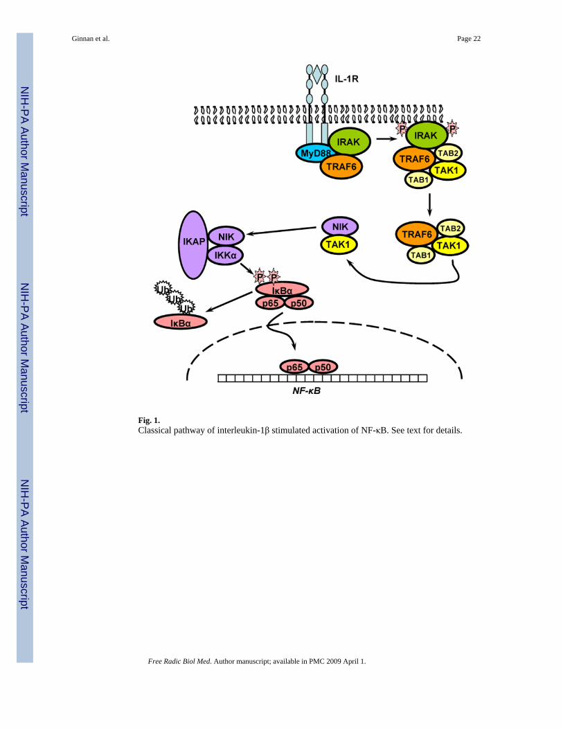
Fig. 1.Classical pathway of interleukin-1β stimulated activation of NF-κB. See text for details.
Ginnan et al. Page 22
Free Radic Biol Med. Author manuscript; available in PMC 2009 April 1.
NIH
-PA Author Manuscript
NIH
-PA Author Manuscript
NIH
-PA Author Manuscript
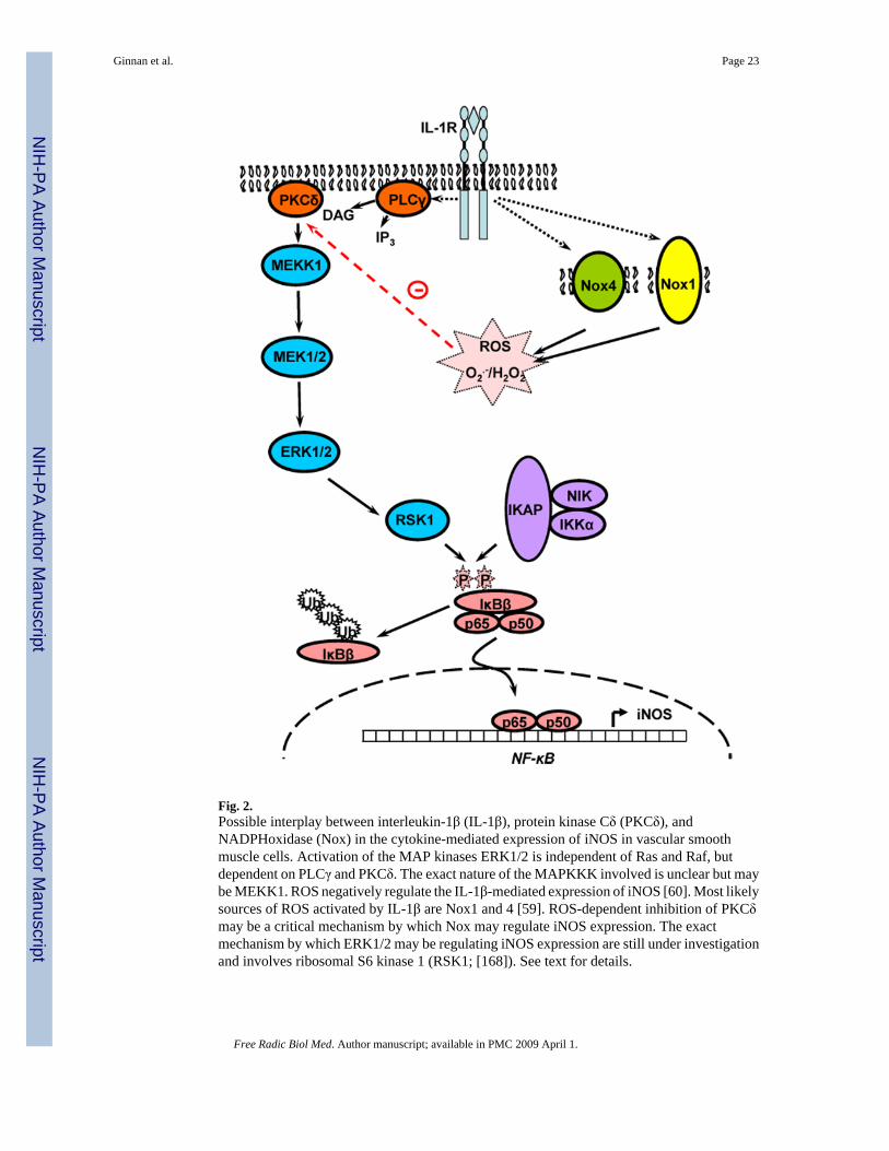
Fig. 2.Possible interplay between interleukin-1β (IL-1β), protein kinase Cδ (PKCδ), andNADPHoxidase (Nox) in the cytokine-mediated expression of iNOS in vascular smoothmuscle cells. Activation of the MAP kinases ERK1/2 is independent of Ras and Raf, butdependent on PLCγ and PKCδ. The exact nature of the MAPKKK involved is unclear but maybe MEKK1. ROS negatively regulate the IL-1β-mediated expression of iNOS [60]. Most likelysources of ROS activated by IL-1β are Nox1 and 4 [59]. ROS-dependent inhibition of PKCδmay be a critical mechanism by which Nox may regulate iNOS expression. The exactmechanism by which ERK1/2 may be regulating iNOS expression are still under investigationand involves ribosomal S6 kinase 1 (RSK1; [168]). See text for details.
Ginnan et al. Page 23
Free Radic Biol Med. Author manuscript; available in PMC 2009 April 1.
NIH
-PA Author Manuscript
NIH
-PA Author Manuscript
NIH
-PA Author Manuscript
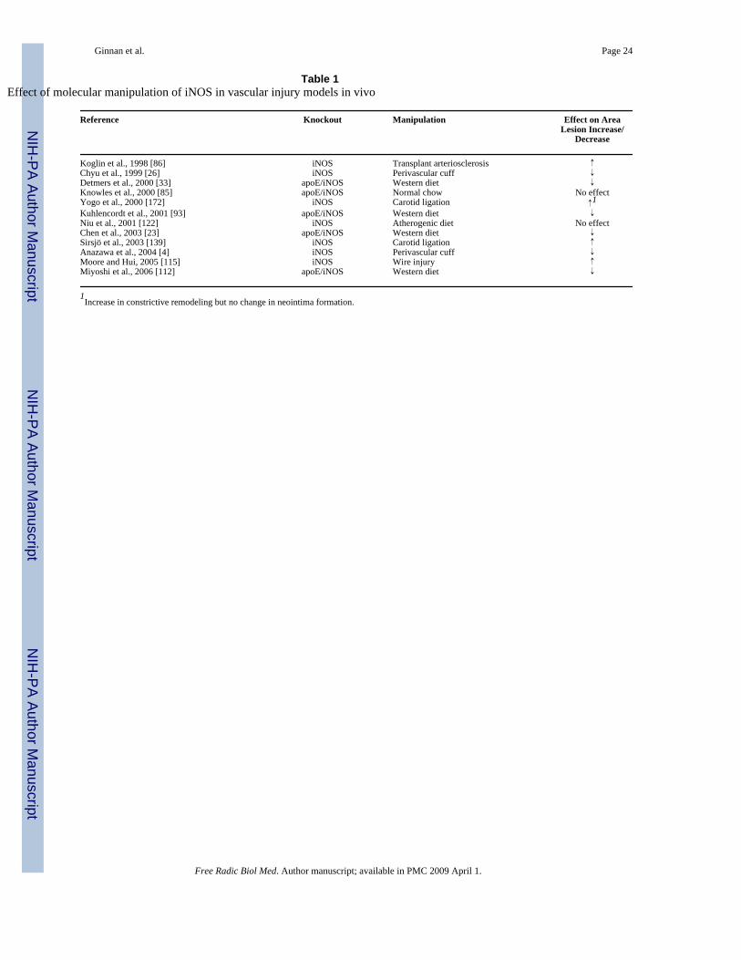
NIH
-PA Author Manuscript
NIH
-PA Author Manuscript
NIH
-PA Author Manuscript
Ginnan et al. Page 24
Table 1Effect of molecular manipulation of iNOS in vascular injury models in vivo
Reference Knockout Manipulation Effect on AreaLesion Increase/
Decrease
Koglin et al., 1998 [86] iNOS Transplant arteriosclerosis ↑Chyu et al., 1999 [26] iNOS Perivascular cuff ↓Detmers et al., 2000 [33] apoE/iNOS Western diet ↓Knowles et al., 2000 [85] apoE/iNOS Normal chow No effectYogo et al., 2000 [172] iNOS Carotid ligation ↑1Kuhlencordt et al., 2001 [93] apoE/iNOS Western diet ↓Niu et al., 2001 [122] iNOS Atherogenic diet No effectChen et al., 2003 [23] apoE/iNOS Western diet ↓Sirsjö et al., 2003 [139] iNOS Carotid ligation ↑Anazawa et al., 2004 [4] iNOS Perivascular cuff ↓Moore and Hui, 2005 [115] iNOS Wire injury ↑Miyoshi et al., 2006 [112] apoE/iNOS Western diet ↓
1Increase in constrictive remodeling but no change in neointima formation.
Free Radic Biol Med. Author manuscript; available in PMC 2009 April 1.