refractive surgery on the side? - the ophthalmologist
TRANSCRIPT
JANUARY / FEBRUARY 2021
In My ViewOn establishing audacious targets for global eye care 13
In PracticeUncovering the hidden secrets of the cornea
28 – 31
ProfessionA practical guide to patient-initiated harassment
42 – 45
Sitting Down WithHumanitarian, Ashiyana Nariani
50 – 51
# 51
www.theophthalmologist.comN O R T H A M E R I C A
Refractive Surgery on the Side?Not anymore. Four accomplished surgeons share their views on the subspecialty’s future in our second refractive roundtable 16 – 23
EYL.20.04.0075_REEYR21308_EYLEA RS MEfRVO Ad_8.27x10.48_FINAL.indd 1-2EYL.20.04.0075_REEYR21308_EYLEA RS MEfRVO Ad_8.27x10.48_FINAL.indd 1-2 1/12/21 12:53 PM1/12/21 12:53 PM
EYL.20.04.0075_REEYR21308_EYLEA RS MEfRVO Ad_8.27x10.48_FINAL.indd 1-2EYL.20.04.0075_REEYR21308_EYLEA RS MEfRVO Ad_8.27x10.48_FINAL.indd 1-2 1/12/21 12:53 PM1/12/21 12:53 PM

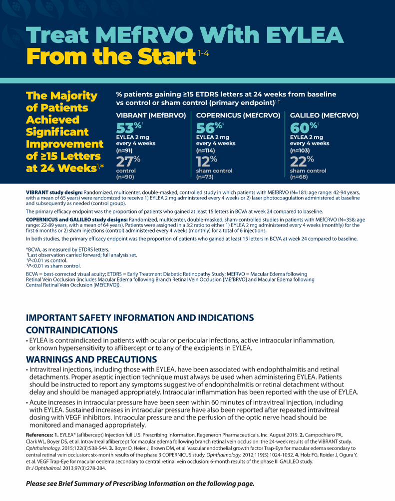
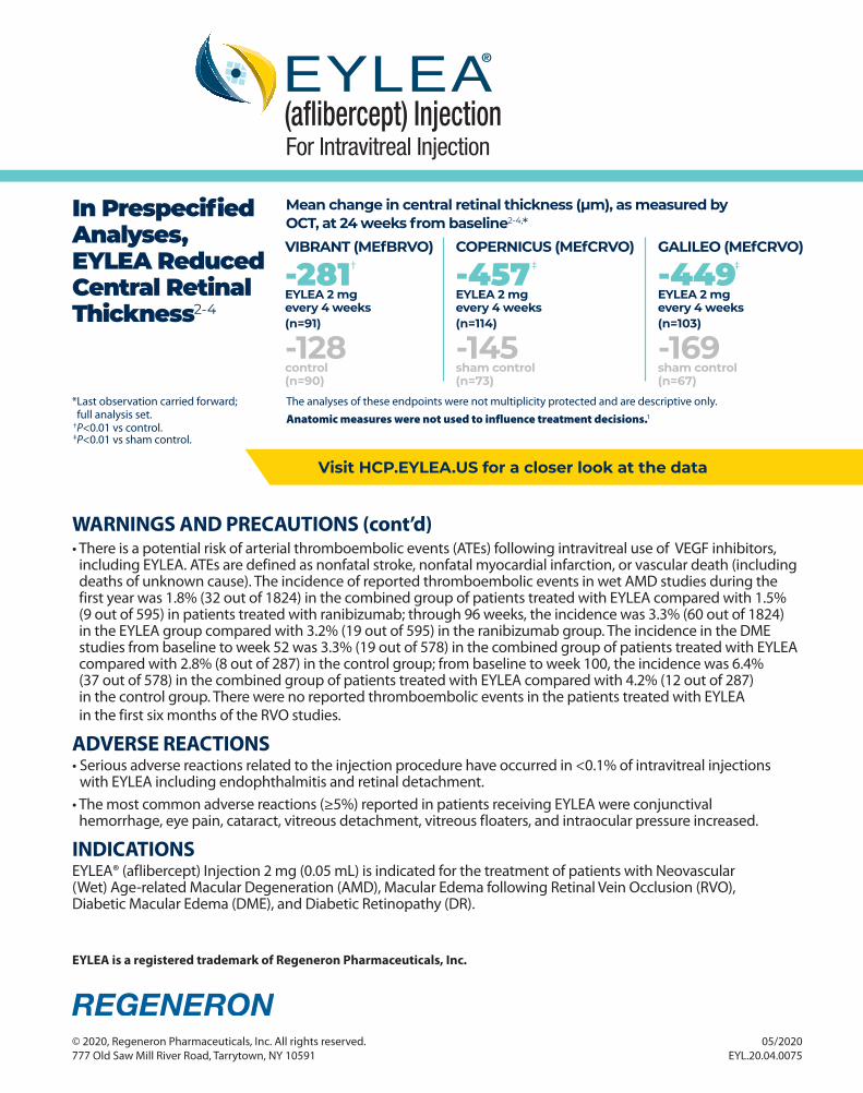
1 INDICATIONS AND USAGE EYLEA is a vascular endothelial growth factor (VEGF) inhibitor indicated for the treatment of:Neovascular (Wet) Age-Related Macular Degeneration (AMD); Macular Edema Following Retinal Vein Occlusion (RVO); Diabetic Macular Edema (DME); Diabetic Retinopathy (DR).4 CONTRAINDICATIONS4.1 Ocular or Periocular Infections EYLEA is contraindicated in patients with ocular or periocular infections. 4.2 Active Intraocular Inflammation EYLEA is contraindicated in patients with active intraocular inflammation. 4.3 Hypersensitivity EYLEA is contraindicated in patients with known hypersensitivity to aflibercept or any of the excipients in EYLEA. Hypersensitivity reactions may manifest as rash, pruritus, urticaria, severe anaphylactic/anaphylactoid reactions, or severe intraocular inflammation.5 WARNINGS AND PRECAUTIONS 5.1 Endophthalmitis and Retinal Detachments. Intravitreal injections, including those with EYLEA, have been associated with endophthalmitis and retinal detachments [see Adverse Reactions (6.1)]. Proper aseptic injection technique must always be used when administering EYLEA. Patients should be instructed to report any symptoms suggestive of endophthalmitis or retinal detachment without delay and should be managed appropriately [see Patient Counseling Information (17)].5.2 Increase in Intraocular Pressure. Acute increases in intraocular pressure have been seen within 60 minutes of intravitreal injection, including with EYLEA [see Adverse Reactions (6.1)]. Sustained increases in intraocular pressure have also been reported after repeated intravitreal dosing with vascular endothelial growth factor (VEGF) inhibitors. Intraocular pressure and the perfusion of the optic nerve head should be monitored and managed appropriately.5.3 Thromboembolic Events. There is a potential risk of arterial thromboembolic events (ATEs) following intravitreal use of VEGF inhibitors, including EYLEA. ATEs are defined as nonfatal stroke, nonfatal myocardial infarction, or vascular death (including deaths of unknown cause). The incidence of reported thromboembolic events in wet AMD studies during the first year was 1.8% (32 out of 1824) in the combined group of patients treated with EYLEA compared with 1.5% (9 out of 595) in patients treated with ranibizumab; through 96 weeks, the incidence was 3.3% (60 out of 1824) in the EYLEA group compared with 3.2% (19 out of 595) in the ranibizumab group. The incidence in the DME studies from baseline to week 52 was 3.3% (19 out of 578) in the combined group of patients treated with EYLEA compared with 2.8% (8 out of 287) in the control group; from baseline to week 100, the incidence was 6.4% (37 out of 578) in the combined group of patients treated with EYLEA compared with 4.2% (12 out of 287) in the control group. There were no reported thromboembolic events in the patients treated with EYLEA in the first six months of the RVO studies.6 ADVERSE REACTIONS The following potentially serious adverse reactions are described elsewhere in the labeling: • Hypersensitivity [see Contraindications (4.3)] • Endophthalmitis and retinal detachments [see Warnings and Precautions (5.1)] • Increase in intraocular pressure [see Warnings and Precautions (5.2)] • Thromboembolic events [see Warnings and Precautions (5.3)]6.1 Clinical Trials Experience. Because clinical trials are conducted under widely varying conditions, adverse reaction rates observed in the clinical trials of a drug cannot be directly compared to rates in other clinical trials of the same or another drug and may not reflect the rates observed in practice.A total of 2980 patients treated with EYLEA constituted the safety population in eight phase 3 studies. Among those, 2379 patients were treated with the recommended dose of 2 mg. Serious adverse reactions related to the injection procedure have occurred in <0.1% of intravitreal injections with EYLEA including endophthalmitis and retinal detachment. The most common adverse reactions (≥5%) reported in patients receiving EYLEA were conjunctival hemorrhage, eye pain, cataract, vitreous detachment, vitreous floaters, and intraocular pressure increased.
Neovascular (Wet) Age-Related Macular Degeneration (AMD). The data described below reflect exposure to EYLEA in 1824 patients with wet AMD, including 1223 patients treated with the 2-mg dose, in 2 double-masked, controlled clinical studies (VIEW1 and VIEW2) for 24 months (with active control in year 1).Safety data observed in the EYLEA group in a 52-week, double-masked, Phase 2 study were consistent with these results.
Table 1: Most Common Adverse Reactions (≥1%) in Wet AMD StudiesBaseline to Week 52 Baseline to Week 96
Adverse ReactionsEYLEA
(N=1824)
Active Control (ranibizumab)
(N=595)EYLEA
(N=1824)
Control (ranibizumab)
(N=595)Conjunctival hemorrhage 25% 28% 27% 30%Eye pain 9% 9% 10% 10%Cataract 7% 7% 13% 10%Vitreous detachment 6% 6% 8% 8%Vitreous floaters 6% 7% 8% 10%Intraocular pressure increased 5% 7% 7% 11%Ocular hyperemia 4% 8% 5% 10%Corneal epithelium defect 4% 5% 5% 6%Detachment of the retinal pigment epithelium 3% 3% 5% 5%Injection site pain 3% 3% 3% 4%Foreign body sensation in eyes 3% 4% 4% 4%Lacrimation increased 3% 1% 4% 2%Vision blurred 2% 2% 4% 3%Intraocular inflammation 2% 3% 3% 4%Retinal pigment epithelium tear 2% 1% 2% 2%Injection site hemorrhage 1% 2% 2% 2%Eyelid edema 1% 2% 2% 3%Corneal edema 1% 1% 1% 1%Retinal detachment <1% <1% 1% 1%
Less common serious adverse reactions reported in <1% of the patients treated with EYLEA were hypersensitivity, retinal tear, and endophthalmitis.
Macular Edema Following Retinal Vein Occlusion (RVO). The data described below reflect 6 months exposure to EYLEA with a monthly 2 mg dose in 218 patients following CRVO in 2 clinical studies (COPERNICUS and GALILEO) and 91 patients following BRVO in one clinical study (VIBRANT).
Table 2: Most Common Adverse Reactions (≥1%) in RVO StudiesCRVO BRVO
Adverse ReactionsEYLEA
(N=218)Control (N=142)
EYLEA (N=91)
Control (N=92)
Eye pain 13% 5% 4% 5%Conjunctival hemorrhage 12% 11% 20% 4%Intraocular pressure increased 8% 6% 2% 0%Corneal epithelium defect 5% 4% 2% 0%Vitreous floaters 5% 1% 1% 0%Ocular hyperemia 5% 3% 2% 2%Foreign body sensation in eyes 3% 5% 3% 0%Vitreous detachment 3% 4% 2% 0%Lacrimation increased 3% 4% 3% 0%Injection site pain 3% 1% 1% 0%Vision blurred 1% <1% 1% 1%Intraocular inflammation 1% 1% 0% 0%Cataract <1% 1% 5% 0%Eyelid edema <1% 1% 1% 0% Less common adverse reactions reported in <1% of the patients treated with EYLEA in the CRVO studies were corneal edema, retinal tear, hypersensitivity, and endophthalmitis.
Diabetic Macular Edema (DME) and Diabetic Retinopathy (DR). The data described below reflect exposure to EYLEA in 578 patients with DME treated with the 2-mg dose in 2 double-masked, controlled clinical studies (VIVID and VISTA) from baseline to week 52 and from baseline to week 100.
Table 3: Most Common Adverse Reactions (≥1%) in DME StudiesBaseline to Week 52 Baseline to Week 100
Adverse ReactionsEYLEA
(N=578)Control
(N=287)EYLEA
(N=578)Control
(N=287)Conjunctival hemorrhage 28% 17% 31% 21%Eye pain 9% 6% 11% 9%Cataract 8% 9% 19% 17%Vitreous floaters 6% 3% 8% 6%Corneal epithelium defect 5% 3% 7% 5%Intraocular pressure increased 5% 3% 9% 5%Ocular hyperemia 5% 6% 5% 6%Vitreous detachment 3% 3% 8% 6%Foreign body sensation in eyes 3% 3% 3% 3%Lacrimation increased 3% 2% 4% 2%Vision blurred 2% 2% 3% 4%Intraocular inflammation 2% <1% 3% 1%Injection site pain 2% <1% 2% <1%Eyelid edema <1% 1% 2% 1% Less common adverse reactions reported in <1% of the patients treated with EYLEA were hypersensitivity, retinal detachment, retinal tear, corneal edema, and injection site hemorrhage. Safety data observed in 269 patients with nonproliferative diabetic retinopathy (NPDR) through week 52 in the PANORAMA trial were consistent with those seen in the phase 3 VIVID and VISTA trials (see Table 3 above).6.2 Immunogenicity. As with all therapeutic proteins, there is a potential for an immune response in patients treated with EYLEA. The immunogenicity of EYLEA was evaluated in serum samples. The immunogenicity data reflect the percentage of patients whose test results were considered positive for antibodies to EYLEA in immunoassays. The detection of an immune response is highly dependent on the sensitivity and specificity of the assays used, sample handling, timing of sample collection, concomitant medications, and underlying disease. For these reasons, comparison of the incidence of antibodies to EYLEA with the incidence of antibodies to other products may be misleading. In the wet AMD, RVO, and DME studies, the pre-treatment incidence of immunoreactivity to EYLEA was approximately 1% to 3% across treatment groups. After dosing with EYLEA for 24-100 weeks, antibodies to EYLEA were detected in a similar percentage range of patients. There were no differences in efficacy or safety between patients with or without immunoreactivity.
8 USE IN SPECIFIC POPULATIONS.8.1 Pregnancy Risk Summary Adequate and well-controlled studies with EYLEA have not been conducted in pregnant women. Aflibercept produced adverse embryofetal effects in rabbits, including external, visceral, and skeletal malformations. A fetal No Observed Adverse Effect Level (NOAEL) was not identified. At the lowest dose shown to produce adverse embryofetal effects, systemic exposures (based on AUC for free aflibercept) were approximately 6 times higher than AUC values observed in humans after a single intravitreal treatment at the recommended clinical dose [see Animal Data].Animal reproduction studies are not always predictive of human response, and it is not known whether EYLEA can cause fetal harm when administered to a pregnant woman. Based on the anti-VEGF mechanism of action for aflibercept, treatment with EYLEA may pose a risk to human embryofetal development. EYLEA should be used during pregnancy only if the potential benefit justifies the potential risk to the fetus.All pregnancies have a background risk of birth defect, loss, or other adverse outcomes. The background risk of major birth defects and miscarriage for the indicated population is unknown. In the U.S. general population, the estimated background risk of major birth defects and miscarriage in clinically recognized pregnancies is 2-4% and 15-20%, respectively.DataAnimal Data In two embryofetal development studies, aflibercept produced adverse embryofetal effects when administered every three days during organogenesis to pregnant rabbits at intravenous doses ≥3 mg per kg, or every six days during organogenesis at subcutaneous doses ≥0.1 mg per kg. Adverse embryofetal effects included increased incidences of postimplantation loss and fetal malformations, including anasarca, umbilical hernia, diaphragmatic hernia, gastroschisis, cleft palate, ectrodactyly, intestinal atresia, spina bifida, encephalomeningocele, heart and major vessel defects, and skeletal malformations (fused vertebrae, sternebrae, and ribs; supernumerary vertebral arches and ribs; and incomplete ossification). The maternal No Observed Adverse Effect Level (NOAEL) in these studies was 3 mg per kg. Aflibercept produced fetal malformations at all doses assessed in rabbits and the fetal NOAEL was not identified. At the lowest dose shown to produce adverse embryofetal effects in rabbits (0.1 mg per kg), systemic exposure (AUC) of free aflibercept was approximately 6 times higher than systemic exposure (AUC) observed in humans after a single intravitreal dose of 2 mg.8.2 Lactation Risk Summary There is no information regarding the presence of aflibercept in human milk, the effects of the drug on the breastfed infant, or the effects of the drug on milk production/excretion. Because many drugs are excreted in human milk, and because the potential for absorption and harm to infant growth and development exists, EYLEA is not recommended during breastfeeding. The developmental and health benefits of breastfeeding should be considered along with the mother’s clinical need for EYLEA and any potential adverse effects on the breastfed child from EYLEA.8.3 Females and Males of Reproductive Potential Contraception Females of reproductive potential are advised to use effective contraception prior to the initial dose, during treatment, and for at least 3 months after the last intravitreal injection of EYLEA.
Infertility There are no data regarding the effects of EYLEA on human fertility. Aflibercept adversely affected female and male reproductive systems in cynomolgus monkeys when administered by intravenous injection at a dose approximately 1500 times higher than the systemic level observed humans with an intravitreal dose of 2 mg. A No Observed Adverse Effect Level (NOAEL) was not identified. These findings were reversible within 20 weeks after cessation of treatment.8.4 Pediatric Use. The safety and effectiveness of EYLEA in pediatric patients have not been established.8.5 Geriatric Use. In the clinical studies, approximately 76% (2049/2701) of patients randomized to treatment with EYLEA were ≥65 years of age and approximately 46% (1250/2701) were ≥75 years of age. No significant differences in efficacy or safety were seen with increasing age in these studies.17 PATIENT COUNSELING INFORMATION In the days following EYLEA administration, patients are at risk of developing endophthalmitis or retinal detachment. If the eye becomes red, sensitive to light, painful, or develops a change in vision, advise patients to seek immediate care from an ophthalmologist [see Warnings and Precautions (5.1)]. Patients may experience temporary visual disturbances after an intravitreal injection with EYLEA and the associated eye examinations [see Adverse Reactions (6)]. Advise patients not to drive or use machinery until visual function has recovered sufficiently.
BRIEF SUMMARY—Please see the EYLEA full Prescribing Information available on HCP.EYLEA.US for additional product information.
Manufactured by: Regeneron Pharmaceuticals, Inc. 777 Old Saw Mill River Road Tarrytown, NY 10591
EYLEA is a registered trademark of Regeneron Pharmaceuticals, Inc. © 2019, Regeneron Pharmaceuticals, Inc. All rights reserved.
Issue Date: 08/2019 Initial U.S. Approval: 2011
Based on the August 2019 EYLEA® (aflibercept) Injection full Prescribing Information.
EYL.19.07.0306
EYL.20.04.0075_REEYR21308_EYLEA RS MEfRVO Ad_8.27x10.48_FINAL.indd 3EYL.20.04.0075_REEYR21308_EYLEA RS MEfRVO Ad_8.27x10.48_FINAL.indd 3 1/12/21 12:53 PM1/12/21 12:53 PM
www.theophthalmologist.com
D very year, Pantone – the company known for its standardized color system and eponymous swatches – chooses a Color of the Year. In 2019, Living Coral cheerfully symbolized “our innate desire for playful expression.” Classic Blue
came in 2020, “highlighting our need for a dependable and stable foundation on which to build as we cross the threshold into a new era” (foreshadowing). But 2021 was given a gift: not one, but two colors; Illuminating, “a bright and cheerful yellow, sparkling with vivacity” and Ultimate Gray, a “solid and dependable” shade.
“Practical and rock solid but at the same time warming and optimistic, this is a color combination that gives us resilience and hope. We need to feel encouraged and uplifted: this is essential to the human spirit,” explained Leatrice Eiseman, executive director of the Pantone Color Institute (1).
Though our content is not dependent on the whims of Pantone’s color forecasting team (believe it or not), we fully endorse the message of practical positivity. After a year of relentless challenges – and with more on the horizon – the focus is on doing your best with what you have. Nowhere is this more apparent than Change the Things You Can (page 42) by Lauren Hock, a practical guide to dealing with patient-initiated verbal harassment – principles that can be applied to any form of identity-based discrimination.
In an ideal world, no ophthalmologist would face discrimination in the workplace but we do not live in an ideal world. A national survey of mostly female ophthalmologists and ophthalmology trainees showed that 59 percent had experienced sexual harassment during their careers, most commonly during training (2). While Hock is hopeful this will change in time, she knows it won’t happen overnight; until then, she wants residents to be equipped with the necessary tools to handle discriminatory behavior on their own. Practical positivity.
Ashiyana Nariani, Assistant Professor in the Department of Ophthalmology at King Edward Memorial Hospital and Seth G.S. Medical College, Mumbai, India (see page 50), shares the same philosophy. Nariani is raising funds to build a refractive surgery suite for India’s poorest, most underserved population. “It is a multimillion-dollar endeavor, so it might not seem realistic, but we can achieve it if we just keep putting one foot in front of the other. I’ve started noticing that, when there is a specific, concrete need, people come forward and help” she says.
Perhaps this is how we should all navigate the next few months – by putting one foot in front of the other and hoping that all will come well in the end.
Phoebe HarkinDeputy Editor
Editor ia l
The Hue of HopePractical, enduring, uplifting – why 2021 needs to be about feeding the human spirit
References1. Pantone, “Color of the Year” (2020).
Available at: https://bit.ly/2LJBrcb2. N Fnais et al., “Harassment and
discrimination in medical training: a systematic review and meta-analysis,” Acad Med, 89, 817 (2014). PMID: 24667512.

You’ve never seen anything like this.See what’s new in dry eye.
Naturalblink design.
Patented smart system.
Ultra- precise clearance.
TearCare.com
Indications for Use: TearCare® is a tool indicated for the application of localized heat when the current medical community recommends the application of warm compress to the eyelids. Such applications would include Meibomian Gland Dysfunction (MGD), Dry Eye, or Blepharitis. TearCare® may not be right for everyone. Please see Instructions for Use or visit TearCare.com for Contraindications, Warnings and Precautions.
©2019 Sight Sciences. All rights reserved. 06296.A
06296_A TO.indd 106296_A TO.indd 1 1/10/20 9:59 AM1/10/20 9:59 AM

I S S U E 5 1 - J A N / F E B 2 0 2 1
Feel free to contact any one of us: [email protected]
Content TeamEditor - Aleksandra Jones
Phoebe Harkin (Deputy Editor)
Commercial TeamPublishing Director - Neil Hanley
Sam Blacklock (Associate Publisher)Paul Longley (Business Development Executive)
Ross Terrone (Business Development Executive Americas)
Design TeamHead of Design - Marc Bird
Hannah Ennis (Senior Designer) Charlotte Brittain (Designer)
Digital Team
Digital Team Lead - David RobertsPeter Bartley (Digital Producer Web/Email)
Abygail Bradley (Digital Producer Web/App)
Audience TeamAudience Growth Strategy Manager
– Brice Agamemnon
CRM & ComplianceCRM & Compliance Manager - Tracey Nicholls
Hayley Atiz (CRM Assistant)
Commercial Support TeamInternal Systems Manager - Jody Fryett
Dan Marr (Campaign Reporting Analyst)
Commercial ServicesCommercial Service and
Social Media Manager- Matt EverettKevin O’Donnell (Marketing Executive)
Alice Daniels-Wright (Video Project Manager)Jess Lines (Video and Project
Support Coordinator Lindsey Vickers (Sales Support Project Manager)Jennifer Bradley (Sales Support Coordinator)
Marketing TeamMarketing Manager - Katy PearsonJo Baylay (Marketing Executive)
Accounts TeamKerri Benson (Accounts Assistant),
Emily Scragg (Accounts Apprentice)
Human ResourcesHuman Resource Manager - Tara Higby
Management TeamChief Executive Officer - Andy Davies Chief Operating Officer - Tracey Peers
Senior Vice President (North America) - Fedra Pavlou Financial Director - Phil Dale
Commercial Director - Richard HodsonContent Director - Rich Whitworth
Change of address [email protected] Atiz, The Ophthalmologist, Texere Publishing,
175 Varick St, New York, NY 10014.
General enquiries www.texerepublishing.com | [email protected]
+44 (0) 1565 745 200 | [email protected]
Distribution: The Ophthalmologist North America(ISSN 2398-9270), is published monthly by Texere Publishing
Inc, 175 Varick St, New York, NY 10014. Single copy sales $15 (plus postage, cost available on request info@info@
theophthalmologist.com). Non-qualified annual subscription cost is available on request.
Reprints & Permissions – [email protected] copyright in the materials contained in this publication and the
typographical arrangement of this publication belongs to Texere Publishing Limited. No person may copy, modify, transmit, distribute, display,
reproduce, publish, licence or create works from any part of this material or typographical arrangement, or otherwise use it, for any public or commercial
use without the prior written consent of Texere Publishing Limited.The names, publication titles, logos, images and presentation style appearing
in this publication which identify Texere Publishing Limited and/or its products and services, including but without limitation Texere and The Ophthalmologist are proprietary marks of Texere Publishing Limited. Nothing contained in this publication shall be deemed to confer on any
person any licence or right on the part of Texere Publishing Limited with respect to any such name, title, logo, image or style.
Sitting Down With
50 Ashiyana Nariani, Cornea and Refractive Surgeon and an Ocular Oncologist, Assistant Professor in the department of Ophthalmology at King Edward Memorial (KEM) Hospital and Seth G.S. Medical College in Mumbai, India
05 Editorial The Hue of Hope by Phoebe Harkin
Content s
In My View
12 The Burden of Eye Disease in Women by ME Hartnett
13 Disruption Ahead by Jeff Pettey
Feature
16 Refractive Surgery on the Side? Four young and accomplished refractive specialists share their views on the future of the subspecialty, finding the best training, and making a great start in the refractive surgery space.
50
In Practice
24 Breaking Point by Rajesh Sinha and Aditi Agarwal
28 Hidden Secrets of the Cornea by Riccardo Vinciguerra
32 Monovision Revisited by Raymond Radford
37 Putting Dry Eye to the Test by Ashley Brissette
Profession
38 Crisis Management by David Mandell
39 Change the Things You Can by Lauren Hock
40 Protecting Non-Profits by Noelle Whitestone and Hunter Cherwek
You’ve never seen anything like this.See what’s new in dry eye.
Naturalblink design.
Patented smart system.
Ultra- precise clearance.
TearCare.com
Indications for Use: TearCare® is a tool indicated for the application of localized heat when the current medical community recommends the application of warm compress to the eyelids. Such applications would include Meibomian Gland Dysfunction (MGD), Dry Eye, or Blepharitis. TearCare® may not be right for everyone. Please see Instructions for Use or visit TearCare.com for Contraindications, Warnings and Precautions.
©2019 Sight Sciences. All rights reserved. 06296.A
06296_A TO.indd 106296_A TO.indd 1 1/10/20 9:59 AM1/10/20 9:59 AM
08 Upfront The latest news, views and research – from neuroprosthetic implants to the life and legacy of Jack Kanski
06
Two years ago, on January 5, 2019, ophthalmology lost one of its greatest figures – Jack Jerzy Kanski. His magnum opus, Clinical Ophthalmology: A Systematic Approach, first published in 1984 (but updated many times and translated into multiple languages), has been called “the bible of ophthalmology.” The chosen textbook of ophthalmology students and residents, it takes pride of place at many hospitals, practices and academic institutions.
Andrzej Grzybowski, Professor of Ophthalmology and Chair of the Department of Ophthalmology at the University of Warmia and Mazury in Olszt yn, Poland, remembers the ophthalmic legend. “Although nearly everybody knows Kanski ’s ophthalmology textbooks, few know that he was born in Warsaw, Poland, in 1939, and left the country with his mother when he was seven years old. They crossed a green border and eventually reached Great Britain, where Kanski spent the rest of his life. Although he lived away
from Poland, he actively supported Polish ophthalmology, inviting young Polish ophthalmologists to train in the UK under his guidance during the communist era and contributing to many ophthalmology meetings in Poland.”
Kanski studied at the London Hospital Medical School and developed his career at the London Hospital and Moorfields Eye Hospital before taking up a consultant surgeon’s position at King Edward VII Hospital’s Prince Charles Eye Unit in Windsor, UK, where he worked from 1974 to 2000. He established a famous teaching and training program, popular
among ophthalmologists from around the globe. Kanski was a Fellow of the Royal Society of Medicine, the Royal College of Surgeons, and the Royal College of Ophthalmologists in the UK. His main clinical interest was in retinal detachment, but he was also well-known for his work in childhood uveitis.
Although Kanski has authored around 30 ophthalmic books, as well as 10 books in his Concise Outline of History series, he will chiefly be remembered for Clinical Ophthalmology – still the main textbook used by ophthalmology residents around the world.
What You Leave BehindOn the second anniversary of Jack J. Kanski’s death, we celebrate his legacy
8 Upfront
Locked-Down GlaucomaWhat do the Glaucoma Research Foundation survey results tell us about patients’ pandemic experiences?
INFOGRAPHIC
Who took part in the survey?
UpfrontResearch
Innovation Trends
of patients had to delay or cancel their
appointment
1,051 glaucoma patients from 49 states

9Upfront
The latest eye-related research – in 60 words or less
• Scientists at Trinity College Dublin in Ireland have developed a new gene therapy with the potential to treat dominant optic atrophy (1). The approach improves mitochondrial performance in cells containing mutations in the OPA1 gene (see figure 1).
• A novel imaging technique – serial block face scanning electron microscopy – has been used to digitally reconstruct eye tissues of the outer retina, providing new understanding into age-related diseases resulting in sight loss, such as AMD (2).
• New research reveals that massive apoptosis of chandelier cells – branched neurons that can inhibit the signaling of cells in their vicinity, affecting the brain’s integration of visual information – plays a key role in binocular vision development. Blocking this apoptosis results in visual function deficiencies (3).
• Twice-a-year injection of a viscous hydrogel could replace
daily eye drops or surgical treatments of glaucoma. Once in the eye, the injected polymer holds open a channel in the suprachoroidal space, allowing aqueous humor to drain and sustaining pressure reduction for four months – hopefully longer in future (4).
• Genes that increase the risk of diabetic retinopathy (DR) have been identified by a team from the University of Illinois in Chicago, USA. FLCN, which codes for the protein folliculin, reacts differently to high glucose in patients with and without DR (5).
Reference1. DM Maloney et al., Front Neurosci, 14
(2020). PMID: 33324145.2. E Keeling et al., Int J Mol Sci, 21, 8408
(2020). PMID: 33182490.3. BS Wang et al., Neuron, [Online ahead of
print] (2020). PMID: 33290732. 4. JJ Chae et al., Adv Sci (2020). DOI:
202001908.5. AD Skol et al., Elife, 9 (2020). PMID:
33164750.
Monkey SeeImplant gives monkeys artificial sight by interfacing directly with the brain
Researchers at the Netherlands Institute for Neuroscience have successfully delivered high-resolution implants in areas V1 and V4 of the visual cortex of monkeys, allowing the subjects to recognize artificially induced shapes. The neuroprosthetic implants consist of 1,024 electrodes. When electrical stimulation is delivered to the brain via these implanted electrodes, it generates the percept of a dot of light – also known as a phosphene – at a particular location in visual space.
The monkeys were asked to perform simple behavioral tasks. First, their eye movements were monitored to see if they could report the location of a phosphene elicited during electrical stimulation. Second, they were tested on more complex direction-of-motion tasks, in which microstimulation was delivered to up to 15 electrodes simultaneously to create a percept in the form of a letter or motion. The first results were promising; the monkey immediately recognized the percepts, offering hope for the future of artificial sight.
Reference1. XX Chen et al., Science, 370, 1191 (2020).
PMID: 33273097
B I T E S I Z E BR E A K T H ROUGH S
www.theophthalmologist.com
worried that their vision
would worsen or be lost
Only 1 in 25 patients would prefer a telemedicine visit to
being seen in person
2/3 of patients felt comfortable returning to the
ophthalmologist’s office
Reference1. Glaucoma Research Foundation (2020). Available at: https://bit.ly/3oz476h.
Researchers have found a way to effectively deliver drugs to the back of the eye in preclinical models of proliferative vitreoretinopathy (PVR). The team from the Schepens Eye Research Institute had previously linked transcription factor RUNX1 to the abnormal blood vessel growth in patients with proliferative diabetic retinopathy (1). “We recent ly showed TNF-alpha regulation of RUNX1 activity in endothelial cells, suggesting that RUNX1 is responsive to inflammatory cytokines that play a role in a variety of ocular diseases” explains Leo A. Kim, Assistant Professor of Ophthalmology at Harvard Medical School, Mass Eye and Ear Infirmary, Schepens Eye Research Institute and lead author of the study. The team built on their findings by administering the microscopic nanoparticles via daily eye drops to rabbits with PVR – a condition which currently lacks an approved medical treatment. “We initially identified a key
role for RUNX1 in pathologic ocular angiogenesis and proliferative diabetic activity. But while we believe RUNX1 inhibition may have the potential to treat pathologic angiogenesis, we focused on this particular condition as the current standard of care, vitreoretinal surgery, often fails due to recurrent PVR – especially in retinal detachment cases associated with ocular trauma.”
But packaging the inhibitor into microscopic nanoparticles was not easy. “One of the issues we had with Ro5-3335 was its relatively low solubility – if we just used an aqueous vehicle, the Ro5-3335 would precipitate out of the solution. The nano-emulsion – a detergent and aqueous phase mixture – was our way to solubilize Ro5-3335
and allow effective delivery inside the eye.” The result was a reduced severity of PVR, with no adverse effects (1). The success of the study suggests RUNX1 inhibition could be a feasible topical treatment for a number of blinding eye diseases, from wet AMD to PVR. “RUNX1 is a very interesting target – especially for ocular diseases,” says Kim. “Our work to date is only the beginning of our understanding of this important molecule.”
Reference1. S Delgado-Tirado et al., “Topical delivery of a
small molecule RUNX1 transcription factor inhibitor for the treatment of proliferative vitreoretinopathy”, Sci Rep, 10, 20554 (2020). PMID: 33257736.
RUNX1 For Your SightCould an experimental drop help PVR patients avoid surgery?
10 Upfront
Leber’s hereditary optic neuropathy (LHON) patients taking part in a phase 3 clinical trial received injections of a gene therapy vector into the vitreous cavity of one eye. As a result, 78 percent unexpectedly noted significant improvement of visual
f u n c t i o n i n b o t h eye s . Even more surprisingly, both eyes followed the s a me t r a je c tor y over two years of follow-up. The effect is possibly due to the viral vector DNA transferring from the injected eye into the other; the DNA was detected in the anterior segment, retina, and optic nerve of the untreated eye three months after injection. The therapy saves
retinal ganglion cells from a mutation that causes
LHON by replacing the defective gene. Once the mechanism of the bilateral improvement
is better understood, it could potentially be used
with other sight-saving gene therapies.
Reference1. P Yu-Wai-Man et al., Sci Transl Med, 12
(2020). PMID: 33298565.
Two for OneLHON gene therapy injected into one eye causes significant vision improvement in both

IOP Drive-ThroughA speedy way of measuring patients’ eye pressure when all visits are canceled
With many in-person appointments canceled during the COVID-19 pandemic, telemedicine has stepped in to provide continuity of care to ophthalmic patients. But it doesn’t have all the answers. When it comes to measuring intraocular pressure (IOP), a Zoom call can’t cut it. That’s why researchers from the Department of Ophthalmology at the University of California, San Francisco decided to try an unorthodox approach – and set up a drive-through IOP screening clinic. From April to June 2020, they collected data from the 135 patients who used the service 151 times and found that the mean IOP was 18.2 mmHg, with pressure greater than 30 mmHg in 14 eyes. Almost one-third of the drive-through screening visits resulted in a change of disease management.
Reference1. M Sundararajan et al., JAMA
Ophthalmol, [Online ahead of print] (2021). PMID: 33410865.
On the Bubble
This month’s image, Silicone Oil Bubbles, won the Slit Lamp Imaging Competition organized by Haag-Streit Diagnostics, beating almost 300 other entries.
Credit: Mel Yeneralski, Medical Photographer, Cambridge University Hospitals, UK.
Would you like your photo featured in Image of the Month? Send it to [email protected]
11Upfront
Q U O T E O F T H E M O N T H
“[I’m] proud to be an American healthcare worker. I choose to vaccinate so that I can care for my patients in
the clinic and in the hospital; because I trust the scientific process; because I am exposed every day I work; because
risk of COVID-19 far outweighs risk of vaccine; because vaccination is the way out of this health crisis.”Steve Gieser, Glaucoma Specialist at Wheaton Eye
Clinic, Illinois, USA, on having his first dose of vaccine administered on December 18, 2020.
I M A G E O F T H E M O N T H
www.theophthalmologist.com
12 In My V iew
In the past, when the Global Burden of Disease (GBD) surveyed a population, data were not specific to the sex of the individual. Instead, estimates of the number of cases were based on the percentage of men and women in a population. This year the GBD in collaboration with the Vision Loss Expert Group published two articles that highlight analyses from disaggregated data – information specific to the sex of the individual. So, for the first time, we can see how sex and age affect the global disease burden. The findings confirmed what we suspected: prevalence of almost every major cause of blindness, except glaucoma, is higher in women than men, with 55 percent of women affected by vision loss. Even in conditions such as uncorrected refractive error and cataract, this disparity exists. These studies highlight the need to understand and address the causes of the disparities. The causes can be broadly applicable across disease categories, but may also be specific, depending on how the pathophysiology of the disease interacts with sex and gender, or based on genetics. Additionally, causes may be influenced regionally based on access
to care, cultural differences within the region or by the burden associated with caring for others and local resources. Gender is an important factor across these considerations.
What leads to the disparity? The problems are complex and mining the existing data collected is needed. Regarding access to care, we hear how disparity may come to be, but we would like to better understand and work out creative solutions. Poverty increases vulnerability to poor health,
including eye disease and vision loss. In many cultures, women are the caregivers for men, women and children. It follows that women may place themselves lower in priority than family members to seek eye care. In some cultures, women are also shopkeepers, fulfilling an important role in the economy of the region. If women become visually impaired, this can then adversely impact the economy and exacerbate poverty. There is concern that the disparity in gender may become more pronounced by the current pandemic, in which “non-essential” healthcare was slowed for a period of time.
In some age-related eye diseases, there was the thinking that vision loss occurred more often in women than men because women live longer. An example is age-related macular degeneration. In the studies recently published in Lancet (1), greater burden of vision loss and blindness occurred in women regardless of age. When data were analyzed based on disease, blindness was greater in women than men for AMD, cataract, uncorrected refractive error and diabetic retinopathy, (glaucoma
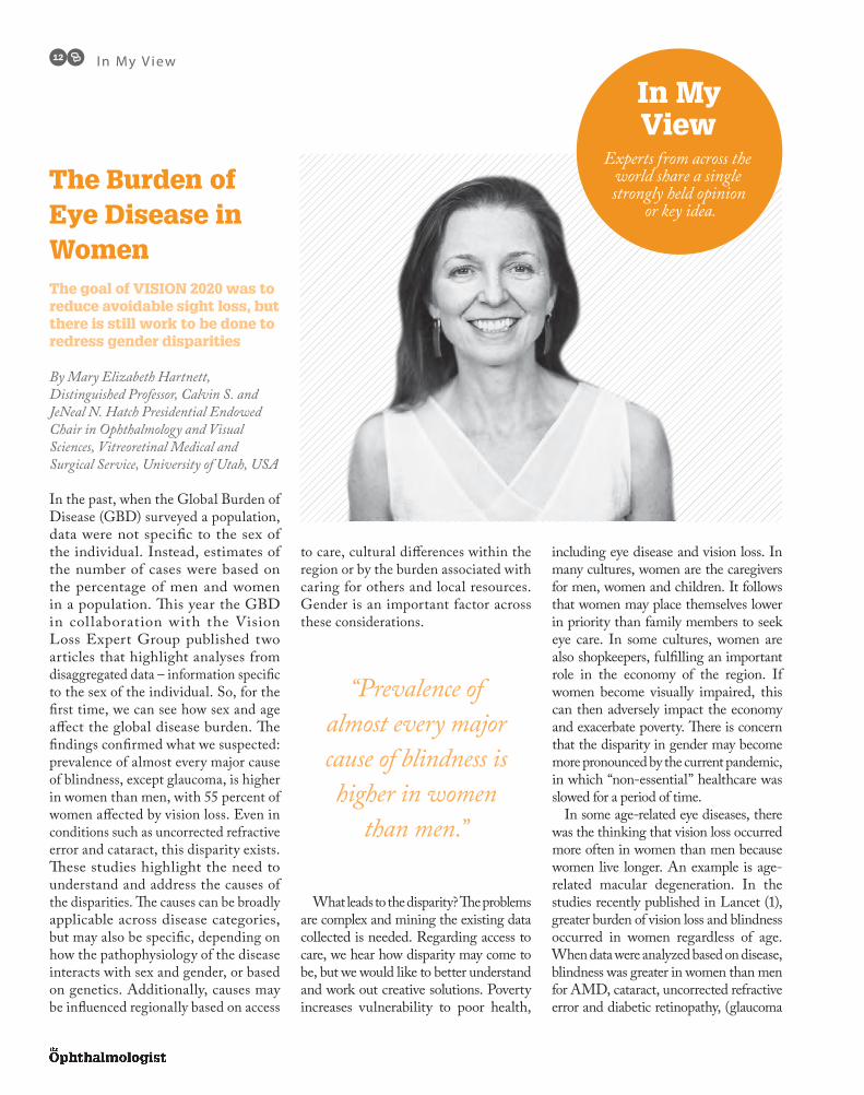
The Burden of Eye Disease in Women The goal of VISION 2020 was to reduce avoidable sight loss, but there is still work to be done to redress gender disparities
By Mary Elizabeth Hartnett, Distinguished Professor, Calvin S. and JeNeal N. Hatch Presidential Endowed Chair in Ophthalmology and Visual Sciences, Vitreoretinal Medical and Surgical Service, University of Utah, USA
In My View
Experts from across the world share a single strongly held opinion
or key idea.
“Prevalence of almost every major cause of blindness is higher in women
than men.”
www.theophthalmologist.com
13In My V iew
The final blow of 2020: the Global Burden of Disease study found that public health services failed to meet WHO targets, with no significant reduction in the number of people with treatable sight loss worldwide since 2010. This landmark study puts global blindness and severe vision impairment on track to double by 2050 (researchers estimate that 61 million people will be blind, and 474 million will have moderate and severe vision impairment) (1). The authors’ findings change our understanding of the entire landscape of the burden of eye disease from our prior understanding. So, what happened? Did we underestimate
Disruption Ahead A failure to meet preventable blindness targets speaks to the need for change
By Jeff Pettey, Vice Chair of Education at the John A. Moran Eye Center, Associate Professor at the University of Utah Department of Ophthalmology and Visual Sciences, and Co-Medical Director of Moran Eye Center Global Outreach Division, USA.
alone affected men more than women (2)). And these outcomes existed for age-standardized prevalence. These outcomes imply that the burden was not simply due to women living longer than men.
There are eye conditions that are affected in women compared to men in specific situations. As an example, uncontrolled diabetic retinopathy can lead to vision loss, and pregnancy can increase the progression
of diabetic retinopathy (3). Therefore, education on diabetes care is essential to all genders, but particularly for women at ages when they can conceive.
We need to understand root causes of the disparities, and grant support is needed to mine data specifically related to barriers to eye care and health based on gender. As director of Women’s Eye Health and in cooperation with the the Vision Loss Expert Group’s Principal Investigator, Rupert Bourne, Professor of Ophthalmology at Cambridge University Hospital and Anglia Ruskin University, UK, we are exploring ways to identify funding sources to support investigators with this interest now that the data are collected. As resources for this needed research are being identified, there are other means to serve our patients. One includes the use of telemedicine. Telemedicine programs are already helping us compensate for not having enough ophthalmologists to meet the global demand, particularly in remote or rural areas without ophthalmologists. A patient with diabetes potentially can have their eyes imaged at a primary care clinic and the images read by an artificial intelligence program or sent to an ophthalmologist remotely. By combining telemedicine
with methods to focus on patients who need treatment for their retinopathy, ophthalmologists may meet the demand. The use of imaging is helpful to educate patients and emphasize what the patient can do to slow progression of retinopathy or reverse vision loss through the use of anti-angiogenic therapies.
Congratulations to the investigators of the Global Burden of Disease and Vision Loss Expert Group who provide us with data showing progress progress and areas for improvement.
References1. The Lancet, “Trends in prevalence of blindness
and distance and near vision impairment over 30 years: an analysis for the GBD study” (2020). Available at: https://bit.ly/35PmOeL
2. The Lancet, “Causes of blindness and vision impairment in 2020 and trends over 30 years: an evaluation of the prevalence of avoidable blindness in relation to VISION2020: the right to sight” (2020). Available to: https://bit.ly/2Ngsacs
3. J Bourry et al., “Progression of Diabetic Retinopathy and Predictors of Its Development and Progression During Pregnancy in Patients With Type 1 Diabetes: A Report of 499 Pregnancies”, Diabetes Care, 44, 181 (2021). PMID: 33177172
“We need to understand root causes of the disparities, and grant support is needed to mine data specifically related to barriers to eye care and health based on gender.”
14 In My V iew
Enjoying your monthly copy of The Ophthalmologist? Want the full experience? Join your peers today at
theophthalmologist.com/register
“An excellent source for up-to-date ophthalmology news in a well-designed layout.”
“The Ophthalmologist has amalgamated everything all of us in the community look for. Thank you!”
Register online and you will get:
access to all of our articles, including online exclusives a monthly print or digital magazine (or both – your choice!) a weekly newsletter covering the top trends and topics in ophthalmologya community of over 70,000 ophthalmologists across the world
TOP House ad.indd 1TOP House ad.indd 1 02/12/2020 10:2702/12/2020 10:27
the scale of the problem, or were our goals too optimistic? Perhaps both. Population growth is outpacing our gains in patient care, and our aims were audacious and aspirational. However, for a challenge of this magnitude, we need aspirational targets that push us beyond traditional models.
When all you have is a hammer, everything looks like a nail, and as surgeons and clinicians, we often view problems through the lens of our skillset. Though performing surgery and providing care is fundamental to reversing these trends, in isolation, it is a wholly incomplete solution. Compounding this perception is the inadequate ratio of ophthalmologists to population. It is tempting to imagine the solution is foreign surgeons traveling to these regions to provide needed care. However, without magnifying impact through capacity building, these efforts likely do little to address the overall need.
So how can you individually be better equipped to turn the tide of increasing blindness? Fundamental to effective care
delivery models like the Aravind Eye Care System is sound public health principles and sustainable financial models. While ophthalmologists may have some basic understanding of these fields, the vast majority of us complete training with little to no formal training in either domain. Everyone participating in global ophthalmology must become a student of public health and sustainable care delivery models, seeking opportunities such as the AAO’s global ophthalmology offerings or free online courses offered at The London School of Hygiene and Tropical Medicine (2).
Beyond individual contributions, the challenge requires a profession-wide response and 2020 disrupted our collective global ophthalmology efforts. Beyond the cancellation of international meetings, COVID-19 globally strained health care systems and slowed the delivery of eye care. While the quantity of delivered care has rebounded in many parts of the world, quality of care was also compromised, particularly for progressive conditions like glaucoma and diabetic eye disease.
The John A. Moran Eye Center Global Outreach Division (3) at the University of Utah and academic departments throughout the world are committed to turning back the global tide of blindness and visual impairment. Academic medicine has a vital role to play through advances in patient care and innovative discoveries aimed at low resource settings. However, I purport academia’s greatest impact on global eyecare will likely be found in the third leg of academia’s tripartite mission: education.
Blindness can only be cured and prevented where there are adequate numbers of ophthalmologists to provide services. As ophthalmologists, we are uniquely aware of the enormous time and f inancial expense required to become a fully trained and this model simply cannot meet the ever-growing
need. Our ophthalmology training model needs disruption.
In the final measure, I believe COVID- 19’s disruptions will be the catalyst to global ophthalmology training’s needed paradigm shift. Teleophthalmology and remote mentoring adoption leaped years forward. Virtual/distance learning is now commonplace, with platforms like Orbis’ Cybersite and Moran Clinical Ophthalmology Resource for Education (CORE) modules seeing 600 percent growth in 2020.
However, we know the greatest challenge to training surgeons is not acquiring facts and knowledge; it is the time-intensive and high-risk work of training surgeons. Thankfully we can also dramatically shorten the surgical learning curve through simulation models from low-cost models to high fidelity virtual reality platforms from Eyesi (4) and Helpmesee (5). Trainee assessment can be effectively done remotely or even crowdsourced to bring trainees safely along the early learning curve.
As 2020 disrupted, we innovated. Did we innovate enough to change the equation of growing global vision loss? The answer will be largely be found in whether we can steepen the curve to increase the numbers of well-trained surgeons to every corner of the world.
Reference1. The Lancet, “Global Burden of Disease”
(2020). Available at: https://bit.ly/2L2HpVF2. London School of Hygiene and Tropical
Medicine, “Global Blindness: Planning and Managing Eye Care Services” (2020). Available at: https://bit.ly/2MPN5Tl
3. Moran Eye Center, “International and Local Outreach” (2020). Available at: https://bit.ly/3iabRtd
4. VRmagic, “Eyesi Surgical” (2020). Available at: https://bit.ly/35Cw6uq
5. Help Me See (2020). Available at: https://bit.ly/2XEceCN
“The greatest challenge to training surgeons is not acquiring facts and knowledge; it is the time-intensive and high-risk work of training surgeons.”

www.theophthalmologist.com
Enjoying your monthly copy of The Ophthalmologist? Want the full experience? Join your peers today at
theophthalmologist.com/register
“An excellent source for up-to-date ophthalmology news in a well-designed layout.”
“The Ophthalmologist has amalgamated everything all of us in the community look for. Thank you!”
Register online and you will get:
access to all of our articles, including online exclusives a monthly print or digital magazine (or both – your choice!) a weekly newsletter covering the top trends and topics in ophthalmologya community of over 70,000 ophthalmologists across the world
TOP House ad.indd 1TOP House ad.indd 1 02/12/2020 10:2702/12/2020 10:27
Feature16
Not anymore. Four young and accomplished refractive specialists share how they made their start in the refractive surgery space and consider what’s next for this rapidly shifting subspecialty.
R E F R A C T I V E S U R G E R Y o n t h e S I D E ?

Feature18
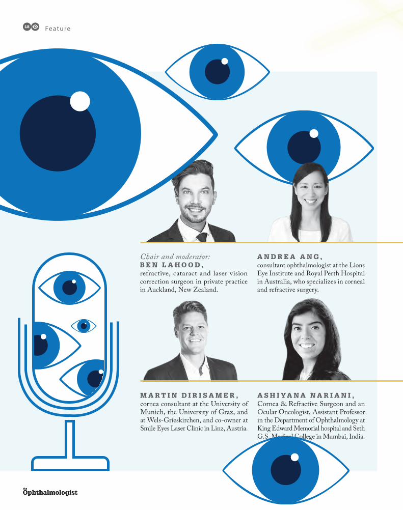
M e e t t h e S U R G E O N S
M A R T I N D I R I S A M E R , cornea consultant at the University of Munich, the University of Graz, and at Wels-Grieskirchen, and co-owner at Smile Eyes Laser Clinic in Linz, Austria.
A S H I Y A N A N A R I A N I , Cornea & Refractive Surgeon and an Ocular Oncologist, Assistant Professor in the Department of Ophthalmology at King Edward Memorial hospital and Seth G.S. Medical College in Mumbai, India.
Chair and moderator: B E N L A H O O D , refractive, cataract and laser vision correction surgeon in private practice in Auckland, New Zealand.
A N D R E A A N G ,consultant ophthalmologist at the Lions Eye Institute and Royal Perth Hospital in Australia, who specializes in corneal and refractive surgery.
www.theophthalmologist.com
Feature 19
W H Y I S R E F R A C T I V E S U R G E R Y A N A T T R A C T I V E O P T I O N F O R Y O U N G O P H T H A L M O L O G I S T S ?
Ben LaHood: People used to ask about the subspecialty I would be choosing when I already knew I wanted to be an ophthalmologist. When I answered honestly that I wanted to be a refractive surgeon, people often pointed me to the “right answer” – perhaps a corneal surgeon with a bit of refractive work on the side. I feel that perceptions are now changing – refractive surgery has really become a subspecialty in its own right, and there are more people stating from the start that this is what they want to do.
Andrea Ang: These days, to be a good cataract surgeon, you also have to be a good refractive surgeon. Our generation is really fortunate to have access to such amazing technology – diagnostics, IOLs, and surgical instruments – so the real push is to get excellent refractive outcomes.
People might see refractive surgery as a cosmetic procedure, but we are actually able to improve our patients’ quality of life through our work! I can’t think of many things that are so rewarding. Our patients are amazed at the difference in their eyesight – and their improved ability to do sports or play with their (grand)children.
Ashiyana Nariani: Historically, refractive surgery was always deemed an add-on to phaco or corneal surgery. But now it is finally becoming mainstream as a standalone subspecialty. This change is really exciting.
I may have a different outlook to the other panelists, as I work in India with underserved patients. We do a lot of work to correct refractive error – a congenital defect – and being able to help my patients gives me great satisfaction. There are substantial and important movements in global ophthalmology these days – from India or Nepal to Africa – to address refractive error with refractive surgery. I’m sure there are young ophthalmologists out there whose dream is to go to underserved populations, perform refractive surgery and treat blindness in this way.
LaHood: Ashiyana makes a great point. For ophthalmologists removing cataracts in developing countries, addressing the refractive error is vital; we cannot leave patients with a -6 D error – we must enable them to see clearly.
Martin Dirisamer: For people our age, there aren’t many subspecialties in ophthalmology that offer so much – but refractive surgery fits the bill. When you can make a difference to your friends and family, it really matters.
W H A T A D V I C E W O U L D Y O U G I V E A B O U T F I N D I N G T H E R I G H T R E F R A C T I V E F E L L O W S H I P A N D T R A I N I N G ?
LaHood: Most people going through medical school and moving on to ophthalmology tend to be A-type personalities, wanting to be the best in their field. I knew that I wanted to become a refractive surgeon, but the right fellowship was difficult to find. I was looking for a very hands-on fellowship, but it seemed that, especially in the US, there was a lot of red tape attached to trainees pushing the button – and that put me off a little bit, even though some of the fellowships seemed really great. Now that the Refractive Surgery Alliance is on the scene, I hope fellowships and training will be easier to find.
Dirisamer: In Austria, we don’t have formal fellowships, so it’s crucial to find a mentor who you can observe for a sufficiently long period of time; you must count on their willingness to teach you what they know. But I would have preferred a more formal fellowship – with guaranteed training and a set framework. I am happy with my education, but it wasn’t easy to come by!
Nariani: I did my cornea, external disease, and refractive fellowship at the Duke Eye Center in North Carolina in the US. I feel very fortunate to have received such great training, with a significant volume of knowledge and practice in refractive surgery. Contrary to what Ben found, I think the US has such a wide variety of fellowships that you are bound to find the right one for you. There are many different aspects to look at when choosing the right training, so decide if you want to focus more on refractive surgery, more on cornea, or do a bit of both… Ask yourself what you’d like to be doing in 10 years’ time. Would it be private practice, a group setting, or a different environment? Then you should be able to choose the best mentor for you. This aspect is really important – wherever you decide to go, try to find a mentor who is ahead of the game and willing to try new things with you involved; after all, as Andrea mentioned, refractive is a fast-moving field with really exciting new technologies. If your mentor moves with the times, then when you graduate you are very likely to have the same mindset, wanting to try new things – and this is an essential attribute of an excellent and successful surgeon.
Ang: I was lucky enough to get into the Cincinnati Eye Institute, training with Edward Holland. Refractive surgery wasn’t the main focus of the fellowship, but we did laser refractive surgery and cataract surgery. I then worked with Donald Tan in Singapore, who also did LASIK. The most important aspects of my training were learning about patient selection, reading corneal topography – basically pre- and postoperative management, as it is done in most surgeries. If your fellowship focuses on these aspects, you’ll be best prepared in terms of practical experience. Mentors are also essential – as the others have mentioned!
You need a really experienced person you can talk to and discuss specific cases. A formal fellowship might not be necessary, but you need a trusted person to whom you can turn for advice or help.
Feature20
H O W D O Y O U G O A B O U T M A K I N G I M P O R T A N T D E C I S I O N S A B O U T Y O U R P R A C T I C E – W H O D O Y O U T R U S T ?
Ang: These days, I attend the Australian Cataract Refractive Meeting, where younger ophthalmologists sit among people we’ve looked up to, our mentors, and we still have the opportunity to ask them questions about cases, new techniques, or products. Building your network is very important.
Dirisamer: It’s crucial to be able to engage in a meaningful discussion. It seems like innovations are popping up every week! New technologies, new IOLs; everything has the same tag: “best and latest.” You need to talk to other people and follow your personal rules when it comes to picking specific products or techniques; for example, basing decisions on big, prospective studies, and working out specific differences between certain products, such as IOLs. I’m not always an early adopter; sometimes I prefer to wait the first wave so that I have a clearer picture of what is worth my time and money.
It’s important to remember that we will never be 100 percent in the right. There are examples of products that everyone gets excited about and then they turn out not to be as great as everyone thought. But even if we are not always right, we must remember that it is our task to filter out the best technologies for our patients. Additionally, these are individual decisions; what works well for one surgeon, might not work as well for another.
LaHood: There is definitely room for conservative refractive surgeons; the subspecialty sometimes gets a bad reputation for being too experimental. And even a conservative refractive surgeon tends to be more “gung ho” than a conservative vitreoretinal surgeon. In Australia and New Zealand, we are usually blessed with early releases of new equipment and products to which we have immediate access, but I still think that physicians on this side of the world are quite conservative. It can be difficult to start a new trend if colleagues in your immediate vicinity are not keen on trying something new.
Ang: I always try to start from a thorough research of the product – there are so many technologies on the market, with new ones coming out all the time! First, I aim to understand the science behind the product and look at clinical trial results. Patient selection is extremely important, so, for any technique or product
I want to try, I first pick patients that appear to be most suited to it. Such a conservative approach works for me.
I think that we should always try to build our processes up slowly and carefully. Putting multifocal lenses in the first 10 patients at the start of your refractive career is not the way to go. It is vital to keep the balance right between being an early adopter and sticking to what you know well – but always remember, the science comes first, and open discussions with patients should follow.
H O W D O E S G O I N G S O L O C O M P A R E W I T H J O I N I N G A N E S T A B L I S H E D P R A C T I C E ? O R D O E S A P U B L I C S E T T I N G W I N ?
LaHood: After my fellowship, I went straight into a fully private practice, which I slightly regret; I miss public practice and I’m trying to go back to that – working as a registrar in teaching. For a narrow refractive practice, there aren’t many opportunities in the public system in Australia and New Zealand; it is easier if you also specialize in cornea or ocular oncology.
Nariani: W hen I asked myself what I wanted to do after my fellowship, I knew I wanted to come to India,
so I worked backwards from that goal. As I had a passion for refractive surgery, I
tried to work out how I could make both these pieces of the puzzle fit
together. I started off in private practice and I noticed that it wasn’t easy for those with
their own businesses, so I wasn’t keen on establishing my own; I
wanted to work in a government-funded hospital. Now, with the pandemic
so widespread, I watch my friends who decided to invest in their businesses struggle to repay loans – it’s
a huge financial burden, so if that’s your chosen route, you have to be aware of the risks.
Having said that, it should not be a deterrent. Just make sure you are realistic and know that it’s not plain sailing, and you have to take those aspects into consideration. This advice is perhaps more important for a refractive surgeon than those in other ophthalmic subspecialties.
My personal dream is to build a surgery suite for the neglected patient population. It requires a lot of funding, and doesn’t pay back, so I have to figure out ways to keep it sustainable, while providing the highest quality of treatment. As for all of us, improving patients’ quality of life as much as I can, is my highest priority.
Dirisamer: I have found the public setting, at the University Clinic in Munich, to be the best way to start a refractive career
www.theophthalmologist.com
Feature 21
here in Austria, as it is free of financial risks and you can figure out whether it really is the career for you and if you are passionate about it. The refractive route might not look very difficult at first glance, but it is very demanding. You must care for a special group of patients, so there is always the possibility that it won’t suit you. I know many ophthalmologists who tried refractive surgery and decided it wasn’t for them, and the public system allows you to do that. However, it can be limiting: the likelihood is that you won’t be able to perform LASIK or SMILE in a public setting, and even if you do, the numbers won’t be as high as in a private practice. And that’s why I now divide my week between the academic setting (two days a week), and a private practice. I think I will continue in that direction for the next few years as it has worked really well for me. If you are set on a career that’s purely in private practice, my advice would be to join a group, as it offers you a little more safety and reassurance.
Ang: I work one day a week in a public setting, and the rest in a private practice. It is a group practice, so fortunately my costs weren’t too high. I can see many advantages of joining an established practice: you can rely on colleagues with more experience, the equipment is already there… I knew I wanted to work with those particular people, and the working environment was amazing. Of course, you have less control over important decisions – purchasing new equipment or hiring staff, but you don’t have to deal with all the bureaucracy on your own. When you are at the start of your career, it might be a good idea to become a locum in a few different practices, to see what suits you best. I didn’t do it, as I had a very clear idea of where I wanted to work, but if you don’t – it’s a really good way to start the process.
H O W D I D Y O U M A N A G E T O S T E P O U T F R O M Y O U R M E N T O R ’ S S H A D O W A N D E S T A B L I S H Y O U R S E L F I N T H E F I E L D ?
Ang: I work with Graham Barrett, one of the best-known figures in the field. It’s been my
honor and privilege to be mentored by him and by Steven Wiffen, who I also share a practice with. I have enjoyed working with and learning from them, and I haven’t really seen myself as being in their shadow. We shouldn’t underestimate what we have to offer, even at the start of our careers. There is always something we bring to the table; for example, we might be more inclined to consider introducing new technologies. These days, industry perceives us as a generation of influencers in the refractive sphere..
Of course, you have to build up your own practice, your results, and your relationships with your patients – we rely heavily on word of mouth in this field. But patients don’t always go for the biggest name, and once you can share good results, they speak for themselves.
Dirisamer: In my practice, I have a senior partner, and a junior partner, so I’m pretty much in the middle. I see huge potential
in colleagues within a practice specializing in slightly different areas, as the whole group benefits. It doesn’t
make sense to just copy what your senior partner does or try to do every single thing they do as
well as they do. To help you grow, choose your own specialisms. And then you can help more junior colleagues find their own preferred niche.
Nariani: I don’t necessarily go out of my way to differentiate myself, but I do try to compete with myself, and not with others.
In India, a country of over a billion people, there is plenty of work to go around. If I feel that there is a person out there who knows more than I do, I try to learn more and become a better surgeon. The most important thing for me to remember is to keep making a difference to my patients and to stay passionate about what I do. Tomorrow, I want to be a better doctor than I am today, but this learning process can work in many ways: I learn from my residents,
just as they learn from me. I don’t feel like I have to be better than them as we
are all working towards the common goal.LaHood: Going into refractive surgery for
the right reasons – not simply to make money – is incredibly important. I feel like there has been a big
change in how refractive specialists are perceived over the past few years – from “laser jockeys” simply pressing the button to doctors who help people. The subspecialty has gone from
being seen as a cosmetic luxury to more of an art form. It’s been a very positive change.
Dirisamer : It’s vital to always remember that healthy eyes are the ultimate outcome. Ask yourself: “Would I perform this procedure on my mother or another family member? Is this the safest option there is? If there is a risk, is it necessary?” Asking those questions is key to being a good refractive surgeon.
Ang: I think more women entering the subspecialty has helped soften the way refractive surgeons are perceived. It used to be mainly older white men, and now it’s becoming a lot more diverse. However, patient expectations have increased, so our generation has to make sure that we are well equipped to meet those demands. People expect to be fully spectacle-free, and we have to find ways to achieve such outcomes.
I S I T I M P O R T A N T F O R R E F R A C T I V E S P E C I A L I S T S T O H A V E A N O N L I N E P R E S E N C E ?
LaHood: Patients feel welcome to tell everyone how good or bad they feel their surgeon was, and this includes Google reviews and similar. It’s so easy for them to rate their doctors, so it’s important for us to stay on top of it and maintain our reputation online. Sometimes I wish I could rate my patients online and give them reviews! Maintaining an image is especially vital for the younger surgeons. I run an Instagram page as I’ve been told that it is something you have to do these days. You have to be “out there,” seen presenting and shaking hands, almost like a politician. I don’t particularly enjoy it, but I play the game.
Ang: I don’t have Instagram, and I’m not even on Facebook! It’s quite unusual these days. Of course, you need a website and be out there promoting your name, but I also think that the old-fashioned way – getting good results and getting your patients’ opinions to do the promotion for you – still works. Speaking at meetings and publishing articles certainly helps build your reputation in the field. I should point out that our laser vision center keeps our Facebook and Instagram up to date, and younger generations do consider it important.
Dirisamer: Younger colleagues definitely have easier access to social media than more experienced specialists. You have to find your own style of communicating with patients, but you have to remember that the group that is attracted to social media channels is exactly the group you want to reach, so you need some kind of an online presence. It’s actually an inexpensive way to market yourself, but it is time consuming. Patients always want to know their doctors, see pictures, watch interviews, and hear explanations of their treatment. Nevertheless, I agree with Andrea, it is possible to do things the old-fashioned way – it’s just harder to reach the younger patient population.
LaHood: When I left a practice in Auckland, New Zealand, I was suddenly left with no online presence, as it had all been done through the practice. It felt like I disappeared! I listened to my colleagues’ advice who said that I needed a website – somewhere patients could go and look me up, to see that I was still practicing. I have had patients contact me via social media to enquire about specific equipment, such as lasers, but not in huge numbers.
Feature22
Nariani: Even if we don’t want to keep active social media channels, it’s a necessity these days, and part of the life we and our patients live. When we, as doctors, help someone, we don’t necessarily want to shout about it, but sometimes we have to. I limit social media posts to one a day, and don’t spend more than five minutes on it. The rest of my day is for my patients. I also remind myself to post meaningful messages and show myself as a real person – it doesn’t always have to be rosy. Patients will like you for who you are. If we are real and honest, patients will appreciate it.
LaHood: I have a podcast about being honest as a surgeon. We fear that when we admit to making a mistake, others will comment negatively. But actually, my experience is different. When I admitted to causing some complications once, I got a lot of feedback from my colleagues about being brave. They commented how nice it was to finally see someone openly say, “Yes, I’ve messed up, and this is how it can be corrected.” It feels to me like there isn’t enough honesty there on social media, but at the more intimate conferences, such as the AUSCRAS Meeting, people share their failures as well as their successes, so we can all learn from each other’s mistakes.
W H A T E X C I T E S Y O U A B O U T R E F R A C T I V E S U R G E R Y R I G H T N O W ?
Dirisamer: Very quick procedures to address big issues! Being able to relieve someone from glasses or contact lenses
within a very short amount of time is a brilliant feeling. In terms of new technologies, I’m looking forward to seeing the next step in Relax SMILE, maybe something like the wavefront-guided SMILE. With regards to IOLs, it will be great to see multifocals with no glare or halo widely used, as well as more precise IOL calculations.
Ang:There are many things that excite me, such as improvements in biometry measurements that are made available in both private and public settings (there is still a discrepancy, with private practices having access to more advanced technologies). I appreciate incremental improvements that boost accuracy. And, like Martin, I’m looking forward to second-generation SMILE, without some of the issues that the first generation has. Barriers are also being pushed in lens development, with companies squeezing in better optics with each design.
LaHood: I’ll play the devil’s advocate and say that I think we are reaching a point where we are getting as good as we can preoperatively. I might look back on this in five years’ time and regret saying it! But I’m more excited about the idea that we will be able to adjust outcomes postoperatively.
Nariani: I find the idea of light-adjustable IOLs really exciting, I’m really waiting for this innovation. But I’m also looking forward to the day when we get used to the idea of global refractive surgery for vulnerable/poor populations. Providing multifocal lenses to these patients and relieving them of glasses will be a huge step forward.
Feature 23
www.theophthalmologist.com
Put simply, a stable tear film protects the ocular surface epithelium from drying. When the tear film components are insufficient or impaired, the tear film breaks up, resulting in dry eye disease (DED). Tear film breakup is a core mechanism of dry eye, and abnormal breakup time and symptoms are considered part of the diagnostic criteria for dry eye. Thus, tear film-oriented diagnostic methods based on the tear film breakup patterns are considered essential for the diagnosis of DED (1). TFBUT – which has become the standard diagnostic procedure for DED – measures the interval of time that elapses between a complete blink and the appearance of the first break in the tear film. Short breakup time has become widely recognized as a major contributor to DED in recent years (2); indeed, more attention is now paid to unstable tear films than to the tear volume or superficial punctate keratopathy (3). According to the Tear Film and Ocular Surface Society Dry Eye Workshop II (TFOS DEWS II), TFBUT is the most frequently employed test of tear film stability in clinical practice (4).
But TFBUT does not consider the blinking rate. Blinking spreads the tear film, mucin, and lipids on the cornea and conjunctiva, maintaining the eye’s moisture and protecting the eye from
Breaking PointEnter the Ocular Protection Index: the test beyond tear film breakup time By Rajesh Sinha, Aditi Agarwal
I n Prac t ice24
InPracticeSurgical Procedures
DiagnosisNew Drugs
irritants (5, 6). Notably, blink rates – measured as interblink interval (IBI, the mean time between two blinks) – are found to differ between normal participants and patients with DED (6). As IBI correlates with clinical characteristics of DED (7), it should therefore be considered as one of the parameters for its diagnosis. The Ocular Protection Index (OPI) was developed to overcome the disadvantages of TFBU T by measur ing mult iple causative factors of DED (8); however, it is not being used in routine clinical practice because of a lack of awareness among eyecare professionals.
Here, we highlight why the OPI is a better measure than TFBUT alone, explore OPI research and its use in clinical trials, and discuss how OPI can be used in routine clinical practice.
Why OPI?DED is caused by a lower tear production rate and/or a short tear film stable time. The ocular surface becomes dry and lacks lubrication, eventually damaging the ocular surface (9). A short TFBUT is a
key factor for the diagnosis of DED (10), with a cut-off value of less than 10 seconds.
But the preservation of a stable tear film over the ocular surface also depends on spontaneous eye blinking, in addition to the amount of tear secretion and lipid quality. Spontaneous blinking is a rapid, automatic, and unconscious opening and closing of the eyelids – unlike reflex and voluntary blinking (11). The process is critical for spreading the tear film over the ocular surface, lipid secretion into the tear film, and tear drainage, and it is essential for maintaining optical quality. Unfortunately, spontaneous blinking is affected by both age and mental activity. Stimulation of the ocular surface increases the spontaneous blinking rate, while reduced blinking rate is associated with increasing tear film evaporation and the development of DED. Blinking
rate is both a cause and a consequence of DED (12).
In healthy patients, the mean IBI (the time between two blinks) is approximately 7.5 seconds (13). Note that it is the interaction between tear film stability and IBI that maintains the health of the ocular surface, so the number of seconds quantifying TFBUT does not provide pathophysiologic information to completely understand the nature and severity of the case of dry eye. The OPI was developed to quantify the interaction between blinking and the tear film and is simply a ratio of TFBUT and IBI (TFBUT divided by IBI). An OPI score <1.0 indicates an exposed ocular surface, which leads to the development or exacerbation of the signs and symptoms of dry eye. An OPI score >1.0 indicates a tear-protected ocular surface, potentially resulting in
I n Prac t ice 25
“The Ocular Protection Index was developed to overcome the disadvantages of TFBUT by measuring multiple causative factors of DED.”
www.theophthalmologist.com
Determining the OPIIBI is measured by dividing 60 by the number of blinks per minute (preferably counted while an ECP is taking history, either during routine patient conversation or while the patient reads the vision chart). Next, TFBUT is measured by instilling fluorescein onto the inferior bulbar conjunctival surface using a moistened fluorescein-impregnated paper and having the subject blink several times to mix the fluorescein dye with their tear film. After two controlled blinks, the patient is then asked to stare straight ahead without blinking for as long as possible. The dorsolateral corneal surface is observed with 10X magnification with light
passed through the cobalt-blue filter of a slit lamp biomicroscope. TFBUT is measured as the time from eyelid opening to the first sign of tear film breakup, evident as the appearance of 1 or more dark spots within the fluorescent green tear film (14). OPI score is determined by dividing TFBUT by the IBI (13).
An OPI score of less than 1.0 suggests an exposed ocular surface, which may lead to the development or exacerbation of the signs and symptoms of DED; an OPI score of more than 1.0 indicates a tear-protected ocular surface, which potentially results in less severe dry eye signs and symptoms. It can also be used to measure the changes in the severity of DED over time and to evaluate the effect of treatments for DED in promoting tear film stability (13).
fewer dry eye signs and symptoms (13).The advantage of OPI is that it measures
two components involved in DED. The disadvantages of OPI are i) TFBUT has to be measured using fluorescein dye (such staining is invasive; the TFOS DEWS II Diagnostic Methodology Subcommittee prefers measurement of the tear breakup time with a non-invasive technique – NIBUT), and ii) the IBI measurement must be performed at a different time (13).
OPI research: a brief historyThe concept of OPI was first introduced by Ousler and colleagues in 2002 when they were trying to understand the factors that influence the IBI – and the relationship between IBI and TFBUT. In the study, subjects underwent baseline examinations including visual acuity, ocular discomfort (0-4 scale), and blink rate. Complete blinks
were measured non-invasively using a digital micro-camera equipped with an infrared illuminator mounted to a headset extension and directed towards the eye. Subjects were placed in a controlled adverse environment (CAE) for 90 minutes. During CAE exposure, blink rate was measured every 10 minutes while ocular discomfort was recorded every five minutes. The mean blink rate increased significantly from 11 blinks/minute pre-CAE to 20 blinks/minute post-CAE. The mean ocular discomfort also increased significantly from 0.63-units pre-CAE to 2.38 units post-CAE. Ultimately, the study showed that the OPI quantifies the relationship between TFBUT and the IBI and is useful in assessing dry eye and the effect of its therapeutic agents (15).
DED can on ly be d iagnosed
appropr iately by eva luat ing the changes in different components of tear film integrity. Although TFBUT is a recognized method of diagnosis of DED, it does not take the role of blinking into account. The OPI quantifies the interaction of tear film integrity and the blinking process, therefore offering a better indication of ocular surface health than TFBUT alone. The OPI has proven to be a useful and clinically relevant tool when it comes to the diagnosis of DED in routine clinical practice – and we believe it is worth integrating into your own clinic.
Rajesh Sinha is Professor of Ophthalmology in Cornea, Cataract and Refractive Surgery at the Dr. Rajendra Prasad Centre for Ophthalmic Sciences, AIIMS, New Delhi, India.
I n Prac t ice26
OPI 2.0An “OPI 2.0 System” was developed to evaluate ocular surface protection under a natural blink pattern and normal visual conditions. It is calculated by determining the mean breakup area and dividing it by the IBI. Breakup area can be calculated by automated software algorithms, providing a real-time measurement of corneal exposure for each interblink interval during a one-minute video. The software analyzes a series of artificial images and still image frames captured during an actual clinical session using fluorescein staining videography (16). However, as this method requires advanced software to measure mean break up area, it is difficult to use in routine clinical practice.

www.theophthalmologist.com
I n Prac t ice 27
Aditi Agarwal is Medical Director and Cornea Consultant at Krishna Netralaya, Gurgaon, and Senior cornea consultant at Fortis hospital, Gurgaon, India.
References1. N Yokoi and G Georgiev, “Tear-film-oriented
diagnosis for dry eye,” Jpn J Ophthalmol, 63, 127 (2019). PMID: 30783943.
2. K Tsubota, “Short tear film breakup time-type dry eye,” Invest Ophthalmol Vis Sci, 59, DES64-DES70 (2018). PMID: 30481808.
3. N Yokoi and G Georgiev, “Tear-film-oriented diagnosis for dry eye,” Jpn J Ophthalmol, 63, 127 (2019). PMID: 30783943.
4. J Wolffsohn et al., “TFOS DEWS II Diagnostic Methodology Report,” Ocul Surf, 15, 539 (2017). PMID: 28736342.
5. M Al-Abdulmunem, “Relation between tear breakup time and spontaneous blink rate,” Int Contact Lens Clin, 26, 117 (1999). PMID: 11166137.
6. T Inomata et al., “Maximum blink interval is associated with tear film breakup time: A new simple, screening test for dry eye disease,” Sci Rep, 8, 13443 (2018). PMID: 30194447.
7. Y Su et al., “Spontaneous eye blink patterns in dry eye: Clinical correlations,” Invest Ophthalmol Vis Sci, 59, 5149 (2018). PMID: 30372746.
8. G Ousler et al., “Factors that influence the inter-blink interval (IBI) as measured by the ocular protection index (OPI),” Invest Ophthalmol Vis Sci, 43, 56 (2002).
9. J Craig et al., “TFOS DEWS II Report Executive Summary,” Ocul Surf., 15, 802 (2017). PMID: 28797892.
10. K Tsubota, “Short tear film breakup time-type dry eye,” Invest Ophthalmol Vis Sci, 59, DES64 (2018). PMID: 30481808.
11. C McMonnies, “The clinical and experimental significance of blinking behavior,” J Optom, 13, 74 (2020). PMID: 31992536.
12. Y Su et al., “Spontaneous eye blink patterns in dry eye: Clinical correlations,” Invest Ophthalmol Vis Sci, 59, 5149 (2018). PMID: 30372746.
13. G Ousler et al., “The Ocular Protection Index,” Cornea, 27, 509 (2018). PMID: 18520496.
14. R Abelson et al., “Measurement of ocular surface protection under natural blink conditions,” Clin Ophthalmol, 5, 1349 (2011). PMID: 22034554.
15. G Ousler et al., “The Ocular Protection Index,” Cornea, 27, 509 (2008). PMID: 18520496.
16. R Abelson et al., “Validation and verification of the OPI 2.0 System,” Clin Ophthalmol, 6, 613 (2012). PMID: 22570541.
17. M Yazdani et al., “Evaluation of the ocular surface disease index questionnaire as a discriminative test for clinical findings in dry eye disease patients,” Curr Eye Res, 44, 941 (2019). PMID: 30955380.
18. B Tashbayev et al., “Utility of tear osmolarity measurement in diagnosis of dry eye disease,” Sci Rep, 10, 5542 (2020). PMID: 32218518.
19. M Rolando et al., “Protecting the ocular surface and improving the quality of life of dry eye patients: a study of the efficacy of an HP-guar containing ocular lubricant in a population of dry eye patients,” J Ocul Pharmacol Ther, 25, 271 (2009). PMID: 19366323.
20. G Ousler et al., “An evaluation of tear film breakup time extension and ocular protection index scores among 3 marketed lubricant eye drops,” Cornea, 26, 949 (2007). PMID: 17721294.
21. K Lebow, “An evaluation of two artificial tears in improvement of tear film stability as measured by tear film break-up time and ocular protection index (OPI).” Paper presented at the AAO Annual Conference 2007; 075316.
22. G Torkildsen et al., “Ocular comfort and drying effects of three topical antihistamine/mast cell stabilizers in adults with allergic conjunctivitis: a randomized, double-masked crossover study,” Clin Ther, 30, 1264 (2008). PMID: 18691985.
23. M Nebbioso et al., “Fixed topical combinations in glaucomatous patients and ocular discomfort,” Expert Opin Pharmacother, 13, 1829 (2012). PMID: 22770575.
24. P Johnston et al., “The interblink interval in normal and dry eye subjects,” Clin Ophthalmol, 7, 253 (2013). PMID: 23403736
25. Dry eye: diagnostic test template (2020). Available at: https://bit.ly/2L3ZpP8.
Practical considerationsAs the OPI is measured using IBI and TFBUT, control should be taken to accurately measure both these parameters (24).
• To determine TFBUT, care must be taken while instilling the fluorescein dye so that reflex tearing is not induced. Changes in tear volume may lengthen TFBUT (25).
• Appropriate patient instructions should be given before the measurement of TFBUT. If patients are not told to blink freely prior to TFBUT being assessed, reflex tearing may
result in skewing the subsequent measurements.
• Volume of fluorescein should be accurate for assessment. Large volumes of fluorescein instilled may also artificially lengthen TFBUT (25).
• A slit lamp with an online video camera system may be used to capture TFBUT. Video capture with an on-screen timer allows for precise measurement of the time between the last complete blink and the appearance of the first, growing micelle (25).
• In routine clinical practice, the IBI of the patient can be measured while an ECP is taking history, either during routine conversation or while the patient reads the vision chart.

I n Prac t ice28
The ability to assess the cornea, pre- and post-operatively has been available to refractive and corneal surgeons for more than two decades now. With the introduction of the Orbscan corneal topographer (Bausch+Lomb), we gained the ability to assess the anterior and posterior surface of the cornea, while higher-order wavefront aberrometers taught us about corneal aberrations and their impact on vision. We had a new tool at our disposal with the introduction of Ocular Response Analyzer (ORA; Reichert Technologies), which enabled us to measure cornea biomechanics through a parameter called corneal hysteresis (1).Originally intended as a method for assessing glaucoma progression, ORA gained a following in the use of corneal biomechanics assessment. These innovations were followed by Scheimpflug systems and other means of assessing the biomechanical properties of the cornea. But many clinicians still consider these tools “for research use
only.” And, as a result, they are not regularly used in clinical practice. In this article, I seek to understand why this is the case and hopefully convince you why we should integrate these devices into the routine care of our refractive patients.
The technology lowdownThe primary diagnostic technology used in corneal assessment for refractive screening fall into two main categories: Scheimpflug tomography systems (which are sometimes combined with a Placido topographer), and non-contact, air-puff tonometers, used alongside several other technologies used primarily for research purposes.
• Corneal Tomographers: Scheimpflug Imaging Systems
The Pentacam. Oculus was the first company to introduce the technology (first patented in 1904) into an ophthalmic imaging device. The instrument used a rotational Scheimpflug camera to provide three-dimensional, non-contact imaging of the anterior segmentIt measures topography and elevation of the anterior and posterior corneal surface and the corneal thickness. Since the introduction of the first Pentacam in 2003, the company has introduced three additional models: The Pentacam HR that
Hidden Secrets of the CorneaWhy corneal biomechanical properties matter – and why you should introduce them into your everyday practice By Riccardo Vinciguerra
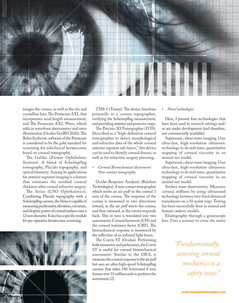
www.theophthalmologist.com
I n Prac t ice 29
images the cornea, as well as the iris and crystalline lens; The Pentacam AXL that incorporates axial length measurement; and The Pentacam AXL Wave, which adds in wavefront aberrometry and retro illumination (Oculus GmBH 2020). The Belin/Ambrosio software of the Pentacam is considered to be the gold standard for screening for subclinical keratoconus based on corneal tomography.
The Galilei (Ziemer Ophthalmic Systems). A blend of Scheimpf lug tomography, Placido topography, and optical biometry. Among its applications for anterior segment imaging is a feature that estimates the residual corneal thickness after corneal refractive surgery.
The Sirius (CSO Ophthalmics). Combining Placido topography with a Scheimpflug camera, the Sirius is capable of measuring pachymetry, elevation, curvature, and dioptric power of corneal surfaces over a 12 mm diameter. It also has a specific module for pre-operative keratoconus screening.
TMS-5 (Tomey). The device functions primarily as a cornea topographer, verifying the Scheimpflug measurement, and providing anterior and posterior maps.
The Precisio 3D Tomographer (IVIS). Described as a “high-definition corneal tomographer to detect morphological and refractive data of the whole corneal anterior segment sub-layers,” this device can be used to identify corneal disease, as well as for refractive surgery planning..
• Corneal Biomechanical Assessment: Non-contact tomography
Ocular Response Analyzer (Reichert Technologies). A non-contact tomographer which emits an air puff in the central 3 mm of the cornea. The response of the cornea is measured in two directions; inward, as the air puff meets the cornea, and then outward, as the cornea responds back. This in turn is translated into two assessments: Corneal hysteresis (CH) and the corneal resistance factor (CRF). The biomechanical response is monitored by the reflection of an infrared light beam.
The Corvis ST (Oculus). Performing both tonometry and pachymetry, the Corvis ST is useful for corneal biomechanical assessment. Similar to the ORA, it measures the cornea’s response to the air puff but uses an ultra-high-speed Scheimpflug camera that takes 140 horizontal 8 mm frames over 33 milliseconds to perform the assessment (2).
• Novel technologies
Here, I present four technologies that have been used in research settings and/or are under development (and therefore, not commercially available):
Supersonic, shear-wave imaging. Uses ultra-fast, high-resolution ultrasonic technology to do real-time, quantitative mapping of corneal viscosity in an animal-eye model.
Supersonic, shear-wave imaging. Uses ultra-fast, high-resolution ultrasonic technology to do real-time, quantitative mapping of corneal viscosity in an animal-eye model.
Surface wave elastrometry. Measures corneal stiffness by using ultrasound technology between two-fixed distanced transducers on a 10-point map. Testing has been successfully done in animal and human cadaver models.
Elastography through a gonioscope lens. Uses a scanner to cover the entire
“Fundamentally, assessing corneal
mechanics is a safety issue.”
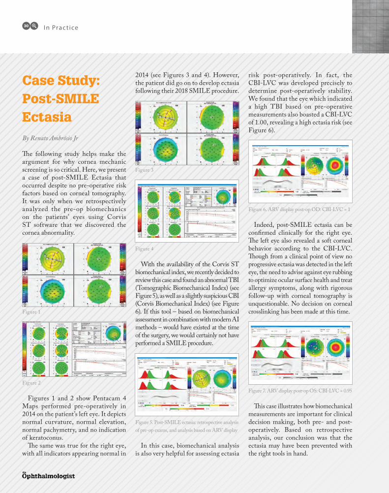
Case Study: Post-SMILE EctasiaBy Renato Ambrósio Jr
The following study helps make the argument for why cornea mechanic screening is so critical. Here, we present a case of post-SMILE Ectasia that occurred despite no pre-operative risk factors based on corneal tomography. It was only when we retrospectively analyzed the pre-op biomechanics on the patients’ eyes using Corvis ST software that we discovered the cornea abnormality.
Figures 1 and 2 show Pentacam 4 Maps performed pre-operatively in 2014 on the patient’s left eye. It depicts normal curvature, normal elevation, normal pachymetry, and no indication of keratoconus.
The same was true for the right eye, with all indicators appearing normal in
2014 (see Figures 3 and 4). However, the patient did go on to develop ectasia following their 2018 SMILE procedure.
With the availability of the Corvis ST biomechanical index, we recently decided to review this case and found an abnormal TBI (Tomographic Biomechanical Index) (see Figure 5), as well as a slightly suspicious CBI (Corvis Biomechanical Index) (see Figure 6). If this tool – based on biomechanical assessment in combination with modern AI methods – would have existed at the time of the surgery, we would certainly not have performed a SMILE procedure.
In this case, biomechanical analysis is also very helpful for assessing ectasia
risk post-operatively. In fact, the CBI-LVC was developed precisely to determine post-operatively stability. We found that the eye which indicated a high TBI based on pre-operative measurements also boasted a CBI-LVC of 1.00, revealing a high ectasia risk (see Figure 6).
Indeed, post-SMILE ectasia can be confirmed clinically for the right eye. The left eye also revealed a soft corneal behavior according to the CBI-LVC. Though from a clinical point of view no progressive ectasia was detected in the left eye, the need to advise against eye rubbing to optimize ocular surface health and treat allergy symptoms, along with rigorous follow-up with corneal tomography is unquestionable. No decision on corneal crosslinking has been made at this time.
This case illustrates how biomechanical measurements are important for clinical decision making, both pre- and post-operatively. Based on retrospective analysis, our conclusion was that the ectasia may have been prevented with the right tools in hand.
Figure 1
Figure 3
Figure 4
Figure 5. Post-SMILE ectasia: retrospective analysis of pre-op exams, and analysis based on ARV display
Figure 6. ARV display post-op OD: CBI-LVC = 1
Figure 7. ARV display post-op OS: CBI-LVC = 0.95Figure 2
I n Prac t ice30
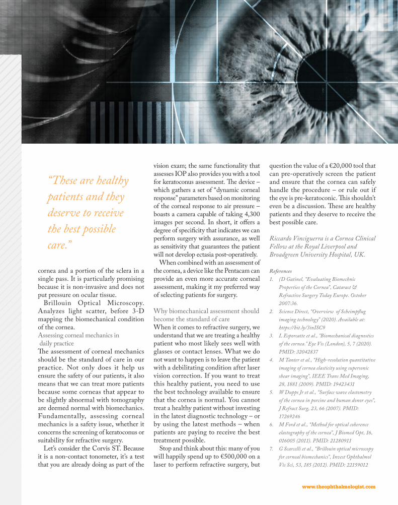
www.theophthalmologist.com
cornea and a portion of the sclera in a single pass. It is particularly promising because it is non-invasive and does not put pressure on ocular tissue.
Brillouin Optical Microscopy. Analyzes light scatter, before 3-D mapping the biomechanical condition of the cornea.Assessing corneal mechanics in daily practiceThe assessment of corneal mechanics should be the standard of care in our practice. Not only does it help us ensure the safety of our patients, it also means that we can treat more patients because some corneas that appear to be slightly abnormal with tomography are deemed normal with biomechanics. Fundamentally, assessing corneal mechanics is a safety issue, whether it concerns the screening of keratoconus or suitability for refractive surgery.
Let’s consider the Corvis ST. Because it is a non-contact tonometer, it’s a test that you are already doing as part of the
vision exam; the same functionality that assesses IOP also provides you with a tool for keratoconus assessment. The device – which gathers a set of “dynamic corneal response” parameters based on monitoring of the corneal response to air pressure – boasts a camera capable of taking 4,300 images per second. In short, it offers a degree of specificity that indicates we can perform surgery with assurance, as well as sensitivity that guarantees the patient will not develop ectasia post-operatively.
When combined with an assessment of the cornea, a device like the Pentacam can provide an even more accurate corneal assessment, making it my preferred way of selecting patients for surgery.
Why biomechanical assessment should become the standard of careWhen it comes to refractive surgery, we understand that we are treating a healthy patient who most likely sees well with glasses or contact lenses. What we do not want to happen is to leave the patient with a debilitating condition after laser vision correction. If you want to treat this healthy patient, you need to use the best technology available to ensure that the cornea is normal. You cannot treat a healthy patient without investing in the latest diagnostic technology – or by using the latest methods – when patients are paying to receive the best treatment possible.
Stop and think about this: many of you will happily spend up to €500,000 on a laser to perform refractive surgery, but
question the value of a €20,000 tool that can pre-operatively screen the patient and ensure that the cornea can safely handle the procedure – or rule out if the eye is pre-keratoconic. This shouldn’t even be a discussion. These are healthy patients and they deserve to receive the best possible care.
Riccardo Vinciguerra is a Cornea Clinical Fellow at the Royal Liverpool and Broadgreen University Hospital, UK.
References1. (D Gatinel, “Evaluating Biomechnic
Properties of the Cornea”, Cataract & Refractive Surgery Today Europe. October 2007:36.
2. Science Direct, “Overview of Scheimpflug imaging technology” (2020). Available at: https://bit.ly/3inISC9
3. L Esporcatte et al., "Biomechanical diagnostics of the cornea." Eye Vis (London), 5, 7 (2020). PMID: 32042837
4. M Tanter et al., “High-resolution quantitative imaging of cornea elasticity using supersonic shear imaging”, IEEE Trans Med Imaging, 28, 1881 (2009). PMID: 19423431
5. W Dupps Jr et al., “Surface wave elastometry of the cornea in porcine and human donor eyes”, J Refract Surg, 23, 66 (2007). PMID: 17269246
6. M Ford et al., “Method for optical coherence elastography of the cornea”, J Biomed Opt, 16, 016005 (2011). PMID: 21280911
7. G Scarcelli et al., “Brillouin optical microscopy for corneal biomechanics”, Invest Ophthalmol Vis Sci, 53, 185 (2012). PMID: 22159012
“These are healthy patients and they deserve to receive the best possible care.”

I n Prac t ice32
Monovision RevisitedOne of The Ophthalmologist’s most-read articles of the past five years was called “The Misnomer of Monovision.” Here, I present an update on binocular spectacle-free vision for 2021. By Raymond Radford

www.theophthalmologist.com
I n Prac t ice 33
In the optically auspicious year of 2020, when the ophthalmology world planned to reach worldwide goals in vision, eye care, and patient outcomes, suddenly – like the punchline in a Greek myth – almost all routine ophthalmology stopped. However, time gifted by adversity has allowed us to reflect on our practice – and, in my case, revisit the topic of monovision (1).
Cataract removal and lens replacement remains the most common operation, with demand increasing, and waiting times for surgery being extended by six months and more. When patients eventually get their surgery, what is the best outcome we can achieve for them? For those who are not in love with their glasses, for reasons of habit or image, the option of spectacle-free vision exists with binocular spectacle-free vision (BSFV) – otherwise poorly labeled as “monovision.” If only every patient could achieve monocular 6/6 and N6 in each eye with a standard monofocal implant (true monovision perhaps)! Some patients have unpredictably gifted natural optics – for everyone else, you might want to consider BSFV.
The five-year trend: 2015-2020On the final summary day at the ESRCS in Barcelona in 2015, a colleague made the observation that all modern technologies in refractive surgery are using monovision some of the time or use it to optimize outcomes for spectacle freedom. Lens manufacturers have spent considerable sums marketing and advocating their premium lens designs, yet uptake remains less than 5 percent of all surgeries. Why is this? Surgeons want happy patients free from aberrations, glare, “vaseline vision,” positive and negative dysphotopsia. Concern about these effects and the frequency with which they occur has led to redesigned and refined multifocals: bifocals and trifocals, including removable lenses, such as “piggy back” trifocals.
How common is explantation? Accurate data on explantation rates is hard to find, and the data that exist are likely underreported. Lens marketing doesn’t communicate the message: “Try it out and see if aberrations occur; if it is a problem,
we can remove the lens.” If this scenario happens, the surgeon is likely to incur the cost unless risk of removal is “priced into” the costs of the surgery, spread among all patients. The extended depth of focus (EDOF) lens has been promoted by some as never requiring removal because it is aberration-free and “not like multifocals, almost a monofocal.” However, I have met patients who experienced removal of EDOF lenses because of intolerable dysphotopsia. A recent study showed that 30 percent of EDOF patients experience significant photic phenomena (3). Reports on EDOF lenses consistently show less near vision without a complimentary monovision strategy (4, 5).
Taking all this into consideration, and aware of the popularity of monovision, 80 percent of all surgeons use it, as audited in ESCRS annual reports. Given the low uptake of “premium technology,” manufacturers are now offering “enhanced monofocal” lenses to “increase monovision effectiveness.” We are yet to see
“For those who are not in love with their glasses, for reasons of habit or image, the option of spectacle-free vision exists with BSFV.”
What do we already know about BSFV
• It works in 70–94 percent of cases – dependent on appropriate management, case selection and expectations (2).
• It takes time to explain to patients.• It depends – like all lens
replacement surgery – on biometry accuracy (but to a lesser degree than other types).
• It does not bring direct financial reward to the surgeon.
• It has been used successfully for
decades in contact lens use and laser refractive surgery.
• Scientific studies support its use.• It occurs naturally in some
patients with much appreciated positive benefit.
• It sometimes happens accidentally to patients of all cataract surgeons (biometry error range).
• A small number of patients don’t tolerate any difference in refraction (whether it is a psychological or an optical issue).
• Even with “premium presbyopia correcting lenses” mini-monovision is advocated by refractive surgeons to improve satisfaction and reduce spectacle dependence.

I n Prac t ice34
independent studies that will show patient experience of unwanted optical effects of these new options. What we know already is that with spherical aberration induced, the known optical effect is reduction in contrast sensitivity. And that is most likely to manifest itself as issues with reading
in low light, such as studying restaurant menus, or driving at night.
The surgeon’s realityHappy patients make for a happy, content, and well-rewarded surgeon. Any patient who has the above frequently-reported problems with “premium lenses” is likely to require six months or more of discussions, repeat visits, negative emotions, and concern from the surgeon as to whether they can rectify the patient’s disappointment and avoid further problems. Patient disappointment results from the failure of the “experts’ recommendation” and the additional financial costs incurred to “benefit” from a premium lens.
Of course, the majority of premium lens insertions are reported as successful, with fully satisfied patients. However, it is important to consider all the data available, including the relatively high percentage (compared with monovision patients) of unhappy dysphotopsia patients.
Words of adviceIf you decide to go down the monovision route, how should you discuss the chance of spectacle free-vision with BSFV? Firstly, outline the options available:
• Two eyes with best distance focus, with glasses for reading (just like most people over the age of 45-50 your patients may know: “safety in the herd”). Expect 6/6 good distance (equivalent to preop pinhole) and N24 or less.
• Two eyes with reading vision, with distance glasses or contact lenses; aiming for -2.0 to -3.0 D.
• Having the dominant eye with the best distance focus and non-dominant eye with near focus.
• Compromise and have -0.5 to -1.0 D in both eyes (as we used to do prior to 1999, usually without discussion).
Important advice incoming: resist the temptation to tell the patient what to do or what not to do. Most patients instinctively make a decision. And, if they are confident, they don’t try to change their mind. If they ask for clarity, be patient with them and explain again. Be clear that there is time to make the final decision before surgery.
My important advice with regards to point 2 is: show the patient what -0.5, -1.0, -1.5 and -2.0 D near aim looks like in terms of working distance and expected font size. Everyone has their own preferences, interests, and regular activities – ask the patient about theirs.
My very important advice with regards to point 3 is that you make it very clear that the outcome depends (as for premium lenses) on accuracy of biometry and individual optical properties of each eye. The outcome is not certain. Doing the myopic eye first gives you some room for the dominant distance lens power choice (which rarely has a radically different outcome to the first eye). Even the latest biometry clinical studies show that about 70–80 percent of biometry is accurate to within 0.5 D of aim for all formulae (6). Usually, the contralateral eye will have a similar error to the first eye, but not always. Doing the myopic eye first can be advantageous, especially when reading ability is considered a bonus, but distance is deemed to be most important by the patient.
How much myopia?Be guided by the patient’s reading, working distance, and font size expectat ions. Look ing at their occupation and hobbies, you should be able to decide together what to aim for. For the majority of patients, -1.5 D is the most required and the maximum amount of anisometropia. However, for myopes over -3.0 D and those who have existing myopic anisometropia greater than -2.0 D, there is room for postop
Cortical processing and summationSpherical aberration results in a blur focus, which may be used to give an increased depth of focus with reduced contrast sensitivity. Combining two maximally focused images, distance in one eye and near in the other, allows cortical processing to sort the blending with any additional compromise to optics of the retinal image. In the competition of evolution versus engineering, one has a few billion years’ head start.
A key aspect of the success in cataract surgery is the greater improvement experienced when both eyes have been operated on. Clear retinal images in both eyes allows the higher visual centers to achieve the best levels of stereoscopic and cortical processing, allowing summation. Typically, patients with 1.5 D disparity between eyes achieve a better line improvement for distance and near measurement binocularly than they do uniocularly. This improvement is a positive cortical phenomenon, not a depth of focus elongation, with reduction in contrast sensitivity process of retinal image associated with “premium lenses.”

www.theophthalmologist.com
anisometropia of higher levels to help achieve the desired myopic near point patients are adjusted to.
My most important advice remains – as ever – to under promise and over deliver. Total BSFV with -1.5 D disparity is a likely outcome, but not guaranteed. Based on studies, satisfaction is typically reported at 85 percent overall. Reduced spectacle dependence is very likely with any amount of near focus achieved. For many patients, especially those with -1.0 D or less reading result or N8 or less outcome, off-the-shelf reading glasses of +2.0 or +2.5 D will provide increased magnification and will allow easy and comfortable reading for prolonged periods in good light.
VariabilityDepending on individual characteristics, there is a range of possible outcomes for a particular aim. Therefore, -0.75 D aim results in N12 vision in one patient, and N6 in another. Similarly, a -1.5 D aim
typically ensures N6 or better, but in a few patients only N9 might be achieved. This is independent of the variability of biometry accuracy, ending up with a more myopic result than planned (7).
Words of cautionDon’t convince a patient they should choose monovision or a premium lens. Monofocal lenses have 1 percent incidence of dysphotopsia. The disturbing “blinkers
Astigmatism The majority of patients have astigmatism of less than 2 D. In non-private cataract surgery and in keeping with the maxim of “doing the best we can with the resources and time we have,” this astigmatism can often be reduced quickly and efficiently with paired clear corneal incisions, with the main incision initially placed along the steepest axis. This initial incision is enlarged at the end of surgery to 4-6 mm internally and
4-5 mm externally using the keratome in a sweeping fashion. This is paired with a similar opposite incision. While performing these incisions, the anterior chamber can be kept watertight after re-inflation via the side port. This technique maximizes the clarity of the uncorrected image, especially in the distance-focused eye. Some astigmatism is better tolerated in the myopic eye. Where possible, toric lenses can be used to correct high degrees of astigmatism and achieve excellent BSFV using 1.5 D spherical disparity between eyes.
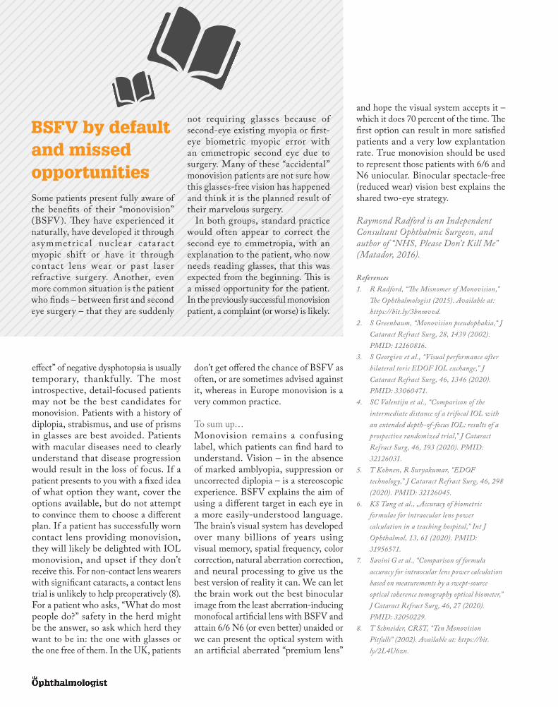
I n Prac t ice36
effect” of negative dysphotopsia is usually temporary, thankfully. The most introspective, detail-focused patients may not be the best candidates for monovision. Patients with a history of diplopia, strabismus, and use of prisms in glasses are best avoided. Patients with macular diseases need to clearly understand that disease progression would result in the loss of focus. If a patient presents to you with a fixed idea of what option they want, cover the options available, but do not attempt to convince them to choose a different plan. If a patient has successfully worn contact lens providing monovision, they will likely be delighted with IOL monovision, and upset if they don’t receive this. For non-contact lens wearers with significant cataracts, a contact lens trial is unlikely to help preoperatively (8). For a patient who asks, “What do most people do?” safety in the herd might be the answer, so ask which herd they want to be in: the one with glasses or the one free of them. In the UK, patients
don’t get offered the chance of BSFV as often, or are sometimes advised against it, whereas in Europe monovision is a very common practice.
To sum up…Monovision remains a confusing label, which patients can find hard to understand. Vision – in the absence of marked amblyopia, suppression or uncorrected diplopia – is a stereoscopic experience. BSFV explains the aim of using a different target in each eye in a more easily-understood language. The brain’s visual system has developed over many billions of years using visual memory, spatial frequency, color correction, natural aberration correction, and neural processing to give us the best version of reality it can. We can let the brain work out the best binocular image from the least aberration-inducing monofocal artificial lens with BSFV and attain 6/6 N6 (or even better) unaided or we can present the optical system with an artificial aberrated “premium lens”
and hope the visual system accepts it – which it does 70 percent of the time. The first option can result in more satisfied patients and a very low explantation rate. True monovision should be used to represent those patients with 6/6 and N6 uniocular. Binocular spectacle-free (reduced wear) vision best explains the shared two-eye strategy.
Raymond Radford is an Independent Consultant Ophthalmic Surgeon, and author of “NHS, Please Don’t Kill Me” (Matador, 2016).
References1. R Radford, “The Misnomer of Monovision,”
The Ophthalmologist (2015). Available at: https://bit.ly/3bnmvvd.
2. S Greenbaum, “Monovision pseudophakia,“ J Cataract Refract Surg, 28, 1439 (2002). PMID: 12160816.
3. S Georgiev et al., “Visual performance after bilateral toric EDOF IOL exchange,” J Cataract Refract Surg, 46, 1346 (2020). PMID: 33060471.
4. SC Valentijn et al., “Comparison of the intermediate distance of a trifocal IOL with an extended depth-of-focus IOL: results of a prospective randomized trial,” J Cataract Refract Surg, 46, 193 (2020). PMID: 32126031.
5. T Kohnen, R Suryakumar, “EDOF technology,” J Cataract Refract Surg, 46, 298 (2020). PMID: 32126045.
6. KS Tang et al., „Accuracy of biometric formulae for intraocular lens power calculation in a teaching hospital,” Int J Ophthalmol, 13, 61 (2020). PMID: 31956571.
7. Savini G et al., “Comparison of formula accuracy for intraocular lens power calculation based on measurements by a swept-source optical coherence tomography optical biometer,” J Cataract Refract Surg, 46, 27 (2020). PMID: 32050229.
8. T Schneider, CRST, “Ten Monovision Pitfalls” (2002). Available at: https://bit.ly/2L4U6zn.
BSFV by default and missed opportunitiesSome patients present fully aware of the benefits of their “monovision” (BSFV). They have experienced it naturally, have developed it through asymmetr ica l nuc lear cataract myopic shift or have it through contact lens wear or past laser refractive surgery. Another, even more common situation is the patient who finds – between first and second eye surgery – that they are suddenly
not requiring glasses because of second-eye existing myopia or first-eye biometric myopic error with an emmetropic second eye due to surgery. Many of these “accidental” monovision patients are not sure how this glasses-free vision has happened and think it is the planned result of their marvelous surgery.
In both groups, standard practice would often appear to correct the second eye to emmetropia, with an explanation to the patient, who now needs reading glasses, that this was expected from the beginning. This is a missed opportunity for the patient. In the previously successful monovision patient, a complaint (or worse) is likely.

www.theophthalmologist.com
I n Prac t ice 37
An abnormal tear film underlies ocular surface symptoms associated with dry eye disease (DED). Measuring osmolarity at the point of care, therefore, has become a recommended tool in the diagnosis of ocular surface disease (1, 2, 3, 4). The measurements derived from osmolarity testing also provide an objective value for determining the severity of disease and for assessing the efficacy of treatments. When DED improves, osmolarity numbers go down – but how useful is a normal osmolarity result in making a diagnosis?
What does a normal tear test tell us?In 2018, my colleagues at Weill Cornell Medicine, Department of Ophthalmology, and I conducted a prospective observational cohort s t udy to e x plore t h e d i a g n o s t i c utility of normal tear osmolar it y in patients with symptoms suggestive of DED (5). Our aim was to evaluate for the presence of any
alternate ocular surface disease (OSD) in patients with DED-like symptoms but with normal tear osmolarity.
We evaluated 100 patients who underwent tear osmolarity testing (TearLab), if they reported one or more symptoms indicative of potential DED.
Patients were included for the study if they had a normal
tear osmolarity test (value < 308 mOsm/L in each eye, and
an inter-eye difference <
8 mOsm/L). The main outcome measure was the presence of any alternate diagnosis to explain the patient’s symptoms.
Among these patients, the mean tear osmolarity was 293.40 mOsms/L ( ± 6.82), with a mean absolute difference of 2.85
Putting Dry Eye to the TestNarrowing down the root causes of OSD with point-of-care diagnostics By Ashley Brissette
“The main outcome measure was the
presence of any alternate diagnosis to explain the patient’s
symptoms.”
DED-like symptoms, actual diagnosisIn a study of 100 patients with DED-like symptoms and normal tear osmolarity, the most frequent OSD diagnoses included:
• anterior blepharitis (26%)• allergic conjunctivitis (21%)• epithelial basement membrane
dystrophy (EBMD) (8%)• contact lens intolerance (6%)• conjunctivochalasis (5%)• neuropathic pain (4%)• computer vision syndrome (4%)

I n Prac t ice38
mOsms/L ( ± 1.98) between the eyes. A possible alternate diagnosis was established in 89 percent of patients with normal tear osmolarity testing. The most frequent diagnoses included anterior blepharitis (26 percent) and allergic conjunctivitis (21 percent). Our study highlights the diagnostic value of a normal osmolarity with an extremely high proportion of patients exhibiting an alternate OSD diagnosis to account for their symptoms.
Overlapping conditions call for different treatmentsDED is not a simple disease – nor is it a simple diagnosis. The signs and symptoms
of dr y eye can overlap with other conditions – most often blepharitis and meibomian gland disease (see Figures 1 and 2). Measuring tear osmolarity is an important diagnostic step that provides diagnostic information no matter the result. A normal test can alert the clinician that there is likely a different OSD cause for the DED-like symptoms. Other alternate causes for irritation can be epithelial basement membrane dystrophy, keratoneuralgia, contact lens intolerance, conjunctivochalasis, and computer vision syndrome/situational DED (see box: “DED-like symptoms, actual diagnosis).
As part of my diagnostic workup for OSD, I also test for matrix metalloprotease-9 (Inf lammaDry; Quidel), which indicates inflammation on the surface of the eye. The slit-lamp exam is, of course, the cornerstone of any workup. If I know the patient has normal osmolarity, during the exam I can be honing in on evidence of other disease, with blepharitis or allergic conjunctivitis likely culprits. I will ask directed questions about their symptoms to elicit a specific setting or situation like computer use that might be contributing to the eye symptoms.
Ultimately, as eye care specialists, we want to drill down to the underlying cause of the patient's discomfort so that we can ensure our treatments are
targeted and effective. If we treat dry eye and the patient
has allergic conjunctivitis, we have not made an accurate diagnosis and our patient will still have symptoms. Point-of-care testing provides the data needed to help uncover the underlying cause of symptoms – and that’s why it is so important. For patients with multiple diagnoses, we should be treating each root cause of their symptoms.
Aggressive approach to pre-op therapyFor our presurgical patients, we want the surface of the eyes to be pristine so we can obtain accurate biometry and keratometry measurements. Because our refractive decisions – and outcomes – depend on these values, they must be precise. Another reason why patients should have an optimally pretreated ocular surface is because the surgery itself can lead to an increase in dryness and irritation, especially in the postoperative period. If a patient has irritation after
The ASCRS algorithm at a glance• Osmolarity. Tear hyperosmolarity
(TearLab) is central to the modern definition of DED.
• MMP-9. The enzyme MMP-9 (InflammaDry; Quidel) plays a key role in the breakdown of the ocular surface.
• Further diagnostic tests can be done to identify OSD subtypes, such as lipid layer thickness, meibography, noninvasive tear breakup time, quantification of tear meniscus height, tear lactoferrin levels, topography or tomography, aberrometry, and Objective Scatter Index (HD Analyzer, Visiometrics).
• Clinical Exam. Consider the mnemonic, look, lift, pull, push, for the quick focused ocular surface exam.
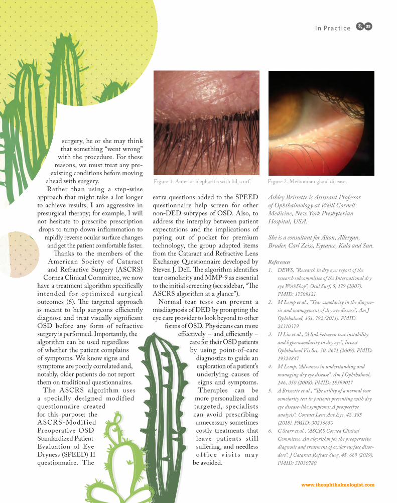
www.theophthalmologist.com
I n Prac t ice 39
surgery, he or she may think that something “went wrong”
with the procedure. For these reasons, we must treat any pre-
existing conditions before moving ahead with surgery.Rather than using a step-wise
approach that might take a lot longer to achieve results, I am aggressive in presurgical therapy; for example, I will not hesitate to prescribe prescription drops to tamp down inflammation to
rapidly reverse ocular surface changes and get the patient comfortable faster.
Thanks to the members of the American Society of Cataract and Refractive Surgery (ASCRS)
Cornea Clinical Committee, we now have a treatment algorithm specifically intended for opt imized surgica l outcomes (6). The targeted approach is meant to help surgeons efficiently diagnose and treat visually significant OSD before any form of refractive surgery is performed. Importantly, the algorithm can be used regardless of whether the patient complains of symptoms. We know signs and symptoms are poorly correlated and, notably, older patients do not report them on traditional questionnaires.
The ASCRS algorithm uses a special ly designed modif ied questionnaire created for this purpose: the ASCRS-Modif ied Preoperative OSD Standardized Patient Evaluation of Eye Dryness (SPEED) II questionnaire. The
extra questions added to the SPEED questionnaire help screen for other non-DED subtypes of OSD. Also, to address the interplay between patient expectations and the implications of paying out of pocket for premium technology, the group adapted items from the Cataract and Refractive Lens Exchange Questionnaire developed by Steven J. Dell. The algorithm identifies tear osmolarity and MMP-9 as essential to the initial screening (see sidebar, “The ASCRS algorithm at a glance”).
Normal tear tests can prevent a misdiagnosis of DED by prompting the eye care provider to look beyond to other
forms of OSD. Physicians can more effectively – and efficiently –
care for their OSD patients by using point-of-care
diagnostics to guide an exploration of a patient’s underlying causes of signs and symptoms. Therapies can be
more personalized and targeted, specia l ists can avoid prescribing unnecessary sometimes costly treatments that leave patients st i l l suffering, and needless o f f i c e v i s i t s m a y
be avoided.
Ashley Brissette is Assistant Professor of Ophthalmology at Weill Cornell Medicine, New York Presbyterian Hospital, USA.
She is a consultant for Alcon, Allergan, Bruder, Carl Zeiss, Eyeance, Kala and Sun.
References1. DEWS, “Research in dry eye: report of the
research subcommittee of the International dry eye WorkShop”, Ocul Surf, 5, 179 (2007). PMID: 17508121
2. M Lemp et al., “Tear osmolarity in the diagno-sis and management of dry eye disease”, Am J Ophthalmol, 151, 792 (2011). PMID: 21310379
3. H Liu et al., “A link between tear instability and hyperosmolarity in dry eye”, Invest Ophthalmol Vis Sci, 50, 3671 (2009). PMID: 19324847
4. M Lemp, “Advances in understanding and managing dry eye disease”, Am J Ophthalmol, 146, 350 (2008). PMID: 18599017
5. A Brissette et al., “The utility of a normal tear osmolarity test in patients presenting with dry eye disease-like symptoms: A prospective analysis”, Contact Lens Ant Eye, 42, 185 (2018). PMID: 30236650
6. C Starr et al., “ASCRS Cornea Clinical Committee. An algorithm for the preoperative diagnosis and treatment of ocular surface disor-ders”, J Cataract Refract Surg, 45, 669 (2019). PMID: 31030780
Figure 1. Anterior blepharitis with lid scurf. Figure 2. Meibomian gland disease.
Tensions have been high since the onset of COVID-19 for all investors, including ophthalmologists. For the first time ever, many ophthalmology offices closed in the spring of 2020, decimating practice revenue and personal income. On top of this came a stock market downturn in March, where values plummeted more rapidly than seen in decades. Though the US market rebounded well, eventually hitting new highs, much of this can be attributed to the federal government stimulus. Further, the real economy continues to struggle and COVID-19 hospitalizations and deaths are at record levels.
As a result of these factors, you might be among many ophthalmologists asking “What can/should I do to manage and protect my finances?” Here, I lay out five actions you can take to proactively manage your wealth during trying times. Ideally, you will do so with your trusted professional
advisor – a financial planner, wealth manager, attorney, or accountant.
One: Focus on the long term – macroeconomicsOne of the topics we encourage doctors to discuss with their trusted financial advisors is the long-term history of the US stock market and economy. Looking at 100+ years of data can help nervous investors reduce stress when seeing previous serious shocks to the system, such as world wars, the Great Depression, and the Great Recession, as well as subsequent recoveries. Doing this can help physicians apply the ancient wisdom “this too shall pass” to the financial arena.
Two: Focus on the long term – microeconomicsPerhaps more valuable than reviewing long-term macroeconomic history i s r e-e xa min ing you r per sona l
(microeconomic) long-term future. This means reviewing your long-term financial model with your financial advisor, using assumptions that reflect our new reality – ideally, through adjustable, iterative software where variables can be altered, and best/medium/worst cases saved for future review. Once again, most ophthalmologists who are years away from retirement may see that even the short-term pain of today will have a relatively minor impact on their long-term plans. This realization can be burden-relieving.
Another benefit of looking at one’s personal planning model is to re-focus on cash reserves and personal spending. In good times (such as the last decade), many physicians reduced their concentration on personal spending and maintaining a sufficient “rainy day fund.” Times likes these can lead to an appropriate re-emphasis on these two key elements of financial modeling.
Crisis ManagementWhat five financial steps should ophthalmologists take during the COVID-19 pandemic? By David Mandell
ProfessionYour career
Your businessYour life
www.theophthalmologist.com
Three: Make tactical investment changes… Or don’tMoving from the long term to the short term, there may be tactical investment changes to implement during this crisis. For some, this will simply mean rebalancing asset class allocations to their long-term strategic percentages. As an example, an investor with a long-term strategic model of 70 percent stocks and 30 percent bonds and alternatives might see those percentages move significantly from those benchmarks during a stock downturn, especially if stocks lose value when bonds and alternatives remain steady or gain in value. Simply rebalancing back to the 70/30 split would require some trading – even if both the client and advisor agree nothing should change for the long-term model.
For others, who need cash to maintain their practices or pay personal bills, securities may need to be sold regardless of, or in addition to, rebalancing. Determining which assets to liquidate and how to minimize tax implications is extremely important in these situations.
Finally, many investors may make no changes to their portfolios. In all three cases, of course, physicians should be driven by rational decision-making, ideally with the assistance of a professional advisor.
Four: Make sure your financial advisor is acting in your best interestUnderstanding the distinction between a financial advisor operating under a fiduciary or suitability standard is crucial – yet it is one that even many experienced investors do not comprehend.
Stated succinctly, one set of investment advisors operates under a professional standard that requires them to make suitable recommendations to their clients without having to place their interests below that of the client. This type of advisor can, for example, choose among a set of suitable fund choices for a client and choose the one that has the highest charges and pays themselves the highest sales commission. Doing so would not
violate any professional duty – as the advisor has still provided a “suitable” investment.
A key distinction in terms of loyalty is also important, in that this type of advisor’s duty is to the firm he or she works for, not necessarily the client served.
In contrast, another set of investment advisors operates under the fiduciary standard, meaning they have a fiduciary duty to their clients; they have a fundamental obligation to provide suitable investment advice and always act in their clients’ best interests. Using the same example above, if this type of advisor selected the most expensive among a choice of suitable funds based on a higher commission payout, they may face professional liability for doing so.
Even more profound is the fact that, for a fiduciary advisor, such a conflict typically will not even arise. Why? Because fiduciary advisors are typically compensated by a management fee and take no sales commissions on the financial products they may recommend for a client. In this way, the fiduciary advisor’s business model encourages the advisor to choose the lowest cost among equal options for a client – as they get absolutely no benefit from recommending anything else. In fact, as many fiduciary firms charge their fee based on their client’s assets, they are incentivized to reduce the client’s product costs as much as possible, so the client’s investments grow as rapidly as possible.
There is no better time than during this crisis to understand how one’s advisors make money and to whom they owe their duty. Ask the right questions and you will learn the answers.
Five: Protect against other risksAs we all deal with COVID-19, we are primarily attentive to the healthcare, practice, and personal financial risks directly impacted by the crisis. For those who have the capacity to do so, this can be a good time to focus on protecting against other risks as well. Physicians can re-examine their insurance policies, from disability insurance and life
insurance to long-term care coverages for themselves or family members. Others may finally get around to legal planning that they have put off, including asset protection and estate planning.
The author recently published “Wealth Planning for the Modern Physician” – his first book for physicians in five years. To receive free print copies or ebook downloads, text OPHTH to 47177 or visit www.ojmbookstore.com and enter promotional code OPHTH.
David B. Mandell is an attorney and author of more than a dozen books for physicians. He is a partner in the wealth management firm OJM Group.
Disclosure:OJM Group, LLC. (OJM) is an SEC registered investment adviser with its principal place of business in the State of Ohio. SEC registration does not constitute an endorsement of OJM by the SEC nor does it indicate that OJM has attained a particular level of skill or ability. OJM and its representatives are in compliance with the current notice filing and registration requirements imposed upon registered investment advisers by those states in which OJM maintains clients. OJM may only transact business in those states in which it is registered or qualifies for an exemption or exclusion from registration requirements. For information pertaining to the registration status of OJM, please contact OJM or refer to the Investment Adviser Public Disclosure website www.adviserinfo.sec.gov. For additional information about OJM, including fees and services, send for our disclosure brochure as set forth on Form ADV using the contact information herein. Please read the disclosure statement carefully before you invest or send money.
This article contains general information that is not suitable for everyone. The information contained herein should not be construed as personalized legal or tax advice. There is no guarantee that the views and opinions expressed in this article will be appropriate for your particular circumstances. Tax law changes frequently, accordingly information presented herein is subject to change without notice. You should seek professional tax and legal advice before implementing any strategy discussed herein.

Sexual harassment is defined by the US Equal Employment Opportunity Commission as “unwelcome sexual advances, requests for sexual favors, and other verbal or physical conduct of a sexual nature” that “explicitly or implicitly affects an individual’s employment, unreasonably interferes with an individual’s work performance, or creates an intimidating, hostile, or offensive work environment” (1). A national survey of mostly female ophthalmologists and ophthalmology trainees showed that 59 percent had experienced sexual harassment during their careers, most commonly during training (2). Of the ophthalmologists who reported experiencing sexual harassment, 45 percent had been harassed by patients. Few had reported their most significant experience with sexual harassment to an authority – in part because they were not taught how.
Terra incognitaI know this because it happened to me. Over and over again. As a first-year resident, I found myself in one-on-one situations with patients who were incredibly inappropriate. Some insisted on calling me by my first name, despite my having been introduced as “doctor.” Others commented on my body while in the exam lane. One had even looked me up on the Internet and started commenting on my physical appearance and hobbies. I remember telling people what happened, shocked and embarrassed. They laughed or rolled their eyes or told me to ignore it, but nobody challenged it. It just happened over and over, sometimes multiple times per day and distracted me from clinical learning. It seemed that there were no consequences – and that bothered me. I wanted to do something about it, but I wasn’t quite sure what could be done. My supervisors, who were mostly men, agreed that it wasn’t right, but didn’t know what
could be done about it either. This came as no surprise, because institutional training at the time focused on harassment initiated by supervisors or workers and guidelines on responding to sexual harassment initiated by patients and their families were virtually nonexistent (3).
One day, it happened to a junior medical student who was working with me. It was one of her first experiences dealing with a patient and she was – rightly – upset. She had assumed all her patients would respect her role as a soon-to-be physician; the fact that an inappropriate comment from a male patient shattered that illusion made me angry. Around the same time, another patient put his hand on the knee of a team member while she was trying to counsel them about severe vision loss. He had a medical problem that required multiple follow-up appointments and, eventually, surgery. Every time he came in, he would look for me in the hallway to harass me – even though I asked not to see him. I brought it up during one of our
Profess ion42
Change the Things You CanEverything you need to know about responding to patient-initiated verbal harassment By Lauren Hock
grand rounds to get faculty input. Everyone
agreed that I should call out the behavior, but nobody knew
how – so I decided to find out.
Forms, sources, and the impact of harassmentIn my research, I came across an interesting paper. It concerned the Massachusetts General Hospital Department of Surgery, who established the Gender Equity Task Force (GETF) to address gender-based discrimination in the local training environment. In 2017, the GETF surveyed 371 residents at two academic hospitals to better understand perceived sources, frequency, forms, and effects of harassment. They found that female trainees were more likely to endorse personal experience of gender-based and sexual harassment than men (P<0.0001) across all specialties, with patients and nursing staff the most frequently identified sources of harassment. Although an overwhelming majority of both male (86 percent) and female (96 percent) respondents had either experienced or observed harassment in the training environment, less than 5 percent had formally reported such experiences, most frequently citing a belief that nothing would happen (4). These findings are similar to those in a 2019 national survey of U.S. ophthalmology trainees (n = 112) in
which 87 percent of female trainees reported having experienced sexual harassment from patients. Among all ophthalmology trainee respondents, only one-third rated their institution’s sexual harassment training as helpful in preparing them to address harassment by patients (5). These papers gave me a firm foundation to build on and confirmed what I already knew: that sexual harassment disproportionately affects female trainees and that individual training
programs have a responsibility to combat and manage inappropriate patient behavior.
Although the majority of my harassment came from men – both younger and older – I found that female patients could certainly also be harassers. But although their comments were disrespectful, they didn’t convey the same threat to physical safety. They were also far less common. I found that my older male patients didn’t like being in a vulnerable position. They were used to having control,
www.theophthalmologist.com
Profess ion 4343
If you are harassed and decide to respond…• Use “I” statements.
“I feel uncomfortable when you comment on my physical appearance.”
• Address the behavior, not the harasser. “I felt disrespected when you said that” is less likely to make a harasser respond defensively than, “You are disrespectful.”
• Separate intent from impact. “I’m sure you didn’t mean to be hurtful when you said that, but it made me feel…”
• Appeal to egalitarianism. “I went through the same medical training as my colleagues and want to be treated with the same level of respect.”
• Consider what’s in it for the patient. “I want to give you the best care I can, but your comments make me feel unsafe
and don’t allow me to care for you to the best of my ability.””
• Use humor with caution. Exaggeration of an inappropriate comment or gentle sarcasm may be misconstrued as reinforcement of prejudice.
• Set boundaries as needed. “I’m leaving the room because I don’t feel comfortable with your behavior.”
• Offer an alternative. “I would prefer you to call me ‘Doctor,’ rather than ‘baby’ or ‘honey.’”
• Report harassment that threatens your safety or creates an intimidating, hostile, or offensive work environment.
• Here is an example of what you can say: “I’m sure you didn’t mean to be hurtful, but I feel uncomfortable when you comment on my [appearance/identity/background]. I want to give you the best care that I can so [let’s keep our conversation professional/I would prefer you to call me ‘Doctor’/please treat me with the same respect as you do other doctors].”
particularly in interactions with a younger woman, and sought to invert the power structure by being inappropriate, even at the risk of compromising their care. But the impact of that harassment cannot be understated. Studies have found that people who are repeatedly harassed experience increased rates of depression, anxiety, insomnia, absenteeism, and post-traumatic stress disorder (2). Among female ophthalmologists who had experienced sexual harassment, 87 percent reported a significant impact on their professional lives, including interference with their ability to work (1). For a person who spends their entire life in a position of relative comfort, inappropriate behavior can be easily dismissed – but for somebody who has
not enjoyed the same privileges, it can be shattering.
Assembling the toolboxAs I researched social sciences literature on discrimination and harassment, I found some great resources, including a piece by Diane Goodman. She studied diversity and social justice and created a framework for educators dealing with biased comments from people of privilege, including some undergrads. I adopted her protocol and tailored it to a physician–patient dynamic. The final document, a Toolkit for Responding to Patient-Initiated Verbal Harassment available at EyeRounds.org is designed to supplement existing sexual harassment training and packaged in the most accessible way possible. It is pocket-sized and double-
sided. One side tells you what to do if you are the victim of inappropriate behavior; the other explains what to do if you witness inappropriate behavior.
Practice makes perfectWe presented the toolkit to our department last spring in a workshop session focused on script rehearsal. When a patient says something inappropriate, it’s easy to feel like a deer in the headlights. This is when you fall back on the script – a way of communicating clearly, calmly, and respectfully that a behavior is not acceptable. Residents and faculty were put in pairs, with one acting as the patient and the other the physician. We asked all workshop participants to repeat the script in a non-confrontational, nonjudgmental tone at least three times until it felt like a natural response. In my experience, most patients take the script well. But even when they don’t, I feel better for having said something. Inappropriate behavior is known to escalate; if you let people get away with a small thing, they may try again with a more threatening behavior. It is not difficult to imagine inappropriate comments leading to inappropriate touching.
Harassment is not unique to any one generation or demographic. It is a cultural issue – but one that cannot be changed without buy-in from male leadership. It wasn’t until I led the workshop that I truly convinced my male supervisors that harassment was a problem. It was only then that they, too, began calling it out. Our institution has now incorporated harassment training based on the workshop into transitional professional development for all incoming interns and as part of our medical students’ transition to clinical rotations. The message is simple: it is okay to speak up. Residents often hesitate to talk about negative workplace experiences, but they shouldn’t. You are entitled to feel safe in your workplace. To give your best intellectual and empathic care to a patient, you need to feel comfortable – and that means calling out
If you witness harassment...Assess the situation. • Does the person who was harassed
appear uncomfortable or upset?• Nonverbal cues can indicate whether
the person would appreciate help handling the situation.
Respond to the harassment in real time.• “Dr Y is a skilled physician and
a talented surgeon, and their [appearance/identity/background] is not relevant/Most of our physicians prefer to be called ‘doctor.’”
• “Mr Z, we want to give you the best care we can and ask that you treat all of our team members with respect.”
• “We don’t tolerate that kind of language here/Let’s keep it professional.”
• Provide the harassed with an opportunity to leave the room
Offer support.• “That was a difficult encounter.
How are you doing?”• “It seems like Mr X’s comments made
you uncomfortable. How can I help to make this situation better?”
• “I want to hear when things like this happen. It’s important that everyone feels safe and comfortable here.”
Empower to respond.• “I want you to feel empowered to
speak up in situations like this. You have my support.”
• Refer the person to tools for responding to harassment.
Assess the situation. • Encourage reporting of severe or
pervasive sexual harassment.• Create a written record of
the incident.• Report problems to the Office
of the Sexual Misconduct Response Coordinator.
www.theophthalmologist.com
Profess ion 4545
inappropriate behavior. There is no “right” way to report it; create a script that works for you and practice it, so you feel empowered should you need it. In more severe cases, keep a written record of what happened and who was there. This is important if you want to make a case for a pattern of inappropriate behavior.
Before I created the toolkit, male faculty would often say, “If a patient makes you feel uncomfortable, just don’t see them alone. Bring another person into the room.” It’s not always possible to have a male colleague come with you (if, for example, you work in an all-female team) – and you shouldn’t have to. People need to feel equipped to handle such situations on their own, which is why the toolkit is available online for any who need it. In July 2020, in partnership with Dr Nkanyezi Ferguson, Director of Diversity, Equity, and Inclusion for Graduate Medical Education, we were awarded an Innovation Grant from the American Medical Association (AMA) as part of their Accelerating Change in Medical Education Program to expand the toolkit and workshop to address other kinds of identity-based harassment. The $30,000 will allow us to train new workshop facilitators across our institution, who will help sustain the implementation of this curriculum in the coming years.
We have four key aims: to identify key gaps in knowledge pertaining to the prevalence and forms of identity-based, patient-initiated harassment; prepare resident physicians to respond effectively to patient-initiated harassment; establish upstander training to address the important role of supervisors and colleagues in monitoring and responding to harassment; and create a rigorous and sustainable train-the-trainer educational model to allow formal widespread institutional education on best practices for faculty and resident physicians on responding to identity-based harassment at critical points of entry into professional practice. Aside from teaching important communication strategies, we hope that these workshops
support vital conversations between trainees and faculty on how to improve approaches to diversity, equity, and inclusion challenges as training programs and departments.
Now, as a chief resident, I continue to receive inappropriate comments from patients, but knowing how to handle them has transformed my experience. Instead of fighting the instinct to flee the room, I calmly address the patient with a phrase from the toolkit. The other week, one of our junior residents told me that participating in the expanded workshop had given her the confidence to call out a patient who had disparaged her ethnic background. There is much work to be done to improve workplace culture on identity-based discrimination, but these projects can help create a safer, more supportive environment for everyone in our medical communities. Until then, we need to look out for each other. We are all in this together. If we speak up against identity-based discrimination, we can change the culture in our institution and in medicine.
Lauren Hock is an ophthalmologyresident in the University of Iowa
Departmentof Ophthalmology and Visual Sciences, USA.
References1. US Equal Employment Opportunity
Commission, “Facts About Sexual Harassment” (1997). Available at: https://bit.ly/2DcmMSw.
2. N Fnais et al., “Harassment and discrimination in medical training: a systematic review and meta-analysis,” Acad Med, 89, 817 (2014). PMID: 24667512.
3. SK McKinley et al., ““Yes, I’m the doctor”: One department’s Approach to assessing and addressing gender-based discrimination in the modern medical training era,” Acad Med, 94, 1691 (2019). PMID: 31274522.
4. B Scruggs et al., “A U.S. survey of sexual harassment in ophthalmology training using a novel standardized scale”, J Acad Ophthalmolo-gy, 12, e27 (2020). DOI: 10.1055/s-0040-1705092
5. L Hock et al., “Tools for Responding to Patient-Initiated Verbal Sexual Harassment”, EyeRounds.org [Online ahead of print] (2020). Available at: https://bit.ly/37CTUja
6. D Dahlerup, “The story of the theory of critical mass,” Scan Polit Stud, 2, 511 (2006). DOI: 10.1017/S1743923X0624114X
Change is not easy, but it is necessaryThere was a theory that, once women reached critical mass in the workplace (between 30 and 50 percent, depending on the circumstances), harassment would disappear (5). But women reached critical mass in ophthalmology and nothing changed. I was personally empowered by the #MeToo movement. It is much easier to set up a program like ours when you
can see examples of others being held to account. It is no coincidence that the first wave of papers on sexual harassment came after Anita Hill’s testimony during the 1991 Senate confirmation hearing for Supreme Court Justice Clarence Thomas. During her televised testimony, Hill accused Thomas of workplace sexual harassment while he was her supervisor. Though Thomas was still narrowly confirmed, Hill’s powerful testimony sparked a movement toward discussing and reporting workplace sexual harassment, including in medicine. In the late 1990s, that momentum faded, but #MeToo brought it back. People are ready for change.
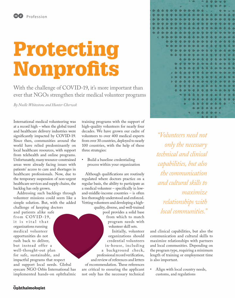
Profess ion46
International medical volunteering was at a record high – when the global travel and healthcare delivery industries were significantly impacted by COVID-19. Since then, communities around the world have relied predominantly on local healthcare resources, with support from telehealth and online programs. Unfortunately, many resource-constrained areas were already facing issues with patients’ access to care and shortages in healthcare professionals. Now, due to the temporary suspension of non-urgent healthcare services and supply chains, the backlog has only grown.
Addressing such backlogs through volunteer missions could seem like a simple solution. But, with the added challenge of keeping doctors and patients alike safe f r o m C OV I D -19, i t i s v i t a l t h a t organizations running medical volunteer opportunities do not rush back to deliver, but instead of fer a wel l-thought-out plan for safe, sustainable, and impactful programs that respect and support local needs. Global eyecare NGO Orbis International has implemented hands-on ophthalmic
training programs with the support of high-quality volunteers for nearly four decades. We have grown our cadre of volunteers to over 400 medical experts from over 30 countries, deployed to nearly 100 countries, with the help of these three strategies:
• Build a baseline credentialing process within your organization
Although qualifications are routinely regulated where doctors practice on a regular basis, the ability to participate as a medical volunteer – specifically in low- and-middle-income countries – is often less thoroughly understood and enforced. Vetting volunteers and developing a high-
quality, diverse, and well-trained pool provides a solid base
from which to match program needs with volunteer skill sets.
Initially, volunteer organizations should
credential volunteers in-house , inc lud ing
a background check , professional record verification,
and review of references and letters of recommendation. These references
are critical to ensuring the applicant not only has the necessary technical
and clinical capabilities, but also the communication and cultural skills to maximize relationships with partners and local communities. Depending on the program type, requiring a minimum length of training or employment time is also important.
• Align with local country needs, customs, and regulations
Protecting NonprofitsWith the challenge of COVID-19, it’s more important than ever that NGOs strengthen their medical volunteer programs By Noelle Whitestone and Hunter Cherwek
“Volunteers need not only the necessary
technical and clinical capabilities, but also the communication
and cultural skills to maximize
relationships with local communities.”

www.theophthalmologist.com
Profess ion 4747

Profess ion48
Although a volunteer’s desire to help is valuable, without aligning with local partners on what support is most needed, poor outcomes and limited sustainability are likely. Organizations should implement hands-on training and clinical care through partnerships with local hospitals and engage with partner staff on multiple levels to ensure that programs can be tailored specifically to local training needs a n d i n t e r e s t s . Requirements vary per location and have increased in recent years, but in-countr y licensure is another k e y e l e m e nt f o r successful volunteer programs – from both a legal and an ethical standpoint. Collaborate with local partners to secure in-country licensure for the volunteers selected for each program location,
in addition to initial internal vetting and clinical privileges
• Hold your organization to a higher standard
Organizations must demand more of themselves. Using qualified volunteers to deliver medical care has always been important; its value is highlighted even more with health systems globally under
signif icant stress. As countries cautiously reopen to visitors, organizations must ensure that they do not cause a greater disservice through the delivery
of poor care or training. Volunteer efforts need to be
impactful – there is no t ime or room for waste or “good enough.” Taking an “it’s
better than they have now” approach
is detrimental to both patients and volunteers.
In this sense, renewing and refreshing credentials is just as important for volunteers as for staff medics. Organizations need to commit to the ongoing process of updating credentials and meeting the changing requirements of host countries and their Ministries of Health. Additional internal controls – including incident reporting and follow-up discussions through a clinical oversight committee – further promote continuous improvement.
Dedication to consistent, high-quality programming is critical as we prepare to come out of the pandemic – and that can only be achieved with appropriately skilled volunteers. This pause provides an opportunity to reevaluate how medical volunteer programs are organized and implemented, ensuring they safely meet the needs of both volunteers and local communities. Furthermore, volunteers themselves should use this pause to evaluate what type of organization they want to travel with – and, more importantly, how they want their time and skills to make a meaningful and lasting impact in the future.

W E E K L Y N E W S L E T T E R S Brought to you by Texere Publishing,
the parent company of The Ophthalmologist
Taking the effort out of staying informed
The week in cannabis science
The emerging science of the outbreak
Everything cell and gene therapy
T E X E R E N E W S L E T T E R S . C O MTaking the effortout of staying informed
S U B S C R I B E
Newsletters New TOP.indd 1Newsletters New TOP.indd 1 19/01/2021 12:2019/01/2021 12:20
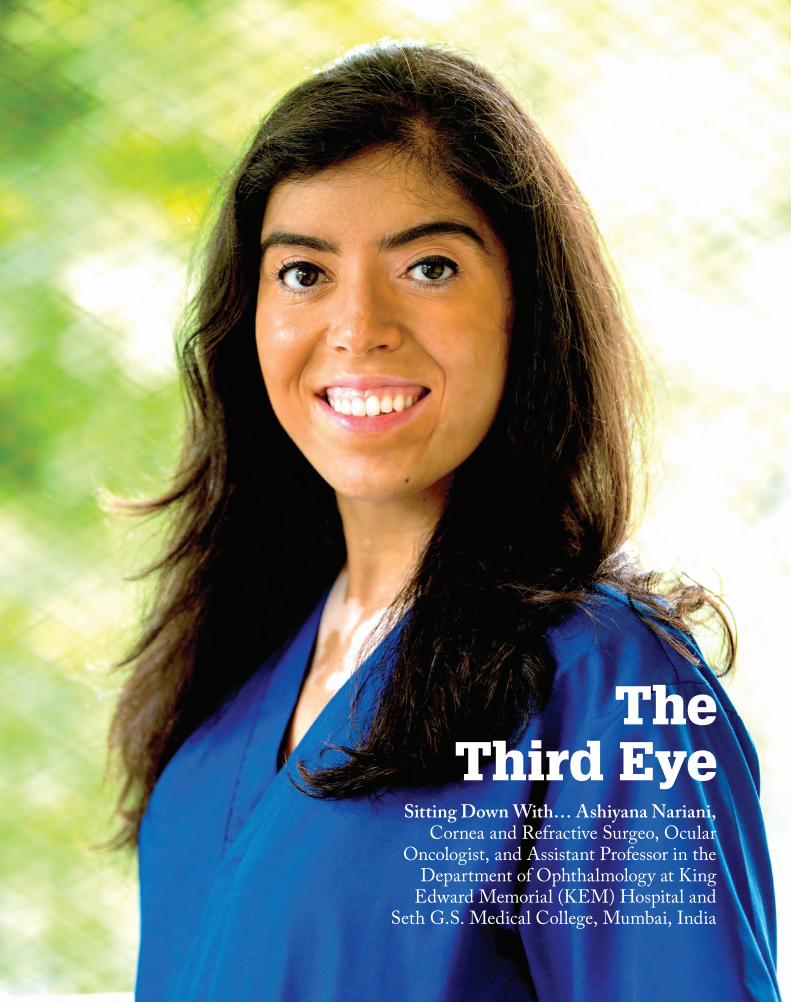
The Third Eye
Sitting Down With… Ashiyana Nariani, Cornea and Refractive Surgeo, Ocular
Oncologist, and Assistant Professor in the Department of Ophthalmology at King
Edward Memorial (KEM) Hospital and Seth G.S. Medical College, Mumbai, India
www.theophthalmologist.com
51Sit t ing D own W ith
Who inspired you to move from the US to India to practice ophthalmology?When I was growing up, my biggest dream was to serve the underserved. As a young girl, I met my spiritual mentor, Dada J.P. Vaswani – a figure famous for dedicating himself to the service of humanity – and he became an example for me to live by. He lived in complete humility and compassion, giving himself to each and every person. Dada Vaswani showed me that we have one life and we should make the most of it by serving other humans and taking care of animals. Another hero of mine is Albert Schweitzer, a Nobel Peace Prize-winning physician who left his life in Europe to serve patients in Africa. When I was in medical school in Boston, doing my Master’s degree in Public Health, I came across the Albert Schweitzer Fellowship of Service and I was inspired to work in clinics for the local underprivileged population.
Tell me about moving to India…When I finished my corneal transplant fellowship at Duke University, I had to choose between a career in academia in the US or helping the underserved abroad. I had several positions open to me in the US and no job lined up in India – I didn’t even have a license to practice here. Nevertheless, I moved here in 2018. I started writing to the chief ministers and medical council bodies, who weren’t sure what to make of me – they had never known a US doctor to move to India. I worked hard to explain that I came to work for those who needed my help – to reverse blindness with cataract surgery or a corneal transplant.
I went up the ranks of ministers, attending meetings, calling, and emailing. Eventually, I ended up at the top, explaining to Prime Minister Narendra Modi’s government that my intentions were pure and I wanted to do
things based on merit – not money under the table. Finally, I was sent to New Delhi to meet the Head of the Medical Council of India, who apologized to me for all the delays – and that’s how I got certified to work in India. It has not been a straight path, but I am so happy to be working and living here. I never thought this is where I would end up, but I followed my intuition and my beliefs.
Where are you based now?I got a faculty position at the Municipal Hospital in Mumbai. I normally see between 100 and 200 patients a day – this has been reduced to 70 or fewer patients a day during the pandemic. These patients don’t have any money to pay for treatment. Their procedures are free of charge, but they are normally required to pay for the supplies. Unfortunately, 49 rupees (less than a dollar) for a bottle of steroid drops is often out of their reach – so we often pay for their lenses or medication ourselves.
What have been your proudest achievements in India so far?I am very proud of improving the quality of training at my hospital and of starting online lectures and an ophthalmology educational core series available to not only my residents, but ophthalmologists around the world. I managed to get experts from all subspecialties – huge names, such as Carol Shields or Richard Lindstrom – to come together and teach online once a month, with attendees from 41 countries. The US and Europe have excellent conferences and events, but many can’t afford tickets, travel, and accommodation – not just those at the start of their career, but often ophthalmologists with over 20 years of experience. That’s why free, high-quality online teaching programs are so vital. This project has been even more important since the start of the
COVID-19 pandemic, so my goal for 2021 is to make it even bigger, with a proper website built to host the sessions and major organizations and eye institutes helping deliver the training.
What goals do you hope to achieve in the near future? We are trying to get a refractive surgery suite for the Municipal Hospital. It would be amazing if one day we could provide refractive surgery – normally only available to wealthy patients – India’s poorest population. It is a multimillion-dollar endeavor, so it might not seem realistic, but we can achieve it if we just keep putting one foot in front of the other. I’ve started noticing that, when there is a specific, concrete need, people come forward and help.
What are your long-term plans?I have none. That’s very different from my life 10 years ago, when I had everything planned: medical school, residency, fellowship. The move to India has taught me that I don’t have to plan far in advance. What matters is that I do the best I can for my patients. I’m taking it one step at a time – making arrangements for what is needed right now, and not thinking beyond that.
Do you think you will stay in India?As ophthalmologists, we are responsible for taking care of our patients’ two eyes but, in certain cultures, people believe we also have a third eye – an insight into ourselves – that we need to discover and open to find the true meaning of our lives.
My deep feeling – perhaps what I can see with my third eye – is that I belong here in India, where folks don’t have a loud voice, but deserve the same care and attention as people in developed countries. I’m staying.
© 2020 Santen Inc. All rights reserved. PP-GLAU-US-0067
Santen is partnering with glaucoma surgeons to improve glaucoma surgical outcomes.
Hear from your peers in a new video series AdvancingGlaucomaSurgery.com
W O R K I N G T O E M P O W E R A N E W E R A O F
PROACTIVE GLAUCOMA SURGERY“
““We might see a day in which the subjective portion of surgery is minimal and we have more objective ways of lowering IOP.
— Dr. Arsham Sheybani
SNTN_19_0032_GST_JournalAd_Phase-II_TheOpthal_M04Colors: 4CBleed: 8.506”w x 10.716”h Trim size: 8.27” w x 10.48” h Live: 7.876”w x 10.086”hFile built at 100%
SNTN_19_0032_GST_JournalAd_Phase-II_TheOpthal_M04.indd 1SNTN_19_0032_GST_JournalAd_Phase-II_TheOpthal_M04.indd 1 9/9/20 12:51 PM9/9/20 12:51 PM