reduction in mitral regurgitation during therapy guided by measured filling pressures in the escape...
TRANSCRIPT

Reduction in Mitral Regurgitation During Therapy Guided byMeasured Filling Pressures in the ESCAPE Trial
Maryse Palardy, MD1, Lynne W. Stevenson, MD1, Gudaye Tasissa, PhD2, Michele A.Hamilton, MD3, Robert C. Bourge, MD4, Thomas G. DiSalvo, MD5, Uri Elkayam, MD6, JamesA. Hill, MD7, and Sharon C. Reimold, MD [on behalf of for the ESCAPE investigators]81Brigham and Women’s Hospital, Boston, MA2Duke Medical Center, Durham, NC3UCLA Medical Center, Los Angeles, CA4University of Alabama, Birmingham, AL5Vanderbilt University Hospital, Nashville, TN6University of Southern California, Los Angeles, CA7University of Florida, Gainesville, FL8UT Southwestern Medical Center, Dallas, TX
AbstractBackground—Dynamic mitral regurgitation (MR) contributes to decompensation in chronicdilated heart failure. Reduction of MR was the primary physiologic endpoint in the ESCAPE trial,which compared acute therapy guided by JVP, edema, and weight (CLIN) to therapy guidedadditionally by pulmonary artery catheters (PAC) toward pulmonary wedge pressure ≤15 and rightatrial pressure ≤8 mmHg.
Methods and Results—Patients were randomized to PAC or CLIN during hospitalization withchronic HF and mean LVEF 20%, and at least 1 symptom and 1 sign of congestion. MR and mitralflow patterns, measured blinded to therapy and timepoint, were available at baseline and dischargein 133 patients, and at 3 months in 104 patients. Changes in MR and related transmitral flow patternswere compared between PAC and CLIN patients. Jugular venous pressure, edema, and weightsdecreased similarly during therapy in the hospital for both groups. In PAC but not in CLIN patients,MR jet area, MR/LAA ratio, and E velocity were each significantly reduced and deceleration timeincreased by discharge. By 3 months, patients had clinical evidence of increased JVP, edema, andweight since discharge, reaching significance in the PAC arm, and the change in MR was no longerdifferent between the 2 groups, although the change in E velocity remained greater in PAC patients.
Conclusions—During hospitalization, therapy guided by PAC to reduce left-sided pressuresimproved MR and related filling patterns more than therapy guided clinically by evidence of systemicvenous congestion. This early reduction did not translate into improved outcomes out of the hospital,where volume status reverted toward baseline.
Corresponding author: Lynne Warner Stevenson, MD, Cardiovascular Division, Brigham and Women’s Hospital, 75 Francis Street,Boston, MA 02115, Phone: 617-732-7406, Fax: 617-264-5265, [email protected] of Interest DisclosuresDr. Stevenson serves as a consultant and has received research support from Medtronics, Inc. The other authors have no conflicts ofinterest to disclose in relation to this manuscript.
NIH Public AccessAuthor ManuscriptCirc Heart Fail. Author manuscript; available in PMC 2010 May 1.
Published in final edited form as:Circ Heart Fail. 2009 May ; 2(3): 181–188. doi:10.1161/CIRCHEARTFAILURE.108.822999.
NIH
-PA Author Manuscript
NIH
-PA Author Manuscript
NIH
-PA Author Manuscript

Keywordshemodynamics; heart failure; cardiomyopathy; mitral regurgitation
IntroductionMitral regurgitation (MR) is a central feature of progression of dilated left ventricular failure,in which it plays a role as both cause and effect[1;2]. From the spectrum of asymptomatic leftventricular dysfunction through evaluation for transplantation, the severity of mitralregurgitation carries strong prognostic weight[3–6].
Acute therapy tailored to reduce measured left-ventricular filling pressures in decompensatedheart failure has been shown to cause marked reduction in mitral regurgitation[7]. In theabsence of inotropic therapy, the increase in forward stroke volume results primarily fromredistribution of regurgitant volume[8;9]. Echocardiographic measurements focusing preciselyon the mechanics of mitral regurgitation in dilated heart failure have demonstrated that themajor change during therapy with vasodilators and diuretics is attributable to reduction ofeffective regurgitant orifice area[9]. This reduction in regurgitant orifice area is related in partto decrease in mitral annular distension with improved leaflet coaptation[9]. While therapy inthese studies was targeted toward pulmonary capillary wedge pressure ≤15 mmHg, it was notknown whether similar reduction of mitral regurgitation would result from therapy guided byclinical examination, which is dominated by evidence of right-sided pressures as approximatedfrom jugular vein inspection[10].
The Evaluation Study of Congestive Heart Failure and Pulmonary Artery Catheterization wasdesigned to compare the impact of therapy guided by clinical assessment of filling pressuresto therapy guided additionally by pulmonary artery catheterization for patients hospitalizedwith an exacerbation of advanced heart failure[11]. The primary clinical outcome variable ofdays alive out of hospital during the 6 months was neutral. The primary physiologic variablewas pre-specified to be mitral regurgitation, selected for its importance in prognosis, itssensitivity to filling pressures, and the ability to be measured blindly without potential influencefrom patient or physician knowledge of treatment arm. The hypothesis was that mitralregurgitation would be more effectively reduced when therapy to relieve congestion was guidedby filling pressure goals of pulmonary capillary wedge pressure ≤15 and right atrial pressure≤8 mm Hg in addition to clinical assessment of volume status, which reflects predominantlyright-sided filling pressures.
MethodsTrial design—The Evaluation Study of Congestive Heart Failure and Pulmonary ArteryCatheterization Effectiveness (ESCAPE) trial randomized 433 patients at 26 sites betweenJanuary 18, 2000 and November 17, 2003. Inclusion criteria included hospitalization withchronic advanced heart failure despite recommended therapies. Patients were randomlyassigned to therapy guided by pulmonary artery catheterization (PAC group) or by clinicalassessment (CLIN group), as previously described[11]. The goals in both groups werereduction of filling pressures, assessed by jugular venous pressure (JVP), edema, andsymptoms in the CLIN group and additionally with goals of pulmonary capillary wedgepressure ≤15 and right atrial pressure ≤8 mmHg in the PAC group. After discharge, the protocolspecified that patients return for clinical assessment and adjustment of medications at 2 weeks,1 month, 2 months, and 3 months after hospital discharge.
Clinical Data—Changes in weight, estimated JVP, and edema, as assessed on the 0–4 scale,were determined between baseline and discharge. In those patients randomized to PAC,
Palardy et al. Page 2
Circ Heart Fail. Author manuscript; available in PMC 2010 May 1.
NIH
-PA Author Manuscript
NIH
-PA Author Manuscript
NIH
-PA Author Manuscript

differences between RA pressure and pulmonary capillary wedge pressure at the time ofinsertion and removal were also described.
Echocardiographic examinations—All sites provided the echocardiographic corelaboratory with a validation echocardiogram according to defined standardized views for two-dimensional and Doppler measurements, as pre-specified for review prior to initiation. Theprotocol specified that echocardiograms be obtained in patients at the time of randomization(baseline), hospital discharge (DC), and at 3 months. However, to maximize enrollment, it wasemphasized that neither randomization nor discharge should be delayed due to difficulties inscheduling echocardiograms.
Echocardiograms were analyzed at the core laboratory at the University of Texas Southwesternin Dallas. All studies were analyzed blinded to randomization and study order. The studieswere analyzed offline by a single sonographer using a calibrated ultrasound measuring system.A portion of the studies was re-measured to evaluate intra-observer variability. Two-dimensional and Doppler echocardiographic parameters were measured in accordance withAmerican Society of Echocardiography criteria. Available measurements included: mitralregurgitant color jet area from the apical 4 chamber view, left ventricular end-diastolicdimension, left ventricular end-systolic dimension, left atrial diameter, left ventricular ejectionfraction using apical single plane disc method, left atrial area (LAA) measured from the apical4 chamber view. The protocol included calculation of mitral regurgitant volume and effectiveregurgitant orifice from color proximal isovelocity surface area on the apical projection, butthese measurements could not be included in the analysis since too few recorded tapes providedadequate data on the majority of patients. The mitral regurgitant color jet area was measuredand the ratio of this area to left atrial area determined. These were considered to be adequatefor measurement if the origin of the jet was seen at the level of the mitral valve and the bordersof the jet were clearly delineated. The left atrial area was measured if the left atrial borderswere clearly identified.
Mitral filling patterns included measurement of early mitral inflow velocity (E wave), anddeceleration time (Decel time). The peak systolic and diastolic pulmonary venous flowvelocities were measured and the ratio of systolic to diastolic peak velocities calculated (PVS/D).
Changes in echocardiographic variables for each patient were determined from baseline todischarge and baseline to three months. The number of paired samples that could be analyzedfor each variable varied, depending on the number of adequate measurements from eachechocardiographic study. Echocardiographic results were calculated both for mean changesand for median relative changes compared to individual baseline values.
Statistics—Continuous variables were summarized by mean and standard deviation (SD)statistics. Paired T-tests were used to assess significance of changes between baseline anddischarge and baseline and 3 months. Standard t-tests were used to compare changes in thePAC group to changes in the CLIN group. A p-value of <0.05 was statistically significant. Thequestion of whether the two strategies led acutely to different reduction of mitral regurgitationwas considered to be separate from the question of whether there were differences sustainedat 3 months, for which the outpatient therapies and compliance would be important factors.Thus the strategies were compared separately at the two time points. No correction was madefor multiple comparisons.
ResultsStudy population—Both baseline and discharge echocardiograms were available foranalysis in 198 patients (Figure 1). Measurements of MR were adequate for comparison
Palardy et al. Page 3
Circ Heart Fail. Author manuscript; available in PMC 2010 May 1.
NIH
-PA Author Manuscript
NIH
-PA Author Manuscript
NIH
-PA Author Manuscript

between baseline and discharge for 133 patients, in whom demographics and hemodynamicsconfirmed severe heart failure with reduced ejection fraction (average 20%), low systolic bloodpressure compared to most hospitalized HF populations (average 104 mmHg), and markedlyelevated jugular venous pressures (Table 1). Baseline profiles were not different between the133 patients with paired MR measurements, the 106 patients who had an echocardiogram atbaseline but not at discharge, and the 65 for whom both echocardiograms were obtained butchange in MR could not be assessed from the views provided (Table 1). Estimated JVPelevation and edema were comparable in the two randomized groups CLIN and PAC, and theresting pulmonary capillary wedge pressure was 25 ±9 mmHg and RA pressure 13 ±9 mm Hgin the PAC group. LV dilation, LVEF, mitral regurgitation and flow patterns were comparablein the two groups at baseline (Table 2).
Changes from baseline to discharge—Patient weight decreased significantly in bothgroups during therapy in hospital, by 3.2 ±3.8 Kg in the CLIN group and 4.1 ±5.7 Kg in thePAC group, without significant difference between groups (p =0.24). Estimated JVP and edemadecreased comparably in both groups (Figure 2). For the PAC group, measured PCW decreasedby average 8 ±9 mmHg and RA pressure by 5 ±12 mmHg.
The MR color jet area decreased by 2 cm2 in the PAC group (p <0.004), but did not changedin the CLIN group (between group difference for change p <0.01, Table 2). The median %reduction relative to baseline was 2% in CLIN and 20% in PAC (Figure 3). The ratio of MR/LAA decreased significantly from 0.3 to 0.2 in the PAC group (18% relative reductioncompared to individual baseline), but not in the CLIN Group (between group p =0.02). Dopplerpatterns representing left heart filling patterns also demonstrated greater improvement in PACpatients (Table 2, Figure 3). There was a significant decrease in E wave velocity in the PACgroup but not in the CLIN group (between group p =0.01). Deceleration time increasedsignificantly only in PAC patients (p =0.05), without significant between-group difference.Changes in pulmonary vein inflow patterns could be measured at baseline and discharge inonly 61 patients, but there was a trend for increasing systolic/diastolic ratio only in the PACgroup (difference between PAC and CLIN p =0.14). Left ventricular dimensions and ejectionfraction did not change significantly from randomization to the time of discharge.
Changes from baseline to 3 monthsIncrease in Evidence of Fluid Retention After Discharge: By 3 months after discharge, JVPand weights had increased back toward baseline in both groups, changes which were significantin the PAC group. Peripheral edema increased significantly in both groups (Figure 2).
Of the 198 patients with echoes at baseline and discharge, 124 had echoes also at 3 months.An additional 43 patients had paired echoes at baseline and 3 months but not at discharge(Figure 1). After 3 months, there was no longer a difference seen between the PAC and CLINgroups for either MR area or MR/LAA area (Table 3). MR/LAA area was reduced comparedto baseline in both groups, and MR area was reduced compared to baseline in the CLIN group.Mitral flow patterns continued to show trends for improved filling for patients after initialtailored therapy with PAC. The reduction in E velocity in PAC patients was less than atdischarge, but remained significantly reduced compared to the CLIN patients at 3 months. At3 months, Decel time remained longer than at baseline for PAC (p =0.02), but not for CLIN.LV end-diastolic dimensions remained unchanged for all groups. In the absence of detectablechange in LV dimension, the 5 point increase in LV ejection fraction in the PAC group at 3months compared to baseline (p =0.01) may be a chance finding and is of unclear clinicalsignificance.
Palardy et al. Page 4
Circ Heart Fail. Author manuscript; available in PMC 2010 May 1.
NIH
-PA Author Manuscript
NIH
-PA Author Manuscript
NIH
-PA Author Manuscript

DiscussionThe primary physiologic endpoint of the ESCAPE trial was mitral regurgitation, which wasmore effectively reduced when therapy to relieve congestion was guided by pulmonary arterycatheter goals than by clinical assessment alone during hospitalization. The reduction of mitralregurgitation was accompanied by parallel improvement seen in the inflow E velocity and inthe deceleration time, which both reflect left ventricular filling and may be surrogates for leftventricular filling pressures. The differences seen between the strategies at hospital dischargewere largely lost after three months of outpatient management, during which therapy wasguided in both groups only by clinical assessment, which revealed increased fluid retentionafter discharge.
Assessment of Filling PressuresSerial assessment of filling pressures is a Level I recommendation accompanying adjustmentof diuretics to treat fluid retention in the outpatient setting[12]. Inpatient hospitalization isassociated with excess volume in 83% patients admitted in the ADHERE registry[13], and itis recommended that optimal volume status be restored prior to discharge, based on clinicalassessment. The most helpful clinical sign used to detect elevated left-sided filling pressuresin chronic heart failure remains the elevated jugular venous pressure, as confirmed recently bythe analysis of physician estimates of hemodynamics for the ESCAPE trial[10]. However, thejugular venous pressure actually reflects right-sided filling pressures. For most patients withchronic heart failure, right and left-sided filling pressures track together[14], but it is not knownwhether the degree of elevation is sufficiently mirrored to optimize the left side consistentlyduring serial assessment of the right side. Furthermore, clinical examination of jugular veinscan be done well by experts, but is still less reliable than direct measurement[10;15]. Reductionof JVP during therapy in the hospital was similar in both the CLIN and PAC arms, as wasreduction of peripheral edema. The overall fluid loss, as estimated by acute change in weight,was similar in the two groups, although numerically greater in the PAC arm. Nonetheless, therewas slightly but significantly less renal dysfunction in the PAC arm than the CLIN arm[16].It is plausible but cannot be proven from existing data that monitoring of the left-sided fillingpressures guided adjustment of the “optimal” amount of diuresis and vasodilation for individualpatients, achieving more normal left-sided filling pressures than when they were assessedindirectly from the jugular venous pressures and other clinical signs reflecting the right sideof the heart.
Reduction of Mitral RegurgitationMitral regurgitation is at least moderate in almost all patients at the time of hospital admissionwith decompensated heart failure and dilated left ventricular failure[17–19]. Therapy tailoredto reduce measured filling pressures to near-normal levels has been shown to decrease valvularregurgitation [17;20]. This decrease in mitral regurgitant volume during unloading therapy wasrecognized initially with nitroprusside, and has been shown to be the major component ofincreased stroke volume during vasodilator and diuretic therapy[8;17;21]. This redistributionof flow can be attributed to reduction of effective regurgitant orifice area[9]. Most of the studiesquantitating reduction of mitral regurgitation were performed during monitoring with apulmonary artery catheter to guide reduction of filling pressures, but lessons learned from thesestudies have been translated into therapy without invasive monitoring. Thus it was not knownwhether similar results could be achieved during contemporary therapy guided by refinedclinical assessment alone. The clinical importance of mitral regurgitation, its relationship toelevated filling pressures, and the ability to isolate its measurement from knowledge oftreatment strategy were key factors in the decision to define mitral regurgitation as the primaryphysiologic variable in the ESCAPE trial, comparing an invasive hemodynamic strategy toclinical assessment.
Palardy et al. Page 5
Circ Heart Fail. Author manuscript; available in PMC 2010 May 1.
NIH
-PA Author Manuscript
NIH
-PA Author Manuscript
NIH
-PA Author Manuscript

The reduction of MR in the PAC arm between baseline and discharge appears to be a robustfinding. The improvements between baseline and discharge for PAC patients, and thedifference between CLIN and PAC strategies are evident whether analyzing the absolutechanges in MR measurements, or the relative changes expressed as a percentage of baselinevalues (Table 2 and Figure 3). The clinical significance of this change in MR is emphasizedby concordant improvements in other measurements. The E velocity declined only in the PACarm, consistent with a meaningful decrease in left ventricular filling pressure, reflecting MRreduction[22;23]. E velocity has been correlated with MR regurgitant fraction severity[22;23]. Deceleration time, which increased significantly only in the PAC patients, has correlatedinversely with pulmonary capillary wedge pressures[24]. Left ventricular end-diastolicdimension is generally insensitive to acute changes, particularly without inotropic therapy, anddid not change in this study. Pulmonary vein profiles could be measured at baseline anddischarge in only 61 patients, with a trend toward improvement only in the PAC group.
Outcomes After Hospital DischargeAlthough MR at the time of discharge was significantly reduced in the PAC arm compared tothe CLIN arm at discharge, six-month outcomes were no different between the groups[11].The failure of acute reduction of MR to improve outcomes in this trial could indicate that 1)MR is an epi-phenomenon that is not itself important to outcomes, 2) any benefit of reducedMR can be outweighed by deleterious effects of the therapies used, or 3) reduced MR isassociated with improved outcomes only if it can be sustained after discharge.
The presence and severity of MR have consistently been associated with worse prognosisthroughout the spectrum of heart failure, from asymptomatic patients early post-infarctionthrough candidates for cardiac transplantation [3;4;6;25]. Changes in prognostic factors do notnecessarily translate into changes in prognosis, although the contribution of mitral regurgitationto elevate left-sided filling pressures and to reduce effective forward stroke volume, make itattractive as a therapeutic target. Surgery to repair mitral regurgitation in heart failure has notled to better outcomes[26], in part because mitral regurgitation often recurs or is replaced bymitral stenosis[27]. For the related measurement of deceleration time, better outcome has beenassociated with restrictive pattern reversibility and prolongation of the deceleration time inpatients during HF therapy[28–31], shown by Xie et al. in 1994[32] and recently by Grayburnet al. in the Beta-blocker Evaluation of Survival Trial[33].
With regard to the therapies used in hospital to decrease volume, the overall use of diureticswas similar in the two arms, with a trend towards slightly lower diuretic use at discharge in thePAC arm[16]. Inotropic therapy was associated with a worse outcome in the ESCAPE trial,and there was slightly higher use of inotropic therapy in the PAC arm than the CLIN arm, butthere was still no difference in outcomes when patients receiving inotropic therapy wereexcluded from analysis[11].
With regard to maintenance of early improvement in MR, the changes observed in the hospitalwere less evident at 3 months in the PAC group, although improvements in the E velocity anddeceleration time that occurred in the PAC group were still significant at 3 months. Onceventricular remodeling is advanced, perhaps a large acute reduction in MR cannot easily bemaintained. On the other hand, there was a significant decrease in MR after discharge in theCLIN group during HF management such that the CLIN and PAC groups converged by 3months of outpatient HF management.
It is known from similar populations that most of the interventions during the 3 months afterhospital discharge involve telephone-directed adjustments in diuretic regimens, mostcommonly based on changes in weight at home[34]. Weight changes at home are less reliablethan previously thought for changes in ventricular filling pressures[35]. The ESCAPE protocol
Palardy et al. Page 6
Circ Heart Fail. Author manuscript; available in PMC 2010 May 1.
NIH
-PA Author Manuscript
NIH
-PA Author Manuscript
NIH
-PA Author Manuscript

specified that patients return for clinical assessment and adjustment of medications at 2 weeks,1 month, 2 months, and 3 months after hospital discharge. The physicians assessing fluid statusduring these visits were the same as those whose assessments correlated well with measuredright-sided pressures at the time of randomization[10]. Despite these frequent opportunitiesfor adjustment of medications, the current study reveals that jugular venous pressures andweights increased between discharge and 3 months, changes which were significant in the PACpatients. This may also relate to the decrement reported between one month and 3 months inthe symptomatic improvement seen in the PAC patients (as reflected in Minnesota Living withHeart Failure scores)[11]. It is not clear whether these changes could have been prevented byeven greater vigilance, or whether physiologic factors in advanced heart failure limit prolongedmaintenance of lower volume status achieved during hospitalization.
There was a late increase in left ventricular ejection fraction in both CLIN and PAC, with animprovement of 5 percentage points in the PAC arm that reached significance. Multiplemechanisms could be invoked to explain such an improvement, including lower wall stressand reduced myocardial oxygen demand, better coronary perfusion and coronary venousdrainage. However, the absence of demonstrable changes in ventricular dimensions may renderthis more likely to be a chance finding.
LimitationsMultiple protocol issues complicate the interpretation of these results. The substantial amountof missing data confounds analysis, but comparison of groups with and without echoes andwithout and without measurable MR does not suggest any systematic bias of this missing data.The echoes that were obtained did not have consistently adequate views for measurement.Thus, the requirement that each site provide a validation echocardiographic tape with all of thekey views did not ensure that subsequent tapes provided similar image quality.
At best, echocardiographic evaluation at multiple sites remains technically challenging inpatients with low ejection fraction, and changes are affected by multiple factors as the pressuregradient between LV and left atria can vary widely, influenced by systemic blood pressure,contractile reserve and diastolic pressure, not solely on the size of the abnormal regurgitantorifice. The ability to index the color jet of mitral regurgitation to left atrial area does providesome improvement in correlation with angiographic evaluation of MR[36]. The measurementof E velocity and deceleration time provided more quantitative evaluation and clearly supportedthe significance of the changes observed in mitral regurgitation.
It is unfortunate that the data obtained did not allow more quantitative estimation in a largernumber of patients. However, the data obtained are consistent with more rigorous prospectivedata that was obtained previously from several non-randomized experiences in single centerinvestigations, demonstrating significant reduction of mitral regurgitation for therapy guidedby a strategy using invasive monitoring. For this larger study in 26 centers, the use of a corelab to perform all measurements blinded to therapy and timepoint increases the confidence inthe results obtained.
With all the limitations, this is a unique dataset in which to assess the impact of current HFtherapy guided with and without a PAC, as well as the limited influence of acute hemodynamicimprovement on outcomes after hospital discharge.
CONCLUSIONSThis study of strategies for decompensated heart failure demonstrates that mitral regurgitationwas more effectively reduced when measured right and left-sided filling pressures were usedto guide therapy in hospital than when estimated jugular venous pressure and edema were
Palardy et al. Page 7
Circ Heart Fail. Author manuscript; available in PMC 2010 May 1.
NIH
-PA Author Manuscript
NIH
-PA Author Manuscript
NIH
-PA Author Manuscript

reduced to a similar level during clinical assessment alone. Current management of volumestatus after discharge may not be adequate to maintain improvements that can be achieved inhospital. The limitations of outpatient therapy may reflect disparity between visible right andoccult left-sided filling pressure elevations, difficulty in assessing right-sided filling pressures,and more relaxed goals for volume status in patients who appear stable in the non-acute setting.It is not known whether other strategies would better sustain the acute reduction in mitralregurgitation or whether chronic reduction in mitral regurgitation would translate intoimproved clinical outcomes with advanced heart failure.
Clinical Summary
Therapy during heart failure hospitalization focuses on relief of elevated filling pressures,which can currentlybe guided by clinical assessment with or without invasive monitoring.In the randomized ESCAPE study in decompensated chronic dilated heart failure,comparison of the clinical and invasive strategies showed that mitral regurgitation waseffectively reduced in hospital only with the invasive strategy, targeting right atrial pressure< 8 mmHg and pulmonary capillary wedge pressure < 15 mmHg, not with the clinicalassessment strategy, which targeted and achieved similar reduction of elevated jugularvenous pressure, edema, and orthopnea. It has been shown earlier that renal function wasalso slightly better with the invasive strategy, as was symptomatic improvement in the firstmonth after discharge. However, these differences between strategies were lost by 3 months,during which there was evidence of recurrent increases in filling pressures. As there wasno difference in 6 month re-hospitalization or survival between the clinical and invasivestrategies, there is currently no rationale for routine use of invasive monitoring to adjusttherapy or to reduce mitral regurgitation. The key components of outpatient therapy foradvanced heart failure remains optimal tolerated doses of neurohormonal antagonists anddiuretic therapy to maintain fluid balance. It is not clear whether future strategies to decreaserecurrent heart failure events should select different targets during hospitalization, ordevelop better strategies to maintain acute reductions in filling pressures and mitralregurgitation during chronic heart failure management.
ABBREVIATIONS USEDCLIN, clinical assessment of filling pressures from symptoms and physical examination andthe group of patients randomized to therapy guided by clinical assessment aloneDecel, deceleration timeE, early mitral inflow velocityJVP, jugular venous pressureLAA, left atrial areaLV, left ventricleLVEDD, left ventricular end-diastolic dimensionLVEDI, left ventricular end-diastolic dimension indexLVEF, left ventricular ejection fractionMR, mitral regurgitationPAC, pulmonary artery catheter and the group of patients randomized to therapy guided by apulmonary artery catheter in addition to clinical assessmentPCW, pulmonary capillary wedge pressurePV S/D, pulmonary vein systolic/diastolic flow ratioRAP, right atrial pressures
Palardy et al. Page 8
Circ Heart Fail. Author manuscript; available in PMC 2010 May 1.
NIH
-PA Author Manuscript
NIH
-PA Author Manuscript
NIH
-PA Author Manuscript

AcknowledgmentsSources of Funding
The ESCAPE study was supported by contract N01-HV-98177 from the National Heart, Lung, and Blood Institute.
Reference List1. Karagiannis SE, Karatasakis GT, Koutsogiannis N, Athanasopoulos GD, Cokkinos DV. Increased
distance between mitral valve coaptation point and mitral annular plane: significance and correlationsin patients with heart failure. Heart 2003;89:1174–1178. [PubMed: 12975411]
2. Otsuji Y, Handschumacher MD, Schwammenthal E, Jiang L, Song JK, Guerrero JL, Vlahakes GJ,Levine RA. Insights from three-dimensional echocardiography into the mechanism of functional mitralregurgitation: direct in vivo demonstration of altered leaflet tethering geometry. Circulation 1997 Sep16;96:1999–2008. [PubMed: 9323092]
3. Bruch C, Klem I, Breithardt G, Wichter T, Gradaus R. Diagnostic usefulness and prognosticimplications of the mitral E/E' ratio in patients with heart failure and severe secondary mitralregurgitation. Am J Cardiol 2007 Sep 1;100:860–865. [PubMed: 17719334]
4. Okura H, Takada Y, Kubo T, Asawa K, Taguchi H, Toda I, Yoshiyama M, Yoshikawa J, Yoshida K.Functional mitral regurgitation predicts prognosis independent of left ventricular systolic and diastolicindices in patients with ischemic heart disease. J Am Soc Echocardiogr 2008;21:355–360. [PubMed:17658723]
5. Amigoni M, Meris A, Thune JJ, Mangalat D, Skali H, Bourgoun M, Warnica JW, Barvik S, ArnoldJM, Velazquez EJ, Van de WF, Ghali J, McMurray JJ, Kober L, Pfeffer MA, Solomon SD. Mitralregurgitation in myocardial infarction complicated by heart failure, left ventricular dysfunction, orboth:prognostic significance and relation to ventricular size and function. Eur Heart J 2007;28:326–333. [PubMed: 17251259]
6. Cioffi G, Tarantini L, De FS, Pulignano G, Del SD, Stefenelli C, Di LA, Opasich C. Functional mitralregurgitation predicts 1-year mortality in elderly patients with systolic chronic heart failure. Eur JHeart Fail 2005;7:1112–1117. [PubMed: 15919238]
7. Hamilton MA, Stevenson LW, Child JS, Moriguchi JD, Walden J, Woo M. Sustained reduction invalvular regurgitation and atrial volumes with tailored vasodilator therapy in advanced congestiveheart failure secondary to dilated (ischemic or idiopathic) cardiomyopathy. Am J Cardiol 1991 Feb1;67:259–263. [PubMed: 1990789]
8. Weiland DS, Konstam MA, Salem DN, Martin TT, Cohen SR, Zile MR, Das D. Contribution of reducedmitral regurgitant volume to vasodilator effect in severe left ventricular failure secondary to coronaryartery disease or idiopathic dilated cardiomyopathy. Am J Cardiol 1986 Nov 1;58:1046–1050.[PubMed: 3776843]
9. Rosario LB, Stevenson LW, Solomon SD, Lee RT, Reimold SC. The mechanism of decrease indynamic mitral regurgitation during heart failure treatment: importance of reduction in the regurgitantorifice size. J Am Coll Cardiol 1998;32:1819–1824. [PubMed: 9857857]
10. Drazner MH, Hellkamp AS, Leier CV, Shah MR, Miller LW, Russell SD, Young JB, Califf RM,Nohria A. Value of Clinician Assessment of Hemodynamics in Advanced Heart Failure: TheESCAPE Trial. Circ Heart Fail 2008;1:170–177. [PubMed: 19675681]
11. ESCAPE Investigators and Coordinators: Evaluation study of congestive heart failure and pulmonaryartery catheterization effectiveness: the ESCAPE trial. JAMA 2005 Oct 5;294:1625–1633. [PubMed:16204662]
12. Hunt SA, Abraham WT, Chin MH, Feldman AM, Francis GS, Ganiats TG, Jessup M, Konstam MA,Mancini DM, Michl K, Oates JA, Rahko PS, Silver MA, Stevenson LW, Yancy CW, Antman EM,Smith SC Jr, Adams CD, Anderson JL, Faxon DP, Fuster V, Halperin JL, Hiratzka LF, Jacobs AK,Nishimura R, Ornato JP, Page RL, Riegel B. ACC/AHA 2005 Guideline Update for the Diagnosisand Management of Chronic Heart Failure in the Adult: a report of the American College ofCardiology/American Heart Association Task Force on Practice Guidelines (Writing Committee toUpdate the 2001 Guidelines for the Evaluation and Management of Heart Failure): developed incollaboration with the American College of Chest Physicians and the International Society for Heart
Palardy et al. Page 9
Circ Heart Fail. Author manuscript; available in PMC 2010 May 1.
NIH
-PA Author Manuscript
NIH
-PA Author Manuscript
NIH
-PA Author Manuscript

and Lung Transplantation: endorsed by the Heart Rhythm Society. Circulation 2005 Sep20;112:e154–e235. [PubMed: 16160202]
13. Gheorghiade M, Filippatos G. Reassessing treatment of acute heart failure syndromes: the ADHERERegistry. Eur Heart J 2005;7:B13–B19.
14. Drazner MH, Hamilton MA, Fonarow G, Creaser J, Flavell C, Stevenson LW. Relationship betweenright and left-sided filling pressures in 1000 patients with advanced heart failure. J Heart LungTransplant 1999;18:1126–1132. [PubMed: 10598737]
15. McGee SR. Physical examination of venous pressure: a critical review. Am Heart J 1998;136:10–18.[PubMed: 9665212]
16. Nohria A, Hasselblad V, Stebbins A, Pauly DF, Fonarow GC, Shah M, Yancy CW, Califf RM,Stevenson LW, Hill JA. Cardiorenal interactions: insights from the ESCAPE trial. J Am Coll Cardiol2008 Apr 1;51:1268–1274. [PubMed: 18371557]
17. Stevenson LW, Bellil D, Grover-McKay M, Brunken RC, Schwaiger M, Tillisch JH, Schelbert HR.Effects of afterload reduction (diuretics and vasodilators) on left ventricular volume and mitralregurgitation in severe congestive heart failure secondary to ischemic or idiopathic dilatedcardiomyopathy. Am J Cardiol 1987 Sep 15;60:654–658. [PubMed: 3661430]
18. Strauss RH, Stevenson LW, Dadourian BA, Child JS. Predictability of mitral regurgitation detectedby Doppler echocardiography in patients referred for cardiac transplantation. Am J Cardiol 1987 Apr1;59:892–894. [PubMed: 3548306]
19. Nieminen MS, Brutsaert D, Dickstein K, Drexler H, Follath F, Harjola VP, Hochadel M, KomajdaM, Lassus J, Lopez-Sendon JL, Ponikowski P, Tavazzi L. EuroHeart Failure Survey II (EHFS II): asurvey on hospitalized acute heart failure patients: description of population. Eur Heart J2006;27:2725–2736. [PubMed: 17000631]
20. Stevenson LW, Tillisch JH. Maintenance of cardiac output with normal filling pressures in patientswith dilated heart failure. Circulation 1986;74:1303–1308. [PubMed: 3779915]
21. Guiha NH, Cohn JN, Mikulic E, Franciosa JA, Limas CJ. Treatment of refractory heart failure withinfusion of nitroprusside. N Engl J Med 1974;291:587–592. [PubMed: 4855255]
22. Ozdemir K, Altunkeser BB, Sokmen G, Tokac M, Gok H. Usefulness of peak mitral inflow velocityto predict severe mitral regurgitation in patients with normal or impaired left ventricular systolicfunction. Am Heart J 2001;142:1065–1071. [PubMed: 11717613]
23. Thomas L, Foster E, Schiller NB. Peak mitral inflow velocity predicts mitral regurgitation severity.J Am Coll Cardiol 1998;31:174–179. [PubMed: 9426037]
24. Temporelli PL, Scapellato F, Corra U, Eleuteri E, Firstenberg MS, Thomas JD, Giannuzzi P. Chronicmitral regurgitation and Doppler estimation of left ventricular filling pressures in patients with heartfailure. J Am Soc Echocardiogr 2001;14:1094–1099. [PubMed: 11696834]
25. Feinberg MS, Schwammenthal E, Shlizerman L, Porter A, Hod H, Friemark D, Matezky S, BoykoV, Mandelzweig L, Vered Z, Behar S, Sagie A. Prognostic significance of mild mitral regurgitationby color Doppler echocardiography in acute myocardial infarction. Am J Cardiol 2000 Nov 1;86:903–907. [PubMed: 11053696]
26. Wu AH, Aaronson KD, Bolling SF, Pagani FD, Welch K, Koelling TM. Impact of mitral valveannuloplasty on mortality risk in patients with mitral regurgitation and left ventricular systolicdysfunction. J Am Coll Cardiol 2005 Feb 1;45:381–387. [PubMed: 15680716]
27. Magne J, Senechal M, Mathieu P, Dumesnil JG, Dagenais F, Pibarot P. Restrictive annuloplasty forischemic mitral regurgitation may induce functional mitral stenosis. J Am Coll Cardiol 2008 Apr29;51:1692–1701. [PubMed: 18436122]
28. Yong Y, Nagueh SF, Shimoni S, Shan K, He ZX, Reardon MJ, Letsou GV, Howell JF, Verani MS,Quinones MA, Zoghbi WA. Deceleration time in ischemic cardiomyopathy: relation toechocardiographic and scintigraphic indices of myocardial viability and functional recovery afterrevascularization. Circulation 2001 Mar 6;103:1232–1237. [PubMed: 11238266]
29. Temporelli PL, Corra U, Imparato A, Bosimini E, Scapellato F, Giannuzzi P. Reversible restrictiveleft ventricular diastolic filling with optimized oral therapy predicts a more favorable prognosis inpatients with chronic heart failure. J Am Coll Cardiol 1998;31:1591–1597. [PubMed: 9626839]
Palardy et al. Page 10
Circ Heart Fail. Author manuscript; available in PMC 2010 May 1.
NIH
-PA Author Manuscript
NIH
-PA Author Manuscript
NIH
-PA Author Manuscript

30. Pozzoli M, Traversi E, Cioffi G, Stenner R, Sanarico M, Tavazzi L. Loading manipulations improvethe prognostic value of Doppler evaluation of mitral flow in patients with chronic heart failure.Circulation 1997 Mar 4;95:1222–1230. [PubMed: 9054853]
31. Giannuzzi P, Temporelli PL, Bosimini E, Silva P, Imparato A, Corra U, Galli M, Giordano A.Independent and incremental prognostic value of Doppler-derived mitral deceleration time of earlyfilling in both symptomatic and asymptomatic patients with left ventricular dysfunction. J Am CollCardiol 1996;28:383–390. [PubMed: 8800114]
32. Xie GY, Berk MR, Smith MD, Gurley JC, DeMaria AN. Prognostic value of Doppler transmitralflow patterns in patients with congestive heart failure. J Am Coll Cardiol 1994;24:132–139. [PubMed:8006256]
33. Grayburn PA, Appleton CP, DeMaria AN, Greenberg B, Lowes B, Oh J, Plehn JF, Rahko P, St JohnSM, Eichhorn EJ. Echocardiographic predictors of morbidity and mortality in patients with advancedheart failure: the Beta-blocker Evaluation of Survival Trial (BEST). J Am Coll Cardiol 2005 Apr5;45:1064–1071. [PubMed: 15808765]
34. Shah MR, Flavell CM, Weintraub JR, Young MA, Hasselblad V, Fang JC, Nohria A, Lewis EF,Givertz MM, Mudge G Jr, Stevenson LW. Intensity and focus of heart failure disease managementafter hospital discharge. Am Heart J 2005;149:715–721. [PubMed: 15990758]
35. Zile MR, Bennett TD, St John SM, Cho YK, Adamson PB, Aaron MF, Aranda JM Jr, Abraham WT,Smart FW, Stevenson LW, Kueffer FJ, Bourge RC. Transition from chronic compensated to acutedecompensated heart failure: pathophysiological insights obtained from continuous monitoring ofintracardiac pressures. Circulation 2008 Sep 30;118:1433–1441. [PubMed: 18794390]
36. Spain MG, Smith MD, Grayburn PA, Harlamert EA, DeMaria AN. Quantitative assessment of mitralregurgitation by Doppler color flow imaging: angiographic and hemodynamic correlations. J AmColl Cardiol 1989 Mar 1;13:585–590. [PubMed: 2918164]
Palardy et al. Page 11
Circ Heart Fail. Author manuscript; available in PMC 2010 May 1.
NIH
-PA Author Manuscript
NIH
-PA Author Manuscript
NIH
-PA Author Manuscript

Figure 1.Consort diagram indicating the number of patients with echocardiograms submitted at the 3timepoints: baseline in hospital prior to randomization, discharge, and three months afterdischarge.
Palardy et al. Page 12
Circ Heart Fail. Author manuscript; available in PMC 2010 May 1.
NIH
-PA Author Manuscript
NIH
-PA Author Manuscript
NIH
-PA Author Manuscript
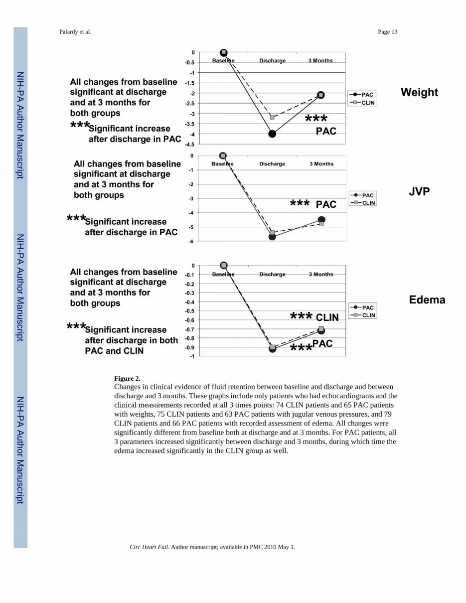
Figure 2.Changes in clinical evidence of fluid retention between baseline and discharge and betweendischarge and 3 months. These graphs include only patients who had echocardiograms and theclinical measurements recorded at all 3 times points: 74 CLIN patients and 65 PAC patientswith weights, 75 CLIN patients and 63 PAC patients with jugular venous pressures, and 79CLIN patients and 66 PAC patients with recorded assessment of edema. All changes weresignificantly different from baseline both at discharge and at 3 months. For PAC patients, all3 parameters increased significantly between discharge and 3 months, during which time theedema increased significantly in the CLIN group as well.
Palardy et al. Page 13
Circ Heart Fail. Author manuscript; available in PMC 2010 May 1.
NIH
-PA Author Manuscript
NIH
-PA Author Manuscript
NIH
-PA Author Manuscript

Figure 3.Relative improvement between baseline and discharge in mitral regurgitation (MR area), mitralregurgitation area expressed as a proportion of left atrial area (MR/LAA) and E velocity, forall of which a decrease is an improvement. For deceleration (Decel) time, an increase representsan improvement. Changes are expressed here as the median of individual % changes betweenbaseline and discharge divided by baseline values (contrast to Table 2, where they are expressedas mean absolute change from baseline). CLIN is patients who were randomized to therapyguided by clinical assessment of filling pressures, and PAC is patients randomized to therapyguided additionally by a pulmonary artery catheter.
Palardy et al. Page 14
Circ Heart Fail. Author manuscript; available in PMC 2010 May 1.
NIH
-PA Author Manuscript
NIH
-PA Author Manuscript
NIH
-PA Author Manuscript

NIH
-PA Author Manuscript
NIH
-PA Author Manuscript
NIH
-PA Author Manuscript
Palardy et al. Page 15Ta
ble
1C
linic
al C
hara
cter
istic
s of P
atie
nts W
ith a
nd W
ithou
t Ech
ocar
diog
ram
s and
MR
Mea
sure
men
ts
Bas
elin
e an
d D
isch
arge
MR
Mea
sure
men
t(S
D)
Bas
elin
ew
ithou
tD
isch
arge
Ech
o
Ech
oes
With
out
MR
Mea
sure
men
t
CL
INPA
CT
otal
N66
6713
310
665
Age
58 (1
5)56
(13)
57 (1
4)56
(14)
54 (1
4)
Gen
der
74%
77%
76%
72%
78%
Min
ority
27%
37%
32%
37%
46%
CA
D44
%51
%48
%48
%51
%
Bas
elin
e LV
EF20
(6)
20 (5
)20
(7)
19 (6
)21
(6)
SBP
mm
Hg
104
(12)
104
(15)
104
(14)
106
(14)
108
(15)
Est J
VP
mm
Hg
13 (4
)13
(4)
13 (4
)13
(4)
13 (4
)
Cre
atin
ine
mg/
dl1.
5 (0
.6)
1.6
(0.6
)1.
5 (0
.6)
1.5
(0.5
)1.
3 (0
.5)
RA
P* mm
Hg
14
(11)
14 (1
1)12
(6)
14 (8
)
PCW
* mm
Hg
25
(10)
25 (1
0)24
(10)
24 (1
0)
* Obt
aine
d on
ly in
pat
ient
s ran
dom
ized
to P
AC
-gui
ded
ther
apy
CA
D =
cor
onar
y ar
tery
dis
ease
Est J
VP
= cl
inic
ian-
estim
ated
jugu
lar v
enou
s pre
ssur
e
PCW
= p
ulm
onar
y ca
pilla
ry w
edge
pre
ssur
e
RA
P =
right
atri
al p
ress
ure
Circ Heart Fail. Author manuscript; available in PMC 2010 May 1.

NIH
-PA Author Manuscript
NIH
-PA Author Manuscript
NIH
-PA Author Manuscript
Palardy et al. Page 16Ta
ble
2M
itral
Reg
urgi
tatio
n an
d Fl
ow P
atte
rns:
Bas
elin
e an
d C
hang
es D
urin
g H
ospi
taliz
atio
n
CL
INPA
C- G
uide
d T
hera
py
Bas
elin
eC
hang
eP-
valu
e*w
ithin
Gro
up
Bas
elin
eC
hang
eP-
valu
e*W
ithin
Gro
up
P-va
lue*
Bet
wee
nG
roup
s
MR
are
a cm
2 (n =
133)
10.3
±6.
70.
3 ±4
.2
9.3
±6.7
−2.0
±5.
40.
004
0.01
MR
are
a / L
AA
(n =
132)
0.3
±0.2
0.0
±0.1
0.
3 ±0
.2−0
.1 ±
0.2
0.01
0.02
E V
eloc
ity c
m/s
ec (n
= 14
3)97
±29
7 ±4
10.
1610
0 ±3
0−8
±25
0.01
0.01
Dec
el T
ime
mse
c (n
= 11
6)14
7 ±6
37
±54
13
5 ±3
719
±67
0.05
Pulm
Vei
n Sy
s/D
ias (
n= 6
1)0.
5 ±1
.1−0
.25
±.5
0.
5 ±0
.40.
24 ±
1.0
0.20
0.14
LVED
I cm
(n=
175)
3.3
±0.7
0.02
±0.
28
3.4
±0.6
−0.0
2 ±0
.28
LVEF
% (n
= 13
0)20
±9
−0.4
±8.
0
20 ±
10−0
.2 ±
9.7
* P-va
lues
list
ed if
≤0.
20
Dec
el =
dec
eler
atio
n
LAA
= le
ft at
rial a
rea
LVED
D =
left
vent
ricul
ar e
nd-d
iast
olic
dim
ensi
on
LVED
I = le
ft ve
ntric
ular
end
-dia
stol
ic d
imen
sion
inde
x
LVEF
= le
ft ve
ntric
ular
eje
ctio
n fr
actio
n
MR
= m
itral
regu
rgita
tion
Sys/
Dia
s = sy
stol
e/di
asto
le
Circ Heart Fail. Author manuscript; available in PMC 2010 May 1.

NIH
-PA Author Manuscript
NIH
-PA Author Manuscript
NIH
-PA Author Manuscript
Palardy et al. Page 17Ta
ble
3C
hang
es a
t 3 M
onth
s Afte
r Dis
char
ge
CL
INP-
valu
eC
hang
eW
ithin
CL
IN
PAC
dur
ing
hosp
P-va
lue
Cha
nge
With
inPA
C
P-va
lue
Cha
nge
inC
LIN
vs.
PAC
MR
are
a cm
2 (n =
104)
−2.4
±5.
3<
0.00
1−1
.0 ±
6.0
0.
17
MR
are
a / L
AA
(n =
103)
−0.1
±0.
2<
0.01
−0.1
±0.
20.
01
E V
eloc
ity c
m/s
ec (n
=12
9)5.
7 ±2
.80.
16−8
±25
0.09
0.02
Dec
el T
ime
mse
c (n
=11
3)13
±69
27
±86
0.05
Pulm
Vei
n Sy
s/D
ias (
n =4
7)−0
.2 ±
1.6
0.
1 ±0
.4
LVED
I cm
(n =
158)
0.01
±0.
44
−0.0
2 ±0
.37
LVEF
% (n
=11
0)2.
5 ±1
10.
115.
1 ±1
0<0
.01
0.21
Sam
e as
in T
able
2.
Circ Heart Fail. Author manuscript; available in PMC 2010 May 1.