recommendations for the diagnosis of pediatric tuberculosis
TRANSCRIPT
REVIEW
Recommendations for the diagnosis of pediatric tuberculosis
E. Chiappini1 & A. Lo Vecchio2 & S. Garazzino3 & G. L. Marseglia4 & F. Bernardi5 &
E. Castagnola6 & P. Tomà7 & D. Cirillo8 & C. Russo9 & C. Gabiano3 & D. Ciofi1 &
G. Losurdo6 & M. Bocchino10 & E. Tortoli8 & M. Tadolini11 & A. Villani12 & A. Guarino2 &
S. Esposito13& for the Italian Pediatric TB Study Group
Received: 27 September 2015 /Accepted: 7 October 2015# Springer-Verlag Berlin Heidelberg 2015
Abstract Tuberculosis (TB) is still the world’s second mostfrequent cause of death due to infectious diseases after HIVinfection, and this has aroused greater interest in identifyingand managing exposed subjects, whether they are simply
infected or have developed one of the clinical variants of thedisease. Unfortunately, not even the latest laboratory tech-niques are always successful in identifying affected childrenbecause they are more likely to have negative cultures and
The Italian Pediatric TB Study Group also includes: Nicola Principi,Samantha Bosis, Claudia Tagliabue, Laura Senatore, Beatrice Ascolese(Pediatric Highly Intensive Care Unit, Università degli Studi di Milano,Fondazione IRCCS Ca’ Granda Ospedale Maggiore Policlinico, Milan,Italy); Laura Lancella, Laura Cursi, Annalisa Grandin, CaterinaMarabotto (Unit of General Pediatrics and Pediatric Infectious Diseases,IRCCS Bambino Gesù Hospital, Rome, Italy); Luisa Galli, Maurizio deMartino, Carlotta Montagnani, Filippo Festini, Martina Anziati, SabrinaBecciani, Giulia Remaschi, Sara Sollai, Chiara Tersigni, ElisabettaVenturini (Department of Health Science, University of Florence, AnnaMeyer Clidren’s University Hospital, Florence, Italy); Riccardo Scotto(Section of Pediatrics, Department of Translational Medical Science,University of Naples Federico II, Naples, Italy); Elisa Bertazzoni(Pharmacology Unit, University of Verona, Verona, Italy); FrancescoBlasi (Pneumology Unit, Università degli Studi di Milano, FondazioneIRCCS Ca’ Granda Ospedale Maggiore Policlinico, Milan, Italy); LucaAssante (Pneumology Unit, University of Naples Federico II, Naples,Italy); Luigi Codecasa (Referral Center for Tuberculosis, LombardyRegion, Milan, Italy); Giuseppe Di Mauro (primary care pediatrician,Caserta, Italy); Marino Faccini (Prevention Department ASL Milano,Milan, Italy); Daniele Le Serre, Irene Raffaldi (Pediatric InfectiousDiseases Unit, Regina Margherita Hospital, University of Turin, Turin,Italy); Amelia Mascolo (Pediatric Clinic, Fondazione IRCCS PoliclinicoSan Matteo, Pavia, Italy); Amelia Di Comite, Mauro Stronati(Neonatology and Neonatal Intensive Care Unit, Fondazione IRCCSPoliclinico San Matteo, Pavia, Italy); Giovanni Battista Migliori,Rosella Centis, Lia D’Ambrosio (World Health OrganizationCollaborating Centre for Tuberculosis and Lung Diseases, FondazioneS. Maugeri, Care and Research Institute, Tradate, Italy); AlbertoMatteelli (World Health Organization, Global Tuberculosis Programme,Geneva, Switzerland); Angela Pasinato (primary care pediatrician,Vicenza, Italy); Franco Scaglione (Pharmacology Section, Universitàdegli Studi di Milano, Milan, Italy); Elisabetta Scala (MOIGEAssociation, Rome, Italy).Scientific Societies involved in the Italian Pediatric TB Study Group:Società Italiana di Neonatologia (SIN), represented by Amelia Di
Comite and Mauro Stronati; Società Italiana di Infettivologia Pediatrica(SITIP), represented by Susanna Esposito, Maurizio de Martino, LuisaGalli, Alfredo Guarino, Laura Lancella, Andrea Lo Vecchio, NicolaPrincipi, Samantha Bosis, Elio Castagnola, Clara Gabiano, SilviaGarazzino, Giuseppe Losurdo, Carlotta Montagnani, Martina Anziati,Beatrice Ascolese, Sabrina Becciani, Laura Cursi, Annalisa Grandin,Daniele Le Serre, Caterina Marabotto, Irene Raffaldi, Giulia Remaschi,Riccardo Scotto, Laura Senatore, Sara Sollai, Claudia Tagliabue, ChiaraTersigni and Elisabetta Venturini; Società Italiana di Pediatria (SIP),represented by Alberto Villani, Cristina Russo and Paolo Tomà; SocietàItaliana di Malattie Respiratorie Infantili (SIMRI), represented by FilippoBernardi; Società Italiana di Immunologia e Allergologia Pediatrica(SIAIP) represented byGianluigiMarseglia andAmeliaMascolo; SocietàItaliana di Pediatria Preventiva e Sociale (SIPPS), represented byGiuseppe Di Mauro and Elena Chiappini; Società Italiana per le Cureprimarie Pediatriche (SiCUPP), represented by Angela Pasinato; SocietàItaliana di Malattie Respiratorie (SIMER), represented by FrancescoBlasi, Marialuisa Bocchino and Luca Assante; Associazione ItalianaPneumologi Ospedalieri (AIPO), represented by Luigi Codecasa; SocietàItaliana di Malattie Infettive e Tropicali (SIMIT), represented by AlbertoMatteelli; Associazione Microbiologi Clinici Italiani (AMCLI), repre-sented by Enrico Tortoli; Società Italiana di Chemioterapia (SIC), repre-sented by Elisa Bertazzoni; Società Italiana di Farmacologia (SIF), rep-resented by Francesco Scaglione; STOP TB, represented by DanielaCirillo, Marino Faccini, Giovanni Battista Migliori, Rosella Centis andMarina Tadolini; Società Italiana di Scienze Infermieristiche Pediatriche(SISIP), represented by Filippo Festini and Daniele Ciofi; MOIGE, rep-resented by Elisabetta Scala.
* S. [email protected]
1 Department of Health Sciences, Pediatric Infectious DiseasesDivision, Anna Meyer Children’s University Hospital, University ofFlorence, Florence, Italy
Eur J Clin Microbiol Infect DisDOI 10.1007/s10096-015-2507-6
brought to you by COREView metadata, citation and similar papers at core.ac.uk
provided by Archivio della ricerca - Università degli studi di Napoli Federico II
tuberculin skin test results, equivocal chest X-ray findings,and atypical clinical manifestations than adults. Furthermore,they are at greater risk of progressing from infection to activedisease, particularly if they are very young. Consequently,pediatricians have to use different diagnostic strategies thatspecifically address the needs of children. This document de-scribes the recommendations of a group of scientific societiesconcerning the signs and symptoms suggesting pediatric TB,and the diagnostic approach towards children with suspecteddisease.
Introduction
Tuberculosis (TB) is still the world’s second most fre-quent cause of death due to infectious diseases afterHIV infection [1], and this has aroused greater interestin identifying and managing exposed subjects, whetherthey are simply infected or have developed one of theclinical variants of the disease. Unfortunately, not eventhe latest laboratory techniques are always successful inidentifying affected children because they are more likelyto have negative cultures and tuberculin skin test (TST)results, equivocal chest X-ray findings, and atypical clin-ical manifestations than adults [2]. Furthermore, they areat greater risk of progressing from infection to activedisease, particularly if they are very young [2].Consequently, pediatricians have to use different diag-nostic strategies that specifically address the needs ofchildren.
This document describes the recommendations of a groupof scientific societies concerning the signs and symptoms sug-gesting pediatric TB, and the diagnostic approach towardschildren with suspected disease.
Methodology
Using the Consensus Conference method on the basis of theNational Institutes of Health and the Italian NationalProgramme Guidelines [3, 4] (Table 1), relevant publicationsin English were identified by means of a systematic review ofMEDLINE and the Cochrane Database of SystematicReviews from their inception until 31 December 2014. Thesearch strategy was Bchildren[Title/Abstract] ORpediatric[Title/Abstract] OR paediatric[Title/Abstract] ANDtuberculosis[Title/Abstract] AND diagnosis[Title/Abstract]OR signs[Title/Abstract] or symptoms[Title/Abstract] orTST[Ti t le /Abst rac t ] or IGRA[Ti t le /Abst rac t ] ormicrobioogy[Title/Abstract] or radiography[Title/Abstract]AND English[lang])^.
The Working Group agreed on a list of clinical problemsrelated to diagnosing TB, and the evidence review proceduresconcentrated on patients aged 0–18 years, and includedsection-specific targeted searches as well as formal systematicreviews of selected aspects. The clinical recommendationsmade in the updated international guidelines were alsoreviewed and critically compared. The literature was criticallyappraised by trained personnel using the ScottishIntercollegiate Guidelines Network methodological checklists[5], and all of the data were entered in tables of evidence foreach subject. The bibliographical material and a preliminarydraft document were given to the panel members before thepublished evidence was presented and discussed at variousmeetings. The Delphi method was used to reach a consensuswhen the evidence did not provide consistent and unambigu-ous recommendations [5]. The final text was revised on thebasis of these discussions and submitted by e-mail to the par-ticipants at the Consensus Conference for final approval.
The multidisciplinary panel of clinicians and experts inevidence-based medicine were identified with the help ofthe participating scientific societies, and included expertsin the fields of general pediatrics, pediatric infectious dis-eases, neonatology, infectious diseases, pneumology, mi-crobiology, radiology, pharmacology, public health andmethodology. The panel was coordinated by the ItalianSociety of Pediatric Infectious Diseases (SITIP). No panelmember declared any conflict of interest concerning thecontents of the guideline topics. The panel met on threeoccasions, but many of the consultations involved in de-veloping the document took place interactively by e-mailor telephone.
Eur J Clin Microbiol Infect Dis
2 Section of Pediatrics, Department of Translational Medical Science,University of Naples Federico II, Naples, Italy
3 Pediatric Infectious Diseases Unit, Regina Margherita Hospital,University of Turin, Turin, Italy
4 Pediatric Clinic, Fondazione IRCCS Policlinico San Matteo,Pavia, Italy
5 Pediatric Emergency Unit, University of Bologna, Bologna, Italy6 Infectious Diseases Unit, IRCCS Giannina Gaslini, Genoa, Italy7 Radiology Unit, IRCCS Bambino Gesù Hospital, Rome, Italy8 Microbiology Unit, IRCCS San Raffaele Hospital, Milan, Italy9 Virology Unit, IRCCS Bambino Gesù Hospital, Rome, Italy10 Pneumology Unit, University of Naples Federico II, Naples, Italy11 Infectious Diseases Unit, Department of Medical and Surgical
Sciences, Alma Mater Studiorum University of Bologna,Bologna, Italy
12 Unit of General Pediatrics and Pediatric Infectious Diseases, IRCCSBambino Gesù Hospital, Rome, Italy
13 Pediatric Highly Intensive Care Unit, Department ofPathophysiology and Transplantation, Fondazione IRCCS Ca’Granda Ospedale Maggiore Policlinico, Università degli Studi diMilano, Via Commenda 9, 20122 Milan, Italy
When should pediatric TB be clinically suspected?
Pulmonary signs and symptoms
Most diagnoses of pediatric TB (about 65 %) are made on thebasis of symptoms rather than case tracking, regardless ofwhether TB is endemic or not in the country in which theyare made [6, 7].
The lungs are the most frequent site of pediatric TBthroughout the world: a recent retrospective study of morethan 2,500 children in the United States found that pulmonaryforms accounted for about 70 % of cases [8], which is essen-tially similar to the 75.5 % found in an Italian study of morethan 200 affected children [9].
Given the extreme variability of pulmonary involvementduring the course of TB, the initial symptoms may also varywidely depending on age, the child’s general clinical condi-tion, the presence of underlying diseases, and the natural his-tory of the disease; furthermore, TB symptoms may mimicsome common acute respiratory diseases.
The most frequently reported symptoms in children withTB are cough lasting >4 weeks; a poor response to first-linetreatment; dyspnea and asthenia; chest pain, particularly inolder children and adolescents; and hemoptysis. In some caseseries, the presence of individual symptoms such as coughlasting >4 weeks have been highly predictive of TB (oddsratio [OR] 13.8, 95 % confidence interval [95 % CI] 2.3–83.1) [10], but such signs are often associated with a rangeof general symptoms such as persistent and often moderateevening fever, night sweats, general malaise and asthenia, andweight loss.
Combinations of multiple symptoms have proved to behighly predictive of TB in a number of large cohorts de-scribed in South Africa. One study of more than 1,000children classified as being at low (aged >3 years andnot infected with HIV) and high risk of TB (age<3 years or HIV-infected) tested the sensitivity, specificityand positive predictive value (PPV) of various symptoms
and signs [11], and found that the combination of persis-tent and unremitting cough for >2 weeks, weight loss inthe previous 3 months, and asthenia had a sensitivity of82.3 %, a specificity of 90.2 %, and a PPV of 82.3 % inlow-risk children, and corresponding values of 51.8, 92.%and 90.1 % in high-risk children. However, symptomswere poorly predictive of TB in the HIV-infected patients.
It has been found that the combination of cough and weightloss (OR 5.4; 95 % CI 1.7–16.9; p=0.001) or cough, weightloss and anorexia (OR 5.3: 95 % CI 1.5–18.8; p=0.004) issignificantly predictive of TB in children aged <15 years;however, the presence of weight loss was required to reachstatistical significance [12]. Similar findings have been report-ed in patients aged ≥15 years, in whom the probability of TBincreased about 17-fold in the case of associated weight loss,chest/pleural pain and night sweats [13].
The pulmonary manifestations of pediatric TB are sig-nificantly different at different ages. Breastfeeding infantsand school-aged children have extremely variable formsthat may be clinically and radiographically similar tocommon respiratory infections but, as children grow, thepathognomic pulmonary characteristics of TB becomegradually more similar until, by the time of adolescence,they are the same as those observed in adults [6]. In somecohorts of adolescents (aged 10–14 years) with anascertained diagnosis of TB, the percentage of cavitaryforms exceeds 80 % [12].
The radiological pictures of pediatric pulmonary TBare also extremely heterogeneous and age related.Moderate and aspecific parenchymal involvement (lobaror interstitial pneumonia) is common, but there may alsobe more typical pictures, such as cavitations, pleural effu-sion, calcifications or multi-lobar involvement (particular-ly the lower lobes). The presence of cavitations (OR 7.7,95 % CI: 1.0–57.7) and the involvement of the uppersegments of the inferior lobes (OR 12.6, 95 % CI 1.2–134.8) were found to be significantly associated with ahigh risk of TB in a small Taiwanese case series [10].
Table 1 Quality of evidence andstrength of recommendation Quality of evidence
I Evidence from more than one properly designed, randomized, controlled studyand/or systematic review of randomized studies
II Evidence from one properly designed, randomized, controlled study
III Evidence from cohort studies or their meta-analysis
IV Evidence from retrospective case-controlled studies or their meta-analysis
V Evidence from case series without control group
VI Evidence from opinions of respected authorities, based on clinical experience
Strength of recommendation
A The panel strongly supports a recommendation for use
B The panel moderately supports a recommendation for use
C The panel marginally supports a recommendation for use
Eur J Clin Microbiol Infect Dis
Signs and symptoms of extra-pulmonary TB (EPTB)
EPTB accounts for 20–25 % of all cases of TB [14], and isusually more difficult to diagnose than pulmonary forms notonly because it is less frequent, but also because its clinicalmanifestations are extremely variable. The greatest diagnosticdifficulties are due to its aspecific manifestations, which mim-ic those of other inflammatory or neoplastic diseases. It istherefore necessary to maintain a high degree of suspicion inorder to make an early diagnosis, especially in a country witha low prevalence of TB such as Italy.
Furthermore, EPTB can involve relatively inaccessiblesites and is characterised by a reduced bacillary load [14],and it is often necessary to consider epidemiological, clinical,immunological and imaging factors in order to make adiagnosis.
In addition to localised symptoms, EPTB may also bereflected by systemic symptoms; weight loss and a lack ofappetite are characteristic of disseminated and gastrointestinalTB, whereas fever and night sweats, which are common indisseminated, cerebral and gastrointestinal forms, are less fre-quent in the case of lymph node, bone and genito-urinary TB[15–18].
Extra-pulmonary involvement may be associated with apulmonary localisation, which makes diagnosis easier. In thecase of clinical suspicion, after the initial investigations haveexcluded other possible etiologies, it is recommended to startanti-TB treatment as soon as possible, particularly in the caseof severe forms such as disseminated TB and tuberculousmeningitis and pericarditis.
About 50 % of the forms of EPTB involve peripherallymph nodes, of which the most frequently involved are thelaterocervical and supraclavicular lymph nodes.Lymphadenitis usually manifests itself in the form of the grad-ual, non-painful swelling of one or more peripheral lymphnodes. The nodes are initially hard and clearly delimited andthe overlying is skin is normal but, subsequently, the skintends to become inflamed and the nodes come together toform hard lumps that may undergo spontaneous fistulasationand release caseous material. Systemic symptoms are not fre-quent except in HIV-infected subjects. The presence of a pain-less lymphadenopathy of >2 cm that persists for >4 weeks andis unresponsive to the usual antibiotics should always raise asuspicion of TB [19, 20].
About 1% of the cases of TB in small childrenmay have anosteoarticular localisation, which represents about 10–15% ofthe forms of EPTB [21]. This is more frequent in children thanadults because the epiphyseal region of bones is morevascularised in the former. The spinal column is affected in50 % of cases, whereas the hips, elbows and knees are lessfrequently involved, and involvement of the extremities iseven less frequent. The spinal form usually affects the thoracicand lumbar vertebrae, usually two contiguous vertebrae but
sometimes more and sometimes separately. Systemic symp-toms are not frequent in the case of bone and joint TB. Themost frequent symptom is pain, which usually progressesmore gradually than in other bacterial forms [21], and maybe accompanied by swelling and slight deformity. Kyphosisaffecting the thoracic region is a late sign suggesting spinal TB[22]. Even in the early phases, there may sometimes be aparavertebral or psoas abscess, with consequent muscle spasmand movement alterations; neurological complications (Pott’sdisease) usually appear later.
In terms of laboratory findings, TB may be suggested by adiscrepancy between white blood cell counts and the erythro-cyte sedimentation rate (ESR) [21]. C-reactive protein (CRP)levels are usually normal, but there may be signs of chronicinflammation: anemia, hypoalbuminemia, thrombocytosis[23–27]. Furthermore, TB should be included in the differen-tial diagnosis of isolated articular lesions (mainly of the hipsand knees), particularly if they are not painful. Nevertheless,synovial fluid aspiration remains diagnostically decisive [26].
Tuberculous meningo-encephalitis (TBM) is the most seri-ous form of pediatric TB, and appears in about 4–5 % of casesof TB [28]. Mortality may be as high as 10–15 %, and abouthalf of the survivors have permanent neurological sequelae.The early symptoms are aspecific, and include anorexia, mal-aise, headache, and behavioural alterations. Breastfeeding in-fants may only manifest a lack of appetite, irritability, somno-lence, behavioural alterations, and convulsions. The prodromicphase may last even several weeks, after which more specificsigns such as fever, headache, vomiting and nuchal rigidityappear, the evolution of which is slower than in the case ofbacterial meningitis. These signs are followed by focal neuro-logical symptoms associated with altered consciousness, re-gression of neurodevelopmental cornerstones, and convulsion.Cranial nerve paralysis particularly involves the III and VIcranial nerves. Other neurological signs may develop depend-ing on the site affected by arteritis or infarction: cerebellarsigns, extra-pyramidal movements, hemiparesis.
A number of studies have defined the clinical characteris-tics predictive of TBM [29–33]. In addition to the symptomsmentioned above, these highlight the importance of their du-ration (>5 days) and the characteristics of cerebrospinal fluid(CSF): a clear appearance; leukocytes 5–750/mL, with a prev-alence of lymphocytes; proteins 0.5–3 g/dL; and a CSF/bloodglucose ratio of <50 %. These characteristics make it possibleto differentiate TBM from other bacterial forms, but they arenot decisive in the case of HIV-positive subjects insofar asthey do not allow the differentiation of TBM and cryptococcalmeningitis.
Pediatric abdominal TB is not very frequent [34]. It mayinvolve any tract of the intestine and peritoneum, although themost common site is the ileocecal tract and the other parts ofthe colon and rectum are less frequently affected. The mostfrequent symptom is pain, which has a sub-acute or chronic
Eur J Clin Microbiol Infect Dis
course in two-thirds of cases, and may mimic appendicitis orintestinal obstruction in the remaining third [35]. It may some-times manifest itself as a palpable tumour-like mass with mal-absorption or, at rectal level, as fistulae, abscesses, or analfissuring and bleeding. A combination of fever and abdominaldistension in a subject with ascites should arouse the suspicionof a tuberculous form. Diagnostic confirmation always re-quires invasive procedures [36].
Genito-urinary TB is one of the most frequent forms ofadult EPTB, but it is rare in small children and not very fre-quent in adolescents because it appears at least 5 years after afirst infection [37]. The symptoms are local rather than sys-temic, e.g. dysuria, hematuria and pollakiuria are frequent,and associated with pain in the side. The symptoms are sovague or mis-recognised that they are often only diagnosedat an advanced stage of renal damage. It is sometimes diag-nosed after a routine urine test; the presence of hematuria andsterile pyuria in acidic urine should always prompt a search forurinary M. tuberculosis [38].
Pericarditis is a rare form of TB that is still sometimes fatal,particularly in HIV-positive subjects [39]. The initial symp-toms are mainly systemic and aspecific, whereas cardiopul-monary symptoms (cough, dyspnea, orthopnea, lower limbedema, a dull retrosternal pain often influenced by positionand inspiration) appear later. The progression of the diseaseand effusion may be accompanied by friction rubs, a sign ofcardiac tamponade or constrictive pericarditis. However, inaddition to images of exudative pericarditis, diagnosis alwaysrequires pericardiocentesis or even pericardiotomy, particular-ly in countries with a low prevalence of TB [39].
Disseminated TB is caused by the hematogenous dissemi-nation of tubercular bacilli due to inadequate host defences,and is therefore more frequent in children aged less than 2–3 years, whose cell-mediated immune responses are still im-mature [40] and, in breastfeeding infants, it may develop in10–20 % of cases of TB due to a recent primary infection. Ithas various clinical manifestations. The signs and symptomsat the time of onset are generally systemic and non-specific,e.g. fever, weight loss, night sweats, anorexia, and the loss ofappetite. The other symptoms depend on the predominantlyinvolved site; cough and other respiratory symptoms are asso-ciated with pulmonary TB with a characteristic miliary ap-pearance, whereas headache and altered consciousness areassociated with meningeal involvement. Choroidal tubercles,which are pathognomic of miliary TB, can be observed in30 % of cases [40]. However, no systematic diagnostic ap-proach model has yet been defined [41].
How should pediatric TB be diagnosed?
A detailed anamnesis aimed at investigating possible exposureto M. tuberculosis as a result of contact with someone with
bacilliferous TB or coming from an endemic area, and factorsfavouring the development of the disease such as poor socio-economic conditions is fundamental for a diagnosis of TB[42]. Particular attention should be given to the presence ofconcomitant medical conditions favouring immunosuppres-sion, such as malnutrition, HIV-1 infection, or treatment withcorticosteroids or immunosuppressants [42].
Clinical analysis also plays a fundamental role, and all ofthe signs and symptoms suggesting active disease should becarefully evaluated: chronic cough, weight loss, asthenia, pro-fuse sweating, fever, chest pain, hemoptysis and respiratorydistress [43].
Tuberculin skin test (TST)
Since 1907, a TST has been the reference diagnostic means ofdetecting M. tuberculosis infection [15, 44]. The test is basedon the delayed hypersensitivity reaction induced by the intra-dermal inoculation of a standardised dose of purified proteinderivative (PPD) tuberculin: theWHO recommends the use offive units of PPD-S or two units of PPD tuberculin RT23.Tuberculin reactivity usually appears between two and12 weeks after the initial infection (median 3–4 weeks).
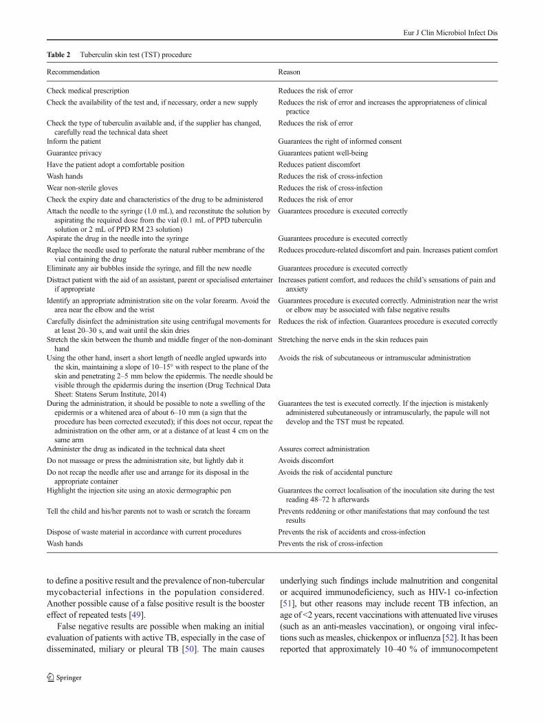
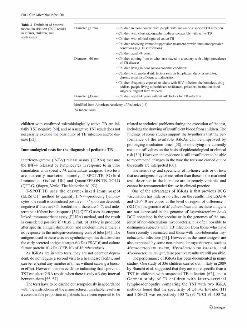
PPD is intradermally injected in a lesion-free area of thevolar forearm (Table 2). T lymphocytes sensitised by a previ-ous infection are recruited at the site of the inoculation, wherethey release lymphokines that induce local vasodilation, ede-ma, fibrin deposition and the recruitment of other inflamma-tory cells, resulting in an infiltrate that reaches its maximumsize 48–72 h after the injection [44]. The diameter of theswelling should therefore be measured (in millimetres) withinthis period by trained medical personnel. The result of the testshould be interpreted individually in relation to the presenceof conditions predisposing to TB, and the timing and type ofdisease exposure as shown in Table 3.
ATST has some limitations, for example, it is an operator-dependent test that requires the patient to make at least twovisits to a healthcare centre, and may give rise to false positiveor false negative results [44–47]. The possibility of false pos-itive results is due to the cross-reactivity of the PPD antigenswith those of non-tuberculous mycobacteria and even of BCGvaccine, even though the reactions rarely exceed 15 mm indiameter [48]. The guidelines of the American Academy ofPediatrics recommend using the same interpretation of TSTfor children regardless of whether or not they have previouslyreceived BCG [44] because the post-vaccination intradermalreaction to tuberculin can be influenced by many factors, suchas the child’s immune status or age at the time of vaccination,the quality and type of the vaccine strain, and the time intervalbetween the vaccination and the TST. Nevertheless, the sameguidelines suggest considering any hardening of more than15 mm as positive in previously vaccinated children. Thespecificity of the test obviously depends on the criteria used
Eur J Clin Microbiol Infect Dis
to define a positive result and the prevalence of non-tubercularmycobacterial infections in the population considered.Another possible cause of a false positive result is the boostereffect of repeated tests [49].
False negative results are possible when making an initialevaluation of patients with active TB, especially in the case ofdisseminated, miliary or pleural TB [50]. The main causes
underlying such findings include malnutrition and congenitalor acquired immunodeficiency, such as HIV-1 co-infection[51], but other reasons may include recent TB infection, anage of <2 years, recent vaccinations with attenuated live viruses(such as an anti-measles vaccination), or ongoing viral infec-tions such as measles, chickenpox or influenza [52]. It has beenreported that approximately 10–40 % of immunocompetent
Table 2 Tuberculin skin test (TST) procedure
Recommendation Reason
Check medical prescription Reduces the risk of error
Check the availability of the test and, if necessary, order a new supply Reduces the risk of error and increases the appropriateness of clinicalpractice
Check the type of tuberculin available and, if the supplier has changed,carefully read the technical data sheet
Reduces the risk of error
Inform the patient Guarantees the right of informed consent
Guarantee privacy Guarantees patient well-being
Have the patient adopt a comfortable position Reduces patient discomfort
Wash hands Reduces the risk of cross-infection
Wear non-sterile gloves Reduces the risk of cross-infection
Check the expiry date and characteristics of the drug to be administered Reduces the risk of error
Attach the needle to the syringe (1.0 mL), and reconstitute the solution byaspirating the required dose from the vial (0.1 mL of PPD tuberculinsolution or 2 mL of PPD RM 23 solution)
Guarantees procedure is executed correctly
Aspirate the drug in the needle into the syringe Guarantees procedure is executed correctly
Replace the needle used to perforate the natural rubber membrane of thevial containing the drug
Reduces procedure-related discomfort and pain. Increases patient comfort
Eliminate any air bubbles inside the syringe, and fill the new needle Guarantees procedure is executed correctly
Distract patient with the aid of an assistant, parent or specialised entertainerif appropriate
Increases patient comfort, and reduces the child’s sensations of pain andanxiety
Identify an appropriate administration site on the volar forearm. Avoid thearea near the elbow and the wrist
Guarantees procedure is executed correctly. Administration near the wristor elbow may be associated with false negative results
Carefully disinfect the administration site using centrifugal movements forat least 20–30 s, and wait until the skin dries
Reduces the risk of infection. Guarantees procedure is executed correctly
Stretch the skin between the thumb and middle finger of the non-dominanthand
Stretching the nerve ends in the skin reduces pain
Using the other hand, insert a short length of needle angled upwards intothe skin, maintaining a slope of 10–15° with respect to the plane of theskin and penetrating 2–5 mm below the epidermis. The needle should bevisible through the epidermis during the insertion (Drug Technical DataSheet: Statens Serum Institute, 2014)
Avoids the risk of subcutaneous or intramuscular administration
During the administration, it should be possible to note a swelling of theepidermis or a whitened area of about 6–10 mm (a sign that theprocedure has been corrected executed); if this does not occur, repeat theadministration on the other arm, or at a distance of at least 4 cm on thesame arm
Guarantees the test is executed correctly. If the injection is mistakenlyadministered subcutaneously or intramuscularly, the papule will notdevelop and the TST must be repeated.
Administer the drug as indicated in the technical data sheet Assures correct administration
Do not massage or press the administration site, but lightly dab it Avoids discomfort
Do not recap the needle after use and arrange for its disposal in theappropriate container
Avoids the risk of accidental puncture
Highlight the injection site using an atoxic dermographic pen Guarantees the correct localisation of the inoculation site during the testreading 48–72 h afterwards
Tell the child and his/her parents not to wash or scratch the forearm Prevents reddening or other manifestations that may confound the testresults
Dispose of waste material in accordance with current procedures Prevents the risk of accidents and cross-infection
Wash hands Prevents the risk of cross-infection
Eur J Clin Microbiol Infect Dis
children with confirmed microbiologically active TB are ini-tially TST negative [50], and so a negative TST result does notnecessarily exclude the possibility of TB infection and/or dis-ease [52].
Immunological tests for the diagnosis of pediatric TB
Interferon-gamma (INF-γ) release assays (IGRAs) measurethe INF-γ released by lymphocytes in response to in vitrostimulation with specific M. tuberculosis antigens. Two testsare currently marketed, namely, T-SPOT.TB (OxfordImmunotec, Oxford, UK) and QuantiFERON-TB GOLD(QFT-G, Qiagen, Venlo, The Netherlands) [53].
T-SPOT.TB uses the enzyme-linked immunospot(ELISPOT) method to quantify IFN-γ-producing lympho-cytes; the result is considered positive if >7 spots are detected,negative if there are <5, borderline if there are 5–7, and inde-terminate if there is no response [54]. QFT-G uses the enzyme-linked immunosorbent assay (ELISA) method, and the resultis considered positive if >0.35 UI/mL of IFN-γ is producedafter specific antigen stimulation, and indeterminate if there isno response in the mitogen-containing control tube [54]. Theantigens used in these tests are synthetic peptides that simulatethe early secreted antigenic target 6-kDa (ESAT 6) and culturefiltrate protein 10-kDa (CFP-10) of M. tuberculosis.
As IGRAs are in vitro tests, they are not operator depen-dent, do not require a second visit to a healthcare facility, andcan be repeated any number of times without causing a boost-er effect. However, there is evidence indicating that a previousTSTcan alter IGRA results when there is only a 3-day intervalbetween them [55–57].
The tests have to be carried out scrupulously in accordancewith the instructions of the manufacturer; unreliable results ina considerable proportion of patients have been reported to be
related to technical problems during the execution of the test,including the drawing of insufficient blood from children. Thefindings of some studies support the hypothesis that the per-formance of the available IGRAs can be improved byprolonging incubation times [58] or modifying the currentlyused cut-off values on the basis of epidemiological or clinicalrisk [59]. However, the evidence is still insufficient to be ableto recommend changes in the way the tests are carried out orthe results are interpreted [60].
The sensitivity and specificity of in-house tests or of teststhat use antigens or cytokines other than those in the marketedtests described in the literature are extremely variable, andcannot be recommended for use in clinical practice.
One of the advantages of IGRAs is that previous BCGvaccination has little or no effect on the results. The ESAT-6and CFP-10 are coded at the level of region of difference 1(RD1) of the genome ofM. tuberculosis and, as these antigensare not expressed in the genome of Mycobacterium bovisBCG contained in the vaccine or in the genomes of the ma-jority of non-tuberculous mycobacteria, it is often possible todistinguish subjects with TB infection from those who havebeen recently vaccinated and those with non-tubercular my-cobacterial infections [61]. However, as the same antigens arealso expressed by some non-tubercular mycobacteria, such asMycobacterium avium, Mycobacterium kansaii, andMycobacterium szuigai, false positive results are still possible.
The performance of IGRAs has been documented in manystudies. One study of 336 children carried out in Italy in 2009by Bianchi et al. suggested that they are more specific than aTST in children with suspected TB infection [62], and aGerman study of 73 children with latero-cervicallymphoadenopathy comparing the TST with two IGRAmethods found that the specificity of QFT-G In-Tube (IT)and T-SPOT was respectively 100 % (95 % CI 91–100 %)
Table 3 Definition of positivetuberculin skin test (TST) resultsin infants, children, andadolescents
Diameter ≥5 mm • Children in close contact with people with known or suspected TB infection
• Children with chest radiography findings compatible with active TB
• Children with clinical signs of active TB
• Children receiving immunosuppressive treatment or with immunodepressiveconditions (e.g. HIV infection)
• Children aged <4 years
Diameter ≥10 mm • Children coming from or who have stayed in a country with a high prevalenceof TB disease
• Children living in poor socio-economic conditions
• Children with medical risk factors such as lymphoma, diabetes mellitus,chronic renal insufficiency, malnutrition
• Children frequently exposed to adults with HIV infection, the homeless, drugaddicts, people living at healthcare residences, prisoners, institutionalisedsubjects, migrant farm workers
Diameter ≥15 mm • Children aged >4 years without risk factors for TB infection
Modified from American Academy of Pediatrics [44]
TB tuberculosis
Eur J Clin Microbiol Infect Dis
and 98 % (95 % CI 87–100 %), whereas that of the TST wasonly 58 % (95 % CI 42–73 %) [63].
A number of systematic reviews have revealed differencesin sensitivity and specificity in different populations [64].However, these meta-analyses need to be interpreted criticallybecause the studies involved often used different diagnosticcriteria to define active TB, only a few were based on head-to-head comparisons of T-SPOT.TB and QTF-G-IT, and variouscut-off values were used to define TST positivity.
It is also necessary to remember that the TST and IGRAscannot differentiate active TB and latent infection, and thattheir performance in children aged <5 years andimmunodepressed subjects seems to be sub-optimal. The re-ported sensitivity of T-SPOT.TB and QFT-G- IT in children,respectively, range from 62 to 89 % and from 66 to 83 %. In2010, Bamford et al. [65] studied 333 children in the UnitedKingdom and found that the sensitivity of the TST, QFT-G ITand T-SPOT.TB was 82, 78 and 66 %, respectively, whereasthe combined use of TST and QFT-G IT had a sensitivity of96%, and that of TSTand T-SPOT.TBwas 91%. It is possiblethat the TST is more sensitive because it is capable of explor-ing multiple TH1- and TH2-mediated immunological mecha-nisms, whereas IGRAs exclusively explore TH1-mediated re-sponses, which may be immature in children in a manner thatis inversely proportional to their chronological age [64].Furthermore, antigen-presenting cells in children are less ca-pable of synthesising IL-12, a fundamental mediator in theinitial phase of TH1 polarisation [64].
Pediatric studies have also highlighted a larger proportionof indeterminate results in children than in adults [64]. A re-cent systematic review found that this proportion was 6.5 % inthe case of QFT-G IT and 3.5 % in the case of T-SPOT.TB[66], but studies involving children aged <5 years have shownthat it may range from 0 to 40 % [67]. Some authors havereported a larger proportion of indeterminate results not onlyin children aged <5 years [68], but also in the case of helminthco-infection, HIV-related immunosuppression, and immuno-suppressive treatment [66]. A recent study has found that con-comitant bacterial pneumonia may also be a risk factor in anindeterminate result, particularly in children aged <5 years[68]. In these situations, the increased risk may be due topossibly age-related immune system immaturity leading to areduction in stimulated INF-γ production or an imbalance inTH1/TH2-mediated responses.
The guidelines of the American Academy of Pediatricsunderline the fact that IGRA-positive children should be con-sidered infected, and a negative or indeterminate result doesnot exclude infection [44]. Furthermore, IGRAs are not rec-ommended in children aged <5 years because of the lack ofunequivocal data or in immunocompromised children. The2011 revision of the British NICE guidelines suggest a two-step approach: a TST followed by the IGRA confirmation ofthe TST-positive cases [69]. However, they also indicate
situations in which it is advisable to use an IGRA alone, suchas in the case of subjects who for any reason cannot be reacheda second time 48–72 h after a TST, or if it is necessary toscreen large numbers of children (including subjects aged≥5 years), or when selected cases previously vaccinated withBCG need to be investigated as a result of coming into contactwith subjects with contagious TB [69].
With reference to children aged <5 years, Detjen et al. eval-uated the diagnostic accuracy of TST, T-SPOT.TB and QFT-G-IT in 73 children with a mean age of 39 months—28 withbateriologically confirmed TB, 23 with lymphadenitis due tonon-tubercular nycobacteria, and 22 controls [63]. The speci-ficity of QFT-G-IT for TB was 100 % (95 % CI 91–100 %)and that of T-SPOT.TB was 98% (95 % CI 87–100 %, but thespecificity of the TST was much less (58 %, 95 % CI 42–73 %); the sensitivity of both IGRAs was 0.93 (95 % CI0.77–0.99) and that of the TST was 1.00 (95 % CI 0.88–1.00; k=0.91). In another study Okada et al. compared QFT-G-IT and TST results in 195 Cambodian children living withpeople with active TB, and found that the TST was morespecific in those who had previously undergone BCG vacci-nation (k=0.63) [70]. Debord et al. retrospectively assessedthe performance of QFT-G-IT in 19 immunocompetentFrench children with active TB aged <5 years (mean age1.52 years) [71]. There were no indeterminate results and thenumber of positive cases was 6/10 children aged <2 years and9/9 children aged 2–5 years, thus suggesting that QFT-G-ITmay be a useful means of improving the diagnosis of TB inassociation with TST in children aged <5 years [71]. Thestudies of 397 South African children aged <3 years showedgood concordance between QFT-G-IT and TST results (k=0.79); however, the sensitivity of both tests in detecting activeTB disease seemed to be poor (respectively, 38 and 35%) [72,73]. On the contrary, Pavic et al. reported considerable discor-dance between QFT-G-IT and TST results in 142 Croatianchildren aged <5 years (k=0.59), and concluded that bothcould be used in high-risk children aged <5 years providedthat positivity to either was considered a sign of infection [74].
The findings of a large-scale study of more than 1,000adults suggest that IGRA (particularly T.SPOT.TB) can beused to screen immunodeficient contacts of patients with TB[75], but there are extremely few pediatric data. Bruzzese et al.studied 80 HIV-negative immune-compromised children infollow-up for liver transplantation or rheumatologic diseaseson biological treatment after coming into contact with TBusing both QFT-IT and T-SPOT; QFT-IT was positive in onecase (1.2%), whereas a significantly larger proportion of cases(9.4 %) were positive to T-SPOT.TB (p=0.02) [76]. The au-thors concluded that the high proportion of discordant or in-determinate results meant that IGRAs were of little help indiagnosing TB infection or disease in immunocompromisedchildren living in a country with a low prevalence of TB. Theother available studies suggest that IGRA may play a role in
Eur J Clin Microbiol Infect Dis
diagnosing TB infection and disease in the immunodepressed[77–81], but their sample sizes were small.
IGRAs have little positive predictive value in indicating theprogression of active TB. Studies of adults and children haveclearly shown that the serial repetition of the tests during anti-TB treatment does not aid the monitoring of therapeutic re-sponses [82, 83].
What type of microbiological diagnosis shouldbe used for children with suspected TB?
The microbiological diagnosis of pediatric pulmonary TB isstill largely unsatisfactory, and its limited sensitivity is evenmore marked in the case of immunocompromised subjects.There are two main reasons for this: (1) the disease is typicallypaucibacillary in children and (2) children are generally inca-pable of providing sputum samples of acceptable quality [84].It is therefore necessary to use alternative sampling methods,all of which are sub-optimal [85].
Gastric aspiration is the most widely used method ofcollecting swallowed respiratory secretions from children. Itis normally recommended that these samples should beneutralised with sodium bicarbonate, but it has been recentlyreported that this can significantly reduce culture yields inliquid medium [86].
An alternative method is to induce sputum using a hyper-tonic aerosol solution. This technique is not invasive but itdoes require patient collaboration, which is not easy to obtain,especially in the case of small children. A number of studieshave compared the diagnostic yield of gastric lavage and in-duced sputum, sometimes including a comparison with naso-pharyngeal aspirates. The results are not always concordant,but the majority of studies have found that gastric lavage is themost appropriate [87–94].
Broncholavage has the advantage of being aimed at thelesioned site, but is rather invasive [95].
Another possibility is the string test. The patient swallows agelatin capsule containing a length of thread. The capsuledissolves in the stomach, thus allowing the thread to becomeimpregnated with gastric secretions. Four hours later, thethread is recovered and washed with 1–2 mL of saline solu-tion, which is then used for the search [96].
A still inadequately evaluated approach is to search stoolfor possible swallowed mycobacteria [97, 98].
It goes without saying that sputum is the sample of electionin the case of adolescents and children capable of producing it,whereas gastric aspirate is the sample of election from theyoungest patients [94].
All of the samples should be collected in disposable, sterilecontainers with screw caps [94] and, if they cannot be sent tothe laboratory immediately, should be stored in a refrigerator.In order to increase sensitivity, microbiological investigations
for pulmonary TB are usually carried out on three samplescollected on consecutive days [99]; a single sample can beused only in the case of broncholavage.
The sensitivity of a microbiological diagnosis of both adultand pediatric TBM is limited by the availability of a sufficientamount of CSF in which to seek Koch’s bacillus [94].
Diagnosing lymph node TB is less of a problem when abioptic sample is available [94].
A biopsy is also the sample of election in the case of pleuralTB; alternatively, searching an induced sputum sample ismuch more useful than searching a pleural fluid sample [100].
Pediatric TB is quite rare in other body districts, and itsdiagnosis raises similar problems to those associated with di-agnosing their adult counterparts [94].
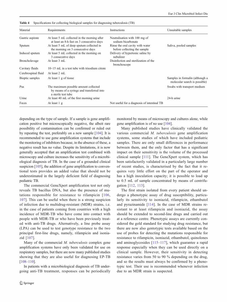
Table 4 shows the specifications of the various types ofbiological samples, which may be examined microscopically,cultured, or tested by means of gene amplification.
Although not very sensitive, a positive microscopic exam-ination of a sample previously stained in such a way as toreveal acid-fast bacilli allows prompt confirmation of a myco-bacterial etiology [101]. The two most widely used stains areZiehl Neelsen and the more sensitive fluorescence staining,but neither are capable of determining whether the mycobac-terial infection is tubercular or not.
A culture examination is the gold standard ofmycobacteriological diagnostics, but it is less sensitive whenusing respiratory samples taken from children than those takenfrom adults [102]. With few exceptions (particularly CSF),most biological samples are contaminated and therefore haveto be treated in order to eliminate the accompanying and rap-idly multiplying bacterial flora that would otherwise irreme-diably prevent the growth of possible mycobacteria. A myco-bacterial culture requires the inoculation of a solid (generallyLöwenstein-Jensen) and liquid medium (the most widely usedis mycobacterial growth indicator tube [MGIT]), andprolonged incubation at a temperature of 35–37 °C for up tosix (liquid media) or 8 weeks (solid media) before it can beconsidered negative.
In the case of a positive culture, it is essential to identify thespecies, or at least differentiate non-tubercular mycobacteriafrom those belonging to theM. tuberculosis complex, becauseboth can grow in the most frequently used media. The isola-tion ofM. tuberculosis complex is diagnostic for TB, but it isoften difficult to interpret the significance of finding a non-tubercular mycobacterium [103].
Although gene amplification is less sensitive than a culture,positive findings greatly reduce the time of diagnosis [104].Laboratories equipped for mycobacterial diagnostics general-ly also use amplification tests specific for the M. tuberculosiscomplex whose sensitivity in the presence of a positive mi-croscopic examination is practically 100 %, thus allowing arapid diagnosis; however, in the case of microscopically neg-ative samples, their sensitivity is much less and also varies
Eur J Clin Microbiol Infect Dis
depending on the type of sample. If a sample is gene amplifi-cation positive but microscopically negative, the albeit rarepossibility of contamination can be confirmed or ruled outby repeating the test, preferably on a new sample [104]. It isrecommended to use gene amplification systems that includethe monitoring of inhibitors because, in the absence of these, anegative result has no value. Despite its limitations, it is nowgenerally accepted that an amplification test combined withmicroscopy and culture increases the sensitivity of a microbi-ological diagnosis of TB. In the case of a grounded clinicalsuspicion [105], the addition of gene amplification to conven-tional tests provides an added value that should not beunderestimated in the largely deficient field of diagnosingpediatric TB.
The commercial GeneXpert amplification test not onlyreveals TB bacillus DNA, but also the presence of mu-tations responsible for resistance to rifampicin [106,107]. This can be useful when there is a strong suspicionof infection due to multidrug-resistant (MDR) strains, i.e.in the case of patients coming from countries with a highincidence of MDR-TB who have come into contact withpeople with MDR-TB or who have been previously treat-ed with anti-TB drugs. Alternatively, a line probe assay(LPA) can be used to test genotype resistance to the twoprincipal first-line drugs, namely, rifampicin and isonia-zid [107].
Many of the commercial M. tuberculosis complex geneamplification systems have only been validated for use onrespiratory samples, but there are nowmany published studiesshowing that they are also useful for diagnosing EP-TB[108–110].
In patients with a microbiological diagnosis of TB under-going anti-TB treatment, responses can be periodically
monitored by means of microscopy and cultures alone, whilegene amplification is of no use [104].
Many published studies have clinically validated thevarious commercial M. tuberculosis gene amplificationsystems, some studies of which have included pediatricsamples. There are only small differences in performancebetween them, and the only factor that has a significantimpact on their sensitivity is the volume of the processedclinical sample [111]. The GeneXpert system, which hasbeen satisfactorily validated in a particularly large numberof recent studies, is characterised by the fact that it re-quires very little effort on the part of the operator andhas a high inoculation capacity; it is possible to load upto 0.5 mL of sample concentrated by means of centrifu-gation [112, 113].
The first strain isolated from every patient should un-dergo a phenotypic assay of drug susceptibility, particu-larly its sensitivity to isoniazid, rifampicin, ethambutoland pyrazinamide [114]. In the case of MDR strains re-sistant to at least rifampicin and isoniazid, the assayshould be extended to second-line drugs and carried outat a reference centre. Phenotypic assays are currently con-sidered the gold standard for studying drug resistance, butthere are now also gentotypic tests available based on theuse of probes for detecting the mutations responsible forresistance to rifampicin, isoniazid, ethambutol, quinolonesand aminoglycosides [115–117], which guarantee a rapidresponse especially when they can be used directly on aclinical sample. However, their sensitivity in detectingresistance varies from 50 to 90 % depending on the drug,and so the results must always be confirmed by a pheno-typic test. Their use is recommended whenever infectiondue to an MDR strain is suspected.
Table 4 Specifications for collecting biological samples for diagnosing tuberculosis (TB)
Material Requirements Instructions Unsuitable samples
Gastric aspirate At least 5 mL collected in the morning afterat least an 8-h fast on 3 consecutive days
Neutralisation with 100 mg ofsodium bicarbonate
Sputum At least 5 mL of deep sputum collected inthe morning on 3 consecutive days
Rinse the oral cavity with waterbefore collecting the sample
Saliva, pooled samples
Induced sputum At least 5 mL collected in the morning on3 consecutive days
Delivery of hypertonic saline bynebulizer
Broncholavage At least 3 mL Disinfection and sterilization of thebronchoscope
Cavitary fluids 10–15 mL in a test tube with trisodium citrate
Cerebrospinal fluid At least 2 mL
Bioptic samples At least 1 g of tissue Samples in formalin (although amolecular search is possible)
Pus The maximum possible amount collectedby means of a syringe and transferred intoa sterile test tube
Swabs with transport medium
Urine At least 40 mL of the first morning urine 24-h urine
Feces At least 1 g Not useful for a diagnosis of intestinal TB
Eur J Clin Microbiol Infect Dis
What radiological methods should be used in the caseof suspected pediatric TB?
Radiology for pulmonary TB
Pulmonary TB is associated with a variable spectrum of le-sions which, if described as distinct pathologic entities, wouldinclude pulmonary consolidations; lymph node disease, withor without airway obstruction; a miliary pattern; cavitary le-sions; and pleural effusion [118].
The radiological examination is based on antero-posteriorand lateral chest X-rays. The lateral projection improves visu-a l i s a t i on o f po s t e r i o r l ymphadenomega l y andlymphadenomegalies inferior to the intermediate bronchus[118]. Furthermore, the usefulness of the lateral projectionand low radiation dose required lead to an advantageouscost/benefit ratio [119].
Although the indications have not been standardised, theinvestigation can be completed using computed tomography(CT) with contrast medium. CT is clinically useful above all inthe case of children aged <5 years, particularly those aged<2 years in whom conventional chest radiography is ofteninsufficient [118] because of the physiological protrusion ofthe thymus. CT is also better than conventional radiology indetecting calcifications, which are rare in the first years of life,and the contrast medium makes it possible to assess intratho-racic lymphadenomegalies [120], which are generally hilarand mediastinic, and usually prevalent on one side. Normallymph node size in childhood is up to 7 mm (short axis).Although aspecific, another suggestive pattern is that of theBrim sign^ (low density in the centre of the lymph nodesurrounded by a peripheral ring enhancement). Differentialdiagnosis includes lymphoma, other infections such as HIVinfection or minor acute or chronic inflammation, and sarcoid-osis (extremely rare).
In brief, even in the absence of clinical symptoms, the mainsigns of active TB are consolidation and non-calcified hilarlymphadenomegaly [121].
CT with contrast medium is the technique of choice fordefining lymphobronchial disease, a complication of pediatricTB due to bronchial compression by an infected lymph node,erosion, ulceration, infiltration, the intrabronchial flow of case-ous material, and the formation of granulated tissue [122], thusleading to air trapping, atelectasis, consolidation, necrosis andcavitation. It is also better for evaluating tuberculous consoli-dation and cavitation, identifying granulomas/tuberculomas,and investigating complications (lymphobronchial disease,pericarditis, outcomes). The use of CT for follow-up purposesis strictly related to the clinical/radiological evolution of TB[122]. Chest ultrasonography can play a role in evaluatingpleural and pericardial effusion [118], but there are no pediatricdata concerning the role of positron emission tomography(PET).
The anatomo-radiological classification of Marais et al.makes it possible to distinguish parenchymal and lymph nodedisease and their evolutionary stages [123]. A parenchymalGhon focus may not be self-limiting, but cavitate and lead tointrabronchial spreading and subsequent consolidation; thisexpression is typical in adults but, although rare, is also pos-sible in early infancy. However, what is typical in early infan-cy is lymph node (lymphobronchial) disease with a progres-sive cascade of the complications described above, which canbe best identified with CT. The disease may spread into thepleural space, the pericardium or hematogenously (miliaryTB), with possible outcomes being calcification, parenchymaldestruction with fibrosis, and bronchiectasis. Andronikouet al. have in fact integrated the classification of Marais et al.with the findings of tomographic techniques [118].
Radiology for EPTB
Children with suspected EPTB should always undergo stan-dard chest radiography [124], which is positive in 40–87 % ofthe patients with the disease [125].
Cervical, submandibular, supraclavicular and pre-auricularsites are the most typical locations of peripheral tuberculouslymphadenitis. Imaging only plays a relatively important role[126]; the examination of choice is ultrasonography, but theonly ultrasonographic sign suggesting tuberculous lymphade-nitis is calcification, which usually appears late.
Osteo-articular TB often presents with symptoms that aredifficult to interpret, and so imaging represents a first ap-proach [126]. Standard radiography reveals juxta-articular os-teoporosis, peripheral bone erosion, gradual joint spacenarrowing and, sometimes, a ‘cystic’ pattern with scleroticfissuring. Tuberculous dactylitis deserves particular mention;it is characterised by the painless involvement of the shorttubular bones of the hands and feet, and radiographically re-vealed fusiform tumefaction of the soft tissues with or withoutperiostitis. Subsequent alterations are a coarse trabecular pat-tern with acro-osteolysis, reactive sclerosis, and joint involve-ment [126].
Magnetic resonance imaging (MRI) is the technique ofchoice for obtaining a complete osteo-articular anatomicalpicture of all body regions, including an evaluation of possiblesynovitis, soft tissue involvement, and cartilage destruction[126].
Tuberculous spondylitis accounts for 30–50 % of cases oftuberculous osteomyelitis [127], with the frequently affectedsite in childhood being the dorso-lumbar junction. Standardradiography may show disc space narrowing and various de-grees of bone destruction, with possible kyphosis. Diagnosismay be elusive or difficult, particularly in the lumbo-sacralregion. CT provides a complete picture of all bone segmentsand is also capable of identifying collections of pus, but MRIis the investigation of choice for a sinal evaluation (vertebral
Eur J Clin Microbiol Infect Dis
bodies, discs, the spinal canal, and paraspinal tissues). Bonescintigraphy provides a whole-body evaluation of skeletal in-volvement (an alternative is whole-body MRI mainly basedon STIR sequences).
The follow-up is based on radiography, with MRI beingused more frequently in cases of spinal locations, which re-quire a careful evaluation not only of the bone [126, 127]. Theuse of CT is largely limited to conditions that contraindicatethe use of MRI [126, 127].
In the case of central nervous system (CNS) TB, the typicalalterations are granulomatous lesions in the leptomeninges, tothe junction between the grey and white matter of the brain,and in spinal cord [126]. The presence of tuberculomas can becharacteristic, whereas another manifestation is arteritis,which may be responsible for severe ischemic events [126].
In the absence of other known manifestations, the diagnosisof CNS TB can be difficult because the different neuroradiolog-ical pictures simulate other pathological conditions, particularlyneoplasias. However, a combination of meningeal and paren-chymal lesions should always arouse a suspicion of TB [128].
In an early phase, a cerebral CT scan without contrast me-dium may be negative (10–15 % of cases), or show the pres-ence of an iso-hyperdense exudate in the CSF-filled subarach-noid spaces (basal cisterns and sulci) or iso-hyperdense,roundish or oval masses (tuberculomas) that rarely containcalcifications and are delimited by more or less extensive ede-ma [128]. The administration of contrast medium leads tointense enhancement of the basal cisterns, and marginal ordiffuse impregnation of the tuberculomas.
MRI provides much more information about CNS TB thanCT, the use of which is currently limited to emergency situa-tions or the monitoring of hydroencephalus secondary to tu-berculous meningo-encephalitis [129]. An MRI protocol forstudying suspected CNS TB should always include T1-weighted (with and without contrast medium), turbo spin-echo (TSE) and T2-weighted FLAIR images, diffusion-weighted (DW) images, and possibly angio-MR arterial im-ages (MRA). DW images are very useful for bettercharacterising tuberculoma signals and evaluating complica-tions (ischemia, cerebritis) [129]. T1-weighted images withcontrast medium can reveal meningeal involvement as intenseenhancement and nodularity (an aspecific but very importantsign that is highlighted better by MRI than CT); vasculitis ofperforating vessels, which appears as puntate/linear enhance-ment at the level of the basal ganglia; ventriculitis; plexitis(rare); and pachymengitis (infrequent). Pienaar et al. foundthat MRI showed basal meningeal enhancement in 97 % oftheir patients with tuberculous meningo-encephalitis (asagainst 70% in the case of CT), infarctions in 83% (as against70 %), and granulomas in 40 % (as against 16 %) [129].Angio-RMI can reveal vascular involvement such as alter-ations in arterial profiles (stenoses, irregularities) or signs ofocclusion [129].
In the case of CNS TB, MRI with contrast medium shouldalso be extended to the whole spinal column in order to ex-clude spinal meningitis, which is characterised by irregularintradural enhancement, often with the formation ofperimedullary pseudo-cysts, medullary granulomas, andmisinterpreted vertebral alterations more or less associatedwith CNS lesions [126].
Although challenging, imaging is also used to diag-nose abdominal TB [128–131]. Standard radiographycan reveal signs of occlusion but ultrasonography and/or CT are necessary as they can reveal porta hepatis, inpara-aortic sites, and mesenteric lymphadenomegalies(the last being common in children). The presence ofcalcifications and/or rim signs can suggest TB, whereasthe signs of peritonitis (thickening, stranding) areaspecific. Gastrointestinal involvement manifests itselfas thickening and parietal contrast enhancement (particu-larly at ileo-cecal level).
Micro- (0.5–2.0 mm) and macronodular hepatosplenic le-sions are typical of disseminated disease [129–131]. The le-sions are hypoechogenic/hypodense, and can suggest TBwhen/if they calcify.
Renal TB is characterised by hydrocalycosis (papillary ne-crosis) and lobar calcifications [129–131].
Conclusions
After analysing the published evidence and on the basis oftheir own clinical experience, the group of experts reachedthe following conclusions:
& Pediatric pulmonary TB should be suspected in the pres-ence of the following symptoms and signs of pulmonaryinvolvement:
– Persistent, unremitting cough; cough lasting >4 weeks;persistent chest pain; or hemoptysis [III-A].
– Cough or other respiratory signs and symptoms associat-ed with long-lasting weight loss, asthenia/malaise and anevening temperature of ≥37.5 °C [III-A].
– Forms of pneumonia poorly responding to first-line anti-biotics or remitting/relapsing in nature, especially in sub-jects at risk [V-B].
– Radiological pictures of pneumonia with signs typ-ical of TB: calcifications, voluminous hilar andmediastinic lymphadenomegalies prevalently on oneside, with or without airway obstruction (<5 years),a miliary pattern, pulmonary consolidations/cavitations (typical sites: the apical and posteriorsegments of the upper lobe, and the apical segmentof the lower lobe), pleural effusion [III-A].
Eur J Clin Microbiol Infect Dis
& Tuberculous lymphadenopathy should be suspected in thepresence of:
– Persistent cervical lymphadenopathy (>4 weeks) with adiameter of >2 cm, in the absence of a response to anti-biotics or other local cause [III-B].
& Vertebral TB should be suspected in the presence of:
– Progressively increasing spinal pain; a discrepancy be-tween leukocyte counts and the ESR; laboratory signsof chronic inflammation (anemia, thrombocytosis, hypo-albuminemia) [IV-B].
& Tuberculous meningitis (TBM) should be suspected in thepresence of:
– Headache, vomiting, irritability, lethargy, convulsions,coma, nuchal rigidity, paralysis of the cranial nerves (par-ticular III and VI) lasting >5 days. CSF with leukocytes<1.000×10³/mL, of which >50 % lymphocytes; protein>100 mg/dL; CSF/plasma glucose ratio <50 %. CT/MRIfindings of basal meningeal enhancement, hydrocepha-lus, cerebral infarction [IV-A].
& The differential diagnosis of less frequent cerebraltuberculomas and other space-occupying forms is basedmore on imaging than clinical criteria (convulsions, vary-ing focal neurological signs depending on site,endocranial hypertension) [V-B].
& Abdominal TB should be suspected in the presence ofdistension and chronic abdominal pain, ascites, and is of-ten associated with general symptoms such as fever andweight loss [V-B].
& Genito-urinary TB should be suspected in the presence ofsymptoms of recurrent urinary pathway infections or he-maturia associated with sterile pyuria [V-B].
& In addition to images of exudative pericarditis, the diag-nosis of tuberculous pericarditis always requirespericardiocentesis or pericardiotomy, particularly in coun-tries with a low prevalence of TB [V-B].
& Disseminated TB should be suspected in the presence ofclinical manifestations that vary depending on the pre-dominantly involved site and are associated with initiallyaspecific systemic signs and symptoms [V-B]. The onlypathognomonic sign is the ophthalmoscopic presence ofchoroidal tubercles in about one-third of cases [V-A].
& The use of a TSTand/or IGRA is recommended in order todiagnose TB in children aged ≥5 years [III-A]. The choiceof one or the other method should be based on individualconsiderations, including an evaluation of the likelihoodthat the child will or will not return to the centre for theinterpretation of TST results, whether or not he or she has
received BCG vaccination (a confirmatory IGRA is anycase recommended if the TST result is <15 mm), and theavailability of a TST and/or IGRA at the centre [III-A].
& The use of an IGRA alone is not recommended for diag-nosing TB in children aged <5 years; a TST is the only testthat should be used [V-C]. However, the combined use ofa TSTand IGRA is possible in selected cases, especially ifthe child has previously received BCG vaccination [V-C],although it must be remembered that there are still doubtsas to how to interpret discordant results [V-C].
& Regardless of age, a negative or indeterminate IGRA re-sult should never be considered as excluding a diagnosisof tubercular infection [III-A].
& The performance of TSTs and IGRAs is suboptimal in thecase of children with T lymphocyte immunodepression[III-A]. The risk of tuberculous infection or disease in suchpatients can be evaluated by combining the results of bothtests with the findings of clinical, radiological and microbi-ological investigations [V-B]. Nevertheless, the interpreta-tion of conflicting test results remains doubtful, and soimmunodepressed children should prudently be consideredinfected even if only one of the tests is positive [V-B].
& The use of serial IGRAs is not recommended because it isnot helpful in monitoring the response to anti-TB therapy,and does not predict the risk of latent infection evolvinginto TB disease in children [III-A].
& A microbiological diagnosis must be based on samplesoriginating from the involved site and preferably collectedbefore the start of anti-TB treatment [III-A]. In order toincrease sensitivity, it is recommended to test for pulmo-nary TB using gastric aspirate or induced sputum samplescollected on three consecutive days [III-A]. A single sam-ple can be used only in the case of broncho-lavage [III-A].Induced sputum samples should always be indicated assuch because their salivary appearancemay otherwise leadto them being considered Bunsuitable^ by the laboratory[III-A].
& Minimummicrobiological diagnostics must include micros-copy and cultures [III-A]. A microscopic examination isonly moderately sensitive in the case of pediatric TB; a geneamplification test is more likely to be diagnostic [IV-B].Negative microscopic results do not exclude a diagnosis ofTB even if obtained onmultiple samples [III-A]. In additionto microscopy and culture tests, a gene amplification testshould be requested until a diagnosis has been established[IV-B]. If the laboratory is so equipped, it may be useful toreserve one of the available clinical samples for testing onthe GeneXpert platform [V-B]. If MDRTB is suspected in apatient with a microbiological diagnosis of TB, it is advis-able to carry out a genotypic resistance assay (e.g.GeneXpert or LPA) before starting treatment [IV-B].
& Chest radiography remains the most suitable imagingtechnique for evaluating pediatric pulmonary TB [IV-A].
Eur J Clin Microbiol Infect Dis
The presence of asymmetric/unilateral mediastinic and hi-lar lymphadenomegalies should prompt a suspicion ofTB, especially in children aged <5 years [V-B].Cavitations are also suggestive [V-B]. The lateral projec-tion may be useful in doubtful cases [IV-B].
& After traditional radiology, the investigation can be com-pleted using CT with contrast medium, which is particu-larly useful in children aged <5 years (especially thoseaged <2 years) in whom conventional chest radiographyis often insufficient because of the physiological protru-sion of the thymus [IV-B].
& TheMarais anatomo-radiological classification can distin-guish parenchymal and lymph node disease and their re-spective evolutions [V-B].
& All patients with extra-pulmonary TB should undergochest radiography [IV-A], which can be integrated withCT in the cases described above [IV-B].
& Ultrasonography is the investigation of first choice in thecase of peripheral tuberculous lymphadenitis [IV-B].
& If osteo-articular TB is suspected, the first diagnostic andfollow-up approach is conventional radiology [IV-A].MRI with contrast medium generally allows a better eval-uation of the extent of bone and joint involvement, al-though it does not usually increase diagnostic specificity[V-B]. MRI with contrast medium is fundamental for thevolumetric evaluation of ankylosis and the assessment ofosteopenia in patients with spondylitis/spondylodiscitis,as well as in order to obtain a complete spinal pictureduring follow-up [IV-B].
& MRI provides much more information about CNS TBthan CT, the use of which is currently limited to emergen-cy situations or the monitoring of hydroencephalus sec-ondary to tuberculous meningo-encephalitis [IV-A].
& Imaging is also often used to diagnose abdominal TB;standard radiography can reveal signs of occlusion butultrasonography and/or CT are the recommended imagingtechniques [IV-B].
Acknowledgments This study was supported by a grant obtained fromthe Italian Ministry of Health (Bando Giovani Ricercatori 2009).
Compliance with ethical standards
Conflicts of interest The authors declare that they have no competinginterests.
References
1. World Health Organization. Global tuberculosis control: WHOreport 2014. Geneva: World Health Organization, Geneve,Switzerland, 2014. Available at: http://www.who.int/tb/publications/global_report/en/ Accessed on 16 August 2015
2. World Health Organization. Guidelines for treatment of tubercu-losis. 4th ed. World Health Organization, Geneva, Switzerland,2010. Available at http://www.who.int/tb/publications/2010/9789241547833/en/ Accessed on 15 August 2015
3. Il Programma Nazionale per le Linee Guida (PNLG).Methodological handbook-how to produce, disseminate and up-date clinical practice recommendations. Available at http://www.pnlg.it/en_method. Accessed on 30 December 2014
4. Guidelines for the planning and management of NIH ConsensusDevelopment Conferences Online Bethesda (MD): NationalInstitutes of Health, Office of the Director, Office of MedicalApplications of Research; 1993. Updated October 2001
5. Scottish Intercollegiate Guidelines Network (SIGN). Available athttp://www.sign.ac.uk/. Accessed on 30 December 2014
6. de Pontual L, Balu L, Ovetchkine P, Maury-Tisseron B,Lachassinne E, Cruaud P, Jeantils V, Valeyre D, Fain O,Gaudelus J (2006) Tuberculosis in adolescents: a French retro-spective study of 52 cases. Pediatr Infect Dis J 25:930–932
7. Phongsamart W, Kitai I, Gardam M et al (2009) A population-based study of tuberculosis in children and adolescents in Ontario.Pediatr Infect Dis J 28:416–419
8. Winston C, Menzies HJ (2012) Pediatric and adolescent tubercu-losis in the United States 2008–2010. Pediatrics 130:e1425–e1432
9. Buonsenso D, Lancella L, Delogu G, Krzysztofiak A, Testa A,Ranno O, D’Alfonso P, Valentini P (2012) A twenty-year retro-spective study of pediatric tuberculosis in two tertiary hospitals inRome. Pediatr Infect Dis J 31:1022–1026
10. Wong KS, Huang YC, Lai SH, Chiu CY, Huang YH, Lin TY(2010) Validity of symptoms and radiographic features inpredicting positive AFB smears in adolescents with tuberculosis.Int J Tuberc Lung Dis 14:155–159
11. Marais BJ, Gie RP, Hesseling AC, Schaaf HS, Lombard C,Enarson DA, Beyers N (2006) A refined symptom-basedappraoch to diagnose pulmonary tuberculosis in children.Pediatrics 118:e1350–e1359
12. Marais BJ, Gie RP, Obihara CC, Hesseling AC, Schaaf HS,Beyers N (2005) Well defined symptoms are of value in the diag-nosis of childhood pulmonary tuberculosis. Arch Dis Child 90:1162–1165
13. English RG, Bachmann MO, Bateman ED, Zwarenstein MF,Fairall LR, Bheekie A, Majara BP, Lombard C, Scherpbier R,Ottomani SE (2006) Diagnostic accuracy of an integrated respira-tory guideline in identifying patients with respiratory symptomsrequiring screening for pulmonary tuberculosis: a cross sectionalstudy. BMC Pulm Med 6:22
14. Solovic I, Jonsson J, Korzeniewska-Koseła M, Chiotan DI, Pace-Asciak A, Slump E, Rumetshofer R, Abubakar I, Kos S, Svetina-Sorli P, Haas W, Bauer T, Sandgren A, van der Werf MJ (2013)Challenges in diagnosing extrapulmonary tuberculosis in theEuropean Union, 2011. Eur Surveill 18:pii:20432
15. American Thoracic Society (2000) Diagnostic standards and clas-sification of tuberculosis in adults and children. Am J Respir CritCare Med 161:1376–1395
16. Cruz AT, Karam LB, Orth RC, Starke JR (2014) Disseminatedtuberculosis in two children with inflammatory bowel disease re-ceiving infliximab. Pediatr Infect Dis J 33:779–781
17. Golden MP, Vikram HR (2005) Extrapulmonary tuberculosis: anoverview. Am Fam Physician 72:1761–1768
18. Maltezou HC, Spyridis P, Kafetzis DA (2000) Extra-pulmonarytuberculosis in children. Arch Dis Child 83:342–346
19. Marais BJ, Gie RP, Schaaf HS, Beyers N (2006) Childhood pul-monary tuberculosis: old wisdom and new challenges. Am JRespir Crit Care Med 173:1078–1090
20. Polesky A, Grove W, Bhatia G (2005) Peripheral tuberculouslymphadenitis: epidemiology, diagnosis, treatment, and outcome.Medicine (Baltimore) 84:350–362
Eur J Clin Microbiol Infect Dis
21. Wang D (2005) Diagnosis of tuberculous vertebral osteomyelitis(TVO) in a developed country and literature review. Spinal Cord43:531–542
22. Hosalkar HS, Agrawal N, Reddy S, Sehgal K, Fox EJ, Hill RA(2009) Skeletal tuberculosis in children in the Western world: 18new cases with a review of the literature. J Child Orthop 3:319–324
23. Cormican L, Hammal R,Messenger J, Milburn HJ (2006) Currentdifficulties in the diagnosis and management of spinal tuberculo-sis. Postgrad Med J 82:46–51
24. Hayes AJ, Choksey M, Barnes N, Sparrow OCE (1996) Spinaltuberculosis in developed countries; difficulties in diagnosis. J RColl Surg Edinb 41:192–196
25. Teo HE, Peh WC (2004) Skeletal tuberculosis in children. PediatrRadiol 34:853–860
26. Thwaites GE, Chau TT, Stepniewska K, Phu NH, Chuong LV,Sinh DX,White NJ, Parry CM, Farrar JJ (2002) Diagnosis of adulttuberculous meningitis by use of clinical and laboratory features.Lancet 360:1287–1292
27. Trecarichi EM, Di Meco E, Mazzotta V, Fantoni M (2012)Tuberculous spondylodiscitis: epidemiology, clinical features,treatment, and outcome. Eur Rev Med Pharmacol Sci 16:58–72
28. Principi N, Esposito S (2012) Diagnosis and therapy of tubercu-lous meningitis in children. Tuberculosis 92:377–383
29. Kumar R, Singh SN, Kohli N (1999) A diagnostic rule for tuber-culous meningitis. Arch Dis Child 81:221–224
30. Marais S, Thwaites G, Schoeman JF, Török ME, Misra UK,Prasad K, Donald PR, Wilkinson RJ, Marais BJ (2010)Tuberculous meningitis: a uniform case definition for use in clin-ical research. Lancet Infect Dis 10:803–812
31. Pasco PM (2012) Diagnostic features of tuberculous meningitis: across-sectional study. BMC Res Notes 5:49
32. Török ME, Nghia HD, Chau TT, Mai NT, Thwaites GE,Stepniewska K, Farrar JJ (2007) Validation of a diagnostic algo-rithm for adult tuberculous meningitis. Am J Trop Med Hyg 77:555–559
33. Youssef FG, Afifi SA, Azab AM, Wasfy MM, Abdel-Aziz KM,Parker TM, Oun SA, Jobanputra NN, Hajjeh RA (2006)Differentiation of tuberculous meningitis from acute bacterialmeningitis using simple clinical and laboratory parameters.Diagn Microbiol Infect Dis 55:275–278
34. Uygur-Bayramicli O, Dabak G, Dabak R (2003) A clinical dilem-ma: abdominal tuberculosis. World J Gastroenterol 9:1098–1101
35. Tinsa F, Essaddam L, Fitouri Z, Brini I, Douira W, Ben Becher S,Boussetta K, Bousnina S (2010) Abdominal tuberculosis in chil-dren. J Pediatr Gastroenterol Nutr 50:634–638
36. Duicu C, Marginean O, Kiss E, Lőrinczi L, Banescu C (2013)Genitourinary tuberculosis in children—a diagnostic challenge.Rev Rom Med Lab 21:301–309
37. Khalilzadeh S, Boloorsaz MR (2006) Renal tuberculosis in a four-year-old child. Tanaffos 5:71–74
38. Hugo-Hamman CT, Scher H, De Moor MM (1994) Tuberculouspericarditis in children: a review of 44 cases. Pediatr Infect Dis J13:13–18
39. Locham KK, Singh J (2004) Choroid tubercles. Indian Pediatr 41:1167
40. Hussey G, Chisholm T, Kibel M (1991) Miliary tuberculosis inchildren: a review of 94 cases. Pediatr Infect Dis J 10:832–836
41. Rangaka MX, Wilkinson KA, Glynn JR, Ling D, Menzies D,Mwansa-Kambafwile J (2012) Predictive value of interferon-γrelease assays for incident active tuberculosis: a systematic reviewand meta-analysis. Lancet Infect Dis 12:45–55
42. Starke JR (2004) Tubercolosis in children. Semin Respir Crit CareMed 25:353–364
43. Cruz AT, Starke JR (2007) Clinical manifestations of tuberculosisin children. Paediatr Respir Rev 8:107–117
44. American Academy of Pediatrics (1996) Update on tuberculosisskin testing of children. American Academy of PediatricsCommittee on Infectious Diseases. Pediatrics 97:282–284
45. Antonucci G, Girardi E, Raviglione MC, Ippolito G (1995) Riskfactors for tuberculosis in HIV-infected persons. JAMA 274:143–148
46. Cattamanchi A, Steingart KR, Metcalfe JZ, Date A, Coleman C,Marston BJ (2011) Interferon-gamma release assays for the diag-nosis of latent tuberculosis infection inHIV-infected individuals—a systematic review and meta-analysis. J Acquir Immune DeficSyndr 56:230–238
47. Pollock L, Basu Roy R, Kampmann B (2013) How to use: inter-feron γ release assays for tuberculosis. Arch Dis Child Educ PractEd 98:99–105
48. Snider DE Jr (1985) Bacille Calmette-Guerin vaccinations andtuberculin skin tests. JAMA 253:3438–3439
49. Menzies D (1999) Interpretation of repeated tuberculin tests.Boosting, conversion, and reversion. Am J Respir Crit CareMed 159:15–21
50. Auld SC, Click ES, Heilig CM, Miramontes R, Cain KP, BissonGP, Mac Kenzie WR (2013) Association between tuberculin skintest result and clinical presentation of tuberculosis disease. BMCInfect Dis 13:460
51. Madhi SA, Gray GE, Huebner RE, Sherman G, McKinnon D,Pettifor JM (1999) Correlation between CD4+ lymphocyte counts,concurrent antigen skin test and tuberculin skin test reactivity inhuman immunodeficiency virus type 1-infected and -uninfectedchildren with tuberculosis. Pediatr Infect Dis J 18:800–805
52. American Academy of Pediatrics (2009) Tuberculosis. In:Pickering LK (ed) Red book: report of the committee on infec-tious diseases, 28th edn. American Academy of Pediatrics, ElkGrove Village, pp 680–701
53. Mazurek G, Jereb J, Vernon A (2010) Updated guidelines forusing interferon gamma release assays to detect mycobacteriumtuberculosis infection—United States, 2010. MMWR 59:12–25
54. Lalvani A, Pareek M (2010) A 100 year update on diagnosis oftuberculosis infection. Br Med Bull 93:69–84
55. Naseer A, Naqvi S, Kampmann B (2007) Evidence for boostingMycobacterium tuberculosis-specific IFN-gamma responses at6 weeks following tuberculin skin testing. Eur Respir J 29:1282–1283
56. Richeldi L, Bergamini BM, Vaienti F (2008) Prior tuberculin skintesting does not boost QuantiFERON-TB results in paediatric con-tacts. Eur Respir J 32:524–525
57. van Zyl-Smit RN, Zwerling A, Dheda K, Pai M (2009) Within-subject variability of interferon-γ assay results for tuberculosisand boosting effect of tuberculin skin testing: a systematic review.PLoS One 4:e8517
58. Schepers K, Mouchet F, Dirix V, De Schutter I, Jotzo K,Verscheure V (2014) Long-incubation-time gamma interferon re-lease assays in response to purified protein derivative, ESAT-6,and/or CFP-10 for the diagnosis of Mycobacterium tuberculosisinfection in children. Clin Vaccine Immunol 21:111–118
59. McMullen SE, Pegues DA, Shofer FS, Sheller AC, Wiener EB(2014) Performance of quantiFERON-TB gold and tuberculinskin test relative to subjects’ risk of exposure to tuberculosis.Clin Infect Dis 58:1260–1266
60. Chiappini E, Bonsignori F, Mangone G, Galli L (2012) Serial T-SPOT.TB and quantiFERON-TB-Gold In-Tube assays to monitorresponse to antitubercular treatment in Italian children with activeor latent tuberculosis infection. Pediatr Infect Dis J 31:974–977
61. Young DB (2003) Building a better tuberculosis vaccine. Nat Med9:503–504
62. Bianchi L, Galli L, Moriondo M, Veneruso G, Becciolini L,Azzari C (2009) Interferon-gamma release assay improves the
Eur J Clin Microbiol Infect Dis
diagnosis of tuberculosis in children. Pediatr Infect Dis J 28:510–514
63. Detjen AK, Keil T, Roll S, Hauer B, Mauch H, Wahn U, MagdorfK (2007) Interferon-gamma release assays improve the diagnosisof tuberculosis and nontuberculous mycobacterial disease in chil-dren in a country with a low incidence of tuberculosis. Clin InfectDis 45:322–328
64. Lewinsohn DA, Lobato MN, Jereb JA (2010) Interferon-gammarelease assays: new diagnostic tests for Mycobacterium tubercu-losis infection, and their use in children. Curr Opin Pediatr 22:71–76
65. Bamford AR, Crook AM, Clark JE, Nademi Z, Dixon G, PatonJY, Riddell A, Drobniewski F, Riordan A, Anderson ST, WilliamsA, Walters S, Kampmann B (2010) Comparison of interferon-gamma release assays and tuberculin skin test in predicting activetuberculosis (TB) in children in the UK: a paediatric TB networkstudy. Arch Dis Child 95:180–186
66. Mandalakas AM, van Wyk S, Kirchner HL, Walzl G, Cotton M,Rabie H, Kriel B, Gie RP, Schaaf HS, Hesseling AC (2013)Detecting tuberculosis infection in HIV-infected children: a studyof diagnostic accuracy, confounding and interaction. Pediatr InfectDis J 32:e111–e118
67. Bergamini BM, Losi M, Vaienti F, D’Amico R, Meccugni B,Meacci M, De Giovanni D, Rumpianesi F, Fabbri LM, Balli F,Richeldi L (2009) Performance of commercial blood tests for thediagnosis of latent tuberculosis infection in children and adoles-cents. Pediatrics 123:e419–e424
68. Basu Roy R, Sotgiu G, Altet-Gómez N, Tsolia M, Ruga E,Velizarova S, Kampmann B (2012) Identifying predictors ofinterferon-γ release assay results in pediatric latent tuberculosis:a protective role of bacillus Calmette-Guerin? A pTB-NET col-laborative study. Am J Respir Crit Care Med 186:378–384
69. National Institute for Health and Clinical Excellence.Tuberculosis. Clinical diagnosis andmanagement of tuberculosis,and measures for its prevention and control. NICE ClinicalGuideline 117. March 2011. Available at http://guidance.nice.org.uk/CG117/NICEGuidance/pdf/English
70. Okada K, Mao TE, Mori T (2008) Performance of an interferon-gamma release assay for diagnosing latent tuberculosis infectionin children. Epidemiol Infect 136:1179–1187
71. Debord C, De Lauzanne A, Gourgouillon N, Guérin-El KhouroujV, Pédron B, Gaudelus J, Faye A, Sterkers G (2011) Interferon-gamma release assay performance for diagnosing tuberculosis dis-ease in 0- to 5-year-old children. Pediatr Infect Dis J 30:995–997
72. Losi M, Bocchino M, Matarese A, Bellofiore B, Roversi P,Rumpianesi F, Alma MG (2011) Role of the quantiferon-TB testin ruling out pleural tuberculosis: a multi-centre study. Int JImmunopathol Pharmacol 24:159–165
73. Moyo S, Isaacs F, Gelderbloem S, Verver S, Hawkridge AJ,Hatherill M (2011) Tuberculin skin test and QuantiFERON® as-say in young children investigated for tuberculosis in SouthAfrica. Int J Tuberc Lung Dis 15:1176–1181
74. Pavić I, Zrinski Topić R, Raos M, Aberle N, Dodig S (2011)Interferon-γ release assay for the diagnosis of latent tuberculosisin children younger than 5 years of age. Pediatr Infect Dis J 30:866–870
75. Richeldi L, Losi M, D’Amico R, Luppi M, Ferrari A, Mussini C,Codeluppi M (2009) Performance of tests for latent tuberculosis indifferent groups of immunocompromised patients. Chest 136:198–204
76. Bruzzese E, Bocchino M, Assante LR, Alessio M, Bellofiore B,Bruzzese D, Iorio R, Matarese A, Santoro G, Vajro P, Guarino A,Sanduzzi A (2009) Gamma interferon release assays for diagnosisof tuberculosis infection in immune-compromised children in acountry in which the prevalence of tuberculosis is low. J ClinMicrobiol 47:2355–2357
77. Davies MA, Connell T, Johannisen C, Wood K, Pienaar S,Wilkinson KA (2009) Detection of tuberculosis in HIV-infectedchildren using an enzyme-linked immunospot assay. AIDS 23:961–969
78. Haustein T, Ridout D, Hartley JC, Thaker U, Shingadia D, KleinNJ, Novelli V, Dixon GLJ (2009) The likelihood of an indetermi-nate test result from a whole-blood interferon-γ release assay forthe diagnosis of mycobacterium tuberculosis infection in childrencorrelates with age and immune status. Pediatr Infect Dis J 28:669–673
79. Ling DI, Nicol MP, Pai M, Pienaar S, Dendukuri N, Zar HJ (2013)Incremental value of T-SPOT.TB for diagnosis of active pulmo-nary tuberculosis in children in a high-burden setting: a multivar-iable analysis. Thorax 68:860–866
80. RoseMV, KimaroG, Nissen TN, Kroidl I, HoelscherM, BygbjergIC, Mfinanga SG, Ravn P (2012) QuantiFERON®-TB gold in-tube performance for diagnosing active tuberculosis in childrenand adults in a high burden setting. PLoS One 7:e37851
81. Stefan DC, Dippenaar A, Detjen AK, Schaaf HS, Marais BJ,Loebenberg L, Walzl G, Hesseling AC (2010) Interferon-gammarelease assay for the detection of Mycobacterium tuberculosis in-fection in children with cancer. Int J Tuberc Lung Dis 14:689–694
82. Chiappini E, Fossi F, Bonsignori F, Sollai S, Galli L, deMartinoM(2012) Utility of interferon-γ release assay results to monitor anti-tubercular treatment in adults and children. Clin Ther 34:1041–1048
83. Zrinski Topić R, Zoričić-Letoja I, Pavić I, Dodig S (2011)Indeterminate results of QuantiFERON-TB Gold In-Tube assayin non immunosuppressed children. Arch Med Res 42:138–143
84. Pearce EC, Woodward JF, Nyandiko WM, Vreeman RC, AyayaSO (2012)A systematic review of clinical diagnostic systems usedin the diagnosis of tuberculosis in children. AIDS Res Treat 2012:401896
85. Brown M, Varia H, Bassett P, Davidson RN, Wall R, Pasvol G(2007) Prospective study of sputum induction, gastric washing,and bronchoalveolar lavage for the diagnosis of pulmonary tuber-culosis in patients who are unable to expectorate. Clin Infect Dis44:1415–1420
86. Parashar D, Kabra SK, Lodha R, Singh V, Mukherjee A, Arya T,Grewal HM, Singh S (2013) Does neutralization of gastric aspi-rates from children with suspected intrathoracic tuberculosis affectmycobacterial yields onMGITculture? J ClinMicrobiol 51:1753–1756
87. Berggren Palme I, Gudetta B, Bruchfeld J, ErikssonM,Giesecke J(2004) Detection of Mycobacterium tuberculosis in gastric aspi-rate and sputum collected from Ethiopian HIV-positive and HIV-negative children in a mixed in- and outpatient setting. ActaPaediatr 93:311–315
88. Franchi LM, Cama RI, Gilman RH, Montenegro-James S, SheenP (1998) Detection of mycobacterium tuberculosis in nasopharyn-geal aspirate samples in children. Lancet 352:1681–1682
89. Hatherill M, Hawkridge T, Zar H, Whitelaw A, Tameris M,Workman L, Geiter L, Hanekom W, Hussey G (2009) Inducedsputum or gastric lavage for community-based diagnosis of child-hood pulmonary tuberculosis? Arch Dis Child 94:195–201
90. Mukherjee A, Singh S, Lodha R, Singh V, Hesseling A, GrewalHM, Kabra S (2013) Ambulatory gastric lavages provide betteryields of Mycobacterium tuberculosis than induced sputum inchildren with intra-thoracic tuberculosis. Pediatr Infect Dis J 32:1313–1317
91. Owens S, Abdel-Rahman IE, Balyejusa S, Musoke P, Cooke RP,Parry CM, Coulter JB (2007) Nasopharyngeal aspiration for diag-nosis of pulmonary tuberculosis. Arch Dis Child 92:693–696
92. Planting NS, Visser GL, Nicol MP, Workman L, Isaacs W, Zar HJ(2014) Safety and efficacy of induced sputum in young children
Eur J Clin Microbiol Infect Dis
hospitalised with suspected pulmonary tuberculosis. Int J TubercLung Dis 18:8–12
93. Qureshi UA, Gupta AK, Mahajan B, Qurieshi MA, Altaf U,Parihar R, Bhau KS (2011) Microbiological diagnosis of pulmo-nary tuberculosis in children: comparative study of induced spu-tum and gastric lavage. Indian J Pediatr 78:1429–1430
94. Zar HJ, Hanslo D, Apolles P, Swingler G, Hussey G (2005)Induced sputum versus gastric lavage for microbiological confir-mation of pulmonary tuberculosis in infants and young children: aprospective study. Lancet 365:130–134
95. Khan AE, Starke JR (1995) Diagnosis of tuberculosis in children:increased need for better methods. Emerg Infect Dis 1:115–123
96. Chow F, Espiritu N, Gilman RH, Gutierrez R, Lopez S, EscombeAR, Evans CA, Moore DA (2006) La cuerda dulce—a tolerabilityand acceptability study of a novel approach to specimen collectionfor diagnosis of paediatric pulmonary tuberculosis. BMC InfectDis 4:6–67
97. Cordova J, Shiloh R, Gilman RH, Sheen P, Martin L, Arenas F,Caviedes L, Kawai V, Soto G, Williams DL, Zimic M, EscombeAR, Evans CA (2010) Evaluation of molecular tools for detectionand drug susceptibility testing of Mycobacterium tuberculosis instool specimens from patients with pulmonary tuberculosis. J ClinMicrobiol 48:1820–1826
98. Oramasionwu GE, Heilig CM, Udomsantisuk N, Kimerling ME,Eng B, Nguyen HD, Thai S, Keo C, McCarthy KD, Varma JK,Cain KP (2013) The utility of stool cultures for diagnosing tuber-culosis in people living with the human immunodeficiency virus.Int J Tuberc Lung Dis 17:1023–1028
99. Moore HA, Apolles P, de Villiers PJ, Zar HJ (2011) Sputum in-duction for microbiological diagnosis of childhood pulmonarytuberculosis in a community setting. Int J Tuberc Lung Dis 15:1185–1190
100. Conde MB, Loivos AC, Rezende VM, Soares SL, Mello FC,Reingold AL, Daley CL, Kritski AL (2003) Yield of sputum in-duction in the diagnosis of pleural tuberculosis. Am J Respir CritCare Med 167:723–725
101. Hepple P, Ford N, McNerney R (2012) Microscopy compared toculture for the diagnosis of tuberculosis in induced sputum sam-ples: a systematic review. Int J Tuberc Lung Dis 16:579–588
102. CLSI (2008) Laboratory detection and identification ofmycobacteria; approved guideline. CLSI, Wayne
103. Griffith DE, Aksamit T, Brown-Elliott BA, Catanzaro A, Daley C,Gordin F, Holland SM, Horsburg R, Huit G, Iademarco MF,Iseman M, Olivier K, Rouss S, von Reyn CF, Wallace RJ Jr,Winthrop K (2007) ATS Mycobacterial Disease Subcommittee,American Thoracic Society, Infectious Disease Society ofAmerica. An official ATS/IDSA statement: diagnosis, treatmentand prevention of nontuberculous mycobacterial disease. Am JRespir Crit Care Med 175:367–416
104. Centers for Disease Control and Prevention (CDC) (2009)Updated guidelines for the use of nucleic acid amplification testsin the diagnosis of tuberculosis. MMWRMorb Mortal Wkly Rep58:7–10
105. Catanzaro A, Perry S, Clarridge JE, Dunbar S, Goodnight-WhiteS, Lo Bue PA, Peter C, Pfyffer GE, SierraMF,Weber R,WoodsG,Mathews G, Jonas V, Smith K, Della LP (2000) The role of clin-ical suspicion in evaluating a new diagnostic test for active tuber-culosis: results of a multicenter prospective trial. JAMA 283:639–645
106. Tukvadze N, Kempker RR, Kalandadze I, Kurbatova E, LeonardMK, Apsindzelashvili R, Bablishvili N, Kipiani M, Blumberg HM(2012) Use of a molecular diagnostic test in AFB smear positivetuberculosis suspects greatly reduces time to detection of multi-drug resistant tuberculosis. PLoS ONE 7:e31563
107. Weyer K, Mirzayev F, Migliori GB, Van Gemert W, D’AmbrosioL, Zignol M, Floyd K, Centis R, Cirillo DM, Tortoli E, Gilpin C,
de Dieu Iragena J, Falzon D, Raviglione M (2013) Rapid molec-ular TB diagnosis: evidence, policy making and global implemen-tation of Xpert MTB/RIF. Eur Respir J 42:252–271
108. Laraque F, Griggs A, Slopen M, Munsiff SS (2009) Performanceof nucleic acid amplification tests for diagnosis of tuberculosis in alarge urban setting. Clin Infect Dis 49:46–54
109. Piersimoni C, Bornigia S, Gherardi G (2012) Performance of acommercial nucleic acid amplification test with extrapulmonaryspecimens for the diagnosis of tuberculosis. Eur J Clin MicrobiolInfect Dis 31:287–293
110. Tortoli E, Russo C, Piersimoni C, Mazzola E, Dal Monte P,Pascarella M, Borroni E, Mondo A, Piana F, Scarparo C,Coltella L, Lombardi G, Cirillo DM (2012) Clinical validationof Xpert MTB/RIF for the diagnosis of extrapulmonary tubercu-losis. Eur Respir Rev 40:442–447
111. Ling DI, Flores LL, Riley LW, Pai M (2008) Commercial nucleic-acid amplification tests for diagnosis of pulmonary tuberculosis inrespiratory specimens: meta-analysis and meta-regression. PLoSONE 3:e1536
112. Bates M, O’Grady J, Maeurer M, Tembo J, Chilukutu L, ChabalaC, Kasonde R, Mulota P, Mzyece J, Chomba M, Mukonda L,Mumba M, Kapata N, Rachow A, Clowes P, Hoelscher M,Mwaba P, Zumla A (2013) Assessment of the Xpert MTB/RIFassay for diagnosis of tuberculosis with gastric lavage aspiratesin children in sub-Saharan Africa: a prospective descriptive study.Lancet Infect Dis 13:36–42
113. Scott LE, McCarthy K, Gous N, Nduna M, van Rie A, Sanne I,Venter WF, Duse A, Stevens W (2011) Comparison of XpertMTB/RIF with other nucleic acid technologies for diagnosing pul-monary tuberculosis in a high HIV prevalence setting: a prospec-tive study. PLoS Med 8:e1001061
114. CLSI (2011) Susceptibility testing of mycobacteria, nocardiae andother aerobic actinomycetes: approved standard, 2nd edn. CLSI,Wayne
115. Miotto P, Cabibbe AM, Mantegani PBE, Fattorini L, Tortoli E,Migliori GB, Cirillo DM (2012) GenoType MTBDRsl perfor-mance on clinical samples with diverse genetic background. EurRespir J 40:690–698
116. Miotto P, Piana F, Penati V, Canducci F, Migliori GB, Cirillo DM(2006) Use of genotype MTBDR assay for molecular detection ofrifampin and isoniazid resistance in Mycobacterium tuberculosisclinical strains isolated in Italy. J Clin Microbiol 44:2485–2491
117. Tortoli E, Marcelli F (2007) Use of the INNO LiPA Rif.TB fordetection of Mycobacterium tuberculosis DNA directly in clinicalspecimens and for simultaneous determination of rifampin suscep-tibility. Eur J Clin Microbiol Infect Dis 21:51–55
118. Andronikou S, Vanhoenacker FM, De Backer AI (2009)Advances in imaging chest tuberculosis: blurring of differencesbetween children and adults. Clin Chest Med 30:717–744
119. Britton P, Perez-Velez CM,Marais BJ (2013) Diagnosis, treatmentand prevention of tuberculosis in children. NSW Public HealthBull 24:15–21
120. de Jong PA, Nievelstein RJ (2012) Normal mediastinal and hilarlymphnodes in children on multi detector row chest computedtomography. Eur Radiol 22:318–321
121. Gwee A, Pantazidou A, Ritz N, Tebruegge M, Connell TG, CainT, Curtis N (2013) To X-ray or not to X-ray? Screening asymp-tomatic children for pulmonary TB: a retrospective audit. Arch DisChild 98:401–404
122. Lucas S, Andronikou S, Goussard P, Gie R (2012) CT features oflymphobronchial tuberculosis in children, including complicationsand associated abnormalities. Pediatr Radiol 42:923–931
123. Marais BJ, Gie RP, Schaaf HS, Starke JR, Hesseling AC, DonaldPR, Beyers N (2004) A proposed radiological classification ofchildhood intra-thoracic tuberculosis. Pediatr Radiol 34(11):886–894
Eur J Clin Microbiol Infect Dis
124. Hopewell PC, Pai M, Maher D, Uplekar M, Raviglione MC(2006) International standards for tuberculosis care. LancetInfect Dis 6:710–725
125. Farinha NJ, Razali KA, Holzel H, Morgan G, Novelli VM (2000)Tuberculosis of the central nervous system in children: a 20-yearsurvey. J Infect 41:61–68
126. Burkett EA, Bradshaw WT (2011) Neonatal tuberculosis. AdvNeonatal Care 11:376–381
127. Boachie-Adjei O, Squillante RG (1996) Tuberculosis of the spine.Orthop Clin N Am 27:95–103
128. Andronikou S, Wieselthaler N (2004) Modern imaging of tuber-culosis in children: thoracic, central nervous system and abdomi-nal tuberculosis. Pediatr Radiol 34:861–875
129. Pienaar M, Andronikou S, van Toorn R (2009) MRI to demon-strate diagnostic features and complications of TBM not seen withCT. Childs Nerv Syst 25:941–947
130. Shah I, Uppuluri R (2010) Clinical profile of abdominal tubercu-losis in children. Indian J Med Sci 64:204–209
131. Leder RA, Low VH (1995) Tuberculosis of the abdomen. RadiolClin N Am 33:691–705
Eur J Clin Microbiol Infect Dis