evolution in pediatric pharmacology
TRANSCRIPT
Miriam G. Mooij
Evolution in Pediatric PharmacologyMicrodosing, Metabolism, and Membrane Transporters

Evolution in Pediatric Pharmacology
Microdosing,
Metabolism, and
Membrane Transporters
The studies described in this thesis were supported by:Netherlands Organization for Health Research and Development (ZonMw) – research grant (113202007)Novartis – investigator initiated grantTravel grants from: Erasmus Trustfonds, Dutch Society of Clinical Pharmacology and Biopharmaceutics, F1000 review platform
Cover and chapter design: Inge J.S.M.L. Vanhooymissen
ISBN: 978-94-6169-823-0
© 2016 M.G. Mooij, Rotterdam, The NetherlandsAll rights reserved. No part of this thesis may be reproduced, stored in a retrieval system, or transmitted in any form by any means, without prior written permission of the copy-right owner.
Layout and printing: Optima Grafische Communicatie, Rotterdam, The Netherlands
Evolution in Pediatric PharmacologyMicrodosing, metabolism, and membrane transporters
Evolutie in de pediatrische farmacologieMicrodosing, metabolisme en membraan transporters
Proefschrift
ter verkrijging van de graad van doctor aan deErasmus Universiteit Rotterdam
op gezag van derector magnificus
Prof.dr. H.A.P. Pols
en volgens besluit van het College voor Promoties.
De openbare verdediging zal plaatsvinden opdinsdag 22 maart 2016 om 13:30 uur
door
Miriam Geerthe Mooij
geboren te Zwolle

ProMotiEcoMMissiE
Promotor: Prof.dr. D. Tibboel
overige leden: Prof.dr. T. van Gelder Prof.dr. K. Allegaert Prof.dr. J.S. Leeder
copromotor: Dr. S.N. de Wildt

Voor mama

contEnts
Part i introduction
Chapter 1 General introduction 11
Chapter 2 Significant oral drug use in critically ill children: rational therapy or a black box?
21
Chapter 3 Ontogeny of oral drug absorption processes 31
Chapter 4 Human ontogeny of drug transporters: review and recommendations of the pediatric transporter working group
53
Chapter 5 Development of human membrane transporters: drug disposition and pharmacogenetics
93
Part ii Membrane transporters
Chapter 6 Ontogeny of human hepatic and intestinal transporter gene expression during childhood: age matters
127
Chapter 7 Intestinal peptide transporter PEPT1 expression and tissue distribution across the pediatric age range
145
Chapter 8 The effect of age on human hepatic membrane transporter protein expression
161
Part iii Phase ii drug metabolism
Chapter 9 Pediatric microdose study of [14C]paracetamol to study drug metabolism using accelerated mass spectrometry: proof of concept
187
Chapter 10 Pediatric microdose and microtracer studies using [14C] in Europe 203
Chapter 11 Microdosing to study ontogeny of drug metabolism: the case of [14C]paracetamol
213
Part iV Discussion and summary
Chapter 12 General discussion 235
Chapter 13 Summary / Samenvatting 261
Part V Appendices
List of abbreviations 273
About the author 277
List of publications 278
PhD Portfolio 280
Dankwoord 283

Part IIntroduction

1 General introduction

General introduction 13
1suitAblE DruG trEAtMEnt for chilDrEn
A major part (40-70%) of drugs prescribed to children are used either unlicensed or off-label, leading to increased risk of drug toxicity or therapeutic failure [1, 2]. Despite increasing efforts by US and EU regulatory authorities who require that the pharma-ceutic industry perform pediatric studies while developing drugs, most drug treatment is based on adult studies [3, 4]. Appropriate pediatric dose-selection solely based on adult data is not acceptable due to development causing changes in drug absorption, distribution, metabolism and excretion [5]. Simple size- or weight-based extrapolations from adult to pediatric doses is not enough, particularly not in neonates and young infants. Simulation models like physiologically-based pharmacokinetic models (PBPK) and population pharmacokinetic (popPK) are used to optimize pediatric dose predic-tions [6-8]. The limitation of these models is the limited ontogeny data that is available for input or to validate these models. Subsequently new data on the impact of age on the processes affecting drug disposition during childhood are needed.
orAl DruG trEAtMEnt for chilDrEn
Nowadays, most drugs prescribed to children in the community are taken orally [1]. Even critically ill neonates and children receive many oral drug formulations, due to the unavailability of intravenous formulations, intravenous access issues or cost and safety concerns or patients soon being discharged from the intensive care unit (ICU). Extrinsic factors, like food and drug formulation, and intrinsic factors of physiological nature affect the extent of absorption of orally administered drugs [9]. Intrinsic factors include many gastrointestinal processes such as gastric pH, gastric and intestinal motil-ity, gastrointestinal fluids, the pH and buffer capacity of these fluids, digestive enzymes, intestinal membrane transporters and intestinal drug metabolism.
Important progress has been made to elucidate age-related changes in phase I hepatic drug metabolism and renal excretion [10, 11]. Meanwhile, our knowledge on developmental changes governing variation in oral bioavailability including intestinal and hepatic drug transport and phase II drug metabolism is far less developed. A major reason for a lack of studies in children is the ethical and practical limitations. Hence, the development of innovative methods and a thorough understanding of the ethical challenges and potential solutions are highly needed.
14 Chapter 1
MEMbrAnE trAnsPortErs
Plasma membrane transporters are proteins that facilitate uptake and excretion of compounds in and out of the cell. More than 400 transporters are identified nowadays [12]. Located on, amongst others, the enterocyte, hepatocyte and renal cells, they play an essential role in mediating the uptake, distribution and excretion of many drugs [13, 14]. The classification of transporters is according to their properties and grouped in superfamilies. The disposition of drugs is most often associated with two superfamilies of transporters: the solute carrier (SLC) and ATP-binding cassette transporters (ABC) families. Their physiological function is to facilitate translocation of endogenous com-pounds, such as bile salts and steroids. Exogenous compounds, such as nutrients, drugs and metabolites, are also substrates for specific transporters [15]. The clinical relevance of transporters in drug disposition has been shown in drug-drug interaction and phar-macogenomics studies in adults [13, 16]. Little is known on the ontogeny of membrane transporters in pediatric organ tissues and their roles in pediatric pharmacotherapy. Growth and maturation are likely to impact on activity of transporters as has been shown in drug metabolizing enzymes, especially as transporters function in endogenous pro-cesses. Animal studies have indeed shown maturational changes in transporter expres-sion. Human data in fetuses and children are very limited and definitive conclusions on their impact on drug disposition across the pediatric age range can hardly be drawn.
innoVAtiVE MEthoD: Ex ViVo trAnsPortEr stuDiEs
To date, no solid substrate can be identified to study in vivo transporter activity in children and in vitro expression studies might serve as alternatives. For in vitro studies on transporters, human organ tissue is necessary and to study the ontogeny specifi-cally, pediatric tissue is essential. Nevertheless, pediatric tissue is scarce and difficult to harvest. Tissues can be harvested from diagnostic biopsies, surgical waste material, postmortem or be collected within the framework of a biobank. Within the context of the postmortem biobank and surgical tissue waste collection for specific projects was available. This tissue availability has been shown to be unique, even worldwide.
This relative scarcity demands study techniques that rely on efficient use of minimal amounts of pediatric tissue. Protein expression methods, especially, require large amounts of samples, can only quantify single transporters and are very labor-intensive. Recently, liquid chromatography tandem mass spectrometry (LC-MS/MS) has been increasingly used to study protein expression, including membrane transporters in humans. It may also be an attractive method for pediatric studies, as it only requires very limited sample volume and enables the quantification of multiple transporters at once.

General introduction 15
1PhAsE ii DruG MEtAbolisM
Drug metabolizing enzymes are abundant in the liver and gut and contribute to the first-pass metabolism of many orally administered drugs. Drugs are often metabolized in two phases. Phase I reactions involve formation of a new or modified group (oxida-tion, reduction, hydrolysis), whereas phase II reactions involve conjugation with an endogenous substance (e.g. glucuronic acid, sulfate, glycine) which serves the purpose of enhancing the water-solubility of the substrate and consequently its excretion by liver or kidneys. Uridine 5’-diphospho-glucuronosyltransferase (UGT) iso-enzymes and sulfotransferases (SULT) are families of drug metabolizing enzymes, important in the phase II drug metabolism. UGTs add glucuronic acid to a substrate, i.e., drugs, bilirubin, bile salts, and SULTs add a sulfo-group to a substrate.
In vitro, human hepatic UGTs show an enzyme-specific developmental pattern in early years of age of UGT1A9, UGT1A1, and UGT1A6 [17, 18]. A limitation is that liver samples of children younger than 2 years were not included. In vivo studies showed that postnatal age and postmenstrual age co-determine the interindividual variability in for instance tramadol glucuronidation in neonates after 10 days of age [19]. Morphine glucuronidation changes with age due to UGT2B7 maturation [20]. The ontogeny of other UGTs or SULTs are less well studied and especially not during the continuum of childhood age but in small populations of limited ages.
Paracetamol (acetaminophen, AAP) is a suitable probe drug to study UGT activity in vivo in children. Its main metabolism pathways are glucuronidation and sulfation; mainly via UGT1A1, 1A6, 1A9 and 2B15 and SULT1A1, 1A3, 1A4, and 2A1 [21]. Furthermore, AAP can be given orally and intravenously, is frequently prescribed to children, has dose-linear pharmacokinetics, and UGT metabolism can be detected by the ratio of glucuro-nide (AAP-glu) and sulfate (AAP-sul) metabolites in plasma and urine. Its metabolism has been characterized in children, but information gaps remain, especially in the first two years of age. Moreover, its metabolism after exclusive oral administration has not been very well characterized.
innoVAtiVE MEthoD: MicroDosinG to PhEnotyPE DruG MEtAbolisM
Pediatric drug studies are hampered by practical and ethical limitations. To protect children as a vulnerable population often incapable of expressing themselves, stringent medical ethical criteria are justified for research trials. Nevertheless, to ensure safe and effective drug treatment, adequate and reliable research tools are necessary. Pharma-cokinetic studies, according to a classic design, are done by giving a ‘non-therapeutic’ drug after which multiple blood samples are taken to determine the drug concentra-

16 Chapter 1
tions. Ethical and practical arguments limit these studies in children, because it means ‘non-therapeutic’ drug doses with exposure to unnecessary effects and toxicity followed by extensive blood sampling, which is often a painful procedure. Blood sampling, for example, is especially limited in premature neonates in whom circulating blood volume is very small (e.g., blood sampling volume would be 0.4 mL per 24 h in a 500-gram-neonate with a circulating blood volume of around 40 mL).
To overcome the limitations of a drug dose only for non-therapeutic reasons, a mi-crodose can be given. Microdosing promises to study the pharmacokinetics of drugs in children, without the risk of adverse events and with minimal burden [22, 23]. Microdos-ing is defined by the EMA and FDA as the lowest of i) one-hundredth of the No Observed Adverse Effect Level (NOAEL) or ii) one-hundredth of the predicted pharmacologic dose based on animal data or iii) as 100 micrograms of the new drug [4, 24]. Additionally, label-ing of the drug enables oral bioavailability studies when the microdose is administered per one route and the (therapeutic) unlabeled dose is administered via another route. Even after simultaneously administering the doses, separation of drug concentration levels is possible and so limits blood sampling time points.
Dose linearity between the microdose and therapeutic dose is a prerequisite in obtain-ing pharmacokinetic data for clinically relevant dosing guidelines. For several drugs such dose-linearity has been shown in adults [22, 25]. Sensitive measurements are needed to detect the extremely low dose levels in plasma or urine. Accelerator mass spectrometry (AMS) measures low attomolar to zeptomolar isotope ratios ranges, and quantifies [14C] labeled drug/metabolite levels [26]. The extremely low dose implies that a [14C] labeled microdose can be administered simultaneously with a non-labeled therapeutic dose. And thus, the bioavailability can be studied. Radiation associated with [14C] labeling in adults is less than 10 µSv which is negligible in the light of the yearly background exposure of 2.5 mSv in the Netherlands. In premature neonates, microdosing has been used once in a small pharmacokinetic study of ursodiol in the US [27].
We chose to use [14C] labeled microdosing to delineate developmental changes in the intestinal and hepatic UGT metabolism pathway involved in paracetamol. The contribu-tion of intestinal and hepatic drug metabolism of paracetamol during childhood needs to be elucidated. Dose linearity of paracetamol has been shown in adults under normal conditions and after probenecid glucuronidation inhibition [25].
General introduction 17
1AiMs AnD outlinE of this thEsis
The aims of this thesis are:• Tostudytheextentoforaldrugsusedinneonatalandpediatricintensivecare.• Toreviewthecurrentknowledgeofage-relatedvariationinprocessesthatgovern
oral drug absorption.• Toreviewthecurrentliteratureonhumanmembranetransportersduringchildhood.• Toassesstheontogenyofrelevanthumanmembranetransportersgeneandprotein
expression in pediatric intestinal and hepatic tissues.• Tostudythefeasibilityof[14C]-labeled microdosing studies in children.• Toinvestigatetheeffectofageonthecombinedintestinalandhepaticglucuronida-
tion and sulfation in young children, using a paracetamol microdosing study.
Part i describes a study on the extent of the oral drugs used in neonatal and pediatric intensive care [chapter 2].The available data on age-related changes in gastro-intestinal processes that govern oral drug absorption is highlighted in chapter 3. In chapter 4 and 5 extensive up-to-date overviews are given on the ontogeny of membrane transport-ers in children. Chapter 4 is a complete history of available transporter ontogeny data. Chapter 5 reviews the literature on the role of transporter ontogeny on pediatric drug disposition and effect.
Part ii focusses on the hepatic and intestinal membrane transporters. In chapter 6, the age-related changes in gene expression of MDR1, MRP2, OATP1B1, OATP1B3 and OATP2B1 are studied in intestinal and hepatic fetal and pediatric tissues. Intestinal PEPT1 mRNA expression and PEPT1, MDR1, MRP2 and OATP2B1 protein localizations are discussed in chapter 7. Chapter 8 evaluates the protein expression in hepatic tissues during fetal and childhood age.
Part iii presents the results of a [14C] labeled paracetamol microdosing study in children. Chapter 9 presents pilot data to describe the proof-of-concept of microdosing studies in children. Chapter 10 evaluates our study and a second study using [14C]paracetamol in children of 0-2 years. The effect of age on intestinal and hepatic paracetamol glucuroni-dation and sulfation is described in chapter 11.
Part iV summarizes and discusses the results of these studies in the respect of available relevant literature, and speculates on areas of future research [chapter 12 and 13].

18 Chapter 1
rEfErEncEs
[1] Schirm E, Tobi H, de Vries TW, Choonara I, De Jong-van den Berg LT. Lack of appropriate formula-tions of medicines for children in the community. Acta Paediatr 2003 Dec; 92(12): 1486-9.
[2] t Jong GW, van der Linden PD, Bakker EM, van der Lely N, Eland IA, Stricker BH, et al. Unlicensed and off-label drug use in a paediatric ward of a general hospital in the Netherlands. Eur J Clin Pharmacol 2002 Jul; 58(4): 293-7.
[3] Hoppu K, Anabwani G, Garcia-Bournissen F, Gazarian M, Kearns GL, Nakamura H, et al. The status of paediatric medicines initiatives around the world--What has happened and what has not? Eur J Clin Pharmacol 2012 Jan; 68(1): 1-10.
[4] Food and Drug Administration US Department of Health and Human Services Guidance for Industry Investigators and Reviewers. Exploratory IND Studies. January 2006:1-13. http://www.fda.gov/downloads/drugs/guidancecomplianceregulatoryinformation/guidances/ucm078933.pdf
[5] Kearns GL, Abdel-Rahman SM, Alander SW, Blowey DL, Leeder JS, Kauffman RE. Developmental pharmacology--drug disposition, action, and therapy in infants and children. N Engl J Med 2003 Sep 18; 349(12): 1157-67.
[6] Krekels EH, Danhof M, Tibboel D, Knibbe CA. Ontogeny of hepatic glucuronidation; methods and results. Curr Drug Metab 2012 Jul; 13(6): 728-43.
[7] De Cock RF, Piana C, Krekels EH, Danhof M, Allegaert K, Knibbe CA. The role of population PK-PD modelling in paediatric clinical research. Eur J Clin Pharmacol 2011 May; 67 Suppl 1: 5-16.
[8] Barrett JS, Della Casa Alberighi O, Laer S, Meibohm B. Physiologically based pharmacokinetic (PBPK) modeling in children. Clin Pharmacol Ther 2012 Jul; 92(1): 40-9.
[9] Atkinson AJ, Abernethy DR, Daniels CE, Dedrick RL, Martkey SP. Drug Absorption and Bioavail-ability. Principles of Clinical Pharmacology. 2nd ed: Academic Press; 2007. p. 37-58.
[10] Hines RN, McCarver DG. The ontogeny of human drug-metabolizing enzymes: phase I oxidative enzymes. J Pharmacol Exp Ther 2002 Feb; 300(2): 355-60.
[11] Smits A, Kulo A, de Hoon JN, Allegaert K. Pharmacokinetics of Drugs in Neonates: Pattern Recog-nition Beyond Compound Specific Observations. Curr Pharm Des 2012 Feb; 18(21): 3119-46.
[12] Giacomini KM, Huang SM. Transporters in drug development and clinical pharmacology. Clin Pharmacol Ther 2013 Jul; 94(1): 3-9.
[13] International Transporter C, Giacomini KM, Huang SM, Tweedie DJ, Benet LZ, Brouwer KL, et al. Membrane transporters in drug development. Nat Rev Drug Discov 2010 Mar; 9(3): 215-36.
[14] Kell DB, Dobson PD, Oliver SG. Pharmaceutical drug transport: the issues and the implications that it is essentially carrier-mediated only. Drug Discov Today 2011 Aug; 16(15-16): 704-14.
[15] Klaassen CD, Aleksunes LM. Xenobiotic, bile acid, and cholesterol transporters: function and regulation. Pharmacol Rev 2010 Mar; 62(1): 1-96.
[16] DeGorter MK, Xia CQ, Yang JJ, Kim RB. Drug transporters in drug efficacy and toxicity. Annu Rev Pharmacol Toxicol 2012; 52: 249-73.
[17] Miyagi SJ, Collier AC. The development of UDP-glucuronosyltransferases 1A1 and 1A6 in the pediatric liver. Drug Metab Dispos 2011 May; 39(5): 912-9.
[18] Miyagi SJ, Milne AM, Coughtrie MW, Collier AC. Neonatal development of hepatic UGT1A9: impli-cations of pediatric pharmacokinetics. Drug Metab Dispos 2012 Jul; 40(7): 1321-7.
[19] Allegaert K, Vanhole C, Vermeersch S, Rayyan M, Verbesselt R, de Hoon J. Both postnatal and postmenstrual age contribute to the interindividual variability in tramadol glucuronidation in neonates. Early Hum Dev 2008 May; 84(5): 325-30.
General introduction 19
1 [20] Krekels EH, Neely M, Panoilia E, Tibboel D, Capparelli E, Danhof M, et al. From pediatric covariate
model to semiphysiological function for maturation: part I-extrapolation of a covariate model from morphine to Zidovudine. CPT Pharmacometrics Syst Pharmacol 2012; 1: e9.
[21] Mazaleuskaya LL, Sangkuhl K, Thorn CF, FitzGerald GA, Altman RB, Klein TE. PharmGKB summary: pathways of acetaminophen metabolism at the therapeutic versus toxic doses. Pharmacogenet Genomics 2015 Aug; 25(8): 416-26.
[22] Lappin G, Kuhnz W, Jochemsen R, Kneer J, Chaudhary A, Oosterhuis B, et al. Use of microdosing to predict pharmacokinetics at the therapeutic dose: experience with 5 drugs. Clin Pharmacol Ther 2006 Sep; 80(3): 203-15.
[23] Lappin G, Shishikura Y, Jochemsen R, Weaver RJ, Gesson C, Brian Houston J, et al. Comparative pharmacokinetics between a microdose and therapeutic dose for clarithromycin, sumatriptan, propafenone, paracetamol (acetaminophen), and phenobarbital in human volunteers. Eur J Pharm Sci 2011 Jun 14; 43(3): 141-50.
[24] European Medicines Agency. ICH Topic M3 (R2) Non-Clinical Safety Studies for the Conduct of Hu-man Clinical Trials and Marketing Authorization for Pharmaceuticals. 2008 July:1-22. http://www.ema.europa.eu/docs/en_GB/document_library/Scientific_guideline/2009/09/WC500002941.pdf
[25] Tozuka Z, Kusuhara H, Nozawa K, Hamabe Y, Ikushima I, Ikeda T, et al. Microdose study of 14C-acetaminophen with accelerator mass spectrometry to examine pharmacokinetics of parent drug and metabolites in healthy subjects. Clin Pharmacol Ther 2010 Dec; 88(6): 824-30.
[26] Salehpour M, Possnert G, Bryhni H. Subattomole sensitivity in biological accelerator mass spec-trometry. Anal Chem 2008 May 15; 80(10): 3515-21.
[27] Vuong LT, Blood AB, Vogel JS, Anderson ME, Goldstein B. Applications of accelerator MS in pediat-ric drug evaluation. Bioanalysis 2012 Aug; 4(15): 1871-82.


2 S ignifi cant oral drug use in critically ill children: rational therapy or a black box?
Miriam G. Mooij, Jonathan D. Windster, Joost van Rosmalen, Lidwien M. Hanff , Dick Tibboel, Saskia N. de Wildt
22 Chapter 2
AbstrAct
introduction
The disposition of orally prescribed drugs in critically ill children may be affected by age and critical illness, resulting in erratic effects and safety. We aimed to study oral drug prescribing in the neonatal and pediatric intensive care unit (NICU and PICU).
Methods
A one-year retrospective cohort study of all drug prescriptions, including route of ad-ministration for all children admitted to the NICU and PICU of the Erasmus MC - Sophia’s children’s hospital.
results
1723 children with 2091 unique admissions received per admission (median [IQR]) 5 (3-10) drugs; 1 (0-2) orally and 3 (1-7) intravenously (IV). During mechanical ventilation 15% and 75% of drugs were given orally and IV, respectively. In non-ventilated patients, 27% of drugs were given orally and 60% IV. The 5 most frequently orally prescribed drugs were: vitamin K, spironolactone, oral probiotics, amphotericin B (prophylaxis) and trimethoprim.
conclusion
Critically ill infants receive a considerable proportion of drugs orally, which might expose them to an increased risk of ineffective or unsafe drug therapy. This may similarly apply to other patient populations with clinical conditions potentially affecting intestinal drug absorption.

Significant oral drug use in critically ill children 23
2
introDuction
In the intensive care unit (ICU) it is assumed that patients typically receive considerably smaller proportions of oral drugs compared to patients in the community or at general hospital wards. Surprisingly, the extent to which ICU patients receive drugs orally has not been studied.
Most ICU patients have more than one intravenous (IV) line centrally or peripherally. Due to critical illness, oral drug absorption may be erratic, e.g., by changes in intestinal motility, gastric pH, intestinal wall permeability and venous portal flow. Moreover, in children the absorption of oral drugs may be affected by age-related changes in the pro-cesses involved in drug absorption leading to even more variation in oral drug absorption [1, 2]. Hence, in critically ill children, intravenous administration is generally preferred for life-saving drugs. Nevertheless, several reasons can be identified for oral drug adminis-tration: i.e., appropriate IV formulations may not be available, IV administration may be associated with more severe adverse events (e.g., infections associated with central lines and hypotension with sildenafil), or the site of action is the gastrointestinal tract (e.g., antifungal prophylaxis). Moreover, polypharmacy with incompatible drugs through the available intravenous access ports, inability to gain any venous access and higher costs of intravenous administration may all be reasons for oral drug administration.
Consequently despite the notion that oral drug administration may lead to erratic ab-sorption, many drugs are prescribed orally to critically ill patients for legitimate reasons. As a first step towards improving the efficacy and safety of drug therapy in critically ill children, we aim to identify the prevalence of oral drug administration in this population, and to identify which types of drugs are most frequently administered orally. This study also serves as a proof of concept to further study oral drug use in other, non-critically ill patient populations with underlying disease also affecting drug absorption, such as oncology, heart failure and intestinal disease patients.
MAtEriAls / MEthoDs
Data on drug administration were collected from all patients admitted to the neonatal or pediatric ICU of the Erasmus MC – Sophia children’s hospital over a one-year period. The medical ethical review board of the Erasmus MC waived the need for ethics board approval for this study, according to the Dutch law on medical research with humans, as only patient chart data were collected.
In the electronic patient data management system (PDMS), the ICU nurse checks off the drug order as soon as the drug has been administered to a patient, and these verified orders were used for analysis. Other collected data from electronic medical records were:

24 Chapter 2
gender, date of birth, primary diagnosis, intensive care (IC) admission and IC discharge date, invasive mechanical ventilation (yes/no), and all drugs administered to the patient with the associated route of administration. Oral route includes: oral, buccal, enteral, per feeding tube (gastric/duodenal), gastrostomy. Intravenous also includes per central line. Other routes included: rectal, nasal, eye, ear, tracheal, (sub)cutaneous, intramuscular local, topical, peritoneal, loco-regional, or epidural.
All data were collected per day of admission. Receiving a drug on a day was defined as a minimum of one dose (independent of actual dose). Parenteral nutrition was excluded. A patient was considered to have received invasive mechanical ventilation if the dura-tion for a specific day was at least 6 hours. The primary outcome was the proportion of orally administered drugs in relation to drugs administered intravenously and by other routes. Secondary outcomes were the proportion of orally administered drugs, in ven-tilated and non-ventilated patients. Furthermore, we described the 10 most frequently prescribed oral drugs by age group.
The data were expressed as median values with ranges or numbers with percentages. The data have a multilevel structure, where the administration (including mode of ad-ministration) of each drug is measured for each admission each day. Each admission consists of one or more admission days, and multiple admissions are included for some patients. Descriptive data (age, admission days per patient, days with mechanical ven-tilation or enteral/tube feeding) are described on the patient level, whereas diagnosis, proportion drugs per route and most frequently prescribed drugs are described on the level of an admission. For the proportion of drugs per admission, the admission days are pooled, so that a patient was considered to have received a drug during an admission if the drug was administered on any day of the admission. For the secondary analysis, ventilated and non-ventilated days were pooled separately and per patient one or two proportions resulted (not ventilated at all and ventilated all days was one proportion, partially ventilated was two proportion: with and without ventilation). Data manage-ment and statistical analyses were performed using R and IBM SPSS Statistics software (SPSS Statistics for Windows, version 21.0; IBM, Armonk, NY).
rEsults
From July 1, 2014 to June 30, 2015, 2091 admissions of 1723 unique patients were recorded, with 17256 days of admission with medication use. Patient demographics are summarized by age group in table 1. In total 499 unique drug/route combinations were given. The median (IQR) number of drugs patients received per admission was 5 (3-10), this was 1 (0-2) per oral route, and 3 (1-7) per IV route. In mechanically ventilated pa-tients (578 admissions) 15% of drugs were given orally and 75% IV. When patients were
Significant oral drug use in critically ill children 25
2
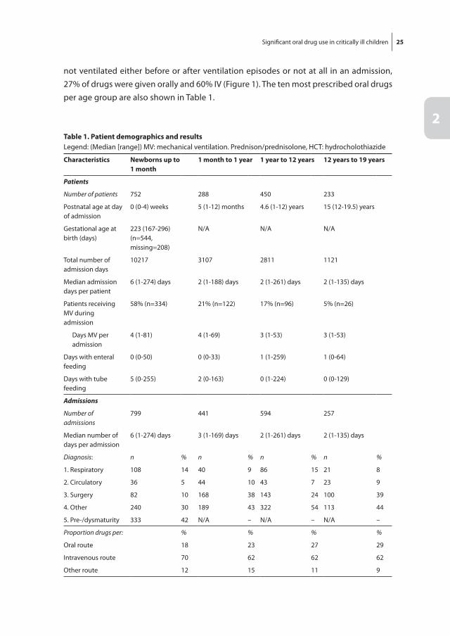
not ventilated either before or after ventilation episodes or not at all in an admission, 27% of drugs were given orally and 60% IV (Figure 1). The ten most prescribed oral drugs per age group are also shown in Table 1.
table 1. Patient demographics and resultsLegend: (Median [range]) MV: mechanical ventilation. Prednison/prednisolone, HCT: hydrocholothiazide
characteristics newborns up to 1 month
1 month to 1 year 1 year to 12 years 12 years to 19 years
Patients
Number of patients 752 288 450 233
Postnatal age at day of admission
0 (0-4) weeks 5 (1-12) months 4.6 (1-12) years 15 (12-19.5) years
Gestational age at birth (days)
223 (167-296)(n=544, missing=208)
N/A N/A N/A
Total number of admission days
10217 3107 2811 1121
Median admission days per patient
6 (1-274) days 2 (1-188) days 2 (1-261) days 2 (1-135) days
Patients receiving MV during admission
58% (n=334) 21% (n=122) 17% (n=96) 5% (n=26)
Days MV per admission
4 (1-81) 4 (1-69) 3 (1-53) 3 (1-53)
Days with enteral feeding
0 (0-50) 0 (0-33) 1 (1-259) 1 (0-64)
Days with tube feeding
5 (0-255) 2 (0-163) 0 (1-224) 0 (0-129)
Admissions
Number of admissions
799 441 594 257
Median number of days per admission
6 (1-274) days 3 (1-169) days 2 (1-261) days 2 (1-135) days
Diagnosis: n % n % n % n %
1. Respiratory 108 14 40 9 86 15 21 8
2. Circulatory 36 5 44 10 43 7 23 9
3. Surgery 82 10 168 38 143 24 100 39
4. Other 240 30 189 43 322 54 113 44
5. Pre-/dysmaturity 333 42 N/A – N/A – N/A –
Proportion drugs per: % % % %
Oral route 18 23 27 29
Intravenous route 70 62 62 62
Other route 12 15 11 9
26 Chapter 2
Discussion
In critically ill children, intravenous drug administration is preferable but for many reasons drugs are administered orally. The extent and nature of oral drug prescribing in this population is not known. Data in our level III NICU and PICU shows that 15 and 27% of drugs are given orally to ventilated and non-ventilated patients, respectively. As ventilated children are not able to take drugs by mouth and may not tolerate any oral food or drug, we also analyzed these groups separately. From a pharmacological point-of-view, this outcome is rather surprising as it shows a considerable extent of oral drug use and possible erratic drug absorption. The most often prescribed oral drugs
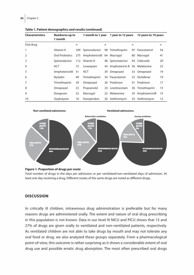
table 1. Patient demographics and results (continued)
characteristics newborns up to 1 month
1 month to 1 year 1 year to 12 years 12 years to 19 years
Oral drug n n n n
1 Vitamin K 299 Spironolacton 98 Trimethoprim 97 Paracetamol 54
2 Oral Probiotics 275 AmphotericinB 64 Macrogol 80 Macrogol 41
3 Spironolacton 112 Vitamin K 48 Spironolacton 64 Celecoxib 29
4 HCT 72 Lorazepam 44 Amphotericin B 56 Melatonine 22
5 AmphotericinB 51 HCT 39 Omeprazol 53 Omeprazol 19
6 Nystatin 49 Trimethoprim 34 Paracetamol 52 Diclofenac 19
7 Trimethoprim 29 Omeprazol 26 Prednison 41 Prednison 17
8 Omeprazol 23 Propranolol 24 Levetiracetam 36 Trimethoprim 15
9 Doxapram 22 Macrogol 22 Melatonine 35 AmphotericinB 15
10 Oxybutynin 16 Domperidon 20 Azithromycin 33 Azithromycin 12
INTRAVENOUS ROUTE60%
ORAL ROUTE27%
OTHER ROUTE13%
Non-ventilated admissions
INTRAVENOUS ROUTE61%
ORAL ROUTE27%
OTHERROUTE11%
Before/after ventilation
INTRAVENOUS ROUTE75%
ORAL ROUTE15%
OTHER ROUTE9%
During ventilation
Ventilated admissions
figure 1. Proportion of drugs per routeTotal number of drugs in the days per admission or per ventilated/non-ventilated days of admission. At least one day receiving a drug. Different routes of the same drugs are noted as different drugs.

Significant oral drug use in critically ill children 27
2
consisted of oral probiotics, amphotericin B, and macrogol, for which erratic oral absorp-tion is less relevant as its target is the gut or the gut flora. In contrast, other frequently prescribed drugs for which erratic oral absorption may affect their efficacy and safety are: analgesics, diuretics, vitamin K, omeprazole, and lorazepam. To date, data on the efficacy and safety of for example omeprazole, hydrochlorothiazide, spironolactone and lorazepam in this population are very scarce or completely lacking [3]. Moreover, no licensed liquid formulations are available. Extratemporaneous formulations are used, including IV formulations and crushed tablets. This further illustrates the risks associated with oral drug use in this setting.
Information is available about drug utilization in pediatric pharmacotherapy; most studies, however, are aimed on the statistics of the use of off-label and unlicensed drugs, but did not specify differences in label status by administration route [4-6]. Interest-ingly very little data are available on the extent of drug prescription per route. One drug utilization study in a NICU in India found 92% of prescribed drugs in 6 months were intravenously used [7]. This is far more than the 70% of IV drugs in neonates of our study, although the non-western setting may be quite different.
Maat et al. looked at the rate of pharmacy interventions after electronic prescribing in a four-year study in a tertiary children’s hospital (excluding the ICUs) [8]. Interestingly, of all medication-related characteristics, the oral dosage form and oral route of admin-istration had relatively highest risk for interventions on prescriptions done by clinical pharmacists (OR=1.63 [95%CI 1.41-1.88] and OR=1.80 [95%CI 0.38-0.67]).
The strengths of this study include the large sample size, recent data and a reflection of current daily practice. Moreover, our data display actual administered drugs to the patient instead of prescriptions that might be changed before it arrives to the patient. To our knowledge, this is the first study specifically identifying oral drug use in the neonatal and pediatric ICU. There are some limitations. First, input mistakes on route of administration are rare but cannot be excluded. Second, in our institution, PDMS recordings continue while a patient is away from the ICU during surgery and drugs used during surgery are also recorded. This may have contributed to an underestimation of the proportion of oral drug use, as in this setting oral drug use is virtually absent. Third, drug dose and frequency of administration per day were not analyzed. Finally, this single-center study was performed in a tertiary Dutch referral hospital and data may not be completely translatable to other centers.
Oral prescriptions may not be all bad, as oral administration may reduce drug admin-istration costs and IV line related infections. Physicians’ choosing the oral route should nevertheless be aware of the uncertainties related to the oral disposition of drugs in critically ill children and should weigh these uncertainties against the apparent advan-tages. Our paper presents data on oral prescription in a specific patient population; i.e., critically ill children. The uncertainties in oral drug exposure may similarly apply to other

28 Chapter 2
patient populations, including adults, where oral drug absorption may also be affected by underlying clinical conditions disease, e.g., intestinal disease, heart and liver failure, cancer and in palliative care.
In conclusion, orally administered drugs comprise up to 27% of the pharmacotherapy of patients in the ICU, possibly leaving children at risk of ineffective or unsafe drug therapy.

Significant oral drug use in critically ill children 29
2
rEfErEncEs
[1] Kearns GL, Abdel-Rahman SM, Alander SW, Blowey DL, Leeder JS, Kauffman RE. Developmental pharmacology--drug disposition, action, and therapy in infants and children. N Engl J Med 2003 Sep 18; 349(12): 1157-67.
[2] Mooij MG, de Koning BA, Huijsman ML, de Wildt SN. Ontogeny of oral drug absorption processes in children. Expert Opin Drug Metab Toxicol 2012 Oct; 8(10): 1293-303.
[3] Segar JL. Neonatal diuretic therapy: furosemide, thiazides, and spironolactone. Clin Perinatol 2012 Mar; 39(1): 209-20.
[4] Kumar P, Walker JK, Hurt KM, Bennett KM, Grosshans N, Fotis MA. Medication use in the neonatal intensive care unit: current patterns and off-label use of parenteral medications. J Pediatr 2008 Mar; 152(3): 412-5.
[5] Kieran EA, O’Callaghan N, O’Donnell CP. Unlicensed and off-label drug use in an Irish neonatal intensive care unit: a prospective cohort study. Acta Paediatr 2014 Apr; 103(4): e139-42.
[6] Hsieh EM, Hornik CP, Clark RH, Laughon MM, Benjamin DK, Jr., Smith PB, et al. Medication use in the neonatal intensive care unit. Am J Perinatol 2014 Oct; 31(9): 811-21.
[7] Chatterjee S, Mandal A, Lyle N, Mukherjee S, Singh AK. Drug utilization study in a neonatology unit of a tertiary care hospital in eastern India. Pharmacoepidemiol Drug Saf 2007 Oct; 16(10): 1141-5.
[8] Maat B, Au YS, Bollen CW, van Vught AJ, Egberts TC, Rademaker CM. Clinical pharmacy interven-tions in paediatric electronic prescriptions. Arch Dis Child 2013 Mar; 98(3): 222-7.

3 Ontogeny of oral drug absorption processes in children
Miriam G. Mooij, Barbara A.E. de Koning, Mark L. Huijsman, Saskia N. de Wildt
Expert Opinion on Drug Metabolism & Toxicology 2012,
Oct;8(10):1293-303
32 Chapter 3
AbstrAct
introduction
A large proportion of prescribed drugs to children is administered orally. Age-related change in factors affecting oral absorption can have consequences for drug dosing.
Areas covered
For each process affecting oral drug absorption, a systematic search has been performed using Medline to identify relevant articles (from inception till February 2012) in humans. This review presents the findings on age-related changes of the following processes affecting oral drug absorption: gastric pH, gastrointestinal motility, bile salts, pancreatic function, intestinal pH, intestinal drug-metabolizing enzymes and transporter proteins.
Expert opinion
Clinicians should bear in mind the ontogeny of oral drug absorption processes when prescribing oral drugs to children. The authors’ review shows large information gaps on almost all drug absorption processes. It is important that more knowledge is acquired on intestinal transit time, intestinal pH, and the ontogeny of intestinal drug-metabolizing enzymes and drug transporter proteins. Furthermore, the ultimate goal in this field should be to predict more precisely the oral disposition of drugs in children across the entire pediatric age range.

Ontogeny of oral drug absorption processes in children 33
3
introDuction
orally administered drugs in children
A large proportion of drugs prescribed to children is administered orally [1]. Absorption of orally administered drugs may be affected by extrinsic factors (food and formulation) and intrinsic factors of a physiological nature. The latter includes volume of gastroin-testinal fluids, the pH and buffer capacity of these fluids, contraction patterns, gastro-intestinal transit, digestive enzymes, intestinal cellular transporters, drug metabolism enzymes, and intestinal bacterial flora [2]. Solubility and intestinal permeability of the individual drug will influence the impact of gastrointestinal (GI) processes on its absorption. A theory-based oral drug classification based on solubility and permeability characteristics of drugs, such as the biopharmaceutics classification system (BCS), may serve to predict which extrinsic or intrinsic variables will alter oral drug absorption [2, 3].
As many of the GI processes change with age, oral drug absorption expectedly will change with age as well [4]. The current EU and US regulations aimed at stimulating the study of drugs across the pediatric age range, have given an impetus to promoting clinical trials in children [5, 6]. Age-specific information on the processes governing drug disposition in children is needed for modelling and simulation approaches. Important progress has been made to elucidate age-related changes in: hepatic drug metabolism and renal excretion [7, 8]. By contrast, our knowledge on developmental changes in the GI processes involved in oral drug absorption is far less developed [4, 9-11].
The aim of this review is to present the available data on age-related variation in GI processes that govern oral drug absorption processes. We performed a systematic search in the literature using Medline. Reference lists of relevant retrieved papers were screened for additional relevant articles. We discuss current information gaps and pro-vide suggestions for future research that may lead to develop evidence-based dosing guidelines for oral drugs in children.
Age-related changes in oral drug absorption processes
Gastric pHGastric pH is an important factor determining the stability of a drug passing through the stomach. Studies on gastric pH across the pediatric age range used pH measurement of gastric fluid aspirates and 24-h intragastric pH monitoring [12-29]. Figure 1 displays the mean and median gastric pH values in healthy children in the first three months of life.
The mean gastric pH in newborns min after delivery is 7.05, and within a few hours it declines to a pH of 2.7 [13, 28]. A less acidic stomach environment in these newborns af-ter delivery is most likely explained by swallowing of amnion fluids, which is supported by the decrease in pH within a few hours after birth [28]. More than seventy years ago,

34 Chapter 3
Miller observed a decrease in acidity over the first 10 days of life [30]. Many more recent studies report that the gastric pH declines already within a few hours after birth [13, 28]. Miller titrated gastric juice with NaOH and then determined the amount of HCl as a measure of acidity. However, Miller did not provide information on the acidity of the primary gastric content; therefore, we are unable to translate this outcome to pH. Many other studies subsequently showed that the gastric pH remains low at a pH around 2 and 3 in children of all ages [12, 14-27, 29].
More rarely, gastric pH can be described in terms of the proportion of time it peaks above 4 measured over 24 hours. In preterm infants, this proportion ranged from 46% to 70% over a 24-h period [31, 32]. In children up to 2 years of age it was around 51%; in older children it was 34% [33]. The higher proportion in younger children might be explained by the buffering effects of milk formula, older children are less frequently fed and receive more solid foods [23, 25, 33]. 24-h pH monitoring reflects the buffering ef-fect as well; in preterm infants, the gastric pH first increased to 7 postprandially, but then immediately steadily declined to a pH of 2 [23]. Another study showed a similar pattern with a mean gastric pH returning to a value of 1.8 within 180 min postprandially [25]. Apparently, during the day, younger children tend to have more often a basic gastric environment than older children, although the mean gastric pH remains around 2 or 3 in children of all ages.
figure 1. Gastric ph measured in neonates by 24-h monitoring or gastric aspirates.References correspond with references in the text.A: Gastric aspirates 3 minutes after delivery; vaginal delivery [12]B: Gastric aspirates 3 minutes after delivery; caesarean section [12]C: Less than 3 hours after delivery [13]C, D, F, G: Continuous 24-h gastric pH monitoring [13, 18, 19, 25]A, B, E, H: Gastric aspirates obtained by gentle suction [12, 12, 27, 26]

Ontogeny of oral drug absorption processes in children 35
3
Interestingly, this overview gives reason to contradict the widespread notion that absorption of gastric pH-dependent drugs in both neonates and young infants is af-fected by high gastric pH [34, 35]. Gastric pH may be high due to continuous enteral feeds, but is comparable to adult values when oral feedings are given at longer than 3 hour intervals. Children older than one week of age will typically receive such intermit-tent feeding, especially during the night. We have not been able to identify studies that compared effect of different feeding regimes (continuous and intermittent) on drug absorption in neonates.
The consequences of changes in gastric pH are relevant for acid-labile drugs. These may be absorbed more efficiently in a higher gastric pH environment achieved by very frequent or continuous feeding regimens. Huang et al. found that serum penicillin levels in premature and term newborns were higher than those in infants and children. These age-related changes were hypothesized to be either due to higher gastric pH in the first 10 days of life or to altered renal function [36].
Gastrointestinal motility
Gastric emptyingNext to intestinal motility, gastric emptying is a primary determinant of the rate at which drugs are presented to the small intestinal mucosa for absorption. Gastric emptying is usually measured by the following methods: gastric emptying breath test, scintigraphic procedure by Technetium-99M liquid gastric emptying scan or the paracetamol absorp-tion test. Gastric emptying time is reported in various ways: gastric emptying time, gastric half-emptying time or residual gastric activity at 1 h.
We identified three studies using the gastric emptying breath tests. Hoekstra et al. tested the effect of glucose and fructose on gastric emptying using an L-glycine-1-13C breath test in four healthy children (mean age 14.3 years; age range 12.1-16.0 years). Gastric half-emptying time after fructose intake was 45.5 min (SD 4.9); after glucose intake 64.3 min (SD 2.4). Gastric half-emptying time was significantly longer when fruc-tose and glucose were administered together, that is, 85.3 min (SD 7.0) [37]. The authors considered all values to be in the normal ranges established with other methods. Using the 13C-octanoic acid breath test, Perri et al. found a gastric half-emptying time of mean 121 min (SD 42) in nine healthy control patients (mean age 9 years; age range 4-16 years). These children had eaten a standard test meal (bread, ham, juice, egg), however, which hardly compares with a smaller fructose and/or glucose administration [38]. Hoff-man et al. subjected 22 patients with gastroesophageal reflux symptoms (mean age 13.2 years) to the 13C-octanoic acid breath test and compared half-emptying results between patients with or without pathologic acid exposure (84 (SD 24) vs 86 (SD 26) min) and with or without duodenogastroesophageal reflux (105 (SD 47) vs 76 (SD 24) min) were
36 Chapter 3
compared. Results were not statistically significantly different [39]. In conclusion, breath test measurements for gastric emptying rate are highly variable and, probably for practi-cal reasons, that tests have only been performed in relatively older children.
Scintigraphic imaging makes it possible to measure the gastric emptying time or gastric half-emptying time as well as the residual gastric activity at 1 h. We identified seven useful scintigraphy studies. In 10 preterm infants (median gestational age 28.9 weeks; range 26-33), the median gastric half-emptying time at a postnatal median age of 9 days (range 6-37) was 60 min (30-180 min) [40]. Patients were all hourly fed although not receiving a standard meal size. The residual gastric activity at 1 h was 37.5% (range 19-100%) [40]. Di Lorenzo and colleagues conducted a study in 477 patients across a wide age span; 291 patients with and 186 patients without gastroesophageal reflux dis-ease (GERD) (based on pH and/or scintigraphy investigations) [41]. In children without bolus or acidic gastroesophageal reflux, gastric residual activity at 1 h was around 65% in those up to 3 years of age; it decreased to 51% in the age group 4-6 years; and to 45% in children over 6 years of age [41]. Seibert et al. reported an opposite outcome in children being evaluated for gastroesophageal reflux (GER): the percentage emptied at 1 h instead of the percentage residual activity [42]. The percentage emptied at 1 h was 48% (SD 16) in 44 infants (mean age 5.7 months, range 1-23) and 51% (SD 7) in 8 children (mean age 9.1 years, range 2-14.5 years). When converted to residual gastric activity, values are still comparable (respectively 52% and 49%). Note that in the study reported by Di Lorenzo et al., a delay in gastric emptying was not related to GER symptoms until the age of 6 years [41]. Cucchiara et al. studied a poorly described control group suffer-ing from diarrhoea and failure to thrive not related to gastrointestinal symptoms. The gastric emptying activity at 1 h was 38.1% (SD 6.5) [43]. Miele et al. reported a 43.3% (SD 8.7) gastric emptying activity at 1 h in a control group of 11 children without gastroin-testinal or neurologic disorders (mean age 5.6 years; SD 3.9 years; range 2-12 years) [44]. Describing gastric emptying alternatively as a mean emptying half time, Yahav et al., reported 87.8 min (SD 22.9) mean gastric emptying time in a control group with a mean age of 10.4 months for which no other details are reported [45]. Demirbilek et al. found an average gastric emptying time of 51.6 min (SD 8.04) in a selected group of children with GERD (mean age 3.2 years; SD 1.1); the selection might have resulted in bias [46].
To relate these results to adults, reported healthy adult gastric emptying times range between 56 (32-85) and 104 (49-126) min, for liquid and solid markers respectively [47, 48].
Finally, the paracetamol absorption test was used in two small cohorts. In 15 critically ill patients (median age 5.3 years; interquartile range 1.2-6.5) who were food tolerant, it revealed a median 1.5 (interquartile range 0.7-2.2) ratio of time to reach paracetamol peak to the maximum paracetamol concentration (Tmax/Cmax) [49]. In seven adoles-cents (mean age 16.4 years; SD 0.7 years; range 15.5-17.5), it revealed a paracetamol
Ontogeny of oral drug absorption processes in children 37
3
absorption ratio of 1.4 for high-fat meals and 0.5 for low-fat meals [50]. The evidence of these two studies is too limited to conclude on age-related changes.
However, population pharmacokinetic analysis applied in another study yielded a significantly lower oral paracetamol absorption rate in the first days of life before stabilizing after 1 week [51]. The lag time reflects the time to reach and permeate the absorbing surface of the intestine [2]. Considering that a lag time was observed after oral paracetamol administration only and not after rectal administration, it suggests that gastric emptying may be the primary determinant of a lag time for oral absorption of paracetamol.
Antroduodenal contractionsAntroduodenal motor activity plays a role in the gastric emptying next to fundic con-traction, pyloric sphincter relaxation and intestinal motor activity. It can be determined by antroduodenal manometry, which measures intraluminal pressures of the distal stomach and the proximal small bowel.
Fasting antral motor activity and antral motor activity in response to intraduodenal feeding did not significantly differ between term and preterm infants [52]. By contrast, the proportion of antral clusters temporally associated with duodenal activity in pre-term infants was significantly lower than that in term infants. Moreover, the degree of association of antral and duodenal activity increased significantly with gestational age [53]. In preterm infants 29 to 32 weeks of gestational age, the frequency of contractions, the number of contractions per burst and the intraluminal peak pressure of duodenal motility during contractions all increased with postgestational age, resulting in a more efficient motility [54]. Similarly Bisset et al. reported that both the magnitude and or-ganization of motor activity increased with increasing gestational age [55]. Berseth et al. reported shorter lasting individual duodenal cluster activity during fasting periods in preterm than in term infants, but duodenal motor activity in response to feeding in-creased similarly in both groups [52]. The timing of introducing food seems to influence the preterm neonates’ (28-32 weeks of gestational age) duodenal motor activity; intro-ducing formula early (day 3-5 postnatally) resulted in more mature motor complexes than introducing formula late (day 10-14 postnatally) [56]. Preterm infants showed more immature duodenal motor activity response to bolus feeding then did term infants [57].
In conclusion, proximal intestinal (duodenal) motor activity in contrast to antral motor activity matures throughout the first weeks of life, with increasing frequency, amplitude, and duration of propagating contractions. Regrettable, there are no such studies in healthy children beyond the newborn period.
38 Chapter 3
Intestinal transit timeOverall, gastrointestinal motility can be expressed as orocecal transit time (OCTT). This can be measured by different techniques: hydrogen breath test, 13C Ureide breath test, radiotransmitting capsule, red carmine marker test or scintigraphy. Most common is the hydrogen breath test with lactulose as nonabsorbable carbohydrate substrate. This breath test has limited use in the general population, which may include hydrogen-non-responders. Also lactulose may accelerate transit time by its osmotic laxative effect. Accordingly, Vajro et al. reported in 11 control patients that the mean OCTT after a meal was significantly longer than that after lactulose [58]. Although this method can be used to compare groups in standardized studies, it is merely an approach to the physiological situation of intestinal motility.
We identified four studies using the hydrogen breath test to measure OCTT in differ-ent pediatric age groups [58-62]. The populations were quite heterogeneous, but there does not appear to be an age-related difference in OCTT. In the whole age range from 1 to 17 years, the mean OCTT was roughly between 60 and 110 min, as in adults [62]. The mean OCTT measured by the lactose-13C-ureide breath test was 255 min (range 165-390) in children from 3 to 17 years of age [59]. This method cannot be used in infants below 6 months of age as they lack the intestinal bacterial enzymatic activity. In adults, the latter test was validated in respect to scintigraphy [63]. The lactulose-H2 breath test yielded a significant shorter OCTT than did the labeled ureide test, which may be due to the effect of lactulose [64]. Fallingborg et al. distinguished small intestinal and colonic transit times with the use of a radiotransmitting capsule in a small population of 12 healthy children (8 to 14 years) [65]. Small intestinal transit time was 7.5 h and colonic transit time was 17.2 h. Interestingly, from the number of observations in each segment they estimated that the capsule resided in the duodenum for 8% of the small intestinal passage, in the proximal part of the small intestines for 5%, in the mid part for 12% and in the distal part for 75%. The small intestinal transit time of 7.1 h is considerably longer than that established by the breath tests. The fact that the capsule, which was larger than 2 mm, was located in the distal part of the terminal ileum for 75% of the small intestinal transit time suggests a longer ileo-cecal transit for large particles. By means of scintigraphy, Bodé et al. measured a mean OCTT of 3.1 h (range 1.3-6.1 h) in nine premature infants (mean gestational age 28.9 weeks) [40].
Bile acidsBile is a complex secretory product produced by the liver. It eliminates waste products from the body and it promotes digestion and absorption of lipids by the intestines. In preterm neonates, the concentration of the bile acids was found to be 4.55 mmol/l in the first few weeks postnatally [66]. In 65 healthy preterm newborns, the total bile acid concentration was consistently higher in those fed with human milk in comparison with

Ontogeny of oral drug absorption processes in children 39
3
those fed with formula. Concentrations did not significantly increase over a 3-week follow-up period [67]. Concentration did not differ between small- and appropriate-for-gestational-age premature infants [66]. Challacombe et al. compared three age groups, that is, 2 days postnatal (n=12), 2 to 7 days (n=8), and 10 days to 7 months (n=14). Gestational ages were not documented. The total bile acid concentration in the oldest group was much higher than that in both other groups and at a value comparable to those in adults [68].
Changes in biliary function can influence solubilization and consequently absorption of lipophilic drugs [3].
Pancreatic functionThe exocrine pancreas is a specialized secretory gland, which secretes juice rich in HCO3- and digestive enzymes that neutralizes the acidic gastric contents and helps digest food. Functioning of the exocrine pancreas is typically measured by the fecal elastase-1 (E-1) concentration. The E-1 enzyme is highly specific for the pancreas and is not degraded during the intestinal passage. Age-related differences in E-1 concentrations were absent in a large cohort of healthy subjects (mean age 11.2 years (SD 0.5); age range 2 months to 52 years) [69]. Even as many as 96.8% of preterm and term infants up to the age of 12 months without known bowel or pancreatic disorders had adult E-1 values after 2 weeks of life, independent of gestational age [70]. However, up to 48 hours after birth, none of the preterm infants had a fecal E-1 concentration of greater than 30 µg/g meconium, whereas 43% of the term infants had normal adult values. This discrepancy may be due to either immaturity or insufficiency of the exocrine pancreatic function in premature neonates. However, the small sample size did not allow differentiating between these two possible causes. Deficient exocrine pancreas function as seen in cystic fibrosis patients was associated with lower oral bioavailability of mycophenolate mofetil [71]. This suggests an effect on oral drug absorption in neonates with immature pancreas function, but this has not been studied to date to our knowledge.
intestinal ph
In comparison with gastric pH, remarkably little is known about the intestinal pH in children. Fallingborg et al. measured gastrointestinal pH with a radiotransmitting pH-sensitive capsule in 12 healthy children aged 8-14 years. The mean value of pH rose from 1.5 in the stomach to 6.4 in the duodenum; in the distal part of the small intestine, it reached an alkaline peak value of 7.4. The pH profile was almost identical to that in healthy adults. A broad conclusion on the development of the intestinal pH cannot be drawn as his small population consisted merely of older children. It would be worthwhile to repeat the experiment in other age groups [65].
40 Chapter 3
Intestinal drug metabolismMany developmental changes in hepatic drug metabolism and renal clearance have been well documented. Data on the ontogeny of intestinal metabolism remain scarce. What is known is that enzymes of the cytochrome P450, especially the 3A (CYP3A) sub-family, are abundant in liver and gut and contribute to the first-pass metabolism of many orally administered drugs in adults [72]. Hepatic CYP3A forms present a developmental expression in fetal and pediatric samples; CYP3A4 and CYP3A7 expression levels show to be age dependent with respectively increasing and decreasing levels of total CYP3A expression levels [73].
CYP3A ontogeny can be reported as changes in mRNA, protein or activity levels. We identified two in vitro studies on CYP3A ontogeny in the intestine. One studied 59 his-tologically normal duodenal biopsies from children aged 1 month to 17 years for CYP3A mRNA by quantification and CYP3A proteins localization by immunohistochemistry [74]. The other studied duodenal biopsies and surgical sections from 104 children aged 2 weeks to 17 years and 11 fetuses for CYP3A protein expression by immunohistochem-istry and activity by the formation of 6beta-hydroxytestosterone from testosterone [75]. CYP3A4 and CYP3A5 mRNA expression levels were to decrease with age, showing expression levels were high in the first year of life and decreased thereafter [74]. This is in contrast with protein expression levels reported in the second study showing CYP3A protein expression significantly increased with age [75]. The discrepancy of decreasing mRNA expression and increasing protein levels with age might reflect a posttranscrip-tional regulatory mechanism that is not elucidated to date according to the authors [74]. Dissociation between protein and mRNA levels during the maturation process was already reported for CYP2D6 liver enzymes [76]. The location of the CYP3A protein in enterocytes assumes a maturation profile occurs. In the duodenal biopsies of children less than 6 months of age, CYP3A protein was detected in only 50% of the enterocytes; in the older children, however, CYP3A protein was expressed in all cells [74]. Moreover, the increase in CYP3A protein levels with age is mirrored with increasing CYP3A4 activity. It changes from undetectable in fetal samples, low in neonates and adult levels in children older than 5 years of age, as reflected by 6beta-hydroxytestosterone formation [75].
Intestinal CYP3A4, CYP3A5 mRNA levels have been established in pediatric liver recipi-ents (age 0.1-15 years) at the time of transplant surgery [77]. Unfortunately, the authors did not study the effect of age within their cohort. Adult data show similar CYP3A4 and CYP3A5 expression levels [78]. This suggests that intestinal CYP3A expression does not change beyond childhood. However, because the range of levels reported in children was very wide, age-related changes from 0.1 year of age onwards cannot be excluded [78]. Intestinal CYP3A5 mRNA levels were significantly higher in CYP3A5*1 gene carriers (expressors) than CYP3A5*3 homozygous patients (non-expressors) and observed in

Ontogeny of oral drug absorption processes in children 41
3
both the children and adult study. In CYP3A5*1 carriers, CYP3A5 mRNA accounted for 20-30% of all CYP3A mRNA detected [77, 78].
In vivo studies on oral bioavailability of CYP3A substrates in relation to age are scarce. Our own research showed that median midazolam oral bioavailability in preterm infants (28-32 weeks, <10 days of age) is significantly higher than in adults (50 vs. 30%) [79-81]. This probably reflects developmentally low intestinal and hepatic CYP3A activity, as midazolam is a validated probe drug for CYP3A4/5 activity.
Interestingly, the type of feeding (breast milk or formula) seems to impact the devel-opmental pattern of combined intestinal and hepatic CYP3A in neonates. In children who received oral dextromethorphan six times between two weeks and 6 months of age, the urinary metabolite/dextromethorphan ratio as a measure of CYP3A4 activity clearly increased over this period. Moreover, this increase was faster for formula- than breastmilk-fed children [82]. This finding suggests a differential effect of components of these milk formulations on the induction of intestinal and hepatic CYP3A activity in the first months of life.
The ontogeny of other drug metabolizing enzymes in the intestine remains to be elucidated.
Intestinal drug transporterMultidrug resistance protein 1 (MDR1/P-glycoprotein) is a plasma membrane glyco-protein acting as an efflux system. Based on in vitro studies, it is currently considered the most prominent gut transporter [83]. It is located in many tissues and specifically within the brush border in the small intestine. Its expression is genetically controlled by the ABCB1 gene [84]. MDR1 action in the enterocyte reduces the bioavailability of orally administered drugs as these are expelled into the intestinal lumen. MDR1 protein can be localized by immunohistochemistry and mRNA quantification in intestinal tissue. In the earlier mentioned study evaluating 59 duodenal biopsies of children aged from 1 month to 17 year, MDR1 mRNA expression was highly variable and not related to age [74]. MDR1 protein was detected in all the enterocytes and was located on the apical surface. In the biopsies in children younger than 3 years, additional staining was located on a limited upper part of the lateral surface.
A possible age effect in relation to of the ABCB1 genotype was found for oral bioavail-ability of the MDR1 substrate cyclosporine. One hundred and four children with renal disease (age 0.36-16.3 years) were grouped by age and genotyped for ABCB1 gene. The pre-hepatic extraction ratio of cyclosporine was ABCB1 genotype dependent only in children older than 8 years, resulting in corresponding differences in oral bioavailability. No such association was found in younger patients, which suggests an interaction of age and genotype on MDR1 activity [85].

42 Chapter 3
In the context of the previously mentioned study in pediatric liver recipients, MDR1 mRNA was determined as well and results were similar as for CYP3A; that is, the median MDR1 mRNA expression did not differ between children and adults, but widely ranged in the pediatric population [77, 78].
Interestingly, in noninflamed duodenal biopsies of children with Crohn’s disease, MDR1 mRNA expression was significantly higher than that in normal biopsies. Expression of MDR1 was highly variable in both groups [86]. The effect of age was not examined in this study. The higher levels of MDR1 expression could have been induced by systemic inflammation present in Crohn’s disease, which is likely to lead to an elevated first-pass metabolism of xenobiotics used in the treatment.
To our knowledge, the intestinal ontogeny of other members of the ATP-binding cassette transporters, such as multidrug resistance protein 2 (MRP2/ABCC2) or breast cancer resistance protein (BCRP/ABCG2), has not been studied to date [83, 87-89].
conclusion
This literature review makes clear that GI processes that govern drug absorption change from the neonatal period up to adulthood. Consequently, these changes could have an impact on drug absorption depending on the drug characteristics [3, 4]. The review also brought to light important knowledge gaps regarding these processes and especially their impact on drug absorption.
Key findings in the research done so far are the following. Apart from a brief peak postnatally, the gastric pH is about 2-3 in children of all ages. Postprandial, its rise is due to the buffering effect of milk-based feeding. Especially in frequently fed neonates, the pH may, therefore, be higher for a longer period during a 24-h period, than in older chil-dren who eat less frequently. Gastric emptying time reported in the literature is highly variable. Standard gastric emptying tests do not reveal evident age-related changes. Population pharmacokinetic analysis shows a markedly paracetamol absorption de-crease in the first few days of life, which suggests delayed gastric emptying. This delay could perhaps be explained by maturation of antroduodenal contractions.
There are no studies done examining antroduodenal contractions beyond the neona-tal period. Intestinal transit time (in terms of mean OCTT) does not appear to be subject to age-related changes; it is roughly between 60 and 110 min in the age range of 1-17 years, as measured by hydrogen breath test. The one study that used a capsule to mea-sure OCTT showed a much longer transit time than any of the other studies using the lactulose breath test. This latter test probably rather measures intra-individual changes in OCTT or differences between cohorts. A developmental change in biliary function ap-pears to be present, with bile acids concentration reaching adult values around the age

Ontogeny of oral drug absorption processes in children 43
3
of 4 years. Pancreatic function appears to be sufficient in the large majority of healthy newborns, independent of gestational age. Intestinal pH has only been studied in a cohort of older children. Adult values were found in this cohort; therefore, possible age-related changes remain to be elucidated. For a large proportion of drugs, there seems to be a developmental pattern in CYP3A, which is the most important drug-metabolizing enzyme in the intestines. CYP3A protein and activity levels were found to increase with age. These in vitro data are in line with higher oral bioavailability of midazolam in prema-ture children compared with adults. Evidence on possible age-related effect on MDR1 activity is contradictory and not elusive yet.
ExPErt oPinion
information gaps
In summary, the main information gaps on the ontogeny of GI processes governing oral drug absorption have not yet been bridged. We need more knowledge on intestinal transit time, intestinal pH and the ontogeny of intestinal drug-metabolizing enzymes and drug transporter proteins. The ultimate goal of research efforts in this field should be to predict more precisely the oral disposition of drugs in children across the pediatric age range. Below we describe some research approaches, both in vitro and in vivo, which are promising for future research to provide a better understanding of oral drug absorp-tion in children.
in vitro drug dissolution/solubility model (tiM)
The Dutch Institute of Innovative Research has developed the TNO Gastro-Intestinal Tract model (TIM), a computer-controlled dynamic system that mimics the physiological hu-man conditions in stomach and intestines [90, 91]. Parameters such as pH, temperature, peristaltic movements, transit time, secretion of digestion enzymes, bile and pancreatic juices can be adjusted. Intraluminal processing of drug dosage forms, including transit, release and dissolution, can be simulated [92]. Removal of dissolved drug molecules from the intestinal compartments allows assessing the fraction of drug potentially avail-able for small intestinal absorption [91]. This model has been extensively validated to simulate these processes in adults. It appears an interesting approach to test oral drug absorption in conditions with typical age-related physiological characteristics. Espe-cially, the additional impact of existing oral formulations frequently given to children can be studied. It may also help to study the effect of drug manipulations to enhance drug ingestion by children (e.g., dissolving tablets in apple juice, apple sauce, ‘hiding’ in regular food, crushing). Representative drugs of the different BCS classes can also be studied systematically for dissolution and solubility.
44 Chapter 3
in vitro drug metabolism and transporter studies
The extensive studies on in vitro hepatic drug metabolism, for example, by the group of Hines and colleagues could serve as an example [93]. Similarly, the ontogeny of drug-metabolizing enzymes and transporters should be studied in intestinal samples from the different parts of the intestine and from children across the pediatric age span. New methods are quickly becoming available, to study not only drug transporter expression (mRNA) but also protein content, using sensitive LC-MS-MS methods.
Modeling and simulation: Pb-PK models and population PK
The available data on age-related changes in relevant GI processes as well as possibly those from the TIM simulations can be incorporated in population-based pharma-cokinetics (PB-PK) software programs such as Simcyp®, PKsim® or GastroPlus®. These programs can then simulate fate of drugs given to children of different ages and provide guidance for age-appropriate dosing.
At this time, the usefulness of these programs is still hampered by the relative lack of physiological data across all age groups. Moreover, validation of the model is also still limited as we have little pharmacokinetic data to validate the model; especially in the neonatal and infant age groups, data are scarce [94]. It is to be expected that increasing use of these programs will generate sufficient data to further validate the models.
Mechanism-based approach for in vivo studies
Another approach to learn more about the ontogeny of specific (intestinal) drug-metabolizing enzyme and/or transporter pathways is a mechanism-based one [95]. The pharmacokinetics of drugs that represent a single pathway, studied in children of all ages, may provide valuable information on the ontogeny of that specific pathway. For example, determination of the plasma clearance of midazolam is a validated and widely used method to study interindividual variation in CYP3A activity in both adults and children [96].
To elucidate age-related changes in intestinal enzymes/transporters, independent of hepatic activity, we will need both oral and intravenous pharmacokinetic data, prefer-ably from the same patients. At this time, these data are scarce in children, even for CYP3A/midazolam. Full PK studies to determine bioavailability for a probe drug using a multi-day cross-over design are hardly feasible in children for ethical and practical reasons. As a major reason, children will not benefit from the drug but rather will experi-ence the drug effect and risk adverse events and have significant burden. Alternatively, a stable-labeled isotope or a (very weak) radioactive-labeled microdose can be used [97, 98]. In both, a labeled probe drug is added to an intravenous therapeutic dose. Parent compound and metabolites can, therefore, be traced in serum and urine. This enables simultaneous determination of the pharmacokinetics of therapeutic IV and the labeled
Ontogeny of oral drug absorption processes in children 45
3
oral dose. It eliminates the risk of therapeutic effect/toxicity as the child already receives the drug for clinical reasons. A prerequisite for the use of microdosing in this context is that dose linearity exists across the dosing range. For a number of drugs, dose linearity for microdosing has been established, whereas others clearly do not qualify [99, 100].
Microdosing is a relatively novel technique used in adults. The microdose (one-hundredth of the predicted pharmacologic dose or 100 µg) contains a natural occurring radioactive carbon label (carbon 14, 14C), which can be detected with highly sensitive methods as accelerator mass spectometry (AMS) [99]. Developmental changes in intesti-nal drug-metabolizing enzymes can be delineated by investigating multiple age groups. Microdosing has been used once in preterm infants in a small pharmacokinetic study of ursodiol in the US [101].
46 Chapter 3
rEfErEncEs
[1] Schirm E, Tobi H, de Vries TW, Choonara I, De Jong-van den Berg LT. Lack of appropriate formula-tions of medicines for children in the community. Acta Paediatr 2003 Dec; 92(12): 1486-9.
[2] Atkinson AJ, Abernethy DR, Daniels CE, Dedrick RL, Martkey SP. Drug Absorption and Bioavail-ability. Principles of Clinical Pharmacology. 2nd ed: Academic Press; 2007. p. 37-58.
[3] Martinez MN, Amidon GL. A mechanistic approach to understanding the factors affecting drug absorption: a review of fundamentals. J Clin Pharmacol 2002 Jun; 42(6): 620-43.
[4] Kearns GL, Abdel-Rahman SM, Alander SW, Blowey DL, Leeder JS, Kauffman RE. Developmental pharmacology--drug disposition, action, and therapy in infants and children. N Engl J Med 2003 Sep 18; 349(12): 1157-67.
[5] Jacqz-Aigrain E. Drug policy in Europe Research and funding in neonates: current challenges, future perspectives, new opportunities. Early Hum Dev 2011 Mar; 87 Suppl 1: S27-30.
[6] FDA. U.S. Food and Drug Administration. [cited; Available from: http://www.fda.gov/ScienceRe-search/SpecialTopics/PediatricTherapeuticsResearch/default.htm
[7] Hines RN, McCarver DG. The ontogeny of human drug-metabolizing enzymes: phase I oxidative enzymes. J Pharmacol Exp Ther 2002 Feb; 300(2): 355-60.
[8] Smits A, Kulo A, de Hoon JN, Allegaert K. Pharmacokinetics of Drugs in Neonates: Pattern Recog-nition Beyond Compound Specific Observations. Curr Pharm Des 2012 Feb 27.
[9] Strolin Benedetti M, Whomsley R, Baltes EL. Differences in absorption, distribution, metabolism and excretion of xenobiotics between the paediatric and adult populations. Expert Opin Drug Metab Toxicol 2005 Oct; 1(3): 447-71.
[10] Yokoi T. Essentials for starting a pediatric clinical study (1): Pharmacokinetics in children. J Toxicol Sci 2009; 34 Suppl 2: SP307-12.
[11] Bowles A, Keane J, Ernest T, Clapham D, Tuleu C. Specific aspects of gastro-intestinal transit in children for drug delivery design. Int J Pharm 2010 Aug 16; 395(1-2): 37-43.
[12] Cote CJ, Goudsouzian NG, Liu LM, Dedrick DF, Szyfelbein SK. Assessment of risk factors related to the acid aspiration syndrome in pediatric patients-gastric ph and residual volume. Anesthesiol-ogy 1982 Jan; 56(1): 70-2.
[13] Datta S, Houle GL, Fox GS. Concentration of lidocaine hydrochloride in newborn gastric fluid after elective caesarean section and vaginal delivery with epidural analgesia. Can Anaesth Soc J 1975 Jan; 22(1): 79-83.
[14] Goresky GV, Finley GA, Bissonnette B, Shaffer EA. Efficacy, duration, and absorption of a paediatric oral liquid preparation of ranitidine hydrochloride. Can J Anaesth 1992 Oct; 39(8): 791-8.
[15] Jahr JS, Burckart G, Smith SS, Shapiro J, Cook DR. Effects of famotidine on gastric pH and residual volume in pediatric surgery. Acta Anaesthesiol Scand 1991 Jul; 35(5): 457-60.
[16] Kelly EJ, Chatfield SL, Brownlee KG, Ng PC, Newell SJ, Dear PR, et al. The effect of intravenous ranitidine on the intragastric pH of preterm infants receiving dexamethasone. Arch Dis Child 1993 Jul; 69(1 Spec No): 37-9.
[17] Kelly EJ, Newell SJ, Brownlee KG, Primrose JN, Dear PR. Gastric acid secretion in preterm infants. Early Hum Dev 1993 Dec 31; 35(3): 215-20.
[18] Kraus G, Krishna DR, Chmelarsch D, Schmid M, Klotz U. Famotidine. Pharmacokinetic properties and suppression of acid secretion in paediatric patients following cardiac surgery. Clin Pharma-cokinet 1990 Jan; 18(1): 77-81.

Ontogeny of oral drug absorption processes in children 47
3
[19] Maekawa N, Mikawa K, Yaku H, Nishina K, Obara H. Effects of 2-, 4- and 12-hour fasting intervals on preoperative gastric fluid pH and volume, and plasma glucose and lipid homeostasis in children. Acta Anaesthesiol Scand 1993 Nov; 37(8): 783-7.
[20] Miller BR, Tharp JA, Issacs WB. Gastric residual volume in infants and children following a 3-hour fast. J Clin Anesth 1990 Sep-Oct; 2(5): 301-5.
[21] Nishina K, Mikawa K, Maekawa N, Tamada M, Obara H. Omeprazole reduces preoperative gastric fluid acidity and volume in children. Can J Anaesth 1994 Oct; 41(10): 925-9.
[22] Oderda G, Rapa A, Chiorboli E, Ronchi B, Zavallone A, Strigini L. Measurement of postprandial changes in urine acid output to detect changes of gastric acid secretion after proton pump in-hibitors in children. Dig Dis Sci 2002 Aug; 47(8): 1843-9.
[23] Omari TI, Davidson GP. Multipoint measurement of intragastric pH in healthy preterm infants. Arch Dis Child Fetal Neonatal Ed 2003 Nov; 88(6): F517-20.
[24] Rogers IM, Drainer IK, Moore MR, Buchanan KD. Plasma gastrin in congenitial hypertrophic pyloric stenosis. A hypothesis disproved. Arch Dis Child 1975 Jun; 50(6): 467-71.
[25] Smith LJ, Kaminsky S, D’Souza SW. Neonatal fat digestion and lingual lipase. Acta Paediatr Scand 1986 Nov; 75(6): 913-8.
[26] Splinter WM, Schaefer JD. Unlimited clear fluid ingestion two hours before surgery in children does not affect volume or pH of stomach contents. Anaesth Intensive Care 1990 Nov; 18(4): 522-6.
[27] Splinter WM, Stewart JA, Muir JG. Large volumes of apple juice preoperatively do not affect gastric pH and volume in children. Can J Anaesth 1990 Jan; 37(1): 36-9.
[28] Whetstine LJ, Hulsey TC, Annibale DJ, Pittard WB. Supplemental oxygen and gastric pH in unfed preterm infants. South Med J 1995 Apr; 88(4): 458-61.
[29] Yildiz F, Tryba M, Kuehn K, Hausdoerfer J. Reduction of gastric acid secretion. The efficacy of pre-anaesthetic oral cimetidine in children. Anaesthesia 1984 Apr; 39(4): 314-8.
[30] Miller RA. Observations on the gastric acidity during the first month of life. Arch Dis Child 1941 Mar; 16(85): 22-30.
[31] Lopez-Alonso M, Moya MJ, Cabo JA, Ribas J, del Carmen Macias M, Silny J, et al. Twenty-four-hour esophageal impedance-pH monitoring in healthy preterm neonates: rate and characteristics of acid, weakly acidic, and weakly alkaline gastroesophageal reflux. Pediatrics 2006 Aug; 118(2): e299-308.
[32] Omari TI, Haslam RR, Lundborg P, Davidson GP. Effect of omeprazole on acid gastroesophageal reflux and gastric acidity in preterm infants with pathological acid reflux. J Pediatr Gastroenterol Nutr 2007 Jan; 44(1): 41-4.
[33] Demir H, Ozen H, Kocak N, Saltik-Temizel IN, Gurakan F. Does simultaneous gastric and esopha-geal pH monitoring increase the diagnosis of gastroesophageal reflux disease? Turk J Pediatr 2005 Jan-Mar; 47(1): 14-6.
[34] Kleinman RE, Goulet OJ, Mieli-Vergani G, Sanderson IR, Sherman PM, Shneider BL. Walker’s Pedi-atric Gastrointestinal Disease. Shelton, CT: People’s Medical Publishing House; 2008.
[35] Boyle JT. Acid secretion from birth to adulthood. J Pediatr Gastroenterol Nutr 2003 Nov-Dec; 37 Suppl 1: S12-6.
[36] Huang NN, High RH. Comparison of serum levels following the administration of oral and paren-teral preparations of penicillin to infants and children of various age groups. J Pediatr 1953 Jun; 42(6): 657-8.
[37] Hoekstra JH, van den Aker JH, Kneepkens CM, Stellaard F, Geypens B, Ghoos YF. Evaluation of 13CO2 breath tests for the detection of fructose malabsorption. J Lab Clin Med 1996 Mar; 127(3): 303-9.
48 Chapter 3
[38] Perri F, Pastore M, Zicolella A, Annese V, Quitadamo M, Andriulli A. Gastric emptying of solids is delayed in celiac disease and normalizes after gluten withdrawal. Acta Paediatr 2000 Aug; 89(8): 921-5.
[39] Hoffman I, Tertychnyy A, Ectors N, De Greef T, Haesendonck N, Tack J. Duodenogastro-esophageal reflux in children with refractory gastro-esophageal reflux disease. J Pediatr 2007 Sep; 151(3): 307-11.
[40] Bode S, Dreyer M, Greisen G. Gastric emptying and small intestinal transit time in preterm infants: a scintigraphic method. J Pediatr Gastroenterol Nutr 2004 Oct; 39(4): 378-82.
[41] Di Lorenzo C, Piepsz A, Ham H, Cadranel S. Gastric emptying with gastro-oesophageal reflux. Arch Dis Child 1987 May; 62(5): 449-53.
[42] Seibert JJ, Byrne WJ, Euler AR. Gastric emptying in children: unusual patterns detected by scintig-raphy. AJR Am J Roentgenol 1983 Jul; 141(1): 49-51.
[43] Cucchiara S, Bortolotti M, Colombo C, Boccieri A, De Stefano M, Vitiello G, et al. Abnormalities of gastrointestinal motility in children with nonulcer dyspepsia and in children with gastroesopha-geal reflux disease. Dig Dis Sci 1991 Aug; 36(8): 1066-73.
[44] Miele E, Tozzi A, Staiano A, Toraldo C, Esposito C, Clouse RE. Persistence of abnormal gastroin-testinal motility after operation for Hirschsprung’s disease. Am J Gastroenterol 2000 May; 95(5): 1226-30.
[45] Yahav J, Avigad S, Frand M, Shem-Tov A, Barzilay Z, Linn S, et al. Assessment of intestinal and cardiorespiratory function in children with congenital heart disease on high-caloric formulas. J Pediatr Gastroenterol Nutr 1985 Oct; 4(5): 778-85.
[46] Demirbilek S, Karaman A, Gurunluoglu K, Akin M, Tas E, Aksoy RT, et al. Delayed gastric emptying in gastroesophageal reflux disease: the role of malrotation. Pediatr Surg Int 2005 Jun; 21(6): 423-7.
[47] Graff J, Brinch K, Madsen JL. Simplified scintigraphic methods for measuring gastrointestinal transit times. Clin Physiol 2000 Jul; 20(4): 262-6.
[48] Bennink R, Peeters M, Van den Maegdenbergh V, Geypens B, Rutgeerts P, De Roo M, et al. Evalu-ation of small-bowel transit for solid and liquid test meal in healthy men and women. Eur J Nucl Med 1999 Dec; 26(12): 1560-6.
[49] Mayer AP, Durward A, Turner C, Skellett S, Dalton N, Tibby SM, et al. Amylin is associated with delayed gastric emptying in critically ill children. Intensive Care Med 2002 Mar; 28(3): 336-40.
[50] Lodefalk M, Aman J, Bang P. Effects of fat supplementation on glycaemic response and gastric emptying in adolescents with Type 1 diabetes. Diabet Med 2008 Sep; 25(9): 1030-5.
[51] Anderson BJ, van Lingen RA, Hansen TG, Lin YC, Holford NH. Acetaminophen developmental pharmacokinetics in premature neonates and infants: a pooled population analysis. Anesthesiol-ogy 2002 Jun; 96(6): 1336-45.
[52] Berseth CL, Ittmann PI. Antral and duodenal motor responses to duodenal feeding in preterm and term infants. J Pediatr Gastroenterol Nutr 1992 Feb; 14(2): 182-6.
[53] Ittmann PI, Amarnath R, Berseth CL. Maturation of antroduodenal motor activity in preterm and term infants. Dig Dis Sci 1992 Jan; 37(1): 14-9.
[54] Morriss FH, Jr., Moore M, Weisbrodt NW, West MS. Ontogenic development of gastrointestinal motility: IV. Duodenal contractions in preterm infants. Pediatrics 1986 Dec; 78(6): 1106-13.
[55] Bisset WM, Watt JB, Rivers RP, Milla PJ. Measurement of small-intestinal motor activity in the preterm infant. J Biomed Eng 1988 Apr; 10(2): 155-8.
[56] Berseth CL, Nordyke CK, Valdes MG, Furlow BL, Go VL. Responses of gastrointestinal peptides and motor activity to milk and water feedings in preterm and term infants. Pediatr Res 1992 Jun; 31(6): 587-90.

Ontogeny of oral drug absorption processes in children 49
3
[57] al Tawil Y, Berseth CL. Gestational and postnatal maturation of duodenal motor responses to intragastric feeding. J Pediatr 1996 Sep; 129(3): 374-81.
[58] Vajro P, Silano G, Longo D, Staiano A, Fontanella A. Orocoecal transit time in healthy and consti-pated children. Acta Paediatr Scand 1988 Jul; 77(4): 583-6.
[59] Van Den Driessche M, Van Malderen N, Geypens B, Ghoos Y, Veereman-Wauters G. Lactose-[13C]ureide breath test: a new, noninvasive technique to determine orocecal transit time in children. J Pediatr Gastroenterol Nutr 2000 Oct; 31(4): 433-8.
[60] Khin M, Bolin TD, Tin O, Thein Win N, Kyaw-Hla S, Thein Thein M. Investigation of small-intestinal transit time in normal and malnourished children. J Gastroenterol 1999 Dec; 34(6): 675-9.
[61] Murphy MS, Nelson R, Eastham EJ. Measurement of small intestinal transit time in children. Acta Paediatr Scand 1988 Nov; 77(6): 802-6.
[62] Vreugdenhil G, Sinaasappel M, Bouquet J. A comparative study of the mouth to caecum transit time in children and adults using a weight adapted lactulose dose. Acta Paediatr Scand 1986 May; 75(3): 483-8.
[63] Geypens B, Bennink R, Peeters M, Evenepoel P, Mortelmans L, Maes B, et al. Validation of the lactose-[13C]ureide breath test for determination of orocecal transit time by scintigraphy. J Nucl Med 1999 Sep; 40(9): 1451-5.
[64] Wutzke KD, Heine WE, Plath C, Leitzmann P, Radke M, Mohr C, et al. Evaluation of oro-coecal transit time: a comparison of the lactose-[13C, 15N]ureide 13CO2- and the lactulose H2-breath test in humans. Eur J Clin Nutr 1997 Jan; 51(1): 11-9.
[65] Fallingborg J, Christensen LA, Ingeman-Nielsen M, Jacobsen BA, Abildgaard K, Rasmussen HH, et al. Measurement of gastrointestinal pH and regional transit times in normal children. J Pediatr Gastroenterol Nutr 1990 Aug; 11(2): 211-4.
[66] Boehm G, Bierbach U, Senger H, Jakobsson I, Minoli I, Moro G, et al. Activities of lipase and trypsin in duodenal juice of infants small for gestational age. J Pediatr Gastroenterol Nutr 1991 Apr; 12(3): 324-7.
[67] Jarvenpaa AL, Rassin DK, Kuitunen P, Gaull GE, Raiha NC. Feeding the low-birth-weight infant. III. Diet influences bile acid metabolism. Pediatrics 1983 Nov; 72(5): 677-83.
[68] Challacombe DN, Edkins S, Brown GA. Duodenal bile acids in infancy. Arch Dis Child 1975 Nov; 50(11): 837-43.
[69] Walkowiak J, Herzig KH, Strzykala K, Przyslawski J, Krawczynski M. Fecal elastase-1 is superior to fecal chymotrypsin in the assessment of pancreatic involvement in cystic fibrosis. Pediatrics 2002 Jul; 110(1 Pt 1): e7.
[70] Nissler K, Von Katte I, Huebner A, Henker J. Pancreatic elastase 1 in feces of preterm and term infants. J Pediatr Gastroenterol Nutr 2001 Jul; 33(1): 28-31.
[71] de Winter BC, Monchaud C, Premaud A, Pison C, Kessler R, Reynaud-Gaubert M, et al. Bayesian es-timation of mycophenolate mofetil in lung transplantation, using a population pharmacokinetic model developed in kidney and lung transplant recipients. Clin Pharmacokinet 2012 Jan 1; 51(1): 29-39.
[72] Hines RN. The ontogeny of drug metabolism enzymes and implications for adverse drug events. Pharmacol Ther 2008 May; 118(2): 250-67.
[73] Stevens JC, Hines RN, Gu C, Koukouritaki SB, Manro JR, Tandler PJ, et al. Developmental expression of the major human hepatic CYP3A enzymes. J Pharmacol Exp Ther 2003 Nov; 307(2): 573-82.
[74] Fakhoury M, Litalien C, Medard Y, Cave H, Ezzahir N, Peuchmaur M, et al. Localization and mRNA expression of CYP3A and P-glycoprotein in human duodenum as a function of age. Drug Metab Dispos 2005 Nov; 33(11): 1603-7.
50 Chapter 3
[75] Johnson TN, Tanner MS, Taylor CJ, Tucker GT. Enterocytic CYP3A4 in a paediatric population: developmental changes and the effect of coeliac disease and cystic fibrosis. Br J Clin Pharmacol 2001 May; 51(5): 451-60.
[76] Treluyer JM, Jacqz-Aigrain E, Alvarez F, Cresteil T. Expression of CYP2D6 in developing human liver. Eur J Biochem 1991 Dec 5; 202(2): 583-8.
[77] Fukudo M, Yano I, Masuda S, Goto M, Uesugi M, Katsura T, et al. Population pharmacokinetic and pharmacogenomic analysis of tacrolimus in pediatric living-donor liver transplant recipients. Clin Pharmacol Ther 2006 Oct; 80(4): 331-45.
[78] Fukudo M, Yano I, Yoshimura A, Masuda S, Uesugi M, Hosohata K, et al. Impact of MDR1 and CYP3A5 on the oral clearance of tacrolimus and tacrolimus-related renal dysfunction in adult living-donor liver transplant patients. Pharmacogenet Genomics 2008 May; 18(5): 413-23.
[79] de Wildt SN, Kearns GL, Hop WC, Murry DJ, Abdel-Rahman SM, van den Anker JN. Pharmacokinet-ics and metabolism of oral midazolam in preterm infants. Br J Clin Pharmacol 2002 Apr; 53(4): 390-2.
[80] Paine MF, Shen DD, Kunze KL, Perkins JD, Marsh CL, McVicar JP, et al. First-pass metabolism of midazolam by the human intestine. Clin Pharmacol Ther 1996 Jul; 60(1): 14-24.
[81] Mandema JW, Tuk B, van Steveninck AL, Breimer DD, Cohen AF, Danhof M. Pharmacokinetic-pharmacodynamic modeling of the central nervous system effects of midazolam and its main metabolite alpha-hydroxymidazolam in healthy volunteers. Clin Pharmacol Ther 1992 Jun; 51(6): 715-28.
[82] Blake MJ, Abdel-Rahman SM, Pearce RE, Leeder JS, Kearns GL. Effect of diet on the development of drug metabolism by cytochrome P-450 enzymes in healthy infants. Pediatr Res 2006 Dec; 60(6): 717-23.
[83] Dietrich CG, Geier A, Oude Elferink RP. ABC of oral bioavailability: transporters as gatekeepers in the gut. Gut 2003 Dec; 52(12): 1788-95.
[84] Cascorbi I. P-glycoprotein: tissue distribution, substrates, and functional consequences of genetic variations. Handb Exp Pharmacol 2011(201): 261-83.
[85] Fanta S, Niemi M, Jonsson S, Karlsson MO, Holmberg C, Neuvonen PJ, et al. Pharmacogenetics of cyclosporine in children suggests an age-dependent influence of ABCB1 polymorphisms. Pharmacogenet Genomics 2008 Feb; 18(2): 77-90.
[86] Fakhoury M, Lecordier J, Medard Y, Peuchmaur M, Jacqz-Agrain E. Impact of inflammation on the duodenal mRNA expression of CYP3A and P-glycoprotein in children with Crohn’s disease. Inflamm Bowel Dis 2006 Aug; 12(8): 745-9.
[87] Zhao W, Fakhoury M, Deschenes G, Roussey G, Brochard K, Niaudet P, et al. Population pharmaco-kinetics and pharmacogenetics of mycophenolic acid following administration of mycophenolate mofetil in de novo pediatric renal-transplant patients. J Clin Pharmacol 2010 Nov; 50(11): 1280-91.
[88] Ohmann EL, Burckart GJ, Brooks MM, Chen Y, Pravica V, Girnita DM, et al. Genetic polymorphisms influence mycophenolate mofetil-related adverse events in pediatric heart transplant patients. J Heart Lung Transplant 2010 May; 29(5): 509-16.
[89] Zhao W, Elie V, Roussey G, Brochard K, Niaudet P, Leroy V, et al. Population pharmacokinetics and pharmacogenetics of tacrolimus in de novo pediatric kidney transplant recipients. Clin Pharma-col Ther 2009 Dec; 86(6): 609-18.
[90] Blanquet S, Zeijdner E, Beyssac E, Meunier JP, Denis S, Havenaar R, et al. A dynamic artificial gas-trointestinal system for studying the behavior of orally administered drug dosage forms under various physiological conditions. Pharm Res 2004 Apr; 21(4): 585-91.
Ontogeny of oral drug absorption processes in children 51
3
[91] Brouwers J, Anneveld B, Goudappel GJ, Duchateau G, Annaert P, Augustijns P, et al. Food-dependent disintegration of immediate release fosamprenavir tablets: in vitro evaluation using magnetic resonance imaging and a dynamic gastrointestinal system. Eur J Pharm Biopharm 2011 Feb; 77(2): 313-9.
[92] Wilding IR, Kenyon CJ, Hooper G. Gastrointestinal spread of oral prolonged-release mesalazine microgranules (Pentasa) dosed as either tablets or sachet. Aliment Pharmacol Ther 2000 Feb; 14(2): 163-9.
[93] Hines RN. Ontogeny of human hepatic cytochromes P450. J Biochem Mol Toxicol 2007; 21(4): 169-75.
[94] Johnson TN, Rostami-Hodjegan A, Tucker GT. Prediction of the clearance of eleven drugs and associated variability in neonates, infants and children. Clin Pharmacokinet 2006; 45(9): 931-56.
[95] Ince I, de Wildt SN, Tibboel D, Danhof M, Knibbe CA. Tailor-made drug treatment for children: creation of an infrastructure for data-sharing and population PK-PD modeling. Drug Discov Today 2009 Mar; 14(5-6): 316-20.
[96] de Wildt SN, Ito S, Koren G. Challenges for drug studies in children: CYP3A phenotyping as ex-ample. Drug Discov Today 2009 Jan; 14(1-2): 6-15.
[97] Gorski JC, Jones DR, Haehner-Daniels BD, Hamman MA, O’Mara EM, Jr., Hall SD. The contribution of intestinal and hepatic CYP3A to the interaction between midazolam and clarithromycin. Clin Pharmacol Ther 1998 Aug; 64(2): 133-43.
[98] Lappin G, Shishikura Y, Jochemsen R, Weaver RJ, Gesson C, Brian Houston J, et al. Comparative pharmacokinetics between a microdose and therapeutic dose for clarithromycin, sumatriptan, propafenone, paracetamol (acetaminophen), and phenobarbital in human volunteers. Eur J Pharm Sci 2011 Jun 14; 43(3): 141-50.
[99] Lappin G, Garner RC. The utility of microdosing over the past 5 years. Expert Opin Drug Metab Toxicol 2008 Dec; 4(12): 1499-506.
[100] Lappin G, Kuhnz W, Jochemsen R, Kneer J, Chaudhary A, Oosterhuis B, et al. Use of microdosing to predict pharmacokinetics at the therapeutic dose: experience with 5 drugs. Clin Pharmacol Ther 2006 Sep; 80(3): 203-15.
[101] Baillie R, Bosley J, Blood A, Vasquez H, Vuong L. Compartmental analysis of ursodiol PK neonates using AMS. Clin Pharmacol Ther; Feb 89.

4 H uman ontogeny of drug transporters: review and recommendations of the pediatric transporter working group
Kim L.R. Brouwer, Lauren Aleksunes, Barbara Brandys, George P. Giacoia, Gregory Knipp, Viera Lukacova, Bernd Meibohm, Sanjay Nigam, Michael Rieder and Saskia N. de Wildt
Clinical Pharmacology and Therapeutics 2015, Sep;98(3):266-87
Collaborators:
Susan Abdel-Rahman, Sharyn D. Baker, Michael B. Bolger, Kevin T.
Bush, Barbara Clancy, John M. DeSesso, Jeff ery Fisher, Tsuyoshi
Fukuda, Richard B. Kim, Gideon Koren, Grace S. Lee, J. Steven Leeder,
Tomoyuki Mizuno, Miriam G. Mooij, Matthias Schwab, Evita van de
Steeg, Margaret M. Thomson, Aksana Vasilyeva, Alexander A. Vinks,
Wei Wu
54 Chapter 4
AbstrAct
The critical importance of membrane-bound transporters in pharmacotherapy is widely recognized, but little is known about drug transporter activity in children. In this white paper, the Pediatric Transporter Working Group presents a systematic review of the ontogeny of clinically relevant membrane transporters (e.g., SLC, ABC superfamilies) in intestine, liver, and kidney. Different developmental patterns for individual transporters emerge, but much remains unknown. Recommendations to increase our understanding of membrane transporters in pediatric pharmacotherapy are presented.

Human ontogeny of drug transporters 55
4
oVErViEw of trAnsPortErs in PEDiAtrics
Transporters are membrane-bound proteins that are present in many tissues through-out the body, notably in the apical and basolateral membranes of organs involved in absorption and excretion such as the gastrointestinal tract, liver, and kidney (Figure 1).[1] The biological role of transporters is to facilitate movement of important compounds across membranes; in addition to their physiological substrates, many transporters also have the capacity to transport drugs. It is now clear that drug transporters are critical determinants of tissue and cellular drug disposition, not only for the organs noted above, but also for sanctuary sites such as the brain.[2] Moreover, the sodium tauro-cholate cotransporting polypeptide (NTCP), a hepatic bile acid transporter with affinity for some drug substrates, can act as a viral entry receptor for the hepatitis B virus.[3] A considerable body of in vitro and animal work, and a growing body of in vivo adult data, have demonstrated that variations in the activity of drug transporters—whether on the basis of genetic differences, drug–drug interactions, or environmental influences such as diet—impact the disposition of drugs and may influence drug efficacy and/or safety.[2, 4] One important example is the statins, which are among the most commonly used medications in developed countries. It is clear that variations in transporter activity (e.g., OATP1B1) are key clinical determinants of statin-related myopathy. OATP1B1 activity can be modulated by polymorphisms in the SLCO1B1 gene. In fact, this has led to changes in the product label by drug regulatory agencies with respect to the maximum dosages of some statins in specific ethnic populations.[5, 6] This point is further highlighted by the Clinical Pharmacogenetics Implementation Consortium Guidelines, which recommend the use of haplotypes for dosing decisions of simvastatin.[7] While there are a growing number of examples of the importance of drug transporters in adult medicine, there has been relatively little work in this area with respect to children, or the potential impact of ontogeny of transport function on clinically relevant outcomes.
Recent data suggest that variability in drug transporter activity may be critically important for safe and effective antileukemia drug therapy in children. A high-dose methotrexate study performed in 1,883 acute lymphoblastic leukemia patients enrolled in a multicenter Children’s Oncology Group clinical trial revealed a relationship between multiple single nucleotide polymorphism (SNP) variants in the OATP1B1 gene SLCO1B1 and high-dose methotrexate toxicity.[8] The study demonstrated that methotrexate clearance was lower in 1,279 patients carrying one of several loss-of-function SNP variants of SLCO1B1. Significant correlations also were established between lower clear-ance rates after high-dose methotrexate administration in older children, girls, and in patients receiving delayed infusion of the drug.[8] Lower doses, increased hydration, and/or altered urine alkalinization were recommended, although the extent to which

56 Chapter 4
Human Ontogeny of Drug Transporters: Review and Recommendations of the Pediatric Transporter Working Group
Clinical Pharmacology & TherapeuticsVolume 98, Issue 3, pages 266-287, 18 AUG 2015 DOI: 10.1002/cpt.176http://onlinelibrary.wiley.com/doi/10.1002/cpt.176/full#cpt176-fig-0001
figure 1. human transport Proteins for Drugs and Endogenous substances.Schemes depict localization of transporters [protein (gene) nomenclature] on the apical (luminal) and ba-solateral membrane of human intestinal epithelia, hepatocytes, and kidney proximal tubule cells. Develop-mental changes have been reported for some human transporters (green circles), but no information, or only limited data, are available for other transporters (purple circles). Yellow circles depict other drug and/or endogenous substrate transporters that were not included in the present literature search. Transporters recommended for evaluation in the 2012 FDA Draft Drug Interaction Guidance include: MDR1 P-glycopro-tein (P-gp; ABCB1), breast cancer resistance protein (BCRP; ABCG2); two members of the organic anion trans-porting polypeptide (OATP) family (OATP1B1 (SLCO1B1), OATP1B3 (SLCO1B3)); two members of the organic anion transporter (OAT) family (OAT1 (SLC22A6), OAT3 (SLC22A8)); and organic cation transporter 2 (OCT2 (SLC22A2)). Transporters proposed for prospective investigation in drug development include: multidrug and toxin extrusion protein 1 and 2 (MATE1 (SLC47A1) and MATE2-K (SLC47A2)). Transporters recommend-ed for retrospective inhibition studies based on preclinical and clinical observations include: multidrug resistance-associated protein 2 (MRP2 (ABCC2)) and bile salt export pump (BSEP (ABCB11)). Other transport-ers that are of importance include: peptide transporter 1 and 2 (PEPT1 (SLC15A1) and PEPT2 (SLC15A2)); ileal apical sodium/bile acid cotransporter (ASBT (SLC10A2)); monocarboxylic acid transporter 1 (MCT1 (SLC16A1)); OCT1 (SLC22A1); heteromeric organic solute transporter (OSTα–OSTβ); sodium/taurocholate cotransporting polypeptide (NTCP (SLC10A1)); OATP2B1 (SLCO2B1); OAT2 (SLC22A7); OAT7 (SLC22A9); MRP3 (ABCC3); MRP4 (ABCC4); MRP6 (ABCC6); OAT4 (SLC22A11); urate transporter 1 (URAT1 (SCL22A12)); organic cation/ergothioneine transporter 1 and 2 (OCTN1 (SLC22A4)) and OCTN2 (SLC22A5)); OATP4C1 (SLCO4C1); and OAT3 (SLC22A8). Adapted from Giacomini et al.[1] and Zamek-Gliszczynski et al.[13]
Human ontogeny of drug transporters 57
4
these adjustments would alleviate methotrexate toxicity was not clear. The impact of these SLCO1B1 variants on the clearance of other drug substrates in children remains to be determined. Polymorphisms in the MRP2 gene ABCC2 also have been associated with variability in methotrexate pharmacokinetics and an increased risk for methotrexate toxicity including leukopenia, thrombocytopenia, anemia, oral mucositis, and vomiting in children with acute lymphoblastic leukemia.[9, 10]
The clinical relevance of transporter genetic variability and pediatric drug therapy ex-tends beyond childhood leukemia. As an example, combined polymorphisms in ABCC2 and the UGT1A9 and UGT2B7 genes can be important predictors of interindividual variability in mycophenolic acid exposure, and have been associated with higher area under the curve (AUC) in pediatric kidney transplant recipients.[11] Lower morphine clearance was reported in children with defective OCT1 variants undergoing outpatient adenotonsillectomy. These findings are consistent with the role of OCT1 in the hepatic uptake of morphine.[12] Racial differences in the allelic frequency of these variants may explain, in part, the higher incidence of morphine-related adverse effects in Caucasian compared with African-American children. Examples continue to emerge demonstrat-ing the importance of transport proteins in determining drug efficacy and toxicity in the pediatric population.
MEthoDs to stuDy DruG trAnsPortErs in PEDiAtrics
Researchers have used primarily two approaches to characterize the ontogeny of trans-porters in the intestines, liver, and kidneys. The first approach is the quantification of transporter mRNA or protein expression in surgical or postmortem samples from humans at different ages. Typically, these studies are small in number and are not able to control for factors such as race/ethnicity, sex, environmental exposures, comorbidities, and other potentially important variables due to the limited sample size. The second approach is the assessment of transporter mRNA and/or protein levels, and to a lesser extent functional changes, in animal models such as rats and mice at various prenatal and postnatal ages.
Transport activity has been related to protein expression levels in adult tissue samples assuming that the transporter is localized predominantly in the plasma membrane, and that there is a correlation between the protein expression level and transporter function. Given that transporter expression at the mRNA and protein level do not always correlate well, it becomes important to understand transporter ontogeny at the protein level. Protein expression levels of membrane transporters have been estimated traditionally by western blot analysis using highly sensitive and specific, antibody-based methods. However, mass spectrometry-based targeted proteomics is being used increasingly to provide a quantitative assessment of protein expression levels.
58 Chapter 4 Human ontogeny of drug transporters 59
4
ontoGEny of trAnsPortErs
Key transport proteins relevant to drug disposition in the intestine, liver, and kidney were selected based on recent reviews by the International Transporter Consortium (see Figure 1 for protein and gene nomenclature and localization).[1, 13] A systematic search of the PubMed and EMBASE databases was performed to identify relevant published studies of drug transporters within these three organs. The search strategies were adapted to accommodate the unique searching features of each database, including database-specific MeSH and Emtree controlled vocabulary terms. Search queries for each transporter were combined with matching queries for the gastrointestinal tract (hereafter referred to as the intestine), the liver, or the kidney. Searches were limited to the pediatric population but were not limited by date, language, or publication status (see Appendix 1 for a detailed summary of the search strategies). The literature was reviewed, by organ, to evaluate changes in transporter expression with age, and the role that individual transporters may play from a developmental biology perspective. In addition, the age at which transporters reach adult levels and children “functionally” be-come adults with respect to transporter activity was evaluated, where possible, for each transporter. The following sections and accompanying tables summarize the relevant pediatric transporter data recovered from the literature. Transport proteins depicted in Figure 1 were excluded from discussion for a particular organ if no pediatric data were available, or if the data were so limited that a broader discussion of the temporal or spatial expression, or activity as a function of development, was not possible. In all cases, human proteins are denoted by upper case text and rodent proteins by lower case text, with gene nomenclature in italics.
intestine
We searched for human and animal studies exploring the developmental changes of the following intestinal transporters: MDR1 P-glycoprotein (P-gp), BCRP, MRP1, MRP2, MRP3, OATP2B1, OATP1A2, OCT1, MCT1, and PEPT1. Only original, peer-reviewed research pub-lications that explicitly presented fetal and/or pediatric data on transporter expression and/or localization were included. The focus was on human data, but in the absence of convincing human data relevant animal data are presented, as they may provide an indication of the expected developmental changes in humans (Table 1). When reported, the intestinal localization of the protein is denoted. For the fetal studies, no precise localization patterns are available due to intestinal immaturity.
P-gpP-gp was the intestinal transport protein for which there is the most data relevant to pe-diatrics. Numerous studies were evaluated to determine developmental differences in
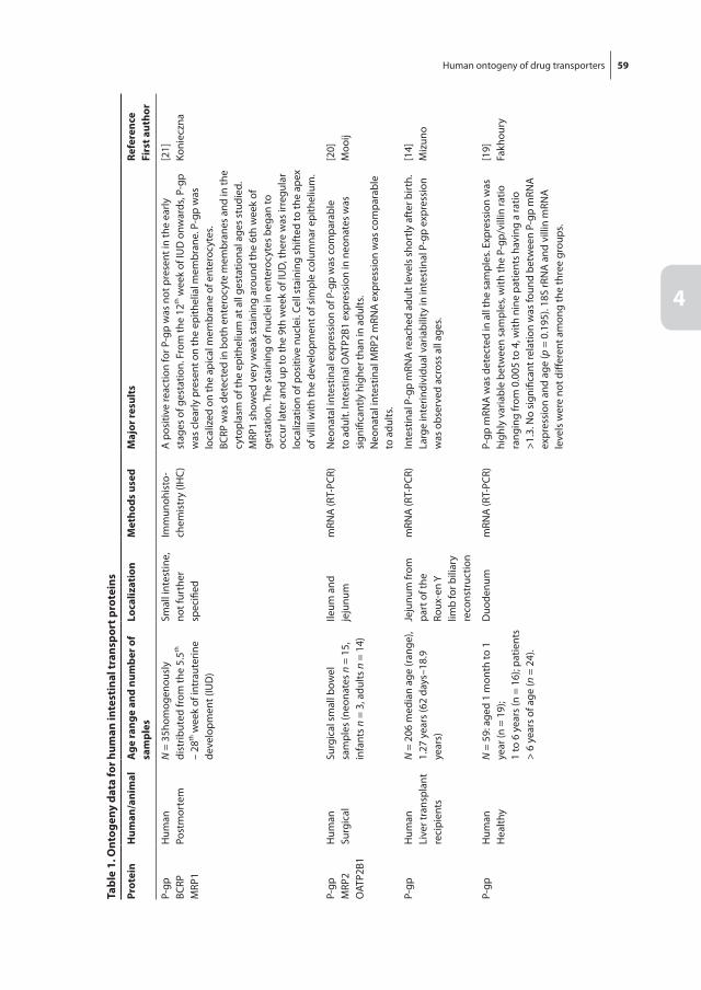
58 Chapter 4 Human ontogeny of drug transporters 59
4
tabl
e 1.
ont
ogen
y da
ta fo
r hum
an in
test
inal
tran
spor
t pro
tein
s
Prot
ein
hum
an/a
nim
alA
ge ra
nge
and
num
ber o
f sa
mpl
eslo
caliz
atio
nM
etho
ds u
sed
Maj
or re
sult
sre
fere
nce
firs
t aut
hor
P-gp
BCRP
MRP
1
Hum
anPo
stm
orte
mN
= 3
5hom
ogen
ousl
y di
strib
uted
from
the
5.5th
–
28th
wee
k of
intr
aute
rine
deve
lopm
ent (
IUD
)
Smal
l int
estin
e,
not f
urth
er
spec
ified
Imm
unoh
isto
-ch
emis
try
(IHC)
A p
ositi
ve re
actio
n fo
r P-g
p w
as n
ot p
rese
nt in
the
early
st
ages
of g
esta
tion.
Fro
m th
e 12
th w
eek
of IU
D o
nwar
ds, P
-gp
was
cle
arly
pre
sent
on
the
epith
elia
l mem
bran
e. P
-gp
was
lo
caliz
ed o
n th
e ap
ical
mem
bran
e of
ent
eroc
ytes
.BC
RP w
as d
etec
ted
in b
oth
ente
rocy
te m
embr
anes
and
in th
e cy
topl
asm
of t
he e
pith
eliu
m a
t all
gest
atio
nal a
ges
stud
ied.
MRP
1 sh
owed
ver
y w
eak
stai
ning
aro
und
the
6th
wee
k of
ge
stat
ion.
The
sta
inin
g of
nuc
lei i
n en
tero
cyte
s be
gan
to
occu
r lat
er a
nd u
p to
the
9th
wee
k of
IUD
, the
re w
as ir
regu
lar
loca
lizat
ion
of p
ositi
ve n
ucle
i. Ce
ll st
aini
ng s
hift
ed to
the
apex
of
vill
i with
the
deve
lopm
ent o
f sim
ple
colu
mna
r epi
thel
ium
.
[21]
Koni
eczn
a
P-gp
MRP
2O
ATP2
B1
Hum
anSu
rgic
alSu
rgic
al s
mal
l bow
el
sam
ples
(neo
nate
s n
= 15
, in
fant
s n
= 3,
adu
lts n
= 1
4)
Ileum
and
je
junu
mm
RNA
(RT-
PCR)
Neo
nata
l int
estin
al e
xpre
ssio
n of
P-g
p w
as c
ompa
rabl
e to
adu
lt. In
test
inal
OAT
P2B1
exp
ress
ion
in n
eona
tes
was
si
gnifi
cant
ly h
ighe
r tha
n in
adu
lts.
Neo
nata
l int
estin
al M
RP2
mRN
A e
xpre
ssio
n w
as c
ompa
rabl
e to
adu
lts.
[20]
Moo
ij
P-gp
Hum
anLi
ver t
rans
plan
t re
cipi
ents
N =
206
med
ian
age
(ran
ge),
1.27
yea
rs (6
2 da
ys–1
8.9
year
s)
Jeju
num
from
pa
rt o
f the
Ro
ux-e
n Y
limb
for b
iliar
y re
cons
truc
tion
mRN
A (R
T-PC
R)In
test
inal
P-g
p m
RNA
reac
hed
adul
t lev
els
shor
tly a
fter
birt
h.
Larg
e in
terin
divi
dual
var
iabi
lity
in in
test
inal
P-g
p ex
pres
sion
w
as o
bser
ved
acro
ss a
ll ag
es.
[14]
Miz
uno
P-gp
Hum
anH
ealth
yN
= 5
9: a
ged
1 m
onth
to 1
ye
ar (n
= 1
9);
1 to
6 y
ears
(n =
16)
; pat
ient
s >
6 ye
ars
of a
ge (n
= 2
4).
Duo
denu
mm
RNA
(RT-
PCR)
P-gp
mRN
A w
as d
etec
ted
in a
ll th
e sa
mpl
es. E
xpre
ssio
n w
as
high
ly v
aria
ble
betw
een
sam
ples
, with
the
P-gp
/vill
in ra
tio
rang
ing
from
0.0
05 to
4, w
ith n
ine
patie
nts
havi
ng a
ratio
>1
.3. N
o si
gnifi
cant
rela
tion
was
foun
d be
twee
n P-
gp m
RNA
ex
pres
sion
and
age
(p =
0.1
95).
18S
rRN
A a
nd v
illin
mRN
A
leve
ls w
ere
not d
iffer
ent a
mon
g th
e th
ree
grou
ps.
[19]
Fakh
oury
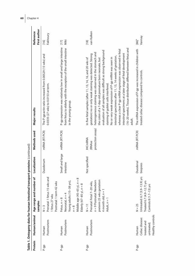
60 Chapter 4 Human ontogeny of drug transporters 61
4
tabl
e 1.
ont
ogen
y da
ta fo
r hum
an in
test
inal
tran
spor
t pro
tein
s (c
ontin
ued)
Prot
ein
hum
an/a
nim
alA
ge ra
nge
and
num
ber o
f sa
mpl
eslo
caliz
atio
nM
etho
ds u
sed
Maj
or re
sult
sre
fere
nce
firs
t aut
hor
P-gp
Hum
anPo
stm
orte
mN
= 3
Pren
atal
: 1 fe
tus
15 w
ks a
nd
1 fe
tus
27 w
ksPo
stna
tal:
1 te
rm 4
2 w
ks
Duo
denu
mm
RNA
(RT-
PCR)
The
P-gp
:act
in ra
tio in
crea
sed
from
0.0
0529
(15
wks
) and
0.03
35 (2
7 w
ks) t
o 0.
0153
at t
erm
.[1
6]Fa
khou
ry
P-gp
Hum
anPo
stm
orte
mFe
tus,
n =
8N
eona
tal,
n =
4Yo
ung
adul
ts (1
5–38
yrs
), n
= 8
Mid
dle
age
(45–
65 y
), n
= 8
Elde
rly (6
7–85
y),
n =
8
Smal
l and
larg
e in
test
ine
mRN
A (R
T-PC
R)P-
gp e
xpre
ssio
n w
as re
lativ
ely
low
in s
mal
l and
larg
e in
test
ine
from
fetu
s to
eld
erly
with
the
exce
ptio
n of
the
smal
l int
estin
e in
the
youn
g gr
oup.
[17]
Mik
i
P-gp
Hum
anPo
stm
orte
mN
= 1
1Pr
enat
al: F
etal
7–2
0 w
ks,
n =
9 Po
stna
tal:
New
born
pr
eter
m 2
5 w
ks g
esta
tion
4 m
onth
old
, n =
1Ad
ult,
n =
1
Not
spe
cifie
dIH
CmRN
A(R
NA
se
prot
ectio
n as
say)
In fi
ve fe
tal s
ampl
es (a
fter
11,
13,
14,
16,
and
20
wks
of
gest
atio
n) n
o or
onl
y w
eak
stai
ning
was
det
ecte
d. S
tron
g he
tero
gene
ous
stai
ning
was
obs
erve
d in
the
stom
ach
and
the
colo
n of
a 7
-day
-old
pre
mat
ure
born
neo
nate
, but
ev
alua
tion
of a
ll sa
mpl
es w
as d
ifficu
lt as
str
ong
back
grou
nd
stai
ning
of g
oble
t cel
ls in
terf
ered
.N
o or
ver
y w
eak
expr
essi
on o
f P-g
p m
RNA
was
see
n in
in
test
inal
spe
cim
ens
afte
r 11,
13,
14
wee
ks o
f ges
tatio
n,
whe
reas
a c
lear
P-g
p m
RNA
exp
ress
ion
was
obs
erve
d in
feta
l in
test
inal
spe
cim
ens
at a
late
r sta
ge o
f fet
al d
evel
opm
ent
(16–
20 w
eeks
). Ti
ssue
dis
trib
utio
n di
ffere
d be
twee
n fe
tus
and
adul
t.
[18]
van
Kalk
en
P-gp
Hum
anCe
liac
dise
ase:
tr
eate
d an
d un
trea
ted;
Hea
lthy
cont
rols
N =
25
Unt
reat
ed: 6
.7 (3
.9–1
3.9)
yrs
Trea
ted
6.7
(4.9
–12.
7) y
rsCo
ntro
ls 8
(1.7
–13)
yrs
Duo
dena
l bi
opsi
esm
RNA
(RT-
PCR)
The
mRN
A e
xpre
ssio
n of
P-g
p w
as in
crea
sed
in c
hild
ren
with
tr
eate
d ce
liac
dise
ase
com
pare
d to
con
trol
s.[8
6]a
Vann
ay
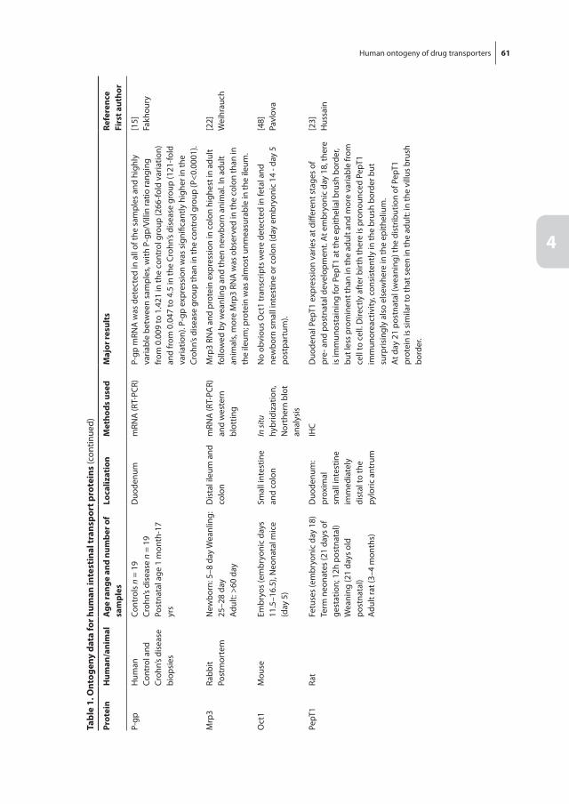
60 Chapter 4 Human ontogeny of drug transporters 61
4
tabl
e 1.
ont
ogen
y da
ta fo
r hum
an in
test
inal
tran
spor
t pro
tein
s (c
ontin
ued)
Prot
ein
hum
an/a
nim
alA
ge ra
nge
and
num
ber o
f sa
mpl
eslo
caliz
atio
nM
etho
ds u
sed
Maj
or re
sult
sre
fere
nce
firs
t aut
hor
P-gp
Hum
anCo
ntro
l and
Cr
ohn’
s di
seas
e bi
opsi
es
Cont
rols
n =
19
Croh
n’s
dise
ase
n =
19Po
stna
tal a
ge 1
mon
th-1
7 yr
s
Duo
denu
mm
RNA
(RT-
PCR)
P-gp
mRN
A w
as d
etec
ted
in a
ll of
the
sam
ples
and
hig
hly
varia
ble
betw
een
sam
ples
, with
P-g
p/Vi
llin
ratio
rang
ing
from
0.0
09 to
1.4
21 in
the
cont
rol g
roup
(266
-fold
var
iatio
n)
and
from
0.0
47 to
4.5
in th
e Cr
ohn’
s di
seas
e gr
oup
(121
-fold
va
riatio
n). P
-gp
expr
essi
on w
as s
igni
fican
tly h
ighe
r in
the
Croh
n’s
dise
ase
grou
p th
an in
the
cont
rol g
roup
(P<0
.000
1).
[15]
Fakh
oury
Mrp
3Ra
bbit
Post
mor
tem
New
born
: 5–8
day
Wea
nlin
g:
25–2
8 da
yAd
ult:
>60
day
Dis
tal i
leum
and
co
lon
mRN
A (R
T-PC
R)
and
wes
tern
bl
ottin
g
Mrp
3 RN
A a
nd p
rote
in e
xpre
ssio
n in
col
on h
ighe
st in
adu
lt fo
llow
ed b
y w
eanl
ing
and
then
new
born
ani
mal
. In
adul
t an
imal
s, m
ore
Mrp
3 RN
A w
as o
bser
ved
in th
e co
lon
than
in
the
ileum
; pro
tein
was
alm
ost u
nmea
sura
ble
in th
e ile
um.
[22]
Wei
hrau
ch
Oct
1M
ouse
Embr
yos
(em
bryo
nic
days
11
.5–1
6.5)
, Neo
nata
l mic
e (d
ay 5
)
Smal
l int
estin
e an
d co
lon
In si
tu
hybr
idiz
atio
n,
Nor
ther
n bl
ot
anal
ysis
No
obvi
ous
Oct
1 tr
ansc
ripts
wer
e de
tect
ed in
feta
l and
ne
wbo
rn s
mal
l int
estin
e or
col
on (d
ay e
mbr
yoni
c 14
- da
y 5
post
part
um).
[48]
Pavl
ova
PepT
1Ra
tFe
tuse
s (e
mbr
yoni
c da
y 18
)Te
rm n
eona
tes
(21
days
of
gest
atio
n; 1
2h p
ostn
atal
)W
eani
ng (2
1 da
ys o
ld
post
nata
l)Ad
ult r
at (3
–4 m
onth
s)
Duo
denu
m:
prox
imal
sm
all i
ntes
tine
imm
edia
tely
di
stal
to th
e py
loric
ant
rum
IHC
Duo
dena
l Pep
T1 e
xpre
ssio
n va
ries
at d
iffer
ent s
tage
s of
pr
e- a
nd p
ostn
atal
dev
elop
men
t. A
t em
bryo
nic
day
18, t
here
is
imm
unos
tain
ing
for P
epT1
at t
he e
pith
elia
l bru
sh b
orde
r, bu
t les
s pr
omin
ent t
han
in th
e ad
ult a
nd m
ore
varia
ble
from
ce
ll to
cel
l. D
irect
ly a
fter
birt
h th
ere
is p
rono
unce
d Pe
pT1
imm
unor
eact
ivity
, con
sist
ently
in th
e br
ush
bord
er b
ut
surp
risin
gly
also
els
ewhe
re in
the
epith
eliu
m.
At d
ay 2
1 po
stna
tal (
wea
ning
) the
dis
trib
utio
n of
Pep
T1
prot
ein
is s
imila
r to
that
see
n in
the
adul
t: in
the
villu
s br
ush
bord
er.
[23]
Hus
sain
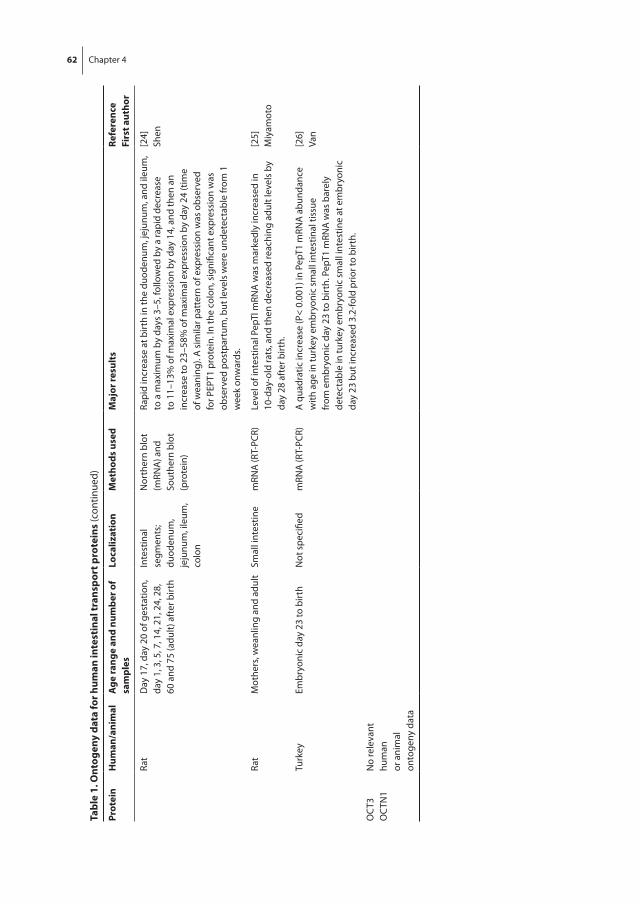
62 Chapter 4
tabl
e 1.
ont
ogen
y da
ta fo
r hum
an in
test
inal
tran
spor
t pro
tein
s (c
ontin
ued)
Prot
ein
hum
an/a
nim
alA
ge ra
nge
and
num
ber o
f sa
mpl
eslo
caliz
atio
nM
etho
ds u
sed
Maj
or re
sult
sre
fere
nce
firs
t aut
hor
Rat
Day
17,
day
20
of g
esta
tion,
da
y 1,
3, 5
, 7, 1
4, 2
1, 2
4, 2
8,
60 a
nd 7
5 (a
dult)
aft
er b
irth
Inte
stin
al
segm
ents
; du
oden
um,
jeju
num
, ile
um,
colo
n
Nor
ther
n bl
ot
(mRN
A) a
nd
Sout
hern
blo
t (p
rote
in)
Rapi
d in
crea
se a
t birt
h in
the
duod
enum
, jej
unum
, and
ileu
m,
to a
max
imum
by
days
3–5
, fol
low
ed b
y a
rapi
d de
crea
se
to 1
1–13
% o
f max
imal
exp
ress
ion
by d
ay 1
4, a
nd th
en a
n in
crea
se to
23–
58%
of m
axim
al e
xpre
ssio
n by
day
24
(tim
e of
wea
ning
). A
sim
ilar p
atte
rn o
f exp
ress
ion
was
obs
erve
d fo
r PEP
T1 p
rote
in. I
n th
e co
lon,
sig
nific
ant e
xpre
ssio
n w
as
obse
rved
pos
tpar
tum
, but
leve
ls w
ere
unde
tect
able
from
1
wee
k on
war
ds.
[24]
Shen
Rat
Mot
hers
, wea
nlin
g an
d ad
ult
Smal
l int
estin
em
RNA
(RT-
PCR)
Leve
l of i
ntes
tinal
Pep
Tl m
RNA
was
mar
kedl
y in
crea
sed
in
10-d
ay-o
ld ra
ts, a
nd th
en d
ecre
ased
reac
hing
adu
lt le
vels
by
day
28 a
fter
birt
h.
[25]
Miy
amot
o
Turk
eyEm
bryo
nic
day
23 to
birt
hN
ot s
peci
fied
mRN
A (R
T-PC
R)A
qua
drat
ic in
crea
se (P
< 0.
001)
in P
epT1
mRN
A a
bund
ance
w
ith a
ge in
turk
ey e
mbr
yoni
c sm
all i
ntes
tinal
tiss
ue
from
em
bryo
nic
day
23 to
birt
h. P
epT1
mRN
A w
as b
arel
y de
tect
able
in tu
rkey
em
bryo
nic
smal
l int
estin
e at
em
bryo
nic
day
23 b
ut in
crea
sed
3.2-
fold
prio
r to
birt
h.
[26]
Van
OC
T3O
CTN
1N
o re
leva
nt
hum
an
or a
nim
al
onto
geny
dat
a
Human ontogeny of drug transporters 63
4
expression or localization of P-gp in the intestine. Collectively samples from 55 fetuses, ranging from 5.5 to 28 weeks of gestation, and 302 additional samples across a pediatric age range, were investigated in this group of studies.[14-21] The majority of mRNA samples (upper jejunum) originated from one study in pediatric liver transplant patients, which may present confounding variation based on hepatic disorders.[14] In most stud-ies, P-gp mRNA expression was determined; localization with immunohistochemistry was only conducted in a small number of samples. All of the studies demonstrated large interindividual variation. The overall developmental pattern of P-gp expression revealed a consistently emerging change from undetectable expression in the first trimester of fetal life to present and apparently stable P-gp expression from ~12 weeks of gestation onwards. Very limited data suggest that P-gp expression increased slowly to reach adult levels at or very shortly after birth. Interestingly, two studies reported higher intestinal P-gp expression in children with treated Crohn’s and celiac disease compared to healthy controls.[15]
BCRPThe localization of BCRP staining appeared stable from 5.5 to 28 weeks of gestation in humans.[21]
MRP1In 35 human fetal samples from 5.5 to 28 weeks of gestation, MRP1 staining was visu-alized in all samples using immunohistochemistry.[21] The intensity was weak during early gestation, but appeared to mature to an adult distribution pattern from 7 weeks of gestation onwards.
MRP2Analogous to P-gp, the mRNA expression of MRP2 appeared stable from neonates to adults in ileal and jejunal surgical samples.[20]
OATP2B1In contrast to MRP2, OATP2B1 mRNA expression was significantly higher in the samples from neonates compared to adults.[20]
Other intestinal transport proteinsHuman data for MRP3 and PEPT1 appear to be missing, but animal data are available. In rabbits, Mrp3 mRNA expression was lowest in newborns and subsequently increased in weanlings until reaching the highest levels in adults.[22] The four available animal stud-ies (three in rat; one in turkey) on PepT1 all support a similar developmental pattern of significant prenatal expression that reached maximal levels in the postnatal period, and
64 Chapter 4 Human ontogeny of drug transporters 65
4
then decreased to adult levels from weaning onwards.[23-26] This finding seems consis-tent with the role of PEPT1 in the absorption of di- and tripeptides, which constitutes an important part of infant nutrition. Intestinal temporal and spatial expression data during development for OATP1A2, OCT1, and MCT1 appear to be lacking.
It is clear from the data in Table 1 that no single developmental pattern can be identi-fied for all of the intestinal transporters. Different patterns are apparent based on the available, albeit limited human data: 1) low in the embryo and then stable from neonate to adult (e.g., P-gp, MRP2); 2) high at birth and decreasing in the first months of life (OATP2B1).
liver
For the liver, all transport proteins designated by the International Transporter Consor-tium as important for disposition of drugs and endogenous substances were reviewed.[1, 13] These include the efflux transporters P-gp, BCRP, MRP2, MRP3, MRP4, MRP6, MATE1, BSEP, the uptake transporters NTCP, OATP1B1, OATP1B3, OATP2B1, OAT2, OCT1, and the bidirectional transporters OAT7 and OSTα/β. Similar to the other organs, this review on hepatic transporters exclusively focuses on human data, if available, but re-sorts to animal data in the absence of any relevant human information (Table 2). Similar to intestinal transporters, most published data on the ontogeny of hepatic transport proteins is available for efflux transporters, particularly P-gp.
P-gpHepatic P-gp was already detectable in the wall of bile canaliculi in early fetal life at 14 weeks by immunohistochemistry and at the mRNA level.[18] Although expression was low initially, it seemed to increase throughout fetal development and was considered moderate at the protein level by fetal week 19.[16, 27] mRNA expression for P-gp in-creased throughout childhood development. In a study with 61 liver specimens, mRNA expression levels were 20–30-fold lower in the fetal and neonatal age group compared to adults. mRNA expression, however, rapidly increased in the early months of life: in infants (1–12 months), mRNA was only 5-fold lower compared to adults, and in children and adolescents it was indistinguishable from adult expression.[20] These observations are supported by less extensive studies from others,[28] and also revealed a similarly high interindividual variability in expression in children compared to adults.[20, 29] The limited results on P-gp protein expression, however, do not corroborate the reported age-dependent P-gp mRNA expression. P-gp protein was detectable in samples as early as 1 month,[29] and relative protein expression was not significantly different in 65 liver specimens from age groups 0.3–0.7, 0.7–2, 2–5, and 5–12 years.[30] This observation may not be surprising in light of the observed lack of correlation between mRNA and protein levels for P-gp in adult liver.[31] P-gp protein expression quantified by liquid

64 Chapter 4 Human ontogeny of drug transporters 65
4
tabl
e 2.
ont
ogen
y da
ta fo
r hum
an h
epat
ic tr
ansp
ort p
rote
ins
Prot
ein
hum
an/a
nim
alA
ge ra
nge
and
num
ber o
f sa
mpl
esM
etho
ds u
sed
Maj
or re
sult
sre
fere
nce
firs
t aut
hor
P-gp
Hum
anPo
stm
orte
mN
= 3
5Fe
tal:
14–2
8 w
eeks
of
gest
atio
n
Imm
unoh
isto
-ch
emis
try
(IHC)
, m
RNA
(RN
Ase
pr
otec
tion
assa
y)
Stai
ning
for P
-gp
in b
ile c
anal
icul
i alre
ady
at 1
4 w
eeks
of g
esta
tion.
No
appa
rent
di
ffere
nce
in s
tain
ing
inte
nsity
in d
iffer
ent f
etal
sta
ges.
Pres
ence
of m
RNA
co
nfirm
ed in
one
spe
cim
en (1
6 w
eeks
).
[18]
Van
Kalk
en
P-gp
MRP
2H
uman
Post
mor
tem
N n
ot d
eter
min
edFe
tal:
14 a
nd 1
9 w
eeks
of
gest
atio
n
IHC
Imm
unop
ositi
vity
of P
-gp
and
MRP
2 w
as lo
caliz
ed to
the
cana
licul
ar m
embr
ane
of d
iffer
entia
ting
and
mat
ure
hepa
tocy
tes.
MPR
2 w
as d
etec
tabl
e in
live
r of
14-
wee
k-ol
d fe
tuse
s, bu
t had
str
ong
expr
essi
on a
t 19
wee
ks. P
-gp
was
de
tect
able
at v
ery
low
leve
ls a
t 14
wee
ks, a
nd w
as m
oder
atel
y ex
pres
sed
at 1
9 w
eeks
.
[27]
Cizk
ova
P-gp
Hum
anPo
stm
orte
mN
= 3
Feta
l: 15
, 27
and
42 w
eeks
of
gest
atio
n
mRN
A (R
T-PC
R)Ex
pres
sion
of P
-gp
was
det
ecta
ble
in th
e 15
wee
k sa
mpl
es, a
nd g
radu
ally
in
crea
sed
with
ges
tatio
nal a
ge u
ntil
42 w
eeks
.[1
6]Fa
khou
ry
P-gp
MRP
2O
ATP1
B1O
ATP1
B3
Hum
anPo
stm
orte
mN
= 7
2Fe
tal:
n =
9 (8
–23
wks
)Pe
diat
ric: n
= 5
2 (0
–17
yrs)
Adul
t: n
= 11
mRN
A (R
T-PC
R)m
RNA
for P
-gp
expr
essi
on in
feta
l and
neo
nata
l gro
ups
was
20–
30 fo
ld lo
wer
th
an in
adu
lts. E
xpre
ssio
n in
infa
nts
was
slig
htly
low
er th
an in
adu
lts, w
ith n
o di
ffere
nce
betw
een
child
ren
1–7
year
s an
d ad
ults
. MRP
2 m
RNA
was
30-
fold
lo
wer
in fe
tal,
200-
fold
low
er in
neo
nata
l, an
d 10
0-fo
ld lo
wer
in in
fant
live
r co
mpa
red
to a
dults
. OAT
P1B1
exp
ress
ion
was
20-
fold
low
er in
feta
l, 50
0-fo
ld
low
er in
neo
nata
l, an
d 90
-fold
low
er in
infa
nt li
ver c
ompa
red
to a
dults
. OAT
P1B3
m
RNA
was
30-
fold
low
er in
feta
l, 60
0-fo
ld lo
wer
in n
eona
tal a
nd 1
00-fo
ld lo
wer
in
infa
nt li
ver c
ompa
red
to a
dults
.
[20]
Moo
ji
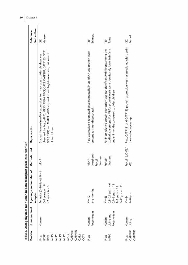
66 Chapter 4
tabl
e 2.
ont
ogen
y da
ta fo
r hum
an h
epat
ic tr
ansp
ort p
rote
ins
(con
tinue
d)
Prot
ein
hum
an/a
nim
alA
ge ra
nge
and
num
ber o
f sa
mpl
esM
etho
ds u
sed
Maj
or re
sult
sre
fere
nce
firs
t aut
hor
P-gp
BCRP
BSEP
MRP
2M
RP3
MRP
4M
RP6
MAT
E1O
ATP1
B1O
ATP1
B3O
AT2
OC
T1
Hum
anPe
rinat
al (0
–30
days
): N
= 6
0–4
year
s: N
= 8
>7 y
ears
: N =
6
mRN
AG
radu
al in
crea
se in
mRN
A e
xpre
ssio
n fr
om n
eona
tes
to o
lder
chi
ldre
n w
as
obse
rved
for P
-gp,
MRP
2, M
RP3,
MRP
6, N
TCP,
OAT
2, O
ATP1
B1, O
ATP1
B3, O
CT1
, BC
RP, B
SEP,
and
MAT
E1. M
RP4
expr
essi
on w
as h
igh
in n
eona
tes,
but l
ower
in
olde
r chi
ldre
n.
[28]
Klaa
ssen
P-gp
Hum
anPo
stm
orte
mN
= 1
21–
6 m
onth
sm
RNA
(N
orth
ern)
Prot
ein
(Wes
tern
)
P-gp
exp
ress
ion
is re
gula
ted
deve
lopm
enta
lly. P
-gp
mRN
A a
nd p
rote
in w
ere
pres
ent a
t 1 m
onth
pos
tnat
al.
[29]
Schu
etz
P-gp
MRP
2H
uman
Livi
ng a
nd
Post
mor
tem
N =
65
0.3–
0.7
yrs:
n =
60.
7–2
yrs:
n =
13
2–5
yrs:
n =
13
5–12
yrs
: n =
33
Prot
ein
(Wes
tern
)Fo
r P-g
p, re
lativ
e pr
otei
n ex
pres
sion
was
not
sig
nific
antly
diff
eren
t am
ong
the
stud
ied
age
grou
ps. F
or M
RP2,
pro
tein
leve
ls w
ere
sign
ifica
ntly
low
er in
infa
nts
unde
r 8 m
onth
s co
mpa
red
to o
lder
chi
ldre
n.
[30]
Tang
P-gp
O
ATP1
B1O
ATP1
B3
Hum
anLi
ving
N =
64
7–70
yrs
Prot
ein
(LC-
MS/
MS)
P-gp
, OAT
P1B1
and
OAT
P1B3
pro
tein
exp
ress
ion
was
not
ass
ocia
ted
with
age
in
the
stud
ied
age
rang
e.[3
2]Pr
asad
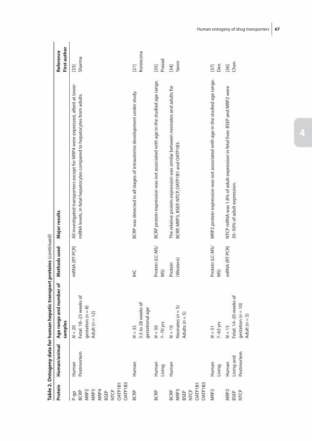
Human ontogeny of drug transporters 67
4
tabl
e 2.
ont
ogen
y da
ta fo
r hum
an h
epat
ic tr
ansp
ort p
rote
ins
(con
tinue
d)
Prot
ein
hum
an/a
nim
alA
ge ra
nge
and
num
ber o
f sa
mpl
esM
etho
ds u
sed
Maj
or re
sult
sre
fere
nce
firs
t aut
hor
P-gp
BCRP
MRP
2M
RP3
MRP
4BS
EPN
TCP
OAT
P1B1
OAT
P1B3
Hum
anPo
stm
orte
mN
= 2
0Fe
tal:
18–2
3 w
eeks
of
gest
atio
n (n
= 8
)Ad
ult (
n =
12)
mRN
A (R
T-PC
R)A
ll in
vest
igat
ed tr
ansp
orte
rs e
xcep
t for
MRP
4 w
ere
expr
esse
d, a
lbei
t at l
ower
m
RNA
leve
ls, i
n fe
tal h
epat
ocyt
es c
ompa
red
to h
epat
ocyt
es fr
om a
dults
.[3
3]Sh
arm
a
BCRP
Hum
anN
= 3
55.
5 to
28
wee
ks o
f ge
stat
iona
l age
IHC
BCRP
was
det
ecte
d in
all
stag
es o
f int
raut
erin
e de
velo
pmen
t und
er s
tudy
.[2
1]Ko
niec
zna
BCRP
Hum
anLi
ving
N =
50
7–70
yrs
Prot
ein
(LC-
MS/
MS)
BCRP
pro
tein
exp
ress
ion
was
not
ass
ocia
ted
with
age
in th
e st
udie
d ag
e ra
nge.
[35]
Pras
ad
BCRP
MRP
3BS
EPN
TCP
OAT
P1B1
OAT
P1B3
Hum
anN
= 1
0N
eona
tes
(n =
5)
Adul
ts (n
= 5
)
Prot
ein
(Wes
tern
)Th
e re
lativ
e pr
otei
n ex
pres
sion
was
sim
ilar b
etw
een
neon
ates
and
adu
lts fo
r BC
RP, M
RP3,
BSE
P, N
TCP,
OAT
P1B1
and
OAT
P1B3
.[3
4]Ya
nni
MRP
2H
uman
Livi
ngN
= 5
17–
63 y
rsPr
otei
n (L
C-M
S/M
S)M
RP2
prot
ein
expr
essi
on w
as n
ot a
ssoc
iate
d w
ith a
ge in
the
stud
ied
age
rang
e.[3
7]D
eo
MRP
2BS
EPN
TCP
Hum
anLi
ving
and
Po
stm
orte
m
N =
15
Feta
l: 14
–20
wee
ks o
f ge
stat
ion
(n =
10)
Adul
t (n
= 5)
mRN
A (R
T-PC
R)N
TCP
mRN
A w
as 1
.8%
of a
dult
expr
essi
on in
feta
l liv
er. B
SEP
and
MRP
2 w
ere
30–5
0% o
f adu
lt ex
pres
sion
.[3
6]Ch
en
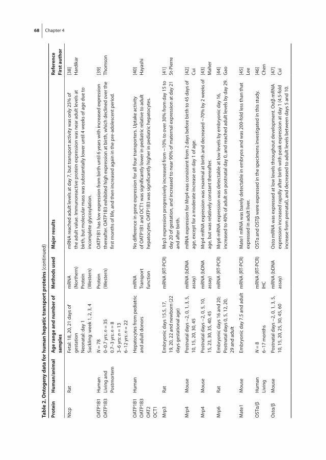
68 Chapter 4
tabl
e 2.
ont
ogen
y da
ta fo
r hum
an h
epat
ic tr
ansp
ort p
rote
ins
(con
tinue
d)
Prot
ein
hum
an/a
nim
alA
ge ra
nge
and
num
ber o
f sa
mpl
esM
etho
ds u
sed
Maj
or re
sult
sre
fere
nce
firs
t aut
hor
Ntc
pRa
tFe
tal:
18, 2
0, 2
1 da
ys o
f ge
stat
ion
Neo
nata
l: da
y 1
Suck
ling:
wee
k 1,
2, 3
, 4
mRN
A
(Nor
ther
n)Pr
otei
n (W
este
rn)
mRN
A re
ache
d ad
ult l
evel
s at
day
7, b
ut tr
ansp
ort a
ctiv
ity w
as o
nly
25%
of
the
adul
t val
ue. I
mm
unor
eact
ive
prot
ein
expr
essi
on w
as n
ear a
dult
leve
ls a
t bi
rth,
but
mol
ecul
ar m
ass
was
sub
stan
tially
low
er u
ntil
4 w
eeks
of a
ge d
ue to
in
com
plet
e gl
ycos
ylat
ion.
[38]
Har
dika
r
OAT
P1B1
OAT
P1B3
Hum
anLi
ving
and
Po
stm
orte
m
N =
78
0–0.
7 yr
s: n
= 3
50.
7–3
yrs:
n =
83–
6 yr
s: n
= 1
36–
12 y
rs: n
= 2
2
Prot
ein
(Wes
tern
)O
ATP1
B1 h
as lo
w e
xpre
ssio
n fr
om b
irth
until
6 y
ears
with
incr
ease
d ex
pres
sion
th
erea
fter
. OAT
P1B3
exh
ibite
d hi
gh e
xpre
ssio
n at
birt
h, w
hich
dec
lined
ove
r the
fir
st m
onth
s of
life
, and
then
incr
ease
d ag
ain
in th
e pr
e-ad
oles
cent
per
iod.
[39]
Thom
son
OAT
P1B1
OAT
P1B3
OAT
2O
CT1
Hum
anH
epat
ocyt
es fr
om p
edia
tric
an
d ad
ult d
onor
sm
RNA
Tran
spor
t fu
nctio
n
No
diffe
renc
e in
gen
e ex
pres
sion
for a
ll fo
ur tr
ansp
orte
rs. U
ptak
e ac
tivity
of
OAT
P1B1
and
OC
T1 w
as s
igni
fican
tly lo
wer
in p
edia
tric
rela
tive
to a
dult
hepa
tocy
tes.
OAT
P1B3
was
sig
nific
antly
hig
her i
n pe
diat
ric h
epat
ocyt
es.
[40]
Hay
ashi
Mrp
3Ra
tEm
bryo
nic
days
15.
5, 1
7,
19, 2
0, 2
2 an
d ne
wbo
rn (2
2 da
ys g
esta
tiona
l age
)
mRN
A (R
T-PC
R)M
rp3
expr
essi
on p
rogr
essi
vely
incr
ease
d fr
om ~
10%
to o
ver 3
0% fr
om d
ay 1
5 to
da
y 20
of g
esta
tion,
and
incr
ease
d to
nea
r 90%
of m
ater
nal e
xpre
ssio
n at
day
21
and
afte
r birt
h.
[41]
St-P
ierr
e
Mrp
4M
ouse
Post
nata
l day
s −2
, 0, 1
, 3, 5
, 10
, 15,
20,
30,
45
mRN
A (b
DN
A
assa
y)m
RNA
exp
ress
ion
for M
rp4
was
con
sist
ent f
rom
2 d
ays
befo
re b
irth
to 4
5 da
ys o
f ag
e, e
xcep
t for
a m
oder
ate
incr
ease
on
day
1 of
age
.[4
2]Cu
i
Mrp
4M
ouse
Post
nata
l day
s −2
, 0, 5
, 10,
15
, 23,
30,
35,
40,
45
mRN
A (b
DN
A
assa
y)M
rp4
mRN
A e
xpre
ssio
n w
as m
axim
al a
t birt
h an
d de
crea
sed
~70%
by
2 w
eeks
of
age,
but
was
rela
tivel
y co
nsta
nt th
erea
fter
.[4
3]M
aher
Mrp
6Ra
tEm
bryo
nic
days
16
and
20;
Post
nata
l day
s 0,
5, 1
2, 2
0,
29 a
nd a
dult
mRN
A (R
T-PC
R)M
rp6
mRN
A e
xpre
ssio
n w
as d
etec
tabl
e at
low
leve
ls b
y em
bryo
nic
day
16,
incr
ease
d to
40%
of a
dult
on p
ostn
atal
day
0, a
nd re
ache
d ad
ult l
evel
s by
day
29.
[44]
Gao
Mat
e1M
ouse
Embr
yoni
c da
y 7.
5 an
d ad
ult
mRN
A (R
T-PC
R)M
ate1
mRN
A w
as b
arel
y de
tect
able
in e
mbr
yos
and
was
200
-fold
less
than
that
ex
pres
sed
in a
dult
liver
.[4
5]Le
e
OST
α/β
Hum
anLi
ving
N =
86–
17 m
onth
sm
RNA
(RT-
PCR)
IHC
OST
α an
d O
STβ
wer
e ex
pres
sed
in th
e sp
ecim
ens
inve
stig
ated
in th
is s
tudy
.[4
6]Ch
en
Ost
α/β
Mou
sePo
stna
tal d
ays
−2, 0
, 1, 3
, 5,
10, 1
5, 2
0, 2
5, 3
0, 4
5, 6
0m
RNA
(bD
NA
as
say)
Ost
α m
RNA
was
exp
ress
ed a
t low
leve
ls th
roug
hout
dev
elop
men
t. O
stβ
mRN
A
expr
essi
on in
crea
sed
rapi
dly
afte
r birt
h w
ith p
eak
expr
essi
on a
t day
1 (4
.5-fo
ld
incr
ease
from
pre
nata
l), a
nd d
ecre
ased
to a
dult
leve
ls b
etw
een
days
5 a
nd 1
0.
[47]
Cui

Human ontogeny of drug transporters 69
4
chromatography/tandem mass spectrometry (LC-MS/MS) was not associated with age in 64 liver specimens in the age range of 7–70 years.[32] Thus, further studies are needed to clarify whether the age-dependent differences in mRNA expression translate into dif-ferences at the protein and ultimately the functional level for P-gp in the liver.
BCRPBCRP was detectable by immunohistochemistry in fetal liver specimens from 5.5–28 weeks[21] and mRNA expression increased from 18–22 week fetal samples to adults.[33] A comparison of relative protein expression between five neonates and five adult livers by western blot analysis indicated no detectable difference,[34] although mRNA expres-sion seemed to increase between neonates and older children.[28] In 50 livers from age 7–70 years, BCRP protein expression was correlated neither with age nor with mRNA expression.[35] These results together suggest that hepatic BCRP is expressed early dur-ing human development and does not undergo relevant developmental changes after term birth.
MRP2Similar to P-gp, MRP2 has been detected by immunohistochemistry in the bile cana-licular membranes of 14-week-old fetuses, with a tendency for higher expression levels in older fetal liver[27] and adults.[33, 36] The differences in age-dependent expression seem to be similar to P-gp in fetal liver (30-fold lower mRNA expression), but were sub-stantially more pronounced in neonates (200-fold lower mRNA expression) and infants (1–12 months) (100-fold lower mRNA expression) compared to adults.[20, 28] These substantial differences at the transcriptional level seem to translate into developmental differences in MRP2 protein expression: MRP2 protein levels determined by western blot were significantly lower in liver specimens from children younger than 8 months (n = 24) compared to older children up to 12 years (n = 59).[30] Later in childhood development, however, MRP2 protein expression assessed by mass spectrometry was independent of age in 51 liver specimens in the age range of 7–63 years.[37]
BSEPAt gestational weeks 14–20, BSEP was detectable by immunohistochemistry (36), and mRNA expression increased from neonates to older children[28] and adults.[33] Func-tional studies in isolated sandwich-cultured fetal and adult hepatocytes suggest that the biliary excretion index for taurocholate is substantially higher in adults compared to fetal hepatocytes. This higher functional activity for the BSEP substrate taurocholate could be explained by a higher expression level of BSEP in adult cells, assuming that there is a correlation between BSEP mRNA and protein expression.[33]
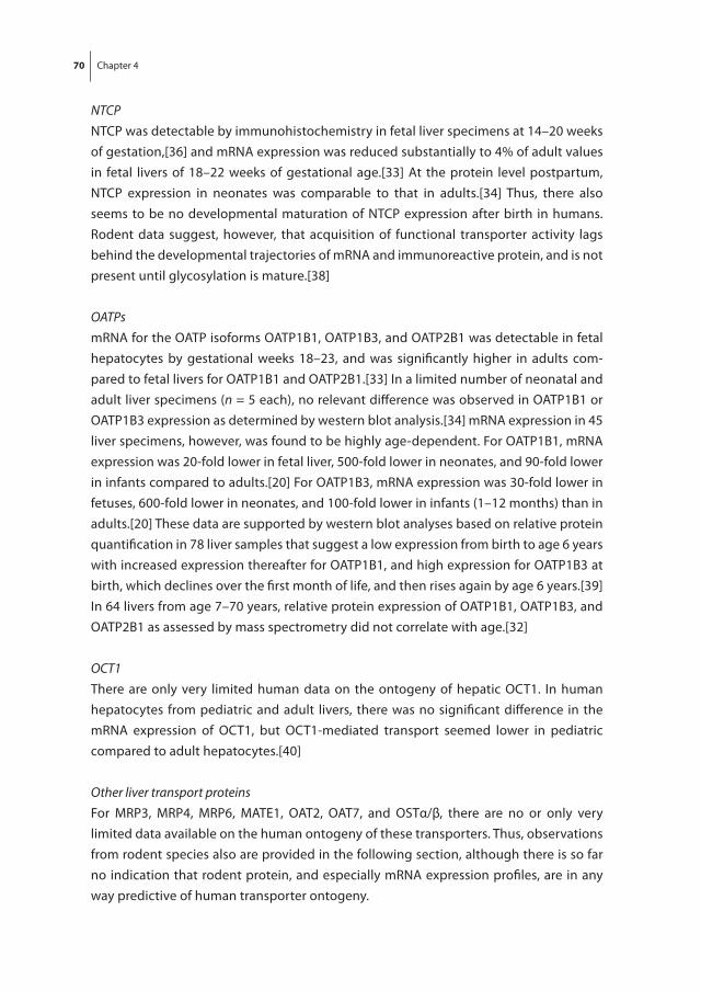
70 Chapter 4
NTCPNTCP was detectable by immunohistochemistry in fetal liver specimens at 14–20 weeks of gestation,[36] and mRNA expression was reduced substantially to 4% of adult values in fetal livers of 18–22 weeks of gestational age.[33] At the protein level postpartum, NTCP expression in neonates was comparable to that in adults.[34] Thus, there also seems to be no developmental maturation of NTCP expression after birth in humans. Rodent data suggest, however, that acquisition of functional transporter activity lags behind the developmental trajectories of mRNA and immunoreactive protein, and is not present until glycosylation is mature.[38]
OATPsmRNA for the OATP isoforms OATP1B1, OATP1B3, and OATP2B1 was detectable in fetal hepatocytes by gestational weeks 18–23, and was significantly higher in adults com-pared to fetal livers for OATP1B1 and OATP2B1.[33] In a limited number of neonatal and adult liver specimens (n = 5 each), no relevant difference was observed in OATP1B1 or OATP1B3 expression as determined by western blot analysis.[34] mRNA expression in 45 liver specimens, however, was found to be highly age-dependent. For OATP1B1, mRNA expression was 20-fold lower in fetal liver, 500-fold lower in neonates, and 90-fold lower in infants compared to adults.[20] For OATP1B3, mRNA expression was 30-fold lower in fetuses, 600-fold lower in neonates, and 100-fold lower in infants (1–12 months) than in adults.[20] These data are supported by western blot analyses based on relative protein quantification in 78 liver samples that suggest a low expression from birth to age 6 years with increased expression thereafter for OATP1B1, and high expression for OATP1B3 at birth, which declines over the first month of life, and then rises again by age 6 years.[39] In 64 livers from age 7–70 years, relative protein expression of OATP1B1, OATP1B3, and OATP2B1 as assessed by mass spectrometry did not correlate with age.[32]
OCT1There are only very limited human data on the ontogeny of hepatic OCT1. In human hepatocytes from pediatric and adult livers, there was no significant difference in the mRNA expression of OCT1, but OCT1-mediated transport seemed lower in pediatric compared to adult hepatocytes.[40]
Other liver transport proteinsFor MRP3, MRP4, MRP6, MATE1, OAT2, OAT7, and OSTα/β, there are no or only very limited data available on the human ontogeny of these transporters. Thus, observations from rodent species also are provided in the following section, although there is so far no indication that rodent protein, and especially mRNA expression profiles, are in any way predictive of human transporter ontogeny.
Human ontogeny of drug transporters 71
4
MRP3In humans, MRP3 mRNA expression was significantly lower in fetal hepatocytes by ges-tational weeks 18–23 compared to adults.[33] Similarly, Mrp3 in fetal rat liver progres-sively increased from about 10% to over 30% of the maternal mRNA levels from day 15 of gestation to day 20, and increased to near 90% of the maternal level at day 21 and after birth.[41] This is consistent with increased mRNA expression observed from neonates to older children and adults in a small set of human specimens (perinatal n = 6, children n = 8, adult n = 6).[28]
MRP4In humans, mRNA expression for MRP4 did not show any significant differences in fetal hepatocytes by gestational week 18–23 compared to adults[33] or when comparing neonates to older children and adults.[28] These observations are supported by mRNA expression in mice.[42, 43]
MRP6Similar to MRP3, MRP6 mRNA expression increased in humans from neonates to older children and adults in a small set of subjects.[28] In rat liver, Mrp6 mRNA expression was detectable on embryonic day 16 at 5% of adult levels, and increased to 40% at birth, but did not reach adult levels until postnatal day 29.[44]
MATE1mRNA expression for MATE1 increased in humans from neonates to older children and adults in a very small set of subjects.[28] Mate1 mRNA expression was absent on embry-onic day 7.5 in mice.[45]
OAT2Similar to MRP3 and MRP6, OAT2 mRNA expression increased from neonates to older children and adults in a small set of human subjects.[28]
OAT7No information could be found on the ontogeny of OAT7 in humans or rodents.
OSTα/βmRNA expression was detectable for OSTα and OSTβ in pediatric liver with an age around 1 year.[46] Although Ostα mRNA is expressed at low levels in liver throughout development from day –2 to day 45 in mice, Ostβ mRNA markedly increased to 4.5-fold of prenatal levels with a peak around 1 day after birth.[47]
72 Chapter 4 Human ontogeny of drug transporters 73
4
Overall, there are limited data available on the human ontogeny of hepatic transport proteins. The emerging picture, however, suggests that there may be substantial dif-ferences between transporters in the time course of development and expression. Some transport proteins such as P-gp, BCRP, and NTCP are expressed early in childhood development, while others such as OATP1B1, BSEP, and MRP2 seem to exhibit delayed maturation and reduced expression levels compared to adults during at least the first months of life. In general, the differences seem to be absent between older children and adults. This conclusion is supported by the notion that localization of the canalicular transporters (BSEP, P-gp, MRP2) in pediatric liver (6–17 months) had reached a similar level and pattern as adult liver, indicating that the pediatric liver around 1 year of age has obtained a mature canalicular structure.[46]
Kidney
The drug transporters generally considered in the context of kidney development were: P-gp, BCRP, OAT1, OAT3, OCT2, and MATEs. Data on MRP2, MRP4, OATPs, OAT2, OCT1, OCTNs, and URAT1 (Rst) also were evaluated when presented in the literature regarding the aforementioned transporters. Currently, there is relatively little informa-tion regarding drug transporter expression, at either the mRNA or protein level, or function in the human developing kidney. Given the paucity of human data, the studies described in Table 3 and Appendix 2 also include the considerable data on the ontogeny of drug transporters in the developing rodent (rat, mouse) kidney. In a limited number of instances, there are also data on postnatal developmental function of particular transporters, for instance, in a knockout mouse model or a study on the developmental clearance of a drug (digoxin) or probe substrate (p-aminohippurate, PAH) in rodents. Although there are differences in individual transporter expression patterns (e.g., P-gp, Bcrp, Mrp2, Mrp4, Mate1, Oat1, Oat3, Oct2, Octn1, Octn2, Urat1), these rodent studies generally indicate low expression of various transporters in the late stages of kidney development, followed by a rapid rise in expression after birth, and a further increase in expression (and function) during postnatal maturation.[48-50] (Please see Table 3 and Appendix 2 for additional rodent information.)
P-gpIn humans, P-gp transcript and protein levels have been analyzed in fetal and adult kidney.[17, 18] P-gp expression is detectable as early as 11 weeks of gestation.[18] In the fetal kidney, reverse-transcription polymerase chain reaction (RT-PCR) of tissue obtained by laser capture microdissection revealed transcripts in the renal tubule.[17]
It is important to note that developmental biology studies suggest that there may be some significant differences in postnatal nephron development and maturation between mice, rats, and humans, as well as between sexes. Of note, recent mRNA data
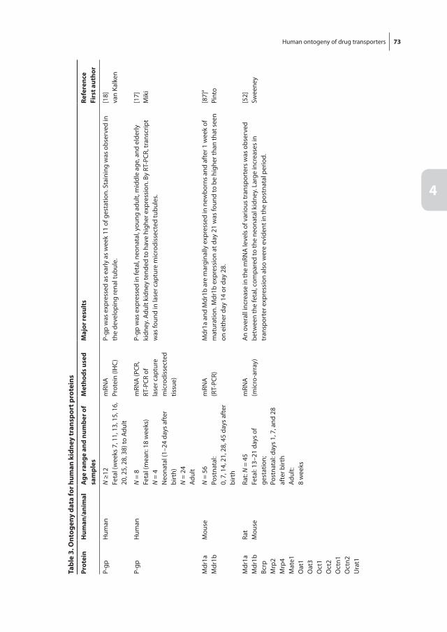
72 Chapter 4 Human ontogeny of drug transporters 73
4
tabl
e 3.
ont
ogen
y da
ta fo
r hum
an k
idne
y tr
ansp
ort p
rote
ins
Prot
ein
hum
an/a
nim
alA
ge ra
nge
and
num
ber o
f sa
mpl
esM
etho
ds u
sed
Maj
or re
sult
sre
fere
nce
firs
t aut
hor
P-gp
Hum
anN
≥12
Feta
l (w
eeks
7, 1
1, 1
3, 1
5, 1
6,
20, 2
5, 2
8, 3
8) to
Adu
lt
mRN
APr
otei
n (IH
C)P-
gp w
as e
xpre
ssed
as
early
as
wee
k 11
of g
esta
tion.
Sta
inin
g w
as o
bser
ved
in
the
deve
lopi
ng re
nal t
ubul
e.[1
8]va
n Ka
lken
P-gp
Hum
anN
= 8
Feta
l (m
ean:
18
wee
ks)
N =
4N
eona
tal (
1–24
day
s af
ter
birt
h)N
= 2
4Ad
ult
mRN
A (P
CR,
RT-P
CR o
f la
ser c
aptu
re
mic
rodi
ssec
ted
tissu
e)
P-gp
was
exp
ress
ed in
feta
l, ne
onat
al, y
oung
adu
lt, m
iddl
e ag
e, a
nd e
lder
ly
kidn
ey. A
dult
kidn
ey te
nded
to h
ave
high
er e
xpre
ssio
n. B
y RT
-PCR
, tra
nscr
ipt
was
foun
d in
lase
r cap
ture
mic
rodi
ssec
ted
tubu
les.
[17]
Mik
i
Mdr
1aM
dr1b
Mou
seN
= 5
6Po
stna
tal:
0, 7
, 14,
21,
28,
45
days
aft
er
birt
h
mRN
A(R
T-PC
R)M
dr1a
and
Mdr
1b a
re m
argi
nally
exp
ress
ed in
new
born
s an
d af
ter 1
wee
k of
m
atur
atio
n. M
dr1b
exp
ress
ion
at d
ay 2
1 w
as fo
und
to b
e hi
gher
than
that
see
n on
eith
er d
ay 1
4 or
day
28.
[87]
a
Pint
o
Mdr
1aM
dr1b
Bcrp
Mrp
2M
rp4
Mat
e1O
at1
Oat
3O
ct1
Oct
2O
ctn1
Oct
n2U
rat1
Rat
Mou
seRa
t: N
= 4
5Fe
tal:
13–2
1 da
ys o
f ge
stat
ion;
Post
nata
l: da
ys 1
, 7, a
nd 2
8 af
ter b
irth
Adul
t:8
wee
ks
mRN
A(m
icro
-arr
ay)
An
over
all i
ncre
ase
in th
e m
RNA
leve
ls o
f var
ious
tran
spor
ters
was
obs
erve
d be
twee
n th
e fe
tal,
com
pare
d to
the
neon
atal
kid
ney.
Lar
ge in
crea
ses
in
tran
spor
ter e
xpre
ssio
n al
so w
ere
evid
ent i
n th
e po
stna
tal p
erio
d.
[52]
Swee
ney
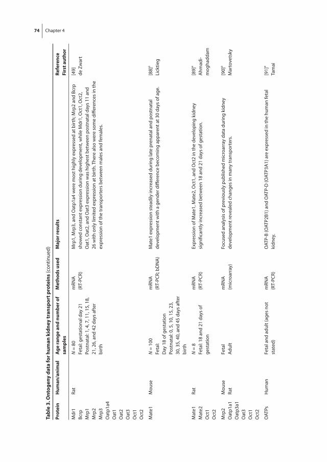
74 Chapter 4 Human ontogeny of drug transporters 75
4
tabl
e 3.
ont
ogen
y da
ta fo
r hum
an k
idne
y tr
ansp
ort p
rote
ins
(con
tinue
d)
Prot
ein
hum
an/a
nim
alA
ge ra
nge
and
num
ber o
f sa
mpl
esM
etho
ds u
sed
Maj
or re
sult
sre
fere
nce
firs
t aut
hor
Mdr
1Bc
rpM
rp1
Mrp
2M
rp3
Oat
p1a4
Oat
1O
at2
Oat
3O
ct1
Oct
2
Rat
N =
80
Feta
l: ge
stat
iona
l day
21
Post
nata
l: 1,
4, 7
, 11,
15,
18,
21
, 26,
and
42
days
aft
er
birt
h
mRN
A(R
T-PC
R)M
rp1,
Mrp
3, a
nd O
atp1
a4 w
ere
mos
t hig
hly
expr
esse
d at
birt
h; M
rp2
and
Bcrp
sh
owed
con
stan
t exp
ress
ion
durin
g de
velo
pmen
t, w
hile
Mdr
1, O
ct1,
Oct
2,
Oat
1, O
at2,
and
Oat
3 ex
pres
sion
was
hig
hest
bet
wee
n po
stna
tal d
ays
11 a
nd
26 w
ith o
nly
limite
d ex
pres
sion
at b
irth.
The
re a
lso
wer
e so
me
diffe
renc
es in
the
expr
essi
on o
f the
tran
spor
ters
bet
wee
n m
ales
and
fem
ales
.
[49]
de Z
war
t
Mat
e1M
ouse
N =
100
Feta
l:D
ay 1
8 of
ges
tatio
nPo
stna
tal:
0, 5
, 10,
15,
23,
30
, 35,
40,
and
45
days
aft
er
birt
h
mRN
A(R
T-PC
R; b
DN
A)
Mat
e1 e
xpre
ssio
n st
eadi
ly in
crea
sed
durin
g la
te p
rena
tal a
nd p
ostn
atal
de
velo
pmen
t with
a g
ende
r diff
eren
ce b
ecom
ing
appa
rent
at 3
0 da
ys o
f age
.[8
8]a
Lick
tieg
Mat
e1M
ate2
Oct
1O
ct2
Rat
N =
8Fe
tal:
18 a
nd 2
1 da
ys o
f ge
stat
ion
mRN
A(R
T-PC
R)Ex
pres
sion
of M
ate1
, Mat
e2, O
ct1,
and
Oct
2 in
the
deve
lopi
ng k
idne
y si
gnifi
cant
ly in
crea
sed
betw
een
18 a
nd 2
1 da
ys o
f ges
tatio
n.[8
9]a
Ahm
adi-
mog
hadd
am
Mrp
2O
atp1
a1O
atp3
a1O
at3
Oct
1O
ct2
Mou
seRa
tFe
tal
Adul
tm
RNA
(m
icro
arra
y)Fo
cuse
d an
alys
is o
f pre
viou
sly
publ
ishe
d m
icro
arra
y da
ta d
urin
g ki
dney
de
velo
pmen
t rev
eale
d ch
ange
s in
man
y tr
ansp
orte
rs.
[90]
a
Mar
tove
tsky
OAT
PsH
uman
Feta
l and
adu
lt (a
ges
not
stat
ed)
mRN
A(R
T-PC
R)O
ATP-
B (O
ATP2
B1) a
nd O
ATP-
D (O
ATP3
A1)
are
exp
ress
ed in
the
hum
an fe
tal
kidn
ey.
[91]
a
Tam
ai

74 Chapter 4 Human ontogeny of drug transporters 75
4
tabl
e 3.
ont
ogen
y da
ta fo
r hum
an k
idne
y tr
ansp
ort p
rote
ins
(con
tinue
d)
Prot
ein
hum
an/a
nim
alA
ge ra
nge
and
num
ber o
f sa
mpl
esM
etho
ds u
sed
Maj
or re
sult
sre
fere
nce
firs
t aut
hor
Oat
p1a1
Oat
p1a6
Oat
p3a1
Mou
seN
= 1
00Fe
tal:
Day
18
of g
esta
tion
Post
nata
l: 0,
5, 1
0, 1
5, 2
2,
30, 3
5, 4
0, a
nd 4
5 da
ys a
fter
bi
rth
mRN
A(R
T-PC
R; b
DN
A)
Oat
p1a1
, Oat
p1a6
, and
Oat
p3a1
dis
play
low
er e
xpre
ssio
n at
birt
h co
mpa
red
to
6 w
eeks
late
r, w
hile
oth
er O
atps
had
sim
ilar l
evel
s of
exp
ress
ion
at b
oth
time
poin
ts.
[92]
a
Chen
g
Oat
1O
at3
Mrp
2M
rp4
Rat
N =
63
Post
nata
l: 1,
2, 4
, 6, 1
4, 2
1,
and
28 d
ays
afte
r birt
h (N
=
5–8
mRN
A; N
= 4
–6 p
rote
in)
mRN
A(R
T-PC
R)Pr
otei
n (w
este
rn
blot
and
imm
u-no
hist
oche
mis
-tr
y (IH
C))
Leve
ls o
f Oat
1 an
d O
at3
mRN
A a
nd p
rote
in in
crea
sed
sign
ifica
ntly
from
po
stna
tal d
ays
1 to
28.
Nei
ther
Mrp
4 m
RNA
nor
pro
tein
cha
nged
sig
nific
antly
ov
er th
e sa
me
time
fram
e. M
rp2
prot
ein
incr
ease
d fr
om p
ostn
atal
day
s 14
to 2
1.
[93]
a
Nom
ura
Oat
sRa
tEn
gine
ered
kid
ney
tissu
e fr
om e
mbr
yoni
c ru
dim
ents
Prot
ein
func
tion
(func
tiona
l up
take
ass
ay)
Diff
eren
tiatin
g tu
bule
s of
eng
inee
red
reco
mbi
ned
embr
yoni
c ki
dney
tiss
ue a
re
func
tiona
lly c
apab
le o
f org
anic
ani
on tr
ansp
ort.
[94]
a
Rosi
nes
Oat
1Ra
tFe
tal:
17–2
0 da
ys o
f ge
stat
ion
Post
nata
l: 0,
1, 2
, and
6 d
ays
of a
geAd
ult:
8–10
wee
ks
mRN
A (N
orth
ern
blot
, in
situ
hy
brid
izat
ion)
Pr
otei
n (W
este
rn
blot
and
IHC)
Oat
1 m
RNA
is d
etec
tabl
e at
day
18
of g
esta
tion,
and
Oat
1 pr
otei
n is
det
ecta
ble
at d
ay 2
0 of
ges
tatio
n. T
he e
xpre
ssio
n of
bot
h O
at1
mRN
A a
nd O
at1
prot
ein
incr
ease
mar
kedl
y af
ter b
irth.
[95]
a
Nak
ajim
a
Oat
1M
ouse
Feta
l: 15
–19
days
of
gest
atio
nAd
ult:
8–10
wee
ks
mRN
A (N
orth
ern
blot
)O
at1
mRN
A e
xpre
ssio
n by
Nor
ther
n bl
ot w
as fo
und
at d
ay 1
8 of
ges
tatio
n an
d co
ntin
ued
to in
crea
se b
efor
e bi
rth.
[96]
a
Lope
z-N
ieto
Oat
1M
ouse
N =
44
Feta
l: 13
.5–1
8.5
days
of
gest
atio
nPo
stna
tal:
1, 7
, 21,
28
days
aft
er b
irth
Adul
t: 8–
10 w
eeks
mRN
A(R
T-PC
R)Th
e ex
pres
sion
of O
at1,
whi
ch w
as in
itial
ly o
bser
ved
at 1
5.5
days
of g
esta
tion,
pr
ogre
ssiv
ely
incr
ease
d to
adu
lt le
vels
.[9
7]a
Parr
eira

76 Chapter 4
tabl
e 3.
ont
ogen
y da
ta fo
r hum
an k
idne
y tr
ansp
ort p
rote
ins
(con
tinue
d)
Prot
ein
hum
an/a
nim
alA
ge ra
nge
and
num
ber o
f sa
mpl
esM
etho
ds u
sed
Maj
or re
sult
sre
fere
nce
firs
t aut
hor
Oat
1O
at3
Mou
seCu
lture
d em
bryo
nic
kidn
ey,
rena
l slic
es fr
om k
nock
out
tissu
e
Prot
ein
func
tion
(func
tiona
l up
take
ass
ay)
Embr
yoni
c ki
dney
in o
rgan
cul
ture
tran
spor
ts a
ntiv
iral d
rugs
by
Oat
1 an
d O
at3.
U
ptak
e of
fluo
resc
ent t
race
rs in
cor
onal
slic
es o
f adu
lt ki
dney
from
Oat
1 an
d O
at3
knoc
kout
mic
e re
veal
ed fu
nctio
nal l
ocal
izat
ion
of O
at1
and
Oat
3. O
at1
func
tion
appe
ared
to b
e co
nsis
tent
with
loca
lizat
ion
in th
e pr
oxim
al tu
bule
, w
hile
Oat
3 fu
nctio
n se
emed
to b
e fo
und
in b
oth
the
prox
imal
and
dis
tal t
ubul
e.
[98]
a
Nag
le
Oat
1O
at3
Mou
seN
= 1
3Fe
tal:
13–1
8 da
ys o
f ge
stat
ion
Post
nata
l: da
ys 1
, 4, 7
, 14,
an
d 21
day
s af
ter b
irth
Adul
t: 8–
10 w
eeks
of a
ge
Prot
ein
(IHC)
Oat
1 w
as fi
rst d
etec
ted
on d
ay 1
5 of
ges
tatio
n an
d lo
caliz
ed to
the
prox
imal
tu
bule
of t
he in
ner c
orte
x. O
at1
expr
essi
on c
ontin
ued
to in
crea
se, a
ppea
ring
in th
e ou
ter c
orte
x at
7 d
ays
afte
r birt
h an
d by
3 w
eeks
Oat
1 di
strib
utio
n w
as
com
para
ble
to th
at s
een
in a
dult
kidn
eys.
Oat
3 w
as fi
rst d
etec
tabl
e on
day
14
of
gest
atio
n in
the
dist
al tu
bule
and
was
late
r loc
aliz
ed to
the
S2 s
egm
ent o
f the
pr
oxim
al tu
bule
. Aro
und
the
time
of b
irth,
Oat
3 w
as lo
caliz
ed to
the
S1 a
nd S
3 se
gmen
ts o
f the
pro
xim
al tu
bule
, and
Oat
3 ex
pres
sion
con
tinue
d to
incr
ease
th
roug
h po
stpa
rtum
day
21.
[99]
a
Hw
ang
Oat
1O
at3
Mou
seCu
lture
d em
bryo
nic
kidn
ey
knoc
kout
tiss
uePr
otei
n(fu
nctio
nal
upta
ke a
ssay
)
Cultu
red
who
le e
mbr
yoni
c ki
dney
s fr
om O
at1
and
Oat
3 kn
ocko
ut m
ice
wer
e ab
le to
tran
spor
t man
y an
tivira
ls e
x vi
vo. C
erta
in a
ntiv
irals
wer
e fo
und
to h
ave
sele
ctiv
e de
pend
ency
on
Oat
1 or
Oat
3.
[100
]a
Truo
ng
Oat
1O
at2
Oat
3
Mou
seN
= 9
0Po
stna
tal:
0, 5
, 10,
15,
20,
25
, 30,
35,
and
40
days
aft
er
birt
h
mRN
A(R
T-PC
R; b
DN
A)
Post
nata
l Oat
1 an
d O
at2
leve
ls o
f exp
ress
ion
incr
ease
d af
ter 2
5 da
ys o
f age
, w
hile
Oat
3 ex
pres
sion
leve
ls g
radu
ally
incr
ease
d fr
om b
irth
to 4
5 da
ys o
f age
. O
vera
ll O
at2
leve
ls in
crea
sed
30-fo
ld, w
hile
Oat
1 an
d O
at3
leve
ls in
crea
sed
abou
t 2-fo
ld.
[101
]a
Buis
t
Oat
1O
at3
Shee
pN
= 2
8Fe
tal:
80, 1
00, 1
20, 1
30, a
nd 1
45
days
of g
esta
tion
Post
nata
l: 1
and
7 da
ys a
fter
bi
rth
mRN
A(R
T-PC
R)Pr
otei
n(IH
C)
Oat
1 w
as e
xpre
ssed
at 8
0 da
ys o
f ges
tatio
n an
d w
as m
ost h
ighl
y ex
pres
sed
at 1
45 d
ays
of g
esta
tion;
the
post
nata
l kid
ney
show
ed c
ompa
rabl
e le
vels
of
expr
essi
on. O
at3
disp
laye
d a
sim
ilar p
atte
rn o
f exp
ress
ion,
alth
ough
sig
nific
ant
incr
ease
s w
ere
evid
ent i
n th
e po
stna
tal k
idne
y. Im
mun
ohis
toch
emic
al a
naly
sis
reve
aled
loca
lizat
ion
of th
e tr
ansp
orte
rs to
tubu
lar e
pith
elia
con
sist
ent w
ith th
e pr
oxim
al tu
bule
.
[102
]a
Woo
d
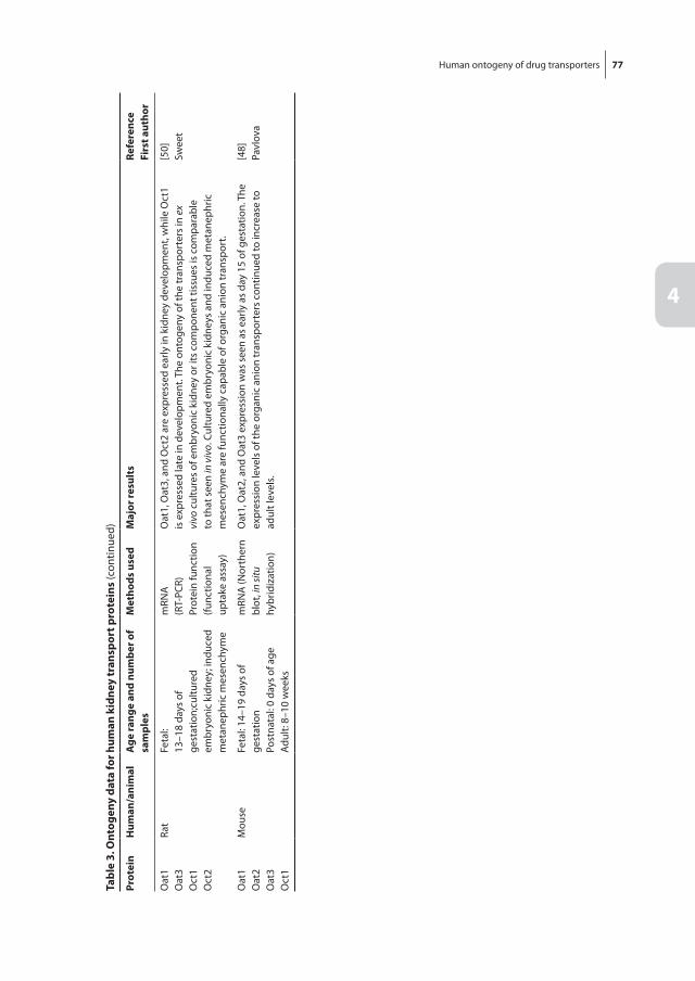
Human ontogeny of drug transporters 77
4
tabl
e 3.
ont
ogen
y da
ta fo
r hum
an k
idne
y tr
ansp
ort p
rote
ins
(con
tinue
d)
Prot
ein
hum
an/a
nim
alA
ge ra
nge
and
num
ber o
f sa
mpl
esM
etho
ds u
sed
Maj
or re
sult
sre
fere
nce
firs
t aut
hor
Oat
1O
at3
Oct
1O
ct2
Rat
Feta
l:13
–18
days
of
gest
atio
n;cu
lture
d em
bryo
nic
kidn
ey; i
nduc
ed
met
anep
hric
mes
ench
yme
mRN
A(R
T-PC
R)Pr
otei
n fu
nctio
n(fu
nctio
nal
upta
ke a
ssay
)
Oat
1, O
at3,
and
Oct
2 ar
e ex
pres
sed
early
in k
idne
y de
velo
pmen
t, w
hile
Oct
1 is
exp
ress
ed la
te in
dev
elop
men
t. Th
e on
toge
ny o
f the
tran
spor
ters
in e
x vi
vo c
ultu
res
of e
mbr
yoni
c ki
dney
or i
ts c
ompo
nent
tiss
ues
is c
ompa
rabl
e to
that
see
n in
viv
o. C
ultu
red
embr
yoni
c ki
dney
s an
d in
duce
d m
etan
ephr
ic
mes
ench
yme
are
func
tiona
lly c
apab
le o
f org
anic
ani
on tr
ansp
ort.
[50]
Swee
t
Oat
1O
at2
Oat
3O
ct1
Mou
seFe
tal:
14–1
9 da
ys o
f ge
stat
ion
Post
nata
l: 0
days
of a
geAd
ult:
8–10
wee
ks
mRN
A (N
orth
ern
blot
, in
situ
hy
brid
izat
ion)
Oat
1, O
at2,
and
Oat
3 ex
pres
sion
was
see
n as
ear
ly a
s da
y 15
of g
esta
tion.
The
ex
pres
sion
leve
ls o
f the
org
anic
ani
on tr
ansp
orte
rs c
ontin
ued
to in
crea
se to
ad
ult l
evel
s.
[48]
Pavl
ova
78 Chapter 4
regarding the developmental expression of renal transporters in humans,[51] presented as an abstract, appears largely consistent with patterns reported in rats.[52]
In summary, information on the ontogeny of human drug transporters is scarce in the case of the kidney. A major knowledge gap exists regarding gene expression, protein abundance, and actual transporter activity in humans. This information is essential to understand how maturational changes impact the role that these transporters play in normal growth and development, and to accurately predict the impact of changes due to pathophysiological conditions on drug disposition, efficacy, and toxicity of medica-tions. This knowledge is requisite to the development of personalized drug therapy in children.
coMPArAtiVE DEVEloPMEnt of orGAn functions
The majority of the data regarding transporter ontogeny has been obtained from devel-opmental studies in rodents, although some literature does exist for larger species. The ability to extrapolate across species, namely rodents, to larger species (e.g., primates) and humans can be limited, however, by variation in the developmental timing of key ana-tomical, physiological, biochemical, and physicochemical events, as well as significant functional variance in isoforms. These differences arise from comparative differences in the gestational length and the timing of parturition between the different species and humans. The conventional approach is to compare developmental milestones relative to birth (prenatal vs. postnatal); however, this may not always be appropriate. For example, in contrast to humans and porcine models, newborn rodents exhibit relatively immature intestines with few villi and little evidence of crypt formation.[53, 54] The timing of tis-sue maturation between rodent species is also dependent on the organ. As an example, nephrogenesis is largely complete prior to birth in mice, which is similar to humans, but continues in rats during the postnatal period.[55] For this reason, the study of transporter ontogeny would be improved by reporting perinatal findings as days postconception, rather than days relative to parturition. By viewing development as a continuum instead of arbitrary categories (such as neonatal, infant, etc.), there will be greater potential to translate rodent and other mammalian studies to humans. One caveat to this approach is that some transporters increase in expression upon commencement of feeding, as has been observed for Ntcp, Bsep, and Mrp4 in the neonatal livers of mice.[42]
It should be noted that another considerable limitation occurs when contrasting mRNA expression data, because linearity in protein transcription and inferred function cannot be assumed. As mentioned in the Methods section, advances in quantitative pro-teomics using LC-MS/MS have led to a significant increase in our ability to quantify drug transporter protein abundance in adult samples. While a number of technical challenges
Human ontogeny of drug transporters 79
4
still exist with this methodology, the application of quantitative proteomic approaches to ontogenic studies with pediatric tissues will yield more useful data for establishing predictive developmental physiologically based pharmacokinetic (PBPK) models. One major limitation is that the availability of human pediatric tissue specimens is limited, and shared pediatric biobanks need to be established. The application of quantitative proteomics in animal tissue developmental studies would provide greater insight into the utility of scaling across species to predict the function of drug transporter activity in pediatric patients.
There is also the potential for significant confounding variables to limit the ability of cross-species comparisons with humans, particularly with respect to pediatric popula-tions. Of considerable concern is the health of the patients in which the specimens were obtained. A significant portion of the human pediatric tissue biopsied specimens origi-nate from patients who suffer from comorbidity or are collected postmortem. Collection procedures and timing also can impact tissue quality and subsequent expression data. Most animal studies are conducted under controlled conditions, whereas human tissue specimens are collected from patients who will have varying xenobiotic and dietary exposures. These confounding factors cannot be readily normalized for in many cases. Another emerging issue is that mRNA expression of housekeeper genes can vary signifi-cantly, thus calling into question quantitative data from qRT-PCR studies normalized to one control.[56, 57] These concerns may be alleviated by the use of RNA-sequencing to quantify human transporter ontogeny, as has been performed in rodents.[47]
There has been limited use of cell-based systems and mathematical models to describe the ontogeny of transporters. However, there may be the potential to complement in vivo rodent, other species, and human biopsy studies with human embryonic or induced pluripotent stem cells undergoing differentiation to hepatocytes, enterocytes, and renal tubule cells. While these cellular systems are artificial and lack the holistic development of an organism, they may be a mechanistic tool to probe the effects of exogenous fac-tors such as hormones, drugs, and exposures on the sequence and timing of transporter expression. Initial studies have begun to profile the expression and activity of transport-ers in hepatocyte-like cells derived from human embryonic and induced pluripotent stem cells,[58] although comparisons with juvenile human livers are needed. In addi-tion, there is a need to develop mathematical and statistical modeling approaches that integrate transporter ontogeny with the maturation of physiological processes, such as tubule reabsorption or intestinal secretion. This effort would provide researchers with the ability to “translate time” between species by developing algorithms that compare and predict development.[59, 60] Moreover, this approach can integrate the ontogeny of multiple tissues in parallel and provide a more global view on whole organism de-velopment. A computational and systems biology approach could be used to integrate genomic, epigenetic, proteomic, and pharmacokinetic endpoints (e.g., changes in pH,
80 Chapter 4
plasma membrane composition, expression of drug metabolizing enzymes) to better assess the ontogeny of transport systems.[61] The utility of data-driven, PBPK models generated in this fashion would help to improve new pediatric drug translation from discovery to the clinical stages.[62]
EMErGinG ArEAs AnD MAjor chAllEnGEs in stuDyinG PEDiAtric DruG trAnsPort
Developmental programming and regulation of transportersLittle is known about the factors that govern the regulation of transporter expression and activity (e.g., induction and inhibition of transporters as a function of gestational age) during growth and development. The age-related variation in mRNA expression of the transcription factor PXR correlated with P-gp expression in a small number of fetal, neonatal, younger and older adult samples of human liver, kidney, and intestine.[17] This finding suggests a role for transcription factor-mediated regulation of age-related transporter expression. Moreover, the mechanism of age-related changes in transcription factors may be related to DNA methylation; in fetal liver, hypermethylation of important cytochrome P450 (CYP) 3A4 transcription factor binding sites has been observed, consistent with low CYP3A4 expression before birth.[63, 64] Endocrine changes in adolescents may impact drug transporter expression through hormonal and growth factor regulation of relevant transcription factors. To date, studies focused on the endocrine regulation of transporters have largely evaluated adult rodents[65, 66] and serve as a basis for future work that should be expanded to hormonal fluctuations during human development. Clearly, considerable work is needed to understand the factors that regulate drug transporters during human growth and development.
PharmacogenomicsGenetic variation may add to the age-related variation in drug transporter expression and function. In contrast to adults, pharmacogenomic drug transporter studies in children are rare. The available studies have been performed primarily in age ranges at which full maturation of transporters can be expected, and the results in general are similar to adults.[12, 67] Further studies in children are needed to elucidate the interplay of age and genetic variation. Decreased transporter expression due to genetic variation may not become apparent until expression is at least at adult levels.[68]
Impact of disease, drug interactions, and/or environmental exposureOther factors impacting drug transporter activity are disease, drug–gene interactions, drug–drug interactions (DDIs), food–drug interactions as well as exposures to environ-
Human ontogeny of drug transporters 81
4
mental chemicals. In adults, the impact of liver disease has been investigated, but the findings may not be applicable to children, as the underlying disease may be very dif-ferent: e.g., alcohol-induced liver steatosis is not a pediatric disease, while biliary atresia is the most prevalent disease in children who receive a liver transplant. As underlying liver disease may impact transporter expression,[69, 70] these pathophysiological dif-ferences mandate studies in pediatric patients. In one study, MRP2, BSEP, and MDR3 expression in livers from patients with pediatric biliary atresia taken at postnatal age 1–2 months was much higher than both fetal and adult expression levels, but unfortunately no age-matched controls were studied.[36] Hence, it is unclear whether the observed differences can be attributed solely to the disease or whether age-related changes also played a role. DDIs also may be different in children due to developmental changes in drug disposition pathways; furthermore, the potential for a specific DDI may change during growth and development.[71] Finally, as nutrition changes during childhood, the impact of frequent milk or different types of formula feedings and fruit juice on drug transporter expression and function must be considered.
Development of human-relevant cell/in vitro and preclinical/in vivo transport models that are representative of the pediatric population in health and diseaseOnce transport protein expression and function have been characterized across the pediatric age spectrum in healthy and diseased tissue, an important next step will be to develop human-relevant in vitro models, such as cell lines, embryonic stem cells, or modified primary cells that mimic transporter expression and function at various ages. Such systems could be useful to predict drug disposition and DDIs in pediatrics. The utility of preclinical, in vivo transporter models as a predictive tool is less clear due to significant species differences in the expression and function of some transport proteins and regulatory machinery, and lack of established correlations between transporter data from animal models and humans.
Development of systems-based, mechanistic modeling approaches to integrate in vitro data and physiological processes to predict transporter-mediated changes in drug disposition as a function of age and diseasePBPK models link information about the anatomical and physiological structure of the body with the physicochemical and biopharmaceutical properties of the drug to predict drug disposition in the body. PBPK models are established tools for predicting human pharmacokinetics based on preclinical data from animals and in vitro studies.[72, 73] Successful applications include scaling the human pharmacokinetics from healthy volunteers to patient populations (e.g., liver cirrhosis) by accounting for changes in physiology.[74, 75] PBPK models are being applied increasingly in scaling adult pharma-cokinetics to pediatric populations. This has been demonstrated for compounds elimi-

82 Chapter 4
nated primarily by metabolism, where the ontogeny has been better characterized than for some transporters.[76, 77] The use of modeling and simulation methods in pediatric drug development was endorsed at a recent US Food and Drug Administration (FDA) Clinical Pharmacology Advisory Committee meeting,[78] and also is recognized by the agency as a potentially useful tool in the design of pediatric clinical trials and helping to expedite pediatric drug development.[79] Between the years 2008 and 2012, the FDA’s Office of Clinical Pharmacology received 33 New Drug Application/Investigational New Drug submissions containing PBPK models; six of them were pediatric submissions.[80] The main applications of pediatric PBPK models in these submissions included dose selection, study design, informing enzyme ontogeny using benchmark drugs, and facilitating covariate analysis. The applications in pediatric drug development generally start with development and validation of an adult model (frequently utilizing in vitro characterization of the drug’s interaction with enzymes and/or transporters) followed by scaling to pediatric populations by accounting for ontogeny of relevant physiologi-cal processes across the age continuum from neonates (including premature neonates) to adults. However, currently there is limited availability of transporter information for modeling and simulation (e.g., PBPK modeling) of drug disposition, tissue exposure, and pharmacodynamic response in pediatrics. A possible approach to circumvent this issue is to utilize available pediatric clinical data for a variety of drugs to estimate the ontogeny of relevant enzymes and transporters. In one example, the ontogeny of renal transport was estimated from age-dependent renal clearance of a model compound known to be a substrate for the same renal transporter as the investigated drug.[81] In another example, a pediatric population model for zidovudine was constructed by utilizing a previously derived pediatric covariate model for morphine glucuronidation.[82] Finally, it is worth noting that systems biology approaches, as well as methods for integration of “omics” data from multiple levels of analysis (e.g., genomics, transcriptomics, proteomics, metabolomics) are advancing rapidly, and there is an ongoing effort to apply these ap-proaches to the adult and developmental contexts.[83] While distinct from the usual methods used in PBPK, it is anticipated that these approaches may begin to converge in the near future, providing a deeper understanding of pediatric drug disposition.
Limited availability of quality pediatric tissue (all age groups) for protein quantification and assessment of transporter functionOne of the major roadblocks to drug transporter research during human growth and development is the dearth of quality pediatric tissue. Current tissue sources include left-over tissue from surgery and biopsies, as well as postmortem tissue from organ transplants and autopsies. Collaboration between clinicians with access to these tissue sources and researchers in need of tissue appears to be a major obstacle. In order to over-come these logistical challenges, a clear understanding and commitment on both sides
Human ontogeny of drug transporters 83
4
regarding the respective challenges and solutions may be the first step to increase the availability of quality pediatric tissue. For example, the logistics of collecting a sample, including asking for informed consent, retrieving a dry-ice container, transporting tissue to a storage facility, and collecting clinical data seem relatively straightforward, but are challenging when success depends on busy clinical staff to organize all these details. Also, a few inches of residual intestine may be very reasonable to obtain in adults, but cannot be considered “left-over” in neonates with a considerably shorter intestinal length. Timely handling of postmortem pediatric tissue is particularly challenging due to parents, who may need time to say goodbye when a child passes away, and the availability of autopsies only during the day. Furthermore, the availability of tissue may be limited compared to adults because the death rate among children is much lower than among adults, there may be reluctance on the part of parents to give consent for autopsy, and there are relatively few liver transplants from pediatric donors.
Ethical and practical challenges with performing nontherapeutic studies in minorsResearch that does not potentially benefit the participating child (“nontherapeutic”) is subject to several limitations,[84] most importantly, the restrictions of minimal risk (a slight increase over what is ordinarily encountered in daily life) and minimal burden. These ethical limitations, which are intended to protect the individual child, limit the possibility of performing nontherapeutic pharmacokinetics and/or DDI studies related to drug transporters in minors. Giving a child a therapeutic dose of a drug solely for these purposes will, in many places, not be considered minimal risk. However, in some centers this approach is acceptable when the child will receive the drug later for therapeutic reasons. One solution is to study the disposition and effect of the drug in the context of clinical drug treatment. This may introduce unwanted complexity to studies, such as variation due to underlying disease and/or comedication, and the need for more sophisticated pharmacokinetic analyses. Microdosing may overcome these limitations, but can only be used for drugs that exhibit dose-linearity.[85] Other challenges include the need for repeated blood sampling as well as limitations with blood volumes. These can be overcome by using indwelling catheters already in place for clinical care, low volume drug assays, and population pharmacokinetic analyses.
Lack of transporter-specific probes to assess in vivo functionIn vivo probes for individual drug metabolizing enzymes (e.g., midazolam for CYP3A4, dextromethorphan for CYP2D6/CYP3A4, and caffeine for CYP1A2) have facilitated investigations about the impact of growth and development, and the effects of phar-macogenomics and disease, on drug metabolism. These critical tools have led to new knowledge. A similar approach is more challenging for drug transporters, as specific probes for individual transporters are lacking. Most drugs are substrates for multiple

84 Chapter 4
transporters, which enable alternate transporter pathways to compensate in case the primary transporter is absent or has reduced activity. Nevertheless, pharmacogenomic studies on individual transporters have elucidated differences in drug disposition, efficacy, and safety in adults. This approach may aid in studying the developmental changes of these transporters in vivo. The use of microdosing in pediatric patients has been explored and may provide a basis for developing a better understanding of drug disposition and metabolomic profiling. While it is important to continue to analyze mRNA and protein expression levels of transporters in developing organs like the liver, kidney, and intestine, ultimately this must be related to physiological processes medi-ated by these particular transporters. Because of unique safety concerns about using exogenous compounds as functional probes in the pediatric population, continued exploration of endogenous metabolites and other markers that can serve as surrogates for assessing transporter function during organ development and maturation is needed. The relevant sets of endogenous metabolites may be specific to each organ, and to particular developmental points, reflecting unique patterns of transporter expression and aspects of organ-specific physiology.
table 4. recommendations
• Buildmultidisciplinary,internationalcollaborativenetworkstofacilitatecollectionandsharingofdataon pediatric transporters, including expertise in preclinical studies (e.g., knockout and in vitro models), pediatrics, clinical pharmacology, pharmacogenomics, pharmacometrics, and pharmacovigilance
• Establishcentral(perhapsregional)tissuerepositorieswheresurgicalandpostmortemsamplescanbestored with clear guidelines for tissue collection and handling to preserve sample integrity
• Continuetosupportthetrainingofscientistsinpediatricclinicalpharmacologywithexpertiseintransporters, pharmacogenomics, pharmacometrics, and pharmacovigilance
• Increasetheawarenessofcliniciansregardingtheimportanceoftransportersinpediatricdrugdisposition
• Identifyexamplesrelevanttopediatricpharmacotherapywheredevelopmentaldifferencesintransporterexpression or activity could translate into clinically relevant effects
• Workwithprofessionalgroupstodevelopguidelinesonhowdrugtherapymaybealteredduetovariations in transporter expression or activity
• Identifyselectiveandspecificbiomarkersfortransporteractivityinpediatricpatients
• Investigatebasicdevelopmentalmechanismsregulatingtransporterexpressionandactivityinthedifferent organs in pediatric health and disease
• Developpediatric-relevantin vitro/in silico and systems biology models to predict transporter function in the context of overall drug disposition
Human ontogeny of drug transporters 85
4
conclusion
Many fundamental and clinically relevant questions remain unanswered about the hu-man ontogeny of drug transporters. As information highlighting the importance of drug transporters in adult medicine continues to emerge, this critical knowledge gap in the pediatric population becomes even more evident. In order to achieve safe and effective drug therapy for children, it is imperative that developmental patterns of transporter gene expression and protein abundance are elucidated, and that drug transporter func-tion is defined across the age spectrum. Recommendations are provided (Table 4) to address some of the major challenges in obtaining this information. Fundamental and applied knowledge about the human ontogeny of drug transporters is absolutely es-sential to ultimately achieve personalized pharmacotherapy in pediatric patients.
Additional Supporting Information may be found in the online version of this article.Appendix 1 Literature Search StrategyAppendix 2 Ontogeny of Rodent Kidney Transport Proteins
86 Chapter 4
rEfErEncEs
[1] Giacomini, K.M. et al. Membrane transporters in drug development. Nat. Rev. Drug Discov. 9, 215–236 (2010).
[2] DeGorter, M.K., Xia, C.Q., Yang, J.J. & Kim, R.B. Drug transporters in drug efficacy and toxicity. Annu. Rev. Pharmacol. Toxicol. 52, 249–273 (2012)
[3] Yan, H. et al. Sodium taurocholate cotransporting polypeptide is a functional receptor for human hepatitis B and D virus. eLife 1, e00049 (2012).
[4] Ho, R.H. & Kim, R.B. Transporters and drug therapy: implications for drug disposition and disease. Clin. Pharmacol. Ther. 78, 260–277 (2005).
[5] Yasuda, S.U., Zhang, L. & Huang, S.M. The role of ethnicity in variability in response to drugs: focus on clinical pharmacology studies. Clin. Pharmacol. Ther. 84, 417–423 (2008).
[6] Niemi, M. Transporter pharmacogenetics and statin toxicity. Clin. Pharmacol. Ther. 87, 130–133 (2010).
[7] Wilke, R.A. et al. The clinical pharmacogenomics implementation consortium: CPIC guideline for SLCO1B1 and simvastatin-induced myopathy. Clin. Pharmacol. Ther. 92, 112–117 (2012).
[8] Ramsey, L.B. et al. Genome-wide study of methotrexate clearance replicates SLCO1B1. Blood 121, 898–904 (2013).
[9] Liu, Y. et al. Association of ABCC2 -24C>T polymorphism with high-dose methotrexate plasma concentrations and toxicities in childhood acute lymphoblastic leukemia. PLoS One 9, e82681 (2014).
[10] Rau, T., Erney, B., Gores, R., Eschenhagen, T., Beck, J. & Langer, T. High-dose methotrexate in pedi-atric acute lymphoblastic leukemia: impact of ABCC2 polymorphisms on plasma concentrations. Clin. Pharmacol. Ther. 80, 468–476 (2006).
[11] Fukuda, T. et al. UGT1A9, UGT2B7, and MRP2 genotypes can predict mycophenolic acid phar-macokinetic variability in pediatric kidney transplant recipients. Ther. Drug Monit. 34, 671–679 (2012).
[12] Venkatasubramanian, R. et al. ABCC3 and OCT1 genotypes influence pharmacokinetics of mor-phine in children. Pharmacogenomics 15, 1297–1309 (2014).
[13] Zamek-Gliszczynski, M.J., Hoffmaster, K.A., Tweedie, D.J., Giacomini, K.M. & Hillgren, K.M. High-lights from the International Transporter Consortium second workshop. Clin. Pharmacol. Ther. 92, 553–556 (2012).
[14] Mizuno, T. et al. Developmental trajectory of intestinal MDR1/ABCB1 mRNA expression in chil-dren. Br. J. Clin. Pharmacol. 77, 910–912 (2014).
[15] Fakhoury, M., Lecordier, J., Medard, Y., Peuchmaur, M. & Jacqz-Agrain, E. Impact of inflammation on the duodenal mRNA expression of CYP3A and P-glycoprotein in children with Crohn’s disease. Inflamm. Bowel Dis. 12, 745–749 (2006).
[16] Fakhoury, M. et al. mRNA expression of MDR1 and major metabolising enzymes in human fetal tissues. Drug Metab. Pharmacokinet. 24, 529–536 (2009).
[17] Miki, Y., Suzuki, T., Tazawa, C., Blumberg, B. & Sasano, H. Steroid and xenobiotic receptor (SXR), cytochrome P450 3A4 and multidrug resistance gene 1 in human adult and fetal tissues. Mol. Cell Endocrinol. 231, 75–85 (2005).
[18] van Kalken, C.K. et al. Multidrug resistance gene (P-glycoprotein) expression in the human fetus. Am. J. Pathol. 141, 1063–1072 (1992).
[19] Fakhoury, M. et al. Localization and mRNA expression of CYP3A and P-glycoprotein in human duodenum as a function of age. Drug Metab. Dispos. 33, 1603–1607 (2005).
Human ontogeny of drug transporters 87
4
[20] Mooij, M.G. et al. Ontogeny of human hepatic and intestinal transporter gene expression during childhood: age matters. Drug Metab. Dispos. 42, 1268–1274 (2014).
[21] Konieczna, A., Erdosova, B., Lichnovska, R., Jandl, M., Cizkova, K. & Ehrmann, J. Differential expres-sion of ABC transporters (MDR1, MRP1, BCRP) in developing human embryos. J. Mol. Histol. 42, 567–574 (2011).
[22] Weihrauch, D., Kanchanapoo, J., Ao, M., Prasad, R., Piyachaturawat, P. & Rao, M.C. Weanling, but not adult, rabbit colon absorbs bile acids: flux is linked to expression of putative bile acid trans-porters. Am. J. Physiol. Gastrointest. Liver Physiol. 290, G439–450 (2006).
[23] Hussain, I., Kellett, L., Affleck, J., Shepherd, J. & Boyd, R. Expression and cellular distribution during development of the peptide transporter (PepT1) in the small intestinal epithelium of the rat. Cell Tissue Res. 307, 139–142 (2002).
[24] Shen, H., Smith, D.E. & Brosius, F.C. 3rd. Developmental expression of PEPT1 and PEPT2 in rat small intestine, colon, and kidney. Pediatr. Res. 49, 789–795 (2001).
[25] Miyamoto, K. et al. Sequence, tissue distribution and developmental changes in rat intestinal oligopeptide transporter. Biochim. Biophys. Acta 1305, 34–38 (1996).
[26] Van, L., Pan, Y.X., Bloomquist, J.R., Webb, K.E., Jr. & Wong, E.A. Developmental regulation of a turkey intestinal peptide transporter (PepT1). Poult. Sci. 84, 75–82 (2005).
[27] Cizkova, D., Morky, J., Micuda, S., Osterreicher, J. & Martinkova, J. Expression of MRP2 and MDR1 transporters and other hepatic markers in rat and human liver and in WRL 68 cell line. Physiol. Res. 54, 419–428 (2005).
[28] Klaassen, C.D. & Aleksunes, L.M. Xenobiotic, bile acid, and cholesterol transporters: function and regulation. Pharmacol. Rev. 62, 1–96 (2010).
[29] Schuetz, E.G., Furuya, K.N. & Schuetz, J.D. Interindividual variation in expression of P-glycoprotein in normal human liver and secondary hepatic neoplasms. J. Pharmacol. Exp. Ther. 275, 1011–1018 (1995).
[30] Tang, L., Hines, R.N., Schuetz, E.G. & Meibohm, B. Age-associated protein expression of P-glyco-protein (MDR1/P-gp) and MRP2 in human pediatric liver. Clin. Pharmacol. Ther. 81, S101 (2007).
[31] Ulvestad, M. et al. Impact of OATP1B1, MDR1, and CYP3A4 expression in liver and intestine on interpatient pharmacokinetic variability of atorvastatin in obese subjects. Clin. Pharmacol. Ther. 93, 275–282 (2013).
[32] Prasad, B. et al. Interindividual variability in hepatic organic anion-transporting polypeptides and P-glycoprotein (ABCB1) protein expression: quantification by liquid chromatography tandem mass spectroscopy and influence of genotype, age, and sex. Drug Metab. Dispos. 42, 78–88 (2014).
[33] Sharma, S. et al. Hepatobiliary disposition of 17-OHPC and taurocholate in fetal human hepato-cytes: a comparison with adult human hepatocytes. Drug Metab. Dispos. 41, 296–304 (2013).
[34] Yanni, S.B. et al. Higher clearance of micafungin in neonates compared with adults: role of age-dependent micafungin serum binding. Biopharm. Drug Dispos. 32, 222–232 (2011).
[35] Prasad, B., Lai, Y., Lin, Y. & Unadkat, J.D. Interindividual variability in the hepatic expression of the human breast cancer resistance protein (BCRP/ABCG2): effect of age, sex, and genotype. J. Pharm. Sci. 102, 787–793 (2013).
[36] Chen, H.L. et al. Developmental expression of canalicular transporter genes in human liver. J. Hepatol. 43, 472–477 (2005).
[37] Deo, A.K., Prasad, B., Balogh, L., Lai, Y. & Unadkat, J.D. Interindividual variability in hepatic expres-sion of the multidrug resistance-associated protein 2 (MRP2/ABCC2): quantification by liquid chromatography/tandem mass spectrometry. Drug Metab. Dispos. 40, 852–855 (2012).
88 Chapter 4
[38] Hardikar, W., Ananthanarayanan, M. & Suchy, F.J. Differential ontogenic regulation of basolateral and canalicular bile acid transport proteins in rat liver. J. Biol. Chem. 270, 20841–20846 (1995).
[39] Thomson, M.M., Hines, R.N., Schuetz, E.G. & Meibohm, B. Age-associated expression of OATP1B1 and OATP1B3 in human pediatric liver. AAPS J. 14(Suppl 2), W4301 (2012).
[40] Hayashi, M., Hui, A., DeKeyser, J., Louie, S., Shou, M. & Xu, L. Evaluation of uptake transporters in human adult and pediatric hepatocytes. Abstracts of the 17th North American Regional ISSX International Society for the Study of Xenobiotics. Drug Metab. Rev. 43, 37–211 (2011).
[41] St-Pierre, M.V. et al. Temporal expression profiles of organic anion transport proteins in placenta and fetal liver of the rat. Am. J. Physiol. Regul. Integr. Comp. Physiol. 287, R1505–1516 (2004).
[42] Cui, J.Y. et al. Bile acids via FXR initiate the expression of major transporters involved in the enterohepatic circulation of bile acids in newborn mice. Am. J. Physiol. Gastrointest. Liver Physiol. 302, G979–996 (2012).
[43] Maher, J.M., Slitt, A.L., Cherrington, N.J., Cheng, X. & Klaassen, C.D. Tissue distribution and hepatic and renal ontogeny of the multidrug resistance-associated protein (Mrp) family in mice. Drug Metab. Dispos. 33, 947–955 (2005).
[44] Gao, B., St Pierre, M.V., Stieger, B. & Meier, P.J. Differential expression of bile salt and organic anion transporters in developing rat liver. J Hepatol 41, 201–208 (2004).
[45] Lee, H.Y., Wei, D. & Loeken, M.R. Lack of metformin effect on mouse embryo AMPK activity: impli-cations for metformin treatment during pregnancy. Diabetes Metab. Res. Rev. 30, 23–30 (2014).
[46] Chen, H.L. et al. Expression of hepatocyte transporters and nuclear receptors in children with early and late-stage biliary atresia. Pediatr. Res. 63, 667–673 (2008).
[47] Cui, J.Y. et al. RNA-Seq reveals different mRNA abundance of transporters and their alternative transcript isoforms during liver development. Toxicol. Sci. 127, 592–608 (2012).
[48] Pavlova, A., Sakurai, H., Leclercq, B., Beier, D.R., Yu, A.S. & Nigam, S.K. Developmentally regulated expression of organic ion transporters NKT (OAT1), OCT1, NLT (OAT2), and Roct. Am. J. Physiol. Renal Physiol. 278, F635–643 (2000).
[49] de Zwart, L. et al. The ontogeny of drug metabolizing enzymes and transporters in the rat. Reprod. Toxicol. 26, 220–230 (2008).
[50] Sweet, D.H., Eraly, S.A., Vaughn, D.A., Bush, K.T. & Nigam, S.K. Organic anion and cation transporter expression and function during embryonic kidney development and in organ culture models. Kidney Int. 69, 837–845 (2006).
[51] Spaans, E. et al. Expression of kidney drug transporters in children. www.ascpt.org (2014). [52] Sweeney, D.E., Vallon, V., Rieg, T., Wu, W., Gallegos, T.F. & Nigam, S.K. Functional maturation of
drug transporters in the developing, neonatal, and postnatal kidney. Mol. Pharmacol. 80, 147–154 (2011).
[53] Pacha, J. Development of intestinal transport function in mammals. Physiol. Rev. 80, 1633–1667 (2000).
[54] Walker, W.A. Development of the intestinal mucosal barrier. J. Pediatr. Gastroenterol. Nutr. 34(suppl. 1), S33–S39 (2002).
[55] Zoetis, T. & Hurtt, M.E. Species comparison of anatomical and functional renal development. Birth Defects Res. B Dev. Reprod. Toxicol. 68, 111–120 (2003).
[56] Zampieri, M. et al. Validation of suitable internal control genes for expression studies in aging. Mech. Ageing Dev. 131, 89–95 (2010).
[57] Li, R. & Shen, Y. An old method facing a new challenge: re-visiting housekeeping proteins as internal reference control for neuroscience research. Life Sci. 92, 747–751 (2013).
Human ontogeny of drug transporters 89
4
[58] Ulvestad, M. et al. Drug metabolizing enzyme and transporter protein profiles of hepatocytes derived from human embryonic and induced pluripotent stem cells. Biochem. Pharmacol. 86, 691–702 (2013).
[59] Clancy, B., Darlington, R.B. & Finlay, B.L. Translating developmental time across mammalian spe-cies. Neuroscience 105, 7–17 (2001).
[60] Nagarajan, R., Darlington, R.B., Finlay, B.L. & Clancy, B. ttime: an R package for translating the timing of brain development across mammalian species. Neuroinformatics 8, 201–205 (2010).
[61] Johnson, T.N. & Rostami-Hodjegan, A. Resurgence in the use of physiologically based pharmaco-kinetic models in pediatric clinical pharmacology: parallel shift in incorporating the knowledge of biological elements and increased applicability to drug development and clinical practice. Paediatr. Anaesth. 21, 291–301 (2011).
[62] Smits, A., Annaert, P. & Allegaert, K. Drug disposition and clinical practice in neonates: cross talk between developmental physiology and pharmacology. Int. J. Pharm. 452, 8–13 (2013).
[63] Bonder, M.J. et al. Genetic and epigenetic regulation of gene expression in fetal and adult human livers. BMC Genomics 15, 860 (2014).
[64] Ivanov, M. et al. Ontogeny, distribution and potential roles of 5-hydroxymethylcytosine in human liver function. Genome Biol. 14, R83 (2013).
[65] Maher, J.M., Cheng, X., Tanaka, Y., Scheffer, G.L. & Klaassen, C.D. Hormonal regulation of renal multidrug resistance-associated proteins 3 and 4 (Mrp3 and Mrp4) in mice. Biochem. Pharmacol. 71, 1470–1478 (2006).
[66] Cheng, X., Maher, J., Lu, H. & Klaassen, C.D. Endocrine regulation of gender-divergent mouse or-ganic anion-transporting polypeptide (Oatp) expression. Mol. Pharmacol. 70, 1291–1297 (2006).
[67] Gijsen, V. et al. Age and CYP3A5 genotype affect tacrolimus dosing requirements after transplant in pediatric heart recipients. J. Heart Lung Transplant. 30, 1352–1359 (2011).
[68] Leeder, J.S. & Kearns, G.L. Interpreting pharmacogenetic data in the developing neonate: the challenge of hitting a moving target. Clin. Pharmacol. Ther. 92, 434–436 (2012).
[69] Hardwick, R.N., Fisher, C.D., Canet, M.J., Scheffer, G.L. & Cherrington, N.J. Variations in ATP-binding cassette transporter regulation during the progression of human nonalcoholic fatty liver disease. Drug Metab. Dispos. 39, 2395–2402 (2011).
[70] Lake, A.D. et al. Analysis of global and absorption, distribution, metabolism, and elimination gene expression in the progressive stages of human nonalcoholic fatty liver disease. Drug Metab. Dispos. 39, 1954–1960 (2011).
[71] Salem, F., Johnson, T.N., Barter, Z.E., Leeder, J.S. & Rostami-Hodjegan, A. Age related changes in fractional elimination pathways for drugs: assessing the impact of variable ontogeny on meta-bolic drug-drug interactions. J. Clin. Pharmacol. 53, 857–865 (2013).
[72] Hosea, N.A. & Jones, H.M. Predicting pharmacokinetic profiles using in silico derived parameters. Mol. Pharm. 10, 1207–1215 (2013).
[73] Jones, H.M., Parrott, N., Jorga, K. & Lave, T. A novel strategy for physiologically based predictions of human pharmacokinetics. Clin. Pharmacokinet. 45, 511–542 (2006).
[74] Edginton, A.N. & Willmann, S. Physiology-based simulations of a pathological condition: predic-tion of pharmacokinetics in patients with liver cirrhosis. Clin. Pharmacokinet. 47, 743–752 (2008).
[75] Johnson, T.N., Boussery, K., Rowland-Yeo, K., Tucker, G.T. & Rostami-Hodjegan, A. A semi-mech-anistic model to predict the effects of liver cirrhosis on drug clearance. Clin. Pharmacokinet. 49, 189–206 (2010).
[76] Johnson, T.N., Rostami-Hodjegan, A. & Tucker, G.T. Prediction of the clearance of eleven drugs and associated variability in neonates, infants and children. Clin. Pharmacokinet. 45, 931–956 (2006).
90 Chapter 4
[77] Parrott, N. et al. Development of a physiologically based model for oseltamivir and simulation of pharmacokinetics in neonates and infants. Clin. Pharmacokinet. 50, 613–623 (2011).
[78] Minutes for the March 14, 2012 meeting of the Advisory Committee for Pharmaceutical Science and Clinical Pharmacology (PDF-44KB). http://www.fda.gov/downloads/AdvisoryCommittees/CommitteesMeetingMaterials/Drugs/AdvisoryCommitteefor PharmaceuticalScienceandClinical-Pharmacology/UCM306989.pdf.
[79] Huang, S.M. PBPK as a tool in regulatory review. Biopharm. Drug Dispos. 33, 51–52 (2012). [80] Huang, S.M., Abernethy, D.R., Wang, Y., Zhao, P. & Zineh, I. The utility of modeling and simulation
in drug development and regulatory review. J. Pharm. Sci. 102, 2912–2923 (2013). [81] Leong, R. et al. Regulatory experience with physiologically based pharmacokinetic modeling for
pediatric drug trials. Clin. Pharmacol. Ther. 91, 926–931 (2012). [82] Krekels, E.H. et al. From pediatric covariate model to semiphysiological function for maturation:
part I. Extrapolation of a covariate model from morphine to Zidovudine. CPT Pharmacometrics Syst. Pharmacol. 1, e9 (2012).
[83] Nigam, S.K. What do drug transporters really do? Nat. Rev. Drug Discov. 14, 29–44 (2015). [84] Kleiber, N., Tromp, K., Mooij, M.G., van de Vathorst, S., Tibboel, D. & de Wildt, S.N. Ethics of drug
research in the pediatric intensive care unit. Paediatr. Drugs 17, 43–53 (2015). [85] Vuong, L.T., Blood, A.B., Vogel, J.S., Anderson, M.E. & Goldstein, B. Applications of accelerator MS
in pediatric drug evaluation. Bioanalysis 4, 1871–1882 (2012).References 86–103 are located in Appendix 3.
[86] Vannay, A., Sziksz, E., Prokai, A., Veres, G., Molnar, K., Szakal, D.N., Onody, A., Korponay-Szabo, I.R., Szabo, A., Tulassay, T., et al. 2010. Increased expression of hypoxia-inducible factor 1alpha in coeliac disease. Pediatr Res 68: 118-122.
[87] Pinto, N., Halachmi, N., Verjee, Z., Woodland, C., Klein, J., and Koren, G. 2005. Ontogeny of renal P-glycoprotein expression in mice: correlation with digoxin renal clearance. Pediatr Res 58: 1284-1289.
[88] Lickteig, A.J., Cheng, X., Augustine, L.M., Klaassen, C.D., and Cherrington, N.J. 2008. Tissue distri-bution, ontogeny and induction of the transporters Multidrug and toxin extrusion (MATE) 1 and MATE2 mRNA expression levels in mice. Life Sci 83: 59-64.
[89] Ahmadimoghaddam, D., Zemankova, L., Nachtigal, P., Dolezelova, E., Neumanova, Z., Cerveny, L., Ceckova, M., Kacerovsky, M., Micuda, S., and Staud, F. 2013. Organic cation transporter 3 (OCT3/SLC22A3) and multidrug and toxin extrusion 1 (MATE1/SLC47A1) transporter in the placenta and fetal tissues: expression profile and fetus protective role at different stages of gestation. Biol Reprod 88: 55.
[90] Martovetsky, G., Tee, J.B., and Nigam, S.K. 2013. Hepatocyte nuclear factors 4alpha and 1alpha regulate kidney developmental expression of drug-metabolizing enzymes and drug transport-ers. Mol Pharmacol 84: 808-823.
[91] Tamai, I., Nezu, J., Uchino, H., Sai, Y., Oku, A., Shimane, M., and Tsuji, A. 2000. Molecular identifi-cation and characterization of novel members of the human organic anion transporter (OATP) family. Biochem Biophys Res Commun 273: 251-260.
[92] Cheng, X., Maher, J., Chen, C., and Klaassen, C.D. 2005. Tissue distribution and ontogeny of mouse organic anion transporting polypeptides (Oatps). Drug Metab Dispos 33: 1062-1073.
[93] Nomura, M., Motohashi, H., Sekine, H., Katsura, T., and Inui, K. 2012. Developmental expression of renal organic anion transporters in rat kidney and its effect on renal secretion of phenolsulfon-phthalein. Am J Physiol Renal Physiol 302: F1640-1649.
Human ontogeny of drug transporters 91
4
[94] Rosines, E., Sampogna, R.V., Johkura, K., Vaughn, D.A., Choi, Y., Sakurai, H., Shah, M.M., and Nigam, S.K. 2007. Staged in vitro reconstitution and implantation of engineered rat kidney tissue. Proc Natl Acad Sci U S A 104: 20938-20943.
[95] Nakajima, N., Sekine, T., Cha, S.H., Tojo, A., Hosoyamada, M., Kanai, Y., Yan, K., Awa, S., and Endou, H. 2000. Developmental changes in multispecific organic anion transporter 1 expression in the rat kidney. Kidney Int 57: 1608-1616.
[96] Lopez-Nieto, C.E., You, G., Bush, K.T., Barros, E.J., Beier, D.R., and Nigam, S.K. 1997. Molecular clon-ing and characterization of NKT, a gene product related to the organic cation transporter family that is almost exclusively expressed in the kidney. J Biol Chem 272: 6471-6478.
[97] Parreira, K.S., Debaix, H., Cnops, Y., Geffers, L., and Devuyst, O. 2009. Expression patterns of the aquaporin gene family during renal development: influence of genetic variability. Pflugers Arch 458: 745-759.
[98] Nagle, M.A., Truong, D.M., Dnyanmote, A.V., Ahn, S.Y., Eraly, S.A., Wu, W., and Nigam, S.K. 2011. Analysis of three-dimensional systems for developing and mature kidneys clarifies the role of OAT1 and OAT3 in antiviral handling. J Biol Chem 286: 243-251.
[99] Hwang, J.S., Park, E.Y., Kim, W.Y., Yang, C.W., and Kim, J. 2010. Expression of OAT1 and OAT3 in differentiating proximal tubules of the mouse kidney. Histol Histopathol 25: 33-44.
[100] Truong, D.M., Kaler, G., Khandelwal, A., Swaan, P.W., and Nigam, S.K. 2008. Multi-level analysis of organic anion transporters 1, 3, and 6 reveals major differences in structural determinants of antiviral discrimination. J Biol Chem 283: 8654-8663.
[101] Buist, S.C., and Klaassen, C.D. 2004. Rat and mouse differences in gender-predominant expression of organic anion transporter (Oat1-3; Slc22a6-8) mRNA levels. Drug Metab Dispos 32: 620-625.
[102] Wood, C.E., Cousins, R., Zhang, D., and Keller-Wood, M. 2005. Ontogeny of expression of or-ganic anion transporters 1 and 3 in ovine fetal and neonatal kidney. Exp Biol Med (Maywood) 230: 668-673.
[103] Gallegos, T.F., Martovetsky, G., Kouznetsova, V., Bush, K.T., and Nigam, S.K. 2012. Organic anion and cation SLC22 “drug” transporter (Oat1, Oat3, and Oct1) regulation during development and maturation of the kidney proximal tubule. PLoS One 7: e40796.
5 D evelopment of human membrane transporters: drug disposition and pharmacogenetics
Miriam G. Mooij, Anne T. Nies, Catherijne A.J. Knibbe, Elke Schaeff eler, Dick Tibboel, Matthias Schwab, Saskia N. de Wildt
Clinical Pharmacokinetics 2014, Sep; Epub ahead of print
94 Chapter 5
AbstrAct
Membrane transporters play an essential role in the transport of endogenous and exogenous compounds, and consequently they mediate the uptake, distribution, and excretion of many drugs. The clinical relevance of transporters in drug disposition and their effect in adults have been shown in drug-drug interaction and pharmacogenomic studies in adults. Little is known, however, about the ontogeny of human membrane transporters and their roles in pediatric pharmacotherapy. As they are involved in the transport of endogenous substrates, growth and development may be important deter-minants of their expression and activity.
This review presents an overview on our current knowledge on human membrane transporters in pediatric drug disposition and effect. Existing pharmacokinetic and pharmacogenetic data on membrane substrate drugs frequently used in children are presented and related, where possible, to existing ex vivo data, providing a basis for developmental patterns for individual human membrane transporters. As data for indi-vidual transporters are currently still scarce, there is a striking information gap regarding the role of human membrane transporters in drug therapy in children.
Development of human membrane transporters 95
5
introDuction
Plasma membrane transporters play an essential role in the uptake of endogenous compounds into cells and their efflux from cells. They also mediate the absorption, distribution and excretion of a large number of drugs [1, 2]. In particular, two major transporter superfamilies are in the focus of pharmacological studies: the adenosine triphosphate (ATP)-binding cassette (ABC) transporters and the solute carrier (SLC) transporter superfamilies [3, 4]. The nomenclature is presented in Table 1. Numerous studies, mostly in adults, have investigated altered membrane transporter functions due to genetic variants or drug-drug interactions by co-medications [1, 5-9]. Studies on the role of membrane transporters in children are scarce, however. Still, growth and matura-tion are likely to have an impact on activity of these transporters in light of their role in endogenous processes. Animal studies have indeed shown developmental changes in membrane transporter expression [10]. The aim of this review is to present an up-to-date overview on our current knowledge on the role of human membrane transporters in pediatric drug disposition and effect. For this purpose, a short overview of ex vivo studies is presented after which results from pharmacokinetic and pharmacogenetic studies of relevant membrane transporters are reported that may broad our insight into developmental patterns for individual human membrane transporters.
Ex vivo stuDiEs on thE ontoGEny of huMAn MEMbrAnE trAnsPortErs
Ex vivo data from pediatric samples may be used to extrapolate existing adult phar-macokinetic data to children, as is done using physiologically based pharmacokinetic (PBPK) modeling [11, 12]. Expression patterns of membrane transporters during human development have been studied in postmortem and surgical tissue samples with the use of different techniques such as immunohistochemistry to visualize tissue localization, reverse transcriptase polymerase chain reaction (RT-PCR) for messenger RNA (mRNA) ex-pression, Western blotting and new liquid chromatography-tandem mass spectrometry (LC-MS/MS) techniques to quantify transporter protein abundance. To the best of our knowledge, transporter activity studies using human pediatric tissue are non-existent. Although animal data may provide valuable insight, potential developmental patterns of membrane transporters in animals are likely to differ from those in humans, as studies on drug metabolizing enzymes (DMEs) have shown [13-15]. Moreover, animal studies do not provide any information when there are no direct orthologs in rodents, as is the case, for example, for human organic anion-polypeptide (OATP) 1B1 and OATP1B3.
From the embryonic and fetal period, most transporter data result from immunohis-tochemistry and mRNA expression studies. These data, often covering a small age range
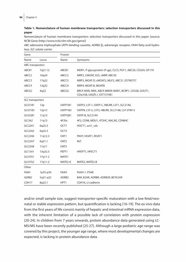
96 Chapter 5
and/or small sample size, suggest transporter-specific maturation with a low fetal/neo-natal or stable expression pattern, but quantification is lacking [16-19]. The ex vivo data from the first years of life consist mainly of hepatic and intestinal mRNA expression data, with the inherent limitation of a possible lack of correlation with protein expression [20-24]. In children from 7 years onwards, protein abundance data generated using LC-MS/MS have been recently published [25-27]. Although a large pediatric age range was covered by this project, the younger age range, where most developmental changes are expected, is lacking in protein abundance data.
table 1. nomenclature of human membrane transporters: selection transporters discussed in this paperNomenclature of human membrane transporters: selection transporters discussed in this paper [source: NCBI Gene (http://www.ncbi.nlm.nih.gov/gene)]ABC adenosine triphosphate (ATP)-binding cassette, ADRB2 β2-adrenergic receptor, FAAH fatty acid hydro-lase, SLC solute carrier
Gene Protein
Name Locus Name Synonyms
ABC transporters
ABCB1 7q21.12 ABCB1 MDR1, P-glycoprotein (P-gp), CLCS, PGY1, ABC20, CD243, GP170
ABCC2 10q24 ABCC2 MRP2, CMOAT, DJS, cMRP, ABC30
ABCC3 17q22 ABCC3 MRP3, MOAT-D, cMOAT2, MLP2, ABC31, EST90757
ABCC4 13q32 ABCC4 MRP4, MOAT-B, MOATB
ABCG2 4q22 ABCG2 BRCP, MXR, MRX, ABCP, BMDP, MXR1, BCRP1, CD338, GOUT1, CDw338, UAQTL1, EST157481
SLC transporters
SLCO1B1 12p OATP1B1 OATP2, LST-1, OATP-C, HBLRR, LST1, SLC21A6
SLCO1B3 12p12 OATP1B3 OATP8, LST-2, LST3, HBLRR, SLC21A8, LST-3TM13
SLCO2B1 11q13 OATP2B1 OATP-B, SLC21A9
SLC3A2 11q13 4F2hc 4F2, CD98, MDU1, 4T2HC, NACAE, CD98HC
SLC22A1 6q25.3 OCT1 HOCT1, oct1_cds
SLC22A2 6q25.3 OCT2
SLC22A6 11q12.3 OAT1 PAHT, HOAT1, ROAT1
SLC22A7 6q21.1 OAT2 NLT
SLC22A8 11q11 OAT3
SLC15A1 13q32.3 PEPT1 HPEPT1, HPECT1
SLC47A1 17q11.2 MATE1
SLC47A2 17q11.2 MATE2-K MATE2, MATE2-B
Other
FAAH 1p35-p34 FAAH FAAH-1, PSAB
ADRB2 5q31-q32 ADRB2 BAR, B2AR, ADRBR, ADRB2R, BETA2AR
CDH17 8q22.1 HPT1 CDH16, LI cadherin

Development of human membrane transporters 97
5
The studies referenced above comprise the most significant studies investigating the maturation of human membrane transporters, with an emphasis on the clinically rel-evant transporters ABCB1, ABCC2, OATP1B1 and OATP1B3. The best-studied transporter during human development is ABCB1 (Fig. 1). Interestingly, its developmental pattern seems organ-specific. In fetal intestinal samples (16th to 20th week of gestation), ABCB1 could be visualized [16] and intestinal mRNA data suggests stable ABCB1 expression from the neonate up to the adult [22, 24]. In the liver, mRNA expression data suggest a pattern of low ABCB1 expression in fetuses, neonates, and infants until 12 months of age, after which it increases to adult levels [21, 22]. ABCB1 protein abundance measured using LC-MS/MS was quite variable (4.8-fold) in 64 subjects in the age range 7-70 years, but this variation could not be explained by either age or sex [25]. In fetal human brain samples ranging from 7 to 28 weeks of gestational age, ABCB1 immunostaining was detected in only one sample from a 28-week fetus [16]. In contrast, in postmortem central nervous system tissue from neonates (n= 28) of 22 - 42 weeks of gestational age and from adults (n=3), immunohistochemistry showed increasing ABCB1 staining with gestational age [28]. ABCB1 gene expression was also detected in the brain of fetuses of 15, 27 and 42 weeks of gestational age [18]. Very recently, the ABCB1 protein was shown limited at birth and to increase postnatally to reach adult levels by 3-6 months of age [29]. Renal ABCB1 mRNA expression appears to be related to maturity of nephrons. A trend towards lower expression in fetuses and neonates than in adults was observed. ABCB1 protein has been identified as early as 5.5th of gestation [16, 17, 21].
ABCB
1 ex
pres
sion
Intestine
Adult Fetus Neonate Child
ABCB
1 ex
pres
sion
Kidney
Fetus Neonate Child Adult
ABCB
1 ex
pres
sion
Liver
Fetus Neonate Child Adult
ABCB
1 ex
pres
sion
Brain
Adult Fetus Neonate Child
figure 1. suggested ontogeny of Abcb1 expression in intestine, liver, kidney and brain.ABC adenosine triphosphate (ATP)-binding cassette
98 Chapter 5
ABCC2 ontogeny shows similarities to ABCB1. Small intestine ABCC2 mRNA expression is stable in neonatal surgical patients compared to adults. While, hepatic ABCC2 mRNA ex-pression is much lower in the fetus, neonate and young infant (up to 200-fold lower) than the adult [22, 27], in children from 7 years onwards its protein expression appears stable [27]. On the protein level, both the localization pattern and intensity of ABCC2 protein staining appear to change during fetal life, in concert with fetal liver maturation [19, 23].
Hepatic mRNA expression of OATP1B1 and OATP1B3 appears to show a different de-velopmental pattern than ABCB1 and ABCC2. Although fetal expression was 2- to 30-fold lower than adult expression, neonatal and infant expression appeared to be even lower (up to 600-fold) [19, 22]. This pattern appears to be supported by protein data (Western blotting) for OATP1B3 but not for OATP1B1. In one study, OATP1B1 protein expression already appears at adult levels in neonates, while in another OATP1B1 only increases after the age of 6 years [30, 31]. Again, for both OATP1B1 and OATP1B3, protein expres-sion appears stable at adult levels from 7 years onwards [25].
PhArMAcoKinEtic AnD PhArMAcoGEnEtic stuDiEs of rElEVAnt MEMbrAnE trAnsPortEr substrAtEs
Pharmacokinetic and pharmacogenetic studies may provide insight into the impact of selected drug transporters in vivo. We identified 16 drugs frequently prescribed to children and that are known substrates of one or more specific membrane transporters (Table 2; see the Electronic Supplemental Material for the search strategy). Age-related differences in pharmacokinetic or pharmacogenetic studies may point to maturational changes in the transporter involved. On the other hand, concordance between adult and pediatric pharmacogenomic studies may support the presence and potentially similar expression of the involved transporters in children as in adults. To further sup-port a potential developmental pattern, we compared the in vivo data with relevant ex vivo data of the individual transporters. As many drugs are also substrate of DMEs and/or multiple transporters, the presented data must be interpreted in the context of the interplay with metabolizing enzymes. It can be speculated that when a specific DME is developmentally low at a certain age, while the transporter is already mature, this may impact the disposition of a drug, by potentially altering the absorption, distribution, metabolism, and excretion (ADME) pathway from largely DME based to transporter based. Where possible, we have only included data that are highly supportive of a role for the transporter(s) involved. Table 2 provides a summary of the pharmacokinetic stud-ies in children and the relationship with transporters. For detailed genetic information on individual transporters, the reader is referred to the pharmgkb.org database (http://pharmacogenetics.ucsf.edu/) and recent reviews [7, 8, 32-34]
Development of human membrane transporters 99
5
table 2. summary PK and PGx studies of relevant membrane transporter substrates.ABC adenosine triphosphate (ATP)-binding cassette, AUC area under the plasma concentration-time curve, BRCP breast cancer resistance protein, GFR glomerular filtration rate, HPT human oligopeptide transporter, IV intravenous, M3G morphine-3-glucuronide, MATE multidrug and toxin extrusion protein, MPA mycophe-nolic acid, mRNA messenger RNA, MRP multidrug resistance-associated protein, OAT organic anion trans-porter, OATP organic anion-transporting polypeptide, OCT organic cation transporter, PGx pharmacogenet-ics, PK pharmacokinetics, UGT uridine 5’-diphospho-glucuronosyltransferase
Drug relevant transporters involved in transport of drug
PK and PGx results in children
Digoxin ABCB1 Higher bodyweight-corrected digoxin clearance in term neonates and young children {Yukawa, 2007 #274;Yukawa, 2011 #273;Hastreiter, 1985 #463}. Renal clearance of digoxin in young children may be more dependent on ABCB1 mediated tubular secretion than in adults {Ratnapalan, 2003 #285}.
Tacrolimus ABCB1 PGx studies of ABCB1 in relation to tacrolimus PK appear contradictory {Gijsen, 2011 #396;de Wildt, 2011 #286;Hawwa, 2009 #97;Guy-Viterbo, 2014 #397}.In pediatric liver transplant recipients, high intestinal ABCB1 mRNA expression was associated with a two-fold higher tacrolimus clearance {Fukudo, 2006 #7}.
Daptomycin ABCB1 Higher body-size corrected renal daptomycin clearance in neonates and younger infants {Bradley, 2014 #458;Woodworth, 1992 #491;Cohen-Wolkowiez, 2012 #465}.
Fexofenadine OATP2B1,ABCB1, MRP2
Apparent bodyweight corrected oral clearance was 1.5-fold lower in children 6 months to 6 years, than in children 6 to 12 years {Martinez, 2014 #461}.
Morphine OCT1, ABCB1, ABCC2, ABCC3, OATP1B1
Neonates and infants have low morphine clearance in the first 10 days of life, increasing thereafter, largely due to immature UGT2B7 metabolism but transporters may contribute {Knibbe, 2009 #277;Krekels, 2014 #429}. Neonates are more prone to morphine-related respiratory depression. {Sadhasivam, 2014 #403}. ABCB1 genotype was associated with respiratory depression in older children, in contrast to an adult study {Kharasch, 2003 #401}. Also ABCB1 genotype affects the M3G-formation and OCT1 genotype is associated with variation in morphine clearance and glucuronide-metabolites formation {Venkatasubramanian, 2014 #430}.
Pravastatin OATP1B1, OATP2B1, OATP1B3, ABCB1, ABCC2
Children with hypercholesterolemia and the SLCO1B1 -11187GA variant had lower mean pravastatin AUCs than those with the wild type, in contrast to an adult study where opposite effect was found {Hedman, 2006 #259;Niemi, 2004 #433}. No age-related variability in pravastatin PK from children age 5 years onwards {Hedman, 2003 #260}.
Atorvastatin OATP1B1, BCRP Atorvastatin PK in older children similar to adult PK {Knebel, 2013 #347}.
Bosentan OATP1B1, OATP1B3, OATP2B1
In children, an exposure limit was found at a much lower dose than in adults, might be due to intestinal OATP2B1 saturation {Beghetti, 2009 #438}.
100 Chapter 5
1. Digoxin – ABCB1The cardiac glycoside digoxin is a well-known ABCB1 substrate (Fig. 2). Its US Food and Drug Administration (FDA) drug label warns about pharmacokinetic interactions with intestinal or renal ABCB1 inducers or inhibitors. Digoxin is mainly renally cleared as unchanged drug, 80 % by glomerular filtration and 20 % by tubular secretion [35]. Pharmacokinetic studies in children show clear age-related differences. In a population pharmacokinetic analysis in 71 neonates (age range 2-29 days), oral digoxin clearance
table 2. summary PK and PGx studies of relevant membrane transporter substrates (continued)
Drug relevant transporters involved in transport of drug
PK and PGx results in children
Ondansetron OCT1 Ondansetron PK and clinical efficacy have been correlated with OCT1 genotypes in adults {Tzvetkov, 2012 #311}. Ondansetron clearance increased with age in children ages 1 to 48 months {Li, 2013 #310}.
Metformin OCT, MATE1, MATE2K Metformin PK in children from 9 years of age onwards was comparable with adult PK, suggesting stable OCT and MATE activity {Sanchez-Infantes, 2011 #409}.
Cimetidine OCT2, MATE1, MATE2K, OAT2
In neonates and children cimetidine (and metabolites) renal clearance accounts for 80-90% of total clearance, whereas in adults it accounts for 60% of total clearance {Somogyi, 1985 #419;Ziemniak, 1984 #418;Lloyd, 1985 #416}. The relatively high renal clearance suggests mature OCT2 activity at birth in the presence of immature GFR {Kurata, 2010 #477}.
Tramadol OCT1 In adults OCT1 genotype was related to metabolite plasma concentrations and prolonged miosis {Tzvetkov, 2011 #332}. Tramadol and metabolite PK show age-related changes in neonates {Allegaert, 2005 #335}
Methotrexate OATP1B1, ABCC2 Increased renal toxicity in children 0-3 months compared to infants 7-12 months {Thompson, 2007 #481}. From 1 year of age onwards body-size corrected methotrexate clearance decreased linearly with age {Radtke, 2013 #492}. SLCO1B1 genotype was associated with increased AUC and was a predictor for toxicity {Radtke, 2013 #483}.
Mycophenolate mofetil
MRP2 In pediatric patients ABCC2 rs717620 allele has been associated with reduced exposure to MPA, more side effects and rejection {Fukuda, 2012 #375}.
Acyclovir/ valacyclovir
4F2hc, HPT1, OAT1, OAT3
In neonates, the IV acyclovir bodyweight-corrected clearance showed a 2-fold increase from 25 to 41 weeks of gestational age {Sampson, 2014 #364}. In older children, 1 month to 5 years, apparent oral clearance of valacyclovir in children less than 3 months of age was 50% than that in older children {Kimberlin, 2010 #453}.
Adefovir OAT1, MRP4 Adefovir is partly renally cleared (45%) {Maeda, 2014 #319;Imaoka, 2007 #455}. In 45 children (age range 2 years – 17 years) receiving oral adefovir dipivoxil, weight-corrected mean apparent clearance and renal clearance were higher in younger children {Sokal, 2008 #340}.
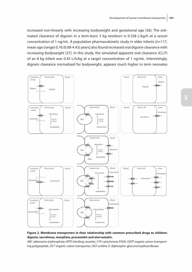
Development of human membrane transporters 101
5
increased non-linearly with increasing bodyweight and gestational age [36]. The esti-mated clearance of digoxin in a term-born 3 kg newborn is 0.338 L/kg/h at a serum concentration of 1 ng/mL. A population pharmacokinetic study in older infants [n=117, mean age (range) 0.76 (0.08-4.43) years] also found increased oral digoxin clearance with increasing bodyweight [37]. In this study, the simulated apparent oral clearance (CL/F) of an 8 kg infant was 0.43 L/h/kg at a target concentration of 1 ng/mL. Interestingly, digoxin clearance normalized for bodyweight, appears much higher in term neonates
figure 2. Membrane transporters in their relationship with common prescribed drugs to children: digoxin, tacrolimus, morphine, pravastatin and atorvastatin.ABC adenosine triphosphate (ATP)-binding cassette, CYP cytochrome P450, OATP organic anion-transport-ing polypeptide, OCT organic cation transporter, UGT uridine 5’-diphospho-glucuronosyltransferase
102 Chapter 5
and younger children than in adults (0.17 L/h/kg). This observation is also in line with higher (per kg) dosing recommendations in term neonates and infants. However, as the drug is mainly renally cleared, and glomerular filtration is still immature at birth, one would expect a lower body size-corrected clearance. This was indeed the case in preterm infants (<2.5 kg) whose digoxin bodyweight-corrected clearance was much lower than that of term infants (0.064 vs. 0.1 L/h/kg), in line with lower dosing recommendations for this age group [38]. Thus, in preterm newborns, both glomerular filtration rate (GFR) and ABCB1 may be immature at birth, while in term infants ABCB1 activity may already be more mature and compensate for developmentally low GFR.
Additionally, clearance decreases non-linearly with increasing concentrations in the range of 0.2 and 2 ng/mL in children up to 4.5 years of age, whereas non-linearity in adults is only found from a serum concentration of 7 ng/mL onwards. We can only speculate as to the underlying mechanism. Earlier transporter saturation due to im-mature ABCB1 activity in intestine and kidney contradicts the observation of higher bodyweight-corrected clearance in young children than in adults.
A clinically relevant interaction was found in eight children who were co-administered the ABCB1 inhibitor carvedilol. Digoxin clearance decreased twofold, while the digoxin clearance to GFR ratio decreased by 45 %, supporting intestinal and renal ABCB1 medi-ated inhibition [39]. In contrast, the same drug-drug interaction resulted in only a mild decrease in digoxin clearance in adults. These findings support our hypothesis that renal clearance of digoxin in young children may be more dependent on ABCB1-mediated tubular secretion than in adults.
Interestingly, the hypothesis of higher renal ABCB1 expression after birth is supported by a mouse study showing a relationship between renal Abcb1 expression and digoxin clearance in young mice, but not by the limited human data from neonates [21, 40].
2. Tacrolimus – ABCB1The calcineurin inhibitor tacrolimus inhibits synthesis of cytotoxic lymphocytes and so prevents transplant rejection. Tacrolimus is a substrate for intestinal and hepatic ABCB1 (Fig. 2) [41]. Weight-normalized oral tacrolimus clearance, which is also dependent on cytochrome P450 (CYP) 3A4/5 metabolism, is higher in infants between 1 and 6 years of age than older children and adults [42, 43]. CYP3A4 activity matures in the first year of life, while CYP3A5 activity, when present, appears stable from fetus to adult [44].
Pharmacogenetic studies of ABCB1 in relation to tacrolimus disposition appear contradictory [45]. In pediatric heart and kidney transplant recipients no relation was found between ABCB1 genotype and tacrolimus dosing requirements or concentra-tion/dose ratio [42, 43]. In pediatric liver transplant patients, homozygous ABCB1 1236TT/2677TT/3435TT carriers needed higher tacrolimus doses than non-carriers both early and later after transplantation [42, 46]. In a population pharmacokinetic analysis

Development of human membrane transporters 103
5
in 114 pediatric liver transplant recipients, the ABCB1 2677G>T allele was associated with a higher pre-dose and concentration/dose ratio at day 1 after transplantation [47]. Such associations were not found for recipient or donor ABCB1 1199G>A and 3435C>T variants. These findings can be understood from a combined ex-vivo/population phar-macokinetic study in 130 pediatric liver transplant recipients. High intestinal ABCB1 mRNA expression was associated with an almost twofold higher tacrolimus clearance early after transplantation, indicative of a switch from primarily intestinal to hepatic tacrolimus clearance upon graft recovery [48].
The results from ex vivo studies suggesting stable ABCB1 mRNA intestinal expression and lower hepatic ABCB1 expression in young infants may explain the pharmacogenetic findings and the impact of the recipient intestinal ABCB1 on tacrolimus disposition [20, 22, 24, 48].
3. Daptomycin – ABCB1The antibacterial daptomycin is used to treat infections caused by Gram-positive bacte-ria including methicillin-resistant Staphylococcus aureus (MRSA). Daptomycin is excreted primarily by the kidney and is an ABCB1 substrate. In 23 adult Caucasian patients, the ABCB1 3435T single nucleotide polymorphism (SNP) was associated with a higher intra-venous daptomycin dose-normalized area under the plasma concentration-time curve (AUC) and lower steady state clearance [49, 50]. In 24 children (age range 3-24 months), intravenous daptomycin clearance in younger infants (approximately 20 mL/h/kg) was higher than previously reported for older children and adults (8-13 mL/h/kg) [51, 52]. Likewise, in 20 preterm and term infants (32-40 weeks of gestational age and 0-85 days’ postnatal age, the mean clearance was approximately 20 mL/kg/h) [53]. No relationship with gestational or postnatal age was found, possibly due to the small sample size. The maturation pattern of daptomycin pharmacokinetics resembles that of digoxin pharma-cokinetics, with higher clearance values early in life. This pattern is not consistent with immature GFR, but may reflect a compensatory role of ABCB1-mediated renal tubular secretion in young children. As discussed earlier, this may be the result of increased renal ABCB1 expression in young infants, but these findings need to be confirmed [16, 18, 21].
4. Fexofenadine – OATP2B1The antihistamine fexofenadine is mainly excreted through bile as parent drug. Only a minor part is metabolized by intestinal microflora and CYP3A4. Its disposition appears to be subject to membrane transport by the uptake transporter OATP2B1, the efflux trans-porter ABCB1 and possibly ABCC2 [54-56]. In 14 healthy men, fexofenadine clearance was related to OATP2B1 polymorphisms and simultaneous apple juice ingestion [57]. This latter finding of a potential food-drug interaction concerning OATP2B1 substrates may be even more relevant for children, as heavy consumers of apple juice. In a population
104 Chapter 5
pharmacokinetic study in 515 Japanese children (6 months to 16 years), CL/F was stable across the age groups (1 L/h/kg) with the exception of the 6- to 12-year olds, whose clear-ance was 1.5-fold higher (1.5 L/h/kg) [58]. In another population pharmacokinetic study including 46 Caucasian and 31 Non-Caucasian children (6 months to 12 years) and 138 adults, apparent bodyweight-normalized oral clearance was lower in children less than 1 year of age than in older children and adults [59]. Interestingly when comparing ethnici-ties, the CL/F was slightly higher in the 6- to 12-year-old Japanese children (1.5 L/kg/h) than in 14 non-Japanese children (0.8 L/h/kg) (13 Caucasian and one non-Caucasian), but this difference may be rather due to the small sample size than ethnicity [58].
Ex vivo data in these age groups are lacking, but OATP2B1 gene expression in 15 neo-natal intestinal samples obtained during surgery was nearly three times higher than in adult samples [22]. This may imply higher oral absorption of fenofexadine in neonates and young infants.
5. Morphine – ABCB1 and OCT1The opioid morphine is almost completely metabolized by uridine 5’-diphospho-gluc-uronosyltransferase (UGT) 2B7 and UGT1A1 to morphine-3-glucuronide and morphine-6-glucuronide. The disposition of both morphine and/or its metabolites is subject to membrane transport by the uptake transporters organic cation transporter (OCT) 1 and OATP1B1 and by the efflux transporters ABCB1, ABCC2 and ABCC3 (Fig. 2) [60, 61]. Hepatic uptake of morphine appeared to be OCT1-mediated in an adult volunteer study and carriers of loss-of-function SLC22A1 gene polymorphisms showed higher morphine AUCs [61]. In other healthy adult volunteers, co-administration of the ABCB1 inhibitor quinidine altered both morphine pharmacokinetics and its opioid effects after oral but not after intravenous morphine administration, suggesting a limited role for ABCB1 in the disposition of morphine at the liver and blood-brain barrier [62]. In adult cancer patients receiving oral morphine, pain relief was more prominent in homozygous carri-ers of the ABCB1 3435T/T SNP, probably due to higher intestinal uptake [63].
Age-related morphine clearance in neonates and infants is very low in the first 10 days of life and increases thereafter [64]. Although this pattern was explained by UGT2B7 maturation, an impact of the maturation of the relevant transporters cannot be ruled out. Interestingly, in follow-up studies in which morphine doses were adjusted to the age-related clearance and similar exposure was reached across the first year of age, pain relief was adequate in neonates (<10 days of age), but the older children still needed high doses of rescue morphine [65]. Neonates are more prone to respiratory depression, which may be explained by increased exposure to morphine when doses are not ad-justed to the age-related changes in its disposition. In addition, immature ABCB1 activ-ity at the blood-brain barrier, as shown very recently, cannot be ruled out to contribute as well and deserves further study [29].
Development of human membrane transporters 105
5
Sadhasivam and co-workers [66, 67] have extensively studied the impact of genetics of transporters in a large cohort of infants and children receiving intravenous morphine for tonsillectomy. In 220 infants, OCT1 homozygous genotypes (SLC22A1 1365GAT>del / 1498G>C) were associated with lower morphine clearance and lower morphine gluc-uronide formation [68]. ABCC3 homozygous -211 CC carriers showed (approximately 40 %) higher metabolite transformation, indicating increased efflux of metabolites into plasma. ABCB1 polymorphisms (3435 C>T) only affected morphine-3-glucuronide for-mation, not morphine or morphine-6-glucuronide pharmacokinetics. It should be noted that only pharmacokinetic data up to 45 min post-dosing were available and, hence, the full pharmacokinetic profile of morphine and metabolites could not be determined. In 263 children of the same cohort, the ABCB1 G allele of rs9282564 polymorphism was as-sociated with respiratory depression, resulting in prolonged hospital stay (odds ratio 4.7 (95 % confidence interval: 2.1-10.8, P=0.0002) [66]. This study contrasts the adult study in which ABCB1 inhibition did not influence the effect of morphine after intravenous administration [62]. In the extended cohort, now including 347 children, the interaction of genetic variants of ABCB1 and two other genes, the fatty acid hydrolase [FAAH, which has been associated with opioid use and addiction acting via cannabinoid receptors type 1, and the β2-adrenergic receptor (ADRB2, receptor blockade has been associated with pain and pain relief ), helped discriminate low and high risk for morphine-related postoperative respiratory depression [67].
As hepatic ABCB1 gene expression only appears to reach adult levels after the first year of life [20, 22, 24, 48], immature hepatic and possibly also blood-brain barrier ABCB1 ex-pression may play a role in higher morphine plasma and brain exposure and the higher risk for respiratory depression in neonates.
6. Pravastatin – OATP1B1Clearance of the cholesterol synthesis inhibitor pravastatin is mainly dependent on non-CYP450-mediated drug metabolism and several uptake and efflux transporters, such as OATP1B1, OATP2B1, OATP1B3, ABCB1 and ABCC2 (Fig. 2) [69]. In adults, SLCO1B1, but not ABCC2, ABCB1, or ABCG2, gene variants were associated with inter-individual variability in pravastatin pharmacokinetics, suggesting a major role of SLCO1B1 in pravastatin disposition [70, 71].
Intriguingly, in one small pharmacogenetic study (n=20; mean ± standard deviation age 10.3 ± 2.9 years), children with familiar hypercholesterolemia with the SLCO1B1 -11187GA genotype had lower mean pravastatin AUCs than children with the wild type [72]. The opposite effect was found in an adult study (n=41) [71]. These results, which should be interpreted with care in view of the small sample size, suggest age-related posttranslational differences in OATP1B1 expression. The pharmacokinetics of pravas-tatin in children (aged 5-16 years) waere similar to adults, which suggests no major
106 Chapter 5
impact of age-related variability in OATP1B1 or other transporter activity from 5 years onwards [73]. These results appear to be in line with the ex vivo results of stable hepatic OATP1B1 expression in older children [25].
7. Atorvastatin – OATP1B1Another cholesterol synthesis inhibitor increasingly used in children is atorvastatin. Like the other statins (HMG-CoA reductase inhibitors), atorvastatin is extensively metabo-lized, largely by CYP3A4, and is a substrate for OATP1B1 (hepatic uptake) and ABCG2 (oral absorption) (Fig. 2). In a population pharmacokinetic study in pediatric hypercho-lesterolemia patients aged 6-17 years, atorvastatin CL/F was described as a function of bodyweight. When scaled allometrically, CL/F was similar to values reported for adults [74]. Atorvastatin metabolism is probably mature from 6 years onwards. The stable clearance and the existing ex vivo data of OATP1B1 suggest similarly mature transporter activity, although an age-related change in the relative contribution of individual trans-porters cannot be ruled out [25, 26]. We could not identify pediatric pharmacogenetic atorvastatin studies, although a comprehensive study in adults clearly indicated the SLCO1B1 variant 388A>G as a major determinant for atorvastatin PK [75].
8. Bosentan – OATP2B1Bosentan is metabolized by CYP3A4 and CYP2C9, with both the parent compound and one metabolite being pharmacological active, but is also subject to hepatic uptake by OATP1B1, OATP1B3 and maybe OATP2B1 [76]. Next to the developmental pattern of CYP3A4, CYP2C9 already shows an increase prenatally, with stable, though variable, activity after the first week of postnatal age [77, 78]. Hence, the impact of the maturation of the transporters may be compounded by CYP3A4 and CYP2C9 maturation in the first year of life, but age-related variation as observed later in life may be more likely due to transporter maturation. At oral doses of approximately 2 mg/kg, bosentan plasma exposures for 19 children (aged 3-15 years) were similar to those for healthy adults [79]. In children [n=36, median (range) age 7.0 (2 - 11) years], plasma levels did not further increase at doses higher than 2 mg/kg (exposure limit), while in adults the exposure limit was 7 mg/kg, with no difference in the other pharmacokinetic parameters [80]. As bosen-tan appears an OATP2B1 substrate, intestinal saturation due to immature OATP2B1, and perhaps other anatomical or physiological age-related differences in oral absorption, may explain this observation. This result does not correspond with the OATP2B1 mRNA expression results, suggesting higher expression in neonates than in adults [22]. In a pediatric population pharmacokinetic-pharmacogenetic study [n=46, mean (range) age 3.8 (25 days – 16.9) years] no relation between bosentan pharmacokinetics and genetic polymorphisms of SLCO1B1, SLCO1B3, SLCO2B1 or CYP3A5 was found [81].

Development of human membrane transporters 107
5
9. Ondansetron – OCT1The anti-emetic ondansetron is mainly metabolized by CYP2D6, which contributes to genetic variation in its disposition and effect. In addition to this CYP2D6 effect, the pharmacokinetics (n=45) and clinical efficacy (n=222) of ondansetron in adults (age range 18-83 years) have also been correlated with SLC22A1 genotypes [82]. OCT1 de-ficiency potentially limits the hepatic uptake and increases plasma concentrations of ondansetron [82]. In a population pharmacokinetic analysis of 124 patients in the age range 1- 48 months, ondansetron bodyweight-normalized clearance was reduced by 76% in a 1-month-old patient and by 31% in a 6-month-old patient, compared with the older children [83]. This contrasts evidence from both in vitro and in vivo studies suggesting maturity of CYP2D6 activity as early as 1-12 weeks postnatal [84-86]. Thus, we hypothesize that lower ondansetron clearance in the first year of life may be related to immature OCT1 activity.
10. Metformin – OCT1 and MATE1Several transporters have been implicated in metformin elimination, tissue distribu-tion and response. OCT1 is a major determinant of the hepatic uptake of metformin, while multidrug and toxin extrusion protein (MATE) 1 /MATE2K determine the efflux of metformin [7, 34]. Recently, the transcription factor hepatocyte nuclear factor 1 was found to regulate OCT1 expression and was related to metformin pharmacokinetics and pharmacodynamics [87, 88]. The combination of SLC22A1 (OCT1) and SLC47A1 (MATE1) genotypes further explains variation in response to metformin in adults [8, 89]. In contrast, in 140 non-obese adolescent girls with androgen excess after precocious pubarche, SLC22A1 genotype was not related to metabolic response at 1 year of metfor-min treatment [90]. In non-obese 9-year-old girls and diabetic patients aged 12-16 years, pharmacokinetics were comparable with those in adults [91, 92]. This suggests stable OCT1 or MATE activity from the age of 9 years onwards, but this needs further study, especially since data in younger children are lacking.
11. Cimetidine – OCT2In adults, renal excretion of the histamine H2-receptor antagonist cimetidine and its me-tabolites accounts for 60 % of total clearance, versus 80-90% in children and neonates [93-95]. Cimetidine is partially metabolized and is a substrate of the uptake transporters OCT2 and organic anion transporter (OAT) 2 and the efflux transporters MATE1 and MATE2-K [8, 34, 96, 97]. Cimetidine is a well-known OCT2 inhibitor and therefore could potentially counteract OCT2-driven cisplatin oto- and renal toxicity [98]. Although the drug is now rarely used in pediatric clinical care, this latter promise necessitates a full un-derstanding of its disposition across the pediatric age range, as part of studies to confirm this new indication. The relatively high renal clearance in neonates and children suggests
108 Chapter 5
an important role for renal tubular secretion and suggests mature activity already at birth. On the other hand, Ziemniak et al. [94] suggest that the unexplained gap between total and renal clearance in adults could be due to secondary metabolite formation in adults, which maybe missing in neonates due to immature metabolism. This is less likely, however, as the higher renal clearance was also observed in older children, whose drug metabolism is largely at adult levels. Moreover, in a rat study, bile duct ligation increased cimetidine renal tubular secretion by up-regulation of OCT2 (but not MATE1), supporting the hypothesis that the non-renal clearance in adults occurs through hepatic/bile excre-tion and is not related to unknown secondary metabolite formation [99]. Nevertheless, as analytical methods to measure drugs and metabolites have become more sensitive since these early studies in the mid-1980s, new studies in children of different ages could help elucidate why renal clearance of cimetidine differs between children and adults and give insight in the role of OCT2 in cimetidine disposition.
12. Tramadol – OCT1Tramadol is a prodrug of the µ-opioid receptor agonist O-desmethyltramadol. It is metabolized mainly by CYP2D6 to its active O-desmethyltramadol metabolite [100]. The variation in tramadol pharmacokinetics cannot solely be explained by variation in CYP2D6, as was shown by Tzvetkov et al. [101] who showed an additive effect of OCT1 on tramadol disposition variation. Loss-of-function SLC22A1 polymorphisms have been related to higher plasma concentrations of the active O-desmethyltramadol and prolonged miosis, as surrogate marker of the opioid effect. These effects are likely due to reduced OCT1-mediated hepatic uptake [101]. Allegaert and co-workers [102] showed that maturational clearance of tramadol, driven by CYP2D6 activity, is almost complete by 44 weeks post-menstrual age. In a pooled population pharmacogenetics-pharmacokinetic study covering the age range from preterm to elderly, only part of the variability in O-desmethyltramadol formation clearance could be explained by CYP2D6 genotype, further supporting a potential role for SLC22A1 genetic variation [86]. A re-lationship between CYP2D6 genotype and tramadol metabolism was shown in young preterm infants, which is surprising as CYP2D6 is not fully mature at birth, especially not in preterm infants, and a genotype effect may have been obscured. It would be worth-while, therefore, to study the impact of SLC22A1 genotype in this young population [10]. In a population pharmacokinetic/pharmacodynamic analysis of 104 older children (2-8 years), age did not clearly contribute to variation in pharmacokinetics or the prediction of response [103].
13. Methotrexate – OATP1B1 and ABCC2Methotrexate is a folic acid antagonist used to treat several forms of cancer and anti-inflammatory diseases. Methotrexate undergoes complex hepatic and intracel-

Development of human membrane transporters 109
5
lular metabolism [104]. Many membrane transporters are responsible for its uptake and excretion and for its metabolism to active polyglutamine metabolites and inactive 7-hydroxy-methotrexate [104, 105]. Methotrexate is eliminated primarily by renal excre-tion through glomerular filtration and renal tubular reabsorption and secretion. Ap-proximately 70–90 % of a dose is excreted unchanged in urine. A small pharmacokinetic study showed only marginally lower methotrexate steady-state clearance (body surface area corrected), but increased renal toxicity, in 0- to 3-month-old infants than in 7- to 12-month-old infants [106, 107]. From 1 year onwards methotrexate clearance (body surface area normalized) decreased linearly with age [107]. A 2014 review concluded that ”although there is no pharmacogenetic marker for MTX [methotrexate] in use in the clinic at present, polymorphisms in SLCO1B1 have an important role in MTX pharmaco-kinetics and toxicity in pediatric ALL [acute lymphoblastic leukemia] patients and show the most consistent and promising results” [105]. For example, in a cohort of almost 500 pediatric acute lymphoblastic leukemia (ALL) patients, methotrexate AUC from time zero to 48 h (AUC0-48h) increased by 26 % (P < 0.001) per SLCO1B1 rs4149056 C allele and was a significant predictor of overall toxic adverse events during methotrexate courses (R2 = 0.043; P <0.001), but no relationship was found for ABCC2 [107]. This study confirmed the results of a genome-wide association study (GWAS) in 434 ALL children, the first to identify SLCO1B1 as an important marker of methotrexate pharmacokinetics and clinical response, and recently validated by five different treatment regimens of high-dose methotrexate ALL treatment protocols at St Jude Children’s Research Hospital (Memphis, TN, USA) [108]. Moreover, a deep sequencing approach for SLCO1B1 demon-strated that rare damaging variants contributed significantly to methotrexate clearance and had larger effect sizes than common SLCO1B1 variants [109, 110]. Other recent stud-ies have detected a relationship between ABCC2 and methotrexate pharmacokinetics and toxicity. In 112 Han Chinese pediatric ALL patients, the ABCC2 -24T allele (rs717620) was associated with significantly higher methotrexate plasma concentrations at 48 h and with significant hematological and non-hematological toxicities. These findings are partially supported by other studies in 127 Lebanese and 151 Spanish pediatric ALL patients [111, 112].
14. Mycophenolate mofetil – ABCC2Mycophenolate mofetil is the prodrug of the active mycophenolic acid (MPA). It is metabolized by carboxylesterase 2 (CES2), after which MPA is further metabolized by several CYPs and UGTs [113]. MPA-glucuronide is excreted in the bile primarily by ABCC2 (encoded by ABCC2) and this transport is essential for enterohepatic circulation. The ABCC2 rs717620 A allele has been associated with reduced exposure to MPA in pediatric renal transplant recipients [114]. In a large multi-center cohort of pediatric heart trans-plant recipients, ABCC2 rs717620 A allele was also associated with more gastrointestinal

110 Chapter 5
intolerance, but with fewer short- and long-term rejection episodes [114]. As ABCC2 is thought to excrete MPA-glucuronide in the bile, carriers of the active A allele, may have increased enterohepatic circulation with an increased concentration of free MPA in the intestine. This is potentially associated with more gastrointestinal intolerance, but simultaneously with higher exposure and efficacy. As SNPs in UGT1A9, UGT2B7, SLCO1B3 and IMPDH have also been associated with altered MPA exposure, the combined effect of these SNPs and potentially interacting co-medication, may define high- and low- risk patients for MPA efficacy and toxicity. Full hepatic ABCC2 maturation appears to occur after infancy, suggesting a lower enterohepatic circulation of MPA, which may result in less gastrointestinal intolerance but potentially also with less efficacy in this age group [22]. This is merely a hypothesis without confirming pharmacokinetic data.
15. Acyclovir/Valacyclovir – OAT1 and OAT3The oral bioavailability of the anti-viral agent acyclovir is poor, and therefore its prodrug valacyclovir was developed. A positive association was found between intestinal expres-sion of 4F2hc (SLC3A2, amino acid transporter heavy chain, a membrane glycoprotein), and HPT1 (human oligopeptide transporter) and plasma levels of valacyclovir, but not peptide transporter 1 [PEPT1 (SLC15A1)] or any of the other investigated intestinal or-ganic anion or cation transporters [115].
After hepatic metabolism, both drugs are mainly renally excreted by both glomerular filtration and renal tubular secretion, most likely by OAT1 and OAT3 [116, 117]. In preterm and term neonates (n=28, median age 30 weeks of gestation), the intravenous acyclovir bodyweight-corrected clearance showed a twofold increase from 25 to 41 weeks of ges-tational age [118]. In children 1 month to 5 yearsold with or at risk for herpes infection, CL/F of valacyclovir (mL/kg/min) in those younger than 3 months was 50 % of that in older children, in whom bodyweight-corrected clearance remained stable [119]. A re-cent study showed markedly increased acyclovir concentrations when co-administered with benzylpenicillin, which was shown to be due to OAT3 and possibly OAT1 inhibition [116]. This could be a very relevant interaction in septic newborns, who often receive both drugs, as increased acyclovir concentrations are associated with neurological adverse events as well as neutropenia.
16. Adefovir – OAT1 and ABCC4Adefovir, the antiviral agent used for treatment of hepatitis B virus or HIV, is 45 % renally cleared through glomerular filtration and OAT1/ABCC4 renal tubular secretion [120, 121]. In 45 children in three age groups (2-6, 7-11, and 12-17 years) receiving oral treatment for hepatitis B virus with the prodrug adefovir dipivoxil bodyweight-corrected mean apparent clearance and renal clearance were decreasing with increasing age [122]. Simi-larly, in a phase I study in children (n=13, age range 6 months to 18 years) receiving oral
Development of human membrane transporters 111
5
adefovir dipivoxil for HIV treatment, systemic exposure was lower in children younger than 5 years [123].
suMMAry AnD Discussion
In summary, ex vivo, pharmacokinetic and pharmacogenetic studies suggest transport-er-specific changes from the human fetus to the adult. At this time, data are very scarce and the impact of these changes on drug therapy in children is still largely unknown. However, despite data scarcity, our review may aid clinical pharmacologists and clini-cians in rationale drug prescribing of the drugs presented, not only by showing how pharmacokinetics are probably similar in certain pediatric age groups compared to adults, but also by pointing out where potential age-related changes in individual trans-porters could impact the drug’s efficacy and safety. It broadens our views on ontogeny of transporters by evaluation of the results of pharmacokinetic and pharmacogenetic studies on relevant transporters. Moreover, this review presents clear information gaps, which may guide future research efforts to elucidate the role of human membrane transporters in the developing child.
For most drugs, the in vivo data to support the ex vivo data in understanding the maturation of individual transporters are limited to older children, and, hence, their usefulness is limited. No clear transporter maturation pattern can be deducted from any of the available pharmacokinetic studies in children. This contrasts with our knowledge from individual DMEs. For example, using midazolam as phenotyping probe, the de-velopmental expression of CYP3A4/5 from the preterm neonate to the adult has been extensively characterized [124, 125] or amikacin clearance to display the maturation of GFR in neonates [125]. Specific phenotyping probes to study individual membrane transporters are suboptimal and have only been validated in adults. For many drugs, multiple transporters are involved in their uptake and excretion, which in turn may also compensate for changes in individual transporter activity. In adults, knowledge has also been gained from pharmacogenetic and drug-drug interaction studies.
This review shows that pharmacogenetic variation in membrane transporter activity also impact drug disposition, effect and toxicity in children. Most pharmacogenetic studies in children are in line with adult data. However, these similar pharmacogenetic relationships should be interpreted with care, especially when it comes to translating these data across the whole pediatric age range. First, most pharmacogenetic study cohorts only contain older children, whereas pharmacokinetic studies in neonates and infants often show clear developmental changes up to 4-6 years of age. Hence, the impact of SNPs may be obscured by more prominent changes due to growth and devel-opment. Second, the relationship between SLCO1B1 SNPs and pravastatin disposition in

112 Chapter 5
adolescents was found to be the opposite of that in adults. If these results are validated in other studies, a potential impact of hormonal changes on individual transporters needs to be elucidated; this may also provide insight into the physiological role these transporters play during adolescence.
The limited data from ex vivo studies of postmortem or surgical samples support the notion of membrane transporter-specific maturational patterns. It is still difficult, how-ever, to determine definitive patterns for the different transporters. One of the reasons for this is that most studies only used limited samples and limited age ranges. Most fetal and neonatal studies only applied immunohistochemistry or mRNA techniques, while the more quantitative protein expression data are mainly from older children, above the age range in which most developmental changes can be expected. In addition, the qual-ity and interpretation is further challenged as the exact origin, handling, and storage of tissues, including detailed patient characteristics and exact procurement site from organ (e.g., where in the intestine?), is often unknown.
The mechanisms underlying maturational changes in transporters are largely un-known. Recent studies on the CYP3A4 show maturational changes in methylation pat-terns to mirror the maturational expression of CYP3A4, which may point towards similar mechanisms for the transporters [126].
Differences between ethnic groups in DME abundance or ethnicity have been de-scribed, even in newborn infants [77, 85, 127]. Like in DMEs, it is credible to believe that ethnicity might have an effect on transporter activity or abundance. Nevertheless, for 27 drug transporters in 95 pathologically normal kidney samples, the expression did not differ between European Americans or African Americans [128].
The interpretation of pharmacokinetic studies, to understand maturation of trans-porters, is complicated by the fact that these transporters are part of the larger system of the ADME processes involved in the disposition of drugs. In contrast to DMEs, where clearance to a specific metabolite can be estimated to understand the DME maturation, studying a single transporter is more difficult. One may not be able to pinpoint one specific membrane transporter involved, and if the dominant transporter is still im-mature early in life, other transporters may compensate, thereby obscuring individual transporter maturation.
futurE DirEctions
Several approaches are needed to increase our understanding of membrane transport-ers in the fetus and child (Table 3). First, the impact of transporter maturation on efficacy and toxicity in daily clinical care needs to be elucidated.
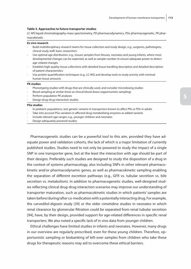
Development of human membrane transporters 113
5
Pharmacogenetic studies can be a powerful tool to this aim, provided they have ad-equate power and validation cohorts, the lack of which is a major limitation of currently published studies. Studies need to not only be powered to study the impact of a single SNP in one transporter gene, but at the least the interaction with age should be part of their designs. Preferably such studies are designed to study the disposition of a drug in the context of systems pharmacology, also including SNPs in other relevant pharmaco-kinetic and/or pharmacodynamic genes, as well as pharmacokinetic sampling enabling the separation of different excretion pathways (e.g., GFR vs. tubular secretion vs. bile secretion vs. metabolism). In addition to pharmacogenetic studies, well-designed stud-ies reflecting clinical drug-drug interaction scenarios may improve our understanding of transporter maturation, such as pharmacokinetic studies in which patients’ samples are taken before/during/after co-medication with a potentially interacting drug. For example, the carvedilol-digoxin study [39] or the older cimetidine studies in neonates in which renal clearance by glomerular filtration could be separated from renal tubular secretion [94], have, by their design, provided support for age-related differences in specific renal transporters. We also noted a specific lack of in vivo data from younger children.
Ethical challenges have limited studies in infants and neonates. However, many drugs in our overview are regularly prescribed, even for these young children. Therefore, op-portunistic sampling or biobanking of left-over samples from children who take these drugs for therapeutic reasons may aid to overcome these ethical barriers.
table 3. Approaches to future transporter studiesLC-MS liquid chromatography-mass spectrometry, PD pharmacodynamics, PGx pharmacogenetic, PK phar-macokinetic
Ex vivo research- Build multidisciplinary research teams for tissue collection and study design, e.g., surgeons, pathologists,
clinical study staff, basic researchers- Use optimal age distribution, e.g., tissues samples from fetuses, neonates and young infants, where most
developmental changes can be expected, as well as sample number to ensure adequate power to detect age-related changes
- Establish high-quality tissue collections with detailed tissue handling description and detailed description of patient characteristics
- Use protein quantification techniques (e.g., LC-MS) and develop tools to study activity with minimal human tissue amounts
PK studies- Phenotyping studies with drugs that are clinically used, and consider microdosing studies- Blood sampling at similar times as clinical blood draws (opportunistic sampling)- Perform population PK analyses- Design drug-drug interaction studies
PGx studies- In pediatric populations, test genetic variants in transporters known to affect PKs or PDs in adults- Take into account PGx variation in affected drug-metabolizing enzymes as added variants- Include relevant age ranges: e.g., younger children and neonates- Design adequately powered studies
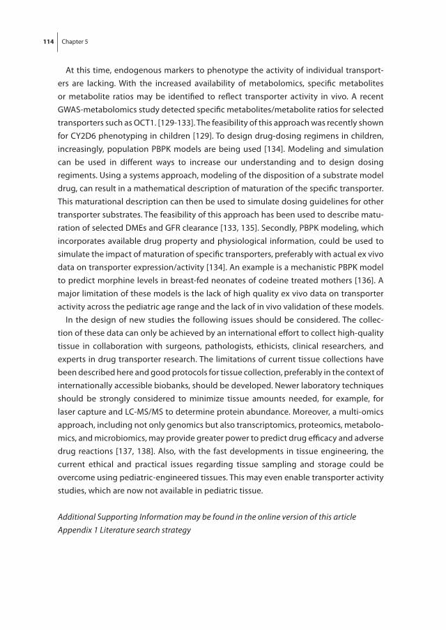
114 Chapter 5
At this time, endogenous markers to phenotype the activity of individual transport-ers are lacking. With the increased availability of metabolomics, specific metabolites or metabolite ratios may be identified to reflect transporter activity in vivo. A recent GWAS-metabolomics study detected specific metabolites/metabolite ratios for selected transporters such as OCT1. [129-133]. The feasibility of this approach was recently shown for CY2D6 phenotyping in children [129]. To design drug-dosing regimens in children, increasingly, population PBPK models are being used [134]. Modeling and simulation can be used in different ways to increase our understanding and to design dosing regiments. Using a systems approach, modeling of the disposition of a substrate model drug, can result in a mathematical description of maturation of the specific transporter. This maturational description can then be used to simulate dosing guidelines for other transporter substrates. The feasibility of this approach has been used to describe matu-ration of selected DMEs and GFR clearance [133, 135]. Secondly, PBPK modeling, which incorporates available drug property and physiological information, could be used to simulate the impact of maturation of specific transporters, preferably with actual ex vivo data on transporter expression/activity [134]. An example is a mechanistic PBPK model to predict morphine levels in breast-fed neonates of codeine treated mothers [136]. A major limitation of these models is the lack of high quality ex vivo data on transporter activity across the pediatric age range and the lack of in vivo validation of these models.
In the design of new studies the following issues should be considered. The collec-tion of these data can only be achieved by an international effort to collect high-quality tissue in collaboration with surgeons, pathologists, ethicists, clinical researchers, and experts in drug transporter research. The limitations of current tissue collections have been described here and good protocols for tissue collection, preferably in the context of internationally accessible biobanks, should be developed. Newer laboratory techniques should be strongly considered to minimize tissue amounts needed, for example, for laser capture and LC-MS/MS to determine protein abundance. Moreover, a multi-omics approach, including not only genomics but also transcriptomics, proteomics, metabolo-mics, and microbiomics, may provide greater power to predict drug efficacy and adverse drug reactions [137, 138]. Also, with the fast developments in tissue engineering, the current ethical and practical issues regarding tissue sampling and storage could be overcome using pediatric-engineered tissues. This may even enable transporter activity studies, which are now not available in pediatric tissue.
Additional Supporting Information may be found in the online version of this articleAppendix 1 Literature search strategy
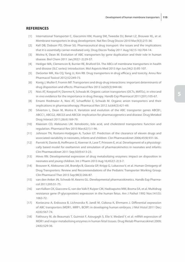
Development of human membrane transporters 115
5
rEfErEncEs
[1] International Transporter C, Giacomini KM, Huang SM, Tweedie DJ, Benet LZ, Brouwer KL, et al. Membrane transporters in drug development. Nat Rev Drug Discov 2010 Mar; 9(3): 215-36.
[2] Kell DB, Dobson PD, Oliver SG. Pharmaceutical drug transport: the issues and the implications that it is essentially carrier-mediated only. Drug Discov Today 2011 Aug; 16(15-16): 704-14.
[3] Moitra K, Dean M. Evolution of ABC transporters by gene duplication and their role in human disease. Biol Chem 2011 Jan; 392(1-2): 29-37.
[4] Hediger MA, Clemencon B, Burrier RE, Bruford EA. The ABCs of membrane transporters in health and disease (SLC series): introduction. Mol Aspects Med 2013 Apr-Jun; 34(2-3): 95-107.
[5] DeGorter MK, Xia CQ, Yang JJ, Kim RB. Drug transporters in drug efficacy and toxicity. Annu Rev Pharmacol Toxicol 2012; 52: 249-73.
[6] Konig J, Muller F, Fromm MF. Transporters and drug-drug interactions: important determinants of drug disposition and effects. Pharmacol Rev 2013 Jul; 65(3): 944-66.
[7] Nies AT, Koepsell H, Damme K, Schwab M. Organic cation transporters (OCTs, MATEs), in vitro and in vivo evidence for the importance in drug therapy. Handb Exp Pharmacol 2011(201): 105-67.
[8] Emami Riedmaier A, Nies AT, Schaeffeler E, Schwab M. Organic anion transporters and their implications in pharmacotherapy. Pharmacol Rev 2012 Jul; 64(3): 421-49.
[9] Silverton L, Dean M, Moitra K. Variation and evolution of the ABC transporter genes ABCB1, ABCC1, ABCG2, ABCG5 and ABCG8: implication for pharmacogenetics and disease. Drug Metabol Drug Interact 2011; 26(4): 169-79.
[10] Klaassen CD, Aleksunes LM. Xenobiotic, bile acid, and cholesterol transporters: function and regulation. Pharmacol Rev 2010 Mar; 62(1): 1-96.
[11] Johnson TN, Rostami-Hodjegan A, Tucker GT. Prediction of the clearance of eleven drugs and associated variability in neonates, infants and children. Clin Pharmacokinet 2006; 45(9): 931-56.
[12] Parrott N, Davies B, Hoffmann G, Koerner A, Lave T, Prinssen E, et al. Development of a physiologi-cally based model for oseltamivir and simulation of pharmacokinetics in neonates and infants. Clin Pharmacokinet 2011 Sep; 50(9): 613-23.
[13] Hines RN. Developmental expression of drug metabolizing enzymes: impact on disposition in neonates and young children. Int J Pharm 2013 Aug 16; 452(1-2): 3-7.
[14] Brouwer K, Aleksunes LM, Brandys B, Giacoia GP, Knipp G, Lukacova V, et al. Human Ontogeny of Drug Transporters: Review and Recommendations of the Pediatric Transporter Working Group. Clin Pharmacol Ther 2015 Sep; 98(3): 266-87.
[15] van den Anker JN, Schwab M, Kearns GL. Developmental pharmacokinetics. Handb Exp Pharma-col 2011; 205: 51-75.
[16] van Kalken CK, Giaccone G, van der Valk P, Kuiper CM, Hadisaputro MM, Bosma SA, et al. Multidrug resistance gene (P-glycoprotein) expression in the human fetus. Am J Pathol 1992 Nov; 141(5): 1063-72.
[17] Konieczna A, Erdosova B, Lichnovska R, Jandl M, Cizkova K, Ehrmann J. Differential expression of ABC transporters (MDR1, MRP1, BCRP) in developing human embryos. J Mol Histol 2011 Dec; 42(6): 567-74.
[18] Fakhoury M, de Beaumais T, Guimiot F, Azougagh S, Elie V, Medard Y, et al. mRNA expression of MDR1 and major metabolising enzymes in human fetal tissues. Drug Metab Pharmacokinet 2009; 24(6): 529-36.
116 Chapter 5
[19] Sharma S, Ellis EC, Gramignoli R, Dorko K, Tahan V, Hansel M, et al. Hepatobiliary disposition of 17-OHPC and taurocholate in fetal human hepatocytes: a comparison with adult human hepato-cytes. Drug Metab Dispos 2013 Feb; 41(2): 296-304.
[20] Fakhoury M, Litalien C, Medard Y, Cave H, Ezzahir N, Peuchmaur M, et al. Localization and mRNA expression of CYP3A and P-glycoprotein in human duodenum as a function of age. Drug Metab Dispos 2005 Nov; 33(11): 1603-7.
[21] Miki Y, Suzuki T, Tazawa C, Blumberg B, Sasano H. Steroid and xenobiotic receptor (SXR), cyto-chrome P450 3A4 and multidrug resistance gene 1 in human adult and fetal tissues. Mol Cell Endocrinol 2005 Feb 28; 231(1-2): 75-85.
[22] Mooij MG, Schwarz UI, de Koning BAE, Leeder JS, Gaedigk R, Samsom JN, et al. Ontogeny of Hu-man Hepatic and Intestinal Transporter Gene Expression during Childhood: Age Matters. Drug Metabolism and Disposition 2014 Aug; 42(8): 1268-74.
[23] Chen HL, Chen HL, Liu YJ, Feng CH, Wu CY, Shyu MK, et al. Developmental expression of canalicu-lar transporter genes in human liver. J Hepatol 2005 Sep; 43(3): 472-7.
[24] Mizuno T, Fukuda T, Masuda S, Uemoto S, Matsubara K, Inui KI, et al. Developmental trajectory of intestinal MDR1/ABCB1 expression in children. Br J Clin Pharmacol 2013 Jul 23.
[25] Prasad B, Evers R, Gupta A, Hop CE, Salphati L, Shukla S, et al. Interindividual variability in hepatic organic anion-transporting polypeptides and P-glycoprotein (ABCB1) protein expression: quan-tification by liquid chromatography tandem mass spectroscopy and influence of genotype, age, and sex. Drug Metab Dispos 2014 Jan; 42(1): 78-88.
[26] Prasad B, Lai Y, Lin Y, Unadkat JD. Interindividual variability in the hepatic expression of the hu-man breast cancer resistance protein (BCRP/ABCG2): effect of age, sex, and genotype. J Pharm Sci 2013 Mar; 102(3): 787-93.
[27] Deo AK, Prasad B, Balogh L, Lai Y, Unadkat JD. Interindividual variability in hepatic expression of the multidrug resistance-associated protein 2 (MRP2/ABCC2): quantification by liquid chroma-tography/tandem mass spectrometry. Drug Metab Dispos 2012 May; 40(5): 852-5.
[28] Daood M, Tsai C, Ahdab-Barmada M, Watchko JF. ABC transporter (P-gp/ABCB1, MRP1/ABCC1, BCRP/ABCG2) expression in the developing human CNS. Neuropediatrics 2008 Aug; 39(4): 211-8.
[29] Lam J, Baello S, Iqbal M, Kelly LE, Shannon PT, Chitayat D, et al. The ontogeny of P-glycoprotein in the developing human blood-brain barrier: implication for opioid toxicity in neonates. Pediatr Res 2015 Oct; 78(4): 417-21.
[30] Thomson MMS, Krauel S, Hines RN, Scheutz EG, Meibohm B. Lack of effect of genetic variants on the age-associated prtoein expression of OATP1B1 and OATP1B3 in human pediatric liver. [Abstract for the 114th American Society for Clinical Pharmacology and Therapeutics Annual Meeting]; 2013.
[31] Yanni SB, Smith PB, Benjamin DK, Jr., Augustijns PF, Thakker DR, Annaert PP. Higher clearance of micafungin in neonates compared with adults: role of age-dependent micafungin serum bind-ing. Biopharm Drug Dispos 2011 May; 32(4): 222-32.
[32] Niemi M, Pasanen MK, Neuvonen PJ. Organic anion transporting polypeptide 1B1: a genetically polymorphic transporter of major importance for hepatic drug uptake. Pharmacol Rev 2011 Mar; 63(1): 157-81.
[33] Wolking S, Schaeffeler E, Lerche H, Schwab M, Nies AT. Impact of Genetic Polymorphisms of ABCB1 (MDR1, P-Glycoprotein) on Drug Disposition and Potential Clinical Implications: Update of the Literature. Clin Pharmacokinet 2015 Jul; 54(7): 709-35.

Development of human membrane transporters 117
5
[34] Damme K, Nies AT, Schaeffeler E, Schwab M. Mammalian MATE (SLC47A) transport proteins: impact on efflux of endogenous substrates and xenobiotics. Drug Metab Rev 2011 Nov; 43(4): 499-523.
[35] Sumner DJ, Russell AJ. Digoxin pharmacokinetics: multicompartmental analysis and its clinical implications. Br J Clin Pharmacol 1976 Apr; 3(2): 221-9.
[36] Yukawa E, Akiyama K, Suematsu F, Yukawa M, Minemoto M. Population pharmacokinetic investi-gation of digoxin in Japanese neonates. J Clin Pharm Ther 2007 Aug; 32(4): 381-6.
[37] Yukawa M, Yukawa E, Suematsu F, Takiguchi T, Ikeda H, Aki H, et al. Population pharmacokinetic investigation of digoxin in Japanese infants and young children. J Clin Pharmacol 2011 Jun; 51(6): 857-63.
[38] Hastreiter AR, van der Horst RL, Voda C, Chow-Tung E. Maintenance digoxin dosage and steady-state plasma concentration in infants and children. J Pediatr 1985 Jul; 107(1): 140-6.
[39] Ratnapalan S, Griffiths K, Costei AM, Benson L, Koren G. Digoxin-carvedilol interactions in chil-dren. J Pediatr 2003 May; 142(5): 572-4.
[40] Pinto N, Halachmi N, Verjee Z, Woodland C, Klein J, Koren G. Ontogeny of renal P-glycoprotein expression in mice: correlation with digoxin renal clearance. Pediatr Res 2005 Dec; 58(6): 1284-9.
[41] Hebert MF. Contributions of hepatic and intestinal metabolism and P-glycoprotein to cyclospo-rine and tacrolimus oral drug delivery. Adv Drug Deliv Rev 1997 Sep 15; 27(2-3): 201-14.
[42] de Wildt SN, van Schaik RH, Soldin OP, Soldin SJ, Brojeni PY, van der Heiden IP, et al. The interac-tions of age, genetics, and disease severity on tacrolimus dosing requirements after pediatric kidney and liver transplantation. Eur J Clin Pharmacol 2011 Dec; 67(12): 1231-41.
[43] Gijsen V, Mital S, van Schaik RH, Soldin OP, Soldin SJ, van der Heiden IP, et al. Age and CYP3A5 genotype affect tacrolimus dosing requirements after transplant in pediatric heart recipients. J Heart Lung Transplant 2011 Dec; 30(12): 1352-9.
[44] Vyhlidal CA, Pearce RE, Gaedigk R, Calamia JC, Shuster DL, Thummel KE, et al. Variability in Expres-sion of CYP3A5 in Human Fetal Liver. Drug Metab Dispos 2015 Aug; 43(8): 1286-93.
[45] Hesselink DA, Bouamar R, Elens L, van Schaik RH, van Gelder T. The role of pharmacogenetics in the disposition of and response to tacrolimus in solid organ transplantation. Clin Pharmacokinet 2014 Feb; 53(2): 123-39.
[46] Hawwa AF, McKiernan PJ, Shields M, Millership JS, Collier PS, McElnay JC. Influence of ABCB1 poly-morphisms and haplotypes on tacrolimus nephrotoxicity and dosage requirements in children with liver transplant. Br J Clin Pharmacol 2009 Sep; 68(3): 413-21.
[47] Guy-Viterbo V, Baudet H, Elens L, Haufroid V, Lacaille F, Girard M, et al. Influence of donor-recipient CYP3A4/5 genotypes, age and fluconazole on tacrolimus pharmacokinetics in pediatric liver transplantation: a population approach. Pharmacogenomics 2014 Jun; 15(9): 1207-21.
[48] Fukudo M, Yano I, Masuda S, Goto M, Uesugi M, Katsura T, et al. Population pharmacokinetic and pharmacogenomic analysis of tacrolimus in pediatric living-donor liver transplant recipients. Clin Pharmacol Ther 2006 Oct; 80(4): 331-45.
[49] Lemaire S, Van Bambeke F, Mingeot-Leclercq MP, Tulkens PM. Modulation of the cellular accumu-lation and intracellular activity of daptomycin towards phagocytized Staphylococcus aureus by the P-glycoprotein (MDR1) efflux transporter in human THP-1 macrophages and madin-darby canine kidney cells. Antimicrob Agents Chemother 2007 Aug; 51(8): 2748-57.
[50] Baietto L, D’Avolio A, Cusato J, Pace S, Calcagno A, Motta I, et al. Effect of SNPs in human ABCB1 on daptomycin pharmacokinetics in Caucasian patients. J Antimicrob Chemother 2015 Jan; 70(1): 307-8.
118 Chapter 5
[51] Bradley JS, Benziger D, Bokesch P, Jacobs R. Single-dose pharmacokinetics of daptomycin in pediatric patients 3-24 months of age. Pediatr Infect Dis J 2014 Sep; 33(9): 936-9.
[52] Woodworth JR, Nyhart EH, Jr., Brier GL, Wolny JD, Black HR. Single-dose pharmacokinetics and an-tibacterial activity of daptomycin, a new lipopeptide antibiotic, in healthy volunteers. Antimicrob Agents Chemother 1992 Feb; 36(2): 318-25.
[53] Cohen-Wolkowiez M, Watt KM, Hornik CP, Benjamin DK, Jr., Smith PB. Pharmacokinetics and toler-ability of single-dose daptomycin in young infants. Pediatr Infect Dis J 2012 Sep; 31(9): 935-7.
[54] Cvetkovic M, Leake B, Fromm MF, Wilkinson GR, Kim RB. OATP and P-glycoprotein transporters mediate the cellular uptake and excretion of fexofenadine. Drug Metab Dispos 1999 Aug; 27(8): 866-71.
[55] Kusuhara H, Miura M, Yasui-Furukori N, Yoshida K, Akamine Y, Yokochi M, et al. Effect of coad-ministration of single and multiple doses of rifampicin on the pharmacokinetics of fexofenadine enantiomers in healthy subjects. Drug Metab Dispos 2013 Jan; 41(1): 206-13.
[56] Akamine Y, Miura M, Sunagawa S, Kagaya H, Yasui-Furukori N, Uno T. Influence of drug-transporter polymorphisms on the pharmacokinetics of fexofenadine enantiomers. Xenobiotica 2010 Nov; 40(11): 782-9.
[57] Imanaga J, Kotegawa T, Imai H, Tsutsumi K, Yoshizato T, Ohyama T, et al. The effects of the SL-CO2B1 c.1457C > T polymorphism and apple juice on the pharmacokinetics of fexofenadine and midazolam in humans. Pharmacogenet Genomics 2011 Feb; 21(2): 84-93.
[58] Martinez JM, Khier S, Morita S, Rauch C, Fabre D. Population pharmacokinetic analysis of fexof-enadine in Japanese pediatric patients. J Pharmacokinet Pharmacodyn 2014 Apr; 41(2): 187-95.
[59] Krishna R, Krishnaswami S, Kittner B, Sankoh AJ, Jensen BK. The utility of mixed-effects covariate analysis in rapid selection of doses in pediatric subjects: a case study with fexofenadine hydro-chloride. Biopharm Drug Dispos 2004 Dec; 25(9): 373-87.
[60] Thorn CF, Klein TE, Altman RB. Codeine and morphine pathway. Pharmacogenet Genomics 2009 Jul; 19(7): 556-8.
[61] Tzvetkov MV, dos Santos Pereira JN, Meineke I, Saadatmand AR, Stingl JC, Brockmoller J. Mor-phine is a substrate of the organic cation transporter OCT1 and polymorphisms in OCT1 gene affect morphine pharmacokinetics after codeine administration. Biochem Pharmacol 2013 Sep 1; 86(5): 666-78.
[62] Kharasch ED, Hoffer C, Whittington D, Sheffels P. Role of P-glycoprotein in the intestinal absorp-tion and clinical effects of morphine. Clin Pharmacol Ther 2003 Dec; 74(6): 543-54.
[63] Campa D, Gioia A, Tomei A, Poli P, Barale R. Association of ABCB1/MDR1 and OPRM1 gene poly-morphisms with morphine pain relief. Clin Pharmacol Ther 2008 Apr; 83(4): 559-66.
[64] Knibbe CA, Krekels EH, van den Anker JN, DeJongh J, Santen GW, van Dijk M, et al. Morphine gluc-uronidation in preterm neonates, infants and children younger than 3 years. Clin Pharmacokinet 2009; 48(6): 371-85.
[65] Krekels EH, Tibboel D, de Wildt SN, Ceelie I, Dahan A, van Dijk M, et al. Evidence-based morphine dosing for postoperative neonates and infants. Clin Pharmacokinet 2014 Jun; 53(6): 553-63.
[66] Sadhasivam S, Chidambaran V, Zhang X, Meller J, Esslinger H, Zhang K, et al. Opioid-induced respiratory depression: ABCB1 transporter pharmacogenetics. Pharmacogenomics J 2014 Oct 14.
[67] Biesiada J, Chidambaran V, Wagner M, Zhang X, Martin LJ, Meller J, et al. Genetic risk signatures of opioid-induced respiratory depression following pediatric tonsillectomy. Pharmacogenomics 2014 Nov; 15(14): 1749-62.

Development of human membrane transporters 119
5
[68] Venkatasubramanian R, Fukuda T, Niu J, Mizuno T, Chidambaran V, Vinks AA, et al. ABCC3 and OCT1 genotypes influence pharmacokinetics of morphine in children. Pharmacogenomics 2014 Jul; 15(10): 1297-309.
[69] Whirl-Carrillo M, McDonagh EM, Hebert JM, Gong L, Sangkuhl K, Thorn CF, et al. Pharmacogenom-ics knowledge for personalized medicine. Clin Pharmacol Ther 2012 Oct; 92(4): 414-7.
[70] Ho RH, Choi L, Lee W, Mayo G, Schwarz UI, Tirona RG, et al. Effect of drug transporter genotypes on pravastatin disposition in European- and African-American participants. Pharmacogenet Genom-ics 2007 Aug; 17(8): 647-56.
[71] Niemi M, Schaeffeler E, Lang T, Fromm MF, Neuvonen M, Kyrklund C, et al. High plasma pravastatin concentrations are associated with single nucleotide polymorphisms and haplotypes of organic anion transporting polypeptide-C (OATP-C, SLCO1B1). Pharmacogenetics 2004 Jul; 14(7): 429-40.
[72] Hedman M, Antikainen M, Holmberg C, Neuvonen M, Eichelbaum M, Kivisto KT, et al. Pharmaco-kinetics and response to pravastatin in paediatric patients with familial hypercholesterolaemia and in paediatric cardiac transplant recipients in relation to polymorphisms of the SLCO1B1 and ABCB1 genes. Br J Clin Pharmacol 2006 Jun; 61(6): 706-15.
[73] Hedman M, Neuvonen PJ, Neuvonen M, Antikainen M. Pharmacokinetics and pharmacodynam-ics of pravastatin in children with familial hypercholesterolemia. Clin Pharmacol Ther 2003 Aug; 74(2): 178-85.
[74] Knebel W, Gastonguay MR, Malhotra B, El-Tahtawy A, Jen F, Gandelman K. Population pharmaco-kinetics of atorvastatin and its active metabolites in children and adolescents with heterozygous familial hypercholesterolemia: selective use of informative prior distributions from adults. J Clin Pharmacol 2013 May; 53(5): 505-16.
[75] Nies AT, Niemi M, Burk O, Winter S, Zanger UM, Stieger B, et al. Genetics is a major determinant of expression of the human hepatic uptake transporter OATP1B1, but not of OATP1B3 and OATP2B1. Genome Med 2013 Jan 11; 5(1): 1.
[76] Treiber A, Schneiter R, Hausler S, Stieger B. Bosentan is a substrate of human OATP1B1 and OATP1B3: inhibition of hepatic uptake as the common mechanism of its interactions with cyclo-sporin A, rifampicin, and sildenafil. Drug Metab Dispos 2007 Aug; 35(8): 1400-7.
[77] Koukouritaki SB, Manro JR, Marsh SA, Stevens JC, Rettie AE, McCarver DG, et al. Developmental expression of human hepatic CYP2C9 and CYP2C19. J Pharmacol Exp Ther 2004 Mar; 308(3): 965-74.
[78] Treluyer JM, Gueret G, Cheron G, Sonnier M, Cresteil T. Developmental expression of CYP2C and CYP2C-dependent activities in the human liver: in-vivo/in-vitro correlation and inducibility. Pharmacogenetics 1997 Dec; 7(6): 441-52.
[79] Barst RJ, Ivy D, Dingemanse J, Widlitz A, Schmitt K, Doran A, et al. Pharmacokinetics, safety, and efficacy of bosentan in pediatric patients with pulmonary arterial hypertension. Clin Pharmacol Ther 2003 Apr; 73(4): 372-82.
[80] Beghetti M, Haworth SG, Bonnet D, Barst RJ, Acar P, Fraisse A, et al. Pharmacokinetic and clinical profile of a novel formulation of bosentan in children with pulmonary arterial hypertension: the FUTURE-1 study. Br J Clin Pharmacol 2009 Dec; 68(6): 948-55.
[81] Taguchi M, Ichida F, Hirono K, Miyawaki T, Yoshimura N, Nakamura T, et al. Pharmacokinetics of bosentan in routinely treated Japanese pediatric patients with pulmonary arterial hypertension. Drug Metab Pharmacokinet 2011 Jun; 26(3): 280-7.
[82] Tzvetkov MV, Saadatmand AR, Bokelmann K, Meineke I, Kaiser R, Brockmoller J. Effects of OCT1 polymorphisms on the cellular uptake, plasma concentrations and efficacy of the 5-HT(3) antago-nists tropisetron and ondansetron. Pharmacogenomics J 2012 Feb; 12(1): 22-9.
120 Chapter 5
[83] Li Q, Guo D, Dong Z, Zhang W, Zhang L, Huang SM, et al. Ondansetron can enhance cisplatin-induced nephrotoxicity via inhibition of multiple toxin and extrusion proteins (MATEs). Toxicol Appl Pharmacol 2013 Nov 15; 273(1): 100-9.
[84] Johnson TN, Tucker GT, Rostami-Hodjegan A. Development of CYP2D6 and CYP3A4 in the first year of life. Clin Pharmacol Ther 2008 May; 83(5): 670-1.
[85] Stevens JC, Marsh SA, Zaya MJ, Regina KJ, Divakaran K, Le M, et al. Developmental changes in human liver CYP2D6 expression. Drug Metab Dispos 2008 Aug; 36(8): 1587-93.
[86] Allegaert K, Holford N, Anderson BJ, Holford S, Stuber F, Rochette A, et al. Tramadol and O-Des-methyl Tramadol Clearance Maturation and Disposition in Humans: A Pooled Pharmacokinetic Study. Clin Pharmacokinet 2014 Sep 26.
[87] O’Brien VP, Bokelmann K, Ramirez J, Jobst K, Ratain MJ, Brockmoller J, et al. Hepatocyte nuclear factor 1 regulates the expression of the organic cation transporter 1 via binding to an evolution-ary conserved region in intron 1 of the OCT1 gene. J Pharmacol Exp Ther 2013 Oct; 347(1): 181-92.
[88] Goswami S, Yee SW, Stocker S, Mosley JD, Kubo M, Castro R, et al. Genetic variants in transcription factors are associated with the pharmacokinetics and pharmacodynamics of metformin. Clin Pharmacol Ther 2014 Sep; 96(3): 370-9.
[89] Becker ML, Visser LE, van Schaik RH, Hofman A, Uitterlinden AG, Stricker BH. Interaction between polymorphisms in the OCT1 and MATE1 transporter and metformin response. Pharmacogenet Genomics 2010 Jan; 20(1): 38-44.
[90] Diaz M, Lopez-Bermejo A, Sanchez-Infantes D, Bassols J, de Zegher F, Ibanez L. Responsiveness to metformin in girls with androgen excess: collective influence of genetic polymorphisms. Fertil Steril 2011 Jul; 96(1): 208-13 e2.
[91] Sanchez-Infantes D, Diaz M, Lopez-Bermejo A, Marcos MV, de Zegher F, Ibanez L. Pharmacokinet-ics of metformin in girls aged 9 years. Clin Pharmacokinet 2011 Nov 1; 50(11): 735-8.
[92] Graham GG, Punt J, Arora M, Day RO, Doogue MP, Duong JK, et al. Clinical pharmacokinetics of metformin. Clin Pharmacokinet 2011 Feb; 50(2): 81-98.
[93] Somogyi A, Becker M, Gugler R. Cimetidine pharmacokinetics and dosage requirements in chil-dren. Eur J Pediatr 1985 May; 144(1): 72-6.
[94] Ziemniak JA, Wynn RJ, Aranda JV, Zarowitz BJ, Schentag JJ. The pharmacokinetics and metabo-lism of cimetidine in neonates. Dev Pharmacol Ther 1984; 7(1): 30-8.
[95] Lloyd CW, Martin WJ, Taylor BD, Hauser AR. Pharmacokinetics and pharmacodynamics of cimeti-dine and metabolites in critically ill children. J Pediatr 1985 Aug; 107(2): 295-300.
[96] Ohta KY, Inoue K, Yasujima T, Ishimaru M, Yuasa H. Functional characteristics of two human MATE transporters: kinetics of cimetidine transport and profiles of inhibition by various compounds. J Pharm Pharm Sci 2009; 12(3): 388-96.
[97] Burckhardt BC, Brai S, Wallis S, Krick W, Wolff NA, Burckhardt G. Transport of cimetidine by floun-der and human renal organic anion transporter 1. Am J Physiol Renal Physiol 2003 Mar; 284(3): F503-9.
[98] Sprowl JA, van Doorn L, Hu S, van Gerven L, de Bruijn P, Li L, et al. Conjunctive therapy of cisplatin with the OCT2 inhibitor cimetidine: influence on antitumor efficacy and systemic clearance. Clin Pharmacol Ther 2013 Nov; 94(5): 585-92.
[99] Kurata T, Muraki Y, Mizutani H, Iwamoto T, Okuda M. Elevated systemic elimination of cimetidine in rats with acute biliary obstruction: the role of renal organic cation transporter OCT2. Drug Metab Pharmacokinet 2010; 25(4): 328-34.
[100] Gong L, Stamer UM, Tzvetkov MV, Altman RB, Klein TE. PharmGKB summary: tramadol pathway. Pharmacogenet Genomics 2014 Jul; 24(7): 374-80.
Development of human membrane transporters 121
5
[101] Tzvetkov MV, Saadatmand AR, Lotsch J, Tegeder I, Stingl JC, Brockmoller J. Genetically polymor-phic OCT1: another piece in the puzzle of the variable pharmacokinetics and pharmacodynamics of the opioidergic drug tramadol. Clin Pharmacol Ther 2011 Jul; 90(1): 143-50.
[102] Allegaert K, Anderson BJ, Verbesselt R, Debeer A, de Hoon J, Devlieger H, et al. Tramadol disposi-tion in the very young: an attempt to assess in vivo cytochrome P-450 2D6 activity. Br J Anaesth 2005 Aug; 95(2): 231-9.
[103] Garrido MJ, Habre W, Rombout F, Troconiz IF. Population pharmacokinetic/pharmacodynamic modelling of the analgesic effects of tramadol in pediatrics. Pharm Res 2006 Sep; 23(9): 2014-23.
[104] Mikkelsen TS, Thorn CF, Yang JJ, Ulrich CM, French D, Zaza G, et al. PharmGKB summary: metho-trexate pathway. Pharmacogenet Genomics 2011 Oct; 21(10): 679-86.
[105] Lopez-Lopez E, Gutierrez-Camino A, Bilbao-Aldaiturriaga N, Pombar-Gomez M, Martin-Guerrero I, Garcia-Orad A. Pharmacogenetics of childhood acute lymphoblastic leukemia. Pharmacogenom-ics 2014 Jul; 15(10): 1383-98.
[106] Thompson PA, Murry DJ, Rosner GL, Lunagomez S, Blaney SM, Berg SL, et al. Methotrexate phar-macokinetics in infants with acute lymphoblastic leukemia. Cancer Chemother Pharmacol 2007 May; 59(6): 847-53.
[107] Radtke S, Zolk O, Renner B, Paulides M, Zimmermann M, Moricke A, et al. Germline genetic variations in methotrexate candidate genes are associated with pharmacokinetics, toxicity, and outcome in childhood acute lymphoblastic leukemia. Blood 2013 Jun 27; 121(26): 5145-53.
[108] Ramsey LB, Panetta JC, Smith C, Yang W, Fan Y, Winick NJ, et al. Genome-wide study of methotrex-ate clearance replicates SLCO1B1. Blood 2013 Feb 7; 121(6): 898-904.
[109] Ramsey LB, Bruun GH, Yang W, Trevino LR, Vattathil S, Scheet P, et al. Rare versus common variants in pharmacogenetics: SLCO1B1 variation and methotrexate disposition. Genome Res 2012 Jan; 22(1): 1-8.
[110] Trevino LR, Shimasaki N, Yang W, Panetta JC, Cheng C, Pei D, et al. Germline genetic variation in an organic anion transporter polypeptide associated with methotrexate pharmacokinetics and clinical effects. J Clin Oncol 2009 Dec 10; 27(35): 5972-8.
[111] Zgheib NK, Akra-Ismail M, Aridi C, Mahfouz R, Abboud MR, Solh H, et al. Genetic polymorphisms in candidate genes predict increased toxicity with methotrexate therapy in Lebanese children with acute lymphoblastic leukemia. Pharmacogenet Genomics 2014 Aug; 24(8): 387-96.
[112] Lopez-Lopez E, Ballesteros J, Pinan MA, Sanchez de Toledo J, Garcia de Andoin N, Garcia-Miguel P, et al. Polymorphisms in the methotrexate transport pathway: a new tool for MTX plasma level prediction in pediatric acute lymphoblastic leukemia. Pharmacogenet Genomics 2013 Feb; 23(2): 53-61.
[113] Lamba V, Sangkuhl K, Sanghavi K, Fish A, Altman RB, Klein TE. PharmGKB summary: mycophenolic acid pathway. Pharmacogenet Genomics 2014 Jan; 24(1): 73-9.
[114] Fukuda T, Goebel J, Cox S, Maseck D, Zhang K, Sherbotie JR, et al. UGT1A9, UGT2B7, and MRP2 genotypes can predict mycophenolic acid pharmacokinetic variability in pediatric kidney trans-plant recipients. Ther Drug Monit 2012 Dec; 34(6): 671-9.
[115] Landowski CP, Sun D, Foster DR, Menon SS, Barnett JL, Welage LS, et al. Gene expression in the human intestine and correlation with oral valacyclovir pharmacokinetic parameters. J Pharmacol Exp Ther 2003 Aug; 306(2): 778-86.
[116] Ye J, Liu Q, Wang C, Meng Q, Sun H, Peng J, et al. Benzylpenicillin inhibits the renal excretion of acyclovir by OAT1 and OAT3. Pharmacol Rep 2013; 65(2): 505-12.
122 Chapter 5
[117] Nagle MA, Truong DM, Dnyanmote AV, Ahn SY, Eraly SA, Wu W, et al. Analysis of three-dimensional systems for developing and mature kidneys clarifies the role of OAT1 and OAT3 in antiviral han-dling. J Biol Chem 2011 Jan 7; 286(1): 243-51.
[118] Sampson MR, Bloom BT, Lenfestey RW, Harper B, Kashuba AD, Anand R, et al. Population pharma-cokinetics of intravenous acyclovir in preterm and term infants. Pediatr Infect Dis J 2014 Jan; 33(1): 42-9.
[119] Kimberlin DW, Jacobs RF, Weller S, van der Walt JS, Heitman CK, Man CY, et al. Pharmacokinetics and safety of extemporaneously compounded valacyclovir oral suspension in pediatric patients from 1 month through 11 years of age. Clin Infect Dis 2010 Jan 15; 50(2): 221-8.
[120] Maeda K, Tian Y, Fujita T, Ikeda Y, Kumagai Y, Kondo T, et al. Inhibitory effects of p-aminohippurate and probenecid on the renal clearance of adefovir and benzylpenicillin as probe drugs for organic anion transporter (OAT) 1 and OAT3 in humans. Eur J Pharm Sci 2014 Aug 1; 59: 94-103.
[121] Imaoka T, Kusuhara H, Adachi M, Schuetz JD, Takeuchi K, Sugiyama Y. Functional involvement of multidrug resistance-associated protein 4 (MRP4/ABCC4) in the renal elimination of the antiviral drugs adefovir and tenofovir. Mol Pharmacol 2007 Feb; 71(2): 619-27.
[122] Sokal EM, Kelly D, Wirth S, Mizerski J, Dhawan A, Frederick D. The pharmacokinetics and safety of adefovir dipivoxil in children and adolescents with chronic hepatitis B virus infection. J Clin Pharmacol 2008 Apr; 48(4): 512-7.
[123] Hughes WT, Shenep JL, Rodman JH, Fridland A, Willoughby R, Blanchard S, et al. Single-dose phar-macokinetics and safety of the oral antiviral compound adefovir dipivoxil in children infected with human immunodeficiency virus type 1. The Pediatrics AIDS Clinical Trials Group. Antimicrob Agents Chemother 2000 Apr; 44(4): 1041-6.
[124] Ince I, Knibbe CA, Danhof M, de Wildt SN. Developmental changes in the expression and function of cytochrome P450 3A isoforms: evidence from in vitro and in vivo investigations. Clin Pharma-cokinet 2013 May; 52(5): 333-45.
[125] De Cock RF, Allegaert K, Schreuder MF, Sherwin CM, de Hoog M, van den Anker JN, et al. Matura-tion of the glomerular filtration rate in neonates, as reflected by amikacin clearance. Clin Pharma-cokinet 2012 Feb 1; 51(2): 105-17.
[126] Kacevska M, Ivanov M, Wyss A, Kasela S, Milani L, Rane A, et al. DNA methylation dynamics in the hepatic CYP3A4 gene promoter. Biochimie 2012 Nov; 94(11): 2338-44.
[127] Croom EL, Stevens JC, Hines RN, Wallace AD, Hodgson E. Human hepatic CYP2B6 developmental expression: the impact of age and genotype. Biochem Pharmacol 2009 Jul 15; 78(2): 184-90.
[128] Joseph S, Nicolson TJ, Hammons G, Word B, Green-Knox B, Lyn-Cook B. Expression of drug trans-porters in human kidney: impact of sex, age, and ethnicity. Biol Sex Differ 2015; 6: 4.
[129] Tay-Sontheimer J, Shireman LM, Beyer RP, Senn T, Witten D, Pearce RE, et al. Detection of an endogenous urinary biomarker associated with CYP2D6 activity using global metabolomics. Pharmacogenomics 2014 Dec; 15(16): 1947-62.
[130] Admiraal R, van Kesteren C, Boelens JJ, Bredius RG, Tibboel D, Knibbe CA. Towards evidence-based dosing regimens in children on the basis of population pharmacokinetic pharmacodynamic modelling. Arch Dis Child 2014 Mar; 99(3): 267-72.
[131] Knibbe CA, Krekels EH, Danhof M. Advances in paediatric pharmacokinetics. Expert Opin Drug Metab Toxicol 2011 Jan; 7(1): 1-8.
[132] Ince I, de Wildt SN, Tibboel D, Danhof M, Knibbe CA. Tailor-made drug treatment for children: creation of an infrastructure for data-sharing and population PK-PD modeling. Drug Discov Today 2009 Mar; 14(5-6): 316-20.
Development of human membrane transporters 123
5
[133] Krekels EH, Neely M, Panoilia E, Tibboel D, Capparelli E, Danhof M, et al. From pediatric covariate model to semiphysiological function for maturation: part I-extrapolation of a covariate model from morphine to Zidovudine. CPT Pharmacometrics Syst Pharmacol 2012; 1: e9.
[134] Mahmood I. Dosing in children: a critical review of the pharmacokinetic allometric scaling and modelling approaches in paediatric drug development and clinical settings. Clin Pharmacokinet 2014 Apr; 53(4): 327-46.
[135] De Cock RF, Allegaert K, Brussee JM, Sherwin CM, Mulla H, de Hoog M, et al. Simultaneous phar-macokinetic modeling of gentamicin, tobramycin and vancomycin clearance from neonates to adults: towards a semi-physiological function for maturation in glomerular filtration. Pharm Res 2014 Oct; 31(10): 2643-54.
[136] Willmann S, Edginton AN, Coboeken K, Ahr G, Lippert J. Risk to the breast-fed neonate from codeine treatment to the mother: a quantitative mechanistic modeling study. Clin Pharmacol Ther 2009 Dec; 86(6): 634-43.
[137] Meyer UA, Zanger UM, Schwab M. Omics and drug response. Annu Rev Pharmacol Toxicol 2013; 53: 475-502.
[138] Emami-Riedmaier A, Schaeffeler E, Nies AT, Morike K, Schwab M. Stratified medicine for the use of antidiabetic medication in treatment of type II diabetes and cancer: where do we go from here? J Intern Med 2015 Feb; 277(2): 235-47.


Part IIMembrane transporters
6 O ntogeny of human hepatic and intestinal transporter gene expression during childhood: age matters
Miriam G. Mooij, Ute I. Schwarz, Barbara A.E. de Koning, J. Steven Leeder, Roger Gaedigk, Janneke N. Samsom, Edwin Spaans, Johannes B. van Goudoever, Dick Tibboel, Richard B. Kim, Saskia N. de Wildt
Drug Metabolism and Disposition 2014, 42:1268-1274
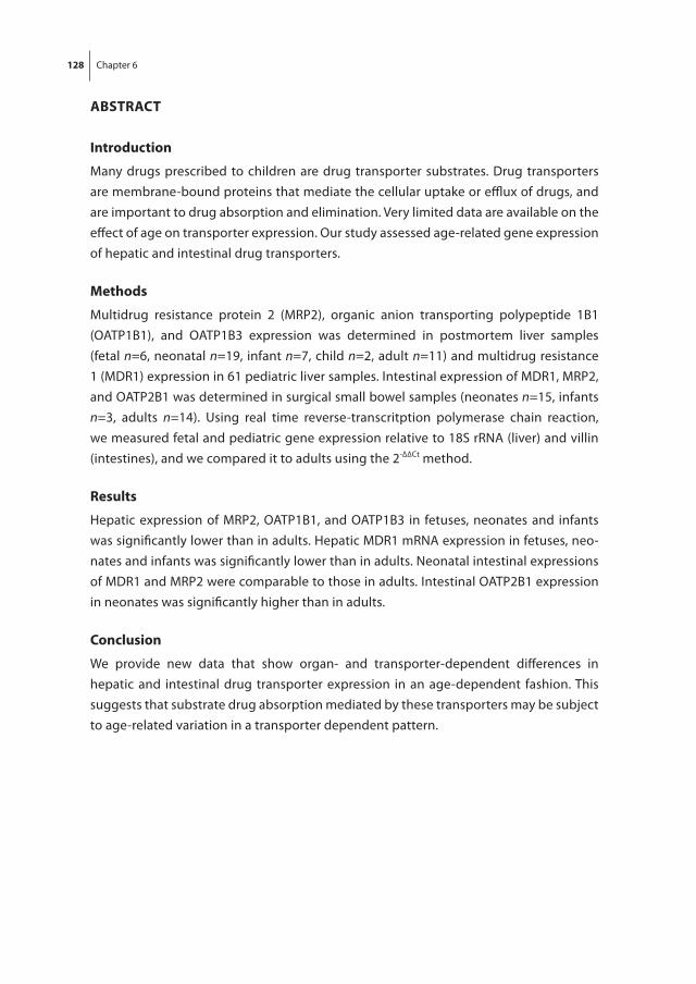
128 Chapter 6
AbstrAct
introduction
Many drugs prescribed to children are drug transporter substrates. Drug transporters are membrane-bound proteins that mediate the cellular uptake or efflux of drugs, and are important to drug absorption and elimination. Very limited data are available on the effect of age on transporter expression. Our study assessed age-related gene expression of hepatic and intestinal drug transporters.
Methods
Multidrug resistance protein 2 (MRP2), organic anion transporting polypeptide 1B1 (OATP1B1), and OATP1B3 expression was determined in postmortem liver samples (fetal n=6, neonatal n=19, infant n=7, child n=2, adult n=11) and multidrug resistance 1 (MDR1) expression in 61 pediatric liver samples. Intestinal expression of MDR1, MRP2, and OATP2B1 was determined in surgical small bowel samples (neonates n=15, infants n=3, adults n=14). Using real time reverse-transcritption polymerase chain reaction, we measured fetal and pediatric gene expression relative to 18S rRNA (liver) and villin (intestines), and we compared it to adults using the 2-∆∆Ct method.
results
Hepatic expression of MRP2, OATP1B1, and OATP1B3 in fetuses, neonates and infants was significantly lower than in adults. Hepatic MDR1 mRNA expression in fetuses, neo-nates and infants was significantly lower than in adults. Neonatal intestinal expressions of MDR1 and MRP2 were comparable to those in adults. Intestinal OATP2B1 expression in neonates was significantly higher than in adults.
conclusion
We provide new data that show organ- and transporter-dependent differences in hepatic and intestinal drug transporter expression in an age-dependent fashion. This suggests that substrate drug absorption mediated by these transporters may be subject to age-related variation in a transporter dependent pattern.
Ontogeny of human hepatic and intestinal transporter gene expression during childhood: age matters 129
6
introDuction
Drug transporters are membrane-bound proteins whose primary function is to facilitate the trafficking of drugs and their metabolites across a biologic membrane. Expressed in structural cells that compose the organs of importance to drug disposition, such as enterocytes and hepatocytes, they are involved in the active uptake and elimination of orally administered drugs. In adults, drug transporters are recognized as key deter-minants of variation in the pharmacokinetics of many drugs, as shown in studies using primary cell and ex vivo organ cultures as well as clinical studies [1]. In contrast, such data in children are scarce and clinical studies are absent [2-7].
Pharmacokinetics in children is known to be affected by developmental changes with age [8]. Drug metabolizing enzyme activity, for example, changes significantly from fetal to adolescent age associated with alterations in metabolic clearance of many drugs [9]. Notably, hepatic and intestinal expression of cytochrome P450 subfamily CYP3A4 shows a clear developmental pattern, and systemic clearance of midazolam, a known substrate of CYP3A, changes with age [10-12]. Similarly, compared with adults, differences in body composition and renal function in children affect drug tissue distribution and renal drug excretion, respectively.
As a consequence, it has become apparent that simply extrapolating drug doses from adults to children based on body weight is likely inaccurate owing to our lack of our understanding of age-dependent differences in the maturation of the drug absorption and elimination processes involved in drug disposition. As the expression of drug metabolizing enzymes appears to be age-related, it is also likely that this is the case for drug transporter expression. Data from animal studies report occurrence of transporter-specific maturation [13]. In humans, several efflux transporters that belong to the ATP-binding cassette family (ABC), including the multidrug resistance protein 1 (MDR1/ABCB1), multidrug resistance-associated protein 2 (MRP2/ABCC2), and breast cancer resistance protein (BCRP/ABCG2), were found to be expressed in fetal liver or intestine, but otherwise little is known of the developmental patterns of individual drug transporters [6, 14, 15]. Systematic characterization of age-related differences in transporter expression will aid the selection of the most appropriate dose for substrate drugs in children.
In a first step towards elucidating the developmental changes, this study determined the gene expression of clinically relevant hepatic and intestinal drug transporters across the pediatric age range. We focused on transporters with well-defined roles in drug pharmacokinetics in adults [1] (Fig. 1). These included the efflux carriers MDR1 and MRP2 in liver and intestine, and uptake carriers of the organic anion transporting polypeptide (OATP/solute carrier organic anion [SLCO]) family, including the liver-specific transport-ers OATP1B1 and OATP1B3, and OATP2B1 in the intestine.

130 Chapter 6
MAtEriAls AnD MEthoDs
1. tissue samples
Autopsy liver tissue samples from fetuses and from children up to 18 years of age were obtained from the Erasmus MC Tissue Bank. An opt-out clause was in place for use of tissue from the tissue bank. The Erasmus MC research ethics board provided a waiver for ethics approval according to the Dutch Law on research in humans. Fresh intestinal tissue was collected during surgery at the time of resection. Intestinal samples from children were derived from other research projects, and informed consent was previ-ously obtained for the use of this tissue in the context of these studies approved by Erasmus MC research ethics beoard. After resection, all liver and intestinal tissues were immediately snap frozen in liquid nitrogen, stored at −80°C and processed on ice for mRNA isolation.
Healthy human adult liver samples were obtained from the Liver Tissue Cell Distri-bution System at the University of Minnesota under National Institues of Health (NIH) contract N01-DK-7-0004/HHSN267200700004C. Adult intestinal samples were obtained from the University of Western Ontario, and the use of these samples was approved by the research ethics board.
Additional pediatric tissue specimens for MDR1 analyses were obtained from the NIH-supported tissue programs: the Liver Tissue Cell Distribution System (LTCDS) from the Minnesota and Pittsburgh collection centers; the University of Maryland Brain and Tissue bank for Developmental Disorders (Baltimore, MD); and the Laboratory of De-velopmental Biology at the University of Washington (Seattle, WA). Additional samples were from Xenotech, LLC (Lenexa, KS). The use of these tissues was declared nonhuman subjects research by the Children’s Mercy Hospitals and Clinics pediatric institutional review board.
The sources of the obtained samples are listed in Table 1.
figure 1. transporters in enterocyte and hepatocyte.Only transporters in enterocyte (A) and (B) hepatocytes were selected for this study.

Ontogeny of human hepatic and intestinal transporter gene expression during childhood: age matters 131
6
2. mrnA isolation and cDnA synthesis
Frozen human tissue samples were mechanically homogenized on ice. Homogenate was applied to a QiaShredder column (Qiagen) and RNA was extracted using the RNeasy Mini Kit (Qiagen, Valencia, CA) according to the instructions provided by the manu-facturer. RNA was treated with DNase to digest genomic DNA remnants. The quantity and integrity of RNA were determined by microfluidics-based analyses using the 2100 BioAnalyzer (Agilent, Santa Clara, CA). Samples with an RNA integrity number (RIN) of <5 were excluded from this study. Complementary DNA (cDNA) was synthesized using the High-capacity cDNA Archive Kit (Applied Biosystems, Foster City, CA) and random priming.
3. real time reverse transcription Polymerase chain reaction
The mRNA expression was measured by SYBR green quantitative real-time polymerase chain reaction with the ABI 7500 sequence detection system (Applied Biosystems). Previ-ously optimized primers were used (Supplemental Material) [16]. Modified primers were used for villin and OATP2B1 (reverse primer only), and primer sequences were obtained via qPrimer Depot (http://primerdepot.nci.nih.gov/). After each polymerase chain reac-tion, a melting curve analysis was performed to confirm product specificity. Eukaryotic 18S rRNA was assessed as the endogenous control with the use of a TaqMan VIC/MGB probe (4319413E, Applied Biosystems). Transporter transcript levels were normalized to18s rRNA, and relative expression was determined using the 2-ΔΔCt method comparing pediatric samples to adult liver or intestine (calibrator samples) [17]. The adult target gene value was determined by using the median of the adult threshold cycle (Ct) values.
For comparison, all 96-well plates analyzing liver samples included a specific liver con-trol sample to normalize expression for the gene of interest and 18S rRNA, if appropriate. Similarly, a specific intestinal sample was added to plates to normalize for the genes of interest, 18S rRNA, and villin if appropriate.
4. statistics
All data were analyzed by Kruskal-Wallis analysis, followed by group comparisons using a nonparametric Mann-Whitney test and post-hoc Bonferroni multiple comparison test. All statistical analyses were performed using GraphPad Prism version 5.00.2 and IBM SPSS Statistics software (SPSS Statistics for Windows, Version 21.0; IBM, Armonk, NY). P<0.05 was considered statistically significant. Data are presented as median and range, unless indicated otherwise.
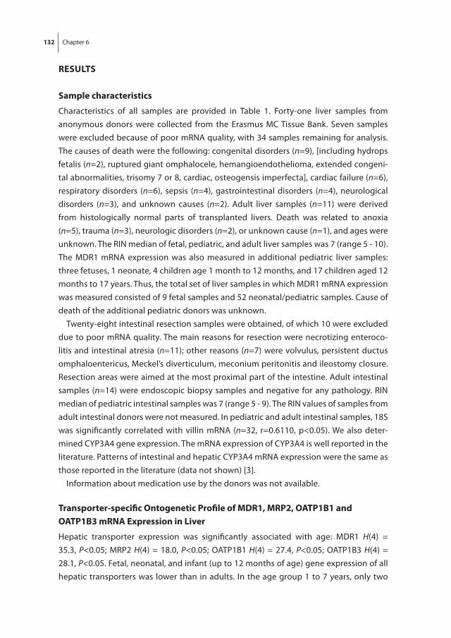
132 Chapter 6
rEsults
sample characteristics
Characteristics of all samples are provided in Table 1. Forty-one liver samples from anonymous donors were collected from the Erasmus MC Tissue Bank. Seven samples were excluded because of poor mRNA quality, with 34 samples remaining for analysis. The causes of death were the following: congenital disorders (n=9), [including hydrops fetalis (n=2), ruptured giant omphalocele, hemangioendothelioma, extended congeni-tal abnormalities, trisomy 7 or 8, cardiac, osteogensis imperfecta], cardiac failure (n=6), respiratory disorders (n=6), sepsis (n=4), gastrointestinal disorders (n=4), neurological disorders (n=3), and unknown causes (n=2). Adult liver samples (n=11) were derived from histologically normal parts of transplanted livers. Death was related to anoxia (n=5), trauma (n=3), neurologic disorders (n=2), or unknown cause (n=1), and ages were unknown. The RIN median of fetal, pediatric, and adult liver samples was 7 (range 5 - 10). The MDR1 mRNA expression was also measured in additional pediatric liver samples: three fetuses, 1 neonate, 4 children age 1 month to 12 months, and 17 children aged 12 months to 17 years. Thus, the total set of liver samples in which MDR1 mRNA expression was measured consisted of 9 fetal samples and 52 neonatal/pediatric samples. Cause of death of the additional pediatric donors was unknown.
Twenty-eight intestinal resection samples were obtained, of which 10 were excluded due to poor mRNA quality. The main reasons for resection were necrotizing enteroco-litis and intestinal atresia (n=11); other reasons (n=7) were volvulus, persistent ductus omphaloentericus, Meckel’s diverticulum, meconium peritonitis and ileostomy closure. Resection areas were aimed at the most proximal part of the intestine. Adult intestinal samples (n=14) were endoscopic biopsy samples and negative for any pathology. RIN median of pediatric intestinal samples was 7 (range 5 - 9). The RIN values of samples from adult intestinal donors were not measured. In pediatric and adult intestinal samples, 18S was significantly correlated with villin mRNA (n=32, r=0.6110, p<0.05). We also deter-mined CYP3A4 gene expression. The mRNA expression of CYP3A4 is well reported in the literature. Patterns of intestinal and hepatic CYP3A4 mRNA expression were the same as those reported in the literature (data not shown) [3].
Information about medication use by the donors was not available.
transporter-specific ontogenetic Profile of MDr1, MrP2, oAtP1b1 and oAtP1b3 mrnA Expression in liver
Hepatic transporter expression was significantly associated with age: MDR1 H(4) = 35.3, P<0.05; MRP2 H(4) = 18.0, P<0.05; OATP1B1 H(4) = 27.4, P<0.05; OATP1B3 H(4) = 28.1, P<0.05. Fetal, neonatal, and infant (up to 12 months of age) gene expression of all hepatic transporters was lower than in adults. In the age group 1 to 7 years, only two
Ontogeny of human hepatic and intestinal transporter gene expression during childhood: age matters 133
6
tabl
e 1.
tis
sue
sour
ces
and
age
dist
ribu
tion
tiss
ueco
llect
ion
tran
spor
ter
exam
ined
num
ber o
f sa
mpl
esse
xG
esta
tion
al a
geat
bir
tha
Post
nata
l ag
ebso
urce
Feta
l and
pe
diat
ric li
ver
Post
mor
tem
Auto
psy
MRP
2, O
ATP1
B1,
OAT
P1B3
Feta
l: 6
Pedi
atric
: 28
Tota
l: 34
Mal
e: 1
9Fe
mal
e: 1
5Fe
tuse
s: 2
2 (2
2-23
)Pe
diat
ric: 3
6 (2
5-41
)Pe
diat
ric 1
wk
(0 –
7 y
ears
)Er
asm
us M
C Ti
ssue
Bio
bank
Feta
l and
pe
diat
ric li
ver
Post
mor
tem
Auto
psy
MD
R1Fe
tal:
9Pe
diat
ric: 5
2To
tal:
61
Mal
e: 3
8Fe
mal
e: 2
2U
nkno
wn:
1
Fetu
ses:
22
(8-2
3)Pe
diat
ric
11 w
k (0
-17
year
s)
1)
Eras
mus
MC
Tiss
ue B
ioba
nk2)
Th
e Li
ver T
issu
e Ce
ll D
istr
ibut
ion
Syst
em;
from
the
Min
neso
ta a
nd P
ittsb
urgh
col
lect
ion
cent
ers;
the
Uni
vers
ity o
f Mar
ylan
d Br
ain
and
Tiss
ue b
ank
for D
evel
opm
enta
l Dis
orde
rs
(Bal
timor
e, M
D);
and
the
Labo
rato
ry o
f D
evel
opm
enta
l Bio
logy
at t
he U
nive
rsity
of
Was
hing
ton
(Sea
ttle
, WA
). Ad
ditio
nal s
ampl
es
wer
e fr
om X
enot
ech,
LLC
(Len
exa,
KS)
.
Adul
t liv
erBi
opsy
MD
R1, M
RP2,
O
ATP1
B1,
OAT
P1B3
11M
ale:
6Fe
mal
e: 2
Unk
now
n: 1
–U
nkno
wn
The
Live
r Tis
sue
Cell
Dis
trib
utio
n Sy
stem
; U
nive
rsity
of M
inne
sota
und
er N
IH C
ontr
act
N01
-DK-
7-00
04/H
HSN
2672
0070
0004
C.
Pedi
atric
inte
stin
eSu
rgic
alje
junu
m 7
ileum
9ce
cum
2
MD
R1, M
RP2,
O
ATP2
B118
Mal
e: 1
2Fe
mal
e: 6
Neo
nate
s: 3
2 (2
5-40
)1
(0 –
14)
wks
Eras
mus
MC
Adul
t int
estin
eBi
opsy
ileum
14
MD
R1, M
RP2,
O
ATP2
B114
Mal
e: 3
Fem
ale:
11
–U
nkno
wn
Uni
vers
ity o
f Wes
tern
Ont
ario
a med
ian(
rang
e) in
wee
ks; b m
edia
n(ra
nge)

134 Chapter 6
samples were assessed for the hepatic transporters MRP2, OATP1B1, and OATP1B3, as a result of which median mRNA expression could not be determined and comparison with adult expression was not possible.
MDR1 mRNA expression in fetal [0.061 (0.027 - 0.792)] and neonatal [0.036 (0.011 - 0.364)] age groups was 20- to 30-fold lower than in adults (Fig. 2). MDR1 mRNA expres-sion in infants was slightly lower [0.248 (0.010 - 1.46)] than in adults (Fig. 2). Median MDR1 gene expression in children aged 1 to 7 years was not different from adults [0.777 (0.012 - 8.928); P=0.663]. Interindividual variability in all age groups was large.
MRP2 mRNA expression was about 30-fold lower in fetal liver samples [0.074 (0.004 - 0.162)], 200-fold lower in neonates [0.012 (0.001 - 0.238)], and 100-fold lower in infants [0.022 (0.008 - 0.357)] than in adult liver samples (Fig. 2). Compared with adult liver, OATP1B1 mRNA expression was 20-fold lower in fetal liver samples [0.042 (0.0004 - 0.683)], 500-fold lower in neonates [0.002 (0.0003 - 0.018)], and 90-fold lower in infants [0.010 (0.001 - 0.086)] (Fig. 2). Expression profiles of OATP1B3 in the liver samples were similar to those of OATP1B1. Expression of hepatic OATP1B3 was 30-fold lower in fetuses [0.046 (0.001 - 1.910)], 600-fold lower in neonates [0.002 (0.0004 - 0.0187)], and 100-fold lower in infants [0.010 (0.001 - 0.021)] than in adults (Fig. 2).
similar MDr1 and MrP2 mrnA Expression in neonatal and Adult intestines, and higher oAtP2b1 in neonatal intestines
Intestinal transporter expression was statistically significantly associated with age for MRP2 H(2) = 9.1, P<0.05 and OATP2B1 H(2) = 15.8, P<0.05, but not for MDR1, H(2) = 1.3, P=0.5. MDR1 expression in intestinal samples of neonates was 0.838 (0.370 - 97.0; P=0.395) and of infants was 1.454 (0.409 - 3.174; P=0.413) and hence similar to adults (Fig. 3). Expression of MRP2 mRNA in the intestinal samples of neonates [1.262 (0.338 - 9.266); P=0.248] was similar to the adult expression (Fig. 3). In infant intestinal samples, however, MRP2 mRNA expression was significantly lower [0.038 (0.023 - 0.074); P<0.05] than in adult samples. Interestingly, the uptake transporter OATP2B1 showed a higher mRNA expression in neonates [2.86 (1.212 - 34.45); P<0.0001]. In infants up to 12 months of age, OATP2B1 was expressed at the same level as in adult intestinal samples [1.21 (0.328 – 2.288); P=0.147] (Fig. 3).

Ontogeny of human hepatic and intestinal transporter gene expression during childhood: age matters 135
6
figure 2. transporter gene expression in liver.Relative mRNA expression of MDR1 (A) MRP2 (B) OATP1B1 (C) OATP1B3 (D) from fetal, pediatric and adult liver samples, normalized to 18S mRNA expression and adult expression using the 2-ΔΔCt method. Lines represent median and interquartile range.
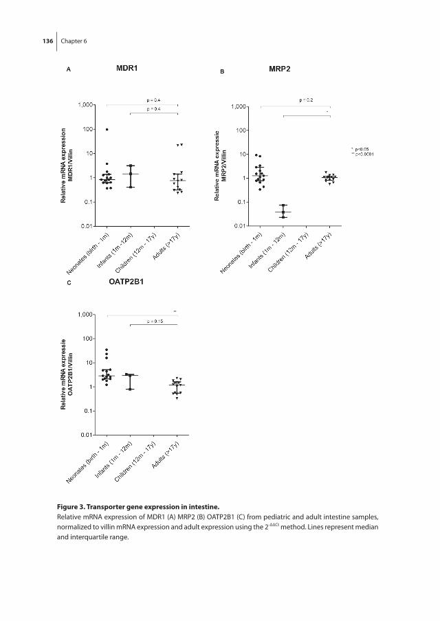
136 Chapter 6
figure 3. transporter gene expression in intestine.Relative mRNA expression of MDR1 (A) MRP2 (B) OATP2B1 (C) from pediatric and adult intestine samples, normalized to villin mRNA expression and adult expression using the 2-ΔΔCt method. Lines represent median and interquartile range.
Ontogeny of human hepatic and intestinal transporter gene expression during childhood: age matters 137
6
Discussion
Little is known about transporter gene expression in children. Our study used samples from fetal to adult age, to examine the gene expression of hepatic transporters MDR1, MRP2, OATP1B1, and OATP1B3; they were found to be age-related, as their expression was lowest in fetal and pediatric liver samples. In contrast, intestinal OATP2B1 expres-sion in neonates was higher than in adults. Intestinal MDR1 and MRP2 expression was similar between neonates and adults.
ontogeny in hepatic transporters
Hepatic MDR1 expression is already detectable in early human fetal life. The maturation pattern found in our study is consistent with previous data on hepatic MDR1 mRNA expression [3, 15, 18]. Our findings are also supported by immunohistochemical staining showing that MDR1 expression followed the architectural changes during fetal develop-ment [18]. We show that MDR1 expression changes after birth, with low mRNA expres-sion until 12 months of age whereupon it increases to adult levels. In a recent study that used a novel liquid chromatography with tandem mass spectrometry method to estimate protein expression, from 7 years onwards, MDR1 was stable up to 70 years of age in a cohort of more than 50 patients [19].
Low fetal MRP2 expression in humans has been reported earlier in two small studies (n=10, 14-20 weeks’ GA; and n=3, 20-21 weeks’ GA) [5, 14]. In the latter, however, two of five control adult liver samples were from normal tissue in patients with hepatocellular carcinoma. Because organic cation transporter 1 (OCT1) expression has been shown to be lower in nontumor hepatocellular carcinoma liver than healthy liver, a similar phe-nomenon with other transporters cannot be excluded [20]. Using immunohistochemical staining, distinct canalicular expression of MRP2 in adult liver was observed but a fuzzier canalicular occurrence in fetal liver, which may suggest immaturity of the localization pattern [5]. In addition, fetal human liver tissues obtained at 19 weeks’ gestational age showed higher MRP2 signal intensity than tissues obtained at 14 weeks [21] In our study, neonatal MRP2 gene expression was lower than in adults. From 7 years onwards, MRP2 protein expression was stable. [22].
The observed lower fetal OATP1B1 and OATP1B3 mRNA expression levels in the pres-ent study are also in line with human data derived of three fetal (21-23 weeks’ gestational age) and three adult livers [14]. In our study, OATP1B1 and OATB1B3 expression was also low in neonates. In contrast, the reported neonatal OATP1B1 and OATP1B3 protein expression (n=5), was similar in adults [23]. In 80 human pediatric liver samples ranging from age 2 months to 12 years, OATP1B1 was low until 6 years of age, whereas OATP1B3 protein levels were high at birth then rapidly decreased in the first year to then increase again from 8 years of age onwards to adult levels [24]. From 7 years onwards, OATP1B1
138 Chapter 6
and OATP1B3 protein expression was stable, with only genotype related to OATP1B1 abundance [19].
ontogeny in intestinal transporters
We observed stable intestinal MDR1 expression from neonatal to adult age, a finding that fills a gap between currently available fetal and pediatric data. Previously, fetal intestinal tissue obtained after induced abortion, showed no or minimal MDR1 mRNA expression in samples after 11, 13, and 14 weeks, but clear expression at 16 and 20 weeks’ gestational age [18]. Another study reported similar small intestinal MDR1 mRNA expression in fetuses (14- 20 weeks’ gestational age), neonates, and adults [3]. However, a limitation is the small sample size [n=5, n=12 (fetus plus neonates)] [3, 18]. Stable MDR1 mRNA expression in children from 1 month to adulthood was shown in nondiseased duodenal biopsy and jejunal tissue from liver donor recipients [2, 4]. We also found stable intestinal MRP2 expression but higher expression levels of OATP2B1 in neonates compared to adults.
The transporter maturation we observed likely reflects physiological roles of these transporters in organ development. Epigenetic mechanisms such as DNA methylation are thought to be involved in the maturation of drug metabolizing enzymes. For ex-ample, DNA methylation seems to partly explain a developmental switch from expres-sion of CYP3A7 to CYP3A4 in human fetal and adult liver [25]. Epigenetic mechanisms may be also involved in the developmental regulation of drug transporter expression, but studies are currently lacking.
Due to mediation by transcription factors (which in their turn can be induced by chemicals or pathological conditions), some transporters show relatively high whereas others show low expression [13]. For example, expression of the steroid- and xenobiotic-sensing pregnane X receptor (PXR) was shown to correlate with intestinal breast cancer resistance protein (BCRP), MRP2 and MDR1 mRNA in many different human tissues (e.g., intestine and liver) [3, 26, 27]. We may further speculate that the ontogeny of transporters is a combined function of developmental changes, genetic heterogeneity, and exposure to substrates (drug or environmental) that induce or inhibit expression and/or activity. For example, CYP1A2 and CYP3A4 activity, assessed by phenotyping caffeine and dextromethorphan, respectively, was shown to be affected by the type of infant feeding (breast milk or formula) [28]. Dietary supplements in infant formula decreased MDR1 expression in colon cell cultures [29]. Moreover, the effect of feeding is more variable when a child’s diet is expanded, as specific nutrients can influence MDR1 expression [30, 31].
This study is one of the first exploratory studies characterizing developmental changes in hepatic and intestinal transporters. Some limitations of the data should be addressed. First, most fetal liver samples were obtained in the second trimester of gestation, thus

Ontogeny of human hepatic and intestinal transporter gene expression during childhood: age matters 139
6
results might not be representative for the entire range of gestational age. Second, the variation in fetal transporter mRNA expression may be partly due to the small number of samples representing ages more distant from the median age. Given the small num-ber per age group, the range of expression might not be representative for the entire population.
Third, all liver samples were collected within 24 hours after death, but the exact time is unknown. Still, RNA stability was minimized by RNA integrity determination.
Also, our samples may display disease-dependent variability in transporter expression. Inflammation has been shown to affect drug metabolism and transport. Decreased mid-azolam clearance in critically ill children is likely a result of reduced CYP3A activity [32]. Inconsistency has been reported from studies assessing the effect of proinflammatory cytokines on intestinal MDR1 expression in vitro and in animal models, but they sug-gest a trend towards decreased expression [33]. In children with Crohn disease, MDR1 expression was higher in both inflamed and nonnflamed duodenal biopsies compared with healthy age-matched controls [34]. Inflammation was also shown to affect the ex-pression of other transporters in primary human hepatocytes (i.e., OATP1B1, OATP1B3, MDR1, MRP2), resulting in reduced mRNA expression, protein, and activity [35]. Thus, evidence exists that the potential effects of underlying disease should be considered when interpreting age-related changes in transporter expression.
Finally, transporter genetic polymorphisms likely contribute to the observed vari-ability in gene expression [1]. Recently, Nies et al. and Prasad et al. showed that hepatic expression of OATP1B1, but not OATP1B3, is significantly affected by genetic variants [19, 36].
The observed age-dependent maturation and large interindividual variability in drug transporter expression may have implications for oral drug absorption and hepatic drug excretion. The question whether posttranscriptional modifications occur and if expressed mRNA level can be extrapolated to protein expression and ultimately in vivo activity remains to be answered [4, 11]. Human hepatic OATP1B1 and OATP1B3 protein expression showed a trend with high interindividual variability in the first year of life followed by lower variability until the age of 8 years, when variability increases again [24]. This typical age-related change in variability at younger age is also reflected by our findings on OATP1B1 and OATP1B3 mRNA expression. In contrast, others could not correlate protein levels with OATP1B1 or MDR1 mRNA expression [37]. Further protein expression and transporter activity studies are needed to confirm these mRNA expres-sion results.
In conclusion, hepatic MDR1, MRP2, OATP1B1, OATP1B3 and intestinal MDR1, MRP2, OATP2B1 drug transporter expression show organ- and transporter-specific maturation patterns during childhood. Therefore, the disposition of drugs substrates for these transporters may be subject to age-related changes other than that in body size alone.

140 Chapter 6
rEfErEncEs
[1] DeGorter MK, Xia CQ, Yang JJ, Kim RB. Drug transporters in drug efficacy and toxicity. Annu Rev Pharmacol Toxicol 2012; 52: 249-73.
[2] Mizuno T, Fukuda T, Masuda S, Uemoto S, Matsubara K, Inui KI, et al. Developmental trajectory of intestinal MDR1/ABCB1 expression in children. Br J Clin Pharmacol 2013 Jul 23.
[3] Miki Y, Suzuki T, Tazawa C, Blumberg B, Sasano H. Steroid and xenobiotic receptor (SXR), cyto-chrome P450 3A4 and multidrug resistance gene 1 in human adult and fetal tissues. Mol Cell Endocrinol 2005 Feb 28; 231(1-2): 75-85.
[4] Fakhoury M, Litalien C, Medard Y, Cave H, Ezzahir N, Peuchmaur M, et al. Localization and mRNA expression of CYP3A and P-glycoprotein in human duodenum as a function of age. Drug Metab Dispos 2005 Nov; 33(11): 1603-7.
[5] Chen HL, Chen HL, Liu YJ, Feng CH, Wu CY, Shyu MK, et al. Developmental expression of canalicu-lar transporter genes in human liver. J Hepatol 2005 Sep; 43(3): 472-7.
[6] Chen HL, Liu YJ, Chen HL, Wu SH, Ni YH, Ho MC, et al. Expression of hepatocyte transporters and nuclear receptors in children with early and late-stage biliary atresia. Pediatr Res 2008 Jun; 63(6): 667-73.
[7] Fukudo M, Yano I, Masuda S, Goto M, Uesugi M, Katsura T, et al. Population pharmacokinetic and pharmacogenomic analysis of tacrolimus in pediatric living-donor liver transplant recipients. Clin Pharmacol Ther 2006 Oct; 80(4): 331-45.
[8] Kearns GL, Abdel-Rahman SM, Alander SW, Blowey DL, Leeder JS, Kauffman RE. Developmental pharmacology--drug disposition, action, and therapy in infants and children. N Engl J Med 2003 Sep 18; 349(12): 1157-67.
[9] de Wildt SN. Profound changes in drug metabolism enzymes and possible effects on drug therapy in neonates and children. Expert Opin Drug Metab Toxicol 2011 Aug; 7(8): 935-48.
[10] Leeder JS, Gaedigk R, Marcucci KA, Gaedigk A, Vyhlidal CA, Schindel BP, et al. Variability of CYP3A7 expression in human fetal liver. J Pharmacol Exp Ther 2005 Aug; 314(2): 626-35.
[11] Johnson TN, Tanner MS, Taylor CJ, Tucker GT. Enterocytic CYP3A4 in a paediatric population: developmental changes and the effect of coeliac disease and cystic fibrosis. Br J Clin Pharmacol 2001 May; 51(5): 451-60.
[12] de Wildt SN, Kearns GL, Hop WC, Murry DJ, Abdel-Rahman SM, van den Anker JN. Pharmacokinet-ics and metabolism of oral midazolam in preterm infants. Br J Clin Pharmacol 2002 Apr; 53(4): 390-2.
[13] Klaassen CD, Aleksunes LM. Xenobiotic, bile acid, and cholesterol transporters: function and regulation. Pharmacol Rev 2010 Mar; 62(1): 1-96.
[14] Sharma S, Ellis EC, Gramignoli R, Dorko K, Tahan V, Hansel M, et al. Hepatobiliary disposition of 17-OHPC and taurocholate in fetal human hepatocytes: a comparison with adult human hepato-cytes. Drug Metab Dispos 2013 Feb; 41(2): 296-304.
[15] Fakhoury M, de Beaumais T, Guimiot F, Azougagh S, Elie V, Medard Y, et al. mRNA expression of MDR1 and major metabolising enzymes in human fetal tissues. Drug Metab Pharmacokinet 2009; 24(6): 529-36.
[16] Glaeser H, Bailey DG, Dresser GK, Gregor JC, Schwarz UI, McGrath JS, et al. Intestinal drug trans-porter expression and the impact of grapefruit juice in humans. Clin Pharmacol Ther 2007 Mar; 81(3): 362-70.
[17] Schmittgen TD, Livak KJ. Analyzing real-time PCR data by the comparative C(T) method. Nat Protoc 2008; 3(6): 1101-8.
Ontogeny of human hepatic and intestinal transporter gene expression during childhood: age matters 141
6
[18] van Kalken CK, Giaccone G, van der Valk P, Kuiper CM, Hadisaputro MM, Bosma SA, et al. Multidrug resistance gene (P-glycoprotein) expression in the human fetus. Am J Pathol 1992 Nov; 141(5): 1063-72.
[19] Prasad B, Evers R, Gupta A, Hop CE, Salphati L, Shukla S, et al. Interindividual variability in hepatic organic anion-transporting polypeptides and P-glycoprotein (ABCB1) protein expression: quan-tification by liquid chromatography tandem mass spectroscopy and influence of genotype, age, and sex. Drug Metab Dispos 2014 Jan; 42(1): 78-88.
[20] Schaeffeler E, Hellerbrand C, Nies AT, Winter S, Kruck S, Hofmann U, et al. DNA methylation is associated with downregulation of the organic cation transporter OCT1 (SLC22A1) in human hepatocellular carcinoma. Genome Med 2011; 3(12): 82.
[21] Cizkova D, Morky J, Micuda S, Osterreicher J, Martinkova J. Expression of MRP2 and MDR1 trans-porters and other hepatic markers in rat and human liver and in WRL 68 cell line. Physiol Res 2005; 54(4): 419-28.
[22] Prasad B, Lai Y, Lin Y, Unadkat JD. Interindividual variability in the hepatic expression of the hu-man breast cancer resistance protein (BCRP/ABCG2): effect of age, sex, and genotype. J Pharm Sci 2013 Mar; 102(3): 787-93.
[23] Yanni SB, Smith PB, Benjamin DK, Jr., Augustijns PF, Thakker DR, Annaert PP. Higher clearance of micafungin in neonates compared with adults: role of age-dependent micafungin serum bind-ing. Biopharm Drug Dispos 2011 May; 32(4): 222-32.
[24] Thomson MMS, Krauel S, Hines RN, Scheutz EG, Meibohm B. Lack of effect of genetic variants on the age-associated prtoein expression of OATP1B1 and OATP1B3 in human pediatric liver. [Abstract for the 114th American Society for Clinical Pharmacology and Therapeutics Annual Meeting]; 2013.
[25] Kacevska M, Ivanov M, Wyss A, Kasela S, Milani L, Rane A, et al. DNA methylation dynamics in the hepatic CYP3A4 gene promoter. Biochimie 2012 Nov; 94(11): 2338-44.
[26] Albermann N, Schmitz-Winnenthal FH, Z’Graggen K, Volk C, Hoffmann MM, Haefeli WE, et al. Expression of the drug transporters MDR1/ABCB1, MRP1/ABCC1, MRP2/ABCC2, BCRP/ABCG2, and PXR in peripheral blood mononuclear cells and their relationship with the expression in intestine and liver. Biochem Pharmacol 2005 Sep 15; 70(6): 949-58.
[27] Synold TW, Dussault I, Forman BM. The orphan nuclear receptor SXR coordinately regulates drug metabolism and efflux. Nat Med 2001 May; 7(5): 584-90.
[28] Blake MJ, Abdel-Rahman SM, Pearce RE, Leeder JS, Kearns GL. Effect of diet on the development of drug metabolism by cytochrome P-450 enzymes in healthy infants. Pediatr Res 2006 Dec; 60(6): 717-23.
[29] Bebawy M, Rasmussen C, Sambasivam S, Bao S. Dietary nucleotide supplements in infant formula modify the expression of P-glycoprotein in the intestinal epithelium in vitro. Int J Vitam Nutr Res 2009 Sep; 79(5-6): 381-7.
[30] Schwarz UI, Hanso H, Oertel R, Miehlke S, Kuhlisch E, Glaeser H, et al. Induction of intestinal P-glycoprotein by St John’s wort reduces the oral bioavailability of talinolol. Clin Pharmacol Ther 2007 May; 81(5): 669-78.
[31] Schwarz UI, Seemann D, Oertel R, Miehlke S, Kuhlisch E, Fromm MF, et al. Grapefruit juice inges-tion significantly reduces talinolol bioavailability. Clin Pharmacol Ther 2005 Apr; 77(4): 291-301.
[32] Vet NJ, de Hoog M, Tibboel D, de Wildt SN. The effect of critical illness and inflammation on midazolam therapy in children. Pediatr Crit Care Med 2012 Jan; 13(1): e48-50.
[33] Fernandez C, Buyse M, German-Fattal M, Gimenez F. Influence of the pro-inflammatory cytokines on P-glycoprotein expression and functionality. J Pharm Pharm Sci 2004 Nov 17; 7(3): 359-71.
142 Chapter 6
[34] Fakhoury M, Lecordier J, Medard Y, Peuchmaur M, Jacqz-Agrain E. Impact of inflammation on the duodenal mRNA expression of CYP3A and P-glycoprotein in children with Crohn’s disease. Inflamm Bowel Dis 2006 Aug; 12(8): 745-9.
[35] Le Vee M, Jouan E, Moreau A, Fardel O. Regulation of drug transporter mRNA expression by interferon-gamma in primary human hepatocytes. Fundam Clin Pharmacol 2011 Feb; 25(1): 99-103.
[36] Nies AT, Niemi M, Burk O, Winter S, Zanger UM, Stieger B, et al. Genetics is a major determinant of expression of the human hepatic uptake transporter OATP1B1, but not of OATP1B3 and OATP2B1. Genome Med 2013 Jan 11; 5(1): 1.
[37] Ulvestad M, Skottheim IB, Jakobsen GS, Bremer S, Molden E, Asberg A, et al. Impact of OATP1B1, MDR1, and CYP3A4 expression in liver and intestine on interpatient pharmacokinetic variability of atorvastatin in obese subjects. Clin Pharmacol Ther 2013 Mar; 93(3): 275-82.


7 I ntestinal peptide transporter PEPT1 expression and tissue distribution across the pediatric age range
Miriam G. Mooij, Barbara A.E. de Koning, Dicky J. Lindenbergh-Kortleve, Ytje Simons-Oosterhuis, Dick Tibboel, Janneke N. Samsom, Saskia N. de Wildt
Submitted for publication

146 Chapter 7
AbstrAct
introduction
The intestinal influx oligopeptide transporter PEPT1 (SLC15A1) is best known for nutrient-derived di- and tripeptide transport. Its role in drug absorption is increasingly recognized. To better understand the disposition of PEPT1 substrate drugs in young infants, we studied intestinal PEPT1 mRNA expression and tissue localization across the pediatric age range.
Methods
PEPT1 mRNA expression was determined using real time RT-PCR in small intestinal tis-sues collected from surgical procedures (neonates, infants) or biopsies (older children, adolescents). PEPT1 mRNA relative to villin mRNA expression were compared between neonates/infants and older children and adolescents. PEPT1 was visualized in infant tissue using immunohistochemical staining. Other transporters (MDR1, MRP2 and OATP2B1) were also stained to describe the localization in relation to PEPT1.
results
22 intestinal samples (n=20 neonates/infants, n=2 pediatric, n=4 adolescents) were ana-lyzed. The young infant samples were collected at a median (range) gestational age at birth of 29.2 weeks (24.7 - 40) and postnatal age of 2.4 weeks (0 – 16.6). The PEPT1 mRNA expression of the neonates/infants was only marginally lower (0.8-fold) than the older children (p<0.05). Similar and clear apical PEPT1 and MRP2 staining, apical and lateral MDR1 staining, and intra-epithelial OATP2B1 staining at the basolateral membrane of the enterocyte was detected in 12 infant and 2 adolescent samples.
conclusion
Although small intestinal PEPT1 expression tended to be lower in neonates than in older children, this difference is small and tissue distribution is similar. This finding suggests similar oral absorption of PEPT1 substrates across the pediatric age range.
Intestinal peptide transporter PEPT1 expression and tissue distribution across the pediatric age range 147
7
introDuction
The influx oligopeptide transporter PEPT1 (SLC15A1) is a member of the solute carrier (SLC) superfamily and is situated on the apical membrane of the enterocyte. Its expres-sion in the human adult jejunum is at least more than 2 to 10 times higher than that of other transporters such as from the ABC-transporter family (MDR1, MRP2, or BCRP) or from the solute carrier (SLC) family (OATP’s) [1]. In histologically normal intestinal biopsies from 10 adults, PEPT1 was expressed most abundantly in duodenum and ileum with a mean relative mRNA expression of 4 compared to <0.5 in colon [2]. In 6 adult intestine organ donors the PEPT1 protein expression accounted for approximately 50% of the total expression of all transporter proteins in the small intestine. In colon, PEPT1 represents 5% of all transporter proteins [3].
PEPT1 is best known for its function as a nutrient-derived di- and tripeptide trans-porter, but may also have a role in (pro)drug transport because it is the most abundant peptide transporter in the gut and drug properties mimicking di- and tripeptides may allow uptake by the peptide transporter [4]. Its role as intestinal transporter has been demonstrated for several (pro)drugs. In PepT1 (orthologue of human PEPT1) knock-out mice, the small intestinal uptake of the pro-drug valacyclovir was attributed for 90% to the PepT1 transporter [5]. Consequently these mice had a delayed Tmax and decreased Cmax of acyclovir (active metabolite of valacyclovir) relative to wild type mice [6].In human adults, however, PEPT1 mRNA expression was not correlated with valacyclovir pharmacokinetic parameters, even though, in vitro, valacyclovir was a PEPT1 substrate [7]. This suggests that valacyclovir may be not be a PEPT1 substrate in human adults, or that PEPT1 mRNA expression may not correlate to PEPT1 activity. The role of PEPT1 in valacyclovir pharmacokinetics in humans may differ from that in animals and remains to be elucidated. Several β-lactam antibiotics appeared to be PEPT1 substrates in Caco-2 cells with varying affinities: e.g. ceftibuten, cyclacillin, cefadroxil, cefaclor, benzylpenicil-lin, and cephalexin. Their structure resembles the tripeptide structure with additional groups [8]. The ACE-inhibitor fosinopril is a PEPT1 substrate; other ACE-inhibitors might be substrates, although this needs to be confirmed [8].
Most drugs prescribed to children are administered orally. Some PEPT1 substrates are dosed to children even very early in life. Considering the wide age-related variation in the processes affecting oral drug absorption, including gastric pH, gastric motility and drug metabolizing enzyme activity, age-related changes in membrane transporters are also very likely [9]. Earlier we showed a transporter-dependent maturation in gene expression in young infants for MDR1, MRP2 and OATP2B1 [10], but overall data on the intestinal ex-pression of membrane transporters during growth and development are very scarce [11].
To the best of our knowledge, studies on the development of intestinal PEPT1 in hu-mans are even lacking. From a pharmacological point a view, it is important to elucidate

148 Chapter 7
the development of PEPT1 expression for known substrates. But this may also be for the development of new drugs in which PEPT1 could enhance oral absorption.
PEPT1 developmental changes have been studied in several animal species. A devel-opmental pattern of PepT1 mRNA and protein expression has been shown in the duo-denum, jejunum, ileum and colon of rats [12]. In the small intestine of newborn rats the expression peaked 3-5 days after birth, after which it rapidly decreased and increased again by the time animals were weaning. The authors ascribed the increase postpartum to suckling. in another study in rats, PepT1 small intestinal mRNA expression was stable from postnatal day 4 till day 21, and then decreased from postnatal day 50 onwards [13]. In neonatal miniature pigs, PepT1 mediated dipeptide (3H-glycylsarcosine) disappear-ance in the ileal segment was highest in the youngest age group (1 week) , but PepT1 expression in post-weaned pigs was higher than in sucklings [14]. These data suggest that in the first weeks of life, intestinal PEPT1 is important for nutritional intake and later for diet transition following weaning
Comparative mRNA expression of various peptide transporters in mice, rats and hu-man adults shows a PEPT1 expression in all species, but expression levels varied in rela-tion to that of other peptide transporters (HPT1, PEPT2) [15]. This suggests that animal data cannot be directly extrapolated to humans and that human studies are needed.
To our knowledge expression or activity of PEPT1 in human fetal or pediatric popu-lation has not been described thus far, let alone a developmental expression pattern. To better understand the disposition of PEPT1 substrate drugs in neonates and young infants, we aimed to compare intestinal PEPT1 mRNA expression and tissue localization in these age groups with those in older children and adolescents. To describe PEPT1 protein staining in relation to other transporters with known mRNA expression data, we also aimed to detect MDR1, MRP2 and OATP2B1 protein in intestine.
MAtEriAl AnD MEthoDs
1. tissue samples
Intestinal tissue samples were obtained surgically at time of resection (neonates/in-fants/adolescents) or as biopsies during duodenoscopies (older children/adolescents). For mRNA isolation, post-resection, tissue was snap frozen in liquid nitrogen and stored at −80°C. For immunohistochemical analysis, tissue was immediately put in 4% formal-dehyde in PBS and processed to paraffin cubes.
Collection of neonatal/infant intestinal tissue and the use of left-over material was approved by the “Central Committee of Research involving Human Subjects” (the Hague, Netherlands) [16]. The Erasmus MC research ethics board in two other protocols ap-
Intestinal peptide transporter PEPT1 expression and tissue distribution across the pediatric age range 149
7
proved collection of intestinal residual tissue from adolescent patients and endoscopy biopsies of older children and adolescents.
Informed consent was obtained from all parents/care-givers and children older than 12 years of age for use of left-over tissue and clinical data.
2. real-time reverse-transcription Polymerase chain reaction (real time rt-Pcr)
Isolation and cDNA synthesis has been previously described [10]. In brief, frozen tissue samples were mechanically homogenized on ice. RNA was extracted using the RNeasy Mini Kit (Qiagen). To digest genomic DNA remnants, RNA was treated with DNase. The RNA integrity numbers (RIN) of the samples were analyzed using the 2100 BioAnalyzer (Agilent, Santa Clara, CA), and a value <5 was considered poor quality and reason to discard the sample. The mRNA expression was measured by SYBR green quantitative real time RT-PCR with 7900 Sequence Detector (Applied Biosystems, ABI prism).
Primers were used for PEPT1, villin and GAPDH, and sequences were designed using Oligo 6.22 software. Primer sequences were: GAPDH forward 5’-GTCGGAGTCAACG-GATT-3’, GAPDH reverse 5’-AAGCTTCCCGTTCTCAG-3’; villin forward 5’-TGCCAACAC-CAAGAGACT-3’, villin reverse 5’-TCCCAATCCAGAAGAAGAC-3’; PEPT1 forward 5’-TTG-GCCCAATGTCTCA-3’, PEPT1 reverse 5’-GGCCCTGCTTGAAGTC-3’. The melting curve was analyzed after every PCR to confirm product specificity. GAPDH and villin mRNA expres-sion were used as the endogenous control. PEPT1 transcript levels were normalized to villin transcript levels (ratio PEPT1/villin), and relative expression was compared across the age groups (neonates/young infants vs children/adolescents)
3. immunohistochemistry
Intestinal sections were dewaxed for immunohistochemistry (IHC), and endogenous per-oxidases were quenched with 3% H2O2 in methanol for 20 min. Antigens were retrieved using Pepsin (0.1% in 0.01N HCL) pretreatment for 7 min at 37°C stove. The sections were blocked for 1h in 10% normal human serum and 10% normal rabbit serum diluted in Teng-T (10 mMTris (pH 8), 5 mM EDTA (pH 8), 0.15 M SodiumCl, 0.25% gelatin and 0.05% Tween-20). Primary antibodies goat anti-PEPT1 C-20 (Santa Cruz Biotechnology Inc., Heidelberg, Germany) were incubated over night at 4°C in 2% human serum. Immunore-active sites were detected with biotinylated secondary rabbit anti-goat serum using the Vectastain ABC Elite Kit (Vector Laboratories, Burlingame, CA), and 3,3 diaminobenzidine tetrahydrochloride (DAB) solution (Sigma-Aldrich, Zwijndrecht, Netherlands). The nuclei were counterstained with hematoxylin (Vector Laboratories). A negative control staining lacking the primary antibody was performed for every slide. A matched goat-antibody negative control was performed to assess background staining. Images were acquired and analyzed with Leica microscope and LAS-AF image acquisition software.
150 Chapter 7
IHC was also performed, on the same tissue samples for three other membrane transporters with known intestinal mRNA expression data, MDR1, MRP2 and OATP2B1, to compare tissue distribution of PEPT1 with these transporters. In case of MDR1 and OATP2B1 staining, microwave pretreatment in citrate buffer (10 mM, pH6.0) was used to retrieve antigens. In case of MRP2, pepsin pretreatment, similar as for PEPT1, was used. Primary antibodies mouse anti-MDR1 and mouse anti-MRP2 were obtained from EMD Millipore, and rabbit anti-OATP2B1 from Abnova.
4. statistical analysis
Data are presented as median and range, unless indicated otherwise. Group comparison (neonates/young infants – older children/adolescents) was made using nonparametric Mann-Whitney U test. All statistical analyses were performed using GraphPad Prism ver-sion 5.00.2 and IBM SPSS Statistics software (SPSS Statistics for Windows, version 21.0; IBM, Armonk, NY). The level of significance was set at P<0.05.
rEsults
Descriptive results
Twenty-six samples (n=20 neonates/infants, n=2 children, n=4 adolescents) were collected (Table 1). The ages of the young infants ranged from gestational age at birth [median (range) GA] 29.2 weeks (24.7 - 40) and to postnatal age (PNA) 2.4 weeks (0 – 16.6). The main reasons for resection were stoma closure (in patients with history of necrotizing enterocolitis (NEC) (n=5), current NEC (n=6) and intestinal atresia (n=5). Other reasons were intestinal volvulus (=3), and Meckel’s diverticulum (n=1). Samples of two children (9 and 10 years old) and two adolescents (16 and 17 years old) who underwent endoscopy for suspicion of inflamma-tory bowel disease, were histologically normal. The two samples for immunohistochemistry were collected from two children who underwent an ileocecal resection (age 15 years, his-tory of Crohns disease, active disease at time of surgery) and ileoanal pouch surgery (age 17 years, history of ulcerative colitis, active disease at time of surgery), respectively.
mRNA was analyzed on samples of which snap frozen tissue was available; i.e. 17 young infant, 2 pediatric, and 2 adolescent samples. Immunohistochemistry was performed on samples of which paraffin-embedded tissue was available; i.e. 12 young infant and two adolescent samples (Table 1).
GAPDH mRNA strongly correlated with villin mRNA in all intestine samples (n=21, r=0.6182, p<0.01). One sample was excluded due to low mRNA expression of villin and GAPDH suggesting loss of enterocytes. Twenty samples remained for mRNA expression analysis.
No information on concomitant medications or nutritional intake was available.

Intestinal peptide transporter PEPT1 expression and tissue distribution across the pediatric age range 151
7
tabl
e 1.
Pat
ient
cha
ract
eris
tics
ihc
mrn
AG
ende
rEt
hnic
ity
Dia
gnos
isG
esta
tion
al a
ge
at b
irth
(wee
ks)
Post
nata
lag
e (w
eeks
)Pa
thol
ogy
repo
rtre
sect
ion
area
1*
Mal
eCa
ucas
ian
NEC
And
sto
ma
clos
ure
25.3
0.2
and
6.9
Nec
rotiz
ing
ente
roco
litis
and
st
oma
clos
ure
Jeju
num
2*
Mal
eCa
ucas
ian
Stom
a cl
osur
e (h
isto
ry o
f NEC
)30
.35.
7–
Ileum
3*
Mal
eCa
ucas
ian
volv
ulus
jeju
num
, mal
rota
tion
39.3
3.9
–Je
junu
m
4*
Mal
eCa
ucas
ian
Stom
a cl
osur
e (h
isto
ry o
f NEC
)25
.616
.6–
Ileum
5*
*M
ale
Unk
now
nN
EC30
.98.
9Re
sect
ion
sten
osis
with
ne
crot
izin
g en
tero
colit
isCe
cum
6*
*M
ale
Unk
now
nje
junu
m a
tres
ia35
.70
sten
osis
Jeju
num
7*
Mal
eU
nkno
wn
NEC
in p
atie
nt w
ith c
ompl
ex c
or v
itium
Dea
th -
Ente
roba
cter
-sep
sis
33.0
0.9
NEC
Ileum
8*
*Fe
mal
eCa
ucas
ian
NEC
26.9
2.7
NEC
Ileum
9*
*M
ale
Unk
now
nCH
D c
ompl
icat
ed b
y vo
lvul
us a
nd in
test
inal
ne
cros
is a
fter
repa
ir he
rnia
36.9
2.0
isch
emia
Jeju
num
10*
*Fe
mal
eCa
ucas
ian
NEC
26.4
2.0
NEC
Ileum
11*
Mal
eU
nkno
wn
NEC
25.3
3.1
Isch
emic
ent
eriti
sJe
junu
m
12*
Fem
ale
Cauc
asia
nSt
oma
clos
ure
(his
tory
of N
EC)
24.7
9.57
–Ile
um
13*
Mal
eCa
ucas
ian
mid
gut v
olvu
lus
28.1
3.86
Infa
rctio
n an
d pe
riton
itis
Ileum
14*
*M
ale
Unk
now
nM
ecke
l’s d
iver
ticul
um40
.03.
1M
ecke
l’s d
iver
ticul
um w
ith
ulce
ratio
ns a
nd n
orm
al s
mal
l in
test
ine
Ileum
15*
*M
ale
Cauc
asia
nje
junu
m a
tres
ia38
.30.
1st
enos
isJe
junu
m
16*
*M
ale
Cauc
asia
nje
junu
m a
tres
ia38
.00.
1Sm
all r
eact
ive
chan
ges
Jeju
num
17*
Mal
eCa
ucas
ian
jeju
num
atr
esia
00
Atr
esia
with
ext
ensi
ve re
activ
e ch
ange
sJe
junu
m
18*
*M
ale
Unk
now
nIle
um a
tres
ia38
.90.
1Ile
umat
resi
a, n
o is
chem
iaIle
um

152 Chapter 7
tabl
e 1.
Pat
ient
cha
ract
eris
tics
(con
tinue
d)
ihc
mrn
AG
ende
rEt
hnic
ity
Dia
gnos
isG
esta
tion
al a
ge
at b
irth
(wee
ks)
Post
nata
lag
e (w
eeks
)Pa
thol
ogy
repo
rtre
sect
ion
area
19*
Fem
ale
Cauc
asia
nN
EC24
.91.
1N
ECIle
um
20*
Fem
ale
Unk
now
nSt
oma
clos
ure
(his
tory
of N
EC)
25.6
13.9
Ileum
21*
Mal
eCa
ucas
ian
M.C
rohn
: act
ive
Adol
esce
nt15
yea
rsIle
ocae
cal r
esec
tion
Ileum
22*
Fem
ale
Cauc
asia
nU
lcer
ativ
e co
litis
: act
ive
Adol
esce
nt17
year
sIle
oana
l pou
ch su
rger
yIle
um
23*
Cauc
asia
nBi
opsy
in c
ase
of a
bdom
inal
com
plai
nts;
non
-IBD
Adol
esce
nt9
year
sN
orm
alIle
um
24*
Cauc
asia
nBi
opsy
in c
ase
of a
bdom
inal
com
plai
nts;
non
-IBD
Adol
esce
nt10
yea
rsN
orm
alIle
um
25*
Cauc
asia
nBi
opsy
in c
ase
of a
bdom
inal
com
plai
nts;
non
-IBD
Adol
esce
nt16
yea
rsN
orm
alIle
um
26*
Cauc
asia
nBi
opsy
in c
ase
of a
bdom
inal
com
plai
nts;
non
-IBD
Adol
esce
nt17
yea
rsN
orm
alIle
um

Intestinal peptide transporter PEPT1 expression and tissue distribution across the pediatric age range 153
7
PEPt1 gene expression
PEPT1 mRNA was detected in all samples. The relative intestinal PEPT1 mRNA expression (PEPT1/villin) in young infants slightly varied (0.15-fold) (Fig 1). In the neonatal/infant group the PEPT1 expression was 0.8 fold lower than in the older age group (p=0.01), with median relative mRNA expression of 0.80 (range 0.77 – 0.92) and 1.02 (1.01 – 1.04), respectively.
figure 1. relationship of age with intestinal PEPt1 gene expressionRelative mRNA expression of PEPT1 in relation to age normalized to villin mRNA expression.
Presence of PEPt1 protein in enterocytes
Clear PEPT1 staining was present at the apical membrane in the brush border of the enterocyte in all samples (Fig 2). Microscopically, PEPT1 staining intensity seemed to vary among samples, but we did not quantify staining. PEPT1 apical localization was similar in neonatal and adolescent intestinal samples. No PEPT1 staining was detected in Goblet cells, most likely due to the artificial effect of enlargement of Goblet cells during the process of paraffin embedding. No staining was observed at the basolateral membrane or at the tight junctions.
figure 2. Age and PEPt1 protein localization in enterocyte.Immunohistochemical detection of PEPT1 in paraffin-embedded intestinal sample. From left to right: pedi-atric intestinal sample, negative control; intestinal sample from adolescent; negative control. Apical stain-ing of PEPT1 on enterocyte. Black arrows indicate PEPT1 detection, grey arrows indicate artifacts.
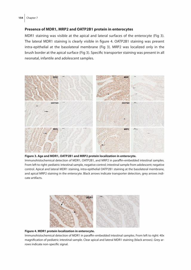
154 Chapter 7
Presence of MDr1, MrP2 and oAtP2b1 protein in enterocytes
MDR1 staining was visible at the apical and lateral surfaces of the enterocyte (Fig 3). The lateral MDR1 staining is clearly visible in figure 4. OATP2B1 staining was present intra-epithelial at the basolateral membrane (Fig 3). MRP2 was localized only in the brush border at the apical surface (Fig 3). Specific transporter staining was present in all neonatal, infantile and adolescent samples.
figure 4. MDr1 protein localization in enterocyte.Immunohistochemical detection of MDR1 in paraffin-embedded intestinal samples. From left to right: 40x magnification of pediatric intestinal sample. Clear apical and lateral MDR1 staining (black arrows). Grey ar-rows indicate non-specific signal.
figure 3. Age and MDr1, oAtP2b1 and MrP2 protein localization in enterocyte.Immunohistochemical detection of MDR1, OATP2B1, and MRP2 in paraffin-embedded intestinal samples. From left to right: pediatric intestinal sample, negative control; intestinal sample from adolescent; negative control. Apical and lateral MDR1 staining, intra-epithelial OATP2B1 staining at the basolateral membrane, and apical MRP2 staining in the enterocyte. Black arrows indicate transporter detection, grey arrows indi-cate artifacts.
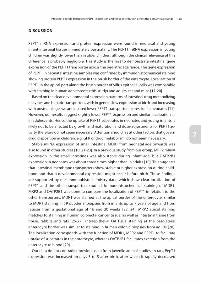
Intestinal peptide transporter PEPT1 expression and tissue distribution across the pediatric age range 155
7
Discussion
PEPT1 mRNA expression and protein expression were found in neonatal and young infant intestinal tissues immediately postnatally. The PEPT1 mRNA expression in young children was slightly lower than in older children, although the clinical relevance of this difference is probably negligible. This study is the first to demonstrate intestinal gene expression of the PEPT1 transporter across the pediatric age range. The gene expression of PEPT1 in neonatal intestine samples was confirmed by immunohistochemical staining showing protein PEPT1 expression in the brush border of the enterocyte. Localization of PEPT1 in the apical part along the brush border of villus epithelial cells was comparable with staining in human adolescents (this study) and adults, rat and mice [17-20].
Based on the clear developmental expression patterns of intestinal drug metabolizing enzymes and hepatic transporters, with in general low expression at birth and increasing with postnatal age, we anticipated lower PEPT1 transporter expression in neonates [11]. However, our results suggest slightly lower PEPT1 expression and similar localization as in adolescents. Hence the uptake of PEPT1 substrates in neonates and young infants is likely not to be affected by growth and maturation and dose-adjustments for PEPT1 ac-tivity therefore do not seem necessary. Attention should lay at other factors that govern drug disposition in children, e.g. GFR or drug metabolism, do not seem necessary.
Stable mRNA expression of small intestinal MDR1 from neonatal age onwards was also found in other studies [10, 21-23]. In a previous study from our group, MRP2 mRNA expression in the small intestines was also stable during infant age, but OATP2B1 expression in neonates was about three times higher than in adults [10]. This suggests that intestinal membrane transporters show stable or higher expression during child-hood and that a developmental expression might occur before birth. These findings are supported by our immunohistochemistry data, which show clear localization of PEPT1 and the other transporters studied. Immunohistochemical staining of MDR1, MRP2 and OATP2B1 was done to compare the localization of PEPT1 in relation to the other transporters. MDR1 was stained at the apical border of the enterocyte, similar to MDR1 staining in 59 duodenal biopsies from infants up to 7 years of age and from fetuses from a gestational age of 16 and 20 weeks [22, 24]. MRP2 apical staining matches to staining in human colorectal cancer tissue, as well as intestinal tissue from horse, rabbits and rats [25-27]. Intraepithelial OATP2B1 staining at the basolateral enterocyte border was similar to staining in human colonic biopsies from adults [28]. The localization corresponds with the function of MDR1, MRP2 and PEPT1 to facilitate uptake of substrates in the enterocyte, whereas OATP2B1 facilitates excretion from the enterocyte to blood [29].
Our data do not contradict previous data from juvenile animal studies. In rats, PepT1 expression was increased on days 3 to 5 after birth, after which it rapidly decreased
156 Chapter 7
and then increased at the time animals were weaning [12]. If we would extrapolate this data to neonates, we would expect a PEPT1 elevation several days after birth, and time of weaning might be translated to infant age at the time of introducing food next to breastfeeding of formula. The slightly higher PEPT1 expression in older children and adolescents than in young infantss might be compared to weaned rats from the animal study. Still, a clinical impact of slightly lower infantile PEPT1 expression is questionable.
PEPT1 has been studied in relation to feeding and nutrition [30]. Interestingly, the PEPT1 transporter expression seems sensitive to nutritional status. In adult short-bowel syndrome patients, small intestinal and colonic mRNA expression of PEPT1 was upregulated compared to healthy controls [17]. Studies in several animal species have further explored the impact of nutrition on intestinal PEPT1 expression. After maternal overnutrition during pregnancy (but not after maternal undernutrition), PepT1 mRNA expression was significantly increased in jejunum of newborn or weaned piglets [31]. In contrast, maternal protein restriction in rats also led to higher duodenal PepT1 mRNA expression in 3- and 16-week-old offspring. Irrespective of feeding, PepT1 mRNA expression in 16-week-old rats was higher than in 3-week-old rats [32]. In another study in adult rats on a protein-rich diet, PepT1 mRNA and protein expressions, as well as transporter activity were increased [33]. In low-birth-weight piglets colonic PepT1 activity (measured by flux of cephalexin) was increased after high- instead of normal-protein formula feeding [34]. The effects of feeding and nutrition may in part explain the observed interindividual variability in PEPT1 gene expression in young infants in the present study. Unfortunately we have no detailed clinical nutritional data to prove this.
Several limitations of this study should be addressed. One, most samples were ob-tained during the first few weeks of life, and pediatric samples after the age of 4 months up to 9 years are lacking. Therefore the expression in this age range is still unknown. Sec-ond, variability in our samples might be due to co-medication, nutritional differences, disease or extent of inflammation. Still, most samples were obtained during surgery of stoma closure and therefore the intestinal tissue can be expected to be relatively nor-mal. Third, variability might be due by genetic polymorphisms in SLC15A1 gene causing a change in PEPT1 expression. Thus farno polymorphism has been proven to be clinically relevant, however, and therefore we did not genotype our samples [35].
In conclusion, although small intestinal PEPT1 expression was slightly lower in neonates than in older children, this difference is small and tissue distribution is similar. Therefore, this finding suggests similar oral absorption of PEPT1 substrates across the pediatric age range.
Intestinal peptide transporter PEPT1 expression and tissue distribution across the pediatric age range 157
7
rEfErEncEs
[1] Hilgendorf C, Ahlin G, Seithel A, Artursson P, Ungell AL, Karlsson J. Expression of thirty-six drug transporter genes in human intestine, liver, kidney, and organotypic cell lines. Drug Metab Dispos 2007 Aug; 35(8): 1333-40.
[2] Meier Y, Eloranta JJ, Darimont J, Ismair MG, Hiller C, Fried M, et al. Regional distribution of solute carrier mRNA expression along the human intestinal tract. Drug Metab Dispos 2007 Apr; 35(4): 590-4.
[3] Drozdzik M, Groer C, Penski J, Lapczuk J, Ostrowski M, Lai Y, et al. Protein abundance of clinically relevant multidrug transporters along the entire length of the human intestine. Mol Pharm 2014 Oct 6; 11(10): 3547-55.
[4] Brandsch M. Drug transport via the intestinal peptide transporter PepT1. Curr Opin Pharmacol 2013 Dec; 13(6): 881-7.
[5] Yang B, Smith DE. Significance of peptide transporter 1 in the intestinal permeability of valacyclo-vir in wild-type and PepT1 knockout mice. Drug Metab Dispos 2013 Mar; 41(3): 608-14.
[6] Yang B, Hu Y, Smith DE. Impact of peptide transporter 1 on the intestinal absorption and pharma-cokinetics of valacyclovir after oral dose escalation in wild-type and PepT1 knockout mice. Drug Metab Dispos 2013 Oct; 41(10): 1867-74.
[7] Landowski CP, Sun D, Foster DR, Menon SS, Barnett JL, Welage LS, et al. Gene expression in the human intestine and correlation with oral valacyclovir pharmacokinetic parameters. J Pharmacol Exp Ther 2003 Aug; 306(2): 778-86.
[8] Brandsch M, Knutter I, Bosse-Doenecke E. Pharmaceutical and pharmacological importance of peptide transporters. J Pharm Pharmacol 2008 May; 60(5): 543-85.
[9] Mooij MG, de Koning BA, Huijsman ML, de Wildt SN. Ontogeny of oral drug absorption processes in children. Expert Opin Drug Metab Toxicol 2012 Oct; 8(10): 1293-303.
[10] Mooij MG, Schwarz UI, de Koning BAE, Leeder JS, Gaedigk R, Samsom JN, et al. Ontogeny of Hu-man Hepatic and Intestinal Transporter Gene Expression during Childhood: Age Matters. Drug Metabolism and Disposition 2014 Aug; 42(8): 1268-74.
[11] Brouwer K, Aleksunes LM, Brandys B, Giacoia GP, Knipp G, Lukacova V, et al. Human Ontogeny of Drug Transporters: Review and Recommendations of the Pediatric Transporter Working Group. Clin Pharmacol Ther 2015 Sep; 98(3): 266-87.
[12] Shen H, Smith DE, Brosius FC, 3rd. Developmental expression of PEPT1 and PEPT2 in rat small intestine, colon, and kidney. Pediatr Res 2001 Jun; 49(6): 789-95.
[13] Rome S, Barbot L, Windsor E, Kapel N, Tricottet V, Huneau JF, et al. The regionalization of PepT1, NBAT and EAAC1 transporters in the small intestine of rats are unchanged from birth to adult-hood. J Nutr 2002 May; 132(5): 1009-11.
[14] Nosworthy MG, Bertolo RF, Brunton JA. Ontogeny of dipeptide uptake and peptide transporter 1 (PepT1) expression along the gastrointestinal tract in the neonatal Yucatan miniature pig. Br J Nutr 2013 Jul 28; 110(2): 275-81.
[15] Kim HR, Park SW, Cho HJ, Chae KA, Sung JM, Kim JS, et al. Comparative gene expression profiles of intestinal transporters in mice, rats and humans. Pharmacol Res 2007 Sep; 56(3): 224-36.
[16] Puiman PJ, Burger-Van Paassen N, Schaart MW, De Bruijn AC, De Krijger RR, Tibboel D, et al. Paneth cell hyperplasia and metaplasia in necrotizing enterocolitis. Pediatr Res 2011 Mar; 69(3): 217-23.
[17] Ziegler TR, Fernandez-Estivariz C, Gu LH, Bazargan N, Umeakunne K, Wallace TM, et al. Distribu-tion of the H+/peptide transporter PepT1 in human intestine: up-regulated expression in the colonic mucosa of patients with short-bowel syndrome. Am J Clin Nutr 2002 May; 75(5): 922-30.
158 Chapter 7
[18] Laforenza U, Miceli E, Gastaldi G, Scaffino MF, Ventura U, Fontana JM, et al. Solute transporters and aquaporins are impaired in celiac disease. Biol Cell 2010 Aug; 102(8): 457-67.
[19] Groneberg DA, Doring F, Eynott PR, Fischer A, Daniel H. Intestinal peptide transport: ex vivo uptake studies and localization of peptide carrier PEPT1. Am J Physiol Gastrointest Liver Physiol 2001 Sep; 281(3): G697-704.
[20] Hussain I, Kellett L, Affleck J, Shepherd J, Boyd R. Expression and cellular distribution during development of the peptide transporter (PepT1) in the small intestinal epithelium of the rat. Cell Tissue Res 2002 Jan; 307(1): 139-42.
[21] Mizuno T, Fukuda T, Masuda S, Uemoto S, Matsubara K, Inui KI, et al. Developmental trajectory of intestinal MDR1/ABCB1 expression in children. Br J Clin Pharmacol 2013 Jul 23.
[22] Fakhoury M, Litalien C, Medard Y, Cave H, Ezzahir N, Peuchmaur M, et al. Localization and mRNA expression of CYP3A and P-glycoprotein in human duodenum as a function of age. Drug Metab Dispos 2005 Nov; 33(11): 1603-7.
[23] Miki Y, Suzuki T, Tazawa C, Blumberg B, Sasano H. Steroid and xenobiotic receptor (SXR), cyto-chrome P450 3A4 and multidrug resistance gene 1 in human adult and fetal tissues. Mol Cell Endocrinol 2005 Feb 28; 231(1-2): 75-85.
[24] van Kalken CK, Giaccone G, van der Valk P, Kuiper CM, Hadisaputro MM, Bosma SA, et al. Multidrug resistance gene (P-glycoprotein) expression in the human fetus. Am J Pathol 1992 Nov; 141(5): 1063-72.
[25] Tyden E, Bjornstrom H, Tjalve H, Larsson P. Expression and localization of BCRP, MRP1 and MRP2 in intestines, liver and kidney in horse. J Vet Pharmacol Ther 2010 Aug; 33(4): 332-40.
[26] Van Aubel RA, Hartog A, Bindels RJ, Van Os CH, Russel FG. Expression and immunolocalization of multidrug resistance protein 2 in rabbit small intestine. Eur J Pharmacol 2000 Jul 21; 400(2-3): 195-8.
[27] Mottino AD, Hoffman T, Jennes L, Vore M. Expression and localization of multidrug resistant protein mrp2 in rat small intestine. J Pharmacol Exp Ther 2000 Jun; 293(3): 717-23.
[28] Kleberg K, Jensen GM, Christensen DP, Lundh M, Grunnet LG, Knuhtsen S, et al. Transporter func-tion and cyclic AMP turnover in normal colonic mucosa from patients with and without colorectal neoplasia. BMC Gastroenterol 2012; 12: 78.
[29] Klaassen CD, Aleksunes LM. Xenobiotic, bile acid, and cholesterol transporters: function and regulation. Pharmacol Rev 2010 Mar; 62(1): 1-96.
[30] Spanier B. Transcriptional and functional regulation of the intestinal peptide transporter PEPT1. J Physiol 2014 Mar 1; 592(Pt 5): 871-9.
[31] Cao M, Che L, Wang J, Yang M, Su G, Fang Z, et al. Effects of maternal over- and undernutrition on intestinal morphology, enzyme activity, and gene expression of nutrient transporters in newborn and weaned pigs. Nutrition 2014 Nov-Dec; 30(11-12): 1442-7.
[32] Pinheiro DF, Pinheiro PF, Buratini J, Jr., Castilho AC, Lima PF, Trinca LA, et al. Maternal protein restriction during pregnancy affects gene expression and immunolocalization of intestinal nutri-ent transporters in rats. Clin Sci (Lond) 2013 Sep; 125(6): 281-9.
[33] Shiraga T, Miyamoto K, Tanaka H, Yamamoto H, Taketani Y, Morita K, et al. Cellular and molecular mechanisms of dietary regulation on rat intestinal H+/Peptide transporter PepT1. Gastroenterol-ogy 1999 Feb; 116(2): 354-62.
[34] Boudry G, Rome V, Perrier C, Jamin A, Savary G, Le Huerou-Luron I. A high-protein formula in-creases colonic peptide transporter 1 activity during neonatal life in low-birth-weight piglets and disturbs barrier function later in life. Br J Nutr 2014 Oct 14; 112(7): 1073-80.
Intestinal peptide transporter PEPT1 expression and tissue distribution across the pediatric age range 159
7
[35] Anderle P, Nielsen CU, Pinsonneault J, Krog PL, Brodin B, Sadee W. Genetic variants of the human dipeptide transporter PEPT1. J Pharmacol Exp Ther 2006 Feb; 316(2): 636-46.
8 T he eff ect of age on human hepatic membrane transporter protein expression
Miriam G. Mooij, Evita van de Steeg, Joost van Rosmalen, Jonathan D. Windster, Barbara A.E. de Koning, Wouter H.J. Vaes, Dick Tibboel, Heleen M. Wortelboer, Saskia N. de Wildt
Submitted for publication

162 Chapter 8
AbstrAct
introduction
Human hepatic membrane-embedded transporter proteins are involved in trafficking endogenous and exogenous substrates. Even though impact of transporters on phar-macokinetics is recognized, little is known on maturation of transporter protein expres-sion levels, especially during early life. We aimed to study the protein expression of 10 transporters in liver tissue from fetuses, infants and adults.
Methods
Transporter protein expression levels (ABCB1, ABCG2, ABCC2, ABCC3, BSEP, GLUT1, MCT1, OATP1B1, OATP2B1, and OCTN2) were quantified using UPLC-MS/MS in snap-frozen post mortem fetal, infant and adult liver samples. Protein expression was quanti-fied in isolated crude membrane fractions. The possible association between postnatal and postmenstrual age versus protein expression was studied.
results
We studied 25 liver samples: 10 fetal [median gestational age 23.2 weeks (range 16.4-37.9)], 12 infantile [gestational age at birth 35.1 weeks (27.1-41.0), postnatal age 1 week (0-11.4)], and 3 adult. Four patterns emerged. ABCB1, OATP1B1, and OATP2B1 expres-sion levels were stable from fetal to adult age. ABCC2, ABCC3, and BSEP were low in fetus and infant compared to adults, while ABCG2, GLUT1, OCTN2 protein levels were higher early in life than in adults. MCT1 expression levels were highest in infants compared to fetus and adult. These patterns were also found for postmenstrual age within the fetal/infant groups.
conclusion
The impact of growth and development on human membrane transporter protein expression is transporter-dependent. The age-related differences in transporter protein expression aid our understanding of normal growth and development, and also may impact the disposition of substrate drugs in neonates and young infants.
The effect of age on human hepatic membrane transporter protein expression 163
8
introDuction
Membrane-embedded transporter proteins are involved in the transport of a myriad of endogenous and exogenous substances. These transporters can be found in various cell types, such as enterocytes, hepatocytes, brain and kidney cells. By trafficking exogenous substrates, they have an important impact on drug absorption, distribution and excre-tion of drugs and their metabolites, thereby determining drug efficacy and toxicity. In the hepatocyte, transporters have been identified on both basal and canalicular mem-branes regulating the uptake and excretion of drugs and their metabolites into and from the systemic circulation as well as excretion into bile [1].
The international transporter consortium has identified a large set of clinically rel-evant membrane transporters, subdivided in families with similar peptide sequences [2]. Transporters belonging to the ATP-binding cassette (ABC) family are multidrug resistance protein 1 (ABCB1/MDR1/P-gp), multidrug resistance-associated protein 2 and 3 (ABCC2/MRP2, ABCC3/MRP3), breast cancer resistance protein (ABCG2/BRCP), and bile salt export pump (ABCB11/BSEP). To the solute carrier organic anion (SLCO) family belong organic anion transporting polypeptide 1B1 and 2B1 (SLCO1B1/OATP1B1, SLCO2B1/OATP2B1), and are acknowledged to be the most clinically relevant. Other transporter proteins taken along in the current study are glucose transporter 1 (SLC2A1/GLUT1), monocarboxylate transporter 1 (SLC16A1/MCT1), and organic cation/carnitine transporter (SLC22A5/OCTN2) which belong to the solute carrier (SLC) family. This selec-tion of transporters were chosen because, they have proven to be important in drug disposition in adults or have shown a large abundancy in human adult liver [3].
Children’s growth and maturation importantly impact the processes involved in the disposition of drugs such as absorption, metabolism and renal excretion [4]. It is there-fore likely that expression and activity of transporters are also subject to age-related changes, as has been convincingly shown for the drug metabolizing enzymes [5]. In-deed, animal studies have shown transporter-specific maturation profiles in transporter expression [1, 6]. For example, hepatic mRNA expression of mouse Oatps gradually increases after birth until adult levels at 6 weeks of age [7]. In spite of the maturation in animal studies, interspecies differences prevent us to translate animal maturation data to humans [8]. For humans, only few transporters have been studied and a clear information gap remains on the developmental trajectory of individual transporters in fetuses and young children and the related impact on drug disposition [6, 9]. Recently, we showed a transporter dependent maturation profile in mRNA expression of hepatic ABCB1, ABCC2, OATP1B1 and OATP1B3 [10]. To predict drug disposition on the basis of these results is a challenge since posttranscriptional variation may result in a discrep-ancy between mRNA and protein levels, as has been shown for some transporters in human adults [11, 12]. Protein expression data in children are limited. For example, a
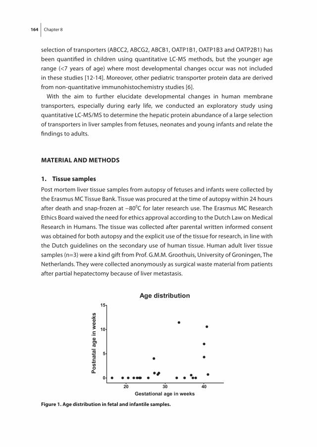
164 Chapter 8
selection of transporters (ABCC2, ABCG2, ABCB1, OATP1B1, OATP1B3 and OATP2B1) has been quantified in children using quantitative LC-MS methods, but the younger age range (<7 years of age) where most developmental changes occur was not included in these studies [12-14]. Moreover, other pediatric transporter protein data are derived from non-quantitative immunohistochemistry studies [6].
With the aim to further elucidate developmental changes in human membrane transporters, especially during early life, we conducted an exploratory study using quantitative LC-MS/MS to determine the hepatic protein abundance of a large selection of transporters in liver samples from fetuses, neonates and young infants and relate the findings to adults.
MAtEriAl AnD MEthoDs
1. tissue samples
Post mortem liver tissue samples from autopsy of fetuses and infants were collected by the Erasmus MC Tissue Bank. Tissue was procured at the time of autopsy within 24 hours after death and snap-frozen at −800C for later research use. The Erasmus MC Research Ethics Board waived the need for ethics approval according to the Dutch Law on Medical Research in Humans. The tissue was collected after parental written informed consent was obtained for both autopsy and the explicit use of the tissue for research, in line with the Dutch guidelines on the secondary use of human tissue. Human adult liver tissue samples (n=3) were a kind gift from Prof. G.M.M. Groothuis, University of Groningen, The Netherlands. They were collected anonymously as surgical waste material from patients after partial hepatectomy because of liver metastasis.
figure 1. Age distribution in fetal and infantile samples.

The effect of age on human hepatic membrane transporter protein expression 165
8
The cohort of selected samples reflected a widespread gestational age, in second and third trimester of pregnancy, as well as postnatal age (up to three months) (age distribu-tion is displayed in figure 1).
2. Membrane isolation and liquid chromatography-tandem Mass spectrometry
Isolation of crude membrane fractions from tissue samples was conducted as described by Wisniewski et al., with some minor adaptations [15]. In brief, approximately 10 mg of liver tissue was homogenized in a hypotonic buffer (0.5 mM sodium phosphate, 0.1 mM EDTA, and a cocktail of protease inhibitors containing 2 mM phenylmethylsulfonyl-fluoride, aprotinin, leupeptin, and pepstatin) using a Potter-Elvehjem homogenizer. The homogenate was centrifuged at 100,000g for 30 minutes at 4°C using a LE-80k Centri-fuge with SW28 rotor (Beckman Counter, USA). The pellet, containing crude membrane protein fraction, was resuspended in 500 µL SDT-lysis buffer (0.1 M Tris pH 7.6, 0.1 M DTT, 4% SDS). Approximately 50 µL of cell lysate, corresponding to 1 mg tissue, was incubated at 95°C for 5 minutes and briefly sonicated on ice, mixed with 200 µL of 8 M urea and loaded onto a 30K Amicon Ultra-0.5 centrifugal filter device (Millipore, Darm-stadt, Germany) and centrifuged at 14,000g for 15 minutes. After adding another 200 µL 8 M urea the tubes were centrifuged again at 14,000g for 15 minutes. Subsequently, 100 µL of 0.05 M iodoacetamide in 8 M urea in 0.1 M Tris-HCl, pH 8.5 was added and the samples were incubated for 20 minutes at room temperature, and centrifuged at 14,000g for 15 minutes. The resulting concentrate was diluted with 100 µL 8 M urea in 0.1 M Tris-HCl, pH 8.5 and centrifuged (14,000g for 15 minutes) again. This step was repeated twice. Subsequently, 100 µL 0.05 M ammoniumbicarbonate was added to the concentrate and centrifuged (14,000g for 15 minutes). This step was also repeated twice. Proteins were digested into peptides by incubating the samples overnight at 37°C in a 0.05 M ammoniumbicarbonate solution containing 0.5 µg/µL trypsin. The digests were collected into a new collection tube by centrifugation (14,000g for 10 minutes,) and the filter device was rinsed with 50 µL 0.5 M NaCl and centrifuged (14,000g for 10 minutes). The combined filtrate was acidified with 25 µL 0.1% CF3COOH, desalted on a 7 mm/ 3 mL extraction disk cartridge (3M Empore, Zoeterwoude, The Netherlands) and eluted with 70% acetonitrile. Subsequently, the samples were vacuum dried (SpeedVac) until the solution was vaporized and stored < −70°C until further analysis.
All samples were analyzed using a UPLC coupled to a 6500 QTrap mass spectrometer (AB Sciex). The crude membrane fractions were injected on an Acquity C18 BEH UPLC column and separated using gradient elution. During analysis, the column was main-tained at 50°C and the samples were kept at 10°C. The mass spectroscope was operated in selective reaction mode using electrospray ionization in positive ion mode, with a capillary voltage of 3.3 kV, a source temperature of 150°C, and a desolvation tempera-

166 Chapter 8
ture of 600°C. Cone voltage and collision energy were optimized for each compound individually. The multiple reaction monitoring transitions were determined from tan-dem mass spectra obtained by direct infusion of 0.5 µg/mL. Per peptide three transitions were chosen (Q3-1, Q3-2 and Q3-3) for quantitation and confirmation. The transitions for the different proteins are listed in Table 1. A peptide labeled with 15N and 13C (AQUA pep-tide) was synthesized (Sigma Aldrich, Steinheim DE) and used as an internal standard for quantification (Table 1). Peak identification and quantification was performed using Analyst software version 1.6.
3. statistics
Data are presented as median and range, unless indicated otherwise. To describe the variation in protein expression, the relative difference between the lowest and highest observed value was calculated per transporter. Median (range) protein expression per age group (fetuses, infants and adults) was calculated to describe the fold-change in
table 1. Multiple reaction monitoring (MrM) transitions of the used peptides and the corresponding internal standards (AQuA)
name labelled Peptide sequencea Molecularweight
Q1 Q3-1 Q3-2 Q3-3 Q3-4
ABCB1 unlabeled NTTGALTTR 934.0 467.7 719.4 216.1 618.4
AQUA NTTGAlTTR 950.0 471.2 726.5
ABCC2 unlabeled VLGPNGLLK 910.1 455.8 698.5 185.3 213.3
AQUA VLGPNGLlK 926.1 459.2 705.4
ABCC3 unlabeled ALVITNSVK 944.1 472.8 760.4 661.4 548.4
AQUA ALVITNSVK 950.1 475.8 766.5
ABCG2 unlabeled SSLLDVLAAR 1.044.2 522.8 644.3 757.5 529.4
AQUA SSLLDVlAAR 1.060.2 526.3 651.3
BSEP unlabeled STALQLIQR 1.029.2 515.3 657.4 841.6 529.4
AQUA STALQlIQR 1.045.2 518.8 664.3
GLUT1 unlabeled VTILELFR 990.2 495.8 790.5 677.4 201.2
AQUA VTILELfR 1.000.2 500.8 800.5
MCT1 unlabeled SITVFFK 841.0 421.2 173.2 641.3 201.1
AQUA SITVFfK 851.0 426.2 651.3
OCTN2 unlabeled WLISQGR 859.0 430.2 560.3 272.2 300.2
AQUA WLiSQGR 875.0 433.7 567.3
OATP1B1 unlabeled LNTVGIAK 815.0 408.2 399.4 588.3 228.2 702.3
AQUA LNTVGiAK 831.0 411.7 402.9
OATP2B1 unlabeled SSISTVEK 849.9 425.7 563.3 676.3 175.1
AQUA SSISTVEK 855.9 428.7 569.3
a AQUA: Amino acid presented in bold is labelled with 13C and 15N.

The effect of age on human hepatic membrane transporter protein expression 167
8
protein expression. The possible relation between transporter protein levels and age was assessed using scatter plots. As these plots suggested a monotonic relationship for almost all transporters, the association between transporter protein levels and age was assessed using Spearman’s rank correlation coefficient. In addition, in the fetal and infant samples, the association between transporter protein expression and postmenstrual age was also assessed using Spearman’s rank correlation.
All statistical analyses were performed using IBM SPSS Statistics software (SPSS Statis-tics for Windows, version 21.0; IBM, Armonk, NY) and a significance level of P<0.05 was used throughout the study.
rEsults
Descriptive results
We studied 25 liver samples: 10 of fetuses [median gestational age (GA) 23.2 weeks (range 16.4-37.9)], 12 of infants [postnatal age 1 week (0-11.4), GA at birth 35.1 weeks (27.1-41.0)] and 3 of adults. Patient characteristics are listed in Table 2. The clinical diag-noses of the fetuses and infants were: therapeutic abortion for severe genetic disorders (non-metabolic diseases) (n=7), fetomaternal transfusion (n=1), hydrops fetalis (n=1), and intrauterine death for unknown reason (n=1). The clinical diagnoses of the infants were congenital malformations (n=5), viral/bacterial infections (n=4), necrotizing en-terocolitis (without liver insufficiency) (n=2), and benign liver tumor (n=1). The adult liver tissue was histologically normal tissue. Information about diagnosis, reason for biopsy or medical history of the three adult patients concerned was not available.
table 2. hepatic protein expressions
fetusesN = 10
infantsN = 12
AdultsN = 3
Profile i‘stable’
OATP1B1 17.6 (5.4-35.6) 22.8 (9.5-47.9) 34.6 (21.5-57.5)
OATP2B1 42.2 (29.3-59.3) 38.5 (27.2-46.3) 44.9 (30.1-48.9)
ABCB1 79.2 (23.2-117.7) 107.9 (60.6-150.6) 96.9 (93.5-103.4)
Profile ii‘low to high’
ABCC2 8.8 (5.5-9.9) 12.6 (5.3-43.9) 19.8 (15.5-22.2)
ABCC3 5.4 (1.6-9.9) 21.9 (3.6-32.8) 23.1 (15.9-23.7)
BSEP 18.4 (11.4-33.7) 35.9 (10.6-49.8) 47.7 (40.3-67.0)
Profile iii‘high to low’
ABCG2 4.0 (2.2-7.0) 2.7 (1.5-4.2) 1.7 (0.8-2.2)
GLUT1 434.2 (49.1-1413.3) 108.7 (39.0-255.4) 5.2 (5.1-7.7)
OCTN2 6.3 (4.1-8.9) 4.4 (1.9-6.6) 3.0 (2.2-4.8)
Profile iV‘non-linear’
MCT1 12.9 (10.0-20.0) 20.3 (10.8-39.0) 9.8 (8.9-12.4)
Data are expressed as median (range) protein expressions in pmol/g tissue
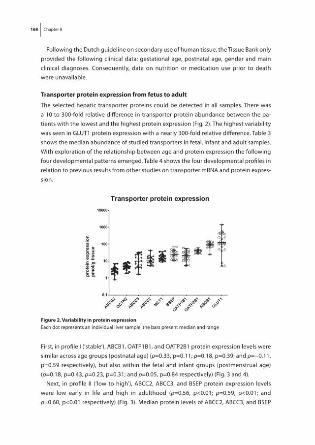
168 Chapter 8
Following the Dutch guideline on secondary use of human tissue, the Tissue Bank only provided the following clinical data: gestational age, postnatal age, gender and main clinical diagnoses. Consequently, data on nutrition or medication use prior to death were unavailable.
transporter protein expression from fetus to adult
The selected hepatic transporter proteins could be detected in all samples. There was a 10 to 300-fold relative difference in transporter protein abundance between the pa-tients with the lowest and the highest protein expression (Fig. 2). The highest variability was seen in GLUT1 protein expression with a nearly 300-fold relative difference. Table 3 shows the median abundance of studied transporters in fetal, infant and adult samples. With exploration of the relationship between age and protein expression the following four developmental patterns emerged. Table 4 shows the four developmental profiles in relation to previous results from other studies on transporter mRNA and protein expres-sion.
figure 2. Variability in protein expressionEach dot represents an individual liver sample, the bars present median and range
First, in profile I (‘stable’), ABCB1, OATP1B1, and OATP2B1 protein expression levels were similar across age groups (postnatal age) (ρ=0.33, p=0.11; ρ=0.18, p=0.39; and ρ=−0.11, p=0.59 respectively), but also within the fetal and infant groups (postmenstrual age) (ρ=0.18, p=0.43; ρ=0.23, p=0.31; and ρ=0.05, p=0.84 respectively) (Fig. 3 and 4).
Next, in profile II (‘low to high’), ABCC2, ABCC3, and BSEP protein expression levels were low early in life and high in adulthood (ρ=0.56, p<0.01; ρ=0.59, p<0.01; and ρ=0.60, p<0.01 respectively) (Fig. 3). Median protein levels of ABCC2, ABCC3, and BSEP
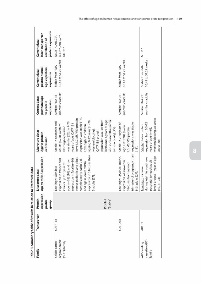
The effect of age on human hepatic membrane transporter protein expression 169
8
tabl
e 3.
sum
mar
y ta
ble
of re
sult
s in
rela
tion
to li
tera
ture
dat
a
fam
ilytr
ansp
orte
rPr
otei
n ex
pres
sion
pr
ofile
gr
oup
lite
ratu
re d
ata:
Age
vs
mrn
A e
xpre
ssio
nli
tera
ture
dat
a:A
ge v
s pr
otei
n ex
pres
sion
curr
ent d
ata:
post
nata
l age
vs
pro
tein
ex
pres
sion
curr
ent d
ata:
post
men
stru
al
age
vs p
rote
in
expr
essi
on
curr
ent d
ata:
inte
r-tr
ansp
orte
r co
rrel
atio
n of
pr
otei
n ex
pres
sion
Solu
te c
arrie
r or
gani
c an
ion
(SLC
O) f
amily
OAT
P1B1
Profi
le I
‘Sta
ble’
Low
-hig
h: w
ith lo
w
expr
essi
on in
fetu
ses
and
infa
nts
up to
1 y
ear o
f ag
e (n
=32)
[10]
, and
low
ex
pres
sion
in fe
tal (
n=30
) ve
rsus
ped
iatr
ic a
nd a
dult
sam
ples
(n=3
0 ea
ch)[2
4],
and
agai
n lo
wer
mRN
A
expr
essi
on in
3 fe
tuse
s th
an
3 ad
ults
[27]
.
Stab
le: I
n ne
onat
es a
nd
adul
ts (n
=10,
wes
tern
bl
ottin
g) e
xpre
ssio
n w
as s
imila
r [26
]. In
7-7
0 ye
ars
of a
ge, O
ATP1
B1
(n=6
4, L
C-M
S/M
S) p
rote
in
expr
essi
on w
as s
tabl
e [1
3].
Low
-hig
h: In
chi
ldre
n ag
eing
0-1
2 ye
ars
(n=7
8,
wes
tern
blo
ttin
g),
OAT
P1B1
pro
tein
ex
pres
sion
was
low
from
bi
rth
until
6 y
ears
of a
ge
and
incr
ease
d th
erea
fter
(a
bstr
act o
nly)
[25]
.
Sim
ilar:
PNA
<3
mon
ths
vs a
dults
Stab
le fr
om P
MA
16
.43
to 5
1.29
wee
ksA
BCB1
*, A
BCC2
*,
BSEP
**, A
BCG
2**,
G
LUT1
*
OAT
P2B1
Low
-hig
h: O
ATP2
B1 m
RNA
ex
pres
sion
was
low
er in
3
fetu
ses
from
sec
ond
trim
este
r of p
regn
ancy
than
in
3 a
dults
[27]
.
Stab
le: I
n 7-
70 y
ears
of
age,
OAT
P2B1
(n=6
4,
LC-M
S/M
S) p
rote
in
expr
essi
ons
was
sta
ble
[13]
.
Sim
ilar:
PNA
<3
mon
ths
vs a
dults
Stab
le fr
om P
MA
16
.43
to 5
1.29
wee
ks
ATP-
bind
ing
cass
ette
(ABC
) fa
mily
ABC
B1Lo
w-h
igh:
Incr
ease
du
ring
feta
l life
, con
tinue
s po
stna
tal t
o re
ach
adul
t le
vels
aro
und
1 ye
ar o
f age
[1
0, 2
1-24
].
Stab
le: S
tabl
e pr
otei
n ex
pres
sion
from
0.3
-12
year
s of
age
(n=6
5,
wes
tern
blo
ttin
g, a
bstr
act
only
) [20
]
Sim
ilar:
PNA
<3
mon
ths
vs a
dults
Stab
le fr
om P
MA
16
.43
to 5
1.29
wee
ksM
CT1
*
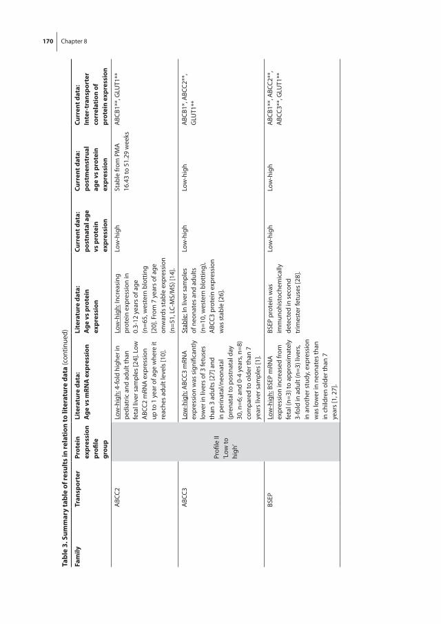
170 Chapter 8 The effect of age on human hepatic membrane transporter protein expression 171
8
tabl
e 3.
sum
mar
y ta
ble
of re
sult
s in
rela
tion
to li
tera
ture
dat
a (c
ontin
ued)
fam
ilytr
ansp
orte
rPr
otei
n ex
pres
sion
pr
ofile
gr
oup
lite
ratu
re d
ata:
Age
vs
mrn
A e
xpre
ssio
nli
tera
ture
dat
a:A
ge v
s pr
otei
n ex
pres
sion
curr
ent d
ata:
post
nata
l age
vs
pro
tein
ex
pres
sion
curr
ent d
ata:
post
men
stru
al
age
vs p
rote
in
expr
essi
on
curr
ent d
ata:
inte
r-tr
ansp
orte
r co
rrel
atio
n of
pr
otei
n ex
pres
sion
ABC
C2
Profi
le II
‘Low
to
high
’
Low
-hig
h: 4
-fold
hig
her i
n pe
diat
ric a
nd a
dult
than
fe
tal l
iver
sam
ples
[24]
. Low
A
BCC2
mRN
A e
xpre
ssio
n up
to 1
yea
r of a
ge w
here
it
reac
hes
adul
t lev
els
[10]
.
Low
-hig
h: In
crea
sing
pr
otei
n ex
pres
sion
in
0.3-
12 y
ears
of a
ge
(n=6
5, w
este
rn b
lott
ing
[20]
. Fro
m 7
yea
rs o
f age
on
war
ds s
tabl
e ex
pres
sion
(n
=51,
LC-
MS/
MS)
[14]
.
Low
-hig
hSt
able
from
PM
A
16.4
3 to
51.
29 w
eeks
ABC
B1**
, GLU
T1**
ABC
C3Lo
w-h
igh:
ABC
C3 m
RNA
ex
pres
sion
was
sig
nific
antly
lo
wer
in li
vers
of 3
fetu
ses
than
3 a
dults
[27]
and
in
per
inat
al/n
eona
tal
(pre
nata
l to
post
nata
l day
30
, n=6
; and
0-4
yea
rs, n
=8)
com
pare
d to
old
er th
an 7
ye
ars
liver
sam
ples
[1].
Stab
le: I
n liv
er s
ampl
es
of n
eona
tes
and
adul
ts
(n=1
0, w
este
rn b
lott
ing)
, A
BCC3
pro
tein
exp
ress
ion
was
sta
ble
[26]
.
Low
-hig
hLo
w-h
igh
ABC
B1*,
ABC
C2**
, G
LUT1
**
BSEP
Low
-hig
h: B
SEP
mRN
A
expr
essi
on in
crea
sed
from
fe
tal (
n=3)
to a
ppro
xim
atel
y 3-
fold
in a
dult
(n=3
) liv
ers,
in a
noth
er s
tudy
, exp
ress
ion
was
low
er in
neo
nate
s th
an
in c
hild
ren
olde
r tha
n 7
year
s [1
, 27]
.
BSEP
pro
tein
was
im
mun
ohis
toch
emic
ally
de
tect
ed in
sec
ond
trim
este
r fet
uses
[28]
.
Low
-hig
hLo
w-h
igh
ABC
B1**
, ABC
C2**
, A
BCC3
**, G
LUT1
**

170 Chapter 8 The effect of age on human hepatic membrane transporter protein expression 171
8
tabl
e 3.
sum
mar
y ta
ble
of re
sult
s in
rela
tion
to li
tera
ture
dat
a (c
ontin
ued)
fam
ilytr
ansp
orte
rPr
otei
n ex
pres
sion
pr
ofile
gr
oup
lite
ratu
re d
ata:
Age
vs
mrn
A e
xpre
ssio
nli
tera
ture
dat
a:A
ge v
s pr
otei
n ex
pres
sion
curr
ent d
ata:
post
nata
l age
vs
pro
tein
ex
pres
sion
curr
ent d
ata:
post
men
stru
al
age
vs p
rote
in
expr
essi
on
curr
ent d
ata:
inte
r-tr
ansp
orte
r co
rrel
atio
n of
pr
otei
n ex
pres
sion
ABC
G2
Profi
le II
I‘H
igh
to
low
’
Stab
le: s
tabl
e A
BCG
2 m
RNA
ex
pres
sion
was
foun
d;
in fe
tal (
n=30
, sec
ond
trim
este
r of p
regn
ancy
), pe
diat
ric (n
=30,
age
1-1
7 ye
ars)
and
adu
lt (a
ge 2
8-80
ye
ars)
live
r sam
ples
and
in a
st
udy
com
parin
g 3
feta
l and
3
adul
t liv
ers
[24,
27]
.
Stab
le: A
BCG
2 pr
otei
n ex
pres
sion
was
sta
ble
in
neon
ates
and
adu
lt liv
ers
(n=1
0, w
este
rn b
lott
ing)
[2
6]. A
nd in
7-7
0 ye
ars
of
age,
ABC
G2
(n=5
6, L
C-M
S/M
S), p
rote
in e
xpre
ssio
ns
was
sta
ble
[12
].A
BCG
2 pr
otei
n im
mun
ohis
toch
emis
try
stai
ning
sho
wed
wea
k co
lorin
g in
firs
t trim
este
r fe
tal h
epat
ocyt
es, w
hile
ad
ult l
iver
sho
wed
str
ong
ABC
G s
tain
ing
[29]
.
Hig
h-lo
wH
igh-
low
OAT
P1B1
**, O
CTN
2**
Solu
te c
arrie
r (SL
C)
fam
ilyG
LUT1
Hig
h-lo
wH
igh-
low
OAT
P1B1
*, A
BCC2
*,
ABC
C3**
, BSE
P**,
A
BCG
2**,
OC
TN2*
*
OC
TN2
Hig
h-lo
wH
igh-
low
GLU
T1**
, ABC
G2*
*
MC
T1Pr
ofile
IV‘N
on-li
near
’N
on-li
near
Low
-hig
hA
BCB1
*
* P<0
.05,
**
P<0.
01
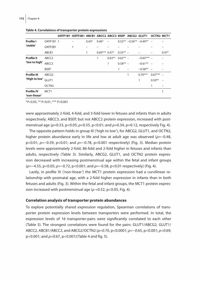
172 Chapter 8 The effect of age on human hepatic membrane transporter protein expression 173
8
were approximately 2-fold, 4-fold, and 3-fold lower in fetuses and infants than in adults respectively. ABCC3, and BSEP, but not ABCC2 protein expression, increased with post-menstrual age (ρ=0.53, p<0.05; ρ=0.55, p<0.01; and ρ=0.34, p=0.12, respectively Fig. 4).
The opposite pattern holds in group III (‘high to low’), for ABCG2, GLUT1, and OCTN2, higher protein abundance early in life and low at adult age was observed (ρ=−0.48, p<0.01; ρ=−0.59, p<0.01; and ρ=−0.78, p<0.001 respectively) (Fig. 3). Median protein levels were approximately 2-fold, 86-fold and 2-fold higher in fetuses and infants than adults, respectively (Table 3). Similarly, ABCG2, GLUT1, and OCTN2 protein expres-sion decreased with increasing postmenstrual age within the fetal and infant groups (ρ=−4.55, p<0.05; ρ=−0.72, p<0.001; and ρ=−0.58, p<0.01 respectively) (Fig. 4).
Lastly, in profile IV (‘non-linear’) the MCT1 protein expression had a curvilinear re-lationship with postnatal age, with a 2-fold higher expression in infants than in both fetuses and adults (Fig. 3). Within the fetal and infant groups, the MCT1 protein expres-sion increased with postmenstrual age (ρ =0.52, p<0.05, Fig. 4).
correlation analysis of transporter protein abundances
To explore potentially shared expression regulation, Spearman correlations of trans-porter protein expression levels between transporters were performed. In total, the expression levels of 16 transporter-pairs were significantly correlated to each other (Table 5). The strongest correlations were found for the pairs: GLUT1/ABCG2, GLUT1/ABCC2, ABCB1/ABCC2, and ABCG2/OCTN2 (ρ=0.70, p<0.0001; ρ=−0.65, p<0.001; ρ=0.69, p<0.001; and ρ=0.67, p<0.001)(Table 4 and fig. 5).
table 4. correlations of transporter protein expressions
oAtP1b1 oAtP2b1 Abcb1 Abcc2 Abcc3 bsEP AbcG2 Glut1 octn2 Mct1
Profile i‘stable’
OATP1B1 1 – 0.43* 0.46* – 0.52** −0.56** −0.49** – –
OATP2B1 1 – – – – – – – –
ABCB1 1 0.69*** 0.47* 0.55** – – – 0.47*
Profile ii‘low to high’
ABCC2 1 0.63** 0.62** – −0.65*** – –
ABCC3 1 0.58** – −0.61** – –
BSEP 1 – −0.58** – –
Profile iii‘high to low’
ABCG2 1 0.70*** 0.67*** –
GLUT1 1 0.59** –
OCTN2 1 –
Profile iV‘non-linear’
MCT1 1
*P<0.05, ** P<0.01, *** P<0.001
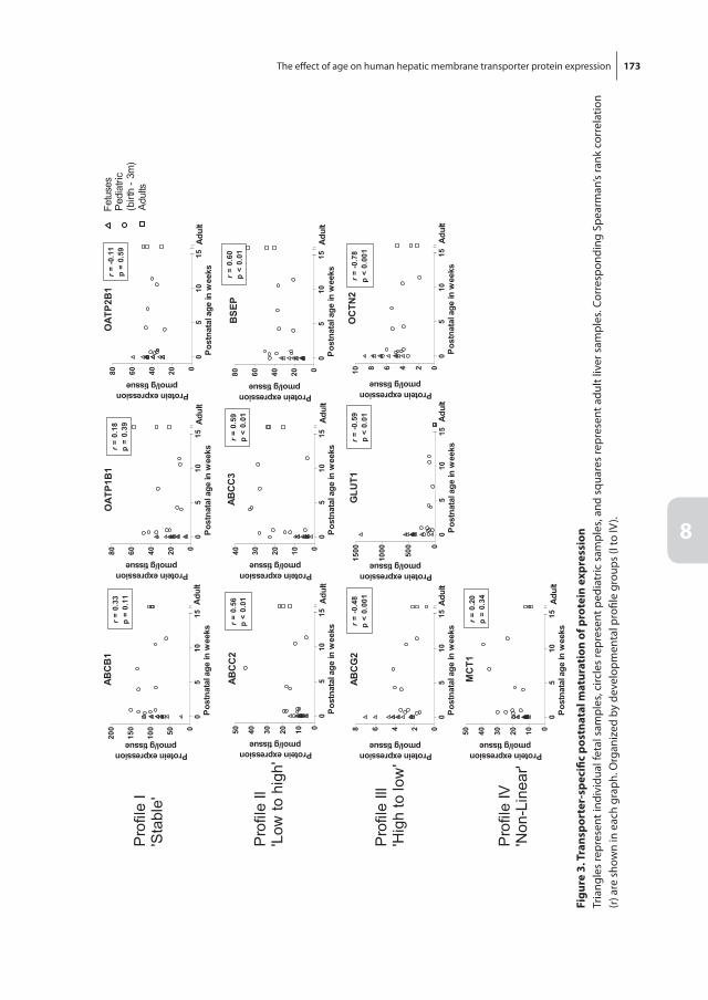
172 Chapter 8 The effect of age on human hepatic membrane transporter protein expression 173
8
figu
re 3
. tra
nspo
rter
-spe
cific
pos
tnat
al m
atur
atio
n of
pro
tein
exp
ress
ion
Tria
ngle
s re
pres
ent i
ndiv
idua
l fet
al s
ampl
es, c
ircle
s re
pres
ent p
edia
tric
sam
ples
, and
squ
ares
repr
esen
t adu
lt liv
er s
ampl
es. C
orre
spon
ding
Spe
arm
an’s
rank
cor
rela
tion
(r) a
re s
how
n in
eac
h gr
aph.
Org
aniz
ed b
y de
velo
pmen
tal p
rofil
e gr
oups
(I to
IV).

174 Chapter 8
figu
re 4
. tra
nspo
rter
-spe
cific
pos
tmen
stru
al m
atur
atio
n of
pro
tein
exp
ress
ion
Clos
ed c
ircle
s re
pres
ent
indi
vidu
al fe
tal s
ampl
es a
nd o
pen
circ
les
repr
esen
t pe
diat
ric li
ver
sam
ples
. Cor
resp
ondi
ng S
pear
man
’s ra
nk c
orre
latio
n (r
) are
sho
wn
in e
ach
grap
h. O
rgan
ized
by
deve
lopm
enta
l pro
file
grou
ps (I
to IV
).

The effect of age on human hepatic membrane transporter protein expression 175
8
figu
re 5
. Exa
mpl
es o
f int
er-t
rans
port
er re
lati
onsh
ip o
f pro
tein
exp
ress
ion
Each
circ
le re
pres
ents
an
indi
vidu
al li
ver s
ampl
e. T
hese
four
pai
rs re
pres
ent t
he s
tron
gest
inte
r-tr
ansp
orte
r cor
rela
tion.
Cor
resp
ondi
ng S
pear
man
’s ra
nk c
orre
latio
n (r
) ar
e sh
own
in e
ach
grap
h.
176 Chapter 8
Discussion
This study reveals important age-dependent differences in protein abundance of ten clinically relevant hepatic membrane transporters. Four different developmental pro-files of protein abundancy could be distinguished in liver samples from fetuses, young infants and adults. These results and existing data from the literature are summarized in Table 4.
stable expression of hepatic transporters: Profile i
Protein expression of ABCB1, OATP1B1, and OATP2B1 transporters was similar in fetuses, infants and adults. This adds to current LC-MS/MS data on patients between 7-70 years of age (n=64), in which ABCB1, OATP1B1, and OATP2B1 protein expressions were also stable [13].
ABCB1 is the most studied transporter in fetuses and children. Examples of ABCB1 substrate drugs are digoxin, dexamethasone, tacrolimus, and morphine [16-19]. Inter-estingly, our finding that ABCB1 protein expression data is consistent in the investigated age-ranges is in line with other protein studies using immunoblotting in pediatric post-mortem and living donors, in which stable protein expression was found from 0.3-12 years of age (abstract only) [20]. This apparent stable ABCB1 protein expression differs from the previously reported immature ABCB1 mRNA expression in the first year of life [10, 21-24]. In a study in adults absence of correlation between ABCB1 mRNA and protein expressions was also found [11].
In contrast to our finding, stable OATP1B1 protein expression during childhood was not found in 78 liver samples from children aged 0-12 years, in which the OATP1B1 pro-tein expression by Western blotting was low from birth until 6 years of age and increased thereafter (abstract only) [25]. Yet, a smaller protein study using Western blotting on liver samples from neonates (n=5) and adults (n=5) confirms our finding regarding OATP1B1 [26]. OATP1B1 mRNA expression data from several other cohorts do not correspond to our protein data, with low expression in fetuses and infants up to 1 year of age (n=32) [10, 24, 27].
Stable OATP2B1 protein expression in infants and adults contrasts to lower OATP2B1 mRNA expression in samples of fetuses (n=3) from the second trimester of pregnancy than in samples from adults (n=3) [27].
low to high expression of hepatic transporters: Profile ii
The ABCC2, ABCC3, and BSEP transporters showed increasing protein expression from fetus to infant and highest expression in adults. We found increasing ABCC2 protein ex-pression with age in the first months of life, confirming the results of protein expression using Western blotting in children from 0-12 years of age (abstract only) [20]. In addition,
The effect of age on human hepatic membrane transporter protein expression 177
8
other studies found lower ABCC2 mRNA expression in samples from fetuses and infants up to 1 year of age than in samples from adults [10, 24]. From 7 years onwards, ABCC2 absolute protein expression levels, also quantified with LC-MS/MS, was stable [14].
The BSEP protein expression profile confirms results from the few available mRNA expression studies. One showed approximately 3-fold increasing expression from fetal (n=3) to adult (n=3) liver samples; a second study, found lower expression in neonates than in children older than 7 years of age [1, 27]. BSEP protein was immunohistochemi-cally detected in second trimester fetuses [28].
Lower expression of ABCC3 early in life is not seen in a small study using liver samples of 5 neonates and 5 adults, in which relative ABCC3 protein expression using Western blotting was similar in both groups [26]. In contrast, mRNA expression of ABCC3 in 3 fetal liver samples was significantly lower than that in 3 adult liver samples [27] and in perinatal/neonatal liver samples (prenatal to postnatal day 30, n=6; and 0-4 years, n=8) compared to liver samples from children older than 7 years [1].
high to low expression of hepatic transporters during childhood: Profile iii
The ABCG2, GLUT1, and OCTN2 transporters showed high protein expression early in life and lower expression at adult age. This adds to current data of stable protein expression from 7 years onwards [12 ].
The higher ABCG2 protein expression early in life does not correspond to the stable ABCG2 protein expression found using Western blotting in 5 neonatal livers and 5 adult livers [26]. ABCG2 protein visualized by immunohistochemistry staining showed weak coloring in first trimester fetal hepatocytes, while adult liver showed strong ABCG2 staining [29]. On the other hand, stable ABCG2 mRNA expression with age was found in two studies; in fetal, pediatric, and adult (n=90) liver samples and a study comparing 3 fetal and 3 adult livers [24, 27].
To the best of our knowledge, there are no human developmental expression data available for GLUT1 and OCTN2. The same holds for MCT1, which forms the fourth developmental profile group with non-linear expression profile amongst the three age categories.
Variability in transporter expression
This exploratory study confirms earlier reports of transporter-specific discrepancy be-tween mRNA and protein expression. Our ABCB1, ABCG2, OATP1B1, OATP2B1 protein developmental patterns do not seem to correspond to earlier mRNA expression data [10, 26, 27]. This discrepancy shows the relative weakness of relying solely on mRNA expression data to understand transporter maturation. An explanation could be post-transcriptional changes – for example mediated by microRNAs – on account of which mRNA does not correspond to protein expression [30]. Recently, developmental changes
178 Chapter 8
in hepatic miRNA expression have been shown, and for some, miRNA expression was correlated to mRNA expression of drug disposition genes [24]. The interpretation of protein data should also be done with care, as transporter protein expression itself may not necessarily translate to transporter activity. Glycosylation, phosphorylation, and ubiquination, might alter proteins into active or non-active forms [1].
It can be speculated that the maturation of transporter expression is in line with the physiological transporter function with respect to growth and development. For ex-ample, transporters that are involved in energy supply in fetal life may be up regulated in fetuses and early in life. Glucose transporter GLUT1 and L-carnitine (molecule that shuttles long-chain fatty acids to the mitochondria for oxidation) transporter OCTN2, are probably both essential for fetal energy supply, which seems to tally with our findings of high protein expression in fetuses and early in life. On the other hand, transporters involved in the uptake and efflux of steroids and bilirubin (i.e. OATPs, ABCC2), may show less variability throughout life.
We hypothesized that pairwise-correlation in transporter expression might reveal a common developmental origin and point towards interlinked physiological processes. We found strong correlations for the following pairs: GLUT1/ABCG2, GLUT1/ABCC2, ABCB1/ABCC2, and ABCG2/OCTN2. Similarly, in adult liver samples correlations between multiple CYP and UGT protein expression were demonstrated [31-33]. To our knowl-edge, these inter-transporter correlations at the protein level have not been described previously. Many drugs have been shown to be substrates for multiple transporters, which may result in compensatory pathways in case a specific transporter is inactive due to genetic variation or drug-drug interactions. In addition, hepatocyte-hopping of substrates using different transporters may enhance efficient hepatic drug clearance [34]. Therefore, when using transporter expression data to predict drug disposition it is recommended to consider drug transport, and also drug metabolism as an interrelated biological system.
Variability in transporter expression may also be explained by other factors than age, such as disease, genetic heterogeneity, gender, drug-drug and drug-food interac-tions. This may explain some of the discrepancy between studies, as the sample origin and clinical characteristics are often only sparsely described. In human hepatocytes, inflammation via interferon-γ resulted in reduced mRNA expression of ABCB1, ABCC2, OATP1B1 and OATP2B1 [35]. More specifically in pediatric patients (1-2 months of age) with biliary atresia, hepatic ABCC2, BSEP, ABCB4, OATP1B1, OATP1B3, but not ABCC3 mRNA expression differed significantly from that in both fetuses and adults [36]. Genetic polymorphisms are well known to govern drug efficacy and toxicity; variants in OATP1B1 and ABCG2 encoding genes, for example, were found to influence statin efficacy/toxicity [37]. Gender did not impact protein expressions of ABCG2, ABCB1, ABCC2, OATP1B1,

The effect of age on human hepatic membrane transporter protein expression 179
8
OATP1B3 and OATP2B1 [12-14]. Due to the limited sample size and the lack of detailed clinical data, we could not incorporate other co-variates than age in our study.
While this explorative study adds importantly to existing pediatric transporter data by studying protein levels in the age group where most developmental changes are ex-pected for 10 transporters, some limitations are present. The sample size was small and most samples are from young infants up to 3 months, so that possible effects of matura-tion later in pediatric life could not be studied. On the other hand, most developmental changes are expected in the first year of life. Second, all liver samples were collected postmortem, within 24h, but the exact time was unknown, the time gap might have influenced the quality of tissue. Further studies should be aimed to extend the pediatric age range, extend the selection of transporters, preferably involve larger sample size, and combining transporter expression with regulator proteins and genotyping.
Eventually, transporter maturation profiles and their interrelation may serve as input for physiologically-based pharmacokinetic (PBPK) models, which can be applied to use existing juvenile infant and human adult pharmacokinetic information to predict pharmacokinetics and drug response in children [38, 39].
In conclusion, we revealed age-dependent differences in absolute protein expression levels of ten different hepatic transporter proteins. Namely, profile I ‘stable’ (OATP1B1, OATP2B1, ABCB1), profile II ‘low-to-high’ (ABCC2, ABCC3, BSEP), profile III ‘high-to-low’ (ABCG2, GLUT1, OCTN2) and profile IV ‘non-linear’ (MCT1). These findings are important as they strongly suggest that disposition of drugs and endogenous transporter sub-strates will be subject to age-related changes, that could impact the efficacy and safety of drugs in the first months of life.
180 Chapter 8
rEfErEncEs
[1] Klaassen CD, Aleksunes LM. Xenobiotic, bile acid, and cholesterol transporters: function and regulation. Pharmacol Rev 2010 Mar; 62(1): 1-96.
[2] International Transporter C, Giacomini KM, Huang SM, Tweedie DJ, Benet LZ, Brouwer KL, et al. Membrane transporters in drug development. Nat Rev Drug Discov 2010 Mar; 9(3): 215-36.
[3] Hilgendorf C, Ahlin G, Seithel A, Artursson P, Ungell AL, Karlsson J. Expression of thirty-six drug transporter genes in human intestine, liver, kidney, and organotypic cell lines. Drug Metab Dispos 2007 Aug; 35(8): 1333-40.
[4] Kearns GL, Abdel-Rahman SM, Alander SW, Blowey DL, Leeder JS, Kauffman RE. Developmental pharmacology--drug disposition, action, and therapy in infants and children. N Engl J Med 2003 Sep 18; 349(12): 1157-67.
[5] de Wildt SN. Profound changes in drug metabolism enzymes and possible effects on drug therapy in neonates and children. Expert Opin Drug Metab Toxicol 2011 Aug; 7(8): 935-48.
[6] Brouwer K, Aleksunes LM, Brandys B, Giacoia GP, Knipp G, Lukacova V, et al. Human Ontogeny of Drug Transporters: Review and Recommendations of the Pediatric Transporter Working Group. Clin Pharmacol Ther 2015 Sep; 98(3): 266-87.
[7] Cheng X, Maher J, Chen C, Klaassen CD. Tissue distribution and ontogeny of mouse organic anion transporting polypeptides (Oatps). Drug Metab Dispos 2005 Jul; 33(7): 1062-73.
[8] Wang L, Prasad B, Salphati L, Chu X, Gupta A, Hop CE, et al. Interspecies variability in expression of hepatobiliary transporters across human, dog, monkey, and rat as determined by quantitative proteomics. Drug Metab Dispos 2015 Mar; 43(3): 367-74.
[9] Mooij MG, Nies AT, Knibbe CA, Schaeffeler E, Tibboel D, Schwab M, et al. Development of Human Membrane Transporters: Drug Disposition and Pharmacogenetics. Clin Pharmacokinet 2015 Sep 26.
[10] Mooij MG, Schwarz UI, de Koning BAE, Leeder JS, Gaedigk R, Samsom JN, et al. Ontogeny of Hu-man Hepatic and Intestinal Transporter Gene Expression during Childhood: Age Matters. Drug Metabolism and Disposition 2014 Aug; 42(8): 1268-74.
[11] Ulvestad M, Skottheim IB, Jakobsen GS, Bremer S, Molden E, Asberg A, et al. Impact of OATP1B1, MDR1, and CYP3A4 expression in liver and intestine on interpatient pharmacokinetic variability of atorvastatin in obese subjects. Clin Pharmacol Ther 2013 Mar; 93(3): 275-82.
[12] Prasad B, Lai Y, Lin Y, Unadkat JD. Interindividual variability in the hepatic expression of the hu-man breast cancer resistance protein (BCRP/ABCG2): effect of age, sex, and genotype. J Pharm Sci 2013 Mar; 102(3): 787-93.
[13] Prasad B, Evers R, Gupta A, Hop CE, Salphati L, Shukla S, et al. Interindividual variability in hepatic organic anion-transporting polypeptides and P-glycoprotein (ABCB1) protein expression: quan-tification by liquid chromatography tandem mass spectroscopy and influence of genotype, age, and sex. Drug Metab Dispos 2014 Jan; 42(1): 78-88.
[14] Deo AK, Prasad B, Balogh L, Lai Y, Unadkat JD. Interindividual variability in hepatic expression of the multidrug resistance-associated protein 2 (MRP2/ABCC2): quantification by liquid chroma-tography/tandem mass spectrometry. Drug Metab Dispos 2012 May; 40(5): 852-5.
[15] Wisniewski RJ. Protocol to enrich and analyze plasma membrane proteins. Methods Mol Biol 2009; 528: 127-34.
[16] Hebert MF. Contributions of hepatic and intestinal metabolism and P-glycoprotein to cyclospo-rine and tacrolimus oral drug delivery. Adv Drug Deliv Rev 1997 Sep 15; 27(2-3): 201-14.
The effect of age on human hepatic membrane transporter protein expression 181
8
[17] Ratnapalan S, Griffiths K, Costei AM, Benson L, Koren G. Digoxin-carvedilol interactions in chil-dren. J Pediatr 2003 May; 142(5): 572-4.
[18] Thorn CF, Klein TE, Altman RB. Codeine and morphine pathway. Pharmacogenet Genomics 2009 Jul; 19(7): 556-8.
[19] Kimura Y, Kioka N, Kato H, Matsuo M, Ueda K. Modulation of drug-stimulated ATPase activity of human MDR1/P-glycoprotein by cholesterol. Biochem J 2007 Jan 15; 401(2): 597-605.
[20] Tang LH, R.N.; Scheutz, E.G.; Meibohm, B. Age-associated protein expression of P-glycoproetin (MDR1/P-gp) and MRP2 in human pediatric liver. Clinical Pharmacology and Therapeutics 2007; 81(Supplement S1): 1.
[21] van Kalken CK, Giaccone G, van der Valk P, Kuiper CM, Hadisaputro MM, Bosma SA, et al. Multidrug resistance gene (P-glycoprotein) expression in the human fetus. Am J Pathol 1992 Nov; 141(5): 1063-72.
[22] Miki Y, Suzuki T, Tazawa C, Blumberg B, Sasano H. Steroid and xenobiotic receptor (SXR), cyto-chrome P450 3A4 and multidrug resistance gene 1 in human adult and fetal tissues. Mol Cell Endocrinol 2005 Feb 28; 231(1-2): 75-85.
[23] Fakhoury M, de Beaumais T, Guimiot F, Azougagh S, Elie V, Medard Y, et al. mRNA expression of MDR1 and major metabolising enzymes in human fetal tissues. Drug Metab Pharmacokinet 2009; 24(6): 529-36.
[24] Burgess KS, Philips S, Benson EA, Desta Z, Gaedigk A, Gaedigk R, et al. Age-Related Changes in MicroRNA Expression and Pharmacogenes in Human Liver. Clin Pharmacol Ther 2015 Aug; 98(2): 205-15.
[25] Thomson MMS, Krauel S, Hines RN, Scheutz EG, Meibohm B. Lack of effect of genetic variants on the age-associated prtoein expression of OATP1B1 and OATP1B3 in human pediatric liver. [Abstract for the 114th American Society for Clinical Pharmacology and Therapeutics Annual Meeting]; 2013.
[26] Yanni SB, Smith PB, Benjamin DK, Jr., Augustijns PF, Thakker DR, Annaert PP. Higher clearance of micafungin in neonates compared with adults: role of age-dependent micafungin serum bind-ing. Biopharm Drug Dispos 2011 May; 32(4): 222-32.
[27] Sharma S, Ellis EC, Gramignoli R, Dorko K, Tahan V, Hansel M, et al. Hepatobiliary disposition of 17-OHPC and taurocholate in fetal human hepatocytes: a comparison with adult human hepato-cytes. Drug Metab Dispos 2013 Feb; 41(2): 296-304.
[28] Chen HL, Chen HL, Liu YJ, Feng CH, Wu CY, Shyu MK, et al. Developmental expression of canalicu-lar transporter genes in human liver. J Hepatol 2005 Sep; 43(3): 472-7.
[29] Konieczna A, Erdosova B, Lichnovska R, Jandl M, Cizkova K, Ehrmann J. Differential expression of ABC transporters (MDR1, MRP1, BCRP) in developing human embryos. J Mol Histol 2011 Dec; 42(6): 567-74.
[30] Koturbash I, Beland FA, Pogribny IP. Role of microRNAs in the regulation of drug metabolizing and transporting genes and the response to environmental toxicants. Expert Opin Drug Metab Toxicol 2012 May; 8(5): 597-606.
[31] Achour B, Russell MR, Barber J, Rostami-Hodjegan A. Simultaneous quantification of the abun-dance of several cytochrome P450 and uridine 5’-diphospho-glucuronosyltransferase enzymes in human liver microsomes using multiplexed targeted proteomics. Drug Metab Dispos 2014 Apr; 42(4): 500-10.
[32] Achour B, Rostami-Hodjegan A, Barber J. Protein expression of various hepatic uridine 5’-di-phosphate glucuronosyltransferase (UGT) enzymes and their inter-correlations: a meta-analysis. Biopharm Drug Dispos 2014 Sep; 35(6): 353-61.

182 Chapter 8
[33] Achour B, Barber J, Rostami-Hodjegan A. Cytochrome P450 Pig liver pie: determination of individual cytochrome P450 isoform contents in microsomes from two pig livers using liquid chromatography in conjunction with mass spectrometry [corrected]. Drug Metab Dispos 2011 Nov; 39(11): 2130-4.
[34] Iusuf D, van de Steeg E, Schinkel AH. Hepatocyte hopping of OATP1B substrates contributes to efficient hepatic detoxification. Clin Pharmacol Ther 2012 Nov; 92(5): 559-62.
[35] Le Vee M, Jouan E, Moreau A, Fardel O. Regulation of drug transporter mRNA expression by interferon-gamma in primary human hepatocytes. Fundam Clin Pharmacol 2011 Feb; 25(1): 99-103.
[36] Chen HL, Liu YJ, Chen HL, Wu SH, Ni YH, Ho MC, et al. Expression of hepatocyte transporters and nuclear receptors in children with early and late-stage biliary atresia. Pediatr Res 2008 Jun; 63(6): 667-73.
[37] DeGorter MK, Xia CQ, Yang JJ, Kim RB. Drug transporters in drug efficacy and toxicity. Annu Rev Pharmacol Toxicol 2012; 52: 249-73.
[38] Bosgra S, van de Steeg E, Vlaming ML, Verhoeckx KC, Huisman MT, Verwei M, et al. Predicting carrier-mediated hepatic disposition of rosuvastatin in man by scaling from individual trans-fected cell-lines in vitro using absolute transporter protein quantification and PBPK modeling. Eur J Pharm Sci 2014 Dec 18; 65: 156-66.
[39] Johnson TN, Rostami-Hodjegan A, Tucker GT. Prediction of the clearance of eleven drugs and associated variability in neonates, infants and children. Clin Pharmacokinet 2006; 45(9): 931-56.


Part IIIPhase II drug metabolism
9 P ediatric microdose study of [14C]paracetamol to study drug metabolism using accelerated mass spectrometry: proof of concept
Miriam G. Mooij, Esther van Duijn, Catherijne A.J. Knibbe, Albert D. Windhorst, N. Harry Hendrikse, Wouter H.J. Vaes, Edwin Spaans, Babs O. Fabriek, Hugo Sandman, Dimitri Grossouw, Lidwien M. Hanff , Paul J.J.M. Janssen, Birgit C.P. Koch, Dick Tibboel, Saskia N. de Wildt
Clinical Pharmacokinetics 2014 Nov;53(11):0176-8
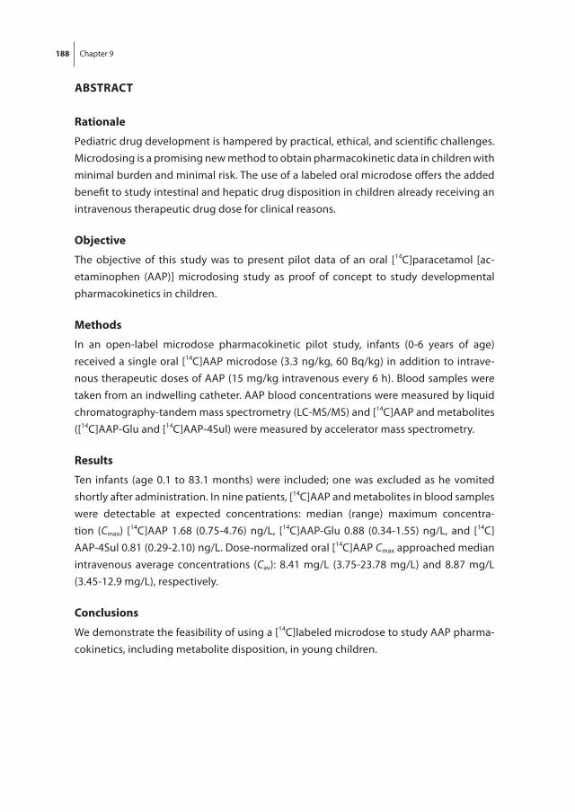
188 Chapter 9
AbstrAct
rationale
Pediatric drug development is hampered by practical, ethical, and scientific challenges. Microdosing is a promising new method to obtain pharmacokinetic data in children with minimal burden and minimal risk. The use of a labeled oral microdose offers the added benefit to study intestinal and hepatic drug disposition in children already receiving an intravenous therapeutic drug dose for clinical reasons.
objective
The objective of this study was to present pilot data of an oral [14C]paracetamol [ac-etaminophen (AAP)] microdosing study as proof of concept to study developmental pharmacokinetics in children.
Methods
In an open-label microdose pharmacokinetic pilot study, infants (0-6 years of age) received a single oral [14C]AAP microdose (3.3 ng/kg, 60 Bq/kg) in addition to intrave-nous therapeutic doses of AAP (15 mg/kg intravenous every 6 h). Blood samples were taken from an indwelling catheter. AAP blood concentrations were measured by liquid chromatography-tandem mass spectrometry (LC-MS/MS) and [14C]AAP and metabolites ([14C]AAP-Glu and [14C]AAP-4Sul) were measured by accelerator mass spectrometry.
results
Ten infants (age 0.1 to 83.1 months) were included; one was excluded as he vomited shortly after administration. In nine patients, [14C]AAP and metabolites in blood samples were detectable at expected concentrations: median (range) maximum concentra-tion (Cmax) [14C]AAP 1.68 (0.75-4.76) ng/L, [14C]AAP-Glu 0.88 (0.34-1.55) ng/L, and [14C]AAP-4Sul 0.81 (0.29-2.10) ng/L. Dose-normalized oral [14C]AAP Cmax approached median intravenous average concentrations (Cav): 8.41 mg/L (3.75-23.78 mg/L) and 8.87 mg/L (3.45-12.9 mg/L), respectively.
conclusions
We demonstrate the feasibility of using a [14C]labeled microdose to study AAP pharma-cokinetics, including metabolite disposition, in young children.
189
9
Pediatric microdose study of [14C]paracetamol to study drug metabolism
introDuction
Up to 70% of drugs prescribed to children are unlicensed or off-label, which brings risk of drug toxicity or therapeutic failure [1, 2]. However, pediatric drug studies face important ethical, practical, and scientific challenges [3]. A major challenge – against the background of developmental changes in drug absorption, distribution, metabolism, and excretion – is appropriate dose selection [4]. Simple size- or weight-based extrapo-lations from adult to pediatric doses do not suffice, particularly in neonates and infants. Current strategies include simulations using population pharmacokinetic (popPK) and physiologically based pharmacokinetic (PBPK) models [5-9]. The usefulness of these models may be limited, as relative little pediatric pharmacokinetic and physiological data are available to validate them. Hence, new data are needed to support these mod-els, as well as alternative methods to collect pharmacokinetic data without the inherent risks of toxicity when a therapeutic dose is given for the first time in a specific age group.
Microdosing is an interesting alternative. The European Medicines Agency (EMA) and the US Food and Drug Administration (FDA) define a microdose as one-hundredth of the No Observed Adverse Effect Level (NOAEL) or predicted pharmacologic dose based on animal data or as 100 µg of the new drug, whichever dose is lower [10, 11]. Dose linearity between the microdose and therapeutic dose is a prerequisite to extrapolate pharmacokinetic data to dosing guidelines [12]. The extremely low dose concentrations call for highly sensitive measurements. Accelerator mass spectrometry (AMS) can mea-sure low attomolar to zeptomolar isotope ratio ranges, to quantify [14C]labeled drug or metabolite levels in urine or plasma samples, even after at least five half-lives following a microdose [13]. [14C] Labeling of a microdose is associated with very low radiation exposure, i.e., less than 10 µSv in adults, when compared to the yearly background expo-sure of 2.5 mSv/year in The Netherlands [14]. Hence, microdosing is safe to use to study pharmacokinetics in children. In clinical care, [14C]urea has been safely used to test for Helicobacter pylori infection [15]. A [14C]ursodiol microdose study in preterm infants was briefly described in an excellent review on the potential for use of AMS in children [16]. Administering a labeled oral microdose to children already receiving a therapeutic drug dose for clinical reasons intravenously offers the added possibility to study oral bioavail-ability [12, 17]. This approach may also serve to delineate developmental changes in the drug-metabolizing enzymes involved in intestinal and hepatic drug metabolism [17, 18].
Paracetamol (acetaminophen; AAP), is much used in children and is an interesting study drug for several reasons. First, its metabolism shifts from primarily sulphation (AAP-sulphate [AAP-Sul]) to glucuronidation (AAP-glucuronide [AAP-Glu]) in the first year of life, as reflected by urinary metabolite kinetics [19, 20]. The relative contribution of intestinal and hepatic drug metabolism has not been studied to date. Second, dose linearity under normal conditions and after probenecid glucuronidation inhibition was
190 Chapter 9
shown in adults [18]. We therefore selected oral [14C]AAP to study the developmental changes in AAP glucuronidation and sulphation in children already receiving the non-labeled or ‘cold’ drug intravenously for analgesia [14]. This paper presents our first pilot data as proof of concept of this promising, safe method to study developmental pharmacokinetics in children.
MEthoDs
study Design
This proof-of-concept study is part of a larger phenotyping study in 60 children delineat-ing developmental changes in AAP to metabolite clearance (EudraCT 2011-005497-28). The Dutch Central Committee on Research Involving Human Subjects (The Hague, The Netherlands) approved the larger study. Parents or legal guardians gave informed con-sent. Radiation exposure was explained during the informed consent procedure in rela-tion to background exposure and exposure from medical imaging. The Dutch Nuclear Research and Service Group estimated individual exposures at approximately 1 µSv for a 0-2-year-old child, well below the minimal risk category 1 (100 µSv) of the International Commission of Radiological Protection and the yearly background exposure (2.5 mSv). Category 1 risk studies are considered minimal risk and are allowed when they provide new scientific knowledge [11].
subjects
Patients admitted to the Intensive Care of the Erasmus MC-Sophia Children’s Hospital (Rotterdam, The Netherlands) were considered for inclusion. Inclusion criteria were age between 0 and 6 years, gestational age >36 weeks, medical need for intravenous AAP, and an indwelling central venous or arterial line in place. To reduce pharmacokinetic variability due to underlying disease, patients with kidney and liver injury or the use of more than one vasopressor drug were excluded, as well as children intolerant to enteral nutrition.
study Procedures
A single oral [14C]AAP microdose (3.3 ng/kg, 60 Bq/kg, 0.25 ml/kg) followed by 1 ml of saline (to ensure rinsing of the enteral feeding tube) was administered in addition to the intravenous therapeutic dose of AAP (15 mg/kg intravenously every 6 h) prescribed by the treating physician to provide analgesia. The [14C]AAP oral dose also contained 1.7 µg/kg non-labeled AAP, but this amount was negligible in relation to the therapeutic intravenous dose and thus considered irrelevant for the pharmacokinetic estimations. The microdose was based on a previous adult [14C]AAP microdose study using 100

191
9
Pediatric microdose study of [14C]paracetamol to study drug metabolism
μg/7.1 kBq/individual and was normalized for weight [21]. Blood samples (0.5 mL) were taken from the indwelling catheter before and at 10-30 min, 1, 2, 4, 6, 12, and 24 h after dosing. Blood samples were centrifuged and plasma was stored at −80 0C until analysis.
Medicinal Products
The AAP formulation (10 mg/mL) was purchased from Fresenius Kabi, Schelle, Belgium. [14C]AAP was purchased from Moravek Biochemicals (Brea, CA, USA).
radiopharmaceutical Preparation
The formulation for oral administration [14C]AAP was prepared by adding [14C]AAP to an AAP formulation for intravenous use at the good manufacturing practice (GMP) radiopharmaceutical production laboratories of the Department of Radiology and Nuclear Medicine at the VU University Medical Center (Amsterdam, The Netherlands) (GMP license no. NL/H 11/0005) at final concentrations of 13 ng/mL for [14C]AAP and 6.7 µg/mL for non-labeled AAP. The mixed formulation was passed over a Millex - GV 0.22 µm filter and dispended in 20 mL sterile vials. Radiochemical purity was > 99%; chemical purity >98%. The radiopharmaceutical was heat sterilized and was shown to be stable for 2 months.
Non-specific binding was tested by running the [14C]AAP formulation followed by 1 mL of saline through enteral feeding tubes. Radioactive recovery measured by liquid scintillation was greater than 95%.
Paracetamol (Acetominophen; AAP) analysis
AAP concentrations were measured in the Hospital Pharmacy laboratory of Erasmus MC with a clinically used enzyme multiplied immunoassay technique (EMIT, Abbott Labora-tories®) with a lower limit of quantification (LLOQ) of 2.8 mg/l.
[14c]AAP and Metabolite Analysis
Plasma Sample Extraction and Ultra Performance Liquid Chromatography SeparationUsing 175 μL 100% v/v methanol containing 6.6 μg/mL APAP in 96-well protein pre-cipitation plates, 45 μL of plasma was extracted. The pellet was washed with 100 μL 0.9% NaCl:100% methanol (1:4 v/v). Resulting filtrates were evaporated to dryness, and re-dissolved in 30 μL 10 mM ammonium phosphate pH3.4 (Eluent A) of which 25 μL was used for ultra- performance liquid chromatography (UPLC) analysis. An AAP solution with a specific radioactivity of 4,100 Bq [14C]AAP /100 μg AAP in blank pooled plasma was used to prepare eight calibrators levels and three quality control (QC) sample levels from 0.4 to 180 mBq/mL, and from 1.7 to 131 mBq/mL, respectively. Calibrators (duplicate), QCs (triplicate), and sample extracts were injected onto a UPLC coupled to a photodiode
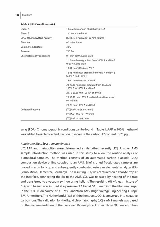
192 Chapter 9
array (PDA). Chromatographic conditions can be found in Table 1. AAP in 100% methanol was added to each collected fraction to increase the carbon-12 content to 25 μg.
Accelerator Mass Spectrometry Analysis[14C]AAP and metabolites were determined as described recently [22]. A novel AMS sample introduction method was used in this study to allow the routine analysis of biomedical samples. The method consists of an automated carbon diaoxide (CO2) combustion device online coupled to an AMS. Briefly, dried fractionated samples are placed in a tin foil cup and subsequently combusted using an elemental analyzer (EA) (Vario Micro, Elementar, Germany). The resulting CO2 was captured on a zeolyte trap at the interface, connecting the EA to the AMS. CO2 was released by heating of the trap and transferred to a vacuum syringe using helium. The resulting 6% v/v gas mixture of CO2 with helium was infused at a pressure of 1 bar at 60 μL/min into the titanium target in the SO110 ion source of a 1 MV Tandetron AMS (High Voltage Engineering Europe B.V., Amersfoort, The Netherlands) [23]. Within the source, CO2 is converted into negative carbon ions. The validation for the liquid chromatography (LC) + AMS analysis was based on the recommendation of the European Bioanalytical Forum. Three QC concentration
table 1. uPlc conditions AAP
Eluent A 10 mM ammonium phosphate pH 3.4
Eluent B 100 % v/v methanol
UPLC column (Waters Acquity) BEH C18 1.7 μm 2.1x100 mm column
Flowrate 0.3 mL/minute
Column temperature 30oC
Pressure 700 Bar
Chromatography conditions 0-1 min 100% A and 0% B
1-10 min linear gradient from 100% A and 0% Bto 95% A and 5% B
10-12 min 95% A and 5% B
12-15 min linear gradient from 95% A and 5% Bto 0% A and 100% B
15-20 min 0% A and 100% B
20-20.10 min linear gradient from 0% A and100% B to 100% A and 0% B
20.10-20.50 min 100 %A and 0% B
20.50-28 min 100% A and 0% B at a flowrate of0.4 ml/min
28-29 min 100% A and 0% B
Collected fractions [14C]AAP-Glu (3.8-5.3 min)
[14C]AAP-4Sul (6.1-7.9 min)
[14C]AAP (8.1-9.8 min)

193
9
Pediatric microdose study of [14C]paracetamol to study drug metabolism
levels were included, QC High 145 mBq/ml, QC Medium 14.5 mBq/ml and QC Low 2.4 mBq/ml. The accuracy of QC High, Medium, and Low analysis corresponded to 104, 103 and 90%, respectively. The precisions, defined by a coefficient of variation, were 9.1%, 6.7% and 6.9% respectively. All values are well within the requirements for LC + AMS analysis [24]. The LLOQ of the method was 0.58 mBq/mL.
Data analysis
Data were summarized as median (range), unless noted otherwise. The plasma [14C]AAP and metabolite concentrations were calculated by converting measured Bq/L to ng/L based on the dose given (3.3 ng/kg contained 60 Bq/kg) and for the metabolites corrected for molecular weight ([14C]AAP 151 g/mol, [14C]AAP-Glu 237 g/mol and [14C]AAP-4Sul 231 g/mol). To compare the disposition of oral [14C]AAP microdose to the intravenous therapeutic doses, the microdose concentrations were dose normalized to 15 mg/kg by multiplying with 5 x 10E6.
rEsults
Patients
Between 13 January and 31 May 2014, 32 patients were eligible. Nine patients were excluded for logistical reasons and 13 for refusal of informed consent. Parents’ informed consent was received for ten patients who were subsequently dosed according to the protocol. One patient vomited within 15 min post-dose and was excluded from pharma-cokinetic analysis. Table 2 provides a summary of patient characteristics.
Detection of [14c]AAP and Metabolites in Plasma
Time profiles of [14C]AAP, [14C]AAP-Glu and [14C]AAP-4Sul metabolite plasma concentra-tions of two representative patients are shown in Fig. 1. These patients were chosen as
figure 1. [14c]AAP and metabolite plasma concentrations[14C]AAP and metabolite plasma concentrations after oral 3.3 ng/kg [14C]AAP dose (LLOQ 0.03 ng/L).

194 Chapter 9
they represent the youngest and the oldest age group. The Electronic Supplementary Material shows graphs of all patients. The median time to maximum concentration (tmax) and maximum concentration (Cmax) median (range) for [14C]AAP were, respectively, 153 min (10-245 min) and 1.68 ng/L (0.75-4.76 ng/L), for [14C]AAP-Glu were 248 min (161-382 min) and 0.88 ng/L (0.34-1.55 ng/L), and for [14C]AAP-4Sul were respectively 193 min (115-343 min) and 0.81 ng/L (0.29-2.10 ng/L). Sample collections were not complete for all patients as the arterial line was prematurely removed or access to the line was restricted for clinical reasons.
Dose-normalized [14c]AAP and AAP Disposition
Semilog plots of the same two patients of dose-normalized 3.3 ng/kg oral [14C]AAP and 15 mg/kg every 6 h intravenous AAP concentrations are shown in Fig. 2. Individual graphs of all patients are displayed in the Electronic Supplemental Material. Dose-normalized median [14C]AAP Cmax concentrations approached median intravenous average concen-trations (Cav) [median (range)]: 8.41 mg/L (3.75 to 23.78 mg/L) and 8.87 mg/L (3.45 to 12.9 mg/L), respectively.
table 2. Patient characteristics
Patient Postnatal age(months)
Gender(male/female)
Primary diagnosis Intervention
1 3.6 Male Post necrotizing enterocolitis sigmoïd stenosis
Postoperative partial proximal colon resection
2 10.6 Female Scaphocephaly Postoperative craniofacial correction
3 1.7 Female Congenital cystic adenomatoid malformation of the lung
Postoperative partial lung resection
4 0.3 Male Congenital diaphragmatic hernia Postoperative hernia correction
5 53.8 Male Scaphocephaly Postoperative craniofacial correction
6 28.9 Male Germ cell tumor Respiratory insufficiency due to mediastinal pressure
7* 83.1 Male Increased intracranial pressure Postoperative craniofacial correction
8 0.1 Male Congenital cardiac disease Monitoring respiratory insufficiency
9 6.2 Male Scaphocephaly Postoperative craniofacial correction
10 5.6 Male Duodenal web Postoperative duodenoduodenostomy and placing gastrostomy tube
* Patient 7 excluded from results after vomiting within 15 minutes after microdose intake.
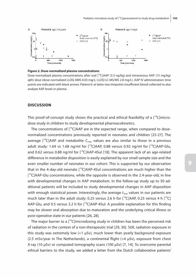
195
9
Pediatric microdose study of [14C]paracetamol to study drug metabolism
Discussion
This proof-of-concept study shows the practical and ethical feasibility of a [14C]micro-dose study in children to study developmental pharmacokinetics.
The concentrations of [14C]AAP are in the expected range, when compared to dose-normalized concentrations previously reported in neonates and children [25-27]. The average [14C]AAP and metabolite Cmax values are also similar to those in a previous adult study: 1.64 vs 1.68 ng/ml for [14C]AAP, 0.88 versus 0.92 ng/ml for [14C]AAP-Glu, and 0.62 versus 0.88 ng/ml for [14C]AAP-4Sul [18]. The apparent lack of an age-related difference in metabolite disposition is easily explained by our small sample size and the even smaller number of neonates in our cohort. This is supported by our observation that in the 4-day-old neonate [14C]AAP-4Sul concentrations are much higher than the [14C]AAP-Glu concentrations, while the opposite is observed in the 2.4-year-old, in line with developmental changes in AAP metabolism. In the follow-up study up to 50 ad-ditional patients will be included to study developmental changes in AAP disposition with enough statistical power. Interestingly, the average tmax values in our patients are much later than in the adult study: 0.25 versus 2.6 h for [14C]AAP, 0.25 versus 4 h [14C]AAP-Glu, and 0.5 versus 3.2 h for [14C]AAP-4Sul. A possible explanation for this finding may be slower oral absorption due to maturation and the underlying critical illness or post-operative state in our patients [26, 28].
The major barrier to a [14C]microdosing study in children has been the perceived risk of radiation in the context of a non-therapeutic trial [29, 30]. Still, radiation exposure in this study was extremely low (<1 μSv), much lower than yearly background exposure (2.5 mSv/year in The Netherlands), a continental flight (>4 μSv), exposure from chest X-ray (10 μSv) or computed tomography scans (100 μSv) [7, 14]. To overcome parental ethical barriers to the study, we added a letter from the Dutch collaborative patients’
figure 2. Dose-normalized plasma concentrationsDose-normalized plasma concentrations after oral [14C]AAP (3.3 ng/kg) and intravenous AAP (15 mg/kg/ q6h) dose (dose-normalized LLOQ AMS 0.03 mg/L; LLOQ LC-MS/MS 2.8 mg/L). AAP IV administration time points are indicated with black arrows. Patient 6: at latter two timpoints insufficient blood collected to also analyze AAP levels in plasma.
196 Chapter 9
organization for rare and genetic diseases (VSOP) to the patient information leaflet, explaining the need for pediatric drug research and the minimal risk involved in this study. Surprisingly, from the informed consent conversations it appeared that most parents perceived there was minimal risk involved. Fear for harmful radiation exposure was not the main reason to deny informed consent. Parents of the other children refused informed consent for reasons relating to the burden of additional procedures and/or blood sampling.
Very sensitive LC-tandem mass spectrometry (LC-MS/MS) techniques to measure pharmacokinetics of an unlabeled microdose may be an alternative to using a radio-labeled microdose in general [31]. Nevertheless, while low drug concentrations can be measured with LC-MS/MS, this analytical method has not reached the very low limits of detection of AMS. Hence, a larger unlabeled dose may be needed, which may increase the risk of a therapeutic or toxic effect [13]. Moreover, the use of an unlabeled micro-dose would also prohibit the separation of the disposition of an oral and intravenous dose given at the same time. This could be overcome by using a stable isotope-labeled probe drugs, which has successfully been used to study oral bioavailability using 15N3-midazolam in adults [32]. This method also has the important disadvantage that a much higher labeled drug dose is needed with similar or even higher risk of therapeutic or toxic effect.
conclusion
We have shown proof of concept for the practical and ethical feasibility of a [14C]labeled microdose to study pharmacokinetics in young children. This approach offers innovative possibilities to perform phase I first-in-child studies, especially for drugs with a small therapeutic window and high toxicity. In addition, it enables studies in vulnerable populations such as critically ill neonates and studies on developmental pharmacoki-netics using probe drugs for specific elimination pathways such as drug-metabolizing enzymes and renal excretion.
197
9
Pediatric microdose study of [14C]paracetamol to study drug metabolism
rEfErEncEs
[1] Schirm E, Tobi H, de Vries TW, Choonara I, De Jong-van den Berg LT. Lack of appropriate formula-tions of medicines for children in the community. Acta Paediatr 2003 Dec; 92(12): 1486-9.
[2] t Jong GW, van der Linden PD, Bakker EM, van der Lely N, Eland IA, Stricker BH, et al. Unlicensed and off-label drug use in a paediatric ward of a general hospital in the Netherlands. Eur J Clin Pharmacol 2002 Jul; 58(4): 293-7.
[3] Hoppu K. Reflection: medicines for children--science alone is not enough. Eur J Clin Pharmacol 2013 May; 69 Suppl 1: 59-63.
[4] Kearns GL, Abdel-Rahman SM, Alander SW, Blowey DL, Leeder JS, Kauffman RE. Developmental pharmacology--drug disposition, action, and therapy in infants and children. N Engl J Med 2003 Sep 18; 349(12): 1157-67.
[5] Krekels EH, Danhof M, Tibboel D, Knibbe CA. Ontogeny of hepatic glucuronidation; methods and results. Curr Drug Metab 2012 Jul; 13(6): 728-43.
[6] De Cock RF, Piana C, Krekels EH, Danhof M, Allegaert K, Knibbe CA. The role of population PK-PD modelling in paediatric clinical research. Eur J Clin Pharmacol 2011 May; 67 Suppl 1: 5-16.
[7] Barrett JS, Della Casa Alberighi O, Laer S, Meibohm B. Physiologically based pharmacokinetic (PBPK) modeling in children. Clin Pharmacol Ther 2012 Jul; 92(1): 40-9.
[8] Knibbe CA, Danhof M. Individualized dosing regimens in children based on population PKPD modelling: are we ready for it? Int J Pharm 2011 Aug 30; 415(1-2): 9-14.
[9] Knibbe CA, Krekels EH, Danhof M. Advances in paediatric pharmacokinetics. Expert Opin Drug Metab Toxicol 2011 Jan; 7(1): 1-8.
[10] Food and Drug Administration US Department of Health and Human Services Guidance for Industry Investigators and Reviewers. Exploratory IND Studies. January 2006.
[11] Agency EM. ICH Topic M3 (R2) Non-Clinical Safety Studies for the Conduct of Human Clinical Trials and Marketing Authorization for Pharmaceuticals. 2008 July 2008.
[12] Lappin G, Kuhnz W, Jochemsen R, Kneer J, Chaudhary A, Oosterhuis B, et al. Use of microdosing to predict pharmacokinetics at the therapeutic dose: experience with 5 drugs. Clin Pharmacol Ther 2006 Sep; 80(3): 203-15.
[13] Salehpour M, Possnert G, Bryhni H. Subattomole sensitivity in biological accelerator mass spec-trometry. Anal Chem 2008 May 15; 80(10): 3515-21.
[14] Ceelie I, de Wildt SN, van Dijk M, van den Berg MM, van den Bosch GE, Duivenvoorden HJ, et al. Effect of intravenous paracetamol on postoperative morphine requirements in neonates and infants undergoing major noncardiac surgery: a randomized controlled trial. JAMA 2013 Jan 9; 309(2): 149-54.
[15] Pathak CM, Kaur B, Khanduja KL. 14C-urea breath test is safe for pediatric patients. Nucl Med Commun Sep; 31(9): 830-5.
[16] Vuong LT, Blood AB, Vogel JS, Anderson ME, Goldstein B. Applications of accelerator MS in pediat-ric drug evaluation. Bioanalysis 2012 Aug; 4(15): 1871-82.
[17] Lappin G, Shishikura Y, Jochemsen R, Weaver RJ, Gesson C, Brian Houston J, et al. Comparative pharmacokinetics between a microdose and therapeutic dose for clarithromycin, sumatriptan, propafenone, paracetamol (acetaminophen), and phenobarbital in human volunteers. Eur J Pharm Sci 2011 Jun 14; 43(3): 141-50.
[18] Tozuka Z, Kusuhara H, Nozawa K, Hamabe Y, Ikushima I, Ikeda T, et al. Microdose study of 14C-acetaminophen with accelerator mass spectrometry to examine pharmacokinetics of parent drug and metabolites in healthy subjects. Clin Pharmacol Ther 2010 Dec; 88(6): 824-30.

198 Chapter 9
[19] Allegaert K, Tibboel D. Comments on “shift from biliary to urinary elimination of acetaminophen-glucuronide in acetaminophen-pretreated rats”. J Pharmacol Exp Ther 2006 Feb; 316(2): 966-7.
[20] van der Marel CD, Anderson BJ, van Lingen RA, Holford NH, Pluim MA, Jansman FG, et al. Paracetamol and metabolite pharmacokinetics in infants. Eur J Clin Pharmacol 2003 Jul; 59(3): 243-51.
[21] Zhou XJ, Garner RC, Nicholson S, Kissling CJ, Mayers D. Microdose pharmacokinetics of IDX899 and IDX989, candidate HIV-1 non-nucleoside reverse transcriptase inhibitors, following oral and intravenous administration in healthy male subjects. J Clin Pharmacol 2009 Dec; 49(12): 1408-16.
[22] van Duijn E, Sandman H, Grossouw D, Mocking JA, Coulier L, Vaes WH. Automated combustion accelerator mass spectrometry for the analysis of biomedical samples in the low attomole range. Anal Chem 2014 Aug 5; 86(15): 7635-41.
[23] Klein MV, W. H. J. Fabriek, B. Sandman, H. Mous, D. J. W. Gottdang, A. The 1 MV multi-element AMS system for biomedical applications at the Netherlands Organization for Applied Scientific Research (TNO). Nuclear Instruments and Methods in Physics Research Section B: Beam Interac-tions with Materials and Atoms 2013; 294: 14-7.
[24] Higton D, Young G, Timmerman P, Abbott R, Knutsson M, Svensson LD. European Bioanalysis Forum recommendation: scientific validation of quantification by accelerator mass spectrometry. Bioanalysis 2012 Nov; 4(22): 2669-79.
[25] Hopkins CS, Underhill S, Booker PD. Pharmacokinetics of paracetamol after cardiac surgery. Arch Dis Child 1990 Sep; 65(9): 971-6.
[26] Anderson BJ, van Lingen RA, Hansen TG, Lin YC, Holford NH. Acetaminophen developmental pharmacokinetics in premature neonates and infants: a pooled population analysis. Anesthesiol-ogy 2002 Jun; 96(6): 1336-45.
[27] Walson PD, Halvorsen M, Edge J, Casavant MJ, Kelley MT. Pharmacokinetic comparison of acet-aminophen elixir versus suppositories in vaccinated infants (aged 3 to 36 months): a single-dose, open-label, randomized, parallel-group design. Clin Ther 2013 Feb; 35(2): 135-40.
[28] Mooij MG, de Koning BA, Huijsman ML, de Wildt SN. Ontogeny of oral drug absorption processes in children. Expert Opin Drug Metab Toxicol 2012 Oct; 8(10): 1293-303.
[29] Ratnapalan S, Bona N, Chandra K, Koren G. Physicians’ perceptions of teratogenic risk associated with radiography and CT during early pregnancy. AJR American journal of roentgenology 2004 May; 182(5): 1107-9.
[30] de Wildt SN, Taguchi N, Koren G. Unintended pregnancy during radiotherapy for cancer. Nat Clin Pract Oncol 2009 Mar; 6(3): 175-8.
[31] Maeda K, Ikeda Y, Fujita T, Yoshida K, Azuma Y, Haruyama Y, et al. Identification of the rate-determining process in the hepatic clearance of atorvastatin in a clinical cassette microdosing study. Clin Pharmacol Ther 2011 Oct; 90(4): 575-81.
[32] Gorski JC, Jones DR, Haehner-Daniels BD, Hamman MA, O’Mara EM, Jr., Hall SD. The contribution of intestinal and hepatic CYP3A to the interaction between midazolam and clarithromycin. Clin Pharmacol Ther 1998 Aug; 64(2): 133-43.
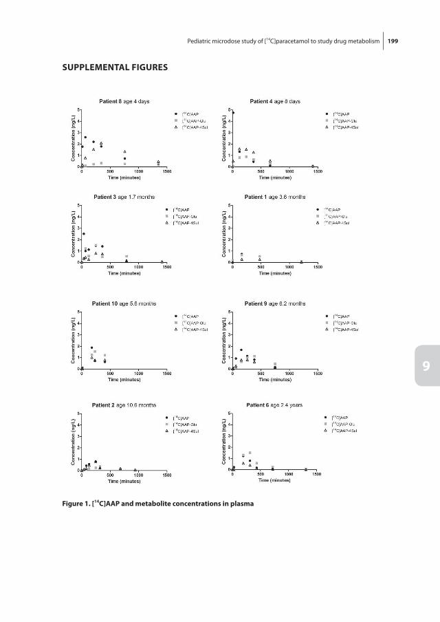
199
9
Pediatric microdose study of [14C]paracetamol to study drug metabolism
suPPlEMEntAl fiGurEs
figure 1. [14c]AAP and metabolite concentrations in plasma
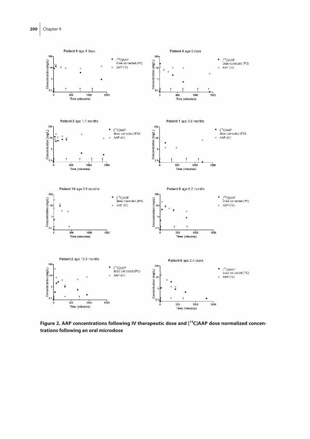
200 Chapter 9
figure 2. AAP concentrations following iV therapeutic dose and [14c]AAP dose normalized concen-trations following an oral microdose


10 P ediatric microdose and microtracer studies using 14C in Europe
Mark A. Turner, Miriam G. Mooij, Wouter H.J. Vaes, Albert D. Windhorst, N. Harry Hendrikse, Catherijne A.J. Knibbe, Lenne-Triin Kõrgvee, Wioletta Maruszak, Grzegorz Grynkiewicz, R. Colin Garner, Dick Tibboel, B. Kevin Park, Saskia N. de Wildt
Clinical Pharmacology and Therapeutics 2015, Sep;89(3):234-7

205
10
Pediatric microdose and microtracer studies using 14C in Europe
introDuction
Important information gaps remain on efficacy and safety of drugs in children. Pediatric drug development encounters several ethical, practical and scientific challenges. One barrier to the evaluation of medicines for children is a lack of innovative methodologies that have been adapted to the needs of children. This article presents our successful experience of pediatric microdose and microtracer studies using 14C-labeled probes in Europe to illustrate the strengths and limitations of these approaches.
Microtracer / microdose studies in pediatric drug development
Dose selection for pediatric phase I/II studies is usually based on extrapolation from human adult and juvenile animal data, increasingly using population pharmacokinetic (PK) and physiology-based pharmacokinetic modeling to simulate dosing regimens. These methods have important limitations. Critical information gaps remain on the developmental changes in the processes involved in the disposition of drugs [1]. Es-pecially for drugs with complex metabolism and/or poor understanding of ontogeny, a useful model is lacking. PK studies with a 14C-labeled drug at a subtherapeutic dose may aid to close these information gaps while overcoming many limitations associated with regular pharmacokinetic studies in children. This can contribute to systems-level understanding (e.g., information about ontogeny) and drug-level understanding (e.g., population PK studies during drug development).
When a radiolabeled drug is administered concomitantly with a therapeutic dose of the same unlabeled drug via either the same (for example, for mass balance studies) or another route (for example, for absolute bioavailability studies), this is referred to as a microtracer dose [2]. For example, a radiolabeled probe administered enterally can gen-erate information about enteric and hepatic metabolism distinguishing between the two routes. A microdose is a subtherapeutic dose in the absence of other dosing with the drug of interest. Following a microdose the disposition will reflect the microdose per se. The European Medicines Agency (EMA) and the US Food and Drug Administration (FDA) defines a microdose as 1/100th of the No Observed Adverse Effect Level (NOAEL) or predicted pharmacologic dose based on animal data, with a maximum of 100 µg of the new drug. Microdosing has been used in adult drug development.
A prerequisite of both microtracer and microdosing is the availability of sensitive analytical techniques to measure the low plasma concentrations. Very sensitive liquid chromatography/tandem mass spectrometry (LC-MS/MS) techniques have been used to measure PK of an unlabeled subtherapeutic dose. However, these techniques are not sensitive enough to detect the low concentrations of metabolites seen during the elimination phase following the administration of a microdose. LC-accelerator mass spectrometry (AMS) can measure low attomolar to zeptomolar range and quantify 14C-
206 Chapter 10
labeled drug or metabolite levels in urine or plasma samples [3]. The exquisite sensitivity of AMS allows extremely low doses of drugs to be given, which minimizes the amount of 14C administered. The administration of doses that have no pharmacological effect and thus no concerns about toxicity would allow a microdose study to be conducted early in pediatric drug development. In addition, AMS uses very small samples of plasma (<20 µL) that are compatible with sampling limits during research involving children.
As a result of the 2006 EU [4] regulation on medicinal products for pediatric use, both European (Era-NET) and national (ZonMW Priority Medicines for Kinderen) funding opportunities encouraged drug studies in children. Two projects: PAMPERS (Paediatric Accelerator Mass Spectrometry Evaluation Research Study) and PEDMIC (Pediatric mi-crodosing: elucidating age-related changes in oral absorption to guide dosing of new formulations) were funded to study the use of 14C-labeled probes in children to elucidate PK. The first results of these studies were published recently, showing the feasibility of the approach, including acceptable dose linearity [5, 6].
PAMPErs and PEDMic project study aims
The PAMPERS project aims to show the feasibility of microdose and microtracer stud-ies for drug development in children. 14C-Paracetamol and 14C-midazolam, frequently prescribed to children and probes for UGT and CYP3A metabolism, were chosen to show feasibility [6].
The PEDMIC study was designed on the premise of dose-linearity of oral absorption, as shown in adults, and aimed to study developmental pharmacology. By administering an oral microtracer (14C-paracetamol or 14C-midazolam) while the patient received the drug for therapeutic reasons i.v., in children from 0 to 6 years of age, the developmental change in intestinal and hepatic drug metabolizing enzymes, specific for these drugs, can be elucidated [5].
Details of both projects can be found in Table 1.
Perceived risks of radioactivity
In discussions with colleagues, we found that the major concern about 14C-labeled microtracer and microdose PK studies in children lay in the perceived risk of adverse effects related to radioactive exposure from a 14C-labeled drug. While this hesitancy is understandable, it is unfounded. Radioactive dose calculations were performed using a worst-case half-life scenario of 40 days, which for an 111Bq/kg 14C-dose gave a radioac-tive exposure between 0.33 and 0.8 microSV. This is much lower than yearly background exposure (2.5 mSv/year in the Netherlands), a regional European flight (1-15 microSv), a computed tomography (CT)-scan of the head (1200 microSv) or chest x-ray (12 microSv) (Figure 1). The International Committee on Radioactivity Protection (ICRP) considers an effective radioactive dose of <100 microSv for a healthy adult volunteer as risk
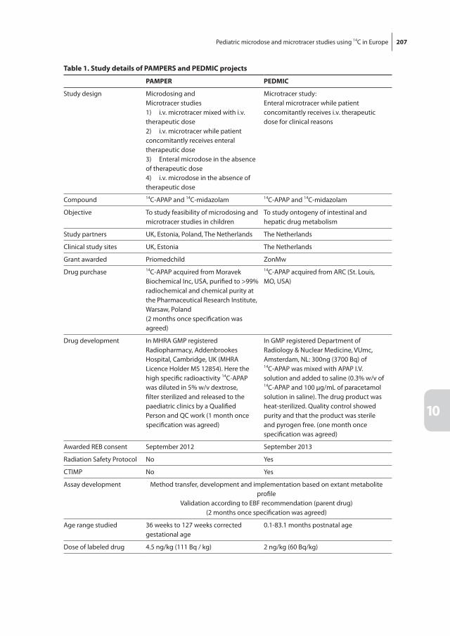
207
10
Pediatric microdose and microtracer studies using 14C in Europe
table 1. study details of PAMPErs and PEDMic projects
PAMPEr PEDMic
Study design Microdosing andMicrotracer studies1) i.v. microtracer mixed with i.v. therapeutic dose2) i.v. microtracer while patient concomitantly receives enteral therapeutic dose3) Enteral microdose in the absence of therapeutic dose4) i.v. microdose in the absence of therapeutic dose
Microtracer study:Enteral microtracer while patient concomitantly receives i.v. therapeutic dose for clinical reasons
Compound 14C-APAP and 14C-midazolam 14C-APAP and 14C-midazolam
Objective To study feasibility of microdosing and microtracer studies in children
To study ontogeny of intestinal and hepatic drug metabolism
Study partners UK, Estonia, Poland, The Netherlands The Netherlands
Clinical study sites UK, Estonia The Netherlands
Grant awarded Priomedchild ZonMw
Drug purchase 14C-APAP acquired from Moravek Biochemical Inc, USA, purified to >99% radiochemical and chemical purity at the Pharmaceutical Research Institute, Warsaw, Poland(2 months once specification was agreed)
14C-APAP acquired from ARC (St. Louis, MO, USA)
Drug development In MHRA GMP registered Radiopharmacy, Addenbrookes Hospital, Cambridge, UK (MHRA Licence Holder MS 12854). Here the high specific radioactivity 14C-APAP was diluted in 5% w/v dextrose, filter sterilized and released to the paediatric clinics by a Qualified Person and QC work (1 month once specification was agreed)
In GMP registered Department of Radiology & Nuclear Medicine, VUmc, Amsterdam, NL: 300ng (3700 Bq) of 14C-APAP was mixed with APAP I.V. solution and added to saline (0.3% w/v of 14C-APAP and 100 µg/mL of paracetamol solution in saline). The drug product was heat-sterilized. Quality control showed purity and that the product was sterile and pyrogen free. (one month once specification was agreed)
Awarded REB consent September 2012 September 2013
Radiation Safety Protocol No Yes
CTIMP No Yes
Assay development Method transfer, development and implementation based on extant metabolite profile
Validation according to EBF recommendation (parent drug)(2 months once specification was agreed)
Age range studied 36 weeks to 127 weeks corrected gestational age
0.1-83.1 months postnatal age
Dose of labeled drug 4.5 ng/kg (111 Bq / kg) 2 ng/kg (60 Bq/kg)
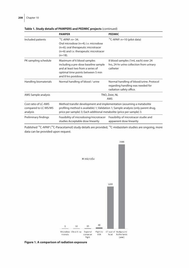
208 Chapter 10
table 1. study details of PAMPErs and PEDMic projects (continued)
PAMPEr PEDMic
Included patients 14C-APAP: n= 34.Oral microdose (n=4); i.v. microdose (n=6); oral therapeutic microtracer (n=6) and i.v. therapeutic microtracer (n=18).
14C-APAP: n=10 (pilot data)
PK sampling schedule Maximum of 6 blood samples including a pre-dose baseline sample and at least two from a series of optimal time points between 5 min and 8 hrs postdose.
8 blood samples (1mL each) over 24 hrs, 24-hr urine collection from urinary catheter
Handling biomaterials Normal handling of blood / urine Normal handling of blood/urine. Protocol regarding handling was needed for radiation safety office.
AMS Sample analysis TNO, Zeist, NLAMS
Cost ratio of LC-AMS compared to LC-MS/MS analysis
Method transfer development and implementation (assuming a metabolite profiling method is available) 1; Validation 1; Sample analysis (only parent drug, price per sample) 3; Each additional metabolite (price per sample) 3.
Preliminary findings Feasibility of microdosing/microtracer studies Acceptable dose linearity
Feasibility of microtracer studie and appaerent dose linearity
Published 14C-APAP (14C-Paracetamol) study details are provided, 14C-midazolam studies are ongoing, more data can be provided upon request.
figure 1. A comparison of radiation exposure
209
10
Pediatric microdose and microtracer studies using 14C in Europe
category 1. Thus, the doses of radiation that can be administered during studies that use AMS pose no increase above minimal risk.
Ethics
With this explanation about the dose of radioactivity, approval by Ethics Committees (equivalent to Independent Review Boards (IRBs)) was unproblematic. The study designs in PAMPERS and PEDMIC for the microdosing arms involve administering a medicine without a direct therapeutic benefit to the individual child. These studies administered a background dose of radioactivity with a medicine in common use and took blood samples through available vascular access. Ethics Committees in three countries deemed this compatible with minimal risk and burden for children receiving pediatric intensive care. Similarly, a neonatal 14C-ursodiol microdose study was approved in the U.S. [7]. All these studies met the criteria of minimal risk and burden in the specific patient popula-tion studied. But for each study an evaluation of the balance between risks and benefits in the specific context needs to be made. For example, a microdose study with urine sampling only may be evaluated as minimal risk and burden in healthy children, while repeated blood sampling may only be allowed in children with available vascular access. Hospital staff also understood the concept of microdosing and supported the studies at all three sites.
consent
The concept and safety of these studies appeared easy to explain to parents. The recruit-ment rate in both projects was around 50% of eligible patients. This is comparable to other interventional studies in the Intensive Care Units at Erasmus MC Sophia, Alder Hey Children’s Hospital and Tartu Children’s Hospital. For most parents who did not consent, information overload during critical illness or the perceived burden of repeated blood sampling were the main reasons not to let their child participate [8]. Fear of radiation was cited as a reason not to consent by a small minority of parents in all three centers.
Dose linearity
A major prerequisite for a microdose to study pharmacokinetics is a linear relationship in PK from the microdose to the therapeutic dose range. 14C-paracetamol and 14C-midazolam show dose-linear pharmacokinetics in the dose range from the microdose to a therapeutic range in healthy adults [9, 10]. For other drugs, microdosing studies in children should preferably be preceded by adult microdosing studies to show dose-linearity, as otherwise extrapolation of data from microdosing studies to pediatric therapeutic doses may be incorrect.
210 Chapter 10
regulatory issues
A major challenge was the preparation from the commercially available chemical compound 14C-paracetamol into a formulation suited for human use according to Good Manufacturing Practice (GMP) guidelines. This challenge was overcome by the addition of a very small amount of 14C-paracetamol acquired from a commercial supplier to saline or glucose with (PEDMIC) or without (PAMPERS) unlabeled i.v. solution paracetamol. Solubility in itself was not an issue considering the extremely low concentrations. Exten-sive GMP quality control showed the drug product to be pure, sterile, and pyrogen free.
In The Netherlands, a single microtracer to study pharmacokinetics was considered a drug so that the study was a clinical trial of an investigational medicinal product (CTIMP). This was not the case in the UK and Estonia, where it was considered a well-characterized probe in a physiological experiment (a study involving a less well-characterized probe would be a CTIMP in the UK). In The Netherlands, the hospital’s Radiation Safety Officer considered the formulation radioactive and requested a Radiation Safety training by the junior researcher, as well as a study-specific radiation handling protocol, in contrast to the UK and Estonia, were it was not considered radioactive. Body fluids and blood/urine samples were considered radioactive only in The Netherlands. The radiation dose, how-ever, was such a low dose that no special handling was required in all three countries.
costs14C AMS studies may be costly. First, the 14C-labeled probe needs to be manufactured and formulated according to GMP, without loss of its unique chemical property. The costs are probe-specific. Second, the AMS analyses are usually more costly than LC-MS/MS analyses. Finally, additional adult studies may be needed to show dose-linearity across the full dose range and possibly also in inhibition and/or induction scenarios. Nevertheless, timely information about disposition will accelerate the development of safe and effective pediatric products, outweighing the financial cost of such studies.
conclusion
We have shown the ethical, practical and scientific feasibility of microtracer and micro-dose studies in children. Microtracer studies could be a valuable tool in pediatric drug research. Taking the challenges such as dose-linearity and costs into account, we believe that microdosing/microtracer trials are of added value for pediatric drug development. Microdosing may be of specific value during the early evaluation of new drugs when metabolism is likely to be complex and the timely determination of the pharmacokinet-ics of a new drug is critical to the risk-benefit analysis of a new medicine in the pediatric population.
211
10
Pediatric microdose and microtracer studies using 14C in Europe
rEfErEncEs
[1] Kearns GL, Abdel-Rahman SM, Alander SW, Blowey DL, Leeder JS, Kauffman RE. Developmental pharmacology--drug disposition, action, and therapy in infants and children. N Engl J Med 2003 Sep 18; 349(12): 1157-67.
[2] Lappin G, Noveck R, Burt T. Microdosing and drug development: past, present and future. Expert Opin Drug Metab Toxicol 2013 Jul; 9(7): 817-34.
[3] Salehpour M, Possnert G, Bryhni H. Subattomole sensitivity in biological accelerator mass spec-trometry. Anal Chem 2008 May 15; 80(10): 3515-21.
[4] The European Parliament, The Council of the European Union. Regulation (EU) No 1901/2006 on medicinal products for paediatric use and amending Regulation (EEC) No 1768/92, Directive 2001/20/EC, Directive 2001/83/EC and Regulation (EC) No 726/2004. In: The European Parliament, editor. 1901/2006; 2006.
[5] Mooij MG, van Duijn E, Knibbe CA, Windhorst AD, Hendrikse NH, Vaes WH, et al. Pediatric micro-dose study of [(14)c]paracetamol to study drug metabolism using accelerated mass spectrom-etry: proof of concept. Clin Pharmacokinet 2014 Nov; 53(11): 1045-51.
[6] Garner RC, Park BK, French NS, Earnshaw C, Schipani A, Selby AM, et al. Observational infant ex-ploratory [ C]paracetamol pharmacokinetic microdose/therapeutic Dose study with accelerator mass spectrometry bioanalysis. Br J Clin Pharmacol 2015 Jan 24.
[7] Vuong LT, Blood AB, Vogel JS, Anderson ME, Goldstein B. Applications of accelerator MS in pediat-ric drug evaluation. Bioanalysis 2012 Aug; 4(15): 1871-82.
[8] Kleiber N, Tromp K, Mooij MG, van de Vathorst S, Tibboel D, de Wildt SN. Ethics of drug research in the pediatric intensive care unit. Paediatr Drugs 2015 Feb; 17(1): 43-53.
[9] Lappin G, Shishikura Y, Jochemsen R, Weaver RJ, Gesson C, Brian Houston J, et al. Comparative pharmacokinetics between a microdose and therapeutic dose for clarithromycin, sumatriptan, propafenone, paracetamol (acetaminophen), and phenobarbital in human volunteers. Eur J Pharm Sci 2011 Jun 14; 43(3): 141-50.
[10] Hohmann N, Kocheise F, Carls A, Burhenne J, Haefeli WE, Mikus G. Midazolam microdose to deter-mine systemic and pre-systemic metabolic CYP3A activity in humans. Br J Clin Pharmacol 2015 Feb; 79(2): 278-85.

11 M icrodosing to study ontogeny of drug metabolism: the case of [14C]paracetamol
Miriam G. Mooij, Esther van Duijn, Catherijne A.J. Knibbe, Karel Allegaert, Albert D. Windhorst, Joost van Rosmalen, N. Harry Hendrikse, Dick Tibboel, Wouter H.J. Vaes, Saskia N. de Wildt
Submitted for publication
214 Chapter 11
AbstrAct
background
We aim to study the feasibility for [14C]microdosing to study the impact of age on drug disposition pathways, more specifically, glucuronidation and sulfation, using [14C]paracetamol (AAP).
Methods
Infants admitted to the pediatric intensive care received a single oral [14C]AAP microdose while receiving intravenous therapeutic AAP q6h. The plasma AUC0-inf and urinary re-covery ratios ([14C]AAP and metabolites measured with accelerator mass spectrometry) were related to age as surrogate markers of metabolism.
results
50 children [median age 6 months (range 3 days - 6.9 years)] received a microdose (3.3 [2.0-3.5] ng/kg; 64 [41-71] Bq/kg). Plasma [14C]AAP CL/F was 0.4 (0.1-2.6) L/h/kg, Vss/F was 1.7 (0.9-8.2) L/kg, T1/2 was 2.8 (1-7) h. With age, plasma and urinary AAP-glu/AAP and AAP-glu/AAP-sul ratios significantly increased by 4-fold, while the AAP-sul/AAP ratio significantly decreased.
conclusion
Using [14C]labeled microdosing, the effect of age on orally administered AAP metabo-lism was successfully elucidated in both plasma and urine.
215
11
Microdosing to study ontogeny of drug metabolism: the case of [14C]paracetamol
introDuction
Drug disposition in children may differ largely from that in adults due to age-related changes in drug absorption, distribution, metabolism and excretion [1, 2]. Drug me-tabolism is one of the main determinants of variation in drug disposition. While several knowledge gaps on the ontogeny of drug metabolizing enzymes (DMEs) have already been elucidated (e.g. cytochrome (CYP) 3A family) much is still unknown, especially on the ‘phase II’ drug metabolizing enzymes [3, 4].
Phenotyping studies can be used to elucidate in vivo drug metabolism pathways but in the pediatric population these studies pose several challenges in children. For one, when a validated phenotype probe for the pathway of interest is available [5], it is often a marketed drug with potentially therapeutic and toxic effects, which may not be ac-ceptable to ethics committees, physicians, parents and their children. Second, repeated blood sampling for pharmacokinetic analyses may be difficult in light of blood volume limits (<5% of estimated circulating volume) and repeated painful punctures. The alter-native method, urinary sampling, is technically challenging in young children especially without urinary catheter and results often in unreliable collections.
To overcome some of these challenges, microdosing with carbon-14 labeled drugs may be an interesting alternative to classic pharmacokinetic phenotyping studies in children [6-8]. A very low drug dose is not expected to be toxic or result in any therapeu-tic effect. Also, only very small blood volumes are needed for highly sensitive analytical methods, such as accelerator mass spectrometry (AMS) [9]. A microdose is defined as a maximum of 100 micrograms or 1/100th of the No Observed Adverse Effect Level (NO-AEL) or animal-based predicted pharmacologic dose [10, 11]. Microdosing has been ac-cepted by the US Food and Drug Administration (FDA) and European Medicines Agency (EMA) as a tool to study pharmacokinetics in adults [9]. If pharmacokinetics shows dose linear behavior, pharmacokinetic data can be extrapolated to therapeutic exposure [12-14]. Moreover, when an labeled oral dose is given simultaneously with a therapeutic intravenous dose (or routes are reversed), bioavailability can easily be determined. The microdose is then referred to as a microtracer. Two recent proof-of-concept studies in a limited number of patients, have shown the ethical and practical feasibility of pediatric microdosing/microtracer studies using [14C]paracetamol (AAP) [13, 14]. Dose linearity was shown in adults for paracetamol from microdose to therapeutic dose under stan-dard and inhibition conditions [15, 16]. Our own proof-of-concept study supported this dose linearity of pharmacokinetics in children [13].
Paracetamol, also known as acetaminophen or N-acetyl-p-aminophenol, is a common analgesic and antipyretic agent. Its main metabolism pathways are glucuronidation and sulfation. Glucuronidation occurs mainly via uridine 5’-diphospho-glucuronosyltransfer-ase (UGT) 1A1, 1A6, 1A9 and 2B15 and sulfation is catalyzed by sulfotransferase (SULT)
216 Chapter 11
1A1, 1A3, 1A4, and 2A1 [17]. Although not specific for individual enzymes, paracetamol pharmacokinetic studies have provided valuable insight in the relative contributions of glucuronidation and sulfation to the disposition of paracetamol. Paracetamol me-tabolism in young children differs from that in adults. In neonates, a higher proportion of urinary paracetamol sulfate than glucuronide suggested immaturity of the involved UGTs, compensated for by more mature sulfotransferases [18]. Other studies showed a significant increase in urinary AAP-glu/AAP-sul ratio from 0.27 in 23-week-old preterm neonates to 0.75 in 3 to 9-year-olds and 2 in adults [19]. Despite these studies, gaps remain in the knowledge on the ontogeny of paracetamol metabolism, especially for children with bodyweights between 3 and 7 kg as well as between 25 and 50 kg [20, 21]. Most developmental changes in drug metabolizing enzymes occur in the first months of life, a better insight in the exact pattern of the age-related change paracetamol me-tabolism may provide valuable novel information. Moreover, studies on exclusive oral paracetamol and metabolite disposition are scarce, while population pharmacokinetic studies using multiple study cohorts have often combined administration routes [20, 21] [22]. Bioavailability of oral and rectal administration differs, hence phenotyping of combined small intestinal and hepatic drug metabolism may be less precise, through by-pass of the liver after rectal administration [23]. Even though the oral bioavailability of paracetamol approaches 90% in adults, data in children are lacking and therefore a con-tribution of intestinal metabolism on oral paracetamol disposition cannot be excluded (Tylenol ® product information). Finally, most studies used the urinary metabolite ratio as surrogate marker for paracetamol metabolism. This method has limitations due to the difficult nature of complete and prolonged urine collections in young children, with incomplete or short collection intervals. Hence, these data may have unexplained large inter-individual variation reducing the power to detect more subtle age-related variation.
Previously, we showed that microdosing is practically and ethically feasible to study AAP pharmacokinetics in children. Now, using this innovative [14C] microdosing method, we aim to study the impact of age on paracetamol disposition exclusively after oral ad-ministration, with plasma and urine analyses including metabolites (AAP-glu, AAP-sul), over the whole age range from 0 to 6 years. In this way, we aimed to further elucidate the exact developmental pattern of glucuronidation and sulphation in young children and to ultimately narrow the information gap on phase II drug metabolism.
MEthoDs
1. subjects
Patients admitted to the pediatric intensive care unit of the Erasmus MC-Sophia Chil-dren’s Hospital were considered for inclusion. Inclusion criteria were age from 0 through
217
11
Microdosing to study ontogeny of drug metabolism: the case of [14C]paracetamol
6 years, medical need for intravenous paracetamol, an indwelling arterial or central venous line in place and written informed consent from parents or legal guardians. To reduce the impact of critical illness or bowel disease on the pharmacokinetics of paracetamol, patients with kidney and liver failure, more than one vasopressor drug, in-flammatory bowel disease, or intolerant to enteral nutrition, or concomitantly receiving co-medication known to interact with paracetamol (according to Micromedex ® www.micromedexsolutions.com) were excluded.
2. study design
The study design has been previously described in detail [13]. The Dutch Central Com-mittee on Research Involving Human Subjects (The Hague) approved the study (EudraCT 2011-005497-28). Purchase of [14C]AAP formulation and radiopharmaceutical prepara-tion have been previously described [13]. A single [14C]AAP microdose (3.3 ng/kg, 60 Bq/kg, 0.25 ml/kg) was given by mouth or enteral feeding tube. The [14C]AAP oral dose also contained 1.7 µg/kg non-labeled AAP, which is negligible in relation to the therapeutic intravenous dose and considered irrelevant for pharmacokinetic estimations. Enteral feeding tubes were flushed with 2 ml saline to ensure complete rinsing. Patients would simultaneously receive therapeutic AAP as part of clinical care, dosed according to the Dutch Pediatric Handbook (www.kinderformularium.nl): 20 mg/kg loading dose, fol-lowed by 10 mg/kg every 6 h (<1 month of age) or 15 mg/kg q6h (≥ one month of age).
Blood samples (1.0 ml) were taken from the indwelling catheter immediately before administration; at around 10, and 30 min; and at 1, 2, 4, 6, 12, 24 hrs after dosing or until the catheter was removed. After centrifuging, plasma was stored at −80 0C until analysis. Urine was collected in patients who needed a urinary catheter for clinical reasons. It was collected for a maximum 24 hrs or until the urinary catheter was removed. Urine volume was entered by the nurses in the clinical electronic patient record. One sample (2 mL) was taken from the 24h-urine collection and stored at −80 0C until analysis.
3. Analytical procedures
Previously a method has been qualified to quantify [14C]AAP, [14C]AAP-glu and [14C]AAP-sul levels in human plasma, using liquid chromatography (LC) in combination with AMS [13, 24]. The LC+AMS qualification was performed in accordance with the recommenda-tion of the European Bioanalytical Forum [25], qualification results are summarized in Mooij et al. [13].
In short the experimental outline was as follows; a 45 µL aliquot of plasma was ex-tracted using 175 µL100% v/v methanol containing 6.6 µg/ml AAP, 8.10 µg/mL AAP-glu, 10.0 µg/mL AAP-sul and 6.6 µg/mL 3-acetamidophenol in 96-well protein precipitation plates. The pellet was washed with 100 µL 0.9% NaCl:100% methanol (1:4 v/v). The filtrates were combined and after evaporation, redissolved in 30 µL 10 mM ammonium
218 Chapter 11
phosphate pH3.4 (Eluent A). For each sample 25 µL is injected on the UPLC. Calibra-tion standards at 8 different levels, ranging from 0.4-180 Bq/L were prepared by spik-ing an [14C]AAP solution (SA 4100 Bq [14C]AAP /100 µg AAP) to blank pooled plasma. Similarly quality standards were prepared at 3 levels, from 1.7 to 131 Bq/L respectively. For the calibration and quality standards only 15 µL of sample was used for extraction. Chromatographic conditions can be found in Table 1 of Mooij et al.[13]. The extraction recovery per sample is determined by the AUC of spiked AAP, AAP-glu and AAP-sul ver-sus the AUC of the corresponding compounds after a direct injection of the extraction solution. The collected fractions of AAP, AAP-glu and AAP-sul were supplemented with 25 µg carbon-12, by adding 25 µL 1.97 mg/mL AAP. Fractions were transferred to a tin foil cup and evaporated to dryness prior to AMS analysis.
Urine samples were processed similarly, with a few exceptions. Urine samples were diluted 20 fold with MilliQ prior to extraction. Like for plasma samples, 45 µL of the 20 fold diluted urine samples was extracted. The extraction solution was spiked with 6.6 µg/mL 3-acetamidophenol only, which was used as an internal standard to determine the recovery of the extraction. As relatively high concentrations of cold AAP, AAP-glu and AAP-sul were present in urine samples due to the therapeutic dose the patients re-ceived, recoveries could not be determined from spiked AAP, AAP-glu and AAP-sul. Each analytical run included the duplicate analysis of the calibrators and triplicate analysis of quality standards, and passed based on the acceptance criteria [25].
Tin foil cups were combusted on an elemental analyzer (Vario Micro, Elementar, Germany). Generated CO2 was transferred to a home-built gas-interface, composed of a zeolite trap and syringe [24]. CO2 adsorbed to the trap on the interface, after heating of the trap the CO2 was transferred to a vacuum syringe using helium. A final CO2/helium mixture of 6% was directed to the AMS ion source, at a pressure of 1 bar and a flow of 60 µL/min. A 1 MV Tandetron AMS (High Voltage Engineering Europe B.V., The Netherlands) [26] was used.
4. Pharmacokinetic analysis
The maximal concentration of [14C]AAP in plasma (Cmax) and time to reach Cmax (Tmax) were extracted from the plasma concentration versus time curve. Area under the concentra-tion-time curve from time zero to the last sampling time point (AUC0-t) was calculated using the log-linear trapezoidal method. The apparent volume of distribution (Vss) and total plasma clearance (Cl) were calculated by using standard non-compartmental tech-niques. Pharmacokinetic parameters of [14C]AAP-glu and [14C]AAP-sul were determined as described above for [14C]AAP. All pharmacokinetic parameters were estimated using the Excel PKsolver add-in software [27].
The plasma AUC0-inf and urinary recovery ratios of [14C]AAP/[14C]AAP-glu, [14C]AAP/[14C]AAP-sul, and [14C]AAP-glu/[14C]-AAP-sul were calculated to serve as ‘surrogate’
219
11
Microdosing to study ontogeny of drug metabolism: the case of [14C]paracetamol
markers for glucuronidation and sulfation and their relative activity, respectively. Since AUC-ratios were used for the primary outcomes, individual AUC’s were not corrected for individual dose. For clarity, the AUC0-inf ratios of [14C]AAP/[14C]AAP-glu are denoted as ‘AAP/AAP-glu’ etc., and the urinary recovery ratios are denoted as ‘U-AAP/AAP-glu’ etc.
Criteria for excluding study patients from all pharmacokinetic analyses were inade-quate intake or too few blood samples (less than 4). If Cl/F, Vss/F or t1/2 were not predicted due to non-negative estimated λz, they were excluded for respectively Cl/F, Vss/F or t1/2 analyses. If the part of AUC0-inf that was extrapolated beyond the last observation was larger than 20% of the actual AUC0-t, then the AUC0-inf was excluded from the analyses as it would introduce unreliable overestimation of the AUC0-inf.
5. statistics
The [14C]AAP and metabolite concentrations were measured using Bq/L and converted to ng/L based on the exact dose given (3.3 ng/kg contained 60 Bq/kg). For the me-tabolites, Cmax was corrected for molecular weight (AAP-Glu/AAP 2,165 and AAP-Sul/AAP 1,530). To compare the disposition of oral [14C]AAP microdose to literature data of therapeutically dosed AAP, the microdose concentrations were dose-normalized to 15 mg/kg (assuming bioavailability is dose-linear) by multiplying the plasma [14C]AAP and metabolite concentrations (in ng/L) by 5*106.
Descriptive statistics of all variables are presented as medians and ranges. Nonpara-metric Spearman’s rank correlation was used to describe the relationship between plas-ma and urinary metabolite ratios. Univariable linear regression analyses were performed with postnatal age as independent variable and the pharmacokinetic parameters (Cmax, Tmax, t1/2, AUC0-inf, weight-corrected Cl/F, and weight-corrected Vss/F), including the AUC ratios as dependent variables. If necessary, dependent and independent variables were log-transformed to ensure normality of the residuals and an adequate model fit. The Cmax was compared between the different modes of oral administration (oral, gastric/duodenal tube, or gastrostomy) using the Mann-Whitney test. All statistical tests were two-sided and used a significance level of P=0.05.
rEsults
Patients
From January 13 2014 till July 1 2015, 50 patients (41 boys) with a median gestational age at birth of 40 (30-42) weeks, and a median postnatal age of 6 months (3 days till 6 years and 11 months) participated in the study (see flowchart Figure 1 and Table 1). The main reason for admission was postsurgical care (38 out of 50 patients). One patient needed mechanical ventilation. Patients received one oral [14C]AAP microdose (median[range]
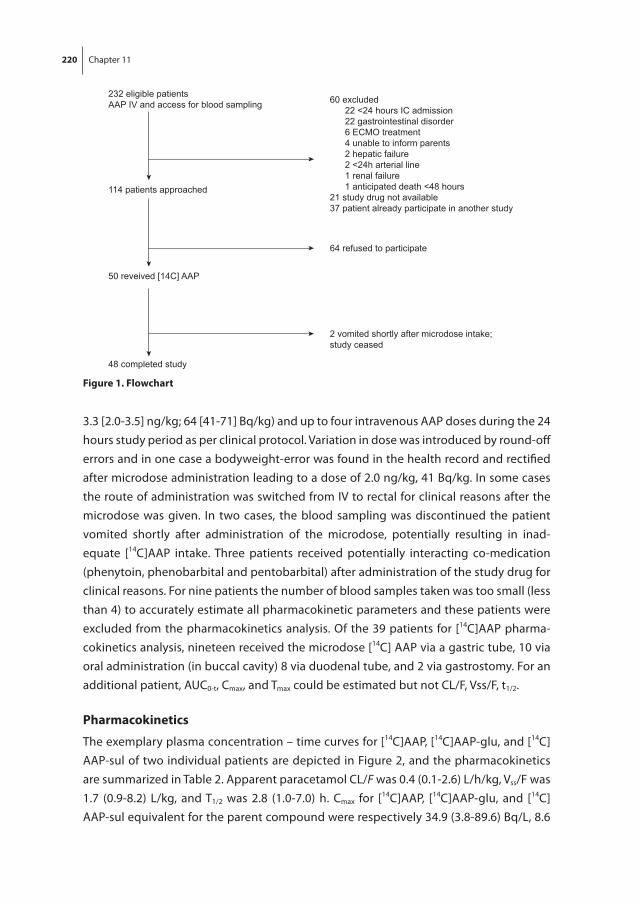
220 Chapter 11
3.3 [2.0-3.5] ng/kg; 64 [41-71] Bq/kg) and up to four intravenous AAP doses during the 24 hours study period as per clinical protocol. Variation in dose was introduced by round-off errors and in one case a bodyweight-error was found in the health record and rectified after microdose administration leading to a dose of 2.0 ng/kg, 41 Bq/kg. In some cases the route of administration was switched from IV to rectal for clinical reasons after the microdose was given. In two cases, the blood sampling was discontinued the patient vomited shortly after administration of the microdose, potentially resulting in inad-equate [14C]AAP intake. Three patients received potentially interacting co-medication (phenytoin, phenobarbital and pentobarbital) after administration of the study drug for clinical reasons. For nine patients the number of blood samples taken was too small (less than 4) to accurately estimate all pharmacokinetic parameters and these patients were excluded from the pharmacokinetics analysis. Of the 39 patients for [14C]AAP pharma-cokinetics analysis, nineteen received the microdose [14C] AAP via a gastric tube, 10 via oral administration (in buccal cavity) 8 via duodenal tube, and 2 via gastrostomy. For an additional patient, AUC0-t, Cmax, and Tmax could be estimated but not CL/F, Vss/F, t1/2.
Pharmacokinetics
The exemplary plasma concentration – time curves for [14C]AAP, [14C]AAP-glu, and [14C]AAP-sul of two individual patients are depicted in Figure 2, and the pharmacokinetics are summarized in Table 2. Apparent paracetamol CL/F was 0.4 (0.1-2.6) L/h/kg, Vss/F was 1.7 (0.9-8.2) L/kg, and T1/2 was 2.8 (1.0-7.0) h. Cmax for [14C]AAP, [14C]AAP-glu, and [14C]AAP-sul equivalent for the parent compound were respectively 34.9 (3.8-89.6) Bq/L, 8.6
232 eligible patientsAAP IV and access for blood sampling
114 patients approached
60 excluded 22 <24 hours IC admission 22 gastrointestinal disorder 6 ECMO treatment 4 unable to inform parents 2 hepatic failure 2 <24h arterial line 1 renal failure 1 anticipated death <48 hours21 study drug not available37 patient already participate in another study
50 reveived [14C] AAP
48 completed study
64 refused to participate
2 vomited shortly after microdose intake; study ceased
figure 1. flowchart

221
11
Microdosing to study ontogeny of drug metabolism: the case of [14C]paracetamol
(0.8-38.1) Bq/L, and 10.0 (1.0-41.0) Bq/L. Tmax for [14C]AAP, [14C]AAP-glu, and [14C]AAP-sul were respectively 1.0 (0.2-6.2)h, 4.1 (0.8-12.6)h, and 3.0 (0.3-12.6)h. Tmax for [14C]AAP was similar for all routes of administration, but with large variability (Table 2).
Dose-normalized (mg/kg dose) Cmax for [14C]AAP, [14C]AAP-glu, and [14C]AAP-sul were respectively 8.4 (0.9-23.0) mg/L, 3.8 (0.4-16.0) mg/L, and 4.6 (0.5-20.5) mg/L.
One outlier could be identified, but was kept in the analysis; this patient had very low plasma [14C]AAP and metabolites concentrations, even though no abnormalities were noticed during oral dose administration. The patient may have partially spit out the buccally given microdose.
table 1. Patient demographics
Age group (birth – 1 month)
Age group(1 month – 6 months)
Age group 3(6 months – 2 years)
Age group 4(2 years- 6 years)
Number 9 15 14 12
Postnatal age in weeks 0 (0-1) 23 (7-25) 35 (26-98) 197 (112 – 361)
Gestational age at birth 38.1 (34.9 – 40.7) 39.8 (33.1-42) 39.9 (30-41.7) 40 (36-40)
Weight (kg) 3 (2 – 3.5) 7 (3.5-9.2) 7.5 (6.1-12.8) 16.1 (12.9-25)
Weight (z-score) −1.12 (−2.1 - 0.1) −0.63 (−2.5-1.5) −1.3 (−2.5-0.9) −0.02 (−2.9-2.1)
Gender (M/F) 8/1 14/1 10/4 9/3
Ethnicity (Caucasian/African /Asian/Other)
5/1/2/1 11/1/1/2 12/0/0/2 11/0/0/1
Reason for admission(surgical/medical)
8/1 12/3 12/2 6/6
Mechanical ventilation at start study (yes/no)
1/8 0/15 0/14 0/12
Enteral/tube feeding (yes/no)
3/6 10/5 9/5 6/6
PELOD (study day) 11 (1-11) 10 (0-30) 6.5 (0-30) 10 (0-20)
Number of failing organs (study day)
2 (1-3) 2 (1-2) 2 (1-3) 2 (1-2)
PRISM 16 (0-31) 18 (4-41) 17 (0-34) 6 (0-44)
PIM (%) 4 (0-20) 1 (0-10) 0 (0-9) 2 (0-33)
PIM II (%) 5 (1-15) 0 (0-17) 0 (0-6) 1 (0-29)
Paracetamol dosing
[14C]AAP dose (ng) 9.8 (6.5 – 11.7) 22.8 (11.7-29.9) 23.4 (15.6-41.6) 52 (41.6-81.2)
[14C]AAP dose (Bq) 209 (125 – 228) 472 (235-639) 477 (313-890) 1035 (833-1594)
[14C]AAP dose normalized (mg/kg)(dose*5E6)/weight)
15.6 (16.3-17.3) 16.2 (16.07-17) 16.3 (10-16.7) 16.3 (16.1-16.7)
Microdose administration via (oral/gastric tube/duodenal tube/gastrostomy)
0/6/1/0 6/4/2/0 3/6/1/0 1/3/4/2
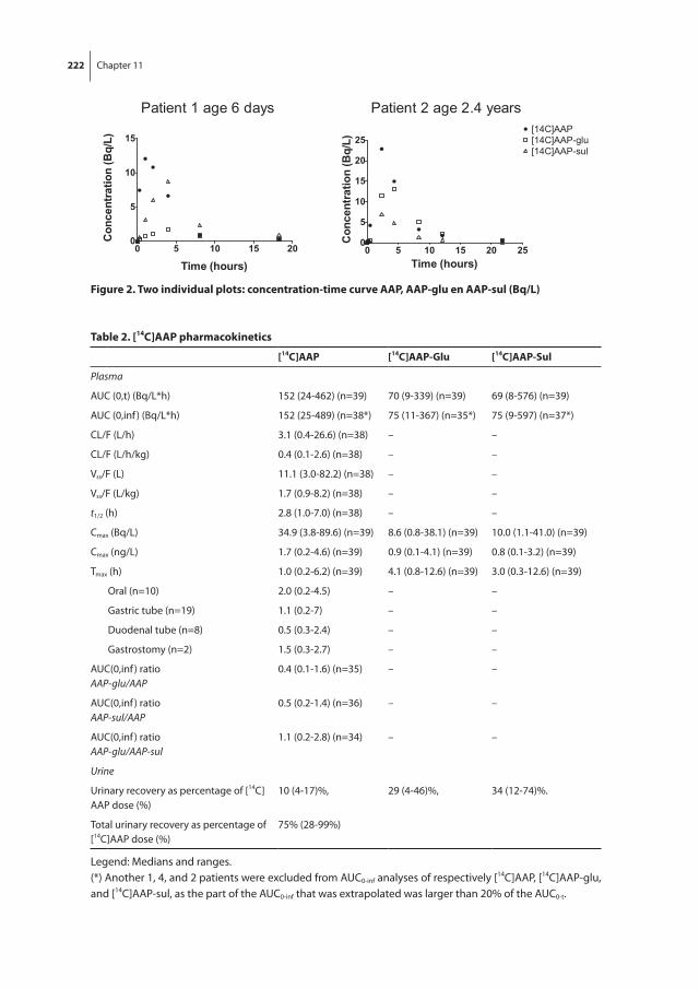
222 Chapter 11
figure 2. two individual plots: concentration-time curve AAP, AAP-glu en AAP-sul (bq/l)
table 2. [14c]AAP pharmacokinetics
[14c]AAP [14c]AAP-Glu [14c]AAP-sul
Plasma
AUC (0,t) (Bq/L*h) 152 (24-462) (n=39) 70 (9-339) (n=39) 69 (8-576) (n=39)
AUC (0,inf ) (Bq/L*h) 152 (25-489) (n=38*) 75 (11-367) (n=35*) 75 (9-597) (n=37*)
CL/F (L/h) 3.1 (0.4-26.6) (n=38) – –
CL/F (L/h/kg) 0.4 (0.1-2.6) (n=38) – –
Vss/F (L) 11.1 (3.0-82.2) (n=38) – –
Vss/F (L/kg) 1.7 (0.9-8.2) (n=38) – –
t1/2 (h) 2.8 (1.0-7.0) (n=38) – –
Cmax (Bq/L) 34.9 (3.8-89.6) (n=39) 8.6 (0.8-38.1) (n=39) 10.0 (1.1-41.0) (n=39)
Cmax (ng/L) 1.7 (0.2-4.6) (n=39) 0.9 (0.1-4.1) (n=39) 0.8 (0.1-3.2) (n=39)
Tmax (h) 1.0 (0.2-6.2) (n=39) 4.1 (0.8-12.6) (n=39) 3.0 (0.3-12.6) (n=39)
Oral (n=10) 2.0 (0.2-4.5) – –
Gastric tube (n=19) 1.1 (0.2-7) – –
Duodenal tube (n=8) 0.5 (0.3-2.4) – –
Gastrostomy (n=2) 1.5 (0.3-2.7) – –
AUC(0,inf ) ratioAAP-glu/AAP
0.4 (0.1-1.6) (n=35) – –
AUC(0,inf ) ratioAAP-sul/AAP
0.5 (0.2-1.4) (n=36) – –
AUC(0,inf ) ratioAAP-glu/AAP-sul
1.1 (0.2-2.8) (n=34) – –
Urine
Urinary recovery as percentage of [14C]AAP dose (%)
10 (4-17)%, 29 (4-46)%, 34 (12-74)%.
Total urinary recovery as percentage of [14C]AAP dose (%)
75% (28-99%)
Legend: Medians and ranges.(*) Another 1, 4, and 2 patients were excluded from AUC0-inf analyses of respectively [14C]AAP, [14C]AAP-glu, and [14C]AAP-sul, as the part of the AUC0-inf that was extrapolated was larger than 20% of the AUC0-t.
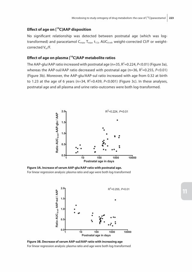
223
11
Microdosing to study ontogeny of drug metabolism: the case of [14C]paracetamol
Effect of age on [14c]AAP disposition
No significant relationship was detected between postnatal age (which was log-transformed) and paracetamol Cmax, Tmax, t1/2, AUC0-inf, weight-corrected Cl/F or weight-corrected Vss/F.
Effect of age on plasma [14c]AAP metabolite ratios
The AAP-glu/AAP ratio increased with postnatal age (n=35, R2=0.224, P<0.01) (Figure 3a), whereas the AAP-sul/AAP ratio decreased with postnatal age (n=36, R2=0.255, P<0.01) (Figure 3b). Moreover, the AAP-glu/AAP-sul ratio increased with age from 0.32 at birth to 1.23 at the age of 6 years (n=34, R2=0.439, P<0.001) (Figure 3c). In these analyses, postnatal age and all plasma and urine ratio-outcomes were both log-transformed.
figure 3A. increase of serum AAP-glu/AAP ratio with postnatal age.For linear regression analysis: plasma ratio and age were both log-transformed
figure 3b. Decrease of serum AAP-sul/AAP ratio with increasing ageFor linear regression analysis: plasma ratio and age were both log-transformed

224 Chapter 11
urinary disposition and effect of age
Urine was collected reliably for at least 12 h (20.4 h [11.8-28.5]) in 24 patients. Total median (range) urinary recovery of [14C]AAP plus [14C]AAP-glu and [14C]AAP-sul as percentage of dose was 75% (28-99%). Percentage recovery of [14C]AAP, [14C]AAP-glu, and [14C]AAP-sul was respectively 10 (4-17)%, 29 (4-46)%, and 34 (12-74)%. Percentage recovery of [14C]AAP-glu increased with postnatal age (n=24, R2=0.306, P<0.01); [14C]AAP-sul recovery decreased (n=24, R2=0.175, P<0.05); while [14C]AAP recovery remained stable. The percentage of total urinary [14C] recovery ([14C]AAP + [14C]AAP-glu + [14C]AAP-sul) did not change with age.
The U-AAP-glu/AAP ratio significantly increased with postnatal age (n=24, R2=0.456, P<0.001) (Figure 4a), whereas the U-AAP-sul/AAP ratio showed a borderline non-signif-icant decrease with age (n=24, R2=0.150, P=0.06) (Figure 4b). The U-AAP-glu/AAP-sul ratio increased linearly with postnatal age (n=24, R2=0.575, p<0.001) (Figure 4c).
figure 3c. increase of serum AAP-glu/AAP-sul ratio with postnatal ageFor linear regression analysis: plasma ratio and age were both log-transformed
figure 4A. urinary recovery u-AAP-glu /AAP ratio vs. postnatal ageFor linear regression analysis: urine ratio and age were both log-transformed

225
11
Microdosing to study ontogeny of drug metabolism: the case of [14C]paracetamol
A strong positive correlation was found between AAP-glu/AAP-sul and U-AAP-glu/AAP-sul ratio (n=19, r=0.89, P< 0.001).
Discussion
Our study shows that [14C]-microdosing allows the study of age-related changes in drug metabolism in children, using both plasma and urine as matrix. Our data present new data covering the infant age range from 0 to 6 years. Both the plasma and the urine [14C]AAP glucuronide/sulfate ratio increased approximately four-fold with age, which reflects the maturation of combined intestinal and hepatic paracetamol glucuronida-tion and sulfation.
figure 4b. urinary recovery u-AAP-sul /AAP ratio vs. postnatal ageFor linear regression analysis: urine ratio and age were both log-transformed
figure 4c. urinary recovery u-AAP-glu/AAP-sul ratio vs. postnatal ageFor linear regression analysis: urine ratio and age were both log-transformed

226 Chapter 11
Few pediatric studies have reported the paracetamol pharmacokinetics after oral administration [18, 21, 22, 28-30]. Our data, with average Cl/F 0.4 (0.1-2.6) L/h/kg and V/F 1.7 (0.9-8.2 L/kg) are largely in line with these studies. In a population pharmacokinetic study combining data of these studies, concerning more than 250 patients receiving oral and/or rectal paracetamol, the mean CL/Foral was 0.2 l/h/kg [95%CI 0.16-0.23] (14.1 L/h/70kg) and Vd/F was 1.12 l/kg [95%CI 1.02-1.21] (78.7 L/70kg) [21]. Cl/F was 0.07 L/h/kg and V/F was 1.088 L/kg in a ‘post-hoc’ NONMEM analysis of a selection of 30 neo-nates[22]. In contrast to this finding, we did not find an effect of age on weight-corrected Cl/F, but is very likely because the number of neonates in our cohort was relatively small.
In our study, both the plasma and the urine AAP-glu/AAP-sul ratio were related to age; they increased from 0.32 in neonates to 1.23 in 6-year-olds and a similar increase in urine. The AAP-glu/AAP-sul ration served as a surrogate marker for the relative contri-bution of glucuronidation and sulfation to AAP metabolism as in previous studies [18, 19, 21, 31]. Previous studies reported changes in the urinary metabolites ratio after oral paracetamol administration from 0.27 in 2-3-day-old neonates, to 0.69 in 12-month-olds, 0.81 in 2-years-olds, 0.75 in 3- to 9-year-olds, 1.61 in 12-year-olds to 1.8 in adults [18, 21, 28]. Limitations of existing studies were: only presenting neonatal data [28], or few patients less than 9 years of age [18] and a combined cohort of oral and rectal admin-istration, with urinary data on AAP and metabolites (instead of plasma) of 15 patients with a small age range (11.8±2.5 months) [21]. To the latter study an additional cohort of >200 children receiving oral and/or rectal AAP was added in the AAP-clearance analysis (without metabolites concentrations). However, as rectal paracetamol completely bypasses the small intestine and partially bypasses the liver by draining in the lower haemorrhoidal circulation its metabolism not optimally represent combined intestinal and hepatic glucuronidation and sulfation activity.
Allegaert et al. showed in 23 preterm and term newborns a significant increase of the urinary glucuronide/total drug ratio with both increasing postnatal and postconception age after IV administration of the pro-drug propacetamol [32]. In 75 children (3 neo-nates, 25 infants, 25 children, and 22 adolescents) who received repeated intravenous paracetamol, glucuronidation was the primary pathway of elimination in the older, but not the neonatal age group, where sulfation dominated [33]. The urinary AAP-glu/AAP-sul ratio collected over 4 h after the last dose, was low in neonates and higher at older age; 0.6 in neonates, 1.0 in infants, 1.4 in children, and 1.2 in adolescents. First-pass drug metabolism after oral drug administration may occur at both the intestine and the liver. Here we see similar ratio AAP-glu/AAP-sul data after oral (our study) and IV administration (Zuppa et al., Allegaert et al.), suggesting that the intestinal contribution to paracetamol metabolism and its ontogeny either equals hepatic metabolism or is negligible.

227
11
Microdosing to study ontogeny of drug metabolism: the case of [14C]paracetamol
Exploring the possibility to model the maturation half-life of AAP-glu and AAP-sul activities as depicted in figure 3, we were able to model a sigmoidal Emax curve for the ratio of AAP-glu to AAP-sul (results not shown). Nevertheless, the confidence intervals of the estimated parameters were wide, suggesting a limited amount of evidence for a sigmoidal relationship. Visual inspection of the maturation curves suggests a quite sudden surge in glucuronidation after 200 days of age, but a more gradual decrease in AAP sulfation from birth to 6 years of age.
This study has several strengths. First, we show the feasibility to perform a drug me-tabolism phenotyping study using [14C]AAP with only 0.2 millionth of the therapeutic dose. Although such a design has been used in healthy adults previously, we now show the feasibility to include a considerable number of pediatric patients in the intensive care unit with a rich blood sampling design and reliable urine collections. In addition, this study provides longitudinal data on both the plasma and the urinary [14C]AAP- me-tabolism after exclusive oral administration in a continuous cohort from 3 days to 6 years of age. These data add to our understanding on the combined intestinal and hepatic glucuronidation and sulfation activities, which is of importance given that most drugs are given orally. Another strength is the combination of urine and plasma collection, showing the feasibility of [14C]microdosing to use either matrix for drug metabolism phenotyping. The very strong correlation between plasma and urinary AAP-glu/AAP-sul ratios, as well as the similar developmental changes in plasma (our study) and urine [28] suggest that both matrices can be used to study paracetamol disposition. Plasma calculations were more reliable (n=39 complete plasma collections, versus n=24 com-plete urine collections) with less variation compared to the urinary recovery, but blood sampling are more invasive. Hence, dependent on the study population (with/without indwelling arterial/venous access), age range, or renal clearance-dependent drug, both plasma and urine could be considered.
A possible barrier for investigators to use a [14C]-microdosing study in children is the perceived risk of radiation [7]. To our own surprise, in this non-therapeutic study with radioactive labelled probe, fear of radiation was hardly a reason for parents to with-hold consent. Similar to other drug studies on our intensive care unit, almost half of all approached parents agreed to participate. Refusal of consent was rather for reasons relating to the burden of blood sampling or additional procedures. The PAMPER group, also using [14C]AAP, experienced the same inclusion rate [14].
A limitation of our study was that study patients did not receive a single microdose exclusively and therefore one could argue that it is not a ‘true’ microdosing study but a microtracer study. We choose this design with the microdose in addition to the same therapeutic drug, as this was one of the first microdosing studies in children, and it was likely more acceptable to Ethics Board and parents. We are now convinced that this is not a major issue for parents, who understood the concept of microdosing clearly and
228 Chapter 11
realized it was associated with minimal burden for the patient. Another limitation is the relatively low number of neonates meeting inclusion criteria.
In conclusion, we show the feasibility of using a [14C]drug microdose to phenotype the ontogeny of drug metabolism in children. The previously reported developmental change of paracetamol metabolism from mainly sulfation to glucuronidation was comparable, while plasma metabolite data were added on the combined intestinal and hepatic pathways in a large pediatric age range. The success of this study design is promising for future studies on the development of drug disposition.

229
11
Microdosing to study ontogeny of drug metabolism: the case of [14C]paracetamol
rEfErEncEs
[1] Mooij MG, de Koning BA, Huijsman ML, de Wildt SN. Ontogeny of oral drug absorption processes in children. Expert Opin Drug Metab Toxicol 2012 Oct; 8(10): 1293-303.
[2] Kearns GL, Abdel-Rahman SM, Alander SW, Blowey DL, Leeder JS, Kauffman RE. Developmental pharmacology--drug disposition, action, and therapy in infants and children. N Engl J Med 2003 Sep 18; 349(12): 1157-67.
[3] Ince I, Knibbe CA, Danhof M, de Wildt SN. Developmental changes in the expression and function of cytochrome P450 3A isoforms: evidence from in vitro and in vivo investigations. Clin Pharma-cokinet 2013 May; 52(5): 333-45.
[4] Krekels EH, Danhof M, Tibboel D, Knibbe CA. Ontogeny of hepatic glucuronidation; methods and results. Curr Drug Metab 2012 Jul; 13(6): 728-43.
[5] de Wildt SN, Ito S, Koren G. Challenges for drug studies in children: CYP3A phenotyping as ex-ample. Drug Discov Today 2009 Jan; 14(1-2): 6-15.
[6] Vuong LT, Blood AB, Vogel JS, Anderson ME, Goldstein B. Applications of accelerator MS in pediat-ric drug evaluation. Bioanalysis 2012 Aug; 4(15): 1871-82.
[7] Turner MA, Mooij MG, Vaes W, Windhorst AD, Hendrikse NH, Knibbe C, et al. Pediatric microdose and microtracer studies using C in Europe. Clin Pharmacol Ther 2015 Jun 10.
[8] Roth-Cline M, Nelson RM. Microdosing Studies in Children: A US Regulatory Perspective. Clin Pharmacol Ther 2015 Sep; 98(3): 232-3.
[9] Lappin G, Noveck R, Burt T. Microdosing and drug development: past, present and future. Expert Opin Drug Metab Toxicol 2013 Jul; 9(7): 817-34.
[10] Food and Drug Administration US Department of Health and Human Services Guidance for Industry Investigators and Reviewers. Exploratory IND Studies. January 2006.
[11] Agency EM. ICH Topic M3 (R2) Non-Clinical Safety Studies for the Conduct of Human Clinical Trials and Marketing Authorization for Pharmaceuticals. 2008 July 2008.
[12] Lappin G, Kuhnz W, Jochemsen R, Kneer J, Chaudhary A, Oosterhuis B, et al. Use of microdosing to predict pharmacokinetics at the therapeutic dose: experience with 5 drugs. Clin Pharmacol Ther 2006 Sep; 80(3): 203-15.
[13] Mooij MG, van Duijn E, Knibbe CA, Windhorst AD, Hendrikse NH, Vaes WH, et al. Pediatric micro-dose study of [(14)c]paracetamol to study drug metabolism using accelerated mass spectrom-etry: proof of concept. Clin Pharmacokinet 2014 Nov; 53(11): 1045-51.
[14] Garner RC, Park BK, French NS, Earnshaw C, Schipani A, Selby AM, et al. Observational infant ex-ploratory [ C]paracetamol pharmacokinetic microdose/therapeutic Dose study with accelerator mass spectrometry bioanalysis. Br J Clin Pharmacol 2015 Jan 24.
[15] Lappin G, Shishikura Y, Jochemsen R, Weaver RJ, Gesson C, Brian Houston J, et al. Comparative pharmacokinetics between a microdose and therapeutic dose for clarithromycin, sumatriptan, propafenone, paracetamol (acetaminophen), and phenobarbital in human volunteers. Eur J Pharm Sci 2011 Jun 14; 43(3): 141-50.
[16] Tozuka Z, Kusuhara H, Nozawa K, Hamabe Y, Ikushima I, Ikeda T, et al. Microdose study of 14C-acetaminophen with accelerator mass spectrometry to examine pharmacokinetics of parent drug and metabolites in healthy subjects. Clin Pharmacol Ther 2010 Dec; 88(6): 824-30.
[17] Mazaleuskaya LL, Sangkuhl K, Thorn CF, FitzGerald GA, Altman RB, Klein TE. PharmGKB summary: pathways of acetaminophen metabolism at the therapeutic versus toxic doses. Pharmacogenet Genomics 2015 Aug; 25(8): 416-26.

230 Chapter 11
[18] Miller RP, Roberts RJ, Fischer LJ. Acetaminophen elimination kinetics in neonates, children, and adults. Clin Pharmacol Ther 1976 Mar; 19(3): 284-94.
[19] Allegaert K, de Hoon J, Verbesselt R, Vanhole C, Devlieger H, Tibboel D. Intra- and interindividual variability of glucuronidation of paracetamol during repeated administration of propacetamol in neonates. Acta Paediatr 2005 Sep; 94(9): 1273-9.
[20] Wang C, Allegaert K, Tibboel D, Danhof M, van der Marel CD, Mathot RA, et al. Population pharma-cokinetics of paracetamol across the human age-range from (pre)term neonates, infants, children to adults. J Clin Pharmacol 2014 Jun; 54(6): 619-29.
[21] van der Marel CD, Anderson BJ, van Lingen RA, Holford NH, Pluim MA, Jansman FG, et al. Paracetamol and metabolite pharmacokinetics in infants. Eur J Clin Pharmacol 2003 Jul; 59(3): 243-51.
[22] Anderson BJ, Woollard GA, Holford NH. A model for size and age changes in the pharmacokinetics of paracetamol in neonates, infants and children. Br J Clin Pharmacol 2000 Aug; 50(2): 125-34.
[23] Atkinson AJ, Abernethy DR, Daniels CE, Dedrick RL, Martkey SP. Drug Absorption and Bioavail-ability. Principles of Clinical Pharmacology. 2nd ed: Academic Press; 2007. p. 37-58.
[24] van Duijn E, Sandman H, Grossouw D, Mocking JA, Coulier L, Vaes WH. Automated combustion accelerator mass spectrometry for the analysis of biomedical samples in the low attomole range. Anal Chem 2014 Aug 5; 86(15): 7635-41.
[25] Higton D, Young G, Timmerman P, Abbott R, Knutsson M, Svensson LD. European Bioanalysis Forum recommendation: scientific validation of quantification by accelerator mass spectrometry. Bioanalysis 2012 Nov; 4(22): 2669-79.
[26] Klein MV, W. H. J. Fabriek, B. Sandman, H. Mous, D. J. W. Gottdang, A. The 1 MV multi-element AMS system for biomedical applications at the Netherlands Organization for Applied Scientific Research (TNO). Nuclear Instruments and Methods in Physics Research Section B: Beam Interac-tions with Materials and Atoms 2013; 294: 14-7.
[27] Zhang Y, Huo M, Zhou J, Xie S. PKSolver: An add-in program for pharmacokinetic and pharma-codynamic data analysis in Microsoft Excel. Comput Methods Programs Biomed 2010 Sep; 99(3): 306-14.
[28] Levy G, Khanna NN, Soda DM, Tsuzuki O, Stern L. Pharmacokinetics of acetaminophen in the human neonate: formation of acetaminophen glucuronide and sulfate in relation to plasma bilirubin concentration and D-glucaric acid excretion. Pediatrics 1975 Jun; 55(6): 818-25.
[29] Anderson BJ, Holford NH, Woollard GA, Chan PL. Paracetamol plasma and cerebrospinal fluid pharmacokinetics in children. Br J Clin Pharmacol 1998 Sep; 46(3): 237-43.
[30] Anderson BJ, van Lingen RA, Hansen TG, Lin YC, Holford NH. Acetaminophen developmental pharmacokinetics in premature neonates and infants: a pooled population analysis. Anesthesiol-ogy 2002 Jun; 96(6): 1336-45.
[31] Allegaert K, Tibboel D. Comments on “shift from biliary to urinary elimination of acetaminophen-glucuronide in acetaminophen-pretreated rats”. J Pharmacol Exp Ther 2006 Feb; 316(2): 966-7.
[32] Allegaert K, Van der Marel CD, Debeer A, Pluim MA, Van Lingen RA, Vanhole C, et al. Pharmacoki-netics of single dose intravenous propacetamol in neonates: effect of gestational age. Arch Dis Child Fetal Neonatal Ed 2004 Jan; 89(1): F25-8.
[33] Zuppa AF, Hammer GB, Barrett JS, Kenney BF, Kassir N, Mouksassi S, et al. Safety and popula-tion pharmacokinetic analysis of intravenous acetaminophen in neonates, infants, children, and adolescents with pain or Fever. J Pediatr Pharmacol Ther 2011 Oct; 16(4): 246-61.


Part IVDiscussion and summary


12 G eneral discussion

General discussion 237
12
Pharmacotherapy is the key ingredient of many treatment approaches for diseases in children. Yet, the effects of pharmacotherapy change as children evolve during life; con-sequently therapy strategies need to be adjusted to growth and development. Several information gaps remain on the impact of age on the processes involved in the dis-position of drugs. More specifically, important factors governing oral bioavailability in children, including intestinal metabolism as well membrane transport, are understudied so far. The extent of oral drug use in critically ill children, the current state of knowledge on oral drug absorption in children, and the ontogeny of membrane transporters and phase II drug metabolism are the subjects presented in this thesis. The lack of pediatric data can partly be ascribed to difficulties in collecting pediatric tissue and performing pharmacokinetic studies. Using two innovative techniques primarily developed for adult studies, novel data were generated on drug metabolism and drug transport in young children.
To end with, future research perspectives, building on the data in this thesis, are presented that ultimately may have the potential to improve efficacy and safety of drug therapy in children.
orAl DruG usE
oral drug prescribing in the nicu and Picu
Oral drug absorption in critically ill children may be erratic and the use of the oral route for life-saving drugs is therefore discouraged. In the intensive care setting intravenous (IV) drugs are preferred and most patients will have an indwelling arterial or venous line. Nevertheless, for several reasons the oral route may still be preferable: i.e., appropri-ate intravenous formulations may not be available (e.g., spironolactone, bosentan); IV administration may be associated with more severe adverse events (e.g., sildenafil and hypotension, infections associated with central lines); or the site of action is the gastro-intestinal tract (e.g., antifungal prophylaxis). Moreover, polypharmacy with incompat-ible drugs through the available intravenous access ports, inability to gain any venous access and higher costs of intravenous administration may all be reason to resort to the oral route.
Very few reports are available on the use of oral drugs in the neonatal and/or pedi-atric intensive care units (ICUs). In one drug utilization study in an Indian NICU 92% of prescribed drugs over 6 months were given intravenously, of which the remainder were prescribed via other routes, including oral [1]. In contrast, we showed that up to 27% of drugs were prescribed orally to children in neonatal and pediatric intensive care units (NICU and PICU) [chapter 5]. The dissimilarity may be explained by differences in age ranges and setting (developing vs. western country).

238 Chapter 12
The potential erratic oral absorption may lead to unwanted variation in drug efficacy and safety. When looking at the 10 most frequently prescribed oral drugs, the target of probiotics, amphotericin B, and macrogol is the gut or gut flora; and therefore erratic oral intestinal absorption is less relevant. Nevertheless, when gut motility is severely impaired in critical illness, the drug may not even reach the relevant intestinal compart-ment (e.g., colon for macrogol). In contrast, for other frequently prescribed oral drugs, erratic oral absorption may be more problematic: e.g., analgesics, diuretics, vitamin K, omeprazole, and lorazepam. Moreover, data from critically ill patients are often lacking. For example, the efficacy and safety of omeprazole for the prevention of ICU related gastric bleeding have not been shown in children. Moreover, no suitable oral formula-tion is available to be given to young children or by gastric tube. And while oral diuretics for neonatal chronic lung disease seem effective, very limited data on their efficacy for treating heart failure or edema are available for older critically ill children [2]. Moreover, no licensed liquid drug formulations suitable for pediatric use are available for the oral diuretics hydrochlorothiazide and spironolactone. Also, for lorazepam no oral formula-tion is available and instead the intravenous formulation or crushed tablets are given, very erratic drug levels and sedative effect could be expected.
Additionally, in a study on the risk of intervention on prescriptions by a clinical pharmacist the oral dosage form and oral route of administration had the highest risk for interventions. This was monitored in electronic dose prescribing for four years in a tertiary children’s hospital (excluding the ICUs) [3]. An intervention was defined as any action taken on a drug prescription to correct or complete the prescription. 81% of all interventions concerned a correction of a prescription that otherwise might have had adverse clinical consequences. This result emphasizes the importance of awareness on oral drug use in the NICU and PICU.
In summary, oral drug use is frequent in critically children and exposes them to poten-tially ineffective and unsafe drug therapy.
Age-related changes in oral drug absorption processes
To better understand the impact of age on the gastrointestinal processes that govern the intestinal absorption of drugs, we reviewed the existing literature. Important age related variation was found for gastric pH. Apart from a brief peak postnatally, the gas-tric pH is about 2-3 in children of all ages. It rises postprandially due to a buffering effect of milk-based feeding [4]. In frequently fed neonates (such as extreme low birth weight infants or in case of continuous nasogastric tube feeding) the pH may therefore peak longer than in older children who eat less frequently. This questions the general idea that pH is basic in the first days to weeks of life, reported more than seventy years ago by Miller et al. [5]. Other, more recent studies report that the gastric pH declines already within a few hours after birth [6, 7]. Many other studies subsequently showed that the

General discussion 239
12
gastric pH remains low at a pH around 2 and 3 in children of all ages [4, 8-22]. Neverthe-less, large information gaps still exist on the impact of age on almost all drug absorption processes: gastric pH, gastrointestinal motility, bile salts, pancreatic function, intestinal pH, intestinal drug metabolizing enzymes (DMEs) and transporter proteins. Hence, as even in critically ill children of all ages many drugs are prescribed orally, it is important to better understand the impact of age on these processes, as well as their interplay, so as to be able to individualize dosing.
The TNO Gastro-Intestinal Tract model (TIM) is an interesting in vitro model to study the passage of a drug across the intestinal tract. TIM is a computer-controlled dynamic system that mimics the physiological conditions in stomach and intestines [23, 24]. Gas-trointestinal parameters can be adjusted and intraluminal processing of drug dosage forms can be simulated [25]. The effects of drug manipulations to enhance drug inges-tion by children can be studied using TIM (e.g., dissolving tablets in apple juice, apple sauce, crushing). In a pilot study, the feasibility to study the influence of different dosage forms on pediatric oral drug disposition was shown using the pediatric TIM model [26].
Once a drug has reached the small intestine it needs to cross the intestinal wall and pass the liver to reach the systemic circulation. Important processes involved in this step of oral drug absorption are drug metabolism and membrane transporters, which will be discussed below.
conclusion
Oral drug absorption in critically ill children may be erratic, leading to inadequate phar-macotherapy, nevertheless up to 27% of drugs are administered via oral route. To date, large information gaps exist regarding the effect of age on gastrointestinal processes involved in oral drug absorption.
MEMbrAnE trAnsPortErs
new insights on ontogeny of transporters
In vitro studies, adult pharmacogenetics and drug-drug interaction studies have shown that human membrane transporters play a clinically relevant role in drug disposition [27]. It seems not unreasonable to assume that they are also important in the develop-ing child. Moreover, as these transporters are also implicated in physiological processes, age-related variation subsequent to their role in normal growth and development can be expected. A quite recent broader interest in pediatric membrane transporters activity has resulted in a white paper on this subject from the International Transporter Work-ing Group [chapter 3]. Not surprisingly, data in fetuses and children about transporter expression and activity are very scarce.
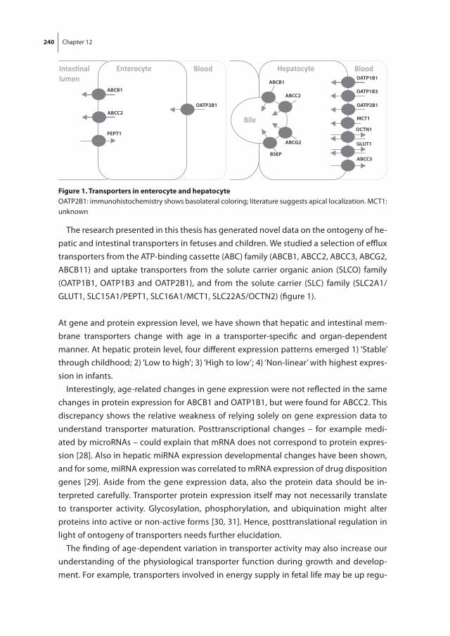
240 Chapter 12
The research presented in this thesis has generated novel data on the ontogeny of he-patic and intestinal transporters in fetuses and children. We studied a selection of efflux transporters from the ATP-binding cassette (ABC) family (ABCB1, ABCC2, ABCC3, ABCG2, ABCB11) and uptake transporters from the solute carrier organic anion (SLCO) family (OATP1B1, OATP1B3 and OATP2B1), and from the solute carrier (SLC) family (SLC2A1/GLUT1, SLC15A1/PEPT1, SLC16A1/MCT1, SLC22A5/OCTN2) (figure 1).
At gene and protein expression level, we have shown that hepatic and intestinal mem-brane transporters change with age in a transporter-specific and organ-dependent manner. At hepatic protein level, four different expression patterns emerged 1) ‘Stable’ through childhood; 2) ‘Low to high’; 3) ‘High to low’; 4) ‘Non-linear’ with highest expres-sion in infants.
Interestingly, age-related changes in gene expression were not reflected in the same changes in protein expression for ABCB1 and OATP1B1, but were found for ABCC2. This discrepancy shows the relative weakness of relying solely on gene expression data to understand transporter maturation. Posttranscriptional changes – for example medi-ated by microRNAs – could explain that mRNA does not correspond to protein expres-sion [28]. Also in hepatic miRNA expression developmental changes have been shown, and for some, miRNA expression was correlated to mRNA expression of drug disposition genes [29]. Aside from the gene expression data, also the protein data should be in-terpreted carefully. Transporter protein expression itself may not necessarily translate to transporter activity. Glycosylation, phosphorylation, and ubiquination might alter proteins into active or non-active forms [30, 31]. Hence, posttranslational regulation in light of ontogeny of transporters needs further elucidation.
The finding of age-dependent variation in transporter activity may also increase our understanding of the physiological transporter function during growth and develop-ment. For example, transporters involved in energy supply in fetal life may be up regu-
BloodEnterocyte
ABCB1
OATP2B1
lumen
ABCC2
PEPT1
BloodHepatocyte
Bile
ABCB1
ABCC2
ABCG2
BSEP
OATP1B1
OATP1B3
OATP2B1
ABCC3
OCTN1
GLUT1
MCT1
figure 1. transporters in enterocyte and hepatocyteOATP2B1: immunohistochemistry shows basolateral coloring; literature suggests apical localization. MCT1: unknown
General discussion 241
12
lated in fetuses and newborns. Glucose transporter GLUT1 and L-carnitine (molecule that shuttles long-chain fatty acids to the mitochondria for oxidation) transporter OCTN2, are probably both essential for fetal energy supply, which seems to agree with our findings of high protein expression in fetuses and newborns [32-34]. On the other hand, transporters involved in the uptake and efflux of steroids and bilirubin (i.e., OATPs) may show less variability throughout life.
The research field on membrane transporters in pediatrics remains with numerous gaps and the following research efforts are recommended:i) Expanding the pediatric age range. Even though most changes are to be expected
in the first months of life, no conclusions can be made for the entire pediatric age range due to the lack of data. More specifically, data on hepatic protein expression between the ages of 3 months and 7 years is completely lacking. Given the func-tional role of transporters to translocate hormones, of particular interest are children reaching puberty, in whom hormonal changes might influence transporter function and gender differences might appear [30].
ii) Given the role of transporters in translocating drug substrates over membranes and mediating pharmacokinetics, the ontogeny of transporters in other organs than liver and intestine should be explored. Human gene and protein expression studies sug-gest ontogeny of transporters in the kidneys (influencing renal clearance of drugs); in the brain (crossing blood-brain barrier causing effect or side-effects) lower ABCB1 protein expression was shown in fetuses or neonates [35-38]. Hence, these and other sites are of particular interest.
iii) Including clinical information in biobank tissue collections to supplement data cohorts. From drug-drug interaction studies we know that drugs may inhibit or induce trans-porter activity. Also, disease state may affect the expression and activity of membrane transporters as has been shown in children with inflammatory bowel disease [39]. Including more patient details asks for more efforts in data collection and storage. While this may provide useful additional information to explain interindividual varia-tion, its benefit should be balanced against the potential decrease in the number of samples collected because of more logistical, practical and ethical hurdles [40, 41].
iV) Expanding the selection of studied transporters. To date more than 400 transporters have been identified [42]. The disposition of drugs is most often associated with two superfamilies: the solute carrier (SLC) and ATP-binding cassette transporters (ABC) families. We have studied a selection of 11 hepatic transporters and 4 intestinal transporters. Even though this selection included the most relevant transporters for mediating drug pharmacokinetics based on adult pharmacogenetic studies or high abundance in liver or intestine, this selection is merely a start. For example, we did not study OCT1, which nevertheless seems important in the disposition of opioids

242 Chapter 12
such as morphine and tramadol [43, 44]. Other transporters as well may appear to play an important role in pediatric drug disposition.
in vivo membrane transporter research
In vivo validation is the next step in transporter research. Gene and protein expression might change during childhood, but does this also results in differences in transporter activity? The in vivo activity of transporters remains a black box in view of the ambiguous nature of substrates to multiple transporters. In vitro activity tests in cell lines or tissue cultures do not account for age-related differences. To study transporter activity in vivo, preferably the disposition of a probe drug or endogenous substrate should be exclusively altered by changes in the activity of an individual transporter. For example, changes in clearance may serve as surrogate marker of transporter activity. Due to the broad mem-brane specificity of most drugs, very few relevant probe drugs have been identified. In addition to drugs, endogenous substrates may serve as phenotyping probes, such as in the case of CYP3A4 and CYP2D6 [45, 46]. This approach avoids the ethical challenges associated with the administration of a non-therapeutic probe drug to children.
The clinical relevance of single transporters in children is mainly based on the oc-currence of unexpected drug-drug interactions. An interesting example of altered transporter activity in children is the drug-drug interaction in a study with 8 children (age 2 weeks – 3.7 years) treated for dilated cardiomyopathy with digoxin and carvedilol [47]. Digoxin renal clearance relies on ABCB1; due to the interaction with carvedilol, less digoxin was excreted in the urine, and serum digoxin concentrations increased. This ef-fect was also observed in adult patients although the digoxin clearance decreased less. This supports the hypothesis that renal ABCB1 is present from the age of 2 years and it suggests that renal digoxin clearance in young children might be more dependent on ABCB1-mediatred tubular secretion than in adults.
The use of pharmacogenetics is another way of studying transporter activity and resulting effect in pharmacokinetics. As an example, in childhood leukemia patients, multiple single nucleotide polymorphism (SNP) loss-of-function variants in the SCLO1B1 gene (encoding for OATP1B1) are related to lower methotrexate clearance leading to toxicity [48]. This points to an important role of OATP1B1 activity in methotrexate phar-macokinetics, as well as maturity of the transporter in the studied age group.
These transporter-specific, organ-dependent ontogeny profiles might already have consequences for a patient’s individual pharmacotherapy. Still it is too early to justify dose adjustments of substrates based on lesser transporter expression in the first month of life. Nevertheless we can argue that closer monitoring of drug levels by therapeutic drug monitoring (TDM) is justified for substrates of transporters with age-related changes in expression (ABCC2, ABCC3, ABCG2, GLUT1, MCT1, OCTN2, BSEP, OATP1B3), with a small therapeutic window and important safety problems.

General discussion 243
12
conclusion
Current data shows maturation profiles in human membrane transporters in an organ-specific and transporter-dependent manner. These differential patterns are likely to affect the disposition of drugs and their metabolites in an age-dependent matter. Further research should cover the entire pediatric age range, and include clinical and genetic co-variates in expression and activity studies. Clinical studies should include observational drug-drug interactions, pharmacogenetic and potential probe-based phenotyping studies.
PhAsE ii DruG MEtAbolisM
new insights on the ontogeny of phase ii drug metabolizing enzymes
Drug metabolism is the biochemical modification of pharmaceutical substances by enzymes and is one of the main determinants of variation in drug disposition. Pheno-typing studies can be used to elucidate age-related changes in in vivo drug metabolism pathways [49]. While phase I metabolism has been studied relatively well in children [50] by means of in vitro and phenotyping studies, phase II metabolism is understudied [51]. Moreover, most studies have focused on hepatic metabolism using intravenous substrates. Since drug metabolizing enzymes are also abundant in the intestine, the combined intestinal and hepatic metabolism forms a notably argument to investigate this aspect of pediatric drug metabolism.
To elucidate the combined intestinal and hepatic phase II metabolism, we chose to study the oral disposition of paracetamol [chapter 11]. Although multiple UGT and SULT enzymes metabolize paracetamol, its disposition has previously shown clear age-related changes [52-55]. However, exclusive oral paracetamol dosing has been sparsely used in these studies. Moreover, in larger pediatric population pharmacokinetic studies, mul-tiple administration routes (oral, rectal and intravenous) have been analyzed together [53, 56]. This may have obscured age-related differences in oral paracetamol disposition, since paracetamol bypasses the gut after intravenous administration, or bypasses the gut and liver after rectal dosing. Furthermore, children with bodyweights 3-7 kg and 25-50 kg) were not included in these population pharmacokinetics studies. Hence, the exact developmental pattern of paracetamol metabolism after oral administration has not been fully elucidated. To bridge these gaps we studied the oral disposition of paracetamol in children in the age range of 0 to 6 years.
To overcome the ethical objections of therapeutic drug dosing solely for research, we used a single [14C]paracetamol microdose in children already receiving intravenous paracetamol for clinical reasons. Since the oral bioavailability of paracetamol in adults is around 85-98% (Tylenol® product information), a major contribution of intestinal me-
244 Chapter 12
tabolism might not be expected. However, bioavailability paracetamol studies in young children are lacking and therefore the role of intestinal paracetamol metabolism is un-clear. In a later stage, this study design will enable to specifically study the hepatic and intestinal contributions to paracetamol metabolism, as well as the ontogeny of both.
Our most important findings agree with literature data [52-55]; we showed the con-verse developmental pattern of paracetamol with mainly sulfation in younger children to mainly glucuronidation in older children. Reported urinary metabolites ratio after oral paracetamol administration rise steady from 0.27 in 2-3 day-old-neonates, to around 1.8 in adolescents and adults [53, 57, 58]. Population pharmacokinetic studies propose a non-linear increase of glucuronidation with increasing age, reaching maturity at 3 years. We have tested a non-linear model, however, in which the confidence intervals were wide, suggesting little evidence for a reliable estimation and therefore we preferred a linear model.
At the in vitro level, the ontogeny of DMEs involved in paracetamol metabolism has been described. Hepatic UGT1A1, UGT1A6, and UGT1A9 activities were almost negligible in neonates [59, 60]. Despite the assumed absence of UGT1A1, UGT1A6 and UGT1A9, urinary AAP-glu is present in neonates, and therefore UGT2B15 is supposed to have substantial activity at birth [57, 58]. The observed maturation of paracetamol glucuronidation may therefore reflect the combined maturation pattern of these three other UGTs, from the UGT1 family, with relatively minor contribution of UGT2B15 from birth onwards.
The relative contributions of different SULT enzymes are different in fetal and pediatric liver samples, with mainly SULT1A3/4 and SULT1E1 in fetal or SULT1A1 in pediatric liver samples contributing to variability in AAP metabolism [61]. The observed slower de-crease in paracetamol sulfation may be a reflection of a gradual switch from SULT1A3/4 and SULT1E1 to SULT1A1 activity. A similar switch is seen for fetal CYP3A7 to postnatal CYP3A4 metabolism, but this switch already occurs in the first days of life [62].
Based on urinary metabolite recoveries, it has been suggested that multiple dosing of paracetamol in neonates, infants, and adults up-regulates glucuronidation [63-65]. A recent population pharmacokinetic study, however, did not show up-regulation of glucuronidation after multiple paracetamol dosing, but only the time-related increase in bodyweight reflected the developmental changes in glucuronidation [66].
Paracetamol is a widely used analgesic agent in the ICU. Although critically ill patients may show altered drug pharmacokinetics, this is of minor relevance for paracetamol clearance. Paracetamol pharmacokinetic data in 38 adult patients, from medium care and intensive care units, showed comparable median and range paracetamol clearance [67]. Moreover, hepatic clearance in healthy adult persons and patients with medium liver-failure was comparable, suggesting no effect of critical illness on paracetamol metabolism [68]. Besides, plasma protein binding of paracetamol is low, so AAP me-

General discussion 245
12
tabolism will not be affected in critical ill patients who often have low albumin levels [68]. Lastly, our outcome measure (AAP-glu/AAP-sul ratio) was the same as reported for healthy neonates and critically ill neonates (0.33) [chapter 11][64, 69]. This suggests only a minor effect of critical illness on paracetamol glucuronidation. Nevertheless it cannot be excluded that it has an effect on individual DMEs, as has been suggested for CYP3A4 [70].
A small portion of AAP is metabolized into other metabolites than glucuronide and sulfate metabolites: 3-hydroxy-paracetamol, 3-methoxy-paracetamol, 3-cysteinyl-paracetamol, paracetamol-mercaptopurate, which in their turn might be conjugated with glucuronide or sulfate (Tylenol ® product information) [71]. Some metabolites have been measured in children receiving therapeutic IV paracetamol dose, though possible age-related differences in the formation of these metabolites are unknown[71]. The highly reactive intermediate NAPQI (N-acetyl-p-benzo-quinone imine) is formed from a small portion of AAP CYP2E1 metabolism. When NAPQI is not detoxified by glutathione it can cause hepatotoxicity. It has been speculated that young children are more resistant to AAP-induced toxicity than adults [72] due to higher glutathione stores, metabolism differences, or relatively larger liver size. These metabolites have not been analyzed in this study due to financial constraints.
conclusion
Paracetamol metabolism shows age-related changes with mainly sulfation in neonates to mainly glucuronidation in older children. Urinary and plasma AAP-glu/AAP-sul AUC ratio can serve as a surrogate marker for paracetamol metabolism. Remaining gap in the ontogeny of paracetamol is the knowledge on formation of the toxic metabolites.
innoVAtiVE MEthoDs to stuDy PEDiAtric PhArMAcoloGy
Children are a vulnerable population who are relatively incapable in understanding and expressing the protection of their own interest and therefore need to be protected against harm. The criteria for pediatric research trials are rather stringent, therefore, so that children are ’therapeutic orphans’, also in view of the fact that many trials in pedi-atric populations fail. The widespread off-label or unlicensed use of drugs is a legitimate reason to generate new knowledge and improve care. The research question addressed in this thesis – to elucidate the ontogeny of membrane transporter and drug me-tabolism – cannot be studied outside the pediatric population however. Animal studies might reveal certain ontogeny, nevertheless the results cannot be simply extrapolated to humans.
246 Chapter 12
Thus we need to explore new ways of performing drug studies within the boundaries of the law and the medical ethical reviews. This thesis describes two innovative meth-ods, LC-MS/MS and microdosing to elucidate developmental changes in membrane transporters and metabolism pathways. Below, we discuss the background as well as the pros and cons of these techniques.
Microdosing
Microdosing is an interesting tool to study pharmacokinetics in children for the fol-lowing reasons: i) it has minimal burden and carries minimal risk; ii) it requires a small blood volume since plasma (or urine) samples can be as little as 30 µL due to the highly sensitive Accelerator Mass Spectrometry (AMS) technique; iii) an oral labeled microdose enables bioavailability studies when a therapeutic IV dose is administered simultane-ously, which reduces the amount of blood samples.
To date, three actual pediatric microdosing studies in both Europe and the US have been performed. Including position papers and reviews, seven reports were published on microdosing in children (table 1) [chapter 11][73-78]. Our experience using micro-dosing is predominantly positive, and rational designed microdosing studies can be of benefit to pediatric drug research.
A few critical notes are in order here. First, to predict pharmacokinetics, dose-linearity is essential. Some compounds show non-linearity in adults and are therefore unsuit-able to study pharmacokinetics in children, e.g., erythromycin and warfarin [79]. It is recommended to test dose-linearity in an adult population first if possible. From an organizational perspective, pediatric microdosing studies are challenging. Radiophar-maceutical preparation requires extra licenses for pharmaceutical and radiochemical handling, which implies that involvement of a common pharmacy alone is insufficient. Extra permissions from institutional radiation offices may be needed [76]. As for the analysis stage, AMS facilities are scarce. Though prices are probe-specific, both radio-pharmaceutical preparation and AMS analysis drive prices and in our study resulted in the main costs expenditures (Euro 500.000,-).
Regulatory issues were differently addressed in the published studies. In our case, a single microtracer was considered ‘a drug in a pharmacokinetics study’ therefore the study was considered as a clinical trial of an investigational medicinal product (CT-IMP). This was not the case in the UK and Estonia [75, 76], where it was considered ‘a well-characterized probe in a physiological experiment’ (a study involving a less well-characterized probe would be a CTIMP in the UK). In the Netherlands, the hospital’s Ra-diation Safety Officer considered the formulation radioactive and requested Radiation Safety training by the junior researcher, as well as a study-specific radiation handling protocol. In the UK and Estonia it was not considered radioactive. Body fluids and blood/urine samples were considered radioactive only in the Netherlands. The radiation dose,
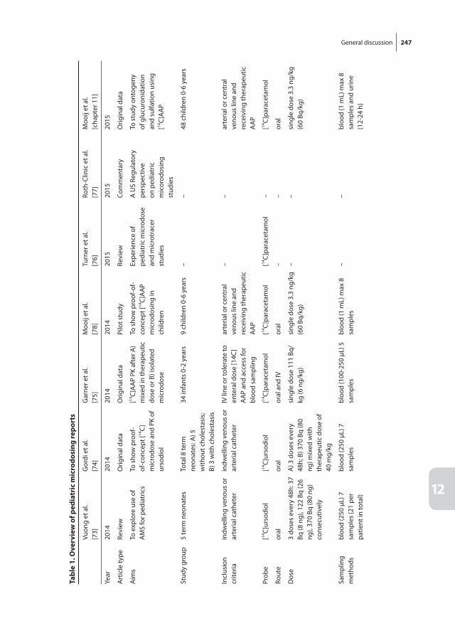
General discussion 247
12
tabl
e 1.
ove
rvie
w o
f ped
iatr
ic m
icro
dosi
ng re
port
s
Vuon
g et
al.
[73]
Gor
di e
t al.
[74]
Gar
ner e
t al.
[75]
Moo
ij et
al.
[78]
Turn
er e
t al.
[76]
Roth
-Clin
ic e
t al.
[77]
Moo
ij et
al.
[cha
pter
11]
Year
2014
2014
2014
2014
2015
2015
2015
Art
icle
type
Revi
ewO
rigin
al d
ata
Orig
inal
dat
aPi
lot s
tudy
Revi
ewCo
mm
enta
ryO
rigin
al d
ata
Aim
sTo
exp
lore
use
of
AM
S fo
r ped
iatr
ics
To s
how
pro
of-
of-c
once
pt [14
C]m
icro
dose
and
PK
of
urso
diol
[14C]
AA
P PK
aft
er A
) m
ixed
in th
erap
eutic
do
se o
r B) i
sola
ted
mic
rodo
se
To s
how
pro
of-o
f-co
ncep
t [14
C]A
AP
mic
rodo
sing
in
child
ren
Expe
rienc
e of
pe
diat
ric m
icro
dose
an
d m
icro
trac
er
stud
ies
A U
S Re
gula
tory
pe
rspe
ctiv
e on
ped
iatr
ic
mic
orod
osin
g st
udie
s
To s
tudy
ont
ogen
y of
glu
curo
nida
tion
and
sulfa
tion
usin
g [14
C]A
AP
Stud
y gr
oup
5 te
rm n
eona
tes
Tota
l 8 te
rm
neon
ates
: A) 5
w
ithou
t cho
lest
asis
; B)
3 w
ith c
hole
stas
is
34 in
fant
s 0-
2 ye
ars
9 ch
ildre
n 0-
6 ye
ars
––
48 c
hild
ren
0-6
year
s
Incl
usio
n cr
iteria
indw
ellin
g ve
nous
or
arte
rial c
athe
ter
indw
ellin
g ve
nous
or
arte
rial c
athe
ter
IV li
ne o
r tol
erat
e to
en
tera
l dos
e [1
4C]
AA
P an
d ac
cess
for
bloo
d sa
mpl
ing
arte
rial o
r cen
tral
ve
nous
line
and
re
ceiv
ing
ther
apeu
tic
AA
P
––
arte
rial o
r cen
tral
ve
nous
line
and
re
ceiv
ing
ther
apeu
tic
AA
P
Prob
e[14
C]ur
sodi
ol[14
C]ur
sodi
ol[14
C]pa
race
tam
ol[14
C]pa
race
tam
ol[14
C]pa
race
tam
ol–
[14C]
para
ceta
mol
Rout
eor
alor
alor
al a
nd IV
oral
––
oral
Dos
e3
dose
s ev
ery
48h:
37
Bq (8
ng)
, 122
Bq
(26
ng),
370
Bq (8
0 ng
) co
nsec
utiv
ely
A) 3
dos
es e
very
48
h; B
) 370
Bq
(80
ng) m
ixed
with
th
erap
eutic
dos
e of
40
mg/
kg
sing
le d
ose
111
Bq/
kg (6
ng/
kg)
sing
le d
ose
3.3
ng/k
g (6
0 Bq
/kg)
––
sing
le d
ose
3.3
ng/k
g (6
0 Bq
/kg)
Sam
plin
g m
etho
dsbl
ood
(250
µL)
7
sam
ples
(21
per
patie
nt in
tota
l)
bloo
d (2
50 µ
L) 7
sa
mpl
esbl
ood
(100
-250
µL)
5
sam
ples
bloo
d (1
mL)
max
8
sam
ples
––
bloo
d (1
mL)
max
8
sam
ples
and
urin
e (1
2-24
h)

248 Chapter 12
tabl
e 1.
ove
rvie
w o
f ped
iatr
ic m
icro
dosi
ng re
port
s (c
ontin
ued)
Vuon
g et
al.
[73]
Gor
di e
t al.
[74]
Gar
ner e
t al.
[75]
Moo
ij et
al.
[78]
Turn
er e
t al.
[76]
Roth
-Clin
ic e
t al.
[77]
Moo
ij et
al.
[cha
pter
11]
Conc
lusi
onRe
gula
tory
and
pr
actic
al fe
asib
le to
de
sign
[14C]
labe
led
com
poun
ds a
nd
AM
S st
udy
for u
se in
pe
diat
ric p
opul
atio
n
Urs
odio
l ex
hibi
ted
linea
r ph
arm
acok
inet
ics
with
no
dose
or
time-
depe
nden
cy
at th
e te
sted
dos
es.
It w
as p
ossi
ble
to d
iffer
entia
te
betw
een
endo
geno
us
and
exog
enou
s co
mpo
unds
The
PK p
aram
eter
s of
ther
apeu
tic d
ose
and
mic
rodo
se in
in
fant
s 35
-127
wee
ks
of p
ostm
enst
rual
age
w
ere
with
in a
two-
fold
rang
e.
[14C]
mic
rodo
sing
is
prac
tical
and
eth
ical
fe
asib
le in
ped
iatr
ics
Taki
ng th
e ch
alle
nges
suc
h as
do
se-li
near
ity a
nd
cost
s in
to a
ccou
nt,
mic
rodo
sing
/m
icro
trac
er tr
ials
are
be
lieve
d of
add
ed
valu
e fo
r ped
iatr
ic
drug
dev
elop
men
t.
Pedi
atric
m
icro
dosi
ng
stud
ies
mus
t firs
t be
sci
entifi
cally
ne
cess
ary.
Due
to
the
abse
nce
of a
th
erap
eutic
effe
ct,
it is
not
con
side
red
unde
r 21
CFR
50,2
5 (p
rosp
ect o
f ben
efit)
[14C]
mic
rodo
se
is fe
asib
le to
ph
enot
ype
the
onto
geny
of
drug
met
abol
ism
in
chi
ldre
n.
Dev
elop
men
tal
chan
ge o
f m
etab
olis
m fo
rm
mai
nly
sulfa
tion
to
gluc
uron
idat
ion
was
sh
own.
General discussion 249
12
however, was so low that in none of these three countries special handling was required in all. Vuong and Gordi et al. did not describe the regulatory issues concerning handling of [14C]-materials within their hospitals [73, 74].
Regarding the use of microdosing studies in pediatrics, the U.S. Food and Drug Administration (FDA) requires they address a question which is a ‘scientific necessity’ and put minimal burden on the child [77]. Correspondingly, the Dutch legislation on non-therapeutic trials allows them only when there is minimal risk and minimal burden.
The major barrier to a [14C]microdosing study in children has been the perceived risk of radiation [80, 81]. Still, doses in the published microdosing studies are low, ranging from 37 Bq (Gordi et al.) to a maximum of 1594 Bq (60 Bq/kg) (our study), and a dose of 115 Bq/kg in 0-2-year-olds (Garner et al.). This leads to very low radiation exposure of 1 µSv in neonates and less than 10 µSv in older children, which is far below the yearly background exposure of 2.5 mSv/year in the Netherlands. And the exposure from the microdose is low compared to the exposure during a European continental flight (>4 µSv), from a chest X-ray (100 µSv) or CT-scans (10 mSv). During informed consent con-versations, most parents appeared to understand the minimal risk involved. Also, fear of harmful radiation exposure was not the main reason to deny informed consent. Consent was usually refused for other reasons: e.g., the burden of additional procedures such as blood sampling [chapters 9 and 10].
Alternatively, microdosing studies without a radioactive label are an option. Very sensitive LC-tandem mass spectrometry techniques measure very low concentrations [82]. Nevertheless, this method has not reached the very low limits of detection of AMS, and a larger unlabeled compound would be necessary. Also the use of an unlabeled microdose excludes the option to administer the dose simultaneously with an (unla-beled) therapeutic dose. Thus, the use of an unlabeled microdose might be suitable to study single dose pharmacokinetics in children or the effect of pharmacogenetics on pharmacokinetics. Blake et al. used a subtherapeutic dose of dextromethorphan to study the CYP2D6 genotype’s correlation with pharmacokinetics [83].
Aside from using microdosing for phenotyping studies, other objectives can be con-sidered. i) An early phase I study to predict the pharmacokinetics of a new drug specifi-cally for the pediatric population, especially when metabolism is likely to be complex. ii) A study in which it is important to distinguish between an exogenous and an endog-enous compound, e.g., as in the ursodiol study [73]. And iii) to study pharmacokinetics in special populations such premature neonates or critically ill children, where the PK may be different than the reference population (e.g. older, healthier children).
tissue sampling and lc-Ms/Ms
Transporter protein abundance in our study was quantified with LC-MS/MS. Few studies so far have used LC-MS/MS to quantify transporter protein levels and to related results
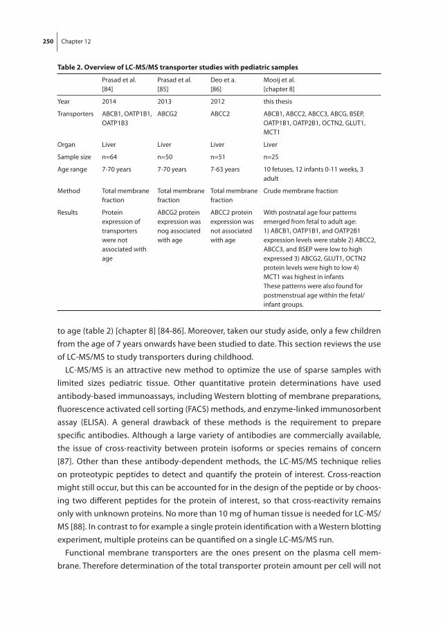
250 Chapter 12
to age (table 2) [chapter 8] [84-86]. Moreover, taken our study aside, only a few children from the age of 7 years onwards have been studied to date. This section reviews the use of LC-MS/MS to study transporters during childhood.
LC-MS/MS is an attractive new method to optimize the use of sparse samples with limited sizes pediatric tissue. Other quantitative protein determinations have used antibody-based immunoassays, including Western blotting of membrane preparations, fluorescence activated cell sorting (FACS) methods, and enzyme-linked immunosorbent assay (ELISA). A general drawback of these methods is the requirement to prepare specific antibodies. Although a large variety of antibodies are commercially available, the issue of cross-reactivity between protein isoforms or species remains of concern [87]. Other than these antibody-dependent methods, the LC-MS/MS technique relies on proteotypic peptides to detect and quantify the protein of interest. Cross-reaction might still occur, but this can be accounted for in the design of the peptide or by choos-ing two different peptides for the protein of interest, so that cross-reactivity remains only with unknown proteins. No more than 10 mg of human tissue is needed for LC-MS/MS [88]. In contrast to for example a single protein identification with a Western blotting experiment, multiple proteins can be quantified on a single LC-MS/MS run.
Functional membrane transporters are the ones present on the plasma cell mem-brane. Therefore determination of the total transporter protein amount per cell will not
table 2. overview of lc-Ms/Ms transporter studies with pediatric samples
Prasad et al.[84]
Prasad et al.[85]
Deo et a.[86]
Mooij et al.[chapter 8]
Year 2014 2013 2012 this thesis
Transporters ABCB1, OATP1B1, OATP1B3
ABCG2 ABCC2 ABCB1, ABCC2, ABCC3, ABCG, BSEP, OATP1B1, OATP2B1, OCTN2, GLUT1, MCT1
Organ Liver Liver Liver Liver
Sample size n=64 n=50 n=51 n=25
Age range 7-70 years 7-70 years 7-63 years 10 fetuses, 12 infants 0-11 weeks, 3 adult
Method Total membrane fraction
Total membrane fraction
Total membrane fraction
Crude membrane fraction
Results Protein expression of transporters were not associated with age
ABCG2 protein expression was nog associated with age
ABCC2 protein expression was not associated with age
With postnatal age four patterns emerged from fetal to adult age: 1) ABCB1, OATP1B1, and OATP2B1 expression levels were stable 2) ABCC2, ABCC3, and BSEP were low to high expressed 3) ABCG2, GLUT1, OCTN2 protein levels were high to low 4) MCT1 was highest in infantsThese patterns were also found for postmenstrual age within the fetal/infant groups.

General discussion 251
12
suffice to relate to possible activity. Therefore, the abundance of transporter proteins in tissues is quantified after subcellular fractionation resulting in enriched membrane fractions. The preparation of the most relevant matrix (e.g., plasma cell membrane) is not uniform across laboratories. This may result in inter-study variability. To incorporate absolute protein abundance information into PBPK models, losses of target proteins, need to be estimated in a standardized manner [89].
An important limitation of this method is the need for a specialized lab and equip-ment, while Western blot is a standard laboratory technique, which most labs can use easily. This likely also results in a difference in costs between the methods.
future challenges
Incorporating ontogeny data for pediatric drug dosingMany processes involved in drug disposition mature and change with age, and the ontogeny of a single process is hardly meaningful without the context of other body processes and their maturation. Modeling and simulation can be used in different ways to design dosing regimens. Examples of evidence-based complex models based on physiologically processes or population pharmacokinetic data, are respectively, physi-ologically based pharmacokinetics (PBPK) or population pharmacokinetics (popPK) models.
These models are valuable tools in designing clinical trials. Simulations from PBPK models, which incorporate available drug property and physiological information, can predict pediatric dosing [90]. Importantly, high-quality ex vivo data must be incorporated and continuous updating of physiological ontogeny data is crucial. PBPK modeling can also be used to simulate the impact of maturation of specific transporters or enzymes, preferably with actual ex vivo data on expression/activity [91]. Ultimately, these models also must be validated with real-life pharmacokinetic data, which still asks for confirma-tory pharmacokinetic sampling in clinical trials including the concept of opportunistic sampling, using doses derived from these simulations [92].
PopPK models of substrate drugs for specific drug disposition pathways have been used to describe the developmental patterns of these pathways, e.g., CYP3A4 and glomerular filtration rate (GFR) clearance [93, 94]. Validation of these models with the use of other substrate drugs has been quite challenging, as drugs may share one main disposition pathway, e.g. GFR clearance, but the contribution of membrane transport-ers may differ, thus affecting the age-related variation in pharmacokinetic parameters. Nevertheless, for individual drugs these models have proven useful to generate dosing guidelines. The final step of evidence-based pharmacotherapy is prospective validation of the popPK models. This approach has been successfully applied in case of morphine in postoperative neonates [95].

252 Chapter 12
Nowadays PBPK and popPK models are used in fields such as in clinical drug develop-ment and investigator-initiated research. It would be worthwhile, though, to make these models more accessible and user-friendly for clinicians, for example as a bed-side tool in the form of an App- or other program. After entering details of the drug in question, age, kidney and liver function, and possible co-medication a pediatrician would receive a dosing suggestion based on substrate properties and in vitro ontogeny data of, among other things, transporter or metabolism. Considering that these models are currently far from ideal, this might still be the best option for a present patient who needs a drug for which no age-appropriate dosing guidelines are available.
The question remains at what point ontogeny data, PBPK or popPK models can be used to design dosing guidelines. Although validated evidence-based dosing guidelines are preferred, often no data is available on essential drugs even though these drugs are already given to children.
conclusions
Although the ontogeny of membrane transporters and drug metabolism pathways remains to be further elucidated and clinically demonstrated, the current maturational profiles can be used in multifactorial dose-prediction models.
For the purpose of ontogeny studies, and taking its limitations into account, LC-MS/MS is suitable to quantify transporter abundance while optimally using sparse pediatric tissue samples; and [14C]-labeled microdosing studies in children are a feasible tool to phenotype the ontogeny of drug metabolism, as has been shown for paracetamol metabolism.
The ultimate goal is to provide optimal age-appropriate drug dosing in children.
General discussion 253
12
rEfErEncEs
[1] Chatterjee S, Mandal A, Lyle N, Mukherjee S, Singh AK. Drug utilization study in a neonatology unit of a tertiary care hospital in eastern India. Pharmacoepidemiol Drug Saf 2007 Oct; 16(10): 1141-5.
[2] Segar JL. Neonatal diuretic therapy: furosemide, thiazides, and spironolactone. Clin Perinatol 2012 Mar; 39(1): 209-20.
[3] Maat B, Au YS, Bollen CW, van Vught AJ, Egberts TC, Rademaker CM. Clinical pharmacy interven-tions in paediatric electronic prescriptions. Arch Dis Child 2013 Mar; 98(3): 222-7.
[4] Omari TI, Davidson GP. Multipoint measurement of intragastric pH in healthy preterm infants. Arch Dis Child Fetal Neonatal Ed 2003 Nov; 88(6): F517-20.
[5] Miller RA. Observations on the gastric acidity during the first month of life. Arch Dis Child 1941 Mar; 16(85): 22-30.
[6] Datta S, Houle GL, Fox GS. Concentration of lidocaine hydrochloride in newborn gastric fluid after elective caesarean section and vaginal delivery with epidural analgesia. Can Anaesth Soc J 1975 Jan; 22(1): 79-83.
[7] Whetstine LJ, Hulsey TC, Annibale DJ, Pittard WB. Supplemental oxygen and gastric pH in unfed preterm infants. South Med J 1995 Apr; 88(4): 458-61.
[8] Cote CJ, Goudsouzian NG, Liu LM, Dedrick DF, Szyfelbein SK. Assessment of risk factors related to the acid aspiration syndrome in pediatric patients-gastric ph and residual volume. Anesthesiol-ogy 1982 Jan; 56(1): 70-2.
[9] Goresky GV, Finley GA, Bissonnette B, Shaffer EA. Efficacy, duration, and absorption of a paediatric oral liquid preparation of ranitidine hydrochloride. Can J Anaesth 1992 Oct; 39(8): 791-8.
[10] Jahr JS, Burckart G, Smith SS, Shapiro J, Cook DR. Effects of famotidine on gastric pH and residual volume in pediatric surgery. Acta Anaesthesiol Scand 1991 Jul; 35(5): 457-60.
[11] Kelly EJ, Chatfield SL, Brownlee KG, Ng PC, Newell SJ, Dear PR, et al. The effect of intravenous ranitidine on the intragastric pH of preterm infants receiving dexamethasone. Arch Dis Child 1993 Jul; 69(1 Spec No): 37-9.
[12] Kelly EJ, Newell SJ, Brownlee KG, Primrose JN, Dear PR. Gastric acid secretion in preterm infants. Early Hum Dev 1993 Dec 31; 35(3): 215-20.
[13] Kraus G, Krishna DR, Chmelarsch D, Schmid M, Klotz U. Famotidine. Pharmacokinetic properties and suppression of acid secretion in paediatric patients following cardiac surgery. Clin Pharma-cokinet 1990 Jan; 18(1): 77-81.
[14] Maekawa N, Mikawa K, Yaku H, Nishina K, Obara H. Effects of 2-, 4- and 12-hour fasting intervals on preoperative gastric fluid pH and volume, and plasma glucose and lipid homeostasis in children. Acta Anaesthesiol Scand 1993 Nov; 37(8): 783-7.
[15] Miller BR, Tharp JA, Issacs WB. Gastric residual volume in infants and children following a 3-hour fast. J Clin Anesth 1990 Sep-Oct; 2(5): 301-5.
[16] Nishina K, Mikawa K, Maekawa N, Tamada M, Obara H. Omeprazole reduces preoperative gastric fluid acidity and volume in children. Can J Anaesth 1994 Oct; 41(10): 925-9.
[17] Oderda G, Rapa A, Chiorboli E, Ronchi B, Zavallone A, Strigini L. Measurement of postprandial changes in urine acid output to detect changes of gastric acid secretion after proton pump in-hibitors in children. Dig Dis Sci 2002 Aug; 47(8): 1843-9.
[18] Rogers IM, Drainer IK, Moore MR, Buchanan KD. Plasma gastrin in congenitial hypertrophic pyloric stenosis. A hypothesis disproved. Arch Dis Child 1975 Jun; 50(6): 467-71.
254 Chapter 12
[19] Smith LJ, Kaminsky S, D’Souza SW. Neonatal fat digestion and lingual lipase. Acta Paediatr Scand 1986 Nov; 75(6): 913-8.
[20] Splinter WM, Schaefer JD. Unlimited clear fluid ingestion two hours before surgery in children does not affect volume or pH of stomach contents. Anaesth Intensive Care 1990 Nov; 18(4): 522-6.
[21] Splinter WM, Stewart JA, Muir JG. Large volumes of apple juice preoperatively do not affect gastric pH and volume in children. Can J Anaesth 1990 Jan; 37(1): 36-9.
[22] Yildiz F, Tryba M, Kuehn K, Hausdoerfer J. Reduction of gastric acid secretion. The efficacy of pre-anaesthetic oral cimetidine in children. Anaesthesia 1984 Apr; 39(4): 314-8.
[23] Blanquet S, Zeijdner E, Beyssac E, Meunier JP, Denis S, Havenaar R, et al. A dynamic artificial gas-trointestinal system for studying the behavior of orally administered drug dosage forms under various physiological conditions. Pharm Res 2004 Apr; 21(4): 585-91.
[24] Brouwers J, Anneveld B, Goudappel GJ, Duchateau G, Annaert P, Augustijns P, et al. Food-dependent disintegration of immediate release fosamprenavir tablets: in vitro evaluation using magnetic resonance imaging and a dynamic gastrointestinal system. Eur J Pharm Biopharm 2011 Feb; 77(2): 313-9.
[25] Wilding IR, Kenyon CJ, Hooper G. Gastrointestinal spread of oral prolonged-release mesalazine microgranules (Pentasa) dosed as either tablets or sachet. Aliment Pharmacol Ther 2000 Feb; 14(2): 163-9.
[26] Havenaar R, Anneveld B, Hanff LM, de Wildt SN, de Koning BA, Mooij MG, et al. In vitro gastroin-testinal model (TIM) with predictive power, even for infants and children? Int J Pharm 2013 Nov 30; 457(1): 327-32.
[27] DeGorter MK, Xia CQ, Yang JJ, Kim RB. Drug transporters in drug efficacy and toxicity. Annu Rev Pharmacol Toxicol 2012; 52: 249-73.
[28] Koturbash I, Beland FA, Pogribny IP. Role of microRNAs in the regulation of drug metabolizing and transporting genes and the response to environmental toxicants. Expert Opin Drug Metab Toxicol 2012 May; 8(5): 597-606.
[29] Burgess KS, Philips S, Benson EA, Desta Z, Gaedigk A, Gaedigk R, et al. Age-Related Changes in MicroRNA Expression and Pharmacogenes in Human Liver. Clin Pharmacol Ther 2015 Aug; 98(2): 205-15.
[30] Thomson MMS, Krauel S, Hines RN, Scheutz EG, Meibohm B. Lack of effect of genetic variants on the age-associated prtoein expression of OATP1B1 and OATP1B3 in human pediatric liver. [Abstract for the 114th American Society for Clinical Pharmacology and Therapeutics Annual Meeting]; 2013.
[31] Klaassen CD, Aleksunes LM. Xenobiotic, bile acid, and cholesterol transporters: function and regulation. Pharmacol Rev 2010 Mar; 62(1): 1-96.
[32] Virgintino D, Robertson D, Benagiano V, Errede M, Bertossi M, Ambrosi G, et al. Immunogold cytochemistry of the blood-brain barrier glucose transporter GLUT1 and endogenous albumin in the developing human brain. Brain Res Dev Brain Res 2000 Sep 30; 123(1): 95-101.
[33] Morris ME, Felmlee MA. Overview of the proton-coupled MCT (SLC16A) family of transporters: characterization, function and role in the transport of the drug of abuse gamma-hydroxybutyric acid. AAPS J 2008 Jun; 10(2): 311-21.
[34] Lahjouji K, Elimrani I, Lafond J, Leduc L, Qureshi IA, Mitchell GA. L-Carnitine transport in human placental brush-border membranes is mediated by the sodium-dependent organic cation trans-porter OCTN2. Am J Physiol Cell Physiol 2004 Aug; 287(2): C263-9.

General discussion 255
12
[35] Miki Y, Suzuki T, Tazawa C, Blumberg B, Sasano H. Steroid and xenobiotic receptor (SXR), cyto-chrome P450 3A4 and multidrug resistance gene 1 in human adult and fetal tissues. Mol Cell Endocrinol 2005 Feb 28; 231(1-2): 75-85.
[36] Lam J, Koren G. P-glycoprotein in the Developing Human Brain: A Review of the Effects of Ontog-eny on the Safety of Opioids in Neonates. Therapeutic Drug Monitoring 2014 Dec; 36(6): 699-705.
[37] Daood M, Tsai C, Ahdab-Barmada M, Watchko JF. ABC transporter (P-gp/ABCB1, MRP1/ABCC1, BCRP/ABCG2) expression in the developing human CNS. Neuropediatrics 2008 Aug; 39(4): 211-8.
[38] Lam J, Baello S, Iqbal M, Kelly LE, Shannon PT, Chitayat D, et al. The ontogeny of P-glycoprotein in the developing human blood-brain barrier: implication for opioid toxicity in neonates. Pediatr Res 2015 Oct; 78(4): 417-21.
[39] Fakhoury M, Lecordier J, Medard Y, Peuchmaur M, Jacqz-Agrain E. Impact of inflammation on the duodenal mRNA expression of CYP3A and P-glycoprotein in children with Crohn’s disease. Inflamm Bowel Dis 2006 Aug; 12(8): 745-9.
[40] Brisson AR, Matsui D, Rieder MJ, Fraser DD. Translational research in pediatrics: tissue sampling and biobanking. Pediatrics 2012 Jan; 129(1): 153-62.
[41] Gillio-Meina C, Cepinskas G, Cecchini EL, Fraser DD. Translational research in pediatrics II: blood collection, processing, shipping, and storage. Pediatrics 2013 Apr; 131(4): 754-66.
[42] Giacomini KM, Huang SM. Transporters in drug development and clinical pharmacology. Clin Pharmacol Ther 2013 Jul; 94(1): 3-9.
[43] Tzvetkov MV, dos Santos Pereira JN, Meineke I, Saadatmand AR, Stingl JC, Brockmoller J. Mor-phine is a substrate of the organic cation transporter OCT1 and polymorphisms in OCT1 gene affect morphine pharmacokinetics after codeine administration. Biochem Pharmacol 2013 Sep 1; 86(5): 666-78.
[44] Gong L, Stamer UM, Tzvetkov MV, Altman RB, Klein TE. PharmGKB summary: tramadol pathway. Pharmacogenet Genomics 2014 Jul; 24(7): 374-80.
[45] Tay-Sontheimer J, Shireman LM, Beyer RP, Senn T, Witten D, Pearce RE, et al. Detection of an endogenous urinary biomarker associated with CYP2D6 activity using global metabolomics. Pharmacogenomics 2014 Dec; 15(16): 1947-62.
[46] Shibasaki H, Kuroiwa M, Uchikura S, Tsuboyama S, Yokokawa A, Kume M, et al. Use of endogenous cortisol 6beta-hydroxylation clearance for phenotyping in vivo CYP3A activity in women after sequential administration of an oral contraceptive (OC) containing ethinylestradiol and levonorg-estrel as weak CYP3A inhibitors. Steroids 2014 Sep; 87: 137-44.
[47] Ratnapalan S, Griffiths K, Costei AM, Benson L, Koren G. Digoxin-carvedilol interactions in chil-dren. J Pediatr 2003 May; 142(5): 572-4.
[48] Ramsey LB, Panetta JC, Smith C, Yang W, Fan Y, Winick NJ, et al. Genome-wide study of methotrex-ate clearance replicates SLCO1B1. Blood 2013 Feb 7; 121(6): 898-904.
[49] de Wildt SN, Ito S, Koren G. Challenges for drug studies in children: CYP3A phenotyping as ex-ample. Drug Discov Today 2009 Jan; 14(1-2): 6-15.
[50] Hines RN. Ontogeny of human hepatic cytochromes P450. J Biochem Mol Toxicol 2007; 21(4): 169-75.
[51] de Wildt SN. Profound changes in drug metabolism enzymes and possible effects on drug therapy in neonates and children. Expert Opin Drug Metab Toxicol 2011 Aug; 7(8): 935-48.
[52] Wang C, Allegaert K, Tibboel D, Danhof M, van der Marel CD, Mathot RA, et al. Population pharma-cokinetics of paracetamol across the human age-range from (pre)term neonates, infants, children to adults. J Clin Pharmacol 2014 Jun; 54(6): 619-29.

256 Chapter 12
[53] van der Marel CD, Anderson BJ, van Lingen RA, Holford NH, Pluim MA, Jansman FG, et al. Paracetamol and metabolite pharmacokinetics in infants. Eur J Clin Pharmacol 2003 Jul; 59(3): 243-51.
[54] Allegaert K, Anderson BJ, Naulaers G, de Hoon J, Verbesselt R, Debeer A, et al. Intravenous paracetamol (propacetamol) pharmacokinetics in term and preterm neonates. Eur J Clin Pharma-col 2004 May; 60(3): 191-7.
[55] Anderson BJ, Woollard GA, Holford NH. A model for size and age changes in the pharmacokinetics of paracetamol in neonates, infants and children. Br J Clin Pharmacol 2000 Aug; 50(2): 125-34.
[56] Anderson BJ, van Lingen RA, Hansen TG, Lin YC, Holford NH. Acetaminophen developmental pharmacokinetics in premature neonates and infants: a pooled population analysis. Anesthesiol-ogy 2002 Jun; 96(6): 1336-45.
[57] Miller RP, Roberts RJ, Fischer LJ. Acetaminophen elimination kinetics in neonates, children, and adults. Clin Pharmacol Ther 1976 Mar; 19(3): 284-94.
[58] Levy G, Khanna NN, Soda DM, Tsuzuki O, Stern L. Pharmacokinetics of acetaminophen in the human neonate: formation of acetaminophen glucuronide and sulfate in relation to plasma bilirubin concentration and D-glucaric acid excretion. Pediatrics 1975 Jun; 55(6): 818-25.
[59] Miyagi SJ, Collier AC. The development of UDP-glucuronosyltransferases 1A1 and 1A6 in the pediatric liver. Drug Metab Dispos 2011 May; 39(5): 912-9.
[60] Miyagi SJ, Milne AM, Coughtrie MW, Collier AC. Neonatal development of hepatic UGT1A9: impli-cations of pediatric pharmacokinetics. Drug Metab Dispos 2012 Jul; 40(7): 1321-7.
[61] Adjei AA, Gaedigk A, Simon SD, Weinshilboum RM, Leeder JS. Interindividual variability in acet-aminophen sulfation by human fetal liver: implications for pharmacogenetic investigations of drug-induced birth defects. Birth Defects Res A Clin Mol Teratol 2008 Mar; 82(3): 155-65.
[62] Lacroix D, Sonnier M, Moncion A, Cheron G, Cresteil T. Expression of CYP3A in the human liver--evidence that the shift between CYP3A7 and CYP3A4 occurs immediately after birth. Eur J Biochem 1997 Jul 15; 247(2): 625-34.
[63] Gelotte CK, Auiler JF, Lynch JM, Temple AR, Slattery JT. Disposition of acetaminophen at 4, 6, and 8 g/day for 3 days in healthy young adults. Clin Pharmacol Ther 2007 Jun; 81(6): 840-8.
[64] Allegaert K, de Hoon J, Verbesselt R, Vanhole C, Devlieger H, Tibboel D. Intra- and interindividual variability of glucuronidation of paracetamol during repeated administration of propacetamol in neonates. Acta Paediatr 2005 Sep; 94(9): 1273-9.
[65] Allegaert K, Verbesselt R, Rayyan M, Debeer A, de Hoon J. Urinary metabolites to assess in vivo ontogeny of hepatic drug metabolism in early neonatal life. Methods Find Exp Clin Pharmacol 2007 May; 29(4): 251-6.
[66] Krekels EH, van Ham S, Allegaert K, de Hoon J, Tibboel D, Danhof M, et al. Developmental changes rather than repeated administration drive paracetamol glucuronidation in neonates and infants. Eur J Clin Pharmacol 2015 Sep; 71(9): 1075-82.
[67] de Maat MM, Tijssen TA, Bruggemann RJ, Ponssen HH. Paracetamol for intravenous use in medium--and intensive care patients: pharmacokinetics and tolerance. Eur J Clin Pharmacol 2010 Jul; 66(7): 713-9.
[68] Gateway M. 2012-2013 [cited; Available from: http://www.micromedexsolutions.com/home/dispatch
[69] Allegaert K, Tibboel D. Comments on “shift from biliary to urinary elimination of acetaminophen-glucuronide in acetaminophen-pretreated rats”. J Pharmacol Exp Ther 2006 Feb; 316(2): 966-7.
[70] Vet NJ, de Hoog M, Tibboel D, de Wildt SN. The effect of critical illness and inflammation on midazolam therapy in children. Pediatr Crit Care Med 2012 Jan; 13(1): e48-50.
General discussion 257
12
[71] Zuppa AF, Hammer GB, Barrett JS, Kenney BF, Kassir N, Mouksassi S, et al. Safety and popula-tion pharmacokinetic analysis of intravenous acetaminophen in neonates, infants, children, and adolescents with pain or Fever. J Pediatr Pharmacol Ther 2011 Oct; 16(4): 246-61.
[72] Pineiro-Carrero VM, Pineiro EO. Liver. Pediatrics 2004 Apr; 113(4 Suppl): 1097-106. [73] Vuong LT, Blood AB, Vogel JS, Anderson ME, Goldstein B. Applications of accelerator MS in pediat-
ric drug evaluation. Bioanalysis 2012 Aug; 4(15): 1871-82. [74] Gordi T, Baillie R, Vuong le T, Abidi S, Dueker S, Vasquez H, et al. Pharmacokinetic analysis of 14C-
ursodiol in newborn infants using accelerator mass spectrometry. J Clin Pharmacol 2014 Sep; 54(9): 1031-7.
[75] Garner RC, Park BK, French NS, Earnshaw C, Schipani A, Selby AM, et al. Observational infant ex-ploratory [ C]paracetamol pharmacokinetic microdose/therapeutic Dose study with accelerator mass spectrometry bioanalysis. Br J Clin Pharmacol 2015 Jan 24.
[76] Turner MA, Mooij MG, Vaes W, Windhorst AD, Hendrikse NH, Knibbe C, et al. Pediatric microdose and microtracer studies using C in Europe. Clin Pharmacol Ther 2015 Jun 10.
[77] Roth-Cline M, Nelson RM. Microdosing Studies in Children: A US Regulatory Perspective. Clin Pharmacol Ther 2015 Sep; 98(3): 232-3.
[78] Mooij MG, van Duijn E, Knibbe CA, Windhorst AD, Hendrikse NH, Vaes WH, et al. Pediatric micro-dose study of [(14)c]paracetamol to study drug metabolism using accelerated mass spectrom-etry: proof of concept. Clin Pharmacokinet 2014 Nov; 53(11): 1045-51.
[79] Lappin G, Kuhnz W, Jochemsen R, Kneer J, Chaudhary A, Oosterhuis B, et al. Use of microdosing to predict pharmacokinetics at the therapeutic dose: experience with 5 drugs. Clin Pharmacol Ther 2006 Sep; 80(3): 203-15.
[80] Ratnapalan S, Bona N, Chandra K, Koren G. Physicians’ perceptions of teratogenic risk associated with radiography and CT during early pregnancy. AJR Am J Roentgenol 2004 May; 182(5): 1107-9.
[81] de Wildt SN, Taguchi N, Koren G. Unintended pregnancy during radiotherapy for cancer. Nat Clin Pract Oncol 2009 Mar; 6(3): 175-8.
[82] Maeda K, Ikeda Y, Fujita T, Yoshida K, Azuma Y, Haruyama Y, et al. Identification of the rate-determining process in the hepatic clearance of atorvastatin in a clinical cassette microdosing study. Clin Pharmacol Ther 2011 Oct; 90(4): 575-81.
[83] Blake MJ, Gaedigk A, Pearce RE, Bomgaars LR, Christensen ML, Stowe C, et al. Ontogeny of dextro-methorphan O- and N-demethylation in the first year of life. Clin Pharmacol Ther 2007 Apr; 81(4): 510-6.
[84] Prasad B, Evers R, Gupta A, Hop CE, Salphati L, Shukla S, et al. Interindividual variability in hepatic organic anion-transporting polypeptides and P-glycoprotein (ABCB1) protein expression: quan-tification by liquid chromatography tandem mass spectroscopy and influence of genotype, age, and sex. Drug Metab Dispos 2014 Jan; 42(1): 78-88.
[85] Prasad B, Lai Y, Lin Y, Unadkat JD. Interindividual variability in the hepatic expression of the hu-man breast cancer resistance protein (BCRP/ABCG2): effect of age, sex, and genotype. J Pharm Sci 2013 Mar; 102(3): 787-93.
[86] Deo AK, Prasad B, Balogh L, Lai Y, Unadkat JD. Interindividual variability in hepatic expression of the multidrug resistance-associated protein 2 (MRP2/ABCC2): quantification by liquid chroma-tography/tandem mass spectrometry. Drug Metab Dispos 2012 May; 40(5): 852-5.
[87] Miliotis T, Hilgendorf C. Quantification of membrane drug transporters and application in drug discovery and development. European Pharmaceutical Review 2012; 17(2): 5.
[88] Wisniewski RJ. Protocol to enrich and analyze plasma membrane proteins. Methods Mol Biol 2009; 528: 127-34.
258 Chapter 12
[89] Harwood MD, Russell MR, Neuhoff S, Warhurst G, Rostami-Hodjegan A. Lost in centrifugation: accounting for transporter protein losses in quantitative targeted absolute proteomics. Drug Metab Dispos 2014 Oct; 42(10): 1766-72.
[90] Salem F, Ogungbenro K, Vajjah P, Johnson TN, Aarons L, Rostami-Hodjegan A. Precision criteria to derive sample size when designing pediatric pharmacokinetic studies: which measure of vari-ability should be used? J Clin Pharmacol 2014 Mar; 54(3): 311-7.
[91] Mahmood I. Dosing in children: a critical review of the pharmacokinetic allometric scaling and modelling approaches in paediatric drug development and clinical settings. Clin Pharmacokinet 2014 Apr; 53(4): 327-46.
[92] Admiraal R, van Kesteren C, Boelens JJ, Bredius RG, Tibboel D, Knibbe CA. Towards evidence-based dosing regimens in children on the basis of population pharmacokinetic pharmacodynamic modelling. Arch Dis Child 2014 Mar; 99(3): 267-72.
[93] De Cock RF, Allegaert K, Brussee JM, Sherwin CM, Mulla H, de Hoog M, et al. Simultaneous phar-macokinetic modeling of gentamicin, tobramycin and vancomycin clearance from neonates to adults: towards a semi-physiological function for maturation in glomerular filtration. Pharm Res 2014 Oct; 31(10): 2643-54.
[94] Ince I, de Wildt SN, Wang C, Peeters MY, Burggraaf J, Jacqz-Aigrain E, et al. A Novel Maturation Function for Clearance of the Cytochrome P450 3A Substrate Midazolam from Preterm Neonates to Adults. Clin Pharmacokinet 2013 Jul; 52(7): 555-65.
[95] Krekels EH, Tibboel D, de Wildt SN, Ceelie I, Dahan A, van Dijk M, et al. Evidence-based morphine dosing for postoperative neonates and infants. Clin Pharmacokinet 2014 Jun; 53(6): 553-63.


13 S ummarySamenvatting


Summary 263
13
suMMAry
Appropriate pediatric dosing needs to account for developmental changes in drug absorption, distribution, metabolism and excretion. There is a lack of knowledge on the age-related changes in drug transporters and metabolism pathways. This thesis addresses the ontogeny of intestinal and hepatic membrane transporters, as well as of glucuronidation and sulfation, which comprise important building blocks of drug disposition pathways in children. As pediatric drug studies are hampered with practical and ethical challenges, this thesis addresses the use of innovative clinical methods.
Part i introduction
While oral drug absorption in critically ill children may be erratic and the use of this route therefore is discouraged for drugs. We show in chapter 2 that up to 27% of drugs are prescribed orally to children in neonatal and pediatric intensive care. This stresses the importance to study the processes involved in oral drug absorption in this population and the impact of age-related changes on these processes. The current state of knowl-edge is reviewed in chapter 3. Large information gaps exist on the impact of growth and development of almost all drug absorption processes (gastric pH, gastrointestinal motil-ity, bile salts, pancreatic function, intestinal pH, intestinal drug metabolizing enzymes, and transporter proteins). More in depth on the membrane transporters; chapters 4 and 5 present our current knowledge on developmental changes in clinically relevant transporters in intestine, liver, and kidney. In chapter 4, based on in vitro studies, dif-ferent developmental patterns for individual transporters emerge. Following on from this work, in chapter 5 drug disposition and pharmacogenetics studies in children are reviewed to add to our understanding of age-related changes in individual transporters. Overall, a striking information gap remains on the role of membrane transporters in pediatric drug therapy.
Part ii Membrane transporters
The age-related changes in gene and protein expression of intestinal and hepatic drug transporters are presented in chapters 6, 7, and 8. A selection of efflux transporters from ATP-binding cassette (ABC) family (ABCB1, ABCC2, ABCC3, ABCG2, ABCB11/BSEP) and uptake transporters from solute carrier organic anion (SLCO) family (OATP1B1, OATP1B3 and OATP2B1), and from the solute carrier (SLC) family (SLC2A1/GLUT1, SLC15A1/PEPT1, SLC16A1/MCT1, SLC22A5/OCTN2), are studied in one or more chapters.
chapter 6 shows that hepatic ABCB1, ABCC2, OATP1B1 and OATP1B3 gene expres-sions in fetuses, neonates and infants are significantly lower than in adults. Neonatal intestinal expressions of ABCB1 and ABCC2 are comparable to those in adults. Intesti-nal OATP2B1 expression in neonates is significantly higher than in adults. chapter 7

264 Chapter 13
presents slightly lower, but clinically insignificant, intestinal PEPT1 gene expression in neonates and young infants than in adolescents. Furthermore, we found comparable localization of PEPT1, ABCB1, ABCC2 and OATP2B1 proteins in the neonates and the young infants compared to adolescents. They are visualized in the apical part of the brush border (PEPT1, ABCB1, ABCC2) and intraepithelial at the basolateral enterocyte border (OATP2B1). chapter 8 focuses on hepatic transporter protein expression, using innovative liquid chromatography tandem mass spectrometry (LC-MS/MS). Four pat-terns emerge with postnatal and postmenstrual age: ABCB1, OATP1B1, and OATP2B1 expression levels are stable from fetal to adult age. ABCC2, ABCC3, and BSEP are low in fetuses and infants and higher in adults, while ABCG2, GLUT1, OCTN2 protein levels are higher early in life to low in adults. MCT1 expression levels are highest in infants compared to fetuses and adults. Interestingly, age-related changes in gene expression are not reflected in the same changes in protein expression for ABCB1 and OATP1B1. In conclusion, these studies show transporter-specific maturation, with a discrepancy between gene and protein expression for selected transporters.
Part iii Phase ii Drug metabolism
Drug metabolism is one of the main determinants of variation in drug disposition. Phe-notyping studies can be used to elucidate in vivo drug metabolism pathways in the pe-diatric population, but they are hampered by practical, ethical and scientific challenges. To overcome these challenges, an innovative approach of an open-label microdose study was performed using oral [14C]paracetamol(AAP). The results of this project are presented in chapters 9, 10, and 11. chapter 9 presents pilot data, showing that micro-dosing is a feasible and promising tool to study AAP pharmacokinetics. In collaboration with the European PAMPER consortium, chapter 10, evaluates the experiences from two microtracer and -dosing studies, and concludes that if challenges in dose-linearity and costs are overcome this technique is of added value for pediatric drug develop-ment. chapter 11 presents the final results of this phenotyping microdosing study to investigate the age-related changes in glucuronidation and sulfation. Metabolite ratio of paracetamol-glucuronide (AAP-glu) and paracetamol-sulfate (AAP-sul) was used as a surrogate marker for paracetamol metabolism. In 50 children [median age 6 months (range 3 days - 6.9 years)] both plasma and urinary AAP-glu/AAP-sul ratios significantly increased by 4-fold. These data confirm literature data on the developmental pattern of paracetamol sulfation and glucuronidation, and add data on combined intestinal and hepatic pathways in a large pediatric age range. [14C]labeled microdosing appears feasible to phenotype age-related differences in drug metabolism in children.

Summary 265
13
Part iV General discussion
chapter 12 discusses the results of our research in light of the current literature and makes recommendations for future studies on ontogeny of transporters and drug me-tabolism. We conclude thatA) major gaps remain on the ontogeny of oral drug disposition processes, in particular
of membrane transporters;b) intestinal and hepatic transporter expressions change in transporters-specific pat-
terns in an age-dependent manner;c) AAP glucuronidation and sulfation are age-related, from mainly sulfation in neo-
nates, changing to mainly glucuronidation in older children;D) [14C]labeled microdosing is a feasible and promising approach for phenotyping stud-
ies in children to elucidate age-related changes in drug disposition;E) innovative methods in drug development i.e., microdosing and LC-MS/MS protein
abundances offer excellent opportunities to overcome ethical and practical limita-tions in pediatric drug research.
This thesis shows new insights of the ontogeny of transporters, presenting one of the first transporter expressions (gene and protein) data in children. Due to the ambiguous nature of substrates for transporters no solid marker can yet be defined to study activity in vivo. Hence, at this moment we rely on expression studies. LC-MS/MS serves as an elected tool as it quantifies a palette of multiple transporters in a minimal amount of pediatric tissue. First, transporters in other organs, regulation factors (i.e., epigenetics, transcription factors) and influencing factors (i.e., disease, nutrients, substrate exposure) need to be identified. In vivo studies should aim to study amongst others; pharmacoge-netics of transporters and drug-drug interactions of transporter substrates.
Our data adds, in a meaningful way, to the information about combined intestinal and hepatic metabolism of paracetamol using an oral probe, and about the important age gap (1-2 years). Furthermore, this was reached using an inventive minimal risk and minimal burden microdosing study, which serves as a stepping stone on the path to increasing pediatric drug study options. The use of this promising technique should be expanded into other substrates for phenotyping drug metabolism (if dose linearity is present), especially in vulnerable populations (i.e., prematures, the critically ill), but also in the early evaluation of new drugs when metabolism is likely to be complex and suited for a specific pediatric population.
Data from in vitro membrane transporter studies and in vivo pharmacokinetic pheno-typing studies serve as input for complex multifactorial models (physiologically-based or population pharmacokinetic models), which predict pediatric drug dosing. The ultimate goal is to provide better age-appropriate drug dosing for children, taking into account the evolution of drug disposition pathways.
266 Chapter 13
sAMEnVAttinG
Bij kinderen wordt de juiste dosering van een geneesmiddel bepaald door de mate van absorptie, distributie, metabolisme, en excretie van dat geneesmiddel op een bepaalde leeftijd. Over de leeftijdsafhankelijke veranderingen in de transport en omzetting van geneesmiddelen is nog weinig bekend. Dit proefschrift beschrijft de ontwikkeling van membraantransporters en geneesmiddel metaboliserende enzymen in de darmen en in de lever, als belangrijke processen voor de dispositie van geneesmiddelen bij kinderen. Geneesmiddelenstudies bij kinderen kennen praktische en ethische beperkingen. In-novatieve onderzoeksmethoden zijn toegepast om de onderzoeksvraag te kunnen beantwoorden met minimale belasting en risico voor het individuele kind.
Deel i introductie
Bij kritiek zieke kinderen kan de absorptie van orale geneesmiddelen sterk wisselen, en daarom wordt de orale toediening vaak ontmoedigd. In hoofdstuk 2 laten wij zien dat tot 27% van alle geneesmiddelen in onze neonatale en pediatrische intensive care units oraal wordt gegeven. Dit benadrukt het belang van meer kennis over de absorptie van orale geneesmiddelen bij deze kritisch zieke kinderen. De huidige stand van kennis over de invloed van leeftijd op orale absorptie wordt beschreven in hoofdstuk 3. Er zijn hiaten in de kennis over het effect van groei en ontwikkeling op bijna alle processen die betrokken zijn bij de absorptie van orale geneesmiddelen (zuurgraad van de maag en de darm, snelheid van maag en darm ontlediging, galzouten, alvleesklierfunctie, omzetting en transport van geneesmiddelen in de darmwand). hoofdstukken 4 en 5 beschrijven specifiek het effect van leeftijd op membraan-transporteiwitten in de dar-men, lever en nieren. hoofdstuk 4 beschrijft resultaten van in vitro studies, hieruit blijkt dat er verschillende ontwikkelingspatronen zijn voor verschillende transportereiwitten. Vervolgens is in hoofdstuk 5 gekeken naar studies over geneesmiddeldispositie en farmacogenetica bij kinderen met de vraag of deze iets toevoegen aan ons inzicht in veranderingen bij individuele transporters. Over het geheel genomen blijft de rol van membraantransporters bij de farmacotherapie voor kinderen een grotendeels onont-gonnen terrein. De beperkte beschikbare data wijzen er wel op dat er een effect van leeftijd is op de activiteit van transporters, maar dat dit effect per transporter verschilt. Dit suggereert dat het effect en de veiligheid van geneesmiddelen, die substraat zijn voor transporters hierdoor kan variëren bij kinderen van verschillende leeftijden.
Deel ii Membraantransporters
Nieuwe data die leeftijdsafhankelijke veranderingen in de gen- en eiwitexpressie van transporters in de darmen en de lever beschrijven worden gepresenteerd in de hoofd-stukken 6, 7 en 8. Het betreft hier uitstroomtransporters van de ATP-binding cassette

Samenvatting 267
13
(ABC) familie (ABCB1, ABCC2, ABCC3, ABCG2, ABCB11/BSEP), en opnametransporters van de solute carrier organic anion (SLCO) familie (OATP1B1, OATP1B3 en OATP2B1) en van de solute carrier (SLC) familie (SLC2A1/GLUT1, SLC15A1/PEPT1, SLC16A1/MCT1, SLC22A5/OCTN2).
De genexpressie van de transporters ABCB1, ABCC2, OATP1B1 en OATP1B3 in de lever bij foetussen, pasgeborenen en kinderen blijkt lager te zijn dan bij volwassenen (hoofdstuk 6). De genexpressie van ABCB1 en ABCC2 in de darm bij pasgeborenen is vergelijkbaar met die bij volwassenen. De genexpressie van OATP2B1 in de darm bij pasgeborenen is significant hoger dan bij volwassenen. De genexpressie van PEPT1 in de darm van pasgeborenen en jonge kinderen is iets lager dan bij adolescenten, maar dit lijkt klinisch niet relevant (hoofdstuk 7). Bovendien was de lokalisatie van PEPT1, ABCB1, ABCC2, en OATP2B1 eiwitten in darmcoupes van pasgeborenen, jonge kinde-ren en adolescenten vergelijkbaar. PEPT1, ABCB1, ABCC2 eiwitten kleurden aan in het apicale gedeelte van de borstelzoom en OATP2B1 kleurde intra-epitheliaal aan aan de basolaterale zijde van de enterocyt. hoofdstuk 8 beschrijft het effect van leeftijd op de eiwitexpressie in een selectie lever transporters met behulp van ‘liquid chromatography tandem mass spectrometry’ (LC-MS/MS). Vier patronen konden worden onderscheiden: De expressie van ABCB1, OATP1B1 en OATP2B1 is stabiel van de foetale naar de vol-wassen leeftijd. De expressie van ABCC2, ABCC3 en BSEP is laag op de foetale en jonge kinderleeftijd en hoog op de volwassen leeftijd, terwijl voor ABCG2, GLUT1 en OCTN2 het omgekeerde geldt. Ten slotte, de expressie van MCT1 was het hoogst bij jonge kinderen vergeleken met foetussen en volwassenen. Een belangrijke bevinding was dat de leeftijdsafhankelijke verschillen in genexpressie van ABCB1 en OATP1B1 niet in overeenstemming zijn met de verschillen in de eiwitexpressie. We mogen concluderen dat er sprake is van een transporter-specifieke rijping, en dat een discrepantie bestaat tussen de gen- en eiwitexpressie van geselecteerde transporters.
Deel iii fase ii geneesmiddelen metabolisme
Het metabolisme is een van de belangrijke determinanten van de variatie in de geneesmiddeldispositie. Zogenaamde fenotyperingstudies zijn nuttig om de meta-bolismeroutes bij kinderen in vivo te ontrafelen, maar kennen praktische, ethische en wetenschappelijke bezwaren. Een alternatief is het innovatieve concept van ‘microdo-sing’ met een [14C] gelabelde microdosis. Hierbij wordt een zeer lage dosering, zeer zwak radioactief gelabelde dosis van een geneesmiddel gegeven, waarna de farmacokinetiek bepaald kan worden. Dit kan doordat we met de zeer gevoelige analysemethode ‘ac-celerated mass spectrometry’, de zeer lage concentraties kunnen meten in plasma en urine. Door de zeer lage dosis kan er geen effect van het geneesmiddel zelf optreden. Deze methode om farmacokinetiek te bestuderen is hierdoor minimaal belastend voor de proefpersoon en gaat gepaard met minimaal risico. De resultaten van ons micro-

268 Chapter 13
dosing onderzoek worden gepresenteerd in de hoofdstukken 9, 10 en 11. In een pilot studie bleek microdosing met [14C]paracetamol goed uitvoerbaar te zijn bij kinderen en veelbelovend ten aanzien van onderzoek naar de farmacokinetiek van paracetamol (hoofdstuk 9). In samenwerking met het Europese PAMPER consortium hebben we de ervaringen bij twee microtracer en microdosing studies geëvalueerd. We concluderen dat deze methode nuttig kan zijn voor geneesmiddelenstudies op de kinderleeftijd, mits er sprake is van een ‘dosis-lineair’ effect en de kosten beperkt kunnen worden (hoofd-stuk 10). De uiteindelijke resultaten van de fenotyperingstudie waarbij de leeftijdsaf-hankelijke veranderingen in de glucuronidering en sulfatering van paracetamol zijn onderzocht worden beschreven in hoofdstuk 11. De plasma en urine metaboliet-ratio’s van paracetamol-glucuronide (AAP-glu) en paracetamol-sulfaat (AAP-sul) dienden als surrogaatmarkers voor het metabolisme van paracetamol. Het betrof 50 kinderen met een mediane leeftijd van 6 maanden (spreiding 3 dagen – 6.9 jaar)] en zowel in plasma als in urine steeg de AAP-glu/AAP-sul ratio met de leeftijd viervoudig. Dit bevestigt het al bekende ontwikkelingspatroon van paracetamol sulfatering en glucuronidering. Het concept van [14C]gelabeld microdosing blijkt geschikt om het fenotype van geneesmid-del metabolisme te bepalen bij kinderen en daarmee naar het effect van leeftijd te kijken.
Deel iV Algemene discussie
In hoofdstuk 12 wordt het volgende geconcludeerd:A) er zijn grote hiaten in de kennis over dispositieprocessen van orale geneesmiddelen,
vooral op het gebied van membraantransporters;b) de expressie van transporters in de darmen en in de lever verandert met een toene-
mende leeftijd volgens een transporter-specifiek patroon;c) de glucuronidering en sulfatering van paracetamol zijn leeftijdsafhankelijk, dat wil
zeggen dat er bij pasgeboren vooral sprake is van sulfatering en bij oudere kinderen vooral van glucuronidering;
D) [14C]gelabeld microdosing is uitvoerbaar en veelbelovend voor fenotyperings stu-dies bij kinderen om leeftijdsafhankelijke veranderingen in geneesmiddeldispositie te ontrafelen, het bevestigt eerder onderzoek naar het effect van leeftijd op parace-tamolmetabolisme;
E) innovatieve methoden zoals microdosing en LC-MS/MS eiwitbepaling bieden uitste-kende mogelijkheden om de ethische en praktische beperkingen van geneesmid-delenonderzoek bij kinderen te verminderen.
Als gevolg van het wisselende affiniteit van substraten voor vaak meerdere transpor-ters en de noodzaak een therapeutische middel te geven, zijn er tot op heden weinig goede markers om de activiteit in vivo te onderzoeken bij kinderen. Zodoende zijn
Samenvatting 269
13
we momenteel in belangrijke mate afhankelijk van laboratorium studies. LC-MS/MS is een geschikte methode omdat deze verschillende transporters kan kwantificeren in een minimale hoeveelheid weefsel. Dit is van belang bij kinderen, omdat het vaak een uitdaging is om voldoende weefsel voor onderzoek te kunnen verzamelen. De volgende aspecten dienen in de toekomst onderzocht te worden: transporters in andere organen dan de darmen en lever, regulerende factoren (zoals epigenetica, transcriptiefactoren) en beïnvloedende factoren (zoals ziekte, voedingsstoffen, blootstelling aan substraten). In vivo studies die ons begrip over membraantransporters kunnen vergroten, zijn farma-cogenetica en geneesmiddeleninteractie-studies van betrokken transportersubstraten.
De gebruikte innovatieve microdosing methode kent een minimaal veiligheidsrisico en is weinig belastend, en is daarom veelbelovend is voor geneesmiddelenstudies bij kinderen. Deze methode kan ook worden gebruikt voor andere geneesmiddelen (mits de farmacokinetiek van het middel dosis-lineair is), met name bij kwetsbare prematuren en kritiek zieke kinderen, maar ook in de eerste fasen van de ontwikkeling van genees-middelen voor kinderen waarbij een complex metabolisme wordt verwacht.
Data van in vitro studies naar membraantransporters en in vivo studies naar de fenotypering van farmacokinetiek kunnen dienen als de bron voor complexe multifac-toriële modellen (op fysiologie of populatie gebaseerd) om doseringen voor kinderen te voorspellen. Het uiteindelijke doel is om te komen tot betere, op leeftijd gebaseerde geneesmiddeldoseringen met inachtneming van de maturatie van de betrokken farma-cokinetische processen.

Part VAppendices

List of abbreviations 273
A
list of AbbrEViAtions
14C Carbon 14
AAP Paracetamol or acetominophen
AAP-glu Paracetamol-glucuronide
AAP-sul Paracetamol-sulfate
ABC ATP-Binding Cassette
ALL Acute Lymphatic Leukemia
AMS Accelerator Mass Spectrometry
AUC Area Under the Curve
AUC0-inf Area Under the Curve from time zero to infinity
AUC0-t Area Under the Curve from time zero to last observed timepoint
BCRP (ABCG2) Breast Cancer Resistance Protein
BCS Biopharmaceutics Classification System
bDNA assay branched DNA assy
Bq Bequerel
BSEP (ABCB11) Bile Salt Export Pump
CAR Constitutive Androstane Receptor
Cav Average concentration
CES2 Carboxylesterase 2
CL/F Oral clearance
Clast Last observed concentration
C max Maximum concentration
CO2 Carbon dioxide
CT Computer Tomography
CTIMP Clinical Trial of Investigational Medicinal Product
CYP Cytochrome P450
DDI Drug-Drug Interaction
DME Drug Metabolizing Enzyme
E-1 Elastase 1
EA Elemental Analyzer
EMA European Medicines Association
EMIT Enzyme Multiplied Immunoassay Technique
FDA US Food and Drug Administration
GA Gestational Age
GERD Gastroesophageal Reflux Disease
GFR Glomerular Filtration Rate
GI Gastrointestinal
GLUT1 Glucose transporter 1
GMP Good Medicinal Practice
274 List of abbreviations
GWAS Genome-Wide Association Study
HCC Hepatocellular carcinoma
HIV Human Immunedeficiency Virus
HMG-CoA 3-hydroxy-3-methylglutaryl-coenzyme A
ICRP International Committee on Radioactivity Protection
IHC Immunohistochemistry
IRB Independent Review Boards
IV Intravenous
LC Liquid Chromatography
LLOQ Lower Limit Of Quantification
MATE (SLC47A) Multidrug And Toxin Extrusion protein
MCT1 Monocarboxylate transporter 1
MDR1 (P-gp; ABCB1) Multi-Drug Resistance 1; P-glycoprotein
miRNA microRNA
MPA Mycophenolic acid
mRNA Messenger RNA
MRP (ABCC) Multidrug Resistance-associated Protein
MRSA Methicillin-Resistant Staphylococcus Aureus
MTX Methotrexate
NAPQI N-acetyl-p-benzoquinone imine
NOAEL No Observed Adverse Effect Level
NTCP (SLC10A1) Na+-Taurocholate Cotransporting Polypeptide
OAT (SLC22A) Organic Anion Transporter
OATP (SLCO) Organic Anion Transporting Polypeptide
OCT (SLC22A) Organic Cation Transporter
OCTN (SLC22A) Organic cation/ergothioneine transporter
OCTT Orocecal Transit Time
OSTα/β Organic Solute Transporter
PAH P-aminohippurate
PAMPER Paediatric Accelerator Mass Spectrometry Evaluation Research study
PBPK Physiologically-based pharmacokinetic
PD Pharmacodynamic
PDA Photodiode array
PEDMIC PEDiatric MICrodosing
PEPT (SLC15A) Peptide transporter
PGx Pharmacogenetics; pharmacogenomics
PK Pharmacokinetics
PMA Postmenstrual age
PNA Postnatal age
popPk Population pharmacokinetic
PXR Pregnane X Receptor

List of abbreviations 275
A
QC Quality Control
qRT-PCR Quantitative Reverse Transcription Polymerase Chain Reaction
RIN RNA integrity number
RNAse Ribonuclease
RT-PCR Reverse Transcription Polymerase Chain Reaction
SD Standard Deviation
SLC Solute carrier
SLCO Solute carrier organic anion
SNP Single-Nucleotide Polymorphism
SULT Sulfotransferase
Sv Sievert
t1/2 Elimination half-life
λz Terminal elimination slope
TIM TNO gastro-Intestinal tract Model
Tlag Time delay of absorption
tmax Time at maximum concentration
UGT Uridine 5’-diphospho-glucuronosyltransferase
UK United Kingdom; Great Britain
UPLC Ultra-Performance Liquid Chromatography
UPLC-MS/MS Ultra-Performance Liquid Chromatography tandem Mass Spectrometry
URAT1 (SCL22A12) Urate transporter 1
US United States of America
VSOP Dutch collaborative patients’ organization for rare and genetic diseases; Vereniging van Samenwerkende Ouder- en Patientorganisaties voor zeldzame en genetische aandoeningen
VSS/F Apparent volume of distribution


About the author 277
A
About thE Author
Miriam Geerthe Mooij was born in Zwolle, the Netherlands, on May 30th 1986. In 2004 she received her Athenaeum degree at Agnieten College Meander in Zwolle. That year, she started her medical training at the Maastricht University in Maastricht. Miriam combined her studies with several extra-curricular activities including a board membership of the Tropical course committee of the International Federation of Medical Student Associa-tion. In her fourth year of medical school she arranged an Elective Community Medicine internship and went to Vellore (India). In her final year Miriam left for a scientific Elective internship in Paris (France) at the molecular biology department of the Université de Pierre et Marie Curie, studying the epigenetic mechanisms of the fruit fly. After returning from Paris, she spent her final internship at the pediatrics department of the Laurentius hospital in Roermond.
In July 2010, Miriam finished medical school (Master’s degree). She then commenced a residency at the pediatrics and neonatology department of the Sint Franciscus Gast-huis hospital in Rotterdam. While obtaining clinical experience in the field, she became interested in particular in clinical pharmacology, and pursued a PhD position. Under the supervision of Dr. S.N. de Wildt and Prof. dr. D. Tibboel, she started her PhD project on the age-related changes on drug transporters and metabolism in children, at the Intensive Care and department of pediatric surgery of the Erasmus MC – Sophia children’s hospi-tal. In the second year of her PhD research Miriam was awarded with a travel grant by the Dutch Society of Clinical Pharmacology and Biopharmaceutics, which enabled her to conduct a research project at the University of Western Ontario in London (Ontario, Canada), under the guidance of Dr. R.B. Kim. Alongside her PhD Miriam was selected for the TULIPS (training upcoming leaders in pediatric science) PhD curriculum and was trained as a clinical pharmacologist.
In January 2016 Miriam started her pediatrics residency at the Leiden University Medi-cal Centre – Willem Alexander Children’s hospital in Leiden.
278 List of publications
list of PublicAtions
1. Mooij MG, van Heel WJM, Eggink-Meester BJ, Oranje AP. Aplasia cutis congenita en foetus papyraceus. Case report. In Dutch Tijdschrift Kindergeneeskunde 2012;80(3).
2. Mooij MG, de Koning BA, Huijsman ML, de Wildt SN. Ontogeny of oral drug absorp-tion processes in children. Expert Opin Drug Metab Toxicol. 2012 Oct;8(10):1293-303.
3. Havenaar R, Anneveld B, Hanff LM, de Wildt SN, de Koning BA, Mooij MG, Lelievel JP, Minekus M. In vitro gastrointestinal model (TIM) with predictive power, even for infants and children? Int J Pharm. 2013 Nov 30;457(1):327-32.
4. Mooij MG, Schwarz UI, de Koning BAE, Leeder JS, Gaedigk R, Samsom JN, Spaans E, van Goudoever JB, Tibboel D, Kim RB, de Wildt SN. Ontogeny of Human Hepatic and Intestinal Transporter Gene Expression during Childhood: Age Matters. Drug Metab Dispos. 2014 Aug;42(8):1268-74.
5. Mooij MG, van Duijn E, Knibbe CA, Windhorst AD, Hendrikse NH, Vaes WH, Spaans E, Fabriek BO, Sandman H, Grossouw D, Hanff LM, Janssen PJ, Kosch BC, Tibboel D, de Wildt SN. Pediatric microdose study of [14C]paracetamol to study drug metabolism using accelerated mass spectrometry: proof of concept. Clin Pharmacokinet. 2014 Nov;53(11):1045-51.
6. Vet NJ, de Wildt SN, Verlaat CW, Knibbe CA, Mooij MG, Hop WC, van Rosmalen J, Tibboel D, de Hoog M. Daily interruption of sedation in critically ill children: study protocol for a randomized controlled trial. Trials. 2014;15:55.
7. Kleiber N, Tromp K, Mooij MG, van de Vathorst S, Tibboel D, de Wildt SN. Ethics of drug research in the pediatric intensive care unit. Paediatr Drugs. 2015 Feb;17(1):43-53.
8. Turner MA, Mooij MG, Vaes W, Windhorst AD, Hendrikse NH, Knibbe CA, Kõrgvee LT, Maruxzak W, Grynkiewicz G, Garner RC, Tibboel D, Park BK, de Wildt SN. Pediatric microdose and microtracer studies using 14C in Europe. Clin Pharmacol Ther. 2015 Sep;98(3):234-7.
9. Mooij MG, Nies AT, Knibbe CA, Schaeffeler E, Tibboel D, Schwab M, de Wildt SN. Development of Human Membrane Transporters: Drug Disposition and Pharmaco-genetics. Clin Pharmacokinet. 2015 Sep 26.
10. Brouwer K, Aleksunes LM, Brandys B, Giacoia GP, Knipp G, Lukacova V, Meibohm B, Nigam SK, Rieder M, de Wildt SN. Human Ontogeny of Drug Transporters: Review and Recommendations of the Pediatric transporter working Group. Clin Pharmacol Ther. 2015 Sep;98(3):266-87.
11. Vet NJ, de Wildt SN, Verlaat CW, Knibbe CA, Mooij MG, van Woensel JB, van Rosmalen J, Tibboel D, de Hoog M. A randomized controlled trial of daily sedation interruption in critically ill children. Intensive Care Med. 2015 Nov 24.
List of publications 279
A
12. Vet NJ, Brussee JM, de Hoog M, Mooij MG, Verlaat CW, Jerchel IS, van Schaik RH, Koch BC, Tibboel D, Knibbe CA, de Wildt SN, SKIC. Inflammation and organ failure severely affect midazolam clearance in critically ill children. Am J Respir Crit Care Med. 2016 Jan 21.
Submitted for Publication13. Mooij MG, van de Steeg E, van Rosmalen J, Windster JD, de Koning BA, Vaes WH, Tib-
boel D, Wortelboer HM, de Wildt SN. The effect of age on human hepatic membrane transporter protein expression.
14. Mooij MG, de Koning BA, Lindenbergh-Kortleve DJ, Simons-Oosterhuis I, Tibboel D, Samsom JN, de Wildt SN. Intestinal peptide transporter PEPT1 expression and tissue distribution across the pediatric age range.
15. Mooij MG, van Duijn E, Knibbe CA, Allegaert K, Windhorst AD, van Rosmalen J, Hen-drikse NH, Tibboel D, Vaes WH, de Wildt SN. Microdosing to study ontogeny of drug metabolism: the case of [14C]paracetamol.
Book chapter16. de Koning BA, Mooij MG, Johnson TN, de Wildt SN. Chapter 3: Development changes
in the processes governing oral drug absorption. Pediatric Formulations, Springer New York, 2014, ISBN 9781489980113, DOI 10.1007/978-1-4899-8011-3, editors Daniel Bar-Shalom, Klaus Rose.
280 PhD Portfolio
PhD Portfolio
Name PhD student: Miriam G. MooijErasmus MC Department: Intensive Care and Department of Pediatric SurgeryPhD period: 2011-2016Promotor: Prof. dr. D. TibboelCopromotor: Dr. S.N. de Wildt
year workload(Ects)
General academic skills
BROK (Basiscursus Regelgeving Klinisch Onderzoek) Erasmus MC 2011 1.0
Systematic literature search and Endnote (medical library) 2011 0.6
MolMed - Research management for PhD-students 2011 1.0
CPO mini-course 2011 0.3
MolMed - Course presenting skills 2012 1.0
Individual presentation training (Ricard Jutte) 2012, 2015 0.2
Biomedical English writing and communication 2012-2013 4.0
Erasmus MC trial IT - Introduction to Open Clinica 2013 0.1
research skills
MolMed - Introduction to SPSS 2011 1.0
MolMed - SNP course 2011 2.0
MolMed - Biomedical research techniques Xth edition 2011 1.5
MolMed - Genetics for dummies 2011 0.5
NIH - Principles of Clinical Pharmacology, course of webinars 2011-2012 2.0
NIH – Pediatric Clinical Pharmacology webinars – Sumner J. Yaffe Memorial 2012-2013 0.5
Radiation-course (Radiation Safety Office) 2013 0.5
CHDR - NONMEM course PKPD 2013 0.9
NIHES - Biostatistical methods 1: basic principles CCO2 2014 5.7
symposia and workshops
GRIP post graduate clinical pharmacology course 2013 0.3
Lareb – Introduction day and Adverse Events Day 2013, 2014 0.3
Young investigators day of Dutch Society of Pediatrics, Annual meetings (3x) 2012-2015 0.3
TULIPS Phd curriculum 2014-2015 1.1
Teach the Teacher course (NVKFB) 2015 0.5

PhD Portfolio 281
A
year workload(Ects)
national conferences
NVK annual meeting, TULIPS Late Breakers symposium, Veldhoven: oral presentation 2012 1.0
FIGON Dutch Medicines Days, Ede: poster presentation 2012 1.0
NVK annual meeting, Veldhoven: oral presentation 2014 1.0
Figon Dutch Medicines Day, annual meeting, Ede: oral & poster presentation 2014 1.0
Sophia research days, Rotterdam: poster presentation (first prize winner) 2014 1.0
NVK annual meeting, TULIPS Late Breakers symposium, Veldhoven: oral presentation 2015 1.0
international conferences
ASCPT annual meeting, Indianapolis, USA: poster presentation (2x) 2013 2.0
ESPNIC annual meeting, Rotterdam, Netherlands: poster presentation 2013 1.0
ESDPPP biennial meeting, Salzburg, Austria: oral & poster presentation 2013 2.0
IATDMCT annual meeting, Rotterdam, Netherlands: poster presentation 2015 1.0
ESDPPP biennial meeting, Belgrade, Servia: oral presentation 2015 1.0
ASCPT annual meeting, New Orleans, USA: poster walk presentation (2x) 2015 2.0
teaching activities
Supervising Master’s Theses, co-supervision, medical student (n=2) 2012-2015 1.0
Teaching polypharmacy 3rd year medical students 2012-2015 0.2
other
Writing F1000 evaluations (n=8) 2013 1.2
Pharmacology Days, Erasmus MC –Sophia: oral presentations (3x) 2011-2015 0.9
Pediatric Pharmacology Research Meetings (weekly): multiple oral presentations 2011-2015 2.0
Clinical Pharmacology and Pharmacogenetics meetings (weekly): multiple oral presentations
2012-2015 1.0
Fellowship Clinical Pharmacology 2012-2015


Dankwoord 283
A
DAnKwoorD
Dit proefschrift is tot stand gekomen dankzij de steun, mede- en samenwerking van velen, die ik graag wil bedanken. Ten eerste, de ouders en patiënten; in mijn ogen zijn jullie de echte helden, omdat in tijden van onzekerheid en ziekte jullie toch de kracht vinden om belangeloos mee te doen aan wetenschappelijk onderzoek. De ICK verpleeg-kundigen, altijd betrokken bij de patiënt en bereidwillig om bloed en urine af te nemen of te bellen wanneer ouders er zijn. De ICK stafleden, fellows en arts-assistenten, geïnte-resseerd en behulpzaam om een patiënt te overleggen, onderzoekers op de hoogte te brengen of een keer geduldig langs ouders te lopen wanneer zij behoefte hadden aan extra informatie.
Prof. dr. Tibboel, beste Dick, vier jaar geleden kwam ik als een onervaren onderzoeker op uw geoliede onderzoeksafdeling met uiteenlopende onderzoeken en onderzoekers. Ik bewonder uw veelzijdigheid en gedrevenheid voor de wetenschap. Telkens hield u de grote lijnen in de gaten en stelde u kritische vragen wanneer ik te gedetailleerd met een ‘transportertje’ bezig was en met onmogelijke afkortingen gooide. Dank voor uw waardevolle begeleiding.
Dr. de Wildt, beste Saskia, de afgelopen vier jaren heb jij mij met twee dingen besmet: passie voor kinderfarmacologie en wielrennen waarvoor ik je tot op de dag van vandaag dankbaar ben. Ik heb bewondering voor jouw grote toewijding en inzet in de klinische farmacologie zowel binnen het Sophia als internationaal. Ik heb ontzettend genoten van onze discussies. Of het nu ging over radioactiviteit op de afdeling, een klinisch farmacologisch consult of wat er in de bidon moest. Terwijl we vele kilometers aflegden op de wielrenfiets waren we ook op wetenschappelijk gebied een goed team. Dank voor de afgelopen jaren!
De leden van de kleine promotiecommissie, Prof. dr. van Gelder, Prof. dr. Allegaert, en Prof. dr. Leeder, wil ik hartelijk danken voor het beoordelen van mijn proefschrift. Prof. van Gelder, beste Teun, dank voor de leerzame klinische farmacologie en farmacogene-tica bijeenkomsten. Prof. Allegaert, beste Karel, dank voor uw kritische commentaren. Prof. Leeder, dear Steve, thank you for an inspiring and productive brainstorm session in Rotterdam and for your willingness to oppose me in my PhD defense. Prof. dr. de Hoog, dr. Samsom en dr. Vaes wil ik hartelijk danken voor het plaats nemen in de grote commissie.
Alle co-auteurs wil ik bedanken voor de prettige samenwerking de afgelopen jaren. In het bijzonder onze samenwerkingspartners van TNO; dr. Wouter Vaes, dr. Esther van
284 Dankwoord
Duijn, dr. Evita van de Steeg en dr. Heleen Wortelboer. Ik wil jullie danken voor de suc-cesvolle samenwerking met laagdrempelig overleg. Het was keer op keer stimulerend om met jullie over transporters of microdosing te praten. En ik hoop van harte dat de samenwerking een lang vervolg krijgt.
Dr. Richard Kim, dr. Ute Schwarz, dr. Marianne DeGorter, dr. Mathilde Leon-Ponte, thank you for your hospitality during my stay at your department in London (Ontario) and for learning me the ins and outs of RT-PCRs. Dear Sara and Al Gallien, thank you so much for taken me into your house when I was ripped off by my landlady. Staying with you and your cats made my Canadian experience unforgettable.
Beste Ko Hagoort, je bereidheid om een manuscript snel van scherp commentaar te voorzien vanuit Rotterdam of Lombok is fantastisch. Dr. Joost van Rosmalen, bedankt voor je statistische hulp. Dr. Monique van Dijk, dank voor de klinische promovendi bij-eenkomsten en met name de kerstontbijten. Annemarie Illsley, dank voor je hulp en de gezellig kletspraatjes. Chantal, Judith, Marie-Louise en Marja, bedankt voor jullie hulp. Dr. Barbara de Koning, dank voor je enthousiasme over transporters.
Joke Dunk, je steun was onmisbaar. Databases controleren, samples afdraaien of een kopje thee, je staat altijd voor elke onderzoeker klaar. Je betrokkenheid bij de patiënt, het onderzoek, en de onderzoeker is bewonderenswaardig. Hiervoor ben ik jou heel erg dankbaar. Marianne Maliepaard, dank voor je inzet in de GUTS-studie. Studenten: Mark dank voor je hulp; Jonathan, held, dank voor je enorme inzet!
Mede klinische farmacologie fellows, dank voor jullie samenwerking en farma-enthousi-asme. Dr. Lidwien Hanff, altijd geïnteresseerd en enthousiast.
De club Sophia-onderzoekers is een grote familie, met onvergetelijke borrels, diners en ski-trips. Sp-1324, deze kleine ruimte liep over van gezelligheid, Nespresso en briljante research. Nienke, je hebt mij betrokken bij de DSI-studie en mij vanaf moment één ont-zettend enthousiast gemaakt voor IC-onderzoek. De vele koffies en goede gesprekken over van alles en nog wat waren heerlijk. Binnenkort een dagje Blijdorp! Brabantse MC, altijd lachen en vastberaden. Bram, een gedisciplineerd en gepassioneerd anesthesist. Erik, jouw filosofische gedachten waren hilarisch en we hebben veel gelachen.
Sp-2430, Dorian, Kitty, Lisette, Marlous, Manuel, Raisa en Willem. Dorian, je staat altijd voor iemand klaar, een mooie eigenschap die niemand mag overzien. Kitty, je wordt een slimme anesthesist. Marlous, je ziet altijd het positieve in de mens. Willem, blij met de nieuwste technische snufjes, ik ben benieuwd naar jouw boekje! Lisette, Manuel, Navin en Raisa dank voor de gezelligheid. Andere (oud)-onderzoekers van Prof. Tibboel of de

Dankwoord 285
A
kinderchirurgie, dank voor jullie hulp en gezelligheid. Gerbrich, de vrolijke noot, dank voor je gezelligheid. Niina, thanks for your honesty, humour, and most of all your peptalk over a cheese fondue. All the best for you and your girls in Montreal.
Suzanne, lotgenoot in de laatste weken met blauwe vingers van het typen. Je positivi-teit inspireert me, jullie grote zee-avontuur gaat bijna beginnen! Michiel, bedankt voor het nodige uitblazen en afstampen op de pedalen. En daarnaast beide bedankt voor de Mont Ventoux avonturen, evenals de andere dappere fietsers.
Lieve Alexandra, Alex, mijn promotietijd had er heel anders uitgezien zonder jou! Het was één groot avontuur: succesvol een meer overroeien in Canada, vakkundig ons eten in een (te dun) boompje hangen voor de beren; wielrennen naar Zwolle of langs 11-steden in Friesland, Jut en Jul op de spinningfiets, menig skipiste onveilig maken als een Roze Konijn en Banaan. Een blik, mailtje of foto zegt dikwijls genoeg. Dank voor je adviezen over van alles, je luisterend oor, en je steun. Je discipline, je energie en devotie zijn allen onuitputtelijk. Op naar een volgend avontuur!
Lieve Joke en Alexandra, vriendinnetjes sinds een hele lange tijd, samen in Zwolle en Maastricht. Onvergetelijke momenten hebben wij meegemaakt en jullie zijn mij onge-looflijk dierbaar. Lieve Rob, het begon allemaal met de mosselpan, en sindsdien maatjes voor het leven, dank dat je erbij bent. Timon, Franse marktjes afstruinen als de beste! Inge, dank voor het prachtige omslag. Je mag er trots op zijn! Ellen, Char en Laura, SFG-maatjes. Fleur, ik kom bij jou helemaal tot rust in het mooie Mestreech. Iris, zo trots op mijn ‘kleine zusje’, we blijven onafscheidelijk.
SSA, lief en leed samen delen, de heerlijke weekendjes waar geen moment stiltes val-len, en de uitgebreide diners zijn onbetaalbaar. Lieve Jolien, je vrolijkheid zal ik nooit vergeten. Verbandje voor altijd!
Lieve Kapjes en Keizers, Bertha en Bennie, dank voor jullie steun en interesse in alles. Lieve Anne-Greet, wat is het fijn om jou in de buurt te hebben. Arjan, nooit gedacht dat toen ik jouw promotie bijwoonde, ik er zelf een keer zou staan.. Gelukkig is het niet zo’n moeilijk onderwerp als de Japanse economie. Arnold, Riny, Kathelijne en Charlotte, dank voor de dierbare momenten wanneer het gaat ‘sneeuwen’.
Mijn paranimfen, Sanne en Irene, wat ben ik blij dat jullie vandaag naast mij staan. Sanne, India was onvergetelijk en het begin van een hechte, dierbare vriendschap. Ik bewonder je moed en open kijk in de wereld. Irene, op de eerste dag van de geneeskunde studie heb ik je ontmoet; goudeerlijk, vrolijk en behulpzaam. St Petersburg en All I want for Christmas zeggen genoeg!

286 Dankwoord
Lieve Hanneke, ik ben harstikke trots op jou als mijn zus. De dierenarts die stoer in de Ierse heuvels loopt. Je bent er altijd voor me. Lieve pap, ik kan je onmogelijk bedanken voor je onvoorwaardelijke steun en liefde. Je hebt mij nooit tegengehouden in mijn keuzes en je hebt altijd alles voor Hanneke en mij over. Mama en jij hebben ons geleerd altijd trouw te blijven aan onszelf.
James, everything is fine are the magic words, I am so happy with you in my life!

