recent development of drug delivery systems through
TRANSCRIPT
�����������������
Citation: Ma, Z.; Li, B.; Peng, J.; Gao,
D. Recent Development of Drug
Delivery Systems through
Microfluidics: From Synthesis to
Evaluation. Pharmaceutics 2022, 14,
434. https://doi.org/10.3390/
pharmaceutics14020434
Academic Editors: Yusuke Sato and
Ruggero Bettini
Received: 10 January 2022
Accepted: 2 February 2022
Published: 17 February 2022
Publisher’s Note: MDPI stays neutral
with regard to jurisdictional claims in
published maps and institutional affil-
iations.
Copyright: © 2022 by the authors.
Licensee MDPI, Basel, Switzerland.
This article is an open access article
distributed under the terms and
conditions of the Creative Commons
Attribution (CC BY) license (https://
creativecommons.org/licenses/by/
4.0/).
pharmaceutics
Review
Recent Development of Drug Delivery Systems throughMicrofluidics: From Synthesis to EvaluationZhiyuan Ma , Baicheng Li , Jie Peng and Dan Gao *
State Key Laboratory of Chemical Oncogenomics, Guangdong Provincial Key Laboratory of Chemical Biology,Tsinghua Shenzhen International Graduate School, Shenzhen 518055, China;[email protected] (Z.M.); [email protected] (B.L.); [email protected] (J.P.)* Correspondence: [email protected]
Abstract: Conventional drug administration usually faces the problems of degradation and rapidexcretion when crossing many biological barriers, leading to only a small amount of drugs arrivingat pathological sites. Therapeutic drugs delivered by drug delivery systems to the target sites in acontrolled manner greatly enhance drug efficacy, bioavailability, and pharmacokinetics with minimalside effects. Due to the distinct advantages of microfluidic techniques, microfluidic setups provide apowerful tool for controlled synthesis of drug delivery systems, precisely controlled drug release, andreal-time observation of drug delivery to the desired location at the desired rate. In this review, wepresent an overview of recent advances in the preparation of nano drug delivery systems and carrier-free drug delivery microfluidic systems, as well as the construction of in vitro models on-a-chip fordrug efficiency evaluation of drug delivery systems. We firstly introduce the synthesis of nano drugdelivery systems, including liposomes, polymers, and inorganic compounds, followed by detaileddescriptions of the carrier-free drug delivery system, including micro-reservoir and microneedledrug delivery systems. Finally, we discuss in vitro models developed on microfluidic devices for theevaluation of drug delivery systems, such as the blood–brain barrier model, vascular model, smallintestine model, and so on. The opportunities and challenges of the applications of microfluidicplatforms in drug delivery systems, as well as their clinical applications, are also discussed.
Keywords: microfluidic; drug delivery system; carrier-free; in vitro model; micro-reservoir; microneedles
1. Introduction
Many therapeutic drugs might face problems such as low biodistribution, limitedsolubility, poor absorption, and drug aggregation. Drug delivery systems (DDSs) aiming totransport therapeutic drugs to desired areas, including tissues, organs, cells, subcellularorgans, and so on, are a usual approach to improving pharmacological activity [1]. Recently,various drug carriers have been developed for transporting drugs to the target site andprotecting them from improper degradation, which ensures maximum drug efficacy andreduces side effects [2]. However, the development of DDSs is an arduous, multi-stepprocess involving mass production, chemical characterization, toxicity testing, and clinicaltrials, which prevents rapid application from the lab to the clinic. Furthermore, the existenceof internal barriers in our body, such as the mucosal diffusion barrier and the cellularpermeability barrier, may preclude the efficiency of conventional drug delivery routes.Conventional drug delivery methods resort to different routes of administration such ashypodermic injections, oral administration, and inhalation to enter our body, but theystill show a weak ability to help some pharmaceutical molecules traverse barriers andcontrol their release to the desired location [3]. Fortunately, emerging nanotechnologieshold significant promise in developing the next generation of DDSs, aiming to proposemore powerful and self-regulated delivery tools [3].
Known as a “lab on a chip” (LOC), microfluidics technology represents a rapidly grow-ing versatile technology for manipulating nanoliter amounts of fluids within microscale
Pharmaceutics 2022, 14, 434. https://doi.org/10.3390/pharmaceutics14020434 https://www.mdpi.com/journal/pharmaceutics
Pharmaceutics 2022, 14, 434 2 of 22
channels. It has emerged in recent years as a distinct new area of research, especiallyin chemistry, medicine, and physical sciences. With its unprecedented advantage in theprecise control of fluid, it not only can improve the fabrication efficiency and quality of drugdelivery, but also shows great potential for the predictive power of preclinical drug carriertesting through biomimetic microfluidic platforms [4]. Compared to the conventional meth-ods, the microfluidics platform enables continuous, scalable, and reproducible productionand improves the uniformity, yield, and batch-to-batch reproducibility of nanoparticles dueto its miniaturization and automation [5]. To date, many microfluidics platforms [6,7] havebeen proposed for integrating multiple processes and functions, varying from synthesisto testing. Studies on microneedle-based systems also show great potential for futureapplications [8,9]. Although the fantastic manipulating capability enables the mass produc-tion of emulsion templates for synthesizing microparticles, the issues of industrial-scaleproduction using microfluidics platforms are still under consideration. Several bouts ofresearch have shown great effort put towards sizing up the volume to milliliter scales andfurther improving the throughput by combining multiple reactions in parallel [10–12].
Owing to the miniaturization of the fluidic environment, microfluidics has brought ahighly controllable, reproducible, and scalable fabrication platform to the production ofdrug carriers. Among the presently developed drug carriers, nanoparticles have shownexcellent properties of improving the therapeutic index, reducing side effects, and enhanc-ing uptake and penetration [13,14]. The development of microfabrication technologies hasoffered capabilities to produce nanoparticles in a controllable and reproducible manner,including flow focusing, template assembly, and droplet technology. The physicochemicalproperties such as size, shape, and composition of nanoparticles can be precisely controlledat a larger dynamic range [15], allowing for an increase in drug transport efficacy, releaseprofile, and elimination during treatment. Furthermore, high-throughput fabrication isrealizable by parallelization control and reproducible scale-up production. Apart from thefabrication of nanocarriers for drugs, carrier-free DDSs have also been widely used as adirect drug delivery method. By storing the drugs directly in the microfluidic chip, it canfurther improve efficiency and combat the problems that carrier-based delivery systemshave not covered [16]. For example, carrier-free DDSs do not need any redundant synthesissteps, and they can ensure that drugs release precisely into target sites [17]. Without chang-ing the dose or shape of the drugs, the system can not only achieve the zero-order releaseof the drug, but also lead to a controllable and reproducible release profile [2]. Furthermore,carrier-free DDSs can minimize side effects.
In addition to the advantages with respect to the establishment of controlled DDSs, thegreat potential of microfluidics for mimicry of the complex biological environment in vitroprovides a powerful tool for the validation and further assessment of the biocompatibilityand efficacy of drug delivery in a biological context. Several recent reviews have summa-rized microfluidic-based cell culture models to engineer micro-sized human tissues andorgans. These models can help accelerate drug development by resolving the discrepanciesin animal models in certain aspects. Besides, they can bring great convenience in termsof the observation of the drug delivery process through real-time imaging and in vitromicroscopic observation technology.
In this review, we first highlight the application of microfluidics technology in the es-tablishment of a variety of emerging DDSs over the past five years, including the fabricationof nanoparticle-based systems and carrier-free systems, and then summarize biomimeticin vitro models on microfluidic chips to assess DDSs. Overall, recent innovations andadvances in microfluidic-based systems are expected to accelerate the transition of newDDSs to clinical evaluation.
2. Synthesis of Drug Delivery Carriers on Microfluid Chips
Due to the excellent ability to manipulate nanoliter flows, microfluidics has been exten-sively applied in the fabrication of nanoparticles as well as the preparation of nanoparticle-based DDSs. Generally, the nanocarriers include lipid-based nanoparticles, polymeric
Pharmaceutics 2022, 14, 434 3 of 22
nanoparticles, and inorganic nanoparticles. A big challenge for conventional nanoparticlesystems is that it is hard to control the homogeneity of the particle size and shape. Fur-thermore, the safety and biocompatibility of the nanomaterials themselves are extremelyimportant factors that need to be focused on [14]. Recent research in microfluidic technolo-gies has shown great potential in nanoparticles’ synthesis, characterization, and assessmentfor drug delivery application [15]. They can provide a controllable and reproducible meansfor nanoparticle production, and the sizes, shapes, and surface compositions can be pre-cisely adjusted to meet the needs of different kinds of drugs. Several platforms have beenput forward for nanoparticle synthesis, which can be divided into single-phase systems andmultiphase systems [18]. In single-phase systems, a continuous laminar flow of fluid pro-vides a homogenous environment for diffusion and fast mixing and stirring, ensuring thenanoparticles’ nucleation and growth. As for the multiphase system, the discrete segmentscaused by immiscible fluids will act as individual reaction chambers where mixing andsynthesizing are generated. In this part, we will discuss the synthesis of nanoparticle-basedDDSs on microfluidic devices.
2.1. Lipid-Based Nanoparticles
Having a similar structure to the cell membrane, lipid-based nanoparticles exhibit nu-merous merits in terms of biocompatibility, penetration ability, ease of surface modification,and high drug-loading capacity, which have been popularly applied in DDSs [14]. The sizeof lipid-based nanoparticles is a significant factor for drug delivery efficiency and thera-peutic efficiency. However, conventional studies suffer from the complicated processes ofpreparing the liposomes, because a post-processing step is required to maintain uniformityfor the liposomes. Microfluidic platforms have gained substantial attention for overcom-ing these disadvantages. Studies on the fabrication of liposomes in microfluidics dateback to 2004 [19], using flow-focusing methods to assemble the liposomes automaticallythrough precise fluid control. Generally, the size of liposomes synthesized in microflu-idics is controlled by the flow rate of the solutions, the mixing efficiency, and the flowrate ratio. To enhance nanoparticles’ uniformity, Le et al. [20] proposed an acousticallyenhanced micromixer, which has been applied in the synthesis of highly uniform nanoscalebudesonide without the addition of stabilizers. A 3D microfluidic geometry structurehas been employed for rapidly and efficiently mixing the solvent and antisolvent phases,and the results are quite inspiring, with a mean diameter of less than 150 nm, reducingthe size by 40-fold in comparison to the earlier works. Furthermore, a more precise sizecontrol approach [21] capable of fulfilling 10 nm intervals ranging from 20 to 100 nm(Figure 1E) was also proposed. The basic structure of this approach is 20 sets of the bafflemixer structure, which is similar to a zigzag-shaped microchannel and can control thesize of the lipid nanoparticle as well as improve the drug delivery efficiency. However,limitations from the small dimensional scale of microfluidic reactors, such as low pro-duction rates, short equipment life, and high manufacturing costs, hindered their furtherindustrial application. To overcome these difficulties, reactions with larger dimensionswere needed. For example, the Yanar research group [22] developed millimeter-scale flowreactors capable of synthesizing liposomes at the industrial scale with high productionrates and high stability. Apart from flow-focusing methods, nanoscale liposomes with ahigher range of diameters can be generated in a micromixer structure [16]. At present,the micromixer structures used to improve mixing efficiency include Y-type, T-type, andstaggered herringbone micromixers [23]. Specifically, SHM can provide the most efficientmixing and has been widely used for nanoparticle production. As the synthesis efficiencyand the size of nanoparticles would be affected by the mixer dimensions, the explorationof the optimal design of passive micromixers is critical. Wang et al. [24] built a library ofthousands of different randomly designed mixers and used the non-dominated sortinggenetic algorithm II (NSGA-II) to optimize the random chips in order to achieve Paretoefficiency (Figure 1D). In addition, the lipid concentration, composition [25], and solventwill affect the liposomes’ formation. With the adjustment of flow rate ratio (FRR), the
Pharmaceutics 2022, 14, 434 4 of 22
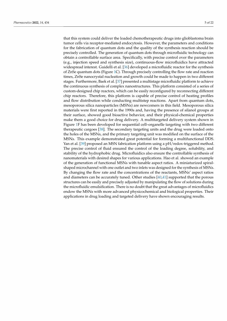
compositions of nanoparticles are changeable. Lin et al. [26] investigated the effects ofFRR on liposomes’ size and drug-loading efficiency. They found that the loading efficiencyof hydrophilic drugs had a positive linear correlation with the FRR, and the maximumdrug-loading efficiency could reach up to 90%. Balbino et al. [27] designed an integratedmicrofluidic device with two different regions for efficient generation of liposomes andlipoplexes in a continuous flow, which significantly reduced the steps of lipoplex synthesis(Figure 1A). In vitro transfection assays showed that microfluidic-obtained lipoplexes hadthe same transfection ability as the conventionally obtained lipoplexes.
2.2. Polymeric Nanoparticles
Polymeric nanoparticles, including natural polymers, synthetic polymers, and so on,have been widely used in the field of drug delivery. The main mechanism is that polymericnanoparticles can interact with mucus through electrostatic, van der Waals, hydrophobic,or hydrogen-bonding interactions, resulting in a long residence time for drug absorption.Among various polymeric nanoparticles, poly lactate glycolic acid (PLGA) is the mostcommonly used polymeric material. PLGA is a family of linear copolymers that are non-toxic and biodegradable and which have been widely used for medical applications. Thesurface of PLGA can be easily modified with different kinds of active groups to realize atargeting function towards specific disease [28]. With the advantage of improved controlover size, size distribution, and morphologies, a microfluidic-assisted nanoprecipitationstrategy was widely employed. Leung et al. [29] presented a hydrodynamic flow-focusing-based device for nanoprecipitation, and the surfactant-free curcumin-encapsulated PLGAnanoparticles were successfully synthesized with diameters ranging from 30 to 70 nm [29].Apart from the diffusion methods such as hydrodynamic flow focusing mentioned above,droplet-based synthetic methods were also a powerful choice for polymeric nanoparticlesynthesis. Generating reversed microemulsion droplets and controlling the evaporationof organic solvent are the key steps. Yu et al. [30] have established a one-step synthesis ofmonodisperse functional polymeric microspheres on a droplet-based microfluidic device.This method relies on the formation of a stable microemulsion first and then a rapidevaporation of the oil from the droplets during the synthesis process for the formationof polymeric microspheres and the inclusion of functional materials. Pavithra et al. [31]further studied the factors that may impact the evolution from droplets into particles. Theyproposed a particle generation technique by making the droplet containing polymericsolution dissolve into the surrounding partially immiscible liquid to drive the particles’formation (Figure 1B). A digital microfluidics platform was also used for the manipulationof nanoparticles. Differing from the conventional droplet-based methods, the digitalmicrofluidics platform is on an open surface and enables individual control of each droplet,offering great power in parallel processing [32]. Alsaeed et al. [33] proposed a digitalmicrofluidics platform for PLGA synthesis and characterization. The diameters of themonodisperse PLGA nanoparticles were uniform at the small size of 115 nm.
2.3. Inorganic Nanoparticles
With their unique physicochemical properties, such as surface plasmon resonance,inorganic particles including quantum dots, graphene, iron oxide, and silica nanoparticlesshow superior performance in drug delivery. Controlled synthesis and surface engineeringof inorganic nanomaterials enable a high flexibility for functional design. As one of thewidely used nanomaterials for biological imaging, quantum dots also play an important rolein targeted therapy and drug delivery. Li et al. [34] reported quantum dots functionalizedwith multiple paired α-carboxyl and amino groups structurally mimicking large aminoacids, which can bind to large neutral amino acid transporter 1 (LAT1). LAT1 is selectivelyand highly expressed in tumor cells, so it serve as a promising drug carrier with goodselectivity for tumor imaging. Furthermore, a triple conjugated system by conjugatingtransferrin and loading two anticancer drugs onto carbon dots with sizes of 3.5 nm has beendeveloped to solve the difficulty of entering malignant brain tumors [35]. Results showed
Pharmaceutics 2022, 14, 434 5 of 22
that this system could deliver the loaded chemotherapeutic drugs into glioblastoma braintumor cells via receptor-mediated endocytosis. However, the parameters and conditionsfor the fabrication of quantum dots and the quality of the synthesis reaction should beprecisely controlled. The generation of quantum dots through microfluidic technology canobtain a controllable surface area. Specifically, with precise control over the parameters(e.g., injection speed and synthesis size), continuous-flow microfluidics have attractedwidespread interest. Guidelli et al. [36] developed a microfluidic reactor for the synthesisof ZnSe quantum dots (Figure 1C). Through precisely controlling the flow rate and reactiontimes, ZnSe nanocrystal nucleation and growth could be made to happen in two differentstages. Furthermore, Baek et al. [37] presented a multistage microfluidic platform to achievethe continuous synthesis of complex nanostructures. This platform consisted of a series ofcustom-designed chip reactors, which can be easily reconfigured by reconnecting differentchip reactors. Therefore, this platform is capable of precise control of heating profilesand flow distribution while conducting multistep reactions. Apart from quantum dots,mesoporous silica nanoparticles (MSNs) are newcomers in this field. Mesoporous silicamaterials were first reported in the 1990s and, having the presence of silanol groups attheir surface, showed good bioactive behavior, and their physical-chemical propertiesmake them a good choice for drug delivery. A multitargeted delivery system shown inFigure 1F has been developed for sequential cell–organelle targeting with two differenttherapeutic cargoes [38]. The secondary targeting units and the drug were loaded ontothe holes of the MSNs, and the primary targeting unit was modified on the surface of theMSNs. This example demonstrated great potential for forming a multifunctional DDS.Yan et al. [39] proposed an MSN fabrication platform using a pH/redox-triggered method.The precise control of fluid ensured the control of the loading degree, solubility, andstability of the hydrophobic drug. Microfluidics also ensure the controllable synthesis ofnanomaterials with desired shapes for various applications. Hao et al. showed an exampleof the generation of functional MSNs with tunable aspect ratios. A miniaturized spiral-shaped microchannel with one outlet and two inlets was designed for the synthesis of MSNs.By changing the flow rate and the concentrations of the reactants, MSNs’ aspect ratiosand diameters can be accurately tuned. Other studies [40,41] supported that the porousstructures can be easily and precisely adjusted by manipulating the flow of solutions duringthe microfluidic emulsification. There is no doubt that the great advantages of microfluidicsendow the MSNs with more advanced physicochemical and biological properties. Theirapplications in drug loading and targeted delivery have shown encouraging results.
Pharmaceutics 2022, 14, 434 6 of 22
(A)
(C) (D)
(B)
(E) (F)
Figure 1. Different approaches for the fabrication of nanoparticles. (A) Schematic of the microfluidicdevice for one-step formation of plasmid DNA (pDNA)/cationic liposome (CL) lipoplexes and itsin vitro efficacy. (B) Schematic of the flow-focusing device used to engineer and monitor Janus drugparticle formation. (C) Schematic of the microfluidic reactor used for the synthesis of ZnSe quantumdots. (D) The simulated microfluidic mixer unit with two inlets and two outlets. (E) Three-dimensionalviews and top views of the iLiNP device. (F) The strategy employed for dual-targeted two-drugnanocarriers based on mesoporous silica nanoparticles. (A) Balbino et al. [27]; (B) Sundararajan et al. [31];(C) Guidelli et al. [36]; (D) Wang et al. [24]; (E) Kimura et al. [21]; (F) Castillo et al. [38].
3. Microfluidic Chip for Carrier-Free Drug Delivery
Microfluidic chips can not only be used to synthesize a variety of complicated nan-odrug carriers, but can also characterize the efficiency of drug delivery directly throughvarious in vivo models [42–44]. For nano drug carrier systems, a large amount of expensivedrugs may be used, and the synthesis of drug carriers may face various challenges. For in-stance, the composition of carriers must be strictly controlled to ensure the efficacy of drugs.For microfluidic carrier-free DDSs, [2,45] direct drug delivery can ensure drug release tothe special application site quickly and effectively. Moreover, many of the carrier-freedrug delivery systems can avoid immune rejection and reduce the toxicity caused bynon-drug carriers [2,46,47]. Carrier-free direct drug delivery can be roughly divided intotwo categories: drug delivery based on microneedles (MNs) and drug delivery based onmicro-reservoirs [17,48–50].
3.1. MNs-Based System
In recent decades, research has explored a novel transdermal drug delivery strategy,MNs, which can overcome some limitations of traditional oral and subcutaneous adminis-tration. MNs can easily pass through the stratum corneum (SC) of the skin, a barrier to drugmolecules. Besides, patients can easily administer drugs by themselves through MNs. Com-pared with subcutaneous administration, the length of MNs can be controlled to be between10 µm and 1 mm, which is only a small fraction of the subcutaneous administration needle,
Pharmaceutics 2022, 14, 434 7 of 22
so that they can achieve painless treatment due to their short size [51–56]. Compared withtraditional oral administration, MNs’ percutaneous administration can avoid the poorabsorption caused by the degradation of drugs through the metabolic system [52,57,58]. Sofar, there are several kinds of MNs reported for drug delivery application, such as solidMNs, coated MNs, dissolving MNs, hollow MNs, and hydrogel-based MNs [2,59,60].
3.1.1. Solid MNs
Solid MNs are made of different materials such as silicone, metals, and polymers,which are manufactured by 3D printing, digital light processing (DLP), stereolithography,laser cutting, and chemical or mechanical technologies into specific shapes and lengths.The working principle of solid MNs involves a two-step process. The solid MNs arefirstly used to create a micro-channel on the surface of the skin, and then the drugs arecoated onto the skin surface to make the drug diffuse passively to achieve the therapeuticeffect [59,60]. The effect of DDSs of solid MNs depends on the shape and length of theMNs, and many efforts have been made to improve the drug delivery efficiency of thesolid MNs. For example, Narayanan et al. [61] developed solid silicon MNs and exploredtheir best aspect ratio, mechanical strength, and fracture strength conditions by usingsingle-step lithography and an anisotropic wet etching tetramethylammonium hydroxide(TMAH) process to achieve the best drug delivery effect. They successfully synthesizedsolid MNs with an average height of 158 µm, width of 110.5 µm, aspect ratio of 1.43,tip angle of 19.4◦, and tip diameter of 0.40 µm. Additionally, the microhardness valueis 44.4 (HRC), which is 52.2 times higher than the skin’s ultimate tensile strength (UTS),resulting in a great improvement of its percutaneous drug administration ability. Similarly,Li et al. [62] built solid MNs using polylactic acid (PLA) materials and systematicallyexplored the mechanical strength and biodegradable ability. They found that the MN size,drug concentration, viscosity of the drug solution, and drug administration time on theskin have great influence on administration efficiency. Under the optimized conditions,insulin could be highly efficiently delivered to the skin by the MNs.
3.1.2. Coated MNs
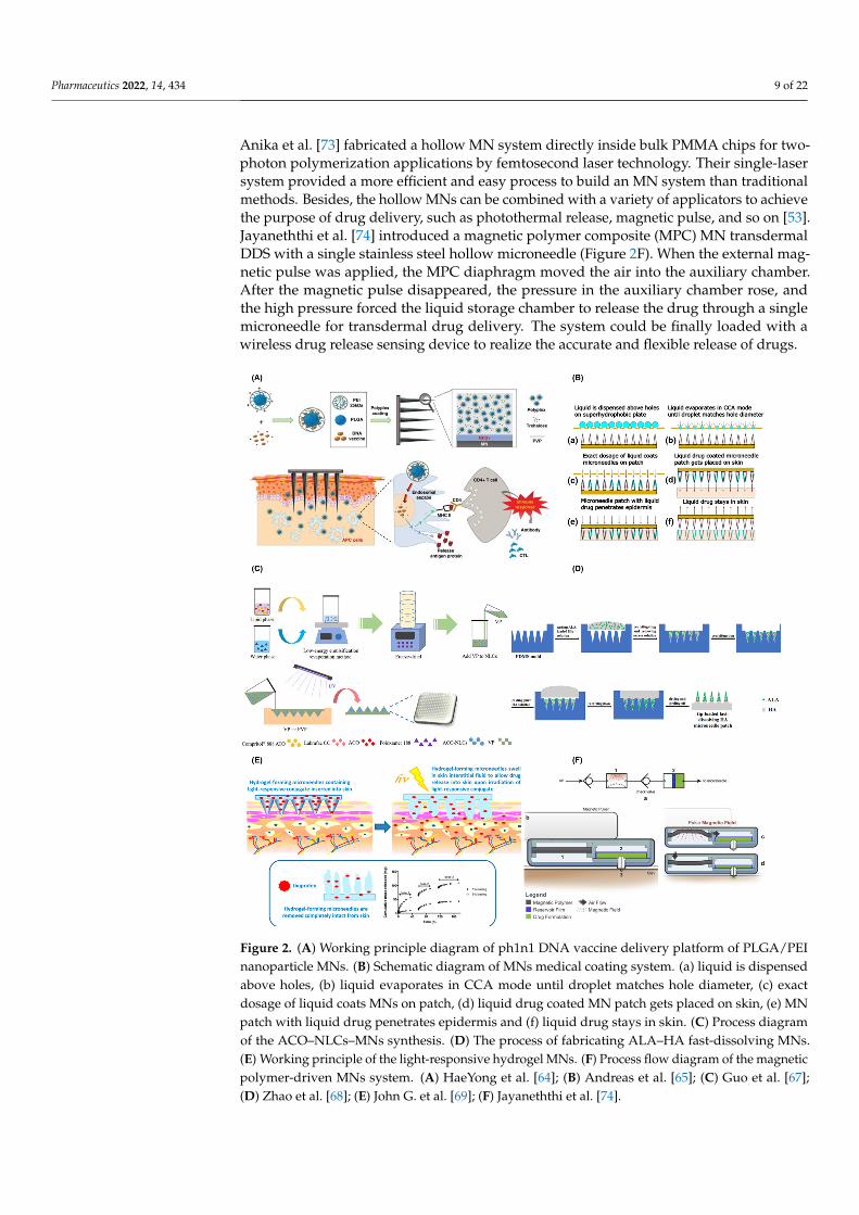
The coated MNs are coated with drugs and biodegradable materials directly on thesurface of the MNs. When the MNs enter the skin of a patient, the drugs will be dissolvedand released into the body to achieve the purpose of treatment. Storing drugs by coatingthem onto the surface of MNs can enhance their long-term stability [59,60]. In recent years,a large quantity of research focused on the realization of combined drug delivery or drugcombination adjuvant administration. For example, DeMuth et al. [63] used layer-by-layercoating techniques to realize multi-loading and multi-functional effects of DNA vaccines forthe first time. Both DNA and its adjuvants were accumulated in this MN. HaeYong et al. [64]developed an intradermal pH1N1 DNA vaccine delivery platform using MNs coatedwith polymers containing polylactic acid co glycolic acid/polyethyleneimine (PLGA/PEI)nanoparticles (NPs) (Figure 2A). The performance of MNs coated with polymer particlesvia intramuscular polymer delivery and the naked pDNA vaccine in porcine skin werecompared. The results indicated that the polyplex-coated MNs had a better performancetowards immune response. Furthermore, to ensure the accurate control of drug dosage,the drugs coated onto MNs need to be strictly controlled. Andreas et al. [65] proposed acoated-MN-based transdermal DDS through the usage of evaporation-induced droplettransport. As shown in Figure 2B, a rough amount of the liquid containing drugs wasplaced on a perforated metal plate, and when the evaporated liquid reached a certain smallvolume, it penetrated the MN surface along the hole. Then, the MN patch stuck into theskin and left the drug behind after removing the needle. The system could ensure theaccurate dosage of the drug by controlling the time of the needle exposure to the solution;the surface tension matching the degree between the needle material and the liquid; thecharacteristics of the liquid material; and the needle structure.
Pharmaceutics 2022, 14, 434 8 of 22
3.1.3. Dissolving MNs
Dissolving MNs are composed of biodegradable and biocompatible materials suchas polyvinyl alcohol (PVA) and polyvinylpyrrolidone (PVP). Drug release is mainly con-trolled by dissolving the materials when the MNs enter the skin [59,60]. Compared withother types of MNs, the dissolving MNs can be dissolved in the body, which means thereis no risk of leaving hard residues behind. However, this kind of MN takes minutes todissolve, and its mechanical strength is relatively weak, resulting in insufficient penetrationinto the skin. In order to prepare a dissolving microneedle with sufficient mechanicalstrength and good degradation ability, Cha et al. [66] built a polylactic acid (PLA) dissolv-ing MN. Two different penetration and dyeing methods on pig skin successfully simulatedthe effect of transdermal DDSs. Guo et al. [67] also reported a novel nanostructuredlipid carriers (NLCs)-loaded dissolving MN to achieve an efficient drug delivery. Asshown in Figure 2C, they used NLCs as an aconitine (ACO) barrier and then embeddedit in polyvinylpyrrolidone-based dissolving MNs by an ultraviolet cross-linking method.In vivo micro-dialysis proved that the MN device had an obvious inhibitory effect on pawswelling and inflammation in adjuvant-induced arthritis model rats and resulted in the im-provement of the ACO-induced arrhythmia. To accelerate the dissolution of the dissolvingMNs, Zhao et al. [68] built a sodium hyaluronate (HA) fast-dissolving MN by two castingmethods to improve the photodynamic therapy (PDT) efficacy of a subcutaneous tumor(Figure 2D). Each microneedle can load 122 µg 5-aminolevulinic acid (ALA) in the tip; thedrug loading amount was greatly improved and waste of drugs was avoided. When theneedle is inserted into the skin, the MN is dissolved and the drug released. Comparedto the 66% tumor inhibition rate of traditional ALA injection, the developed MNs couldincrease the tumor inhibition rate to 97%, and about 75% of the MN was dissolved withinonly 4 min.
3.1.4. Hydrogel MNs
The hydrogel MNs are constructed with swelling polymer materials. This MN can beswelled in the body, and then, the expansion gap will form a 3D network to let the storeddrug release. The efficiency of hydrogel MN drug delivery depends on the type of materialsprepared, the mechanical strength, and the crosslinking density of the swollen hydrogelnetwork [59,60]. The materials for the fabrication of hydrogel MNs are the most importantfactor for drug delivery efficiency. Recently, a lot of novel materials have been explored forthe fabrication of MNs. For example, John G. et al. [69] first proposed a photo-responsestimulus hydrogel MN array system for the delivery of ibuprofen. The MN arrays wereprepared from 2-hydroxyethyl methacrylate (HEMA) and ethylene glycol dimethacrylate(EGDMA) by micro-molding, with good mechanical properties. The system could loadup to 5% (w/w) ibuprofen in the photo-responsive 3,5-dimethoxybenzoin compounds(Figure 2E). Similarly, Chen et al. [70] developed a novel enzyme-free polymeric componentMN array patch, which consisted of a boronate-containing hydrogel semi-interpenetratingnetwork with biocompatible silk fibroin. The semi-interpenetrating network gel is com-posed of two components, 4-(2-acrylamidoethylcarbamoyl)-3-fluorophenylboronic acid(AmECFPBA, pKa 7.2) and an acrylamide derivative, which could enhance the glucoseresponse to insulin. Crystalline silk fibroin [71] extracted from silkworms was used asmatrix stiffener to enhance the skin penetration ability of the MNs.
3.1.5. Hollow MNs
The hollow MNs are made up of several different materials such as glass, silicone, andmetals. Hollow MNs are the same as those used in traditional subcutaneous administra-tion. When the MNs enter the skin, the drugs are released into the human body throughthe hollow pores of the MNs. Compared with other types of MNs, they can accuratelydeliver high-dose drugs without changing their formula [59,60], but they face difficultiesdue to their insufficient mechanical strength [72]. Hollow MNs can be built by variousmethods, such as 3D printing [71], micro-molding, dipping, and so on [53]. For example,
Pharmaceutics 2022, 14, 434 9 of 22
Anika et al. [73] fabricated a hollow MN system directly inside bulk PMMA chips for two-photon polymerization applications by femtosecond laser technology. Their single-lasersystem provided a more efficient and easy process to build an MN system than traditionalmethods. Besides, the hollow MNs can be combined with a variety of applicators to achievethe purpose of drug delivery, such as photothermal release, magnetic pulse, and so on [53].Jayaneththi et al. [74] introduced a magnetic polymer composite (MPC) MN transdermalDDS with a single stainless steel hollow microneedle (Figure 2F). When the external mag-netic pulse was applied, the MPC diaphragm moved the air into the auxiliary chamber.After the magnetic pulse disappeared, the pressure in the auxiliary chamber rose, andthe high pressure forced the liquid storage chamber to release the drug through a singlemicroneedle for transdermal drug delivery. The system could be finally loaded with awireless drug release sensing device to realize the accurate and flexible release of drugs.
Pharmaceutics 2022, 14, x FOR PEER REVIEW 9 of 23
culties due to their insufficient mechanical strength [72]. Hollow MNs can be built by var-ious methods, such as 3D printing [71], micro-molding, dipping, and so on [53]. For ex-ample, Anika et al. [73] fabricated a hollow MN system directly inside bulk PMMA chips for two-photon polymerization applications by femtosecond laser technology. Their sin-gle-laser system provided a more efficient and easy process to build an MN system than traditional methods. Besides, the hollow MNs can be combined with a variety of applica-tors to achieve the purpose of drug delivery, such as photothermal release, magnetic pulse, and so on [53]. Jayaneththi et al. [74] introduced a magnetic polymer composite (MPC) MN transdermal DDS with a single stainless steel hollow microneedle (Figure 2F). When the external magnetic pulse was applied, the MPC diaphragm moved the air into the auxiliary chamber. After the magnetic pulse disappeared, the pressure in the auxiliary chamber rose, and the high pressure forced the liquid storage chamber to release the drug through a single microneedle for transdermal drug delivery. The system could be finally loaded with a wireless drug release sensing device to realize the accurate and flexible re-lease of drugs.
Figure 2. (A) Working principle diagram of ph1n1 DNA vaccine delivery platform of PLGA/PEI nanoparticle MNs. (B) Schematic diagram of MNs medical coating system. (a) liquid is dispensed above holes,(b) liquid evaporates in CCA mode until droplet matches hole diameter, (c) exact dos-age of liquid coats MNs on patch, (d)liquid drug coated MN patch gets placed on skin, (e) MN patch with liquid drug penetrates epidermis and (f) liquid drug stays in skin. (C) Process diagram of the ACO–NLCs–MNs synthesis. (D) The process of fabricating ALA–HA fast-dissolving MNs. (E)
Figure 2. (A) Working principle diagram of ph1n1 DNA vaccine delivery platform of PLGA/PEInanoparticle MNs. (B) Schematic diagram of MNs medical coating system. (a) liquid is dispensedabove holes, (b) liquid evaporates in CCA mode until droplet matches hole diameter, (c) exactdosage of liquid coats MNs on patch, (d) liquid drug coated MN patch gets placed on skin, (e) MNpatch with liquid drug penetrates epidermis and (f) liquid drug stays in skin. (C) Process diagramof the ACO–NLCs–MNs synthesis. (D) The process of fabricating ALA–HA fast-dissolving MNs.(E) Working principle of the light-responsive hydrogel MNs. (F) Process flow diagram of the magneticpolymer-driven MNs system. (A) HaeYong et al. [64]; (B) Andreas et al. [65]; (C) Guo et al. [67];(D) Zhao et al. [68]; (E) John G. et al. [69]; (F) Jayaneththi et al. [74].
Pharmaceutics 2022, 14, 434 10 of 22
3.2. Micro-Reservoir System
The micro-reservoir system is composed of one or more drug reservoirs to realizedrug storage. Compared with traditional drug release systems, the micro-reservoir systemcan realize a variety of delivery schemes such as zero-order, pulsatile, and on-demanddosing [42,75]. In particular, the micro-reservoir system manufactured by microfluidictechnology can realize efficient and accurate drug delivery in vitro and in vivo [2,76].Moreover, both in vitro and implanted micro-reservoirs can significantly increase drugstability and prolong the drug delivery time. The key part of the microfluidic micro-reservoir system is to prepare a driving system to meet the requirements of precise andstable release of drugs. Based on different actuation mechanisms, the micro-reservoirsystem can be actuated in active mode and passive mode [2,47,77].
3.2.1. Active Actuation Mode for Micro-Reservoir System
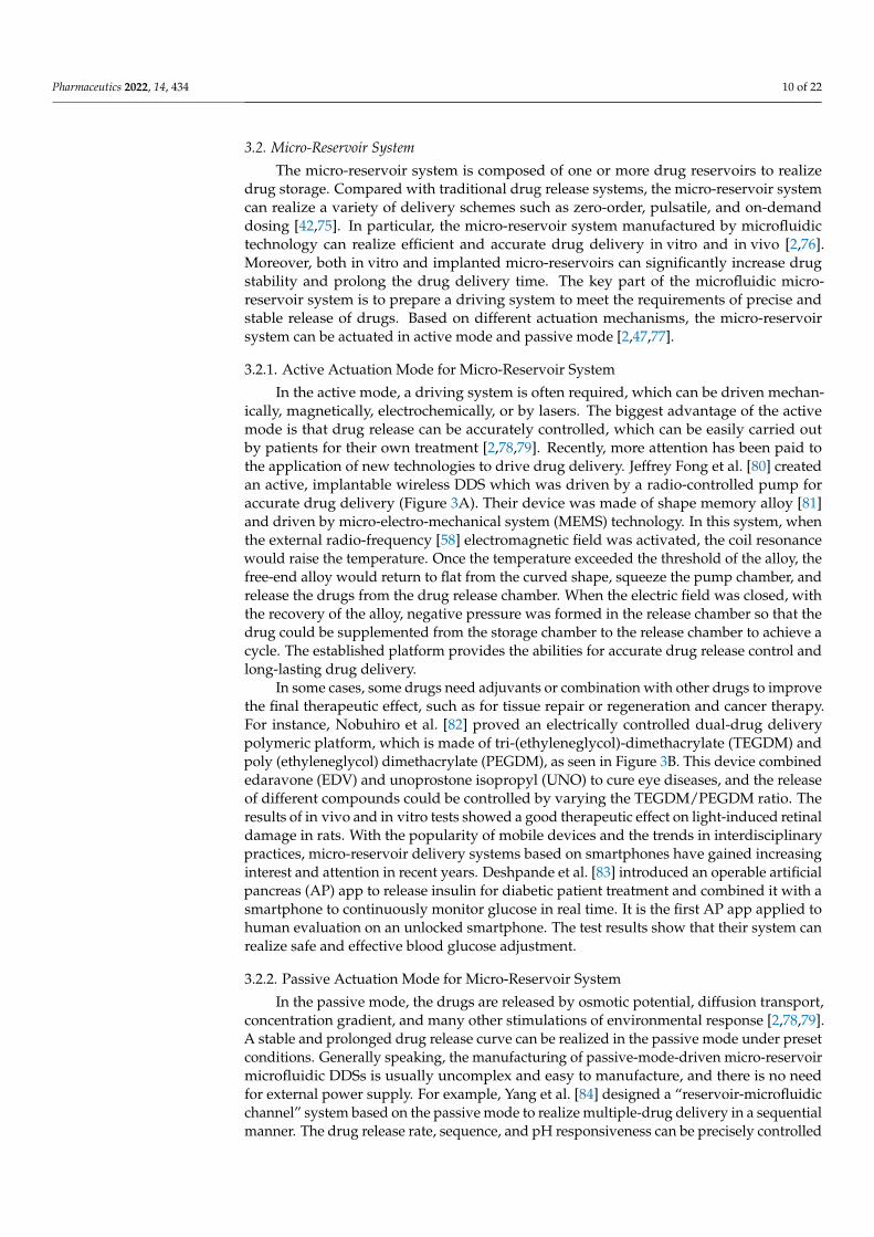
In the active mode, a driving system is often required, which can be driven mechan-ically, magnetically, electrochemically, or by lasers. The biggest advantage of the activemode is that drug release can be accurately controlled, which can be easily carried outby patients for their own treatment [2,78,79]. Recently, more attention has been paid tothe application of new technologies to drive drug delivery. Jeffrey Fong et al. [80] createdan active, implantable wireless DDS which was driven by a radio-controlled pump foraccurate drug delivery (Figure 3A). Their device was made of shape memory alloy [81]and driven by micro-electro-mechanical system (MEMS) technology. In this system, whenthe external radio-frequency [58] electromagnetic field was activated, the coil resonancewould raise the temperature. Once the temperature exceeded the threshold of the alloy, thefree-end alloy would return to flat from the curved shape, squeeze the pump chamber, andrelease the drugs from the drug release chamber. When the electric field was closed, withthe recovery of the alloy, negative pressure was formed in the release chamber so that thedrug could be supplemented from the storage chamber to the release chamber to achieve acycle. The established platform provides the abilities for accurate drug release control andlong-lasting drug delivery.
In some cases, some drugs need adjuvants or combination with other drugs to improvethe final therapeutic effect, such as for tissue repair or regeneration and cancer therapy.For instance, Nobuhiro et al. [82] proved an electrically controlled dual-drug deliverypolymeric platform, which is made of tri-(ethyleneglycol)-dimethacrylate (TEGDM) andpoly (ethyleneglycol) dimethacrylate (PEGDM), as seen in Figure 3B. This device combinededaravone (EDV) and unoprostone isopropyl (UNO) to cure eye diseases, and the releaseof different compounds could be controlled by varying the TEGDM/PEGDM ratio. Theresults of in vivo and in vitro tests showed a good therapeutic effect on light-induced retinaldamage in rats. With the popularity of mobile devices and the trends in interdisciplinarypractices, micro-reservoir delivery systems based on smartphones have gained increasinginterest and attention in recent years. Deshpande et al. [83] introduced an operable artificialpancreas (AP) app to release insulin for diabetic patient treatment and combined it with asmartphone to continuously monitor glucose in real time. It is the first AP app applied tohuman evaluation on an unlocked smartphone. The test results show that their system canrealize safe and effective blood glucose adjustment.
3.2.2. Passive Actuation Mode for Micro-Reservoir System
In the passive mode, the drugs are released by osmotic potential, diffusion transport,concentration gradient, and many other stimulations of environmental response [2,78,79].A stable and prolonged drug release curve can be realized in the passive mode under presetconditions. Generally speaking, the manufacturing of passive-mode-driven micro-reservoirmicrofluidic DDSs is usually uncomplex and easy to manufacture, and there is no needfor external power supply. For example, Yang et al. [84] designed a “reservoir-microfluidicchannel” system based on the passive mode to realize multiple-drug delivery in a sequentialmanner. The drug release rate, sequence, and pH responsiveness can be precisely controlled
Pharmaceutics 2022, 14, 434 11 of 22
by the length, width, and straightness of the micro-reservoirs and microchannels throughpassive diffusion behavior (Figure 3C). Besides, the passive mode is usually used whenself-regulation is needed. Lee et al. [85] proposed a wet microcontact printing (µCP) systemto store liquid drugs. As shown in Figure 3D, the drug solution was transferred to themicro-reservoir by the wet µCP. They explored the impact of many factors on drug delivery,such as the number of printings, the size of the printed picture, drug concentration, thematerials of the carrier, and so on. The results of the drug-transferring experiments withtheir simple and accessible device showed good drug delivery performance.
Pharmaceutics 2022, 14, x FOR PEER REVIEW 11 of 23
reservoir microfluidic DDSs is usually uncomplex and easy to manufacture, and there is no need for external power supply. For example, Yang et al. [84] designed a “reservoir-microfluidic channel” system based on the passive mode to realize multiple-drug delivery in a sequential manner. The drug release rate, sequence, and pH responsiveness can be precisely controlled by the length, width, and straightness of the micro-reservoirs and mi-crochannels through passive diffusion behavior (Figure 3C). Besides, the passive mode is usually used when self-regulation is needed. Lee et al. [85] proposed a wet microcontact printing (μCP) system to store liquid drugs. As shown in Figure 3D, the drug solution was transferred to the micro-reservoir by the wet μCP. They explored the impact of many fac-tors on drug delivery, such as the number of printings, the size of the printed picture, drug concentration, the materials of the carrier, and so on. The results of the drug-transferring experiments with their simple and accessible device showed good drug delivery perfor-mance.
Figure 3. (A) Schematic diagram of wireless implantable magnetic driven DDSs and its working principle. (B) Working diagram of dual-drug delivery system. (C) The “reservoir-microfluidic chan-nel” system optimization and drug release diagram. (D) Schematic diagram of the wet μCP system manufacturing: (1) PDMS stamp set and drug solution, (2) tip contact with drug solution,(3)drug coated stamps,(4)The position of drug coated stamp above PDMS reservoir,(5) stamp contact on a target surface and (6) final drug loading formation. (A) Jeffrey Fong et al. [80]; (B) Nobuhiro et al. [82]; (C) Yang et al. [84]; (D) Lee et al. [85].
4. In Vitro Models of Microfluidic Devices for the Evaluation of Drug Delivery After the fabrication of nanomaterials for DDSs, the following critical work is to eval-
uate their drug delivery efficiency. In recent decades, a large number of DDSs have been constructed, but only a small amount succeed in translating from bench to clinic. One of the main reasons is that drug efficacy is largely affected by DDSs. When drugs enter the body, it is difficult to enter the blood circulatory system and transport to specific targets due to the influence of drug barriers and the first-pass effect. There are four types of bio-logical barriers, namely the blood–brain barrier, the mucosal diffusion barrier, the bio-chemical barrier, and the cellular permeability barrier. The conventional methods are dif-ficult to use to mimic the biological barriers for DDS evaluation. The commonly used methods are based on animal models but show poor predictive results for human re-sponses to drugs. To overcome these problems, it is necessary to develop accurate and
Figure 3. (A) Schematic diagram of wireless implantable magnetic driven DDSs and its workingprinciple. (B) Working diagram of dual-drug delivery system. (C) The “reservoir-microfluidicchannel” system optimization and drug release diagram. (D) Schematic diagram of the wet µCPsystem manufacturing: (1) PDMS stamp set and drug solution, (2) tip contact with drug solution,(3) drug coated stamps, (4) The position of drug coated stamp above PDMS reservoir, (5) stampcontact on a target surface and (6) final drug loading formation. (A) Jeffrey Fong et al. [80];(B) Nobuhiro et al. [82]; (C) Yang et al. [84]; (D) Lee et al. [85].
4. In Vitro Models of Microfluidic Devices for the Evaluation of Drug Delivery
After the fabrication of nanomaterials for DDSs, the following critical work is toevaluate their drug delivery efficiency. In recent decades, a large number of DDSs havebeen constructed, but only a small amount succeed in translating from bench to clinic. Oneof the main reasons is that drug efficacy is largely affected by DDSs. When drugs enter thebody, it is difficult to enter the blood circulatory system and transport to specific targets dueto the influence of drug barriers and the first-pass effect. There are four types of biologicalbarriers, namely the blood–brain barrier, the mucosal diffusion barrier, the biochemicalbarrier, and the cellular permeability barrier. The conventional methods are difficult touse to mimic the biological barriers for DDS evaluation. The commonly used methods arebased on animal models but show poor predictive results for human responses to drugs. Toovercome these problems, it is necessary to develop accurate and highly efficient strategiesfor the evaluation of DDSs. Recent developments in microfluidics, especially organs-on-a-chip, provide powerful tools for dynamic drug activity evaluation and whole-bodyresponses in real time. An organ-on-a-chip is a platform in which different types of cells areco-cultured on a chip following the tissue-specific tridimensional to simulate a given single-or multi-organ system. It provides a high level of convenience for drug-delivery-related
Pharmaceutics 2022, 14, 434 12 of 22
studies, which reduces the usage of animal experiments and fills the gap between animalstudies and clinical trials to some extent [81]. In this section, we mainly focus on themucosal model, vessel model, gut model, and blood–brain barrier models developed inrecent years.
4.1. Mucosal Diffusion Barrier Model
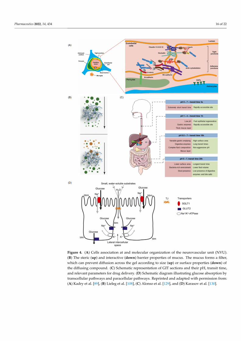
Having the largest surface area (about 300–400 m2) in our body, the mucosal diffusionbarrier is a significant obstruction for the uptake and absorption of drug carriers. Secretedby goblet cells, mucus plays a major role in promoting digestion, protecting the epitheliumfrom harmful substances or bacteria, and enhancing the exchange of nutrients [86]. Themucin network can form a size exclusion filter for larger compounds (Figure 4B) [87].Therefore, the mucosal diffusion barrier is considered an important effector that influ-ences drug absorption and bioavailability. Several novel strategies have been applied toachieve effective mucosal drug delivery and sufficient drug bioavailability. Among them,using nanoparticles as drug carriers is widespread, being proven to protect drugs fromdegradation in the GI tract as well as the gastric environment [88,89]. As the diffusioncapacity through mucus is highly dependent on hydrodynamic diameters and biointerfa-cial properties of nanoparticles, further understanding of the transportation mechanismsthrough mucus layers would highly accelerate the research and development process.Jia et al. [90] developed an in vitro mucus-model-on-a-chip by confining the mucin solu-tion within the microchannel, forming a robust and reproducible model of mucus–aqueoussolution interface, which can be used to readily observe the transport of nanoparticlesunder fluorescence microscopy. Result showed that the biointerfacial properties of mucusare critical for diffusion. Apart from spontaneous diffusion, biochemical and physicalmembrane disruption methods have been proposed. Ramesan et al. [91] explored theusage of high-frequency sound waves to promote drug diffusion through mucosa andcontrollably locate the drug in the mucosa of a porcine buccal model. By tuning the sys-tem parameters, the penetration depths can be changed so that over 95% of drugs arelocated within the mucosal layer with the preservation of their structural integrity. Asthe epithelial cells perform an important role in drug absorption, thorough simulation ofhuman epithelia and mucosae models is extremely important [92]. From single epitheliallayers to complex 3D models, cell-based mucus models provide an enhanced bio-relevantin vitro platform to better resemble physiological conditions. Hagiwara et al. [93] proposeda microphysiological-system-on-a-chip with 3D cultured Caco-2 tubules to mimic the gas-trointestinal environment for the study of drug permeability in the stomach. Assessmentusing this model can reproduce similar results to those obtained from conventional meth-ods. Gholizadeh et al. [94] developed a nasal-epithelial-mucosa-on-a-chip (NEM-on-a-chip)model integrated with a novel carbon-nanofibers-modified carbon electrode, aiming tomonitor real-time quantitative drug transport rate in the nasal environment. Results indi-cated the great importance of using NEM-on-a-chip to emulate dynamic in vivo conditions.As discussed above, a model with the presence of epithelial cells and the right amount ofmucus is needed for the development of a useful platform to test the nanocarrier.
4.2. Vessel Model
Blood vessels are one of the most important aspects of drug delivery, as most drugsare delivered by blood [95]. As an emerging platform for rapidly and accurately predict-ing in vivo behaviors of DDSs, the vessel-on-a-chip can provide insight to nanoparticles’hemodynamics in microcirculation through blood. Generally, vessels-on-a-chip are basedon the permeability and biocompatibility of the substrate materials, such as polydimethyl-siloxane (PDMS) and hydrogel. Through the construction of a supporting structure inthe microchannel, endothelial cells can be embedded to form vasculature. Mimickingthe tumor microenvironment and vasculature system on a chip is beneficial for the directand real-time study of drug delivery pathways. Studies on vessels-on-a-chip aim to un-derstand the behavior of nanocarriers in vivo, especially the selectively extravasate and
Pharmaceutics 2022, 14, 434 13 of 22
accumulative capacity. By using the co-culture of blood vessel cells in the microchannelto study the interaction between blood vessel cells and nanoparticles, the effects of vas-cular targeting, enhanced permeation, and retention [96] could be revealed [23]. Vasculartargeting or particle targeting is a complex process in which intravascularly administerednanoparticles navigate the circulation and adhere to or cross the vessel wall to localize attheir target site. Related research has attracted great attention to simulating fluid dynamicsin the microvasculature and recapitulating the in vivo transportation process. Based onthe establishment of the tumor microenvironment on a chip, fluorescently labeled goldnanoparticles were taken to trace and evaluate the whole transport processes. Severalstudies indicated that nanoparticle localization is highly dependent on vessel geometryand local flow features, especially shear stress. Generated by blood flow, shear stress has agreat effect on transportation, accumulation, cellular uptake, and ultimately therapeuticefficacy [97]. For example, high-shear regions will adhere more strongly adhesive particles,and the weakly adhesive particles can be used for recirculation flows. Maria et al. [98]mapped the deposition of nanoparticles in a reconstructed vessel model. The result showedthat the spatial distribution of the nanoparticles was dependent on physiological conditionsand hemodynamic structures. The periodic changes of shear stress in vivo can prevent theadhesion of the nanoparticles. In addition to the vessel geometry, the size and shape ofnanoparticles also have an impact on drug delivery efficiency. As the target site of drugs isalways in the tissue beyond the wall, the function of margination and wall adhesion is pre-requisite, known as “near wall excess”. Nanoparticles that do not exhibit “near wall excess”in microvessels may be prevented from acting as drug delivery carriers. Cooley et al. [99]utilized four distinct shapes of nanoparticles, indicating that micro-scale non-sphericalparticles undergo enhanced margination and adhesion effect. In this study, a parallel plateflow chamber with red blood cells was established to observe the particle adhesion andretention in blood vessels. Considered to be the standard of drug delivery, the EPR effectwas affected by vascular dynamics and nanoparticle design. Maneesha [23] developed anin vitro cancer-model-on-a-chip containing tumor cells and a vascular network to analyzethe uptake of gold nanoclusters in tumor cells. Results indicated that cells near the endothe-lial gap absorbed more nanoparticles, which may be attributed to the leaky nature of tumorvasculature and the EPR effect. Moreover, the tumor size, number, and location wouldalso affect drug transport and distribution. From Figure 5C, a tumor-vasculature-on-a-chip,which is constructed with a blood vessel channel and a tumor channel sandwiched with aporous membrane, was used to conduct numerical investigations [100] and revealed theinfluence of drug concentration heterogeneity in tumors.
Furthermore, 3D bioprinting technology can fabricate more complex architectures,which is a promising approach to improving the degree of biomimetic quality. For example,Cao et al. [101] proposed an improved hollow blood vessel along with a lymphatic vesselpair hosted in a 3D tumor microenvironment-mimetic hydrogel matrix. With 3D bioprintingtechnology, vessel permeability can be tuned individually, which is benefit for the diffusionprofile study of biomolecules and anticancer drugs (Figure 5A). To improve drug deliveryacross the blood vessel, ultrasound is another feasible approach [102]. The cavitation effectcaused by ultrasound is another approach to changing the permeability of vessels. Ablood-vessel-on-a-chip [102] consisting of one tissue chamber and two independent vascu-lar channels has been used to mimic tumor microvasculature and enable further studieson ultrasound-driven delivery, as it can temporarily open the endothelial intercellularjunctions [96].
4.3. Gut-on-a-Chip
As the most important digestive organ, the gut plays a vital role in food digestion, ab-sorption, and metabolism [103], provided by the villi and microvilli, and contains symbioticmicrobial flora [104]. Generally, the intestinal barrier is comprised of mucus, the intestinalepithelium, and the biochemical environment [105], which represents the major barrier fordrug absorption. For the intestinal epithelium barrier, there are four kinds of intestinal
Pharmaceutics 2022, 14, 434 14 of 22
epithelial cells that are responsible for absorption (enterocyte), mucus secretion (gobletcells), hormone secretion (enteroendocrine cells), and defensive peptide secretion (Panethcells), respectively [106]. The cell membranes of these epithelial cells form a physical barrier,which contributes to the selective transportation and efficiently protects our body fromharmful substances or bacteria. In general, the membrane only allows hydrophilic solutes topermeate, but the proteins in the membrane can function as specific transporters, allowingnutrients to pass through. Besides, the tight junctions between adjacent epithelial cells playan important role in nutrient exchange as well as drug absorption. They are in charge ofthe integrity of the intestinal epithelium barrier and regulate the permeability of nutrientsthrough two ways: the “pore” pathway, which is highly selective, and the “leak” pathway,which limits selectivity [107]. Besides, mucus secreted by goblet cells is considered animportant effector that influences drug absorption and bioavailability. It is composed ofhydrogel with mucin and a small number of proteins and carbohydrates. When drugs enterthe mucus layer, mucus will be secreted and shed continuously to eliminate the attachmentof drugs. As shown in Figure 4B [108], the mucin network can form a size exclusion filterfor larger compounds [87].
Furthermore, the biochemical environment of the intestine, in which there exists ahigh concentration of digestive enzymes, also poses a great challenge for drug delivery.Therefore, therapeutic drugs, especially through oral drug delivery, generally face the prob-lem of degradation, leading to unsustainable and un-targeted release. The development ofbiocompatible anti-digestive biomaterials provides promising potential to deal with thischallenge. Ren’s research group synthesized an injectable covalent hydrogel through thephotopolymerization of glycidyl methacrylate-modified xanthan (xan-GMA) on a microflu-idic chip and cultured intestinal epithelial cells-6 on its surface to perform a gut barrierfunction [109]. The xan-GMA hydrogel showed good anti-digestion compared with anexisting product, fibrin sealant, and showed good potential for closing gastrointestinalfistula. It is expected to be an outstanding gut repair material and drug delivery carrier. Tofurther mimic the microenvironment and diversified function of intestine in vitro, in thisstudy, a 2D gut microfluidic chip was used to replace the animal model of GI fistula. How-ever, lacking 3D tissue architecture and cell–cell interactions makes it difficult to effectivelyrecapitulate the real intestinal microenvironment and mimic the diversified function andstructure of the gut with a 2D model. As a result, the 3D and dynamic culture models, madeby co-culturing different kinds of cells in microchannels, are needed. Lee et al. [110] haveproposed a 3D gut–liver chip to predict the first-pass metabolism. Two separate layer chipswere used for the co-culture of gut (Caco-2) and liver (HepG2) cell lines in both 2D and 3Dmodes to reproduce a similar human PK profile and predict the pharmacological effectsof drugs. This study demonstrated the importance of the employment of the multi-organchip system. Herland et al. [111] proposed a multi-organ-chip system containing intestine,liver, and kidney cells to study drug pharmacokinetics (PKs) and pharmacodynamics (PDs)in vitro. The result obtained using this model is in accordance with the previously reportedpatient data, showing the accurate forecasting ability of pharmacodynamics.
4.4. Blood–Brain Barrier (BBB)
The BBB is a specific structure that protects our brain from harmful agents but resultsin bad drug delivery [112]. It functions as a selective physiological barrier which controlssubstance transport and maintains brain homeostasis [113]. The selective barrier worksmainly through endothelial cells [114] in the tight junctions, the basement membraneof pericytes, and the end-feet of astrocytes [115]. From Figures 4A and 5B, we can seethat these cells are not functionally separated but are involved in complex activities andinteractions among BBB-related cells because of the integrity of the BBB. For example,tightly connected ECs are able to prevent large molecules (approximately greater than400 Da) from crossing the barrier by paracellular transport. Pericytes and astrocytes areclosely attached to ECs and regulate the permeability of the tight junctions by releasingcertain chemicals [116]. As a result, the BBB only allows a few water-soluble substances
Pharmaceutics 2022, 14, 434 15 of 22
to enter the brain by active transport. However, some gas molecules and lipid-solublesubstances can easily cross through passive diffusion [116]. Because of the existence ofthe BBB, many anticancer drugs fail to transport into the brain and cannot effectively treatbrain tumors and nervous system diseases.
In order to overcome the poor BBB penetration of drugs, various approaches have beendeveloped to enhance the transport efficiency and further improve the drug efficacy [117].For small molecules (e.g., <500 Da), the methods of their delivery include local invasive(direct injection/infusion) delivery, induction of enhanced permeability, and applicationof global physiological targeting strategies [118]. For large hydrophilic molecules (e.g.,enzymes, RNAi, genes), they may be degraded by the endosomal/lysosomal or ubiqui-tin/proteasomal system after internalization. In this case, some systems (e.g., endosomalescape mechanisms) could be applied to bypass these routes of degradation [119], andDDSs may help large molecules cross the BBB and enter the brain [120]. To date, the BBBstill remains a bottleneck in brain drug development [121]. To boost the drug developmentof central nervous system diseases, a high-fidelity model of the BBB is necessary.
Compared to the conventional BBB in vitro models based on cell culture platform,the emerging BBB models on microfluidic devices have their own advantages of precisefluid control on a micro or nano scale and versatile integration capabilities. To evaluatedifferent types of nanoparticles, various BBB models established by 2D and 3D culturemodes on microfluidic devices have been reported. Wang et al. [121] proposed a pumplessmicrofluidic BBB model by the co-culture of brain microvascular endothelial cells andrat primary astrocytes on the two sides of a porous membrane (Figure 5D). The pump-less design was based on gravity-driven flow with minimized shear stress for long-termmaintenance of BBB barrier function. The drug permeability of FITC-dextrans and modeldrugs was analyzed, with the results comparable to in vivo values. Eliana et al. [122]integrated the human primary astrocytes into a microfluidic platform and investigatedthe transmigration of T lymphocytes. To realize the parallel analytical performance, abilayer chip with a 4 × 4 intersecting microchannel array was designed [123]. By theco-culture of neural endothelial cells and astrocytes in 16 BBB sites, this multisite BBBchip provided a more accurate prediction for drug permeability and toxicity through theBBB. Besides, 3D BBB models have been proposed to study brain-specific penetration’smechanism using hydrogel as an extracellular matrix scaffold. Le et al. [124] developeda model to verify peptides’ receptor-mediated transcytosis and its cytotoxicity or cellulardamage. Related quantification assays can be further performed for the evaluation of otherbrain-targeting drugs and carrier candidates. Related studies [125] also showed that a 3DBBB model including human induced pluripotent stem cell-derived endothelial cells, brainpericytes, and astrocytes exhibited more similar perusable and selective microvasculaturethan a rat brain. Such BBB models are more robust and physiologically relevant, whichprovides a more innovative and valuable platform for further drug discovery. Studies onthe hypoxia-enhanced BBB chip further revealed the barrier’s function and studied theshuttling of drugs and antibodies [126]. Inducing pluripotent stem cells under hypoxic con-ditions can form the in vitro BBB-on-a-chip, and some relevant physiologically functionsare exhibited for at least 1 week. The robustness of BBB-on-a-chip enables its applica-tion for testing nanoparticle transport mechanisms. Edwin et al. [127] have proposed astrategy by using fluorescence spectroscopy to analyze the transendothelial delivery ofnanoparticles using a filter-free BBB model. Data confirmed that such a model is suitablefor quantitatively studying nanoparticle transcytosis. Besides, Ahn et al. [128] fulfilled 3Dmapping of nanoparticle distributions at cellular levels in their BBB model, demonstratingthe distinction between cellular uptakes and BBB penetrations.
Pharmaceutics 2022, 14, 434 16 of 22
Pharmaceutics 2022, 14, x FOR PEER REVIEW 16 of 23
cles using a filter-free BBB model. Data confirmed that such a model is suitable for quan-titatively studying nanoparticle transcytosis. Besides, Ahn et al. [128] fulfilled 3D map-ping of nanoparticle distributions at cellular levels in their BBB model, demonstrating the distinction between cellular uptakes and BBB penetrations.
Figure 4. (A) Cells association at and molecular organization of the neurovascular unit (NVU). (B) The steric (left) and interactive (right) barrier properties of mucus. The mucus forms a filter, which can prevent diffusion across the gel according to size (left) or surface properties (right) of the dif-fusing compound. (C) Schematic representation of GIT sections and their pH, transit time, and rel-evant parameters for drug delivery. (D) Schematic diagram illustrating glucose absorption by trans-cellular pathways and paracellular pathways. Reprinted and adapted with permission from: (A) Kadry et al. [89], (B) Lieleg et al. [108], (C) Alonso et al. [129], and (D) Karasov et al. [130].
Figure 4. (A) Cells association at and molecular organization of the neurovascular unit (NVU).(B) The steric (up) and interactive (down) barrier properties of mucus. The mucus forms a filter,which can prevent diffusion across the gel according to size (up) or surface properties (down) ofthe diffusing compound. (C) Schematic representation of GIT sections and their pH, transit time,and relevant parameters for drug delivery. (D) Schematic diagram illustrating glucose absorption bytranscellular pathways and paracellular pathways. Reprinted and adapted with permission from:(A) Kadry et al. [89], (B) Lieleg et al. [108], (C) Alonso et al. [129], and (D) Karasov et al. [130].
Pharmaceutics 2022, 14, 434 17 of 22Pharmaceutics 2022, 14, x FOR PEER REVIEW 17 of 23
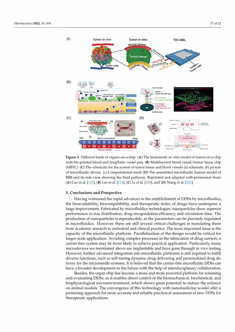
Figure 5. Different kinds of organs-on-a-chip. (A) The biomimetic in vitro model of tumor-on-a-chip with bio-printed blood and lymphatic vessel pair. (B) Multilayered blood vessel/tumor tissue chip (MBTC). (C) The schematic for the system of tumor tissue and blood vessels (a) schematic (b) picture of microfluidic device. (c) Computational mesh (D) The assembled microfluidic human model of BBB and its side view showing the fluid pathway. Reprinted and adapted with permission from: (A) Cao et al. [101], (B) Lee et al. [114], (C) Li et al. [100], and (D) Wang et al. [121].
5. Conclusions and Prospective Having witnessed the rapid advances in the establishment of DDSs by microfluidics,
the bioavailability, biocompatibility, and therapeutic index of drugs have undergone a huge improvement. Fabricated by microfluidics technologies, nanoparticles show supe-rior performance in size distribution, drug encapsulation efficiency, and circulation time. The production of nanoparticles is reproducible, as the parameters can be precisely regu-lated in microfluidics. However, there are still several critical challenges in translating them from academic research to industrial and clinical practice. The most important issue is the capacity of the microfluidic platform. Parallelization of the design would be critical for larger-scale application. Avoiding complex processes in the fabrication of drug carri-ers, a carrier-free system may be more likely to achieve practical application. Particularly, many microdevices we mentioned above are implantable and have gone through in vivo testing. However, further advanced integration into microfluidic platforms is still re-quired to fulfill diverse functions, such as self-tuning dynamic drug delivering and per-sonalized drug delivery for the microneedle systems. It is believed that the carrier-free microfluidic DDSs can have a broader development in the future with the help of inter-disciplinary collaboration.
Besides, the organ chip has become a more and more powerful platform for screening and evaluating DDSs, as it enables direct control of the biomechanical, biochemical, and biophysiological microenvironment, which shows great potential to reduce the reliance on animal models. The convergence of this technology with nanomedicine would offer a promising approach for more accurate and reliable preclinical assessment of new DDSs for therapeutic applications.
Figure 5. Different kinds of organs-on-a-chip. (A) The biomimetic in vitro model of tumor-on-a-chipwith bio-printed blood and lymphatic vessel pair. (B) Multilayered blood vessel/tumor tissue chip(MBTC). (C) The schematic for the system of tumor tissue and blood vessels (a) schematic (b) pictureof microfluidic device. (c) Computational mesh (D) The assembled microfluidic human model ofBBB and its side view showing the fluid pathway. Reprinted and adapted with permission from:(A) Cao et al. [101], (B) Lee et al. [114], (C) Li et al. [100], and (D) Wang et al. [121].
5. Conclusions and Prospective
Having witnessed the rapid advances in the establishment of DDSs by microfluidics,the bioavailability, biocompatibility, and therapeutic index of drugs have undergone ahuge improvement. Fabricated by microfluidics technologies, nanoparticles show superiorperformance in size distribution, drug encapsulation efficiency, and circulation time. Theproduction of nanoparticles is reproducible, as the parameters can be precisely regulatedin microfluidics. However, there are still several critical challenges in translating themfrom academic research to industrial and clinical practice. The most important issue is thecapacity of the microfluidic platform. Parallelization of the design would be critical forlarger-scale application. Avoiding complex processes in the fabrication of drug carriers, acarrier-free system may be more likely to achieve practical application. Particularly, manymicrodevices we mentioned above are implantable and have gone through in vivo testing.However, further advanced integration into microfluidic platforms is still required to fulfilldiverse functions, such as self-tuning dynamic drug delivering and personalized drug de-livery for the microneedle systems. It is believed that the carrier-free microfluidic DDSs canhave a broader development in the future with the help of interdisciplinary collaboration.
Besides, the organ chip has become a more and more powerful platform for screeningand evaluating DDSs, as it enables direct control of the biomechanical, biochemical, andbiophysiological microenvironment, which shows great potential to reduce the relianceon animal models. The convergence of this technology with nanomedicine would offer apromising approach for more accurate and reliable preclinical assessment of new DDSs fortherapeutic applications.
Pharmaceutics 2022, 14, 434 18 of 22
Author Contributions: Conceptualization, Z.M., D.G.; writing—original draft preparation, Z.M.and B.L. and J.P.; writing—review and editing, Z.M., D.G.; supervision, project administration andfunding acquisition D.G. All authors have read and agreed to the published version of the manuscript.
Funding: This work was supported by the Natural Science Foundation of Guangdong Province,China (No. 2020A1515010660) and the Shenzhen Fundamental Research and Discipline Layoutproject (JCYJ20180508152244835).
Institutional Review Board Statement: This study did not require ethical approval.
Informed Consent Statement: This study did not involve humans.
Data Availability Statement: This study did not report any data.
Conflicts of Interest: The authors declare no conflict of interest.
References1. Li, C.; Wang, J.; Wang, Y.; Gao, H.; Wei, G.; Huang, Y.; Yu, H.; Gan, Y.; Wang, Y.; Mei, L.; et al. Recent progress in drug delivery.
Acta Pharm. Sin. B 2019, 9, 1145–1162. [CrossRef] [PubMed]2. Sanjay, S.T.; Zhou, W.; Dou, M.; Tavakoli, H.; Ma, L.; Xu, F.; Li, X. Recent advances of controlled drug delivery using microfluidic
platforms. Adv. Drug Deliv. Rev. 2018, 128, 3–28. [CrossRef] [PubMed]3. Park, K. Controlled drug delivery systems: Past forward and future back. J. Control. Release 2014, 190, 3–8. [CrossRef] [PubMed]4. Cochrane, A.; Albers, H.J.; Passier, R.; Mummery, C.L.; van den Berg, A.; Orlova, V.V.; van der Meer, A.D. Advanced in vitro
models of vascular biology: Human induced pluripotent stem cells and organ-on-chip technology. Adv. Drug Deliv. Rev. 2019,140, 68–77. [CrossRef] [PubMed]
5. Bendre, A.; Bhat, M.P.; Lee, K.-H.; Altalhi, T.; Alruqi, M.A.; Kurkuri, M. Recent developments in microfluidic technology forsynthesis and toxicity-efficiency studies of biomedical nanomaterials. Mater. Today Adv. 2022, 13, 100205. [CrossRef]
6. Hu, W.; Gao, D.; Su, Z.; Qian, R.; Wang, Y.; Liang, Q. A cellular chip-MS system for investigation of Lactobacillus rhamnosus GGand irinotecan synergistic effects on colorectal cancer. Chin. Chem. Lett. 2021, in press. [CrossRef]
7. Wang, C.; Hu, W.; Guan, L.; Yang, X.; Liang, Q. Single-cell metabolite analysis on a microfluidic chip. Chin. Chem. Lett. 2021, in press.[CrossRef]
8. Zhou, H.; Gao, H.; Yuan, F.; Yang, F.; Gao, Y. Pressure-Controlled-Type Flexible Micro-Needle Delivery System, Has Micro-PumpConnected with One-Way Valve, and Microfluidic Network Access Part Connected with Micro-Fluidic Channel and MicrofluidicNetwork. CN207085071-U, 29 June 2018.
9. Zhou, H.; Gao, H.; Yuan, F.; Yang, F.; Gao, Y. Pressure-Controlled Flexible Microneedle Drug Delivery System, Has Micro-NeedleDelivery System Main Body Connected with Microfluidic Network and Micro-Fluidic Channel that Is Provided with Micro-Pumpand One-Way Valve. CN106390277-A, 6 February 2017.
10. Shi, Y.; Gao, Y.; Kong, D.; Fu, M.; Jiang, B. Microfluidic Chip for High-Throughput Screening of Nano Particles for TransvascularTransport, Has First Fluid Channel Communicated with Second Fluid Channel Through Gap Channel, End of Second FluidChannel Provided with seepage Outlet. CN110773244-A; CN110773244-B; KR2021048990-A; KR2249533-B1, 9 March 2020.
11. Yu, H. Microfluidic Mixing Chip Box for Generating Parallel high Throughput Nanometer Particle, Has Liquid Outlet of Mixing Pipelineone End of Tail Portion Connected to Liquid Outlet Pipeline which is Connected to Liquid Outlet Port. CN111389281-A, 29 July 2020.
12. Kapur, R.; Smith, K.C.; Toner, M. Microfluidic Device Used to Extract and Concentrate Particles from Fluid, Comprises Fluid ExchangeModule, and Particle Concentration Module, Fluidly Coupled to Fluid Exchange Module. US2020139370-A1, 20 May 2020.
13. Huang, G.; Huang, H. Hyaluronic acid-based biopharmaceutical delivery and tumor-targeted drug delivery system. J. Control.Release 2018, 278, 122–126. [CrossRef]
14. Zhang, L.; Chen, Q.; Ma, Y.; Sun, J. Microfluidic Methods for Fabrication and Engineering of Nanoparticle Drug Delivery Systems.ACS Appl. Bio Mater. 2020, 3, 107–120. [CrossRef]
15. Lababidi, N.; Sigal, V.; Koenneke, A.; Schwarzkopf, K.; Manz, A.; Schneider, M. Microfluidics as tool to prepare size-tunable PLGAnanoparticles with high curcumin encapsulation for efficient mucus penetration. Beilstein J. Nanotechnol. 2019, 10, 2280–2293.[CrossRef]
16. Ogundele, M.; Okafor, H. Transdermal drug delivery: Microneedles, their fabrication and current trends in delivery methods. J.Pharm. Res. Int. 2017, 18, 1–14. [CrossRef]
17. Riahi, R.; Tamayol, A.; Shaegh, S.A.M.; Ghaemmaghami, A.; Dokmeci, M.R.; Khademshosseini, A. Microfluidics for AdvancedDrug Delivery Systems. Curr. Opin. Chem. Eng. 2015, 7, 101–112. [CrossRef] [PubMed]
18. Chiesa, E.; Dorati, R.; Pisani, S.; Conti, B.; Bergamini, G.; Modena, T.; Genta, I. The Microfluidic Technique and the Manufacturingof Polysaccharide Nanoparticles. Pharmaceutics 2018, 10, 267. [CrossRef] [PubMed]
19. Jahn, A.; Vreeland, W.N.; Gaitan, M.; Locascio, L.E. Controlled Vesicle Self-Assembly in Microfluidic Channels with HydrodynamicFocusing. J. Am. Chem. Soc. 2004, 126, 2674–2675. [CrossRef]
20. Le, N.H.A.; Phan, H.V.; Yu, J.; Chan, H.-K.; Neild, A.; Alan, T. Acoustically enhanced microfluidic mixer to synthesize highlyuniform nanodrugs without the addition of stabilizers. Int. J. Nanomed. 2018, 13, 1353–1359. [CrossRef]
Pharmaceutics 2022, 14, 434 19 of 22
21. Kimura, N.; Maeki, M.; Sato, Y.; Note, Y.; Ishida, A.; Tani, H.; Harashima, H.; Tokeshi, M. Development of the iLiNP Device: FineTuning the Lipid Nanoparticle Size within 10 nm for Drug Delivery. ACS Omega 2018, 3, 5044–5051. [CrossRef]
22. Yanar, F.; Mosayyebi, A.; Nastruzzi, C.; Carugo, D.; Zhang, X. Continuous-Flow Production of Liposomes with a Millireactorunder Varying Fluidic Conditions. Pharmaceutics 2020, 12, 1001. [CrossRef]
23. Shaji, M.; Mudigunda, V.S.; Appidi, T.; Jain, S.; Rengan, A.K.; Unni, H.N. Microfluidic design of tumor vasculature andnanoparticle uptake by cancer cells. Microfluid. Nanofluid. 2021, 25, 46. [CrossRef]
24. Wang, J.; Zhang, N.; Chen, J.; Rodgers, V.G.J.; Brisk, P.; Grover, W.H. Finding the optimal design of a passive microfluidic mixer.Lab Chip 2019, 19, 3618–3627. [CrossRef]
25. Lou, G.; Anderluzzi, G.; Woods, S.; Roberts, C.W.; Perrie, Y. A novel microfluidic-based approach to formulate size-tuneable largeunilamellar cationic liposomes: Formulation, cellular uptake and biodistribution investigations. Eur. J. Pharm. Biopharm. 2019,143, 51–60. [CrossRef]
26. Lin, W.-Z.S.; Malmstadt, N. Liposome production and concurrent loading of drug simulants by microfluidic hydrodynamicfocusing. Eur. Biophys. J. 2019, 48, 549–558. [CrossRef] [PubMed]
27. Balbino, T.A.; Serafin, J.M.; Radaic, A.; de Jesus, M.B.; de la Torre, L.G. Integrated microfluidic devices for the synthesis ofnanoscale liposomes and lipoplexes. Colloids Surf. B Biointerfaces 2017, 152, 406–413. [CrossRef] [PubMed]
28. Huang, N.; Lu, S.; Liu, X.-G.; Zhu, J.; Wang, Y.-J.; Liu, R.-T. PLGA nanoparticles modified with a BBB-penetrating peptideco-delivering Aβ generation inhibitor and curcumin attenuate memory deficits and neuropathology in Alzheimer’s disease mice.Oncotarget 2017, 8, 81001–81013. [CrossRef] [PubMed]
29. Leung, M.H.M.; Shen, A.Q. Microfluidic Assisted Nanoprecipitation of PLGA Nanoparticles for Curcumin Delivery to LeukemiaJurkat Cells. Langmuir 2018, 34, 3961–3970. [CrossRef]
30. Yu, X.; Cheng, G.; Zhou, M.-D.; Zheng, S.-Y. On-Demand One-Step Synthesis of Monodisperse Functional Polymeric Microsphereswith Droplet Microfluidics. Langmuir 2015, 31, 3982–3992. [CrossRef]
31. Sundararajan, P.; Wang, J.; Rosen, L.A.; Procopio, A.; Rosenberg, K. Engineering polymeric Janus particles for drug delivery usingmicrofluidic solvent dissolution approach. Chem. Eng. Sci. 2018, 178, 199–210. [CrossRef]
32. Naderi, A.; Bhattacharjee, N.; Folch, A. Digital Manufacturing for Microfluidics. Annu. Rev. Biomed. Eng. 2019, 21, 325–364.[CrossRef]
33. Abualsayed, A.M.; Abouelmagd, S.A.; Abdelgawad, M. Miniaturised preparation of polymeric nanoparticles using dropletmanipulation on open surfaces. Micro Nano Lett. 2019, 14, 1312–1316. [CrossRef]
34. Li, S.; Su, W.; Wu, H.; Yuan, T.; Yuan, C.; Liu, J.; Deng, G.; Gao, X.; Chen, Z.; Bao, Y.; et al. Targeted tumour theranostics in micevia carbon quantum dots structurally mimicking large amino acids. Nat. Biomed. Eng. 2020, 4, 704–716. [CrossRef]
35. Hettiarachchi, S.D.; Graham, R.M.; Mintz, K.J.; Zhou, Y.; Vanni, S.; Peng, Z.; Leblanc, R.M. Triple conjugated carbon dots as anano-drug delivery model for glioblastoma brain tumors. Nanoscale 2019, 11, 6192–6205. [CrossRef]
36. Guidelli, E.J.; Lignos, I.; Yoo, J.J.; Lusardi, M.; Bawendi, M.G.; Baffa, O.; Jensen, K.F. Mechanistic Insights and Controlled Synthesisof Radioluminescent ZnSe Quantum Dots Using a Microfluidic Reactor. Chem. Mater. 2018, 30, 8562–8570. [CrossRef]
37. Baek, J.; Shen, Y.; Lignos, I.; Bawendi, M.G.; Jensen, K.F. Multistage Microfluidic Platform for the Continuous Synthesis of III–VCore/Shell Quantum Dots. Angew. Chem. Int. Ed. 2018, 57, 10915–10918. [CrossRef]
38. Castillo, R.R.; Lozano, D.; Vallet-Regí, M. Building Block Based Construction of Membrane-Organelle Double Targeted Nanosys-tem for Two-Drug Delivery. Bioconj. Chem. 2018, 29, 3677–3685. [CrossRef]
39. Yan, J.; Xu, X.; Zhou, J.; Liu, C.; Zhang, L.; Wang, D.; Yang, F.; Zhang, H. Fabrication of a pH/Redox-Triggered MesoporousSilica-Based Nanoparticle with Microfluidics for Anticancer Drugs Doxorubicin and Paclitaxel Codelivery. ACS Appl. Bio Mater.2020, 3, 1216–1225. [CrossRef]
40. Zhao, X.; Liu, Y.; Yu, Y.; Huang, Q.; Ji, W.; Li, J.; Zhao, Y. Hierarchically porous composite microparticles from microfluidics forcontrollable drug delivery. Nanoscale 2018, 10, 12595–12604. [CrossRef]
41. Bchellaoui, N.; Hayat, Z.; Mami, M.; Dorbez-Sridi, R.; El Abed, A.I. Microfluidic-assisted Formation of Highly Monodisperse andMesoporous Silica Soft Microcapsules. Sci. Rep. 2017, 7, 16326. [CrossRef]
42. Eltorai, A.E.; Fox, H.; McGurrin, E.; Guang, S. Microchips in Medicine: Current and Future Applications. Biomed. Res. Int. 2016,2016, 1743472. [CrossRef]
43. Gensler, H.; Sheybani, R.; Li, P.Y.; Mann, R.L.; Meng, E. An implantable MEMS micropump system for drug delivery in smallanimals. Biomed. Microdevices 2012, 14, 483–496. [CrossRef]
44. Vladisavljevic, G.T.; Khalid, N.; Neves, M.A.; Kuroiwa, T.; Nakajima, M.; Uemura, K.; Ichikawa, S.; Kobayashi, I. Industriallab-on-a-chip: Design, applications and scale-up for drug discovery and delivery. Adv. Drug Deliv. Rev. 2013, 65, 1626–1663.[CrossRef]
45. Silva, A.C.Q.; Vilela, C.; Santos, H.A.; Silvestre, A.J.D.; Freire, C.S.R. Recent trends on the development of systems for cancerdiagnosis and treatment by microfluidic technology. Appl. Mater. Today 2020, 18, 100450. [CrossRef]
46. Liu, D.; Zhang, H.; Fontana, F.; Hirvonen, J.T.; Santos, H.A. Current developments and applications of microfluidic technologytoward clinical translation of nanomedicines. Adv. Drug Deliv. Rev. 2018, 128, 54–83. [CrossRef]
47. Logesh, D.; Vallikkadan, M.S.; Leena, M.M.; Moses, J.A.; Anandharamakrishnan, C. Advances in microfluidic systems for thedelivery of nutraceutical ingredients. Trends Food Sci. Technol. 2021, 116, 501–524. [CrossRef]
Pharmaceutics 2022, 14, 434 20 of 22
48. Khan, I.U.; Serra, C.A.; Anton, N.; Vandamme, T. Microfluidics: A focus on improved cancer targeted drug delivery systems. J.Control. Release 2013, 172, 1065–1074. [CrossRef]
49. Muheem, A.; Shakeel, F.; Jahangir, M.A.; Anwar, M.; Mallick, N.; Jain, G.K.; Warsi, M.H.; Ahmad, F.J. A review on the strategiesfor oral delivery of proteins and peptides and their clinical perspectives. Saudi Pharm. J. 2016, 24, 413–428. [CrossRef]
50. Nguyen, N.T.; Shaegh, S.A.; Kashaninejad, N.; Phan, D.T. Design, fabrication and characterization of drug delivery systems basedon lab-on-a-chip technology. Adv. Drug Deliv. Rev. 2013, 65, 1403–1419. [CrossRef]
51. Yang, Q.; Zhong, W.; Xu, L.; Li, H.; Yan, Q.; She, Y.; Yang, G. Recent progress of 3D-printed microneedles for transdermal drugdelivery. Int. J. Pharm. 2021, 593, 120106. [CrossRef]
52. Yang, J.; Liu, X.; Fu, Y.; Song, Y. Recent advances of microneedles for biomedical applications: Drug delivery and beyond. ActaPharm. Sin. B 2019, 9, 469–483. [CrossRef]
53. Singh, P.; Carrier, A.; Chen, Y.; Lin, S.; Wang, J.; Cui, S.; Zhang, X. Polymeric microneedles for controlled transdermal drugdelivery. J. Control. Release 2019, 315, 97–113. [CrossRef]
54. Amjadi, M.; Sheykhansari, S.; Nelson, B.J.; Sitti, M. Recent Advances in Wearable Transdermal Delivery Systems. Adv. Mater.2018, 30, 1704530. [CrossRef]
55. Larrañeta, E.; Lutton, R.E.M.; Woolfson, A.D.; Donnelly, R.F. Microneedle arrays as transdermal and intradermal drug deliverysystems: Materials science, manufacture and commercial development. Mater. Sci. Eng. R Rep. 2016, 104, 1–32. [CrossRef]
56. Prausnitz, M.R.; Langer, R. Transdermal drug delivery. Nat. Biotechnol. 2008, 26, 1261–1268. [CrossRef]57. Bilal, M.; Mehmood, S.; Raza, A.; Hayat, U.; Rasheed, T.; Iqbal, H.M.N. Microneedles in Smart Drug Delivery. Adv. Wound Care
2021, 10, 204–219. [CrossRef]58. Ahmad, N.N.; Ghazali, N.N.N.; Wong, Y.H. Concept Design of Transdermal Microneedles for Diagnosis and Drug Delivery: A
Review. Adv. Eng. Mater. 2021, 23, 2100503. [CrossRef]59. Thakur Singh, R.R.; Tekko, I.; McAvoy, K.; McMillan, H.; Jones, D.; Donnelly, R.F. Minimally invasive microneedles for ocular
drug delivery. Expert Opin. Drug Deliv. 2017, 14, 525–537. [CrossRef]60. Ortner, G.; Tzanaki, E.; Rai, B.P.; Nagele, U.; Tokas, T. Transperineal prostate biopsy: The modern gold standard to prostate cancer
diagnosis. Turk. J. Urol. 2021, 47, S19–S26. [CrossRef]61. Pradeep Narayanan, S.; Raghavan, S. Solid silicon microneedles for drug delivery applications. Int. J. Adv. Manuf. Technol. 2016,
93, 407–422. [CrossRef]62. Li, Q.Y.; Zhang, J.N.; Chen, B.Z.; Wang, Q.L.; Guo, X.D. A solid polymer microneedle patch pretreatment enhances the permeation
of drug molecules into the skin. RSC Adv. 2017, 7, 15408–15415. [CrossRef]63. DeMuth, P.C.; Su, X.; Samuel, R.E.; Hammond, P.T.; Irvine, D.J. Nano-layered microneedles for transcutaneous delivery of
polymer nanoparticles and plasmid DNA. Adv. Mater. 2010, 22, 4851–4856. [CrossRef]64. Seok, H.; Noh, J.Y.; Lee, D.Y.; Kim, S.J.; Song, C.S.; Kim, Y.C. Effective humoral immune response from a H1N1 DNA vaccine
delivered to the skin by microneedles coated with PLGA-based cationic nanoparticles. J. Control. Release 2017, 265, 66–74.[CrossRef]
65. Tröls, A.; Hintermüller, M.A.; Saeedipour, M.; Pirker, S.; Jakoby, B. Drug dosage for microneedle-based transdermal drug deliverysystems utilizing evaporation-induced droplet transport. Microfluid. Nanofluid. 2019, 23, 91. [CrossRef]
66. Cha, K.J.; Kim, T.; Park, S.J.; Kim, D.S. Simple and cost-effective fabrication of solid biodegradable polymer microneedle arrayswith adjustable aspect ratio for transdermal drug delivery using acupuncture microneedles. J. Micromech. Microeng. 2014, 24, 115015.[CrossRef]
67. Guo, T.; Cheng, N.; Zhao, J.; Hou, X.; Zhang, Y.; Feng, N. Novel nanostructured lipid carriers-loaded dissolving microneedles forcontrolled local administration of aconitine. Int. J. Pharm. 2019, 572, 118741. [CrossRef] [PubMed]
68. Zhao, X.; Li, X.; Zhang, P.; Du, J.; Wang, Y. Tip-loaded fast-dissolving microneedle patches for photodynamic therapy ofsubcutaneous tumor. J. Control. Release 2018, 286, 201–209. [CrossRef] [PubMed]
69. Hardy, J.G.; Larraneta, E.; Donnelly, R.F.; McGoldrick, N.; Migalska, K.; McCrudden, M.T.; Irwin, N.J.; Donnelly, L.; McCoy, C.P.Hydrogel-Forming Microneedle Arrays Made from Light-Responsive Materials for On-Demand Transdermal Drug Delivery. Mol.Pharm. 2016, 13, 907–914. [CrossRef]
70. Chen, S.; Matsumoto, H.; Moro-oka, Y.; Tanaka, M.; Miyahara, Y.; Suganami, T.; Matsumoto, A. Microneedle-Array Patch Fabricatedwith Enzyme-Free Polymeric Components Capable of On-Demand Insulin Delivery. Adv. Funct. Mater. 2018, 29, 1807369. [CrossRef]
71. Vithani, K.; Goyanes, A.; Jannin, V.; Basit, A.W.; Gaisford, S.; Boyd, B.J. An overview of 3D printing technologies for soft materialsand potential opportunities for lipid-based drug delivery systems. Pharm. Res. 2019, 36, 1–20. [CrossRef]
72. Ito, Y.; Hagiwara, E.; Saeki, A.; Sugioka, N.; Takada, K. Feasibility of microneedles for percutaneous absorption of insulin. Eur J.Pharm. Sci. 2006, 29, 82–88. [CrossRef]
73. Trautmann, A.; Roth, G.L.; Nujiqi, B.; Walther, T.; Hellmann, R. Towards a versatile point-of-care system combining femtosecondlaser generated microfluidic channels and direct laser written microneedle arrays. Microsyst. Nanoeng. 2019, 5, 6. [CrossRef]
74. Jayaneththi, V.R.; Aw, K.; Sharma, M.; Wen, J.; Svirskis, D.; McDaid, A.J. Controlled transdermal drug delivery using a wirelessmagnetic microneedle patch: Preclinical device development. Sens. Actuators B Chem. 2019, 297, 126708. [CrossRef]
75. Hwang, T.H.; Kim, J.B.; Yang, D.S.; Park, Y.-i.; Ryu, W. Targeted electrohydrodynamic printing for micro-reservoir drug deliverysystems. J. Micromech. Microeng. 2013, 23, 35012. [CrossRef]
Pharmaceutics 2022, 14, 434 21 of 22
76. Nadia Ahmad, N.F.; Nik Ghazali, N.N.; Wong, Y.H. Wearable patch delivery system for artificial pancreas health diagnostic-therapeutic application: A review. Biosens. Bioelectron. 2021, 189, 113384. [CrossRef]
77. Bertrand, N.; Wu, J.; Xu, X.; Kamaly, N.; Farokhzad, O.C. Cancer nanotechnology: The impact of passive and active targeting inthe era of modern cancer biology. Adv. Drug Deliv. Rev. 2014, 66, 2–25. [CrossRef]
78. Eljarrat-Binstock, E.; Pe’er, J.; Domb, A.J. New techniques for drug delivery to the posterior eye segment. Pharm. Res. 2010, 27,530–543. [CrossRef]
79. Mansoor, S.; Kuppermann, B.D.; Kenney, M.C. Intraocular sustained-release delivery systems for triamcinolone acetonide. Pharm.Res. 2009, 26, 770–784. [CrossRef]
80. Fong, J.; Xiao, Z.; Takahata, K. Wireless implantable chip with integrated nitinol-based pump for radio-controlled local drugdelivery. Lab Chip 2015, 15, 1050–1058. [CrossRef]
81. Kroon, E.; Martinson, L.A.; Kadoya, K.; Bang, A.G.; Kelly, O.G.; Eliazer, S.; Young, H.; Richardson, M.; Smart, N.G.;Cunningham, J.; et al. Pancreatic endoderm derived from human embryonic stem cells generates glucose-responsive insulin-secreting cells in vivo. Nat. Biotechnol. 2008, 26, 443–452. [CrossRef]
82. Nagai, N.; Kaji, H.; Onami, H.; Katsukura, Y.; Ishikawa, Y.; Nezhad, Z.K.; Sampei, K.; Iwata, S.; Ito, S.; Nishizawa, M.; et al. Aplatform for controlled dual-drug delivery to the retina: Protective effects against light-induced retinal damage in rats. Adv.Healthc. Mater. 2014, 3, 1555–1560. [CrossRef]
83. Deshpande, S.; Pinsker, J.E.; Zavitsanou, S.; Shi, D.; Tompot, R.; Church, M.M.; Andre, C.; Doyle, F.J., 3rd; Dassau, E. Design andClinical Evaluation of the Interoperable Artificial Pancreas System (iAPS) Smartphone App: Interoperable Components withModular Design for Progressive Artificial Pancreas Research and Development. Diabetes Technol. Ther. 2019, 21, 35–43. [CrossRef]
84. Yang, D.; Lee, J.S.; Choi, C.K.; Lee, H.P.; Cho, S.W.; Ryu, W. Microchannel system for rate-controlled, sequential, and pH-responsivedrug delivery. Acta Biomater. 2018, 68, 249–260. [CrossRef]
85. Lee, H.P.; Ryu, W. Wet microcontact printing (microCP) for micro-reservoir drug delivery systems. Biofabrication 2013, 5, 025011.[CrossRef]
86. Ahadian, S.; Finbloom, J.A.; Mofidfar, M.; Diltemiz, S.E.; Nasrollahi, F.; Davoodi, E.; Hosseini, V.; Mylonaki, I.; Sangabathuni, S.;Montazerian, H. Micro and nanoscale technologies in oral drug delivery. Adv. Drug Deliv. Rev. 2020, 157, 37–62. [CrossRef][PubMed]
87. Boegh, M.; Nielsen, H.M. Mucus as a Barrier to Drug Delivery—Understanding and Mimicking the Barrier Properties. Basic Clin.Pharmacol. Toxicol. 2015, 116, 179–186. [CrossRef] [PubMed]
88. Boegh, M.; Foged, C.; Müllertz, A.; Mørck Nielsen, H. Mucosal drug delivery: Barriers, in vitro models and formulation strategies.J. Drug Deliv. Sci. Technol. 2013, 23, 383–391. [CrossRef]
89. Kadry, H.; Noorani, B.; Cucullo, L. A blood–brain barrier overview on structure, function, impairment, and biomarkers ofintegrity. Fluids Barriers CNS 2020, 17, 1–24. [CrossRef]
90. Jia, Z.; Guo, Z.; Yang, C.-T.; Prestidge, C.; Thierry, B. “Mucus-on-Chip”: A new tool to study the dynamic penetration ofnanoparticulate drug carriers into mucus. Int. J. Pharm. 2021, 598, 120391. [CrossRef]
91. Ramesan, S.; Rezk, A.R.; Yeo, L.Y. High frequency acoustic permeabilisation of drugs through tissue for localised mucosal delivery.Lab Chip 2018, 18, 3272–3284. [CrossRef]
92. Lechanteur, A.; das Neves, J.; Sarmento, B. The role of mucus in cell-based models used to screen mucosal drug delivery. Adv.Drug Deliv. Rev. 2018, 124, 50–63. [CrossRef]
93. Hagiwara, Y.; Kumagai, H.; Ouwerkerk, N.; Gijzen, L.; Annida, R.; Bokkers, M.; van Vught, R.; Yoshinari, K.; Katakawa, Y.;Motonaga, K.; et al. A Novel In Vitro Membrane Permeability Methodology Using Three-dimensional Caco-2 Tubules in aMicrophysiological System Which Better Mimics In Vivo Physiological Conditions. J. Pharm. Sci. 2022, 111, 214–224. [CrossRef]
94. Gholizadeh, H.; Ong, H.X.; Bradbury, P.; Kourmatzis, A.; Traini, D.; Young, P.; Li, M.; Cheng, S. Real-time quantitative monitoringof in vitro nasal drug delivery by a nasal epithelial mucosa-on-a-chip model. Expert Opin. Drug Deliv. 2021, 18, 803–818. [CrossRef]
95. Ahn, J.; Ko, J.; Lee, S.; Yu, J.; Kim, Y.; Jeon, N.L. Microfluidics in nanoparticle drug delivery; From synthesis to pre-clinicalscreening. Adv. Drug Deliv. Rev. 2018, 128, 29–53. [CrossRef]
96. Roovers, S.; Segers, T.; Lajoinie, G.; Deprez, J.; Versluis, M.; De Smedt, S.C.; Lentacker, I. The Role of Ultrasound-Driven MicrobubbleDynamics in Drug Delivery: From Microbubble Fundamentals to Clinical Translation. Langmuir 2019, 35, 10173–10191. [CrossRef][PubMed]
97. Thomas, A.; Tan, J.; Liu, Y. Characterization of nanoparticle delivery in microcirculation using a microfluidic device. Microvasc.Res. 2014, 94, 17–27. [CrossRef] [PubMed]
98. Khoury, M.; Epshtein, M.; Zidan, H.; Zukerman, H.; Korin, N. Mapping deposition of particles in reconstructed models of humanarteries. J. Control. Release 2020, 318, 78–85. [CrossRef]
99. Cooley, M.; Sarode, A.; Hoore, M.; Fedosov, D.A.; Mitragotri, S.; Gupta, A.S. Influence of particle size and shape on theirmargination and wall-adhesion: Implications in drug delivery vehicle design across nano-to-micro scale. Nanoscale 2018, 10,15350–15364. [CrossRef]
100. Li, W.; Wang, H.-F.; Li, Z.-Y.; Wang, T.; Zhao, C.-X. Numerical investigation of drug transport from blood vessels to tumour tissueusing a Tumour-Vasculature-on-a-Chip. Chem. Eng. Sci. 2019, 208, 115155. [CrossRef]
101. Cao, X.; Ashfaq, R.; Cheng, F.; Maharjan, S.; Li, J.; Ying, G.; Hassan, S.; Xiao, H.; Yue, K.; Zhang, Y.S. A Tumor-on-a-Chip Systemwith Bioprinted Blood and Lymphatic Vessel Pair. Adv. Funct. Mater. 2019, 29, 1807173. [CrossRef]
Pharmaceutics 2022, 14, 434 22 of 22
102. De Luca, R.; Silvani, G.; Scognamiglio, C.; Sinibaldi, G.; Peruzzi, G.; Chinappi, M.; Kiani, M.F.; Casciola, C.M. Towardscavitation-enhanced permeability in blood vessel on a chip. AIP Conf. Proc. 2017, 1873, 020010. [CrossRef]
103. Prausnitz, M.R.; Elias, P.M.; Franz, T.J.; Schmuth, M.; Tsai, J.C.; Menon, G.K.; Holleran, W.M.; Feingold, K.R. Skin barrier andtransdermal drug delivery. Dermatology 2012, 3, 2065–2073.
104. Xiang, Y.; Wen, H.; Yu, Y.; Li, M.; Fu, X.; Huang, S. Gut-on-chip: Recreating human intestine in vitro. J. Tissue Eng. 2020, 11,2041731420965318. [CrossRef]
105. Citi, S. Intestinal barriers protect against disease. Science 2018, 359, 1097–1098. [CrossRef]106. Xu, Y.; Shrestha, N.; Préat, V.; Beloqui, A. Overcoming the intestinal barrier: A look into targeting approaches for improved oral
drug delivery systems. J. Control. Release 2020, 322, 486–508. [CrossRef] [PubMed]107. Di Tommaso, N.; Gasbarrini, A.; Ponziani, F.R. Intestinal Barrier in Human Health and Disease. Int. J. Environ. Res. Public Health
2021, 18, 12836. [CrossRef] [PubMed]108. Lieleg, O.; Ribbeck, K. Biological hydrogels as selective diffusion barriers. Trends Cell Biol. 2011, 21, 543–551. [CrossRef] [PubMed]109. Huang, J.; Li, Z.; Hu, Q.; Chen, G.; Ren, Y.; Wu, X.; Ren, J. Bioinspired Anti-digestive Hydrogels Selected by a Simulated Gut
Microfluidic Chip for Closing Gastrointestinal Fistula. iScience 2018, 8, 40–48. [CrossRef]110. Lee, D.W.; Ha, S.K.; Choi, I.; Sung, J.H. 3D gut-liver chip with a PK model for prediction of first-pass metabolism. Biomed.
Microdevices 2017, 19, 100. [CrossRef] [PubMed]111. Herland, A.; Maoz, B.M.; Das, D.; Somayaji, M.R.; Prantil-Baun, R.; Novak, R.; Cronce, M.; Huffstater, T.; Jeanty, S.S.F.; Ingram, M.; et al.
Quantitative prediction of human drug pharmacokinetic responses using multiple vascularized organ chips coupled by fluidtransfer. Nat. Biomed. Eng. 2020, 4, 421–436. [CrossRef] [PubMed]
112. Abbott, N.J.; Rönnbäck, L.; Hansson, E. Astrocyte–endothelial interactions at the blood–brain barrier. Nat. Rev. Neurosci. 2006, 7,41–53. [CrossRef]
113. Ningaraj, N.S. Drug delivery to brain tumours: Challenges and progress. Expert Opin. Drug Deliv. 2006, 3, 499–509. [CrossRef]114. Lee, J.; Kim, S.-E.; Moon, D.; Doh, J. A multilayered blood vessel/tumor tissue chip to investigate T cell infiltration into solid
tumor tissues. Lab Chip 2021, 21, 2142–2152. [CrossRef]115. Deng, Z.; Sheng, Z.; Yan, F. Ultrasound-Induced Blood-Brain-Barrier Opening Enhances Anticancer Efficacy in the Treatment of
Glioblastoma: Current Status and Future Prospects. J. Oncol. 2019, 2019, 2345203. [CrossRef]116. Nair, K.G.S.; Ramaiyan, V.; Sukumaran, S.K. Enhancement of drug permeability across blood brain barrier using nanoparticles in
meningitis. Inflammopharmacology 2018, 26, 675–684. [CrossRef] [PubMed]117. Begley, D.J. Delivery of therapeutic agents to the central nervous system: The problems and the possibilities. Pharmacol. Ther.
2004, 104, 29–45. [CrossRef] [PubMed]118. De Boer, A.G.; Gaillard, P.J. Drug targeting to the brain. Annu. Rev. Pharm. Toxicol 2007, 47, 323–355. [CrossRef]119. Ciechanover, A. Proteolysis: From the lysosome to ubiquitin and the proteasome. Nat. Rev. Mol. Cell Biol. 2005, 6, 79–87.
[CrossRef]120. Kim, J.; Ahn, S.I.; Kim, Y. Nanotherapeutics engineered to cross the blood-brain barrier for advanced drug delivery to the central
nervous system. J. Ind. Eng. Chem. 2019, 73, 8–18. [CrossRef]121. Wang, Y.I.; Abaci, H.E.; Shuler, M.L. Microfluidic blood–brain barrier model provides in vivo-like barrier properties for drug
permeability screening. Biotechnol. Bioeng. 2017, 114, 184–194. [CrossRef]122. Lauranzano, E.; Campo, E.; Rasile, M.; Molteni, R.; Pizzocri, M.; Passoni, L.; Bello, L.; Pozzi, D.; Pardi, R.; Matteoli, M.; et al.
A Microfluidic Human Model of Blood–Brain Barrier Employing Primary Human Astrocytes. Adv. Biosyst. 2019, 3, 1800335.[CrossRef]
123. Jeong, S.; Kim, S.; Buonocore, J.; Park, J.; Welsh, C.J.; Li, J.; Han, A. A Three-Dimensional Arrayed Microfluidic Blood–BrainBarrier Model with Integrated Electrical Sensor Array. IEEE Trans. Biomed. Eng. 2018, 65, 431–439. [CrossRef]
124. Chung, B.; Kim, J.; Nam, J.; Kim, H.; Jeong, Y.; Liu, H.-W.; Cho, Y.; Kim, Y.H.; Oh, H.J.; Chung, S. Evaluation of Cell-PenetratingPeptides Using Microfluidic In Vitro 3D Brain Endothelial Barrier. Macromol. Biosci. 2020, 20, 1900425. [CrossRef]
125. Campisi, M.; Shin, Y.; Osaki, T.; Hajal, C.; Chiono, V.; Kamm, R.D. 3D self-organized microvascular model of the humanblood-brain barrier with endothelial cells, pericytes and astrocytes. Biomaterials 2018, 180, 117–129. [CrossRef]
126. Park, T.-E.; Mustafaoglu, N.; Herland, A.; Hasselkus, R.; Mannix, R.; FitzGerald, E.A.; Prantil-Baun, R.; Watters, A.; Henry, O.;Benz, M.; et al. Hypoxia-enhanced Blood-Brain Barrier Chip recapitulates human barrier function and shuttling of drugs andantibodies. Nat. Commun. 2019, 10, 2621. [CrossRef]
127. De Jong, E.; Williams, D.S.; Abdelmohsen, L.K.E.A.; Van Hest, J.C.M.; Zuhorn, I.S. A filter-free blood-brain barrier model toquantitatively study transendothelial delivery of nanoparticles by fluorescence spectroscopy. J. Control. Release 2018, 289, 14–22.[CrossRef]
128. Ahn, S.I.; Sei, Y.J.; Park, H.-J.; Kim, J.; Ryu, Y.; Choi, J.J.; Sung, H.-J.; MacDonald, T.J.; Levey, A.I.; Kim, Y. Microengineered humanblood–brain barrier platform for understanding nanoparticle transport mechanisms. Nat. Commun. 2020, 11, 175. [CrossRef]
129. Duran-Lobato, M.; Niu, Z.; Alonso, M.J. Oral Delivery of Biologics for Precision Medicine. Adv. Mater. 2020, 32, e1901935.[CrossRef]
130. Karasov, W.H. Integrative physiology of transcellular and paracellular intestinal absorption. J. Exp. Biol. 2017, 220, 2495–2501.[CrossRef]