drug delivery to tumours: recent strategies
TRANSCRIPT
1231
Review Article
JPP 2005, 57: 1231–1242
� 2005 The Authors
Received April 9, 2005
Accepted June 6, 2005
DOI 10.1211/jpp.57.10.0001
ISSN 0022-3573
Drug Delivery Research
Laboratory, Center of Relevance
and Excellence in NDDS,
Pharmacy Department,
G. H. Patel Building, Donor’s
Plaza, Fatehgunj, M.S. University
of Baroda, Baroda-390002,
Gujarat, India
L. Harivardhan Reddy
Correspondence: L. H. Reddy,
Drug Delivery Research
Laboratory, Center of Relevance
and Excellence in NDDS,
Pharmacy Department, G.H. Patel
Building, Donor’s Plaza,
Fatehgunj, M.S. University of
Baroda, Baroda-390002,
Gujarat, India. E-mail:
Drug delivery to tumours: recent strategies
L. Harivardhan Reddy
Abstract
Despite several advancements in chemotherapy, the real therapy of cancer still remains a challenge.
The development of new anti-cancer drugs for the treatment of cancer has not kept pace with the
progress in cancer therapy, because of the nonspecific drug distribution resulting in low tumour
concentrations and systemic toxicity. The main hindrance for the distribution of anti-cancer agents
to the tumour site is the highly disorganized tumour vasculature, high blood viscosity in the tumour,
and high interstitial pressure within the tumour tissue. Recently, several approaches such as drug
modifications and development of new carrier systems for anti-cancer agents have been attempted
to enhance their tumour reach. Approaches such as drug delivery through enhanced permeability
and retention (EPR) effect have resulted in a significant improvement in concentration in tumours,
while approaches such as drug-carrier implants and microparticles have resulted in improvement in
local chemotherapy of cancer. This review discusses different strategies employed for the delivery of
anti-cancer agents to tumours, such as through EPR effect, local chemotherapeutic approaches using
drug delivery systems, and special strategies such as receptor-mediated delivery, pH-based carriers,
application of ultrasound and delivery to resistant tumour cells and brain using nanoparticles.
Introduction
Cancer is defined by two of its characteristic features – the uncontrolled cell growth notregulated by external signals and the capacity to invade tissues, metastasize andcolonize at distant sites. Cancers are of two types: those of epithelial origin areknown as carcinomas, and those of non-epithelial (mesenchymal) origin are calledsarcomas (Fenton & Longo 1998).
Despite several modes of therapy, such as chemotherapy, immunotherapy andradiotherapy, the therapy of cancer still remains a challenge. Systemic chemotherapyis the main treatment available for disseminated malignant disease. Progress in drugtherapy has resulted in the treatment of curative chemotherapy regimens for severaltumours. Chemotherapy, whether curative or palliative, requires multiple cycles oftreatment. Chemotherapeutic agents exhibit a dose–response effect and the cell kill isproportional to the drug exposure (Fenton & Longo 1998). Normal cells are alsosusceptible to the cytotoxic effects of chemotherapeutic agents and exhibit a dose–response effect, but the response curve is shifted relative to that of malignant cells(Fenton & Longo 1998). This difference represents the therapeutic index; the usefulnessof many chemotherapeutic drugs is limited by the fact that they have a narrow ther-apeutic index. Proliferative normal tissues, such as bone marrow and gastrointestinalmucosa, are generally the most susceptible to chemotherapy-induced toxicity. The abilityof chemotherapy to eradicate tumour cells without causing lethal host toxicity dependson drug selectivity. Although cytokinetics are important, other differences betweennormal and tumour cells in cellular processes, such as metabolic pathways and suscept-ibility to programmed cell death (Kerr et al 1972), also contribute. Chemotherapy hasbeen widely adopted for the treatment of cancer and as a result several chemotherapeuticagents have been introduced with potential anti-cancer activity. However, due to thenon-specificity of these drugs, chronic administration leads to severe systemic toxicity(Son et al 2003). In an attempt to improve therapy with these anti-cancer agents andreduce the associated toxicity, drug delivery systems have been introduced that candeliver these drugs at the site of interest (Poupaert & Couvreur, 2003).
Several drug delivery systems were introduced, namely liposomes, microparticles,polymeric conjugates, micelles and nanoparticles, to facilitate effective chemotherapy
with anti-cancer agents. The introduction of doxorubicinlong-circulating liposomes in the market for cancer ther-apy has brought a renewed interest in the field of targeteddrug delivery to cancers.
Recently, several new approaches have been adoptedfor delivering anti-cancer agents to the tumour tissues.
Enhanced permeability and retention
(EPR) effect
Tumours exhibit a unique character, namely enhancedpermeability and retention (EPR) effect, greatly differentfrom the normal tissues. The EPR effect renders tumourtissues to display several distinctive characteristics, such ashypervasculature, defective vascular architecture and adeficient lymphatic drainage system, which lead macro-molecules to be accumulated preferentially and to beretained to a greater extent in tumour tissues than innormal tissues (Maeda et al 2000).
Many anti-cancer drugs exert their action by binding tomacromolecules (Au et al 2001). Conjugation of low-molecular-weight drugs to polymeric carriers passivelytargets the chemotherapeutic agent to solid tumours,since high-molecular-weight soluble polymeric conjugatesare frequently found to be tumoritropic (Maeda &Matsumura 1989).
Polymeric drugs, such as SMANCS or SMA (polystyr-ene-co-maleic acid-half-butylate) copolymer conjugatedwith neocarzinostatin, which has a mass of 16 kDa, withalbumin-binding and lipophilic characteristics, have beenused experimentally in many patients with liver and othercancers. This drug was approved in 1994 in Japan for usein hepatoma treatment (Maeda et al 1979). Another drug,L-asparaginase conjugated with poly(ethylene glycol),developed by Enzon, was approved as an orphan drug in1994 in the USA for use in lymphoma and leukaemiatreatment (Maeda et al 2001).
Nomura et al (1998) studied the therapeutic effects ofmacromolecular prodrugs of mitomycin C and mitomycinC–dextran conjugates after intratumoral administrationin rats bearing Walker 256 carcinosarcoma (Table 1).Conjugation of mitomycin C with dextran has signifi-cantly enhanced the tumour retention of mitomycin C.Among cationic and anionic conjugates, the cationic con-jugate showed relatively high tumour retention properties.Ulbrich et al (2003) synthesized PHPMA poly(2-hydroxypropyl methacrylate) conjugates, wherein the doxorubicinis attached to the HPMA (N-(2-hydroxy propyl) metha-crylate) backbone either via a spacer containing a hydra-zone bond or a cisaconitic acid residue. The conjugateswere stable in blood at pH 7.4, but degraded hydrolyti-cally and released doxorubicin in the mildly acidic envir-onment of the target cells (pH� 5–6; e.g., in endosomes orlysosomes). The copolymer drug was also conjugated withantibody. The in-vivo anti-tumour activity of hydrazoneconjugates containing doxorubicin was significantlyenhanced (EL4 T-cell lymphoma bearing mice) comparedwith the free doxorubicin. Antibody-targeted conjugateswith doxorubicin bound via a hydrazone bond exhibited
even more pronounced anti-tumor activity, indicating thesignificance of the chemical nature of the conjugates in thetumour delivery. The use of PEG conjugates in the deliv-ery of BH3 and LHRH peptides has been reported byDharap et al (2003).
Caiolfa et al (2000) synthesized soluble copolymers ofN-(2-hydroxy propyl) methacrylate loaded with camp-tothecin by covalent linkage at its �-hydroxyl group tothe polymers through a Gly-Phe-Leu-Gly-spacer. Theseconjugates, upon intravenous administration to HT-498human CNS tumour-bearing mice, led to 90% tumourinhibition and some complete tumour regressions and notoxic deaths. Son et al (2003) prepared doxorubicin-con-jugated glycol-chitosan nanoaggregates with a diameterof 250–300 nm. These nanoaggregates, upon intraven-ous administration in human mesothelioma cell 1145tumour-bearing rats, showed sustained blood levels up to8 days, implying their sustained circulation property. Theconcentration of nanoaggregates in tumour tissueincreased gradually with time by the EPR effect, and thetumour size was suppressed over 10 days post injection.Nakanishi et al (2001) synthesized a polymeric micellecarrier system consisting of polyethylene glycol and con-jugated doxorubicin–polyaspartic acid, having a particlesize of 40 nm diameter. The anti-tumour activity wastested in P338 tumour-implanted mice. The micelle carriershowed higher concentrations of doxorubicin in plasmaand tumour tissue after intravenous administration, indi-cating the accumulation of carrier by the EPR effect. Forin-depth information on the application of micelles fordelivering anti-cancer drugs, readers are directed to arecent review by Garrec et al (2004).
Nanoparticles are one of the delivery systems recentlyattempted for drug delivery to tumours. Chawla & Amiji(2002, 2003) used poly(e-caprolactone) nanoparticles asdrug carrier for the delivery of tamoxifen (indicated inbreast cancer). The in-vitro cytotoxicity of drug-loadednanoparticles was performed by incubating with MCF-7breast cancer cells. The study revealed that a fraction ofnanoparticles were taken up by the cells through non-spe-cific endocytosis. The nanoparticles were found concen-trated mainly in the perinuclear region of the cell after 1 hof incubation. Fonseca et al (2002) used poly(D,L-lactide-co-glycolide) nanoparticles for delivering paclitaxel totumours. The in-vitro anti-tumour activity of the drug-loaded nanoparticles was assessed in NCI-H69 cells(human small-cell lung carcinoma cell line). The cell uptakeof nanoparticles was found to be significantly higher thanthat of the free paclitaxel. Also the drug-free nanoparticlesdid not show considerable cytotoxicity on the cells.Williams et al (2003) produced irinotecan-loaded lipidnanoparticles by a hot homogenization technique. Theanti-tumour effect of drug-loaded nanoparticles was deter-mined after intravenous injection in an HT-29 (humanadenocarcinoma of colon) mice tumour model and showedprolonged blood levels up to 24 h. The nanoparticles alsohad a higher anti-tumour activity at low doses and lessdosing frequency compared with the free irinotecan.
Prodrugs of N,N-di-(2-chloroethyl)-4-phenylene dia-mine (PDM) based on soluble poly[N-(2-hydroxyethyl)-L-
1232 L. Harivardhan Reddy
glutamine] (PHEG) were synthesized by Soyez et al(1999) as tumour-targeted drugs. These materials weredesigned to exploit the enhanced permeability of tumourvasculature, combining a passive tumour tropism with sys-temic liberation of free PDM. This derivative, upon intra-venous administration, showed low systemic toxicity andbetter anti-tumour activity against a C26 colorectal carci-noma tumour model, compared with no activity for the freedrug.
Junping et al (2003) reported injectable microemulsionsof vincristine as drug carriers for anti-cancer agents fortumour delivery through the EPR effect. The microemul-sion contained poly(ethylene glycol) (PEG)-lipid and cho-lesterol as surfactants, and the vitamin E solution of oleicacid and drug as an oil phase. The vincristine-loaded micro-emulsion showed a significantly higher tumour-weight
inhibition rate (67%) compared with the free vincristine(42%) in a solid M5076 tumour mice model.
Greish et al (2004) used copolymer of styrene-maleicacid (SMA) to construct micelles containing doxorubicinthrough a hydrophobic interaction between the styrenemoiety of SMA and doxorubicin. The micelles thusobtained had a high solubility in water and a constantin-vitro doxorubicin release rate of about 3–4% per day.The SMA–doxorubicin micelle preparation was less(36–70%) cytotoxic to the SW480 human colon cancercell line in-vitro compared with free doxorubicin. In-vivoassay of SMA–doxorubicin in ddY mice bearing S-180tumour revealed a potent anti-cancer effect with noremarkable toxicity up to a dose of 100mg kg�1 of freedoxorubicin equivalent. The drug concentration intumour after administration of SMA–doxorubicin was
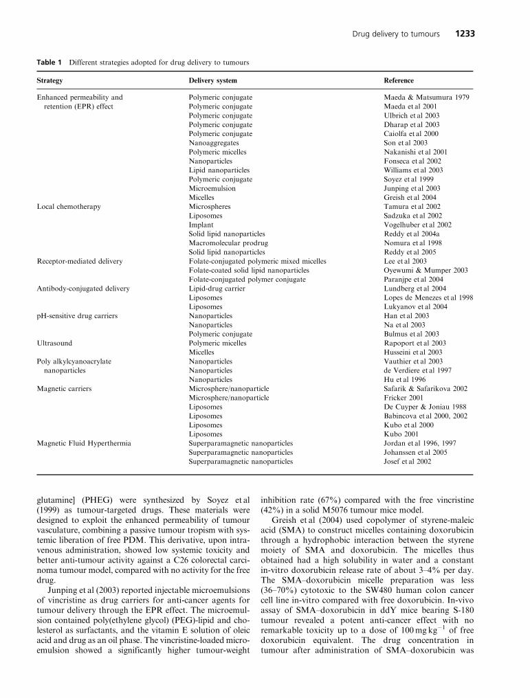
Table 1 Different strategies adopted for drug delivery to tumours
Strategy Delivery system Reference
Enhanced permeability and Polymeric conjugate Maeda & Matsumura 1979
retention (EPR) effect Polymeric conjugate Maeda et al 2001
Polymeric conjugate Ulbrich et al 2003
Polymeric conjugate Dharap et al 2003
Polymeric conjugate Caiolfa et al 2000
Nanoaggregates Son et al 2003
Polymeric micelles Nakanishi et al 2001
Nanoparticles Fonseca et al 2002
Lipid nanoparticles Williams et al 2003
Polymeric conjugate Soyez et al 1999
Microemulsion Junping et al 2003
Micelles Greish et al 2004
Local chemotherapy Microspheres Tamura et al 2002
Liposomes Sadzuka et al 2002
Implant Vogelhuber et al 2002
Solid lipid nanoparticles Reddy et al 2004a
Macromolecular prodrug Nomura et al 1998
Solid lipid nanoparticles Reddy et al 2005
Receptor-mediated delivery Folate-conjugated polymeric mixed micelles Lee et al 2003
Folate-coated solid lipid nanoparticles Oyewumi & Mumper 2003
Folate-conjugated polymer conjugate Paranjpe et al 2004
Antibody-conjugated delivery Lipid-drug carrier Lundberg et al 2004
Liposomes Lopes de Menezes et al 1998
Liposomes Lukyanov et al 2004
pH-sensitive drug carriers Nanoparticles Han et al 2003
Nanoparticles Na et al 2003
Polymeric conjugate Bulmus et al 2003
Ultrasound Polymeric micelles Rapoport et al 2003
Micelles Husseini et al 2003
Poly alkylcyanoacrylate Nanoparticles Vauthier et al 2003
nanoparticles Nanoparticles de Verdiere et al 1997
Nanoparticles Hu et al 1996
Magnetic carriers Microsphere/nanoparticle Safarik & Safarikova 2002
Microsphere/nanoparticle Fricker 2001
Liposomes De Cuyper & Joniau 1988
Liposomes Babincova et al 2000, 2002
Liposomes Kubo et al 2000
Liposomes Kubo 2001
Magnetic Fluid Hyperthermia Superparamagnetic nanoparticles Jordan et al 1996, 1997
Superparamagnetic nanoparticles Johanssen et al 2005
Superparamagnetic nanoparticles Josef et al 2002
Drug delivery to tumours 1233
13 times higher than that after the free drug. This resultcan be attributed to the enhanced permeability and reten-tion (EPR) effect of macromolecular drugs observed insolid tumours.
Local chemotherapy
Apart from the EPR effect, another strategy employed toenhance the tumour delivery of anti-cancer agents is localchemotherapy. Local delivery of chemotherapeutic drugshas long been recognized as one potential method ofdelivering high drug doses at the target site(s) while mini-mizing systemic exposure (Almond et al 2003). In this typeof delivery, the drugs or drug delivery systems are admi-nistered at the local site of the tumour to facilitate moreeffective exposure of cancer cells to drugs and hencegreater anti-tumour activity. This is true especially in thecase of inoperable accumulation of malignant ascitescaused by peritoneal carcinomatosis (Tamura et al 2002)and in some other types of cancers. Theoretically it shouldbe possible to carry out intraperitoneal chemotherapy byincreasing local drug exposure, resulting in less systemictoxicity. However, administration of drugs alone does notresult in reduction in systemic toxicity (Casper et al 1983;Lopez et al 1985). One possible way of avoiding the sys-temic toxicity associated with drug while maintaining thehigher concentrations of drug in the local regions oftumours is their incorporation into drug carriers, such asliposomes (Sadzuka et al 2002), microparticles (Tamuraet al 2002) or implants (Vogelhuber et al 2002). Sadzukaet al (2000) studied the anti-tumour effect of liposomesafter intraperitoneal administration in Ehrlich ascitesabdominal tumours. Liposomes were prepared using dif-ferent lipids such as L-�-dimyristoyl phosphatidyl choline(DMPC), L-�-distearoyl phosphatidyl choline (DSPC)and dimyristoyl phosphatidyl glycerol (DMPG). The posi-tively charged liposomes were prepared from DSPC–cholesterol–doxorubicin. The liposomal size and composi-tion was found to significantly influence the liposomaldisposition from the abdominal cavity after intraperito-neal administration. The liposomes significantly pro-longed the survival time of the tumour-bearing micecompared with the free doxorubicin. Tamura et al (2002)evaluated the anti-tumour effect of cisplatin-loaded poly(lactic acid) microspheres in various types of tumoursimplanted peritoneally. The cisplatin-loaded microspheresshowed effective anti-tumour activity (tumour-growth-inhibition rate of 70.3%) against Li-7 (human liver can-cer) xenografts transplanted into the peritoneal cavity ofmice. The microspheres also resulted in an increased lifespan of 47.2%, where as the cisplatin solution was foundto be ineffective, with a very low tumour-inhibition rateand survival. Similarly, the microspheres also enhancedthe mean survival time of mice with Li-7 xenografts trans-planted into the spleen compared with the cisplatin solu-tion.
Recently, Reddy et al (2004a) reported an enhancedtumour concentration of doxorubicin-loaded poly(butylcyanoacrylate) nanoparticles administered subcuta-neously in Dalton’s lymphoma tumour-bearing mice.
Owing to the surface hydrophobicity and negative charge,the nanoparticles underwent lymphatic absorption andconcentrated in the lymphoma.
Vogelhuber et al (2002) produced implants of BCNU(anti-cancer agent) loaded into polymer containing a 1:1(w/w) mixture of poly(1,3-bis[p-carboxyphenoxypropane]-co-sebacic acid) 20:80 and poly(D,L-lactide-co-glycolide)50and implanted subcutaneously at the tumour site in aU-87 MG glioblastoma mice model and also injectedBCNU solution intraperitoneally. Subcutaneous implan-tation of BCNU implants resulted in significant tumourregression, while BCNU solution did not have significanteffect on the tumour growth, indicating the effectivenessof BCNU implants in local chemotherapy. On the otherhand, subcutaneous injection of high doses of BCNUresulted in a 16-fold size reduction compared with shamoperated mice. The combination of BCNU and paclitaxelled to complete remission in some mice. The studies indi-cate the importance of administration route and the pos-sibility of combination chemotherapy in effective tumourtherapy. Seong et al (2003) prepared BCNU wafer bycompressing BCNU-loaded poly(D,L-lactide-co-glycolide)microparticles. In-vitro anti-tumour activity of thesewafers was performed after incubation with XF498human CNS cancer cells. The drug-loaded wafers showedprolonged cytotoxicity over 1 month, while the cytotoxi-city of BCNU powder disappeared after 12 h, suggestingthat the wafer formulation facilitates prolonged release ofBCNU in-vivo and results in effective treatment of malig-nant glioma. Gliadel Wafer (Polifeprosan 20 with carmus-tine (BCNU)) manufactured by Guilford Pharmaceuticals(USA) is the chemotherapeutic implant approved by theFDA in 1996 for the treatment of a specific brain tumourcalled a high-grade malignant glioma (recurrent glioblas-toma multiforme). Gliadel Wafers, when implanted in thecavity remaining after the surgical removal of tumour,slowly deliver BCNU directly to the tumour site.
When the drug is directly injected into a tumour, suchas by intratumoral injection, or by direct instillation intoperitumoral space, such as in intravesical therapy ofsuperficial bladder cancer and in intraperitoneal dialysisof ovarian cancer, the drug transport to tumour cellsis primarily by diffusion through interstitial space(Markman et al 1995; Nativ et al 1997; Markman 1998;Song et al 1997). Nomura et al (1998) reported the phar-macokinetics and therapeutic effects of the macromolecu-lar prodrugs of mitomycin C (MMC), MMC–dextranconjugates (MMC-D), after intratumoral injection inrats bearing a Walker 256 carcinosarcoma. MMC imme-diately disappeared from the tumour preparation follow-ing direct intratumoral injection, while cationic andanionic MMC-D were retained in the tumour longer,demonstrating that the intratumoral clearance of MMCcan be greatly retarded by dextran conjugation.
Reddy et al (2004b) studied the biodistribution andtumour retention properties of 99mTc-labelled etoposide-loaded tripalmitin nanoparticles (ETPL) after intratumoraladministration in Dalton’s lymphoma tumour-bearing mice.The disposition of nanoparticles from the tumour site wassignificantly lower than etoposide, exhibiting a 14-fold
1234 L. Harivardhan Reddy
greater tumour retention even 48h post injection. A greateranti-tumour effect can be expected to result due to theincreased exposure of tumour cells to etoposide andincreased cell uptake of nanoparticles by endocytosis.
Different routes of administration may result in vary-ing effects on the biodistribution pattern of drug carriers.Allen et al (1993) compared the biodistribution patternof liposomes administered through different routesand reported wide differences in the tissue distribution.Thus, the route of administration appears to be an importantparameter in the delivery of anti-cancer agents to tumours.Reddy et al (2005) administered etoposide-loaded solid lipid(ETP) nanoparticles by three different routes, intravenous,intraperitoneal and subcutaneous, and the tumour distribu-tion was studied in Dalton’s lymphoma tumour-bearingmice. Dalton’s lymphoma is a T-cell lymphoma and isgrown in the hind leg of mice after subcutaneous implanta-tion of tumour cells. For effective penetration into the lym-phatic interstitium, the carrier systems need to possesssmaller size, hydrophobicity and negative surface charge(Porter 1997). The smaller size of the drug carriers has alsobeen reported to facilitate the infiltration into tumour inter-stitium through the leaky tumour vasculature (Moghimi2001). The ETP nanoparticles administered through subcu-taneous injection at 2 cm below the tumour region showedgreater tumour distribution to the Dalton’s lymphoma solidtumour implanted in the right hind limbofmice. Theorder oftumour distribution of nanoparticles was subcutaneous >intraperitoneal > intravenous route. The very low distribu-tion of ETP nanoparticles after intravenous injection wasattributed to the barriers hindering the transport of drugand particles into solid tumour, such as highly disorganizedtumour vasculature, high viscosity of blood within thetumour and abnormally high pressure in the interstitialmatrix retarding the molecular transport across the vesselwalls and into the interstitium (Jain 1994).
Special strategies
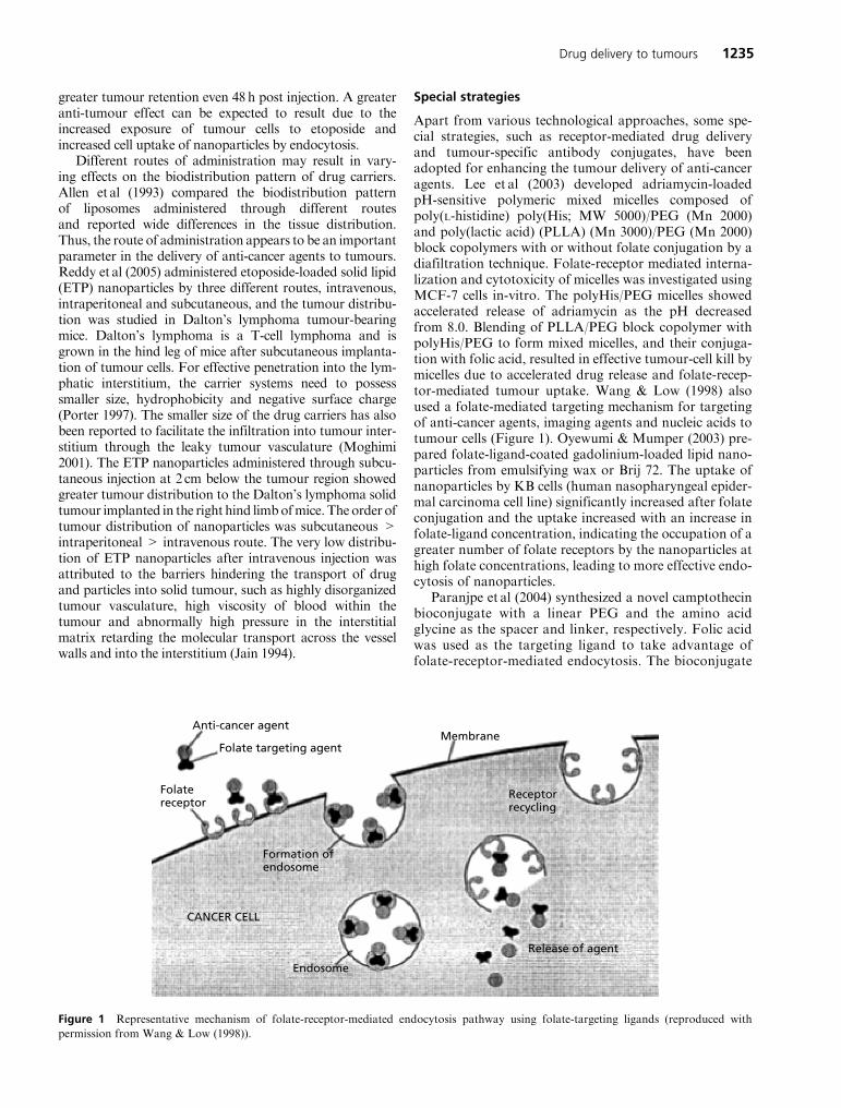
Apart from various technological approaches, some spe-cial strategies, such as receptor-mediated drug deliveryand tumour-specific antibody conjugates, have beenadopted for enhancing the tumour delivery of anti-canceragents. Lee et al (2003) developed adriamycin-loadedpH-sensitive polymeric mixed micelles composed ofpoly(L-histidine) poly(His; MW 5000)/PEG (Mn 2000)and poly(lactic acid) (PLLA) (Mn 3000)/PEG (Mn 2000)block copolymers with or without folate conjugation by adiafiltration technique. Folate-receptor mediated interna-lization and cytotoxicity of micelles was investigated usingMCF-7 cells in-vitro. The polyHis/PEG micelles showedaccelerated release of adriamycin as the pH decreasedfrom 8.0. Blending of PLLA/PEG block copolymer withpolyHis/PEG to form mixed micelles, and their conjuga-tion with folic acid, resulted in effective tumour-cell kill bymicelles due to accelerated drug release and folate-recep-tor-mediated tumour uptake. Wang & Low (1998) alsoused a folate-mediated targeting mechanism for targetingof anti-cancer agents, imaging agents and nucleic acids totumour cells (Figure 1). Oyewumi & Mumper (2003) pre-pared folate-ligand-coated gadolinium-loaded lipid nano-particles from emulsifying wax or Brij 72. The uptake ofnanoparticles by KB cells (human nasopharyngeal epider-mal carcinoma cell line) significantly increased after folateconjugation and the uptake increased with an increase infolate-ligand concentration, indicating the occupation of agreater number of folate receptors by the nanoparticles athigh folate concentrations, leading to more effective endo-cytosis of nanoparticles.
Paranjpe et al (2004) synthesized a novel camptothecinbioconjugate with a linear PEG and the amino acidglycine as the spacer and linker, respectively. Folic acidwas used as the targeting ligand to take advantage offolate-receptor-mediated endocytosis. The bioconjugate
Anti-cancer agent
Folate targeting agentMembrane
Release of agent
Receptorrecycling
Endosome
Formation ofendosome
CANCER CELL
Folatereceptor
Figure 1 Representative mechanism of folate-receptor-mediated endocytosis pathway using folate-targeting ligands (reproduced with
permission from Wang & Low (1998)).
Drug delivery to tumours 1235
was evaluated in-vitro for specific targeting to folate-receptor-expressing KB cells. The bioconjugate exhib-ited significantly higher efficacy in comparison withcamptothecin. A control conjugate without PEGdemonstrated no improvement in efficacy over untar-geted camptothecin, emphasizing the importance of thespacer between the anti-cancer compound and targetingmoiety.
The ability to selectively target anti-cancer agents viaspecific ligands against antigens expressed on malignantcells could greatly improve the therapeutic indices ofdrugs. Lundberg et al (2004) developed sterically stabilizedlipid drug carriers (emulsion and liposomes) and cova-lently attached the anti-CD74 antibody to the surface ofthe carrier using a PEG-based hetero-bifunctional cou-pling agent. During a 24-h in-vitro incubation with thetarget Raji B-lymphoma cells, about 30% of the com-plexes were found to be associated with the cells. Theresults indicated that the antibody-conjugated drug carrierwas selectively targeted to B-cells and showed selectivetoxicity of the incorporated drug. On similar lines, severalstudies with PEG-liposomes targeted with tumour-cell-specific monoclonal antibodies have shown improvedtherapeutic activity over non-targeted formulation (Lopesde Menezes et al 1998).
Lukyanov et al (2004) modified the commerciallyavailable doxorubicin-loaded long-circulating liposomes(Doxil; Alza Pharmaceuticals) with the monoclonalnucleosome (NS)-specific 2C5 antibody (mAb 2C5) thatrecognizes a broad variety of tumours via the tumour cellsurface-bound NSs. For incorporation into liposomes,mAb 2C5 was modified with poly(ethylene glycol)-phos-phatidyl ethanolamine conjugate (PEG-PE) with the freePEG terminus activated with the p-nitrophenylcarbonylgroup (pNP-PEG-PE). 2C5-targeted Doxil liposomesacquired the ability to recognize NSs and specificallybound to various tumour cells. Doxorubicin-loadedlong-circulating liposomes modified with the mAb 2C5showed a greater cell-kill effect in-vitro than non-targeteddoxorubicin-loaded liposomes.
Drug delivery based on tumour pH
The tumour extracellular pH is a consistently distinguish-ing phenotype of most solid tumours from surroundingnormal tissues. The measured pH values of most solidtumours in patients using invasive microelectrodes are inthe range 5.7–7.8, with a mean value of 7.0 (Ojugo et al1999). The acidity of the tumour interstitial fluid is mainlyattributed, if not entirely, to the higher rate of aerobic andanaerobic glycolysis in cancer cells than in normal cells(Stubbs et al 2000). Such acidic extracellular pH promptedthe establishment of pH-sensitive anti-cancer drug deliv-ery systems. Recently, Han et al (2003) introduced newpH-sensitive functional group (a weak acid of sulfona-mide) containing polymers. The water-soluble polymersmodified with sulfonamide self-assembled nanoparticlesshowed enhanced drug release and interaction with, andinto, cells at tumour pH (Na et al 2003).
Cytoplasmic delivery of enzyme-susceptible biomolecu-lar drugs is one of the major limitations in many therapeu-tic strategies. Synthetic, pH-sensitive polymers have alsobeen investigated and showed enhanced endosomal deliv-ery of biomolecular therapeutics (Lackey et al 1999, 2002;Murthy et al 2003). Most of the studies (Vinogradov et al1998; Richardson et al 1999) were focused on cationicamino polymers. Murthy et al (1999) prepared pH-sensitivepolymers that mimicked viral peptides and contained acombination of acidic –COOH groups and hydrophobicalkyl groups. This combination was used for cytoplasmicdelivery applications, because such polymer compositionscan be varied to cause disruption of lipid membranes atspecific pHs. When these polymers are protonated at endo-somal pH, they increase in hydrophobicity, leading toenhanced endosomal membrane disruption. A novel pH-responsive polymeric carrier was synthesized by Bulmuset al (2003) for the enhanced cytoplasmic delivery ofenzyme-susceptible drugs, such as antisense oligonucleo-tides, proteins and peptides. A novel functionalized mono-mer, pyridyl disulfide acrylate, was synthesized andincorporated into an amphiphilic copolymer consisting ofmethacrylic acid and butyl acrylate, which resulted in aglutathione- and pH-sensitive, membrane-disruptive terpo-lymer with functional groups that allow thiol-containingmolecules to be readily conjugated. The polymer had twokey actions: firstly, pH-dependent, endosomal membranedisruption and escape into the cytoplasm; and secondly,this is followed by reaction of disulfide-conjugated drugwith glutathione, a normal constituent of the cytoplasmof cells, causing release of the drug from the polymer.
Drug delivery by ultrasound
Drug delivery to tumours using ultrasound has been therecent mechanism adopted by Rapaport et al (2003). Theydeveloped a new drug modality based on drug encapsula-tion into polymeric micelles followed by a controlledrelease at the tumour site triggered by ultrasound focusedon the tumour. Ultrasound not only released the drugfrom micelles, but also enhanced the local uptake ofboth free and encapsulated drug by tumour cells, thusproviding effective drug targeting. Ultrasound also pro-motes extravasation of drug-loaded carriers into thetumour interstitium. The in-vivo results revealed thatapplication of a low-frequency ultrasound (20–70 kHz)significantly reduced the tumour size when comparedwith insonated controls. A similar approach was reportedby Husseini et al (2000), involving the encapsulation andrelease of doxorubicin from micelles through ultrasound.
Drug delivery to resistant tumour cells
Several types of cancer show resistance to conventionalchemotherapy. Cancer-cell resistance is considered to beone of the major reasons for failure of chemotherapy forthe majority of cancer patients (Ambudkar et al 1999;Silverman 1999). Some tumours are intrinsically resistantto treatment, whereas others acquire resistance with expo-sure to structurally unrelated drugs (Vauthier et al 2003).
1236 L. Harivardhan Reddy
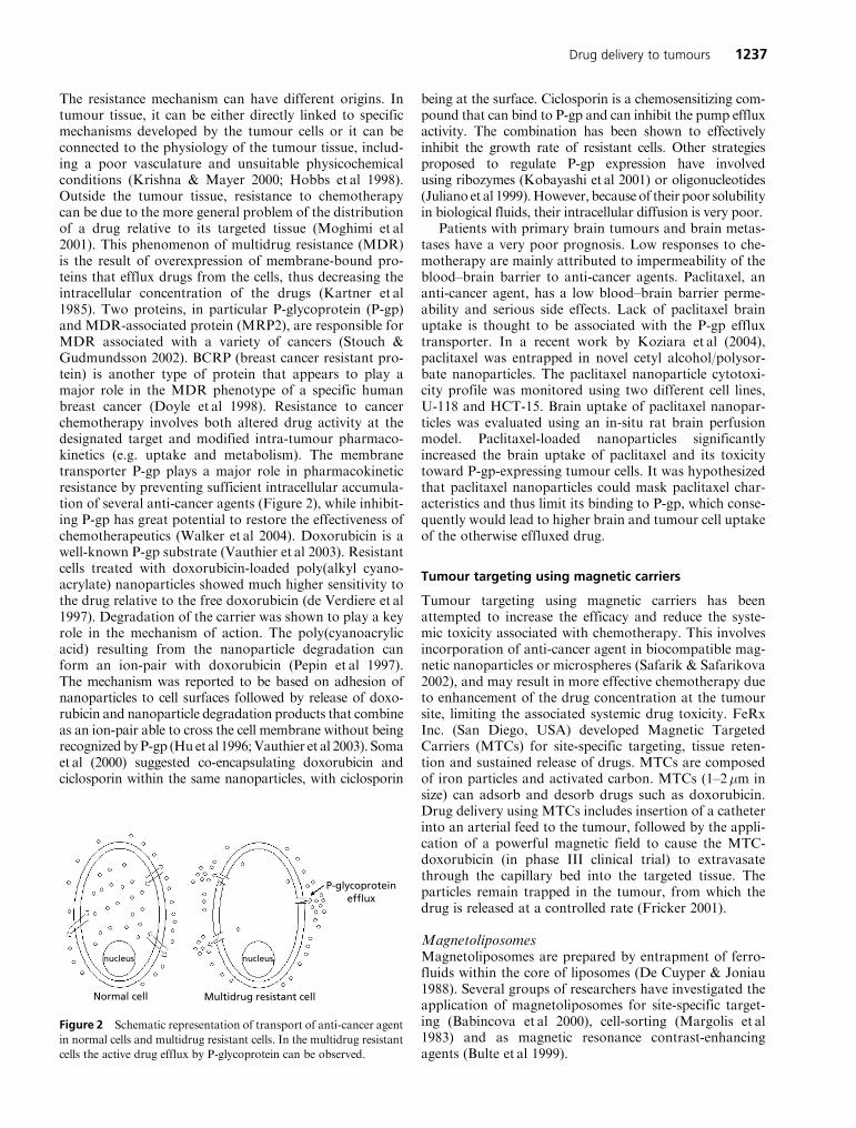
The resistance mechanism can have different origins. Intumour tissue, it can be either directly linked to specificmechanisms developed by the tumour cells or it can beconnected to the physiology of the tumour tissue, includ-ing a poor vasculature and unsuitable physicochemicalconditions (Krishna & Mayer 2000; Hobbs et al 1998).Outside the tumour tissue, resistance to chemotherapycan be due to the more general problem of the distributionof a drug relative to its targeted tissue (Moghimi et al2001). This phenomenon of multidrug resistance (MDR)is the result of overexpression of membrane-bound pro-teins that efflux drugs from the cells, thus decreasing theintracellular concentration of the drugs (Kartner et al1985). Two proteins, in particular P-glycoprotein (P-gp)and MDR-associated protein (MRP2), are responsible forMDR associated with a variety of cancers (Stouch &Gudmundsson 2002). BCRP (breast cancer resistant pro-tein) is another type of protein that appears to play amajor role in the MDR phenotype of a specific humanbreast cancer (Doyle et al 1998). Resistance to cancerchemotherapy involves both altered drug activity at thedesignated target and modified intra-tumour pharmaco-kinetics (e.g. uptake and metabolism). The membranetransporter P-gp plays a major role in pharmacokineticresistance by preventing sufficient intracellular accumula-tion of several anti-cancer agents (Figure 2), while inhibit-ing P-gp has great potential to restore the effectiveness ofchemotherapeutics (Walker et al 2004). Doxorubicin is awell-known P-gp substrate (Vauthier et al 2003). Resistantcells treated with doxorubicin-loaded poly(alkyl cyano-acrylate) nanoparticles showed much higher sensitivity tothe drug relative to the free doxorubicin (de Verdiere et al1997). Degradation of the carrier was shown to play a keyrole in the mechanism of action. The poly(cyanoacrylicacid) resulting from the nanoparticle degradation canform an ion-pair with doxorubicin (Pepin et al 1997).The mechanism was reported to be based on adhesion ofnanoparticles to cell surfaces followed by release of doxo-rubicin and nanoparticle degradation products that combineas an ion-pair able to cross the cell membrane without beingrecognized byP-gp (Hu et al 1996;Vauthier et al 2003). Somaet al (2000) suggested co-encapsulating doxorubicin andciclosporin within the same nanoparticles, with ciclosporin
being at the surface. Ciclosporin is a chemosensitizing com-pound that can bind to P-gp and can inhibit the pump effluxactivity. The combination has been shown to effectivelyinhibit the growth rate of resistant cells. Other strategiesproposed to regulate P-gp expression have involvedusing ribozymes (Kobayashi et al 2001) or oligonucleotides(Juliano et al 1999).However, because of their poor solubilityin biological fluids, their intracellular diffusion is very poor.
Patients with primary brain tumours and brain metas-tases have a very poor prognosis. Low responses to che-motherapy are mainly attributed to impermeability of theblood–brain barrier to anti-cancer agents. Paclitaxel, ananti-cancer agent, has a low blood–brain barrier perme-ability and serious side effects. Lack of paclitaxel brainuptake is thought to be associated with the P-gp effluxtransporter. In a recent work by Koziara et al (2004),paclitaxel was entrapped in novel cetyl alcohol/polysor-bate nanoparticles. The paclitaxel nanoparticle cytotoxi-city profile was monitored using two different cell lines,U-118 and HCT-15. Brain uptake of paclitaxel nanopar-ticles was evaluated using an in-situ rat brain perfusionmodel. Paclitaxel-loaded nanoparticles significantlyincreased the brain uptake of paclitaxel and its toxicitytoward P-gp-expressing tumour cells. It was hypothesizedthat paclitaxel nanoparticles could mask paclitaxel char-acteristics and thus limit its binding to P-gp, which conse-quently would lead to higher brain and tumour cell uptakeof the otherwise effluxed drug.
Tumour targeting using magnetic carriers
Tumour targeting using magnetic carriers has beenattempted to increase the efficacy and reduce the syste-mic toxicity associated with chemotherapy. This involvesincorporation of anti-cancer agent in biocompatible mag-netic nanoparticles or microspheres (Safarik & Safarikova2002), and may result in more effective chemotherapy dueto enhancement of the drug concentration at the tumoursite, limiting the associated systemic drug toxicity. FeRxInc. (San Diego, USA) developed Magnetic TargetedCarriers (MTCs) for site-specific targeting, tissue reten-tion and sustained release of drugs. MTCs are composedof iron particles and activated carbon. MTCs (1–2�m insize) can adsorb and desorb drugs such as doxorubicin.Drug delivery using MTCs includes insertion of a catheterinto an arterial feed to the tumour, followed by the appli-cation of a powerful magnetic field to cause the MTC-doxorubicin (in phase III clinical trial) to extravasatethrough the capillary bed into the targeted tissue. Theparticles remain trapped in the tumour, from which thedrug is released at a controlled rate (Fricker 2001).
MagnetoliposomesMagnetoliposomes are prepared by entrapment of ferro-fluids within the core of liposomes (De Cuyper & Joniau1988). Several groups of researchers have investigated theapplication of magnetoliposomes for site-specific target-ing (Babincova et al 2000), cell-sorting (Margolis et al1983) and as magnetic resonance contrast-enhancingagents (Bulte et al 1999).
Normal cell Multidrug resistant cell
P-glycoproteinefflux
nucleus nucleus
Figure 2 Schematic representation of transport of anti-cancer agent
in normal cells and multidrug resistant cells. In the multidrug resistant
cells the active drug efflux by P-glycoprotein can be observed.
Drug delivery to tumours 1237
Babincova et al (Babincova et al 2002) developed mag-netoliposomes encapsulated with doxorubicin for site-specific chemotherapy in response to an externally appliedAC magnetic field. The results revealed that specificallyheating the magnetoliposomes to 42�C resulted in a mas-sive release of encapsulated doxorubicin. Another in-vivostudy for site-specific targeting using magnetoliposomesincorporated with doxorubicin showed that administra-tion of magnetic liposomes under an applied externalmagnetic force produced an approximately 4-fold highermaximum doxorubicin concentration in the tumour com-pared with doxorubicin solution. These results suggestthat systemic chemotherapy could effectively control theprimary tumour without significant side effects, due to thetargeting of magnetic doxorubicin liposomes (Kubo et al2000, 2001).
Magnetic fluid hyperthermiaMagnetic fluid hyperthermia (MFH) is a new techniquefor interstitial hyperthermia or thermoablation based onAC magnetic field-induced excitation of biocompatiblesuperparamagnetic nanoparticles. Magnetic fluids havebeen investigated as potential hyperthermia-causingagents due to their high specific absorption rate.Hyperthermia is a promising approach for cancer treat-ment, which uses AC magnetic fields to heat target areas(cancer tissue) containing magnetic fluids. To study thebiological effects of AC magnetic field excited ferrofluids,both in-vitro and in-vivo studies have been carried out incancer cell lines and spontaneously induced tumours inanimal models. The results of these studies indicated thatmagnetic fluid hyperthermia is able to reduce the viabilityof cancer cells, thereby indicating the potential of thistherapy (Jordan et al 1997).
Johannsen et al (2005) evaluated the potential of MFHas a minimally invasive treatment for prostate cancer bycarrying out a systematic analysis of the effects of MFH inthe orthotopic Dunning R3327 tumour model of the rat.Rats received two MFH treatments following a singleintratumoral injection of a magnetic fluid. Treatmentswere carried out on days 10 and 12 after tumour inductionusing an AC magnetic field applicator system operating ata frequency of 100 kHz and a variable field strength(0–18 kAm�1). The rats were sacrificed after day 20, andthe tumour weights were determined and compared withcontrols. The results indicated that the MFH led to asignificant growth inhibition in an orthotopic model ofthe aggressive MatLyLu tumour variant.
Jordan et al (1996) investigated the cellular uptake andbiological effects of biocompatible magnetic fluids excitedby an AC magnetic field on human carcinoma cells in-vitro. One of the fluids tested was a dextran magnetite,which possesses very low cytotoxicity. The results indicatedthat there is a sensitizer effect of ferrofluids at 43 �C prob-ably caused by free ferric ions, which induce oxidativestress, and there was no cytotoxic effect of intracellulardextran magnetite particles excited with AC magnetic field.
In a recent report by Josef et al (2002), a patient withadvanced hepatocellular carcinoma was treated with an
intravenous infusion of pegylated liposomal doxorubicin(PLD, Caelyx) in combination with ultrasound hyperther-mia of the liver.
Future perspectives
Despite extensive research in the field of drug delivery totumours using delivery systems, only a few products havefound their way onto the market. A better understandingof physiological barriers and biochemical mechanisms ofcancer, which a drug has to face to reach the cancer site,enables drug delivery researchers to develop a successfuldelivery system for cancer therapy. Sterically stabilizedliposomes, such as Doxil and Daunosome, and GliadelWafers, are a few recent examples of the products thathave reached the market for cancer therapy. For injectabledelivery systems several factors, such as stability, particlesize, sterility and injectability, are important for effectivedelivery and therapeutic efficacy. Many a time, the deliv-ery systems, such as solid lipid nanoparticles, show anincrease in particle size upon storage, which may alterthe biodistribution and therapeutic efficacy of the incor-porated drug.
For polymeric nanoparticles, large-scale manufactur-ing is most critical andmany of the reported techniques useorganic solvents, which is not advisable for large scaleproduction. Residual solvents in the nanoparticles maylead to toxic effects upon administration. Hence, completeevaporation of the organic solvents should be ensured inthe final formulation and levels should be below the pre-scribed limits. The stabilizers used in the preparation ofnanoparticles need to be biocompatible and nontoxic.Also, the batch-to-batch uniformity of particle size andstability should be ensured. Solid lipid nanoparticles pos-sess the advantage of feasibility of large-scale production.However, the nanoparticles prepared from glyceride lipidspossess the problems of recrystallization, particle growthand expulsion of drug from the particle matrix leading to adecrease in the amount of the incorporated drug on sto-rage. Hence, selection of the type of lipid is essential toproduce nanoparticles with better stability. Storage ofnanoparticles in a dry form can improve the long-termstability. Spray drying is one of the better techniques torecover nanoparticles from aqueous dispersions, and isalso feasible for large-scale manufacturing purposes.
For rapidly growing tumours, the extensive angiogen-esis to meet the nutritional requirements of newly formedcells can be taken advantage of in delivering macromole-cular drugs and colloidal systems. This can be achievedthrough the EPR effect. For such delivery, the systemsshould possess long blood circulation time, stability in thecirculation and slow drug release until it reaches thetumour. Polymer–drug conjugation is another means ofdelivering low-molecular-weight anti-cancer agents speci-fically to tumour cells. These conjugates allow passivetargeting of tumours by the EPR effect and they can befurther conjugated to bio-responsive linkers or receptor-specific ligands, such as folic acid, to enhance the cellspecificity. In the case of MDR cancers, some specialstrategies, such as co-incorporation of P-gp inhibitors
1238 L. Harivardhan Reddy
into the delivery systems, or a prodrug approach can beused. Drug delivery to some types of cancers, such aslymphatic carcinomas, by conventional means is very dif-ficult since in lymphomas, the lymph nodes are theaffected regions of the lymphatic system. The lymphnodes are not present superficially, and are deep seatedin the body. The strategies applied to improve the deliveryof anti-cancer agents to other types of tumours, such asEPR effect, may not result in high tumour concentrationsin the case of lymphomas. As lymphomas are more richlysupplied by lymph than blood, drug carriers administeredintravenously may not have better access to the lympho-mas, resulting in poor drug concentrations in lymphnodes. In such cases, the delivery route can be subcuta-neous or intraperitoneal, as these routes have relativelybetter lymphatic access. Nanoparticles with hydrophobi-city, high molecular weight, negative charge and small sizecan be effectively delivered to the lymphatic systemthrough the above routes.
Anti-cancer agents can be delivered specifically to thetumours through low-density lipoprotein (LDL) recep-tors. LDL, a normal blood constituent, is the body’sprincipal means of delivering cholesterol to tissues.Cancer cells need large amounts of cholesterol becauseof the rapid formation of new membranes. Thus LDLcould be used as a carrier for anti-cancer agents, whereinthe drugs can be delivered specifically to cancer cells.Different strategies can be adopted to deliver the drugs,such as chemically linking the drugs to LDL or incorpora-tion of drugs into the delivery systems made from choles-terol derivatives.
An important drawback is the non-availability ofappropriate cancer models for evaluation of the anti-can-cer agent-loaded drug delivery systems. The need exists forthe development of appropriate models for individualtypes of cancer, as the physiology may differ for all can-cers. Studies should be more focused on direct evaluationin animal models than in cell cultures, as many of thephysiological processes are different in the above twocases. Results obtained by testing in appropriate cancermodels would lead to better conclusions.
Conclusion
The recent strategies adopted for delivering anti-canceragents have been discussed. Drug delivery to tumourstaking advantage of leaky tumour vasculature has beenproved to be beneficial in enhancing the tumour drugconcentration. Receptor-mediated drug delivery totumours has resulted in successful enhancement of drugconcentrations in tumours and effective tumour therapy.Local chemotherapy through drug carriers has been foundto be the potential method of delivering a high drug doseat the target site leading to greater anti-tumour activity,while minimizing the systemic drug exposure. Directadministration of drugs or drug delivery systems into thetumour results in significantly high tumour concentra-tions. However, this approach may not be possible forremote tumours. Despite extensive research in the areaof drug delivery to tumours, only very few products have
successfully reached the market. Hence the focus of theresearch in this area needs to be carried out with anindustrial viewpoint to develop commercially successfulproducts for effective cancer therapy.
References
Allen, T. M., Hansen, C. B., Guo, L. S. S. (1993) Subcutaneousadministration of liposomes: a comparison with the intrave-nous and intraperitoneal routes of injection. Biochim. Biophys.Acta 1150: 9–16
Almond, B. A., Hadba, A. R., Freeman, S. T., Cuevas, B. J.,York, A. M., Detrisac, C. J., Goldberg, E. P. (2003) Efficacy ofmitoxantrone-loaded albumin microspheres for intratumoralchemotherapy of breast cancer. J. Control. Release 91:147–155
Ambudkar, S. V., Dey, S., Hrycyna, C. A., Ranmachandra, M.,Pastan, I., Gottesman, M. M. (1999) Biochemical, cellular andpharmacological aspects of the multidrug transporter. Annu.Rev. Pharmacol. Toxicol. 39: 361–398
Au, J. L.-S., Jang, S. H., Zheng, J., Chen, C.-T., Song, S., Hu, L.,Wientjes, M. G. (2001) Determinants of drug delivery andtransport to solid tumors. J. Control. Release 74: 31–46
Babincova, M., Altanerova, V., Lampert, M., Altaner, C.,Machova, E., Sramka, M., Babinec, P. (2000) Site-specific invivo targeting of magnetoliposomes using externally appliedmagnetic field. Z. Naturforsch. (C) 55: 278–281
Babincova, M., Cicmanec, P., Altanerova, V., Altaner, C.,Babinec, P. (2002) AC-magnetic field controlled drug releasefrom magnetoliposomes: design of a method for site-specificchemotherapy. Bioelectrochemistry 55: 1–19
Bulmus, V., Woodward, M., Lin, L., Murthy, N., Stayton, P.,Hoffman, A. (2003) A new pH-responsive and glutathione-reactive, endosomal membrane-disruptive polymeric carrierfor intracellular delivery of biomolecular drugs. J. Control.Release 93: 105–120
Bulte, J. W., Cuyper, M. D., Despres, D., Frank, J. A. (1999) Short-vs. long-circulating magnetoliposomes as bone marrow-seekingMR contrast agents. J. Magn. Reson. Imaging 9: 329–335
Caiolfa, V. R., Zamai, M., Fiorino, A., Frigerio, E., Pellizzoni, C.,d’Argy, R., Ghiglieri, A., Castelli, M. G., Farao, M., Pesenti,E., Gigli, M., Angelucci, F., Suarato, A. (2000) J. Control.Release 65: 105–119
Casper, E. S., Kelsen, D. P., Alcock, N. W., Lewis, J. L. (1983)IP cisplatin in patients with malignant ascites: pharmacoki-netic evaluation and comparison with the iv route. CancerTreat. Rep. 67: 235–238
Chawla, J. S., Amiji, M. M. (2002) Biodegradable poly(e-capro-lactone) nanoparticles for tumor-targeted delivery of tamox-ifen. Int. J. Pharm. 249: 127–138
Chawla, J. S., Amiji, M. M. (2003) Cellular uptake and concen-trations of tamoxifen upon administration in poly(e-caprolac-tone) nanoparticles. AAPS Journal 5: Article 3
De Cuyper, M., Joniau, M. (1988) Magnetoliposomes: formationand structural characterization. Eur. Biophys. J. 15: 311–319
de Verdiere, A. C., Dubernet, C., Nemati, F., Soma, E., Appel,M.,Ferte, J., Bernard, S., Puisieux, F., Couvreur, P. (1997)Reversion of multidrug resistance with polyalkyl cyanoacrylatenanoparticles; towards a mechanism of action. Br. J. Cancer 76:198–205
Dharap, S. S., Qui, B., Williams, G. C., Sinko, P., Stein, S.,Minko, T. (2003) Molecular targeting of the drug deliverysystems to ovarian cancer by BH3 and LHRH peptides. J.Control. Release 91: 61–73
Drug delivery to tumours 1239
Doyle, L. A., Yang, W., Abruzzo, L. V., Krogmann, T., Gao, Y.,Rishi, A. K., Ross, D. D. (1998) A multidrug resistance trans-porter from human MCF-7 breast cancer cells. Proc. NatlAcad. Sci. 95: 15665–15670
Fenton, R. G., Longo, D. I. (1998) Cell biology of cancer. In:Fauci, A. S., Braunwald, E., Isselbacher, K. J., Wilson, J. D.,Martin, J. B., Kasper, D. L., Hauser, S. L., Longo, D. I. (eds)Harrison’s internal medicine. Vol. 1, 14th edn, McGraw Hill,New York, pp 505–511
Fonseca, C., Siomes, S., Gaspar, R. (2002) Paclitaxel-loadedPLGA nanoparticles: preparation, physicochemical character-ization and in vitro anti-tumoral activity. J. Control. Release83: 273–286
Fricker, J. (2001) Drugs with a magnetic attraction to tumors.Drug Discov. Today 6: 387–389
Garrec, D. L., Ranger, M., Leroux, J. C. (2004) Micelles inanticancer drug delivery. Am. J. Drug Deliv. 2: 15–42
Greish, K., Sawa, T., Fang, J., Akaike, T., Maeda, H. (2004)SMA–doxorubicin, a new polymeric micellar drug for effectivetargeting to solid tumours. J. Control. Release 97: 219–230
Han, S. K., Na, K., Bae, Y. H. (2003) Sulfonamide based pH-sensitive polymeric micelles: physicochemical characteristicsand pH dependant aggregation. Colloids Surf. APhysicochem. Eng. Aspects 214: 49–59
Hobbs, S. K., Monsky, W. L., Yuan, F., Roberts, W. G.,Griffith, L., Torchilin, V. P., Jain, R. K. (1998) Regulationof transport pathways in tumor vessels: role of tumor type andmicroenvironment. Proc. Natl Acad. Sci. 95: 4607–4612
Hu, Y.-P., Jarillon, S., Dubernet, C., Couvreur, P., Robert, J.(1996) On the mechanism of action of doxorubicin encapsula-tion in nanospheres for the reversal of multidrug resistance.Cancer Chemother. Pharmacol. 37: 556–560
Husseini, G. A., Myrup, G. D., Pitt, W. G., Christensen, D. A.,Rapoport, N. Y. (2000) Factors affecting acoustically trig-gered release of drugs from polymeric micelles. J. Control.Release 69: 43–52
Jain, R. K. (1994) Barriers to drug delivery in solid tumors. Sci.Am. 271: 58–65
Johannsen, M., Thiesen, B., Jordan, A., Taymoorian, K.,Gneveckow, U., Waldofner, N., Scholz, R., Koch, M., Lein,M., Jung, K., Loening, S. A. (2005) Magnetic fluid hyperther-mia (MFH) reduces prostate cancer growth in the orthotopicDunning R3327 rat model. Prostate 64: 283–292
Jordan, A., Wust, P., Scholz, R., Tesche, B., Fahling, H.,Mitrovics, T., Vogl, T., Cervos-Navarro, J., Felix, R. (1996)Cellular uptake of magnetic fluid particles and their effects onhuman adenocarcinoma cells exposed to AC magnetic fields invitro. Int. J. Hyperthermia 12: 705–722
Jordan, A., Scholz, R., Wust, P., Fahling, H., Krause, J.,Wlodarczyk, W., Sander, B., Vogl, T., Felix, R. (1997)Effects of magnetic fluid hyperthermia (MFH) on C3H mam-mary carcinoma in vivo. Int. J. Hyperthermia 13: 587–605
Josef, D., Zdenek, Z., Bohuslav, M., Pavel, J., Jindriska, M.,Ivana, M., Dagmar, D., Jiri, P. (2002) Pegylated liposomaldoxorubicin in combination with hyperthermia in the treat-ment of a case of advanced hepatocellular carcinoma. J. Clin.Gastroenterol. 34: 96–98
Juliano, R. L., Alahari, S., Yoo, H., Kole, R., Cho, M. (1999)Antisense pharmacodynamics: critical issues in the transportand delivery of antisense oligonucleotides. Pharm. Res. 16:494–502
Junping, W., Takayama, K., Nagai, T., Maitani, Y. (2003)Pharmacokinetics and antitumor effects of vincristine carriedby microemulsions composed of PEG-lipid, oleic acid,vitamin E and cholesterol. Int. J. Pharm. 251: 13–21
Kartner, N., Evernden-Porelle, D., Bradley, G., Ling, V. (1985)Detection of P-glycoprotein in multidrug-resistant cell lines bymonoclonal antibodies. Nature 316: 820–823
Kerr, J. F., Wyllie, A. H., Curie, A. R. (1972) Apoptosis: a basicbiological phenomenon with wide-ranging implications in tis-sue kinetics. Br. J. Cancer 26: 239–257
Kobayashi, H., Takamura, Y., Miyachi, H. (2001) Novelapproaches to reversing anti-cancer drug resistance usinggene-specific therapeutics. Hum. Cell 14: 172–184
Koziara, J. M., Lockman, P. R., Allen, D. D., Mumper, R. J.(2004) Paclitaxel nanoparticles for the potential treatment ofbrain tumors. J. Conrol. Release 99: 259–269
Krishna, R., Mayer, L. D. (2000) Multidrug resistance(MDR) in cancer mechanisms, reversal using modulatorsof MDR and the role of MDR modulators in influencingthe pharmacokinetics of anticancer drugs, Eur. J. CancerSci. 11: 265–283
Kubo, T., Sugita, T., Shimose, S., Nitta, Y., Ikuta, Y.,Murakami, T. (2000) Targeted delivery of anticancer drugswith intravenously administered magnetic liposomes in osteo-sarcoma-bearing hamsters. Int. J. Oncol. 17: 309–315
Kubo, T., Sugita, T., Shimose, S., Nitta, Y., Ikuta, Y.,Murakami, T. (2001) Targeted systemic chemotherapy usingmagnetic liposomes with incorporated adriamycin for osteo-sarcoma in hamsters. Int. J. Oncol. 18: 121–125
Lackey, C., Murthy, N., Press, O., Tirrell, D., Hoffman, A.,Stayton, P. (1999) Hemolytic activity of pH-responsive poly-mer–streptavidin bioconjugate. Bioconjug. Chem. 10:401–405
Lackey, C., Press, O., Hoffman, A., Stayton, P. (2002) A biomi-metic pH-responsive polymer directs endosomal release andintracellular delivery of an endocytosed antibody complex,Bioconjug. Chem. 13: 996–1001
Lee, E. S., Na, K., Bae, Y. H. (2003) Polymeric micelle for tumorpH and folate mediated targeting. J. Control. Release 91:103–113
Lopes de Menezes, D. E., Pilarski, L. M., Allen, T. M. (1998) Invivo and in vitro targeting of immunoliposomal doxorubicinto human B-cell lymphoma. Cancer Res. 58: 3320–3330
Lopez, J. A., Krikorian, J. G., Reich, S. D., Smyth, R. D., Lee,F. H., Issell, B. F. (1985) Clinical pharmacology of intraper-itoneal cisplatin. Gynecol. Oncol. 20: 1–9
Lukyanov, A. N., Elbayoumi, T. A., Chakilam, A. R., Torchilin,V. P. (2004) Tumor-targeted liposomes: doxorubicin-loadedlong-circulating liposomes modified with anti-cancer anti-body. J. Control. Release 100: 135–144
Lundberg, B. B., Griffiths, G., Hansen, H. J. (2004) Cellularassociation and cytotoxicity of anti-CD74-targeted lipiddrug-carriers in B lymphoma cells. J. Control. Release 94:155–161
Maeda, H., Matsumura, Y. (1989) Tumoritropic and lympho-tropic principles of macromolecular drugs. Crit. Rev. Ther.Drug Carrier Syst. 6: 183–210
Maeda, H., Takeshita, J., Kanamaru, R. (1979) A lipophilicderivative of neocarzinostatin. A polymer conjugation of anantitumor protein antibiotic. Int. J. Pept. Protein Res. 14:81–87
Maeda, H., Wu, J., Sawa, T., Matsumura, Y., Hori, K. (2000)Tumor vascular permeability and the EPR effect in macro-molecular therapeutics. A review. J. Control. Release 65:271–284
Maeda, H., Sawa, T., Konno, T. (2001) Mechanism of tumor-targeted delivery of macromolecular drugs, including the EPReffect in solid tumor and clinical overview of the prototypepolymeric drug SMANCS. J. Control. Release 74: 47–61
1240 L. Harivardhan Reddy
Margolis, L. B., Namiot, V. A., Kljukin, L. M. (1983)Magnetoliposomes: another principle of cell sorting. Biochim.Biophys. Act. 735: 193–195
Markman, M. (1998) Intraperitoneal therapy of ovarian cancer.Semin. Oncol. 25: 356–360
Markman, M., Francis, P., Rowinsky, E., Hoskins, W. (1995)Intra-peritoneal paclitaxel: a possible role in the managementof ovarian cancer. Semin. Oncol. 22: 84–87
Moghimi, S. M., Hunter, A. C., Murray, J. C. (2001) Long-circulating and target-specific nanoparticles: theory to prac-tice. Pharmacol. Rev. 53: 283–318
Murthy, N., Robichaud, J., Tirrell, D., Stayton, P., Hoffman, A.(1999) The design and synthesis of polymers for eukaryoticmembrane disruption. J. Control. Release 61: 137–143
Murthy, N., Chang, I., Stayton, P., Hoffman A. (2001) pH-sensitive hemolysis by random copolymer of alkyl acrylatesand acrylic acid. Macromol. Symp. 172: 49–55
Murthy, N., Campbell, J., Fausto, N., Hoffman, A. S., Stayton,P. S. (2003) Bioinspired pH-responsive polymers for the intra-cellular delivery of biomolecular drugs. Bioconjug. Chem. 14:412–419
Na, K., Lee, E. S., Bae, Y. H. (2003) Adriamycin loaded pullulanacetate/sulfonamide conjugate nanoparticles responding totumor pH: pH dependant cell interaction, internalization andcytotoxicity in vitro. J. Control. Release 87: 3–13
Nakanishi, T., Fukushima, S., Okamoto, K., Suzuki, M.,Matsumura, Y., Yokoyama, M., Okano, T., Sakurai, Y.,Kataoka, K. (2001) Development of the polymeric micellecarrier system for doxorubicin. J. Control. Release 74: 295–302
Nativ, O., Aronson, M., Medalia, O., Moldavsky, T., Sabo, E.,Ringel, I., Kravtsov, V. (1997) Anti-neoplastic activity ofpaclitaxel on experimental superficial bladder cancer: in vivoand in vitro studies. Int. J. Cancer 70: 297–301
Nomura, T., Saikawa, A., Morita, S., Sakaeda Kakutani, T.,Yamashita, F., Honda, K., Takakura, Y., Hashida, M.(1998) Pharmacokinetic conjugates after intratumoral injec-tion. J. Control. Release 52: 239–252
Ojugo, A. S. E., Mcsheehy, P. M. J., Mcintyre, D. J. O., Mccoy,C., Stubbs, M., Leach, M. O., Judson, I. R., Griffiths, J. R.(1999) Measurement of the extracellular pH of solid tumors inmice by magnetic resonance spectroscopy: a comparison ofexogenous 19F and 31P probes. NMR Biomed. 12: 495–504
Oyewumi, M. O., Mumper, R. J. (2003) Influence of formula-tion parameters on gadolinium entrapment and tumor celluptake using folate-coated nanoparticles. Int. J. Pharm. 251:85–97
Paranjpe, P. V., Chen, Y., Kholodovych, V., Welsh, W., Stein,S., Sinko, P. J. (2004) Tumor-targeted bioconjugate baseddelivery of camptothecin: design, synthesis and in vitro evalua-tion. J. Control. Release 100: 275–292
Pepin, X., Attali, L., Domrault, C., Gallet, S., Metreau, J. M.,Reault, Y., Cardot, P. J., Imalalen, M., Dubernet, C., Soma,E., Couvreur, P. (1997) On the use of ion-pair chromatogra-phy to elucidate doxorubicin release mechanism from poly-alkyl cyanoacrylate nanoparticles at the cellular level. J.Chromatogr. B 702: 181–197
Porter, C. J. H. (1997) Drug delivery to the lymphatic system.Crit. Rev. Ther. Drug Carrier Syst. 14: 333–393
Poupaert, J. H., Couvreur, P. (2003) A computationally derivedstructural model of doxorubicin interacting with oligomericpolyalkylcyanoacrylate in nanoparticles. J. Control. Release92: 19–26
Rapaport, N., Pitt, W. G., Sun, H., Nelson, J. L. (2003) Drugdelivery in polymeric micelles: from in vitro to in vivo. J.Control. Release 91: 85–95
Reddy, L. H., Sharma, R. K., Murthy, R. S. R. (2004a)Enhanced tumor uptake of doxorubicin loaded Poly(butylcyanoacrylate) nanoparticles in mice bearing Dalton’s lym-phoma tumor. J Drug Target. 12: 443–451
Reddy, L. H., Sharma, R. K., Murthy, R. S. R. (2004b)Tumor retention and biodistribution studies of etoposideloaded tripalmitin nanoparticles after intratumoral admin-istration in Dalton’s lymphoma tumor bearing mice.Alasbimn. J. 6: 1–15
Reddy, L. H., Sharma, R. K., Chuttani, K., Mishra, A. K.,Murthy, R. S. R. (2005) Influence of administration route ontumor uptake and biodistribution of etoposide loaded tripal-mitin nanoparticles in Dalton’s Lymphoma tumor bearingmice. J. Control. Release 105: 185–198
Richardson, S., Ferruti, S., Duncan, R. (1999) Poly(amidoamine)sas potential endosomolytic polymers: evaluation in vitro andbody distribution in normal and tumour bearing animals. J.Drug Target. 6: 391–404
Sadzuka, Y., Hirota, R., Sonobe, T. (2000) Intraperitonealadministration of doxorubicin encapsulating liposomesagainst peritoneal dissemination. Toxicol. Lett. 116: 51–59
Sadzuka, Y., Hirama, R., Sonobe, T. (2002) Effects of intraper-itoneal administration of liposomes and methods of preparingliposomes for local therapy. Toxicol. Lett. 126: 83–90
Safarik, I., Safarikova M. (2002) Magnetic nanoparticles andbiosciences. Mon. Chem. 133: 737–759
Seong, H., An, T. K., Khang, G., Choi, S.-U., Lee, C. O., Lee, H.B. (2003) BCNU loaded poly(D, L-lactide-co-glycolide) waferand antitumor activity against XF-498 human CNS tumorcells in vitro. Int. J. Pharm. 251: 1–12.
Silverman, J. A. (1999) Multidrug resistance transporters. In:Amidon, G. L., Sadee, W. (eds) Membrane transporters asdrug targets. Vol. 12, Kluwer Academic/Plenum, New York,pp 353–386
Soma, C. E., Dubernet, C., Bentolila, D., Benita, S., Couvreur,P. (2000) Reversion of multidrug resistance by co-encapsula-tion of doxorubicin and cyclosporin A in polyalkylcyanoacry-late nanoparticles. Biomaterials 21: 1–7
Son, Y. J., Jang, J.-S., Cho, Y. W., Chung, H., Park, R.-W.,Kwon, I. C., Kim, I.-S., Park, J. Y., Seo, S. B., Park, C. R.,Jeong, S. Y. (2003) Biodistribution and anti-tumor efficacy ofdoxorubicin loaded glycol-chitosan nanoaggregates by EPReffect. J. Control. Release 91: 135–145
Song, D., Wientjes, M. G., Au, J. L. S. (1997) Bladder tissuepharmacokinetics of intravesical taxol. Cancer Chemother.Pharmacol. 40: 285–292
Soyez, H., Seymour, L. W., Schacht, E. (1999) Macromolecularderivatives of N, N-di-(2-chloroethyl)-4-phenylene diaminemustard. 2. In vitro cytotoxicity and in vivo anticancer effi-cacy. J. Control. Release 57: 187–196
Stouch, T. R., Gudmundsson, O. (2002) Progress in understand-ing the structure-activity relationships of P-glycoprotein. Adv.Drug Del. Rev. 54: 315–328
Stubbs, M., Mcsheehy, R. M. J., Griffiths, J. R., Bashford, L.(2000) Causes and consequences of tumor acidity and implica-tions for treatment. Mol. Med. Today 6: 15–19
Tamura, T., Fujita, F., Tanimoto, M., Koike, M., Suzuki, A.,Fujita, M., Horikiri, Y., Sakamoto, Y., Suzuki, T., Yoshino,H. (2002) Anti-tumor effect of intraperitoneal administrationof cisplatin-loaded microspheres to human tumor xenograftednude mice. J. Control. Release 80: 295–307
Ulbrich, K., Etrych, T., Chytil, P., Jelinkova, M., Rihova, B.(2003) HPMA copolymers with pH-controlled release of dox-orubicin. In vitro cytotoxicity and antitumor activity. J.Control. Release 87: 33–47
Drug delivery to tumours 1241
Vauthier, C., Dubernet, C., Chauvierre, C., Brigger, I.,Couvreur, P. (2003) Drug delivery to resistant tumors: thepotential of polyalkyl cyanoacrylate nanoparticles. J.Control. Release 93: 151–160
Vinogradov, S. V., Bronich, T. K., Kabanov, A. V. (1998) Self-assembly of polyamine– poly(ethylene glycol) copolymers withphosphorothioate oligonucleotides. Bioconjug. Chem. 9:805–812
Vogelhuber, W., Spru�, T., Bernhardt, G., Buschauer, A.,Gopferich, A. (2002) Efficacy of BCNU and paclitaxel loadedsubcutaneous implants in the interstitial chemotherapy of U-87 MG human glioblastoma xenografts. Int. J. Pharm. 238:111–121
Walker, J., Martin, C., Callaghan, R. (2004) Inhibition ofP-glycoprotein function by XR9576 in a solid tumor modelcan restore anticancer drug efficacy. Eur. J. Cancer 40: 594–605
Wang, S., Low, P. S. (1998) Folate-mediated targeting of anti-neoplastic agents, imaging agents and nucleic acids to cancercells. J. Control. Release 53: 39–48
Williams, J., Lansdown, R., Sweitzer, R., Romanowski, M.,LaBell, R., Ramaswami, R., Unger, E. (2003) Nanoparticledrug delivery system for intravenous delivery of topo-isomerase inhibitors. J. Control. Release 91: 167–172
Yoo, H. S., Park, T. G. (2004) Folate-receptor-targeted deliveryof doxorubicin nano-aggregates stabilized by doxorubicin–PEG–folate conjugate. J. Control. Release 100: 247–256
1242 L. Harivardhan Reddy