the x-ray treatment of malignant tumours in the
TRANSCRIPT
THE BRITISH JOURNALOF
OPHTHALMOLOGY
MARCH, 1940
COMMUNICATIONS
THE X-RAY TREATMENT OF MALIGNANTTUMOURS IN THE REGION
OF THE EYESBY
D. WALDRON SMITHERS, M.D., D.M.R.FROM THE X-RAY DEPARTMENT OF THE
ROYAL CANCER HOSPITAL (FREE)
The Effect of X-rays on the Eyes
THERE is no part of the body where greater care and accuracy isnecessary in the radiation treatment of malignant tumours thanthe region of the eyes. The effect of radiation on the eye hasbeen the subject of extensive research, a very full account of whichwas given by Desjardins.1 The changes produced by irradiationof the eyes have been so thoroughly investigated that the dangersof X-ray and radium treatment of tumours in this vicinity havebeen exaggerated. Desjardins suggested that this was verylargely due to the reports of Birch-Hirschfeld, who has been themost important single contributor to the literature on this subject.Desjardins said: " The partly unwarranted conclusions whichBirch-Hirschfeld drew from his experimental studies and clinicalobservations made such a profound impression that manyophthalmologists and radiologists have never quite recovered
on July 24, 2022 by guest. Protected by copyright.
http://bjo.bmj.com
/B
r J Ophthalm
ol: first published as 10.1136/bjo.24.3.105 on 1 March 1940. D
ownloaded from
D. WALDRON SMITHERS
their assurance and still hesitate to irradiate the eye for fear ofinjury."The effects of radium and X-rays on the eye are essentially the
same and there is an absolute relationship between the dose ofradiation given and the pathological changes produced. Theeyelids are slightly more radio-sensitive than the surroundingskin of the face and the conjunctiva slightly more radio-sensitivethan the eyelids. The cornea is distinctly less sensitive than thelids and conjunctiva, and the iris is less sensitive still. The retinaand optic nerve are remarkably insensitive to radiation. The lens,in adults, is not very susceptible to the effects of radiation butthe late development of cataract is a serious complication thatmust be guarded against. Clapp2 in 1932 found 34 cases in theliterature from 1903 in which it had been suggested that cataractdeveloped as the result of irradiating the eye. In two of thesecases the latent period was 35 years, which suggests that radiationmay have played little part in the causation of the pathologicalchange. There is little doubt, however, that cataract does occuras the result of radiation and that a 5-year latent period is notuncommon. The protection was quite inadequate in most of thecases that Clapp reviewed. In X-ray treatment, therefore, theimmediate danger to the eye is severe conjunctivitis followed bycorneal ulceration and the late danger, the development of cataract.Treatment of tumours of the inner canthus may damage thelacrymal duct and result in troublesome epiphora; this may bedue to contraction during healing as the result of treatment tothis region or to the fact that the duct was itself involved by thegrowth.
Methods of TreatmentTumours which invade the eyeball or encroach upon the walls
of the orbit are a problem apart, for their situation renders damageto the eye as a result of treatment an unavoidable risk and asecondary consideration. Much can be done to protect the eyefrom serious damage even in these circumstances, but completeprotection is not possible if the tumour is to be treated adequately.Tumours of the eyelids or neighbouring skin are in a differentcategory and any injury to the eye in the treatment of suchtumours should now be regarded as due to a serious error intechnique.
In the past radium held the field as the method of choice in thetreatment of these tumours. Martin3 advocated stitching the lidstogether when interstitial irradiation of the tissues round the eyewas proposed, so as to limit the degree to which the conjunctivacould swell by the pressure of the lids. She favoured the
106
on July 24, 2022 by guest. Protected by copyright.
http://bjo.bmj.com
/B
r J Ophthalm
ol: first published as 10.1136/bjo.24.3.105 on 1 March 1940. D
ownloaded from
X-RAY TREATMENT OF MALIGNANT TUMOURS
1 gramme radium unit for treatment of tumours in this neighbour-hood as she stated that the conjunctival reaction was less, stitchingthe lids together was often unnecessary, and that the onset of thereaction was more gradual so that there was ample time forclosure of the eye should this be required. X-rays were formerlyless adaptable to such delicate work as the effect could not belocalized to a small enough volume of tissue. Treatment in mostcases was given either by means of a single massive dose or afew large doses the usual factors being 100 K.V. about 20 cms.F.S.D. and little or no added filtration. With the introductionof short-distance low-voltage X-ray therapy (Chaoul or Contacttherapy) with special apparatus by Chaoul and Adam' this hasbecome the method of choice wherever available. This form oftherapy employs an X-ray tube with the anode at one end so thata short focal-skin distance can be obtained and the effect of theradiation limited to a small volume of tissue. A full account ofthis method has been given by Flood and Smithers5 and thispaper should be referred to for a description of the technique anddosage employed in the treatment of the patients discussed below.
Eye-ProtectionNumerous methods of protecting the eyes have been described
and several substances employed such as gold, gold-plated brass,silver, lead glass and lead. Lead is the most effective shield, forthe protection afforded increases with the density and atomicnumber of the element. Several forms of lead shield have beenadvocated according to the type of treatment employed. Regaud,Coutard, Monod and Richard6 used a sheet of lead 2 mm. thicklined with rubber on its under surface. Wblfflin7 constructed alead shield covered with a layer of nickel to absorb the charac-teristic radiation of the lead. This device is inserted under thelids and its use is advocated by Cutler, Jaffe and Grossman-8Several radiotherapists use a simple curved lead shield, such asthat described by Watson and Wuester,9 coated with paraffin toabsorb the secondary radiation and offer a less abrasive surfaceto the cornea. These shields are effective in giving adequateprotection to the eye in most cases, but have certain disadvan-tages. They do not fit accurately, tend to lose their shape andare liable, in themselves, to damage the eye.To overcome these disadvantages an accurate contact glass lens
was constructed for a particular patient and covered with a layerof lead 1 mm. thick. The absorption was measured under contacttherapy conditions and it was found that less than 04 per cent.of the radiation was transmitted. The patient was treated for anepithelioma involving the inner canthus of the eye with the lead
107
on July 24, 2022 by guest. Protected by copyright.
http://bjo.bmj.com
/B
r J Ophthalm
ol: first published as 10.1136/bjo.24.3.105 on 1 March 1940. D
ownloaded from
D.. WALDRON SMITHERS
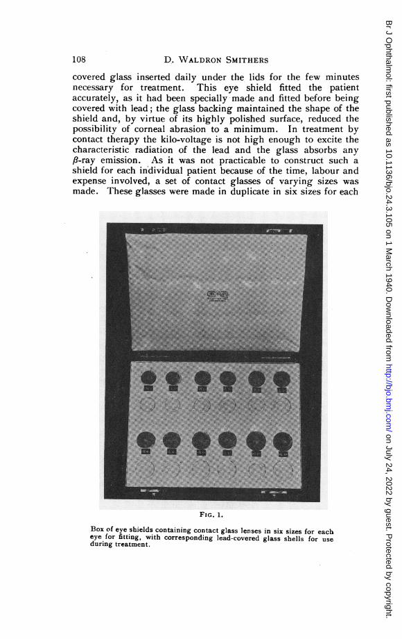
covered glass inserted daily under the lids for the few minutesnecessary for treatment. This eye shield fitted the patientaccurately, as it had been specially made and fitted before beingcovered with lead; the glass backing maintained the shape of theshield and, by virtue of its highly polished surface, reduced thepossibility of corneal abrasion to a minimum. In treatment bycontact therapy the kilo-voltage is not high enough to excite thecharacteristic radiation of the lead and the glass absorbs anyPl-ray emission. As it was not practicable to construct such ashield for each individual patient because of the time, labour andexpense involved, a set of contact glasses of varying sizes wasmade. These glasses were made in duplicate in six sizes for each
FIG. 1.
Box of eye shields containing contact glass lenses in six sizes for eacheye for fitting, with corresponding lead-covered glass shells for useduring treatment.
108
on July 24, 2022 by guest. Protected by copyright.
http://bjo.bmj.com
/B
r J Ophthalm
ol: first published as 10.1136/bjo.24.3.105 on 1 March 1940. D
ownloaded from
X-RAY TREATMENT OF MALIGNANT TUMOURS
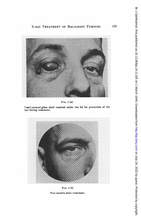
FIG. 2 (a)
Lead-covered glass shell inserted under the lid for protection of theeye during treatment.
FIG. 2 (b)
Two months after treatment.
109
on July 24, 2022 by guest. Protected by copyright.
http://bjo.bmj.com
/B
r J Ophthalm
ol: first published as 10.1136/bjo.24.3.105 on 1 March 1940. D
ownloaded from
D. WALDRON SMITHERS
eye, one set being covered with lead (Fig. 1). For any individualpatient it is now only necessary to fit the glass shell that is mostsuitable and then employ the corresponding lead-covered shell forthat patient during treatment (Fig. 2). There must be ampleclearance between the inside corneal radius and the front surfaceof the cornea in order to avoid abrasion and the shell must bemade larger than the normal contact glass in the scleral portion,to afford maximum protection. The eye is anaesthetized withsome drops of 10 per cent. " decicaine " before the lead-coveredshell is introduced; after treatment the shell is removed and twodrops of " parolein " applied to protect the eye while it is stillanaesthetized. These shells offer no protection to the eyelashes,and in treating one eyelid, a separate lead strip should be appliedover the other to preserve the lashes.These shells proved satisfactory in practice but were too easily
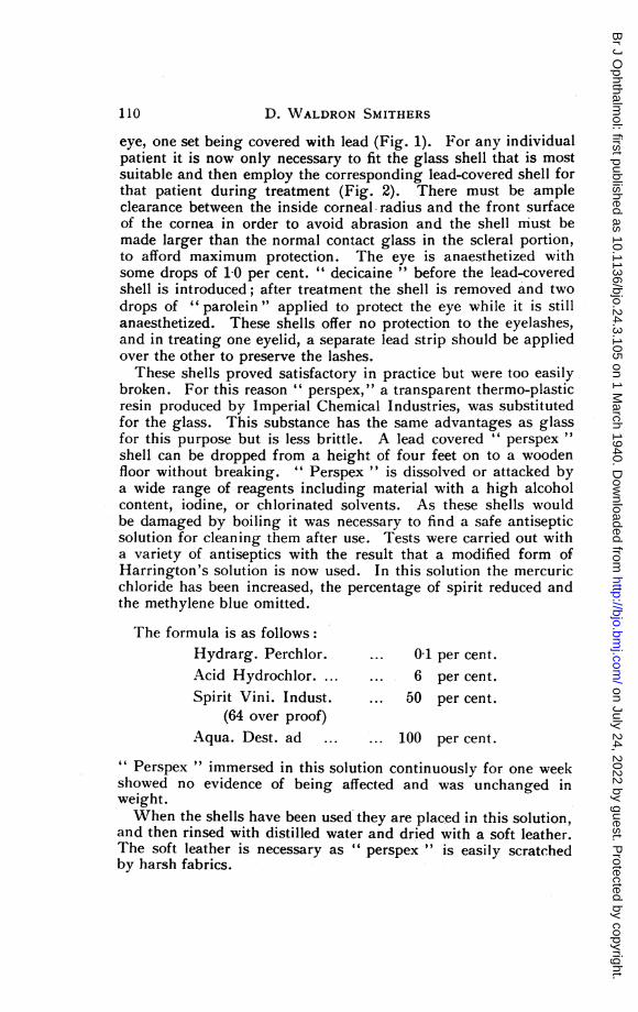
broken. For this reason " perspex," a transparent thermo-plasticresin produced by Imperial Chemical Industries, was substitutedfor the glass. This substance has the same advantages as glassfor this purpose but is less brittle. A lead covered " perspex "
shell can be dropped from a height of four feet on to a woodenfloor without breaking. " Perspex " is dissolved or attacked bya wide range of reagents including material with a high alcoholcontent, iodine, or chlorinated solvents. As these shells wouldbe damaged by boiling it was necessary to find a safe antisepticsolution for cleaning them after use. Tests were carried out witha variety of antiseptics with the result that a modified form ofHarrington's solution is now used. In this solution the mercuricchloride has been increased, the percentage of spirit reduced andthe methylene blue omitted.
The formula is as follows:Hydrarg. Perchlor. . 01 per cent.Acid Hydrochlor ... ... 6 per cent.Spirit Vini. Indust. ... 50 per cent.
(64 over proof)Aqua. Dest. ad ... ... 100 per cent.
Perspex " immersed in this solution continuously for one weekshowed no evidence of being affected and was unchanged inweight.When the shells have been used they are placed in this solution,
and then rinsed with distilled water and dried with a soft leather.The soft leather is necessary as " perspex " is easily scratchedby harsh fabrics.
110
on July 24, 2022 by guest. Protected by copyright.
http://bjo.bmj.com
/B
r J Ophthalm
ol: first published as 10.1136/bjo.24.3.105 on 1 March 1940. D
ownloaded from
X-RAY TREATMENT OF MALIGNANT TUMOURS
Construction of set of lead-covered Contact"Perspex" Shells
Selection was made from a large number of copies of actualcontact lenses previously constructed for patients by Messrs.Clement Clarke in order to give a range of fixed sizes of bothright and left scleral fittings. The inside corneal radius wasmodified to ensure effective clearance of the cornea and limbus.These lenses are those used for preliminary filling to determinewhich shell affords maximum scleral protection with ample cornealclearance. Further shells of the exact size and shape are thencovered on the outside with a coating of lead 1-0 mm. in thicknessand these are used during treatment. At first three trial shells,large, medium and small were used. During the last nine monthssince the full set illustrated in Fig. 1 has been available therehas been occasion to use each size at least once. Sizes3, 4, and .5 have been used most frequently. In a departmentwhere a number of patients require- protection of this kind thefull set is of value, but where such shells would be requiredinfrequently those numbered 3, 4, and 5 without fitting glasseswill probably prove sufficient for all but a very few cases.
Results of Treatment
This method of short-distance low-voltage X-ray treatment hasonly been employed in this country for four years so that it is stilltoo early to lay much stress on the results obtained. Furthermore,the technique of treatment is still in process of development andthe method of eye protection described has only recently beenintroduced. Nevertheless the results published by Chaoul10 andother workers show over 90 per cent. of cases of cutaneous cancersymptom-free from one to five years, and there are certain advan-tages inherent in the method itself which suggest that it is thebest form of treatment at present available for accessible malignanttumours. These advantages apply with special force to the treat-ment of tumours in the region of the eyes. The concentrationof the radiation effect to a small volume of tissue in itself tendsto protect the eye so that no other protection is necessary unlessthe actual lids or canthi are themselves involved. In the reportof cases that follows only those where extra eye protection wasconsidered necessary are included and lesions close to the eye butnot actualiy involving the lids, such as that illustrated in Fig. 3,are omitted. Treatment takes only a few minutes each day forfrom ten to fifteen days in most cases, though small superficiallesions may be treated with a single massive dose. Hospitalisation
ill
on July 24, 2022 by guest. Protected by copyright.
http://bjo.bmj.com
/B
r J Ophthalm
ol: first published as 10.1136/bjo.24.3.105 on 1 March 1940. D
ownloaded from
D. WALDRON SMITHERS
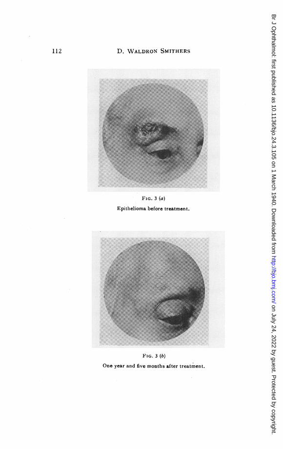
FIG. 3 (a)
Epithelioma before treatment.
FIG. 3 (b)
One year and five months after treatment.
112
on July 24, 2022 by guest. Protected by copyright.
http://bjo.bmj.com
/B
r J Ophthalm
ol: first published as 10.1136/bjo.24.3.105 on 1 March 1940. D
ownloaded from
X-RAY TREATMENT OF MALIGNANT TUMOURS
is avoided in the majority of cases and large numbers of patientscan be treated at comparatively low cost.During the four years that this treatment has been employed
at the Royal Cancer Hospital (Free) 57 patients with malignanttumours involving the eyelids or canthi have been treated; 18 ofthese were treated for recurrence following some other form oftreatment and 39 were treated primarily by this method.
Primary Treatment GroupOf the 39 primary cases, 3 required re-treatment to a small
volume of persistent growth at the tumour edge, due to the failureto include sufficient surrounding tissue in the original field, allthree were situated at the inner canthus and all are now free fromgrowth. All but two of the primary treatment cases are now aliveand well with no sign of recurrence. These two patients died ofinter-current disease one one month and one three months aftercompleting treatment. Microscopical confirmation was onlyobtained in 13 cases, 8 epitheliomata and 5 rodent ulcers, theremainder being classed clinically as rodent ulcers, with the excep-tion of one pitch wart. Ihe period of observation ranges from3 years and 6 months to 3 months. Of the 37 patients alive andsymptom-free, 9 have been observed for more than 2 years, 10
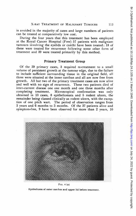
FIG. 4 (a)
Epithelioma of outer canthus and upper lid before treatment.
113
on July 24, 2022 by guest. Protected by copyright.
http://bjo.bmj.com
/B
r J Ophthalm
ol: first published as 10.1136/bjo.24.3.105 on 1 March 1940. D
ownloaded from
D. WALDRON SMITHERS
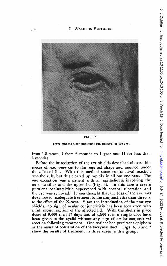
FIG. 4 (b)
Three months after treatment and removal of the eye.
from 1-2 years, 7 from 6 months to 1 year and 11 for less than6 months.
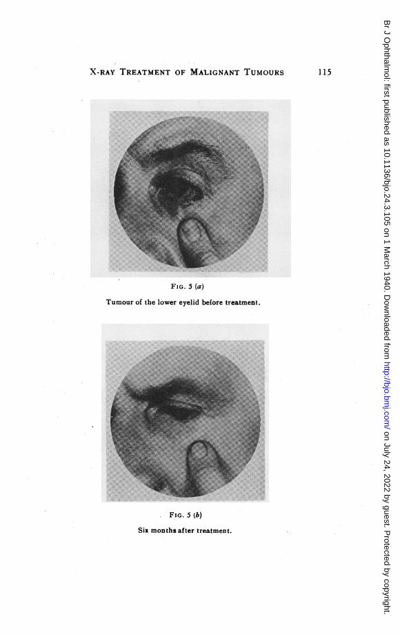
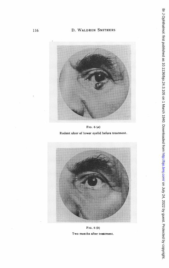
Before the introduction of the eye shields described above, thinpieces of lead were cut to the required shape and inserted underthe affected lid. With this method some conjunctival reactionwas the rule, but this cleared up rapidly in all but one case. Theone exception was a patient with an epithelioma involving theouter canthus and the upper lid (Fig. 4). In this case a severepurulent conjunctivitis supervened with corneal ulceration andthe eye was removed. It was thought that the loss of the eye wasdue more to inadequate treatment to the conjunctivitis than directlyto the effect of the X-rays. Since the introduction of the new eyeshields, no sign of ocular conjunctivitis has been seen even witha full moist reaction of the affected lid. With the shells in placedoses of 9,000 r. in 17 days and of 4,500 r. in a single dose havebeen given to the eyelid without any sign of ocular conjunctivalreaction following treatment. One patient has persistent epiphoraas the result of obliteration of the lacrymal duct. Figs. 5, 6 and 7show the results of treatment in three cases in this group.
114
on July 24, 2022 by guest. Protected by copyright.
http://bjo.bmj.com
/B
r J Ophthalm
ol: first published as 10.1136/bjo.24.3.105 on 1 March 1940. D
ownloaded from
X-RAY TREATMENT OF MALIGNANT TUMOURS
FIG. 5 (a)
Tumour of the lower eyelid before treatment.
FIG. 5 (b)
Six months after treatment.
115
on July 24, 2022 by guest. Protected by copyright.
http://bjo.bmj.com
/B
r J Ophthalm
ol: first published as 10.1136/bjo.24.3.105 on 1 March 1940. D
ownloaded from
D. WALDRON SMITHERS
.!:
;..:ie ..£g
j.;Tiki iii.:. ..
': !:.
*si!....:.!!: !::!:x,iji:j..::*::!!*::
FIG. 6 (a)
Rodent ulcer of lower eyelid before treatment.
FIG. 6 (b)
Two months after treatment.
116
on July 24, 2022 by guest. Protected by copyright.
http://bjo.bmj.com
/B
r J Ophthalm
ol: first published as 10.1136/bjo.24.3.105 on 1 March 1940. D
ownloaded from
X-RAY TREATMENT OF MALIGNANT TUMOURS
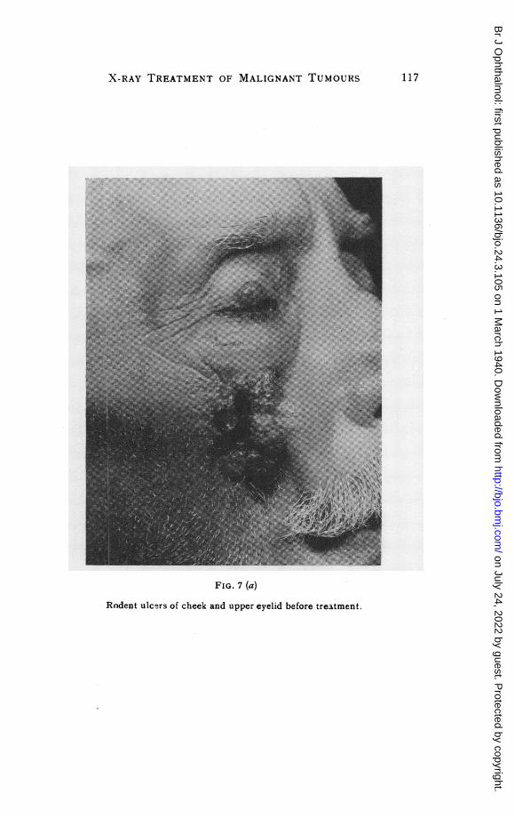
FIG. 7 (a)
Rodent ulcers of cheek and upper eyelid before treatment.
117
on July 24, 2022 by guest. Protected by copyright.
http://bjo.bmj.com
/B
r J Ophthalm
ol: first published as 10.1136/bjo.24.3.105 on 1 March 1940. D
ownloaded from
D. WALDRON SMITHERS
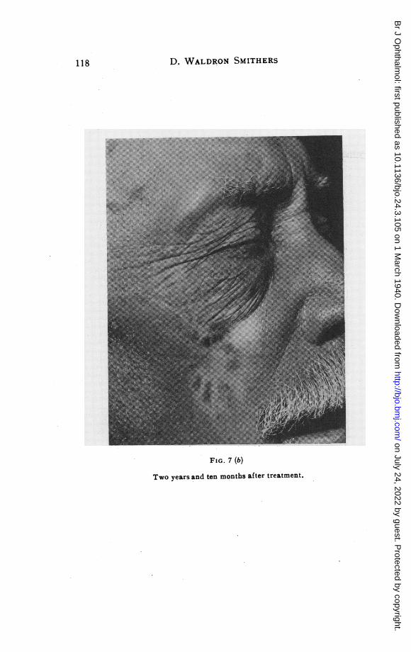
FIG. 7 (b)
Two years and ten months after treatment.
118
on July 24, 2022 by guest. Protected by copyright.
http://bjo.bmj.com
/B
r J Ophthalm
ol: first published as 10.1136/bjo.24.3.105 on 1 March 1940. D
ownloaded from
X-RAY TREATMENT OF MALIGNANT TUMOURS
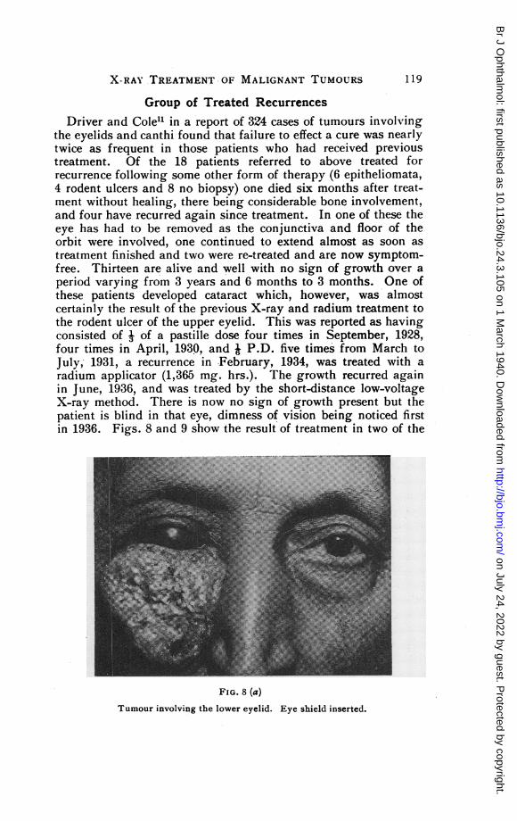
Group of Treated RecurrencesDriver and Cole'1 in a report of 324 cases of tumours involving
the eyelids and canthi found that failure to effect a cure was nearlytwice as frequent in those patients who had received previoustreatment. Of the 18 patients referred to above treated forrecurrence following some other form of therapy (6 epitheliomata,4 rodent ulcers and 8 no biopsy) one died six months after treat-ment without healing, there being considerable bone involvement,and four have recurred again since treatment. In one of these theeye has had to be removed as the conjunctiva and floor of theorbit were involved, one continued to extend almost as soon astreatment finished and two were re-treated and are now symptom-free. Thirteen are alive and well with no sign of growth over aperiod varying from 3 years and 6 months to 3 months. One ofthese patients developed cataract which, however, was almostcertainly the result of the previous X-ray and radium treatment tothe rodent ulcer of the upper eyelid. This was reported as havingconsisted of I of a pastille dose four times in September, 1928,four times in April, 1930, and W P.D. five times from March toJuly, 1931, a recurrence in February, 1934, was treated with aradium applicator (1,365 mg. hrs.). The growth recurred againin June, 1936, and was treated by the short-distance low-voltageX-ray method. There is now no sign of growth present but thepatient is blind in that eye, dimness of vision being noticed firstin 1936. Figs. 8 and 9 show the result of treatment in two of the
FIG. 8 (a)Tumour involving the lower eyelid. Eye shield inserted.
119
on July 24, 2022 by guest. Protected by copyright.
http://bjo.bmj.com
/B
r J Ophthalm
ol: first published as 10.1136/bjo.24.3.105 on 1 March 1940. D
ownloaded from
D. WALDRON SMITHERS
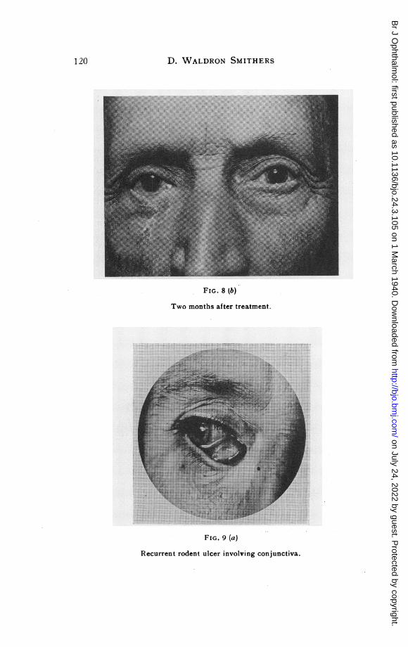
FIG. 8 (b)
Two months after treatment.
FIG. 9 (a)
Recurrent rodent ulcer involving conjunctiva.
120
on July 24, 2022 by guest. Protected by copyright.
http://bjo.bmj.com
/B
r J Ophthalm
ol: first published as 10.1136/bjo.24.3.105 on 1 March 1940. D
ownloaded from
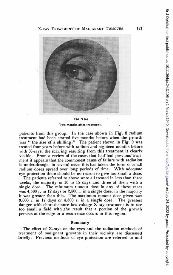
X-RAY TREATMENT' OF MALIGNANT TUMOURS
FIG. 9 (b)
Two months after treatment.
patients from this group. In the case shown in Fig. 8 radiumtreatment had been started five months before when the growthwas " the size of a shilling." The patient shown in Fig. 9 wastreated four years before with radium and eighteen nMonths beforewith X-rays, the scarring resulting from this treatment is clearlyvisible. From a review of the cases that had had previous treat-ment it appears that the commonest cause of failure with radiationis under-dosage, in several cases this has taken the form of-smallradium doses spread over long periods of time. With adequateeye protection there should be no reason to give too small a dose.The patients referred to above were all treated in less than three
weeks, the majority in 10 to 15 days and three of them with asingle dose. The minimum tumour dose in any of these caseswas 4,500 r. in 12 days or 2,500 r. in a single dose, in the majorityit was greater than this. The maximum tumour dose given was9,000 r. in 17 days or 4,500 r. in a single dose. The greatestdanger with short-distance low-voltage X-ray treatment is to usetoo small a field with the result that a portion of the growthpersists at the edge or a recurrence occurs in this region.
SummaryThe effect of X-rays on the eyes and the radiation methods of
treatment of malignant growths in their vicinity are discussedbriefly. Previous methods of eye protection are referred to and
121
I' -:
on July 24, 2022 by guest. Protected by copyright.
http://bjo.bmj.com
/B
r J Ophthalm
ol: first published as 10.1136/bjo.24.3.105 on 1 March 1940. D
ownloaded from
122A.SYORPIS
new lead-covered contact " perspex " shells for protection of theeyes in short-distance low-voltage X-ray treatment are described.The advantages of this form of X-ray therapy and the resultsobtained in the treatment of tumours of the lids and canthi duringthe last four years at the Royal Cancer Hospital (Free) arediscussed.
Acknowledgment.-I am greatly indebted to Mr. E. A. Plaiceand Mr. W. Hoad of Messrs. Clement Clarke who have assistedme throughout in the preparation of the eye shields. They havebeen entirely responsible for their construction and I am mostgrateful to them for furnishing me with some notes on this subjectwhich have been included in this paper. I am also indebted toMr. J. H. Wood the pharmacist at the Royal Cancer Hospital whocarried out the tests on " perspex " with various antiseptics.
REFERENCES
1. DESJARDINS, A. U.-Amer. Ji. Roentgenol., Vol. XXVI, pp. 639-679, 789-819,923-942, 1931.
2. CLAPP, C. A.-Amer. Ji. Ophthal., Vol. XV, pp. 1039-1044, 1932.3. MARTIN, P.-Brit. Med. JI., Vol. I, pp. 651-654, 1937.4. CHAOUL, H., and ADAM, A.-Strahlentherapie, Vol. XLVIII, pp. 31-50, 1933.5. FLOOD, P. A., and SMITHERS, D. W.-Brit. Jl. Radiol., Vol. XII, pp. 462-
485, 1939.6. REGAUD, C., COUTARD, H., MONOD, 0. and RICHARD, G.-Ann. d'Ocul.,
Vol. CLXIII, pp. 1-30, 1926.7. W6LFFLIN, E.-Strahlentherapie, Vol. XLIV, pp. 800-801, 1932.8. CUTLER, M., JAFFE, H. L. and GROSSMAN, A.-Amer. JI. Ophthal., Vol.
XXI, pp. 747- 754, 1938.9. WATSON, W. L. and WUESTER, W.-Amer. Ji. Ophthal., Vol. XXI, pp. 261-
263, 1938.10. CHAOUL, H.-Strahlentherapie, Vol. LVIII, pp. 611-613, 1937.11. DRIVER, J. R. and COLE, H. N.-Amer. Ji. Roentgenol., Vol. XLT, pp. 616-
624, 1939.
POST-CATARACT HYPHAEMABY
A. SEYMOUR PHILPS, F.R.C.S.LONDON
POST-CATARACT HYPHAEMA has long been one of the bugbears ofophthalmic surgery, for though it does not often affect the ultimate-visual result, it prolongs the patient's stay in hospital sometimes.for several weeks. In an endeavour to ascertain the origin andfate of the blood in these patients I have collected and recordedevery such hyphaema that occurred in the wards of two LondonHospitals during the years 1937 and the greater part of 1938.
122 A. SEYMOUR PHILPS
on July 24, 2022 by guest. Protected by copyright.
http://bjo.bmj.com
/B
r J Ophthalm
ol: first published as 10.1136/bjo.24.3.105 on 1 March 1940. D
ownloaded from