drug delivery: - principles and applications
TRANSCRIPT

DRUG DELIVERY

DRUG DELIVERY:
Principles and Applications
BINGHE WANGGeorgia State University
TERUNA SIAHAANUniversity of Kansas
RICHARD SOLTERODirectPharma, Inc.
A JOHN WILEY & SONS, INC. PUBLICATION

Copyright # 2005 by John Wiley & Sons, Inc. All rights reserved.
Published by John Wiley & Sons, Inc., Hoboken, New Jersey.
Published simultaneously in Canada.
No part of this publication may be reproduced, stored in a retrieval system, or transmitted in any form or
by any means, electronic, mechanical, photocopying, recording, scanning, or otherwise, except as
permitted under Section 107 or 108 of the 1976 United States Copyright Act, without either the prior
written permission of the Publisher, or authorization through payment of the appropriate per-copy fee to
the Copyright Clearance Center, Inc., 222 Rosewood Drive, Danvers, MA 01923, 978-750-8400,
fax 978-646-8600, or on the web at www.copyright.com. Requests to the Publisher for permission
should be addressed to the Permissions Department, John Wiley & Sons, Inc., 111 River Street,
Hoboken, NJ 07030, (201) 748-6011, fax (201) 748-6008.
Limit of Liability/Disclaimer of Warranty: While the publisher and author have used their best efforts
in preparing this book, they make no representations or warranties with respect to the accuracy or
completeness of the contents of this book and specifically disclaim any implied warranties of
merchantability or fitness for a particular purpose. No warranty may be created or extended by sales
representatives or written sales materials. The advice and strategies contained herein may not be suitable
for your situation. You should consult with a professional where appropriate. Neither the publisher
nor author shall be liable for any loss of profit or any other commercial damages, including but not
limited to special, incidental, consequential, or other damages.
For general information on our other products and services please contact our Customer Care Department
within the U.S. at 877-762-2974, outside the U.S. at 317-572-3993 or fax 317-572-4002.
Wiley also publishes its books in a variety of electronic formats. Some content that appears in print,
however, may not be available in electronic format.
Library of Congress Cataloging-in-Publication Data:
Wang, Binghe.
Drug delivery : principles and applications / Binghe Wang, Teruna Siahaan,
Richard soltero.
p. cm.
Includes index.
ISBN 0-471-47489-4 (cloth)
1. Drug delivery systems. 2. Pharmaceutical chemistry. I. Siahaan, Teruna.
II. Soltero, Richard. III. Title.
RS199.5.W36 2005
6150.6–dc22 2004020585
Printed in the United States of America
10 9 8 7 6 5 4 3 2 1
CONTENTS
Preface vii
List of Contributors ix
1 Factors That Impact the Developability of Drug Candidates:An Overview 1
Chao Han and Binghe Wang
2 Physiological, Biochemical, and Chemical Barriers toOral Drug Delivery 15
Anna Maria Calcagno and Teruna J. Siahaan
3 Pathways for Drug Delivery to the Central Nervous System 29
Yan Zhang and Donald W. Miller
4 Physicochemical Properties, Formulation, and Drug Delivery 57
Dewey H. Barich, Mark T. Zell, and Eric J. Munson
5 Targeted Bioavailability: A Fresh Look at Pharmacokineticand Pharmacodynamic Issues in Drug Delivery 73
William F. Elmquist
6 Presystemic and First-Pass Metabolism 83
W. Griffith Humphreys
7 Cell Culture Models for Drug Transport Studies 103
D. Nedra Karunaratne, Peter S. Silverstein, Veena Vasandani,
Amber M. Young, Erik Rytting, Bradley Yops, and Kenneth L. Audus
v

8 Prodrug Approaches to Drug Delivery 125
Longqin Hu
9 Receptor-Mediated Drug Delivery 167
Christopher P. Leamon and Philip S. Low
10 Oral Protein and Peptide Drug Delivery 189
Rick Soltero
11 Metabolic Activation and Drug Targeting 201
Xiangming Guan
12 Ultrasound-Mediated Drug Delivery 245
Ka-yun Ng and Terry O. Matsunaga
13 Polycationic Peptides and Proteins in Drug Delivery:Focus on Nonclassical Transport 279
Lisa A. Kueltzo and C. Russell Middaugh
14 Gene Therapy and Gene Delivery 305
Naoki Kobayashi, Makiya Nishikawa, and Yoshinobu Takakura
15 Parenteral Formulation for Peptides, Proteins,and Monoclonal Antibodies Drugs: A CommercialDevelopment Overview 321
John A. Bontempo
16 Pulmonary Drug Delivery: Pharmaceutical Chemistry andAerosol Technology 341
Anthony J. Hickey
17 Antibody-Directed Drug Delivery 363
Herve Le Calvez, John Mountzouris, Kosi Gramatikoff, and Fang Fang
18 Efflux Transporters in Drug Excretion 381
Shuzhong Zhang and Marilyn E. Morris
19 Liposomes as Drug Delivery Vehicles 411
Guijun Wang
20 Regulatory and Intellectual Property Issues in Drug DeliveryResearch 435
Shihong Nicolaou
Index 443
vi CONTENTS

PREFACE
The pharmaceutical industry, at least the part represented by the major pharmaceu-
tical companies, has undergone a fundamental transformation during the last de-
cade. This transformation has changed the ‘‘silo’’ structure, where different ‘‘steps’’
of the drug discovery process were treated independent of each other, to that of an
integrated approach. This transformation was brought about by several factors. One
important reason was that about 60% of new chemical entities (NCE) fail clinical
trials because of poor pharmacokinetics, undesirable metabolic properties, and toxi-
city. NCEs do not fail due to a lack of biological activities per se. In addition, the
transformation came about because of development in areas such as combinatorial
chemistry, high throughput screening, genomics, and proteomics that has allowed
for the rapid identification of a large number of biologically active compounds.
Therefore, there is a tremendous pressure to have a mechanism to identify a ‘‘win-
ner’’ among a large pool of candidates or ‘‘play out’’ the failures early on. An inte-
grated approach helps to achieve this goal by evaluating various factors such as
formulation, permeation, metabolism, and toxicity early in the drug discovery
and development process. Such a practice also helps to bring issues that impact
development to the awareness of medicinal chemists so that many structural fea-
tures detrimental to clinical development would be avoided at the designing stage.
Among all the factors that effect the clinical development of a NCE, drug deliv-
ery occupies a special place. In an integrated drug discovery approach, one has to
start considering the delivery properties of a NCE at the design stage. This means
that medicinal chemists are the first line of researchers who have to consider this
issue. However, most entry-level medicinal chemists are trained as synthetic che-
mists, and have very little exposure to drug delivery. Without a basic understanding
of the issues that effect drug delivery properties, it is hard for the medicinal che-
mists to address this issue. This book systematically examines various subject areas
vii

important to drug delivery, and should be an excellent desk reference for medicinal
chemists interested in gaining an overview of this field. In addition, there is a need
to strengthen the education of our future medicinal chemists in the area of drug
delivery. This book can also serve as a textbook for graduate students or advanced
level undergraduate students interested in a career in the pharmaceutical industry.
The book starts with chapters that cover general drug delivery issues such as
physicochemical and biological barriers, various pathways for drug delivery, for-
mulation, pharmacokinetic and pharmacodynamic issues, metabolism, and cell cul-
ture models used in studying drug delivery. Then it moves on to cover specific drug
delivery strategies. At the end, we have added one chapter on intellectual property
so as to give readers a general idea of how to protect their intellectual property
when doing drug delivery research. Each chapter is structured in such a way that
it gives an overview of the specific subject, and also goes into details with selected
examples so that there is an in depth discussion with extensive references. With this
kind of structure, the book will be valuable to both novices and experts.
We would like to thank Ms. Neeta Raje for her diligent work in assisting BW in
organizing and coordinating the editing and processing of the manuscripts.
BINGHE WANG
TERUNA SIAHAAN
RICHARD SOLTERO
viii PREFACE

CONTRIBUTORS
Kenneth L. Audus, Department of Pharmaceutical Chemistry, School of
Pharmacy, The University of Kansas, 2095 Constant Avenue, Lawrence, KS 66047
Dewey H. Barich, Department of Pharmaceutical Chemistry, University of
Kansas, 2095 Constant Avenue, Lawrence, KS 66047
John A. Bontempo, Biopharmaceutical Product Development Consultant, 18
Benjamin Street, Somerset, NJ 08873
Anna Maria Calcagno, Department of Pharmaceutical Chemistry, The University
of Kansas, Lawrence, KS 66047
Herve Le Calvez, Abgent Inc., 6310 Nancy Ridge Drive, Suite 106, San Diego,
CA 92121
William F. Elmquist, Department of Pharmaceutics, University of Minnesota,
Minneapolis, MN 55455
Fang Fang, NexBio, Inc., 6330 Nancy Ridge Drive, Suite 105, San Diego, CA
92121
Kosi Gramatikoff, Abgent Inc., 6310 Nancy Ridge Drive, Suite 106, San Diego,
CA 92121
Xiangming Guan, Department of Pharmaceutical Sciences, College of Pharmacy,
South Dakota State University, Brookings, SD 57007
Chao Han, GlaxoSmithKline, Collegeville, PA 19426
Anthony J. Hickey, School of Pharmacy, University of North Carolina, Kerr Hall,
Chapel Hill, NC 27599
ix

Longqin Hu, Department of Pharmaceutical Chemistry, Ernest Mario School of
Pharmacy, Rutgers, The State University of New Jersey, Piscataway, NJ 08854
W. Griffith Humphreys, Department of Metabolism and Pharmacokinetics,
Bristol-Myers Squibb Pharmaceutical Research Institute, P.O. Box 5400, Princeton,
NJ 08543
D. Nedra Karunaratne, Department of Pharmaceutical Chemistry, School of
Pharmacy, The University of Kansas, 2095 Constant Avenue, Lawrence, KS 66047
Naoki Kobayashi, Department of Biopharmaceutics and Drug Metabolism,
Graduate School of Pharmaceutical Sciences, Kyoto University, Sakyo-ku, Kyoto
606-8501, Japan
Lisa A. Kueltzo, Department of Pharmaceutical Sciences, University of Colorado
Health Sciences Center, Denver, CO 80262
Christopher P. Leamon, Endocyte, Inc., 1205 Kent Avenue, West Lafayette, IN
47906
Philip S. Low, Purdue University, West Lafayette, IN 47907
Terry O. Matsunaga, Imarx Therapeutics Inc., Tucson, AZ 85719
C. Russell Middaugh, Department of Pharmaceutical Chemistry, University of
Kansas, Lawrence, KS 66047
Donald W. Miller, Department of Pharmaceutical Sciences, University of
Nebraska Medical Center, Omaha, NE 68198-6025
Marilyn E. Morris, Department of Pharmaceutical Sciences, School of Pharmacy
and Pharmaceutical Sciences, University at Buffalo, State University of New York,
Amherst, NY 14260
John Mountzouris, Abgent Inc., 6310 Nancy Ridge Drive, Suite 106, San Diego,
CA 92121
Eric J. Munson, Department of Pharmaceutical Chemistry, University of Kansas,
2095 Constant Avenue, Lawrence, KS 66047
Ka-yun Ng, Department of Pharmaceutical Sciences, School of Pharmacy, Univer-
sity of Colorado Health Sciences Center, Denver, CO 80262
Shihong Nicolaou, Technology Transfer and Intellectual Property Services,
University of California, San Diego, 9500 Gilman Drive, La Jolla, CA 92093
Makiya Nishikawa, Department of Biopharmaceutics and Drug Metabolism,
Graduate School of Pharmaceutical Sciences, Kyoto University, Sakyo-ku, Kyoto
606-8501, Japan
Erik Rytting, Department of Pharmaceutical Chemistry, School of Pharmacy, The
University of Kansas, 2095 Constant Avenue, Lawrence, KS 66047
x CONTRIBUTORS
Teruna J. Siahaan, Department of Pharmaceutical Chemistry, The University of
Kansas, Lawrence, KS 66047
Peter S. Silverstein, Department of Pharmaceutical Chemistry, School of
Pharmacy, The University of Kansas, 2095 Constant Avenue, Lawrence, KS 66047
Rick Soltero, Pharma Directions, Inc.
Yoshinobu Takakura, Department of Biopharmaceutics and Drug Metabolism,
Graduate School of Pharmaceutical Sciences, Kyoto University, Sakyo-ku, Kyoto
606-8501, Japan
Veena Vasandani, Department of Pharmaceutical Chemistry, School of Pharmacy,
The University of Kansas, 2095 Constant Avenue, Lawrence, KS 66047
Binghe Wang, Department of Chemistry, Georgia State University, Atlanta, GA
30303
Guijun Wang, Department of Chemistry, University of New Orleans, New
Orleans, LA 70148
Amber M. Young, Department of Pharmaceutical Chemistry, School of Pharmacy,
The University of Kansas, 2095 Constant Avenue, Lawrence, KS 66047
Bradley Yops, Department of Pharmaceutical Chemistry, School of Pharmacy, The
University of Kansas, 2095 Constant Avenue, Lawrence, KS 66047
Mark T. Zell, Pfizer Global Research and Development, Ann Arbor Laboratories,
2800 Plymouth Road, Ann Arbor, MI 48105
Shuzhong Zhang, Department of Pharmaceutical Sciences, School of Pharmacy
and Pharmaceutical Sciences, University at Buffalo, State University of New
York, Amherst, NY 14260
Yan Zhang, Department of Pharmaceutical Sciences, University of Nebraska
Medical Center, Omaha, NE 68198-6025
CONTRIBUTORS xi

1FACTORS THAT IMPACT THEDEVELOPABILITY OF DRUGCANDIDATES: AN OVERVIEW
CHAO HAN
GlaxoSmithKline, Collegeville, PA 19426
BINGHE WANG
Department of Chemistry, Georgia State University, Atlanta, GA 30303
1.1. Issues facing the pharmaceutical industry
1.2. Factors that impact developability
1.2.1. Commercial goal
1.2.2. The chemistry efforts
1.2.3. Target validation in animal models
1.2.4. Pharmacokinetics and drug metabolism
1.2.5. Preparation for pharmaceutical products
1.2.6. Remarks on developability criteria
1.3. Drug delivery factors that impact developability
References
1.1. ISSUES FACING THE PHARMACEUTICAL INDUSTRY
Drug discovery is a long, arduous, and expensive process. It was estimated that the
total expenditure for research and development in the U.S. pharmaceutical industries
Drug Delivery: Principles and Applications Edited by Binghe Wang, Teruna Siahaan,
and Richard Soltero
ISBN 0-471-47489-4 # 2005 John Wiley & Sons, Inc.
1

was over $20 billion a year in the late 1990s,’ and this figure has been increasing.1
The average cost for every new drug (a new chemical entity, NCE) from research
laboratory to patients is a staggering number: $400 to $650 million,2–4 and the
whole process may take up to 14 years!5 Because of the high cost, there is tremen-
dous pressure to maximize efficiency and minimize the time it takes to discover and
bring a drug to the market. In order to do this, it is necessary to analyze the entire
drug discovery and development process and identify steps where changes can be
made to increase efficiency and save time. Analyzing the entire drug discovery and
development process will help reveal where maximal improvements can be
expected with some effort.
The entire endeavor of bringing a new drug from idea to market is generally
divided into several stages: target/disease identification, hit identification/discovery,
hit optimization, lead selection and further optimization, candidate identification,
and clinical trials.6 Each stage has many aspects and components. A target is iden-
tified early in the discovery period, when there is sufficient evidence to validate the
relationship between this target and a disease of interest. Tens of thousands of
new compounds are then synthesized and screened against the target to identify a
few compounds (hits) with the desired biological activity. Analogs of these selected
compounds are then screened further for better activity and optimized in order to
identify a small number of compounds for testing in pharmacological models.
These efficacious compounds (leads) are further optimized for their biopharmaceu-
tical properties, and the most drug-like compounds (drug candidate, only one or
two) are then selected for further development. The drug discovery and develop-
ment path, with emphasis on the discovery stages, is schematically illustrated in
Figure 1.1.
Number of compounds
100,000 100 10 1-2 ~1
In vitro activity
Efficacy & developability
Compoundlibrary
Hits Leads Drugcandidates
Marketedmedicine
Candidateselection
IND, clinical trial and NDA
Therapeutics areasUnmet needsIdeas, targets
Successfulmedicine
Molecule Drug
Figure 1.1. A schematic illustration of the drug discovery and development process with the
estimated number of compounds shown for each step.
2 FACTORS THAT IMPACT THE DEVELOPABILITY OF DRUG CANDIDATES
Of those drug candidates with most drug-like properties, only about 40% make
their way to evaluation in humans (Phase I clinical trial).7 Unfortunately, the histor-
ical average reveals an almost 90% overall attrition rate in clinical trials;7 in another
words, only 1 compound makes it to market from among 10 compounds tested in
humans. Results from another statistical analysis gave a similar success rates for
NCEs for which an IND (investigational new drug) was filed during 1990–1992.8
This high attrition rate obviously does not produce the long-term success desired by
both the pharmaceutical and health care industries.
In order to reduce the failure rate, it is necessary to analyze how and where fail-
ures occur. More than 10 years ago, Prentis et al.9 analyzed the cause of the high
attrition rate based on data from seven UK-based pharmaceutical companies from
1964 to 1985. The results revealed that 39% of the failure was due to poor pharma-
cokinetic properties in humans; 29% was due to a lack of clinical efficacy; 21% was
due to toxicity and adverse effects; and about 6% was caused by commercial lim-
itations. Although not enough detailed information was available, it is believed that
some of these causes are interrelated. For instance, toxicity or lack of efficacy can
be caused by poor or undesired pharmacokinetic properties. With the understanding
that most failure was not due to a lack of ‘‘biological activities’’ per se as defined by
in vitro testing, there is a drive to incorporate the evaluation of the other major
factors that may potentially precipitate developmental failures in the early drug
discovery and candidate selection processes. This is intended to reduce the rate
of late-stage failures, which is most costly. This point is further substantiated by
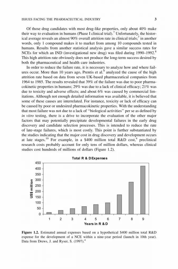
the studies indicating that the major cost in drug discovery and development occurs
at late stages.10 For example, in a $400 million total R&D cost,4 preclinical
research costs probably account for only tens of million dollars, whereas clinical
studies cost hundreds of millions of dollars (Figure 1.2).
Total R & D Expenses
0
50
100
150
200
250
300
350
400
450
1 2 3 4 5 6 7 8 9Years in R & D
US
$ m
illio
n
Figure 1.2. Estimated annual expenses based on a hypothetical $400 million total R&D
expense for the development of a NCE within a nine-year period (launch in 10th year).
Data from Drews, J. and Ryser, S. (1997).4
ISSUES FACING THE PHARMACEUTICAL INDUSTRY 3

Another factor that is fueling the movement for early integration of multiple dis-
ciplines in the drug discovery and development processes is the rapid development
of chemical and biological sciences. The past decade has seen tremendous advances
in both areas. Advances in combinatorial chemistry, molecular and cellular biology,
high-throughput screening, and genomic research have provided both great oppor-
tunities and challenges to the pharmaceutical industry. With the rapid development
in biological sciences, current interests in therapeutic targets are more focused on
rational targets such as receptors, enzymes, and hormones with well-characterized
structures and functions. New technologies such as combinatorial chemistry, auto-
mation in high-throughput screening, and better instrumentation in bioanalysis have
also significantly accelerated the lead identification and discovery process11 for a
given target. With these new technologies, large pharmaceutical research organiza-
tions are capable of synthesizing and screening several thousand compounds or
more in a year or two to find potential drug candidates.12 These efforts typically
result in the discovery of many lead compounds or potential candidates for a target
in the drug discovery process. Then there is the question of how to pick a winner
and how to minimize failures. This requires a thorough evaluation of all the factors
that are known to affect the developability of a NEC at the early stages. These fac-
tors may include efficacy, pharmacokinetics, pharmacodynamics, toxicology, and
drug-drug interactions based on the metabolism and substrate properties of certain
transporters and enzymes, as well as physicochemical properties, many of which
are related to drug delivery issues. For this reason, a drug discovery and develop-
ment program is more like a symphony (not just a cross-functional action) of
multiple sciences including chemistry, biology, toxicology, clinical science, and
pharmaceutical engineering.
Under the pressure to reduce the cost and shorten the time needed to bring an
NCE to the market, many major pharmaceutical organizations have undergone
rapid and drastic changes in the past decade, both in terms of organizational struc-
tures and fundamental approaches, in order to develop an integrated approach to
drug discovery and development.13 A conference entitled ‘‘Opportunities for Inte-
gration of Pharmacokinetics, Pharmacodynamics, and Toxicokinetics in Rational
Drug Development’’14 was the landmark event in this fundamental change in the
pharmaceutical industry.15 A brand new concept, ‘‘ensuring developability,’’ was
introduced and well accepted, which employs criteria for drug development
throughout the entire drug discovery and development processes. Under the gui-
dance of such criteria, a drug discovery and development team will not only max-
imize the chance of success by selecting the best developable drug candidate, but
will also play off the failures faster and more cheaply.
The paradigm shifts mostly involve the integration of research activities in func-
tional areas such as pharmacokinetics and drug metabolism, pharmaceutical devel-
opment, safety assessment, and process chemistry into drug discovery and
development process in the very early stages of discovery. The inputs from these
functional areas, as well as those from clinical, regulatory, commercial, and market-
ing groups in the early stages, help to minimize costly mistakes in late stages of
development and have become more and more important to the success of the
4 FACTORS THAT IMPACT THE DEVELOPABILITY OF DRUG CANDIDATES

drug discovery and development process. Developability is an overall evaluation of
the drug-like properties of a NCE. Many of the recent changes in the pharmaceu-
tical industry have been driven by the concept of ensuring developability. These
changes, that is, the integration of multifunctional areas in drug discovery and
development, ensure that the NCEs of interest will be successful in every step
toward the final goal.
Below is a brief introduction to the factors that impact developability and a dis-
cussion on why the examination of drug delivery issues is very important in helping
to ensure the developability of a drug candidate.
1.2. FACTORS THAT IMPACT DEVELOPABILITY
In most pharmaceutical companies, many efforts have been made to create a clear
framework for selecting compound(s) with minimal ambiguity for further progres-
sion. Such a framework is not a simple list of the factors that impact the quality of a
drug-like molecule. This framework, which is more often referred as ‘‘developabil-
ity criteria,’’ is a comprehensive summary of the characteristics, properties, and
qualities of the NCE(s) of interest, which normally consist of preferred profiles
with a minimally acceptable range. The preferred profile describes the optimal
goal for selection and further progression of a candidate, whereas the minimum
range gives the acceptable properties for a compound that is not ideal but may suc-
ceed. Molecules that do not meet the criteria will not be considered further. Such
criteria cover all the functional areas in drug development. Some of the major
developability considerations are briefly described in the following subsections.
1.2.1. Commercial Goal
It does not need to be emphasized that we are in a business world. Generally speak-
ing, a product needs to be profitable to be viable. Therefore, early inputs from com-
mercial, marketing, and medical outcome professionals are very important for
setting up a projective product profile, which profoundly affects the creation of
the developability criteria for the intended therapeutics. In general, this portfolio
documents the best possible properties of the product and the minimum acceptable
ones that may succeed based on the studies of market desires. These studies should
be based on the results of professional analyses of the medical care needs, potential
market, and existing leading products for the same, similar, or related indications.
The following aspects need to be well thought out and fully justified before the
commencement of a project: (1) therapeutic strategy; (2) dose form and regimen;
and (3) the best possible safety profile, such as the therapeutic window, potential
drug interactions, and any other potentially adverse effects. Using the development
of an anticancer agent as an example for therapeutic strategy selection, one may
consider the choice of developing a chemotherapeutic (directly attacking the cancer
cells) versus an antiangiogenic agent (depriving cancer cells of their nutrients),
or combined or stand-alone therapy. In deciding the optimal dose form and regimen,
FACTORS THAT IMPACT DEVELOPABILITY 5

one may consider whether an oral or intravenous (iv) formulation, or both, should
be developed, and whether the drug should be given once daily or in multiple doses.
The results of such an analysis form the framework for developing the developabil-
ity criteria and become the guideline in setting up the criterion for each desired
property. For example, pharmacokinetic properties such as the half-life and oral
bioavailability of a drug candidate will have a direct impact on developing a
drug that is to be administered orally once a day.
1.2.2. The Chemistry Efforts
Medicinal chemistry is always the starting point and driver of drug discovery pro-
grams. In a large pharmaceutical R&D organization, early discovery of bioactive
compounds (hits) can be carried out either by random, high-throughput screening
of compound libraries, by rational design, or both. Medicinal chemists will then use
the structural information of the pharmacophore thus identified to optimize the
structures. Chemical tractability needs to be examined carefully at the very begin-
ning when a new chemical series is identified. Functional modifications around the
core structure are carefully analysed. After the examination of a small number of
compounds, the initial exploratory structure–activity relationship (SAR) or quanti-
tative SAR (QSAR) should be developed. Blackie et al.16 described how the estab-
lishment of exploratory SAR helped the discovery of a potent oral bioavailable
phospholipase A2 inhibitor. In this example, numerous substructural changes
were made, leading to the most active compounds; this is normally done in parallel
with several different chemical series. For medicinal chemists, it is important that
many different SARs are considered, developed, and integrated into their efforts at
the same time, providing more opportunities to avoid undesirable properties unre-
lated to their intended biological activities. Such factors, again, may include poten-
tial P450 inhibition, permeability, selectivity, stability, solubility, etc.
Structural novelty of the compounds (i.e., can this product be patented?), com-
plexity of synthetic routes, scalability (can the syntheses be scaled up in an indus-
trial way?) and the cost of starting materials (cost of goods at the end of the game),
and potential environmental and toxicity issues will all need to be closely examined
at early stages of the drug discovery and development processes. It is never too
early to put these thoughts into action.
1.2.3. Target Validation in Animal Models
Although drug discovery efforts almost always start with in vitro testing, it is well
recognized that promising results of such testing do not always translate into effi-
cacy. There are numerous reasons for this to happen, some of which are well under-
stood and others that are not. Therefore, target validation in animal models before
clinical trials in humans is a critical step. Before a drug candidate is fully assessed
for its safety and brought to a clinical test, demonstration of the efficacy of a
biologically active compound (e.g., active in an enzyme binding assay) in
6 FACTORS THAT IMPACT THE DEVELOPABILITY OF DRUG CANDIDATES

pharmacological models (in vivo, if available) is considered a milestone in the pro-
cess of discovering a drug candidate. Many cases exemplify the challenges and
importance of pharmacological models. For example, inhibitors of the integrin
receptor avb3 have been shown to inhibit endothelial cell growth, which implies
their potential as clinically useful antiangiogenic agents for cancer treatment.17
However, the proposed mechanism did not work in animal models, although com-
pounds were found to be very active in vitro.18,19 What has been recognized is that
the integrin receptor avb3 may not be the exclusive pathway on which cell growth
depends. Its inhibition may induce a compensatory pathway for angiogenesis.
Ideally, an in vivo model should comprise all biochemical, cellular, and physio-
logical complexities, as in a real-life system, which may predict the behavior of a
potential drug candidate in human much more accurately than an in vitro system. In
order to have a biological hypothesis tested in the system with validity, a compound
has to be evaluated in many other regards. Knowing the pharmacokinetic para-
meters such as absorption, distribution, and metabolism in the animal species
that is used in the pharmacological model is critical. Showing successful drug deliv-
ery in an animal model serves as an important milestone.
The pharmacokinetics/pharmacodynamics relationship, systemic and tissue
levels of drug exposure, frequency of dosing following which the drug may demon-
strate efficacy, and the strength of efficacy are very important factors that may affect
further development of an NEC. They are all directly or indirectly related to drug
delivery.
1.2.4. Pharmacokinetics and Drug Metabolism
Pharmacokinetics and drug metabolism are more often abbreviated as DMPK. The
importance of DMPK in drug discovery and development practices is reflected in
the statistics of the attrition rate.9 Most of the changes in the pharmaceutical indus-
try during the past decade occurred in DMPK15 and related fields. The overall goal
of DMPK in drug discovery and development is to predict the behavior of a drug
candidate in humans. Nevertheless, the focus could be different at different stage of
the process. Pharmacokinetics (PK) parameters in animal species that will be used
in pharmacological (as noted briefly in the previous paragraph) and safety assess-
ment models provide very important insights (systemic and tissue exposures) for
those studies. The results of PK studies in several animal species generate the
data for physiologically based models or allometric scaling20,21 to predict the basic
pharmacokinetic behavior of a compound in humans. Assays using human tissues,
cells, and genetically engineered cell lines provide a tremendous amount of infor-
mation before the real clinical studies begin. Optimizing DMPK developability fac-
tors is immensely beneficial for finding the candidate with best potential for
success.22
The desirable (or undesirable) biological effects of a drug in vivo normally are
directly related to its exposure. One of these factors, namely, the total systemic
exposure, maximum concentration, or duration of the concentration above a certain
FACTORS THAT IMPACT DEVELOPABILITY 7

level, is usually used as a parameter that is correlated with the drug’s efficacy and
adverse effects.23 The exposure at a given dose is governed by (1) the ability of the
body to remove the drug as a xenobiotic and (2) the route by which the drug is
delivered. Blood or plasma clearance is often used as a measure of the ability to
eliminate a drug molecule from the systemic circulation. A low to moderate clear-
ance molecule is desirable in most situations unless a fast-action, short-duration
drug is needed.24
A drug can be directly introduced into the systemic circulation by several meth-
ods. However, for convenience and many other reasons, oral dosage forms are pre-
ferred in many situations. Therefore, oral bioavailability of the compound is one of
the very important developability criteria for oral drug delivery. Many factors affect
the oral bioavailability of a drug. These factors will be discussed in detail in several
chapters. In addition to clearance and bioavailability, other major pharmacokinetic
parameters also should be evaluated.
Volume of distribution is a conceptual pharmacokinetic parameter that scales the
extent of a drug distributed into the tissues. A well-known parameter, elimination
half-life, can be derived from clearance and volume of distribution. It is a very
important developability criterion that warrants the desired dose regimen. It should
be noted here that half-life must be discussed in the context of a biologically rele-
vant concentration. A purely mathematically derived half-life is sometimes biolo-
gical irrelevant. Some more definitive explanations and comprehensive discussion
of the major pharmacokinetic parameters and their biological relevance have been
extensively reviewed.25,26 These parameters should be examined across several dif-
ferent preclinical species to predict the behavior in humans. The DMPK topics will
be discussed in Chapters 5 and 6.
Inhibition and induction of drug-metabolizing enzymes,27,28 P-glycoprotein
(P-gp) substrate property,29,30 plasma protein binding and binding kinetics,31,32
and metabolic stability in the microsomes or hepatocytes from different species
including humans,33 as well as the metabolic pathway and the metabolite identi-
fied,34 are all very important developability measurements in the assessment of
safety, potential drug-drug interaction, and predictability. These factors need to
be optimized and carefully examined against developability criteria. Drug metabo-
lism–related issues are outlined and discussed in Chapter 5. The impact of the
transporter, including the efflux transporter in drug delivery and the models used
to study and address the issues, will be discussed in Chapters 18, 2, and 3.
1.2.5. Preparation for Pharmaceutical Products
Before the early 1990s, the solid state, salt form, aqueous solubility, and dosing for-
mulation for agents used in pharmacological, pharmacokinetic, and toxicological
studies were not of major concern. However, an inappropriate salt version or solid
form may cause potential drug delivery and stability problems (both physicochemi-
cally and chemically) during formulation and pharmaceutical engineering. It is
now understood that the investigation of the physicochemical properties of an
NCE against developability criteria should start early in the R&D processes.
8 FACTORS THAT IMPACT THE DEVELOPABILITY OF DRUG CANDIDATES

Chapter 4 discusses the physicochemical properties that have a major impact on
drug delivery.
Aqueous solubility is one of the most important physicochemical properties. It is
believed that a drug has to be in solution to be absorbed.35 From the pharmaceutical
development point of view, the solid state form is another important factor that
affects solubility, the dissolution rate, and eventually developability. The solid state
form is the determinant of, to some extent, physicochemical stability, intellectual
property, and formulation scalability; this factor should be carefully examined
and optimized. Change in crystallinerity from different chemical processes, in
some cases, results in a big difference in bioavailability when the drug is delivered
by a solid dosage formulation.
Many of these properties could change when the salt version and form change.
The salt with the best solubility, dissolution rate (which therefore could result in the
best bioavailability if given as a solid dose), stability, and other properties such as
moisture absorption should be selected before a molecule enters full development.36
In situ salt screening is a new technology used to select the right salt form for a drug
candidate.37 For instance, the HCl salt38 was formerly almost the default version for
a weak base; however, it has been shown in many cases not to be the best.39 Appli-
cation of these screening processes in early drug development is one of the major
steps in integrating pharmaceutical development into drug discovery and develop-
ment.
Preclinical safety assessment (toxicology) is another functional area, which
serves as a milestone in drug discovery and development. The NCEs have to be
evaluated for their potential genetic toxicity, as well as for acute, short-term, and
long-term toxicity. The results are crucial for further development of the compound.
Although the principle and importance of toxicology will not be discussed in this
book, many efforts in DMPK and pharmaceutics are made to assure drug delivery in
the animal models used in toxicological studies. Metabolic profiles of a drug can-
didate in the species used in the toxicology studies should be compared with those
from human tissues for major differences. The profiles are also examined for poten-
tial active/toxic metabolite(s). The factors that have an impact on drug delivery will
be extensively discussed in the following chapters.
Process chemistry is a large functional area that can have major impacts on a
drug’s developability, but it will not be covered in this book. Although the devel-
opability criteria in this area will not be discussed here, it is important to point out
that quite often collaboration with process chemists is also required early on in
order to find the right salt and solid state form.
1.2.6. Remarks on Developability Criteria
The concept of ensuring developability in drug discovery and development repre-
sents an integration of all functional areas that impact the efficiency, success rate,
and timetable of a drug’s development. Coordination of these multifunctional, inter-
linked, parallel, ongoing scientific and technological research activities is a new
challenge to the management of a drug discovery and development enterprise.
FACTORS THAT IMPACT DEVELOPABILITY 9

Figure 1.3 is a simplified scheme of the interrelationship of major functional areas
and their roles in drug discovery and development.
1.3. DRUG DELIVERY FACTORS THAT IMPACTDEVELOPABILITY
Delivery of a pharmaceutical agent to the systemic circulation, and consequently to
the site of action to produce a desired pharmacological effect, is the ultimate goal of
drug delivery. The developability of a drug candidate from a drug delivery perspec-
tive has become the core of developability criteria in drug development. As dis-
cussed in the previous subsections, many other factors in developability criteria
are closely related to drug delivery; this holds true from the research laboratory to
clinical trails and from early discovery to postmarket development. In order to
accomplish this task, one has to overcome numerous barriers that hinder drug delivery.
In a biological system, multiple mechanisms exist to protect the system from
exposure to almost any foreign substance while preserving nutrient uptake. The
physiological arrangement and the chemical and biochemical barriers associated
with the physiological structures form the first line of defense. Any drug, delivered
by any route, will almost certainly encounter some of these barriers before reaching
at the site of action. These barriers, as well as their physiological and biochemical
PharmaceuticalDevelopment
SafetyAssessment
Pharmacology
Medicinal Chemistry
Proc. Chem.
CommercialInput
Biology/biochemistryResearch
Pharmacokinetics & Drug Metabolism
• Therapeutic area• Target
• Activity• Selectivity
• Efficacy• Target Validation• Exposure• PK/PD
• Salt form• Solid state
• Scale up• Material Supply
• In vitro safety assessment• In vivo toxicology
• DMPK developability
• DMPK developability of Leads• Exposure in Tox species• Drug delivery (salt/solid state)
Figure 1.3. A simplified illustration of the involvement, collaboration, and interrelationship
of different functional areas in a preclinical research and development organization. The
bullet points summarize the major developability factors examined at different stages.
10 FACTORS THAT IMPACT THE DEVELOPABILITY OF DRUG CANDIDATES

functions and their role in drug delivery, will be discussed in detail in Chapter 2.
The special situations related to drug delivery to the central nervous system (CNS)
is covered in Chapter 3.
How a drug molecule interacts with these barriers is very much determined by
the properties of the molecule. These properties are the physicochemical and bio-
chemical characteristics of the molecule. In Chapter 4, physicochemical properties
and their implications for formulation and drug delivery will be extensively discussed.
Pharmacokinetics and pharmacodynamics provide a general approach by allow-
ing mathematical modeling of the interaction of a drug molecule with the entire
biological system to predict drug concentrations in the systemic circulation and
therefore providing a prediction of pharmacological responses. Better understand-
ing of the system will allow a pharmaceutical scientist to utilize and manipulate the
system for the purpose of drug delivery. Chapter 5 discusses the basic principles
and topics in pharmacokinetics and pharmacodynamics. Approaches in drug deliv-
ery based on an understanding of pharmacokinetic principles are essential in phar-
maceutical development.
Developability in drug delivery is an overall assessment of all important factors.
Take oral drug delivery as an example.40 Solubility is important because a drug
molecule has to be dissolved to be absorbed. Some lipophilicity is essential for
the molecule to cross cell membranes by diffusion. In order to finally reach the sys-
temic circulation, the molecule has to survive various chemical and biochemical
attacks in the gastrointestinal system and the liver. A flow chart describing sequen-
tially the factors that can impact drug delivery is illustrated in Figure 1.4. The order
Solubility
Dissolution rate
Permeability
Metabolic stability• Metabolism in intestine
Metabolism in liver•
Stability in st omach
GOOD BIOAVAILABILITY
Good
Poor
Fast
Slow• Screen for better salt• Screen for different crystalline• Finer particle size• Further structure modification
Good
Not stable
Poor
Metabolically stable
Stable
Yes
NoP-gp substrate • Increase lipophilicity
• Structure modification• Dose with P-gp inhibitor
• Protection by formulation• Screen for more stable compounds
Not stable• Metabolite identification• Structure modification
Figure 1.4. The evaluation steps of various factors that impact the oral bioavailability of a
drug candidate.
DRUG DELIVERY FACTORS THAT IMPACT DEVELOPABILITY 11

in which these factors are listed could also be the order of logical thinking when
one plans to tackle an oral drug delivery problem. It can also be a reference point
for other routes of delivery.
It is believed that the permeability and metabolic stability of a drug molecule are
two major factors in drug delivery or in the prediction of a drug’s absorption41 when
the molecule is in solution. Permeability can be further detailed by passive diffusion
and transporter-mediated process. Metabolism of a drug molecule in the liver and
intestine can be evaluated by in vitro experimental methods. In many cases, in vitro
metabolism (intrinsic clearance) can be used to predict in vivo metabolic clear-
ance.42 Drug metabolism–related issues are discussed in depth in Chapter 6. It is
obvious that when efflux transporters such as P-gp are involved, the predictability
of in vivo clearance using metabolic intrinsic clearance becomes uncertain.43 A
more in-depth understanding of drug transporters and their function in combination
with our knowledge of drug metabolism will help predict oral absorption.44,45
Transporter-related drug deliver issues, as well as in vivo and in vitro models
used to address these issues, are discussed in the following chapters.
Although not discussed in detail in this book, in addition to parenteral (e.g., iv
infusion) drug delivery, many other routes of drug delivery are developed for con-
venience, safety, specific targeting, and delivery of special agents. Most of the phy-
siological and biochemical issues discussed in oral and CNS delivery can be
extrapolated to the situations in other drug delivery routes. Knowledge of the phy-
siological and biological barriers for each specific delivery route will help medic-
inal chemists to design drug candidates with optimal drug delivery properties or at
least to avoid obvious problems. Prodrug approaches, utilization of metabolic acti-
vation to target a specific organ, and taking advantages of a substrate of specific
transporters or carriers are some invaluable examples in modern drug delivery.
Many of these issues are discussed in various chapters.
The aim of this book is to provide a basic understanding of the major issues
in drug delivery. More detailed examination of various topics can be found in the
references cited.
REFERENCES
1. Price, Waterhouse, Coopers. Pharma 2005. New York, 1998, pp. 1–20.
2. Collins, M. A.; Shaw, I.; Billington, D. D. Drug Des. Discov. 1999, 16, 181–194.
3. Drews, J.; Ryser, S. Nature Biotechnol. 1997, 15, 1318–1319.
4. Drews, J.; Ryser, S. Drug Disc. Today 1997, 2, 365–372.
5. DiMasi, J. A. Clin. Pharmacol. Ther. 2001, 69, 286–296.
6. Kuhlmann, J. Int. J. Clin. Pharmacol. Ther. 1997, 35, 541–552.
7. Venkatesh, S.; Lipper, R. A. J. Pharm. Sci. 1999, 89, 145–154.
8. DiMasi, J. A. Clin. Pharmacol. Ther. 2001, 69, 297–307.
9. Prentis, R. A.; Lis, Y.; Walker, S. R. Br. J. Clin. Pharmacol. 1988, 25, 387–396.
10. Huff, J. L.; Barry, P. A. Emerg. Infect. Dis. 2003, 9, 246–250.
12 FACTORS THAT IMPACT THE DEVELOPABILITY OF DRUG CANDIDATES

11. Drews, J. Science 2000, 287, 1960–1964.
12. Hopfinger, A. J.; Duca, J. S. Curr. Opin. Chem. Biol. 2000, 11, 97–103.
13. Railkar, A. S.; Sandhu, H. K.; Spence, E.; Margolis, R.; Tarantino, R.; Bailey, C. A.
Pharm. Res. 1996, 13, S-278.
14. Integration of Pharmacokinetics, Pharmacodynamics, and Toxicokinetics in Rational
Drug Development. Plenum Press: New York and London, 1993.
15. Lesko, L. J.; Rowland, M.; Peck, C. C.; Blaschke, T. F. Pharm. Res. 2000, 17, 1335–1344.
16. Blackie, J. A.; Bloomer, J. C.; Brown, M. J. B.; Cheng, H. Y.; Elliott, R. L.; Hammond, B.;
Hickey, D. M. B.; Ife, R. J.; Leach, C. A.; Lewis, V. A.; Macphee, C. H.; Milliner, K. J.;
Moores, K. E.; Pinto, I. L.; Smith, S. A.; Stansfield, I. G.; Stanway, S. J.; Taylor, M. A.;
Theobald, C. J.; Whittaker, C. M. Bioorg. Med. Chem. Lett. 2002, 12, 2603–2606.
17. Eliceiri, B. P.; Cheresh, D. A. J. Clin. Invest. 1999, 103, 1227–1230.
18. Miller, W. H.; Alberts, D. P.; Bhatnger, P. K.; et al. J. Med. Chem. 2000, 43, 22–26.
19. Carron, C. P.; Meyer, D. M.; Pegg, J. A.; Engleman, V. W.; Mickols, M. A.; Settle, S. L.;
Westlin, W. F.; Ruminski, P. G.; Nickols, G. A. Cancer Res. 1998, 58, 1930–1935.
20. Mahmood, I. Am. J. Ther. 2002, 9, 35–42.
21. Mahmood, I.; Balian, J. D. Clin. Pharmacokinet. 1999, 36, 1–11.
22. Lin, J. H. Ernst Schering Research Foundation Workshop 2002, 37, 33–47.
23. Woodnutt, G. J. Antimicrob. Chemother. 2000, 46, 25–31.
24. Bodor, N.; Buchwald, P. Med. Res. Rev. 2000, 20, 58–101.
25. Benet, L. Z. Eur. J. Respir. Dis. 1984, 65, 45–61.
26. Benet, L. Z.; Zia-Amirhosseini, P. Toxicol. Pathol. 1995, 23, 115–123.
27. Rodrigues, A. D.; Lin, J. L. Curr. Opin. Chem. Biol. 2001, 5, 396–401.
28. Lin, J. H. Curr. Drug Metab. 2000, 1, 305–331.
29. van Asperen, J.; van Tellingen, O.; Beijnen, J. H. Pharm. Res. 1998, 37, 429–435.
30. Wacher, V. J.; Salphati, L.; Benet, L. Z. Adv. Drug Delivery Rev. 2001, 46, 89–102.
31. Tawara, S.; Matsumoto, S.; Kamimura, T.; Goto, S. Antimicrob. Agents Chemother. 1992,
36, 17–24.
32. Talbert, A. M.; Tranter, G. E.; Holmes, E.; Francis, P. L. Anal. Chem. 2002, 74, 446–452.
33. Rodrigues, A. D.; Wong, S. L. Adv. Pharmacol. 2000, 43, 65–101.
34. Baillie, T. A.; Cayen, M. N.; Fouda, H.; Gerson, R. J.; Green, J. D.; Grossman, S. J.;
Klunk, L. J.; LeBlanc, B.; Perkins, D. G.; Shipley, L. A. Toxicol. Appl. Pharmacol. 2002,
182, 188–196.
35. Rowland, M.; Tozer, T. N. Clinical Pharmacokinetics: Concepts and Applications.
Lippincott Williams & Wilkins: Philadelphia, 1995, chapter 9, pp. 119–136.
36. Morris, K. R.; Fakes, M. G.; Thakur, A. B.; Newman, A. W.; Singh, A. K.; Venit, J. J.;
Spagnuolo, C. J.; Serajuddin, A. T. M. Int. J. Pharm. 1994, 105, 209–217.
37. Tong, W.-Q.; Whitesell, G. Pharm. Dev. Technol. 1998, 3, 215–223.
38. Berge, S. M.; Bighley, L. D.; Monkhouse, D. C. J. Pharm. Sci. 1977, 66, 1–19.
39. Gould, P. L. Int. J. Pharm. 1986, 33, 201–217.
40. Martinez, M. N.; Amidon, G. L. J. Clin. Pharmacol. 2002, 42, 620–643.
41. Lipinski, C. A.; Lombardo, F.; Dominy, B. W.; Feeney, P. J. Adv. Drug Delivery Rev. 1997,
23, 3–25.
REFERENCES 13

42. Houston, J. B. Biochem. Pharmacol. 1994, 47, 1469–1479.
43. Wu, C. Y.; Benet, L. Z.; Hebert, M. F.; Gupta, S. K.; Rowland, M.; Gomez, D. Y.; Wacher,
V. J. Clin. Pharmacol. Ther. 1995, 58, 492–497.
44. Benet, L. Z.; Cummins, C. L. Adv. Drug Delivery Rev. 2001, 50, S3–S11.
45. Polli, J. W.; Wring, S. A.; Humpherys, J. E.; Huang, L.; Morgan, J. B.; Webster, L.;
Serabjit-singh, C. S. JEPT 2001, 299, 620–628.
14 FACTORS THAT IMPACT THE DEVELOPABILITY OF DRUG CANDIDATES

2PHYSIOLOGICAL, BIOCHEMICAL,AND CHEMICAL BARRIERS TO ORALDRUG DELIVERY
ANNA MARIA CALCAGNO AND TERUNA J. SIAHAAN
Department of Pharmaceutical Chemistry
The University of Kansas
Lawrence, KS 66047
2.1. Introduction
2.2. Physiological barriers to drug delivery
2.2.1. Paracellular pathway
2.2.1.1. Tight junctions
2.2.1.2. Adherens junctions
2.2.1.3. Desmosomes
2.2.2. Transcellular pathway
2.3. Biochemical barriers to drug delivery
2.3.1. Metabolizing enzymes
2.3.2. Transporters and efflux pumps
2.4. Chemical barriers to drug delivery
2.4.1. Hydrogen-bonding potential
2.4.2. Other properties
2.5. Drug modifications to enhance transport across biological barriers
2.5.1. Prodrugs and structural modifications
2.5.2. Formulations
2.6. Conclusions
References
Drug Delivery: Principles and Applications Edited by Binghe Wang, Teruna Siahaan,
and Richard Soltero
ISBN 0-471-47489-4 # 2005 John Wiley & Sons, Inc.
15
2.1. INTRODUCTION
The development of various biotechnological drug products (peptidomimetics,
peptides, proteins, and oligonucleotides) has brought about new challenges in
drug delivery. These challenges are due to the physicochemical properties of the
peptides and peptidomimetics and the presence of physiological, biochemical,
and chemical barriers. The oral delivery of these molecules has been the focus of
many review articles.1–3 This chapter focuses on the various barriers that these
types of drugs must overcome to reach their sites of action after oral delivery.
The body contains many biological barriers that serve to protect its interior from
a variety of external invaders and toxins. The skin is the largest such obstacle, while
the blood-brain barrier forms the tightest barrier to penetration of molecules from
the bloodstream to the brain. Similarly, for a drug molecule to be orally bioavail-
able, it has to traverse the epithelial layer of the gastrointestinal tract. Thus, many
factors for enhancing the delivery of molecules through this intestinal mucosal bar-
rier must be considered. The various components of the biological barriers will be
discussed in greater detail in this chapter.
Several different obstacles must be overcome for the delivery of drugs through
the intestinal mucosa or the blood-brain barrier. These obstructions to drug delivery
can be categorized as physiological, biochemical, and chemical barriers. The phy-
siological barrier in the intestinal mucosa or the blood-brain barrier protects the
body from various molecules such as toxins by inhibiting their passage through
the barriers. Next, the drug must overcome the biochemical barrier which consists
of metabolizing enzymes, that can degrade it. Finally, the drug has to have optimal
physiochemical properties for its permeation across the biological barriers. Thus,
these various barriers have to be taken into account when designing drugs with
improved absorption characteristics.
2.2. PHYSIOLOGICAL BARRIERS TO DRUG DELIVERY
The luminal side of the gastrointestinal tract is covered with an aqueous mucus
layer that is secreted by the goblet cells. Before reaching the epithelial layer of
the intestinal mucosa, a drug molecule must penetrate this mucus layer, which
has a thickness that varies from 100 to 150 mm. This mucus layer acts as a filter
for molecules with a molecular mass of 600–800 Daltons.3 The mucus is composed
of glycoproteins, which trap water within this layer with a turnover rate of 12–24
hours. Drug penetration through this mucus and unstirred water layer is the rate-
limiting step before the drug reaches the surface of the enterocytes.3
Immediately below this mucus layer is a single layer of columnar epithelial cells
that are joined together by tight intercellular junctions to form a barrier to the sys-
temic delivery of orally administered drugs. This layer of cells is composed of
enterocytes, goblet cells, endocrine cells, and paneth cells.4 The number of goblet
cells from the small intestine to the distal colon differs; only 10% of the cells in the
small intestine are goblet cells, increasing to 24% in the distal colon.5 There are
16 PHYSIOLOGICAL, BIOCHEMICAL, AND CHEMICAL BARRIERS

four regions of the gastric epithelium from the proximal to the distal stomach—
nonglandular stratified squamous, cardiac, glandular proper gastric (fundic), and
pyloric. Each region has a different physiological function. The toughness of the
stratified squamous region allows it to resist food abrasion, while the cardiac region
is responsible for the production of mucus and bicarbonate. Pepsinogen and hydro-
chloric acid are secreted from the proper gastric region. The pyloric section is asso-
ciated with the release of gastrin and pepsinogen.6 Both villi and crypts are lined by
the epithelial cell layer. The microvilli amplify the surface area of the intestine,7
and absorb nutrients and secrete mucus. The crypts are responsible for cell renewal.
The epithelial layer is in immediate contact with the lumen of the gastrointest-
inal tract. The lamina propria, which functions as a structural support for the epithe-
lial layer, is situated on the basolateral side of the epithelial layer. The lamina
propria contains lymph vessels, smooth muscle cells, nerves, and blood vessels,
which nourish the epithelium. The muscularis mucosa makes up the deepest layer,
which is thought to be involved in contractility.8 A more detailed description of the
forces that hold together the epithelial layer is provided below.
A drug can cross the intestinal mucosa via several different mechanisms,
depending upon its physiochemical properties. Hydrophobic drugs that can parti-
tion through cell membranes are more likely to cross the intestinal mucosa through
the transcellular pathway (Figure 2.1, Pathway A). Hydrophilic drugs cannot pene-
trate the cellular membranes; therefore, they must use the paracellular pathway
(Figure 2.1, Pathway B). Unfortunately, this pathway is restricted by the presence
of tight junctions. Only molecules that have a hydrodynamic radius (<11 A can
pass through this pathway.9 Therefore, the transport of peptides via this pathway is
very limited. Pathway C is another way in which a drug can penetrate the intestinal
Figure 2.1. The pathways that a drug can take to cross the intestinal mucosa barrier. Path-
way A is the transcellular route in which a drug passively permeates the cell membranes.
Pathway B is the paracellular route; the drug passively diffuses via the intercellular junctions.
Pathway C is the route of active transport of the drug by transporters. Pathway D is the route
of drug permeation that is modified by efflux pumps.
PHYSIOLOGICAL BARRIERS TO DRUG DELIVERY 17

mucosa. This pathway involves receptor-mediated uptake of the drug. For example,
dipeptide transporters have been found to transport drugs from the lumen to the
blood side of the intestinal mucosa. Finally, the intestinal mucosa has efflux pumps
(Figure 2.1, Pathway D). These pumps can create an efflux of drugs, which have
been partitioned through the membranes. The characteristics of these pumps will
be described in a later section.
2.2.1. Paracellular Pathway
This physiological barrier exists to provide protection from the entry of toxins, bac-
teria, and viruses from the apical side to the basolateral side, and it allows the pas-
sage of selective molecules and cells. The intercellular junctions can be divided into
three different regions: (1) tight junctions (zonula occludens), (2) adherens junc-
tions (zonula adherens), and (3) desmosomes. The intercellular junctions form an
80-nm-long tortuous path between the two adjacent cells that runs the entire lateral
side of the cell, as discovered by transmission electron microscopic studies.4
2.2.1.1. Tight Junctions At the most apical portion of the cells, the tight junc-
tions or zonula occludens function to bring adjacent cells into close apposition.
This is defined as the gate function of the tight junction.10 The components of
the intercellular junctions are depicted in Figure 2.2. These areas of apposition
have been referred to as ‘‘kisses,’’ and they form branching fibrils that circumscribe
the cells, as seen by freeze-fracture electron microscopy.11 Tight junctions cause
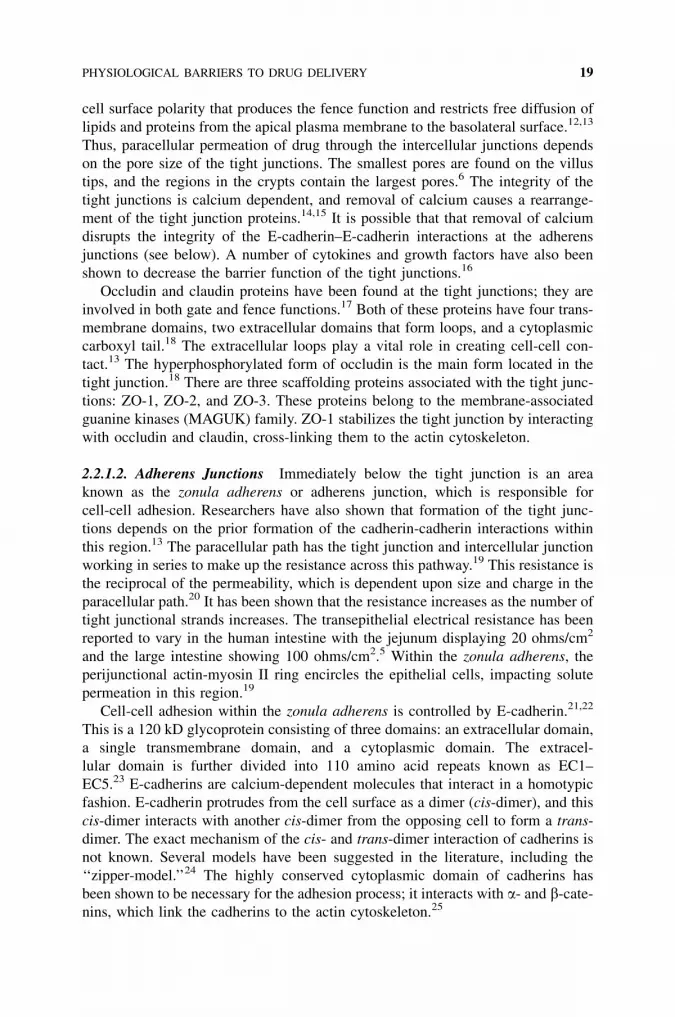
Figure 2.2. The intercellular junction is mediated by proteins at different levels: (1) tight
junction (zonula occludens), (2) adherens junction (zonula adherens), and (3) desmosomes.
18 PHYSIOLOGICAL, BIOCHEMICAL, AND CHEMICAL BARRIERS
cell surface polarity that produces the fence function and restricts free diffusion of
lipids and proteins from the apical plasma membrane to the basolateral surface.12,13
Thus, paracellular permeation of drug through the intercellular junctions depends
on the pore size of the tight junctions. The smallest pores are found on the villus
tips, and the regions in the crypts contain the largest pores.6 The integrity of the
tight junctions is calcium dependent, and removal of calcium causes a rearrange-
ment of the tight junction proteins.14,15 It is possible that that removal of calcium
disrupts the integrity of the E-cadherin–E-cadherin interactions at the adherens
junctions (see below). A number of cytokines and growth factors have also been
shown to decrease the barrier function of the tight junctions.16
Occludin and claudin proteins have been found at the tight junctions; they are
involved in both gate and fence functions.17 Both of these proteins have four trans-
membrane domains, two extracellular domains that form loops, and a cytoplasmic
carboxyl tail.18 The extracellular loops play a vital role in creating cell-cell con-
tact.13 The hyperphosphorylated form of occludin is the main form located in the
tight junction.18 There are three scaffolding proteins associated with the tight junc-
tions: ZO-1, ZO-2, and ZO-3. These proteins belong to the membrane-associated
guanine kinases (MAGUK) family. ZO-1 stabilizes the tight junction by interacting
with occludin and claudin, cross-linking them to the actin cytoskeleton.
2.2.1.2. Adherens Junctions Immediately below the tight junction is an area
known as the zonula adherens or adherens junction, which is responsible for
cell-cell adhesion. Researchers have also shown that formation of the tight junc-
tions depends on the prior formation of the cadherin-cadherin interactions within
this region.13 The paracellular path has the tight junction and intercellular junction
working in series to make up the resistance across this pathway.19 This resistance is
the reciprocal of the permeability, which is dependent upon size and charge in the
paracellular path.20 It has been shown that the resistance increases as the number of
tight junctional strands increases. The transepithelial electrical resistance has been
reported to vary in the human intestine with the jejunum displaying 20 ohms/cm2
and the large intestine showing 100 ohms/cm2.5 Within the zonula adherens, the
perijunctional actin-myosin II ring encircles the epithelial cells, impacting solute
permeation in this region.19
Cell-cell adhesion within the zonula adherens is controlled by E-cadherin.21,22
This is a 120 kD glycoprotein consisting of three domains: an extracellular domain,
a single transmembrane domain, and a cytoplasmic domain. The extracel-
lular domain is further divided into 110 amino acid repeats known as EC1–
EC5.23 E-cadherins are calcium-dependent molecules that interact in a homotypic
fashion. E-cadherin protrudes from the cell surface as a dimer (cis-dimer), and this
cis-dimer interacts with another cis-dimer from the opposing cell to form a trans-
dimer. The exact mechanism of the cis- and trans-dimer interaction of cadherins is
not known. Several models have been suggested in the literature, including the
‘‘zipper-model.’’24 The highly conserved cytoplasmic domain of cadherins has
been shown to be necessary for the adhesion process; it interacts with a- and b-cate-nins, which link the cadherins to the actin cytoskeleton.25
PHYSIOLOGICAL BARRIERS TO DRUG DELIVERY 19

2.2.1.3. Desmosomes The last region of the paracellular pathway is the desmo-
some, which is located nearest the basolateral membrane surface of the enterocyte.
Studies indicate that the intermediate filaments are connected to the desmosomes by
the desmoplakins.26 The major desmosomal cadherin located in this area is desmo-
glein. Desmocollin, a second desmosomal cadherin, is required for the binding of
desmoplakins to the intermediate filaments.26 This region appears to be less critical
for the function of the paracellular path than the two regions situated nearer the
apical membrane.
2.2.2. Transcellular Pathway
A drug with the appropriate physiochemical characteristics can traverse through the
cell by passive diffusion. In the case of peptides or peptidomimetics, their physico-
chemical properties may not be suitable for permeation through the cell membrane
via the transcellular pathway. The drug molecules must travel through the lipid
bilayers that make up the membranes. The bilayers consist of four regions: (1)
the outermost region, which has a large number of water molecules and is accoun-
table for the interactions with other proteins and membranes; (2) the next region,
which contains the polar headgroups, causing this region to have the highest mole-
cular density and making it the most difficult region for diffusion; (3) the third
region, which contains the nonpolar tails that form the barrier to penetration based
on limitations on molecular size and shape; and (4) the inner region, which is the
most hydrophobic and acts as the hydrophobic barrier.27 The resistance across the
transcellular path can be visualized as resistors in a series, where the apical and
basolateral membranes act as the two resistors.19 These membranes form the
rate-limiting barriers to the passive flow of molecules.
The membranes form one of the obstacles in this delivery route. The drug mole-
cules must also traverse the cytosol before exiting through the basolateral mem-
branes. Within the cytosol, various drug-metabolizing enzymes reside, which
metabolize the drug molecules and can lower the drug transport via this route.
2.3. BIOCHEMICAL BARRIERS TO DRUG DELIVERY
Great interindividual variability can be seen in the metabolism of drugs as a result
of differing enzyme activity due to inhibition, induction, genetic polymorphisms, or
even disease state.28 Enzymes found within the intestine are from two sources,
mammalian and bacteria-associated. The mammalian enzymes are located within
the lumen and in the enterocytes. Enzymes from the microflora within the ileum
and colon have also been identified.8 This discussion will focus on degradation
by the mammalian enzymes.
2.3.1. Metabolizing Enzymes
Within the lumen of the stomach, a mixture of hydrochloric acid and proteolytic
pepsins is the first metabolic barrier that a peptide drug will encounter.
20 PHYSIOLOGICAL, BIOCHEMICAL, AND CHEMICAL BARRIERS

Subsequently, the hydrolysis of acidic proteins occurs at pH 2–5; this is especially
the case for peptides containing aspartate residues.3 Larger proteins are quite sus-
ceptible to this gastric proteolysis, while smaller peptides are unaffected by this
mixture.
Fricker and Drewe describe the luminal enzymes of the upper small intestine as
the second barrier.3 Trypsin, chymotrypsin, elastase, and carboxypeptidase A and B
are positioned in the lumen of the duodenum. Their highest activity occurs at pH 8.
These enzymes degrade 30–40% of large proteins within the duodenum to small
peptides within 10 minutes.3 Small peptides have been shown to be stable against
these pancreatic proteases.
The major enzymatic barrier occurs within the brush border and in the cytosol of
the enterocytes, both of which contain peptidases. These enzymes degrade smaller
peptides ranging from di- to tetrapeptides.3 Furthermore, there is an increase in
brush border peptidase activity from the upper duodenum to the lower ileum.3
The peptidases selective for tripeptides are located primarily within the brush bor-
der, whereas cytosolic proteases have dipeptides as their substrates. Evidence has
shown that the metabolic enzyme activity decreases along the intestine to a nearly
negligible rate within the colon, yet the permeability of the colon epithelium
remains good.2 This highlights the potential for targeting the colon to bypass the
enzymatic barrier of the intestine for peptide delivery, preventing the degradation
that occurs within the intestine. The pH at the intestinal surface on the brush border
is 5.5–6.0, which is more acidic than the pH of the lumen.29 The enterocyte has an
intercellular pH of 7.0–7.2. In addition, gastrointestinal pH changes in the fasted
and fed states; this topic has been reviewed elswhere.30
The proximal small intestine shows the greatest metabolic activity due to its
large surface area and the plethora of intestinal enzymes and transporters.8 Phase
I and II enzymes have also been identified in the intestine. The most notable Phase I
enzymes are those of the CYP superfamily. The P450 enzymes are also present in
the intestinal walls in concentrations approximately 20 times less than those seen
within the liver; however, their metabolism of drugs is comparable to the activity
seen in the liver.28,31 The activity of the enzymes varies within the area of the gas-
trointestinal tract. The highest activity of the P450 enzymes is displayed in the
proximal part of the gastrointestinal tract, and their activity decreases distally.28
The greatest concentration of P450 enzymes is found in the villus tips of the upper
and middle third of the intestine.27 Great variability in activity has been noted both
intra- and interindividually due to exposure of the enterocytes to external stimuli
such as food and drugs that can either induce or inhibit these enzymes. These intest-
inal P450 enzymes are more responsive to inducers than are their hepatic counter-
parts.27 Although the blood flow to the intestine is lower than to the liver, the villus
tip has a large surface where the enzyme can interact with its substrate, allowing
extensive metabolism.32 Metabolic activity in the intestine has been shown to be
route-dependent, and the metabolism is greater for oral administration of drugs
than for intravenous dosing.28,33 In this case, intestinal metabolism occurs during
the initial absorption of the drug across the intestinal barrier, and the metabolism
is lower with the recirculation of the drug. The major factor that influences the
BIOCHEMICAL BARRIERS TO DRUG DELIVERY 21

route-dependent metabolism is the residence time of the drug within the enterocyte.
The residence time can be lengthened by binding in the cytoplasm, the activity of
efflux pumps, and limited blood flow or, conversely, shortened by basolateral clear-
ance and basolateral transporters.28
The CYP1, CYP2, and CYP3 subfamilies are mostly involved in xenobiotic
metabolism. Various isoenzymes that possess their own drug substrates are present
for each subfamily. Within the human small intestine, CYP1A1, CYP2C, CYP2D6,
and CYP3A4 have been isolated.8 Because of the numerous polymorphs of
CYP2D6, characterization of intestinal levels of this enzyme has been quite dif-
ficult. CYP3A4 is the most abundant of the intestinal P450 enzymes, making up
more than 70% of the intestinal CYPs.34 Reports indicate that there are structural
similarities between the intestinal and hepatic CYP enzymes; however, they appear
to be independently regulated.8 Food interactions have been shown to affect the reg-
ulation of the intestinal CYP enzymes. Grapefruit juice inhibits CYP3A, while
grilled and smoked foods induce CYP1A1 activity.8 Variations in the population
in reference to these enzymes can also confound the issue of degradation for pep-
tide pharmaceuticals.
Conjugating enzymes, also referred to as ‘‘Phase II metabolizers,’’ are also
found in the intestine. Glucuronyltransferase, N-acetyltransferase, sulfotransferase,
and glutathione-S-transferase show high activity for the intestinal Phase II
enzymes.8 Conjugates that are formed by these enzymes within the cell are reported
to be substrates of the multidrug resistance–associated protein family (MRP) of
transporters and are excreted into the lumen.35 The MRP family of transporters con-
sists of ATP-dependent transporters that excrete organic anions. At this time, addi-
tional studies are needed to understand the role of these enzymes in the degradation
of peptide drugs. Metabolizing enzymes and drug transporters in the process of
drug delivery will be discussed in greater detail in another chapter.
2.3.2. Transporters and Efflux Pumps
Active transporters for peptides within the intestine were first detected in experi-
ments in the late 1960s and early 1970s. Since that time, many characteristics of
these transporters have been determined. Substrates are generally peptides consist-
ing of two or three amino acids that can be transported through the brush border
membrane in a carrier-mediated, pH-dependent fashion.3 Energy is required to
move these peptides against a concentration gradient into the cell, and these carriers
also display saturability.
Although most transporters are situated on the apical membrane, researchers
have found some that are located only on the basolateral membrane surface. The
Naþ/A amino acid transporter, Naþ/ASC amino acid transporter, GLUT2 hexose
transporter, and the Naþ-independent folic acid transporter are examples of such
basolateral transporters.27 PepT1, an apical Hþ/dipeptide transporter, is most
abundant in the villus tip, and its concentration increases from the duodenum to
the ileum. In times of starvation, there is an increase in the expression of this
22 PHYSIOLOGICAL, BIOCHEMICAL, AND CHEMICAL BARRIERS

transporter. On the basolateral membrane, PepT2 functions as the Hþ/dipeptide
transporter to allow the substrate to exit the enterocyte.27
P-glycoprotein (P-gp) is a known MRP that serves as an efflux pump.36,37 It is
located within the brush borders of the villus tips of the intestine and has been
found throughout the small and large intestines. The concentration of P-gp
increases from the stomach to the colon.6 The substrate specificity for P-gp covers
a broad range of molecular structures, and the affinity varies as a function of the
intestinal site.27 A common feature of the substrates is hydrophobicity. As men-
tioned previously, the efflux pumps assist the intestinal metabolism by returning
the drug to the lumen, allowing the metabolizing enzymes to work on the drug
another time as well as preventing product inhibition by removing primary metabo-
lites that have been formed.31 This interaction is enhanced due to the colocalization
of the CYP3A enzymes and P-gp on the apical membrane, as well as the overlap in
substrate specificities and shared inducers and inhibitors.27 Grapefruit juice also
interferes with the transport mediated by P-gp;7 however, not all substrates for
the CYP3A enzyme behave as substrates for P-gp.6 P-gp functions as a defense
mechanism against xenobiotics in other biological barriers, as it is also expressed
in apical surfaces of epithelial cells of the liver, kidney, pancreas, and colon as well
as in the capillary endothelium of the brain.38
2.4. CHEMICAL BARRIERS TO DRUG DELIVERY
The chemical structure of a drug determines its solubility and permeability profiles.
In turn, the concentration at the intestinal lumen and the permeation of the drug
across the intestinal mucosa are responsible for the rate and extent of absorption.39
Unfavorable physicochemical properties have been a limiting factor in the oral
absorption of peptides and peptidomimetics.40 The structural factors involved in
the permeation of peptides will be described here.
2.4.1. Hydrogen-Bonding Potential
Hydrogen-bonding potential has been shown to be an important factor in the per-
meation of peptides. Studies in vivo and in various cell culture models of the blood-
brain barrier and intestinal mucosa indicate that desolvation or hydrogen-bonding
potential regulates the permeation of peptides.39,41–43 The energy needed to de-
solvate the polar amide bonds in the peptide to allow it to enter and traverse the
cell membrane is the principle behind the concept of hydrogen-bonding potential.
For small organic molecules, the octanol-water partition coefficient is the best pre-
dictor of cell membrane permeation with a sigmoidal relationship.42 However, this
is not the case with peptides; the desolvation energy or hydrogen-bonding potential
is a better predictor for membrane permeation of peptides. Burton et al. have
reported partition coefficients of model peptides in n-octanol/Hanks’ balanced
salt solution (HBSS), isooctane/HBSS, and heptane/ethylene glycol systems.41
Two experimental methods were developed to measure the desolvation energy or
CHEMICAL BARRIERS TO DRUG DELIVERY 23

the hydrogen-bonding potential of peptides. In the first method, the hydrogen-bonding
potential is calculated from the difference between the partition coefficients of octa-
nol/water and isooctane/water. The second method involves measuring the partition
coefficient of peptide in heptane/ethylene glycol; this method correlates well with the
hydrogen-bonding potential and provides a simpler and more direct measurement.41
2.4.2. Other Properties
Physicochemical properties of the peptide are also important determinants in the
passage of drugs via the paracellular path. Size, charge, and hydrophilicity are
the factors influencing paracellular permeation.40 A change in the hydrophilicity
of a peptide may alter its route of permeation; as the hydrophilicity of a peptide
decreases, its lipophilicity increases, causing a shift in permeation of the peptide
from the paracellular to the transcellular route. Molecules with radii larger than
11 A are unable to penetrate the tight junctions.44 Studies of Caco-2 cells confirm
that drug permeation via the paracellular path is size-dependent, and this highlights
the sieving abilities of the intercellular junctions.40
Although the paracellular path is negatively charged, the effect of charge on
paracellular permeation is not well understood. One study suggests that a positive
net charge on a peptide produces the best paracellular permeation, but another study
suggests that a �1 or �2 charge is most effective in paracellular transport.40 It has
also been suggested that the effect of charge is negligible as the molecular size of
the peptide increases.40
2.5. DRUG MODIFICATIONS TO ENHANCE TRANSPORTACROSS BIOLOGICAL BARRIERS
Several methods have been explored to improve drug permeation across biological
barriers.1,44–47 One method involves chemical modification of drug entities such as
prodrugs and peptidomimetics. Another method is to design a formulation that
enhances drug permeation through the biological barriers.
2.5.1. Prodrugs and Structural Modifications
A prodrug approach has been utilized to optimize drugs; a prodrug is defined as a
chemical derivative that is inactive pharmacologically until it is converted in vivo to
the active drug moiety. Recently, a targeted prodrug design has emerged in which
prodrugs have been used to target membrane transporters or enzymes.45 This
method improves oral drug absorption or site-specific drug delivery. Extensive
knowledge of the structure, the distribution within biological barriers, and substrate
specificities is needed to target a desired transporter.
Prodrug strategies have been very successful with small molecules; however, the
use of prodrugs for peptides has been less frequent.44 The cyclic peptide prodrug
approach has been shown to improve membrane permeation. In this method, the
24 PHYSIOLOGICAL, BIOCHEMICAL, AND CHEMICAL BARRIERS

N and C termini of the peptide are connected via a linker to form a cyclic peptide.
The linker can be cleaved by esterase to release the peptide. The cyclic peptide pro-
drug formation increases the intramolecular hydrogen bonding and lowers the
hydrogen-bonding potential to water molecules as solvent. In addition, the lipophi-
licity of the cyclic prodrug increases, which shifts its transport from paracellular to
transcellular.48 It has also been reported that cyclic peptides are less susceptible to
amino- and carboxypeptidases than linear peptides because the amino and carboxy
terminals are protected from these enzymes.44
Peptide structural modification has been applied to improve membrane permea-
tion of peptides. Metabolism of peptide pharmaceuticals can occur in various
regions along the route to oral absorption, and inhibition of this degradation is
advantageous in enhancing drug delivery. To improve enzymatic stability, peptides
have been converted to peptidomimetics. In this case, the peptide bond is converted
to its bioisostere, which is stable to proteolytic enzymes. Other structural modifica-
tion strategies to improve membrane permeation of peptides include lipidization,
halogenation, glycosylation, cationization, and conjugation to polymers.46
2.5.2. Formulations
Peptide absorption can be improved by designing an optimal formulation.47,49 Sev-
eral methods to enhance peptide absorption have been suggested, including addition
of ion-pairing and complexation molecules, nonsurfactant membrane permeation
enhancers, surfactant adjuvants, or combinations of these additives.47 Addition of
perturbants of tight junctions such as cytoskeletal agents, oxidants, hormones, cal-
cium chelators, and bacterial toxins the formulation has been investigated to
improve drug permeation.49 Another novel delivery system involves the use of
mucoadhesives to enhance drug delivery because of their long retention time at
the targeted mucosal membrane; lectins have been identified as potential carriers
for peptides in an oral mucoadhesive system.1 Coadministration of peptides with
inhibitors of metabolizing enzymes has also been suggested to increase oral absorp-
tion.47,50,51
Modulation of the intercellular junctions by inhibiting the cadherin-cadherin
interaction at the adherens junction has also been investigated. Peptides derived
from the sequence of the extracellular domain of E-cadherin have been shown to
modulate the intercellular junction of bovine brain microvessel endothelial cell
(BBMEC) and Madin-Darby canine kidney (MDCK) cell monolayers. These
peptides enhance the paracellular penetration of marker molecules such as 14C-
mannitol and lower the transepithelial resistance of the monolayers.52–55 The use
of these cadherin-derived peptides as adjuvants to enhance paracellular permeabil-
ity of drugs is still under investigation.
2.6. CONCLUSIONS
The absorption of an orally administered peptide depends on the successful passage
of the peptide through the several barriers to drug delivery. The peptide can pass
CONCLUSIONS 25

either between or through the cells, depending on its physicochemical properties.
The gastrointestinal epithelial layer is a formidable obstacle to the passage of a pep-
tide. Recent studies have shown that metabolism within the intestine forms a major
obstruction to drug absorption. The concerted activity of these drug-metabolizing
enzymes and efflux systems increases the problem. Although many challenges exist
in traversing the intestinal epithelial layer, pharmaceutical scientists and medicinal
chemists are overcoming them with innovative peptide drug products that optimize
pharmacological activity and enhance drug delivery.
REFERENCES
1. Kompella, U. B.; Lee, V. H. Adv. Drug. Deliv. Rev. 2001, 46, 211–245.
2. Lipka, E.; Crison, J.; Amidon, G. L. J. Contr. Rel. 1996, 39, 121–129.
3. Fricker, G.; Drewe, J. J. Pept. Sci. 1996, 2, 195–211.
4. Hochman, J. H.; Artursson, P. J. Contr. Rel. 1994, 29, 253–267.
5. Hilgendorf, C.; Spahn-Langguth, H.; Regardh, C. G.; Lipka, E.; Amidon, G. L.; Langguth,
P. J. Pharm. Sci. 2000, 89, 63–75.
6. Martinez, M.; Amidon, G.; Clarke, L.; Jones,W.W.;Mitra, A.; Riviere, J. Adv. Drug Deliv.
Rev. 2002, 54, 825–850.
7. Fricker, G.; Miller, D. S. Pharmacol. Toxicol. 2002, 90, 5–13.
8. Doherty, M. M.; Charman, W. N. Clin. Pharmacokinet. 2002, 41, 235–253.
9. Adson, A.; Raub, T. J.; Burton, P. S.; Barsuhn, C. L.; Hilgers, A. R.; Audus, K. L.; Ho, N. F.
J. Pharm. Sci. 1994, 83, 1529–1536.
10. Yap, A. S.; Mullin, J. M.; Stevenson, B. R. J. Membr. Biol. 1998, 163, 159–167.
11. Anderson, J. M.; Van Itallie, C. M. Am. J. Physiol. 1995, 269, G467–G475.
12. Cereijido, M.; Valdes, J.; Shoshani, L.; Contreras, R. G. Annu. Rev. Physiol. 1998, 60,
161–177.
13. Mitic, L. L.; Anderson, J. M. Annu. Rev. Physiol. 1998, 60, 121–142.
14. Gonzalez-Mariscal, L.; Chavez de Ramirez, B.; Cereijido, M. J. Membr. Biol. 1985, 86,
113–125.
15. Rothen-Rutishauser, B.; Riesen, F. K.; Braun, A.; Gunthert, M.; Wunderli-Allenspach, H.
J. Membr. Biol. 2002, 188, 151–162.
16. Walsh, S. V.; Hopkins, A. M.; Nusrat, A. Adv. Drug Deliv. Rev. 2000, 41, 303–313.
17. Tsukita, S.; Furuse, M.; Itoh, M. Curr. Opin. Cell Biol. 1999, 11, 628–633.
18. Lapierre, L. A. Adv. Drug Deliv. Rev. 2000, 41, 255–264.
19. Madara, J. L. Annu. Rev. Physiol. 1998, 60, 143–159.
20. Burton, P. S.; Goodwin, J. T.; Vidmar, T. J.; Amore, B. M. J. Pharmacol. Exp. Ther. 2002,
303, 889–895.
21. Takeichi, M. Annu. Rev. Biochem. 1990, 59, 237–252.
22. Shore, E. M.; Nelson, W. J. J. Biol. Chem. 1991, 266, 19672–19680.
23. Chothia, C.; Jones, E. Y. Annu. Rev. Biochem. 1997, 66, 823–862.
24. Pertz, O.; Bozic, D.; Koch, A. W.; Fauser, C.; Brancaccio, A.; Engel, J. Embo J. 1999, 18,
1738–1747.
26 PHYSIOLOGICAL, BIOCHEMICAL, AND CHEMICAL BARRIERS

25. Yap, A. S.; Brieher, W.M.; Gumbiner, B.M. Annu. Rev. Cell. Dev. Biol. 1997, 13, 119–146.
26. Cowin, P.; Burke, B. Curr. Opin. Cell Biol. 1996, 8, 56–65.
27. Martinez, M. N.; Amidon, G. L. J. Clin. Pharmacol. 2002, 42, 620–643.
28. Agoram, B.; Woltosz, W. S.; Bolger, M. B. Adv. Drug Deliv. Rev. 2001, 50, S41–S67.
29. Tsuji, A.; Tamai, I. Pharm. Res. 1996, 13, 963–977.
30. Charman,W.N.; Porter, C. J.;Mithani, S.;Dressman, J. B. J. Pharm. Sci. 1997, 86, 269–282.
31. Engman, H. A.; Lennernas, H.; Taipalensuu, J.; Otter, C.; Leidvik, B.; Artursson, P. J.
Pharm. Sci. 2001, 90, 1736–1751.
32. Wacher, V. J.; Salphati, L.; Benet, L. Z. Adv. Drug Deliv. Rev. 2001, 46, 89–102.
33. Cong, D.; Doherty, M.; Pang, K. S. Drug Metab. Dispos. 2000, 28, 224–235.
34. Benet, L. Z.; Izumi, T.; Zhang, Y.; Silverman, J. A.; Wacher, V. J. J. Contr. Rel. 1999, 62,
25–31.
35. Suzuki, H.; Sugiyama, Y. Eur. J. Pharm. Sci. 2000, 12, 3–12.
36. Wagner, D.; Spahn-Langguth, H.; Hanafy, A.; Koggel, A.; Langguth, P. Adv. Drug Deliv.
Rev. 2001, 50, S13–S31.
37. Benet, L. Z.; Cummins, C. L. Adv. Drug Deliv. Rev. 2001, 50, S3–S11.
38. Zhang, Y.; Benet, L. Z. Clin. Pharmacokinet. 2001, 40, 159–168.
39. Goodwin, J. T.; Conradi, R. A.; Ho, N. F.; Burton, P. S. J. Med. Chem. 2001, 44, 3721–
3729.
40. Pauletti, G. M.; Okumu, F. W.; Borchardt, R. T. Pharm. Res. 1997, 14, 164–168.
41. Burton, P. S.; Conradi, R. A.; Hilgers, A. R.; Ho, N. F.; Maggiora, L. L. J. Contr. Rel. 1992,
19, 87–98.
42. Burton, P. S.; Conradi, R. A.; Ho, N. F.; Hilgers, A. R.; Borchardt, R. T. J. Pharm. Sci.
1996, 85, 1336–1340.
43. Chikhale, E. G.; Ng, K. Y.; Burton, P. S.; Borchardt, R. T. Pharm. Res. 1994, 11, 412–419.
44. Pauletti, G. M.; Gangwar, S.; Siahaan, T. J.; Aube, J.; Borchardt, R. T. Adv. Drug Deliv.
Rev. 1997, 27, 235–256.
45. Han, H. K.; Amidon, G. L. AAPS Pharm. Sci. 2000, 2.
46. Witt, K. A.; Gillespie, T. J.; Huber, J. D.; Egleton, R. D.; Davis, T. P. Peptides 2001, 22,
2329–2343.
47. Aungst, B. J. J. Pharm. Sci. 1993, 82, 979–987.
48. Okumu, F. W.; Pauletti, G. M.; Vander Velde, D. G.; Siahaan, T. J.; Borchardt, R. T.
Pharm. Res. 1997, 14, 169–175.
49. Lutz, K. L.; Siahaan, T. J. J. Pharm. Sci. 1997, 86, 977–984.
50. Mizuma, T.; Ohta, K.; Koyanagi, A.; Awazu, S. J. Pharm. Sci. 1996, 85, 854–857.
51. Gotoh, S.; Nakamura, R.; Nishiyama, M.; Quan, Y. S.; Fujita, T.; Yamamoto, A.;
Muranishi, S. J. Pharm. Sci. 1996, 85, 858–862.
52. Pal, D.; Audus, K. L.; Siahaan, T. J. Brain. Res. 1997, 747, 103–113.
53. Lutz, K. L.; Siahaan, T. J. Drug Deliv. 1997, 10, 187–193.
54. Makagiansar, I. T.; Avery, M.; Hu, Y.; Audus, K. L.; Siahaan, T. J. Pharm. Res. 2001, 18,
446–453.
55. Sinaga, E.; Jois, S. D.; Avery, M.; Makagiansar, I. T.; Tambunan, U. S.; Audus, K. L.;
Siahaan, T. J. Pharm. Res. 2002, 19, 1170–1179.
REFERENCES 27

3PATHWAYS FOR DRUG DELIVERYTO THE CENTRAL NERVOUS SYSTEM
YAN ZHANG1
AND DONALD W. MILLER2
Department of Pharmaceutical Sciences, University of Nebraska Medical Center,
Omaha, NE 68198-6025
3.1. Introduction
3.1.1. Importance of drug delivery to the central nervous system (CNS)
3.1.2. Cellular barriers to drug delivery in the CNS
3.1.3. General approaches for increasing brain penetration of drugs
3.2. Direct administration of drugs into the brain
3.2.1. Intracerebral administration
3.2.2. Intrathecal administration
3.2.3. Nasal administration
3.3. BBB disruption
3.3.1. Osmotic agents
3.3.2. Bradykinin analogs
3.3.3. Alkylglycerols
3.4. Transcellular delivery routes in the BBB
3.4.1. Passive diffusion
3.4.2. Inwardly directed transport systems in the BBB
3.4.2.1. Amino acid transporters
3.4.2.2. Glucose transporters
3.4.2.3. Monocarboxylic acid transporter
3.4.2.4. Nucleoside transporters
Drug Delivery: Principles and Applications Edited by Binghe Wang, Teruna Siahaan,
and Richard Soltero
ISBN 0-471-47489-4 # 2005 John Wiley & Sons, Inc.
1Current address, Pfizer Global Research and Development, PDM Department, Building 20, Room 332 E,
2800 Plymouth Road, Ann Arbor, MI 481052Corresponding author
29

3.4.2.5. Peptide transport systems
3.4.2.6. Considerations for carrier-mediated transport in the CNS
3.4.3. Vesicular transport in the BBB
3.4.3.1. Transferrin receptor-mediated vesicular transport
3.4.3.2. Insulin receptor-mediated vesicular transport
3.4.4. Drug efflux transporter systems in the BBB
3.4.4.1. P-glycoprotein (P-gp)
3.4.4.2. Multidrug resistance–associated protein (MRP)
3.4.4.3. Breast cancer resistance protein (BCRP)
3.4.4.4. Modulation of drug efflux activity to increase
CNS drug delivery
3.5. Summary
References
3.1. INTRODUCTION
3.1.1. Importance of Drug Delivery to the CentralNervous System (CNS)
Most drugs produce their required pharmacological response by altering cell
function or structure in a concentration-dependent and reversible manner. Unless
the target tissue is the blood and the drug is given intravenously or intra-arterially,
the pharmacological response is dependent on the distribution of the drug to the tar-
get tissue in therapeutically relevant concentrations for sufficient periods of time to
produce the clinically desired response. Issues pertaining to drug absorption and
tissue distribution are often overlooked in the high-throughput screening process
used to identify compounds with pharmacological activity. However, for CNS
active compounds, such considerations are essential due to the presence of the
blood-brain and blood-cerebral spinal fluid barriers, which restrict the passage of
most compounds into the brain.
The impact of the blood-brain barrier (BBB) in CNS drug therapy has recently
been eloquently reviewed by Pardridge.1 The problems associated with the delivery
of large molecules to the brain in therapeutically relevant concentrations have long
been recognized. However, misconceptions concerning the ability of small mole-
cules to penetrate the BBB and the effectiveness of small molecules in treating
CNS-related pathologies have contributed to an underappreciation of the impor-
tance of drug delivery to the brain. Thus, the search for new and effective treat-
ments for CNS diseases must concentrate not only on the drug target within the
CNS but also on the efficient delivery of the molecule to the site of action within
the brain. The purpose of this chapter is to provide a basic understanding of the
current obstacles in drug delivery to the brain and the various approaches that
have been used to increase CNS concentrations of drugs.
30 PATHWAYS FOR DRUG DELIVERY TO THE CENTRAL NERVOUS SYSTEM

3.1.2. Cellular Barriers to Drug Delivery in the CNS
There are two cellular barriers that separate the brain extracellular fluid from the
blood (see Figure 3.1). The first and largest interface is the brain capillary endothe-
lial cells that form the BBB. The brain capillaries are a continuous layer of endothe-
lial cells connected by well-developed tight junctional complexes.2 As a result of
the tight junctions, passive diffusion of drugs and solutes between the endothelial
cells is restricted. In addition, brain capillary endothelial cells lack fenestrations
(water-filled pores or channels within the plasma membrane) and have reduced
pinocytic activity. These characteristics further restrict the movement of com-
pounds from the blood into the extracellular environment of the brain.3–5
A. Blood-Brain Barrier
Blood
Brain Extracellular Fluidd
B. Blood-Cerebral Spinal Fluid Barrier
Blood
Choroid Interstitual Space
Cerebral Spinal Fluid
Choroidal Epithelium
Choroidal Capillary
Endothelial Cell
Brain Capillary
Endothelial Cell
Pericyte Astrocyte Astrocyte
Figure 3.1. Schematic representation of the cellular components of the blood-brain (Panel
A) and blood-cerebral spinal fluid (Panel B) barriers. The blood-brain barrier consists of
continuous type endothelial cells with complex tight junctions to limit paracellular
diffusion. The astrocytes and pericytes located in close proximity to the brain endothelial
cells release various endogenous factors that modulate endothelial cell permeability. In con-
trast, the choroid endothelial cells are fenestrated and the blood-cerebral spinal fluid barrier
properties are provided by the tight junctions formed between the choroid epithelial cells.
INTRODUCTION 31

The presence of tight junctions between the brain capillary endothelial cells
means that the paracellular pathway for drug delivery is highly restricted. Lipid-
soluble drugs with a molecular mass of less than 600 can pass through the BBB
via passive diffusion through the brain endothelial cells.3 Factors that influence pas-
sive diffusion include molecular volume, charge, and the hydrogen-bonding
potential of the compound.6,7 Besides the diffusional pathway, compounds can
also move across the BBB through vesicular transport and via specific transport
or carrier systems within the brain endothelial cells. Furthermore, a variety of
enzymes in the brain endothelial cells act as a two-way metabolic barrier capable
of breaking down blood-borne substances and brain metabolites. These enzymes
also play important roles in controlling the transport of a variety of compounds,
both endogenous and exogenous, from the blood entering the brain.
The second barrier involved in regulation of the extracellular environment of the
brain is the blood–cerebrospinal fluid barrier (BCSFB). The BCSFB is a composite
barrier made up of the choroid plexuses and the arachnoid membranes of the cir-
cumventricular organs. Unlike the capillaries that form the BBB, the circumventri-
cular organs of the brain have fenestrated ‘‘leaky’’ capillaries, and their barrier
function is provided by the tight junctions between the epithelial cells in the chor-
oid plexus.8,9 The epithelial cells in the choroid plexus that form the BCSFB have
complex tight junctions on the CSF (apical) side of the cells. These tight junctions
formed by the epithelial cells in the choroid plexus are slightly more permeable
than those found in the endothelial cells of the BBB.9,10
Although the apical membrane of the epithelial cells forming the BCSFB has
numerous microvilli, the total surface area is still substantially smaller than the
BBB.11 It has been estimated that in the human brain there are approximately
100 billion capillaries with a total surface area of 20 m2.1 Given the density of
the capillary network in the brain, and the close proximity of neuronal cells to these
capillaries, most drug delivery approaches have focused on either circumventing
the BBB or increasing drug passage through the BBB.
3.1.3. General Approaches for Increasing Brain Penetration of Drugs
There are three general routes for brain delivery of drugs. One approach is to cir-
cumvent the difficulties associated with drug permeability in the BBB and/or
BCSFB by direct central administration of the drug into the brain. An alternative
approach is to temporarily break down the BBB, allowing the therapeutic agents to
enter the brain from the blood through a more permeable BBB. The third and final
approach uses chemical modifications of the drug and/or knowledge of the biology
of the brain endothelial cells forming the BBB to improve the transcellular passage
of drugs into the CNS. Each of these strategies is discussed below.
3.2. DIRECT ADMINISTRATION OF DRUGS INTO THE BRAIN
3.2.1. Intracerebral Administration
One strategy for delivering drugs to the brain is to circumvent the problems asso-
ciated with penetration of the BBB by direct injection of drugs into the brain. This
32 PATHWAYS FOR DRUG DELIVERY TO THE CENTRAL NERVOUS SYSTEM

approach is invasive, requiring a craniotomy in which a small hole is drilled in the
head for intracerebroventricular (ICV) or intracerebral (IC) drug administration into
the brain. An advantage of this approach is that a wide range of compounds and
formulations can be considered for ICV or IC administration. Thus, both large-
and small-molecule therapeutics can be delivered, either alone or in various poly-
mer formulations, to achieve sustained release.
Aside from the invasiveness of the craniotomy procedure and the implications
this may have for long-term therapy, the biggest disadvantage of direct implantation
of drug into the CNS is related to the limited brain distribution of the drug. This is
illustrated in the studies by Krewson and coworkers,12 in which polymer implants
containing radiolabeled nerve growth factor were placed in rat brain and diffusion
of the factor monitored by autoradiography. In these studies, diffusion of nerve
growth factor from the polymer implant was limited to 2–3 mm. Limited CNS dis-
tribution was also observed following one-time bolus ICV injections of brain-
derived neurotrophic factor in rats.13 This phenomenon of limited tissue distribution
is not restricted to large macromolecules. Hoistad et al.14 reported a diffusion dis-
tance of only 1 mm following striatal IC infusion of radiolabeled dopamine and
mannitol in rats.
Despite these limitations, successful application of the central administration
route for drug delivery to the brain has been reported in laboratory animals. Studies
by Mairs et al.15 showed significant tumor uptake and retention of radioiodode-
oxyuridine following IC administration in a rat glioma model. Furthermore, IC
administration of sustained-release polymers containing the antiepileptic agent phe-
nytoin was effective in preventing cobalt-induced seizures in rats.16 However, these
results should be tempered by the scaling issues present when going from small
laboratory animals to humans. For small laboratory animals, a focal seizure area
or tumor mass may be treatable with a 2–3 mm diffusion radius from the point
of drug administration. However, from the perspective of human brain diseases
there are few, if any, conditions that would be expected to respond to therapy
with such a limited brain tissue distribution. At best, this approach to CNS delivery
would require a highly potent therapeutic agent and a tissue target with a highly
restricted area of distribution.
3.2.2. Intrathecal Administration
Recently, intrathecal administration has been reexamined as a means of circum-
venting the BBB delivery problems associated with large macromolecules such
as proteins and peptides. Intrathecal administration involves the injection or infu-
sion of drugs into the cerebrospinal fluid (CSF) that surrounds the spinal cord. At
first glance, intrathecal administration as a method for delivering drugs to the brain
would seem contradictory, as this route is commonly employed to produce loca-
lized analgesia without the CNS complications that administration of systemic
anesthetics and analgesic agents produce. This lack of CNS effect observed with
intrathecal administration of anesthetic and analgesic agents is due to the rapid
removal of drug from the CSF to the systemic bloodstream.17 However, it should
be noted that the agents commonly administered by the intrathecal route for pain
DIRECT ADMINISTRATION OF DRUGS INTO THE BRAIN 33

management are small lipophilic molecules. Evidence suggests that proteins admi-
nistered intrathecally have a much slower clearance from the CSF.18 Thus, the
intrathecal route of administration may be a viable strategy for delivering large
molecular weight macromolecules with limited lipophilic properties.
Evidence of the validity of the intrathecal route for delivery of proteins to the
brain is found in studies with the centrally acting adipose regulatory protein, leptin.
This 16 kDa protein is secreted by fat cells and transported into the CNS, where it
acts to reduce appetite and promote weight loss. Recent studies by McCarthy et al.19
examined the appearance of leptin in the brain following intrathecal injection
in baboons using positron emission tomography. The studies were remarkable in
that they indicate that leptin, administered via intrathecal injection, was able to tra-
vel from the lower lumbar region to the hypothalamus within 3 hours of injection.
Even more remarkable is that the amount of leptin detected in the brain following
intrathecal administration was estimated to be well above the concentrations
required for biological activity.19 In support of these studies, separate experiments
by Yaksh et al.20 reported dose-dependent suppression of body weight and food
consumption in rats given 14-day continuous intrathecal infusion of leptin.
Together these studies provide support for the intrathecal route for the delivery
of large molecular weight molecules to the brain.
3.2.3. Nasal Administration
Another delivery option for bypassing the BBB is through intranasal (IN) adminis-
tration. Compared to intracerebral and intrathecal administration, IN administration
is a noninvasive means of delivery of therapeutic agents to the CNS.21,22 Because of
the unique connection between the nose and the brain, the olfactory neural pathway
provides a route of delivery for various compounds into the CNS. These include
toxic agents such as pathogens, viruses, and toxic metals. However, the same path-
way can also be used to delivery various therapeutic agents including small mole-
cules and proteins to the CNS (for review, see Refs. 21 and 23).
Small molecules such as cocaine and cephalexin can be transported directly to
the CNS from the nasal cavity.24,25 Sakane and colleagues25 reported that cepha-
lexin preferentially entered the CSF after nasal administration compared to intrave-
nous (IV) and intraduodenal administration in rats. The levels of cephalexin in CSF
were 166-fold higher 15 minutes after nasal administration than those of the other
two routes. Most recently, studies by Wang and colleagues showed that the ratio
of the methotrexate AUCCSF value between the IN route and the IV injection
was over 13-fold.26
In addition to small molecules, a number of protein therapeutic agents, such as
neurotrophic factors27 and insulin,28 have been successfully delivered to the CNS
using IN delivery in a variety of species. The therapeutic benefit of IN delivery of
proteins has been demonstrated by Liu et al. in rat stroke models.29 Their studies
demonstrated that insulin-like growth factor I (IGF-I) could be delivered to the
brain directly from the nasal cavity, even though IGF-I did not cross the BBB effi-
ciently by itself. As a consequence, IN IGF-I markedly reduced infarct volume and
improved neurological function following focal cerebral ischemia. Research in
34 PATHWAYS FOR DRUG DELIVERY TO THE CENTRAL NERVOUS SYSTEM

humans has also provided evidence for direct delivery of therapeutic agents to the
CNS from the nasal cavity. Studies by Kern and colleagues have demonstrated CNS
effects of IN insulin in humans without altering plasma glucose or insulin level.28
IN administration is a promising approach for rapid-onset delivery of medica-
tions to the CNS bypassing the BBB. However, there are also limitations. One of
the biggest limitations is insufficient drug absorption through the nasal mucosa.
Many drug candidates cannot be developed for the nasal route because they are
not absorbed well enough to produce therapeutic effects.30 Another constraint con-
cerning nasal administration is that a small administration volume is required,
beyond which the formulation will be drained out into the pharynx and swallowed.31
In summary, the advantages of IN delivery to the CNS are considerable.
IN administration has been shown to improve the bioavailability of many pre-
systemically metabolized drugs entering CNS, eliminating the need for systemic
delivery and reducing unwanted systemic side effects. It is also rapid and noninva-
sive. Furthermore, IN delivery does not require any modification of the therapeutic
drugs and does not require the drugs to be coupled to any carriers.
3.3. BBB DISRUPTION
Under normal conditions, the complex tight junctions that form between the brain
capillary endothelial cells restrict the paracellular diffusion of molecules and
solutes in the BBB. Modification of the tight junctions, causing controlled and tran-
sient increases in the permeability properties of the brain capillaries, is another
strategy that has been used to increase drug delivery to the brain. Methods for dis-
rupting BBB integrity through the breakdown of tight junctions include the sys-
temic administration of hyperosmotic solutions,32,33 vasoactive compounds such
as bradykinin and related analogs,34 and various alkylglycerols.35,36 Each of these
approaches is described in greater detail below.
3.3.1. Osmotic Agents
Disruption of the BBB through the use of osmotic agents has been extensively stu-
died in both laboratory animals and clinically in the treatment of brain tumors.37,38
In most cases, a hypertonic solution of an inert sugar, such as mannitol or arabinose,
ranging from 1.4 to 1.8 M, is delivered into the cerebral circulation through bolus
injection or short-term infusion into the carotid artery.33, 39–41 The proposed cellular
mechanism behind osmotic disruption of the BBB involves the physical pulling
apart/breaking of tight junctions due to the shrinkage of cerebral endothelial
cells and expansion of the blood volume caused by the addition of the hyperosmotic
agent.32,33,40 As the disruption of the BBB is contingent on the presence of hyper-
osmotic agents in the blood, the BBB resumes its normal barrier functions within
hours of returning the osmolarity of the blood to normal.42 During this period
when the tight cellular junctions between the brain capillary endothelial cells
have been compromised, paracellular diffusion of water-soluble drugs and solutes
into the brain is enhanced.
BBB DISRUPTION 35

Increased delivery of drugs to the brain following osmotic disruption of the BBB
has been demonstrated in a variety of settings. Increases in both small- and large-
molecule delivery to the brain have been reported.41 The time course for disruption
of BBB integrity and the subsequent return of the barrier function following osmo-
tic disruption appear to be variable. Studies in rats suggested that the onset of BBB
opening was rapid, with maximal responses observed within 5 minutes of hyperos-
motic mannitol administration.41 Likewise, return of normal BBB integrity was
noted within minutes following cessation of the osmotic agent. More recent studies
in humans suggest that while disruption of BBB permeability in response to hyper-
osmotic mannitol was rapid, with increases in BBB permeability observed within
1 minute of the osmotic agent, the barrier properties were not reestablished for sev-
eral hours.42
Osmotic disruption of the BBB has been used to increase the delivery of che-
motherapeutic agents to the brain in the treatment of CNS tumors in rats.43–45
The clinical benefits of increased delivery of chemotherapeutic agents to the brain
through osmotic disruption of the BBB have been demonstrated by the increased
survival rates observed in patients with primary CNS lymphoma46,47 and malignant
gliomas.47,48
While osmotic BBB disruption has been used primarily for enhancing the
response to small-molecule chemotherapeutic agents in brain tumors, applications
in gene and protein therapy in the CNS are also being explored. Studies in rats
using radiolabeled monoclonal antibodies demonstrated a 100-fold greater delivery
of antibodies from the blood to the brain following osmotic BBB disruption.49
While the actual amount of antibody delivered to the brain following osmotic
BBB disruption was still relatively small, 0.72% of the total dose, the relatively
long residence time observed for the monoclonal antibody in the CNS suggests
that biologically relevant levels of proteins can be delivered to the brain with this
approach. Osmotic disruption has also proven successful in the delivery of viral
vectors to the brain.50–52 Studies using adenoviral vector containing the Escherichia
coli beta-galactosidase gene showed no detectable viral delivery to the brain under
normal conditions following intracarotid infusion of the virus.50 However, osmotic
disruption of the BBB resulted in detectable beta-galactosidase expression in the
brain. The extent of beta-galactosidase expression in the brain was directly corre-
lated with the magnitude of BBB disruption.50 More recent studies by Abe et al.52
examined responses of human brain tumor xenografts to tumor suppressor gene
therapy. In these studies, BBB disruption with mannitol increased the tumor
response to p53 adenoviral vector. Together these studies suggest that osmotic
disruption of the BBB can be used for a variety of both small- and large-molecule
therapeutic agents.
3.3.2. Bradykinin Analogs
The BBB can also be disrupted by pharmacological means. Several endogenous
proinflammatory vasoactive agents, such as bradykinin, histamine, nitric oxide,
and various leukotrienes, are known to induce increases in BBB permeability in a
36 PATHWAYS FOR DRUG DELIVERY TO THE CENTRAL NERVOUS SYSTEM

concentration- and time-dependent manner. These vasoactive compounds are char-
acteristically ultra-short-acting due to either rapid deactivation through metabolic
processes or tachyphylaxis at the receptor signal transduction level. Furthermore,
many of these endogenous agents have narrow therapeutic windows and dose-limit-
ing side effects. Thus, although capable of producing increases in BBB permeability,
these endogenous agents have proven difficult to apply safely for CNS drug delivery.
In an effort to circumvent the problems associated with the use of endogenous
vasoactive agents, investigators have explored the use of structurally modified ana-
logs. The best example of this is the bradykinin analog labradimil (Cereport).
Bradykinin-induced increases in BBB permeability occur through a kininergic
B2 receptor-mediated opening of tight junctions between the brain capillary
endothelial cells.53,54 However, with a plasma half-life on the order of seconds,55
the effects of bradykinin on BBB permeability are short-lived, requiring carotid
artery infusion. In contrast, Cereport has amino acid substitutions that result in sig-
nificantly greater plasma and tissue stability than bradykinin.56 This, coupled with
the B2 receptor selectivity of Cereport, provides for a more consistent and repro-
ducible effect on BBB permeability without many of the toxic side effects observed
with bradykinin (see Ref. 57 for review). Based on studies in both rats and dogs, the
effects of Cereport on BBB permeability can be produced following either intracar-
otid or IV administration, with both the time to onset following administration and
the time to restoration of BBB permeability following cessation of Cereport being
observed within 5 minutes.58
As with osmotic disruption of the BBB, Cereport has been used to increase
delivery of chemotherapeutic agents in experimental brain tumor models.59–61
Interestingly, as opposed to osmotic disruption, the permeability increases observed
with Cereport were greater in the area in and around the brain tumor compared to
nontumor regions of the brain.62,63 The effect of Cereport on BBB and blood-tumor
barrier permeability has been shown to cause significant increases in tumor respon-
siveness to chemotherapeutic agents in various brain tumor models.64,65 While
fewer human studies have been performed with Cereport than with osmotic disrup-
tion of the BBB, Phase II multinational clinical trials indicate improved therapeutic
responses using IV or intra-arterial Cereport together with carboplatin in the treat-
ment of gliomas.66
Although Cereport appears to have greater effects on the brain microvasculature
in and around the tumor site, it has also been used to increase BBB permeability in
conditions other than brain tumor therapy. Emerich and colleagues67 studied the
delivery of the centrally acting opiate analgesic, loperamide, and the resulting
effects on pain perception in rodents. In these studies, coadministration of lopera-
mide with Cereport produced a twofold enhancement in pain response time using
the tail flick assay. As these effects were inhibited by the opiate receptor antagonist,
naloxone, the investigators concluded that Cereport could be used to enhance drug
delivery to the brain under normal conditions.67 Recent studies by Bidanset and
coworkers68 described the use of Cereport to enhance the delivery and effectiveness
of antiviral therapy in the CNS. In these studies, acyclovir was administered
either alone or in conjunction with Cereport in herpes simplex virus–infected
BBB DISRUPTION 37

rats. Coadministration of acyclovir with Cereport resulted in a two- to threefold
increase in acyclovir accumulation in the brain.68 The increased delivery of acyclo-
vir observed with Cereport also resulted in a significantly lower viral load through-
out the brain. Together these studies suggest that short-term, reversible disruption of
BBB permeability with Cereport can be used to impact CNS drug delivery in a
variety of conditions.
3.3.3. Alkylglycerols
A relatively new approach for transient disruption of the BBB involves the systemic
administration of various alkylglycerols. Erdlenbruch and colleagues36 reported a
reversible and concentration-dependent increase in BBB permeability to several
anticancer and antibiotic agents. The extent of BBB disruption varied from a
2-fold to a 200-fold increase in methotrexate, depending on the length of the alkyl
group and the number of glycerols present in the structure. As with the bradykinin
analog Cereport, the time to onset and reversal of the BBB-disrupting actions of the
alkylglycerols are very short, occurring within minutes of application and returning
to normal within minutes of cessation of alkylglycerol administration.36 Recent stu-
dies examining the effects of alkylglycerols on large-macromolecule permeability
in isolated brain capillaries suggests that the increases in permeability are due to
temporary breakdown of the tight junctions between the cells.35 Although the exact
mechanism(s) for the transient BBB disruption observed with the alkylglycerols are
unknown, the concentration and structure dependency of the response would sug-
gest interaction with receptor sites within the brain microvasculature.
The use of alkylglycerols to increase BBB permeability of anticancer agents
has been examined in a rat glioma tumor model.35,69,70 In these studies, several
different alkylglycerols were examined for increasing the CNS delivery of metho-
trexate in C6 glioma-bearing rats. Studies indicated significantly greater methotrex-
ate accumulation at both the tumor site and non-tumor-bearing sites within the
brain.35,70 The effects of the alkylglycerols were concentration-dependent. How-
ever, the magnitude of the increase in methotrexate delivery to the tumor site
detected with the highest doses of alkylglycerols examined was comparable to
that observed with osmotic disruption, and was significantly greater than that
observed with the bradykinin analog, Cereport.70 While human clinical trials
have not been initiated, studies in the rat glioma brain tumor model have detected
no long-term signs of toxicity with the alkylglycerols, suggesting that these agents
may have promise in the treatment of brain tumors.
3.4. TRANSCELLULAR DELIVERY ROUTES IN THE BBB
Aside from either circumventing the BBB completely through central administra-
tion of a drug or reversibly opening the BBB, increased delivery of therapeutic
agents to the brain can be accomplished through improved transcellular migration.
The transcellular routes available in brain capillary endothelial cells are shown in
38 PATHWAYS FOR DRUG DELIVERY TO THE CENTRAL NERVOUS SYSTEM

Figure 3.2 and include passive diffusion, specific transport systems, and endocytic
processes present in the brain microvasculature. The design of drugs or drug deliv-
ery systems that improve CNS delivery through one or more of the transcellular
pathways identified in Figure 3.2 have several advantages over the other strategies.
First, since they increase the transcellular permeability of a drug across the BBB,
a much greater area of the brain can be treated compared to direct administration
of the drug into the brain or ventricle. Second, since they alter the transcellular
permeability of a specific drug, there is less chance of neurotoxicity compared to
that associated with BBB disruption techniques. And finally, by focusing on specific
transcellular transport processes present in the BBB, one can achieve a targeted
delivery of drugs to the brain.
3.4.1. Passive Diffusion
Key factors influencing the passive diffusion of drugs across the BBB are lipid solu-
bility and molecular size. The relationship is described by the equation
D ¼ log P=MS1=2, where D is diffusion, log P is lipophilicity, and MS is molecular
size. Thus, improving the passive diffusion of drugs across the BBB can be accom-
plished by either increasing lipophilicity or reducing molecular size. As lipophili-
city is dependent on polarity and ionization, modification and/or masking of
TranscellularDiffusion
Transport Systems(Directed towards Blood)
Transport/Carrier Systems
(Directed towards Brain)
Endocytosis
A. B. C. D.
Blood
Brain Capillary Endothelial Cells
Brain Extracellular Fluid
DD D D
D
DD
D
Figure 3.2. Potential mechanisms for drug movement across the blood-brain barrier. Routes
of passage include passive diffusion through the brain capillary endothelial cells (A); utiliza-
tion of inwardly directed (i.e. towards brain) transport or carrier systems expressed on brain
capillary endothelial cells (B); utilization of outwardly directed (i.e. towards blood) efflux
transport systems (C); or inclusion in various endocytic vesicular transport processes occur-
ring within the brain capillary endothelial cells (D).
TRANSCELLULAR DELIVERY ROUTES IN THE BBB 39

functional groups on drugs provide a method for improving passive diffusion across
the BBB.
One method employed for increasing the lipophilicity of a drug is the creation of
a prodrug. In this approach, water-soluble compounds with polar functional groups
such as acids or amides, are chemically modified to create derivatives with
increased lipid-solubility (See Chapter 15 for more information on prodrugs.)
The most common prodrugs are esters, since by appropriate esterification of mole-
cules containing carboxylic, hydroxyl, or thiol functional groups, it is feasible to
obtain derivatives with almost any desired lipophilicity or hydrophilicity.71 The
prototypical example of such an approach to enhance drug delivery in the brain
is heroin. Heroin, the diacetyl ester of morphine, rapidly enters the brain due to
its high lipophilicity. Once in the brain, it is hydrolyzed to morphine, which is
less lipophilic and has a slower diffusion rate back into the bloodstream, prolonging
its time course of action in the brain.72 This same approach has been employed with
other, more clinically relevant therapeutic agents such as the anticancer agent,
chlorambucil,73 and the neurotransmitters dopamine74 and gamma-aminobutyric
acid (GABA).75
Chemical design approaches aimed at increasing the lipophilicity of macromo-
lecules for improved CNS distribution have also been examined. Peptides have the
potential to be potent pharmaceutical agents for the treatment of many CNS dis-
eases. However, the delivery of conventional linear peptides or peptidomimetics
to the brain presents a significant challenge to pharmaceutical scientists. This is
due to the combination of high enzymatic degradation of peptides in biological
fluids and limited diffusion of peptides across biological membranes.76 One
approach that has been shown to improve both the metabolic stability and mem-
brane permeability of peptides is a process termed ‘‘cyclization.’’77–80 In this
approach, linear peptides are converted into a cyclic conformation via the addition
of a chemical linker between selected amino acids.
It should be noted that not all cyclization reactions result in improved CNS
delivery. Recent studies by Yang et al. examined the delivery of an opioid peptide,
H-Tyr-D-Ala-Gly-Phe-D-Leu-OH (DADLE), using the cyclic prodrug approach.76
In these studies, a cyclic conformation of DADLE was created using esterase-
sensitive linkages. The DADLE prodrugs exhibited metabolic stability to exo-
and endopeptidases, and esterase-catalyzed bioconversion of the prodrugs to
DADLE was observed both in vitro and in vivo. The cyclic prodrugs displayed
improved in vivo stability compared with the parent drug, DADLE. However, the
cyclized prodrugs of DADLE were unable to deliver significant amounts of DADLE
to the brain because of their rapid biliary excretion and poor BBB permeation. The
poor BBB permeability of these cyclized peptides was attributed to interactions
with drug efflux transporters, such as P-glycoprotein (P-gp) and multidrug resis-
tance–associated protein 2 (MRP2) expressed in the endothelial cells forming the
BBB. If these efflux transporters were inhibited, the cell permeation of the prodrugs
was significantly enhanced compared to that of DADLE.76
Lower than expected BBB permeability has also been noted for cyclosporin A
(CSA), a neutral cyclic peptide composed of 11 hydrophobic amino acids.81 The
40 PATHWAYS FOR DRUG DELIVERY TO THE CENTRAL NERVOUS SYSTEM

high lipid solubility of CSA allows it to traverse the gastrointestinal barrier, making
it one of the few peptide drugs that is efficacious following oral ingestion.82 How-
ever, despite the high degree of lipid solubility of CSA, the transport of this peptide
through the BBB is surprisingly slow.83 The low transport of CSA results not only
from the plasma protein binding, but also from the substrate activity for P-gp.84
Increasing lipophilicity through chemical modifications of the drug has to be
weighed against other factors that influence the overall therapeutic efficacy of the
drug. As discussed above in regard to the cyclic peptides, creating a more lipophilic
compound can change not only the passive permeability into the capillary endothe-
lial cell but also potential interactions with drug efflux transport proteins that result
in the removal of drugs from the brain capillaries. This phenomenon has been
observed with small-molecule ester prodrugs, where the ester derivative is removed
from the brain capillary endothelial cell before its conversion to the active drug.85
Additional considerations include the effects that chemical modifications may
have on the interaction of the drug with its receptor or target site, plasma protein
binding, and enzyme metabolism.86 This is particularly the case for small mole-
cules where the chemical modifications can have major impacts on binding to
the target site. A final consideration is the impact of the chemical modification
on drug accumulation at other nontarget tissue sites. This is especially true for those
chemical modifications that improve lipophilicity, as such compounds will have
enhanced tissue penetration outside of the CNS.
3.4.2. Inwardly Directed Transport Systems in the BBB
To meet the metabolic needs of the brain, the capillary endothelial cells that form
the BBB express many selective carrier/transport systems for delivering essential
nutrients from the blood to the brain. These transport systems include those for var-
ious amino acids, glucose, and assorted nucleosides.71,87 An approach to increasing
the transcellular passage of drugs across the BBB into the brain is to design drugs
that structurally resemble or can be linked to endogenous compounds that are trans-
ported into brain by the carriers or transporters expressed in the brain microvessel
endothelial cells.88,89
3.4.2.1. Amino Acid Transporters Several amino acids carrier systems are pre-
sent in the BBB. These include a large neutral amino acid transporter, System L,
a cationic amino acid transporter, System yþ (both of which are Naþ-independent
systems), the anionic amino acid transporter, System X�, and neutral and/or catio-
nic amino acid transporters, Systems A and Bo,þ, that require an Naþ gradient.90 Of
the amino acid transporters, System L has been most exploited for drug delivery
purposes.91L-Dopa is the prototypical example of a drug that is transported by Sys-
tem L in the BBB and was one of the first drugs demonstrated to be taken up into
the brain by a carrier mechanism.92,93L-Dopa is an endogenous large amino acid
and is a precursor of the neurotransmitter dopamine.86 System L is also involved
in the transport of other drugs such as melphalan, baclofen, and gabapentin across
the BBB 92,94–97.
TRANSCELLULAR DELIVERY ROUTES IN THE BBB 41

3.4.2.2. Glucose Transporters The brain has a high metabolic demand for glu-
cose. To accommodate the energy requirements of the CNS, glucose is transported
from the blood to the brain through specific transport systems. The primary glucose
transporter (GLUT) present in the brain capillary endothelial cells is GLUT1.98
Compared to other nutrient transport/carrier systems in the BBB, GLUT1 has the
highest capacity (more than 10–50 times greater than that of amino acid and car-
boxylic acid transporters) and therefore represents an attractive target for drug
delivery to the CNS. Unfortunately, while various glucose-based analogs have
been shown to act as ligands for the GLUT1 transporter, few have shown transpor-
ter activity.99,100 Glycosylated analogs of various opioid compounds have shown
increased CNS analgesic properties compared to the unglycosylated com-
pounds.101,102 However, it is unclear with the glycosylated peptide analogs whether
the increased CNS response is due to enhanced BBB penetration or simply to
increased stability of the peptides in blood.
3.4.2.3. Monocarboxylic Acid Transporter Systems for transporting monocar-
boxylic acids such as lactic acid, acetic acid, and ketone bodies both into and
out of the CNS are abundant in the BBB. The best-characterized organic acid trans-
porter in the BBB is the monocarboxylic acid transporter (MCT). The MCT has
been detected on both the luminal (blood) and ablumenal (brain) plasma mem-
branes of brain capillary endothelial cells103 and likely participates in the bidirec-
tional transport of organic acids into and out of the brain. An example of a drug
entering the CNS through the MCT is salicylic acid.104 More recently, the various
cholesterol-lowering 3-hydroxy-3-methylglutaryl coenzyme A (HMG-CoA) reduc-
tase inhibitors have displayed carrier-mediated transport activity in brain capillary
endothelial cells.105 Given the presence of the moncarboxylic acid moiety in these
agents and the MCT1-specific transport observed in intestinal epithelial cells, it has
been postulated that the pH-dependent uptake of the HMG-CoA reductase inhibi-
tors in the BBB is due, at least in part, to MCT.91
3.4.2.4. Nucleoside Transporters There are two general types of nucleoside
transporter expressed in the brain capillary endothelial cells forming the BBB
(see Ref. 91 for review): facilitative nucleoside transporters that carry selective
nucleosides either into or out of the cell, depending on the presence of a concentra-
tion gradient (referred to as ‘‘equilibrative nucleoside transporters’’), and active,
sodium-dependent transporters that can move selective nucleosides into the cell
against a concentration gradient (referred to as ‘‘concentrative nucleoside transpor-
ters’’). Studies in rats indicate both equilibrative106 and concentrative107 nucleoside
transport systems in the BBB and BCSFB, suggesting that these transporters
have important roles in maintaining the appropriate nucleoside levels in the brain.
The various equilibrative and concentrative nucleoside transporters display selectiv-
ity for either purine or pyrimidine nucleosides. Based on in vivo studies examining
the BBB permeability of purine and pyrimidine nucleoside analogs, it would appear
that the nucleoside transporters for purine-based nucleosides are more functionally
active than the pyrimidine-selective transporters.108
42 PATHWAYS FOR DRUG DELIVERY TO THE CENTRAL NERVOUS SYSTEM

There are several examples of drugs that are substrates for various nucleoside
transport systems. The anticancer agent, gemcitabine, has high affinity for the
concentrative nucleoside transporter, CNT1, and much lower affinity for an equili-
brative nucleoside transporter.109 The antiviral agents, 30-azido-30-azidodeoxythy-
midine (AZT) and 20,30-dideoxycytidine (ddC), are low-affinity substrates for the
pyrimidine-sensitive, CNT1.110 While the various nucleoside transporters would
appear to be likely targets for CNS delivery of nucleoside-based antiviral and
anticancer drugs, most studies to date have shown only limited BBB permeability
with these agents.111 This could be due to the relatively low affinity of these ther-
apeutic agents for the nucleoside transporters present in the BBB. Alternatively, in
the case of AZT and related compounds, a high-capacity efflux transport system(s)
in the BBB efficiently removes these agents from the brain.112
3.4.2.5. Peptide Transport Systems Both inwardly and outwardly directed pep-
tide transport systems are present in the brain capillary endothelial cells forming
the BBB.91 Unlike the intestinal epithelial barrier, where the primary peptide trans-
porters are PepT1 and PepT2, there is little evidence for these transporters in the
BBB. It should be noted that despite the absence of PepT1 and PepT2 in the brain
endothelial cells of the BBB, recent studies indicate the presence of PepT2 in chor-
iod epithelial cells forming the BCSFB.113 The exact molecular nature of these pep-
tide transporters remains to be determined, and to date they have been defined in
more functional terms based on substrates. Nonetheless, specific saturable transport
systems have been identified in the BBB for glutathione,114 various opioid pep-
tides,115 peptide hormones such as arginine vasopressin, melanocyte stimulating
hormone, and luteinizing hormone releasing hormone,116–118 and various growth
factors and cytokines including epithelial growth factor, interleukin 1, and tumor
necrosis factor.119–121
3.4.2.6. Considerations for Carrier-Mediated Transport in the CNS The design
of drugs that utilize one or more of the various endogenous transport systems in the
BBB is an attractive approach for increasing drug delivery to the brain. Taking
advantage of specific transport systems provides a more targeted approach to
CNS drug delivery than physicochemical alterations aimed at creating a more lipo-
philic therapeutic agent or prodrug. However, there are several considerations when
using carrier-mediated transport systems to increase drug permeability in the BBB.
First, the transport systems prevalent in the BBB are relatively selective systems
designed to aid the passage of essential nutrients and metabolites into and out of
the brain. Thus, chemical modifications made to therapeutic agents to target specific
transport or carrier systems in the BBB are much more restricted than those used to
enhance lipophilicity. In addition, the resulting modifications are likely to result in
compounds with lower affinity for the transporter than the endogenous ligand. An
example of this can be seen with the nucleoside-based antiviral agents. Both AZT
and ddC are transported by the concentrative nucleoside transporter, CNT1.
However, the affinity of these agents for the transporter is approximately 25-fold
lower than that of endogenous pyrimidine-based nucleosides.110 An additional
TRANSCELLULAR DELIVERY ROUTES IN THE BBB 43

consideration is the potential transporter interactions of the drug with the endogen-
ous ligand. An example of this is the well-characterized diminished therapeutic
effectiveness of L-Dopa in patients with Parkinson’s disease when the drug is taken
following a protein-rich meal. The decrease in pharmacological activity can be
explained in terms of lowered distribution of L-dopa into the brain due to saturation
of the System L amino acid transporter at the BBB with high plasma concentrations
of amino acids generated from the meal.122 Thus, caution must be used in the
design and administration of carrier-mediated drugs to ensure that (1) plasma
fluctuations of endogenous substrates do not severely disrupt drug delivery to the
CNS and (2) the drug does not reduce brain nutrient delivery below the minimum
tolerable level.93
3.4.3. Vesicular Transport in the BBB
There are two general types of vesicular transport processes: fluid-phase endocyto-
sis and adsorptive endocytosis. While both processes require energy and can be
inhibited by metabolic inhibitors, only adsorptive endocytosis involves an initial
binding or interaction of the molecule with the plasma membrane of the cell.
As such, vesicular transport due to adsorptive endocytosis is a saturable, ligand-
selective phenomenon. In general, vesicular transport in the brain capillary
endothelial cells is reduced compared to other capillary beds. However, several
large macromolecules of importance for normal brain function are transported
from the blood into the brain through receptor-mediated endocytosis. Thus, the
design of therapeutic agents and biomacromolecules to utilize these specific recep-
tor-mediated transport processes in the brain capillary endothelial cells represents
another approach for enhancing transcellular permeability across the BBB. The
most well-characterized receptor-mediated vesicular transport processes in the
BBB are discussed below.
3.4.3.1. Transferrin Receptor–Mediated Vesicular Transport Serum transferrin
is a monomeric glycoprotein with a molecular weight of 80 kDa that is crucial
for the transport of iron throughout the body.123 Iron enters the cell as a complex
with transferrin through an endocytic process that is initiated by the binding of
transferrin to its receptor on the plasma membrane.124 The brain capillary endothe-
lial cells have a high density of transferrin receptors on their surface compared to
other types of cells.125 The binding of transferrin to its receptor on the brain capil-
lary endothelial cells triggers the internalization of the transferrin-iron complex.
Inside the brain endothelial cell, the iron is removed from the transferrin in the
endosome, and through the vesicular cell-sorting process, iron is released into the
brain extracellular fluid, and transferrin and its receptor are recycled back to
the luminal (blood) plasma membrane.
The prevalence of transferrin receptors in the BBB and the resulting vesicular
transport that occurs following binding to the receptor have stimulated interest in
the potential use of this transport system for targeted drug delivery to the brain.
However, the biggest limitation in utilizing this transport pathway is that transferrin
itself undergoes cellular processing and is ultimately recycled back to the luminal
44 PATHWAYS FOR DRUG DELIVERY TO THE CENTRAL NERVOUS SYSTEM

surface of the brain capillary endothelial cell. Because of this, the use of the trans-
ferrin molecule as a drug carrier is not likely to enhance BBB permeability. To get
around this issue, researchers have identified a murine monoclonal antibody to the
transferring receptor, OX26, which appears to be suitable for use as a drug carrier
for this transport system.126,127 There are three important characteristics of OX26
that make it ideal as a drug carrier. First, unlike other antibodies to the transferrin
receptor, this antibody binds to the receptor and triggers endocytosis. Second, the
cellular processing of the internalized antibody is such that a significant portion of
the internalized OX26 actually undergoes exocytosis (release) at the abluminal
(brain side) plasma membrane.126,128 The third important characteristic is that
the OX26 antibody binds to an extracellular epitope on the transferrin receptor
that is distinct from the transferrin ligand-binding site; thus the OX-26 monoclonal
antibody does not interfere with transferrin binding to its receptor on the brain
endothelial cells.129 The OX-26 antibody has proven to be an effective brain deliv-
ery vector, as it has been conjugated to a variety of drugs including methotrexate,128
nerve growth factor,130 and brain-derived neurotrophic factor.131,132
3.4.3.2. Insulin Receptor–Mediated Vesicular Transport Insulin is a pancreatic
peptide hormone with important functions in glucose regulation. The presence of
insulin receptors in the CNS,133 coupled with the neurotropic and neuromodulatory
actions of insulin in neuronal cells, suggest that insulin may have important func-
tions within the brain as well.134,135 The finding that few neurons even express insu-
lin mRNA136 and that brain levels of insulin are directly correlated with the
concentration of the peptide in the blood suggests that insulin has a non-CNS ori-
gin. Studies demonstrating the presence of high-affinity insulin receptors on the
luminal plasma membrane of brain microvessel endothelial cells and their involve-
ment in the vesicular transport of insulin indicate that the peptide penetrates the
BBB through a receptor-mediated transport process.137,138
Several studies support the potential use of insulin as a transport vector for
the delivery of therapeutic agents and macromolecules to the brain. Studies by
Kabanov et al.139 examined the use of polymer micelles for the delivery of the
antipsychotic agent, haloperidol, to the brain in mice. Conjugation of insulin to
the polymer micelles improved the CNS responses to haloperidol while decreasing
the deposition of the micelles in peripheral organs such as the lung and liver. These
studies, together with more recent investigations in cultured brain microvessel
endothelial cells demonstrating that the transport of the insulin-conjugated micelles
is a saturable process inhibited by excess free insulin,140 suggest that the insulin-
conjugated micelles undergo a receptor-mediated vesicular transport process in the
BBB. Insulin has also been used as a BBB transport vector for proteins. Studies by
Fukuta and coworkers141 examined horseradish peroxidase (HRP) activity in the
brain following IV injections of either HRP or HRP conjugated to insulin. Those
mice receiving IV injections of the insulin-conjugated HRP had significantly higher
peroxidase activity in the brain compared to either vehicle or HRP-treated mice.141
Together these studies demonstrate the feasibility of the insulin receptor as a trans-
port system into the brain.
TRANSCELLULAR DELIVERY ROUTES IN THE BBB 45

Despite the encouraging results observed in laboratory animals with the use of
insulin as a transport vector in the BBB, there are some issues that need to be
resolved prior to human use. In the studies of Fukuta and colleagues described
above, administration of the insulin-HRP conjugate resulted in hypoglycemia.
Efforts to identify biologically inactive peptide fragments of insulin that retain
receptor binding and transcellular transport properties have had only limited
success.142 Another issue is the relatively rapid metabolic clearance of insulin
from the blood. In this regard, IGF-I and -II have receptors on the brain endothelial
cells that appear to serve a transport function in the BBB. An advantage of devel-
oping these systems for drug transport into the brain is that it takes much higher
concentrations of IGF to cause hypoglycemia.127,142
3.4.4. Drug Efflux Transporter Systems in the BBB
While much effort has been spent understanding the various inwardly directed (i.e.,
from blood to brain) carriers and transporters and their potential utilization for drug
delivery, the impact of outwardly directed (i.e., from brain to blood) transport sys-
tems on CNS drug delivery is a relatively recent area of interest. Of special interest
are the drug efflux transporters such as P-gp, MRP, and breast cancer resistance pro-
tein (BCRP). These transporters are part of the larger ATP binding cassette (ABC)
family of proteins that remove a wide variety of compounds from the cell through
an ATP-dependent active transport process.143 Originally found to be overexpressed
in various drug-resistant cancer cells, these same proteins are present in normal
cells such as intestinal and renal epithelial cells, hepatocytes, and brain capillary
endothelial cells, where they influence the absorption, distribution, and elimination
of a variety of drugs.
3.4.4.1. P-glycoprotein (P-gp) P-gp is the best-characterized drug efflux trans-
porter. The protein was first discovered in 1976 by Juliano and Ling144 in drug-resis-
tant Chinese hamster ovary (CHO) cells. As this transporter influenced the cellular
levels of a variety of anticancer agents, it was termed ‘‘permeability-glycoprotein’’
(P-gp). The transporter was originally identified in brain capillary endothelial cells
forming the BBB in the late 1980s.145,146 However, the actual functional conse-
quences of P-gp expression in the BBB were noted well in advance of its molecular
characterization. This is shown in studies by Levin147 in which some 25 compounds
were evaluated for BBB permeability. For the most part, there was a clear correlation
between BBB permeability and lipophilicity. However, there were several com-
pounds whose BBB permeability was much lower than would be predicted based
on lipophilicity.147 While a clear explanation for these findings could not be pre-
sented at the time, it is interesting to note that all the compounds displaying lower
than anticipated BBB permeability in Levin’s original drug screen have since been
found to be P-gp substrates.
More objective evidence of the role of P-gp in limiting BBB permeability can be
found in the studies using mdr1 knockout mice that lack the gene that encodes P-gp.
The studies by Schinkel and colleagues148 were the first to demonstrate the impact
46 PATHWAYS FOR DRUG DELIVERY TO THE CENTRAL NERVOUS SYSTEM

that P-gp has on the brain distribution of selected drugs using mdr1 knockout mice.
In these studies the brain levels of drugs such as vincristine and ivermectin
increased by as much as 80–100-fold in the mice lacking P-gp. Since these initial
studies, P-gp drug efflux in the BBB has been implicated in the reduced brain pene-
tration of a number of structurally diverse drugs including digoxin,149 CSA,150
intraconazole,151 various antiviral protease inhibitors,152 and opioid analgesics.153
3.4.4.2. Multidrug Resistance–Associated Protein (MRP) There are currently
nine different homologs of MRP (MRP1-MRP9) (see Table 3.1). Although there
is some overlap in substrates for P-gp and MRP, the MRP proteins preferentially
transport organic anions, glutathione-, glucuronide-, and sulfate conjugates, while
P-gp favors more lipophilic compounds that are either neutral or positive in charge
(for reviews, see Refs. 154). The ability of MRP1 to transport hydrophobic, nona-
nionic drugs such as vincristine and etoposide requires the cotransport of reduced
glutathione.156 While all the proteins in the MRP family examined to date can
transport organic anions, the MRP4 and MRP5 homologs show selectivity for
nucleoside and nucleoside-based therapeutics.156
TABLE 3.1 General Characteristics of MRP Homologues
Transport
MRPCharacteristicsa
Homolo Gene Tissue Distribution OA CNJ NU BBB Expressionb
MRP1 MRP1 Ubiquitous þ þ � þABCC1
MRP2 MRP2 Liver, kidney
ABCC2 and intestine þ þ � þ/�MRP3 MRP3 Liver, kidney, intestine, þ þ � þ/�
ABCC3 adrenal and pancreas
MRP4 MRP4 Prostate, lung, testis, ovary þ þ/� þ þþABCC4 pancreas, skeletal muscle,
bladder, gallbladder
MRP5 MRP5 Ubiquitous þ � þ þþABCC5
MRP6 MRP6 Liver and kidney þ þ ? þþABCC6
MRP7 MRP7 Skin, testis, colon ? ? ? ?
ABCC7
MRP8 MRP8 Ubiquitous ? ? ? ?
ABCC8
MRP9 MRP9 Testis, ovary and prostate ? ? ? ?
ABCC9
aOA-organic anion; CNJ-drug conjugates (includes glucuronide and/or glutathione, sulfate conjugates);
NU-nucleosides.bþ/� variable expression; þ —— þþþ low to high expression; ? expression unknown.
TRANSCELLULAR DELIVERY ROUTES IN THE BBB 47

In contrast to P-gp, unequivocal demonstration of the importance of MRP trans-
porters in restricting BBB permeability has been difficult. Historically, a high-
capacity efflux transporter(s) for organic anions such as taurocholic acid,157
valproic acid,158 and the nucleoside-based antiviral drug, AZT,159 has been widely
recognized in the BBB even if the exact molecular nature of the transporter(s)
was unclear. Studies using freshly isolated brain capillaries and cultured brain
microvessel endothelial cells have reported the expression of several different
MRP analogs in brain capillary endothelial cells.155,160,161 These findings, together
with the known substrate characteristics of the MRP transportors, suggest that at
least a portion of the carrier-mediated efflux of organic anions in the BBB is attri-
butable to MRP. However, examination of the brain accumulation of MRP-sensitive
compounds in mrp1 knockout mice has been somewhat disappointing. Studies by
Wijnholds et al.162 reported no significant increases in brain accumulation of etopo-
side in mrp1 knockout mice compared to wild-type controls. Using fluorescein to
access MRP transporter activity, Sun et al.163 reported significant increases in the
brain accumulation of fluorescein following treatment with probenecid. However,
when brain levels of fluorescein were compared following IV administration in
mrp1 knockout mice, no significant differences were observed compared to wild-
type mice. In contrast, recent studies by Sugiyama et al.164 reported significant
increases in the brain accumulation of the glucuronide conjugate of 17 beta estra-
diol in mrp1 knockout mice compared to controls. Given the data supporting the
expression of multiple homologs of MRP in the BBB, the apparent discrepancies
observed in the mrp1 knockout mice studies may be due to interaction of the var-
ious compounds with other MRP-related drug efflux transport systems in the BBB.
3.4.4.3. Breast Cancer Resistance Protein (BCRP) BCRP is one of the newer
ABC transport proteins to be examined. The transporter was first identified in
drug-resistant breast cancer cells.165 Structurally, BCRP differs from both P-gp
and MRP in that it is a ‘‘half-transporter’’ and requires the formation of a protein
dimer for functional transporter activity.166 There is a great deal of substrate overlap
between P-gp and BCRP.167 In addition to being overexpressed in various tumor
cells, BCRP has been detected in normal tissues such as the placenta, small intes-
tine, liver, and capillary and venous endothelial cells.156
The examination of BCRP in the BBB and its influence on drug permeability
is in its early stages. However, preliminary studies suggest that BCRP, along
with P-gp and MRP, may be an important contributor to the limited brain penetra-
tion of drugs. Recent studies have shown the expression of BCRP, or a closely
related protein, in cultured porcine brain capillary endothelial cells.168,169 Interest-
ingly, the expression of this particular drug efflux transporter in cultured brain
endothelial cells was increased in the presence of hydrocortisone,168 suggesting
that BCRP may be induced by exposure to selected agents. While the potential
role of BCRP in BBB permeability remains to be determined, studies using cultured
porcine brain capillary endothelial cells expressing BCRP showed bidirectional dif-
ferences in daunorubicin permeability, consistent with a BCRP-mediated efflux
transport system.169
48 PATHWAYS FOR DRUG DELIVERY TO THE CENTRAL NERVOUS SYSTEM

3.4.4.4. Modulation of Drug Efflux Activity to Increase CNS Drug Delivery The
most promising strategy to increase the CNS delivery of drugs that are subject to
active efflux has been coadministration with another substrate, or inhibitor of the
transporter, to competitively saturate the capacity of the efflux transporters.170–172
To date, most attention has focused on identification of P-gp-modulating agents.
While the first generation of P-gp-modulating agents was very effective in inhibit-
ing P-gp function in vitro, the low potency of the modulators resulted in significant
toxicity when the agents were introduced into animal models.173,174 Transporter
selectivity of the modulating agents was also a limitation.175 The newer P-gp
modulators have much greater potency and selectivity for the P-gp efflux trans-
porter.175 These newer P-gp modulators have been used to increase the brain
penetration of a number of compounds with characteristically low BBB perme-
ability such as antiviral protease inhibitors,176 the anticancer agent, paclitaxel,177
and the antifungal agent, itraconazole.178 In each case, increases in the brain
accumulation of drug were observed despite similar drug levels in the plasma,
indicating that the increased brain penetration was due to inhibition of P-gp at
the BBB.
An alternative to using pharmacological agents as selective modulators of drug
efflux activity, is the use of various polymer formulation components to inhibit drug
efflux transporters.179–181 An advantage of using formulational components to inhi-
bit drug efflux transporters over pharmacological agents is the general safety record
of the various polymer formulations in humans. The mechanism by which polymers
modulate drug efflux transporter activity involves alterations in membrane fluidity
and/or alterations in ATP availability.180,181 One particular polymer formulation,
Pluronic block copolymer (P85), has been shown to enhance the permeability of
a wide variety of drugs in an in vitro model of the BBB.181 These studies suggest
that P85 can be used to selectively improve drug permeability in the BBB through
inhibition of drug efflux transporter activity.
3.5. SUMMARY
The delivery of both small- and large-molecule therapeutics to the brain at sufficient
levels to treat CNS pathologies is a challenge for many current and emerging drugs.
The challenge should not be viewed as insurmountable, as there are a number of
approaches that can be used to improve brain delivery of therapeutic agents. In
deciding on the delivery approach, there are several considerations. One is the nat-
ure of the disease. An approach such as BBB disruption that might be acceptable
for acute or intermittent delivery of therapeutic agents may be unpractical and/or
toxic for chronic brain pathologies. Furthermore, if the disease involves a small
focal lesion, one might consider direct administration of drug or drug-polymer
matrix at or near the site of the lesion. A second consideration is the therapeutic
molecule itself. Drug delivery approaches considered for large molecules such as
peptides and proteins may not be feasible for small-molecule drugs. While the
intrathecal route may prove adequate for delivering highly potent peptide or protein
SUMMARY 49

therapeutics, such a route would be ineffective for a small molecule. Likewise,
targeting of the therapeutic agent to one of the many nutrient/metabolite carriers
in the BBB may be a reasonable approach for a low molecular weight drug but
not for delivering macromolecules to the brain. By understanding both the disease
process and the obstacles limiting drug penetration in the brain, a rational strategy
for improving delivery can be selected and developed.
ACKNOWLEDGMENTS
The authors acknowledge research support grants from the National Institutes of
Health (NS36831, CA93558 and AG17294), the Nebraska Research Initiative, and
the Nebraska Department of Health, Cancer and Smoking Related Diseases Grant
Program.
REFERENCES
1. Pardridge, W. Mol. Intervent. 2003, 3, 90–105.
2. Reese, T. S.; Karnovsky, M. J. J. Cell Biol. 1967, 34, 207–217.
3. Oldendorf, W. H. Proc. Soc. Exp. Biol. Med. 1974, 147, 813–816.
4. Rapoport, S. I.; Ohno, K.; Pettigrew, K. D. Brain Res. 1979, 172, 354.
5. Zlokovic, B. V. Pharm. Res. 1995, 12, 1395–1406.
6. Abraham, M.; Chadha, H. S.; Mitchell, R. C. J. Pharm. Sci. 1994, 83, 1257–1268.
7. Seelig, A.; Gottschlich, R.; Devant, R. M. Proc. Natl. Acad. Sci. USA 1994, 91,
68–72.
8. Davson, H.; Segal, M. B. Physiology of the CSF and Blood-Brain Barrier. CRC Press:
Boca Raton, FL, 1996.
9. Segal, M. In Introduction to the Blood-Brain Barrier: Methodology, Bilogy and
Pathology; W. M. Pardridge, Ed. Cambridge University Press: Cambridge, 1998, pp.
251–258.
10. Meller, K. Cell Tissue Res. 1985, 239, 189–201.
11. Johanson, C. E. In Implications of the Blood-Brain Barrier and Its Manipulation: Basic
Science Aspects; E. A. Neuwelt, Ed. Plenum Publishing: New York, 1989, vol. 1, pp.
223–260.
12. Krewson, C.; Klarman, M.; Saltzman, W. Brain Res. 1995, 680, 196–206.
13. Yan, Q, M. C.; Sun, J.; Radeke, M. J.; Feinstein, S. C.; Miller, J. A. Exp. Neurol. 1994,
127, 23–26.
14. Hoistad, M. K. J.; Andbjer, B.; Jansson, A.; Fuxe, K. Eur J. Neurosci. 2000, 12, 2505–
2514.
15. Mairs, RJ, W. C.; Angerson, W. J.; Whateley, T. L.; Reza, M. S.; Reeves, J. R.; Robertson,
L. M.; Neshasteh-Riz, A.; Rampling, R.; Owens, J.; Allan, D.; Graham, D. I. Br. J.
Cancer 2000, 82, 74–80.
16. Tamargo, RJ, R. L.; Kossoff, E. H.; Tyler, B. M.; Ewend, M. G.; Aryanpur, J. J. Epilepsy
Res. 2002, 48, 145–155.
50 PATHWAYS FOR DRUG DELIVERY TO THE CENTRAL NERVOUS SYSTEM

17. Bernards, C. In Spinal Drug Delivery; TL Yaksh, Ed. Elsevier: Amsterdam, 1999,
pp. 239–252.
18. LeBel, C. In Spinal Drug Delivery; TL Yaksh, Ed. Elsevier: Amsterdam, 1999,
pp. 543–554.
19. McCarthy TJ, B. W.; Farrell, C. L.; Adamu, S.; Derdeyn, C. P.; Synder, A. Z.; LaForest,
R.; Litzinger, D. C.; Martin, D.; LeBel, C. P.; Welch, M. J.; J. Pharmacol. Exp. Ther.
2002, 301, 878–883.
20. Yaksh, TL, S. B.; LeBel, C. L.; Neuroscience 2002, 110, 703–710.
21. Frey, W. H. Drug Deliv. Technol. 2002, 2, 46–49.
22. Illum, L. Eur. J. Pharm. Sci. 2000, 11, 1–18.
23. Mathison, S.; Nagilla, R.; Kompella, U. B. J. Drug Target 1998, 5, 415–441.
24. Chow, H. N. S.; Chen, Z.; Natsuura, G. T. J. Pharm. Sci. 1999, 88, 754–758.
25. Sakane, T.; Akizuki, M.; Yoshida, M.; Yamashita, S.; Nadai, T.; Hashida, M.; Sezaki, H.
J. Pharm. Pharmacol. 1991, 43, 449–451.
26. Wang, F.; Jiang, X.; Lu, W. Int. J. Pharm. 2003, 263, 1–7.
27. Frey, W. H.; Liu, J.; Chen, X.; Thorne, R. G. Drug Deliv. 1997, 4, 87–92.
28. Kern, W.; Born, J.; Schreiber, H.; Fehm, H. L. Diabetes 1999, 48, 557–563.
29. Liu, X.-F.; Fawcett, J. R.; Thorne, R. G.; DeFor, T. A.; Frey, W. H. J. Neurosci. 2001, 187,
91–97.
30. Li, L.; Gorukanti, S.; Choi, Y. M.; Kim, K. H. Int. J. Pharm. 2000, 199, 65–76.
31. Gizurarson, S. Adv. Drug Deliv. Rev. 1993, 11, 329–347.
32. Brightman, M. W.; Hori, M.; Rapoport, S. I.; Reese, T. S.; Westergaard, E. J. Comp.
Neurol. 1973, 152, 317–326.
33. Rapoport, S. I.; Robinson, P. J. Ann. N.Y. Acad. Sci. 1986, 481, 250–266.
34. Raymond, J. J.; Robertson, D. M.; Dinsdale, H. B. Can. J. Neurol. Sci. 1986, 13, 214–220.
35. Erdlenbruch, B, A. M.; Fricker, G.; Miller, D. S.; Kugler, W.; Eibl, H.; Lakomek, M. Br.
J. Pharm. 2003, 140, 1201–1210.
36. Erdlenbruch, B, J. V.; Eibl, H.; Lakomek, M.; Exp. Brain Res. 2000, 135, 417–422.
37. Gumerlock, M. K.; Neuwelt, E. A. In Physiology and Pharmacology of the Blood-Brain
Barrier, M. W. B. Bradbury, Ed. Springer-Verlag: Berlin, 1992, vol. 103, pp. 525–542.
38. Rapoport, S. I. Cell. Mol. Neurobiol. 2000, 20, 217–230.
39. Greenwood, J. In Physiology and Pharmacology of the Blood-Brain Barrier, M. W. B.
Bradbury, Ed. Springer-Verlag: Berlin, 1992, vol. 103, pp. 459–486.
40. Greenwood, J.; Luthert, P. J.; Pratt, O. E.; Lantos, P. L. J. Cereb. Blood Flow Metab.
1988, 8, 9–15.
41. Cosolo, W. C.; Martinello, P.; Louis, W. S.; Christophidis, N. Am. J. Physiol. 1989, 256,
R443–R447.
42. Siegal, T. R. R.; Bokstein, F.; Schwartz, A.; Lossos, A.; Shalom, E.; Chisin, R.; Gomori,
J. M. J. Neurosurg. 2000, 92, 599–605.
43. Kroll, RA, P. M.; Muldoon, L. L.; Roman-Goldstein, S.; Fiamengo, S. A.; Neuwelt, E. A.
Neurosurgery 1998, 43, 879–886.
44. Remsen, LG, T. P.; Hellstrom, I.; Hellstrom, K. E.; Neuwelt, E. A. Neurosurgery 2000,
46, 704–709.
REFERENCES 51

45. Neuwelt EA, B. P.; McCormick, C. I.; Remsen, L. G.; Kroll, R. A.; Sexton, G. Clin
Cancer Res. 1998, 4, 1549–1555.
46. Neuwelt, E. A.; Goldman, D. L. J. Clin. Oncol. 1991, 9, 1580–1590.
47. Williams, P. C.; Henner, W. D. Neurosurgery 1995, 37, 17–28.
48. Neuwelt, E. A.; Howieson, J.; Frenkel, E. P.; et al. Neurosurgery 1986, 19, 573–582.
49. Neuwelt, EA, M. J.; Frenkel, E.; Barnett, P. A.; McCormick, C. I. Am. J. Physiol. 1986,
250, R875–R883.
50. Nilaver, G, M. L.; Kroll, R. A.; Pagel, M. A.; Breakefield, X. O.; Davidson, B. L.;
Neuwelt, E. A. Proc. Natl. Acad. Sci. USA 1995, 92, 9829–9833.
51. Doran, SE, R. X.; Betz, A. L.; Pagel, M. A.; Neuwelt, E. A.; Roessler, B. J.; Davidson, B.
L. Neurosurgery 1995, 36, 965–970.
52. Abe, T, W. H.; Bookstein, R.; Maneval, D. C.; Chiocca, E. A.; Basilion, J. P. Cancer Gene
Ther. 2002, 9, 228–235.
53. Sanovich, E.; Bartus, R. T.; Friden, P. M.; Dean, R. L.; Le, H. Q.; Brightman, M. W. Brain
Res. 1995, 705, 125–135.
54. Bartus, R. T.; Elliot, P. J.; Hayward, N. J.; Dean, R. L.; McEwen, E.; Fisher, S. K.
Immunopharmacology 1996, 33, 270–278.
55. McCarthy, DA, P. D.; Nicolaides, E. D. J. Pharmacol. Exp. Ther. 1965, 148, 117–122.
56. Marceau, F, K. M.; Regoli, D. Can. J. Physiol. Pharmacol. 1983, 59, 921–926.
57. Borlongan, CV, a. E. D. Brain Res. Bull. 2003, 60, 297–306.
58. Fike, JR, G. G.; Mesiwala, A. H.; Shin, H. J.; Nakagawa, M.; Lamborn, K. R.; Seilhan, T.
M.; Elliott, P. J. J. Neurooncol. 1998, 37, 199–215.
59. Elliott, PJ, H. N.; Dean, R. L.; Blunt, D. G. Cancer Res. 1996, 56, 3998–4005.
60. Elliott, PJ, H. N.; Huff, M. R.; Nagle, T. L.; Black, K. L.; Bartus, R. T. Exp. Neurol. 1996,
141, 214–224.
61. Emerich D, S. P.; Dean, R.; Lafreniere, D.; Agostino, M.; Wiens, T.; Xiong, H.; Hasler,
B.; March, J.; Pink, M.; Kim, B. S.; Bartus, R. J. Pharm. Exp. Ther. 2000, 296, 632–641.
62. Inamura T, a. B. K. J. Cereb. Blood Flow Metab. 1994, 14, 862–870.
63. Inamura T, N. T.; Bartus, R.; Black, K. J. Neurosurg. 1994, 81, 752–758.
64. Emerich, D, S. P.; Dean, R.; Agostino, M.; Hasler, B.; Pink, M.; Xiong, H.; Kim, B. S.;
Bartus, R. Br. J. Cancer 1999, 80, 964–970.
65. Bartus, R, S. P.; Marsh, J.; Agostino, M.; Perkins, A.; Emerich, D. J. Pharm. Exp. Ther.
2000, 293, 903–911.
66. Gregor, A.; Lind, M.; Newman, H.; Grant, R.; Hadley, D.; Barton, T.; Osborn, C. J.
Neurooncol. 1999, 44, 137–145.
67. Emerich D, S. P.; Pink, M.; Bloom, F.; Bartus, R. Brain Res. 1998, 801, 259–266.
68. Bidanset, DJ, P. L.; Rybak, R.; Palmer, J.; Sommadossi J.-P.; Kern, E. R. Antimicrob.
Agents Chemother. 2001, 45, 2316–2323.
69. Erdlenbruch, B, J. V.; Kugler, W.; Eibl, H.; Lakomek, M. Cancer Chemother. Pharmacol.
2002, 50, 299–304.
70. Erdlenbruch, B, S. C.; Kugler, W.; Heinemann, D. E. H.; Herms, J.; Eibl, H.; Lakomek,
M. Br. J. Pharm. 2003, 139, 685–694.
71. Greig, N. H. In Physiology and Pharmacology of the Blood-Brain Barrier, M. W. B.
Bradbury, Ed. Springer-Verlag: Berlin, 1992, vol. 103, pp. 487–523.
52 PATHWAYS FOR DRUG DELIVERY TO THE CENTRAL NERVOUS SYSTEM
72. Oldendorf, W. H. In The Ocular and Cerebrospinal Fluids L. Z. Bito, H. Davson, J. D.
Fenstermacher, Eds. Academic Press: 1977, pp. 177–190.
73. Greig, N. H.; Daly, E.; Sweeney, D. J.; Rapoport, S. I. Cancer Chemother. Pharmacol.
1990, 25, 320–325.
74. Bodor, N.; Simpkins, J. W. Science 1983, 221, 65–67.
75. Jacob, J. N.; Shashoua, V. E.; Campbell, A.; Baldessarini, R. J. J. Med. Chem. 1985, 28,
106–110.
76. Yang, J. Z.; Chen, W.; Borchardt, R. T. J. Pharmacol. Exp. Ther. 2002, 303, 840–848.
77. Weber, S. J.; Greene, D. L.; Sharma, S. D.; Yamamura, H. I.; Kramer, T. H.; Burks, T. F.;
Hruby, V. J.; Hersh, L. B.; Davis, T. P. J. Pharmacol. Exp. Ther. 1991, 259, 1109–1117.
78. Weber, S. J.; Greene, D. L.; Hruby, V. J.; Yamamura, H. I.; Porreca, F.; Davis, T. P. J.
Pharmacol. Exp. Ther. 1992, 263, 1308–1316.
79. Hruby, V. J. Life Sci. 1982, 31, 189–199.
80. Cardona, V. M. F.; Hartley, O.; Botti, P. J. Peptide Res. 2003, 61, 152–157.
81. Wenger, R. In Cyclosporin A; D. J. G. White, Ed. Elsevier Biomedical Press: New York,
1982, pp. 19–34.
82. Pardridge, W. M. In Peptide Drug Delivery to the Brain; W. M. Pardridge, Ed. Raven
Press: New York, 1991, pp. 123–148.
83. Cefalu, W. T.; Pardridge, W. M. J. Neurochem. 1985, 45, 1954–1956.
84. Schinkel, AH, W. E.; van Deemter, L.; Mol, C. A.; Borst, P. J. Clin. Invest. 1995, 96,
1698–1705.
85. Cox, DS, S. K.; Gao, H.; Raje, S.; Eddington, N. D. J. Pharm. Sci. 2001, 90, 1540–
1552.
86. Greig, N. H. In Implications of the Blood-Brain Barrier and Its Manipulation; E. A.
Neuwelt, Ed. Plenum: New York, 1989, vol. 1, pp. 311–367.
87. Smith, Q. R.; Takasato, Y. Ann. NY Acad. Sci. 1986, 481, 186–201.
88. Audus, K. L.; Chikhale, P. J.; Miller, D. W.; Thompson, S. E.; Borchardt, R. T. Adv. Drug
Res. 1992, 23, 3-53.
89. Miller, D. W.; Kato, A.; Ng, K.-Y.; Chikhale, E. G.; Borchardt, R. T. In Peptide Based
Drug Design: Controlling Transport and Metabolism; M. D. Taylor, G. L. Amidon, Eds.
American Chemical Society: Washington, DC, 1995, pp. 475–500.
90. Smith, Q. R.; Stoll, J. In Introduction to the Blood-Brain Barrier; M. Pardridge, Ed.
Cambridge University Press: Cambridge, 1988.
91. Tamai, I.; Tsuji, A. J. Pharm. Sci. 2000, 89, 1371–1388.
92. Wade, L. A.; Katzman, R. J. Neurochem. 1975, 25, 837.
93. Smith, Q. R. In Frontiers in Cerebral Vascular Biology: Transport and Its Regulation,
L. R. Drewes, A. L. Betz, Eds. Plenum Press: New York, 1993, pp. 83–93.
94. Greig, N. H.; Momma, S.; Sweeney, D. J.; Smith, Q. R.; Rapoport, S. I. Cancer Res.
1987, 47, 1571–1576.
95. van Bree, J. B. M. M.; Audus, K. L.; Borchardt, R. T. Pharm. Res. 1988, 5, 369–371.
96. van Bree, J. B. M. M.; Heijigers-Feijen, C. D.; de Boer, A. G.; Danhof, M.; Breimer, D.
D. Pharm. Res. 1991, 8, 259–262.
97. Welty, D. F.; et al. Epilepsy Res. 1993, 16, 175–181.
98. Pardridge, W. Physiol. Rev. 1983, 63, 1481–1535.
REFERENCES 53

99. Halmos, T, S. M.; Antonakis, K.; Scherman, D. Eur. J. Pharmacol. 1996, 318, 477–
484.
100. Brunet-Desruet, MD, G. C.; Morin, C.; Comet, M.; Fagret, D. Nucl. Med. Biol. 1998, 25,
473–480.
101. Polt, R, P. F.; Szabo, L. Z.; Bilsky, E. J.; Davis, P.; Abbruscato, T. J.; Davis, T. P.; Horvath,
R.; Yamamura, H. I.; Hruby, V. J. Proc. Natl. Acad. Sci. USA 1994, 7114–7118.
102. Negri, L.; Lattanzi, R.; Tabacco, F.; Scolaro, B.; Rocchi, R. Br. J. Pharmacol. 1998, 124,
1516–1522.
103. Gerhart, DZ, E. B.; Zhdankina, O. Y.; Leino, R. L.; Drews, L. R. Am. J. Physiol. 1997,
273, E207–E213.
104. Terasaki, T, K. Y.; Ohnishi, T.; Tsuji, A. J. Pharm. Pharmacol. 1991, 43, 172–176.
105. Saheki, A, T. T.; Tamai, I.; Tsuji A. Pharm. Res. 1994, 11, 305–311.
106. Anderson, C.; Xiong, W.; Geiger, J.; Young, J.; Cass, C.; Baldwin, S.; Parkinson, F. J.
Neurochem. 1999, 73, 867–873.
107. Anderson, C.; Xiong, W.; Young, J.; Cass, C.; Parkinson, F. Mol. Brain Res. 1996, 42,
358–361.
108. Cornford, E.; Oldendorf, W. Biochim. Biophys. Acta 1975, 394, 211–219.
109. Mackey, J.; Mani, R.; Selner, M.; Mowles, D.; Young, J.; Belt, J.; Crawford, C.; Cass, C.
Cancer Res. 1998, 58, 4349–4357.
110. Yao, S.; Cass, C.; Young, J. Mol. Pharm. 1996, 50, 388–393.
111. Wu, D.; Clement, J.; Pardridge, W. Brain Res. 1998, 791, 313–316.
112. Takasawa, K.; Terasaki, T.; Suzuki, H.; Sugiyama, Y. J. Pharmacol. Exp. Ther. 1997,
281, 369–375.
113. Novotny, A.; Xiang, J.; Stummer, W.; Teuscher, N.; Smith, D.; Keep, R. J. Neurochem.
2000, 75, 321–328.
114. Kannan, R.; Kuhlenkamp, J.; Ookhtens, M.; Kaplowitz, N. J. Pharmacol. Exp. Ther.
1992, 263, 964–970.
115. Zlokovic, B.; Mackic, J.; Djunricic, B.; Davson, H. J. Neurochem. 1989, 53, 1333–1341.
116. Zlokovic, B.; Hyman, S.; McComb, J.; Lipovac, M.; Tang, G.; Davson, H. Biochim.
Biophys. Acta. 1990, 1025, 191–198.
117. Wilson, J. Psychopharmacology 1988, 96, 262–266.
118. Barrera, C.; Kastin, A.; Fasold, M.; Banks, W. Am. J. Physiol. 1991, 261, E312–E318.
119. Pan, W.; Kastin, A. Peptides 1999, 20, 1091–1098.
120. Banks, W.; Kastin, A.; Durham, D. Brain Res. Bull. 1989, 23, 433–437.
121. Gutierrez, E.; Banks, W.; Kastin, A. J. Neuroimmunol. 1993, 47, 169–176.
122. Nutt, J. G.; Woodward, W. R.; Hammerstad, J. P.; Carter, J. H.; Anderson, J. L. N. Engl. J.
Med. 1984, 310, 483–488.
123. Aisen, P.; Listowsky, I. Annu. Rev. Biochem. 1980, 49, 357–393.
124. McClelland, A.; Kuhn, L. C.; Ruddle, F. H. Cell 1984, 39, 267–274.
125. Jefferies, W. A.; Brandon, M. R.; Hunt, S. V.; Williams, A. F.; Gatter, K. C.; Mason, D. Y.
Nature 1984, 312, 162–163.
126. Pardridge, W. M.; Buciak, J. L.; Friden, P. M. J. Pharmacol. Exp. Ther. 1991, 259, 66–70.
127. Pardridge, W. M.; Peptide Drug Delivery to the Brain. Raven Press: New York, 1991.
54 PATHWAYS FOR DRUG DELIVERY TO THE CENTRAL NERVOUS SYSTEM

128. Friden, P. M.; Walus, L. R.; Musso, G. F.; Taylor, M. A.; Malfroy, B.; Starzyk, R. M.
Proc. Natl. Acad. Sci. USA 1991, 88, 4771–4775.
129. Tsuji, A.; Tamai, I. In Introduction to the Blood-Brain Barrier: Methodology, Biology
and Pathology; W. M. Pardridge, Ed. Cambridge University Press: Cambridge, 1998, pp.
238–247.
130. Friden, P. M.; Walus, L. R. Adv. Exp. Med. Biol. 1993, 331, 129–136.
131. Wu, D.; Pardridge, W. M. Proc. Natl. Acad. Sci. USA 1999, 96, 254–259.
132. Zhang, Y.; Pardridge, W. M. Brain Res. 2001, 889, 49–56.
133. Baskin, D.; Wilcox, B.; Figlewicz, D.; Dorsa, D. Trends Neurosci. 1988, 11, 107–111.
134. Knusel, B.; Michel, P.; Schaber, J.; Hefti, F. J. Neurosci. 1990, 10, 558–570.
135. Palovik, R.; Phillips, M.; Kappy, M.; Raizada, M. Brain Res. 1984, 309, 187–191.
136. Young, W. Neuropeptides 1986, 8, 93–97.
137. Pardridge, W. M.; Eisenberg, J.; Yang, J. J. Neurochem. 1985, 44, 1771–1778.
138. Miller, D. W.; Keller, B. T.; Borchardt, R. T. J. Cell. Physiol. 1994, 161, 333–341.
139. Kabanov, A.; Chekhonin, V.; Alakhov, V.; Batrakova, E.; Lebedev, A.; Melik-
Nubarov, N.; Arzhakov, S.; Levashov, A.; Morozov, G.; Severin, E.; et al. FEBS Lett.
1989, 258, 343–345.
140. Batrakova, E.; Han, H.; Miller, D.; Kabanov, A. Pharm. Res. 1998, 15, 1525–1532.
141. Fukuta, M.; Okada, H.; Iinuma, S.; Yanai, S.; Toguchi, H. Pharm. Res. 1994, 11, 1681–
1688.
142. Witt, K. W.; Gillespie, T. J.; Huber, J. D.; Egleton, R. D.; Davis, T. P. Peptides 2001, 22,
2329–2343.
143. Borges-Walmsley, M.; McKeegan, K.; Walmsley, A. Biochem. J. 2003, 376, 313–338.
144. Juliano, R. L.; Ling, V. Biochim. Biophys. Acta 1976, 455, 152–162.
145. Thiebaut, F.; Tsuruo, T.; Hamada, H.; Gottesman, M. M.; Pastan, I.; Willingham, M. C.
J. Histochem. Cytochem. 1989, 37, 159–164.
146. Cordon-Cardo, C.; O’Brien, J. P.; Casals, D.; Rittman-Grauer, L.; Biedler, J. L.;
Melamed, M. R.; Bertino, J. R. Proc. Natl. Acad. Sci. USA 1989, 86, 695–698.
147. Levin, V. J. Med. Chem. 1980, 23, 682–684.
148. Schinkel, A. H.; Smith, J. J. M.; van Tellingen, O.; Beijnen, J. H.; Wagenaar, E.; van
Deemter, L.; Mol, C. A. A. M.; van der Valk, M. A.; Robanus-Mandaag, E. C.; te Riele,
H. P. J.; Berns, A. J. M.; Borst, P. Cell 1994, 77, 491–502.
149. Batrakova, E.; Miller, D.; Li, S.; Alakhov, V.; Kabanov, A.; Elmquist, W. J. Pharmacol.
Exp. Ther. 2001, 296, 556–562.
150. Kwei, G.; Alvaro, R.; Chen, Q.; Jenkins, H.; Hop, C.; Keohane, C.; Ly, V.; Strauss, J.;
Wang, R.; Wang, Z.; Pippert, T. Drug Metab. Dispos. 1999, 27, 581–587.
151. Miyama, T.; Takanaga, H.; Matsuo, H.; Yamano, K.; Yamamoto, K.; Iga, T.; Naito, M.;
Tsuruo, T.; Ishizuka, H.; Kawahara, Y.; Sawada, Y. Antimicrob. Agents Chemother. 1998,
42, 1738–1744.
152. Kim, R.; Fromm, M.; Wandel, C.; Leake, B.; Wood, A.; Roden, D.; Wilkinson, G. J. Clin.
Invest. 1998, 101, 289–294.
153. Chen, C.; Pollack, G. J. Pharmacol. Exp. Ther. 1998, 287, 545–552.
154. Borst, P.; Evers, R.; Kool, M.; Wijnholds, J. J. Natl. Cancer Inst. 2000, 92, 1295–1302.
155. Zhang, Y.; Han, H.; Elmquist, W. F.; Miller, D. W. Brain Res. 2000, 876, 148–153.
REFERENCES 55

156. Schinkel, A.; Jonker, J. Adv. Drug Deliv. 2003, 55, 3–29.
157. Kitazawa, T.; Terasaki, T.; Suzuki, H.; Kakee, A.; Sugiyama, Y. J. Pharmacol. Exp. Ther.
1998, 286, 890–895.
158. Adkinson, K. D. K.; Artu, A. A.; Powers, K. M.; Shen, D. D. J. Pharmacol. Exp. Ther.
1994, 268, 797–805.
159. Wang, Y.; Sawchuk, R. J. J. Pharm. Sci. 1995, 84, 871–876.
160. Huai-Yun, H.; Secrest, D. T.; Mark, K. S.; Carney, D.; Brandquist, C.; Elmquist, W. F.;
Miller, D. W. Biochem. Biophys. Res. Commun. 1998, 243, 816–820.
161. Seetharaman, S.; Barrand, M. A.; Maskell, L.; Scheper, R. J. J. Neurochem. 1998, 70,
1151–1159.
162. Wijnholds, J.; Scheffer, G. L.; van der Valk, M.; Beijnen, J. H.; Scheper, R. J.; Borst, P. J.
Exp. Med. 1998, 188, 797–808.
163. Sun, H.; Johnson, D. R.; Finch, R. A.; Sartorelli, A. C.; Miller, D. W.; Elmquist, W. F.
Biochem. Biophys. Res. Commun. 2001, 284, 863–869.
164. Sugiyama, D.; Kusuhara, H.; Lee, Y.; Sugiyama, Y. Pharm. Res. 2003, 20, 1394–1400.
165. Doyle, L.; Yang, W.; Abruzzo, L.; Krogmann, T.; Gao, Y.; Rishi, A.; Ross, D. Proc. Natl.
Acad. Sci. USA 1998, 95, 15665–15670.
166. Kage, K.; Tsukahara, S.; Sugiyama, T.; Asada, S.; Ishikawa, E.; Tsuruo, T.; Sugimoto, Y.
Int. J. Cancer 2002, 97, 626–630.
167. Litman, T.; Druley, T. E.; Stein, W. D.; Bates, S. E. Cell Mol. Life Sci. 2001, 58, 931–959.
168. Eisenblatter, T.; Galla, H. Biochem. Biophys. Res. Commun. 2002, 293, 1273–1278.
169. Eisenblatter, T.; Huwel, S.; Galla, H. Brain Res. 2003, 971, 221–231.
170. Samuel, B. L.; et al.J. Clin. Pharmacol. Ther. 1993, 54, 421–429.
171. Drion, N.; Lemaire, M.; Lefauconnier, J. M.; Scherrmann, J. M. J. Neurochem. 1996, 67,
1688–1693.
172. Hughes, C. S.; Vaden, S. L.; Manaugh, C. A.; Price, G. S.; Hudson, L. C. J. Neurooncol.
1998, 37, 45–54.
173. Pennock, G. D.; Dalton, W. S.; Roeske, W. R.; Appleton, C. P.; Mosley, K.; Plezia, P.;
Miller, T. P.; Salmon, S. E. J. Natl. Cancer Inst. 1991, 83, 105–110.
174. Habgood, M. D.; Begley, D. J.; Abbott, N. J. Cell. Mol. Neurobiol. 2000, 20, 231–253.
175. Dantzig, A.; Alwis, D. D.; Burgess, M. Adv. Drug Deliv. Rev. 2003, 55, 133–150.
176. Choo, E.; Leake, B.; Wandel, C.; Imamura, H.; Wood, A.; Wilkinson, G.; Kim, R. Drug
Metab. Dispos. 2000, 28, 655–660.
177. Kemper, E.; Zandbergen, A. V.; Cleypool, C.; Mos, H.; Boogerd, W.; Beijnen, J.;
Tellingen, O. V. Clin. Cancer Res. 2003, 9, 2849–2855.
178. Imbert, F.; Jardin, M.; Fernandez, C.; Gantier, J.; Dromer, F.; Baron, G.; Mentre, F.;
Beijsterveldt, L. V.; Singlas, E.; Gimenez, F. Drug Metab. Dispos. 2003, 31, 319–325.
179. Zastre, J.; Jackson, J.; Bajwa, M.; Liggins, R.; Iqbal, F.; Burt, H. Eur. J. Pharm.
Biopharm. 2002, 54, 299–309.
180. Rege, B.; Kao, J.; Polli, J. Eur. J. Pharm. Sci. 2002, 16, 237–246.
181. Kabanov, A.; Batrakova, E.; Miller, D. Adv. Drug Deliv. Rev. 2003, 55, 151–164.
56 PATHWAYS FOR DRUG DELIVERY TO THE CENTRAL NERVOUS SYSTEM

4PHYSICOCHEMICAL PROPERTIES,FORMULATION, AND DRUGDELIVERY
DEWEY H. BARICH AND ERIC J. MUNSON
Department of Pharmaceutical Chemistry, University of Kansas, 2095 Constant Avenue,
Lawrence, KS 66047
MARK T. ZELL
Pfizer Global Research and Development, Ann Arbor Laboratories, 2800 Plymouth Road,
Ann Arbor, MI 48105
4.1. Introduction
4.2. Physicochemical properties
4.2.1. Solubility
4.2.1.1. Salt versus free forms
4.2.1.2. Inclusion compounds
4.2.1.3. Prodrugs
4.2.1.4. Solid form selection
4.2.1.5. Dissolution rate
4.2.2. Stability
4.2.2.1. Physical stability
4.2.2.2. Chemical stability
4.3. Formulations
4.3.1. Processing steps
4.3.2. Influence of physicochemical properties on drugs in formulations
4.3.3. Other issues
4.4. Drug Delivery
4.4.1. Duration of release
4.4.2. Site of administration
Drug Delivery: Principles and Applications Edited by Binghe Wang, Teruna Siahaan,
and Richard Soltero
ISBN 0-471-47489-4 # 2005 John Wiley & Sons, Inc.
57

4.4.3. Methods of administration
4.4.3.1. Oral administration
4.4.3.2. Parenteral administration
4.4.3.3. Transdermal administration
4.4.3.4. Aerosol administration
4.4.3.5. Other delivery methods
4.5. Conclusion
Reference
4.1. INTRODUCTION
The goal of drug formulation and delivery is to administer a drug at a therapeutic
concentration to a particular site of action for a specified period of time. The design
of the final formulated product for drug delivery depends upon several factors. First,
the drug must be administered using a narrow set of parameters that are defined by
the therapeutic action of the drug. These parameters include the site of action
(either targeted to a specific region of the body or systemic), the concentration of
the drug at the time of administration, the amount of time the drug must remain at a
therapeutic concentration, and the initial release rate of the drug for oral/controlled
release systems. Second, the drug must remain physically and chemically stable in
the formulation for at least 2 years. Third, the choice of delivery method must
reflect the preferred administration route for the drug, such as oral, parenteral, or
transdermal.
A complete knowledge of the relevant therapeutic and physicochemical
properties of the drug is required to determine the proper formulation and delivery
method of a drug. For example, the physicochemical properties of the drug strongly
influence the choice of delivery methods. This creates a problem in dividing this
chapter into specific sections, (See Figure 4.1 below for a pictorial representation
of the interrelationships of the three main topics of this chapter) as a discussion
DeliveredDrug
Physico-Stablility
ChemicalSolubility
Properties
Blending
lationsMilling/Grinding
Formu-
Oral
DeliveryParenteral
Drug
Compaction
Aerosol
Figure 4.1. Schematic diagram showing the interdependence of physicochemical properties,
formulation, and drug delivery.
58 PHYSICOCHEMICAL PROPERTIES, FORMULATION, AND DRUG DELIVERY
of the important physicochemical properties of a drug will be different for oral
administration of a solid tablet compared to parenteral administration of a drug
in solution. For this reason, we have chosen to take a broad approach in the phy-
sicochemical properties section in discussing the basic physicochemical properties
that are determined for almost all drugs. A similar approach has been taken in the
formulation and delivery sections.
This chapter is divided into three sections. In Section 4.2, the two most relevant
physicochemical properties for drug delivery, solubility and stability, are discussed.
In addition to providing a basic understanding of the importance of solubility and
stability to drug delivery, methods to enhance solubility and physical and chemical
stability are described. Section 4.3 focuses on the processes required for the proper
drug formulation. Since most drugs are administered in the solid state, the formula-
tion process for tablets is described in detail. Finally, Section 4.4 discusses some
of the basic drug delivery methods, with an emphasis on the physicochemical prop-
erties that impact those methods.
4.2. PHYSICOCHEMICAL PROPERTIES
The most important goal in the delivery of a drug is to bring the drug concentration
to a specific level and maintain it at that level for a specified period of time. Sta-
bility and solubility are two key physicochemical properties that must be consid-
ered when designing a successful drug formulation. Many challenges must be
overcome to formulate a product that has sufficient chemical and physical stability
to not degrade during the shelf life of the product, yet has sufficient solubility (and
dissolution rate) to reach the required therapeutic level.
The physicochemical properties of the drug both in solution and in the solid state
play a critical role in drug formulation. The solid-state form of the drug is often
preferred, because it is often more chemically stable, easier to process, and more
convenient to administer than liquid formulations. However, if the drug is in the
solid state, it must dissolve before it can be therapeutically active, and once it is
in solution, it must be both sufficiently soluble and chemically stable. For these rea-
sons, it is critical to determine the physicochemical properties of the drug both in
solution and in the solid state.
There are several parameters that affect the solubility and chemical stability of a
drug in solution. The pH of the solution can dramatically affect both the solubility
and chemical stability of the drug. Buffer concentration/composition and ionic
strength can also have an effect, especially on chemical stability. The hydropho-
bic/hydrophilic nature of the drug influences solubility. A typical characterization
of a drug will start with a study of the chemical stability of the drug as a function of
pH. The structure of the degradation products will be characterized to determine the
mechanism of the degradation reaction.
In the solid state, the form of the drug will affect both its solubility and its phy-
sicochemical stability. A full characterization of the drug in the solid state will often
include a determination of the melting point and heat of fusion using differential
PHYSICOCHEMICAL PROPERTIES 59

scanning calorimetry, loss of solvent upon heating using thermogravimetric
analysis, and a characterization of the molecular state of the solid using diffraction
and spectroscopic techniques.
In the following two sections, solubility and stability will be discussed as they
relate to drug formulation. In the solubility section, the emphasis is on methods to
increase solubility. In the stability section, the emphasis is on describing the types
of reactions that lead to decreased stability.
4.2.1. Solubility
A drug must be maintained at a specific concentration to be therapeutically active.
In many cases the drug’s solubility is lower than the required concentration, in
which case the drug is no longer effective.1 There is a trend in new drug molecules
toward larger molecular weights, which often leads to lower solubility. The ability to for-
mulate a soluble form of a drug is becoming both more important and more challenging.
This has resulted in extensive research on methods to increase drug solubility.
Solubility is affected by many factors. One of the most important factors is pH.
Other factors that affect the solubility of the drug include temperature, hydrophobicity of
the drug, solid form of the drug, and the presence of complexing agents in solution.
For drugs with low solubility, special efforts must be made to bring the concen-
tration into the therapeutically active range. In this section, some of the common
methods to increase solubility will be discussed: salt versus free form, inclusion
compounds, prodrugs, solid form selection, and dissolution rate. It should be noted
that efforts to increase solubility also have an influence (often negative) on the sta-
bility of a compound. For this reason, the most soluble form is often not the first
choice when formulating the drug.
4.2.1.1. Salt versus Free Forms One of the easiest ways to increase the solubility
of a therapeutic agent is to make a corresponding salt form of the drug. The salt
form must be made from either the free acid or free base. Carboxylic acids are
the most common acidic functional groups found in drug molecules, while amines
are the most common basic groups. An important consideration in the choice of salt
versus free form of a drug is that the pH changes, depending upon its location in the
intestinal tract. In the stomach, the pH is typically 1–3, and changes to 6–8 in the
small intestine. Since the majority of adsorption occurs in the small intestine, it is
often desirable to have the maximum solubility at neutral to basic pH values. In
general, the acid form of a drug will be ionized at intestinal pH values and therefore
will be more soluble, whereas the basic form will be unionized and less soluble.
Salts are typically more soluble than the free forms, although this often comes
with increased hydrophilicity and a possible decrease in chemical stability due to
increased moisture sorption.
Usually the choice of salt versus free forms is based upon the physicochemical
properties of the individual compound. However, some generalizations can be
made. Free acid forms of a drug usually have adequate solubility and dissolution
rates at pH values found in the intestine, and salts of weak bases may be preferred
60 PHYSICOCHEMICAL PROPERTIES, FORMULATION, AND DRUG DELIVERY

to the free forms because of higher solubility and dissolution rates. It should also be
noted that the counter ion can have a dramatic effect upon the solubility and/or sta-
bility of the drug. Salt form screening is routinely performed on compounds to determine
the counter ion that possesses the best combination of solubility and stability.
4.2.1.2. Inclusion Compounds Another method for improving solubility is to
create an inclusion compound between the drug molecule and a host molecule.
To be effective, the host/guest inclusion compound must have a higher solubility
than the individual drug molecule. An inclusion complex of a drug is usually not
crystalline and thus should have higher solubility than a crystalline material. Cyclo-
dextrins complexed to drugs are an example of inclusion compounds commonly
used in pharmaceutics.
Cyclodextrins are nonreducing cyclic oligosaccharides made up of six to eight
glucopyranose molecules. This class of molecules has a unique structure that is
often represented as a tapered doughnut (with the opening at one side larger than
the other). The guest molecule then fits inside this cavity and is much less likely to
crystallize. Such complexes are also used to improve drug stability by reducing
interactions between the drug and its environment. Chemically modified cyclodex-
trins, which exhibit different stabilizing effects than the natural forms, are also
used. They also increase the solubility of insoluble drugs by complexing the drug
with the cyclodextrin, generating a metastable form of the drug. Two examples of
drugs whose solubility is enhanced by cyclodextrins are prednisolone2 and prosta-
glandin E1.3 Figure 4.2 shows an example demonstrating the improvement in solu-
bility provided by sulfobutyl ether-b-cyclodextrin (Captisol) for prednisolone.4
Figure 4.2. Plot of percent prednisolone released versus time for different complexes of
cyclodextrin and prednisolone. Used with permission from Kluwer Academic/Plenum Pub-
lishers and the original authors.
PHYSICOCHEMICAL PROPERTIES 61

4.2.1.3. Prodrugs Prodrugs are chemically modified forms of the drug that com-
monly contain an additional functional group (e.g., an ester group) designed to
enhance solubility, stability, and/or transport across a biological membrane. Once
the prodrug is inside the body, the additional functional group is cleaved off, either
hydrolytically or enzymatically, leaving the drug so that it may fulfill its therapeutic
function. Examples of prodrugs (given as prodrug [drug]) that improve solubility
include fosphenytoin [phenytoin],5–7 valacyclovir [acyclovir],8–10 and capecitabine
[5-fluorouracil].11
4.2.1.4. Solid Form Selection A drug can exist in multiple forms in the solid
state. If the two forms have the same molecular structure but different crystal pack-
ing, then they are polymorphs. Pseudopolymorphs (or solvatomorphs) differ in the
level of hydration/solvation between forms. Polymorphs and pseudopolymorphs in
principle will have a different solubility, melting point, dissolution rate, etc. While
less thermodynamically stable, polymorphs have higher solubilities; they also have
the potential to convert to the more thermodynamically stable form. This form con-
version can lead to reduced solubility for the formulated product. One example is
ritonavir, a protease inhibitor compound used to treat acquired immune deficiency
syndrome (AIDS). Marketed by Abbott Labs as Norvir, this compound began pro-
duction in a semisolid form and an oral liquid form. In July 1998, dissolution tests
of several new batches of the product failed. The problem was traced to the appear-
ance of a previously unknown polymorph (Form II) of the compound. This form is
thermodynamically more stable than Form I and therefore is less soluble. In this
case, the solubility is at least a factor of 2 below that of Form I.12 The discovery
of this new polymorph ultimately led to a temporary withdrawal of the solid form of
Norvir from the market and a search for a new formulation.
4.2.1.5. Dissolution Rate While not directly related to solubility, the ability to
rapidly reach the therapeutic concentration may be useful for fast-acting therapeutic
agents, and may compensate for drugs that may have sufficient solubility but are
metabolized/excreted too quickly to reach the desired concentration. An example
of a method to enhance dissolution is the WOWTAB technology developed by
Yamanouchi Pharma.13
4.2.2. Stability
Formulation scientists must consider two types of stability: chemical and physical.
Physical stability is the change in the physical form of the drug—for example, an
amorphous form changing into a crystalline form. The chemical composition
remains the same as it was prior to crystallization, but the drug now has different
physical properties. Chemical stability is a change in the molecular structure
through a chemical reaction. Hydrolysis and oxidation are two common chemical
degradation pathways.
4.2.2.1. Physical Stability Physical stability can refer to molecular level changes,
such as polymorphic changes, or macroscopic changes, such as dissolution rate or
62 PHYSICOCHEMICAL PROPERTIES, FORMULATION, AND DRUG DELIVERY

tablet hardness. At the molecular level, form changes include amorphous to crystal-
line, changes in crystalline form (polymorphism), and changes in solvation state
(solvatomorphism). The impact of polymorphic changes on the solubility of ritona-
vir was discussed in the previous section. In general, a metastable solid form may
convert to a more thermodynamically stable form, and it is usually desirable to mar-
ket the most stable form if possible to avoid such transformations. The presence of
seed crystals of the more stable form may initiate or accelerate the conversion from
the metastable form to the more stable form. In addition, the presence of solvents,
especially water, may cause formation of a solvate with significantly different phy-
sicochemical properties. Desolvation is also a possible reaction. For drug formula-
tions, the choice of salt forms (hydrates, solvates, polymorphs) plays a role in
identifying the most suitable form for the pharmaceutical product. Polymorphism
in drug formulations makes the characterization of polymorphic forms very impor-
tant. This is most commonly done with X-ray powder diffraction or solid-state
nuclear magnetic resonance (NMR) spectroscopy.
When improvements in the physical stability of a product are needed, choices
must be based upon the nature of the problem and the desired goal. One of the first
choices made is to use the most stable polymorph of the drug. This may involve an
extensive polymorph screening effort to attempt to find the most stable polymorph.
If the most stable polymorph is undesirable for some reason (e.g., solubility issues),
then avoiding contamination of the desired polymorph with seeds of the most stable
polymorph becomes very important. In a product that uses an amorphous form of a
drug, it is critical to inhibit crystallization to avoid dramatic changes in stability and
solubility.
4.2.2.2. Chemical Stability Chemical degradation of the drug includes reactions
such as hydrolysis, dehydration, oxidation, photochemical degradation, or reaction
with excipients. The constant presence of water and oxygen in our environment
means that exposure to moisture or oxygen can affect the chemical stability of a
compound. Chemical stability is very important, not only because a sufficient
amount of the drug is needed at the time of administration for therapeutic purposes,
but also because chemical degradation products may adversely affect the properties
of the formulated product and may even be toxic.
Determining how a drug degrades and what factors affect degradation is very
important in pharmaceutical product development. The importance of reaching
(or avoiding) the activation barrier of a particular chemical process makes tempera-
ture one of the most important variables in this area.14 A second factor in drug
degradation is pH. The degradation rate depends on the pH of the formulation
and/or the compartments of the body in which the drug is present. Many drug
degradation pathways are catalyzed by either hydronium or hydroxide ions, reiter-
ating the important role of water.14 Described below (with an example or two) are
several degradation reactions including hydrolysis, dehydration, oxidation, photo-
degradation, isomerization, racemization, decarboxylation, and elimination.
Hydrolysis is one of the most common drug degradation reactions. In hydrolysis
reactions, the drug reacts with water to form two degradation products. The two
PHYSICOCHEMICAL PROPERTIES 63

most common hydrolysis reactions encountered in pharmaceutical chemistry are
the hydrolysis of ester or amide functional groups. Esters hydrolyze to form car-
boxylic acids and alcohols, while amides form carboxylic acids and amines. For
example, the ester bond in aspirin is hydrolyzed to produce salicylic acid and acetic
acid, while the amide bond is hydrolyzed in acetaminophen.15–17
Dehydration reactions are another common degradation pathway. Ring closures
are a fairly common type of dehydration, as is seen for both lactose18,19 and glu-
cose.20–22 Both of these compounds dehydrate to form 5-(hydroxymethyl)-2-fur-
fural. Batanopride is another example of a compound which can undergo a
dehydration reaction.23
Elimination degradation pathways are also possible. Decarboxylation, in which a
carboxylic acid releases a molecule of CO2, occurs for p-aminosalicylic acid.24
Oxidation is very common as well, largely due to the presence of oxygen during
manufacture and/or storage. Several examples can be found in Yoshioka and
Stella.14 Isomerization and racemization reactions are other degradation pathways.
Two compounds which undergo isomerization reactions are amphotericin B25 and
tirilizad.26
Photodegradation of pharmaceuticals has been known for decades. A complica-
tion encountered when studying photodegradation reactions is that there are many
degradation pathways that each have the potential to yield different products. When
oxidizers are present, photodegradation can accompany oxidation.
There are several options available to improve the stability of drugs. One is the
use of cyclodextrins, in which the formation of the inclusion complex produces a
more stable form of the drug. Examples of cyclodextrins inhibiting drug degrada-
tion include tauromustine,27 mitomycin C,28 and thymoxamine.29 Another possibi-
lity is to generate a prodrug that has greater stability than the parent compound.
Examples of prodrugs that enhance stability include [prodrug (drug)] enaloprilat
(enalopril),30–32 and dipivefrin (epinephrine).33
4.3. FORMULATIONS
Formulation is the stage of product manufacture in which the drug is combined with
various excipients to prepare a dosage form for delivery of the drug to the patient.
Excipients are defined by IPEC-America34 as ‘‘substances other than the pharma-
cologically active drug or prodrug which are included in the manufacturing process
or are contained in a finished pharmaceutical product dosage form.’’ These include
binders to form a tablet, aggregates to keep the tablet together, disintegrants to aid
dissolution once the drug is administered, and coloring or flavoring agents. Excipi-
ents help keep the drug in the desired form until administration, aid in delivering
the drug, control the release rate of the drug, or make the product more appealing in
some way to the patient.
Formulation is dictated by the physicochemical properties of the drug and exci-
pients. Each drug delivery method has specific formulation issues. As previously
mentioned, the solid dosage form is the most convenient and most preferred means
64 PHYSICOCHEMICAL PROPERTIES, FORMULATION, AND DRUG DELIVERY

of administering drugs; therefore, this discussion will focus on solid dosage forms.
The vast majority of solid dosage forms are tablets, which are produced by com-
pression or molding. Powders are the most common form of both the drug and the
excipients prior to processing. The process of creating tablets from bulk materials
has a number of steps. Some of these are discussed below.
4.3.1. Processing Steps
First, milling is often used to ensure that the particle size distribution is adequate for
mixing. Milling both reduces the particle size and produces size and shape unifor-
mity. There are several milling options; perhaps the most common is the ball mill,
in which balls are placed inside a hard cylindrical container along with the bulk
drug. The cylinder is then turned horizontally along its long axis to cause the balls
to repeatedly tumble over one another, thereby breaking the drug particles into
smaller pieces.
Next, the drug and excipients must be blended or mixed together. It is very
important at this stage that the bulk properties of the materials be conducive to mix-
ing. This means that the materials must have good flowability characteristics. Lubri-
cants such as magnesium stearate may be added to improve the flowability of the
formulation.
Once the formulation has been blended, it must be compressed into a tablet.
Flowability remains important at this stage of processing because a uniform dose
of the blended ingredient mixture must be delivered to the tableting machine. Poor
flowability results in poor tablet weight reproducibility. Lubricants are needed to
ensure that the tablet can be removed intact from the die once it has been com-
pressed. Finally, the tablet may require a coating. This could be as simple as a flavor
coating, or it could be an enteric coating designed to avoid an upset stomach by
delaying dissolution until the tablet enters the small intestine.
4.3.2. Influence of Physicochemical Properties on Drugs in Formulations
Most of the processing steps depend at least indirectly upon the physicochemical
properties of the drug. Particle size, shape, and morphology often are determined by
the solid form of the drug and the conditions from which the drug is crystallized.
Aspirin, for example, can have multiple crystal morphologies, depending upon the
conditions of recrystallization.35 Processing can also result in changes in the form
of the drug. Amorphous drug formation, changes in the polymorphic form of the
drug, or the production of crystal defects can all have a negative effect upon the
solubility and stability of the drug.36 Drug–excipient interactions can affect both
solubility and stability. These interactions impact the physical properties of the
drug by altering the chemical nature of the drug by reactions such as desolvation,
or the Maillard reaction (also known as the ‘‘browning reaction’’ based on the color
of the products).
Physicochemical changes in the form of the drug at the formulation and proces-
sing stages are almost always undesirable. Such changes can be very costly if found
FORMULATIONS 65

only toward the end of product development. Thus, it is often desirable to perform
preformulation studies to determine the optimum form for delivery.14,37
4.3.3. Other Issues
New excipients are needed in the industry, as not all formulation needs are satisfied
by currently known excipients. This situation is likely to worsen over time as new
products, each with potentially unique requirements, are brought to the develop-
ment stage. Despite this need, the introduction of new excipients is becoming
more difficult38 because new excipients face regulatory requirements similar to
those of new drugs themselves. Difficulty in satisfying different nations’ regulatory
requirements for excipients sometimes makes it more difficult for companies to
make a single product that can be marketed in different countries.
4.4. DRUG DELIVERY
For many drugs, the therapeutic nature of the drug dictates the method of admin-
istration. For example, oral drug delivery may be the most logical choice for gas-
trointestinal diseases. If drug release is systemic, then the choice of method often
relies on the physicochemical and therapeutic properties of the drug. Transdermal
drug delivery, although having the advantage of being non-invasive, has several cri-
teria that must be met by the drug in order to be delivered properly, such as high
potency, ready permeability through the stratum corneum, and nonirritation.
In drug delivery, the three most important questions are: When is the drug deliv-
ered? Where is the drug delivered? and How is the drug delivered? For this reason,
the rest of the section is divided into three parts which address the when, where, and
how of drug delivery.
4.4.1. Duration of Release
The goal of drug delivery is to maintain the drug at the appropriate therapeutic level
for a specified period of time. There are several methods to achieve this goal, some
of which are demonstrated in Figure 4.3. The first is the administration of a single
dose, with immediate release of drug to the site of action. This method is useful for
acute therapeutic treatment requiring a short period of action. For chronic problems,
the goal is to maintain the drug at the therapeutic level for a sustained period
of time. Multiple-dose administration is one method for providing sustained thera-
peutic levels of drug. However, there are many disadvantages to multiple-dose ther-
apy, including variations in drug levels during the treatment period and the need for
patient compliance with dosage regimen requirements. To avoid this problem, non-
immediate-release devices are used to deliver the drug over an extended period of
time. Nonimmediate-release devices have three types of release mechanisms:
delayed release, prolonged release, and controlled release. Delayed release allows
66 PHYSICOCHEMICAL PROPERTIES, FORMULATION, AND DRUG DELIVERY

multiple doses to be incorporated into a single dosage form, alleviating the pro-
blems of frequent dosing and patient noncompliance. The prolonged-release device
extends the release of the drug, for example, by slowing the dissolution rate of the
drug compared to that of an immediate release device. The controlled-release
device meters out the drug to maintain a constant release rate throughout the desired
dosage period. In the prolonged and controlled-release dose, there is usually an
initial release of drug to bring the drug into the therapeutic window, followed by
additional drug that is released over a longer period of time. Nonimmediate-release
devices maintain a more consistent level of drug than multiple doses while retaining
the advantage of requiring fewer doses, which increases patient compliance. The
disadvantage of nonimmediate-release delivery devices is the inability to stop deliv-
ery if adverse reactions are observed in the patient. The concentration characteris-
tics of different nonimmediate-release systems are shown in Figure 4.4.
Nonimmediate- or sustained-release devices can be divided into two categories.
The first is a reservoir device whereby the drug is loaded into the reservoir as either
a solid or a liquid. Drug release occurs by diffusion through either a semipermeable
membrane or a small orifice. Lasers are commonly used to generate uniform ori-
fices through which the drug will diffuse. Osmotic pressure is commonly used to
provide the driving force for drug dispersion. The second is a matrix diffusion
device whereby the drug is dispersed evenly in a solid matrix. Polymers are com-
monly used as the matrix. Drug delivery is accomplished by either dissolution of
the matrix, with corresponding release of drug, or diffusion of the drug from the
insoluble matrix.
Figure 4.3. Plot of drug concentration versus time for single and multiple doses.
DRUG DELIVERY 67

The physicochemical properties of the drug are critical in the design of the
dosage form. Solubility, stability, and pH can strongly affect whether a drug can
be delivered effectively from a controlled delivery device. Because sustained-
release devices often contain multiple doses that if released immediately would
reach toxic levels, the physicochemical properties and formulation process may
have to be more tightly controlled compared with immediate-release systems.
4.4.2. Site of Administration
Targeted drug delivery is often used if the desired site of action is located in a dis-
eased organ or tissue and release of the drug systemically would produce toxic or
deleterious effects. One approach to targeted drug delivery is to place the delivery
device adjacent to the site of action, which is especially applicable if the device is
controlled release. The other approach is to design the drug so that it has a parti-
cular receptor that is found only within the targeted tissue.
4.4.3. Methods of Administration
An increasing number of systems are available for drug delivery. The drug delivery
method is chosen based upon the physicochemical properties of the drug, the
desired site of action, the duration of action, and the biological barriers (including
rapid drug metabolism) that must be overcome to deliver the drug. Some of the
Time
Toxic level
Therapeutic level
Controlled
Prolonged
Single
Ineffective level
Dru
g co
ncen
trat
ion
Nonimmediate − release systems
Figure 4.4. Plot of drug concentration versus time for different release systems.
68 PHYSICOCHEMICAL PROPERTIES, FORMULATION, AND DRUG DELIVERY

most common delivery methods are tablets (oral), parenteral, transdermal, and aero-
sol. The advantages and disadvantages of each of these methods are described
below.
4.4.3.1. Oral Administration The oral drug delivery method is the most common
and usually the most preferred drug delivery method by both the formulator and the
patient for reasons discussed earlier in this chapter. If the oral delivery method is
not chosen, this is primarily due to incompatibilities with the physicochemical
properties, site of action, or a biological barrier. Disadvantages of oral drug admin-
istration include the low pH of the gastric juices, the first-pass effect of the liver,
oral metabolism, and the fact that some patients may have difficulty swallowing the
dosage form.
4.4.3.2. Parenteral Administration Parenteral dosage forms include a wide vari-
ety of delivery routes, including injections, implants, and liposomes. The advan-
tages of parenteral delivery systems is that they avoid first-pass effects, oral
metabolism, and the harsh chemical environment of the stomach’s gastric juices.
The disadvantage is that the delivery mechanism is invasive.
4.4.3.3. Transdermal Administration Transdermal drug delivery systems have
several advantages over other drug delivery methods. These include avoiding gas-
trointestinal drug adsorption, first-pass effects, replacement of oral administration,
and oral metabolism. These systems also provide for multiday therapy from a single
dose, quick termination of drug administration, and rapid identification of the med-
ication. The biggest disadvantage of transdermal delivery systems is that only rela-
tively potent drugs are suitable for administration in this manner. Other
disadvantages include drug irritation of the skin and adherence of the system to
the skin.
4.4.3.4. Aerosol Administration Aerosols can be used for nasal, oral, and topical
drug delivery. For topical delivery, aerosols have the advantages of convenient use,
protection from air and moisture, and maintaining sterility of the dosage form.
Metered dose inhalers (MDI) are used for oral and nasal delivery of drugs. MDIs
are used most effectively for the treatment of asthma and are being developed for
the delivery of insulin.39 They have the advantages of avoiding first-pass effects and
degradation within the gastrointestinal tract, and rapid onset of action. Some of the
disadvantages of aerosol delivery systems for oral and nasal delivery include-lack
of particle size uniformity of the drug for proper delivery.
4.4.3.5. Other Delivery Methods In addition to the methods described above, sus-
pensions, emulsions, ointments, and suppositories are all effective drug delivery
methods. New delivery methods are continually being developed, as many of the
new drugs have low solubility and stability, requiring improved methods for deliv-
ery of these drugs. Improvements in the delivery methods for peptides and proteins
are necessary as they continue to be developed as drugs.
DRUG DELIVERY 69

4.5. CONCLUSION
Designing a successful drug delivery method for a new therapeutic agent requires a
thorough understanding of the physicochemical properties of the drug. If all of the
relevant physicochemical properties are not determined, the drug may not be cor-
rectly formulated, resulting in product failure at scale-up or even after the drug is on
the market. In this chapter we have tried to explain some of the relevant physico-
chemical properties that must be considered in the proper formulation of a drug
delivery method.
REFERENCES
1. Lee, T. W.-L.; Robinson, J. R. In Remington: The Science and Practice of Pharmacy;
Limmer, D., Ed. Lippincott Williams & Wilkins: Baltimore, 2000, p. 2077.
2. Rao, V. M.; Haslam, J. L.; Stella, V. J. J. Pharm. Sci. 2001, 90, 807–816.
3. Uekama, K.; Hieda, Y.; Hirayama, F.; Arima, H.; Sudoh, M.; Yagi, A.; Terashima, H.
Pharm. Res. 2001, 18, 1578–1585.
4. Okimoto, K.; Miyake, M.; Ohnishi, N.; Rajewski, R. A.; Stella, V. J.; Irie, T.; Uekama, K.
Pharm. Res. 1998, 15, 1562–1568.
5. Stella, V. J.; Martodihardjo, S.; Katsuhide, T.; Venkatramana, M. R. J. Pharm. Sci. 1998,
87, 1235–1241.
6. Stella, V. J. Adv. Drug Deliv. Rev. 1996, 19, 311–330.
7. Boucher, B. A. Pharmacotherapy 1996, 16, 777–791.
8. Acosta, E. P.; Fletcher, C. V. Ann. Pharmacotherapy 1997, 31, 185–191.
9. Perry, C. M.; Faulds, D. Drugs 1996, 52, 754–772.
10. Shinkai, I.; Ohta, Y. Bioorg. Med. Chem. 1996, 4, 1–2.
11. Budman, D. R. Invest. New Drugs 2000, 18, 355–363.
12. Bauer, J.; Spanton, S.; Henry, R.; Quick, J.; Dziki, W.; Porter, W.; Morris, J. Pharm. Res.
2001, 18, 859–866.
13. Yamanouchi Inc., 2003.
14. Yoshioka, S.; Stella, V. J. Stability of Drugs and Dosage Forms. Kluwer Academic/
Plenum: New York, 2000.
15. Edwards, L. J. Trans. Faraday Soc. 1950, 46, 723–735.
16. Garrett, E. R. J. Am. Chem. Soc. 1957, 79, 3401–3408.
17. Koshy, K. T.; Lach, J. L. J. Pharm. Sci. 1961, 50, 113–118.
18. Brownley, C. A., Jr.; Lachman, L. J. Pharm. Sci. 1964, 53, 452–454.
19. Koshy, K. T.; Duvall, R. N.; Troup, A. E.; Pyles, J. W. J. Pharm. Sci. 1965, 54, 549–554.
20. Wolfrom, M. L.; Schuetz, R. D.; Calvalieri, L. F. J. Am. Chem. Soc. 1948, 70, 514–517.
21. Taylor, R. B.; Jappy, B. M.; Neil, J. M. J. Pharm. Pharmacology 1972, 24, 121–129.
22. Taylor, R. B.; Sood, V. C. J. Pharm. Pharmacol. 1978, 30, 510–511.
23. Nassar, M. N.; House, C. A.; Agharkar, S. N. J. Pharm. Sci. 1992, 81, 1088–1091.
24. Jivani, S. G.; Stella, V. J. J. Pharm. Sci. 1985, 74, 1274–1282.
70 PHYSICOCHEMICAL PROPERTIES, FORMULATION, AND DRUG DELIVERY

25. Hamilton-Miller, J. M. T. J. Pharm. Pharmacology 1973, 25, 401–407.
26. Snider, B. G.; Runge, T. A.; Fagerness, P. E.; Robins, R. H.; Kaluzny, B. D. Int. J. Pharm.
1990, 66, 63–70.
27. Loftsson, T.; Baldvinsdottir, J. Acta Pharm. Nord. 1992, 4, 129–132.
28. van der Houwen, O. A. G. J.; Teeuwsen, J.; Bekers, O.; Beijnen, J. H.; Bult, A.; Underberg,
W. J. M. Int. J. Pharm. 1994, 105, 249–254.
29. Musson, D. G.; Evitts, D. P.; Bidgood, A. M.; Olejnik, O. Int. J. Pharm. 1993, 99, 85–92.
30. Allen, L. V., Jr.; Erickson, M. A., III Am. J. Health-Sys. Pharm. 1998, 55, 1915–1920.
31. Nahata, M. C.; Morosco, R. S.; Hipple, T. F. Am. J. Health-Sys. Pharm. 1998, 55, 1155–
1157.
32. Stanisz, B. J. Pharm. Biomed. Anal. 2003, 31, 375–380.
33. Jarho, P.; Jarvinen, K.; Urtti, A.; Stella, V. J.; Jarvinen, T. Int. J. Pharm. 1997, 153, 225–
233.
34. I. P. E. C.-Americas, http://www.ipecamericas.org. 2003.
35. Byrn, S. R.; Pfeiffer, R. R.; Stowell, J. G. Solid-State Chemistry of Drugs, 2nd ed. SSCI:
West Lafayette, IN, 1999.
36. Zell, M. T.; Padden, B. E.; Grant, D. J. W.; Schroeder, S. A.; Wachholder, K. L.; Prakash,
I.; Munson, E. J. Tetrahedron 2000, 56, 6603–6616.
37. Gibson, M., Ed. Pharmaceutical Preformulation and Formulation: A Practical Guide
from Candidate Drug Selection to Commercial Dosage Form. Interpharm Press: Engle-
wood, CO, 2002.
38. Moreton, R. C. In Excipients and Delivery Systems for Pharmaceutical Formulations;
Royal Society of Chemistry: Cambridge, 1995, pp. 12–22.
39. Owens, D. R.; Zinman, B.; Bolli, G. Diabet. Med. 2003, 20, 886–898.
REFERENCES 71
5TARGETED BIOAVAILABILITY:A FRESH LOOK ATPHARMACOKINETIC ANDPHARMACODYNAMIC ISSUESIN DRUG DELIVERY
WILLIAM F. ELMQUIST
Department of Pharmaceutics, University of Minnesota, Minneapolis, MN 55455
5.1. Introduction
5.2. The goal of PK/PD in drug discovery and development
5.3. Locations of variability in drug response
5.4. Novel means to determine site-specific drug concentrations
5.5. Mechanistic versus black-box PK/PD analysis
5.6. Selected examples of mechanistic PK/PD models
5.7. Summary
References
5.1. INTRODUCTION
Pharmacokinetics and pharmacodynamics have always had an important role in
drug delivery research. Investigating pharmacokinetic and pharmacodynamic issues
during lead optimization is a critical part of the drug discovery and development
process. The decision to move forward with a particular compound often depends
on pharmacokinetic (PK) and pharmacodynamic (PD) evaluations at several stages
in drug development, from early preclinical through Phase I, II, and III studies. It is
Drug Delivery: Principles and Applications Edited by Binghe Wang, Teruna Siahaan,
and Richard Soltero
ISBN 0-471-47489-4 # 2005 John Wiley & Sons, Inc.
73

increasingly recognized that it is in the early phase of drug discovery and develop-
ment that optimization of key parameters that describe the absorption, distribution,
metabolism, and excretion (ADME) of a drug candidate is required to reduce the
failure rate at later stages of development. With the advent of combinatorial chem-
istry and high-throughput screening, the bottleneck in drug development is not in the
identification of new active compounds (hits), but rather in the optimization of lead
compounds. Therefore, decision making at every stage of development becomes
crucial. PK and PD can identify the key properties of a compound that will help
in the decision to move forward with development. Because of this critical position
in drug discovery and development, it is necessary to constantly review and assess
the purpose of PK and PD analyses and to develop new ways to view and apply
these concepts. Therefore, it is the intent of this chapter to describe an approach
for considering various PD and PD issues in drug delivery and not to serve as a
review of the literature in this area. In assessing PK/PD issues in drug delivery,
it is necessary to carefully consider the definition of bioavailability; this chapter
introduces the concept of ‘‘targeted bioavailability,’’ a term which extends the
idea that the true bioavailability of a drug is the fraction of the administered
dose that reaches the site of action.
5.2. THE GOAL OF PK/PD IN DRUG DISCOVERYAND DEVELOPMENT
The overall goal of PK/PD analysis in drug development is to describe quantita-
tively the concentration-time course of active compounds (parent or metabolites)
in the body and correlate those concentration-time profiles with the effects, either
therapeutic or toxic, of those active compounds. The PK/PD modeling process can
characterize crucial absorption and disposition properties of a drug in vivo, which
then allows prediction of the intensity and duration of the response. In other words,
the primary concern of drug discovery is to design molecules that have not only the
desired activity but also the necessary potency and duration of action, attributes that
can be somewhat predicted by the bioavailability and half-life of a compound.
However, as pointed out by Levy, there can be significant variability in drug res-
ponse given a particular dosage regimen, in the preclinical and especially the clini-
cal arena, and a careful PK/PD analysis may give some insights into the source
of that variability.1–4 Therefore, it is necessary in this PK/PD analysis to include
mechanistic information in PK and PD modeling and data analysis. The following
discussion will outline an overall view of the variability in PK and PD and the
mechanistic causes of that variability.
5.3. LOCATIONS OF VARIABILITY IN DRUG RESPONSE
Effective drug delivery to the site of action is dependent on several factors that
influence the PK and PD of drugs. One way of viewing the many processes involved
in the delivery of a drug from its site of administration to its site of action is to
74 PHARMACOKINETIC AND PHARMACODYNAMIC ISSUES IN DRUG DELIVERY

consider each barrier along the delivery path as a ‘‘location’’ of possible variability
in drug response, where one or more mechanism(s) may be affecting the rate and
extent of drug that reaches the site of action (see Figure 5.1). The locations depicted
in Figure 5.1 are representative examples for oral administration and are not meant
to be exhaustive. The importance of various barriers will depend in large part on
the site of administration, the physicochemical characteristics of the drug, and the
eventual site of action.
When considering these locations of variability, it is instructive to delineate what
is classically meant by the term ‘‘bioavailability.’’ The Food and Drug Administra-
tion defines bioavailability5 as ‘‘the rate and extent to which the active ingredient
or active moiety is absorbed from a drug product and becomes available at the site
of action. For drug products that are not intended to be absorbed into the blood-
stream, bioavailability may be assessed by measurements intended to reflect the
rate and extent to which the active ingredient or active moiety becomes available
at the site of action.’’ Typically one modifies this definition to limit the delivery
path from the site of administration to the bloodstream, that is, bioavailability is
defined as ‘‘the fraction of the oral dose that actually reaches the systemic circula-
tion,’’6 and is ‘‘commonly applied to both the rate and extent of drug input into the
systemic circulation.’’7 Upon closer examination, one can see that these definitions
inherently assume that the concentration in the systemic circulation is a reasonable
Presystemic bioavailability questions (“traditional” bioavailability)
Site-specific bioavailability
Oraldosageform
Systemiccirculation
Drugaction
Targeted bioavailability
Intestinalabsorption
Liver metabolism
Cellular delivery
Intestinalmetabolism
Tissue distribution Target site
questions (drug targeting)
‘‘Locations’’ of variability in drug response
Figure 5.1. This is a schematic representation of some of the barriers that an orally admi-
nistered compound must pass before reaching the site of action. The barriers that a compound
must pass to reach the systemic circulation are traditionally thought to contribute to the final
bioavailability of a compound, whereas the barriers that must be overcome after the drug
leaves the bloodstream to reach the site of action are related to drug targeting. The overall
consideration of barriers from the site of administration to the site of action, which is usually
extravascular, can be thought of as related to targeted bioavailability.
LOCATIONS OF VARIABILITY IN DRUG RESPONSE 75

surrogate for the concentration at the site of action. It is certainly possible that this
assumption breaks down in several cases, which would lead to variability in PD
measurements (i.e., drug response, efficacy, and toxicity) that is not reflected in
the variability seen in the PK measurements (i.e., in its narrowest scope, the blood
concentration-time profiles). Thus, the relevance of a concentration–effect relation-
ship must be reconsidered when the concentration input to such a relationship is
not directly related to the effect of the drug. Therefore, one must carefully take
into consideration which concentration is appropriate to input into the PK and PD
system analysis. As more sophisticated methods become available to measure drug
concentrations at sites outside of the bloodstream, the closer we will be to determin-
ing the true or ‘‘targeted’’ bioavailability of a compound. Furthermore, important
for drug discovery and development, we will then be more able to determine and
characterize the sources of variability at each ‘‘location’’ in targeted bioavailability
and, hence, drug efficacy and toxicity.
5.4. NOVEL MEANS TO DETERMINE SITE-SPECIFICDRUG CONCENTRATIONS
As pointed out in the previous section, often the drug concentration of real interest
for true bioavailability and drug action is not that in the blood but, rather, the drug
concentration in the ‘‘biophase,’’ or close to the site of action.1,8 Therefore, the dis-
tribution of a compound to the biophase takes on new importance in the assessment
of targeted bioavailability. In order to quantitatively assess the various mechanisms
that may influence the distribution of drug from the blood to the site of action (i.e.,
components of targeted bioavailability; see Figure 5.1), new innovations in sam-
pling techniques will be necessary. Molecular imaging methods may be important
in the future to yield site-specific data on drug distribution.9 Remarkable progress in
the development of imaging techniques has taken place over the past 30 years, with
techniques such as magnetic resonance imaging, X-ray computed tomography, and
positron emission tomography becoming common in clinical use.10 In the future,
these techniques, and possibly others in development, may be modified to measure
the distribution of analytes to tissues, which will allow the necessary temporal and
spatial resolution to carefully characterize drug concentrations further along the
chain from the site of administration to the site of action.11 Exciting new optical
methodologies like fluorescence and bioluminescence imaging techniques are of
particular interest to the drug discovery and development process because of their
low cost and versatility with a number of solutes.10 Novel detection systems, such
as the ultrasensitive accelerator mass spectroscopy, may be necessary to measure
the concentrations at the targeted site of action.12 Additional opportunities in pre-
clinical studies exist for the use of novel sampling, such as in vivo microdialysis
and quantitative whole body autoradiography.13 Success in characterizing the
mechanisms responsible for targeted bioavailability will, in large part, depend on
the ability to collect the appropriate data, which will include the precise measure-
ment of drug distribution (concentration-time profiles) outside of the blood.
76 PHARMACOKINETIC AND PHARMACODYNAMIC ISSUES IN DRUG DELIVERY

5.5. MECHANISTIC VERSUS BLACK-BOX PK/PD ANALYSIS
PK and PD systems analysis (modeling) has been used as a descriptive tool to quan-
tify drug disposition and response. The recent trend in PK/PD analysis has been to
move from purely compartmental modeling to a more mechanistic approach. This
is particularly useful to the current discussion of how each ‘‘location’’ of variability
can be influenced by multiple mechanisms that affect drug disposition. This rela-
tionship is shown in Figure 5.2. Several biological and biochemical mechanisms
have an effect on the eventual targeted bioavailability of a compound and, there-
fore, on the pharmacological and toxicological effects of the compound. To some
extent, each of these mechanisms can be considered at each location of variability
(barrier to drug delivery). The overall system to be analyzed then becomes more
realistic, and the PK/PD analysis takes on new meaning. The system itself is com-
posed of anatomical, physiological, and pathological components that, depending
on the input (physicochemical properties of the compound and the input rate),
can have several mechanisms at each barrier that affect the targeted bioavailability
and the resultant pharmacological and toxicological responses (see Figure 5.2).
Many mechanisms shown in Figure 5.2 are interrelated and, therefore, would best
be considered together in the final systems analysis of potential effect on drug dis-
position. A simple example is the effect of gene regulation on expression of a mem-
brane transport protein, leading to the induction of the transport system that, in turn,
Physicochemical properties
Physiology/pa
thology Drug
metabolism
Receptoraffinity
Proteinbinding
Membranepermeability
Drug transport
Generegulation
Targeted
bioavailability
Dosa
geregim
en
Proteinexpression
Pharmacological / Toxicological response
Figure 5.2. This scheme shows the interplay between the body, the compound, and the dose,
considering the various mechanisms that influence the delivery of a compound to its site of
action. The resultant targeted bioavailability generates the observed pharmacological
response.
MECHANISTIC VERSUS BLACK-BOX PK/PD ANALYSIS 77

influences the metabolic activity of an enzyme that serves as a metabolic barrier
(e.g., at the enterocyte or the hepatocyte) to drug delivery to the bloodstream—in
many cases the first step on the journey to the site of action.
The interplay among several of the mechanisms that may affect the targeted
bioavailability of drugs is depicted in Figure 5.3. The major challenge in modeling
these systems will be in recognizing which delivery locations require which inter-
dependent mechanistic processes to be included in the model to adequately predict
the observations.
There have been remarkable advances in our knowledge PK/PD of the regu-
lation and expression of many of the important factors in drug response that are
depicted in Figure 5.2. Many of these parameters affecting drug delivery are dis-
cussed in other chapters in this book, including such mechanistic factors as drug
metabolism, membrane permeability, receptor affinity for receptor-mediated deli-
very, and efflux transport. The incorporation of these parameters into predictive
systems for drug discovery and development has been limited by the need for
high-throughput methods to determine the parameters in vitro or in silico. Recent
development of these tools should allow mechanistic PK/PD modeling to be a
viable alternative for determining key parameters early in the drug discovery and
development process. However, the next real challenge in this process will be incor-
porating these parameters into quantitative PK/PD models in a sensible and realistic
fashion. The use of information developed from in vitro or in silico experiments in
fitting models to in vivo data remains a significant hurdle. There have been some
attempts to develop tools to estimate absorption, hepatic metabolism, and distri-
bution from in silico/in vitro-based experiments. Recently, physiologically based
pharmacokinetic (PB/PK) models in which in silico and in vitro prediction tools
have added mechanistic ADME parameters to the model have been proposed
for modeling and simulation for the prediction of PK parameters of leading drug
Genetic factors- drug receptors- drug transporters- drug metabolizing enzymes
Environmental factors- induction- inhibition
Physiological factors- age, disease, etc.
Sources of variability intargeted bioavailability
Figure 5.3. The dynamic interplay among various factors that can influence the targeted
bioavailability of drugs. Each factor can influence the effects of another, therefore raising
the complexity of the PK/PD analysis.
78 PHARMACOKINETIC AND PHARMACODYNAMIC ISSUES IN DRUG DELIVERY

candidates.14–16 Poulin and Theil suggest that, through the use of generic and inte-
grative PB/PK models of drug disposition, a priori simulations and mechanistic
evaluations can be achieved that will aid in the improved selection and optimization
of lead drug candidates.15
One convention to abandon when attempting to incorporate mechanistic para-
meters into useful PK/PD modeling is the idea that the data input into the model
are generally limited to blood or plasma concentration-time data and the time course
of a response. It is important to recognize that quantitative analysis for each loca-
tion of variability can be applied to data collected from several types of in vitro
experiments, as described above. It may be valuable to model in stages, using the
conventional compartmental or physiologically based approach for determination
of parameters that describe the time course of the plasma concentration, and then
add further complexity to the system by including the mechanism-based para-
meters, while using the concentration-time predictions as a fixed input. As with
any modeling or systems analysis exercise, the success of this approach will depend
on the initial recognition of what parameters are important, how those parameters
are connected to one another (model structure), the quality of the data (i.e., the level
of information about the parameters that is contained within the data), and the
sophistication of the statistical methods used to estimate model parameters.
5.6. SELECTED EXAMPLES OF MECHANISTIC PK/PD MODELS
Recently there have been excellent examples of hybrid mechanistic PK/PD models
that address several of the questions posed by specific locations of variability in
the targeted bioavailability of compounds. One of these is the integration of a
quantitative structure–property relationship (QSPR) model with a PD model of the
genomic effects of corticosteroids from the Jusko group.17 In this work, the authors
build on a previously developed PD model for corticosteroid action18 by adding
quantitative structure-property information.19 In the PD model, information regard-
ing the effects of corticosteroids on the activity of tyrosine aminotransferase is
used, including the time courses of the glucocorticoid receptor (GR), GR mRNA,
tyrosine aminotransferase (TAT), and TAT mRNA. Given this information about
the corticosteroid receptor/gene-mediated mechanism of action, the overall model-
ing procedure was to use the QSPR model to predict relative receptor affinity, which
is then used to determine the EC50 parameter in the mechanistic PD model. So, in
essence, given the physicochemical and structural properties of a drug, the steroid–
receptor interaction (equilibrium dissociation constant) can be predicted; this is
then combined with the PD model to predict the TAT activity profiles given a parti-
cular dosing regimen (see Figure 5.4). It is instructive to compare this mechanistic
analysis to the schemes outlined in Figures 5.1 and 5.2. The terminal location of
variability would be the target sites within the cell (Figure 5.1). Explicit mecha-
nisms in this model include many of those outlined in Figure 5.2, i.e., receptor
affinity, gene regulation, protein binding, membrane permeability, and protein ex-
pression, with drug metabolism and drug transport implicit in the ‘‘black-box’’ PK
SELECTED EXAMPLES OF MECHANISTIC PK/PD MODELS 79

model. The use of this hybrid PK/PD model resulted in an excellent prediction of
TAT dynamics following the acute dosing of methylprednisolone and provided
precise estimations of the system parameters.17 Moreover, it can be hypothesized
that, given that the system parameters are well defined, the TAT activity profiles
following the dosing of other corticosteroids could be predicted simply by knowing
the drug pharmacokinetics and using the correct KD value in the QSPR model. The
authors concluded this study by stating that it demonstrates how pharmacokinetic/
pharmacological properties and QSPR models combined with mechanistic PD
models that have been developed for one drug can predict in vivo responses to other
chemically related molecules. As these models are further refined, this example will
represent a real step forward in the use of PK/PD modeling to examine critical
issues in the targeted bioavailability of a compound. This should become important
for successful drug discovery and development.
Another illustration of mechanistic PK/PD modeling that results in insight into
the targeted bioavailability of a drug is the example recently put forth by the group
of Jessie Au.20 In this study, the interdependence of P-glycoprotein-mediated efflux
and intracellular drug binding on paclitaxel PK was determined. This study, like the
previous study on glucocorticoid action, was built on the previous models of cellu-
lar PK and PD.21,22 Kuh et al. described a study to determine the mechanisms by
which paclitaxel exerted its pharmacological effects and saw that the intracellular
PK of paclitaxel depends on several concomitant and interdependent processes,
including (1) saturable drug binding to extracellular proteins, (2) saturable and
nonsaturable binding to intracellular components, (3) time- and concentration-
dependent drug depletion from the culture medium, (4) cell density-dependent drug
Pharmacokinetic Model
a
Dp
KD Bmax
D
TAT
TAT
Dfree
Blood
GR
GR
Protein binding
Gene regulation
Physicochemical properties
Dose, metabolism and transport
Protein expression
Receptor affinity
Membrane permeability
(Drug conc plasma)
Nucleus
Pharmacodynamic Model
Dreceptor(nucleus)
mRNA mRNA
Cell
Pharmacodynamic Response
Cell
QSPR Model
Dcell
receptor
Figure 5.4. Schematic representation of the hybrid QSPR-PD model for corticosteroid action
on tyrosine aminotransferase (TAT) induction in rat hepatocytes. In this model, the free intracel-
lular concentration is a constant fraction (a) of the plasma concentration, Bmax is the total amount
of glucocorticoid receptor (GR), and KD is an equilibrium dissociation constant.17
80 PHARMACOKINETIC AND PHARMACODYNAMIC ISSUES IN DRUG DELIVERY

accumulation, and (5) time- and drug concentration-dependent enhancement of
tubulin concentration.21 The ensuing study by Jang et al. shows, through the use
of a computational method to examine the effects of each of these factors, singly
and in combination (a type of sensitivity analysis), on the cellular PK of paclitaxel,
that all five factors play important roles in determining the intracellular concentra-
tion of paclitaxel.20 Moreover, their results showed that when one or more of these
mechanisms were not included in the experimental design, erroneous conclusions
with regard to the importance of other factors occurred. In conclusion, this study is a
good example of how a quantitative systems analysis can depict biological mechan-
isms (see Figure 5.2) such as drug transport, drug binding, and the affinity to bind-
ing sites that will influence the targeted bioavailability of drugs, particularly in
getting an effective concentration from the blood to the site of action (see Figure 5.1).
The next big challenge in this type of analysis will be the inclusion of additional barriers
to drug delivery or locations of variability to quantitatively model the effects of changes in
dosing and/or treatment schedule on the antitumor activity of paclitaxel.
5.7. SUMMARY
The conceptual basis of this chapter is the idea that PK and PD issues in drug deli-
very need to be viewed from a broad perspective, a perspective which includes
an examination of the mechanisms that affect delivery of drug across biological
barriers from the site of administration to the site of action. In other words, often
we should view effective bioavailability not just as the fraction of a dose that
reaches the systemic circulation but rather as the fraction of a dose that reaches
its target, i.e., targeted bioavailability. While drug delivery research has recognized
the value of mechanistic evaluations for decades, it is now time to incorporate these
mechanisms into PK/PD analyses. In doing so, it is instructive to view the delivery
path from administration to active site as having several locations of variability, and
to understand that the causes of such variability can come from genetic, environ-
mental, and physiological/pathological influences on each mechanistic process at
each location or barrier. The determination of the extent and regulation of the
specific mechanisms by which each barrier limits drug delivery will be achieved
through a variety of techniques, including in silico, in vitro, biochemical, and
genetic methods, and it will be necessary to incorporate the parameters that quanti-
tatively describe these determinants into PK and PD model systems. Moreover, it
is appreciated that significant advances in techniques available to measure drug
distribution will be necessary to achieve the concentration-time data needed for
model input. These technical advances are expected, and even now, examples of
integrated mechanistic PK/PD models are can be found in the literature. The two
excellent examples offered in this chapter exemplify PK/PD systems analyses that
have evolved, and are currently evolving, as more information about the system
becomes available. All of the advances in quantitative techniques indicate that
exciting times are ahead for the pharmaceutical scientist to apply cutting-edge tools
to develop quantitative models that will help predict the targeted bioavailability of
novel compounds.
SUMMARY 81
ACKNOWLEDGMENT
Research in the Elmquist laboratory related to determining the targeted bioavailabi-
lity of drugs has been funded by NIH Grants CA71012, CA75466, and NS42549.
REFERENCES
1. Levy, G. Clin Pharmacokinet. 1998, 34, 323–333.
2. Levy, G. Clin. Pharmacol. Ther. 1994, 56, 248–252.
3. Levy, G. Pharm. Res. 1987, 4, 3–4.
4. Levy, G. Clin. Pharmacol. Ther. 1994, 56, 356–358.
5. 21CFR320.1 Code of Federal Regulations, Title 21, Volume 5, Parts 300 to 499, 1999;
Guidance for Industry, CDER, FDA, Bioavailability and Bioequivalence Studies for
Orally Administered Drug Products—General Considerations.
6. Gibaldi, M.; Perrier, D. In Pharmacokinetics, 2nd ed., Dekker, 1982, p. 145.
7. Rowland, M.; Tozer, T. In Clinical Pharmacokinetics, 3rd ed., Baltimore: Williams &
Wilkins, 1995, p. 34.
8. Eichler, H.-G.; Muller, M. Clin. Pharmacokinet. 1998, 34, 95–99.
9. Workman, P. Curr. Pharm. Des. 2003, 9, 891–901.
10. Rudin, M.; Weissleder, R. Nature Rev. Drug Discov. 2003, 2, 123–131.
11. Fischman, A. J.; Alpert, N. M.; Rubin, R. H. Clin. Pharmacokinet. 2002, 41, 581–602.
12. Lappin, G.; Garner, R. C. Nature Rev. Drug Discov. 2003, 2, 233–240.
13. Elmquist, W. F.; Sawchuk, R. J. Pharmaceut. Res. 1997, 14, 267–288.
14. Poulin, P.; Theil, F.-P. J. Pharm. Sci. 2002, 91, 129–156.
15. Poulin, P.; Theil, F.-P. J. Pharm. Sci. 2002, 91, 1358–1370.
16. Theil, F.-P.; Guentert, T. W.; Haddad, S.; Poulin, P. Toxicol. Lett.. 2003, 138, 29–49.
17. Mager, D. E.; Pyszczynski, N. A.; Jusko, W. J. J. Pharm. Sci. 2003, 92, 881–889.
18. Ramakrishnan, R.; DuBois, D. C.; Almon, R. R.; Pyszczynski, N. A.; Jusko, W. J. J.
Pharmacokinet. Pharmacodyn. 2002, 29, 1–24.
19. Mager, D. E.; Jusko, W. J. J. Pharm. Sci. 2002, 91, 2441–2451.
20. Jang, S. H.; Wientjes, M. G.; Au, J. L.-S. J. Pharmacol. Exp. Ther. 2003, 304, 773–780.
21. Kuh, H.-J.; Jang, S. H.; Wientjes, M. G.; Au, J. L.-S. J. Pharmacol. Exp. Ther. 2000, 294,
761–770.
22. Jang, S. H.; Wientjes, M. G.; Au, J. L.-S. J. Pharmacol. Exp. Ther. 2001, 298, 1236–1242.
82 PHARMACOKINETIC AND PHARMACODYNAMIC ISSUES IN DRUG DELIVERY

6PRESYSTEMIC AND FIRST-PASSMETABOLISM
W. GRIFFITH HUMPHREYS
Department of Metabolism and Pharmacokinetics, Bristol-Myers Squibb Pharmaceutical
Research Institute, P.O. Box 5400, Princeton, NJ 08543
6.1. Introduction
6.2. Phase I enzyme systems
6.2.1. Esterase/amidase enzymes
6.2.1.1. General
6.2.1.2. Tissue distribution
6.2.2. Cytochrome P450 enzymes
6.2.2.1. General
6.2.2.2. CYP enzymes in the intestine
6.2.2.3. CYP Enzymes in the liver
6.2.3. Flavin-containing monoxygenase enzymes
6.3. Phase II enzyme systems
6.3.1. Glucuronosyl transferases
6.3.1.1. Activity in intestinal tissue
6.3.1.2. Activity in liver tissue
6.3.2. Sulfotransferases
6.4. Other enzymes
6.5. Transporters
6.6. Methods to study first-pass metabolism
6.6.1. General
6.6.2. In vitro methods to study first-pass metabolism
Drug Delivery: Principles and Applications Edited by Binghe Wang, Teruna Siahaan,
and Richard Soltero
ISBN 0-471-47489-4 # 2005 John Wiley & Sons, Inc.
Correspondence should be addressed to: Dr. W. G. Humphreys Bristol-Myers Squibb PRI; P. O. Box
4000; Princeton, NJ 08543; Telephone: 609-252-3630; Fax: 609-252-6802;
E-mail: [email protected]
83

6.6.3. Prediction of human clearance
6.6.3.1. In vitro–in vivo correlations
6.6.3.1.1. Oxidative biotransformation in microsomes
6.6.3.1.2. In vitro–in vivo correlations: other systems
6.6.3.2. Allometric scaling
6.6.4. In vivo methods to study first-pass metabolism
6.6.5. Screening strategies
6.6.6. In Silico methods to study first-pass metabolism
6.7. Rationale for optimization of clearance properties of new chemical entities
6.8. Summary
References
6.1. INTRODUCTION
Prior to entering the systemic circulation, drugs and xenobiotics are subject to
preabsorptive and first-pass metabolism. This is often a significant limitation to
the delivery of drugs via the oral route.1,2 Although problems with drug stability
and dissolution can sometimes be addressed with formulation approaches, it is
difficult to use these approaches to significantly alter delivery of drugs that are
subject to metabolism. Drugs that have low bioavailability (%F) due to high preab-
sorptive and first-pass metabolism are likely to have a high degree of inter- and
intrapatient variability3 and are more likely to suffer from drug-drug interactions.
Also, drugs with low %F are likely to require suboptimal dosing regimes, i.e., b.i.d.
or t.i.d., and/or relatively high doses. The former leads to poor patient compliance,
suboptimal efficacy, and marketing issues, and the latter may lead to unanticipated
toxicities due to a large flux of drug and drug metabolites. An additional considera-
tion is the fact that interspecies differences in metabolic enzyme systems will make
the prediction of human absorption, distribution, metabolism, and excretion
(ADME) properties much more challenging than for drugs cleared via direct
elimination.
These considerations make it important to understand the preabsorptive and
first-pass metabolism characteristics of candidate drug molecules and to optimize
these characteristics preclinically when possible. Drug metabolism plays a central
role in modern drug discovery and candidate optimization, recent reviews have
detailed how metabolism has impacted the discovery process and challenges that
the field faces in the future.4–8
The special case of prodrugs requires an even greater understanding of the
metabolism characteristics because of the delicate balance between preabsorptive
stability and postabsorptive instability that must be achieved. Also, the potential
for prodrugs to be metabolized by pathways not leading to direct bioactivation
must be considered.
This chapter will review the enzyme systems responsible for preabsorptive and
first-pass metabolism in the intestine and liver. Also covered are methods to study
the effects of these enzyme systems in vitro and in vivo.
84 PRESYSTEMIC AND FIRST-PASS METABOLISM

6.2. PHASE I ENZYME SYSTEMS
6.2.1. Esterase/Amidase Enzymes
6.2.1.1. General Esterase activity can be found in many mammalian tissues and
in blood9–11 and can be the result of catalysis by a number of distinct enzyme
families, including carboxylesterases, paraoxonase, and cholinesterases. The esterase
enzymes generally have wide substrate specificities and are capable of the hydro-
lysis of a wide range of hydrolytic biotransformations, although certain members
such as cholinesterase have highly specialized functions. Substrates for bio-
transformations mediated by esterase enzymes include a wide variety of ester- or
amide-containing compounds and also include carbamates and thioesters. Several
recent papers have covered substrate preferences for different esterases and have
demonstrated some structure–metabolism relationships.12,13 The apparent differ-
ences in substrate preference found between rodents and humans should be an
important consideration when designing ester-based prodrugs. The most important
family of esterase enzymes is the carboxylesterase family (EC 3.1.1.1), and mem-
bers of this family are responsible for the hydrolysis of a variety of drugs.12 These
enzymes are localized to the endoplasmic reticulum and are strongly inhibited by
sulfonyl or phosphonyl fluorides. The enzymes employ a Ser-His-Glu triad as their
catalytic domain. Improved nomenclature has recently been suggested.12 The major
forms of the enzymes are designated hCE-1, hCE-2, and hCE-3.11,14 These
enzymes have important differences in substrate recognition that can lead to dra-
matic interspecies differences in enzyme activity. The carboxylesterase enzymes
are responsible for the hydrolysis of the majority of prodrug esters. An important
substrate for hCE-1/hCE-2 is CPT-11,15,16 which is a carbamate derivative of SN-
38, a molecule much more active against the target topoisomerase enzyme. The
hydrolysis to SN-38 is thought to be crucial for maximum efficacy, and one hypoth-
esis for a poor patient response to this therapy is low conversion due to polymorphic
variants of carboxylesterase enzymes.17,18
Paraoxonase enzymes clearly belong to a distinct enzyme family.11,19 They are
not serine or cysteine proteases but instead use divalent metal ion for catalysis. The
enzymes have limited substrate specificity but do hydrolyze a variety of aromatic
and aliphatic lactones, including several statin lactones.20
6.2.1.2. Tissue Distribution Although the highest esterase activity is normally
found in liver, the intestine is also high in this activity. The carboxylase most abun-
dant in human intestinal tissue is designated hCE-2.11 This enzyme is found
in human liver, but hCE-1 is the most abundant liver carboxylesterse enzyme.11 In gen-
eral, human blood/plasma has lower overall esterase activity relative to rodent plasma.10
6.2.2. Cytochrome P450 Enzymes
6.2.2.1. General The cytochrome P450 (CYP) enzymes are a large family of
related enzymes expressed predominantly in the liver, but found in many tissues,
PHASE I ENZYME SYSTEMS 85

that play a role in the metabolic clearance of well over 50% of drugs. Various
aspects of CYP enzymology, substrate specificity, and clinical importance have
been the subject of recent reviews.21–24 The enzymes have broad and somewhat
overlapping substrate specificity. Although over 50 human CYP enzymes have been
characterized, only 5 seem to be are responsible for the majority of drug metabo-
lism: CYP1A2, CYP2C9, CYP2C19, CYP2D6, and CYP3A4. Of these, by far the most
important enzyme is CYP3A4. This enzyme is found in the greatest quantity (>30% of
total liver CYP enzyme)25 and has the broadest range of known substrates.24,26,27
The mechanism of action of the enzymes is a complex multistep process that
leads to the biotransformation of substrate, most often to an oxidized product.28–30
The process of oxidation involves high-energy intermediates and often involves the
generation of reactive electrophilic intermediates at the enzyme active site that
are sometimes released and can react with cellular components.31–35 This process
is thought to contribute to the acute or idiosyncratic toxicity displayed by some
compounds.32–34
The variability in expression and activity of the CYP enzymes is a major con-
cern in modern clinical pharmacology. This variability is due to a combination
of genetic variation, environmental factors, and inhibition or induction by drugs or
xenobiotics. Drugs that are cleared via metabolism by a single CYP enzyme often
have significant variability in exposure that can have significant consequences for
the efficacy and toxicology of the compound. This is especially problematic for
drugs that are cleared primarily by CYP3A4 and CYP2D6. CYP3A4 expression
is controlled by genetic factors,36 and the enzyme is inhibited and/or induced by
an extensive list of pharmaceutical agents, so new drugs that rely on this enzyme
for a large percentage of their clearance will likely be the subject of multiple drug-
drug interactions.24 CYP2D6 is polymorphically expressed in the human popula-
tion, and between 1% and approximately 10% of the population are functionally
deficient in this enzyme activity and can be defined as poor metabolizers.37 There
are also significant populations which overexpress this enzyme.37 Drugs that rely on
CYP2D6 for a large portion of their metabolism will likely have widely variable
pharmocokinetics due to these factors. Other CYP enzymes either have polymor-
phic distribution or are subject to induction or inhibition, but the magnitude of
the interactions is not as important as those seen with CYP2D6 and CYP3A4.
6.2.2.2. CYP Enzymes in the Intestine Characterization of the CYP content of
the human intestine has been challenging, and the exact complement of enzymes
present remains unclear.38–40 It is clear that the major drug-metabolizing enzymes
present are CYP2C9, CYP2C19, CYP2D6, and CYP3A4. The major enzyme
present in intestinal tissue is CYP3A4, and the activity of intestinal microsomes
for a variety of substrates has been shown to be in good agreement with activity
in liver. The importance of intestinal CYP oxidation to the first-pass metabolism
of drugs has been clearly shown with experiments performed during the anhepatic
phase of liver transplantation. These experiments have shown that intestinal
oxidation is responsible for significant first-pass elimination of cyclosporin and
midazolam.41,42 The content of CYP3A4 in the intestine has been shown to be
86 PRESYSTEMIC AND FIRST-PASS METABOLISM

quite variable among subjects and contributes to interpatient variability found for
CYP3A4 substrates.42
One aspect of intestinal metabolism that must be considered is the relatively
small amount of enzyme present relative to liver and the potential for higher
drug concentrations during enterocyte transit. This makes the total enzymatic capa-
city of the intestine lower and more susceptible to saturation than the liver, espe-
cially for high-dose drugs.1 Also, the potential for concentration difference may
lead to alterations in the enzyme responsible for metabolism in the two organs,
as hypothesized for the intestinal and liver metabolism of NE-100.43 There is
growing evidence, however, that the P-gp system works in concert with CYP3A4
to effectively limit substrate concentration to the enzyme and minimize the poten-
tial for saturation.44–47
One area that has received a considerable amount of recent attention is the
inhibition of intestinal CYP enzymes by components of grapefruit juice.48 This
effect has been shown to produce substantial increases in the plasma concentrations
of a wide variety of CYP3A substrates.
6.2.2.3. CYP Enzymes in the Liver The liver contains the highest quantity of
CYP enzymes of any organ and has an impressive capacity to metabolize drugs
and xenobiotics. The CYP enzymes of the liver constitute the most impor-
tant barrier to the entry of drug-like molecules into the systemic circulation. All
major CYP2D6 enzymes are present in liver in quantities that are relatively high
compared to other organs. The abundance of the major CYP enzymes in the
average human liver has been reported to be CYP1A2 (13%), CYP2A6 (4.0%),
CYP2B6 (0.2%), CYP2C9/CYP2C19 (18%), CYP2D6 (1.5%), CYP2E1 (6.6%),
and CYP3A4/5 (29%).25
6.2.3. Flavin-Containing Monoxygenase Enzymes
The flavin-containing monoxygenase enzymes (FMOs) are expressed in the endo-
plasmic reticulum and are capable of carrying out a variety of oxidative biotrans-
formations. Various aspects of this enzyme family have recently been reviewed.49,50
Typical reactions are the oxidation of nitrogen or sulfur heteroatoms. The major
FMO enzyme expressed in humans is FMO3 and is quite abundant in adult liver.51
The enzyme has been shown to metabolize a large number of xenobiotics and drugs
in vitro and in vivo, including drugs such as cimitidine, tamoxifen, itopride, and
sulindac. There seems to be a high level of interindividual variability in the expres-
sion of FMO3, but the exact understanding of the magnitude of the variability is
complicated because the enzyme can be degraded during isolation. The variability
is thought to be due largely to genetic polymorphisms in the FMO3 gene.51,52 A
rare genetic defect in the FMO3 gene has been shown to be the cause of trimethyl-
aminuria.53,54 FMO1 is expressed in intestine and kidney, but the significance of
this enzyme for drug metabolism has not been well established.51 The FMO
enzymes are not thought to be inducible by small molecules, and no examples of
drug-drug interactions due to inhibition of the enzyme have been described.51
PHASE I ENZYME SYSTEMS 87

6.3. PHASE II ENZYME SYSTEMS
6.3.1. Glucuronosyl Transferases
The uridine 50-diphosphate glucuronyltransferases (UGTs) are a family of enzymes
found in the endoplasmic reticulum that catalyze the transfer of glucuronic acid to
nucleophilic sites on drugs and xenobiotics.55,56 The enzymes have broad substrate
specificity and will conjugate phenols, carboxylic acids, alcohols, amines, nitrogen-
containing heterocycles, and other moieties. UGT enzymes often catalyze the con-
jugation of metabolites of the CYP enzymes, which leads to their designation as
phase II enzymes; however, it is known that the enzymes directly conjugate
many substrates. Enzymes of this family play important roles in the metabolic
clearance of a number of substrates, including acetaminophen, mycophenolic
acid, propofol, morphine, nonsteroidal anti-inflammatories, and fibrates.
The major polymorphism in this enzyme family is located in the UGT1A1 gene.57
The condition characterized by a complete lack of functional UGT1A1 activity,
either through lack of protein production or production of inactive protein, is called
‘‘Crigler-Najjar syndrome.’’ The more common polymorphism results in a defi-
ciency of UGT1A1 and is known as ‘‘Gilbert’s syndrome.’’ Polymorphisms in UGT
enzymes seem to play a role in determining the efficacy and toxicity of the antitumor agent
CPT-1158,59 and may be an important determinant of the toxicity of other drugs.60 Poly-
morphisms have been found for 6 of the 16 functional UGT genes characterized to date,
but their functional significance is not as clear as for UGT1A1.57,61 UGTactivity that leads
to acylglucuronide formation may be an important pathway for the bioactivation of some
compounds to toxic agents.62–64 Although still controversial, this is one major hypothesis
for the toxicity seen with a number of nonsteriodal anti-inflammatory drugs. Acylglucur-
onides present an analytical challenge because of their instability in biological matrices.63
Also, the conjugates are often hydrolyzed in the gut, which can lead to enterohepatic recir-
culation.
6.3.1.1. Activity in Intestinal Tissue Multiple glucuronosyl transferase enzymes
have been detected in human intestinal tissue, but as with the CYP enzymes, only a
subset of the liver enzymes are represented. The two enzymes that are clearly
expressed are UGT1A1 and 2B7.56 As with CYP enzymes, the most conclusive stu-
dies showing the role of intestinal metabolism involve patients in the anhepatic
phase of liver transplantation. These patients were shown to produce the glucuronide
conjugate of propofol,65 showing that there was extrahepatic metabolism, although
the role of the kidney could not be completely ruled out in these experiments.
6.3.1.2. Activity in Liver Tissue The liver contains the highest content of UGT
enzymes, and most if not all of the 16 functional UGT genes are expressed.56 The
activities of the enzymes expressed in intestine and liver seem to be very similar.
6.3.2. Sulfotransferases
Sulfotransferase enzymes (SULTs) catalyze the sulfation of substrates through
the transfer of the sulfuryl group of adenosine 30-phospate 50-phosphosulfate
88 PRESYSTEMIC AND FIRST-PASS METABOLISM

(PAPS).66–69 The substrates for the enzymes are similar to those of the UGTs and
include alcohols, phenols, and amine-containing compounds. These enzymes are
located in the cytosol and, like other xenobiotic-metabolizing enzymes are made
up of a superfamily of genes.68 In humans, approximately 11 SULTs have been
identified, with the SULT1 and SULT2 families being the most important for drug
metabolism.68 The enzymes metabolize a number of drug substances but also
play an important role in the metabolism of endogenous compounds. SULTs are
expressed at a high level in the human fetus and may play an important role in
detoxifying xenobiotics during early development.
Important drug substrates for the SULT enzymes are acetaminophen, minoxidil,
and isoproterenol. The SULT enzymes play an important role in the bioactivation
of some chemicals through the conjugation of alcohols to form reactive species.
This reaction is most notable in the activation of hydroxylamines derived from
arylamines.70,71
6.4. OTHER ENZYMES
In addition to the enzyme systems mentioned so far, the following enzyme families
can also play a significant role in first-pass metabolism: intestinal peptidases,72
alcohol dehydrogenases,73 and N-acetyl transferases.74,75
6.5. TRANSPORTERS
Although this review focuses specifically on metabolism, the increasing knowledge
of the intimate connections between metabolism and transport must be mentioned.
There are many situations in which the interaction of transporters with drug meta-
bolites has been explored and has led to the hypothesis that CYP, and other
enzymes and transporters, work in concert to keep xenobiotics from crossing the
intestinal epithelium.44–47 Cases in which there seems to be coordinate activity to
preferentially export drug metabolites have been found in Caco-2 cells and in vivo
with esterase enzymes76,77 and with CYP enzymes.78
Direct excretion of metabolites into the bile via transporter-mediated efflux
can be an important determinant of clearance and first-pass extraction.79–81 This
phenomenon is probably much more common than it would seem from the litera-
ture, at least in part because there are no facile ways to study the effect other than
in vivo using bile duct cannulated rats.
6.6. METHODS TO STUDY FIRST-PASS METABOLISM
6.6.1. General
There are several in vivo approaches that can be used to study first-pass metabolism
in preclinical species, and these, along with in vitro results, can often shed
METHODS TO STUDY FIRST-PASS METABOLISM 89

mechanistic insight on the problem of incomplete oral bioavailability. Modern
liquid chromatography tandem mass spectrometry (LC-MS/MS) measurement of
plasma drug concentrations is a tool to rapidly assess oral bioavailability of new
candidate compounds and allows for early definition of bioavailability problems.
When bioavailability concerns do arise, there are several procedures, discussed below,
that can be followed to isolate the factor(s) limiting oral delivery of the compound.
6.6.2. In Vitro Methods to Study First-Pass Metabolism
In vitro experiments can sometimes provide valuable insight into what is happening
in vivo that is limiting oral bioavailability. The typical experiments, often employed
in tandem, to understand bioavailablilty are determinations of compound solubility,
membrane permeability, and stability in subcellular fractions. The membrane per-
meability assays that are most often employed are either a measurement of perme-
ability through an artificial membrane (Parallel artificial membrane permeability
assay, PAMPA, is the most common technique) or a cell monolayer (Caco-2, a
human colon carcinoma–derived cell line, is the most common cell monolayer).
The subcellular fractions most often employed are plasma (for ester-containing com-
pounds) and liver microsomes with the addition of either reduced nicotinamide ade-
nine dinucleotide phosphate (NADPH) or uridine diphosphoglucuronic acid
(UDPGA) as cofactor.
Hepatocytes are also a very useful in vitro tool that provide a more complete
system for studying metabolism. The low availability of high-quality human hepa-
tocytes for study remains a drawback of this approach, although cryopreserved
human cells provide an attractive alternative to freshly isolated cells for performing
biotransformation studies.
All of the assays mentioned can be set up in an automated, medium-throughput
system; however, all require a specific assay to measure the compound concentra-
tion at the end of the assay, which places significant limitations on throughput.
There have been recent attempts to solve the throughput problems inherent in this type
of assay by developing generic endpoint assays for metabolic stability, but there
has been no clear solution to date.
6.6.3. Prediction of Human Clearance
6.6.3.1. In Vitro–In Vivo Correlations
6.6.3.1.1. Oxidative Biotransformation in Microsomes The rapid determination of
pharmacokinetic parameters, solubility, permeability, and in vitro stability in plasma
or liver tissue can often provide a reasonable explanation of the mechanisms limit-
ing oral bioavailability. An approach that is often used is to extrapolate the in vitro
rate of metabolism to estimate the hepatic clearance using in vitro–in vivo correla-
tion methods.82–86 These methods use in vitro kinetic parameters, usually Vmax=Km
or in vitro t1=2, to determine the intrinsic clearance, which is then scaled to hepatic
clearance using the amount of tissue in the in vitro incubation, the weight of the
liver, and the well-stirred model for hepatic clearance.
90 PRESYSTEMIC AND FIRST-PASS METABOLISM

Care must be used when determining in vitro parameters.87–92 The methods used
to extrapolate to the in vivo situation all assume that the drug concentration at
the active site of the CYP enzymes will be much less than the Km value. This is
a reasonable assumption for most drugs used clinically under typical in vitro experi-
mental conditions; however, experimental design, especially in vitro t1=2 deter-
minations, should take this into account. The incorporation of protein-binding
corrections in these calculations has been somewhat controversial. Clearance of
‘‘free drug’’ was included in the first theoretical models for predicting hepatic ex-
traction from in vitro data93 and was the favored method in early in vitro–in vivo
correlation attempts. However, the inclusion of protein binding into the scaling
equations tends to underpredict actual in vivo values, and generally, better results
are achieved when protein binding is left out of the equations.87–89 The reasons for
this could be that (1) the intrahepatic concentration of most drugs is closer to the
total plasma concentration than the free plasma concentration due to efficient uptake
by the liver or (2) protein binding to microsomal proteins may serve to ‘‘cancel
out’’ protein binding in plasma, i.e., the free concentration in microsomal incuba-
tions is closer to the free concentration in plasma, so the effect of protein binding
is already accounted for in the in vitro parameters. Methods used to account for
protein binding or to lessen its effect are (1) to determine the free concentration
in microsomal incubations and then calculate in vitro parameters based on free
drug (this would then be compared to free plasma clearance values) or (2) to use
as little protein in microsomal incubations as possible to lessen the effect of bind-
ing. The scale-up from in vitro parameters to predict in vivo human clearance of
new chemical entities is still difficult prospectively, especially in cases where the
CYP kinetic data do not follow classical Michaelis-Menten behavior.94 The uncer-
tainty of this calculation can be somewhat lessened if the same scale-up methods
provide accurate results when applied to preclinical animal data.
6.6.3.1.2. In Vitro–in Vivo Correlations: Other Systems This method has most
often been applied to scale rates of CYP-mediated oxidative metabolism in micro-
somal systems but can also be applied to conjugation95,96 or FMO-catalyzed reactions97
or data derived from hepatocytes98 or other in vitro systems. The scaled clearance
values can be compared to the values determined in vivo for a single compound or a
set of analogs to give some idea of the predictive capacity of the in vitro systems.
The use of microsomes along with UDPGA as a cofactor assay to measure UGT
enzyme activity has been hampered historically by the fact that this enzymatic
activity in microsomes is often in a ‘‘latent’’ form and requires activation by
physical or detergent-induced disruption of the membrane matrices. Recently, a
generic method involving the addition of the pore-forming peptide alamethicin
to overcome the latency exhibited by this enzyme system has been described.99
The inclusion of alamethicin seems to provide a more consistent method of asses-
sing UGT enzyme activity.
6.6.3.2. Allometric Scaling A second method for predicting clearance is through
allometric scaling.100–103 The method has been employed for many years to predict
METHODS TO STUDY FIRST-PASS METABOLISM 91

human pharmacokinetic parameters and has been shown to provide excellent results
in cases where the compound is cleared predominantly through renal elimination.
The basic method attempts to derive a relationship between animal pharmacoki-
netic parameters and body weight and then scale that to derive human parameters.
As mentioned above, the method is usually employed with renally eliminated drugs
because the allometric relationship often breaks down with drugs cleared through
metabolism. This is because there is no accounting for interspecies differences in
metabolic rates for the compound of interest. Factors such as maximum lifetime
potential have been added to the allometric relationship to try to improve the pre-
dictions for highly metabolized drugs, but they have met with only limited success.
A hybrid method to calculate human clearance using in vitro data as well as animal
pharmacokinetic parameters has been described by Lave et al.104 In this method,
the authors correct each animal clearance parameter by multiplying the ratio of
the in vitro rate of metabolism in that species by the human in vitro rate. The
allometric relation is then derived with the corrected animal pharmacokinetic
parameters.
6.6.4. In Vivo Methods to Study First-Pass Metabolism
The methods outlined above may not lead to a satisfactory determination of the
mechanism of incomplete bioavailability, and additional methods may be necessary
in some cases to fully characterize the factors responsible. Bile duct cannulated
animals provide a powerful model to examine incomplete bioavailability issues,
and the rat provides the most flexibility because terminal studies can routinely be
done. For compounds that are thought to have dissolution limitations or instability
in the gastrointestinal (GI) tract, the GI tract can be removed at the end of the
experiment and the contents assayed for drug and metabolites. The amount of
parent in bile and urine can be quantitated by LC-MS/MS, and this method can
also be used for any metabolites when authentic standards have been prepared.
Alternatively, bile and urine metabolites can be estimated with ultraviolet high-
performance liquid chromatography (HPLC-UV) using the extinction coefficient
of the parent. These measurements will begin to define the total absorption of
the compound and how it relates to the systemic bioavailabilty. The availability
of radiolabeled compound at this point makes the bile duct cannulated rat experi-
ment especially powerful. An interesting new methodology for getting early
estimates on total drug-related material is through the use of quantitative 19F-
nuclear magnetic resonance (19F-NMR).105 In this technique, the 19F-NMR signals
in the chemical shift region of the parent signals are recorded and integrated for
intact samples of urine and bile. The integrated signals are compared with standards
and allow for quantitation of drug-related material. The technique has been shown
to produce results that compare favorably to those achieved with standard scintilla-
tion counting techniques and, with the prevalence of fluorine-containing compounds in
modern drug discovery, could become a useful technique for discovery work.
A second in vivo model system that is very useful in dealing with the problem
of low oral bioavailability is portal vein cannulated animals. There are two ways
92 PRESYSTEMIC AND FIRST-PASS METABOLISM
this experiment can be conducted to determine hepatic extraction: (1) measure the
systemic plasma concentration after oral, portal vein, and systemic administration
or (2) measure portal vein and hepatic vein concentrations after an oral dose. Both
methods yield information on hepatic extraction and the percentage of dose reach-
ing the portal circulation (the product of the fraction absorbed and the fraction
metabolized by the gut wall).
Both the bile duct cannulated model and the portal vein cannulated model can
be combined with a number of methods for modulating absorption or metabolism
to ask specific mechanistic questions regarding stability, permeability, and meta-
bolism. The following are several methods that can be used to modulate metabolism
in vivo. The most commonly used method to modulate oxidative metabolism in vivo
is to coadminister either ketoconazole or 1-aminobenzotriazole106 to inhibit CYP
enzymes. Both of these compounds will inhibit intestinal and liver CYP enzymes
after oral administration. Care must be exercised when using ketoconazole for
this purpose because the compound also has effects on transporters, and these ef-
fects may make interpretation of results ambiguous. Alternatively, a CYP inducer
can be coadministered to determine the effect on the clearance of the compound of
interest. This can be accomplished with one of many known inducers of CYP en-
zymes. For inhibition of esterase enzyme activity, bis-[p-nitrophenyl] phosphate
(BNPP) can be coadministered because this compound does not inhibit cholin-
esterase activity.107 Intestinal peptidases can be inhibited by a variety of specific
inhibitors108 such as bestatin for aminopeptidase109 or aprotinin for chymotrypsin110
in in vitro or in vivo experiments, but the studies must be done by examining one
peptidase at a time or by using inhibitor ‘‘cocktails.’’ Methods for inhibiting phase
II enzymes in vivo are not as well defined, since in most cases good inhibitors of
the enzymes have not been identified. It is possible to deplete co-factor stores by
co-administering large amounts of substrate to ask mechanistic questions, although this
method is most easily applied to SULTenzymes.111 Many phase II enzymes are subject to
induction, which may be an avenue available for modulating enzyme activities in vivo.
6.6.5. Screening Strategies
The approach often taken in candidate optimization is to try to isolate bioavail-
ability problems for a member of a chemotype of interest with a combination of
in vitro and in vivo experiments and then to try to devise rapid techniques to screen
for the liability (Figure 6.1). This approach relies on a good deal of up-front work
to fully understand the bioavailability limitations and periodic checking of the pro-
perty to ensure that the screen is providing reliable results (Figure 6.1). Systematic
decision trees can be employed to allow clear pathways for evaluation of new
compounds (Figure 6.2). Alternatively, screens can be run in parallel so that all
information is generated for all compounds (Figure 6.2). This strategy makes work-
flows simpler and increases efficiency but can lead to information overload and
complex decision making.
For metabolic stability screens to be most effective, the screens must be tightly
linked to some means of gathering information on metabolite structure. Methods
METHODS TO STUDY FIRST-PASS METABOLISM 93

for rapidly determining metabolite molecular weight and limited structural infor-
mation have improved dramatically and allow this approach to be routinely
employed.112 The purpose of this approach is to allow the identification of meta-
bolic ‘‘soft spots,’’ which can then be altered to produce compounds with improved
metabolic stability. The literature on successful structural modification to increase
stability has recently been reviewed.7
6.6.6. In Silico Methods to Study First-Pass Metabolism
Metabolism prediction through in silico methods may be possible in the future,
although a great deal of technical development is needed before it is a viable alter-
native.113 Predictive metabolism programs may allow metabolism scientists and
medicinal chemists to obtain metabolic rate or site of metabolism information prior
to the first synthesis of compounds. The majority of the methods in this area that
have been published to date fall into four categories: (1) expert systems designed to
identify probable sites of metabolism based on comparison to database inform-
ation,114,115 (2) programs to predict the site of metabolism based on molecular
orbital theory,116 (3) programs to predict the rate of metabolism based on physical
chemical attributes of the molecule and quantitative structure–activity relationship
(QSAR) development,117–123 and (4) prediction of the site of metabolism based on
knowledge of the CYP active site.124,125
Figure 6.1. Scheme for incorporation of ADME-based ‘‘developability’’ screens during the
candidate optimization phase of the drug discovery process.
94 PRESYSTEMIC AND FIRST-PASS METABOLISM

IC50
<50
nM
?M
etab
olic
stab
ility
Sta
bili
ty
suff
icie
nt?
AD
ME
sc
reen
ing
p
anel
IC50
<50
nM
?A
DM
E
scre
enin
g
pan
el
Acc
epta
ble
A
DM
E
pro
per
ties
?In
viv
o
effi
cacy
Rec
epto
r b
ind
ing
Rec
epto
r b
ind
ing
In v
ivo
ef
fica
cy
(a)
(b)
yes
yes
yes
yes
Acc
epta
ble
A
DM
E
pro
per
ties
?
yes
New
com
poun
ds
New
com
poun
ds
New
com
poun
ds
IC50
<50
nM
?R
ecep
tor
bin
din
g
yes
AD
ME
sc
reen
ing
p
anel
Acc
epta
ble
A
DM
E
pro
per
ties
?
yes
In v
ivo
ef
fica
cy
(c)
Fig
ure
6.2
.S
trat
egie
sfo
rth
edes
ign
of
dec
isio
ntr
ees
for
new
com
pound
eval
uat
ion.
AD
ME
-bas
edsc
reen
sca
nb
ein
corp
ora
ted
inm
any
pla
ces
ina
dec
isio
ntr
ee,th
ree
exam
ple
sar
eil
lust
rate
d:
A,A
DM
Esc
reen
sar
eru
nas
ap
anel
inp
aral
lel
wit
hea
rly
acti
vit
ysc
reen
s,co
mp
ou
nd
sp
roce
edto
furt
her
bio
log
ical
scre
ens
afte
rm
eeti
ng
set
crit
eria
;B
,A
DM
Esc
reen
sar
eru
naf
ter
aco
mp
ou
nd
mee
tsse
tp
ote
ncy
crit
eria
and
com
po
un
ds
pro
ceed
tofu
rth
er
bio
log
ical
scre
ens
afte
rm
eeti
ng
set
AD
ME
crit
eria
;C
,cr
itic
alA
DM
Esc
reen
(met
abo
lic
stab
ilit
yin
this
case
)is
run
on
com
po
un
ds
that
mee
tp
ote
ncy
crit
eria
and
then
full
AD
ME
scre
enp
anel
isru
nb
efo
reco
mp
ou
nd
sp
roce
ed.
95
Attempts to calculate the site of metabolism based on molecular orbital theory
have met with some success. Currently the state-of-the-art method is to calculate
an electron density map of molecules using quantum mechanics and then calculate
a steric factor using knowledge of the CYP active site.116 Taken together these two
parameters have successfully predicted the sites of oxidation for limited sets of
molecules.116 This approach may be dramatically enhanced as the crystal structures
of more mammalian CYP enzymes become available.126–128
The QSAR approach revolves around the calculation of molecular descriptors as
a means of calculating the rate at which a molecule will be metabolized.117–123 This
QSAR approach to help define the drug-like properties of a molecule could have a
dramatic impact on early drug discovery and allow refinement of early chemical
synthesis and library design.
6.7. RATIONALE FOR OPTIMIZATION OF CLEARANCEPROPERTIES OF NEW CHEMICAL ENTITIES
The major reason to optimize the metabolism of a new chemical entity is that the
clearance properties can be matched to the indication. For most indications involv-
ing chronic oral therapy, that means minimizing clearance through minimization
of metabolism (this is not true in all cases; some indications may require a short
duration of action and thus rapid metabolism). Other benefits of understanding
and reducing metabolism are:
1. decreased clearance, which may translate into a lower overall dose;
2. lower rates of formation and overall amounts of reactive intermediates, which
may mediate acute or idiosyncratic toxicities;
3. an increased pharmacokinetic half-life, which will hopefully translate into a
longer duration of action, less frequent dosing, and better patient compliance;
4. better understanding of the extrapolation of animal data to humans, making
human dose projections more reliable and reducing the risk upon entry into
clinical development (this is especially important in cases where prodrugs are
being developed);
5. lower risk of drug-drug interactions; even if the compound is still dependent
on metabolism for the bulk of its clearance, low-clearance drugs are less sus-
ceptible in drug-drug interaction caused by the coadministration of inhibitor;
6. lower risk of drug-food interactions due to the reduced dose;
7. decreased formation of metabolites that may have pharmacological activity
against the target or may have significant off-target activity.
6.8. SUMMARY
The past several decades have witnessed an explosion in our knowledge of drug-
metabolizing enzymes. It is now possible to fully characterize and predict the
96 PRESYSTEMIC AND FIRST-PASS METABOLISM

metabolic fate of new chemical entities in humans with reasonable certainty. The
in vitro methods used to do this, i.e., human liver microsomes, expressed enzymes,
and cryopreserved or freshly isolated hepatocytes, can be adapted to be run in
medium-throughput fashion and allow the metabolic properties of new compounds
to be optimized during the discovery phase. Also, medium-throughput screens for
bioavailability are now possible with rapid quantitation by LC-MS/MS techniques.
These techniques must continue to evolve to allow metabolism scientists to keep
pace with the increasing speed of drug discovery. The increasing complexity of
the process will place ever greater dependence on information management, and
the ability to turn that information into knowledge will be critical. This is especially
true when one considers the advances in other fields of ADME-related research,
such as transporters, interactions with nuclear hormone receptors, and so on, that
need to be characterized during the discovery of a drug. This complexity ensures
that the field of drug metabolism will continue to play an increasing role in the
endeavor to provide high-quality drug candidates that will become safe, highly effi-
cacious medicines.
REFERENCES
1. Shen, D. D.; Kunze, K. L.; Thummel, K. E. Adv. Drug Deliv. Rev. 1997, 27, 99–127.
2. Hall, S. D.; Thummel, K. E.; Watkins, P. B.; Lown, K. S.; Benet, L. Z.; Paine, M. F.;
Mayo, R. R.; Turgeon, D. K.; Bailey, D. G.; Fontana, R. J.; Wrighton, S. A. Drug Metab.
Dispos. 1999, 27, 161–166.
3. Hellriegel, E. T.; Bjornsson, T. D.; Hauck, W. W. Clin. Pharmacol. Ther. 1996, 60,
601–607.
4. Smith, D. A.; Jones, B. C.; Walker, D. K. Med. Res. Rev. 1996, 16, 243–266.
5. Lin, J. H.; Lu, A. Y. Pharmacol. Rev. 1997, 49, 403–449.
6. White, R. E. Annu. Rev. Pharmacol. Toxicol. 2000, 40, 133–157.
7. Thompson, T. N. Med. Res. Rev. 2001, 21, 412–449.
8. Smith, D.; Schmid, E.; Jones, B. Clin. Pharmacokinet. 2002, 41, 1005–1019.
9. McCracken, N. W.; Blain, P. G.; Williams, F. M. Biochem. Pharmacol. 1993, 45, 31–36.
10. McCracken, N. W.; Blain, P. G.; Williams, F. M. Biochem. Pharmacol. 1993, 46,
1125–1129.
11. Satoh, T.; Taylor, P.; Bosron, W. F.; Sanghani, S. P.; Hosokawa, M.; La Du, B. N. Drug
Metab. Dispos. 2002, 30, 488–493.
12. Satoh, T.; Hosokawa, M. Annu. Rev. Pharmacol. Toxicol., 1998, 38, 257–288.
13. Buchwald, P.; Bodor, N. Pharmazie 2002, 57, 87–93.
14. Xie, M.; Yang, D.; Liu, L.; Xue, B.; Yan, B. Drug Metab. Dispos. 2002, 30, 541–547.
15. Satoh, T.; Hosokawa, M.; Atsumi, R.; Suzuki, W.; Hakusui, H.; Nagai, E. Biol. Pharm.
Bull. 1994, 17, 662–664.
16. Rivory, L. P.; Bowles, M. R.; Robert, J.; Pond, S. M. Biochem. Pharmacol. 1996, 52,
1103–1111.
17. Ma, M. K.; McLeod, H. L. Curr. Med. Chem. 2003, 10, 41–49.
REFERENCES 97

18. Toffoli, G.; Cecchin, E.; Corona, G.; Boiocchi, M. Curr. Med. Chem. Anti-Cancer Agents
2003, 3, 225–237.
19. La Du, B. N.; Billecke, S.; Hsu, C.; Haley, R. W.; Broomfield, C. A. Drug Metab. Dispos.
2001, 29, 566–569.
20. Billecke, S.; Draganov, D.; Counsell, R.; Stetson, P.; Watson, C.; Hsu, C.; La Du,
B. N. Drug Metab. Dispos. 2000, 28, 1335–1342.
21. Danielson, P. B. Curr. Drug Metab. 2002, 3, 561–597.
22. Nebert, D. W.; Russell, D. W. Lancet 2002, 360, 1155–1162.
23. Pelkonen, O. Drug Metab. Rev. 2002, 34, 37–46.
24. Rendic, S. Drug Metab. Rev. 2002, 34, 83–448.
25. Shimada, T.; Yamazaki, H.; Mimura, M.; Inui, Y.; Guengerich, F. P. J. Pharmacol. Exp.
Ther. 1994, 270, 414–423.
26. Gibbs, M.; Hosea, N. Clin. Pharmacokinet. 2003, 42, 969–984.
27. Lewis, D. F. Curr. Med. Chem. 2003, 10, 1955–1972.
28. Guengerich, F. P. Chem. Res. Toxicol. 2001, 14, 611–650.
29. Newcomb, M.; Hollenberg, P. F.; Coon, M. J. Arch. Biochem. Biophys. 2003, 409, 72–79.
30. Makris, T. M.; Davydov, R.; Denisov, I. G.; Hoffman, B. M.; Sligar, S. G. Drug Metab.
Rev. 2002, 34, 691–708.
31. Guengerich, F. P.; Cai, H.; Johnson, W. W.; Parikh, A. Adv. Exp. Med. Biol. 2001, 500,
639–650.
32. Ju, C.; Uetrecht, J. P. Curr. Drug Metab. 2002, 3, 367–377.
33. Uetrecht, J. Drug Metab. Rev. 2002, 34, 651–665.
34. Uetrecht, J. Drug Discov. Today 2003, 8, 832–837.
35. Guengerich, F. P. Arch. Biochem. Biophys. 2003, 409, 59–71.
36. Lamba, J. K.; Lin, Y. S.; Schuetz, E. G.; Thummel, K. E. Adv. Drug Deliv. Rev. 2002, 54,
1271–1294.
37. Dorne, J. L.; Walton, K.; Slob, W.; Renwick, A. G. Food Chem. Toxicol. 2002, 40, 1633–
1656.
38. Paine, M. F.; Khalighi, M.; Fisher, J. M.; Shen, D. D.; Kunze, K. L.; Marsh, C. L.;
Perkins, J. D.; Thummel, K. E. J. Pharmacol. Exp. Ther. 1997, 283, 1552–1562.
39. Obach, R. S.; Zhang, Q. Y.; Dunbar, D.; Kaminsky, L. S. Drug Metab. Dispos. 2001, 29,
347–352.
40. Ding, X.; Kaminsky, L. S. Annu. Rev. Pharmacol. Toxicol. 2003, 43, 149–173.
41. Watkins, P. B.; Turgeon, D. K.; Saenger, P.; Lown, K. S.; Kolars, J. C.; Hamilton,
T.; Fishman, K.; Guzelian, P. S.; Voorhees, J. J. Clin. Pharmacol. Ther. 1992, 52,
265–273.
42. Paine, M. F.; Shen, D. D.; Kunze, K. L.; Perkins, J. D.; Marsh, C. L.; McVicar, J. P.;
Barr, D. M.; Gillies, B. S.; Thummel, K. E. Clin. Pharmacol. Ther. 1996, 60, 14–24.
43. Yamamoto, T.; Hagima, N.; Nakamura, M.; Kohno, Y.; Nagata, K.; Yamazoe, Y. Drug
Metab. Dispos. 2003, 31, 60–66.
44. Suzuki, H.; Sugiyama, Y. Eur. J. Pharm. Sci. 2000, 12, 3–12.
45. Wacher, V. J.; Salphati, L.; Benet, L. Z. Adv. Drug Deliv. Rev. 2001, 46, 89–102.
46. Patel, J.; Mitra, A. K. Pharmacogenomics 2001, 2, 401–415.
98 PRESYSTEMIC AND FIRST-PASS METABOLISM

47. Cummins, C. L.; Salphati, L.; Reid, M. J.; Benet, L. Z. J. Pharmacol. Exp. Ther. 2003,
305, 306–314.
48. Bailey, D. G.; Dresser, G. K.; Bend, J. R. Clin. Pharmacol. Ther. 2003, 73, 529–537.
49. Cashman, J. R. Drug Metab. Rev. 2002, 34, 513–521.
50. Ziegler, D. M. Drug Metab. Rev. 2002, 34, 503–511.
51. Cashman, J. R.; Zhang, J. Drug Metab. Dispos. 2002, 30, 1043–1052.
52. Krueger, S. K.; Williams, D. E.; Yueh, M. F.; Martin, S. R.; Hines, R. N.; Raucy,
J. L.; Dolphin, C. T.; Shephard, E. A.; Phillips, I. R. Drug Metab. Rev. 2002, 34,
523–532.
53. Cashman, J. R.; Camp, K.; Fakharzadeh, S. S.; Fennessey, P. V.; Hines, R. N.; Mamer,
O. A.; Mitchell, S. C.; Nguyen, G. P.; Schlenk, D.; Smith, R. L.; Tjoa, S. S.; Williams,
D. E.; Yannicelli, S. Curr. Drug. Metab. 2003, 4, 151–170.
54. Hernandez, D.; Addou, S.; Lee, D.; Orengo, C.; Shephard, E. A.; Phillips I. R. Hum.
Mutat. 2003, 22, 209–213.
55. King, C. D.; Rios, G. R.; Green, M. D.; Tephly, T. R. Curr. Drug Metab. 2000, 1, 143–161.
56. Fisher, M. B.; Paine, M. F.; Strelevitz, T. J.; Wrighton, S. A. Drug Metab. Rev. 2001, 33,
273–297.
57. Burchell, B. Am. J. Pharmacogenomics. 2003, 3, 37–52.
58. Tukey, R. H.; Strassburg, C. P.; Mackenzie, P. I. Mol. Pharmacol. 2002, 62, 446–450.
59. Gagne, J. F.; Montminy, V.; Belanger, P.; Journault, K.; Gaucher, G.; Guillemette, C.
Mol. Pharmacol. 2002, 62, 608–617.
60. Court, M. H.; Duan, S. X.; von Moltke, L. L.; Greenblatt, D. J.; Patten, C. J.; Miners,
J. O.; Mackenzie, P. I. J. Pharmacol. Exp. Ther. 2001, 299, 998–1006.
61. Miners, J. O.; McKinnon, R. A.; Mackenzie, P. I. Toxicology 2002, 181–182, 453–456.
62. Boelsterli, U. A. Curr. Drug Metab. 2002, 3, 439–450.
63. Shipkova, M.; Armstrong, V. W.; Oellerich, M.; Wieland, E. Ther. Drug Monit. 2003, 25,
1–16.
64. Bailey, M. J.; Dickinson, R. G. Chem. Biol. Interact. 2003, 145, 117–137.
65. Veroli, P.; O’Kelly, B.; Bertrand, F.; Trouvin, J. H.; Farinotti, R.; Ecoffey, C. Br. J.
Anaesth. 1992, 68, 183–186.
66. Negishi, M.; Pedersen, L. G.; Petrotchenko, E.; Shevtsov, S.; Gorokhov, A.; Kakuta, Y.;
Pedersen, L. C. Arch. Biochem. Biophys. 2001, 390, 149–157.
67. Duffel, M. W.; Marshal, A. D.; McPhie, P.; Sharma, V.; Jakoby, W. B. Enzymatic aspects
of the phenol (aryl) sulfotransferases. Drug Metab. Rev. 2001, 33, 369–395.
68. Coughtrie, M. W. Pharmacogenom. J. 2002, 2, 297–308.
69. Chen, G.; Zhang, D.; Jing, N.; Yin, S.; Falany, C. N.; Radominska-Pandya, A. Toxicol.
Appl. Pharmacol. 2003, 187, 186–197.
70. Nagata, K.; Yoshinari, K.; Ozawa, S.; Yamazoe, Y. Mutat. Res. 1997, 376, 267–272.
71. King, R. S.; Teitel, C. H.; Kadlubar, F. F. Carcinogenesis 2000, 21, 1347–1354.
72. Bai, J. P.; Amidon, G. L. Pharm. Res. 1992, 9, 969–978.
73. Jornvall, H.; Hoog, J. O.; Persson, B.; Pares, X. Pharmacology 2000, 61, 184–191.
74. Hein, D. W. Mutat Res. 2002, 506–507, 65–77.
75. Meisel, P. Pharmacogenomics 2002, 3, 349–366.
REFERENCES 99

76. Okudaira, N.; Tatebayashi, T.; Speirs, G. C.; Komiya, I.; Sugiyama, Y. J. Pharmacol.
Exp. Ther. 2000, 294, 580–587.
77. Okudaira, N.; Komiya, I.; Sugiyama, Y. J. Pharmacol. Exp. Ther. 2000, 295, 717–723.
78. Humphreys, W. G.; Obermeier, M. T.; Chong, S.; Kimball, S. D.; Das, J.; Chen, P.;
Moquin, R.; Han, W. C.; Gedamke, R.; White, R. E.; Morrison, R. A. Xenobiotica 2003,
33, 93–106.
79. Kato, Y.; Suzuki, H.; Sugiyama, Y. Toxicology 2002, 181–182, 287–290.
80. Masuda, M.; I’izuka, Y.; Yamazaki, M.; Nishigaki, R.; Kato, Y.; Ni’inuma, K.; Suzuki,
H.; Sugiyama, Y. Cancer Res. 1997, 57, 3506–3510.
81. Humphreys, W. G.; Obermeier, M. T.; Barrish, J. C.; Chong, S.; Marino, A.
M.; Murugesan, N.; Wang-Iverson, D.; Morrison, R. A. Xenobiotica, in press.
82. Houston, J. B.; Carlile, D. J. Drug Metab. Rev. 1997, 29, 891–922.
83. Ito, K.; Iwatsubo, T.; Kanamitsu, S.; Nakajima, Y.; Sugiyama, Y. Annu. Rev. Pharmacol.
Toxicol. 1998, 38, 461–499.
84. Lave, T.; Coassolo, P.; Reigner, B. Clin. Pharmacokinet. 1999, 36, 211–231.
85. Lin, J. H. Drug Metab. Dispos. 1998, 26, 1202–1212.
86. Obach, R. S. Curr. Opin. Drug Discov. Dev. 2001, 4, 36–44.
87. Obach, R. S.; Baxter, J. G.; Liston, T. E.; Silber, B. M.; Jones, B. C.; MacIntyre, F.;
Rance, D. J.; Wastall, P. J. Pharmacol. Exp. Ther. 1997, 283, 46–58.
88. Obach R. S. Drug Metab. Dispos. 1997, 25, 1359–1369.
89. Obach, R. S. Drug Metab. Dispos. 1999, 27, 1350–1359.
90. Austin, R. P.; Barton, P.; Cockroft, S. L.; Wenlock, M. C.; Riley, R. J. Drug Metab.
Dispos. 2002, 30, 1497–1503.
91. Margolis, J. M.; Obach, R. S. Drug Metab. Dispos. 2003, 31, 606–611.
92. Tran, T. H.; Von Moltke, L. L.; Venkatakrishnan, K.; Granda, B. W.; Gibbs, M. A.;
Obach, R. S.; Harmatz, J. S.; Greenblatt, D. J. Drug Metab. Dispos. 2002, 30, 1441–
1445.
93. Rane, A.; Wilkinson, G. R.; Shand, D. G. J. Pharmacol. Exp. Ther. 1977, 200, 420–424.
94. Houston, J. B.; Kenworthy, K. E. Drug Metab. Dispos. 2000, 28, 246–254.
95. Soars, M. G.; Burchell, B.; Riley, R. J. J. Pharmacol. Exp. Ther. 2002, 301, 382–390.
96. Lin, J. H.; Wong, B. K. Curr. Drug Metab. 2002, 3, 623–646.
97. Fisher, M. B.; Yoon, K.; Vaughn, M. L.; Strelevitz, T. J.; Foti, R. S. Drug Metab. Dispos.
2002, 30, 1087–1093.
98. Lau, Y. Y.; Sapidou, E.; Cui, X.; White, R. E.; Cheng, K. C. Drug Metab. Dispos. 2002,
30, 1446–1454.
99. Fisher, M. B.; Campanale, K.; Ackermann, B. L.; VandenBranden, M.; Wrighton, S. A.
Drug Metab. Dispos. 2000, 28, 560–566.
100. Zuegge, J.; Schneider, G.; Coassolo, P.; Lave, T. Clin. Pharmacokinet. 2001, 40, 553–
563.
101. Mahmood, I. Am. J. Ther. 2002, 9, 35–42.
102. Mahmood, I. Drug Metab. Drug. Interact. 2002, 19, 49–64.
103. Hu, T. M.; Hayton, W. L. AAPS PharmSci. 2001, 3, E29.
104. Lave, T.; Dupin, S.; Schmitt, C.; Chou, R. C.; Jaeck, D.; Coassolo, P. J. Pharm. Sci. 1997,
86, 584–590.
100 PRESYSTEMIC AND FIRST-PASS METABOLISM

105. Lenz, E. M.; Wilson, I. D.; Wright, B.; Partridge, E. A.; Rodgers, C. T.; Haycock, P. R.;
Lindon, J. C.; Nicholson, J. K. J. Pharm. Biomed. Anal. 2002, 28, 31–43.
106. Balani, S. K.; Zhu, T.; Yang, T. J.; Liu, Z.; He, B.; Lee, F. W. Drug Metab. Dispos. 2002,
30, 1059–1062.
107. Buch, H.; Buzello, W.; Heymann, E.; Krisch K. Biochem. Pharmacol. 1969, 18, 801–811.
108. Aungst, B. J. J. Pharm. Sci. 1993, 82, 979–987.
109. Agu, R. U.; Vu Dang, H.; Jorissen, M.; Willems, T.; Kinget, R.; Verbeke, N. Int. J.
Pharm. 2002, 237, 179–191.
110. Tozaki, H.; Emi, Y.; Horisaka, E.; Fujita, T.; Yamamoto, A.; Muranishi, S. J. Pharm.
Pharmacol. 1997, 49, 164–168.
111. Kim, H. J.; Cho, J. H.; Klaassen, C. D. J. Pharmacol. Exp. Ther. 1995, 275, 654–658.
112. Watt, A. P.; Mortishire-Smith, R. J.; Gerhard, U.; Thomas, S. R. Curr. Opin. Drug
Discov. Dev. 2003, 6, 57–65.
113. van de Waterbeemd, H.; Gifford, E. Nat. Rev. Drug Discov. 2003, 2, 192–1204.
114. Erhardt, P. W. Drug Metabolism: Databases and High-Throughput Testing During Drug
Design and Development. IUPAC and Blackwell Science: Boston, 1999.
115. Hawkins, D. R. Drug Discov. Today 1999, 4, 466–471.
116. Jones, J. P.; Mysinger, M.; Korzekwa, K. R. Drug Metab. Dispos. 2002, 30, 7–12.
117. Andrews, C. W.; Bennett, L.; Yu, L. X. Pharm. Res. 2000, 17, 639–644.
118. Ekins, S.; Obach, R. S. J. Pharmacol. Exp. Ther. 2000, 295, 463–473.
119. Yoshida, F.; Topliss, J. G. J. Med. Chem. 2000, 43, 2575–2585.
120. Ekins, S.; de Groot, M. J.; Jones, J. P. Drug Metab. Dispos. 2001, 29, 936–944.
121. Lewis, D. F.; Modi, S.; Dickins, M. Drug Metab. Rev. 2002, 34, 69–82.
122. Long, A.; Walker, J. D. Environ. Toxicol. Chem. 2003, 22, 1894–1899.
123. Taskinen, J.; Ethell, B. T.; Pihlavisto, P.; Hood, A. M.; Burchell, B.; Coughtrie, M. W.
Drug Metab. Dispos. 2003, 31, 1187–1197.
124. Lewis, D. F. Drug Metab. Rev. 2002, 34, 55–67.
125. Lewis, D. F. Pharmacogenomics 2003, 4, 387–395.
126. Williams, P. A.; Cosme, J.; Sridhar, V.; Johnson, E. F., McRee, D. E. J. Inorg. Biochem.
2000, 81, 183–190.
127. Williams, P. A.; Cosme, J.; Ward, A.; Angove, H. C.; Vinkovic, D. M.; Jhoti, H. Nature
2003, 424, 464–468.
128. Tennant, M.; McRee, D. E. Curr. Opin. Drug Discov. Dev. 2001, 4, 671–677.
REFERENCES 101

7CELL CULTURE MODELS FORDRUG TRANSPORT STUDIES
D. NEDRA KARUNARATNE, PETER S. SILVERSTEIN,VEENA VASANDANI, AMBER M. YOUNG, ERIK RYTTING,BRADLEY YOPS, AND KENNETH L. AUDUS
Department of Pharmaceutical Chemistry, School of Pharmacy
The University of Kansas, 2095 Constant Avenue, Lawrence, KS 66047
7.1. Introduction
7.2. General considerations
7.3. Intestinal epithelium
7.3.1. The intestinal epithelial barrier
7.3.2. Intestinal epithelial cell culture models
7.4. The blood-brain barrier (BBB)
7.4.1. The blood-brain endothelial barrier
7.4.2. BBB cell culture models
7.5. Nasal and pulmonary epithelium
7.5.1. The respiratory airway epithelial barrier
7.5.2. The nasal epithelial barrier and cell culture models
7.5.3. The airway epithelial barrier and cell culture models
7.5.4. The alveolar epithelial barrier and cell culture models
7.6. The ocular epithelial and endothelial barriers
7.6.1. The corneal epithelial barrier and cell culture models
7.6.2. The blood-retinal epithelial barrier and cell culture models
7.7. The placental barrier
7.7.1. The syncytiotrophoblast barrier
7.7.2. The syncytiotrophoblast barrier and cell culture models
Drug Delivery: Principles and Applications Edited by Binghe Wang, Teruna Siahaan,
and Richard Soltero
ISBN 0-471-47489-4 # 2005 John Wiley & Sons, Inc.
103

7.8. The renal epithelium
7.8.1. The renal epithelial barrier
7.8.2. Renal epithelial cell culture models
7.9. Conclusions
References
7.1. INTRODUCTION
In pharmaceutical research there is a need for cell-based assays that have some pre-
dictive capability with regard to tissue permeability properties of lead compounds.
These assays are generally used after high-throughput screening procedures have
identified compounds with the desired pharmacological properties. Cell-based
assays can be used to identify those leads that have the desired properties with re-
gard to tissue permeability, and then animal models may be used as a final screen.
This approach is not only more cost-effective but also minimizes the use of animal
resources.
The purpose of this chapter is to present overviews of a selection of the major
endothelial and epithelial barriers to drug delivery for which there are either primary
culture or cell line systems that recapitulate the characteristics of the in vivo barrier.
Our objective is to define some general characteristics of cell culture models and high-
light the more commonly applied primary cell cultures and cell lines in use today.
Specifically, we focus on cell culture models for the intestinal epithelium, blood-
brain barrier, pulmonary and nasal epithelium, ocular epithelium, placental barrier,
and renal epithelium. Renal epithelium was included here primarily because some
cell lines derived from this tissue [e.g., Madin-Darby canine kidney cells (MDCK)]
are often used as surrogates for other barriers by pharmaceutical scientists. We have
arbitrarily chosen to exclude the skin and liver from the scope of this overview.
However, it should be noted that hepatocyte cell culture models, for example, are
becoming more widely available and have been the subject of recent reviews.1,2
7.2. GENERAL CONSIDERATIONS
Technologies for isolating and cultivating endothelial and epithelial cells from many
of the tissue interfaces influencing drug distribution have rapidly evolved in the past
20 years. As a consequence, the pharmaceutical scientist now has an assortment of
cell culture systems to characterize fundamental drug delivery processes at the bio-
chemical and molecular levels in major epithelial and endothelial barriers.
Cell culture models are described as either primary cultures or cell lines. Primary
cultures are generated by the growth of cells that migrate out from a fragment of
tissue or by growth of cells isolated by either enzymatic or mechanical dispersal
of tissue.3 A primary culture may be subcultured or passaged by harvesting the cells
104 CELL CULTURE MODELS FOR DRUG TRANSPORT STUDIES

and redistributing (i.e., splitting) them on new growth surfaces. Once a primary cul-
ture is passaged, it is known as a ‘‘cell line’’ (Figure 7.1). Generally, subsequent pas-
saging of the cells selects for cells with rapid growth, and by the third passage, the
cultures are more stable. Most cell lines may be stable for a limited number of pas-
sages. A continuous cell line that may be grown for essentially unlimited passages
may occur through in vitro transformation and may arise spontaneously or through
chemical or viral induction. Cell cultures derived from most normal tissues rarely
give rise to continuous cell lines spontaneously.3 Researchers prefer to work with con-
tinuous cell lines because of the convenience and probability of working with a homo-
geneous cell population versus primary cultures that have to be continuously generated
and can vary significantly due to inconsistencies among different preparations.
The appropriateness of any cell culture model used to study drug delivery proces-
ses, whether a primary or a cell line, should be based on certain basic criteria. These
include but are not limited to the presence of a restrictive paracellular pathway that
allows effective characterization of transcellular permeability, the presence of phy-
siologically realistic cell architecture reflective of the tissue barrier of interest, the
expression of functional transporter mechanisms representative of the tissue barrier
of interest, and the ease, convenience, and reproducibility of the culture methods.4
However, pharmaceutical scientists must realize that cell culture models in use
today are not ideal with respect to these criteria, and there will be advantages and
limitations with any choice.
Dissection of specific organs or tissues
Fine dissection and/or chopping
Primary explant tissue cultures Enzyme digestion to separate and disperse cells
Primary cell cultures
Collection, enzyme digestion to separate and disperse cells
Passage #1 cell cultures
Figure 7.1. General methods for establishing primary explant tissue, primary cell, and
passaged cell culture models from animal tissues.
GENERAL CONSIDERATIONS 105

Part of the selection of any cell culture model will also include consideration of
an appropriate transport configuration (Figure 7.2) and the quantitative processes
necessary to appropriately analyze transport data.5 Options generally available to
the pharmaceutical scientist include growth of cells on multiwell plastic dishes
for assessment of cellular uptake; this provides for analysis of only one-half of
the transcellular transport process. This format is favored for high-throughput types
of assays where quick assessment of cellular uptake, retention, or efflux of mater-
ials can be quickly assayed with the appropriate markers. More likely, one will
consider either the Transwell or side-by-side types of configurations, which provide
the advantage of allowing evaluation of transcellular transport and access to both
apical and basolateral surfaces of cells grown on permeable supports. Criteria for
selection of an appropriate transport model include the solute’s properties, sufficient
stirring conditions, filter (permeable support) properties, assay conditions, matrix
(growth surface treatments), assay medium properties, and the cell type chosen.
Further elaboration on each of these points and the quantitative evaluation of
Donor 3.0 ml
Receiver 3.0 ml
Stir bar Thermostated water jacket Physiological medium
Donor 0.5 ml
Receiver 1.5 ml
Permeable support (e.g., polycarbonate membranewith 0.4 µm pores)
1
2
3
Plastic growth surface in multiwell plates
Cell suspension
Figure 7.2. General uptake and transport configurations for cell culture models. Depending
on the cell type, cell suspensions may be (1) seeded into and grown on plastic surfaces
for uptake studies, (2) seeded and grown on permeable supports in TranswellTM-type sys-
tems, or (3) seeded and grown on permeable supports that can be placed in a side-by-sideTM
diffusion apparatus.
106 CELL CULTURE MODELS FOR DRUG TRANSPORT STUDIES
transport processes are beyond the scope of this short review. Readers are directed
to the extensive review by Ho et al.5 for further details.
7.3. INTESTINAL EPITHELIUM
7.3.1. The Intestinal Epithelial Barrier
The general desire for orally administered drugs that are convenient, affordable, and
highly bioavailable dictates that the basic characteristics of the intestinal barrier are
fully elucidated. Many promising drug candidates are developed at great expense,
only to be removed from consideration due to poor oral bioavailability; this is due,
in large part, to poor intestinal absorption. Implementation of an accurate model of
this barrier at the earliest possible stage of drug design and development will pro-
vide valuable information and result in significant economic advantage.
A monolayer of multiple cell types comprises the intestinal epithelium. Entero-
cytes, the absorptive cells, are the main focus in drug delivery issues. In addition to
enterocytes, there are undifferentiated crypt cells, mucus-secreting goblet cells, and M
cells, which sample the gut lumen for the lymphatic system.6 However, it is the columnar
villar enterocyte that forms the primary barrier to absorption. An extensive network of
microvilli on the apical surface of villar enterocytes provides a vast amount of surface
area for absorption of nutrients, pharmaceuticals, and xenobiotics.6,7
Drugs can cross the intestinal epithelial barrier in a number of ways. They may
permeate either through the cell (transcellular) or between adjacent cells (para-
cellular). Enterocytes have tight intercellular junctions that restrict paracellular
transport to small hydrophilic molecules.7 These cells possess active and facilita-
tive transporters for nutrients, as well as an array of efflux transporters [e.g.,
P-glycoprotein (P-gp) and related transporters] and enzymes (e.g., cytochrome
P450 type 3A4) that restrict transcellular absorption. Transcytotic transport of
macromolecules is possible, but compounds are often destroyed in lysosomes.
With the exception of M-cells, transcytosis is not considered a major mechanism
of the transcellular pathway for absorption of macromolecules across gastrointes-
tinal epithelium.6
7.3.2. Intestinal Epithelial Cell Culture Models
Many in vivo and in vitro techniques exist to model the intestinal barrier, but recent
trends are toward an inexpensive in vitro model that accurately represents human
oral absorption of pharmaceuticals.6,8 Multiple cell culture models have been deve-
loped, each with its own benefits and drawbacks.
In order to effectively assess drug transport, a cell line must form polarized
monolayers of differentiated enterocytes. In addition, the cells must retain the
morphological and biochemical characteristics of the human intestine.8,9 Primary
cultures of human enterocytes have low viability and do not differentiate or form
monolayers.10 Immortalized cell lines have been developed with mixed results.11
The Caco-2 cell line, derived from a human colon carcinoma, is the cell culture
INTESTINAL EPITHELIUM 107

model most extensively utilized for studies of oral bioavailability. Isolated in 1974,
it was not employed as a model of oral drug delivery until the late 1980s.12 Caco-2
cells are easily grown in tissue culture, and they form differentiated, polarized
monolayers on polycarbonate membranes. The monolayer formed differs from
the human intestinal barrier in several ways. Since goblet cells are absent from cul-
ture, there is no mucus layer on the apical surface. Intestinal mucosa is considered
an additional barrier to drug delivery, so Caco-2 permeability studies may yield
artificially high values for certain compounds.9 Biochemical and morphological
characteristics are similar to those of enterocytes but are not identical.11–17
Caco-2 cells must be grown for 20 days to form a fully polarized monolayer suit-
able for drug transport studies. Compared to other cells lines such as MDCK,18,19
which can be established and used for studies in less than a week, Caco-2 cells are
labor-intensive and expensive to maintain. For that reason, MDCK cells have begun
to be used as a surrogate for Caco-2 cells in screening compounds that have en-
hanced permeation by simple passive diffusion. There have been attempts to reduce
the time necessary to form competent Caco-2 monolayers to 3–5 days, but these
efforts have met with limited success.20 At this time, Caco-2 cells are still the most
convenient model that provides reasonable in vivo–in vitro correlations. Caco-2 is
particularly helpful in predicting the apparent permeabilities of passively absorbed
drugs. Carrier-mediated processes will differ among cell lines due to differences in
expression levels of transporters. Even if permeability values differ from the actual
permeability in the intestine, Caco-2 is a valuable tool for predicting the rank order
of permeability for a class of compounds. The differences in tight junctions
between Caco-2 and enterocytes in vivo make prediction of paracellular perme-
ability difficult.1
Caco-2 also expresses efflux transporters, such as P-glycoprotein, and CYP450
enzymes that limit oral bioavailability and cause potential drug-drug interactions.
Such interactions are not predictable based on current models of transporter-
substrate interactions, so Caco-2 cells represent a valuable tool with which to obtain
this information.20
Caco-2 cells are currently used at all levels of pharmaceutical research and
development. Automation technologies allow tissue culture labs to easily maintain
a large number of Caco-2 cells as well as to perform numerous permeability studies
without the introduction of many common human errors. Combinatorial chemistry
provides vast arrays of compounds, and Caco-2 assays can be used to assess
potential permeability and metabolic issues before much money is invested in the
candidate.6
These cells are also helpful at the formulation stages. Very little is known about
the effect of excipients on intestinal permeability; therefore, it is advantageous to
measure the permeation of the active ingredients in the presence of excipients,20 as
revealed in recent studies.21
It is apparent that no single method is sufficient for studying or predicting drug
absorption across the intestinal epithelium.6 Consequently, simple algorithms based
on physicochemical values are only marginally successful in predicting perme-
ability, but the use of Caco-2 data increases predictive capabilities.20 Thus, Caco-2
108 CELL CULTURE MODELS FOR DRUG TRANSPORT STUDIES

permeability values are also helpful in developing in silico models and, in combi-
nation, are more useful in predicting drug absorption.6
7.4. THE BLOOD-BRAIN BARRIER (BBB)
7.4.1. The Blood-Brain Endothelial Barrier
The BBB controls the exchange of nutrients, xenobiotics, and drugs between the
brain and the systemic circulation. The BBB is a significant obstacle to achieving
optimal concentrations of desirable therapeutics in the central nervous system,
including agents for the treatment of stroke, cancer, Alzheimer’s disease, and
human immunodeficiency virus-1 (HIV-1) infection.22
The BBB is localized to the cerebral microvascular endothelial cells and is com-
posed of at least four major components. The first component is the endothelial
cells, which differ from their counterparts in peripheral organs in that those in
the brain have few pinocytic vesicles and exhibit limited transcytotic activity. Tight
junctions effectively seal the spaces between the endothelial cells and restrict para-
cellular movement of molecules. Thus, the tight junctions form another component
of the BBB.23 These intercellular junctions are considered to be dynamic, and
permeability can be regulated by several mechanisms.24 Another component of
the BBB, and probably the least understood, is the metabolic activity present
in the endothelial cell. The presence in endothelial cells of some of the members
of the cytochrome P450 family of drug-metabolizing enzymes could restrict access
of pharmaceutical agents to the central nervous system (CNS).25
Numerous transmembrane transporters in the endothelial cells are part of
the fourth component of the BBB, and are probably the component of greatest
interest to researchers in the pharmaceutical sciences. Generally facilitative in
nature, transporters at the BBB are involved in the uptake of substances (such as
glucose, biotin, amino acids, and various other nutrients and xenobiotics) from
the extracellular environment.26 Their importance is emphasized by studies target-
ing a select group of these transporters to deliver agents that would not normally
penetrate the BBB to the CNS; this involves a strategy of conjugation of the agents
to natural substrates.22 Other transporters are involved in the active efflux of
substances from the endothelial cells. The best-characterized efflux transporter
present in the BBB is P-gp, an efflux transporter that was first identified because
of its association with multidrug-resistant cancer.27 P-gp is located on the circula-
tion (blood) side of the endothelial cell. It has a very broad substrate specificity and
has been shown to be responsible for the exclusion of many compounds from the
brain, including HIV-1 protease inhibitors, vincristine, vinblastine, and taxol.27,28
Members of the multidrug resistance–associated protein (MRP) family of transpor-
ters are also present in brain microvascular endothelial cells. The mRNAs from
MRP 1, 4, 5, and 6 have been identified in bovine brain microvascular endothelial
cells (BBMECs), but their functional activities in this tissue remain to be
confirmed.29
THE BLOOD-BRAIN BARRIER (BBB) 109

7.4.2. BBB Cell Culture Models
Currently, there are two primary cell cultures that are used in both academic and
industrial settings: BBMECs and porcine brain microvascular endothelial cells
(PBMECs).4 Over the past two decades, BBMECs have become a very widely
used in vitro cell culture model of the BBB. These cells form monolayers with tight
junctions and retain expression of many transporters and enzymes typical of the
BBB.4 Large quantities of the cells can be isolated from beef brains using material
obtained from freshly sacrificed animals. Brain microvessel-enriched gray matter
serves as a source of cells that are isolated by enzymatic digestion and purified
by centrifugation. The cells can be stored at �80�C for several weeks; for longer
periods they should be stored in liquid nitrogen. For uptake studies the cells can be
seeded on tissue culture plates, whereas for transport studies the cells can be seeded
on polycarbonate membranes. The cells isolated using this procedure form mono-
layers with relatively tight junctions.4,30 Detailed protocols for these procedures
have been provided by Audus et al.30
In primary culture, BBMECs are not exposed to the influence of other cells that
may impact their development. In this regard, glial cells and astrocytes are thought
to be of particular importance.24,31 Two basic strategies have evolved to try to im-
prove the faithfulness of BBMECs in primary culture to the BBB: growth of
BBMECs in astrocyte-conditioned media and growth of BBMECs in proximity
to astrocytes in such a way that soluble factors secreted by the astrocytes can influ-
ence the BBMECs. One way in which this can be accomplished is by growth of the
BBMECs in a Transwell, and growth of the astrocytes in the bottom of the well into
which the Transwell is placed. Most of the changes reported by those using astro-
cyte coculture, or astrocyte-conditioned media, relate to a decrease in paracellular
permeability, as demonstrated by increases in transepithelial electrical resistance
(TEER) or decreases in sucrose permeability.32
The presence of bovine spongiform encephalopathy has limited the use of cow
brains in Europe. However, Galla and coworkers have pioneered the use of
PBMECs.33 Although these cells are not as well characterized as BBMECs, they
do appear to have characteristics similar to those seen in BBMECs.4 Relevant trans-
porters, such as P-gp and at least one MRP, are expressed in PBMECs.34,35 The
procedures for isolating PBMECs are similar to those for isolating BBMECs.36,37
However, with PBMECs, cells are passaged once prior to use, and there is a report
that addition of hydrocortisone to the medium results in a significant decrease in
paracellular permeability.38 Although the literature on PBMECs is not as extensive
as that on BBMECs, it is clear that PBMECs are suitable for pharmaceutical
research.
Although both BBMECs and PBMECs are widely accepted as suitable in vitro
models of the BBB, there are some limitations to the utility of these models.
Over the years, the in vitro cell culture models have proven to be fairly reliable pre-
dictors of BBB permeability. However, as with any in vitro model, confirmation
of results using an animal model is recommended. One should also be aware that
substrate-transporter interactions have been shown to be altered in allelic variants
110 CELL CULTURE MODELS FOR DRUG TRANSPORT STUDIES

of the transporter P-gp.39 Thus, results need to be interpreted with the caveat that
the transporters whose activity was assayed in the experiment may be qualitatively
or quantitatively different from those in the human brain. Regulation of the trans-
porters, along with any receptors that may be affected, may also be different in
other species. A prime example of this is the pregnane-X receptor, which regulates
the expression of P-gp and which has a very species-specific activation profile.40
BBMECs and PBMECs have been the models of choice for in vitro cell culture
models of the BBB. However, it can be assumed that these models will be refined
in the near future. One advance that may not be far off is the development of
immortalized cell lines that are suitable for uptake and transport studies. Such
cell lines would promote standardization of experimental conditions and would
eliminate the need for the constant preparation of primary cells. Thus far, the
immortalized cell lines that have been generated are not a suitable replacement
for either BBMECs or PBMECs. One of the recurrent themes with immortalized
cells is that their characteristics, especially with regard to transport, appear to
change with increasing passage number.4 The generation of immortalized cell lines
would allow new technologies to be employed in studies of transporters. For exam-
ple, the overlap of transporter specificity with regard to substrates and inhibitors has
been a major problem for researchers. Interfering RNA technology, which has the
capability of knocking out either single or multiple genes with high specificity,41
could be used to determine transporter specificity. If immortalized cell lines could
be produced from mice, it might be possible to take advantage of genetic knockout
(KO) mice to generate cell lines that are genetically deficient in transporters.
Clearly, the next few years will be an interesting time for those working with
in vitro models of the BBB.
7.5. NASAL AND PULMONARY EPITHELIUM
7.5.1. The Respiratory Airway Epithelial Barrier
Drug delivery by inhalation is attractive because of improved patient compliance
and ease of administration compared to injection.42 In addition, macromolecules
have shown surprisingly high absorption rates, particularly from the lower respira-
tory tract compared to other routes.43,44 As air passes through the nasopharyngeal
region it is filtered, warmed, and humidified before entering the lungs. The trachea
bifurcates into main bronchi, and branching continues through the small bronchi,
bronchioles, terminal bronchioles, respiratory bronchioles, alveolar ducts, and the
alveoli.45 The epithelial lining of the airways presents the primary barrier to drug
transport throughout the airways; it is composed of a mix of ciliated and nonciliated
pseudostratified columnar epithelial cells, secretory (mucous, goblet, serous, or
Clara), and basal cells in the nasal, tracheal, and bronchial regions. Drugs and drug
particles in the lowest portion of the respiratory tract, the alveolar region, encounter
the thinnest cellular barrier, a large surface area for possible absorption and high
blood flow.43,44
NASAL AND PULMONARY EPITHELIUM 111

Discussion of specific cell types characteristic of the epithelial cells along the
respiratory tract and corresponding cell culture models for studying nasal and
pulmonary drug transport are separated below, with generally more focus on the
airway (tracheal and bronchial) epithelium and the alveolar epithelium.
7.5.2. The Nasal Epithelial Barrier and Cell Culture Models
Upon intranasal administration, a drug is not as susceptible to dilution and first-pass
effects as in oral delivery.46,47 The nasal route may also be an effective means of
delivering drugs to the brain.46 Barriers to nasal delivery include the enzymes of the
nasal mucosa, the epithelial barrier, the mucus layer, and limited absorption time
resulting from mucociliary clearance.48
In primary culture of human nasal cells, the cells most relevant for transport
studies are pseudostratified ciliated columnar epithelial cells from the medium
and inferior turbinates.48 These cells show a high degree of differentiation, grow
to a confluent monolayer in 6 to 8 days, and form tight junctions; however, they
are expensive, their availability and life span are limited, and the model does not
reflect the heterogeneity of cells in vivo.47–49
In a comparison of human nasal epithelial cells in primary culture with the
RPMI 2650 and bovine turbinate cell lines, Werner and Kissel concluded that
only the primary cultured cells appear suitable for drug transport studies because
the cell lines lack differentiation and do not form a confluent monolayer with tight
junctions.49 Another option for studying nasal transport is the use of excised mucosa
of human or animal origin. Wadell and coworkers observed a good correlation for
permeability data for seven of eight drugs tested in porcine nasal mucosa and the
fraction absorbed upon nasal administration in humans.50 Excised mucosa have a
lower TEER than primary cultured cells, which can make it difficult to distinguish
between intact and damaged tissue; one must monitor tissue integrity and viability
with alternative markers.47,48
7.5.3. The Airway Epithelial Barrier and Cell Culture Models
Drug transport in the airway epithelium is by passive paracellular diffusion, trans-
cytosis, and specific receptor binding.51 Only 4% of inhaled air is in the airway,
which has less surface area available than the alveolar region, and airway cells
have tighter junctions than alveolar cells; nevertheless, solute permeation in both
types of cells is similar.43,51 The airway epithelium consists of ciliated, secretory
(mucous, goblet, serous, or Clara), and basal cells. The ‘‘mucociliary escalator’’
serves to remove foreign material from the lungs. The epithelium thickness
decreases from about 60 mm in the trachea to about 10 mm in the bronchioles,
and the ciliated, pseudostratified cells more populous in the trachea and bronchi
give way to an increasing number of secretory cells in the distal airways.43,51,52
Primary cell cultures of the airway epithelium are not as easy to maintain as
cell lines, but they better represent the biological barrier. Primary cultured cells
112 CELL CULTURE MODELS FOR DRUG TRANSPORT STUDIES

resemble in vivo cell layers in terms of tight junctions, apical cilia, and mucus
production, but they are prone to infection and the quantity is often limited.43,51
The Calu-3 human submucosal gland cell line forms polarized cell monolayers
with tight junctions, produces mucus, and develops apical cilia when grown at an
air interface. Transport studies can be performed after 10–14 days in culture, and it
has been shown that Calu-3 cells express P-gp and actively transport amino acids,
nucleosides, and dipeptide analogs; organic anions, organic cations, polyamines,
and efflux pump substrates are not actively transported.43,51,53–56 Because Calu-3
cells are not subject to the influence of multiple in vivo cell types, the expression
of carrier proteins and enzymes may not reflect in vivo levels. Nevertheless, values
obtained in Calu-3 permeability studies correlate well with those obtained from
primary cultured rabbit tracheal epithelial cells and in vivo rat lung absorption
studies.54 Mannitol permeation in Calu-3 cells is about 10 times less than that
in vivo, but this is the same ratio difference between Caco-2 cells and in vivo intesti-
nal epithelium.51
The 16HBE14o- and BEAS-2B cells are viral transformed bronchial cell
lines.52,57 P-gp is expressed in 16HBE14o- cells, tight junctions form, and transport
experiments can be carried out in 8 to 10 days, but the cells lack a representative
mucus layer. Apical microvilli and cilia can be produced when 16HBE14o- cells
are grown at an air interface, but this may not be favorable due to the basal origin
of the cells.51,57 The use of BEAS-2B cells for transport studies is limited by their
low TEER.57
7.5.4. The Alveolar Epithelial Barrier and Cell Culture Models
In the separation of alveolar air space and blood circulation, the alveolar epithelium
is a more restrictive paracellular barrier than the capillary endothelium. About one-
third of the alveolar epithelial cells are type I cells, but these cells make up approxi-
mately 95% of the cellular surface area. The remaining two-thirds of the alveolar
epithelial cells that comprise the remaining 5% of the cellular surface area are the
surfactant-producing cuboidal type II cells.43,45,52 Type I cells have thin cytoplasmic
extensions and exhibit a large number of plasmalemmal invaginations called
‘‘caveolae,’’ which may play a role in macromolecular and protein transport across
the ‘‘blood-air barrier’’ of the lung.44,45,58,59
Although interspecies differences can be circumvented through the use of pri-
mary cultures of human alveolar epithelial cells, routine use of these cells is limited
by availability.58,60 Primary culture of rat type II alveolar epithelial cells is econo-
mical and demonstrates high reproducibility, and its phospholipid secretion is simi-
lar to that of human type II cells.43 P-gp is expressed in both human and rat type I
cells but not in freshly isolated rat type II cells.61 It is assumed that alveolar type II
cells are progenitors for regenerating type I cells in vivo, and type II cells in culture
lose their cuboidal appearance, lamellar bodies, and microvilli, and the number of
surfactant proteins decrease. Monolayers are formed in 5 to 8 days, and the trans-
differentiation to type I-like cells is complete within 7–8 days, characterized by
the development of attenuations, tighter junctions, and increasing expression of
NASAL AND PULMONARY EPITHELIUM 113

P-gp.43,45,61 If necessary, cell culture conditions can be controlled to maintain the
type II cell phenotype.60
The A549 alveolar epithelial cell line exhibits metabolic and transport pro-
perties consistent with those of type II cells in vivo. A549 cells have microvilli
and lamellar bodies and synthesize phospholipids. However, A549 cells do not
form domes like type II cells, they do not differentiate into type I-like cells, and
they do not form tight junctions.42,43,45,62 Because no appreciable TEER is
observed in the A549 cells, performing transport studies with low molecular weight
drugs and xenobiotics is not feasible.62
7.6. THE OCULAR EPITHELIAL AND ENDOTHELIAL BARRIERS
7.6.1. The Corneal Epithelial Barrier and Cell Culture Models
The anterior ocular barrier consists of two distinct components—the cornea and
conjunctiva. The anterior surface of the eyeball is formed by the optically clear
cornea and is composed of multiple cellular and acellular layers. However, it is
the corneal epithelium on the surface of the eye that generally forms the primary
barrier to drug delivery to pharmacological targets within the eye. The stratified
corneal epithelium has tight intercellular junctions between the cells of the outer-
most layer and restricts drug diffusion to the transcorneal epithelial route. Transcor-
neal epithelial passage of drugs is largely dependent on their lipophilic nature.
Therefore, the mechanism for drug distribution across the cornea is considered to
be purely passive diffusion.63 The current method for culturing corneal epithelial
cells is based on that developed by McPherson et al. in the 1950s while studying
the cellular nutritional requirements for the preservation of corneal tissues.64
Primary corneal epithelial cells from both humans and rabbits have been success-
fully cultured.65–69 The advantage of the primary culture model is that the tissue has
recently been removed from an in vivo condition and thus more closely resembles
the structure and function of the epithelium in vivo. The disadvantage is that these
cells degrade quickly in vitro as a result of the physical removal of the cells, as well
as the constantly changing environment. Corneal epithelial cells have been immor-
talized in a variety of ways.70–76 Results obtained with immortalized cells are highly
variable and cell-type dependent; the expression of different phenotypes is altered
or absent altogether. Thus, primary lines propagated by extended subculture
have been more successful.77–82 The latter lines are advantageous in that they very
closely resemble normal, freshly excised cells. A newer, more useful culture model
incorporates an air-interface condition.81
The conjunctiva provides a mucosal covering for the eye and is composed of
a pseudostratified columnar epithelium. The conjunctiva generally has a higher
permeability to hydrophilic compounds than does the cornea, and is also more
permeable to macromolecules due to the larger and more numerous paracellular
spaces.83,84 Passage of drugs across the conjunctiva into the systemic circulation
is generally considered nonproductive absorption because delivery to pharmaco-
logical sites in the eye (i.e., within the aqueous humor) does not occur.63 Primary
114 CELL CULTURE MODELS FOR DRUG TRANSPORT STUDIES

conjunctival cells from rabbits have been cultured. However, these cells lose viabi-
lity after only three passages.63 Despite these disadvantages, primary conjunctival
cells have proven useful for studying drug transport in liquid-covered cultures
(LCC)85 as well as in air-interface cultures (AIC).86,87
Corneal and conjunctival cell cultures both exhibit some potential for application
in drug transport studies. However, neither cell culture system has been extensively
exploited for this purpose.63
7.6.2. The Blood-Retinal Epithelial Barrier and Cell Culture Models
Accessibility of drugs to the posterior portion of the eye is necessary to treat a
number of autoimmune and inflammatory diseases associated especially with aging.
As a consequence, drug delivery across the blood-retinal barrier is important to the
pharmaceutical chemist. The blood-retinal barrier (BRB) limits the passage of
materials between the systemic circulation and the retina88 and is formed by the
tight junctions of a monolayer of retinal pigment epithelium (RPE). The epithelium
comprises the outer portion of the BRB, while the retinal vascular endothelium
(RVE) makes up the inner portion.89 The tight junctions of the RPE retard diffusion
between the RPE cells, delineate the apical and basolateral membrane domains,
and maintain the surface specific distribution of the RPE membrane proteins.
The asymmetric nature of the RPE is essential for the transport of nutrients and
the regulation of fluid flow between the neural retina and the fenestrated capillaries
of the choroids.88 This outer barrier also prevents the paracellular passage of ions
and metabolites that escape the choriocapillaris.80–93 Thus, the RPE is strategically
located within the retina and serves as a model with high interest. The RVE is very
comparable to the BBB.94,95
There are several ways to purify a primary RPE culture.88,96–100 The process of
isolation, culture technique, and cryopreservation is well documented.98,99 Grown
on porous supports, RPE cells have been applied in a limited number of permeabi-
lity and toxicity studies.88 The isolation and culture technique of primary RVE cells
from Macaca monkeys is also documented;101 however, their application in drug
permeability studies has been minimal.88
7.7. THE PLACENTAL BARRIER
7.7.1. The Syncytiotrophoblast Barrier
The placenta separates maternal and fetal circulations with the primary function
of allowing and promoting appropriate metabolic exchange.102 Appropriate
placental function is very important for two reasons. First, respiratory gases and
nutrients necessary for fetal growth are transported from the maternal circulation
across the placenta to the fetus. Second, the metabolic waste products from the fetus
enter the maternal circulation through the placenta. This exchange can occur by
both passive diffusion and active transport involving transport proteins. Nutrients,
THE PLACENTAL BARRIER 115

drugs, and xenobiotics require the presence of specific transporters for their
transfer.103
The human placental barrier is unique among mammalian species and is com-
posed simply of a single layer of multinucleated cells, the syncytiotrophoblasts. The
syncytial trophoblast forms the rate-limiting barrier for drug permeation across
the human placenta and between maternal and fetal compartments.103 Asymmetry
in the distribution in the expression of transporters in the syncytiotrophoblast results
in polarized transport of substrates. As such, transporters specific for transport
of substrates from the maternal circulation to the fetal circulation are expressed
in the maternal facing brush border membrane (e.g., serotonin, norepinephrine,
carnitine, monocarboxylate, folate, and P-gp transporters). Likewise, the fetal
facing surface has a distinct array of transporters (e.g., extraneuronal monoamine
transporter).104,105
7.7.2. The Syncytiotrophoblast Barrier and Cell Culture Models
By comparison with other tissues discussed in this review, the identification of
representative cell culture models of the placental barrier for drug delivery appli-
cations is more recent. Prior to the development of cell culture techniques, isolated
tissues and cells of human placenta were used; these methods have been described
elsewhere.102
Recent advances in cell and tissue culture techniques provide the potential for
evaluation of drug transport or metabolism processes at the placenta. Techniques
are available for culturing trophoblasts of both animal and human origin.106 How-
ever, our focus here is primarily on human systems. Primary explant and isolated
cell cultures of human cytotrophoblasts have been well described;106–109 however,
these systems do not form confluent monolayer systems adequate for transcellular
transport studies.105
Three cell lines (BeWo, JAr, and JEG) have been generated from malignant
trophoblastic cells and are available from the American Tissue Culture Collection
(ATCC). The BeWo cell line, which is heterogeneous by several criteria,110 is
comprised of cytotrophoblasts with minimal differentiation to syncytium111 under
nonactivated conditions.112 This is in contrast to primary cultures of term human
cytotrophoblasts, which spontaneously aggregate and form syncytia.107 The JEG3
cell line was originally derived from BeWo.113 It expresses human chorionic
gonadotrophin and placental lactogen, a characteristic of normal trophoblasts,114
and forms large multinucleated syncytia in culture similar to syncytiotrophoblasts
in vivo.115 The JAr cells synthesize human chorionic gonadotrophin and steroids,
characteristic of early placental trophoblasts,116 and also have the ability to
differentiate into syncytiotrophoblast-like cells in vitro.117 While the JEG3 and
JAr cells are adequate for drug uptake studies, only a clone of the BeWo cell
line forms a confluent monolayer suitable for transcellular drug delivery studies
and has been applied to study a variety of nutrient and metabolic pathways in
the trophoblast. The BeWo cell line has also proven useful in characterizing drug
transport and drug efflux mechanisms present in the normal trophoblast.106,118,119
116 CELL CULTURE MODELS FOR DRUG TRANSPORT STUDIES

7.8. THE RENAL EPITHELIUM
7.8.1. The Renal Epithelial Barrier
Nephrons are the functional units of the kidney. Each nephron consists of a filtering
component, the glomerulus, and a tubular portion made up of three distinct seg-
ments—proximal tubule, Henle’s loop, and distal convoluted tubule. The net renal
elimination is a sum of three distinct mechanisms: glomerular filtration, tubular
reabsorption, and tubular secretion. Glomerular filtration consists of passive removal
of substances of molecular weight less than 5 kDa from the blood. This forms
the ultrafiltrate, which contains glucose, amino acids, vitamins, and proteins. The
ultrafiltrate then passes into the lumen of the tubular portion.120 The tubule is made
up of a single layer of epithelial cells resting on a basement membrane. The apical
side of the cell layer faces the lumen of the tubule, while the basolateral surface lies
adjacent to the blood vessel. In the tubular portion, nutrients like glucose, proteins,
vitamins, and amino acids, ions, and water are salvaged from the ultrafiltrate and
are either transported across the tubular epithelium into blood or metabolized in
the cells. Waste products on the blood side are taken up the tubule and secreted
into the lumen to be eliminated in the urine. The transport across the epithelial cells
occurs either by passive diffusion or by active carrier-mediated transport and can
include a number of drugs.120–124
Various in vivo and in vitro models have been described to study the elimination
of drugs from the kidneys. Glomerular filtration and clearance rates require in vivo
studies or are performed on perfused whole kidneys.121,122 Tubular secretion and
reabsorption of substances have been studied in kidney slices, tubular segments,
tubular suspensions, primary cultures, and established cell lines.
7.8.2. Renal Epithelial Cell Culture Models
Isolation and culture of primary cells from tubular segments of the nephron from dif-
ferent species such as rabbit, rat, flounder, and human have been described.125–129
Although the methods differ, they basically involve homogenization of the organ
followed by protease treatment to dissociate cells. The major drawback of primary
cultures is that cell types other than the intended one are also copurified. In the case
of renal epithelium, fibroblasts are major contaminants; as these grow at a faster
rate than the epithelial cells, they form the majority population in the culture.
Taub et al. have described a defined medium containing hormones and epithelial
cell growth additives which, when used in place of serum, permit selective growth
of only the epithelial cells.125
Primary cultures retain the differentiated properties of the tissues of origin.
The continuous cell lines produce a homogeneous population of cells and are easier
to work with in general. Therefore, a number of renal epithelial cell lines have been
developed. Two of the more widely used and well-characterized cell lines
are MDCK and LLC-PK1. MDCK cells were derived from the distal tubule of
dog kidney, while LLC-PK1 cells originated from the proximal tubule of the pig
THE RENAL EPITHELIUM 117

kidney.18,19 One of the critical requirements for a cell line to be useful for drug
transport studies is that the cells form a polarized monolayer with respect to
expression and function of transporters and enzymes when grown onto a micro-
porous membrane. Both MDCK and LLC-PK1 cells form well-defined jun-
ction structures at confluency, as well as exhibit a significant TEER and a
measurable potential difference.18,130 In comparison to MDCK cells, LLC-PK1
cells express higher activities of proximal tubular brush-border membrane
enzymes—gamma-glutamyltranspeptidase, alkaline phosphatase, and leucine amino-
peptidase as well as lysosomal enzymes—acid phosphatase and N-acetyl glucosami-
nidase. Conversely, the distal tubular membrane marker Naþ-Kþ ATPase shows
higher activity in MDCK cells.131
Many transporters display overlapping substrate specificity and, as most cells
express a number of different transporters, it is difficult to evaluate the specific
interactions between a given drug and transporter. Overexpression of these proteins
in cell lines is a way to study the substrate specificity and transport characteristics.
MDCK and LLC-PK1 cells, due to their polarized nature, are capable of sorting
membrane proteins to either the apical (seen with influenza viral proteins) or
basolateral surface (vesicular stomatitis viral proteins).132 Human and rat multidrug
resistance–associated protein (MRP-2) are located on the apical surface and,
when expressed in MDCK cells, they are predominantly routed to the luminal
membrane.133 When expressed in HEK cells these proteins were localized in intra-
cellular membranes, but they do retain full functional activity. The apical transpor-
ter P-gp, when expressed in MDCK cells, is trafficked to the luminal membrane.134
Overexpression of canalicular multispecific organic anion transporter (cMOAT,
also known as MRP2) in MDCK cells shows that it can transport organic anions
S-(2,4-dinitrophenyl)-glutathione and S- (PGA1)-glutathione, compounds which
were thought to be specific for MRP1. The transport of these compounds is not
completely abolished in the presence of inhibitors specific to MRP1.135 MDCK
cells overexpressing P-gp and canalicular multispecific organic anion transporter
have been shown to transport the anticancer drug etopside.136 Although MDCK
cells are easily transfected with specific transporters, their widespread use for
high-throughput screening may be subject to some limitations due to cell type–
dependent expression and functional differences.137–139
7.9. CONCLUSIONS
Current technologies provide for availability of cell culture models from a selection
of the significant tissue barriers as either primary cell culture systems or continuous
cell line cultures. A number of endothelial and epithelial cell systems can now be
grown onto permeable supports as monolayer systems to facilitate transcellular
transport. These cell culture models provide powerful tools for the pharmaceutical
chemist to characterize fundamental cellular transport mechanisms at the bio-
chemical and molecular levels and effective screening systems to facilitate appro-
priate drug design and development. Moreover, they all have applications in
118 CELL CULTURE MODELS FOR DRUG TRANSPORT STUDIES

pharmacological and toxicological investigations. Expectations are that cell culture
models or cell-based assays will continue to evolve into better representatives of
in vivo tissues and, therefore, will be even more valuable to pharmaceutical scien-
tists in more rapidly evaluating new chemical entities as potential drugs.
ACKNOWLEDGMENTS
Work in the Audus laboratory described in this review has been supported in part
by the National Institute on Child Health and Development (P01 HD39878), the
National Cancer Institute (RO1 CA82801), and Boehringer Ingelheim Pharmaceutics.
REFERENCES
1. Battle, T.; Stacey, G. Cell Biol. Toxicol. 2001, 17, 287–299.
2. LeCluyse, E. L. Eur. J. Pharm. 2001, 13, 343–368.
3. Freshney, R. I. In Culture of Animal Cells. Wiley-Liss; New York, 1987, pp. 7–13.
4. Gumbleton, M.; Audus, K. L. J. Pharm. Sci. 2001, 90, 1681–1698.
5. Ho, N. F. H.; Raub, T. J.; Burton, P. S.; Barsuhn, C. L.; Adson, A.; Audus, K. L.;
Borchardt, R. T. In Transport Processes in Pharmaceutical Systems, Amidon, G. L.,
Lee, P. I.; Topp, E. M., Eds. Marcel Dekker: New York, 2000, pp. 219–316.
6. Hidalgo, I. J. Curr. Top. Med. Chem. 2001, 1, 385–401.
7. Hillgren, K. M.; Kato, A.; Borchardt, R. T. Med. Res. Rev. 995, 15, 83–109.
8. Artursson, P.; Borchardt, R. T. Pharm. Res. 1997, 14, 1655–1658.
9. Wilson, G. Eur. J. Drug Metab. Pharmacokinet. 1990, 15, 159–163.
10. Barthe, L.; Woodley, J.; Houin, G. Fundam. Clin. Pharmacol. 1999, 13, 154–168.
11. Artursson, P.; Palm, K.; Luthman, K. Adv. Drug Deliv. Rev. 1996, 22, 67–84.
12. Hidalgo, I. J.; Raub, T. J.; Borchardt, R. T. Gastroenterology 1989, 96, 736–749.
13. Audus, K. L.; Bartel, R. L.; Hidalgo, I. J.; Borchardt, R. T. Pharm. Res. 1990, 7, 435–451.
14. Lennernas, H. J. Pharm. Sci. 1998, 87, 403–410.
15. Stewart, B. H.; Chan, O. H.; Lu R. H.; Reyner, E. L.; Schmid, H. L.; Hamilton, H. W.;
Steinbaugh, B. A.; Taylor, M. D. Pharm. Res. 1995, 12, 693–699.
16. Artursson, P.; Karlsson, J. Biochem. Biophys. Res. Commun. 1991, 175, 880–885.
17. Bailey, C. A.; Bryla, P.; Malick, A. W. Adv. Drug Deliv. Rev. 1996, 22, 85–103.
18. Handler, J. S.; Perkins, F. M.; Johnson, J. P. Am. J. Physiol. 1980, 238, F1–F9.
19. McRoberts, J. A.; Taub, M.; Saier, M. H. In Functionally Differentiated Cell Lines; Sato,
G., Ed. Alan R. Liss New York, 1981, pp.117–139.
20. Bohets, H.; Annaert, P.; Mannens, G.; van Beijsterveldt, L.; Anciaux, K.; Verboven, P.;
Meuldermans, W.; Lavrijsen, K. Curr. Top. Med. Chem. 2001, 1, 367–383.
21. Hugger, E. D.; Novak, B. L.; Burton, P. S.; Audus, K. L.; Borchardt, R. T. J. Pharm Sci.
2002, 91, 1991–2002.
22. Pardridge, W. M. Nat. Rev. Drug Discov. 2002, 1, 131–139.
23. Audus, K. L.; Borchardt, R. T. Handbook Exp. Pharmacol. 1991, 100, 43–70.
REFERENCES 119

24. Rubin, L. L.; Staddon, J. M. Annu. Rev. Neurosci. 1999, 22, 11–28.
25. El Bacha, R. S.; Minn, A. Cell Mol. Biol. (Noisy.-le-grand) 1999, 45, 15–23.
26. Rochat, B.; Audus, K. L. In Membrane Transporters as Drug Targets; Amidon, G.,
Sadee, W., Eds. Kluwer Academic/Plenum Publishers: New York, 1999, pp. 181–200.
27. Gottesman, M. M.; Pastan, I.; Ambudkar, S. V. Curr. Opin. Genet. Dev. 1996, 6, 610–617.
28. Borst, P.; Elferink, R. O. Annu. Rev. Biochem. 2002, 71, 537–592.
29. Zhang, Y.; Han, H.; Elmquist, W. F.; Miller, D. W. Brain Res. 2000, 876, 148–153.
30. Audus, K. L.; Rose, J. M.; Wang, W.; Borchardt, R. T. In An Introduction to the Blood-
Brain Barrier: Methodology and Biology; Pardridge, W., Ed. Cambridge University
Press: Cambridge, 1998, pp. 86–93.
31. Bauer, H. C.; Bauer, H. Cell Mol. Neurobiol. 2000, 20, 13–28.
32. Raub, T. J. Am. J. Physiol. 1996, 271, C495–C503.
33. Franke, H.; Galla, H.-J.; Beuckmann, C. T. Brain Res. 1999, 818, 65–71.
34. Gutmann, H.; Fricker, G.; Drewe, J.; Toeroek, M.; Miller, D. S. Mol. Pharmacol. 1999,
56, 383–389.
35. Miller, D. S.; Nobmann, S. N.; Gutmann, H.; Toeroek, M.; Drewe, J.; Fricker, G. Mol.
Pharmacol. 2000, 58, 1357–1367.
36. Tewes, B.; Franke, H.; Hellwig, S.; Hoheisel, D.; Decker, S.; Griesche, D.; Tilling, T.;
Wegener, J.; Galla, H.-J. In Transport Across the Blood-Brain Barrier: In Vitro and
in Vivo Techniques, Harwood Academic Publishers: Amsterdam, 1997, pp. 91–97.
37. Franke, H.; Galla, H.-J.; Beuckmann, C. T. Brain Res. Protoc. 2000, 5, 248–256.
38. Hoheisel, D.; Nitz, T.; Franke, H.; Wegener, J.; Hakvoort, A.; Tilling, T.; Galla, H.-J.
Biochem. Biophys. Res. Commun. 1998, 247, 312–315.
39. Hoffmeyer, S.; Burk, O.; von Richter, O.; Arnold, H. P.; Brockmoller, J.; Johne, A.;
Cascorbi, I.; Gerloff, T.; Roots, I.; Eichelbaum, M.; Brinkmann, U. Proc. Natl. Acad. Sci.
USA 2000, 97, 3473–3478.
40. Willson, T. M.; Kliewer, S. A. Nat. Rev. Drug Discov. 2002, 4, 259–266.
41. McManus, M. T.; Sharp, P. A. Nat. Rev. Genet. 2002, 10, 737–747.
42. Ehrhardt, C.; Fiegel, J.; Fuchs, S.; Abu-Dahab, R.; Schafer, U. F.; Hanes, J.; Lehr, C.-M.
J. Aerosol. Med. 2002, 15, 131–139.
43. Mathias, N. R.; Yamashita, F.; Lee, V. H. L. Adv. Drug Deliv. Rev. 1996, 22, 215–249.
44. Gumbleton, M. Adv. Drug Deliv. Rev. 2001, 49, 281–300.
45. Fuchs, S.; Gumbleton, M.; Schafer, U. F.; Lehr, C.-M. In Cell Culture Models of
Biological Barriers, Lehr, C.-M., Ed. Taylor & Francis: New York, 2002, pp. 189–210.
46. Hussain, A. A. Adv. Drug Deliv. Rev. 1998, 29, 39–49.
47. Koch, A. M.; Schmidt, M. C.; Merkle, H. P. In Cell Culture Models of Biological
Barriers; Lehr, C.-M., Ed. Taylor & Francis: New York, 2002, pp. 228–252.
48. Schmidt, M. C.; Peter, H.; Lang, S. R.; Ditzinger, G.; Merkle, H. P. Adv. Drug Deliv. Rev.
1998, 29, 51–79.
49. Werner, U.; Kissel, T. Pharm. Res. 1996, 13, 978–988.
50. Wadell, C.; Bjork, E.; Camber, O. Eur. J. Pharm. Sci. 2003, 18, 47–53.
51. Meaney, C.; Florea, B. I.; Ehrhardt, C.; Lehr, C.-M.; Lehr, C.-M.; Junginger, H. E.;
Borchard, G. In Cell Culture Models of Biological Barriers; Lehr, C.-M., Ed. Taylor &
Francis: New York, 2002, pp. 211–227.
120 CELL CULTURE MODELS FOR DRUG TRANSPORT STUDIES

52. Hamilton, K. O.; Yazdanian, M. A.; Audus, K. L. Curr. Drug Metab. 2002, 3, 1–12.
53. Foster, K. A.; Avery, M. L.; Yazdanian, M.; Audus, K. L. Int. J. Pharm. 2000, 208, 1–11.
54. Mathias, N. R.; Timoszyk, J.; Stetsko, P. I.; Megill, J. R.; Smith, R. L.; Wall, D. A. J. rug
Target. 2002, 10, 31–40.
55. Hamilton, K. O.; Backstrom, G.; Yazdanian, M. A.; Audus, K. L. J. Pharm. Sci. 2001, 90,
647–658.
56. Florea, B. I.; van der Sandt, I. C. J.; Schrier, S. M.; Kooiman, K.; Deryckere, K.; de Boer,
A. G.; Junginger, H. E.; Borchard, G. Br. J. Pharmacol. 2001, 134, 1555–1563.
57. Forbes, B. Pharm. Sci. Tech. Today 2000, 3, 18–27.
58. Fuchs, S.; Hollins, A. J.; Laue, M.; Schafer, U. F.; Roemer, K.; Gumbleton, M.; Lehr,
C.-M. Cell Tissue Res. 2003, 311, 31–45.
59. Kim, K.-J.; Malik, A. B. Am. J. Physiol. 2003, 284, L247–L259.
60. Forbes, B. Methods Mol. Biol. 2002, 188, 65–75.
61. Campbell, L.; Abulrob, A. N.; Kandalaft, L. E.; Plummer, S.; Hollins, A. J.; Gibbs, A.;
Gumbleton, M. J. Pharmacol. Exp. Ther. 2003, 304, 441–452.
62. Foster, K. A.; Oster, C. G.; Mayer, M. M.; Avery, M. L.; Audus, K. L. Exp. Cell. Res.
1998, 243, 359–366.
63. Suhonen, P.; Sporty, J.; Lee, V. H. L.; Urtti, A. In Cell Culture Models of Biological
Barriers; Lehr, C.-M., Ed. Taylor & Francis: New York, 2002, pp. 253–270.
64. McPherson, S. D.; Draheim, J. W. J.; Evans, V. J.; Earle, W. R. Am. J. Ophthalmol. 1956,
41, 513–521.
65. Chan, K. Y.; Haschke, R. H. Exp. Eye Res. 1983, 36, 231–246.
66. Chang, J. E.; Basu, S. K.; Lee, V. H. L. Pharm. Res. 2000, 17, 670–676.
67. Doran, T. I.; Vidrich, A.; Sun, T. T. Cell 1980, 22, 17–25.
68. Jumblatt, M. M.; Neufeld, A. H. Invest. Ophthalmol. Vis. Sci. 1983, 24, 1139–1143.
69. Sun, T. T.; Green, H. Nature, 1963, 269, 489–493.
70. Houweling, A.; van den Elsen, P. J.; van der Eb, A. J. Virology 1980, 105, 537–550.
71. Cone, R. D.; Grodzicker, T.; Jaramillo, M. Mol. Cell. Biol. 1988, 8, 1036–1044.
72. Hronis, T. S.; Steinberg, M. L.; Defendi, V.; Sun, T. T. Cancer Res. 1984, 44, 5797–
5804.
73. Steinberg, M. L.; Defendi, V. J. Invest. Dermatol. 1983, 81, 131S–136S.
74. Agarwal, C.; Eckert, R. L. Cancer Res. 1990, 50, 5947–5953.
75. Halbert, C. L.; Demers, G. W.; Galloway, D. A. J. Virol. 1991, 65, 473–478.
76. Kuppuswamy, M.; Chinnadurai, G. Oncogene 1988, 2, 567–572.
77. Araki, K.; Ohashi, Y.; Sasabe, T.; Kinoshita, S.; Hayashi, K.; Yang, X. Z., Hosaka, Y.;
Aizawa, S.; Handa, H. Invest. Ophthalmol. Vis. Sci. 1993, 34, 2665–2671.
78. Aizawa, S.; Yaguchi, M.; Nakano, M.; Inokuchi, S.; Handa, H.; Toyama, K. J. Cell.
Physiol. 1991, 148, 245–251.
79. Van Doren, K.; Gluzman, Y. Mol. Cell. Biol. 1984, 4, 1653–1656.
80. Kahn, C. R.; Young, E.; Lee, I. H.; Rhim, J. S. Invest. Ophthalmol. Vis. Sci. 1993, 34,
3429–3441.
81. Araki-Sasaki, K.; Ohashi, Y.; Sasabe, T.; Hayashi, K.; Watanabe, H.; Tano, Y.; Handa, H.
Invest. Ophthalmol. Vis. Sci. 1995, 36, 614–621.
REFERENCES 121

82. Griffith, M.; Osborne, R.; Munger, R.; Xiong, X.; Doillon, C. J.; Laycock, N. L.; Hakim,
M.; Song, Y.; Watsky, M. A. Science 1999, 286, 2169–2172.
83. Wang, W.; Sasaki, H.; Chien, D. S.; Lee, V. H. Curr. Eye Res. 1991, 10, 571–579.
84. Hamalainen, K. M.; Kontturi, K.; Murtomaki, L.; Auriola, S.; Urtti, A. J. Control
Release 1997, 49, 97–104.
85. Saha, P.; Kim, K. J.; Lee, V. H. Curr. Eye Res. 1996, 15, 1163–1174.
86. Mathias, N. R.; Kim, K. J.; Robison, T. W.; Lee, V. H. Pharm. Res. 1995, 12, 1499–
1505.
87. Yang, J. J.; Ueda, H.; Kim, K.; Lee, V. H. J. Control Release 2000, 65, 1–11.
88. Eldem, T.; Durlu, Y.; Eldem, B.; Ozguc, M. In Cell Culture Models of Biological
Barriers; Lehr, C.-M., Ed. Taylor & Francis: New York, 2002, pp. 271–288.
89. Pederson, J. E. In Fluid Physiology of the Subretinal Space in Retina, 2nd ed.; Ryan, S. J.,
Ed. Mosby–Year Book: St. Louis, 1994, pp. 1955–1967.
90. Hewitt, A. T.; Adler, R. In The Retinal Pigment Eptithelium and Interphotoreceptor
Matrix: Structure and Specialized Functions in Retina, 2nd ed.; Ryan, S. J., Ed. Mosby–
Year Book: St. Louis, 1994, pp. 58–71.
91. Cunha-Vaz, J. G.; Faria de Abreu, J. R.; Campos, A. J.; Figo, G. M. Br. J. Ophthalmol.
1975, 59, 649–656.
92. Do Cormo, A. D.; Ramos, P.; Reis, A.; Proenca, R.; Cunha-Vaz, J. G. Exp. Eye Res. 1998,
67, 569–575.
93. Jampol, L. E.; Po, S. M. In Macular Edema in Retina, 2nd ed.; Ryan, S. J., Ed. Mosby–
Year Book: St. Louis, 1994, pp. 999–1008.
94. Lee, V. H. L.; Pince, K. J.; Frambach, D. A.; Martenhed, B. In Drug Delivery to the
Posterior Segment in Retina, 2nd ed.; Ryan, S. J., Ed. Mosby–Year Book: St. Louis,
1994, pp. 533–551.
95. Greenwood, J.; Howes, R.; Lightman, S. Lab. Invest. 1994, 70, 39–52.
96. Durlu, K.; Tamai, M. In In Vitro Expression of Epidermal Growth Factor Receptor by
Human Retinal Pigment Epithelial Cells in Degenerative Diseases of the Retina;
Anderson, R. E., Ed. Plenum Press: New York, 1995, pp. 69–76.
97. McKay, B. S.; Burke, J. M. Exp. Cell Res. 1994, 213, 85–92.
98. Song, M.-K.; Lui, G. M. J. Cell Physiol. 1990, 143, 196–203.
99. Durlu, K.; Tamai, M. Cell Transplant. 1997, 6, 149–162.
100. Durlu, Y. K.; Ishiguro, S.-I.; Akaishi, K.; Abe, T.; Chida, Y.; Shibahara, S. In Retinal
Degenerative Diseases and Experimental Therapy; Hollyfield, J., Anderson, R. E.,
LaVail, M. M., Eds. Plenum Press: New York, 1999, pp. 559–567.
101. Yan, Q.; Vernon, R. B.; Hendrickson, A. E.; Sage, E. H. Invest. Ophthalmol. Vis. Sci.
1996, 37, 2175–2184.
102. Enders, A. C.; Blankenship, T. N. Adv. Drug Deliv. Rev. 1999, 38, 1–15.
103. Sastry, B. V. R. Adv. Drug Deliv. Rev. 1999, 38, 17–39.
104. Ganapathy, V.; Prasad, P. D.; Ganapathy, M. E.; Leibach, F. H. J. Pharmacol. Exp. Ther.
2000, 294, 413–420.
105. St.Pierre, M. V.; Serrano, M. A.; Macais, R. I. R.; Dubs, U.; Hoechli, M.; Lauper, U.;
Meier, P. J.; Marin, J. J. G. Am. J. Physiol. 2000, 279, R1495–R1503.
106. Liu, F.; Soares, M. J.; Audus, K. L. Am. J. Physiol. 1997, 273, C1596–C1604.
122 CELL CULTURE MODELS FOR DRUG TRANSPORT STUDIES

107. Kliman, H. J.; Nestler, J. E.; Sermasi, E.; Sanger, J. M.; Straus, J. F., 3rd. Endocrinology
1986, 118, 1567–1582.
108. Cimerikic, B.; Zamah, R.; Ahmed, M. S. Trophoblast Res. 1994, 8, 405–419.
109. Hall, C. S.; James, T. E.; Goodyear, C.; Branchard, C.; Guyda, H.; Giroud, C. J. P.
Steroids 1977, 30, 569–580.
110. Aplin, J. D.; Sattar, A.; Mould, A. P. J. Cell Sci. 1992, 103, 435–444.
111. Pattillo, R. A.; Gey, G. O.; Delfs, E.; Mattingly, R. F. Science 1968, 159, 1467–1469.
112. Speeg, K. V., Jr.; Azizkhan, J. C.; Stromberg, K. Cancer Res. 1976, 36, 4570–4576.
113. Tuan, R. S.; Moore, C. J.; Brittingham, J. W.; Kirwin, J. J.; Atkins, R. E.; Wong, M. J.
Cell Sci. 1991, 98, 333–342.
114. Kohler, P. O.; Bridson, W. E. J. Clin. Endocrinol. Metab. 1971, 32, 683–687.
115. Babalola, G. O.; Coutifaris, C.; Soto, E. A.; Kliman, H. J.; Shuman, H.; Straus, J. F., 3rd,
Dev. Biol. 1990, 137, 100–108.
116. White, T. E.; Saltzman, R. A.; Di Sant’Agnese, P. A.; Keng, P. C.; Sutherland, R. M.;
Miller, R. K. Placenta 1988, 9, 583–598.
117. Grummer, R.; Hohn, H. P.; Mareel, M. M.; Deker, H. W. Placenta 1994, 15, 411–429.
118. Young, A. M.; Fukuhara, A.; Audus, K. L. In Cell Culture Models of Biological Barriers;
Lehr, C.-M., Ed. Taylor & Francis: New York, 2002, pp. 337–349.
119. Young, A. M.; Allen, C. E.; Audus, K. L. Adv. Drug Deliv. Rev. 2003, 55, 125–132.
120. Vander, A. J. In Renal Physiology; McGraw-Hill: New York, 1995,
121. Bonate, P. L.; Reith, K.; Weir, S. Clin. Pharmacokinet. 1998, 34, 375–404.
122. Zhang, L.; Brett, C. M.; Giacomini, K. M. Annu. Rev. Pharmacol. Toxicol. 1998, 38,
431–460.
123. Inui, K.; Masuda, S.; Saito, H. Kidney Int. 2000, 58, 944–958.
124. Van Aubel, R. A.; Masereeuw, R.; Russel, G. Am. J. Physiol. Renal Physiol. 2000, 279,
F216–F232.
125. Chung, S. D.; Alavi, N.; Livingston, D.; Hiller, S.; Taub, M. J. Cell Biol. 1982, 95, 118–126.
126. Sakhrani, L. M.; Fine, L. G. Miner Electrolyte Metabol. 1983, 9, 276–281
127. Wilson, P. D.; Dillingham, M. A.; Breckon, R.; Anderson, R. J. Am. J. Physiol. 1985,248,
F436–F443.
128. Dickman, K. G.; Renfro, J. L. Am. J. Physiol. 1986, 251, F424–F432.
129. Elliget, K. A.; Trump, B. F. In Vitro Cell Dev. Biol. 1991, 27A, 739–748.
130. Misfeldt, D. S.; Hamamoto, S. T.; Pitelka, D. R. Proc. Natl. Acad. Sci. USA 1976, 73,
1212–1216.
131. Gstraunthaler, G.; Pfaller, W.; Kotanko, P. Am. J. Physiol. 1985, 248, F536–F544.
132. Boulan, E. R.; Sabatini, D. Proc. Natl. Acad. Sci. USA 1978, 75, 5071–5075.
133. Cui, Y.; Konig, J.; Buchholz, J. K.; Spring, H.; Leier, I.; Keppler, D. Mol. Pharmacol.
1999, 55, 929–937.
134. Pastan, I.; Gottesman, M. M.; Ueda, K.; Lovelace, E.; Rutherford, A. V.; Willingham,
M. C. Proc. Natl. Acad. Sci. USA 1988, 85, 4486–4490.
135. Evers, R.; Kool, M.; van Deemter, L.; Janssen, H.; Calafat, J.; Oomen, L. C.; Paulusma,
C. C.; Oude Elferink, R. P.; Baas, F.; Schinkel, A. H.; Borst, P. J. Clin. Invest. 1998, 101,
1310–1319.
REFERENCES 123

136. Guo, A.; Marinaro, W.; Hu, P.; Sinko, P. J. Drug Metab. Dispos. 2002, 30, 457–463.
137. Putnam, W. S.; Ramanathan, S.; Pan, L.; Takahashi, L. H.; Benet, L. Z. J. Pharm. Sci.
2002, 91, 2622–2635.
138. Tang, F.; Horie, K.; Borchardt, R. T. Pharm. Res. 2002, 19, 765–772.
139. Tang, F.; Horie, K.; Borchardt, R. T. Pharm. Res. 2002, 19, 773–779.
124 CELL CULTURE MODELS FOR DRUG TRANSPORT STUDIES

8PRODRUG APPROACHES TODRUG DELIVERY
LONGQIN HU
Department of Pharmaceutical Chemistry, Ernest Mario School of Pharmacy, Rutgers, The
State University of New Jersey, Piscataway, NJ 08854
8.1. Introduction
8.2. Basic concepts: definition and applications
8.2.1. Increasing lipophilicity to increase systemic bioavailability
8.2.2. Sustained-release prodrug systems
8.2.3. Improving gastrointestinal tolerance
8.2.4. Improving taste
8.2.5. Diminishing gastrointestinal absorption
8.2.6. Increasing water solubility
8.2.7. Tissue targeting and activation at the site of action
8.2.7.1. Tumor hypoxia and bioreductive activation of anticancer prodrugs
8.2.7.2. Activation of prodrugs by tissue- or tumor-specific enzymes
8.2.7.3. Antibody- or gene-directed enzyme prodrug therapy
8.2.7.4. Tumor-specific transporters
8.3. Prodrug design considerations
8.4. Prodrugs of various functional groups
8.4.1. Ester prodrugs of compounds containing ��COOH or ��OH
8.4.2. Prodrugs of compounds containing amides, imides, and other acidic NH
8.4.2.1. Mannich bases
8.4.2.2. N-a-Acyloxyalkyl derivatives
8.4.3. Prodrugs of amines
8.4.3.1. Amides
8.4.3.2. N-a-Acyloxyalkoxycarbonyl derivatives
8.4.3.3. N-Mannich bases
Drug Delivery: Principles and Applications Edited by Binghe Wang, Teruna Siahaan,
and Richard Soltero
ISBN 0-471-47489-4 # 2005 John Wiley & Sons, Inc.
125

8.4.3.4. Azo prodrugs
8.4.3.5. Schiff base prodrugs
8.4.4. Prodrugs for compounds containing carbonyl groups
8.4.4.1. Schiff bases and oximes
8.4.4.2. Enol esters
8.4.4.3. Oxazolidines
8.4.4.4. Thiazolidines
8.5. Drug release and activation mechanisms
8.5.1. Cascade release facilitated by linear autodegradation reactions
8.5.2. Cascade release facilitated by intramolecular cyclization reactions
8.5.3. Cascade activation through intramolecular cyclization to
form cyclic drugs
8.6. Prodrugs and intellectual property rights—two court cases
References
8.1. INTRODUCTION
Most drugs, in order to produce their desired pharmacological action, have to over-
come many hurdles before reaching the desired site of action. These hurdles include
the intestinal barrier, the blood-brain barrier (BBB), and metabolic reactions that
could render them inactive. These three subjects are covered in Chapters 2, 3,
and 6, respectively, and therefore will not be discussed in detail here. Most drugs
distribute randomly throughout the body, and the amount of drugs reaching the site
of action is relatively small. For an effective amount to reach the site of action and
not cause severe systemic side effects, a drug must possess certain physicochemical
properties that make it conducive to penetration through various biological mem-
branes (i.e., sufficiently bioavailable), to avoid metabolic inactivation by various
enzymes, and to avoid retention in body depot tissues that could lead to undesirable
long-lasting effects. These desired physicochemical properties are not always pre-
sent in pharmacologically active compounds.
With the advance of new technologies such as combinatorial and computational
chemistry, more and more compounds are being identified with extremely potent in
vitro activity but are found to be inactive in vivo. They may have the optimal con-
figuration and conformation needed to interact with their target receptor or enzyme,
but they do not necessarily possess the best molecular form and physicochemical
properties needed for their delivery to the site of action. Some of the problems often
encountered include (1) limited solubility and poor chemical stability preventing
the drug from being adequately formulated, (2) low or variable bioavailability
due to incomplete absorption across biological membranes or extensive first-pass
metabolism, and (3) lack of site specificity. Further structural modifications are
often performed but do not always solve all the problems. Another approach that
126 PRODRUG APPROACHES TO DRUG DELIVERY

is often effective in solving some of these delivery problems is the design of pro-
drugs by attaching a promoiety to the active drug.1–3 This chapter will focus on the
various prodrug approaches that have been used to overcome many of the pharma-
ceutical and pharmacokinetic barriers that hinder optimal delivery of the active
drug.
8.2. BASIC CONCEPTS: DEFINITION AND APPLICATIONS
A prodrug by definition is inactive or much less active and has to be converted to
the active drug within the biological system. There are a variety of mechanisms by
which a prodrug can be activated. These include metabolic activation mediated by
enzymes present in the biological system as well as the less common, simple che-
mical means of activation such as hydrolysis.
Prodrugs occur in nature. One example is proinsulin, which is synthesized in the
pancreas and releases its active moiety, insulin, and an inactive peptide. Codeine is
another example; it can be regarded as a prodrug of morphine, which is responsible
for its analgesic effect.
Most synthetic prodrugs are prepared by attachment of the active drug through a
metabolically labile linkage to another molecule, the ‘‘promoiety’’. The promoiety
is not necessary for activity but may impart some desirable properties to the drug,
such as increased lipid or water solubility or site specificity. Advantages that can be
gained with such a prodrug include increased bioavailability, alleviation of pain at
the site of injection, elimination of an unpleasant taste, decreased toxicity,
decreased metabolic inactivation, increased chemical stability, and prolonged or
shortened duration of action.
8.2.1. Increasing Lipophilicity to Increase Systemic Bioavailability
This is the most successful application of prodrugs. Because of the lipid bilayer
nature of biological membranes, the rate of passive drug transport is affected by
both lipophilicity and aqueous solubility (also called ‘‘hydrophilicity’’). The rate
of passive diffusion across the biological membrane will increase exponentially
with increasing lipophilicity and then level off at higher lipophilicity. This is due
to the fact that an increase in lipophilicity is usually accompanied by a decrease in
water solubility and will eventually decrease the flux over the membrane due to
poor water solubility. The design of prodrugs aims to achieve a balance between
lipophilicity and aqueous solubility in order to improve passive drug transport
across various biological membranes. Using data drawn from U.S. Adopted Names,
the World Drug Lists, and Pfizer internal compound collections, it was concluded
that, to have good membrane permeability, drugs should have a relatively low mole-
cular weight (�500), be relatively nonpolar, and partition between an aqueous and a
lipid phase in favor of the lipid phase but, at the same time, possess certain water
solubility ð�1 � LogP � þ5Þ.4 The majority of effective oral drugs obey this so-
called Lipinski’s rule of 5.
BASIC CONCEPTS: DEFINITION AND APPLICATIONS 127
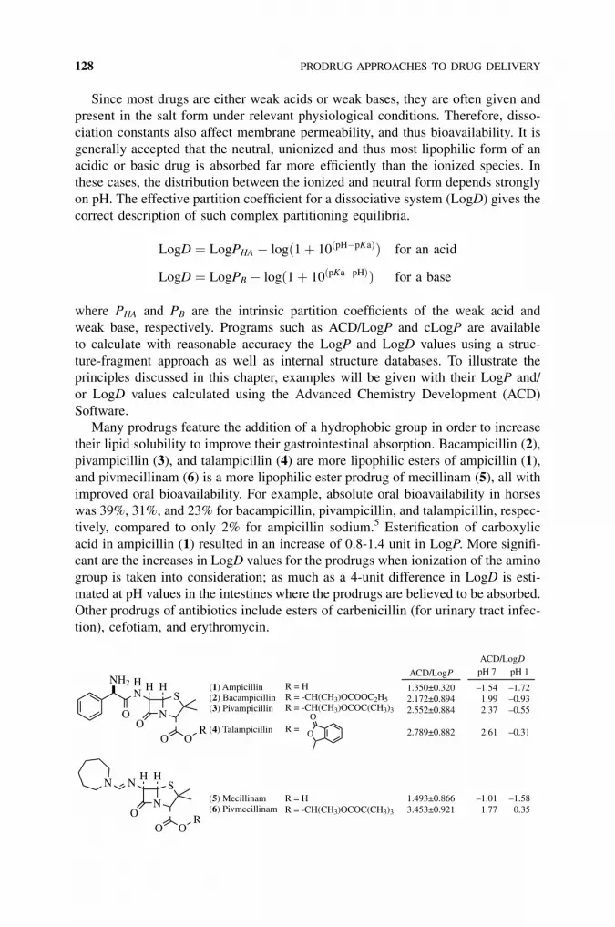
Since most drugs are either weak acids or weak bases, they are often given and
present in the salt form under relevant physiological conditions. Therefore, disso-
ciation constants also affect membrane permeability, and thus bioavailability. It is
generally accepted that the neutral, unionized and thus most lipophilic form of an
acidic or basic drug is absorbed far more efficiently than the ionized species. In
these cases, the distribution between the ionized and neutral form depends strongly
on pH. The effective partition coefficient for a dissociative system (LogD) gives the
correct description of such complex partitioning equilibria.
LogD ¼ LogPHA � logð1 þ 10ðpH�pKaÞÞ for an acid
LogD ¼ LogPB � logð1 þ 10ðpKa�pHÞÞ for a base
where PHA and PB are the intrinsic partition coefficients of the weak acid and
weak base, respectively. Programs such as ACD/LogP and cLogP are available
to calculate with reasonable accuracy the LogP and LogD values using a struc-
ture-fragment approach as well as internal structure databases. To illustrate the
principles discussed in this chapter, examples will be given with their LogP and/
or LogD values calculated using the Advanced Chemistry Development (ACD)
Software.
Many prodrugs feature the addition of a hydrophobic group in order to increase
their lipid solubility to improve their gastrointestinal absorption. Bacampicillin (2),
pivampicillin (3), and talampicillin (4) are more lipophilic esters of ampicillin (1),
and pivmecillinam (6) is a more lipophilic ester prodrug of mecillinam (5), all with
improved oral bioavailability. For example, absolute oral bioavailability in horses
was 39%, 31%, and 23% for bacampicillin, pivampicillin, and talampicillin, respec-
tively, compared to only 2% for ampicillin sodium.5 Esterification of carboxylic
acid in ampicillin (1) resulted in an increase of 0.8-1.4 unit in LogP. More signifi-
cant are the increases in LogD values for the prodrugs when ionization of the amino
group is taken into consideration; as much as a 4-unit difference in LogD is esti-
mated at pH values in the intestines where the prodrugs are believed to be absorbed.
Other prodrugs of antibiotics include esters of carbenicillin (for urinary tract infec-
tion), cefotiam, and erythromycin.
NH2 HN
O N
SHH
OO O
R
(1) Ampicillin (2) Bacampicillin (3) Pivampicillin
(4) Talampicillin O
O
1.350±0.3202.172±0.8942.552±0.884
2.789±0.882
N N
N
SHH
OO O
R
(5) Mecillinam(6) Pivmecillinam
1.493±0.8663.453±0.921
R = HR = -CH(CH3)OCOC(CH3)3
R = HR = -CH(CH3)OCOOC2H5R = -CH(CH3)OCOC(CH3)3
R =
ACD/LogP
ACD/LogD
–1.72–0.93–0.55
–0.31
–1.54 1.99 2.37
2.61
pH 7 pH 1
–1.011.77
–1.580.35
128 PRODRUG APPROACHES TO DRUG DELIVERY

Enalapril (8) is an ester prodrug of enalaprilat (7). The latter binds tightly to the
angiotensin-converting enzyme (ACE) but is transported with low efficacy by the
peptide carrier in the gastrointestinal tract. The prodrug enalapril has a higher affi-
nity for the peptide carrier6 and is much better absorbed, with about 60% oral bioa-
vailability.7,8 As a matter of fact, all ACE inhibitors except captopril and lisinopril
are administered as prodrugs; other commercialized ACE inhibitor prodrugs
include perindopril, quinapril, ramipril, cilazapril, benazepril, spirapril, and trando-
lapril, all based on esterification of the same carboxylic acid group.9 The esters are
hydrolyzed in vivo, after absorption, to the corresponding active but poorly
absorbed dicarboxylate forms.
NH
NRO
O
OO OH
(7) Enalaprilat R = H (8) Enalapril R = Et
2.102±0.5742.983±0.580
ACD/LogPACD/LogD
–0.92–0.10
–1.45–0.12
pH 7 pH 1
Valacyclovir (10)10,11 and famciclovir (12) are ester prodrugs of acyclovir (9)
and penciclovir (11), respectively, for the treatment of viral infections. Both acyclo-
vir and penciclovir exhibit site-specific conversion to the active triphosphate species
by viral thymidine kinase. They show remarkable antiviral selectivity and specifi-
city. However, their oral bioavailability is quite low, 15–20% of an oral dose being
absorbed in humans for acyclovir and 5% for penciclovir.12 Both valacyclovir and
famciclovir have no intrinsic antiviral activity, and both are rapidly hydrolyzed to
acyclovir and penciclovir by esterases present in the liver and gut wall. Valacyclovir
displays a mean absolute bioavailability of 54%, a threefold increase in oral bioa-
vailability over acyclovir, while famciclovir has an absolute bioavailability of 77%
in humans.13–15 Famciclovir’s better bioavailability could be explained by the
increase in lipophilicity; the high oral bioavailability of valacyclovir was also partly
attributed to the involvement of an active transport mechanism through PEPT1.16
Therefore, in addition to increasing lipophilicity, prodrug design can utilize active
transport mechanisms as a means of enhancing bioavailability.
HN
N N
N
O
H2N
OOR
HN
N N
N
O
H2NOR
OR
(9) Acyclovir R = H (10) Valacyclovir R = L-Valyl-
(11) Penciclovir R = H (12) Famciclovir R = CH3CO-
–1.760±0.489 0.040±0.577
–2.031±0.584–0.088±0.265
ACD/LogP
ACD/LogD
–3.76–4.76
–1.76–0.78
pH 7 pH 1
–2.03–0.09
–4.44–3.27
BASIC CONCEPTS: DEFINITION AND APPLICATIONS 129

8.2.2. Sustained-Release Prodrug Systems
Antipsychotic drugs are the mainstay treatment for schizophrenia and similar psy-
chotic disorders. Long-acting depot injections of antipsychotic drugs are exten-
sively used as a means of long-term maintenance treatment. The duration of
action for many antipsychotic drugs with a free hydroxyl group can be considerably
prolonged by the preparation of long-chain fatty acid esters with very high LogP
values (usually 7 or above). Fluphenazine enanthate (14) and fluphenazine decan-
oate (15) were the first of these esters to appear in clinical use and are longer-acting,
with fewer side effects than the parent drug. The ability to treat patients with a sin-
gle intramuscular injection every 1–2 weeks with the enanthate or every 2–3 weeks
with the decanoate esters means that problems associated with patient compliance
with the drug regimens and with drug malabsorption can be reduced.17 Esterifica-
tion of antipsychotic drugs with decanoic acid yields very lipophilic prodrugs
which are dissolved in a light vegetable oil such as Viscoleo or sesame oil.
Intramuscular injection creates an oily depot from which the prodrug molecules
slowly diffuse into the systemic circulation, where they are hydrolyzed quickly
by esterases to the active moieties. These depot forms allow these drugs to be
given only once or twice a month, permitting the long-term treatment of schizo-
phrenia. Antipsychotic drugs available in depot formulation include fluphenazine
(13), flupenthixol, haloperidol, and zuclopenthixol in their enanthate or decanoate
esters.
S
N
N
NO
R
CF3
(13) Fluphenazine R= H (14) Fluphenazine enanthate R = -CO(CH2)5CH3 (15) Fluphenazine decanoate R = -CO(CH2)8CH3
4.841±0.4608.034±0.4539.628±0.453
ACD/LogP
Anabolic steroids such as nandrolone and testosterone, anti-inflammatory gluco-
corticoids such as methylprednisolone, and contraceptives such as estradiol and
levonorgestrel all have slow-release formulations of their ester prodrugs in the
market.
8.2.3. Improving Gastrointestinal Tolerance
Temporary masking of carboxylic acid groups in nonsteroidal anti-inflammatory
drugs was proposed as a promising means of reducing gastrointestinal toxicity
resulting from direct mucosal contact mechanisms. Morpholinoalkyl esters (17
and 19, HC1 salts) of naproxen (16) and indomethacin (18) were evaluated in vitro
and in vivo for their potential use as prodrugs for oral delivery.18 The prodrugs were
freely soluble in simulated gastric fluid and pH 7.4 phosphate buffer and showed a
minimum of a 2000-fold increase in solubility over the parent drugs. The prodrugs
were more lipophilic than the parent drugs and were quantitatively hydrolyzed to
130 PRODRUG APPROACHES TO DRUG DELIVERY

their respective parent drugs in vivo. The prodrugs were 30–36% more bioavailable
orally than the parent drugs following a single dose in rats. They were significantly
less irritating to gastric mucosa than the parent drugs following a single dose as
well as chronic oral administration in rats.
MeO
O
O
RN
O
Cl
MeO
O
OR
(CH2)n N O (CH2)n N O(17) R =
(16) Naproxen (R = H) (18) Indomethacin (R = H)
(19) R =
8.2.4. Improving Taste
Oral drugs with a markedly bitter taste may lead to poor patient compliance if
administered as a solution or syrup. The prodrug approach has been used to
improve the taste of chloramphenicol (20), clindamycin, erythromycin, and metro-
nidazole.19 A prodrug such as chloramphenicol palmitate (21), with LogP of around
10, does not dissolve in an appreciable amount in the mouth and, therefore, does not
interact with the taste receptors.
O2N
HN CCl2
O
OH
OR
(20) Chloramphenicol
(21) Chloramphenicol palmitate
(22) Chloramphenicol sodium
1.018±0.321
9.920±0.756
2.287±0.849
R = H
R = -CO(CH2)14CH3
R = -COCH2CH2COO–Na+
succinate
ACD/LogP
ACD/LogD
1.02
9.92
2.29
1.02
9.92
–0.34
pH 7 pH 1
8.2.5. Diminishing Gastrointestinal Absorption
Many prodrugs have been evaluated in this context for colon-specific drug delivery.
Colon targeting is of value for the topical treatment of diseases of the colon such as
Crohn’s disease, ulcerative colitis, and colorectal cancer. Sustained colonic release
of drugs can be useful in the treatment of nocturnal asthma, angina, and arthritis.
Prodrugs have been designed to pass intact and unabsorbed from the upper gastro-
intestinal tract and undergo biotransformation in the colon, releasing the active drug
BASIC CONCEPTS: DEFINITION AND APPLICATIONS 131
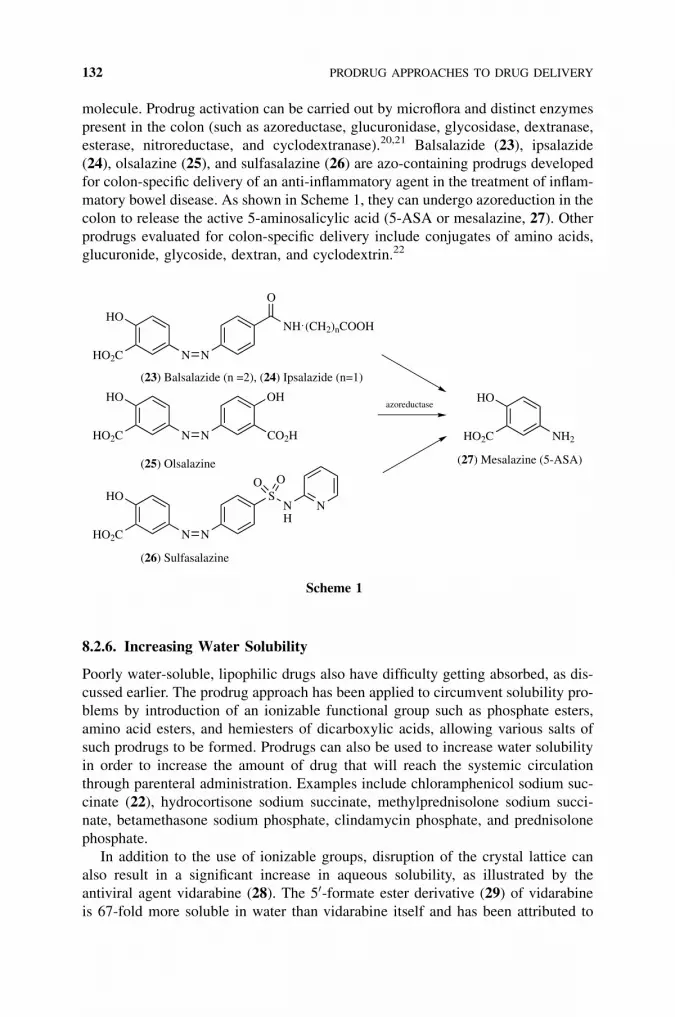
molecule. Prodrug activation can be carried out by microflora and distinct enzymes
present in the colon (such as azoreductase, glucuronidase, glycosidase, dextranase,
esterase, nitroreductase, and cyclodextranase).20,21 Balsalazide (23), ipsalazide
(24), olsalazine (25), and sulfasalazine (26) are azo-containing prodrugs developed
for colon-specific delivery of an anti-inflammatory agent in the treatment of inflam-
matory bowel disease. As shown in Scheme 1, they can undergo azoreduction in the
colon to release the active 5-aminosalicylic acid (5-ASA or mesalazine, 27). Other
prodrugs evaluated for colon-specific delivery include conjugates of amino acids,
glucuronide, glycoside, dextran, and cyclodextrin.22
HO
HO2C N N
OH
CO2H
HO
HO2C N N
NH (CH2)nCOOH
O
azoreductase
HO
HO2C N N
SNH
N
O O
HO
HO2C NH2
(26) Sulfasalazine
(25) Olsalazine (27) Mesalazine (5-ASA)
(23) Balsalazide (n =2), (24) Ipsalazide (n=1)
Scheme 1
8.2.6. Increasing Water Solubility
Poorly water-soluble, lipophilic drugs also have difficulty getting absorbed, as dis-
cussed earlier. The prodrug approach has been applied to circumvent solubility pro-
blems by introduction of an ionizable functional group such as phosphate esters,
amino acid esters, and hemiesters of dicarboxylic acids, allowing various salts of
such prodrugs to be formed. Prodrugs can also be used to increase water solubility
in order to increase the amount of drug that will reach the systemic circulation
through parenteral administration. Examples include chloramphenicol sodium suc-
cinate (22), hydrocortisone sodium succinate, methylprednisolone sodium succi-
nate, betamethasone sodium phosphate, clindamycin phosphate, and prednisolone
phosphate.
In addition to the use of ionizable groups, disruption of the crystal lattice can
also result in a significant increase in aqueous solubility, as illustrated by the
antiviral agent vidarabine (28). The 50-formate ester derivative (29) of vidarabine
is 67-fold more soluble in water than vidarabine itself and has been attributed to
132 PRODRUG APPROACHES TO DRUG DELIVERY

disruption of the strong intermolecular interactions in the crystal, as indicated by
the 85�C drop in the melting point.23
N
N N
N
NH2
O OH
OH
RO
O
H2O
(28) Vidarabine(29) 5′-formate
R = HR = C-H
260 °C175 °C
0.0018 M0.12 M
m.p. solubility (25 °C)
–1.458±0.470–0.364±0.601
ACD/LogP
8.2.7. Tissue Targeting and Activation at the Site of Action
Prodrugs can be designed to target specific tissues. This is especially useful in
improving the therapeutic effectiveness and decreasing the systemic toxicity of
anticancer agents in the treatment of cancer. Anticancer agents are usually highly
toxic, with a very small therapeutic index, and their therapeutic effectiveness is
often limited by their dose-limiting side effects. Here, several strategies for target-
ing chemotherapeutic agents to cancers will be briefly discussed to illustrate the
applications of prodrugs. For details, refer to Chapter 11 on metabolic activation
and drug targeting.
8.2.7.1. Tumor Hypoxia and Bioreductive Activation of Anticancer ProdrugsSolid tumors often contain regions which are subject to chronic or transient defi-
ciencies of blood flow and, therefore, to the development of chronic or acute
hypoxia owing to the primitive state of tumor vasculature.24 Hypoxic cells in a
solid tumor frequently constitute 10–20% and occasionally over half of the
total viable tumor cell population. Agents that are active against proliferating cells
are relatively ineffective against these hypoxic tumor cells, which are not actively
replicating at the time of treatment but are capable of commencing proliferation at
a later time and causing the tumor to regrow. Hypoxic cells also may be resistant
to conventional chemotherapy due to pharmacodynamic considerations.24 To
produce a therapeutic response, appropriate drug concentrations must be
reached. Drugs that have physicochemical properties not conducive to diffusion
into tumor tissue, or that are unstable or metabolized rapidly, may not reach
chronically hypoxic tumor cells located in regions of severe vascular insufficiency.
Therefore, the presence of hypoxic cells in solid tumors is an obstacle to effecting a
cure.
Since hypoxic cells located remotely from the vascular supply of a tumor
mass may have a greater capacity for reductive reactions than their normal, well-
oxygenated counterparts, hypoxia could provide an opportunity for the design of
selective cancer chemotherapeutic agents that could be reductively activated in
these hypoxic cells.24 Several classes of agents are presently known which exhibit
preferential cytotoxicity toward hypoxic cells through reductive activation. They
BASIC CONCEPTS: DEFINITION AND APPLICATIONS 133

include nitro compounds, quinones, and aromatic N-oxides.25,26 Examples include
mitomycin C (30), CB1954 (31), EO9 (32), and AQ4N (33).
N NH
O
O
Me
H2NCH2OCONH2
OMe
NO2
CONH2
O2N
N
NH
O
O
N
OH
OH
O
O
OH
OH
HN
HN
N
NO–
O–
(30) Mitomycin C (31) CB1954
(32) EO9 (33) AQ4N
8.2.7.2. Activation of Prodrugs by Tissue- or Tumor-Specific Enzymes Investi-
gations of the biochemistry and molecular biology of cancer have also identified
several reductive or proteolytic enzymes that are unique to tumors or tissues and
could be used as potential therapeutic targets or prodrug-converting enzymes for
novel cancer therapy. These include DT-diaphorase,27 prostate specific antigen
(PSA),28 plasminogen activator,29 and members of matrix metalloproteinases.30
One such example is the peptide doxorubicin conjugate, glutaryl-Hyp-Ala-Ser-
Chg-Gln-Ser-Leu-Dox, L-377202 (34), which was reported to have the profile of
physical and biological properties needed for further clinical development.31 Con-
jugate 34 was found to have a greater than 20-fold selectivity against PSA-secreting
LNCaP cells relative to non-PSA-secreting DuPRO cells. In nude mouse xenograft
studies, it reduced PSA levels by 95% and tumor weight by 87% at 21 mmol/kg, a
dose below its maximal tolerated dose (MTD). On the basis of these results, this
conjugate was selected for further studies in clinical trials to assess its ability to
inhibit human prostate cancer cell growth and tumorigenesis. It was believed that
PSA cleavage in and around prostate cancer cells would release, as shown in
Scheme 2, dipeptide-doxorubicin conjugate (35), which would be further cleaved
by aminopeptidases to the cytotoxic Leu-doxorubicin (36) and doxorubicin (37).
8.2.7.3. Antibody- or Gene-Directed Enzyme Prodrug Therapy Besides targeting
hypoxic tumor cells and using tumor- or tissue-specific enzymes like PSA to acti-
vate prodrugs, other specific enzymes can be delivered to tumor tissues using
antibodies or expressed by tumor cells through gene therapy and can be used as
prodrug converting enzymes. These strategies are called ‘‘antibody-directed
134 PRODRUG APPROACHES TO DRUG DELIVERY

enzyme prodrug therapy’’ (ADEPT) or ‘‘gene-directed enzyme prodrug therapy’’
(GDEPT). In these approaches, an enzyme is delivered site specifically by chemical
conjugation or genetic fusion to a tumor-specific antibody or by enzyme gene deliv-
ery systems into tumor cells. This is followed by the administration of a prodrug,
which is selectively activated by the delivered enzyme at the tumor cells. A number
of these systems are in development and have been reviewed.32 Among the
enzymes under evaluation is a bacterial nitroreductase from Escherichia coli.
This is a flavin mononucleotide (FMN)–containing flavoprotein capable of reducing
certain aromatic nitro groups to the corresponding amines or hydroxylamines in
the presence of a cofactor NADH or NADPH. The nitroaromatics that were found
to be good substrates of E. coli nitroreductase include dinitroaziridinylbenzamide
CB1954 (31), dinitrobenzamide mustards SN 23862 (38), 4-nitrobenzylcarbamates
(39), and nitrophenyl phosphoramides (40 and 41).33
NO2
O2N
CONH2
N
Cl Cl
NO2
O
NH
DrugO
R1
OP
NHR2O
N(CH2CH2Cl)2
O2N
(38) SN 23862 (39) 4-nitrobenzylcarbamatesnitrophenyl phosphoramides(40) R1 = R2 = H (LH7)(41) R1, R2 = CH2CH2
OOH
OH
O
OMe
HONH
OH
OHO
O
OMe
O
HN
O
NH
OHOH
N
CONH2
O
NH
OHN
OHO
NH
O
N
O
HO
O
HO
PSA
(34) L-377202
H-Ser-Leu-doxorubicin H-Leu-doxorubicin
Doxorubicin
(35) (36)
(37)indicates enzyme cleavage site
Scheme 2
BASIC CONCEPTS: DEFINITION AND APPLICATIONS 135

8.2.7.4. Tumor-Specific Transporters Antibody-drug conjugates would have to
overcome problems inherent in proteins such as susceptibility to proteolytic clea-
vage and high immunogenicity; the latter could lead to an antibody response against
the conjugate, thereby precluding further use. To increase the selectivity of che-
motherapeutic agents, considerable efforts have also been made to identify bio-
chemical characteristics unique to malignant tumor cells that could be exploited
in a therapeutic intervention. The small and nonimmunogenic, tumor-specific mole-
cules like folic acid are among the promising alternatives to antibody molecules as
targeting agents for drug delivery. Folate conjugates of radiopharmaceuticals, mag-
netic resonance imaging (MRI) contrast agents, antisense oligonucleotides and
ribozymes, proteins and protein toxins, immunotherapeutic agents, liposomes
with entrapped drugs, and plasmids have all been successfully delivered to folate
receptor–expressing cells.34 More details can be found in Chapter 9 on receptor-
mediated endocytosis.
8.3. PRODRUG DESIGN CONSIDERATIONS
Often medicinal chemists encounter a situation where a structure has adequate
pharmacological activity but an inadequate pharmacokinetic profile (i.e., absorp-
tion, distribution, metabolism, and excretion). Prodrugs can be designed to improve
physicochemical properties, resulting in improvement in pharmacokinetic as well
as pharmaceutical properties. The pharmaceutical properties that could be
improved, as discussed earlier, include drug product stability, taste and odor, pain
on injection, and gastrointestinal irritation. These are great benefits that can be
achieved through the design of prodrugs. However, regulatory issues should also
be considered in the design process. In general, regulatory agencies are reluctant
to register this type of product. Of particular concern is the fact that toxicological
studies might not be relevant for human use of the drug because of differences in
the rate and/or extent of formation of the active moiety—metabolic aspects. Experi-
ments should thus be designed early to address these concerns. As examples of
interspecies differences, the pivaloyloxyethyl ester of methyldopa was essentially
hydrolyzed presystematically to pivalic acid and methyldopa at the same rate in
human, dog, and rat, while the succinimidoethyl derivative was hydrolyzed faster
in rat than in man and dog.35 This suggests that the succinimidoethyl ester of
methyldopa was more resistant to extrahepatic esterase action in man and dog
but not in rat. For different ester prodrugs of dyphylline, the relative rates of release
were 1.3 to 13 times faster in rabbit plasma than in human plasma.36
The bond between the active moiety (parent drug) and the promoiety plays a
major role in determining the pharmacokinetic properties of a prodrug. Knowledge
about the nature of the bond and the promoiety may help explain the nature of the
biotransformation process and its location in specific tissues or cells. The study of
the fate in the body of the promoiety is particularly important from the safety point
of view and should be investigated just as thoroughly as that of the active moiety. In
some cases, the fate of the released carrier moiety is well known, such as the esters
of methanol or ethanol; no extra study is needed during drug development. In other
cases, additional pharmacokinetic investigations may be necessary.
136 PRODRUG APPROACHES TO DRUG DELIVERY

Rational design of a prodrug should begin with identification of the problem(s)
encountered with the delivery of the parent compound/drug and the physicochem-
ical properties needed to overcome the delivery problem(s). Only then can the
appropriate promoiety be selected to construct a prodrug with the proper physico-
chemical properties that can be effectively transformed to the active drug in the
desired biological compartment.
The most important requirement in prodrug design is naturally the adequate
reconversion of the prodrug to the active drug in vivo at the intended compartment.
This prodrug-drug conversion may take place before absorption (e.g., in the gastro-
intestinal system), during absorption (e.g., in the gastrointestinal wall or in the
skin), after absorption, or at the specific site of drug action. It is important that
the conversion be essentially complete because the intact prodrug, being usually
inactive, represents unavailable drug. However, the rate of conversion would
depend on the specific goal of the prodrug design. A prodrug designed to overcome
poor solubility for an intravenous drug formulation should be converted very
quickly to the active moiety after injection. If the objective of the prodrug is to pro-
duce sustained drug action through rate-limiting conversion, the rate of conversion
should not be too fast.
Prodrugs can be designed to use a variety of chemical and enzymatic reactions to
achieve cleavage to generate their active drug at the desired rate and place. The
design is often limited by the availability of a suitable functional group in the active
drug for the attachment of a promoiety. Table 8.1 lists some of the common rever-
sible prodrug forms for various functional groups that are often present in biologi-
cally active substances.
The most common prodrugs are those that require hydrolytic cleavage, but
reductive and oxidative reactions have also been used for the in vivo regeneration
of the active drug. Besides using the various enzyme systems for the necessary acti-
vation of prodrugs, the buffered and relatively constant physiological pH may be
used to trigger their release.
Enzymes considered important to orally administered prodrugs are found in gas-
trointestinal walls, liver, and blood. In addition, enzyme systems present in the gut
microflora may be important in metabolizing prodrugs before they reach the intest-
inal cells. In addition, site-specific delivery can be accomplished by exploiting
enzymes that are present specifically or at high concentrations in the targeted tis-
sues relative to nontarget tissues. A number of enzymes can also be delivered to
targeted tissues through antibodies or gene-delivery approaches for the activation
of subsequently administered prodrugs, as discussed earlier in this chapter.
8.4. PRODRUGS OF VARIOUS FUNCTIONAL GROUPS
8.4.1. Ester Prodrugs of Compounds Containing ��COOH or ��OH
Due to the presence of a wide variety of esterases in various body tissues, it is not
surprising that esters are the most common prodrugs used to improve gastrointest-
inal absorption. By appropriate esterification of molecules containing a carboxylic
acid or hydroxyl group, it is possible to obtain derivatives with almost any desirable
PRODRUGS OF VARIOUS FUNCTIONAL GROUPS 137

hydrophilicity, lipophilicity, and in vivo lability. It should be noted that enzyme-
catalyzed ester hydrolysis is quite different from nonenzymatic ester hydrolysis
in terms of electronic and steric requirements in the substrates. Enzymatic reactions
are more likely influenced by steric rather than electronic effects. Experimental
determination should be performed to evaluate the rate of cleavage under incuba-
tion with plasma or a homogenate from the intended tissue or organ where the
prodrug would be activated. It should also be kept in mind that there are significant
interspecies variations in the enzyme’s expression level and catalytic capacity.
TABLE 8.1 Reversible Prodrug Forms for Various FunctionalGroups Present in Biologically Active Substances
O
OH
O
OH
NH2
SH
N
NHX
On
O
OR
O
O
O R′
OR
NH
OR
NR
NOH
NO R OR′
OR
NH
O
NH
SO
RO
O R
O
O O
OR
OP
OH
OOH
OR
O O R′
OR
S R
O
S O R′
OR
SS
R
XH
NH2NH
X
NH
R
O
NH
O
OR
N R′
R
NX
NR
R′
On
NH
RR′
R′′
NH
O
O
O
R
R′
O
N O R′
OR
NX
OH
On
NOH
NO R
NO
R
O
NAr N
Ar NO2Ar
OH
OH O NO
R
O
O
O O
ORR′
CH2OH
OP
O
O O
ORR′
OP
O
O R′
OR
OP
OR
OHP
O
R
O
FuntionalGroup Reversible Prodrug Forms
acidic
X = O or S
X = C, n = 1X = S, n = 2
138 PRODRUG APPROACHES TO DRUG DELIVERY

As shown in Scheme 3, esters in the form of 42 can be used as prodrugs for acid
drugs (43), and the alcohol would serve as a promoiety. Esters in the form of 44 can
be used as prodrugs for alcohol drugs (45), and here the acid would serve as a
promoiety.
Both the acyl and the alcohol portion surrounding the cleavable ester bond affect
the enzyme-catalyzed ester hydrolysis. Sometimes because of steric hindrance in
the active drug, direct ester formation with the existing functional group might
not produce a prodrug that is sufficiently labile in vivo. This problem can be sol-
ved by designing the so-called cascade prodrugs containing double esters using
a-acyloxyalkyl, carbonate, or alkoxycarbonyloxyalkyl esters (46, 47, or 48), where
the terminal ester group is accessible for enzymatic cleavage (Scheme 4). Cascade
prodrugs are those prodrugs that require a sequence of two or more reactions for
drug release and activation, usually triggered by a first enzymatic-catalyzed reac-
tion followed by a spontaneous chemical release/activation step(s). A number of
such examples are known, including several prodrugs of b-lactam antibiotics, cor-
ticosteroids, and angiotensin II receptor antagonists. The 2-carboxylic acid on the
thiazolidine ring of b-lactam antibiotics is required for antibacterial activity,
Drug O
O
REsterases
Drug OH
O
R O
O
DrugEsterases
HO Drug
HO R
R OH
O
+
+
Promoiety
Promoiety
(42) (43)
(44) (45)
Scheme 3
R O
O
R′
R O
O
O
R′′
R′
O
R O
O
O
R′′
O
O
R′
Bulky R Bulky R′Simple ester
α-Acyloxyalkyl ester
Alkoxycarbonyloxyalkyl ester
O O
O
R′R
Carbonate ester
(42 or 44)
(46) (47)
(48)
Scheme 4
PRODRUGS OF VARIOUS FUNCTIONAL GROUPS 139
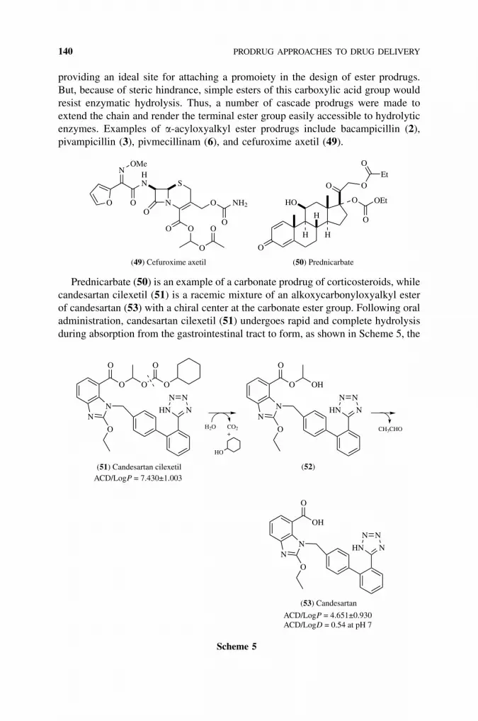
providing an ideal site for attaching a promoiety in the design of ester prodrugs.
But, because of steric hindrance, simple esters of this carboxylic acid group would
resist enzymatic hydrolysis. Thus, a number of cascade prodrugs were made to
extend the chain and render the terminal ester group easily accessible to hydrolytic
enzymes. Examples of a-acyloxyalkyl ester prodrugs include bacampicillin (2),
pivampicillin (3), pivmecillinam (6), and cefuroxime axetil (49).
N
S
O
HN
O
N
O
OMe
O NH2
OO O
O
O
O
H
HO
H H
O
O OEt
O
OEt
O
(49) Cefuroxime axetil (50) Prednicarbate
Prednicarbate (50) is an example of a carbonate prodrug of corticosteroids, while
candesartan cilexetil (51) is a racemic mixture of an alkoxycarbonyloxyalkyl ester
of candesartan (53) with a chiral center at the carbonate ester group. Following oral
administration, candesartan cilexetil (51) undergoes rapid and complete hydrolysis
during absorption from the gastrointestinal tract to form, as shown in Scheme 5, the
NN N
NN
HN
O
O
O
O O
O
CO2
HO
H2O
NN N
NN
HN
O
O
O
OH
CH3CHO
NN N
NN
HN
O
OH
O
(51) Candesartan cilexetil
(53) Candesartan
+
ACD/LogP = 7.430±1.003
ACD/LogP = 4.651±0.930ACD/LogD = 0.54 at pH 7
(52)
Scheme 5
140 PRODRUG APPROACHES TO DRUG DELIVERY

active drug candesartan (53), which is an achiral selective AT1 subtype angiotensin
II receptor antagonist.37
For a more recent effort to find orally active aminomethyl-THF 1b-methylcarba-
penems (Scheme 6), a number of mono and bis double esters were investigated as
potential prodrugs. The bis double ester derivatives such as (55) demonstrated
enhanced oral activity, while the mono double ester derivatives did not demonstrate
significantly improved oral activity due to the presence of a charged group.38
The a-acyloxylalkyl esters have also been extended to include the phosphate
group, phosphonic acids, and phosphinic acids. One such example is fosinopril
(57), an ACE inhibitor, where the phosphinic acid is O-a-acyloxyalkylated to
increase lipophilicity to provide better absorption.
PN
OO OHO OR
O
Me Me
Et
O
(56) Fosinoprilat R = H
(57) Fosinopril R =
3.069±0.660
5.810±0.679
ACD/LogP
ACD/LogD
3.06
5.81
–1.93
2.65
pH 7 pH 1
O-a-acyloxyalkyl ethers are also a useful prodrug type for compounds contain-
ing a phenol group. Such derivatives (58) are hydrolyzed by a sequential reaction
involving formation of an unstable hemiacetal intermediate (59), as shown in
Scheme 7. These kinds of ethers might be better prodrugs than normal phenol esters
N
OH
S
CO2
O
ONH3
N
OH
S
O
O O O OEt
O
O
NH
OO
O
OtBut
Aminomethyl-THF1β-methylcarbapenems (54) A bis double ester prodrug (55)
Scheme 6
O O
R′′
R′
O
R′
O
HO
O OH
R′′
R′′ CHOOH
Enzymatic Spontaneous
(58) (59) (60)
DrugDrug Drug
Scheme 7
PRODRUGS OF VARIOUS FUNCTIONAL GROUPS 141

because they are more stable against chemical hydrolysis, but they are still suscep-
tible to enzymatic hydrolysis by human plasma esterases.
Carboxylic acids have also been masked as ketones and alcohols, which would
require oxidation to convert to the active acid drugs. Nabumetone (61) is a nonaci-
dic nonsteroidal anti-inflammatory prodrug (NSAID).39 After absorption, nabume-
tone undergoes extensive metabolism, the main circulating active form is
6-methoxy-2-naphthylacetic acid (62), a potent COX-2 inhibitor (Scheme 8). Since
nabumetone is not acidic and the active acid metabolite does not undergo entero-
hepatic circulation, nabumetone does not cause gastric irritation and is the most
widely prescribed NSAID in the United States.
8.4.2. Prodrugs of Compounds Containing Amides, Imides, andOther Acidic NH
8.4.2.1. Mannich Bases Mannich base prodrugs could enhance the delivery of
their parent drugs through the skin because of their enhanced water solubility as
well as enhanced lipid solubility. N-Mannich bases, or N-acyl gem-diamines (67),
are generally formed, as shown in Scheme 9, by reaction of an acidic NH com-
MeO
O
MeO
OH
O
Livermetabolism
(61) (62)
Scheme 8
CH2O HNR1
R2
NR1
R2
HO
R NH2
O
NR1
R2
R NH
O
NR1
R2
R NH
O
NR1
R2
H
H+
(68) BH+
(67) B
FormationChemical regeneration N-Mannich base or
N-acyl gem-diamine
(63) (64)(65)
(66)
Scheme 9
142 PRODRUG APPROACHES TO DRUG DELIVERY

pound (66) with an aldehyde, usually formaldehyde, and a primary or secondary
aliphatic amine (63). Aromatic amines do not usually undergo this reaction.
Mannich base prodrugs are regenerated by chemical hydrolysis without enzymatic
catalysis in the reverse direction of their formation.40
Transformation of an amide to an N-Mannich base introduces a readily ionizable
amino functional group (67 Ð 68) that would allow the preparation of sufficiently
stable derivatives with greatly enhanced water solubility at slightly acidic pH.
Clinically useful N-Mannich base prodrugs include rolitetracycline and hetacillin.
The highly water-soluble rolitetracycline (70) is an N-Mannich base of tetracycline
(69) with pyrrolidine and is decomposed to tetracycline quantitatively with a half-
life of 40 minutes at pH 7.4 and 35�C.41 Since the decomposition of N-Mannich
bases does not rely on enzymatic catalysis, the rate of hydrolysis is the same in
plasma and in buffer. Hetacillin (71) is an example of a cyclic N-Mannich base-
type prodrug, which is formed by condensation of ampicillin with acetone. The pro-
drug is readily converted back to the active ampicillin and acetone, with a half-life
of 15–20 minutes at pH 4–8 and 35�C.42,43 The advantage of hetacillin is its higher
stability in concentrated aqueous solutions as compared to ampicillin, which has a
more nucleophilic amine that would react with the strained b-lactam ring.
OH
HO
O OHOH
O
N
OH
NH
O
R
H H CH2N
NH N
O
N
S
COOHO
R2R1
HH
(69) Tetracycline R = H
(70) Rolitetracycline R =
(1) Ampicillin
(71) Hetacillin
–1.187±0.750
0.150±0.750
ACD/LogP
1.350±0.320
2.301±0.901
R1 = R2 = H
R1,R2 = >C(CH3)2
ACD/LogD
–4.29
–4.84
–4.78
–3.02
pH 7 pH 1
–1.72
–0.29
–1.54
–1.61
8.4.2.2. N-a-Acyloxyalkyl Derivatives N-a-Acyloxyalkylation has become a
commonly used approach to obtain prodrug forms of various NH-acidic drug sub-
stances such as carboxamides, carbamates, ureas, and imides. This is because
N-a-acyloxyalkyl derivatives (72) combine high in vitro stability with enzymatic
lability. The derivatives are cascade or double prodrugs. The regeneration of the
parent drug occurs via a two-step mechanism, the enzymatic cleavage of the ester
group followed by spontaneous decomposition of the N-a-hydroxyalkyl intermedi-
ate (73) (Scheme 10). The usefulness of this approach depends on the stability of
the N-a-acyloxyalkyl derivative 72, its susceptibility to esterase-catalyzed hydroly-
sis, and the rate of decomposition of the intermediate 73. N-a-acyloxyalkyl deriva-
tives of imides and secondary amides, as well as ring structures containing such
moieties, showed normal ester stability.44 To make such N-a-acyloxyalkyl deriva-
tives useful as prodrugs, the a-hydroxyalkyl intermediate (73) formed after the
enzyme-catalyzed hydrolysis must decompose quickly to release the original
PRODRUGS OF VARIOUS FUNCTIONAL GROUPS 143

drug molecule (74). The rate of the chemical decomposition step was found to cor-
relate with the pKa of the acidic NH group; a pKa of less than 10.5 is required for
instantaneous decomposition of N-hydroxymethyl derivatives.
However, N-a-acyloxyalkyl derivatives (75) of primary amides, and other pri-
mary amide-type structures such as carbamates and sulfonamides, are extremely
unstable in aqueous solution and quickly undergo decomposition to the correspond-
ing N-hydroxymethyl derivatives, which are stable. Such derivatives of simple pri-
mary amides decompose by an elimination-addition mechanism involving a
reactive N-acylimine intermediate (76) (Scheme 11). For imides and secondary
amides, their inability to form an N-acylimine is believed to contribute to the sta-
bility of their N-a-acyloxyalkyl derivatives (72). At pH 4 and 37�C, half-lives of
hydrolysis of N-a-acyloxyalkyl derivatives (75) range from 1 to 90 minutes,
whereas at pH 7.4 the half-lives of hydrolysis are < 1 minute.45 The resulting N-
hydroxymethyl derivatives (77) are rather stable; the half-life for the decomposition
of N-(hydroxymethyl) benzamide is 183 hours at pH 7.4 and 37�C.45 However,
aldehydes other than formaldehyde can be used to from N-a-hydroxyalkyl deriva-
tives that are more unstable than N-hydroxymethyl analogs. For example, the half-
life for N-(a-hydroxybenzyl)benzamide is only 6.5 minutes at pH 7.4 and 37�C.
The use of aldehydes other than formadehyde may further expand the applicability
of this approach to simple amides with pKa above 11.45
8.4.3. Prodrugs of Amines
The presence of an amino group can affect a drug’s physicochemical and biological
properties in several ways. These include (1) intermolecular or intramolecular
N O
R′
R′′
OO
N OH
R′O
CO2HR′′ CO2HR′′
NH
OEnzymatic Fast
Drug Drug Drug
(72) (73) (74)
Scheme 10
Drug N
O
O R
O
H CO2HR
Drug N
O H2O
Drug NH
O
OH
Enzymatic hydrolysis
Elimination
Stable(75)(76) (77)
Scheme 11
144 PRODRUG APPROACHES TO DRUG DELIVERY

aminolysis leading to reactive and/or potentially toxic substances, (2) solubility
problems when the drug is present with another ionizable functionality such as
COOH (zwitterionic nature under physiological pH, potentially limiting its dissolu-
tion rate and/or its passive permeability), and (3) terminal free amino acid groups
providing recognition sites for proteolytic enzymes, such as aminopeptidase and
trypsin, present in the gastrointestinal tract lumen, the brush border region, and
the cytosol of the intestinal mucosa cells. For all these reasons, prodrug approaches
have been advocated for improving in vivo behavior of active compounds contain-
ing amino groups.
8.4.3.1. Amides Because of the relatively high stability of amides in vivo,
N-acylation of amines was formerly of limited use in prodrug design. Only a few
examples of simple amide prodrugs are known that are sufficiently labile in vivo;
these include the N-L-isoleucyl derivative of dopamine46 and the N-glycyl deriva-
tive midodrine.47,48 With the use of proteases as prodrug-converting enzymes,
amines can be coupled to peptide carboxylates, resulting in amide bonds cleavable
by proteases (e.g., 34).
Midodrine (79) is a glycinamide prodrug, and the therapeutic effect of orally
administered midodrine is due to the major metabolite desglymidodrine (78), an
a-agonist formed by deglycination of midodrine. Midodrine is rapidly absorbed
after oral administration. The plasma level of the prodrug peaks after about half
an hour, and declines with a half-life of approximately 25 minutes, while the meta-
bolite reaches peak blood concentrations about 1 to 2 hours after a dose of mido-
drine and has a half-life of about 3 to 4 hours. The absolute bioavailability of
midodrine (measured as desglymidodrine) is 93% and is not affected by food.
Approximately the same amount of desglymidodrine is formed after intravenous
and oral administration of midodrine. Midodrine has been used successfully in
the treatment of neurogenic orthostatic hypotension and, more recently, in the treat-
ment of dialysis hypotension. It acts through vasoconstriction of the arterioles and
the venous capacitance vessels, thereby increasing peripheral vascular resistance
and augmenting venous return, respectively. The prodrug is a unique agent in the
armamentarium against orthostatic hypotension since it has minimal cardiac and
central nervous system (CNS) effects.47,48
O
O
OHNH
R (78) Desglymidodrine(79) Midodrine
R = HR = -COCH2NH2
0.378±0.279–0.319±0.595
ACD/LogPACD/LogD
–3.42–2.72
–1.14–0.83
pH 7 pH 1
8.4.3.2. N-a-Acyloxyalkoxycarbonyl Derivatives Carbamates are of limited use
in prodrug design due to their general resistance to enzymatic cleavage in vivo.
The introduction of an enzymatically labile ester group in the carbamate structure
could render them sensitive to esterase-catalyzed hydrolysis leading to activation.
Thus, N-a-acyloxyalkoxycarbonyl derivatives (80) of primary and secondary
PRODRUGS OF VARIOUS FUNCTIONAL GROUPS 145

amines may be readily transformed, as shown in Scheme 12, to the parent amine
(83) in vivo.49,50 Esterase-catalyzed hydrolysis of the ester moiety in these deriva-
tives leads to an unstable a-hydroxyalkoxycarbonyl intermediate (81) which spon-
taneously decomposes into the parent amine via a labile carbamic acid (82). These
a-acyloxyalkyl carbamate derivatives are neutral and combine high stability in aqu-
eous solution with high susceptibility to enzymatic reconversion to the active agent
triggered by hydrolysis of the terminal ester functions; they may be promising
reversible prodrugs for amino-containing compounds. These are used in the pre-
paration of orally active aminomethyl-THF 1b-methylcarbapenems (56 ! 57).
This approach has also been applied to peptides and peptidomimetics in order to
improve their unfavorable physicochemical characteristics (e.g., size, charge,
hydrogen-bonding potential), which prevent them from permeating biological bar-
riers such as the intestinal mucosa, and by their lack of stability against enzymatic
degradation.51,52 Many of the structural features of a peptide, such as the N-terminal
amino group, the C-terminal carboxyl group, and the side chain carboxyl, amino,
and hydroxyl groups, which bestow upon the molecule affinity and specificity for its
pharmacological receptor, severely restrict its ability to permeate biological barriers
and render it as a substrate of proteases. Bioreversible cyclization of the peptide
backbone is one of the most promising new approaches in the development of pep-
tide prodrugs. Cyclization of the peptide backbone enhances the extent of intramo-
lecular hydrogen bonding and reduces the potential for intermolecular hydrogen
bonding to aqueous solvent. Linking the N-terminal amino group to the C-terminal
carboxyl group via an a-acyloxyalkoxy promoiety, as in (84), is an interesting
approach that has been shown to work on a number of model peptides (Scheme
13). These cyclic prodrugs were designed to be susceptible to esterase-catalyzed
hydrolysis (slow step), leading to a cascade of chemical reactions resulting in the
generation of the linear peptide. In pH 7.4 buffer at 37�C, the cyclic prodrugs (84)
were shown to degrade quantitatively to their corresponding linear peptides (87). In
human plasma, the rates of hydrolysis of cyclic prodrugs were significantly faster
N O
O
O
R′′
R′
O
Drug
RR′
O
HO
NHDrug
RCO2
O
O
OH
R′′
NDrug
R
OH
O
NDrug
R
R′′
CHO
Enzymatic
Spontaneous
Spontaneous
(80) R = H or alkyl (81)
(82)(83)
Scheme 12
146 PRODRUG APPROACHES TO DRUG DELIVERY

than in buffer and were inhibited by paraoxon, a potent esterase inhibitor. In com-
parison to the linear peptides, the cyclic prodrugs were at least 70 times more
permeable in cell culture models of the intestinal mucosa.
8.4.3.3. N-Mannich Bases N-Mannich bases have been used successfully to
obtain prodrugs of amide- (see Section 8.4.2.1) as well as amine-containing drugs.
Due to their rapid cleavage, with half-lives between 10 and 40 minutes at physio-
logical pH and a pronounced decrease in their basicity of 3–4 pKa units,53 salicy-
lamide N-Mannich bases (90) were evaluated as prodrug forms for primary and
secondary amines (Scheme 14). In this case, the amide part of a Mannich base is
the promoiety. To improve their stability in vitro and avoid stability-associated for-
mulation problems, the hydroxyl group of the salicylamide N-Mannich bases (90)
HN O
O
O
O
CO2
O
OHH2N
HN OH
O
HN O
O
OH
O
OH
O
OH
HCHO
Enzymatic
Spontaneous
Spontaneous
Peptide Peptide
PeptidePeptide
(84) (85)
(86)(87)
Scheme 13
HCHO
NH
O
O
N
OH
NH
O
OH
N
R
O
HO
NH2
O
OHHCHO
HN
NH
O
O
N
O R
O
Enzymatic
Spontaneous
Spontaneous
Drug Drug
DrugDrug
+
(88) (89)
(90)(92)
(91)
Scheme 14
PRODRUGS OF VARIOUS FUNCTIONAL GROUPS 147
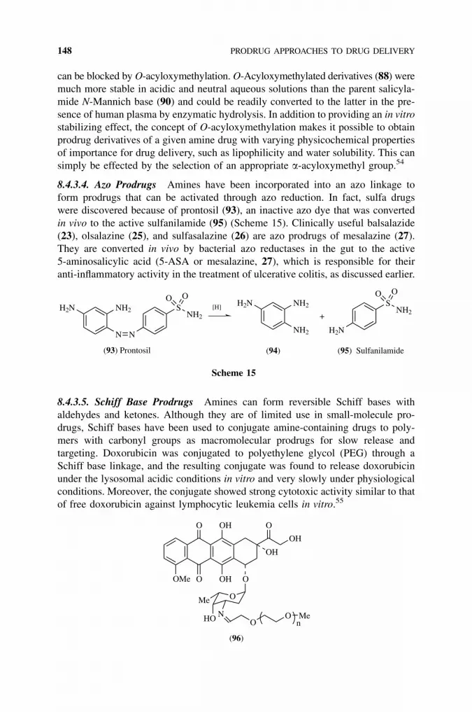
can be blocked by O-acyloxymethylation. O-Acyloxymethylated derivatives (88) were
much more stable in acidic and neutral aqueous solutions than the parent salicyla-
mide N-Mannich base (90) and could be readily converted to the latter in the pre-
sence of human plasma by enzymatic hydrolysis. In addition to providing an in vitro
stabilizing effect, the concept of O-acyloxymethylation makes it possible to obtain
prodrug derivatives of a given amine drug with varying physicochemical properties
of importance for drug delivery, such as lipophilicity and water solubility. This can
simply be effected by the selection of an appropriate a-acyloxymethyl group.54
8.4.3.4. Azo Prodrugs Amines have been incorporated into an azo linkage to
form prodrugs that can be activated through azo reduction. In fact, sulfa drugs
were discovered because of prontosil (93), an inactive azo dye that was converted
in vivo to the active sulfanilamide (95) (Scheme 15). Clinically useful balsalazide
(23), olsalazine (25), and sulfasalazine (26) are azo prodrugs of mesalazine (27).
They are converted in vivo by bacterial azo reductases in the gut to the active
5-aminosalicylic acid (5-ASA or mesalazine, 27), which is responsible for their
anti-inflammatory activity in the treatment of ulcerative colitis, as discussed earlier.
8.4.3.5. Schiff Base Prodrugs Amines can form reversible Schiff bases with
aldehydes and ketones. Although they are of limited use in small-molecule pro-
drugs, Schiff bases have been used to conjugate amine-containing drugs to poly-
mers with carbonyl groups as macromolecular prodrugs for slow release and
targeting. Doxorubicin was conjugated to polyethylene glycol (PEG) through a
Schiff base linkage, and the resulting conjugate was found to release doxorubicin
under the lysosomal acidic conditions in vitro and very slowly under physiological
conditions. Moreover, the conjugate showed strong cytotoxic activity similar to that
of free doxorubicin against lymphocytic leukemia cells in vitro.55
O
OH
OH
O
OMe
HO N
OH
OHO
O
OMe
OO Me
n
(96)
H2N
N N
SNH2
O O
(93) Prontosil
NH2H2N
NH2 H2N
SNH2
O ONH2
+
(95) Sulfanilamide(94)
[H]
Scheme 15
148 PRODRUG APPROACHES TO DRUG DELIVERY
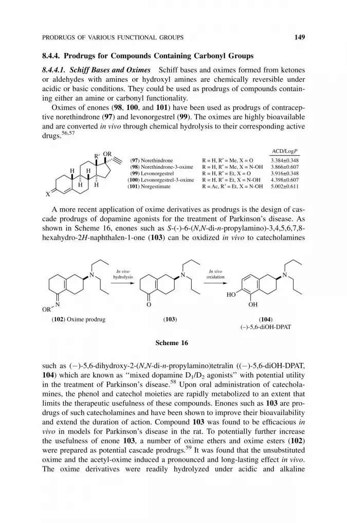
8.4.4. Prodrugs for Compounds Containing Carbonyl Groups
8.4.4.1. Schiff Bases and Oximes Schiff bases and oximes formed from ketones
or aldehydes with amines or hydroxyl amines are chemically reversible under
acidic or basic conditions. They could be used as prodrugs of compounds contain-
ing either an amine or carbonyl functionality.
Oximes of enones (98, 100, and 101) have been used as prodrugs of contracep-
tive norethindrone (97) and levonorgestrel (99). The oximes are highly bioavailable
and are converted in vivo through chemical hydrolysis to their corresponding active
drugs.56,57
X
H H
R′ OR
H H
(97) Norethindrone (98) Norethindrone-3-oxime (99) Levonorgestrel(100) Levonorgestrel-3-oxime(101) Norgestimate
R = H, R′ = Me, X = OR = H, R′ = Me, X = N-OHR = H, R′ = Et, X = OR = H, R′ = Et, X = N-OHR = Ac, R′ = Et, X = N-OH
3.384±0.3483.866±0.6073.916±0.3484.398±0.6075.002±0.611
ACD/LogP
A more recent application of oxime derivatives as prodrugs is the design of cas-
cade prodrugs of dopamine agonists for the treatment of Parkinson’s disease. As
shown in Scheme 16, enones such as S-(-)-6-(N,N-di-n-propylamino)-3,4,5,6,7,8-
hexahydro-2H-naphthalen-1-one (103) can be oxidized in vivo to catecholamines
such as (�)-5,6-dihydroxy-2-(N,N-di-n-propylamino)tetralin ((�)-5,6-diOH-DPAT,
104) which are known as ‘‘mixed dopamine D1/D2 agonists’’ with potential utility
in the treatment of Parkinson’s disease.58 Upon oral administration of catechola-
mines, the phenol and catechol moieties are rapidly metabolized to an extent that
limits the therapeutic usefulness of these compounds. Enones such as 103 are pro-
drugs of such catecholamines and have been shown to improve their bioavailability
and extend the duration of action. Compound 103 was found to be efficacious in
vivo in models for Parkinson’s disease in the rat. To potentially further increase
the usefulness of enone 103, a number of oxime ethers and oxime esters (102)
were prepared as potential cascade prodrugs.59 It was found that the unsubstituted
oxime and the acetyl-oxime induced a pronounced and long-lasting effect in vivo.
The oxime derivatives were readily hydrolyzed under acidic and alkaline
N
NOR
N
O
N
OH
HO
In vivohydrolysis
In vivooxidation
(104) (–)-5,6-diOH-DPAT
(103)(102) Oxime prodrug
Scheme 16
PRODRUGS OF VARIOUS FUNCTIONAL GROUPS 149

conditions. The fact that these oximes as well as 103 were inactive at the dopamine
receptor, yet induced dopamine D1 and D2 receptor-related effects in vivo, sug-
gested that they were acting as prodrugs and were being converted in vivo to the
active species (104).
Oximes can be acylated to make prodrugs, as in the above example and in the
case of FLM 5011, which is a strongly lipophilic, poorly water-soluble, lipoxygen-
ase inhibitor. The water solubility was mproved by using the succinate monoester
prodrug. The bioavailability of FLM 5011 in rabbits after oral administration was
markedly increased by its prodrug.60
OH
NO
R (105) FLM 5011(106) FLM 5011 succinate
R = HR = COCH2CH2COOH
7.705±0.5537.708±0.599
ACD/LogP
ACD/LogD
7.707.71
7.705.23
pH 7 pH 1
8.4.4.2. Enol Esters Enol esters are rather stable, bioreversible derivatives of
ketones and may be useful as prodrugs of agents containing enolizable carbonyl
groups. As shown in Scheme 17, 60-acetylpapaverine enol esters (107), prepared
by acylation of the appropriate Li enolate with the respective anhydride, were
hydrolyzed to 60-acetylpapaverine (108) by esterases present in rat and human
plasma, rat liver, and brain tissue supernatants. The intermediate 60-acetylpapaver-
ine cyclizes rapidly to coralyne (109), which has antitumor activity but has
N
MeO
MeO CH2
O
O
R
OMe
OMe
N
MeO
MeOCH3
O
OMe
OMe
N
MeO
MeO
OMe
OMe
Esterases
(107)6′-Acetylpapaverine enol esters
(109) Coralyne
(108)6′-Acetylpapaverine
Scheme 17
150 PRODRUG APPROACHES TO DRUG DELIVERY

difficulty passing through the blood-brain barrier due to the presence of the positive
charge. 60-Acetylpapaverine enol esters are neutral and stable in aqueous solution
and are potential prodrugs for enhancing delivery of coralyne to brain tissues.61
8.4.4.3. Oxazolidines The kinetics of hydrolysis of several oxazolidines derived
from Tris and various aldehydes and ketones were investigated to explore their suit-
ability as prodrug forms for b-aminoalcohols such as (�) ephedrine (110) and for
carbonyl-containing substances.62-64
Oxazolidines were easily and completely hydrolyzed at pH 1–11 at 37�C. The
hydrolysis rates were subject to general acid-base catalysis by buffer substances and
depended strongly on pH. Most oxazolidines showed sigmoidal pH-rate profiles
with maximum rates at pH 7–7.5. At pH 7.4 and 37�C, the half-lives of hydrolysis
for the various ephedrine oxazolidines (111) ranged from 5 seconds to 30 minutes
(Scheme 18). The reaction rates in neutral and basic solutions decreased with
increasing steric effects of the substituents derived from the carbonyl component
and decreased with increasing basicity of oxazolidines. Oxazolidines are weaker
bases (pKa 5.2–6.9) than the parent b-amino alcohol and are more lipophilic at phy-
siological pH. Thus, oxalolidines can be considered as potentially useful prodrugs
for drugs containing a b-amino alcohol moiety or carbonyl groups.62,63 Molecular
complexation with cyclodextrins might be able to enhance the stability of oxazoli-
dine prodrugs to make them potentially more useful.65,66
The stability characteristics of various N-acylated oxazolidines were also studied
in an attempt to develop approaches which may solve the stability problems asso-
ciated with the use of oxazolidines as prodrug forms. The N-acylated oxazolidines,
including a carbamate derivative, were in fact found to be highly stable in an aqu-
eous solution, but they also proved to be resistant to hydrolysis by plasma enzymes.
The latter limits the use of N-acylated oxazolidines in prodrug design.64
8.4.4.4. Thiazolidines Thiazolidines of some a,b-unsaturated 3-ketone steroids
including progesterone, testosterone, and hydrocortisone (112, 113) were prepared
from the reaction with cysteine alkyl esters and cysteamines as potential pro-
drugs.67,68 The thiazolidines readily reverted to their parent steroidal ketones,
Me
ON Me
R′R
NH
Me
Me
OH
(110) (–) ephedrineOxazolidines
H, HEt, HPh, H2-HOPh, HtBut, HMe, MeCyclohexylidene
518
3005
1800280360
R, R′ t1/2 (s)
(111)
Scheme 18
PRODRUGS OF VARIOUS FUNCTIONAL GROUPS 151

thus meeting the requirements for a prodrug. Most of the thiazolidines were more
lipophilic than their parent steroids, thereby imparting the desired change in the
physicochemical properties to the derivatives of the steroids. Thus, they can func-
tion as bioreversible derivatives of the parent steroids, cysteines, and cysteamines.
Cysteine derivatives are particularly attractive as promoieties due to the release of
cysteine as the by-product upon activation. Both cysteines and cysteamines were
also used as chemoprotective agents against side effects of chemotherapy and radia-
tion therapy. Thus, thiazolidines of cysteines and cysteamines could be used as pro-
drugs for chemoprotection during chemotherapy and radiation therapy.
HO
S
NH
OHH
HH
OO
O
R
(112) R = CO2Et(113) R = H
8.5. DRUG RELEASE AND ACTIVATION MECHANISMS
Most prodrugs rely on enzymatic hydrolysis by esterases or proteases and, to a less
extent, on chemical hydrolysis to achieve a one-step cleavage of the promoiety and
the release of the original active drug (Figure 8.1Ai). These systems, as well as
other one-step activation mechanisms (Figure 8.1Aii–iv) are simple and, in many
cases, sufficient in achieving useful regeneration rates of the active agent. Other-
wise, a cascade release/activation mechanism can be incorporated by taking advan-
tage of autodegradation or intramolecular cyclization reactions to effect the release
and activation of a prodrug. Some of the cascade strategies that have been
employed in the design of prodrugs are shown in Figure 8.1B and will be briefly
discussed here.
8.5.1. Cascade Release Facilitated by Linear Autodegradation Reactions
A number of examples are known for the release of an active drug facilitated by a
linear autodegradation process. This can be achieved through chemically unstable
intermediates such as a-hydroxy amines, amides, and esters. Many of the double
prodrugs discussed earlier belong to this category.
Another interesting linear autodegradation process often used in prodrug design
involves an electron ‘‘push and pull’’ mechanism through a conjugative aromatic
ring that is linked to a good leaving group such as an ester in the para benzylic
position. In such an approach, an electron-donating amino or hydroxy is masked
as an electron-withdrawing nitro group or an amide or ester group in the prodrug.
152 PRODRUG APPROACHES TO DRUG DELIVERY
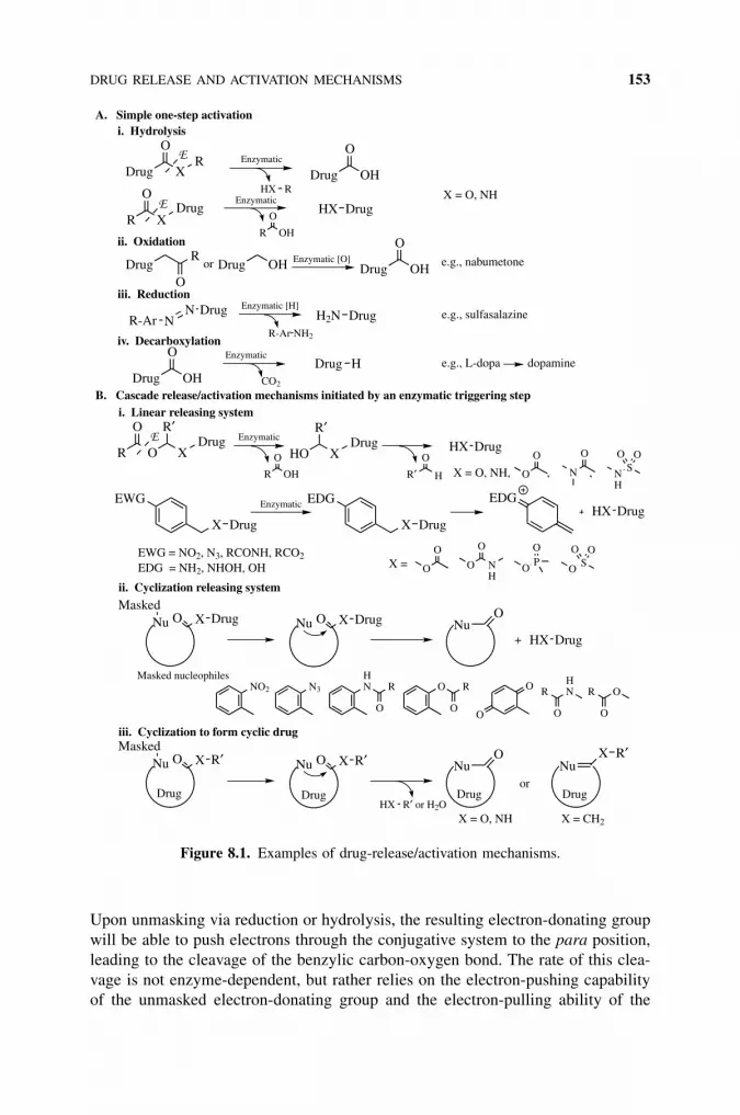
Upon unmasking via reduction or hydrolysis, the resulting electron-donating group
will be able to push electrons through the conjugative system to the para position,
leading to the cleavage of the benzylic carbon-oxygen bond. The rate of this clea-
vage is not enzyme-dependent, but rather relies on the electron-pushing capability
of the unmasked electron-donating group and the electron-pulling ability of the
Drug X
OR
R X
ODrug
HX R
R OH
O
EWG
X Drug
O X DrugNuMasked
NO2
R O
O
XDrug
R′
R OH
O
O X R′NuMasked
DrugNNR-Ar
R-Ar NH2
R
ODrug OHDrug
Drug OH
O
CO2
N3
Nu
Nu
HO XDrug
R′
Drug H
O X R′
Drug OH
O
HX Drug
H2N Drug
O X Drug
HN R
O
X Drug
EDG
Drug OH
O
HX R′ or H2O
R′ H
O
O
O
O R
O
HX Drug
ONu
ONu
O NH
O
O
O
O
O
OPO
EDG
HX Drug
HNR
O
OS
O O
N
O
XNu
R′
OR
O
HX Drug
NH
SO O
Enzymatic
Enzymatic
A. Simple one-step activation
B. Cascade release/activation mechanisms initiated by an enzymatic triggering step
Enzymatic
i. Linear releasing system
ii. Cyclization releasing system
+
X = O, NH
EWG = NO2, N3, RCONH, RCO2
EDG = NH2, NHOH, OH X =
Masked nucleophiles
X = O, NH
Enzymatic
X = O, NH, , ,
Drug Drug Drug
iii. Cyclization to form cyclic drug
Drugor
X = CH2
Enzymatic [H]e.g., sulfasalazine
Enzymatic [O] e.g., nabumetone
i. Hydrolysis
ii. Oxidation
iii. Reduction
or
iv. DecarboxylationEnzymatic
e.g., L-dopa dopamine
Figure 8.1. Examples of drug-release/activation mechanisms.
DRUG RELEASE AND ACTIVATION MECHANISMS 153

leaving group. The formation of the negatively charged species can serve as the
activation mechanism of the drug. This approach has recently been used in our
effects to develop a novel and superior class of nitroaryl phosphoramides as poten-
tial prodrugs for nitroreductase-mediated enzyme-prodrug therapy.33
Several nitroaryl phosphoramides were designed and synthesized, each with a
strategically placed nitro group on the benzene ring in the para position to the
benzylic carbon (Scheme 19). Compound 114 is a cyclophosphamide analog
with the cyclophosphamide ring fused with a benzene ring and a nitro group placed
in the para position to the benzylic carbon. Compound 41 is a 4-nitrophenyl sub-
stituted cyclophosphamide analog, and 40 is an acyclic nitrobenzyl phosphoramide
mustard (LH7). The nitro group is a strong electron-withdrawing group (Hammett
sp electronic parameter ¼ 0.78) and is converted to an electron-donating hydroxy-
lamino group (sp ¼�0.34) upon nitroreductase reduction. This large difference in
electronic effect (�sp ¼ 1.12) is exploited to effect the formation of the highly
cytotoxic phosphoramide mustard or like reactive species. After reduction by
nitroreductase (NTR), the resulting hydroxylamines 115, 117, and 119 relay their
electrons to the para position and facilitate the cleavage of the benzylic C��O bond,
producing the anionic cytotoxic species phosphoramide mustard 121 or like reac-
tive species 116 and 118. Structurally, the phosphoramide portion in 116 and 118
closely resembles phosphoramide mustard 121, the reactive alkylating agent pro-
duced following the metabolic activation of cyclophosphamide in the liver, and
could also be the ultimate cytotoxic alkylating agent.
Phosphoramide mustard is the proven cytotoxic metabolite of cyclophospha-
mide, a successful clinical anticancer prodrug that requires cytochrome P450 acti-
vation in the liver. These nitroaryl phosphoramides in combination with
nitroreductase could effectively move the site of activation from liver in the case
O2N
NH
OP
O
N(CH2CH2Cl)2
O
NH
PO
N(CH2CH2Cl)2
O2N
O
H2N
PO
N(CH2CH2Cl)2
O2N
NTR
NTR
NTRO
H2N
PO
N(CH2CH2Cl)2
NH
HO
O
NH
PO
N(CH2CH2Cl)2
NH
HO
NH
NH
OP
O
N(CH2CH2Cl)2
HO
NH
HO
NH
HO
OP
NHO
N(CH2CH2Cl)2
NH
NH
OP
O
N(CH2CH2Cl)2
HO
OP
N(CH2CH2Cl)2
H2N O
(41)
(114)
(40)
(115) (116)
(117) (118)
(119) (120) (121)
116, 118, and 121 could cross-link DNA strands, leading to cytotoxicity.
Scheme 19
154 PRODRUG APPROACHES TO DRUG DELIVERY

of cyclophosphamide into nitroreductase-expressing tumor tissues. All compounds
were shown to be excellent substrates of E. coli nitroreductase, but with varying
degrees of cytotoxicity against nitroreductase-expressing V79 and SKOV3 cells.
Compounds cis-41 and trans-41, the best of the cyclic series, were over 22,000�more cytotoxic in nitroreductase-expressing Chinese hamster V79 cells and 40, the
acyclic compound LH7, was 167,500� more cytotoxic in the same cell line, with an
IC50 as low as 0.4 nM upon 72-hour drug exposure. This level of activity is about
100� more active and 27� more selective than CB1954. Even when the V79 cells
were exposed to each test compound for 1 hour before the media were replaced
with non-drug-containing fresh media, the IC50 was 10 nM, which was about
30� lower than that of CB1954 (31). The high selectivity of cis-41, trans-41,
and 40 was reproduced in SKOV3 human ovarian carcinoma cells infected with
an adenovirus expressing E. coli nitroreductase. Enzyme kinetic analysis indicates
that compound 40 was a much better substrate of E. coli nitroreductase with a spe-
cificity constant 20� that of CB1954.
8.5.2. Cascade Release Facilitated by Intramolecular Cyclization Reactions
Intramolecular reactions are usually thermodynamically favored over intermolecu-
lar reactions because they have lower activation energy and therefore more stable
transition states. The lower activation energy is attributed to a better entropic situa-
tion. When a reaction is performed between two different reactants, the two mole-
cules need to collide in a specific orientation and the reaction leads to a decrease in
the number of molecules, resulting in an increase in order and therefore a loss of
entropic energy. In an intramolecular reaction, the two reaction centers, e.g., a
nucleophile and an electrophile, are both present within the same molecule and
are in a good position to interact and form the cyclic product. The positioning of
the two reaction centers within the same molecule is very important for the reaction
to take place. Steric factors resulting in less flexible molecules and better position-
ing of the two reaction centers would lead to increased reaction rates. Generally
speaking, intramolecular reactions leading to the formation of 5- or 6-membered
rings are much more favorable and occur at faster rates.
A series of alkylaminoalkyl carbamates of 4-hydroxyanisole (122) were evalu-
ated as prodrugs of the melanocytotoxic phenol (123) that could be activated
through intramolecular cyclization (Scheme 20).69 The carbamates were relatively
stable at low pH but released 4-hydroxyanisole cleanly in a nonenzymatic
fashion at pH 7.4 at rates that were structure-dependent. A detailed study of the
N-methyl-N-[2-(methylamino)ethyl]carbamate showed that generation of the parent
phenol followed first-order kinetics with t1=2 ¼ 36:3 minutes at pH 7.4, 37�C, and
was accompanied by formation of N,N-dimethylimidazolidinone (124). In compar-
ison, the related derivative with three methylene units between the two N atoms
releases the phenol at a much slower rate with t1=2 ¼ 942 minutes. These basic
carbamates are examples of cyclization-activated prodrugs in which generation of
the active drug is not linked to enzymatic cleavage but rather depends solely upon a
predictable, intramolecular cyclization-elimination reaction.
DRUG RELEASE AND ACTIVATION MECHANISMS 155

The terminal amino group in the above system could be masked as an amide,
thus avoiding the stability problem encountered when using the basic carbamates
as prodrugs. Unmasking of the amide could be catalyzed by a specific protease
in vivo at certain target sites, thus achieving target specificity. Another alternative
is to use o-nitroaromatic, as in compounds 125 (Scheme 21), which could be
converted to a nucleophilic aromatic amine upon bioreduction in hypoxic tumor tis-
sues or by other reductases delivered to targeted cells.70–72 Kinetic analysis of the
cyclization activation process indicates that the addition of two a methyl groups to
the ester carbonyl would restrict the rotational freedom of the ground state molecule
and promote the cyclization reaction. The nitro group can be reduced in vivo to a
nucleophilic aromatic amine with a low pKa (<4), which would be present in neu-
tral nucleophilic form under most physiological conditions. At pH 7.4, 37�C, the
O
MeO
N(CH2)nNHR′
O
R
OH
MeO (CH2)n
NN
O
R R′
Me, MeEt, Et Me, HH, MeH,HH,HMe,Me
+pH 7.5
n R, R′ t1/2 (min)
2222233
36.3118304335724910942
(122) (123) (124)
37 °C
Scheme 20
NO2RR
OFUDR
[H]
NH2RR
OFUDR
HNRR
O
R
HMeHMe
n n + FUDR
n t1/2 (min)
0011
14~0
42
pH 7.4n
(125) (126) (127) (128)
37 °C
Scheme 21
156 PRODRUG APPROACHES TO DRUG DELIVERY

amines 126 cyclized quickly to the lactam 127, releasing the anticancer drug flox-
uridine (FUDR) (128).70 For tumor targeting purposes, subsequent drug release
after initial specific enzymatic activation should be very fast (preferably <1 min-
utes) in order to prevent the active drug from escaping the targeted site.
Reduction of quinone propionic esters or amides 129 bearing three Me groups in
the so-called trialkyl lock positions (o-, b-, b-positions) or hydrolysis of the corre-
sponding phenolic esters 130 has been shown to undergo spontaneous lactonization
with the release of alcohol or amine, respectively (Scheme 22).73–75 Several amides
129 were synthesized and tested as model redox-sensitive cascade prodrugs of
amines. The reduction of model amide prodrugs (129) generated hydroxy amide
intermediates 131, the lactonization of which resulted in amine release. The half-
lives for appearance of the product lactone 132 from these intermediates ranged
from 1.4 to 3.4 minutes at pH 7.4 and 37�C. With such rapid lactonization rates,
it is believed that reduction would be the rate-limiting step in the two-step conver-
sion of the prodrugs to amines.75 Comparison of the observed rates of lactonization
O
R2
X
O
R1
Drug
O
R3R3
[H] OH
R2
X
O
R1
Drug
R′
R3R3
O
R2
X
OO
R
R1
Drug
R3R3
OH
O
RH2O
R1
MeHH
R′
HHH
MeHH
R2
O
R2R1
R′
O
R3R3
MeMeH
R3
HX Drug+
R′ = OH or HX = NH or O
t1/2*
65.4 s10.5 h19.4 d
* with p-MePhNH2 as a model amine, at pH 7.5, 30˚C
(129)
(130)
(131)
(132)
(133)
Scheme 22
DRUG RELEASE AND ACTIVATION MECHANISMS 157
at pH 7.5 and 30�C for three hydroxy amides obtained from the hydrolysis of
phenolic ester prodrugs (130) allowed an estimate of the extent of rate enhancement
provided by the addition of a partial or total trimethyl lock for the hydroxy amide
lactonization reaction under near-physiological conditions.74 The half-life for the
hydroxy amide with a full trimethyl lock was 65 seconds, a rate enhancement of
2:54 � 104 as compared to the corresponding hydroxy amide without the three
methyl groups.
Still another intramolecular cyclization system is the coumarin-based prodrug
system 134 that can be used for bioreversible derivatization of amine and alcohol
drugs and the preparation of cyclic peptide prodrugs (Scheme 23).76,77 This system
takes advantage of the known facile lactonization of coumarinic acid and its
derivatives. Such a system can be used for the development of esterase- and phos-
phatase-sensitive prodrugs of amines and alcohols. Esterase-sensitive prodrugs of a
number of model amines prepared by using this system readily released the amines
upon incubation in the presence of porcine liver esterase, with t1/2 values ranging
from 100 seconds to 35 minutes.77
8.5.3. Cascade Activation Through Intramolecular Cyclizationto Form Cyclic Drugs
Pilocarpine (139) is used as a topical miotic for controlling elevated intraocular
pressure associated with glaucoma. The drug presents significant delivery problems
due to its low ocular bioavailability (1–3% or less) and its short duration of action.
The poor bioavailability was partly attributed to its poor permeability across the
corneal membrane due to its low lipophilicity. Because of the low bioavailability,
O
R3
O
R
R2
O
X Drug
R1
H2OOH
O
R
OH
R3R2
O
X Drug
R1
O
R3R2
O
R1HX Drug
R1
MeMeH
HMeH
R2
MeHH
R3
+
t1/2 (min)*
4.8 6.332.5
* with PhCH2NHMe as a model
an at pH 7.4, 37 °C
(134) (135) (136)
Scheme 23
158 PRODRUG APPROACHES TO DRUG DELIVERY

a large ophthalmic dose is required to enable an effective amount of pilocarpine to
reach the inner eye receptors and reduce the intraocular pressure for a suitable dura-
tion. This in turn gives rise to concerns about systemic toxicity, since most of the
applied drug is then available for systemic absorption from the nasolacrimal duct.
The systemic absorption of pilocarpine may lead to undesired side effects, e.g., in
those patients who display sensitivity to cholinergic agents. Upon instillation of
pilocarpine into the eye, intraocular pressure is reduced for only about 3 hours.
As a consequence, the frequency of administration is at an inconvenient three to
six times per day.
To improve the ocular bioavailability and prolong the duration of action, various
pilocarpic acid mono- and diesters were evaluated as prodrugs for pilocarpine. As
shown in Scheme 24, the pilocarpic acid monoesters (138) undergo a quantitative
cyclization to pilocarpine (139) in aqueous solution, the rate of cyclization being a
function of the polar and steric effects within the alcohol portion of the esters. At
pH 7.4 and 37�C, half-lives ranging from 30 to 1105 minutes were observed for the
various esters. A main drawback of these monoesters is their poor solution stability,
but this problem was overcome by esterification of the free hydroxy group. A num-
ber of pilocarpic diesters (137) were highly stable in aqueous solution (shelf lives
were estimated to be more than 5 years at 20�C) and, most significantly, were read-
ily converted to pilocarpine under conditions simulating those occurring in vivo
through a cascade process involving rapid enzymatic hydrolysis of the O-acyl
bond followed by spontaneous lactonization of the intermediate pilocarpic acid
monoester (138). Both the pilocarpic acid monoesters and, in particular, diesters
enhanced the ocular bioavailability of pilocarpine and significantly prolonged the
duration of its activity following topical instillation, as determined by a miosis
study in rabbits.78
Derivatives of N-alkylbenzophenones and peptidoamino-benzophenones undergo
hydrolysis, with subsequent intramolecular condensation that results in the
formation of the 1,4-benzodiazepine hypnotics. A number of such compounds,
e.g. rilmazafone (140), were suggested to have more beneficial pharmacological
properties in comparison to standard benzodiazepines. Rilmazafone is a ring-
opened derivative of 1,4-benzodiazepine (Scheme 25) and was developed in Japan
O N
NO
(139) Pilocarpine
O N
NO
O
R′OR
Enzymatic
OHR′
OH2O
O N
NO
OHR
OH
(137)(138)
Scheme 24
DRUG RELEASE AND ACTIVATION MECHANISMS 159

as an orally active sleep inducer. Rilmazafone is exclusively metabolized by ami-
nopeptidases in the small intestine to the labile desglycylated metabolite 141 and
then to its cyclic form 142. The concentration of 142 in the systemic plasma (i.e.,
bioavailability) after oral administration of rilmazafone has been reported to be
higher than that observed after administration of 142 due to the lower hepatic
extraction of 141 than 142.79
8.6. PRODRUGS AND INTELLECTUAL PROPERTYRIGHTS—TWO COURT CASES
The primary purpose for developing prodrugs is, of course, not to circumvent intel-
lectual property rights but to obviate certain disadvantages that may have precluded
an active agent from being used in clinical applications. Therefore, if undesirable
properties of a drug molecule cannot be overcome by conventional changes in the
pharmaceutical formulation or route of administration, the method of choice is to
use one of the prodrug approaches discussed above. The parent drug usually came
first and was followed by the prodrug. The prodrug is inactive or much less active,
and it was the parent drug that would ultimately act in the human body.
If a prodrug is sufficiently distinct from the parent drug and if it possesses unex-
pected improved properties over the parent drug, the prodrug can be patented.
Chapter 20 focuses on the intellectual property issues related to drug delivery.
Here only a brief discussion involving prodrugs is presented. In many cases, the
patent will be granted to the same inventor or company that developed the parent
drug. Even if a third party patented the prodrug, the prodrug patent would, of
course, not prevent the user of the parent drug from continuing to use it. However,
an interesting question arises when a third party decides to manufacture, use,
import, or sell the prodrug form of a patented drug: whether the owner of a patent
covering the parent drug can object to the use of the prodrug by third parties (irre-
spective of whether the prodrug has been patented). The answer to this question
may have to be determined on a case-by-case basis in the courts. The following
two cases, though they do not fully address this question, do show the potential
legal ramifications.
O
NNN
Cl
Cl
CONMe2
HN
ONH2 O
NNN
Cl
Cl
CONMe2
NH2
H2ON
NNN
Cl
Cl
CONMe2
Enzymatic
(140) Rilmazafone (141) (142)
Scheme 25
160 PRODRUG APPROACHES TO DRUG DELIVERY

The first case relates to a decision by the British House of Lords on the pre-
viously discussed hetacillin (71).80 The question was whether the British patent
covering the antibiotic ampicillin was infringed by the importation and use of
the antibiotic hetacillin in England. The House of Lords ruled that the prodrug heta-
cillin infringed the patent pertaining to ampicillin. It reasoned that when the pro-
drug came into contact with water in the gullet, it underwent a chemical reaction
and became the active substance ampicillin. The court believed that there was
no therapeutic or other added value associated with the use of hetacillin. It was
decided that this was an infringement, despite the existence of more than insigni-
ficant structural diversity between the claims and the infringing product. It was
important to note that the court believed the prodrug did not add any value to
the known invention.80
The second case relates to the relationship between terfenadine (143) and its
active metabolite, now known as fexofenadine (or Allegra, 144) (Scheme 26). As
a now discontinued ‘‘second-generation’’ antihistamine, terfenadine itself was
active and developed as a histamine H1 receptor antagonist; thus, it was not a pro-
drug in the strict sense. But this court case does have ramifications for prodrug
design. Terfenadine was successfully marketed for a long period of time, without
knowledge that terfenadine was, in fact, acting in vivo through its active metabolite,
fexofenadine. On the basis of a mass balance study using 14C-labeled terfenadine,
oral absorption of terfenadine was estimated to be at least 70%. However, terfena-
dine is not normally detectable in plasma at levels >10 ng/ml; it undergoes
HO NHO
CH3
CH3
CH3
HO NHO
COOH
CH3
CH3 HO NH
(143) Terfenadine
First-pass metabolism
+
(144) Fexofenadine (145) Inactive metabolite
Scheme 26
PRODRUGS AND INTELLECTUAL PROPERTY 161

extensive (99%) first-pass metabolism to two primary metabolites, an active acid
metabolite, fexofenadine, and an inactive dealkylated metabolite. This led some
to refer to terfenadine as a ‘‘prodrug’’ of fexofenadine. While the drug–active meta-
bolite relationship was unknown, both to the patent owners and to the public, a new
patent application was filed 7 years later covering the compound fexofenadine (for-
merly known as ‘‘MDL 16,455’’).81,82 Therefore, the owner of both patents and the
public realized only later that the compound fexofenadine was in fact the active
drug all along.
After expiration of the terfenadine patent, the owner of the metabolite patent
believed that the marketing of terfenadine-containing products infringed upon the
substance and use claims of the later-filed metabolite patent. This led the owner of
the metabolite patent to file infringement lawsuit in Germany, the United Kingdom,
and the United States against generic companies that had launched terfenadine-con-
taining drug products. In these cases, it was the alleged infringers, not the plaintiffs,
that received sympathy from the courts. The infringement lawsuits were regarded as
attempts to extend the monopoly of a lapsed patent.
It is clear from the above discussion that prodrugs can be patented if they are
designed to add value to, not to circumvent, a known invention, are sufficiently dis-
tinct from the parent drug, and possess unexpected improved properties compared
to the parent drug. But one should bear in mind the potential legal ramifications
arising from working on prodrugs of the patented inventions of others.
REFERENCES
1. Bundgaard, H. Drug Future 1991, 16, 443–458.
2. Balant, L. P.; Doelker, E. In Burger’s Medicinal Chemistry and Drug Discovery. Volume
1: Principles and Practice, 5th ed.; Wolff, M. E., Ed. John Wiley & Sons: New York,
1995, pp. 949–982.
3. Larsen, C. S.; Ostergaard, J. In Textbook of Drug Design and Discovery, 3rd ed.;
Krogsgaard-Larsen, P., Liljefors, T., Madsen, U., Eds. Taylor & Francis: London and
New York, 2002, pp. 410–458.
4. Lipinski, C. A.; Lombardo, F.; Dominy, B. W.; Feeney, P. J. Adv. Drug Deliv. Rev. 1997,
23, 3–25.
5. Ensink, J. M.; Vulto, A. G.; van Miert, A. S.; Tukker, J. J.; Winkel, M. B.; Fluitman, M. A.
Am. J. Vet. Res. 1996, 57, 1021–1024.
6. Friedman, D. I.; Amidon, G. L. Pharm. Res. 1989, 6, 1043–1047.
7. Ribeiro, W.; Muscara, M. N.; Martins, A. R.; Moreno, H. J.; Mendes, G. B.; de Nucci, G.
Eur. J. Clin. Pharmacol. 1996, 50, 399–405.
8. Todd, P. A.; Goal, K. L. Drugs 1992, 43, 346–383.
9. Menard, J.; Patchett, A. A. In Drug Discovery and Design Advances in Protein Chemistry,
Vol. 56, Scolnick, E. M., Ed. Academic Press: London, Boston, and New York, 2001,
pp. 13–75,
10. Beauchamp, L. M.; Orr, G. F.; De Miranda, P.; Burnette, T.; Krenitsky, T. A. Antiviral
Chem. Chemother. 1992, 3, 157–164.
162 PRODRUG APPROACHES TO DRUG DELIVERY
11. Crooks, R. J.; Murray, A. Antiviral Chem. Chemother. 1994, 5 (suppl.), 31–37.
12. Boyd, M. R.; Safrin, S.; Kern, E. R. Antiviral Chem. Chemother. 1993, 4 (suppl.), 3–11.
13. Shinkai, I.; Ohta, Y. Bioorg. Med. Chem. 1996, 4, 1–2.
14. Pue, M. A.; Pratt, S. K.; Fairless, A. J.; Fowles, S.; Laroche, J.; Georgiou, P.; Prince, W. J.
Antimicrob. Chemother. 1994, 33, 119–127.
15. Cirelli, R.; Herne, K.; McCrary, M.; Lee, P.; Tyring, S. K. Antiviral Res. 1996, 29, 141–
151.
16. Han, H. K.; Oh, D. M.; Amidon, G. L. Pharm. Res. 1998, 15, 1382–1386.
17. Kane, J. M. J. Clin. Psychiatry 1984, 45, 5–12.
18. Tammara, V. K.; Narurkar, M. M.; Crider, A. M.; Khan, M. A. Pharm. Res. 1993, 10,
1191–1199.
19. Waller, D. G.; George, C. F. Br. J. Clin. Pharmacol. 1989, 28, 497–507.
20. Sinha, V. R.; Kumria, R. Pharm. Res. 2001, 18, 557–564.
21. Yang, L. B.; Chu, J. S.; Fix, J. A. Int. J. Pharm. 2002, 235, 1–15.
22. Chourasia, M. K.; Jain, S. K. J. Pharm. Pharm. Sci. 2003, 6, 33–66.
23. Repta, A. J.; Rawson, B. J.; Shaffer, R. D.; Sloan, K. B.; Bodor, N.; Higuchi, T. J. Pharm.
Sci. 1975, 64, 392–396.
24. Kennedy, K. A.; Teicher, B. A.; Rockwell, S.; Sartorelli, A. C. Biochem. Pharmacol. 1980,
29, 1–8.
25. Siim, B. G.; Atwell, G. J.; Anderson, R. F.; Wardman, P.; Pullen, S. M.; Wilson, W. R.;
Denny, W. A. J. Med. Chem. 1997, 40, 1381–1390.
26. Wilson, W. R. In Cancer Biology and Medicine, Vol. 3; Waring, M. J., Ponder, B. A. J.,
Eds. Kluwer Academic: Lancaster, 1992, pp. 87–131.
27. Rauth, A. M.; Goldberg, Z.; Misra, V. Oncol. Res. 1997, 9, 339–349.
28. Ast, G. Curr. Pharm. Design 2003, 9, 455–466.
29. Eisenbrand, G.; Lauck-Birkel, S.; Tang, W. C. Synthesis 1996, 1246–1258.
30. Ray, J. M.; Stetler-Stevenson, W. G. Eur. Respir. J. 1994, 7, 2062–2072.
31. DeFeo-Jones, D.; Garsky, V. M.; Wong, B. K.; Feng, D. M.; Bolyar, T.; Haskell, K.;
Kiefer, D. M.; Leander, K.; McAvoy, E.; Lumma, P.; Wai, J.; Senderak, E. T.; Motzel, S.
L.; Keenan, K.; Van Zwieten, M.; Lin, J. H.; Freidinger, R.; Huff, J.; Oliff, A.; Jones, R. E.
Nat. Med. 2000, 6, 1248–1252.
32. Melton, R. G.; Knox, R. J., Eds. Enzyme-Prodrug Strategies for Cancer Therapy. Kluwer
Academic/Plenum: New York, 1999.
33. Hu, L. Q.; Yu, C. Z.; Jiang, Y. Y.; Han, J. Y.; Li, Z. R.; Browne, P.; Race, P. R.; Knox, R. J.;
Searle, P. F.; Hyde, E. I. J. Med. Chem. 2003, 46, 4818–4821.
34. Leamon, C. P.; Low, P. S. Drug Discov. Today 2001, 6, 44–51.
35. Vickers, S.; Duncan, C. A.; White S, D.; Breault, G. O.; Royds, R. B.; de Schepper, P. J.;
Tempero, K. F. Drug Metab. Dispos. 1978, 6, 640–646.
36. Huang, H. P.; Ayres, J. W. J. Pharm. Sci. 1988, 77, 104–109.
37. Easthope, S. E.; Jarvis, B. Drugs 2002, 62, 1253–1287.
38. Lin, Y. I.; Bitha, P.; Li, Z.; Sakya, S. M.; Strohmeyer, T. W.; Lang, S. A.; Yang, Y. J.;
Bhachech, N.; Weiss, W. J.; Petersen, P. J.; Jacobus, N. V.; Bush, K.; Testa, R. T. Bioorg.
Med. Chem. Lett. 1997, 7, 1811–1816.
39. Mangan, F. R.; Flack, J. D.; Jackson, D. Am. J. Med. 1987, 83, 6–10.
REFERENCES 163
40. Bundgaard, H.; Johansen, M. Int. J. Pharm. 1981, 9, 7–16.
41. Vej-Hansen, B.; Bundgaard, H. Arch. Pharm. Chem. Sci. Ed. 1979, 7, 341–353.
42. Durbin, A. K.; Rydon, A. N. Chem. Commun. 1970, 1249–1250.
43. Schwartz, M. A.; Hayton, W. L. J. Pharm. Sci. 1972, 61, 906–909.
44. Bundgaard, H.; Nielsen, N. M. Acta Pharm. Suecica 1987, 24, 233–246.
45. Bundgaard, H.; Johansen, M. Int. J. Pharm. 1984, 22, 45–56.
46. Biel, J. H.; Somani, P.; Jones, P. H.; Minard, F. N.; Goldberg, L. I. Biochem. Pharmacol.
1974, 23, 748–750.
47. Cruz, D. N. Expert Opin. Pharmacother. 2000, 1, 835–840.
48. McClellan, K. J.; Wiseman, L. R.; Wilde, M. I. Drug. Aging 1998, 12, 76–86.
49. Gogate, U. S.; Repta, A. J.; Alexander, J. Int. J. Pharm. 1987, 40, 235–248.
50. Gogate, U. S.; Repta, A. J. Int. J. Pharm. 1987, 40, 249–255.
51. Pauletti, G. M.; Gangwar, S.; Siahaan, T. J.; Aube, J.; Borchardt, R. T. Adv. Drug Deliv.
Rev. 1997, 27, 235–256.
52. Song, X. P.; Xu, C. R.; He, H. T.; Siahaan, T. J. Bioorg. Chem. 2002, 30, 285–301.
53. Johansen, M.; Bundgaard, H. Int. J. Pharm. 1980, 7, 119–127.
54. Bundgaard, H.; Klixbull, U.; Falch, E. Int. J. Pharm. 1986, 29, 19–28.
55. Ohya, Y.; Kuroda, H.; Hirai, K.; Ouchi, T. J. Bioact. Compat. Pol. 1995, 10, 51–66.
56. Juchem, M.; Pollow, K.; Elger, W.; Hoffmann, G.; Moebus, V. Contraception 1993, 47,
283–294.
57. Li, Q. G.; Huempel, M. Eur. J. Drug Metab. Pharmacokinet. 1992, 17, 281–291.
58. Venhuis, B. J.; Wikstrom, H. V.; Rodenhuis, N.; Sundell, S.; Wustrow, D.; Meltzer, L. T.;
Wise, L. D.; Johnson, S. J.; Dijkstra, D. J. Med. Chem. 2002, 45, 2349–2351.
59. Venhuis, B. J.; Dijkstra, D.; Wustrow, D.; Meltzer, L. T.; Wise, L. D.; Johnson, S. J.;
Wikstroem, H. V. J. Med. Chem. 2003, 46, 4136–4140.
60. Tscheuschner, C.; Neubert, R.; Fuerst, W.; Luecke, L.; Fries, G. Pharmazie 1993, 48, 681–
684.
61. Repta, A. J.; Patel, J. P. Int. J. Pharm. 1982, 10, 29–42.
62. Bundgaard, H.; Johansen, M. Int. J. Pharm. 1982, 10, 165–175.
63. Johansen, M.; Bundgaard, H. J. Pharm. Sci. 1983, 72, 1294–1298.
64. Buur, A.; Bundgaard, H. Arch. Pharm. Chem. Sci. Ed. 1987, 15, 76–86.
65. Bakhtiar, R.; Hop, C. E. C. A.; Walker, R. B. Rapid Commun. Mass Spec. 1997, 11, 598–
602.
66. Walker, R. B.; Dholakia, V. N.; Brasfield, K. L.; Bakhtiar, R. Gen. Pharmacol. 1998, 30,
725–731.
67. Bodor, N.; Sloan, K. B.; Little, R. J.; Selk, S. H.; Caldwell, L. Int. J. Pharm. 1982, 10,
307–321.
68. Bodor, N.; Sloan, K. B. J. Pharm. Sci. 1982, 71, 514–520.
69. Saari, W. S.; Schwering, J. E.; Lyle, P. A.; Smith, S. J.; Engelhardt, E. L. J. Med. Chem.
1990, 33, 97–101.
70. Liu, B.; Hu, L. Q. Bioorg. Med. Chem. 2003, 11, 3889–3899.
71. Hu, L.; Liu, B.; Hacking, D. R. Bioorg. Med. Chem. Lett. 2000, 10, 797–800.
72. Jiang, Y.; Zhao, J.; Hu, L. Tetrahedron Lett. 2002, 43, 4589–4592.
164 PRODRUG APPROACHES TO DRUG DELIVERY

73. Carpino, L. A.; Triolo, S. A.; Berglund, R. A. J. Org. Chem. 1989, 54, 3303–3310.
74. Amsberry, K. L.; Borchardt, R. T. J. Org. Chem. 1990, 55, 5867–5877.
75. Amsberry, K. L.; Borchardt, R. T. Pharm. Res. 1991, 8, 323–330.
76. Shan, D. X.; Nicolaou, M. G.; Borchardt, R. T.; Wang, B. H. J. Pharm. Sci. 1997, 86, 765–
767.
77. Liao, Y.; Wang, B. Bioorg. Med. Chem. Lett. 1999, 9, 1795–1800.
78. Bundgaard, H.; Falch, E.; Larsen, C.; Mosher, G. L.; Mikkelson, T. J. J. Med. Chem. 1985,
28, 979–981.
79. Koike, M.; Futaguchi, S.; Takahashi, S.; Sugeno, K. Drug Metab. Dispos. 1988, 16, 609–
615.
80. House of Lords Beecham Group v. Bristol Laboratories of 30 03 1978, R.P.C. 153.
81. House of Lords Merrell Dow Pharmaceuticals Inc. v. H.N. Norton & Co. Ltd 1996, RPC
76.
82. Marion Merrell Dow v. Geneva Pharmaceuticals, 877 F. Supp. 531 1994, (D. Colo.).
REFERENCES 165

9RECEPTOR-MEDIATED DRUGDELIVERY
CHRISTOPHER P. LEAMON
Endocyte, Inc., 1205 Kent Avenue, West Lafayette, IN 47906
PHILIP S. LOW
Purdue University, West Lafayette, IN 47907
9.1. Introduction
9.2. Selection of a receptor for drug delivery
9.2.1. Specificity
9.2.2. Receptor internalization/recycling
9.3. Design of a ligand-drug conjugate: linker chemistry
9.4. Selection of ligands
9.4.1. Selection of therapeutic drug
9.5. Folate-mediated drug delivery
9.5.1. Expression of FRs in malignant tissues
9.5.2. Expression of FRs in normal tissues
9.5.3. Applications of folate-mediated drug delivery
9.5.3.1. Tumor targeting through the FR: radiodiagnostic imaging
9.5.3.2. Folate-targeted chemotherapy
9.5.3.3. Folate-targeted immunotherapy
9.5.3.3.1. Intracellular delivery versus cell-surface loading
9.6. Conclusions
References
Drug Delivery: Principles and Applications Edited by Binghe Wang, Teruna Siahaan,
and Richard Soltero
ISBN 0-471-47489-4 # 2005 John Wiley & Sons, Inc.
167

9.1. INTRODUCTION
Most current drugs distribute nonspecifically and randomly throughout the body,
entering both healthy and pathological cells with roughly equal efficiency. Not sur-
prisingly, when normal cells are sensitive to such drugs, their health can be com-
promised, leading to side effects that can limit use of the therapeutic agents. In the
case of drugs designed to promote only minor changes in cell behavior (e.g.,
aspirin), such side effects are usually acceptable. However, when the drug is
designed to cause cell death or induce a significant change in cell behavior, toxicity
to normal cells can undermine its use. The development of receptor-targeted
therapeutic agents has been initiated primarily to limit the distribution of toxic
drugs to only the pathological cells, thus minimizing collateral damage to normal
cells. However, as will be noted below, receptor-mediated drug delivery can also
enable otherwise membrane-impermeant drugs to enter target cells by receptor-
mediated endocytosis, or it can induce desirable changes in cell behavior by acti-
vating a receptor’s normal signaling pathway. Because endocytosis is intimately
involved in each of the above merits of receptor-mediated drug delivery, we will
begin this chapter by summarizing the basic characteristics of this process.
Endocytosis constitutes the pathway by which extracellular material is carried
into a cell by membrane invagination and internalization.1–3 Endocytosis occurs
in virtually all eukaryotic cells,4,5 and can assist in such diverse processes as hor-
mone signaling and removal, vitamin and mineral uptake, extracellular solute
uptake, pathogen removal, and even simple membrane turnover. In fact, endocytosis
is so active in some cells that the entire plasma membrane is internalized and
replaced in less than 30 minutes.6
Endocytosis can be divided into three subcategories. The first is commonly
referred to as ‘‘phagocytosis,’’ or the process of cellular ‘‘eating.’’ Phagocytosis
plays a major role in host defense mechanisms by mediating the ingestion and
degradation of microorganisms. Phagocytosis is also essential for tissue remodel-
ing/differentiation and elimination of cellular debris. In contrast to other forms of
endocytosis, phagocytosis is generally carried out in higher eukaryotes by profes-
sional phagocytes, such as polymorphonuclear granulocytes, monocytes, and
macrophages.
The second subcategory of endocytosis is called ‘‘fluid phase pinocytosis,’’
which arises from entrapment of solutes by vesicles that invaginate from the cell
surface. Importantly, the amount of material taken in by this route is proportional
to a component’s concentration in the extracellular environment. As such, pinocy-
tosis is generally regarded as the means by which solutes enter cells nonspecifically.
The third subcategory of endocytosis is referred to as ‘‘receptor-mediated.’’
Receptors belong to a special class of cell surface proteins that utilize the endocy-
tosis machinery to carry exogenous ligands into cells. When a specific cell surface
receptor is overexpressed on a pathological cell, receptor-mediated endocytosis can
be exploited for targeted drug delivery.
The specific events that occur during receptor-mediated endocytosis are
illustrated in Figure 9.1. Initially, exogenous ligands bind to externally oriented
168 RECEPTOR-MEDIATED DRUG DELIVERY

receptors on the cell membrane. This is a highly specific event, i.e., analogous to a
key (ligand) inserting into a lock (receptor). Ligand binding usually occurs
within minutes, but the kinetics of this event are dictated by the rate of ligand
diffusion and the intrinsic affinity of the ligand for its receptor. Immediately
after binding, the plasma membrane surrounding the ligand-receptor complex
begins to invaginate until a distinct internal vesicle, called an ‘‘early endosome,’’
forms within the cell.7 The pH of the vesicle lumen is then often lowered to �5
through the action of proton pumps, after which the ligand often dissociates from
its receptor.
Endosomal vesicles often move to their intracellular destinations along tracks
of microtubules in a random, salutatory motion.8 They may eventually interact
with the trans Golgi reticulum, where they are believed to fuse with membranous
compartments prior to converting into late endosomes or multivesicular bodies.
These latter compartments are capable of sorting the dissociated ligands from their
Figure 9.1. Receptor-mediated endocytosis. Exogenous ligands (yellow ovals) bind specifi-
cally to their cell surface receptors. The plasma membrane invaginates around the ligand-
receptor complexes to form an intracellular vesicle (endosome). As the lumen of the matur-
ing endosome acidifies, the receptor often releases the ligand. Eventually, the fates of both
the ligand and the receptor are determined during a sorting process within late endosomal
compartments.
INTRODUCTION 169

empty receptors. At this juncture, there are four possible fates of the ligand
and receptor:
� Both the ligand and its receptor can be directed to the lysosomes for
destruction (e.g., various hormones).
� The ligand can be directed to a lysosome for destruction, while its receptor is
recycled back to the plasma membrane to participate in another round of
endocytosis (e.g., asialoglycoprotein).
� The ligand can be transferred into the cytosol, while its receptor is recycled
back to the plasma membrane to participate in another round of endocytosis
(e.g., folic acid).
� Both the ligand and its receptor can be recycled back to the plasma membrane
(e.g., transferrin, folic acid).
Peculiarly, the fates of many receptor-ligand complexes can change from one of
the above categories to another, depending on the percentage of occupancy of the
cell surface receptor.9 Thus, high receptor occupancy often causes a traditional
recycling receptor to divert into a degradative pathway. When the same cell surface
receptor is exploited for receptor-mediated drug delivery, the ligand-drug conjugate
generally follows the intracellular itinerary of the free ligand. The only known
exception to this rule arises when multiple ligands are attached to a single therapeu-
tic particle (e.g., a liposome). Under these conditions, the natural endocytic path-
way can be aborted, and the multivalent complex may be trafficked to lysosomes or
some other unnatural destination.
9.2. SELECTION OF A RECEPTOR FOR DRUG DELIVERY
The choice of a receptor for receptor-mediated drug delivery is generally based on
several criteria. First, the receptor should be present at high density on the patho-
logical cell, but largely absent or inaccessible on normal cells. For tumor targeting
applications, receptors expressed on the apical surfaces of epithelial cells often con-
stitute good targets, since such receptors in normal epithelia are inaccessible to par-
enterally administered drugs; however, upon neoplastic transformation these sites
become accessible as a result of loss of cell polarity (also note that 80% of human
cancers derive from epithelial cells). A second criterion often considered in receptor
selection concerns the heterogeneity in its expression on the pathological cells.
Thus, receptors that are present at high levels on only a small percentage of patho-
logical cells would be a poor target for drug delivery because the targeted drug
would enter the diseased tissue unevenly. Third, the receptor should not be shed
in measurable amounts into the circulation, thereby generating a decoy that would
compete for ligand-drug conjugates. And except for applications relating to anti-
body-dependent enzyme-prodrug therapy (ADEPT) or immunotherapy, the receptor
should internalize and recycle in order to permit maximal drug delivery into the
170 RECEPTOR-MEDIATED DRUG DELIVERY

pathological cells. Because receptor specificity and internalization/recycling can be
so important, we will now elaborate on these two characteristics in greater detail.
9.2.1. Specificity
Perhaps the most significant advantage of receptor-mediated drug delivery lies in
the researcher’s ability to restrict drug deposition to tissues that express the ligand’s
receptor. Thus, the biodistribution of a ligand-drug conjugate should, in principle,
follow the expression pattern of the ligand’s receptor in the body. In our experience,
this approximation is, in fact, realized if (1) the affinity of the receptor for the ligand
is high, (2) the attached drug introduces no competing affinity of its own, and (3)
the conjugate does not become trapped in nontargeted compartments.
The ligand-drug conjugate’s specificity for its receptor can and should be eval-
uated in vitro before it is tested in vivo. Typically, such specificity can be established
by showing that (1) a ligand-drug conjugate binds to and becomes internalized by
receptor-positive cell lines, (2) association of the ligand-drug conjugate with these
cells is blocked when an excess of free ligand is either pre- or coincubated with
the cells, and (3) no measurable cell association occurs with either receptor-nega-
tive cell lines or cells from which the receptor has been cleaved.
In vivo specificity can similarly be evaluated by (1) comparing uptake of the
ligand-drug conjugate in a known receptor-positive tissue (e.g., tumor) with its
uptake in several receptor-negative tissues (e.g., lung, liver, heart), and (2) examin-
ing the competitive blockade of the ligand-drug conjugate’s enrichment in target
tissue upon pre- or coinjection of the animal with excess free ligand. While
some nonspecific retention of conjugate in normal tissues cannot usually be
avoided, in our experience nontargeted uptake can be minimized by constructing
the conjugate such that its linker and therapeutic cargo exhibit little affinity for
cell surfaces on their own. In general, the more hydrophilic a conjugate is, the
less it will be plagued by nonspecific tissue adsorption.
9.2.2. Receptor Internalization/Recycling
As noted above, following ligand binding and endocytosis, some (but not all) recep-
tors unload their ligands and recycle back to the cell surface, where they participate
in another round of ligand binding and endocytosis. In order to maximize drug
delivery, it would seem intuitive to try to identify such a receptor, since recycling
receptors can continually deliver ligand-drug conjugates into their target cells. A
simple calculation will serve to emphasize the importance of this consideration.
Assume that a target cell expresses 500,000 molecules of receptor and that the
net efficiency of receptor unloading, endosome escape, and release of free drug
in the cytosol is only �1%. If the receptor cannot recycle back to the cell surface,
then �5000 active drug molecules would enter an average aqueous cytosolic space
of �0.4 picoliters per cell,10 leading to a cytosolic concentration of only 20 nM. In
contrast, if the receptor recycles say, every 2 hours, then a controlled-release for-
mulation of the same drug delivered over a week’s time could establish a cytosolic
SELECTION OF A RECEPTOR FOR DRUG DELIVERY 171

concentration of 1.68 mM. Notably, many drugs are active in the micromolar but not
in the nanomolar range.
9.3. DESIGN OF A LIGAND-DRUG CONJUGATE:LINKER CHEMISTRY
A typical structure for a ligand-drug conjugate is presented in Figure 9.2. While
ligand, linker, and drug can all take on a diversity of sizes, shapes, and chemistries,
a few fundamental principles can be followed to enhance therapeutic efficacy. We
will begin by briefly outlining the desirable features to include in the design of a
linker and then describe the preferred characteristics of both the ligand and the
drug.
Because one’s freedom to change the chemistry of either ligand or drug is fre-
quently limited by the functional roles these components must perform, the inves-
tigator’s greatest creativity is often required in designing a linker that endows the
conjugate with the optimal properties. Thus, not only must the linker be equipped
with appropriate groups to react with available functional moieties on both the
ligand and the drug, but when improvements in water solubility, kidney excretion,
serum protein binding, or other pharmacokinetic properties are required, the linker
is often the only site where such modifications can be made.
The length of the linker can also be critical to drug delivery, since drug moieties
positioned too close to a low molecular weight ligand can sterically reduce or even
eliminate the affinity of the ligand for its receptor. Conversely, drug moieties sepa-
rated too far from their targeting ligands by flexible spacers can often loop back and
interact with the ligand, thereby also compromising the ligand’s affinity for its
receptor. Further, depending on the nature of the drug’s activity, release of an intact
unmodified drug may be critical to full expression of activity. Such processes have,
in fact, evolved in nature to yield plant, fungal, and bacterial protein toxins of extra-
ordinary potency, and similar release strategies have recently been shown to max-
imize the biological activities of ligand-targeted therapeutics (see below).
Interestingly, knowledge gained from the study of protein toxins has proven
highly useful in the design of receptor-targeted drugs. Thus, it was learned early
Figure 9.2. Structural design of a ligand-targeted drug conjugate.
172 RECEPTOR-MEDIATED DRUG DELIVERY

in the characterization of protein toxins that replacement of the natural binding (B)
chain with an alternative ligand would produce a powerful therapeutic agent with
the exogenous ligand’s cell-targeting specificity. These and related studies demon-
strated that such toxins were constructed of independent binding and active
domains, much like the drug conjugate presented in Figure 9.2. Second, it was
shown that natural release of the toxic domain from the binding domain frequently
involves intraendosomal disulfide bond reduction,11–13 suggesting that disulfide
bond reduction might also be exploited to release synthetic drugs from their ligands
following entry into their target cells.14–18
Knowledge that endocytic vesicles rapidly become acidified (to �pH 5)19 has
also prompted some to explore the use of acid-labile linkers in their designs of
ligand-drug conjugates. As anticipated from the pH profiles of endocytic compart-
ments, a drug attached to its ligand via an acid-sensitive linker can often be released
shortly after formation of the endosome. Importantly, most of the progress in this
area has come from studies of hydrazone-, acetal-, and ketal-based linkers. For
example, Neville et al. reported that the potency of a IgG-ketal-diphtheria toxin
conjugate is increased 50-fold over its noncleavable counterpart, and researchers
at Wyeth-Ayerst have successfully demonstrated remarkable activity with an anti-
CD33-hydrazone-calicheamicin construct in acute myelogenous leukemia patients.20,21
Notably, the latter construct has recently been approved by the Food and Drug
Administration (FDA) under the name Myelotarg, and it represents the first mar-
keted antibody-targeted chemotherapeutic agent.22
A third strategy for enabling the release of a drug from its targeting ligand fol-
lowing endocytosis consists of insertion of a peptide linker whose sequence is
recognized and cleaved by endosomal/lysosomal enzymes. Indeed, the peptide
Gly-Phe-Leu-Gly has been used successfully to promote lysosome-specific release
of a variety of drugs,23,24 but its utility is probably limited to use with ligands that
naturally target the destructive lysosomal compartment.
For any of the aforementioned release strategies, one must be concerned about
the chemical nature of the liberated drug fragment. For instance, as illustrated
above for the enzymatic technique, the released drug fragment will contain a por-
tion of the cleaved peptide (Phe-Gly-drug or drug-Gly-Leu if the construct design
were reversed). In some cases, this added chemical baggage may affect the drug’s
intrinsic activity or its ability to traverse the endosomal membrane. The same prin-
ciple applies to all release strategies. In fact, the authors have experienced the inac-
tivation of a potent microtubule-stabilizing drug following its hydroxy esterification
with a thiopropionyl linker moiety (unpublished observations). Overall, we believe
that the best release strategy consists of one that discharges the drug in its original
unmodified form.
9.4. SELECTION OF LIGANDS
A variety of biological ligands have been used to deliver drugs to target cells.
Table 9.1 lists some of the most common ligand-receptor systems exploited to
date for the delivery of therapeutic molecules. Most of these have been coupled
to functionally active peptides and proteins, and in some cases to small molecular
SELECTION OF LIGANDS 173

weight chemotherapeutic drugs. Each receptor system has advantages and disad-
vantages. Unfortunately, limitations of space preclude an in-depth discussion of
each system. Therefore, the reader is referred to the listed references when additional
information is needed. However, for the purpose of completing this discussion on recep-
tor-targeted drug delivery, we shall illustrate in detail how the folate-targeted drug deliv-
ery pathway has been exploited at both the academic and clinical levels.
9.4.1. Selection of Therapeutic Drug
While the nature of the pathology often dictates the choice of the therapeutic agent,
wherever multiple selections exist, a few guidelines can be beneficial. First, because
receptor-mediated delivery pathways are frequently of low capacity, higher activity
will likely occur with those conjugates constructed with the more potent drugs. Sec-
ond, since target cell penetration is mediated by an endocytic pathway, membrane
permeability is often not a necessary property of the drug. In fact, the more hydro-
phobic drugs are frequently less desirable for ligand-targeted conjugates, since they
can promote nonspecific adsorption to cell surfaces and the consequent unwanted
toxicity to nontargeted cells. Finally, if specificity for the pathological cells is high,
the toxicity characteristics of the free drug can be ignored, since targeted delivery
can prevent uptake by the sensitive normal cells. Thus, drugs that have been dis-
carded because of poor toxicity profiles can often be reconsidered for use in tar-
geted drug therapies.
9.5. FOLATE-MEDIATED DRUG DELIVERY
As a more detailed example of receptor-targeted drug delivery, we have elected to
elaborate on the delivery pathway that exploits the cell surface receptor for folic
TABLE 9.1 Ligands Frequently Used for Receptor-Mediated Drug Delivery
Ligand Drug Payload References
Insulin Drugs, enzymes 25, 26
Epidermal growth factor Protein toxins 27, 28
Transferrin Drugs, protein toxins, gene
therapy vectors
29
Thyrotropin-releasing hormone Protein toxins 30
Human chorionic gonadotropin Protein toxins 31, 32
Leutinizing hormone Protein toxins 33, 34
Interleukin-2 Protein toxins 35, 36
Mannose-6-phosphate Enzymes 37, 38
Asialoglycoprotein Drugs, protein toxins and gene
therapy vectors
39
IgG All pharmaceutical classes 40, 41
Vitamin B12 Drugs, peptides 42–45
Folate All pharmaceutical classes See Section 9.5
174 RECEPTOR-MEDIATED DRUG DELIVERY

acid as a means of targeting drugs to receptor-expressing cells. Folates are low
molecular weight vitamins required by all eukaryotic cells for 1-carbon metabolism
and de novo nucleotide synthesis. Since animal cells lack key enzymes of the folate
biosynthetic pathway, their survival and proliferation are dependent on their ability
to acquire the vitamin from their diet. Thus, effective mechanisms for capturing
exogenous folates are needed to sustain all higher forms of life. While most cells
rely on a low-affinity (KD� 1–5 mM) membrane-spanning protein that transports
reduced folates directly into the cell (termed the ‘‘reduced folate carrier’’46), a
few cells also express a high-affinity (KD� 100 pM) receptor, generally referred
to as the ‘‘folate receptor’’ (FR), that preferentially mediates the uptake of oxidized
forms of the vitamin (e.g., folic acid) by receptor-mediated endocytosis.47,48 As will
be explained below, attachment of folic acid (Figure 9.3) via one of its carboxyl
groups to a therapeutic or imaging agent allows targeting of the conjugate to cells
that express FR,49 with no measurable uptake by cells that express the reduced
folate carrier.
9.5.1. Expression of FRs in Malignant Tissues
In 1991, a clinically valuable tumor marker was purified from ovarian cancers, and
sequence analysis showed that it was the receptor for folic acid.50 Subsequent to
that finding, FRs were shown to be overexpressed on the cell surfaces of many
different types of human cancers.50–53 In general, the FR is up-regulated in
malignant tissues of epithelial origin. As detailed in Table 9.2, FR expression has
been detected at very high levels in >90% of ovarian and other gynecological can-
cers and at high to moderate levels in brain, lung, and breast carcinomas.51,54–56
Cancers of the endometrium, kidney, and colon have also been found to frequently
express FR.50–59 Notably, the FR gene was recently mapped to region 11q13, a
chromosomal locus that is amplified in >20% of tissue samples from breast and
head/neck tumors.60,61
FR expression may also be dependent on the histological classification of a tis-
sue or cancer. For example, using a recently developed quantitative assay for mea-
suring functional FR in cells and tissues, it was found that normal ovarian tissue and
the mucinous form of ovarian cancer express very low levels of FR (�1 pmol FR/
mg protein; unpublished data, Endocyte, Inc., West Lafayette, IN). However, serous
ovarian carcinomas express high amounts of FR, with the average expression level
being 30 pmol FR/mg protein, or about 30-fold higher than that of the normal
NH
O
HO
OO
OHNH
N
N
N
N
OH
H2N
Figure 9.3. Structure of folic acid.
FOLATE-MEDIATED DRUG DELIVERY 175

ovary. Endometrioid and metastatic ovarian carcinomas also express FR, although
not as much as the serous form. Interestingly, others have observed a strong corre-
lation between FR expression and both the histological grade and stage of the
tumor.58 In general, highly dedifferentiated metastatic cancers express considerably
more FR than their more localized, low-grade counterparts.
9.5.2. Expression of FRs in Normal Tissues
FRs have also been detected in normal tissues, particularly those involved in the
retention and uptake of the vitamin. For example, the choroid plexus in the brain
expresses high levels of FR. However, the receptor is primarily localized to the
brain side of the blood-brain barrier, where it is inaccessible to blood-borne folates
or folate-drug conjugates.59,65,66 The FR has similarly been located on the apical
membrane surface of the intestinal brush border,67 but again, access to these dock-
ing sites requires either escape into the lumen of the intestine or oral ingestion of
the folates. FR is also expressed at high levels in the proximal tubules of the kidney,
where it is believed to capture folates (and small molecular weight folate-drug con-
jugates) prior to their urinary excretion and then return them back into circulation
via a transcellular reabsorption process.68–70 Recent reports further reveal FR
expression on activated but not resting macrophages,71,72 an observation that could
find utility in the treatment of autoimmune diseases. And finally, FR has also been
TABLE 9.2 FR Levels in Various Human Carcinoma Tissues
IHC55 RT-PCR52 Other Studies
Tissue % Positive N % Positive N % Positive (N)
Ovarian 93% 56 100% 4 91% (34),62 90%
(136),58 83% (40)63
Endometrial 91% 11 100% 3 64% (25)63
Breast 21% 53 80% 5
Lung 33% 18 33% 3 50% (49)64
Adenocarcinoma n.d. n.d. Adenocarcinoma
90% (10)64
Colorectal 22% 27 20% 5
Kidney 50% 18 100% 4 67% (3)63
Prostate 0% 5 n.d.
B-cell lymphoma 0% 21 n.d.
Brain n.d. 100% 8
Mets. 80% 5 n.d.
Head and neck n.d. 50% 4
Pancreas 13% 8 n.d.
Bladder 0% 14 n.d.
n.d., not determined.
176 RECEPTOR-MEDIATED DRUG DELIVERY

detected at low levels in a few other normal tissues,52 but its relative expression
level in these tissues is very low compared to that of many FR-positive cancers.
This latter finding is evidenced by the fact that (1) the FR has been reliably
employed as a tumor marker for many years,50–59 and, more importantly, (2) intra-
venously administered folates or folate-drug conjugates accumulate predominantly
in FR-expressing tumor and kidney tissue.73–75
In summary, FR is expressed on the apical membrane surface of some normal
polarized epithelia, where it is largely inaccessible to folates and their drug conju-
gates present in the blood. FR is also expressed on the surfaces of many malignant
cells, where it is fully accessible to parenterally administered folate-drug conju-
gates due to the collapse of membrane polarization associated with transformation.
Since �80% of human cancers arise from polarized epithelia, and since FR is
further up-regulated in many of these malignancies, the FR has emerged as a useful
target for receptor-directed therapies for cancer. The fact that FRs bind folate-drug
conjugates with high affinity (10�9 M) and that the conjugates are subsequently
transported nondestructively into the target cells only adds to the utility of this strat-
egy for tumor-specific drug delivery.
9.5.3. Applications of Folate-Mediated Drug Delivery
Initial studies on folate conjugate targeting were conducted with radiolabeled and
fluorescent proteins (drug surrogates) covalently attached to folic acid.49 These con-
jugates were shown to bind and become internalized by FR-positive cells via a non-
destructive, functionally active endocytic process.49 When FR was subsequently
identified as a major tumor-associated antigen,50,76 much effort was quickly
devoted to determining the types of attached cargo that might be easily targeted
with folic acid. These studies have revealed that conjugates of radiopharmaceutical
agents,73,74,77–82 magnetic resonance imaging (MRI) contrast agents,83 low molecu-
lar weight chemotherapeutic agents,17,84 antisense oligonucleotides and ribo-
zymes,85–89 proteins and protein toxins,49,90–94 immunotherapeutic agents,95–98
liposomes with entrapped drugs,99–103 drug-loaded nanoparticles,104–106 and plas-
mids107–115 can all be selectively delivered to FR-expressing cancer cells. Indeed,
the major limitation associated with the above targeting efforts appears to be the
intrinsic permeability of the tumor. Thus, where perfusion barriers do not limit
access to FR-expressing tumor cells, folate conjugate binding, FR-mediated endo-
cytosis, and intracellular drug release are readily achievable if the fundamental
principles outlined above are followed.
In the remainder of this chapter, we shall illustrate the techniques of folate-
targeted radiodiagnostic imaging, chemotherapy, and immunotherapy. However,
the reader is encouraged to review the listed references if information on other
folate conjugates is desired.
9.5.3.1. Tumor Targeting Through the FR: Radiodiagnostic Imaging The field
of nuclear medicine has been revitalized with the advent of tissue-specific
radiopharmaceutical targeting technologies. Ligands capable of concentrating at
FOLATE-MEDIATED DRUG DELIVERY 177

pathological sites have been derivatized with chelator-radionuclide complexes and
then used as noninvasive probes for diagnostic imaging. Folate-targeted radiophar-
maceuticals have been explored for the purpose of both (1) developing an imaging
agent for the localization, sizing, and characterization of cancers and (2) obtaining
‘‘proof-of-principle’’ data regarding the ability of folic acid to deliver attached
drugs to human tumors in vivo. Several animal models that contain tumors with
FR levels similar to those found in common human carcinomas have been used
to test uptake of folate-based radiopharmaceuticals in living organisms, including
(1) nude mice with implanted human KB, MDA-231, or IGROV tumors, (2)
C57BL/6 mice implanted with 24JK tumors, (3) Dupont’s c-neu Oncomouse,116
(4) DBA mice implanted with L1210A tumors, and (5) Balb/c mice implanted
with syngeneic M109 tumors. This continually growing list of acceptable tumor
models indicates that the location and nature of the tumor are relatively unimportant
so long as the tumors express appreciable levels of FR.
In 1999, Phase I/II clinical studies were initiated by Endocyte, Inc., to evaluate111In-DTPA-folate.75,82 Patients suspected of having ovarian cancer received a 5
mCi (2 mg) intravenous dose of the radiopharmaceutical, and whole body single
photon emission computed tomographic (SPECT) images were taken 4 hours later
to identify the location of the probe. Representative images from two enrolled
patients are shown in Figure 9.4. The image displayed in Panel A shows that in
a cancer-free patient, 111In-DTPA-folate (a folate-drug conjugate) primarily con-
centrates in the FR-positive kidneys, while the remaining tissues of the body effec-
tively clear the radiopharmaceutical by 4 hours post injection. However, in addition
to the kidneys (and to some extent the liver), the folate-targeted radiopharmaceuti-
cal accumulates in the widely disseminated malignant tissue in ovarian cancer
patients (Panel B). Notably, a similar distribution pattern has also been demon-
strated in patients receiving a new 99mTc-based radiopharmaceutical called EC20
(Ref. 81 and unpublished clinical observations). Taken together, and following
the treatment of more than 45 enrolled subjects, we have noted a pattern of (1) con-
sistent uptake of folate conjugates into malignant masses (including �1 cm meta-
static abdominal lesions), (2) absence of uptake into benign masses, (3) consistent
uptake by the kidneys, and (4) little or no uptake by other normal tissues. In sum-
mary, these clinical results have provided valuable ‘‘proof-of-principle’’ data con-
firming that folate-mediated tumor targeting also occurs in humans. It further
suggests that folate-targeted radiodiagnostic imaging agents may be useful for non-
invasively identifying the loci of pathological FR-positive tissue within patients.
9.5.3.2. Folate-Targeted Chemotherapy The fundamentals of folate-cytotoxin
therapy were first illustrated by the targeted killing of FR-positive cells in vitro
using folate conjugates of numerous protein synthesis–inhibiting enzymes.92 For
example, folate conjugates of cell-impermeant, ribosome-inactivating proteins
(e.g., momordin, saporin, gelonin, ricin A) were found to kill cultured FR-positive
malignant cells without harming receptor-negative normal cells. The selectivity of
this approach was confirmed by many important controls which demonstrated that
(1) an excess of free folic acid quantitatively blocked folate-cytotoxin cell killing,
178 RECEPTOR-MEDIATED DRUG DELIVERY

(2) the underivatized protein was not toxic to the target cells, and (3) pre-treatment
of cells with phosphatidylinositol-specific phospholipase C, an enzyme that
removes glycosylphosphatidylinositol-linked proteins (like the FR) from a cell’s
surface, effectively blocked folate-cytotoxin cell killing. Importantly, the same
folate-cytotoxin conjugates were also shown to selectively kill only malignant
cells when cocultured in the same dish with ‘‘normal,’’ non-transformed cells.94
Overall, high activity (IC50 values of �1 nM) was observed using a number of
FR-expressing cell lines.
While it is tempting to speculate on the antitumor activity that may result from
testing such folate-protein toxin conjugates in vivo, practical pharmaceutical con-
siderations diminished the priority for their development. Instead, the focus shifted
toward folate conjugates of conventional drug molecules. To date, both successes
and failures with this approach have been published. In one study, researchers
linked folate to a maytansinoid derivative (a natural product that blocks tubulin
polymerization) via a disulfide bond. The conjugate was found to exhibit an IC50
of 50 pM for FR-positive KB cells but was nontoxic to FR-negative A375 cells,
Figure 9.4. Anterior SPECT images of two patients receiving 111In-DTPA-folate. (A)
Image of a female patient without cancer. (B) Image of a female patient with stage IIIc ovar-
ian carcinoma.
FOLATE-MEDIATED DRUG DELIVERY 179
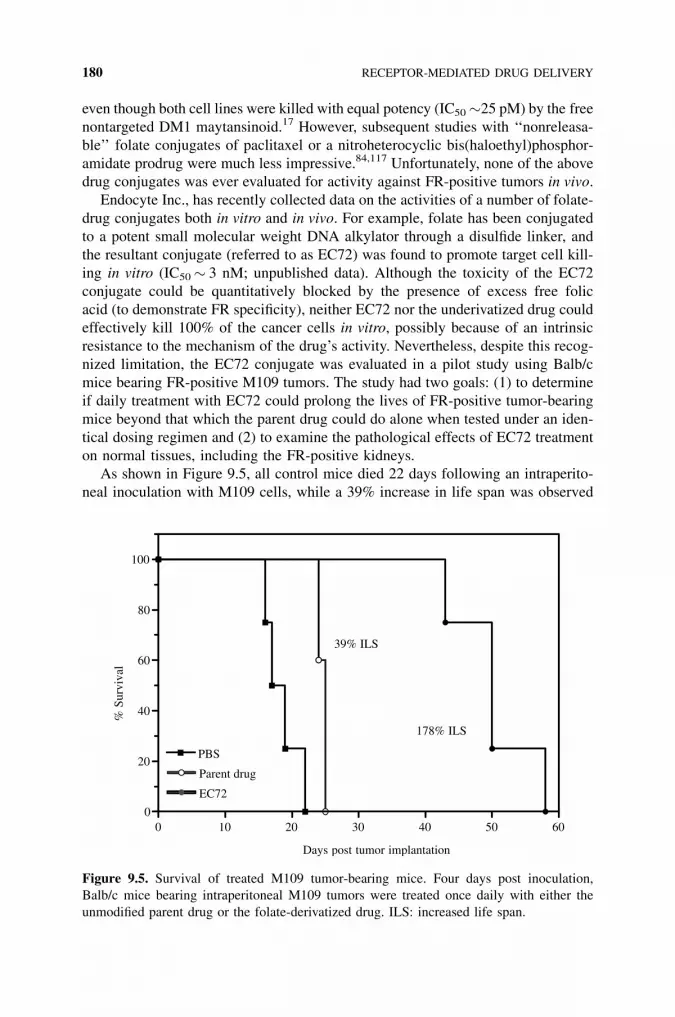
even though both cell lines were killed with equal potency (IC50�25 pM) by the free
nontargeted DM1 maytansinoid.17 However, subsequent studies with ‘‘nonreleasa-
ble’’ folate conjugates of paclitaxel or a nitroheterocyclic bis(haloethyl)phosphor-
amidate prodrug were much less impressive.84,117 Unfortunately, none of the above
drug conjugates was ever evaluated for activity against FR-positive tumors in vivo.
Endocyte Inc., has recently collected data on the activities of a number of folate-
drug conjugates both in vitro and in vivo. For example, folate has been conjugated
to a potent small molecular weight DNA alkylator through a disulfide linker, and
the resultant conjugate (referred to as EC72) was found to promote target cell kill-
ing in vitro (IC50� 3 nM; unpublished data). Although the toxicity of the EC72
conjugate could be quantitatively blocked by the presence of excess free folic
acid (to demonstrate FR specificity), neither EC72 nor the underivatized drug could
effectively kill 100% of the cancer cells in vitro, possibly because of an intrinsic
resistance to the mechanism of the drug’s activity. Nevertheless, despite this recog-
nized limitation, the EC72 conjugate was evaluated in a pilot study using Balb/c
mice bearing FR-positive M109 tumors. The study had two goals: (1) to determine
if daily treatment with EC72 could prolong the lives of FR-positive tumor-bearing
mice beyond that which the parent drug could do alone when tested under an iden-
tical dosing regimen and (2) to examine the pathological effects of EC72 treatment
on normal tissues, including the FR-positive kidneys.
As shown in Figure 9.5, all control mice died 22 days following an intraperito-
neal inoculation with M109 cells, while a 39% increase in life span was observed
0 10 20 30 40 50 600
20
40
60
80
100
PBS
Parent drug
EC72
178% ILS
39% ILS
Days post tumor implantation
% S
urvi
val
Figure 9.5. Survival of treated M109 tumor-bearing mice. Four days post inoculation,
Balb/c mice bearing intraperitoneal M109 tumors were treated once daily with either the
unmodified parent drug or the folate-derivatized drug. ILS: increased life span.
180 RECEPTOR-MEDIATED DRUG DELIVERY
for the animals treated with the unmodified drug. More importantly, animals treated
with the EC72 conjugate lived on average 178% longer.
Following euthanasia, major organs were collected from both the EC72 and par-
ent drug–treated animals, and they were sent to a certified pathologist for examina-
tion. The nontargeted drug-treated animals were found to suffer from massive
myelosuppression (which is a characteristic dose-limiting side effect of the parent
drug’s therapy), and all of the animals in this cohort died from obvious drug-related
side effects. In dramatic contrast, animals treated with EC72 (folate-SS-drug) dis-
played no evidence of myelosuppression or kidney damage. Further, examination of
blood collected from EC72-treated animals indicated normal blood urea nitrogen
and creatinine levels following 30 consecutive daily injections with the conjugate.
We concluded from these results that the use of folate-drug conjugates may be an
effective form of chemotherapy that does not cause injury to normal tissues, includ-
ing the FR-positive kidneys. While the latter conclusion was surprising, these
observations do support the hypothesis that the FRs in the kidney proximal tubules
function primarily to shuttle scavenged folates (or small folate-drug conjugates)
back into systemic circulation rather than to deliver the conjugates into the kidney
cells.69,70
9.5.3.3. Folate-Targeted Immunotherapy
9.5.3.3.1. Intracellular Delivery versus Cell-Surface Loading In contrast to most
hormone receptors, not all FRs endocytose following ligand binding.118 Rather, a
substantial fraction of the occupied FRs remain on the cell surface as a means of
storing folates for use at a later date. Since folate-drug conjugates interact with FR
in the same manner as free folate,49 it was of no surprise to learn that a fraction of
these conjugates also remains extracellularly bound.49,93 This dual destiny of occu-
pied FR obviously allows for two distinct uses of the folate-targeting technology:
(1) delivery of folate-drug conjugates into FR-expressing cells and (2) decoration of
FR-expressing cell surfaces with folate-drug conjugates. This latter application has
led to the advent of folate-targeted immunotherapy,119 as outlined below.
Based on the ability of folate to position an attached drug on the surface of an
FR-expressing cancer cell, it was recognized that a malignant cell could be con-
verted from its normal immunologically invisible state (the condition that permits
its proliferation in vivo) to a state where it is vividly recognized as foreign by the
immune system (and consequently subject to immune-mediated elimination) sim-
ply by targeting a highly immunogenic antigen to its cell surface. This concept is
presented in Figure 9.6. Different from targeted chemotherapy, this treatment
begins with a series of subcutaneous inoculations of a hapten-based vaccine to sti-
mulate production of anti-hapten antibodies in the patient. After induction of an
adequate antibody titer, a folate-hapten conjugate is administered to enable ‘‘mark-
ing’’ of all FR-expressing tumor cells with the hapten. This process rapidly pro-
motes the tumor cell’s opsonization with the previously induced endogenous
anti-hapten antibodies. Mechanistically, the folate-hapten conjugate forms a mole-
cular ‘‘bridge’’ between the tumor cell and the endogenous circulating anti-hapten
FOLATE-MEDIATED DRUG DELIVERY 181

antibody. Ultimately, this marking process enables Fc receptor-bearing effector
cells, such as natural killer (NK) cells and macrophages, to recognize and destroy
the antibody-coated tumor cells.
Although we have found this folate-targeted immunotherapy to be effective in
tumor-bearing rodent models, we have also noted that complete and reproducible
cures are obtained only when the animals are concomitantly dosed with low levels
of the cytokines, interleukin-2 and interferon-a.119 The purpose of adding these
cytokines is to stimulate those immune effector cells (like NK and macrophage
cells) that are responsible for killing opsonized tumor cells. Importantly, the
low cytokine dose levels applied in this technique were found not to produce effec-
tive antitumor responses in the absence of the folate-hapten conjugate. Although the
exact effector mechanisms involved in this therapy have not yet been established,
there is strong evidence to suggest that the cured animals develop independent cel-
lular immunity against the tumor cells and that this cellular immunity prevents the
animals from growing tumors when rechallenged with the same malignant cells,
even without additional treatment.119 These findings also support the notion that
a long-term, perhaps T cell-specific immunity, develops during the hapten-mediated
immunotherapy, and that a shift of the host’s antitumor response from humoral to
cellular immunity occurs. Because this therapy has generated cures in multiple
tumor-bearing animal models, and since the complete therapeutic regimen (vacci-
nation and drug therapy at dose levels approaching 100-fold of the human dose
equivalent) did not produce test article–related toxicities in toxicology studies,
the strategy was approved for clinical trial by the FDA for treatment of advanced
Figure 9.6. Folate-targeted immunotherapy strategy.
182 RECEPTOR-MEDIATED DRUG DELIVERY

ovarian and renal cell cancers beginning in 2003. Results of these clinical studies
should provide much information on the strengths and weaknesses of folate-
targeted cancer therapies in humans.
9.6. CONCLUSIONS
Nature has designed biological membranes to serve as formidable barriers against
the unwanted entry of harmful agents into cells. Life, however, cannot exist without
the cell’s ability to selectively capture and internalize certain biosupportive mole-
cules. Receptor-mediated endocytosis is but one mechanism by which cells retrieve
required molecules from their environment, and it is also a powerful means for deli-
vering normally impermeable molecules into target cells for medicinal purposes.
The list of available ligands for use in this exploitive technique keeps growing,
as does the potential for inventing alternative drug delivery strategies. It is impor-
tant, however, to remember that not all ligand-receptor systems will function in the
same manner. Many variations exist among cell types, including their level of
receptor expression, internalization rate, ligand affinity, intracellular compartmen-
talization, recycling capabilities, etc. Thus, in spite of recent technological
advances, many challenges remain for the future development of receptor-targeted
therapies. The most significant issues will undoubtedly be related to the confirma-
tion of target tissue specificity (i.e., receptor distribution among target and nontar-
get tissues) as well as the design of releasable linkers for certain ligand-linker-drug
conjugates. Receptor-mediated delivery technology is undoubtedly influencing
rational drug design, and it is our hope that the scientific and pharmaceutical com-
munities will continue to invest in this exiting new field of medicine.
ACKNOWLEDGMENT
This work was supported in part by grants from Endocyte, Inc., and the NIH
(CA89581).
REFERENCES
1. McPherson, P. S.; Kay, B. K.; Hussain, N. K. Traffic 2001, 2, 375–384.
2. Mukherjee, S.; Ghosh, R. N.; Maxfield, F. R. Physiol. Rev. 1997, 77, 759–803.
3. Schwartz, A. L. Pediatr. Res. 1995, 38, 835–843.
4. Stahl, P.; Schwartz, A. L. J. Clin. Invest. 1986, 77, 657–662.
5. Smythe, E.; Warren, G. Eur. J. Biochem. 1991, 202, 689–699.
6. Marsh, M.; Helenius, A. J. Mol. Biol. 1980, 142, 439–454.
7. Pastan, I.; Willingham, M. C. The Pathway of Endocytosis; Plenum Press: New York,
1985.
REFERENCES 183

8. Willingham, M. C.; Pastan, I. Cell 1980, 21, 67–77.
9. Lai, W. H.; Cameron, P. H.; Wada, I.; Doherty, J. J., 2nd; Kay, D. G.; Posner, B. I.;
Bergeron, J. J. J. Cell Biol. 1989, 109, 2741–2749.
10. Hanvey, J. C.; Peffer, N. J.; Bisi, J. E.; Thomson, S. A.; Cadilla, R.; Josey, J. A.; Ricca, D.
J.; Hassman, C. F.; Bonham, M. A.; Au, K. G.; Carter, S. G.; Bruckenstein, D. A.; Boyd,
A. L.; Noble, S. A.; Babiss, L. E. Science 1992, 258, 1481–1485.
11. Terada, K.; Manchikalapudi, P.; Noiva, R.; Jauregui, H. O.; Stockert, R. J.; Schilsky, M.
L. J. Biol. Chem. 1995, 270, 20410–20416.
12. Scheiber, B.; Goldenberg, H. Arch. Biochem. Biophys. 1993, 305, 225–230.
13. Liu, Y.; Peterson, D. A.; Kimura, H.; Schubert, D. J. Neurochem. 1997, 69, 581–593.
14. McIntyre, G. D.; Scott, C. F.; Ritz, J.; Blattler,W. A.; Lambert, J. M.Bioconjugate Chem.
1994, 5, 88–97.
15. Leamon, C. P.; Pastan, I.; Low, P. S. J. Biol. Chem. 1993, 268, 24847–24854.
16. Leamon, C. P.; DePrince, R. B.; Hendren, R. W. J. Drug Targeting 1999, 7, 157–169.
17. Ladino, C. A.; Chari, R. V. J.; Bourret, L. A.; Kedersha, N. L.; Goldmacher, V. S. Int. J.
Cancer 1997, 73, 859–864.
18. Liu, C.; Tadayoni, B. M.; Bourret, L. A.; Mattocks, K. M.; Derr, S. M.; Widdison, W. C.;
Kedersha, N. L.; Ariniello, P. D.; Goldmacher, V. S.; Lambert, J. M.; Blattler, W. A.;
Chari, R. V. Proc. Natl. Acad. Sci. USA 1996, 93, 8618–8623.
19. Lee, R. J.; Wang, S.; Low, P. S. Biochim. Biophys. Acta 1996, 1312, 237–242.
20. Neville, D. M.; Srinivasachar, K.; Stone, R.; Scharff, J. J. Biol. Chem. 1989, 264, 14653–
14661.
21. Hamann, P. R.; Hinman, L. M.; Beyer, C. F.; Lindh, D.; Upeslacis, J.; Flowers, D. A.;
Bernstein, I. Bioconjug. Chem. 2002, 13, 40–46.
22. Hamann, P. R.; Hinman, L. M.; Hollander, I.; Beyer, C. F.; Lindh, D.; Holcomb, R.;
Hallett, W.; Tsou, H. R.; Upeslacis, J.; Shochat, D.; Mountain, A.; Flowers, D. A.;
Bernstein, I. Bioconjug. Chem. 2002, 13, 47–58.
23. Rihova, B.; Srogl, J.; Jelinkova, M.; Hovorka, O.; Buresova, M.; Subr, V.; Ulbrich, K.
Ann. NY Acad. Sci. 1997, 831, 57–71.
24. Seymour, L. W.; Ulbrich, K.; Wedge, S. R.; Hume, I. C.; Strohalm, J.; Duncan, R. Br. J.
Cancer 1991, 63, 859–866.
25. Poznansky, M. J.; Singh, R.; Singh, B. Science 1984, 223, 1304–1306.
26. Poznansky, M. J.; Hutchison, S. K.; Davis, P. J. Faseb J. 1989, 3, 152–156.
27. Shaw, J. P.; Akiyoshi, D. E.; Arrigo, D. A.; Rhoad, A. E.; Sullivan, B.; Thomas, J.;
Genbauffe, F. S.; Bacha, P.; Nichols, J. C. J. Biol. Chem. 1991, 266, 21118–21124.
28. Cawley, D. B.; Herschman, H. R.; Gilliland, D. G.; Collier, R. J. Cell 1980, 22, 563–570.
29. Qian, Z. M.; Li, H.; Sun, H.; Ho, K. Pharmacol. Rev. 2002, 54, 561–587.
30. Bacha, P.; Murphy, J. R.; Reichlin, S. J. Biol. Chem. 1983, 258, 1565–1570.
31. Sakai, A.; Sakakibara, R.; Ishiguro, M. J. Biochem. 1989, 105, 275–280.
32. Sakai, A.; Sakakibara, R.; Ohwaki, K.; Ishiguro, M. Chem. Pharm. Bull. (Tokyo) 1991,
39, 2984–2989.
33. Singh, V.; Sairam, M. R.; Bhargavi, G. N., Akhras, R. G. J. Biol. Chem. 1989, 264, 3089–
3095.
34. Singh, V.; Curtiss, R., 3rd. Mol. Cell Biochem. 1994, 130, 91–101.
184 RECEPTOR-MEDIATED DRUG DELIVERY

35. Bacha, P.; Williams, D. P.; Waters, C.; Williams, J. M.; Murphy, J. R.; Strom, T. B. J.
Exp. Med. 1988, 167, 612–622.
36. Shapiro, M. E.; Kirkman, R. L.; Kelley, V. R.; Bacha, P.; Nichols, J. C.; Strom, T. B.
Targeted Diagn. Ther. 1992, 7, 383–393.
37. Karson, E. M.; Neufeld, E. F.; Sando, G. N. Biochemistry 1980, 19, 3856–3860.
38. Sato, Y.; Beutler, E. J. Clin. Invest. 1993, 91, 1909–1917.
39. Wu, J.; Nantz, M. H.; Zern, M. A. Front. Biosci. 2002, 7, d717–25.
40. Pennell, C. A.; Erickson, H. A. Immunol. Res. 2002, 25, 177–191.
41. Garnett, M. C. Adv. Drug Deliv. Rev. 2001, 53, 171–216.
42. Collins, D. A.; Hogenkamp, H. P.; Gebhard, M.W.Mayo Clin. Proc. 1999, 74, 687–691.
43. Swaan, P. W. Pharmaceutical Res. 1998, 15, 826–834.
44. Bauer, J. A.; Morrison, B. H.; Grane, R. W.; Jacobs, B. S.; Dabney, S.; Gamero, A. M.;
Carnevale, K. A.; Smith, D. J.; Drazba, J.; Seetharam, B.; Lindner, D. J. J. Natl. Cancer
Inst. 2002, 94, 1010–1019.
45. Russell-Jones, G. J.; Alpers, D. H. Pharm. Biotechnol. 1999, 12, 493–520.
46. Antony, A. C. Blood 1992, 79, 2807–2820.
47. Kamen, B. A.; Capdevila, A. Proc. Natl. Acad. Sci. USA 1986, 83, 5983–5987.
48. Antony, A. C. Annu. Rev. Nutr. 1996, 16, 501–521.
49. Leamon, C. P.; Low, P. S. Proc. Natl. Acad. Sci. USA 1991, 88, 5572–5576.
50. Coney, L. R.; A., T.; Carayannopoulos, L.; Frasca, V.; Kamen, B. A.; Colnaghi, M. I.;
Zurawski, V. R. J. Cancer Res. 1991, 51, 6125–6132.
51. Weitman, S. D.; Lark, R. H.; Coney, L. R.; Fort, D. W.; Frasca, V.; Zurawski, V. R.;
Kamen, B. A. Cancer Res. 1992, 52, 3396–3401.
52. Ross, J. F.; Chaudhuri, P. K.; Ratnam, M. Cancer 1994, 73, 2432–2443.
53. Weitman, S. D.; Frazier, K. M.; Kamen, B. A. J. Neurol. Oncol. 1994, 21, 107.
54. Boerman, O. C.; van Niekerk, C. C.; Makkink, K.; Hanselaar, T. G.; Kenemans, P.; Poels,
L. G. Int. J. Gynecol. Pathol. 1991, 10, 15–25.
55. Garin-Chesa, P.; Campbell, I.; Saigo, P. E.; Lewis, J. L.; Old, L. J.; Rettig, W. J. Am. J.
Pathol. 1993, 142, 557–567.
56. Mattes, M. J.; Major, P. P.; Goldenberg, D. M.; Dion, A. S.; Hutter, R. V. P.; Klein, K. M.
Cancer Res. Suppl. 1990, 50, 880S.
57. Weitman, S. D.; Weiberg, A. G.; Coney, L. R.; Zurawski, V. R.; Jennings, D. S.; Kamen,
B. A. Cancer Res. 1992, 52, 6708–6711.
58. Toffoli, G.; Cernigoi, C.; Russo, A.; Gallo, A.; Bagnoli, M.; Boiocchi, M. Int. J. Cancer
1997, 74, 193–198.
59. Holm, J.; Hansen, S. I.; Hoier-Madsen, M.; Bostad, L. Biochem. J. 1991, 280, 267–271.
60. Berenson, J. R.; Yang, J.; Mickel, R. A. Oncogene 1989, 4, 1111–1116.
61. Orr, R. B.; Kriesler, A. R.; Kamen, B. A. J. Natl. Cancer Inst. 1995, 87, 299–303.
62. Li, P. Y.; Vecchio, S. D.; Fonti, R.; Carriero, M. V.; Potena, M. I.; Botti, G.; Miotti, S.;
Lastoria, S.; Menard, S.; Colnaghi, M. I.; Salvatore, M. J. Nucl. Med. 1996, 37, 665–
672.
63. Veggian, R.; Fasolato, S.; Menard, S.; Minucci, D.; Pizzetti, P.; Regazzoni, M.;
Tagliabue, E.; Colnaghi, M. I. Tumori 1989, 75, 510–513.
REFERENCES 185

64. Franklin, W. A.; Waintrub, M.; Edwards, D.; Christensen, K.; Prendegrast, P.; Woods, J.;
Bunn, P. A.; Kolhouse, J. F. Int. J. Cancer 1994, Suppl. 8, 89–95.
65. Patrick, T. A.; Kranz, D. M.; van Dyke, T. A.; Roy, E. J. J. Neurooncol. 1997, 32, 111–
123.
66. Kennedy, M. D.; Jallad, K. N.; Low, P. S.; Ben-Amotz, D. Pharmaceut. Res. 2003,
67. Zimmerman, J. Gastroenterology 1990, 99, 964–972.
68. Morshed, K. M.; Ross, D. M.; McMartin, K. E. J. Nutr. 1997, 127, 1137–1147.
69. Birn, H.; Selhub, J.; Christensen, E. I. Am. J. Physiol. 1993, 264, C302–C310.
70. Birn, H.; Nielsen, S.; Christensen, E. I. Am. J. Physiol. 1997, 272, F70–F78.
71. Turk,M. J.; Breur, G. J.; Widmer,W. R.; Paulos, C.M.; Xu, L. C.; Grote, L. A.; Low, P. S.
Arthritis Rheum. 2002, 46, 1947–1955.
72. Nakashima-Matsushita, N.; Homma, T.; Yu, S.; Matsuda, T.; Sunahara, N.; Nakamura,
T.; Tsukano, M.; Ratnam, M.; Matsuyama, T. Arthritis Rheum. 1999, 42, 1609–1616.
73. Mathias, C. J.; Wang, S.; Lee, R. J.; Waters, D. J.; Low, P. S.; Green, M. A. J. Nucl. Med.
1996, 37, 1003–1008.
74. Mathias, C. J.;Wang, S.;Waters, D. J.; Turek, J. J.; Low, P. S.; Green, M. A. J. Nucl. Med.
1998, 39, 1579–1585.
75. Leamon, C. P.; Low, P. S. Drug Discov. Today 2001, 6, 44–51.
76. Campbell, I. G.; Jones, T. A.; Foulkes, W. D.; Trowsdale, J.Cancer Res. 1991, 51, 5329–
5338.
77. Wang, S.; Lee, R. J.; Mathias, C. J.; Green, M. A.; Low, P. S. Bioconjugate Chem. 1996,
7, 56–62.
78. Ilgan, S.; Yang, D. J.; Higuchi, T.; Zareneyrizi, F.; Bayham, H.; Yu, D.; Kim, E. E.;
Podoloff, D. A. Cancer Biother. Radiopharm. 1998, 13, 427–435.
79. Guo, W.; Hinkle, G. H.; Lee, R. J. J. Nucl. Med. 1999, 40, 1563–1569.
80. Linder, K. E.; Wedeking, P.; Ramalingam, K.; Nunn, A. D.; Tweedle, M. F. Soc. Nucl.
Med. Proc. 47th Annual Meeting 2000, 41, 119P.
81. Leamon, C. P.; Parker, M. A.; Vlahov, I. R.; Xu, L. C.; Reddy, J. A.; Vetzel, M.; Douglas,
N. Bioconjugate Chem. 2002,
82. Wang, S.; Luo, J.; Lantrip, D. A.;Waters, D. J.; Mathias, C. J.; Green, M. A.; Fuchs, P. L.;
Low, P. S. Bioconjugate Chem. 1997, 8, 673–679.
83. Konda, S. D.; Aref, M.; Brechbiel, M.; Wiener, E. C. Invest. Radiol. 2000, 35, 50–57.
84. Lee, J. W.; Lu, J. Y.; Low, P. S.; Fuchs, P. L. Bioorg. Med. Chem. 2002, 10, 2397–2414.
85. Citro, G.; Szczylik, C.; Ginobbi, P.; Zupi, G.; Calabretta, B. Br. J. Cancer 1994, 69, 463–
467.
86. Li, S.; Huang, L. J. Liposome Res. 1997, 7, 63–75.
87. Li, S.; Huang, L. J. Liposome Res. 1998, 8, 239–250.
88. Li, S.; Deshmukh, H. M.; Huang, L. Pharm. Res. 1998, 15, 1540–1545.
89. Leopold, L. H.; Shore, S. K.; Newkirk, T. A.; Reddy, R. M.; Reddy, E. P. Blood 1995, 85,
2162–2170.
90. Ward, C. M.; Acheson, N.; Seymour, L. M. J. Drug Target. 2000, 8, 119–123.
91. Lu, J. Y.; Lowe, D. A.; Kennedy, M. D.; Low, P. S. J. Drug. Target. 1999, 7, 43–53.
92. Leamon, C. P.; Low, P. S. J. Biol. Chem. 1992, 267, 24966–24971.
186 RECEPTOR-MEDIATED DRUG DELIVERY

93. Leamon, C. P.; Low, P. S. Biochem. J. 1993, 291, 855–860.
94. Leamon, C. P.; Low, P. S. J. Drug Target. 1994, 2, 101–112.
95. Kranz, D. M.; Patrick, T. A.; Brigle, K. E.; Spinella, M. J.; Roy, E. J. Proc. Natl. Acad.
Sci. USA 1995, 92, 9057–9061.
96. Cho, B. K.; Roy, E. J.; Patrick, T. A.; Kranz, D. M. Bioconjugate Chem. 1997, 8, 338–
346.
97. Kranz, D. M.; Manning, T. C.; Rund, L. A.; Cho, B. K.; Gruber, M. M.; Roy, E. J. J.
Controlled Release 1998, 53, 77–84.
98. Rund, L. A.; Cho, B. K.; Manning, T. C.; Holler, P. D.; Roy, E. J.; Kranz, D. M. Int. J.
Cancer 1999, 83, 141–149.
99. Lee, R. J.; Low, P. S. J. Biol. Chem. 1994, 269, 3198–3204.
100. Lee, R. J.; Low, P. S. Biochim. Biophys. Acta 1995, 1233, 134–144.
101. Vogel, K.; Wang, S.; Lee, R. J.; Chmielewski, J.; Low, P. S. J. Am. Chem. Soc. 1996, 118,
1581–1586.
102. Rui, Y.; Wang, S.; Low, P. S.; Thompson, D. H. J. Am. Chem. Soc. 1998, 120, 11213–
11218.
103. Gabizon, A.; Horowitz, A. T.; Goren, D.; Tzemach, D.; Mandelbaum-Shavit, F.; Qazen,
M. M.; Zalipsky, S. Bioconjugate Chem. 1999, 10, 289–298.
104. Zhang, Y.; Kohler, N.; Zhang, M. Biomaterials 2002, 23, 1553–1561.
105. Oyewumi, M. O.; Mumper, R. J. Bioconjugate Chem. 2002, 13, 1328–1335.
106. Oyewumi, M. O.; Mumper, R. J. Int. J. Pharm. 2003, 251, 85–97.
107. Gottschalk, S.; Cristiano, R. J.; Smith, L. C.; Woo, S. L. C.Gene Ther. 1994, 1, 185–191.
108. Mislick, K. A.; Baldeschwieler, J. D.; Kayyem, J. F.; Meade, T. J. Bioconjugate Chem.
1995, 6, 512–515.
109. Douglas, J. T.; Rogers, B. E.; Rosenfeld, M. E.; Michael, S. I.; Feng, M.; Curiel, D. T.
Nature Biotech. 1996, 14, 1574–1578.
110. Leamon, C. P.; Weigl, D.; Hendren, R. W. Bioconjugate Chem. 1999, 10, 947–957.
111. Guo, W.; Lee, R. J. PharmSci. 1999, 1, article 19.
112. Reddy, J. A.; Dean, D.; Kennedy, M. D.; Low, P. S. J. Pharm. Sci. 1999, 88, 1112–1118.
113. Reddy, J. A.; Low, P. S. J. Controlled Release 2000, 64, 27–37.
114. Reddy, J. A.; Abburi, C.; Hofland, H.; Howard, S. J.; Vlahov, I.; Wils, P.; Leamon, C. P.
Gene Ther. 2002, 9, 1542–1550.
115. Hofland, H. E.; Masson, C.; Iginla, S.; Osetinsky, I.; Reddy, J. A.; Leamon, C. P.;
Scherman, D.; Bessodes, M.; Wils, P. Mol. Ther. 2002, 5, 739–744.
116. Liu, S.; Edwards, D. S.; Barrett, J. A. Bioconjugate Chem. 1997, 8, 621–636.
117. Steinberg, G.; Borch, R. F. J. Med. Chem. 2001, 44, 69–73.
118. Kamen, B. A.; Wang, M. T.; Streckfuss, A. J.; Peryea, X.; Anderson, R. G.W. JBC 1988,
263, 13602–13609.
119. Lu, Y.; Low, P. S. Cancer Immunol. Immunother. 2002, 51, 153–162.
REFERENCES 187

10ORAL PROTEIN AND PEPTIDEDRUG DELIVERY
RICK SOLTERO
Pharma Directions, Inc.
10.1. Introduction
10.2. Overcoming physiological barriers with formulation approaches
10.2.1. Enzyme inhibitors
10.2.2. Absorption enhancers
10.2.2.1. Nanoparticles
10.2.2.2. Emulsions
10.2.2.3. Micelle formulations
10.2.2.4. Liposomes
10.2.3. Chemical modifications
10.3. Improving the oral activity of calcitonin and insulin through chemical modification
10.4. Conclusions
References
10.1. INTRODUCTION
Endogenous peptides play a pivotal role in almost all regulatory processes of the
body functions and act with high specificity and potency. Their vast therapeutic
potential has prompted biomedical scientists to greatly expand research into the
identities of biologically active polypeptides and proteins and their activities.
Drug Delivery: Principles and Applications Edited by Binghe Wang, Teruna Siahaan,
and Richard Soltero
ISBN 0-471-47489-4 # 2005 John Wiley & Sons, Inc.
189

Concurrently, pharmaceutical companies around the world have endeavored to
develop processes for producing therapeutically active entities at commercial
scales.
As a result of this research, the availability of biotechnology products to develop
is no longer an issue. At the present time, over 350 peptide and protein drugs are in
various stages of clinical development. Many other candidates will likely be iden-
tified as genomic studies successfully translate genetic data into knowledge about
proteins and their functions.
The broad availability of structurally diverse peptides possessing a spectrum of
pharmacological effects has not been matched, however, by their clinical develop-
ment, mostly because of the poor delivery properties of peptide- and protein-based
products. Today, the most common means for administering these protein drugs
remains injection (i.e., intravenous, intramuscular, or subcutaneous administration).
Patient compliance with drug administration regimens by any of these parenteral
routes, is generally poor and severely restricts the therapeutic value of the drug,
particularly for diseases such as diabetes.
Delivering therapeutically active proteins and peptides by any route other than
the invasive injection methods has been a challenge and a goal for many decades.1
Among the alternate routes that have been tried with varying degrees of success are
the oral, buccal,2 intranasal,3 pulmonary,4 transdermal,5 ocular,6 and rectal7
approaches. The many benefits of oral administration of therapeutic agents make
oral polypeptide delivery stand out from among these drug delivery alternatives.
In addition to high levels of patient acceptance and long-term compliance, oral
delivery also benefits the patient through avoidance of the pain and discomfort asso-
ciated with injections; greater convenience; implementation of a more socially
compatible dosing regimen; and the elimination of concerns about needle-related
infections. In addition, a growing body of data suggests that for certain polypep-
tides, such as insulin, the oral delivery route is more physiological.8,9 Thus, oral
delivery would be an ideal route if appropriate oral dosage forms of therapeutic
peptides and proteins were available.
The oral route of administration of these therapeutic agents, however, is among
the most problematic delivery regimens. Drug delivery via the gastrointestinal (GI)
tract requires relatively lengthy exposure to a multifaceted system that is designed
to degrade nutrients and dietary materials into small molecules and to prevent the
indiscriminate passage of macromolecules, as well as other large entities such as
microbes or foreign antigens that may present dangers to the host. The various bar-
riers, including physiological, chemical, and biochemical ones, for oral drug deliv-
ery are discussed in detail in Chapter 2 and therefore will not be presented in this
chapter.
In spite of the obstacles to oral delivery, substantial evidence suggests that phar-
maceutical polypeptides are absorbed through the intestinal mucosa, although in
minute amounts.10 Small amounts of polypeptide drugs can be absorbed by the
action of specific peptide transporters in the intestinal mucosa cells.11 This suggests
that properly formulated proteins or peptide drugs may be administered by the oral
route with retention of sufficient biological activity for their therapeutic use.
190 ORAL PROTEIN AND PEPTIDE DRUG DELIVERY

Designing and formulating a polypeptide drug for delivery through the GI tract
requires a multitude of strategies.12,13 The dosage form must initially stabilize the
drug while making it easy to take orally.14 It must then protect the polypeptide from
the extreme acidity and action of pepsin in the stomach.15 When the drug reaches
the intestine, the formulation must incorporate some means for limiting drug degra-
dation by the plethora of enzymes that are present in the intestinal lumen.16 In addi-
tion, the polypeptide and/or its formulations must facilitate both aqueous solubility
at near-neutral pH and lipid layer penetration in order for the protein to traverse the
intestinal membrane and then the basal membrane for entry into the bloodstream.17
To accomplish this, formulation excipients that promote absorption may be
required.18,19 Finally, when the modified polypeptide enters the systemic circula-
tion, the structural modifications may add to the functionality of the drug, e.g.,
by extending its half-life in the circulation. However, any structural changes that
may have been employed to enhance oral bioavailability must not interfere with
receptor binding and uptake at the site of biological activity.
In this chapter, we summarize the general approaches that have been used to suc-
cessfully achieve the formulation goals for oral delivery: minimize enzymatic
degradation; enhance intestinal absorption; maximize blood level reproducibility;
deliver drug through the gut wall; and produce a palatable and acceptable dosage
form. Then insulin will be used as an example to show how oral bioavailability has
been achieved through chemical modification.
10.2. OVERCOMING PHYSIOLOGICAL BARRIERS WITHFORMULATION APPROACHES
Oral delivery of polypeptides has been an ongoing challenge. Nearly every oral
dosage form used for delivery of conventional small-molecule drugs has been
used to explore oral delivery of polypeptides. Except for cases where the polypep-
tide has been chemically modified or where a proprietary absorption enhancer has
been used, the results have been disappointing. There have been only a handful of
human clinical trials where there has been adequate bioavailability and pharmaco-
kinetics to suggest that commercializing an orally delivered polypeptide is feasible.
Some of the approaches used to date are described in general terms in this section.
For more in-depth discussions, readers should consult the numerous reviews that
cover specific areas of research.20–26
10.2.1. Enzyme Inhibitors
Researchers have evaluated the use of protease inhibitors with the aim of slowing
the rate of degradation of proteins and peptides in the GI tract. The hypothesis was
that a slow rate of degradation would increase the amount of protein and peptide
drug available for absorption.
For example, enzyme degradation of insulin is known to be mediated by the serine
proteases trypsin, a-chymotrypsin, and thiol metalloproteinase insulin-degrading
OVERCOMING PHYSIOLOGICAL BARRIERS WITH FORMULATION APPROACHES 191
enzymes. The stability of insulin has been evaluated in the presence of excipients
that inhibit these enzymes. Representative inhibitors of trypsin and a-chymotrypsin
include pancreatic inhibitor and soybean trypsin inhibitor,27 FK-448,28 camostat
mesylate,29 and aprotinin.30 Inhibitors of insulin-degrading enzyme include 1,10
phenanthroline, p-choromeribenzoate,31 and bacitracin.32 Ziv et al.33 reported the
use of a combination of an enhancer, sodium cholate, and a protease inhibitor to
achieve a 10% increase in rat intestinal insulin absorption.
Another approach to enzyme inhibition is to manipulate the pH to inactivate
local digestive enzymes. Lee et al. conducted studies demonstrating that oral
absorption properties of salmon calcitonin can be modulated by changing intestinal
pH. Reducing the intestinal pH in the GI tract, increased absorption of the
intact peptide.34 A sufficient amount of a pH-lowering buffer that lowers local
intestinal pH to values below 4.5 can deactivate trypsin, chymotrypsin, and elastase.
10.2.2. Absorption Enhancers
Permeation enhancers improve the absorption of protein and peptides by increasing
their paracellular and transcellular transports. An increase in paracellular transport
is mediated by modulating the tight junctions of the cells, and an increase in trans-
cellular transport is associated with an increase in the fluidity of the cell membrane.
Chapter 2 describes in depth the various paracellular and transcellular transport
pathways, which will not be discussed here.
Paracellular permeation enhancers include calcium chelators, bile salts, and fatty
acids. Calcium chelators, such as ethylendiaminetetraacetic acid (EDTA),5 act by
inducing calcium depletion, thereby creating global changes in the cells, including
disruption of actin filaments, disruption of adherent junctions, and diminished cell
adhesion.35 Zonula occludens toxin (ZOT) acts specifically on the actin filaments of
tight junctions,36 and chitosan triggers opening of the tight junction between the
cells.37
Transcellular permeation enhancers include surfactants, medium chain fatty
acids, nonionic surfactants,38 sodium cholate and other bile salts,39 and many other
surfactants.
10.2.2.1. Nanoparticles Nanoparticles have been studied in recent decades as
particulate carriers to deliver the protein and peptides drugs orally. This approach
is supported by the literature, which states that particles in the nanosize range are
absorbed intact by the intestinal epithelium, especially Peyer’s patches, and
travel to sites such as the liver, the spleen, and other tissues.40–42 The proteins
and peptides encapsulated in the nanoparticles are less sensitive to enzyme
degradation through their association with polymers. The extended release of pro-
tein and peptide drug from the particles could have pharmacological and clinical
significance.
Many researches have demonstrated that protein and peptide encapsulated in
the nanoparticles have better absorption through the GI tract compared to their
native counterpart.43 The factors affecting uptake include the particle size of the
192 ORAL PROTEIN AND PEPTIDE DRUG DELIVERY

particulate, the surface charge of the particles, the influence of surface ligands, and
the dynamic nature of particle interactions in the gut. In one example, the interac-
tion of nanoparticles consisting of hydrophobic polystyrene, bioadhesive chitosan,
and (PLA-PEG) with two human intestinal cell lines was investigated and com-
pared to the in vivo uptake in rats. After intraduodenal administration of chitosan
nanoparticles in rats, particles were detected in both epithelial cells and Peyer’s
patches. Chitosan nanoparticle seemed to be taken up and transported by adsorptive
transcytosis, while polystyrene nanoparticles uptake was probably mediated by
nonadsorptive transcytosis.44 In another example, insulin was encapsulated in nano-
sphere formulations using phase inversion nanoencapsulation. The encapsulated
insulin was released from the nanospheres over a period of approximately 6 hours,
was shown to be active orally, and had 11.4% of the efficacy of intraperitoneally
delivered insulin.45
One problem in using nanoparticles for peptide and protein delivery is the erratic
nature of nanoparticle absorption. For example, the proportion of intact particles
reaching the systemic circulation was estimated to be generally below 5%.46 Con-
sidering the generally low encapsulation efficiency of the protein in the particulates,
the overall oral bioavailability of proteins and peptides is not significant.
10.2.2.2. Emulsions Liquid emulsions have been used to deliver proteins and
peptide orally. Emulsions are thought to protect the drug from chemical and enzy-
matic breakdown in the intestinal lumen. Drug absorption enhancement is depen-
dent on the type of emulsifying agent, particle size of the dispersed phase, pH,
solubility of drug, type of lipid phase used, etc. Water-in-oil microemulsions
have been shown to enhance oral bioavailability of proteins and peptides.47,48
The lipid phase of microemulsions composed of medium chain fatty acid triglycer-
ides increased the bioavailability of muramyl dipeptide analog.49
Many successful formulations are based on emulsion techniques. In these formu-
lations, oil and water are mixed in such a way that small, uniformly shaped oil dro-
plets are dispersed in the water phase (oil in water) or water droplets are dispersed
in a continuous oil phase (water in oil). An emulsion appears as a cloudy suspen-
sion. When an oil-in-water emulsion has oil droplets so small as to produce a clear
solution, then formulation is called a ‘‘microemulsion.’’ The oil phase of the emul-
sion can provide lipophilic proteins with some protection from enzymatic digestion
while the product is in the intestinal tract. Water-in-oil microemulsion formulations
have been developed for oral insulin delivery.50
10.2.2.3. Micelle Formulations A micelle system can be either water-based or
oil-based. The use of a micelle formulation for poorly water-soluble drugs for sys-
temic delivery has been well recognized. In recent years, the effective development
of self-emulsifying microemulsions or mixed micelle-based lipid formulations pro-
ducts, such as Sandimmun Neoral (cyclosporin), Norvir (ritonavir), and Fortovase
(saquinavir), has substantially increased interest in the application of lipid-based
micelle formulation to improve oral delivery of poorly water-soluble drugs as
well as protein and peptide drugs.51
OVERCOMING PHYSIOLOGICAL BARRIERS WITH FORMULATION APPROACHES 193

P. Modi patented a mixed micellar for administering insulin to the buccal
mucosa using a metered dose inhaler.52 The formulation includes a micellar protei-
nic pharmaceutical agent, lauryl sulphate, salicylate, edetate, and at least one
absorption-enhancing compound such as lecithin, hyaluronic acid, glycolic acid,
oleic acid, linolenic acid, or glycerin.
10.2.2.4. Liposomes Liposomes have also been studied as a way to deliver pep-
tides and proteins orally. Chapter 19 discusses in detail the use of liposomes as drug
delivery vehicles. Therefore, only issues relevant to peptide and protein drug deliv-
ery are discussed here. Liposomes are prone to the combined degrading effects of
the acidic pH of the stomach, bile salts, and pancreatic lipase upon oral administra-
tion. Compared to the parenteral route, there have been fewer attempts to develop
oral formulations to deliver the protein and peptides using a liposome system. In
cases where the encapsulated agents within liposomes have increased bioavailabil-
ity, it is not clear whether the liposome was absorbed intact or if the lipid caused
permeation of the released agent at the site of absorption.53 There are several
reports on the intact liposomes uptake by cells in in vitro and in situ experi-
ments;54–56 the results are, however, not convincing for the oral delivery of proteins
with a liposome system. Attempts have been made to improve the stability of lipo-
somes either by incorporating polymers at the liposome surface57,58 or by using
GI-resistant lipids.59
10.2.3. Chemical Modifications
Chemically modified proteins and peptides offer some significant advantages over
native proteins. Proteins are inherently unstable to digestive enzymes; however,
chemical modifications can be made that inhibit enzyme attack. Such modifications
include prodrug approaches60 and permanent modifications.61 Prodrugs, including
peptide prodrug derivatization, are covered in Chapter 8 in detail and therefore will
not be discussed in this chapter. Permanent modifications, if conducted in the
appropriate functional group, may increase the stability and oral bioavailability
of a peptide or protein without compromising its biological activity. Various perma-
nent modification approaches have been studied, especially with proteins. These
include glycosylation (reference), pegylation (reference), cross-linking (reference),
and other polymer conjugation. So far, two of the most successful examples in pro-
tein drug delivery using chemical modification are calcitonin and insulin.
10.3. IMPROVING THE ORAL ACTIVITY OF CALCITONINAND INSULIN THROUGH CHEMICAL MODIFICATION
Calcitonin is a 32-amino acid peptide hormone that participates in calcium and
phosphorus metabolism. In mammals, the major source of calcitonin is from the
parafollicular or C cells in the thyroid gland. Calcitonin contains a single disulfide
bond, which causes the amino terminus to assume the shape of a ring. It is used to
194 ORAL PROTEIN AND PEPTIDE DRUG DELIVERY
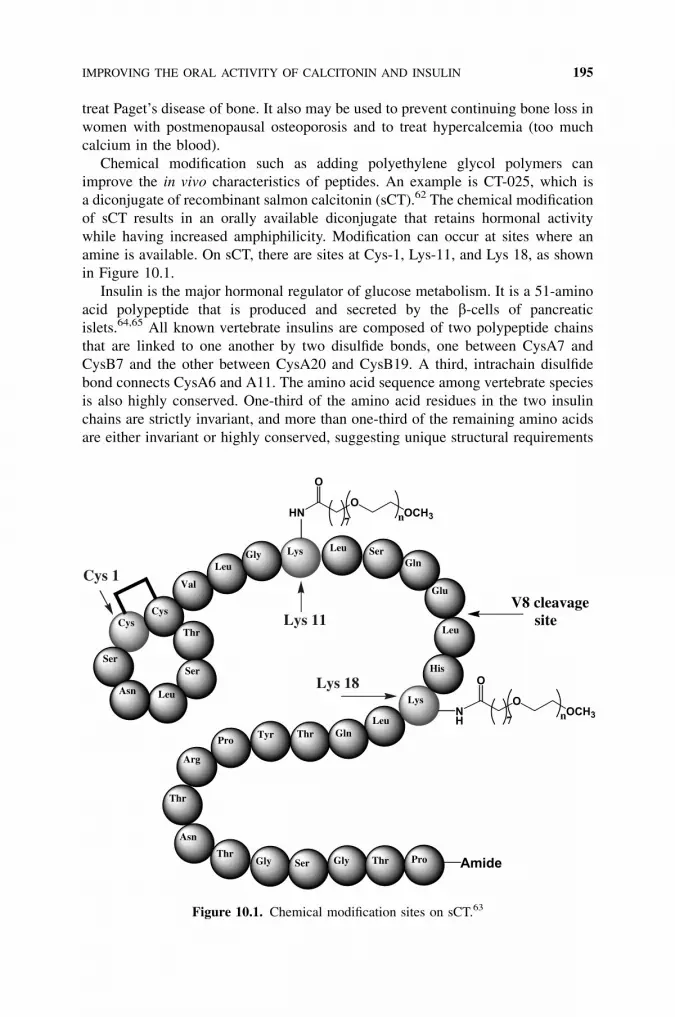
treat Paget’s disease of bone. It also may be used to prevent continuing bone loss in
women with postmenopausal osteoporosis and to treat hypercalcemia (too much
calcium in the blood).
Chemical modification such as adding polyethylene glycol polymers can
improve the in vivo characteristics of peptides. An example is CT-025, which is
a diconjugate of recombinant salmon calcitonin (sCT).62 The chemical modification
of sCT results in an orally available diconjugate that retains hormonal activity
while having increased amphiphilicity. Modification can occur at sites where an
amine is available. On sCT, there are sites at Cys-1, Lys-11, and Lys 18, as shown
in Figure 10.1.
Insulin is the major hormonal regulator of glucose metabolism. It is a 51-amino
acid polypeptide that is produced and secreted by the b-cells of pancreatic
islets.64,65 All known vertebrate insulins are composed of two polypeptide chains
that are linked to one another by two disulfide bonds, one between CysA7 and
CysB7 and the other between CysA20 and CysB19. A third, intrachain disulfide
bond connects CysA6 and A11. The amino acid sequence among vertebrate species
is also highly conserved. One-third of the amino acid residues in the two insulin
chains are strictly invariant, and more than one-third of the remaining amino acids
are either invariant or highly conserved, suggesting unique structural requirements
CysCys
Ser
Asn Leu
Ser
Thr
Val
LeuGly Lys Leu Ser
Gln
Glu
Leu
His
Leu
Lys
GlnThrTyrPro
Thr
Arg
Asn
ThrGly Ser Gly Thr Pro
Lys 11
Lys 18
Cys 1
Amide
V8 cleavage site
HNO
OCH3
O
7 n
OOCH3
O
7 nNH
Figure 10.1. Chemical modification sites on sCT.63
IMPROVING THE ORAL ACTIVITY OF CALCITONIN AND INSULIN 195

for receptor recognition and biological activity. Moreover, there is evidence that
receptor binding is associated with a structural change that must be accommodated
by insulin analogs if biological activity is to be preserved.66
For over 70 years, approved insulin products have been given to diabetic patients
exclusively by parenteral administration. Due to the challenges and obstacles asso-
ciated with oral administration of peptide drugs, no oral dosage form for insulin has
been marketed to date.67 In general, studies of insulin’s oral bioavailability have
shown uptakes that range from near zero to a small percentage of the administered
dose.68–75
As part of continuing efforts to design and develop oral delivery dosage forms
for peptide drugs, researchers have extensively investigated the effects of structural
manipulations and formulation changes on the oral availability of insulin, These
efforts have resulted in the preparation of libraries of amphiphilic oligomers that,
when conjugated in certain patterns and number to the drugs, may be used to
enhance their pharmacological performance. Moreover, when the drug-oligomer
conjugates are used in combination with common formulation strategies, oral bioa-
vailability can be dramatically increased, especially relative to that of the unconju-
gated, native drug.
In previous reports,76–78 researchers showed that conjugation of properly
designed amphiphilic oligomers to insulin provides insulin analogs that maintain
solubility in aqueous and lipophillic matrices, are resistant to enzymatic degrada-
tion, have improved physicochemical properties for absorption across intestinal
mucosa, and are orally active. These results also suggested that a particular insulin
modification, oligomer-conjugation at the epsilon-amino group of lysine-29 on the
B chain of insulin, would be a suitable oral insulin candidate.78 It has long been
recognized that chemical modifications of proteins can improve bioavailability,
delivery to systemic targets, and other pharmaceutical and pharmacological perfor-
mance characteristics.79
Of the functional groups available for modification, the primary amino groups
present at the N termini of the protein chain(s) and the e-amino groups of lysine
residues are frequently selected, since most polypeptides and proteins have at least
one of these sites and a wide variety of chemical agents are available that exhibit
specificity for these functional groups.
Insulin, for example, has three primary amino groups that are available for mod-
ification: the glycine and phenylalanine residues at the N termini of the A and B
chains (GlyA1 and PheB1, respectively) and the e-amino group of lysine-29 on
the B chain (LysB29). Several groups have reported that modification of GlyA1
causes diminished receptor binding and a loss of biological activity.7 Conversely,
(MPEG) modification of PheB1 reduces the antigenicity of the protein, and
PheB1 modification with glucosyl-PEG has been reported to suppress the associa-
tion of insulin monomers/dimers into hexamers.80,81
Likewise, modification of either PheB1 or LysB29 has been shown to improve
the pharmaceutical performance (e.g., increased plasma half-life, reduced immuno-
genicity and antigenicity, improved resistance to proteolysis, increased aqueous/
organic solubilities) of the insulin conjugates thus produced while not significantly
196 ORAL PROTEIN AND PEPTIDE DRUG DELIVERY
compromising receptor binding or biological activity.82,83 A review by Brange and
Volund provides detailed physicochemical and pharmacological characterization of
numerous insulin analogs that have been developed over the past 70 years or are
being investigated today.84
Fortunately, the pKa values of these three amino groups (<7.0, ’8.0, and ’10.5,
respectively, for PheB1, GlyA1, and LysB29) are sufficiently different that some
degree of selectivity can be achieved by controlling the reaction pH of the acylating
agent. This observation was first reported by Lindsay and Shall, who showed that
GlyA1 and PheB1 were preferentially monoacylated by N-hydroxysuccinimyl acet-
ate at pH 6.9, whereas at pH 8.5, the yield of monoacylated PheB1 was decreased
and monoacyl LysB29 insulin could be obtained.85 More recently, this strategy has
been enhanced and used to prepare insulin analogs in which LysB29 is derivatized
with fatty acyl groups.86
To date, true site specificity of insulin modification has been achieved only
through a multistep synthetic approach. First, two of the three reactive amino
groups are masked using selectively labile protecting groups (e.g., Boc or dimethyl-
maleyl groups), and then the remaining unprotected amino group is modified with
activated esters. Following acylation, the protecting groups are removed by treat-
ment with acid; the more stable acyl amide is not affected. For example, Shah
and Shen blocked the two a-amino groups of insulin by reaction with dimethylma-
leic anhydride at a pH of about 7 before acylating the e-amino group of LysB29 at
pH 10; subsequently, the maleyl protecting groups were selectively removed by dia-
lysis at pH 4.87 The site-specifically modified-insulin conjugates thus obtained were
further purified using chromatography.
The real test of a modified therapeutic protein is evaluations in human clinical
trials. Several Phase I and Phase II trials have been conducted with modified insulin
to show the safety and effectiveness of its glucose-lowering effects. Several Phase I
studies were conducted to determine plasma glucose and insulin levels after admin-
istration of oral doses of a chemically modified oral insulin product in fasting, insu-
lin-deprived adult patients with type 1 diabetes. The product was effective in
preventing the expected rise in plasma glucose concentrations in these patients.88,89
Another exploratory study of this product assessed the postprandial glucose-
lowering effects in patients with type 2 diabetes. Single oral doses of the chemically
modified insulin were as effective as subcutaneous regular insulin in controlling
postprandial glycemia with respect to a number of parameters.90
10.4. CONCLUSIONS
Delivering protein and peptides by the oral route is extremely challenging. The very
nature of the digestive system is designed to break down these polypeptides into
amino acids prior to absorption. The techniques and formulations that have been
developed to date must protect against the acidity of the stomach and the natural
enzymatic processes of digestion. Once that protection is provided, the formulation
or chemical modification must enhance the absorption of the polypeptide across the
CONCLUSIONS 197

epithelial layer. These challenges are likely to be overcome only by using multiple
approaches simultaneously.
REFERENCES
1. Wearley, L. L. Crit. Rev. Ther. Drug Carrier Syst. 1991, 8(4), 331–394.
2. Sayani, A. P.; Chien, Y. W. Crit. Rev. Ther. Drug Carrier Syst. 1996, 13(1–2), 85–184.
3. Torres-Lugo, M.; Peppas, N. A. Biomaterials 2000, 21(12), 1191–1196.
4. O’Hagan, D. T.; Illum, L. Crit. Rev. Ther. Drug Carrier Syst. 1990, 7(1), 35–97.
5. Banga, A. K.; Chien, Y. W., Pharm. Res. 1993, 10(5), 697–702.
6. Lee, Y. C.; Yalkowsky, S. H. Int. J. Pharm. 1999, 185(2), 199–204.
7. Burgess, D. J. In Biotechnology and Pharmacy; Pezzuto, J. M., Johnson, M. E., Manasse,
H. R., Eds. Chapman and Hall: New York, 1993, pp. 116–151.
8. Hoffman, A.; Ziv, E. Drug Disiposition 1996, 33, 285–301.
9. Gwinup, G.; Elias, A. N.; Domurat, E. S. Gen. Pharm. 1991, 22, 243–246.
10. Lee, Y. H.; Sinko, P. J. Adv. Drug Deliv. Rev. 2000, 42(3), 225–238.
11. Yang, C. Y.; Dantzig, A. H.; Pidgeon, C. Pharm. Res. 1999, 16, 1331–1343.
12. Cleland, J. L.; Daugherty, A.; Mrsny, R. Curr. Opin. Biotechnol. 2001, 12(2), 212–219.
13. Lee, V. H. Eur. J. Pharm Sci. 2000, 11 (Suppl 2), S41–S50.
14. Sayani, A. P.; Chien, Y. W. Crit. Rev. Ther. Drug Carrier Syst. 1996, 13(1–2), 85–184.
15. Creighton, T. E. Proteins: Structures and Molecular Properties, 2nd ed., W.H. Freeman
and Company: New York, 1997, pp. 429–431.
16. Yamamoto, A.; Taniguchi, T.; Fujita, T.; Murakami, M.; Muranishi, S. Proc. Int. Symp.
Controlled Release Bioact. Mater. 1994, 21, 324–325.
17. Ziv, E.; Bendayan, M. Microsc. Res. Tech. 2000, 49(4), 346–352.
18. Cholewinski, M.; Lueckel, B.; Horn, H. Pharm. Acta Helv. 1996, 71(6), 405–419.
19. Baudys, M.; Mix, D.; Kim, S. W. J. Controlled Rel. 1996, 39, 145–151.
20. Catnach, S. M.; Fairclough, P. D.; Hammond, S. M. Gut 1994, 35, 441–444.
21. Davis, S. S. In Advanced Delivery Systems for Peptides and Proteins—Pharmaceutical
Considerations: Delivery Systems for Peptide Drugs; Davis, S. S., Illum, L., Tomlinson,
E., Eds. Plenum Press: New York, 1986, pp. 1–21.
22. Humphrey, M. J.; Ringrose, P. S. Drug Metab. Rev. 1986, 17, 283–310.
23. Oliyai, R.; Stella, V. J. Annu. Rev. Pharmacol. Toxicol. 1993, 32, 521–544.
24. Oliyai, R. Adv. Drug Del. Rev. 1996, 19, 275–286.
25. Kramer, W.; Wess, G.; Neckermann, G.; Schubert, G.; Fink, J.; Girbig, F.; Gutjahr, U.;
Knowalewski, S.; Baringhaus, K.-H.; Boger, G.; Enhsen, A.; Falk, E.; Friedrich, M.;
Glombik, H.; Hoffmann, A.; Pittius, C.; Urmann, M. J. Biol. Chem. 1994, 269(14),
10621–10627.
26. Lee, V. H. L.; Yamamoto, A. Adv. Drug Del. Rev. 1990, 4, 171–207.
27. Laskowski, M. J.; Haessler, H. A.; et al. Science 1958, 127, 1115–1116.
28. Fujii, S., et al. J. Pharm. Pharmacol. 1985, 37, 545–549.
29. Tozaki, H., et al., J. Pharm. Pharmacol. 1997, 49, 164–168.
198 ORAL PROTEIN AND PEPTIDE DRUG DELIVERY

30. Yamamoto, A., et al. Pharm. Res. 1994, 11, 164–168.
31. Bai, J. P.; Chang, L. L. J. Pharm. Pharmacol. 1996, 48, 1078–1082.
32. Bai, J. P.; Chang, L. L. Pharm. Res. 1995, 12, 1171–1175.
33. Ziv, E., et al. Biochem. Pharmacology 1987, 36, 1035–1039.
34. Lee, Y. H.; Perry, B. A.; et al. Pharm. Res. 1999, 16(8), 1233–1239.
35. Citi, S. J. Cell Biol. 1992, 117, 169–178.
36. Fasano, A.; Uzzau, S. J. Clin. Invest. 1997, 99, 1158–1164.
37. Thanou, M.; Verheof, J. C.; Junginer, H. E. Adv. Drug. Del. Rev. 2001, 50, S91–S101.
38. Touitou, E.; Donbrow, M.; Rubinstein, A. J. Pharm. Pharmacol. 1980, 32, 108–110.
39. Yuichiro, N., et al. J. Pharmacobio-Dyn. 1989, 12, 736–743.
40. Florence, A. T. Pharm. Res. 1997, 14, 259–266.
41. Ermak, T. H. Cell Tissue Res. 1995, 279, 433–436.
42. Sakuma, S.; Hayashi, M.: Akashi, M. Adv. Drug Deliv. Rev. 2001, 47, 21–37.
43. Hussain, N.; Jaitley, V.: Florence, A. T. Adv. Drug Deliv. Rev. 2001, 50, 107–142.
44. Behrens, I.; Pena, A. I.; Alonso, M. J.; Kissel, T. Pharm. Res. 2002, 19, 1185–1193.
45. Carino, G. P.; Jacob, J. S.; Mathiowitz, E. J. Controlled Release 2000, 65, 261–269.
46. Allemann, E.; Leroux, J.; Gurny, R. Adv. Drug Deliv. Rev. 1998, 34, 171–189.
47. Sarciaux, J. M., et al. Int. J. Pharm. 1995, 120, 127.
48. Constantinides, P. P., et al. Pharm. Res. 1996, 13, 210.
49. Lyons, K. C., et al. Int. J. Pharm. 2000, 199, 17.
50. Bruinside, B. A.; Mattes, C. E.; McGuinness, C. M.; Rudnic, E. M.; Belendiuk, G. W. U.S.
Patent 5,824,638, issued October 20, 1998.
51. Christopher, J. H., et al. Adv. Drug Del. Rev., 2001, 50, S127–S147.
52. Modi, P. U.S. Patent 6,432,383, issued October 13, 2002.
53. Arien, A., et al. Life Sci. 1993, 53, 1279–1290.
54. Patel, H. M., et al., Biochim. Biophys. Acta 1985, 839, 40–49.
55. Childers, N. K., et al. Reg. Immunol. 1990, 3, 8–16.
56. Tomizawai, H., et al. Pharm. Res., 1993, 10, 549–552.
57. Iwanaga, K., et al. J. Pharm. Sci. 1999, 88, 248–252.
58. Chen, H., et al. Pharm. Res. 1996, 13, 1378–1383.
59. Sprott, G. D., et al. FEMS Microbiol. Lett. 1997, 154, 17–22.
60. Gangwar, S.; Pauletti, G. M.; Wang, B.; Siahaan, T.; Stella, V. J.; Borchardt, R. T. Drug
Discovery Today 1997, 2, 148–155.
61. Wang, W.; Jiang, J.; Ballard, C. E.; Wang, B. Curr. Pharm. Design 1999, 5, 265–287.
62. Donaldson, S.; Radhakrishnan, B.; Soltero, R. ‘‘Structure Determination of a Modified
Amphiphilic Recomnbinant Salmon Calcitonin Conjugate and Related Conjuates,’’
presented at the AAPS National Meeting, Toronto, November 2002.
63. Reprinted with permission from Nobex Corporation.
64. Shoelson, S. E.; Halban, P. A. Insulin biosynthesis and chemistry. In Joslin’s Diabetes
Mellitus. 13th ed.; Kahn, C. R., Weir, G. C., Eds. Lippincott Williams & Wilkins:
Philadelphia, 1994, pp. 29–55.
65. Cutfield, J.; Cutfield, S.; Dodson, E.; et al. Z. Physiol. Chem. 1981, 362, 755–761.
REFERENCES 199

66. Derewenda, U.; Derewenda, Z.; Dodson, E. J.; Dodson, G. O.; Bing, X.; Markussen, J.
Mol. Biol. 1991, 220, 425–433.
67. Swenson, E. S.; Curatolo, W. J. Adv. Drug Deliv. Rev. 1992, 8, 39–92.
68. Danforth, E.; Moore, R. O. Endocrinology 1959, 119, 123.
69. Spangler, R. S. Diabetes Care 1991, 13(9), 911–922.
70. Touitou, E.; Donbrow, M.; Rubenstein, A. J. Pharm. Pharmacol. 1980, 32(2), 108–110.
71. Choudhari, K. B.; Labhesetwar, V.; Dorle, A. K. J. Microencapsulation 1994, 11(3), 319–
325.
72. Ziv, E.; Kidron, M.; Raz, I; Krausz, M.; Blatt, Y.; Rotman, A.; Bar-On, H. J. Pharm. Sci.
1994, 83(6), 792–794.
73. Hosny, E. A.; Ghilzai, N. M.; Elmazar, M. M. Pharm. Acta Helv. 1997, 2(4), 203–207.
74. Damge, C.; Vranckx, H.; Balschmidt, P.; Couvreur, P. J. Pharm. Sci. 1997, 86(12), 1403–
1409.
75. Musabayane, C. T.; Munjeri, O.; Bwititi, P.; Osim, E. E. J. Endocrinol. 2000, 64(1), 1–6.
76. Marschutz, M. K.; Bernkop-Schnurch, A. Biomaterials 2000, 21(14), 1499–1507.
77. Agarwal, V.; Reddy, I. K.; Khan, M. A. Int. J. Pharm. 2001, 225(1–2), 31–39.
78. Krishnan, B. R.; Rajagopalan, J.; Anderson, W. L.; Simpson, M.; Ackler, S.; Davis, C. M.;
Ansari, A. M.; Harris, T. M.; Ekwuribe, N. Proc. Intl. Symp. Control. Rel. Bioact. Mater.
1998, 25, 124–125.
79. Smith, R. A. G.; Dewdney, J. M.; Fears, R.; Poste, G. TIBTECH 1993, 11, 397–403.
80. Uchio, T.; Baudys, M.; Liu, F.; Kim, S. W. Adv. Drug Deliv. Rev. 1999, 35, 289–306.
81. Baudys, M.; Uchio, T.; Mix, D.; Wilson, D.; Kim, S. W. J. Pharm. Sci. 1995, 84(1), 28–33.
82. Gliemann, J.; Gammeltoft, S. Diabetologia 1974, 10, 105–113.
83. Hashimoto, M.; Takada, K.; Kiso, Y.; Muranishi, S. Pharm. Res. 1989, 6, 171–176.
84. Brange, J.; Volund, A. Adv. Drug Deliv. Rev. 1999, 35, 307–335.
85. Lindsay, D. G.; Shall, S. Biochem. J. 1971, 121, 737–745.
86. Friesen, H. J. In Chemistry, Structure and Function of Insulin and Related Hormones,
Proceedings of the Second International Insulin Symposium, Aachen, Germany, Sept. 4–7,
1979; Brandenburg, D., Wollmer, A., Eds. De Gruyter: Berlin, 1980.
87. Shah, D.; Shen, W.-C. J. Pharm. Sci. 1996, 85(12), 1306–1311.
88. Clement, S.; Still, J. G.; et al. Diabetes Tech. Ther. 2002, 4(4), 459–466.
89. Clement, S.; Dandona, P.; et al. Metabolism 2004, 53(1), 54–58.
90. Kipnes, M.; Dandona, P.; et al. Diabetes Care, 2003, 26(2), 421–426.
200 ORAL PROTEIN AND PEPTIDE DRUG DELIVERY

11METABOLIC ACTIVATION ANDDRUG TARGETING
XIANGMING GUAN
Department of Pharmaceutical Sciences, College of Pharmacy, South Dakota State Univer-
sity, Brookings, SD 57007
11.1. Introduction
11.2. Tumor-targeted anticancer prodrugs and their biochemical basis
11.2.1. Tumor-activated anticancer prodrugs based on hypoxia
11.2.1.1. Tumor-activated anticancer prodrug based on reductive enzymes and
low oxygen tension
11.2.1.1.1. Hypoxia-selective quinone-containing prodrugs
11.2.1.1.2. Hypoxia-selective aromatic or heteroaromatic
nitro-containing prodrugs
11.2.1.1.3. Hypoxia-selective N-oxide-containing prodrugs
11.2.1.1.4. Hypoxia-selective S-oxide-containing prodrugs
11.2.1.2. Activation of prodrugs based on hypoxic irradiation
11.2.1.3. Activation of prodrugs based on low pH of solid tumors
11.2.2. Tumor-activated prodrugs based on proteases
11.2.2.1. Prodrug bond linkage between a peptide and a drug
11.2.2.2. Tumor-selective prodrugs activated by plasmin
11.2.2.3. Tumor-selective prodrugs activated by cathepsin B
11.2.3. Tumor-activated prodrugs based on enzymes with elevated activity at tumor
sites
11.2.3.1. b-Glucuronidase
11.2.3.2. Prostate-specific antigen (PSA)
11.2.3.3. P450 as activating enzymes for anticancer prodrugs
Drug Delivery: Principles and Applications Edited by Binghe Wang, Teruna Siahaan,
and Richard Soltero
ISBN 0-471-47489-4 # 2005 John Wiley & Sons, Inc.
201

11.3. Antibody- and gene-directed enzyme prodrug therapy (ADEPT and GDEPT)
11.3.1. ADEPT
11.3.1.1. ADEPT with CPG2
11.3.1.2. ADEPT with b-lactamase
11.3.2. GDEPT
11.3.2.1. Hypoxia-selective GDEPT
11.4. Summary
References
11.1. INTRODUCTION
Drug targeting through metabolic activation provides a way to deliver a drug to the
desired site of drug action. It has the advantage of increasing the drug’s site
selectivity and reducing systemic adverse effects. Since metabolic activation is
an enzyme-mediated process, drug targeting through metabolic activation requires
that the activating enzyme be unique to or at least highly enriched in the target site.1
Successful examples have been reported in targeting a drug to the colon, kidney,1
and liver.2 In particular, drug targeting through metabolic activation has
unique value in cancer chemotherapy, which will be the focus of this
chapter.
Chemotherapy is one of the major treatments in cancer therapy. However, it is
often associated with severe side effects due to the fact that anticancer drugs are
primarily cytotoxic agents that not only kill cancer cells but also cause damage
to normal cells, especially proliferating cells such as bone marrow and gut epithelia
cells. As a result, the success of chemotherapy is often hampered by the severe sys-
temic side effects of chemotherapeutic drugs. Consequently, increasing the selectiv-
ity of the chemotherapeutic agents has been an intense research effort in improving
chemotherapeutic efficacy. There are three general approaches in increasing the
selectivity: (1). identify agents that will be more selective in killing cancer cells
than normal cells; (2). deliver the chemotherapeutic agent more selectively (ideally,
specifically) to cancer cells; and (3). mask the chemotherapeutic agent in such a
way that it will be released selectively (ideally, specifically) in cancer cells. Discus-
sion of the first two approaches is beyond the scope of this chapter. In the third
approach, the masked agent is called a ‘‘prodrug.’’
By definition, a prodrug is a compound that exhibits no desired biological
activity and will be turned into the desired drug through (most often) an enzyme-
mediated process. The prodrug approach has been applied in improving the
physicochemical or pharmacokinetic properties of a drug, including solubility,
bioavailability, and half-life, which will not be discussed here. This chapter will
focus on prodrugs that are aimed at releasing cytotoxic anticancer drugs selectively
at tumor sites. Different anticancer prodrug approaches and the biochemical
processes based on which the prodrugs are designed will be presented.
202 METABOLIC ACTIVATION AND DRUG TARGETING

11.2. TUMOR-TARGETED ANTICANCER PRODRUGSAND THEIR BIOCHEMICAL BASIS
One of the challenges in anticancer prodrug design is the identification of cancer-
associated biochemical processes that can be utilized to release anticancer drugs
from prodrugs. The obvious advantage of this approach is its high selectivity. Ide-
ally, the cancer-associated biochemical processes do not or barely occur in normal
cells. Two cancer-related features that have been extensively used for anticancer
prodrug design are hypoxia and metastasis. Anticancer prodrugs based on the bio-
chemical processes associated with hypoxia and metastasis have demonstrated high
selectivity in killing cancer cells.3–5 In addition, enzymes with elevated activities in
cancer cells, such as b-glucuronidase, prostate specific antigen, and cytochrome
P450, have also been exploited for anticancer prodrug design. Further, the enzyme
selected to activate an anticancer prodrug can be enhanced or delivered to tumor
sites through gene expression [gene-directed enzyme prodrug therapy
(GDEPT)]6–8 or antibody-aided delivery [antibody-directed enzyme prodrug
therapy (ADEPT)].9–11 In both GDEPT and ADEPT, the therapy involves two steps.
First, an enzyme is delivered to the tumor site, followed by the second step—
prodrug administration. Therefore, GDEPT and ADEPT are also referred to as
‘‘two-step therapy.’’ Therapy involving a prodrug alone is called ‘‘monotherapy.’’
11.2.1. Tumor-Activated Anticancer Prodrugs Based on Hypoxia
Hypoxia appears to be a common and unique property of cells in solid tumors and is
a target for tumor-specific activation of anticancer prodrugs.4 It is now well known
that solid tumors often contain an inefficient microvascular system, and part of solid
tumors exists under a hypoxic condition.12 Hypoxia can be classified into two broad
types: chronic and acute. Chronic hypoxia occurs in cells that are distant from their
blood supply and suffer low oxygen tension permanently. Acute hypoxia results in
cells experiencing temporary cessation of blood flow.
Hypoxia is a major problem in radiotherapy and chemotherapy.13,14 The cyto-
toxic effects of radiation require the presence of oxygen. In chemotherapy, hypoxia
reduces the distribution of chemotherapeutic agents. Further, hypoxic cells receive
not only a reduced amount of oxygen but also a low supply of nutrients, which
causes them to stop or reduce their rate of progress through the cell cycle. Since
most anticancer agents are more effective against rapidly proliferating cells, the
anticancer agent will be far less effective in eradicating hypoxic tissues.
Hypoxia of solid tumors has been exploited in designing hypoxia-selective antic-
ancer drugs or prodrugs. In particular, the elevated activities of various reductive
enzymes under a hypoxic condition have been extensively targeted for tumor-
selective prodrugs. Another feature associated with hypoxia is low extracellular
pH.15 This is likely to be a result of an insufficient blood supply that leads to the
accumulation of acidic metabolites. Anticancer prodrugs activated in a low-pH
environment have also been reported.16
TUMOR-TARGETED ANTICANCER PRODRUGS AND THEIR BIOCHEMICAL BASIS 203

It is worth noting that because only a small proportion of the cells in a solid
tumor are likely to be chronically hypoxic, hypoxia-selective prodrugs are not
expected to be curative on their own. Other agents need to be used to eradicate aero-
bic cancer cells. However, because of acute hypoxia, a significant fraction of solid
tumor cells may be killed, theoretically, by hypoxia-selective prodrugs when they are
experiencing transient hypoxia. Another mechanism by which hypoxia-selective
anticancer prodrugs kill aerobic cells is the diffusion effect. Active drug can
diffuse and cause the killing of surrounding cells (bystander effect). However,
this effect is relatively limited since most active drugs cannot travel far due to their
high chemical reactivity.
Although anticancer prodrugs designed by targeting hypoxia-related enzymes
have demonstrated high tumor selectivity, toxicity associated with this approach
has been observed, specifically retinal toxicity. The inner retina is vascularized, but
the outer retina is avascular and relies on diffusion of oxygen from the inner retinal
vessels and from the choriocapillaris. The retinal toxicity of the 2-nitroimidazole
alkylating agents CI-1010 and triapazamine has been linked to the physiological
hypoxia in the retina.17 Nevertheless, this toxicity does not appear to be related
to some other hypoxia-selective anticancer prodrugs such as the quinone porfiromy-
cin, the anthraquinone N-oxide AQ4N, and the nitrogen mustard prodrugs SN
23816 and SN 25341.17
11.2.1.1. Tumor-Activated Anticancer Prodrug Based On Reductive Enzymes andLow Oxygen Tension Most hypoxia-selective anticancer prodrugs are activated
by reducing enzymes that are also present in aerobic cells. The enzymes that are
involved in the reductive activation of anticancer prodrugs include NADPH-
cytochrome P450 reductase (EC1.6.2.3), NADH-cytochrome b5 reductase (EC 1.6.2.2),
NADH:ubiquinone oxidoreductase (EcbEC 1.6.5.3), ferredoxin-NADPþ reductase
(EC 1.6.7.1), and NAD(P)H:quinone acceptor oxidoreductase (EC1.699.2), also
called ‘‘DT-diaphorase.’’18 These are flavoenzymes, and all catalyze a single-
electron reduction except DT-diaphorase, which carries out two-electron reduc-
tions. The hypoxia selectivity of targeting the single-electron reductases arises
from the fact that upon a single-electron reduction, a prodrug is converted to a single-
electron reduction adduct which, in the absence of molecular oxygen, is further
reduced to the active drug. However, in the presence of molecular oxygen, the
one-electron adduct is oxidized back to the prodrug (Scheme 1). Therefore, forma-
tion of the active drug is restricted to hypoxic tissue. In the case of DT-diaphorase, a
two-electron reductase, the selectivity is derived from the fact that the enzyme is
found to exist at high levels throughout many human solid tumors such as thyroid,
Active drugBioreductive prodrug radical anion
Bioreductive prodrug O2
Single-electron reduction Hypoxia
Further reduction
Scheme 1. Bioactivation of prodrugs by a single-electron reductase.
204 METABOLIC ACTIVATION AND DRUG TARGETING

adrenal, breast, ovarian, colon, and non-small-cell lung cancer.19 Further, bioreduc-
tive activation of a prodrug is not necessarily restricted to one enzyme. It appears
that several different enzymes can participate to different extents with the various
bioreductive agents.20
The reducible organic functional groups employed for these reductases include
quinones, N-oxides, aromatic or heteroaromatic nitro groups, and sulfoxides. These
compounds are also termed ‘‘bioreductive agents.’’
11.2.1.1.1. Hypoxia-Selective Quinone-Containing Prodrugs Quinones can be
reduced by a variety of reductases including one-electron donating enzymes such
as NADPH:cytochrome P450 reductase and xanthine oxidase21–24 and by the two-
electron donating enzyme DT-diaphorase. Hypoxia-selective quinone-containing
prodrugs include some naturally occurring anticancer prodrugs, such as mitomycin,
and compounds designed to be hypoxia-selective.
Mitomycin C (1, Scheme 2) is an anticancer agent that has been shown to
be more cytotoxic to hypoxic tumor cells than to their aerobic counterparts.
Mitomycin C is considered the prototypical bioreductive alkylating agent.25
Although the agent was not designed as a prodrug, its mode of action is thought
to involve a bioreductive activation to a species (1c, Scheme 2) that alkylates
DNA.26 Similar activation occurs with structurally related compounds such as
indolquinone EO9 (2, Scheme 2). Notice that the aziridine ring in the structure pro-
vides an additional site that can be attacked by DNA. This group may account for
the aerobic toxicity of the compound.
An elegant design of a dual-action prodrug by coupling EO9 with another antic-
ancer drug (3, Scheme 2) has been proposed in which the dual-action prodrug, upon
reduction, releases two active anticancer drugs.27
N
ONH2
O
OH
OH
H2N
NH
N
OH
OH
H2N
NH
DNA
+
N
ONH2
O
OMe
O
O
H2N
NH
N
RN
O
O OH
N
ONH2
O
OMe
OH
OH
H2N
NH HOCH3
Hypoxia Aromatization
1c [Active drug (reactive electrophile)]
1 (Mitomycin C)
2 (EO 9): R = OH3 (Dual prodrug): R = O-Drug
Reductase
1a 1b
Scheme 2. Bioreductive activation of mitomycin C and its derivatives.
TUMOR-TARGETED ANTICANCER PRODRUGS AND THEIR BIOCHEMICAL BASIS 205

In a similar fashion, prodrug 4 (Scheme 3) is bioreductively activated in hypoxic
cells to produce species 4b and a nitrogen mustard, 4c, both of which are capable of
reacting with nucleophiles, e.g. DNA, and proteins.4 Notice that the nitrogen mus-
tard in prodrug 4 is much less reactive than the one in 4c due to the reduced electron
density of the aromatic ring by the electron-withdrawing effect of the ester func-
tionality. Under physiological pH, the anionic nature of the carboxylic group
increases the electron density that favors the formation of an aziridinium ring
(4d, Scheme 3), a reactive electrophilic group that alkylates DNA.
11.2.1.1.2. Hypoxia-Selective Aromatic or Heteroaromatic Nitro-Containing Pro-
drugs An aromatic nitro (Ar-NO2) group can be reduced to an aromatic amine
through the intermediates of a nitroso group (Ar-NO) and a hydroxylamine (Ar-
NH-OH) group. The reduction turns a strong electron-withdrawing group (EWG)
(-NO2) into an electron-donating group (EDG) (-NH2). The change in electron den-
sity, as a result of the reduction, has been utilized in turning a chemically stable
compound into a reactive electrophile. The systemic toxicity of nitrogen mustard
anticancer agents is derived from its indiscriminate alkylation to cancer cells and
normal cells. The mode of nitrogen mustard anticancer agents is illustrated in
Scheme 3. The availability of the nitrogen lone pair electrons is crucial in determin-
ing the reactivity of the nitrogen mustard. During the formation of a reactive azir-
idinium ring (Scheme 3), the lone pair electrons aid the elimination of the chloride
through a neighboring group participation effect (4c to 4d, Scheme 3). The result-
ing aziridinium ring alkylates DNA, leading to cell killing. One approach in redu-
cing the systemic toxicity of nitrogen mustard is the use of a prodrug in which the
electron density (or the basicity) of the nitrogen is reduced but will be regained in
tumor cells. This concept is illustrated in Scheme 3, in which an ester functional
group is used to reduce the basicity of the nitrogen. In the following example, a
strong electron-withdrawing nitro group is employed for the same purpose. Scheme 4
is a general scheme demonstrating the activation of aromatic nitro-containing
N
Cl
Cl
O
O
O
O
N
Cl
Cl
O
O
OH
OH
Hypoxia
4Relatively stable nitrogen mustard
Reductase
4a
O
OH
N
Cl
Cl
–O
ON
Cl
–O
O
DNA
(Reactive nitrogen mustard)
+ Aziridinium ring
4b
4c 4d
Scheme 3. Bioreductive activation of prodrug 4.
206 METABOLIC ACTIVATION AND DRUG TARGETING

nitrogen mustard prodrugs. Notice that the one-electron reduction of the nitro group
leads to the nitro radical anion, which, in the presence of oxygen, is reversed back
to the nitro group. Under a hypoxic condition, the reduction proceeds further to a
nitroso compound, hydroxylamine, and amine. Figure 11.1 shows representative
hypoxia-selective aromatic nitro-containing nitrogen mustard prodrugs.17 One of
the most effective bioreductive drugs against hypoxic cells in murine tumors is the
2-nitroimidazole alkylating agent CI-1010 (PD 144872), which is the R-enantiomer
of the racemate RB 6145 [1-[(2-bromoethyl)amino]-3-(2-nitro-1H-imidazole-1-yl)-
2-propanol hydrobromide] (7). However, further clinical evaluation of this com-
pound was terminated due to its severe retinal toxicity, as mentioned earlier.17
Alternatively, an increase in electron density can be achieved through the con-
version of an amide group (a much less EDG) to an amino group (a strong EDG), as
illustrated by prodrug 9 (Scheme 5).28 Upon reduction of the nitro group, the
formed hydroxylamine (9a) rapidly cyclizes to release the alkylating agent 5e.
A very elegant design using an aromatic nitro compound to release an active drug
involves a reduction of the nitro group followed by a spontaneous fragmentation
O2N N
Cl
Cl
–.O2N N
Cl
Cl
O2
ON N
Cl
Cl
Hypoxia(reductase)
(Relatively stable nitrogen mustard)
EWG
Reduced basicity
5a 5b 5c
H2N N
Cl
Cl
HOHN N
Cl
Cl
H2N N
Cl
DNA
EDG
Increased basicity
+
(Reactive nitrogen mustard)5d 5e 5f
Scheme 4. Bioactivation of aromatic nitro-containing hypoxia-selective nitrogen mustard
prodrugs.
O2N N
Cl
ClNO2
H2NOC
NN N
H
Br
NO2
OH O2N N
Cl
ClNO2
NH
N O
6 7 (RB6145) 8 (SN23816)
Figure 11.1. Representative aromatic nitro-containing hypoxia-selective nitrogen mustard
prodrugs.
TUMOR-TARGETED ANTICANCER PRODRUGS AND THEIR BIOCHEMICAL BASIS 207

(1,6-elimination). Scheme 6 provides a general description of this approach and
some of the representative prodrugs based on this design.29–32 Most of the prodrugs
produced by this design contain nitrobenzyl and carbamate functionalities. It has
been reported that release of the active drug occurs after the formation of the hydro-
xylamine (10a, R0 ¼OH) rather than the amine (10a, R0 ¼ H).30
In addition to nitrobenzyl carbamate prodrugs, some nitro heteroaromatic pro-
drugs have also been reported. Scheme 7 shows the general mode of active drug
release.33–36
11.2.1.1.3. Hypoxia-Selective N-Oxide-Containing Prodrugs N-Oxide compounds
can be reduced by a variety of reductases. Compound 20 is a prodrug of compound
20a (Scheme 8),37 which is a structural analog of the metal binding unit of bleo-
mycin, an anticancer drug. The metal binding unit is key to the antitumor activity of
bleomycin. It is believed that bleomycin forms a chelate with iron (Fe2þ). Five of
the six coordination positions of Fe2þ are strongly coordinated to bleomycin. The
sixth is available for coordination to oxygen. The chelate alters the redox potential
of iron such that bound oxygen is reduced, converting the oxygen into a reactive
radical species, the hydroxyl radical, that causes cell killing through DNA degrada-
tion.26 By converting one of the nitrogens involved in metal chelating to N-oxide
(prodrug 20), the structure becomes incapable of metal chelating and therefore
becomes nontoxic.
Tirapazamine (21, Scheme 9) is a benzotriazine di-N-oxide bioreductive anti-
cancer prodrug. Tirapazamine is activated to a DNA-damaging oxidizing radical
by cytochrome P450 reductase and other one-electron reductases in the absence
of oxygen.38,39 Tirapazamine has been demonstrated to be effective in killing
NO2
HNO N
Cl
Cl
ONH
OHO2N
HN NHOH
HNO N
Cl
Cl
ONH
OHO2N
HN
Hypoxia(reductase)
9 9a
O
NH
OHO2N
NHN
O
OH
H2N N
Cl
Cl
H2N N
Cl
+
(Reactive electrophile)
9b
5e 5f
Scheme 5. Bioreductive activation of prodrug 9.
208 METABOLIC ACTIVATION AND DRUG TARGETING

hypoxic cells in murine tumors38 and in sensitizing hypoxic cells to cisplatin.40,41
Recent clinical studies have shown that tirapazamine enhances cisplatin activity
against non-small-cell lung cancer.42 However, like Cl-1010 (R enantiomer of 7,
Figure 11.1), tirapazamine has been shown to produce retinal toxicity.17
The N-oxide approach can also be used in reducing the basicity of the nitrogen in
nitrogen mustards. Nitromin has been used with some success for the treatment of
Yoshida ascites sarcomas in rats.43 Recent studies showed that nitromin (22,
Cl ClO
N
CO
O
MeMe
Thr
O
D-Val
Pro
Sar
MeVal
CO
Thr
O
D-Val
Pro
Sar
MeVal
OH
O
OH
OHO
OOMe OH O O Me
OH
11 12 13
R =
N
O
O
OH
NN
Cl
SO2Me
SO2Me
NCl
NO2N
ONH2
O
OMe
O
O
H2N
N
14 15 16 17
***
*
NHR′
O
HNO R
NO2
O
HNO R
H2N R
NHR′
+FragmentationReduction
(Amine-containing active drugs)
R′ = H, or OH(Prodrugs)
10 10a
10b
10c
Scheme 6. Bioreductive activation through reduction of a nitro group followed by 1,6-
elimination and representative related prodrugs. *The nitrogen is part of the carbamate
structure.
TUMOR-TARGETED ANTICANCER PRODRUGS AND THEIR BIOCHEMICAL BASIS 209

Scheme 10) serves as a bioreductive prodrug of a nitrogen mustard.44,45 NADPH-
dependent cytochrome P450 reductase has been shown to catalyze the reduction of
nitromin to its active nitrogen mustard (22a).45
AQ4N (23) is a prodrug of the DNA binding agent and topoisomerase inhibitor
AQ4 (23a). The less toxic prodrug is activated in hypoxic tissue through reduction
of the N-oxide (Scheme 11).46,47
N
NNH2
ONH
O
RN
NNO2
ONH
O
RNH2
R
N
NNH+
Reduction Fragmentation
(Active drugs containing an amino group)
18(Prodrug)
18a 18b 18c
N
NNH2
OR
OOH
R
O
N
NNO2
OR
O
N
NNH+
Reduction Fragmentation
19b (Active drugs containing a carboxylic acid group)
19(Prodrug)
19a 18c
Scheme 7. Bioreductive activation of heteroaromatic prodrugs via reduction of a nitro group
followed by 1,5-elimination.
N
NH
NH
NHN
O
O
OMe
NH2
O
N
NH
NH
NHN
O
O
OMe
NH2
Metal-chelating site
Reduction
20 20a
Not capable of chelating Fe2+
Scheme 8. Bioreductive activation of prodrug 20 to active drug 20a.
N
NN
NH2
O
O
Hypoxia(reductase)
Reactive radicals
21(Tirapazamine)
Scheme 9. Bioreductive activation of tirapazamine.
210 METABOLIC ACTIVATION AND DRUG TARGETING
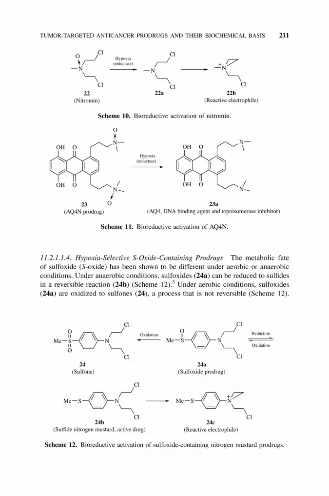
11.2.1.1.4. Hypoxia-Selective S-Oxide-Containing Prodrugs The metabolic fate
of sulfoxide (S-oxide) has been shown to be different under aerobic or anaerobic
conditions. Under anaerobic conditions, sulfoxides (24a) can be reduced to sulfides
in a reversible reaction (24b) (Scheme 12).3 Under aerobic conditions, sulfoxides
(24a) are oxidized to sulfones (24), a process that is not reversible (Scheme 12).
N
Cl
+N
Cl
Cl 22b(Reactive electrophile)
N
Cl
Cl
O
22(Nitromin)
Hypoxia(reductase)
22a
Scheme 10. Bioreductive activation of nitromin.
O
O
N
N
OH
OH
O
O
O
O
N
N
OH
OH
Hypoxia(reductase)
23(AQ4N prodrug)
23a(AQ4, DNA binding agent and topoisomerase inhibitor)
Scheme 11. Bioreductive activation of AQ4N.
Cl
S NMe+
O
O
N
Cl
Cl
SMe N
Cl
Cl
SMe
O
N
Cl
Cl
SMe
24c(Reactive electrophile)
Reduction
Oxidation
Oxidation
24a(Sulfoxide prodrug)
24(Sulfone)
24b(Sulfide nitrogen mustard, active drug)
Scheme 12. Bioreductive activation of sulfoxide-containing nitrogen mustard prodrugs.
TUMOR-TARGETED ANTICANCER PRODRUGS AND THEIR BIOCHEMICAL BASIS 211
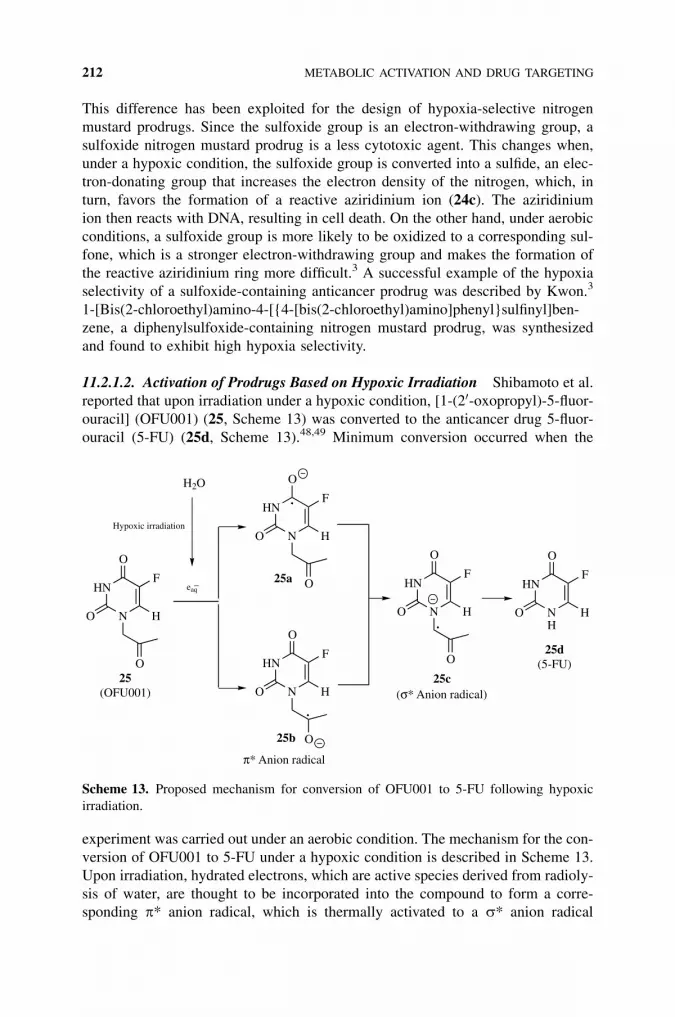
This difference has been exploited for the design of hypoxia-selective nitrogen
mustard prodrugs. Since the sulfoxide group is an electron-withdrawing group, a
sulfoxide nitrogen mustard prodrug is a less cytotoxic agent. This changes when,
under a hypoxic condition, the sulfoxide group is converted into a sulfide, an elec-
tron-donating group that increases the electron density of the nitrogen, which, in
turn, favors the formation of a reactive aziridinium ion (24c). The aziridinium
ion then reacts with DNA, resulting in cell death. On the other hand, under aerobic
conditions, a sulfoxide group is more likely to be oxidized to a corresponding sul-
fone, which is a stronger electron-withdrawing group and makes the formation of
the reactive aziridinium ring more difficult.3 A successful example of the hypoxia
selectivity of a sulfoxide-containing anticancer prodrug was described by Kwon.3
1-[Bis(2-chloroethyl)amino-4-[{4-[bis(2-chloroethyl)amino]phenyl}sulfinyl]ben-
zene, a diphenylsulfoxide-containing nitrogen mustard prodrug, was synthesized
and found to exhibit high hypoxia selectivity.
11.2.1.2. Activation of Prodrugs Based on Hypoxic Irradiation Shibamoto et al.
reported that upon irradiation under a hypoxic condition, [1-(20-oxopropyl)-5-fluor-
ouracil] (OFU001) (25, Scheme 13) was converted to the anticancer drug 5-fluor-
ouracil (5-FU) (25d, Scheme 13).48,49 Minimum conversion occurred when the
experiment was carried out under an aerobic condition. The mechanism for the con-
version of OFU001 to 5-FU under a hypoxic condition is described in Scheme 13.
Upon irradiation, hydrated electrons, which are active species derived from radioly-
sis of water, are thought to be incorporated into the compound to form a corre-
sponding p* anion radical, which is thermally activated to a s* anion radical
HN
N
O
O
O
F
H
HN
N
O
O
O
F
H
.
eaq–
.
.
H2O
HN
NH
O
O
F
H
HN
N
O
O
O
F
H
HN
N
O
O
O
F
H
Hypoxic irradiation
π* Anion radical
25c(σ* Anion radical)
25(OFU001)
25a
25b
25d(5-FU)
Scheme 13. Proposed mechanism for conversion of OFU001 to 5-FU following hypoxic
irradiation.
212 METABOLIC ACTIVATION AND DRUG TARGETING

with a weakened C-N bond. Subsequently, hydrolytic dissociation of the C-N bond
occurs, releasing 5-FU (25d). Since hydrated electrons are rapidly deactivated by
O2 into superoxide anion radicals (O�2 ) under an aerobic condition, the conversion
of OFU001 is therefore hypoxia specific. The 20-oxoalkyl side chain (25, Scheme
13) in the structure is believed to be required for the activation.
11.2.1.3. Activation of Prodrugs Based on Low pH of Solid Tumors Lower
extracellular pH has been observed in many solid tumors as a result of the limited
blood flow.15 The lower extracellular pH is believed to be caused by insufficient
clearance of acidic metabolites from chronically hypoxic cells, a phenomenon
that can lower the mean extracellular pH in tumors to below ca. 6.3—up to one
pH unit lower than the intracellular pH, which is actively regulated. Prodrugs
that are stable at physiological pH and can be activated by this lower extracellular
pH in solid tumors have been described.50 2-Hexenopyranoside of aldophospha-
mide (26, Figure 11.2) is a prodrug of aldophosphamide. The prodrug was stable
at physiological pH (7.4) but readily hydrolyzed to produce aldophosphamide under
an acidic condition.
11.2.2. Tumor-Activated Prodrugs Based on Proteases
Tumor malignancy is often associated with an enhanced expression of proteolytic
enzymes such as cysteine protease cathepsin B, matrix metalloproteinases such as
collagenases and stromelysins, and serine proteases represented by plasminogen
activator and plasmin.51–53 These enzymes are thought to be critically involved
in the events that lead to metastasis because they are capable of degrading the base-
ment membrane and extracellular matrix around tumor tissue, allowing tumor cells
to migrate and invade the surrounding stroma and endothelium. Although these
enzymes are also produced by normal cells, their activity is normally tightly regu-
lated by hormonal controls and by specific inhibitors.54,55 Therefore, these enzymes
are targeted as activating enzymes for anticancer prodrugs. In general, prodrugs tar-
geting proteases contain two components—a peptide and a parent drug. The peptide
chosen is usually a di-, tri-, or tetrapeptide which should give rise to a prodrug
O
O OP
NH2
OC2H5 N
O
CH2OH
Cl
Cl
OH
26(2-Hexenopyranoside of aldophosphamide)
Figure 11.2. Structure of 2-hexenopyranoside of aldophosphamide.
TUMOR-TARGETED ANTICANCER PRODRUGS AND THEIR BIOCHEMICAL BASIS 213

resistant to the serum peptidase but susceptible to enzymes present around the
tumor mass.
11.2.2.1. Prodrug Bond Linkage Between a Peptide and a Drug A peptide can
be connected to a drug directly (Scheme 14) or indirectly through a linker termed a
‘‘spacer’’ (Schemes 15 and 16). The direct linkage of a peptide to a drug leads to a
prodrug that can release either the parent drug or a drug that contains vestiges of the
DrugPeptide Peptide + Drug
Protease
Scheme 14. Prodrugs with a drug directly connected to a peptide.
HN
O X-Drug
H2N
O
O X-Drug
NH
Peptide
O
O
Drug-X-H CO2+ +
a b
Carbamate: X = NHCarbonate: X = O
27(Prodrugs with a carbamate or carbonate bond)
a: Protease; b: 1,6 elimination.
27a
27b 27c
Scheme 15. Hydrolytic activation of prodrugs with a p-aminobenzyl alcohol as a spacer that
contains a carbamate or carbonate bond.
HN
O-Drug
H2N
O-Drug
NH
Peptide
O
CO2Drug-OH + +
a b
28(Prodrugs with a ether bond)
a: Protease; b: 1,6 elimination.
28a
28b 27c
Scheme 16. Hydrolytic activation of prodrugs with a p-aminobenzyl alcohol as a spacer that
contains an ether bond.
214 METABOLIC ACTIVATION AND DRUG TARGETING

bound peptide.56–58 In the latter case, the released drug may have impaired cyto-
toxic activity. An additional consideration for direct drug attachment to peptides
is that the drug may reduce the hydrolysis rate of the peptide by the activating
enzyme, resulting in a slow release of the active drug. This often occurs when a
bulky drug is involved. The problem can be circumvented through an indirect link-
age in which a self-immolative spacer is employed between the drug and the pep-
tide. The spacer spatially separates the drug from the site of enzymatic cleavage so
that the drug will not affect the hydrolysis of the peptide bond. Subsequent frag-
mentation of the spacer releases the active drug. Carl et al. developed one of the
most commonly used spacers—a bifunctional p-aminobenzyl alcohol group, which
is linked to the peptide through the amine moiety and to the drug through the alco-
hol moiety.59 The alcohol moiety of the spacer can form a carbamate bond with an
amino-containing drug or a carbonate bond with an alcohol-containing drug
(Scheme 15). The formed prodrugs are activated upon protease-mediated cleavage
of the amide bond followed by a 1,6-elimination that releases the drug, carbon
dioxide, and remnants of the spacer (Scheme 15). Since the carbonate is easily
susceptible to hydrolysis, only a few alcohol-containing drugs, such as paclitaxel,
can be made stable enough prodrugs through a carbonate bond. Therefore, the
chemistry of this drug attachment has generally been restricted to amine-containing
drugs. For alcohol-containing drugs, a recently reported approach through an ether
bond has proved to be more feasible than carbonate (Scheme 16). However, the
ether bond did not undergo fragmentation as readily as the carbamate bond, and
some of the prepared ether prodrugs did not undergo fragmentation to release the
parent drug.60
It is noteworthy that the length of the spacer plays a significant role in the rate of
enzymatic activation. This is especially true when p-aminobenzyl alcohol carba-
mate is used as a spacer and a bulky drug is involved. As shown in Scheme 15, after
cleavage of the amide bond, the resulting amino group of the aromatic ring is an
electron-donating group and initiates an electronic cascade that leads to the expul-
sion of the leaving group (1,6-elimination), which releases the free drug after elim-
ination of carbon dioxide. The 1,6-elimination of the carbamate prodrug was found
to be virtually instantaneous upon unmasking of the amine group.61 Since the
spacer rapidly eliminates after prodrug activation, the enzymatic activation itself
determines the efficiency of drug release. This assumption is supported by the
fact that prodrugs derived from bulky drugs often have a slower release rate.61
To increase enzyme activation rates, de Groot et al. propose to increase the length
of the spacer between the drug and the peptide to further keep the bulky drug away
from the site of the enzymatic reaction.61 This approach results in a significant
increase in enzyme activation rates and will be discussed in Section 11.2.2.2.
Other spacers such as ethylene diamine, o-aminobenzyl alcohol, p-hydroxylben-
zyl alcohol, and o-hydroxylbenzyl alcohol have also been utilized. The application
of some of these spacers will be illustrated in the examples presented later.
It is worth mentioning that the use of a spacer is not limited to prodrugs invol-
ving a peptide. Spacers discussed above have also been extensively employed in
other prodrugs as well.
TUMOR-TARGETED ANTICANCER PRODRUGS AND THEIR BIOCHEMICAL BASIS 215

11.2.2.2. Tumor-Selective Prodrugs Activated by Plasmin Plasmin is derived
from inactive plasminogen by plasminogen activators (Pas) and is involved in extra-
cellular matrix degradation. The levels of Pas are high in many types of malignant
cells and human tumors such as malignant lung62 and colon63 tumors. Conse-
quently, tumor-associated plasmin activity is highly localized.53,62,64
Plasmin is a protease with specificity for arginine or lysine as amino acids par-
ticipating in bond cleavage.65 Examination of the preferred sites for plasmin clea-
vage of the fibrinogen molecule (the physiological function of plasmin) shows that
the preferred sites involve lysine linked to a hydrophobic amino acid. Thus, the
choice of the peptidic sequence to be used has focused on a peptide having a hydro-
phobic amino acid linked to lysine.66
The idea of selecting plasmin to activate peptide-containing prodrugs was first
proposed in 1980 by Carl et al.65 A D-Val-Leu-Lys tripeptide connected to the
amino functional group AT-125 (29, Figure 11.3) or phenylene diamine mustard
(30, Figure 11.3) was prepared and found to generate the free drug upon treatment
with plasmin. Selection of the D-configuration of the N-terminal amino acid
(D-Val) prevents undesired proteolysis by serum peptidases or other ubiquitous
enzymes. A five- to sevenfold increase in selectivity between Pas-producing cells
in comparison with low-level Pas-containing cells was demonstrated.65
The first prodrugs of doxorubicin for plasmin activation were designed by
Chakravarty et al.67,68 Doxorubicin was directly linked to the tripeptide D-Val-
Leu-Lys (31, Figure 11.4). The formed prodrug showed a sevenfold increase in
selectivity for Pas-producing cells in comparison with low-Pas-containing cells,
but the drug was very inefficiently released by plasmin, a phenomenon likely
caused by steric hindrance of doxorubicin. To overcome this problem, de Groot
et al. placed a spacer between doxorubicin and the peptide.69 Compounds 32a-c
were prepared with a p-aminobenzyl alcohol as a self-immolative 1,6-elimination
spacer to separate doxorubicin or its anthracycline derivatives from the tripeptide
D-Ala-Phe-Lys. All prodrugs were stable in buffer and serum for 3 days and gen-
erated the parent drugs upon incubation with plasmin. Compound 32c demonstrated
the fastest plasmin cleavage rate. Upon incubation with seven human tumor cell
lines, the prodrugs showed a marked decrease in cytotoxicity in comparison with
NO
Cl
NH
COOH
LysLeu
D-Val
HNLys
LeuD-Val
N
ClCl
29 30
Figure 11.3. Anticancer prodrugs activated by plasmin.
216 METABOLIC ACTIVATION AND DRUG TARGETING

OH
O
OH
OHO
OOMe OH O O
HN
OH
LysLeu
D-Val
R1
O
OH
OHO
OOMe OH O O
HN
OH
O
NH
HN
NH
NH3+Cl–
R2
O
O
OO
NH3+Cl–
31
32a: R1 = R2 = H32b: R1 = OH, R2 = H32c: R1 = OH, R2 = Cl
(A prodrug with a direct linkage of a peptide to doxorubicin)
(Prodrugs with one spacer between a peptide and doxorubicin or its derivatives)
OHO
OH
OHO
OOMe OH O O
HNOH
O
NH
HN
NH
NH3+Cl–
O
O
OO
NH3+Cl–
n
33: n=2, a prodrug with two spacers between a peptide and doxorubicin34: n=3, a prodrug with three spacers between a peptide and doxorubicin
Figure 11.4. Prodrugs of doxorubicin and its derivatives with different numbers of spacers
between the drug and a peptide.
TUMOR-TARGETED ANTICANCER PRODRUGS AND THEIR BIOCHEMICAL BASIS 217

the corresponding parent drugs. In vitro selectivity was demonstrated by incubation
of the prodrugs with Pas-transfected MCF-7 cells in comparison with nontrans-
fected cells. The prodrugs showed cytotoxicity similar to that of free doxorubicin
only in the Pas-transfected cells, while exhibiting much less toxicity in the non-
transfected cells.69 The enzyme activation rate was even further increased when a
longer spacer was placed between the drug and the peptide. De Groot et al. placed
two p-aminobenzyl alcohol moieties between doxorubicin and the peptide (33,
Figure 11.4) and found that the enzymatic release rate was increased by twofold.61
The rate was further increased (ca. threefold) when three p-aminobenzyl alcohol
moieties were incorporated (34, Figure 11.4).61
An elegant cyclization spacer was used in the prodrugs of N-nitrosourea.70 Pro-
drugs 35a and 35b contain an ethylene diamine (35a) or monomethylated ethylene
diamine (35b) spacer. Upon cleavage by plasmin, a cyclization reaction led to the
formation of a pentacyclic urea derivative (imidazolidin-2-one), with concomitant
expulsion of the reactive electrophile (35g, Scheme 17). Compared with p-amino-
benzyl alcohol, which undergoes 1,6-elimination, cyclization with an ethylene dia-
mine spacer occurs at a slower rate and often is a rate-determining factor in
releasing the active drug.
De Groot et al. used the same approach to prepare prodrugs of paclitaxel for acti-
vation by plasmin.69 However, most of the prepared prodrugs were either not stable
or resistant to the hydrolysis by plasmin. An alternative approach using a p-amino-
benzyl alcohol group as a spacer appeared to be successful. Prodrug 36 yielded free
paclitaxel (36a) upon incubation with plasmin (Scheme 18). Further, prodrug 36showed a dramatic decrease in cytotoxicity in seven human tumor cell lines in
ClClN
NHNNH
HO NClON
+
NH
NNH
NH3+Cl-
O
O
O
NH3+Cl–
HNN
Cl
O
NO R
HN NH
O
HN
NH
NH3+Cl–
O
O
O
NH3+Cl-
HO
35e(Imidazolidin-3-one)
35c(Peptide)
Plasmin
35a: R =H35b: R = CH3
35g(Reactive species)
35d
35 f
Scheme 17. Hydrolytic bioactivation of prodrugs 35a and 35b.
218 METABOLIC ACTIVATION AND DRUG TARGETING

comparison with paclitaxel (36a). A similar approach to increase the enzymatic
activation rate by increasing the space between paclitaxel and the peptide was
also used.61 Upon incubation with plasmin, the prodrug (37, Figure 11.5) with
two p-aminobenzyl alcohol moieties released paclitaxel at a sixfold increased
rate compared to the single-spacer-containing prodrug (36).61
HN
HN
O
NH
HN
O
O
H3N+
OO
O
O
OH
HO
OO
O
O
O
OHO
NHO
O
O
H3N+
OO
O
O
OH
HO
OO
O
O
OH
OHO
NHO
OH
O
NH
HN
O
O
H3N+
H3N+
36
+
36a(Paclitaxel)
Plasmin
27c 36b
Cl–
Cl–
Cl–
Cl–
2′+
Scheme 18. Hydrolytic bioactivation of prodrug 36 by plasmin.
NH
O
NH
HN
O
O
H3N+
OO
O
O
OH
HO
OO
O
O
O
OHO
NHO
O
O
H3N+
O
O
HNCl–
Cl–
37
Figure 11.5. Prodrug of paclitaxel with two p-aminobenzyl alcohol moieties.
TUMOR-TARGETED ANTICANCER PRODRUGS AND THEIR BIOCHEMICAL BASIS 219

11.2.2.3. Tumor-Selective Prodrugs Activated by Cathepsin B Cathepsins are
cysteine proteases. Cathepsins, especially cathepsin B, are overexpressed in tumors
and may play an important role in cancer invasion.71 Cathepsins may directly
degrade extracellular matrix proteins or activate the Pas system.71 Cathepsin B
has been shown to be clinically relevant in cancer progression, with studies demon-
strating that cytosolic enzyme levels were 11 times higher than those in benign
breast tissue specimens.72 Patients with high intratumor cathepsin B levels have
a significantly worse prognosis than those with low levels.72
A number of prodrugs for cathepsin B activation were prepared (38–43, Figure
11.6). However, not much biological information is available on these compounds.
OO
O
O
O O
HO
OO
OHN
O
OH
O
OHO
O
NH
HN
NH
O
H3N+
O
O
NO
O
38
Cl–
OH
O
OH
OHO
OOMe OH O O
HN
OH
O
O
HN
O
NH
HN
O
O
H3N+
N
O
O39
Cl–
OO
O
O
O O
HO
OO
OHN
O
OH
O
OHO
O
NH
HN
NH
O
O
+H3N
O
O
40
Cl–
Figure 11.6. Representative prodrugs activated by cathepsin B.
220 METABOLIC ACTIVATION AND DRUG TARGETING

The half-lives of cathepsin B cleavage of doxorubicin prodrug 41 and mitomycin C
prodrug 43 were much shorter than those of paclitaxel prodrugs 40 and 42, indicat-
ing steric hindrance by the paclitaxel part of the prodrug.4
Recently, Toki et al. succeeded in developing cathepsin B-activated prodrugs of
combretastatin A-4 (44a, Scheme 19) and etoposide (45a, Scheme 19).60 Combre-
tastatin A-4 is a promising antiangiogenic agent that inhibits the polymerization
of tubulin.73 Etoposide is a clinically approved topoisomerase inhibitor that
has demonstrated utility in chemotherapeutic combinations for the treatment of
OH
O
OH
OHO
OOMe OH O O
HN
OH
O
O
HN
O
NH
HNO
O
O
H3N+
41
Cl–
HN
O
NH
HNO
O
O
H3N+
OO
O
O
OH
HO
OO
O
O
O
OHO
NHO
42
Cl–
O
O
NH
HN
NH
O
O
+H3N
O
ON
ONH2
O
OMe
O
O
H2N
N
43
Figure 11.6. (Continued)
TUMOR-TARGETED ANTICANCER PRODRUGS AND THEIR BIOCHEMICAL BASIS 221

HN
OMe
O
NH
Cit
O
Z-Val
MeO
OMe
OMe
OMe
HO
MeOOMe
OMe
CitZ-Val CO2
44a(Combretastatin A-4)
Cathepsin B
(Z-Val-Cit: N-protected valine-citrulline)
+ + +
44b 27c
O
O
OOO
O
OHHO
H
Me
O
O
O
OMeMeO
HNCit
OValZ
O
O
OOO
O
OHHO
H
Me
O
O
OH
OMeMeO
CitZ-Val CO2HN
+ ++
Cathepsin B
45(Prodrug)
45a(Etoposide)
44b 27c
44(Prodrug)
Scheme 19. Hydrolytic bioactivation of prodrugs by cathepsin B.
222 METABOLIC ACTIVATION AND DRUG TARGETING

leukemia, lymphoma, germ cell tumors, small cell lung tumors, and several other
carcinomas.74 In a prodrug approach, combretastatin A-4 (44a) or etoposide (45a)
was coupled to Z-valine-citrulline peptide, an N-protected valine-citrulline peptide,
through an ether and amide bond using p-aminobenzyl alcohol as a spacer (Scheme
19). The formed prodrugs (44 for 44a and 45 for 45a) were both substrates of cathe-
psin B and released the active drugs upon incubation with the enzyme, suggesting
that these prodrugs can be activated by cathepsin B. It is noteworthy that this is the
first example demonstrating 1,6-fragmentation with an ether bond instead of a car-
bamate bond when p-aminobenzyl alcohol is used as a spacer. The prodrug 44 was
less potent than the parent drug 44a by a factor of 13 in cell killing on L2987
human lung adenocarcinoma, while the prodrug 45 was 20 to 50 times less active
than the parent drug 45a in the cell lines L2987 (human lung adenocarcinoma),
WM266/4 (human melanoma), and IGR-39 (human melanoma), confirming that
the prodrugs were much less cytotoxic.60 However, as mentioned earlier, the frag-
mentation of the ether bond did not proceed as readily as the fragmentation of the
carbamate bond, which is illustrated by a prodrug derived from N-acetylnorephe-
drine (Scheme 20). N-Acetylnorephedrine underwent hydrolysis by cathepsin B
but failed to release the drug N-acetylnorephedrine, suggesting that the alkoxyl
ether bond was not as readily cleaved as those derived from phenoxyl ether-
containing drugs, such as the one in etoposide or combretastatin. This phenomenon
is consistent with the fact that a phenoxyl group is a better leaving group than an
alkoxyl group.
11.2.3. Tumor-Activated Prodrugs Based on Enzymes with ElevatedActivity at Tumor Sites
11.2.3.1. b-Glucuronidase b-Glucuronidase (EC 3.2.1.31) is an exoglycosidase
that cleaves glucuronosyl-O bonds.75 Glucuronidase is intracellularly located in
OHN
Cit
OValZ
NH
O OH2N
NH
O
HO
NH
O
CitZ-Val
HN
Cathepsin B +
+
46(Prodrug)
46a 44b
27c 46c(Norephedrine)
Scheme 20. The hydrolyzed prodrug (46a) fails to undergo 1,6-fragmentation to release nor-
ephedrine (46c).
TUMOR-TARGETED ANTICANCER PRODRUGS AND THEIR BIOCHEMICAL BASIS 223

lysosomes in many organs and body fluids such as macrophages, most blood cells,
liver, spleen, kidney, intestine, lung, muscle, bile, intestinal juice, urine, and
serum.75 There is large interindividual variability in its activity and expression in
the liver, kidney, and serum. The activity of the enzyme is relative high in some
tissues, such as the liver. The rationale for selecting b-glucuronidase to activate
anticancer prodrugs is based on the observation that the activity of the enzyme
has been shown to be elevated in many tumors.76,77 An advantage of targeting
b-glucuronidase is that a glucuronide prodrug exhibits high hydrophilicity, which
greatly reduces the distribution of the prodrug into cells, resulting in reduced sys-
temic toxicity. However, the same feature also hampers the activation of a glucur-
onide prodrug at tumor sites. Since the enzyme is intracellularly located, a
glucuronide prodrug needs to enter tumor cells in order to be activated. Bosslet
et al. demonstrated that b-glucuronidase is liberated extracellularly in high local
concentration in necrotic areas of human cancer.77 Therefore, necrotic areas are
the areas where glucuronide prodrugs can be activated. It is worth mentioning
that, in addition to cancer, increased tissue or serum b-glucuronidase activity has
been observed in other disease states, e.g., inflammatory joint disease, ichthyosi-
form dermatosis, some hepatic diseases and acquired immune deficiency syndrome
(AIDS). For example, serum b-glucuronidase activity has been reported to be
16-fold higher in human immunodeficiency virus (HIV) infected patients than in
healthy individuals.78 The extracellular presence of b-glucuronidase in areas other
than tumor sites leads to activation of b-glucuronidase-mediated prodrugs, resulting
in side effects.
The b-glucuronidase-mediated release approach has been used in the prodrugs of
many anticancer drugs. The prodrugs are formed by linking a drug to glucuronic
acid through an anomeric ether bond directly or, more often, indirectly (Scheme
21). In the latter case, a spacer is used. In general, prodrugs with a spacer are
cleaved more readily by the enzyme, especially when the parent drug molecules
are bulky. Figure 11.7 shows the structures of doxorubicin prodrugs 48 and 49.
Both were much less toxic than the parent drug in a human ovarian cancer
cell line (OVCAR-3), while prodrug 49, with a spacer between the drug and the
glucuronyl group, was activated much faster by b-glucuronidase.79 Similarly,
OOHO
OH
HOCO2H
Drug OHOHO
OH
HOCO2H
Glucuronyl moiety
Anomeric ether bond
+ Drug
47(Glucuronide prodrug)
β-glucuronidase
47a(Glucuronic acid)
47b
Scheme 21. Bioactivation of glucuronide prodrugs by b-glucuronidase.
224 METABOLIC ACTIVATION AND DRUG TARGETING

daunorubicin prodrug 50 showed very low toxicity against five human tumor cell
lines, consistent with the observation that the compound appeared to be very stable
against enzymatic cleavage by b-glucuronidase.80 The spacers used most extensively
are p- or o-hydroxybenzyl alcohol moieties.
Florent et al. prepared and evaluated a series of anthracycline glucuronide pro-
drugs with a p- or o-hydroxybenzyl alcohol as a spacer.81 It was reported that dox-
orubicin glucuronide prodrugs 49, 51a, and 51b (Figure 11.7) appeared to be better
substrates for b-glucuronidase than the corresponding prodrugs 52a–c (Figure
11.7). Prodrugs 52a–c also showed a slower elimination of the spacer.81 Prodrug
49 was selected as the most appropriate prodrug for further evaluation, and it has
been extensively investigated. An attempt to further increase the hydrophilicity of
the prodrugs by introducing a hydrophilic group at the spacer moiety resulted in
compounds 51c–e (Figure 11.7).82 Of these prodrugs, compound 51d appeared
most promising for further development due to its reduced cytotoxicity and fast
hydrolysis by b-glucuronidase.
OH
O
OH
OHO
OOMe OH O
O
OOHO
OH
Me
NH2HOCO2H
OH
O
OH
OHO
OOMe OH O
O
HO
OHO
OH
Me
HN
HOCO2H
O
O
OH
O
OH
OHO
OOMe OH O
O
HO
OHO
OH
Me
HN
HOCO2H
O
O
O
NO2
48 49 (HMR 1826) 50
OH
O
OH
OHO
OOMe OH O
O
HO
OHO
OH
Me
HN
HOCO2H
O
O
O
R
OH
O
OH
OHO
OOMe OH O
O
HO
OHO
OH
Me
HN
HOCO2H
O
O
O
R
51a R = F51b R = Cl51c R = N(CH3)2
51d R = NHCOCH3
51e R = NHCOCF3
52a R = F52b R = Cl52c R = NO2
Figure 11.7. Representative prodrugs bioactivated by b-glucuronidase.
TUMOR-TARGETED ANTICANCER PRODRUGS AND THEIR BIOCHEMICAL BASIS 225

The camptothecins are a new class of very promising anticancer agents, several
derivatives of which are in clinical use.83 Prodrug 53 (Figure 11.8) of 9-amino-
camptothecin showed 20- to 80-fold reduced toxicity in comparison with 9-amino-
camptothecin.84 The prodrug was readily cleaved by b-glucuronidase in vitro.
Schmidt et al. prepared and evaluated compound 54 as a prodrug of a phenol
mustard (Scheme 22).85 After removal of the glucuronic acid (47a) by b-glucuro-
OHO
OH
HOCO2H
O
O
N
N
O
OH O
O
NH
O
53
Figure 11.8. Structure of a glucuronide prodrug of 9-aminocamptothecin.
O
HOOH
HO
CO2H
NO2
ON
OO
N
ClCl
NO2
O–
N
OO
N
ClCl
O
HOOH
HO
CO2H
NO2
ON
O
OH
N
ClCl
β-glucuronidase Cyclization
+
54c
54(Glucuronide nitrogen mustard)
47a
54a
54b
Scheme 22. Activation of glucuronide nitrogen mustard prodrug 54.
226 METABOLIC ACTIVATION AND DRUG TARGETING

nidase, the spacer was eliminated through cyclization liberating the phenol mustard
54c. The prodrug was 80-fold less cytotoxic than the phenol mustard in LoVo cells.
Chemically, the prodrug (54) was much more stable than the corresponding parent
drug (54c) in phosphate buffer.
Another prodrug of 54c is compound 55. Prodrug 55, which contains an
aromatic and aliphatic bis-carbamate spacer, is activated by b-glucuronidase
(Scheme 23).86 A rapid cleavage of the glycosidic bond occurred (t1=2 ¼ 6:6 minutes)
with concomitant appearance of intermediate 55c, of which the ethylene diamine
spacer cyclized with a half-life of 2 hours. The cytotoxicity of 55 against LoVo cells
was about 50 times less than that of the corresponding phenol mustard 54c.
A similar approach was also applied in paclitaxel prodrugs 56, 57a, and 57b.87
After hydrolysis by b-glucuronidase, ring closure occurred, resulting in the release
of paclitaxel (36a) and CO2 (Schemes 24 and 25). The half-lives of b-glucuroni-
dase-mediated activation were 2 hours and 45 minutes, respectively, for prodrugs
56 and 57b. Both prodrugs were two orders of magnitude less cytotoxic than pacli-
taxel. Prodrug 57a appeared to be as cytotoxic as paclitaxel, which was explained
by the fact that the prodrug underwent spontaneous hydrolysis in buffer solution
under physiological conditions.
Glucuronide prodrugs 58a, 58b, and 59 of 5-fluorouracil exhibited half-lives of
�20 minutes when incubated with 25 mg/mL of b-glucuronidase and reduced (six
to nine times less) cytotoxicity against LoVo cells in comparison with the parent
drug 5-fluorouracil (Figure 11.9).88
11.2.3.2. Prostate-Specific Antigen (PSA) PSA is a serine protease that is present
extracellularly in prostate cancers. PSA is inhibited in the bloodstream. As a result,
OHO
OH
HOCO2H
O NN O
O
ON
Cl
ClO
NO2
OHO
OH
HOCO2H
OHO
NO2
55
β-glucuronidase
+
47a 55b
HNN O
ON
Cl
Cl
54cHO
N
Cl
ClN N
O
+Cyclization
55c 55d
Scheme 23. Glucuronidase-mediated bioactivation of prodrug 55.
TUMOR-TARGETED ANTICANCER PRODRUGS AND THEIR BIOCHEMICAL BASIS 227

active PSA is present only in prostate cancer.4 A prodrug (60, Figure 11.10) of dox-
orubicin was formed by coupling the C-terminal carboxylic acid group of a hepta-
peptide to doxorubicin. The sequence His-Ser-Ser-Lys-Leu-Gln was selected
because of its specificity and serum stability.89 In compound 60, an extra leucine
residue was added after glutamine, which increased the distance between the pep-
tide bond to be cleaved (leucine-glutamine) and doxorubicin. This increased
distance reduced the steric hindrance that doxorubicin posed to the leucine-
glutamine bond and facilitated the bond cleavage by PSA.57 The prodrug 60 under-
went cleavage by PSA at the leucine-glutamine bond to release not doxorubicin but
OO
O
O
OH
HO
OO
O
O
O
OHO
NHO
O
HOOH
HO
CO2HO
HNO
O
O
HOOH
HO
CO2H
OH
OO
O
O
OH
HO
OO
O
O
O
OHO
NHO
OH2N
a
56
47a
56a
a: β-glucuronidase
OO
O
O
OH
HO
OO
O
O
OH
OHO
NHO
36a(Paclitaxel)
NH
O
+
Cyclization
56c
Scheme 24. Bioactivation of prodrug 56 by b-glucuronidase.
228 METABOLIC ACTIVATION AND DRUG TARGETING

doxorubicin-leucine, which was an anticancer agent itself. In vitro selectivity was
demonstrated by the fact that 70 nM of the prodrug killed 50% of the PSA-producing
human prostate cancer cells (LNCaP cells), whereas doses as high as 1 microM had
no cytotoxic effect on PSA-nonproducing TSU human prostate cancer cells.57
11.2.3.3. P450 as Activating Enzymes for Anticancer Prodrugs Cytochrome
P450 (P450) comprises a superfamily of enzymes involved in the oxidation of a
OO
O
O
OH
HO
OO
O
O
O
OHO
NHO
O
HOOH
HO
CO2H X
O
NHO
O
O
HOOH
HO
CO2H
OH57a: X = CH2
57b: X = CH2CH2
47a
β-glucuronidase
OO
O
O
OH
HO
OO
O
O
O
OHO
NHO
X
O
H2N
57c
OO
O
O
OH
HO
OO
O
O
OH
OHO
NHO
36a(Paclitaxel)
Cyclization
HN X
O 57d
Scheme 25. Bioactivation of prodrugs 57a and 57b by b-glucuronidase.
TUMOR-TARGETED ANTICANCER PRODRUGS AND THEIR BIOCHEMICAL BASIS 229

large number of exogenous and endogenous compounds.90 Oxidation of compounds
by P450 increases polarity and aids further metabolism or removal of the compound
from the body. Therefore, P450 enzymes are viewed as the most important enzymes
in removing exogenous compounds or toxic molecules from the body. P450
enzymes are present in normal tissues, with the highest levels in the liver consistent
with the role of the liver as the detoxification organ. P450 enzymes are involved not
only in inactivation of anticancer drugs but also in activation of several anticancer
drugs such as cyclophosphamide91 and its isomers ifosfamide and tegafur.92 As
expected, P450-activated anticancer prodrugs are not highly tumor-selective, since
most of them are activated by liver P450. These liver P450-activated prodrugs will
not be discussed here. However, efforts to increase tumor selectivity of P450-acti-
vated anticancer prodrugs have been made. Of particular interest is the approach
used to deliver a P450 enzyme to tumor sites through gene-directed enzyme pro-
drug therapy (GDEPT).93–96 Efforts to further increase tumor selectivity and
decrease systemic toxicity by delivering a P450-activating enzyme to the tumor
site via GDEPT and, at the same time, inhibiting liver P450 have also been
reported.97,98 A more detailed discussion of GDEPT is presented in Section 11.3.2.
O
HOOH
HO
CO2H
O
N
NO
O
F
H
HN
O
O
R
O2N
O
HOOH
HO
CO2H
N
NO
O
F
H
HNO
O
5958a: R = H58b R = COOH
Figure 11.9. Structures of glucuronide prodrugs 58a, 58b, and 59.
OH
O
OH
OHO
OOMe OH O
O
HO
Me
NH Leu-Gln-Leu-Lys-Ser-Ser-His-Mu
Mu = morpholinocarbonyl
60
Figure 11.10. Structure of a doxorubicin prodrug activated by PSA.
230 METABOLIC ACTIVATION AND DRUG TARGETING

11.3. ANTIBODY- AND GENE-DIRECTED ENZYME PRODRUGTHERAPY (ADEPT AND GDEPT)
Antibody-directed enzyme prodrug therapy (ADEPT) involves two steps.9–11 The
first step is to deliver a chosen enzyme to the surface of cancer cells by a mono-
clonal antibody (mAb) followed by a second step—administration of a prodrug that
is activated by the enzyme to release the active parent drug. GDEPT also involves
two steps.6–8 In the first step, a gene encoding a chosen enzyme is transported to
cancer cells. After expression of the chosen enzyme, the prodrug that is activated by
the enzyme is administered. In ADEPT, the enzyme is attached to the surface of
cells, while in GDEPT, the enzyme can be expressed intracellularly or extracellu-
larly.99,100 The potential advantages of extracellular expression of the enzyme are
twofold. First, it should produce an improved bystander effect because the active
drug will be generated in the interstitial spaces within the tumor, rather than inside,
as with an intracellularly expressed activating enzyme. Second, the prodrug does
not need to enter cells to become activated; therefore, non-cell-permeable prodrugs
can be used.
Enzymes used for ADEP or GDEPT approaches can be divided into three major
classes: (I). enzymes of nonmammalian origin that have no mammalian homologs;
(II). enzymes of nonmammalian origin with a mammalian homolog; and (III).
enzymes of mammalian origin.11 Examples of enzymes in class I include carboxy-
peptidase G2 (CPG2), b-lactamase, penicillin G amidase, and cytosine deaminase.
The rationale for using such enzymes is that prodrugs can be designed to be
nontoxic and not substrates for endogenous human enzymes. Since many of the
nonmammalian enzymes are bacterial or are easily expressed in bacteria, they
are available in large quantities. The main disadvantage is that they elicit immune
responses in humans. The criterion for selecting enzymes in class II is that the
activities of the endogenous counterparts of these enzymes should be low. Exam-
ples of class II enzymes include b-glucuronidase and nitroreductase. One advantage
of employing class II enzymes is that enzymes with a higher catalytic rate can be
selected. For example, bacterial b-glucuronidase exhibits a higher turnover rate
than its human counterpart. Additionally, the exogenous enzyme could differ sig-
nificantly enough from the endogenous counterpart that a prodrug can be designed
to be cleaved only by the exogenous enzyme. One example is bacterial nitroreduc-
tase. As with the class I enzyme, class II enzyme also has limitations due to immu-
nogenicity. The major advantage of using class III enzymes is that they are much less
immunogenic than bacterial or fungal enzymes. The obvious disadvantage is that the
designed prodrugs can also be activated by endogenous enzymes, resulting in nonspecific
activation. Enzymes belonging to this class include alkaline phosphatase, carboxypepti-
dase A, b-glucuronidase, and cytochrome P450.
Clearly, in both ADEPT and GDEPT, the activity of the enzyme activating the
prodrug is enhanced at tumor sites, leading to increased tumor selectivity of
the prodrug. Compared with the prodrug therapy discussed earlier that is defined as
‘‘one-step prodrug therapy’’ or ‘‘prodrug monotherapy,’’ ADEPT and GDEPT are
‘‘called two-step prodrug therapy.’’4
ANTIBODY- AND GENE-DIRECTED ENZYME PRODRUG THERAPY 231

11.3.1. ADEPT
Monoclonal antibodies (mAbs) have been used to deliver chemotherapeutic
drugs,101,102 potent plant and bacterial toxins,103 and radionuclides104 to tumor
sites. A number of mAb-based drugs have been approved clinically (Rituxan,
Herceptin, and Panorex), and several others are now in advanced clinical trials.
However, to date, no clinically approved mAb-based drugs are available for solid
tumors. The major challenge in developing mAb-based drug therapy for solid
tumors lies, in part, in the barriers to macromolecule penetration within the tumor
masses and the heterogeneity of target antigen expression.11 This has prompted
research to find an alternative strategy that can dissociate the drug from the mAb
delivery system after it reaches the target. ADEPT was a result of this effort. In the
ADEPT approach, the activated anticancer drug is not linked to the antibody and
can penetrate tumor masses through diffusion. Further, while the concentration of
an anticancer drug is limited by the carrying capacity of the mAb-anticancer drug
conjugate drug delivery system, it is expected to improve in the ADEPT since in
ADEPT system, an active drug is not carried to the site, but rather is continually
generated from the prodrug through enzymatic activation.
The general procedure for ADEPT is as follows. A chosen enzyme is conjugated
to a tumor-specific mAb or a mAb fragment to form a mAb-enzyme conjugate. For-
mation of this conjugate can be achieved through chemical linkage of the mAb and
the enzyme or through recombinant technology.11 After administration, the conju-
gate will bind to the corresponding antigen, which is located on the surface of
tumor cells. A clearance period is given to allow the non-antigen-bound mAb-
enzyme conjugate to be removed from the body before a prodrug is administered.
The administered prodrug will now be selectively activated by the enzyme attached
to the surface of the cancer cells. One advantage of ADEPT is that since the enzyme
is placed on the surface of cancer cells, the prodrug does not need to enter the cells
for activation. Therefore, this approach will allow prodrugs that cannot enter cells
to be activated. However, there are clear disadvantages of ADEPT, as discussed by
de Groot and colleagues.4 The main problem is the immunogenicity of the anti-
body-enzyme conjugate. Also, the mAb-enzyme conjugate does not always localize
to the desired extent, and whole antibodies penetrate tumors poorly. Further, tumor
cells express only a limited number of antigens. The clearance of unbound mAb-
enzyme conjugate is also a problem. The unbound mAb-enzyme conjugate is often
inadequately cleared, resulting in activation of the prodrug at nontumor sites and
leading to systemic toxicity. Finally, antigen expression levels vary among indivi-
duals, which affects the efficacy of the prodrug. Nevertheless, ADEPT is an inter-
esting approach in increasing the tumor selectivity of anticancer prodrugs. In the
following discussion, carboxypeptidase G2 (CPG2) and b-lactamase will be used
as examples to illustrate the application of ADEPT.
11.3.1.1. ADEPT with CPG2 CPG2 is a bacterial zinc-dependent metalloprotei-
nase.7 This enzyme is an exoprotease that specifically cleaves terminal glutamic
acid amides.11 Niculescu-Duvaz et al. describe an ADEPT system that uses a
232 METABOLIC ACTIVATION AND DRUG TARGETING

combination of mAb, CPG2, and nitrogen mustard prodrugs.7 CPG2 is coupled to
F(ab0)2 fragments of the mAb A5B7. A5B7 is an anti-human carcinoembryonic
antigen (CEA) mouse mAb. The antibody enzyme conjugate [F(ab0)2- CPG2 con-
jugate] was investigated for its ability to activate the prodrug (2-chloroethyl)-(2-
mesyloxygen)aminobenzoyl-L-glutamic acid (CMDA) and other N-L-glutamyl
amide nitrogen mustard prodrugs. The general structural features of the prepared
N-L-glutamyl amide nitrogen mustard prodrugs are shown in Scheme 26.
The bond to be cleaved by CPG2 is the amide bond derived from L-glutamic
acid. An additional advantage of including the L-glutamic acid moiety in the pro-
drug is the increased hydrophilicity of the molecule. As mentioned earlier, in
ADEPT an activating enzyme is anchored on the outer membrane of tumor cells.
It is advantageous if the prodrug is more hydrophilic than the parent drug since high
hydrophilicity reduces the ability of the prodrug to cross the cell membrane. There-
fore, the distribution of the prodrug into cells will be decreased, reducing the side
effects to normal cells. The original work was conducted by Springer et al., who
used nude mice implanted with chemoresistant choriocarcinoma xenografts as an
animal model for the study of CMDA (Scheme 26).105 It was demonstrated that
9 of 12 mice were long-time survivors (>300 days), whereas all control mice
were dead by day 111. A clinical trial was also conducted in patients with advanced
colorectal carcinoma of the lower intestinal tract.9,106 A dose of 20,000 enzyme
units/m2 mAb-CPG2 conjugate was administered in the first step. This conjugate
dose produced tumor CPG2 levels comparable to those found to be optimum in
nude mice bearing xenografts. This treatment was followed 24–48 hours later by
administration of a clearing agent, anti-CPG2 galactosylated Ab (220 mg/m2), to
help remove mAb-CPG2 conjugates that were not bound to the antigen. In the last
step, prodrug CMDA was injected over 1–5 days up to a total dose of 1.2–10 g/m2.
NX
ZNH
HO
Y
OHO O
O
CPG2 cleavage
NX
W
YEWG
EDG
X, Y = Cl, Br, I, OSO 2CH3
Z = –, O, NH2, CH2, S
W = COO–, NH2, OH, CH2COO–, SH. 61(N-L-glutamyl amide nitrogen mustard prodrugs)
CMDA: X = Cl, Y = OSO2CH3, Z = -.
61a(Activated nitrogen mustards)
CPG2
Scheme 26. General structures of N-L-glutamyl amide nitrogen mustard prodrugs and
activation of the prodrugs by CPG2.
ANTIBODY- AND GENE-DIRECTED ENZYME PRODRUG THERAPY 233

Oral cyclosporine was coadministered to suppress the host immune response.
From eight evaluable patients, there were four partial responses and one mixed
response. A more recent clinical trial involving patients with colorectal carcinoma
expressing CEA using a similar approach demonstrates that the median tumor:
plasma ratio of enzyme (CPG2) exceeded 10,000:1 at the time of prodrug admin-
istration. Enzyme concentrations in the tumor were sufficient to generate cytotoxic
levels of the active drug.107 A tumor response to the ADEPT was observed.107 Thus
the results obtained are encouraging and prove the feasibility of the ADEPT at the
clinical level.
11.3.1.2. ADEPT with b-Lactamase Activation of b-lactam-based prodrugs is
based on the well-established b-lactam chemistry. It was demonstrated that a mole-
cule attached to the 30 position of cephalosphorins was eliminated through a 1,4-
fragmentation reaction.11 Scheme 27 shows a general scheme of this reaction,
and Figure 11.11 shows the structures of the prodrugs of a vinca derivative (63),
phenylenediamine mustard (64), doxorubicin (65), melphalan (66), paclitaxel
(67), and mitomycin (68) prepared based on this chemistry.11 The first report of
in vivo activity in a mAb-lactamase system used the b-lactamase from E. cloacae
and a cephalosphorin-vinca alkaloid prodrug LY266070 (63, Figure 11.11).108
Nude mice implanted with human colorectal carcinoma were used for the investi-
gation. The tumor inhibitory effects of the ADEPT with mAb-lactamase conjugate
and LY266070 were superior to those produced by prodrug alone. The effects were
also superior to those obtained when the vinca alkaloid (Figure 11.11) was attached
directly to the mAb. Long-term regression of established tumors was observed in
several dosing regimens, even in animals having tumors as large as 700 mm3 at the
initiation of therapy.108
N
S
O
HNR
OHO
O + Drug
62(Prodrugs)
62c(Activated drugs)
β-lactamaseN
S
XO
HNR
OHO
O
β-lactamase
N
S
Drug
HNR
OHO
O
β-lactamase–O
β-lactamase
62a
62b
Drug
Scheme 27. Bioactivation of prodrugs by b-lactamase.
234 METABOLIC ACTIVATION AND DRUG TARGETING

N
S
OO
HNHO
OHO
OO N
O
NClCl
64
N
S
OO
HN
OHO
O NH
O
N
N
NH
N
MeO
HO
CO2Me
Me
OHOH
OHNS
O
63
N
S
OO
HN
OHO
O NH
O
NClCl
HOOC
O
66
N
S
OO
HN
OHO
O NH
O
O
OOH
OHO
OOMe
HO
Me
OH
O
OH
65
N
SHN
O
HN
OHO
O
N
OCONH2
OMe
NH
O
OMe
OO
O
C6H5
OO
MeO
O
O
NH
C6H5
C6H5
O
OOMe
OOH
ON
S
OO
HN
OHO
OHN
O
S
O
67
68
Figure 11.11. Structures of representative prodrugs that are activated by b-lactamase.
ANTIBODY- AND GENE-DIRECTED ENZYME PRODRUG THERAPY 235

In a related study, Kerr et al. reported that ADEPT treatment with a combination
of a mAb-lactamase conjugate and a prodrug of cephalosphorin-phenylenediamine
mustard (64) in nude mice bearing subcutaneous 3677 human tumor xenografts pro-
duced regression in all the treated mice at doses that caused no apparent toxicity.109
At day 120 post tumor implant, four of five mice who had received this treatment
remained tumor free. Significant antitumor effects were even seen in mice that had
large (800 mm3) tumors before the first prodrug treatment.
11.3.2. GDEPT
Unlike ADEPT, the major problem with GDEPT is delivery of the gene to the
tumor—the same problem associated with cancer gene therapy. Almost all clinical
trials currently being performed for cancer gene therapy rely on direct intratumor
injection of the vector. This is not only a major limitation for uniform distribution
of the delivered gene throughout the primary tumor, but it also fails to address the
problem of delivery to sites of metastatic spread, which may be too numerous, invi-
sible, or inaccessible for direct injection. Efforts to overcome this problem include
engineering adenoviral vectors to alter their tropism for infection,110 use of cationic
liposomes with surface ligands that are specific for tumors,111 and use of live
attenuated Salmonella that can preferentially accumulate in tumors by an as yet
unknown mechanism.112,113 Discussion of these efforts is beyond the scope of
this chapter.
A closely related approach to GDEPT is virus-directed enzyme prodrug therapy
(VDEPT).114 The only difference between these approaches is that GDEPT
involves both viral and nonviral vectors. For a more extensive discussion of
GDEPT, readers are referred to earlier reviews.6–8 In this chapter, GDEPT will
be introduced through two representative examples.
11.3.2.1. Hypoxia-Selective GDEPT A very promising approach to deliver the
gene to solid tumors was recently described by Liu and colleagues.115 This
approach takes advantage of the hypoxic/necrotic condition of solid tumors. The
bacterial genus Clostridium comprises a large and heterogeneous group of gram-
positive, spore-forming bacteria that become vegetative and grow only in the
absence (or low levels) of oxygen. Therefore, strains of these bacteria have been
suggested as tools to selectively deliver the gene vector to the hypoxic and necrotic
region of solid tumors.116–118 Liu and coworkers described, for the first time, the
successful transformation of C. sporogenes, a clostridial strain, with the Escheri-
chia coli cytosine deaminase (CD) gene.115 They showed that intravenous injection
of spores of C. sporogenes containing an expression plasmid for E. coli cytosine
deaminase into tumor-bearing mice produced tumor-specific expression of cytosine
deaminase protein, an enzyme that converts the nontoxic prodrug 5-fluoro-
cytosine (5-FC) (69) to the anticancer drug 5-fluorouracil (5-FU) (25d, Scheme 28).
More importantly, significant antitumor efficacy of systemically injected 5-FC
was observed following IV injection of these recombinant spores. The antitumor
efficacy of the prodrug 5-FC following a single IV injection of the recombinant
236 METABOLIC ACTIVATION AND DRUG TARGETING

spores is equivalent to or greater than that produced by the maximum tolerated dose
of the active drug 5-FU given by the same schedule. A major advantage of the clos-
tridial delivery system is that not only is it tumor specific, it is also safe in humans.
As cited by Liu et al.,115 Mose et al. injected themselves with spores of C. sporo-
genes (a strain later named C. oncolyticum) and experienced a mild fever as the
only side effect. The safety of C. oncolyticum was substantiated in clinical trials
with cancer patients who had a variety of solid tumors119 and also in more recent
trials in noncancer patients with a C. beijerinckii strain.120 An additional advantage
is that injection of spores did not appear to elicit an immune response.115
Another example of hypoxia-selective GDEPT is prodrug 70 (CB 1954, Scheme
29). CB 1954 was originally synthesized over 30 years ago. It exhibits dramatic and
highly specific antitumor activity against rat Walker 256 carcinoma cells.121 The
antitumor effect is due to efficient drug activation by rat DT-diaphorase.122 CB
1954 entered clinical trials in 1970s, but little antitumor activity was observed,
N
NH
NH2
O
F
H
HN
NH
O
O
F
H
Cytosine deaminase
695-Fluorocytosine (5-FC)
25d5-Fluorouracil (5-FU)
Scheme 28. Bioactivation of 5-FC by cytosine deaminase.
N
NO2
NO2
H2N
O
N
NHOH
NO2
H2N
O
N
N
NO2
H2N
O
O
O
Reductase Thioester(e.g, acetyl CoA)
705-(Aziridin-1-yl)-2,4-dinitrobenzamide (CB 1954)
70a
70b(DNA cross-linking agent)
Scheme 29. Bioactivation of CB 1954 by reductases.
ANTIBODY- AND GENE-DIRECTED ENZYME PRODRUG THERAPY 237

as human DT-diaphorase is much less active in the reduction of CB 1954 than the
rat enzyme. Recent studies revealed that an amino acid difference at residue 104
between the human and rat enzymes is responsible for the catalytic difference to
CB 1954.123 A nitroreductase (NTR) gene isolated from E. coli has been demon-
strated to activate the prodrug CB 1954 to its toxic form approximately 90-fold
more rapidly than rat DT-diaphorase, suggesting the possibility of using CB 1954
with NTR in ADEPT124,125 and GDEPT.126–130
Shibata et al. successfully transfected human HT 1080 tumor cells with the E.
coli NTR gene.131 The transfected human tumor cells expressed E. coli NTR in a
time- and concentration-dependent manner under a hypoxic condition while expres-
sing only trace levels under an aerobic condition, indicating that the enzyme’s
expression is induced by hypoxia. No NTR was observed with wild-type
HT1080 cells. The expression of NTR conferred increased sensitivity of human
tumor cells to CB 1954 both in vitro and in vivo. The IC50 value obtained with
the transfected cells was reduced by 40- to 50-fold compared to the IC50 value
with the wild cells in an in vitro experiment under a hypoxic condition. Signifi-
cantly, no sensitivity difference was observed between the transfected and wild
HT 1080 cells under an aerobic condition, consistent with the notion that NTR is
induced under hypoxic conditions. A significant tumor growth delay was also
observed in mice implanted with transfected clones of HT 1080 cells under a
hypoxic condition. Similar to the in vitro result, no sensitivity difference to CB
1954 was observed when the in vivo experiment was conducted under an aerobic
condition.
Other examples of GDEPT include using CPG2 to activate nitrogen mus-
tard,99,132 DT-diaphorase, and NADPH-cytochrome c (P450) reductase to activate
mitomycin,133 CYP2B1 to activate cyclophosphamide and ifosfamide,94 and pyri-
midine nucleoside phosphorylase (PyNPase) to activate 50-DFUR to 5-FU.134
11.4. SUMMARY
The search for tumor-selective prodrugs has been a long and ongoing effort.
Numerous approaches have been developed based on the exploitation of the bio-
chemical differences between cancer and normal cells. The differences can be
further amplified by the use of ADEPT and GDEPT. Among the various approaches
used, anticancer prodrugs based on tumor hypoxia and metastasis appear to be the
most extensively explored, and some of these prodrugs have proceeded to clinical
trials. ADEPT and GDEPT provide the advantage of increased tumor selectivity.
The high immune response and cost are the major disadvantage of these two
approaches. However, the advances in genetic technology have prompted very
active research in GDEPT which is expected to play a more significant role in
tumor-selective anticancer prodrug development. Overall, tumor-selective antican-
cer prodrugs have been shown to be an effective way to improve therapeutic effi-
cacy and reduce systemic side effects.
238 METABOLIC ACTIVATION AND DRUG TARGETING

REFERENCES
1. Han, H. K.; Amidon, G. L. AAPS PharmSci. 2000, 2, E6.
2. Bebernitz, G. R.; Dain, J. G.; Deems, R. O.; Otero, D. A.; Simpson, W. R.; Strohschein,
R. J. J. Med. Chem. 2001, 44, 512–523 and reference cited therein.
3. Kwon, C. H. Arch. Pharm. Res. 1999, 6, 533–541.
4. de Groot F. M.; Damen E. W.; Scheeren, H. W. Curr. Med. Chem. 2001a, 8, 1093–1122.
5. Denny, W. A. Eur. J. Med. Chem. 2001, 36, 577–595.
6. Deonarain, M. P.; Spooner, R. A.; Epenetos, A. A. Gene Ther. 1995, 2, 235–244.
7. Niculescu-Duvaz, I.; Cooper, R. G.; Stribbling, S. M.; Heyes, J. A.; Metcalfe, J. A.;
Springer, C. J. Curr. Opin. Mol. Ther. 1999, 1, 480–486.
8. Aghi, M.; Hochberg, F.; Breakefield, X. O. J. Gene Med. 2000, 2, 148–164.
9. Bagshawe, K. D.; Sharma, S. K.; Springer, C. J.; Rogers, G. T. Ann. Oncol. 1994, 5, 879–
891.
10. Melton, R. G.; Sherwood, R. F. J. Natl. Cancer Inst. 1996, 88, 153–165.
11. Senter, P. D.; Springer, C. J. Adv. Drug Deliv. Rev. 2001, 53, 247–264.
12. Vaupel, P.; Kallinowski, F.; Okunieff, P. Cancer Res. 1989, 49, 6449–6465.
13. Adams, G. E.; Hasan, N. M.; Joiner, M. C. Radiother. Oncol. 1997, 44, 101–109.
14. Brown, J. M. Cancer Res. 1999, 59, 5863–5870.
15. Tannock, I. F.; Rotin, D. Cancer Res. 1989, 49, 4373–4384.
16. Prezioso, J. A.; Hughey, R. P.; Wang, N.; Damodaran, K. M.; Bloomer, W. D. Int. J.
Cancer 1994, 56, 874–879.
17. Lee, A. E.; Wilson, W. R. Toxicol. Appl. Pharmacol. 2000, 163, 50–59.
18. Gutierrez, P. L. Free Radic. Biol. Med. 2000, 29, 263–275.
19. Faig, M.; Bianchet, M. A.; Winski, S.; Hargreaves, R.; Moody, C. J.; Hudnott, A. R.;
Ross, D.; Amzel, L. M. Structure (Camb) 2001, 9, 659–667.
20. Workman, P. Int. J. Radiat. Oncol. Biol. Phys. 1992, 22, 631–637.
21. Powis, G. Pharmacol. Ther. 1987, 35, 57–162.
22. Bachur, N. R.; Gordon, S. L.; Gee, M. V.; Kon, H. Proc. Natl. Acad. Sci. USA 1979, 76,
954–957.
23. Pan, S. S.; Andrews, P. A.; Glover, C. J.; Bachur, N. R. J. Biol. Chem. 1984, 259, 959–966.
24. Workman, P.; Walton, M. I. In Eds. Adams, G.E., Breccia, A., Fielden, E. M., Wardman,
P., Selective Activation of Drugs by Redox Processes; Plenum Press; New York, 1990, pp.
173–191.
25. Sartorelli, A. C. Cancer Res. 1988, 48, 775–778.
26. Callery, P. S.; Gannett, P. M. In Cancer and Cancer Chemotherapy; Williams, D. A.,
Lemke, T. L., Eds. Lippincott Williams & Wilkins: Philadelphia, 2002, pp. 924–949.
27. Jaffar, M.; Naylor, M. A.; Robertson, N.; Lockyer, S. D.; Phillips, R. M.; Everett, S. A.;
Adams, G. E.; Stratford, I. J. Anticancer Drug Des. 1998, 13, 105–123.
28. Sykes, B. M.; Atwell, G. J.; Hogg, A.; Wilson, W. R.; O’Connor, C. J.; Denny, W. A. J.
Med. Chem. 1999, 42, 346–355.
29. Mauger, A. B.; Burke, P. J.; Somani, H. H.; Friedlos, F.; Knox, R. J. J. Med. Chem. 1994,
37, 3452–3458
REFERENCES 239

30. Hay, M. P.; Wilson, W. R.; Denny, W. A. Bioorg. Med. Chem. Lett. 1999a, 9, 3417–3422.
31. Shyam, K.; Penketh, P. G.; Shapiro, M.; Belcourt, M. F.; Loomis, R. H.; Rockwell, S.;
Sartorelli, A. C. J. Med. Chem. 1999, 42, 941–946.
32. Reynolds, R. C.; Tiwari, A.; Harwell, J. E.; Gordon, D. G.; Garrett, B. D.; Gilbert, K. S.;
Schmid, S. M.; Waud, W. R.; Struck, R. F. J. Med. Chem. 2000, 43, 1484–1488.
33. Everett, S. A.; Naylor, M. A.; Patel, K. B.; Stratford, M. R.; Wardman, P. Bioorg. Med.
Chem. Lett. 1999, 9, 1267–1272.
34. Parveen, I.; Naughton, D. P.; Whish, W. J.; Threadgill, M. D. Bioorg. Med. Chem. Lett.
1999, 9, 2031–2036.
35. Hay, M. P.; Sykes, B. M.; Denny, W. A.; Wilson, W. R. A. Bioorg. Med. Chem. Lett.
1999b, 9, 2237–2242.
36. Hay, M. P.; Wilson, W.; Denny, W. A. Tetrahedron 2000, 56, 645–657.
37. Highfield, J. A.; Metha, L. K.; Parrick, J.; Candeias, L. P.; Wardman, P. J. Chem. Soc.
Perkin Trans. I, 1999, 2343–2351.
38. Brown, J. M.; Lemmon, M. J. Cancer Res. 1990, 50, 7745–7749.
39. Patterson, A. V.; Saunders, M. P.; Chinje, E. C.; Patterson, L. H.; Stratford, I. J.
Anticancer Drug Des. 1998, 13, 541–573.
40. Dorie, M. J.; Brown, J. M. Cancer Res. 1993, 53, 4633–4636.
41. Siemann, D. W.; Hinchman, C. A. Radiother. Oncol. 1998, 47, 215–220.
42. von Pawel, J.; von Roemeling, R. Proc. Am. Soc. Clin. Oncol. 1998, 17, 454a.
43. Aiko, I.; Owari, S.; Torigoe, M. J. Pharm. Soc. (Japan) 1952, 72, 1297–1300.
44. Connors, T. A. In Structure-Activity Relationships of Antitumor Agents; Reinhoudt, D.
N., Connors, T. A., Pinedo, H. M., van den Poll, K. W., Eds. Martinus Nijhoff: the Hague,
1983, pp. 47–57.
45. White, I. N.; Suzanger, M.; Mattocks, A. R.; Bailey, E.; Farmer, P. B.; Connors, T. A.
Carcinogenesis 1989, 10, 2113–2118.
46. Wilson, W. R.; Denny, W. A.; Pullen, S. M.; Thompson, K. M.; Li, A. E.; Patterson, L. H.;
Lee H. H. Br. J. Cancer Suppl. 1996, 27, S43–S47.
47. Raleigh, S. M.; Wanogho, E.; Burke, M. D.; McKeown, S. R.; Patterson, L. H. Int. J.
Radiat. Oncol. Biol. Phys. 1998, 42, 763–767.
48. Shibamoto, Y.; Zhou, L.; Hatta, H.; Mori, M.; Nishimoto, S. Jpn. J. Cancer Res. 2000,
91, 433–438.
49. Shibamoto, Y.; Zhou, L.; Hatta, H.; Mori, M.; Nishimoto, S. I. Int. J. Radiat. Oncol. Biol.
Phys. 2001, 49, 407–413.
50. Tietze, L. F.; Neumann, M.; Mollers, T.; Fischer, R.; Glusenkamp, K. H.; Rajewsky, M.
F.; Jahde, E. Cancer Res. 1989, 49, 4179–4184.
51. Liotta, L. A.; Rao, C. N.; Wewer, U. M. Annu. Rev. Biochem. 1986, 55, 1037–1057.
52. Mignatti, P.; Rifkin, D. B. Physiol. Rev. 1993, 73, 161–195.
53. Vassalli, J. D.; Pepper, M. S. Nature 1994, 370, 14–15.
54. Naylor, M. S.; Stamp, G. W.; Davies, B. D.; Balkwill, F. R. Int. J. Cancer 1994, 58, 50–
56.
55. Uria, J. A.; Ferrando, A. A.; Velasco, G.; Freije, J. M.; Lopez-Otin, C. Cancer Res. 1994,
54, 2091–2094.
56. Putnam, D. A.; Shiah, J. G.; Kopecek, J. Biochem. Pharmacol. 1996, 52, 957–962.
240 METABOLIC ACTIVATION AND DRUG TARGETING

57. Denmeade, S. R.; Nagy, A.; Gao, J.; Lilja, H.; Schally, A. V.; Isaacs, J. T. Cancer Res.
1998, 58, 2537–2540.
58. Harada, M.; Sakakibara, H.; Yano, T.; Suzuki, T.; Okuno, S. J. Control. Release 2000, 69,
399–412.
59. Carl, P. L.; Chakravarty, P. K.; Katzenellenbogen, J. A. J. Med. Chem. 1981, 24, 479–480.
60. Toki, B. E.; Cerveny, C. G.; Wahl, A. F.; Senter, P. D. J. Org. Chem. 2002, 67, 1866–1872.
61. de Groot, F. M.; Loos, W. J.; Koekkoek, R.; van Berkom, L. W.; Busscher, G. F.; Seelen,
A. E.; Albrecht, C.; de Bruijn, P.; Scheeren, H. W. J. Org. Chem. 2001b, 66, 8815–8830.
62. Markus, G.; Takita, H.; Camiolo, S. M.; Corasanti, J. G.; Evers, J. L.; Hobika, G. H.
Cancer Res. 1980, 40, 841–848.
63. Corasanti, J. G.; Celik, C.; Camiolo, S. M.; Mittelman, A.; Evers, J. L.; Barbasch, A.;
Hobika, G. H.; Markus, G. J. Natl. Cancer Inst. 1980, 65, 345–351.
64. Campo, E.; Munoz, J.; Miquel, R.; Palacin, A.; Cardesa, A.; Sloane, B. F.; Emmert-Buck,
M. R. Am. J. Pathol. 1994, 145, 301–309.
65. Carl, P. L.; Chakravarty, P. K.; Katzenellenbogen, J. A.; Weber, M. J. Proc. Natl. Acad.
Sci. USA 1980, 77, 2224–2228.
66. Cavallaro, G.; Pitarresi, G.; Licciardi, M.; Giammona, G. Bioconjug. Chem. 2001, 12,
143–151.
67. Chakravarty, P. K.; Carl, P. L.; Weber, M. J.; Katzenellenbogen, J. A. J. Med. Chem.
1983a, 26, 633–638.
68. Chakravarty, P. K.; Carl, P. L.; Weber, M. J.; Katzenellenbogen, J. A. J. Med. Chem.
1983b, 26, 638–644.
69. de Groot, F. M.; de Bart, A. C.; Verheijen, J. H.; Scheeren, H. W. J. Med. Chem. 1999, 42,
5277–5283.
70. Eisenbrand, G.; Lauck-Birkel, S.; Tang, W. C. 1996, 1246–1258.
71. Elliott, E.; Sloane, B. F. Persp. Drug Disc. Design 1996, 6, 12.
72. Thomssen, C.; Schmitt, M.; Goretzki, L.; Oppelt, P.; Pache, L.; Dettmar, P.; Janicke, F.;
Graeff, H. Clin. Cancer Res. 1995, 1, 741–746.
73. Horsman, M. R.; Murata, R.; Breidahl, T.; Nielsen, F. U.; Maxwell, R. J.; Stodkiled-
Jorgensen, H.; Overgaard, J. Adv. Exp. Med. Biol. 2000, 476, 311–323.
74. Hande, K. R. Eur. J. Cancer 1998, 34, 1514–1521.
75. Sperker, B.; Backman, J. T.; Kroemer, H. K. Clin. Pharmacokinet. 1997, 33, 18–31.
76. Bosslet, K.; Czech, J.; Hoffmann, D. Tumor Targeting 1995, 1, 45–50.
77. Bosslet, K.; Straub, R.; Blumrich, M.; Czech, J.; Gerken, M.; Sperker, B.; Kroemer, H.
K.; Gesson, J. P.; Koch, M.; Monneret, C. Cancer Res. 1998, 58, 1195–1201.
78. Saha, A. K.; Glew, R. H.; Kotler, D. P.; Omene, J. A. Clin. Chim. Acta 1991, 199, 311–
316.
79. Haisma, H. J.; van Muijen, M.; Pinedo, H. M.; Boven, E. Cell Biophys. 1994, 24–25,
185–192.
80. Leenders, R. G. G.; Scheeren, H. W.; Houba, P. H. J.; Boven, E.; Haisma, H. J. Bioorg.
Med. Chem. Lett. 1995, 5, 2975–2980.
81. Florent, J. C.; Dong, X.; Gaudel, G.; Mitaku, S.; Monneret, C.; Gesson, J. P.; Jacquesy, J.
C.; Mondon, M.; Renoux, B.; Andrianomenjanahary, S.; Michel, S.; Koch, M.; Tillequin,
F.; Gerken, M.; Czech, J.; Straub, R.; Bosslet, K. J. Med. Chem. 1998, 41, 3572–3581.
REFERENCES 241

82. Desbene, S.; Van, H. D.; Michel, S.; Koch, M.; Tillequin, F.; Fournier, G.; Farjaudon, N.;
Monneret, C. Anticancer Drug Des. 1998, 13, 955–968.
83. Pantazis, P.; Giovanella, B. C.; Rothenberg, M. L. The Camptothecins: From Discovery
to the Patient. New York Academy of Sciences: New York, 1996.
84. Leu, Y. L.; Roffler, S. R.; Chern, J. W. J. Med. Chem. 1999, 42, 3623–3628.
85. Schmidt, F.; Florent, J. C.; Monneret, C.; Straub, R.; Czech, J.; Gerken, M.; Bosslet, K.
Bioorg. Med. Chem. Lett. 1997, 7, 1071–1076.
86. Lougerstay-Madec, R.; Florent, J. C.; Monneret, C.; Nemati, F.; Poupon, M. F. Antic-
ancer Drug Des. 1998, 13, 995–1007.
87. de Bont, D. B.; Leenders, R. G.; Haisma, H. J.; van der Meulen-Muileman, I.; Scheeren,
H. W. Bioorg. Med. Chem. 1997, 5, 405–414.
88. Madec-Lougerstay, R.; Florent, J. C.; Monneret, C. J. Chem. Soc., Perkin Trans. I, 1999,
1369–1375.
89. Denmeade, S. R.; Lou, W.; Lovgren, J.; Malm, J.; Lilja, H.; Isaacs, J. T. Cancer Res.
1997, 57, 4924–4930.
90. Guengerich, In de Montellano, P. R. Ed. Plenum Press: New York, 19 pp. 473–535.
91. Chang, T. K.; Weber, G. F.; Crespi, C. L.; Waxman, D. J. Cancer Res. 1993, 53, 5629–
5637.
92. Komatsu, T.; Yamazaki, H.; Shimada, N.; Nakajima, M.; Yokoi, T. Drug Metab. Dispos.
2000, 28, 1457–1463.
93. Wei, M. X.; Tamiya, T.; Chase, M.; Boviatsis, E. J.; Chang, T. K.; Kowall, N. W.;
Hochberg, F. H.; Waxman, D. J.; Breakefield, X. O.; Chiocca, E. A. Hum. Gene Ther.
1994, 5, 969–978.
94. Chen, L.; Waxman, D. J. Cancer Res. 1995, 55, 581–589.
95. Chen, L.; Waxman, D. J.; Chen, D.; Kufe, D. W. Cancer Res. 1996, 56, 1331–1340.
96. Jounaidi, Y.; Hecht, J. E.; Waxman, D. J. Cancer Res. 1998, 58, 4391–4401.
97. Huang, Z.; Waxman, D. J. Cancer Gene Ther. 2001, 8, 450–458.
98. Huang, Z.; Raychowdhury, M. K.; Waxman, D. J. Cancer Gene Ther. 2000, 7, 1034–
1042.
99. Marais, R.; Spooner, R. A.; Light, Y.; Martin, J.; Springer, C. J. Cancer Res. 1996, 56,
4735–4742.
100. Marais, R.; Spooner, R. A.; Stribbling, S. M.; Light, Y.; Martin, J.; Springer, C. J. Nat.
Biotechnol. 1997, 15, 1373–1377.
101. Dubowchik, G. M.; Walker, M. A. Pharmacol. Ther. 1999, 83, 67–123.
102. Chari, R. V. Adv. Drug Deliv. Rev. 1998, 31, 89–104
103. Brinkmann, U. In Vivo 2000, 14, 21–27.
104. Illidge, T. M.; Johnson, P. W. Br. J. Haematol. 2000, 108, 679–688.
105. Springer,C.J.;Bagshawe,K.D.;Sharma,S.K.;Searle,F.;Boden,J.A.;Antoniw,P.;Burke,P.J.;
Rogers, G. T.; Sherwood, R. F.; Melton, R. G. Eur. J. Cancer 1991, 27, 1361–1366.
106. Bagshawe, K. D.; Sharma, S. K.; Springer, C. J.; Antoniw, P.; Boden, J. A.; Rogers, G. T.;
Burke, P. J.; Melton, R. G.; Sherwood, R. F. Dis. Markers 1991, 9, 233–238.
107. Napier, M. P.; Sharma, S. K.; Springer, C. J.; Bagshawe, K. D.; Green, A. J.; Martin, J.;
Stribbling, S. M.; Cushen, N.; O’Malley, D.; Begent, R. H. Clin. Cancer Res. 2000, 6,
765–772.
242 METABOLIC ACTIVATION AND DRUG TARGETING

108. Meyer, D. L.; Jungheim, L. N.; Law, K. L.; Mikolajczyk, S. D.; Shepherd, T. A.;
Mackensen, D. G.; Briggs, S. L.; Starling, J. J. Cancer Res. 1993, 53, 3956–3963.
109. Kerr, D. E.; Schreiber, G. J.; Vrudhula, V. M.; Svensson, H. P.; Hellstrom, I.; Hellstrom,
K. E.; Senter, P. D. Cancer Res. 1995, 55, 3558–3563.
110. Krasnykh, V.; Dmitriev, I.; Navarro, J. G.; Belousova, N.; Kashentseva, E.; Xiang, J.;
Douglas, J. T.; Curiel, D. T. Cancer Res. 2000, 60, 6784–6787.
111. Xu, L.; Pirollo, K. F.; Tang, W. H.; Rait, A.; Chang, E. H. Hum. Gene Ther. 1999, 10,
2941–2952.
112. Pawelek, J. M.; Low, K. B.; Bermudes, D. Cancer Res. 1997, 57, 4537–4544.
113. Low, K. B.; Ittensohn, M.; Le, T.; Platt, J.; Sodi, S.; Amoss, M.; Ash, O.; Carmichael, E.;
Chakraborty, A.; Fischer, J.; Lin, S. L.; Luo, X.; Miller, S. I.; Zheng, L.; King, I.;
Pawelek, J. M.; Bermudes, D. Nat. Biotechnol. 1999, 17, 37–41.
114. Kirn, D.; Niculescu-Duvaz, I.; Hallden, G.; Springer, C. J. Trends Mol. Med. 2002,
8(4 Suppl):S68–S73.
115. Liu, S. C.; Minton, N. P.; Giaccia, A. J.; Brown, J. M. Gene Ther. 2002, 9, 291–296.
116. Minton, N. P.; Mauchline, M. L.; Lemmon, M. J.; Brehm, J. K.; Fox, M.; Michael, N. P.;
Giaccia, A.; Brown, J. M. FEMS Microbiol. Rev. 1995, 17, 357–364.
117. Fox, M. E.; Lemmon, M. J.; Mauchline, M. L.; Davis, T. O.; Giaccia, A. J.; Minton, N. P.;
Brown, J. M. Gene Ther. 1996, 3, 173–178.
118. Lemmon, M. J.; van Zijl, P.; Fox, M. E.; Mauchline, M. L.; Giaccia, A. J.; Minton, N. P.;
Brown, J. M. Gene Ther. 1997, 4, 791–796.
119. Heppner, F.; Mose, J.; Ascher, P. W.; Walter, G.; 13th Int. Cong. Chemother. 1983, 226,
38–45.
120. Fabricius, E. M.; Schneeweiss, U.; Schau, H.; Schmidt, W.; Benedix, A. Res. Microbiol.
1993, 144, 741–753.
121. Cobb, L. M.; Connors, T. A.; Elson, L. A.; Khan, A. H.; Mitchley, B. C.; Ross, W. C.;
Whisson, M. E. Biochem. Pharmacol. 1969, 18, 1519–1527.
122. Knox, R. J.; Boland, M. P.; Friedlos, F.; Coles, B.; Southan, C.; Roberts, J. J. Biochem.
Pharmacol. 1988, 37, 4671–4677.
123. Chen, S.; Knox, R.; Wu, K.; Deng, P. S.; Zhou, D.; Bianchet, M. A.; Amzel, L. M. J. Biol.
Chem. 1997, 272, 1437–1439.
124. Anlezark, G. M.; Melton, R. G.; Sherwood, R. F.; Coles, B.; Friedlos, F.; Knox, R. J.
Biochem. Pharmacol. 1992, 44, 2289–2295.
125. Knox, R. J.; Friedlos, F.; Sherwood, R. F.; Melton, R. G.; Anlezark, G. M. Biochem.
Pharmacol. 1992, 44, 2297–2301.
126. Bridgewater, J. A.; Springer, C. J.; Knox, R. J.; Minton, N. P.; Michael, N. P.; Collins, M.
K. Eur. J. Cancer 1995, 31A, 2362–2370.
127. Clark, A. J.; Iwobi, M.; Cui, W.; Crompton, M.; Harold, G.; Hobbs, S.; Kamalati, T.;
Knox, R.; Neil, C.; Yull, F.; Gusterson, B. Gene Ther. 1997, 4, 101–110.
128. Drabek, D.; Guy, J.; Craig, R.; Grosveld, F. Gene Ther. 1997, 4, 93–100.
129. McNeish, I. A.; Green, N. K.; Gilligan, M. G.; Ford, M. J.; Mautner. V.; Young, L. S.;
Kerr, D. J.; Searle, P. F. Gene Ther. 1998, 5, 1061–1069.
130. Connors, T. A. Gene Ther. 1995, 2, 702–709.
131. Shibata, T.; Giaccia, A. J.; Brown, J. M. Neoplasia 2002, 4, 40–48.
REFERENCES 243

132. Friedlos, F.; Davies, L.; Scanlon, I.; Ogilvie, L. M.; Martin, J.; Stribbling, S. M.; Spooner,
R. A.; Niculescu-Duvaz, I.; Marais, R.; Springer, C. J. Cancer Res. 2002, 62, 1724–
1729.
133. Baumann, R. P.; Hodnick, W. F.; Seow, H. A.; Belcourt, M. F.; Rockwell, S.; Sherman, D.
H.; Sartorelli, A. C. Cancer Res. 2001, 61, 7770–7776.
134. Nagata, T.; Nakamori, M.; Iwahashi, M.; Yamaue, H. Eur. J. Cancer 2002, 38,
712–717.
244 METABOLIC ACTIVATION AND DRUG TARGETING

12ULTRASOUND-MEDIATED DRUGDELIVERY
KA-YUN NG*
Department of Pharmaceutical Sciences, School of Pharmacy, University of Colorado Health
Sciences Center, Denver, CO 80262
TERRY O. MATSUNAGA
Imarx Therapeutics Inc., Tucson, AZ 85719
12.1. Introduction
12.2. Background
12.2.1. Basic physics of sound
12.2.2. History of the development of ultrasound in medicine
12.2.3. Therapeutic applications of ultrasound in drug delivery
12.3. Ultrasound applications in drug delivery
12.3.1. Chemotherapeutic drugs
12.3.1.1. Ultrasound in cancer therapy
12.3.1.2. Mechanisms of ultrasound-enhanced chemotoxicity
12.3.1.3. Ultrasound as adjuvant to overcome the clinical effects of multidrug
resistance
12.3.2. Thrombolytic drugs
12.3.2.1. Thrombosis and its clinical implications
12.3.2.2. Limitations of present treatment modalities for thrombosis
12.3.2.3. Ultrasound and its role in the treatment of thrombosis
12.3.2.4. Mechanisms of ultrasound-enhanced thrombolysis
12.3.2.5. Lower-frequency ultrasound in ultrasound-enhanced
thrombolysis
Drug Delivery: Principles and Applications Edited by Binghe Wang, Teruna Siahaan,
and Richard Soltero
ISBN 0-471-47489-4 # 2005 John Wiley & Sons, Inc.
*To whom correspondence and reprint requests should be addressed at the Department of Pharmaceutical
Sciences, University of Colorado Health Sciences Center, Campus Box C-238, 4200 East Ninth Avenue,
Denver, CO 80262. Phone: 303-315-6997; Fax: 303-315-0274; e-mail: [email protected].
245

12.3.3. Proteins
12.3.3.1. Limitations of transdermal drug delivery
12.3.3.2. Ultrasound-enhanced transdermal delivery of proteins
12.3.3.3. Mechanism of enhancement of transdermal drug delivery
by low-frequency ultrasound
12.3.4. Gene-based drugs
12.3.4.1. Historical perspective of gene therapy
12.3.4.2. Ultrasound-mediated gene delivery and its mechanism
12.4. Ultrasound contrast agents
12.4.1. Ultrasound contrast agents as imaging agents
12.4.2. Targeted ultrasound contrast agents
12.4.3. Ultrasound contrast agents and their roles in thrombolysis
12.4.4. Ultrasound contrast agents and their roles in gene therapy
12.5. Conclusion
References
12.1. INTRODUCTION
Ultrasound is best known for its imaging capability in diagnostic medicine. How-
ever, there have been considerable efforts recently to develop therapeutic uses for it.
The purpose of this review is to summarize some of the recent advances made in the
area of therapeutic ultrasound as they relate to drug delivery. The review will be
divided into three sections. In the first section, we will briefly discuss the physical
principles of ultrasound, as well as provide an overview of the history of the devel-
opment of ultrasound in medicine. This will be followed by an in-depth analysis of
ultrasound applications in drug delivery, with special emphases placed on the
mechanistic effects of ultrasound in enhancing the delivery and efficacy of the fol-
lowing therapeutic drug classes: chemotherapeutic, thrombolytic, protein-, and
gene-based drugs. Finally, because recent experimental evidence suggests that
ultrasound contrast agents can be used as exogenous cavitation nuclei for enhance-
ment of drug and gene delivery, we will conclude our review with an in-depth
examination of this new class of agents and their roles in drug delivery.
In selecting studies for this review, only scientific journals that are published in
English were used. As such, some of the interesting and important ultrasound work
performed in Germany, Japan, and Russia were not included. We have also empha-
sized findings from experiments in which the acoustical parameters are in ranges of
medical ultrasound as defined in NCRP Report No. 74.1 These refer to frequencies
in the megahertz (MHz) range (0.5 to 10 MHz) and average intensity between that
of diagnostic and surgical applications of ultrasound. However, deviation from this
norm may occur from time to time in the review in order to illustrate some recent
and exciting findings on the use of low-frequency ultrasound (in the range of
246 ULTRASOUND-MEDIATED DRUG DELIVERY

20 kHz) for drug delivery purposes. It should also be noted that this review is not
written with a specific route of drug administration in mind. Rather, many drug
administration routes are selected for discussion in order to demonstrate the broad
application of ultrasound for drug delivery purposes.
12.2. BACKGROUND
12.2.1. Basic Physics of Sound
Sound is our experience of the propagation of pressure waves through some physi-
cal elastic medium, such as air or liquid. The pressure waves are generated from
mechanical vibrations, which become vibrating pressure waves, transferring energy
to the medium and to the objects that the wave contacts. Because of this transfer of
energy, the intensity of the sound energy is progressively decreased during its pro-
pagation. The transfer and absorption of sound energy by the objects in the path of
sound propagation also causes a temperature increase. As can be seen later, this rise
in temperature during medical application of ultrasound is referred to as the ‘‘ther-
mal effect’’ of ultrasound.2 Prediction of actual temperature increase produced by
ultrasound is often difficult. It requires the knowledge of not only the acoustic
absorption coefficients but the conduction and convection properties of the tissues
involved.2 Since discussion of these topics will be too complicated for this review,
readers are referred to other technical resources for in depth review of this
subject.3,4
‘‘Ultrasound’’ refers to sound waves with frequencies above 20 kHz, which are
beyond the upper limit of the human audible range.1,5–9 It is commonly classified as
a nonionizing radiation along with microwaves and radiowaves. Unlike microwaves
and radiowaves, which are electromagnetic radiations, ultrasound is completely
mechanical in nature. However, like a beam of electromagnetic radiation, ultra-
sound can be reflected, refracted, diffracted, and focused. Ultrasound is generated
by a transducer, which converts electrical energy to acoustical energy or vice versa.
The phenomenon of conversion of mechanical acoustical energy to electricity or
vice versa is commonly referred to as ‘‘piezoelectricity.’’10 In this regard, nearly
all transducers use piezoelectric materials.11 These materials can be either natural
crystalline solid, such as quartz, or manufactured piezoceramics, such as barium
titanate and lead zirconate titanate. To produce ultrasound, a suitable voltage is
applied to the transducer. When the frequency of the input voltage reaches the reso-
nance frequency for thickness vibrations of the piezoelectric material, the piezo-
electric material responds by undergoing vibrations. The vibrations are then
transmitted to the environment as ultrasonic pressure waves. As noted, this phenom-
enon is entirely reversible. Thus, the same piezoelectric material can be used as
both a transmitter and a receiver of ultrasonic waves. This property forms the basis
of future ultrasound applications such as SONAR (sound navigation and ranging)
and other diagnostic techniques.10
BACKGROUND 247

12.2.2. History of the Development of Ultrasound in Medicine
Since ultrasound in general follows the principles delineated in acoustics, its devel-
opment naturally reflects the developments in acoustics. For the purpose of this
review, only some of the major developments in acoustics and ultrasonics will be
highlighted. For in-depth review of this area, readers are referred to a website devel-
oped by Dr. Joseph Woo,12 which contains some of the best information on the his-
tory of the study of acoustics and biomedical application of ultrasound.
Galileo Galilei (1564–1642) is often said to have started the modern studies of
acoustics. But several ancient philosophers played important role in laying the
groundwork for future study of acoustics. As early as the sixth century BC, Pytha-
goras had studied the properties of vibrating strings. The study was so popular that
it led to the development of a tuning system that bears his name. Two centuries
later, Aristotle correctly assumed that a sound wave resonates in air through motion
of the air. Soon after that, a Roman architect named Vitruvius (first century BC)
correctly determined the mechanism for the movement of sound waves that contrib-
uted significantly to the acoustic design of theatres. And in the sixth century AD,
Boethius, a Roman philosopher, suggested that the human perception of pitch is
related to the physical property of frequency. Galilei further elevated the study of
vibrations by correlating the relationship between pitch and frequency using strict
scientific standards. Then in 1877, the field of acoustics was catapulted to new
heights with the publication of the famous treatise ‘‘The Theory of Sound’’ in
England by Lord Rayleigh, in which he delineated the fundamental physics of
sound vibrations (waves), transmission, and reflections.
Perhaps the most important breakthrough in ultrasound technique came in 1880
when the piezoelectric effects in certain crystals were discovered by the French
scientist Pierre Curie and his brother Jacques Curie.10 They observed that an elec-
tric potential was produced when mechanical pressure was exerted on a quartz crys-
tal such as Rochelle salt; conversely, the application of an electric charge produced
deformation in the crystal, causing it to vibrate. This work forms the basis of echo-
sounding technique and lays the foundation for future ultrasound applications such
as SONAR and other diagnostic techniques. Basic to this technique is a ‘‘pulse-
echo’’ operation in which short pulses of ultrasound are emitted from one or
more transmitting transducers. Upon impinging on an object, pulses are reflected
as echo waves and detected by receiving transducers. The first large-scale applica-
tion of SONAR was in antisubmarine warfare. In the late 1940s and mid-1950s,
pulse-echo methods (similar to those used in SONAR) were introduced and applied
to diagnostic medicine. This effort came, in part, from the desire of physicians to
reduce dependence on other ionizing methods, which were viewed as being unsafe
for imaging purposes, such as X-ray.
Interestingly, applications of ultrasound as a method of therapy predated diag-
nostic ultrasound, utilizing its heating and disruptive effects on animal tissues.
The destructive ability of high-intensity ultrasound had been recognized in the
1920s from the time of Langevin (an eminent French physicist) when he noted
destruction of schools of fishes in the sea and pain induced in the hand when placed
248 ULTRASOUND-MEDIATED DRUG DELIVERY

in a water tank insonated with high-intensity ultrasound. In the 1930s and 1940s,
ultrasound was introduced as a method of therapy in Europe, wherein irradiation
with ultrasound was used to generate heat. Thus, tissue deep in the body can be
warmed by this means. In the 1940s, ultrasound had slowly evolved to become a
neurosurgical tool, being used to destroy part of the basal ganglia in patients with
parkinsonism. Ultrasonics was also extensively used in physical and rehabilitation
therapy. The earliest reported use of ultrasound as a physical therapy in the United
States was at the University of Colorado in 1953 by Jerome Gersten for treatment of
patients with rheumatoid arthritis. For the past 50 years or so, ultrasound has been
accepted as a useful tool in physical therapy. It should be noted that, while the
intensity of ultrasound used for therapeutic (such as physical therapy) and
diagnostic purposes is considered noninvasive, the intensity levels needed for
such applications differ.1 In the case of diagnostic ultrasound, intensities in the
range of 0.0001 to 0.5 watts (W)/cm2 are usually used (see Table 12.1). This is
substantially less than the intensity needed for therapeutic (0.5 to 3 W/cm2) or
surgically based ultrasound (>10 W/cm2) applications.
12.2.3. Therapeutic Applications of Ultrasound in Drug Delivery
The use of ultrasound to promote drug delivery was first reported by Fellinger and
Schmid,13 who developed a successful treatment for polyarthritis by using ultra-
sound to drive hydrocortisone ointment into the inflamed tissues. The technique
of driving molecules of drug across the percutaneous barrier to the target area using
ultrasonic perturbation is termed ‘‘sonophoresis’’ or ‘‘phonophoresis.’’ Since then, a
wide variety of drug/ultrasound combinations have been implemented for sono-
phoresis.14 Most recently, ultrasound application has been used to promote delivery
of high molecular weight proteins through intact skin.15–17 In addition to sonophor-
esis research, ultrasound has been shown to enhance the effects of several therapeu-
tic drug classes, including chemotherapeutic, thrombolytic, and gene-based drugs.
Furthermore, ultrasound contrast agents, which were originally developed for diag-
nostic ultrasound, have been shown to augment the delivery and effectiveness of
certain drugs. These ultrasound contrast agents can also be used as drug carriers
for responsive and targeted drug delivery in the presence of ultrasound insona-
tion.18,19 In the following sections, we will offer an in-depth analysis of ultrasound
TABLE 12.1 Exposure Intensity Levels in MedicalApplications
Category Typical Intensity Range
Surgical >10 W/cm2
Therapeutic 0.5–3 W/cm2
Diagnostic 0.0001–0.5 W/cm2
Source: Reproduced from Ref. 1.
BACKGROUND 249

applications in drug delivery of chemotherapeutic, thrombolytic, protein-, and
gene-based drugs, and some of the recent applications of ultrasound contrast agents
in drug delivery.
12.3. ULTRASOUND APPLICATIONS IN DRUG DELIVERY
12.3.1. Chemotherapeutic Drugs
12.3.1.1. Ultrasound in Cancer Therapy The use of ultrasound to enhance can-
cer therapy has been the subject of numerous biological and clinical investigations.
In most of these studies, ultrasound has been used as an agent to induce hyperther-
mia for either direct treatment of small and localized cancerous tumors20–27 or as
adjuvant therapy to increase the efficacy of radiotherapy28–30 and chemotherapy.31–
41 In the first application, acoustic intensities of thousands of W/cm2 and tempera-
tures in excess of 98�C are often used to coagulate cancerous tissues.20,21,42 This
form of hyperthermia is often referred to as ‘‘thermal therapy’’ because of its
extreme temperature and energy.43 Alternatively, lower ultrasound intensities (0.2
to several W/cm2) produce a mild increase in temperature (41 to 45�C), and
enhance the cytotoxicity of radiation therapy28–30 and chemotherapy.31,33,36,40,41
The enhancing effect of ultrasound on radiation therapy has been linked to the
radiosensitization effect of hyperthermia, which increases radiation damage and
prevents subsequent repair.28 However, the precise mechanism for ultrasound-
enhanced chemotoxicity is still the subject of debate.
12.3.1.2. Mechanisms of Ultrasound-Enhanced Chemotoxicity The mechan-
isms proposed to account for ultrasound-enhanced chemotoxicity include (1) (non-
thermal) and (2) thermal mechanisms. The nonthermal effects of ultrasound may
refer to conditions wherein ultrasound enhances chemotoxicity at or below normal
physiological temperatures.32–35,37–39 In experiments carried out at above physiolo-
gical temperatures (i.e., >37�C), it can also refer to chemotoxicity enhancement
that cannot be accounted for by the effects of heat alone (see
Table 12.2).31,33,36,40,41 The nonthermal effects of ultrasound on a biological system
can occur via one or a combination of three mechanisms: (1) cavitation, (2) radia-
tion pressure, and (3) acoustic microstreaming.1,7,44 ‘‘Cavitation’’ refers to activity
associated with small gaseous bubbles. Radiation pressure is time-averaged pres-
sure elevations at any point in a medium traversed by ultrasound. They can act
on and cause translational displacement of any discrete body by a steady force
referred to as ‘‘radiation force.’’ When radiation force acts on the elements of a
homogeneous medium, such as a fluid, it causes the formation of a steady circula-
tory flow or acoustic streaming. This streaming can be both large and small in scale.
In small-scale acoustic streaming (microstreaming), micro-scale eddying (on the
order of a micron) in the vicinity of a small vibrating object occurs. Microstreaming
is readily set up near gas bubbles; thus it is also closely related to cavitation. The
250 ULTRASOUND-MEDIATED DRUG DELIVERY

nonthermal effects of ultrasound have been shown to influence cell surfaces,1,7,44
and thereby might promote cellular uptake of cytotoxic drugs and enhance chemo-
toxicity. However, under the conditions of the chemotoxicity studies mentioned
above, all three nonthermal mechanisms could occur. Thus, it is conceivable that
each or a combination of these three nonthermal mechanisms of ultrasound might
play an interesting and important role in chemotoxicity enhancement.
Although the nonthermal effects of ultrasound can enhance the cytotoxicity of
chemotherapeutic drugs, not all drugs appear to benefit from such an approach
(Table 12.2). Drugs rendered more cytotoxic include amphotericin B, daunorubicin,
doxorubicin, nitrogen mustards (BCNU), and diaziquone. In contrast, the activity of
cisplatin and mitomycin C are not enhanced by ultrasound. Compared to the drugs
made more cytotoxic by ultrasound, mitomycin C and cisplatin are considered quite
hydrophilic. As such, ultrasound tends to enhance the chemotoxicity of hydropho-
bic drugs. However, recent study indicated that low-frequency ultrasound (kHz
range) could enhance the cytotoxicity of the hydrophilic chemotherapeutic drug,
cytosine arabinoside (see Table 12.2).39 This observation is consistent with recent
findings indicating that low-frequency ultrasound could enhance intracellular accu-
mulation of small molecular and high molecular weight hydrophilic drugs.45–47
This enhancing effect is believed to be caused primarily by ultrasound-induced
cavitation, which is the predominant nonthermal mechanism at the low-frequency
TABLE 12.2 Effect of Ultrasound Exposure on the Activity of Chemotherapy
Chemotherapeutic Agents:
Enhanced Activity (þ) or
No Enhancement (�) Ultrasound Exposure Conditions Ref.
Nitrogen mustard (þ) 2 MHz continuous wave (CW) ultrasound; 10 W/cm2;
temperature: 26–42.5�C31
Doxorubicin (þ);
daunomycin (?)
1.92 MHz CW ultrasound; 0.66 W/cm2; temperature:
35�C32
Doxorubicin(þ);
amphotericin B (þ);
cisplatin (�); etopo-
side (?)
2.025 MHz CW ultrasound; 0.5–2 W/cm2; temperature:
37–43�C33
Doxorubicin (þ) 2.6 MHz CW ultrasound; 2.3 W/cm2; temperature: 37�C 34
Doxorubicin (þ);
diaziquone (þ)
1.765 MHz tone burst and pulsed ultrasound; 0.25
W/cm2; temperature: 37�C35
Doxorubicin (þ) 1.733 MHz CW ultrasound; 0.5–2 W/cm2; temperature:
41–43�C36
Doxorubicin (þ);
diaziquone (þ);
cisplatin (�); mito-
mycin C (�)
1.62 MHz tone burst and pulsed ultrasound; 0.5 W/cm2;
temperature: 37�C37, 38
Ara-C (þ) 48 kHz CW ultrasound; 0.3 W/cm2; temperature: 37�C 39
ULTRASOUND APPLICATIONS IN DRUG DELIVERY 251
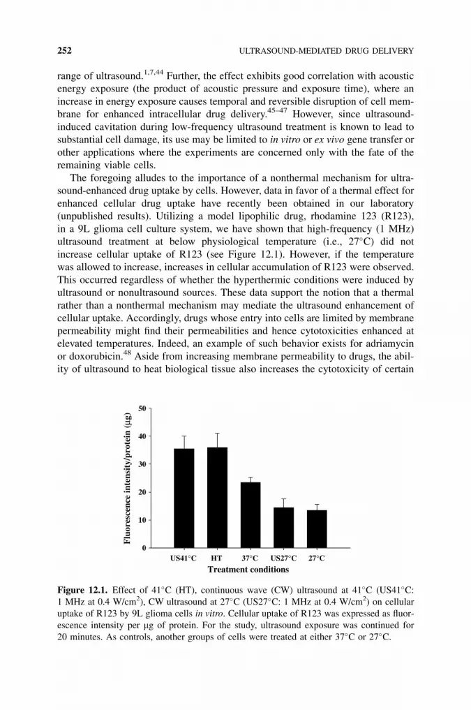
range of ultrasound.1,7,44 Further, the effect exhibits good correlation with acoustic
energy exposure (the product of acoustic pressure and exposure time), where an
increase in energy exposure causes temporal and reversible disruption of cell mem-
brane for enhanced intracellular drug delivery.45–47 However, since ultrasound-
induced cavitation during low-frequency ultrasound treatment is known to lead to
substantial cell damage, its use may be limited to in vitro or ex vivo gene transfer or
other applications where the experiments are concerned only with the fate of the
remaining viable cells.
The foregoing alludes to the importance of a nonthermal mechanism for ultra-
sound-enhanced drug uptake by cells. However, data in favor of a thermal effect for
enhanced cellular drug uptake have recently been obtained in our laboratory
(unpublished results). Utilizing a model lipophilic drug, rhodamine 123 (R123),
in a 9L glioma cell culture system, we have shown that high-frequency (1 MHz)
ultrasound treatment at below physiological temperature (i.e., 27�C) did not
increase cellular uptake of R123 (see Figure 12.1). However, if the temperature
was allowed to increase, increases in cellular accumulation of R123 were observed.
This occurred regardless of whether the hyperthermic conditions were induced by
ultrasound or nonultrasound sources. These data support the notion that a thermal
rather than a nonthermal mechanism may mediate the ultrasound enhancement of
cellular uptake. Accordingly, drugs whose entry into cells are limited by membrane
permeability might find their permeabilities and hence cytotoxicities enhanced at
elevated temperatures. Indeed, an example of such behavior exists for adriamycin
or doxorubicin.48 Aside from increasing membrane permeability to drugs, the abil-
ity of ultrasound to heat biological tissue also increases the cytotoxicity of certain
Treatment conditionsUS41°C HT 37°C US27°C 27°C
Flu
ores
cenc
e in
tens
ity/
prot
ein
(µg)
0
10
20
30
40
50
Figure 12.1. Effect of 41�C (HT), continuous wave (CW) ultrasound at 41�C (US41�C:1 MHz at 0.4 W/cm2), CW ultrasound at 27�C (US27�C: 1 MHz at 0.4 W/cm2) on cellular
uptake of R123 by 9L glioma cells in vitro. Cellular uptake of R123 was expressed as fluor-
escence intensity per mg of protein. For the study, ultrasound exposure was continued for
20 minutes. As controls, another groups of cells were treated at either 37�C or 27�C.
252 ULTRASOUND-MEDIATED DRUG DELIVERY

drugs, such as nitrosoureas, whose cytotoxic reactions follow an Arrhenius-type
law, i.e., the rate constant governing the reaction increases more or less exponen-
tially with temperature.48
12.3.1.3. Ultrasound as Adjuvant to Overcome the Clinical Effects of MultidrugResistance Multidrug resistance (MDR) represents a major obstacle to successful
chemotherapy of metastatic diseases.49 One of the best-understood mechanisms of
MDR is the overexpression of P-glycoprotein (P-gp).50 This 170 kDa plasma mem-
brane protein belongs to a larger family of ATP-binding cassette proteins and con-
fers resistance to tumor cells by extruding many structurally and functionally
unrelated hydrophobic anticancer drugs using the energy of ATP hydrolysis.51
High-frequency ultrasound (MHz) ranging from 0.5 W/cm2 to several watts
could increase treatment temperatures and enhance the activity of several hydro-
phobic drugs (Table 12.1). It would appear that ultrasound-induced heat could
have potential in increasing cellular uptake of P-gp substrates and thereby reducing
the clinical effects of MDR. We recently conducted studies to examine this possi-
bility by investigating how mild heat (41�C) produced by a nonultrasound heat
source (HT) or 1 MHz ultrasound (ultrasound-induced mild heat (USMH)) could
affect (1) the cellular uptake of P-gp substrates, R123 and doxorubicin and (2)
the cytotoxicity of doxorubicin in the parent and P-gp-overexpressing variants of
two human cancer cell lines, MV522 and KB.40 Our results indicated that both
experimental conditions led to increased cellular uptake of R123 and doxorubicin
in the parent and P-gp-overexpressing variants of the two cell lines (Figure 12.2). In
parallel, the cytotoxicity of doxorubicin, was also enhanced. Importantly, the
enhancement of uptake of R123 and doxorubicin, and of the cytotoxicity of doxor-
ubicin produced by HT or USMH, was far better than that produced by the P-gp
modulator, verapamil (Figure 12.3).
Because heating has been shown to induce a reduction in P-gp expression, lead-
ing to inhibition of drug efflux and enhancement of intracellular drug uptake, we
recently examined the expression and activity of P-gp in both heat-treated and
untreated cells. Both the expression level52 and the activity of P-gp were unaffected
by the heat treatment.53 These results imply that a mechanism other than modula-
tion of P-gp activity might be responsible for the enhanced drug accumulation by
USMH. Our recent data suggest that the site of action of USMH might be the cell
membrane and that USMH selectively enhances the permeability of hydrophobic
but not hydrophilic drugs.53 If USMH were to increase membrane permeability
to hydrophobic drugs, it is possible that the enhanced drug entry rate might tem-
porarily overwhelm P-gp and lead to more cellular drug accumulation.
It is reckoned that if the enhanced drug uptake could be maintained in the P-gp-
expressing cells longer, an augmented binding of the anticancer drug to its target
and, therefore, increased cytotoxicity might be obtained. To that end, we tested the
effects of combining USMH, which enhances cellular drug uptake, with PSC 833,
which modulates P-gp activity and hence promotes cellular drug retention, on cyto-
toxicity of doxorubicin in the parent and MDR cells.41 The results of this study
are shown in Figure 12.4. As shown in this figure, cytotoxicity of anticancer drugs
ULTRASOUND APPLICATIONS IN DRUG DELIVERY 253
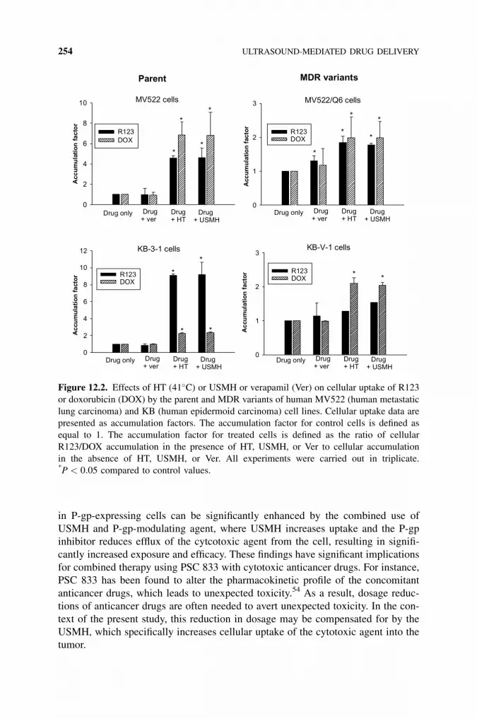
in P-gp-expressing cells can be significantly enhanced by the combined use of
USMH and P-gp-modulating agent, where USMH increases uptake and the P-gp
inhibitor reduces efflux of the cytcotoxic agent from the cell, resulting in signifi-
cantly increased exposure and efficacy. These findings have significant implications
for combined therapy using PSC 833 with cytotoxic anticancer drugs. For instance,
PSC 833 has been found to alter the pharmacokinetic profile of the concomitant
anticancer drugs, which leads to unexpected toxicity.54 As a result, dosage reduc-
tions of anticancer drugs are often needed to avert unexpected toxicity. In the con-
text of the present study, this reduction in dosage may be compensated for by the
USMH, which specifically increases cellular uptake of the cytotoxic agent into the
tumor.
Drug+ USMH
Drug+ ver
Drug only Drug+ HT
Parent MDR variants
MV522 cells MV522/Q6 cells
Acc
umu
lati
on
fact
or
0
2
4
6
8
10
R123DOX
Acc
um
ula
tio
nfa
cto
r
0
1
2
3
Drug+ USMH
Drug+ ver
Drug only Drug+ HT
R123DOX
0
2
4
6
8
10
12
R123DOX
Acc
um
ula
tio
nfa
cto
r
Acc
um
ula
tio
nfa
cto
r
Drug+ USMH
Drug+ ver
Drug only Drug+ HT
Drug+ USMH
Drug+ ver
Drug only Drug+ HT
KB-3-1 cells KB-V-1 cells
*
*
*
*
*
**
**
*
*
*
*
0
1
2
3
R123DOX
* *
Figure 12.2. Effects of HT (41�C) or USMH or verapamil (Ver) on cellular uptake of R123
or doxorubicin (DOX) by the parent and MDR variants of human MV522 (human metastatic
lung carcinoma) and KB (human epidermoid carcinoma) cell lines. Cellular uptake data are
presented as accumulation factors. The accumulation factor for control cells is defined as
equal to 1. The accumulation factor for treated cells is defined as the ratio of cellular
R123/DOX accumulation in the presence of HT, USMH, or Ver to cellular accumulation
in the absence of HT, USMH, or Ver. All experiments were carried out in triplicate.*P < 0:05 compared to control values.
254 ULTRASOUND-MEDIATED DRUG DELIVERY

12.3.2. Thrombolytic Drugs
12.3.2.1. Thrombosis and Its Clinical Implications As a normal hemostatic
response to limit hemorrhage from microscopic or macroscopic vascular injury,
the body undergoes a process termed ‘‘local thrombosis.’’ Specific proenzymes
and proteins, platelets, and calcium participate in this process. The end result is
the formation of insoluble fibrin, which mechanically blocks the flow of blood
through ruptured vessels.55 Thrombosis is usually counterbalanced by physiological
anticoagulation and thrombolysis. Under normal conditions, the thrombus is con-
fined to the area of vessel injury and rarely obstructs flow to critical areas. However,
%in
hib
itio
n
0
20
40
60
80
100
DOX+ USMH
VerDOX+ ver
DOXUSMHDOX+ HT
%in
hib
itio
n
0
20
40
60
80
100
%in
hib
itio
n
0
20
40
60
80
100
%in
hib
itio
n
0
20
40
60
80
100
**
* *
**
**
Parent MDR variants
MV522 cells
KB-3-1 cells
MV522/Q6 cells
KB-V-1 cells
DOX+ USMH
VerDOX+ ver
DOXUSMHDOX+ HT
DOX+ USMH
VerDOX+ ver
DOXUSMHDOX+ HT
DOX+ USMH
VerDOX+ ver
DOXUSMHDOX+ HT
Figure 12.3. Effects of HT (41�C) or USMH or verapamil (Ver) on cytotoxicity enhance-
ment of doxorubicin (DOX) in the parent or MDR variants of human MV522 and KB cell
lines. Data are expressed as % inhibition calculated by the following formula: % inhibi-
tion¼ [1� (counts of viable drug-exposed cells/counts of viable non-drug-exposed
cells)]� 100. Cell viability was determined by hemocytometry technique after trypan blue
staining. All experiments were carried out in triplicates. *P < 0:05 compared to control
values.
ULTRASOUND APPLICATIONS IN DRUG DELIVERY 255

Con
trol
+P
SC
+D
OX
+P
SC
+D
OX
%inhibitionofproliferation
0255075100
125
KB
-V-1
,C
KB
-V-1
,C
(US
HT
)B
Con
trol
+P
SC
+D
OX
+P
SC
+D
OX
%inhibitionofproliferation
0255075
100
125
KB
-3-1
,C
KB
-3-1
,41
˚C(U
SH
T)
A
PS
C+
DO
X+
PS
C+
DO
X
%inhibitionofproliferation
0255075100
125
KB
-V-1
,37°
CK
B-V
-1,4
1°C
(US
HT
)B
PS
C+
DO
X+
PS
C+
DO
X
%inhibitionofproliferation
0255075
100
125
KB
-3-1
,37
°CK
B-3
-1,
41°C
(US
HT
)A
Con
trol
+P
SC
+D
OX
+P
SC
+D
OX
%inhibitionofproliferation
0255075100
125
MV
-522
,37°
CM
V-5
22,4
1°C
(US
HT
)C
Con
trol
+P
SC
+D
OX
+P
SC
+D
OX
%inhibitionofproliferation
0255075100
125
MV
-522
/Q6,
37°C
MV
-522
/Q6,
41°C
(US
HT
)D
Figure
12.4.Effectofcombined
use
ofUSMH(41� C
)andPSC833oncytotoxicityofdoxorubicin
intheparentandMDRvariantofMV522andKB
lines.Datawereexpressed
as%
inhibitioncalculatedbythefollowingform
ula:%
inhibition¼[1
�(countsofviable
drug-exposedcells/countsof
viable
non-drug-exposedcells)]�100.Allexperim
entswerecarriedoutin
triplicate
(mean�standarddeviation).
256

under pathological conditions, the thrombus can dislodge from the area of vessel
injury and enters the blood flow. The thrombus can then lodge in a critical vessel,
and can obliterate valves and other structures that are essential to normal hemody-
namic function. Abnormal thrombosis can occur in any vessel at any location in the
body. The principal clinical syndromes that result are acute myocardial infarction,
deep vein thrombosis, pulmonary embolism, acute non-hemorrhagic stroke, and
acute peripheral arterial occlusion.55
12.3.2.2. Limitations of Present Treatment Modalities for Thrombosis The suc-
cess of treatment of abnormal thrombosis depends on how rapidly one can restore
blood flow to the obstructed vessels, because tissue death is closely related to the
duration of ischemia.55 Although recanalization of acutely thrombosed arteries can
be achieved by mechanical interventions, such as balloon angioplasty,56 this
technique is not considered feasible for conditions such as stroke. Reperfusion
by such techniques have serious limitations, including the requirement for
specialized facilities and highly trained personnel, and these procedures often
result in more complications in older patients.57–62 Consequently, drug-induced
thrombolysis has surfaced as an alternative for treatment of pathological thrombo-
sis.63 Thrombolytic agents available today are serine proteases, such as tissue type
plasminogen activator (t-PA), urokinase, and streptokinase, that work by converting
plasminogen to the natural thrombolytic agent, plasmin. Plasmin lyses thrombotic
vascular occlusions by degrading fibrinogen and fibrin contained in a blood
clot. Thrombolytic agents, however, also have limited success in recanalizing
thrombotically occluded arteries.64,65 For instance, in ischemic stroke
treatment, thrombolytic therapy with t-PA demonstrated a small but significant
improvement in neurological outcome in selected patients. As such, there is a
need to enhance the effectiveness of thrombolytic agents by shortening the time
to reperfusion.
12.3.2.3. Ultrasound and Its Role in the Treatment of Thrombosis Among the
first to study the application of ultrasound to enhance the effectiveness of thrombo-
lytic agents was Kudo,66 who showed that, when used together with recombinant t-
PA, ultrasound can accelerate recanalization of thrombotically occluded arteries in
vivo. This observation was later confirmed by other investigators in various in vitro
studies67–76 and in animal models of arterial9,77 and small-vessel thrombosis.78,79
Consistent findings among these studies are: (1) the rate of thrombolytic enhance-
ment was directly related to the temporal average intensity of the field; (2) the
ultrasound-induced enhancement of thrombolysis was greater at higher throm-
bolytic concentrations; (3) a lower concentration of thrombolytic agent in the
presence of ultrasound could induce more thrombolysis than a higher concentration
in the absence of ultrasound; (4) enhancement was due primarily to accelerated
enzymatic (thrombolytic) action rather than mechanical disruption of the
clot;80 and (5) enhancement was not limited to t-PA-specific drugs; the activity
of other thrombolytic drugs, such as urokinase and streptokinase, was also
increased.80
ULTRASOUND APPLICATIONS IN DRUG DELIVERY 257

12.3.2.4. Mechanisms of Ultrasound-Enhanced Thrombolysis Ultrasound is
known to have several biological effects, depending on the emission characteristics.
At higher energy levels, ultrasound alone has a thrombolytic effect. This effect is
already used for clinical purposes in interventional therapy using ultrasound cathe-
ters. At high intensities, ultrasound delivered via a transducer causes mechanical
fragmentation through direct mechanical contact. In contrast, the use of lower-
intensity ultrasound to accelerate enzymatic thrombolysis without mechanical dis-
ruption of the thrombus offers a conceptually different approach to thrombolytic
therapy. The mechanisms by which ultrasound enhances enzymatic thrombolysis
are multiple and relate primarily to transport (refer to Francis80 for a recent review).
Using 1 MHz ultrasound at 4 W/cm2, Francis et al.81 demonstrated that ultrasound
significantly increases the rate of uptake and penetration of t-PA into the clots.
Ultrasound also increases thrombolysis by pressure-mediated perfusion of thrombo-
lytic agents into the matrix. For instance, Siddiqi et al.82 examined the effects
of ultrasound on fluid permeation through purified fibrin gels and found that at
2 W/cm2, 1 MHz ultrasound increased flow through the gel. Flow through the
gel was dependent on the ultrasound intensity (hence acoustic pressure) and the
resistance of the fibrin gel (a property determined by the fibrin fiber structure). In
the context of this study, ultrasound promotes thrombolysis by altering the fiber
structure. Indeed, when the ultrastructure of fibrin gel after ultrasound treatment
was examined using scanning electron microscopy, significant changes in structure
were observed.83 Such alterations could modify flow resistance as well as create
additional binding sites for thrombolytic enzymes, thereby improving thrombolytic
efficacy.
In order to study if inertial cavitation might be the mechanism by which ultra-
sound enhanced the dissolution of a blood clot when the clot was exposed to a
thrombolytic agent, Everbach and Francis84 examined the dissolution of radiola-
beled fibrin clots exposed to 1 MHz ultrasound in a rotating sample holder, a con-
dition which enhances ultrasound-induced nonthermal effects such as inertial
cavitation.44 To suppress acoustic cavitation, the exposure tank was positioned in
a hyperbaric chamber capable of pneumatic pressurization to 10 atmospheres. In
these studies, static pressure could only reduce the acceleration of thrombolysis
by one-half, suggesting that other processes in addition to inertial cavitation might
be responsible for the enhanced effect of ultrasound on thrombolysis. Hence, much
work is needed to shed further light on the mechanism of this phenomenon.
12.3.2.5. Lower-Frequency Ultrasound in Ultrasound-Enhanced ThrombolysisThe pioneering work by Kudo66 used relatively low-frequency ultrasound (200 kHz
or 0.2 MHz). However, most investigators used higher frequencies (MHz range)
with subcavitation intensity. Suchkova et al.85,86 and others76,87 recently reempha-
sized the advantage of lower-frequency and low-intensity ultrasound (40 kHz; <1
W/cm2) to optimize ultrasound-enhanced thrombolysis. Suchkova et al.85,86
suggested that, at frequencies exceeding 1 MHz, the intensities necessary to achieve
significant enhancement of thrombolysis might lead to substantial and harmful
tissue heating. Lower frequencies should result in lower absorption of ultrasound
258 ULTRASOUND-MEDIATED DRUG DELIVERY

by tissues and cause less heating. In addition, with lower-frequency ultrasound, the
acoustics field can be broader and more uniform, with greater depth of penetration
than occurs at MHz frequencies.
Because high frequency ultrasound is transmitted with a high attenuation
through the skull,88 Daffertshofer et al.76,89,90 have recently examined the potential
use of low frequency ultrasound (20–300 kHz) for accelerating enzymatic throm-
bolysis as a potential therapy for ischemic stroke treatment. As a first step, they
studied ultrasound transmission through a sample of different postmortem skulls.
Their findings indicated very low attenuation in these skulls. It was also demon-
strated that transmission of low-intensity (0.5 W/cm2) and low-frequency ultra-
sound through the skull was sufficient to accelerate t-PA-mediated thrombolysis
(Figure 12.5). These conditions were also found to be safe. No evidence for break-
down of the blood-brain barrier was observed in postmortem ultrasound-treated
rats. Collectively, these data suggest that low-frequency ultrasound may reach the
brain and intracranial vessels with thermally acceptable levels for enhancement of
systemic intracranial t-PA-mediated thrombolysis.
12.3.3. Proteins
12.3.3.1. Limitations of Transdermal Drug Delivery Recent completion of the
Human Genome Project91 and advances in molecular biology have enabled the dis-
covery of many novel drugs that are either peptides or proteins. Because peptides
and proteins are metabolically labile and undergo extensive enzymatic degradation,
they cannot be administered to patients by the traditional oral route. Currently, most
peptide and protein drugs are given to patients by injection. However, injection is
invasive and can instill psychological fears in patients, which decreases patient
Figure 12.5. Thrombolytic activity: spontaneously after 1 hr in a control group, and after 1 hr
treatment with transcranial US (33 kHz, CW, 0.5 W/cm2) insonation only, after 1 hr treatment
with rt-Pa only, and 1 hr treatment with combined USþ rt-PA (from Ref. 90).
ULTRASOUND APPLICATIONS IN DRUG DELIVERY 259

compliance. Thus, pharmaceutical scientists have looked at alternative routes such
as the transdermal route for effective systemic delivery of these compounds.
Transdermal drug delivery offers several advantages over traditional drug deliv-
ery systems such as oral delivery and injections.92 These include avoidance of first-
pass metabolism, elimination of the pain associated with injection, and an opportu-
nity for sustained release of drugs. The transdermal route of administration can be
especially beneficial in the delivery of chronic injectable medications where patient
compliance is low. However, transdermal transport of molecules is low because
human skin is an effective and selective barrier to chemical permeation.93 Experi-
ments have traced the major barrier to drug permeation to the stratum corneum,94
the outermost layer of the skin. The stratum corneum is composed of dead, flattened
corneocytes surrounded by lipid bilayers. This arrangement resembles the ‘‘brick
and mortar’’ structure of a wall where the dead corneocytes comprise the ‘‘brick’’
embedded in a ‘‘mortar’’ composed of multiple lipid bilayers of ceramides, fatty
acids, cholesterol, and cholesterol esters (reviewed in Barry and William95). Indeed
the low permeability of the stratum corneum is the main reason only a handful of
low molecular weight drugs are administered by this route today.92 A possible solu-
tion to this problem is to increase drug flux by reducing this barrier’s hindrance
using physicochemical forces such as chemical penetration enhancers or electric
fields.96 Penetration enhancers swell and disorder stratum corneum, facilitating pas-
sive diffusion of drugs. However, penetration enhancers, whether they are simple
solvents like ethanol or complex compounds such as cyclic alkylamides, have sig-
nificant limitations.97 For instance, simple solvents tend to leave the transdermal
drug delivery system and be absorbed rapidly into the body, and complex com-
pounds may produce questionable metabolic products. Most importantly, penetra-
tion enhancers cause skin irritation and generally do little to enhance the delivery of
hydrophilic high molecular weight proteins. An alternative approach is to use an
electric field (iontophoresis) to drive drug molecules across the skin.92,96 But this
approach works only for charged molecules, and thus is ineffective for delivering
drugs that carry either no charge or relatively low electric charges, such as proteins.
In addition, iontophoresis is known to induce minor skin irritation. Thus, a rela-
tively safe approach that works for a wider range of drugs will need to be developed
for effective transdermal delivery of drugs such as proteins.
12.3.3.2. Ultrasound-Enhanced Transdermal Delivery of Proteins Ultrasound
has been used for enhancing transdermal transport under a variety of conditions,
using different combinations of frequency, intensity, and exposure time.98 This phe-
nomenon is referred to as ‘‘phonophoresis’’ or ‘‘sonophoresis.’’ Most of the earliest
transdermal drug delivery research used high-frequency ultrasound (MHz) to pro-
mote delivery of small molecular weight hydrophobic anti-inflammatory drugs into
the skin for physical therapy.98 Given the heightened need to develop delivery strat-
egy for peptides and proteins, several research groups have looked at the use of
therapeutic ultrasound (i.e., high frequency in the MHz range) to promote transder-
mal delivery of proteins. However, measurable enhancement of protein transport
has been reported in only a few cases.15,16 Previous work has demonstrated that
260 ULTRASOUND-MEDIATED DRUG DELIVERY

application of ultrasound at therapeutic frequencies (MHz range) induces growth
and oscillation of air pockets present in the corneocytes of the stratum corneum.99
These oscillations disorganize the stratum corenum lipid bilayer, thereby enhancing
transdermal transport. Because cavitational effects are inversely proportional to
ultrasound frequency,100 Robert Langer and his group at the Massachusetts Institute
of Technology reasoned that application of ultrasound at frequencies lower than
those corresponding to therapeutic ultrasound might induce sufficient bilayer disor-
ganization so that proteins might be able to diffuse across the skin. Indeed, their
seminal work indicated that application of low-intensity (mW/cm2 range) and
low-frequency ultrasound (20 KHz) increased the in vitro human skin permeability
of various proteins such as insulin (molecular weight �6000), interferon-g(�17,000), and erythropoeitin (�48,000) by several orders of magnitude.17 This
enhancement effect was found to vary nearly exponentially with ultrasound inten-
sity, a result the investigators attributed to the highly nonlinear dependence of cavi-
tation on ultrasound intensity.101 The observations suggest that ultrasound intensity
might potentially be used to control transdermal protein delivery. Their data also
indicated that low-intensity and low-frequency ultrasound did not cause any perma-
nent loss of the barrier properties of the skin; most of the skin barrier properties
could be recovered very quickly (less than a day) after sonophoresis. The results
suggest that sonophoresis delivers intensity-dependent protein doses across the
skin, which provides a platform for future noninvasive delivery of proteins across
the skin. As the investigators correctly alluded to in their study, the success of such
an endeavor would require extensive investigation on the physiological and immu-
nological effects of ultrasound exposure and optimal selection of ultrasound para-
meters, such as frequency, pulse length, and intensity. Because recent studies
indicated that coupling get might influence ultrasound-mediated transdermal drug
delivery,99,102,103 optimal selection of an ultrasound-coupling medium will be
important for effective transdermal delivery of proteins. Finally, the first ultrasound
experiments used laboratory-scale sonicators that are cumbersome. Thus, any rea-
listic commercialization of this innovative concept of drug delivery will depend on
the development of pocket-size sonicators. Realizing this potential problem, engi-
neers at the Pennsylvania State University have miniaturized the system by arraying
tiny cymbal-shaped transducers on a 1.5-in2 patch.104–106 Using this lightweight
ultrasonic device, engineer Nadine Smith and her group recently reported that
they delivered therapeutic levels of insulin to rats.104 These results provide hope
that a portable ultrasound system will one day be developed for transdermal drug
delivery.
12.3.3.3. Mechanism of Enhancement of Transdermal Drug Delivery by Low-Frequency Ultrasound The biophysical modes of ultrasonic action on biological
system can be classified into two categories: (1) thermal mechanism and (2)
nonthermal mechanism.2,107 The thermal effects of ultrasound, which are directly
related to its intensity, result from transfer of energy from the vibrating pressure
waves to the objects that the wave contacts. In transdermal application, this results
in absorption of energy by the skin, causing a rise in skin temperature. Although
ULTRASOUND APPLICATIONS IN DRUG DELIVERY 261

literature supports the observation that increasing temperature leads to an increment
in skin permeability,99 recent studies indicate that thermal effects play an insignif-
icant role in enhanced transdermal drug transport by low-frequency ultra-
sound.108,109 For instance, low-frequency ultrasound (20 kHz at 15 W/cm2 for
2 hours) caused a 20�C rise in temperature that resulted in a 35-fold increase in
permeability of mannitol across in vitro porcine skin. In contrast, when the skin
was heated (in the absence of ultrasound) in a manner similar to the thermal profile
produced by sonophoresis, the permeability of mannitol increased by only 25%.109
These data indicate that the key mechanism responsible for the observed skin per-
meability is related to the nonthermal effects of ultrasound. As mentioned before,
the nonthermal effects of ultrasound on biological system can occur via one or a
combination of three mechanisms: (1) cavitation, (2) radiation pressure, and (3)
acoustic microstreaming.2,107 Among these nonthermal effects, acoustic cavitation
is believed to be the most important. Acoustic cavitation is defined as the acousti-
cally induced activity of gas-filled cavities (involving nucleation, oscillation, and
collapse).107 Two types of cavitational activities are known: (1) noninertial or stable
cavitation and (2) inertial or transient cavitation.107 In noninertial cavitation, the
radius of the bubbles oscillates about an equilibrium value without collapsing
over a considerable number of acoustic cycles. In contrast, the bubbles in inertial
cavitation grow rapidly within one or two acoustic cycles before collapsing vio-
lently during a single compression half cycle. This violent event may generate
defects (pores or holes) in the skin that explain the enhanced skin permeability.
Based on indirect experimental observations, such as the appearance of ‘‘holes’’
or irregularities in the skin tissue after low-frequency sonophoresis,16,110,111 one
could argue for transient cavitation as the predominant mechanism for enhanced
skin permeability. Indeed, a recent study has provided evidences for such a
claim.112 In addition, the critical cavitation site has been identified and found to
locate on, or in the vicinity of, the skin membrane.112 In an effort to determine
the dependence of transport pathways (defects that occur between/within the lipid
lamellar organization in the stratum corneum) during low-frequency sonophoresis
on ultrasound parameters, Tezel et al.108 exposed in vitro porcine skin to low-fre-
quency ultrasound over a range of frequencies (20–100 kHz) and energy densities
(0–2000 J/cm2: product of ultrasound intensity, I, and application time, t). Then the
porous pathway model developed by Tang et al.113 was used to study the depen-
dence of average pore size, porosity, and tortuosity on ultrasound parameters.
The data show that low-frequency ultrasound increases skin permeability by
increasing skin porosity (up to 1700-fold) and/or decreasing tortuosity rather than
by increasing the size of the pores. These findings provide quantitative guidelines
for estimating the efficacy of sonophoresis, which should prove useful for future
transdermal drug delivery research.
12.3.4. Gene-Based Drugs
12.3.4.1. Historical Perspective of Gene Therapy During the past several
decades, the promising concept of gene therapy has emerged. This approach has
262 ULTRASOUND-MEDIATED DRUG DELIVERY

potential implications for the treatment of over 4000 human inherited diseases asso-
ciated with dysfunctional, nonfunctional, or missing gene-coded proteins.114 The
term ‘‘gene therapy’’ was originally coined to describe a proposed treatment of
genetic disorders by directing treatment to the site of the defect itself—the mutant
gene—rather than to secondary or pleiotropic effects of mutant gene products.115
Technically, gene therapy consists of replacing, at least in a functional sense, a de-
fective gene with its normal counterpart. Inevitably, the technical hurdle of gene
therapy is gene delivery: that is, the introduction of genes into the appropriate cells
of the patient. Thus, development of novel methods for rapid and efficient delivery
of therapeutic genes of choice into mammalian cells has formed the basis of numer-
ous gene therapy studies recently. Delivery of nucleic acids into mammalian cells
can be made either via virus vector or by physical and chemical methods.115 Viral
vectors tend to achieve the highest efficiency; however, substantial concerns remain
over their clinical safety and long-term efficacy.116 Genes can also be delivered as
plasmid DNA to cells directly by a number of methods. These techniques are cate-
gorized into two general groups: (1) naked DNA delivery by a physical method,
such as electroporation, gene gun, high-pressure injection and ultrasound,117,118
and (2) delivery mediated by a chemical carrier such as a cationic polymer and
lipid.117,118 Although the efficiency of gene delivery using nonviral techniques is
relatively low (at least 10-fold less than that of viral vectors), the advantages of
using plasmid DNA include (1) easy manipulation and ability to deliver large
inserted sequences; (2) can be produced stably and cheaply to a high level of purity
with minimal risk of replication or incorporation; and (3) plasmid DNA is weakly
immunogenic and can therefore be administered repeatedly to immunocompetent
subjects. As a result, a significant number of gene therapy researchers are attempt-
ing to increase the efficiency of plasmid DNA transfer into cells.
12.3.4.2. Ultrasound-Mediated Gene Delivery and Its Mechanism Ultrasound
generated by a needle-tip sonicator (commonly used in the laboratory for cleaning
and cell disruption) has been shown to enhance gene transfer into both mammalian
and plant cells.119–121 In these studies, relatively high peak ultrasonic power levels
(�100 W) and low frequencies (20–50 kHz) were employed, leading to the possi-
bility that acoustic cavitation played a major role in gene transfer in these studies.
Miller et al.44 demonstrated that the primary biological effect of acoustic cavitation
is cell lysis. Nevertheless, the previous studies suggest that sublethal damage, such
as transient permeabilization of the cell membrane, may also occur during cavita-
tion, which leads to the uptake of molecules into the cells. This phenomenon, called
‘‘sonoporation,’’ has been confirmed in numerous other studies. For example, ultra-
sonic shock waves (ultrasonic waves with very high pressure amplitude and hence
high intensity), which favor production of cavitation, have been shown to increase
cellular uptake of both small (adriamycin and fluorescein)122 and macromolecular
(fluorescein-labeled dextrans, ribosome-inactivating proteins gelonin and
saporin)105,123,124 molecules. The use of ultrasonic shock waves has been evaluated
recently for enhancement of gene transfection. In vitro studies demonstrated that
DNA plasmids entered cultured cells during ultrasound shock wave exposure,
ULTRASOUND APPLICATIONS IN DRUG DELIVERY 263

and that some of the cells survived to express the plasmid gene product.125–130
However, sonoporation was accompanied by substantial cell lysis because of the
very high ultrasound intensity used in these studies, and the transfection efficiency
produced by such a method is very low (0.08% to 0.5%). In this regard, several
studies have evaluated the use of therapeutic levels of ultrasound to enhance
gene transfection. For example, Tata et al.131 studied the ability of low-intensity
(0.33 W/cm2) and high-frequency ultrasound to enhance green fluorescent protein
(GFP) reporter gene expression and obtained 20% transfection at about a 100 Hz
pulse rate and a 120-second exposure in different cell types. Utilizing 1 MHz ultra-
sound at a spatial average peak positive pressure (SAPP) of 0.41 Mpa (�10 W/
cm2), Greenleaf et al.132 obtained even better transfection reaching about 50% of
the living cells. This transfection efficiency was comparable to that of other
high-performance techniques of transfection, such as lipofection.132 Because ultra-
sound is strictly a physical method, it is not bound by limitations currently affecting
lipofection. For instance, the efficacy of plasmid DNA uptake into cells by lipofec-
tion depends on the net charge of the cationic lipids and DNA complexes.133,134
This implies that an optimal cationic lipid to DNA ratio must be determined for
each DNA type and concentration. In addition, the formation of cationic lipid/
DNA complexes is highly dependent on other parameters such as the pH and con-
centration of electrolytes.133,134 As a result, the use of cationic lipids in lipofection
must be optimized for each experimental setting, and inconsistent results may
occur. One possible drawback of using ultrasound to transfect plasmid DNA into
cells is the relatively large amounts of plasmid DNA needed to obtain a competitive
transfection rate.132 However, plasmid DNA is comparatively inexpensive, and the
possible site-specific in vivo applications of this method generally outweigh this
small disadvantage.
It is clear that ultrasound can provide enhanced transfer of DNA plasmids into
cells, but it will be important to extend these findings to an in vivo setting. Indeed,
several studies have shown that ultrasound could enhance reporter gene expressions
in vivo.135–137 The main mechanism by which ultrasound enhances gene transfer is
thought to be acoustic cavitation, which can affect transient nonlethal perforations
in the plasmalemma and possibly the nuclear membranes.138 Because the ultra-
sound contrast agent lowers the threshold for cavitation by ultrasound energy
(see the next section), it is expected that its use during ultrasound application
will further promote pore formation in cell membranes, thus facilitating the entry
of plasmids into cells and their release from endosomes. Indeed, numerous studies
that examined the combination of microbubbles with ultrasound have reported sig-
nificant increase in gene transfection.
Most recently, low-intensity ultrasound has been used to enhance gene transfec-
tion by liposome.139 In this study, ultrasound exposure (1 MHz, 0.4 W/cm2) for
60 seconds enhanced transfection of naked or liposome-complexed luciferase
reporter plasmid into cultured porcine vascular smooth muscle and endothelial
cells. These results with liposome-complexed reporter plasmid are similar to those
of Unger et al., who showed that relatively low levels of ultrasound energy (0.5 W/
cm2) enhanced gene expression.140 In these two studies, ultrasound exposure did
264 ULTRASOUND-MEDIATED DRUG DELIVERY

not begin until 30 minutes139 or 60 minutes140 into the transfection period. Accord-
ingly, it may be hypothesized that ultrasound might enhance gene transfection
through its ability to accelerate DNA escape from endosomes. Other site(s) of
action of ultrasound might include effects on plasmid DNA entry, intracellular traf-
ficking, lysosomal degradation, nuclear translocation, RNA transcription, or protein
translation. Although not proven, these plausible mechanisms might be responsible
for the recently published data on enhanced expression of a plasmid DNA-catio-
nized gelation complex by ultrasound in murine muscle.141
12.4. ULTRASOUND CONTRAST AGENTS
12.4.1. Ultrasound Contrast Agents as Imaging Agents
Over the past decade, the development of stable microbubbles has extended to the
pharmaceutical arena as ultrasound contrast agents. Knowledge of microbubbles or
gas-water interfaces as efficient backscatterers of sound waves has existed for many
decades. Even before it was conceived that they could be used for clinical ultra-
sound image enhancement, the U.S. Navy was interested in bubble technology
because sound or sonar was an efficient means for detecting ships and submar-
ines—the reason being that submarines could be made of stealth components but
could not completely hide their propulsion systems, which would form a trail of
bubbles or cavitation nuclei, detectable by ultrasound.
The first demonstration of the clinical application of microbubbles came in 1968
when Gramiak and Shah142 demonstrated that introduction of agitated saline con-
taining gas bubbles into the aortic root could elicit echo-contrast by diagnostic
ultrasound. Since microbubbles produced by agitation are both large and
unstable,143 they are not considered viable contrast agents. Subsequently, a variety
of techniques were developed to form microbubbles of the right size (1–10 mM) to
traverse the capillaries and, at the same time, stable enough to provide enhancement
over multiple cardiac cycles. Currently, microbubble technology uses a biocompa-
tible gas such as air,142 nitrogen,144 perfluorocarbons,145 or sulfur hexafluoride146 in
a biomaterial coating composed of components such as albumin,147 phospholi-
pids,148 surfactants,149 biopolymers,150 or sugars (galactose).151 In addition, per-
fluorocarbon emulsions,152which can be converted from the liquid phase to the
gas phase upon reaching their boiling point in the body (e.g., perfluoropentane:
bp¼ 29�C), have been utilized as ultrasound contrast agents as well.
The efficiency of ultrasound backscatter owes its sensitivity to the fact that the
reflectivity of sound exhibits a r6 dependence with respect to microbubble diameter.
This was first theorized at the turn of the century by Lord Rayleigh and is appro-
priately referred to as ‘‘Rayleigh scattering.’’ Thus, a 2 mm microbubble exhibits 64
times as much backscatter efficiency as a 1 mm microbubble. The objective of clini-
cally relevant microbubble technology requires formulation of bubbles that are
large enough to maximize the backscatter potential of the gas-water interface but
can still freely course through the capillary bed. In addition, the microbubbles must
ULTRASOUND CONTRAST AGENTS 265

be stable enough to minimize both aggregation and coalescence and, most impor-
tantly, maintain stability in the circulatory system.
The answer to the first problem, i.e., providing a microbubble that is small
enough to freely pass through capillary beds yet large enough to maximize back-
scatter, was solved in a variety of ways, some of which were fortuitous. It turns out
that the size of the microbubbles is highly dependent upon the surface tension of the
shell and gas and is related to the pressure exerted on the bubble by the LaPlace
equation:
P ¼ 2s=r
where
P ¼ pressure
s ¼ surface tension
r ¼ bubble radius
Thus, the use of gases that possess very low surface tension (e.g., perfluorocarbons)
should reduce the pressure against bubble collapse such that the radius of the micro-
bubbles formed would be small. Indeed, perfluorcarbon microbubbles prepared
from perfluoropropane or perfluorobutane form bubble diameters on the order of
1 to 2 mm, which are ideal for passing through capillary beds. In addition, because
of their extremely low solubility in aqueous media, the perfluorocarbon microbub-
bles would not dissolve in the venous blood and are therefore less prone to collapse.
With respect to aggregation and coalescence, techniques similar to liposomal
technology have been employed to maintain microbubble stability. These techni-
ques employ electrostatic charges on the membrane surface to promote electrostatic
repulsion, which hinders aggregation. Typically, membrane lipids that carry
charges, such as phosphatidic acid, are incorporated into the lipid-coated microbub-
bles to help microbubbles to stay apart. To minimize RES uptake and hence main-
tain the stability of the microbubbles in the circulatory system, researchers have
turned to the use of stealth technology. Stealth or pegylation technology was first
used in liposomal research, where drug-filled liposomes were pegylated to extend
their vascular circulating half-life. An example of this was the study by Northfelt et
al.,153 who used membrane-bound polyethyleneglycol (PEG) to prevent RES recog-
nition in their anticancer agent Doxil. Pegylation is now used in a currently mar-
keted microbubble diagnostic agent, Definity, sold by Bristol Myers-Squibb.
Diagnostic applications of microbubbles have been primarily addressed in the
area of echocardiography, where contrasting agents are mainly used to opacify
the left ventricular chamber and aid in the delineation of wall motion.154 In echo-
cardiography, assessment of perfusion in the myocardial tissue has achieved some-
what limited success. Despite the ability of microbubbles to fill the myocardium,
the large amount of contrast in the ventricular chamber causes dynamic range pro-
blems, making imaging of the smaller amount of bubbles in the myocardium very
266 ULTRASOUND-MEDIATED DRUG DELIVERY

difficult. Although some researchers have been successful in qualitatively and semi-
quantitatively determining myocardial perfusion, the technique is far from routine,
with sophisticated pulsing regimes (phase inversion imaging and second harmonic
imaging) and image subtraction often being necessary to image the myocar-
dium.155,156 Currently, microbubble-based ultrasound contrast agents have been
approved only for echocardiography. However, applications for radiologic indica-
tions such as renal and liver perfusion are currently being pursued through the reg-
ulatory (Food and Drug Administration) process and should soon be approved. In
addition, there have been efforts to develop microbubble-based, ultrasound-
enhanced imaging of tumors. The rationale for such development is that the vascu-
larity (or angiogenesis) around the tumor is greater than that of surrounding tissues
and therefore can be contrasted to normal tissue for proper imaging.
12.4.2. Targeted Ultrasound Contrast Agents
Now that it has been demonstrated that stable microbubble technology can be uti-
lized as ultrasound contrast agents, recent research efforts have turned to the devel-
opment of microbubble technology selective identification of vascular landmarks.
Earlier works by Lindner et al.157–159 led to the observation that microbubbles could
adhere to leukocytes in regions of inflammation. It was further demonstrated that
after attachment to neutrophils and monocytes, the microbubbles were phagocy-
tosed intact. Despite viscoelastic damping secondary to encapsulation, the micro-
bubbles remained acoustically reflective. These results led to the conclusion that
microbubbles could potentially image inflammation sites through passive targeting.
The use of targeting ligands to aid in site-selective targeting of microbubbles for
ultrasound diagnostics was soon to follow. Work in the laboratories of Lanza and
coworkers at Washington University in St. Louis introduced the development of a
three-step approach to targeting ultrasound contrast agents selectively to
thrombi.160 Briefly, ligands, which consist of conjugating biotin to antifibrin mono-
clonal antibody that binds to fibrin clots, are administered first. This is followed by
the addition of avidin, which binds to the biotin moiety of the ligands and cross-
links the ligands on the fibrin clots. Finally, biotinlyated perfluorocarbon emulsions
are introduced, which bind to the available sites on the avidin, thereby completing
the ligand-avidin-constrast ‘‘sandwich’’ on the target surface. Using this system,
Lanza and coworkers demonstrated enhanced ultrasound imaging of arterial throm-
bus in a canine model.
A variation of this targeting theme was demonstrated by Lindner and cowor-
kers,161 who hypothesized that tissue retention of microbubbles targeted to the
endothelial cell adhesion molecule P-selectin would provide a means to assess
inflammation with ultrasound imaging. In this technology, phospholipid microbub-
bles targeted to P-selectin (MBp) were made by conjugating the monoclonal anti-
bodies against murine P-selectin to the lipid shell using a small spacer polymer.
Compared with control microbubbles or isotype antibody, the P-selectin and per-
fluorobutane-filled microbubbles were assessed for binding to cremasteric venules
using fluorescent labeling and visualization by intravital microscopy. The results
ULTRASOUND CONTRAST AGENTS 267

indicated that in the P-selection up-regulated [tumor necrosis factor (TNF-a)–stimulated] wild-type mice, microbubble binding of the MBp-conjugated micro-
bubbles was significantly higher than in the P-selectin-deficient mice. Furthermore,
in the P-selectin up-regulated mice, the P-selectin-conjugated microbubbles exhib-
ited greater binding than the control or isotype antibody-conjugated microbubbles.
Thus, it was demonstrated that targeted microbubbles would provide robust targeted
imaging of the thrombus using perfluorocarbon gas microbubbles.
Schumann et al.162 utilized another approach whereby a Arg-Gly-Asp-based
(RGD) peptide ligand (tethered through a PEG moiety to a pseudo-isosteric
lipid-simulating bioconjugate analog, Figure 12.6) that targets GPIIbIIIa receptor
of thrombi was inserted into the lipid membrane of a microbubble for microvascu-
lar imaging of thrombi in the mouse cremasteric arterioles and venules. A pictorial
design for the targeted microbubble is presented in Figure 12.7. Results indicated a
clear linear relationship between bound microbubble and thrombus size, providing
evidence for site targeting of microbubbles. In addition, flow stress studies were
Figure 12.6. Bioconjugate ligand for targeting to GPIIbIIIa receptor of thrombi.
Figure 12.7. Pictorial design of a bioconjugate ligand on the surface of a microbubble for
targeting to the GPIIbIIIa receptor of thrombi.
268 ULTRASOUND-MEDIATED DRUG DELIVERY

conducted which demonstrated that binding of these microbubbles could bind and
adhere to clots under stress flow conditions similar to those of larger vessels.
12.4.3. Ultrasound Contrast Agents and Their Roles in Thrombolysis
Apart from diagnostic imaging, ultrasound-mediated manipulation of microbubbles
may soon have therapeutic potential as well. As noted previously, the efficiency of
thrombolytic agents was increased by nonthermal ultrasound energy such as acous-
tic cavitation. It has recently been shown that ultrasound contrast agents can signif-
icantly lower the acoustic cavitation production threshold.163 This lowering of the
cavitational threshold forms the basis for the induction of local shock waves (due to
destruction of the microbubbles) that could be used as a therapeutic modality for
lysing thrombi or blood clots. This therapeutic intervention, which uses sound
waves to lyse thrombi, has been named ‘‘sonothrombolysis’’ or ‘‘sonolysis.’’ The
earliest study that reported the use of ultrasound contrast agents for sonolysis
was by Tachibana and Tachibana.164 In this study, the authors determined if the pre-
sence of albumin microbubbles (Albunex) used for diagnostic echo contrast could
accelerate thrombolysis caused by urokinase. Their study indicated that the pre-
sence of the ultrasound contrast agent and ultrasound increased thrombolysis by
urokinase. The data thus suggested that the ultrasound contrast agent or microbub-
bles could be used as an adjuvant to enhance ultrasound-induced thrombolysis. This
observation was later confirmed by other investigators in various in vitro72,73,165–167
and in vivo87,113,166 studies. Because targeted microbubbles can detect areas of vas-
cular pathology including stroke, deep vein thrombosis, myocardial infarct, and
peripheral arterial disease, current efforts have focused on targeting the microbub-
ble to thrombi or clots followed by the use of focused or unfocused ultrasound for
disintegration/dissolution of the clots.
12.4.4. Ultrasound Contrast Agents and Their Roles in Gene Delivery
Over approximately the past 5 years, interest has been spurred in the area of ultra-
sound-mediated, microbubble-enhanced gene delivery for reliable and less toxic
gene therapy. The use of microbubbles to carry a drug payload may be somewhat
limiting, as the amount of drug to be carried on a microbubble surface (drugs can-
not be carried in a gaseous interior matrix) may not provide sufficient capabilities to
deliver a therapeutic load. Because gene-based drugs usually require significantly
smaller amounts to generate therapeutic effects, this issue of insufficient drug load-
ing might not be as limiting as was previously thought. In addition, adhering genes
to the surface of the microbubbles may actually reduce the enzymatic degradation
often seen with genes flowing freely in the vascular milieu168 and should promote
the overall efficacy of gene therapy.
Genes are amenable to adherence to the surface of lipid or proteinaceous micro-
bubbles by virtue of their electrostatic (charge-charge) interactions. This has been
achieved through the use of cationic lipids inserted into the membranes of lipid
microbubbles as well as the use of cationic charges from albumin on the surface
ULTRASOUND CONTRAST AGENTS 269
of proteinaceous microbubbles. In the laboratories of Dr. Evan Unger at ImaRx
Therapeutics, avid DNA adherence to the microbubbles was demonstrated after
the cationic lipids 1,2-dimyristyloxypropyl-3-dimethyl-hydroxy ethyl ammonium
bromide (DMRIE) and dioleoylphosphatidylethanolamine (DOPE) were inserted
into lipid microbubble membranes.168 Similarly, utilizing perfluorocarbon-exposed
and sonicated dextrose albumin microbubbles, Dr. Thomas Porter of the University
of Nebraska demonstrated that microbubbles could bind oligonucleotides for the
purpose of drug delivery.154 Successful adherence of genetic materials using catio-
nic gelatin-coated, gas-filled polymeric microspheres was also achieved in the
laboratories of Dr. Fuminori Moriyasu.169 Thus, it was demonstrated in many dif-
ferent formulations by many different laboratories that genes or genetic material
could be bound to microbubble surfaces.
Once it was demonstrated that DNA could be bound to the microbubble surface,
the next step was to demonstrate that microbubbles and ultrasound could be utilized
as a potential gene delivery vehicle. Initial in vitro work with microbubbles was
reported by Lawrie et al.138 from the University of Sheffield. Using both a pGL3
luciferase plasmid and a second plasmid construct whereby the luciferase gene is
driven by the Rous sarcoma virus promoter (pRSVLUC), Lawrie et al. studied the
effect of insonation [1 MHz and a mechanical index (MI) of 1.1 and a 6% duty
cycle for 3 hours] of vascular smooth muscle cells on gene expression. Following
insonation in the presence of albumin-coated microbubbles (Optison), results indi-
cated that there was up to a 300-fold increase in transgene expression compared to
naked DNA alone. This expression was maximized to 3000-fold with the addition
of a polyamine transfection reagent, TransIT-LT1. Since the increase was found
using both constructs, the researchers concluded that the effect of cavitation was
due to microbubbles and ultrasound and probably was not due to ultrasound-
mediated free radical generation. Although the results were compelling, the range
of the data occasionally varied by over an order of magnitude, which made the con-
clusion less robust. Nonetheless, there appeared to be a significant increase in trans-
fection secondary to microbubbles and ultrasound.
Having demonstrated that in vitro transfection could be achieved, a logical next
step was to determine if in vivo expression of ultrasound-mediated gene delivery
could be accomplished with microbubbles. To address this issue, Dr. Mani Vannan
of the University of Michigan studied ultrasound-mediated, microbubble-enhanced
gene delivery of the chloramphenicolacyltransferase gene (CAT) to myocardial tis-
sue in a dog model.170 Upon injection of cationic microbubbles with adherent CAT
into the animal, myocardial tissue was exposed to insonation at 1 MHz ultrasound
using a diagnostic ultrasound machine (HP Sonos 550). The results indicated that
high levels of CAT protein were observed in the myocardium within regions of the
focal zone of ultrasound insonation but not in the myocardial tissue devoid of ultra-
sound insonation. These results demonstrated that ultrasound and microbubbles
could indeed increase delivery of the gene, with subsequent expression of the gene.
Most recently, interesting work in a mouse model has been conducted in the
laboratories of Dr. Martin J.K. Blomley of Imperial College, Hammersmith Hospi-
tal,171 wherein the use of Optison and ultrasound to aid plasmid DNA delivery
270 ULTRASOUND-MEDIATED DRUG DELIVERY

following direct injection into mouse skeletal muscle was investigated. The inves-
tigators used 1 MHz ultrasound at 3 W/cm2 and a 20% duty cycle for 60 seconds.
Results indicated that Optison increased transgene expression in both the absence
and presence of ultrasound. Interestingly, the presence of Optison appeared to
markedly decrease muscle damage that was seen with naked plasmid alone. Of
additional interest was the observation that transgene expression was age-related,
as expression was less efficient in older mice (6 months old) than in younger
mice (4 weeks old). However, the addition of ultrasound increased the transfection
in the older mice but not in the young mice. This result, if further validated, could
imply that cellular age and integrity may have a profound effect on microbubble
and ultrasound-mediated gene delivery.
Clearly, the use of microbubbles and ultrasound for gene delivery is in its
infancy, and many of the results reported in this section have yet to be validated.
Although the exact mechanism by which microbubbles and ultrasound treatment
enhance gene transfer is currently unknown, it is speculated that as the microbub-
bles enter the region of insonation, the microbubbles cavitate, causing local release
of DNA. Cavitation also likely causes a local shock wave that improves cellular
uptake of DNA. Nonetheless, the promising results generated to date, coupled
with the fact that microbubbles and ultrasound may offer higher therapeutic to toxi-
city windows, lead to the conclusion that this avenue of research could result in
compelling new clinical paradigms for the future of gene delivery.
12.5. CONCLUSION
Until recently, medical applications of ultrasound have mostly been associated with
diagnostic medicine. However, the application of ultrasound for therapeutic pur-
poses has recently gained impetus because of the various advances made in the
use of ultrasound to aid drug delivery. Low-intensity (<10 W/cm2) and high- or
low-frequency (0.5 to 10 MHz) ultrasound can enhance the delivery and effective-
ness of several therapeutic drug classes including chemotherapeutic, thrombolytic,
protein-, and DNA-based drugs. Interestingly, ultrasound contrast agents, a recent
invention of diagnostic ultrasound, appear to improve the ability of ultrasound to
enhance the activity of thrombolytic and DNA-based drugs. Ultrasound contrast
agents can significantly lower the threshold for cavitation, a process that has
been suggested to play a partial but very important role in enhancement of drug
effects. As such, ultrasound contrast agents hold great promise in the future of
ultrasound-mediated drug delivery. In summary, the recent successes in
ultrasound-related drug delivery research position ultrasound as a valuable
therapeutic tool for drug delivery in the future.
ACKNOWLEDGMENT
This work was supported by Grant CA79708 from the NIH awarded to K. Ng.
CONCLUSION 271
REFERENCES
1. NCRP Report No. 74. Biological Effects of Ultrasound: Mechanisms and Clinical
Applications. National Council on Radiation Protection and Measurements: Bethesda,
MD, 1983.
2. Barnett, S. B.; Rott, H. D.; ter Haar, G. R.; Ziskin, M. C.; Maeda, K. Ultrasound Med.
Biol. 1997, 23, 805–812.
3. Bamber, J. C. In Ultrasound in Medicine; Duck, F. A., Baker, A. C., Starritt, H. C., Eds.
Institute of Physics Publishing: Bristol and Philadelphia, 1998, chapter 4.
4. Hand, J. W. In Ultrasound in Medicine; Duck, F. A., Baker, A. C., Starritt, H. C., Eds.
Institute of Physics Publishing: Bristol and Philadelphia, 1998, chapter 8.
5. Fry, F. J. Ultrasound: Its Application in Medicine and Biology; Elsevier Scientific: New
York, 1978.
6. Frizzell, L. A. In Therapeutic Heat and Cold; Lehmann, J. F., Ed. Williams & Wilkins:
Baltimore, 1982, chapter 8.
7. Williams, A. R. Ultrasound: Biological Effects and Potential Hazards. Academic Press:
San Francisco, 1983.
8. Asher, R. C. Ultrasonic Sensors. Institute of Physics Publishing: Bristol and Philadel-
phia, 1997.
9. Riggs, P. N.; Francis, C. W.; Bartos, S. R.; Penney, D. P. Cardiovasc. Surg. 1997, 5, 201–
207.
10. Mason, W. P. J. Acoust. Soc. Am. 1998, 70, 1561–1566.
11. Jaffe, B.; Cook, W. R; Jaffe, H. Piezoelectric Ceramics. Academic Press: New York, 1971.
12. Woo, J. http://www.ob-ultrasound.net/history.html
13. Fellinger, K.; Schmid, J. Klinik und Therapie des Chronischen Gelenkreumatismus.
Maudrich: Vienna, 1954, pp. 549–554.
14. Tyle, P.; Agrawala, P. Pharm. Res. 1989, 6, 355–361.
15. Newman, J. T.; Nellermoe, M. D.; Carnett, J. L. J. Am. Podiatr. Med. Assoc. 1992, 82,
432–435.
16. Tachibana, K. Pharm. Res. 1992, 9, 952–954.
17. Mitragotri, S.; Blankschtein, D.; Langer, R. Science 1995, 269, 850–853.
18. Unger, E. C.; McCreery, T. P.; Sweitzer, R. H.; Caldwell, V. E.; Wu, Y. Invest. Radiol.
1998, 33, 886–892.
19. Frinking, P. J.; Bouakaz, A.; de Jong, N.; Ten Cate, F. J.; Keating, S. Ultrasonics 1998,
36, 709–712.
20. Chapelon, J. Y.; Margonari, J.; Vernier, F.; Gorry, F.; Ecochard, R.; Gelet, A. Cancer Res.
1992, 52, 6353–6357.
21. Madersbacher, S.; Kratzik, C.; Susani, M.; Marberger, M. Urologe A 1995, 34, 98–104.
22. Madersbacher, S.; Pedevilla, M.; Vingers, L.; Susani, M.; Marberger, M. Cancer Res.
1995, 55, 3346–3351.
23. Nakamura, K.; Baba, S.; Fukazawa, R.; Homma, Y.; Kawabe, K.; Aso, Y.; Tozaki, H.
Int. J. Urol. 1995, 2, 176–180.
24. Mulligan, E. D.; Lynch, T. H.; Mulvin, D.; Greene, D.; Smith, J. M.; Fitzpatrick, J. M.
Br. J. Urol. 1997, 79, 177–180.
272 ULTRASOUND-MEDIATED DRUG DELIVERY

25. Chen, W.; Wang, Z.; Wu, F.; Bai, J.; Zhu, H.; Zou, J.; Li, K.; Xie, F.; Wang, Z. Zhonghua
Zhong Liu Za Zhi 2002, 24, 278–281.
26. Chen, W.; Wang, Z.; Wu, F.; Zhu, H.; Zou, J.; Bai, J.; Li, K.; Xie, F. Zhonghua Zhong Liu
Za Zhi 2002, 24, 612–615.
27. Ogan, K.; Cadeddu, J. A. J. Endourol. 2002, 16, 635–643.
28. Overgaard, J. Int. J. Radiat. Oncol. Biol. Phys. 1989, 16, 535–549.
29. Sneed, P. K.; Stauffer, P. R.; Gutin, P. H.; Phillips, T. L.; Suen, S.; Weaver, K. A.; Lamb,
S. A.; Ham, B.; Prados, M. D.; Larson, D. A.; et al. Neurosurgery 1991, 28, 206–315.
30. Sneed, P. K.; Phillips, T. L. Oncology (Huntingt) 1991, 5, 99–108; discussion 109–110,
112.
31. Kremkau, F.W.; Kaufmann, J. S.; Walker, M.M.; Burch, P. G.; Spurr, C. L.Cancer 1976,
37, 1643–1647.
32. Yumita, N.; Okumura, K.; Nishigaki, R.; Umemura, K.; Umemura, S. Jpn. J. Hyperther-
mic. Oncol. 1987, 3, 175–182.
33. Saad, A. H.; Hahn, G. M. Cancer Res. 1989, 49, 5931–5934.
34. Loverock, P.; ter Haar, G.; Ormerod, M. G.; Imrie, P. R. Br. J. Radiol. 1990, 63, 542–546.
35. Harrison, G. H.; Balcer-Kubiczek, E. K.; Eddy, H. A. Int. J. Radiat. Biol. 1991, 59, 1453–
1466.
36. Saad, A. H.; Hahn, G. M. Ultrasound Med. Biol. 1992, 18, 715–723.
37. Harrison, G. H.; Balcer-Kubiczek, E. K.; Gutierrez, P. L.UltrasoundMed. Biol. 1996, 22,
355–362.
38. Harrison, G. H.; Balcer-Kubiczek, E. K.; Gutierrez, P. L. Radiat. Res. 1996, 145, 98–101.
39. Tachibana, K.; Uchida, T.; Tamura, K.; Eguchi, H.; Yamashita, N.; Ogawa, K. Cancer
Lett. 2000, 149, 189–194.
40. Liu, Y.; Cho, C. W.; Yan, X.; Henthorn, T. K.; Lillehei, K. O.; Cobb, W. N.; Ng, K. Y.
Pharm. Res. 2001, 18, 1255–1261.
41. Liu, Y.; Lillehei, K.; Cobb, W. N.; Christians, U.; Ng, K. Y. Biochem. Biophys. Res.
Commun. 2001, 289, 62–68.
42. Cheng, S. Q.; Zhou, X. D.; Tang, Z. Y.; Yu, Y.; Wang, H. Z.; Bao, S. S.; Qian, D. C. J.
Cancer Res. Clin. Oncol. 1997, 123, 219–223.
43. Skinner, M. G.; Iizuka, M. N.; Kolios, M. C.; Sherar, M. D. Phys. Med. Biol. 1998, 43,
3535–3547.
44. Miller, M. W.; Miller, D. L.; Brayman, A. A. Ultrasound Med. Biol. 1996, 22, 1131–
1154.
45. Liu, J.; Lewis, T. N.; Prausnitz, M. R. Pharm. Res. 1998, 15, 918–924.
46. Keyhani, K.; Guzman, H. R.; Parsons, A.; Lewis, T. N.; Prausnitz, M. R. Pharm. Res.
2001, 18, 1514–1520.
47. Guzman, H. R.; Nguyen, D. X.; McNamara, A. J.; Prausnitz, M. R. J. Pharm. Sci. 2002,
91, 1693–1701.
48. Hahn, G. M. Hyperthermia and Cancer. Plenum Press: New York, 1982.
49. Ling, V. Cancer Chemother. Pharmacol. 1997, 40 Suppl, S3–S8.
50. Gottesman, M. M.; Pastan, I. Annu. Rev. Biochem. 1993, 62, 385–427.
51. Ambudkar, S. V.; Dey, S.; Hrycyna, C. A.; Ramachandra, M.; Pastan, I.; Gottesman, M.
M. Annu. Rev. Pharmacol. Toxicol. 1999, 39, 361–398.
REFERENCES 273

52. Ng, K. Y.; Cho, C. W.; Henthorn, T. K.; Tanguay, R. T. J. Pharm. Sci. 2004, 93, 896–907.
53. Cho, C. W.; Liu, Y.; Cobb, W. N.; Henthorn, T. K.; Lillehei, K.; Christians, U.; Ng, K. Y.
Pharm. Res. 2002, 19, 1123–1129.
54. Sikic, B. I.; Fisher, G. A.; Lum, B. L.; Halsey, J.; Beketic-Oreskovic, L.; Chen, G.Cancer
Chemother. Pharmacol. 1997, 40 Suppl, S13–S19.
55. Braunwald, E. Harrison’s 15th Edition Principles of Internal Medicine. McGraw-Hill
Professional: New York, 2001.
56. Detre, K.; Holubkov, R.; Kelsey, S.; Cowley, M.; Kent, K.; Williams, D.; Myler, R.;
Faxon, D.; Holmes, D., Jr.; Bourassa, M.; et al. N. Engl. J. Med. 1988, 318, 265–270.
57. Cowley, M. J.; Dorros, G.; Kelsey, S. F.; Van Raden, M.; Detre, K. M. Am. J. Cardiol.
1984, 53, 12C–16C.
58. Holmes, D. R., Jr.; Vlietstra, R. E.; Smith, H. C.; Vetrovec, G. W.; Kent, K. M.; Cowley,
M. J.; Faxon, D. P.; Gruentzig, A. R.; Kelsey, S. F.; Detre, K. M.; et al. Am. J. Cardiol.
1984, 53, 77C–81C.
59. Ellis, S. G.; Roubin, G. S.; King, S. B. 3rd; Douglas, J. S., Jr; Shaw, R. E.; Stertzer, S. H.;
Myler, R. K. J. Am. Coll. Cardiol. 1988, 11, 211–216.
60. Serruys, P. W.; Luijten, H. E.; Beatt, K. J.; Geuskens, R.; de Feyter, P. J.; van den Brand,
M.; Reiber, J. H.; ten Katen, H. J.; van Es, G. A.; Hugenholtz, P. G. Circulation 1988, 77,
361–371.
61. Detre, K.M.; Holmes, D. R., Jr; Holubkov, R.; Cowley, M. J.; Bourassa, M. G.; Faxon, D.
P.; Dorros, G. R.; Bentivoglio, L. G.; Kent, K. M.; Myler, R. K. Circulation 1990, 82,
739–750.
62. Fischman, D. L.; Leon, M. B.; Baim, D. S.; Schatz, R. A.; Savage, M. P.; Penn, I.; Detre,
K.; Veltri, L.; Ricci, D.; Nobuyoshi, M.; et al. N. Engl. J. Med. 1994, 331, 496–501.
63. Collins, R.; Peto, R.; Baigent, C.; Sleight, P. N. Engl. J. Med. 1997, 336, 847–860.
64. Lincoff, A. M.; Topol, E. J. Circulation 1993, 88, 1361–1374.
65. Every, N. R.; Parsons, L. S.; Hlatky, M.; Martin, J. S.; Weaver, W. D. N. Engl. J. Med.
1996, 335, 1253–1260.
66. Kudo, S. Tokyo Jikeikai Med. J. 1989, 104, 1005–1012.
67. Lauer, C. G.; Burge, R.; Tang, D. B.; Bass, B. G.; Gomez, E. R.; Alving, B. M.
Circulation 1992, 86, 1257–1264.
68. Francis, C. W.; Onundarson, P. T.; Carstensen, E. L.; Blinc, A.; Meltzer, R. S.; Schwarz,
K.; Marder, V. J. J. Clin. Invest. 1992, 90, 2063–2068.
69. Blinc, A.; Francis, C. W.; Trudnowski, J. L.; Carstensen, E. L. Blood 1993, 81, 2636–
2643.
70. Luo, H.; Steffen, W.; Cercek, B.; Arunasalam, S.; Maurer, G.; Siegel, R. J. Am. Heart J.
1993, 125, 1564–1569.
71. Harpaz, D.; Chen, X.; Francis, C. W.; Marder, V. J.; Meltzer, R. S. J. Am. Coll. Cardiol.
1993, 21, 1507–1511.
72. Olsson, S. B.; Johansson, B.; Nilsson, A. M.; Olsson, C.; Roijer, A. Ultrasound Med.
Biol. 1994, 20, 375–382.
73. Harpaz, D.; Chen, X.; Francis, C. W.; Meltzer, R. S. Am. Heart J. 1994, 127, 1211–1219.
74. Akiyama, M.; Ishibashi, T.; Yamada, T.; Furuhata, H. Neurosurgery 1998, 43, 828–832;
discussion 832–833.
274 ULTRASOUND-MEDIATED DRUG DELIVERY

75. Sakharov, D. V.; Hekkenberg, R. T.; Rijken, D. C. Thromb. Res. 2000, 100, 333–340.
76. Behrens, S.; Spengos, K.; Daffertshofer, M.; Wirth, S.; Hennerici, M. Echocardiography
2001, 18, 259–263.
77. Kornowski, R.; Meltzer, R. S.; Chernine, A.; Vered, Z.; Battler, A. Circulation 1994, 89,
339–344.
78. Kashyap, A.; Blinc, A.; Marder, V. J.; Penney, D. P.; Francis, C. W. Thromb. Res. 1994,
76, 475–485.
79. Larsson, J.; Carlson, J.; Olsson, S. B. Br. J. Ophthalmol. 1998, 82, 1438–1440.
80. Francis, C. W. Echocardiography 2001, 18, 239–246.
81. Francis, C. W.; Blinc, A.; Lee, S.; Cox, C. Ultrasound Med. Biol. 1995, 21, 419–424.
82. Siddiqi, F.; Blinc, A.; Braaten, J.; Francis, C. W. Thromb. Haemost. 1995, 73, 495–498.
83. Braaten, J. V.; Goss, R. A.; Francis, C. W. Thromb. Haemost. 1997, 78, 1063–1068.
84. Everbach, E. C.; Francis, C. W. Ultrasound Med. Biol. 2000, 26, 1153–1160.
85. Suchkova, V.; Siddiqi, F. N.; Carstensen, E. L.; Dalecki, D.; Child, S.; Francis, C. W.
Circulation 1998, 98, 1030–1035.
86. Suchkova, V. N.; Baggs, R. B.; Francis, C. W. Circulation 2000, 101, 2296–2301.
87. Siegel, R. J.; Atar, S.; Fishbein, M. C.; Brasch, A. V.; Peterson, T. M.; Nagai, T.; Pal, D.;
Nishioka, T.; Chae, J. S.; Birnbaum, Y.; Zanelli, C.; Luo, H. Echocardiography 2001, 18,
247–257.
88. Fry, F. J.; Barger, J. E. J. Acoust. Soc. Am. 1978, 63, 1576–1590.
89. Behrens, S.; Spengos, K.; Daffertshofer, M.; Schroeck, H.; Dempfle, C. E.; Hennerici, M.
Ultrasound Med. Biol. 2001, 27, 1683–1689.
90. Daffertshofer, M.; Fatar, M. Eur. J. Ultrasound 2002, 16, 121–130.
91. The human genome 2001. Nature 2001, 409, 813–958.
92. Naik, A.; Kalia, Y. N.; Guy, R. H. PSTT 2000, 9, 318–326.
93. Barry, B. W. Dermatological Formulations: Percutaneous Absorption. Marcel Dekker:
New York, 1983.
94. Scheuplein, R. J.; Blank, I. H. Physiol. Rev. 1971, 51, 702–747.
95. Barry, B. M.; William, A. C. In Encyclopedia of Pharmaceutical Technology, Vol. 11;
Swarbrick, J.; Boylan, J. C.; Eds. Marcel Dekker: New York, 1995, pp. 449–493.
96. Barry, B. W. Eur. J. Pharm. Sci. 2001, 14, 101–114.
97. Asbill, C. S.; Michniak, B. B. PSTT 2000, 3, 36–41.
98. Machet, L.; Boucaud, A. Int. J. Pharm. 2002, 243, 1–15.
99. Mitragotri, S.; Edwards, D. A.; Blankschtein, D.; Langer, R. J. Pharm. Sci. 1995, 84,
697–706.
100. Gaertner, W. J. Acoust. Soc. Am. 1954, 26, 977.
101. Apfel, R. E. IEEE Trans. Ultrason. Ferroelectr. Freq. Control 1986, UFFC-33, 139.
102. Tachibana, K.; Tachibana, S. Anesthesiology 1993, 78, 1091–1096.
103. Zhang, I.; Shung, K. K.; Edwards, D. A. J. Pharm. Sci. 1996, 85, 1312–1316.
104. Lee, S.; Smith, N. B. Proc. IEEE 2002, Ultrason Symp, Munich, Germany, 2002.
105. Maione, E.; Shung, K. K.; Meyer, R. J., Jr; Hughes, J. W.; Newnham, R. E.; Smith, N. B.
IEEE Trans. Ultrason. Ferroelectr. Freq. Control 2002, 49, 1430–1436.
REFERENCES 275

106. Smith, N. B.; Lee, S.; Maione, E.; Roy, R. B.; McElligott, S.; Shung, K. K. Ultrasound
Med. Biol. 2003, 29, 319–325.
107. Nyborg, W. L. Ultrasound Med. Biol. 2001, 27, 301–333.
108. Tezel, A.; Sens, A.; Mitragotri, S. Pharm. Res. 2002, 19, 1841–1846.
109. Merino, G.; Kalia, Y. N.; Delgado-Charro, M. B.; Potts, R. O.; Guy, R. H. J. Control
Release 2003, 88, 85–94.
110. Yamashita, N.; Tachibana, K.; Ogawa, K.; Tsujita, N.; Tomita, A. Anat. Rec. 1997, 247,
455–461.
111. Wu, J.; Chappelow, J.; Yang, J.; Weimann, L. Ultrasound Med. Biol. 1998, 24, 705–710.
112. Tang, H.; Wang, C. C.; Blankschtein, D.; Langer, R. Pharm. Res. 2002, 19, 1160–1169.
113. Tang, H.; Mitragotri, S.; Blankschtein, D.; Langer, R. J. Pharm. Sci. 2001, 90, 545–568.
114. Perez-Iratxeta, C.; Bork, P.; Andrade, M. A. Nat. Genet. 2002, 31, 316–319.
115. Friedmann, T. Science 1989, 244, 1275–1281.
116. Somia, N.; Verma, I. M. Nat. Rev. Genet. 2000, 1, 91–99.
117. Niidome, T.; Huang, L. Gene Ther. 2002, 9, 1647–1652.
118. Lu, Q. L.; Bou-Gharios, G.; Partridge, T. A. Gene Ther. 2003, 10, 131–142.
119. Fechheimer, M.; Boylan, J. F.; Parker, S.; Sisken, J. E.; Patel, G. L.; Zimmer, S. G. Proc.
Natl. Acad. Sci. USA 1987, 84, 8463–8467.
120. Fechheimer, M.; Taylor, D. L. Methods Cell Biol. 1987, 28, 179–190.
121. Joersbo, M.; Brunstedt, J. J. Virol. Methods 1990, 29, 63–69.
122. Holmes, R. P.; Yeaman, L. D.; Taylor, R. G.; McCullough, D. L. J. Urol. 1992, 147, 733–
737.
123. Gambihler, S.; Delius, M.; Ellwart, J. W. J. Membr. Biol. 1994, 141, 267–275.
124. Delius, M.; Adams, G. Cancer Res. 1999, 59, 5227–5232.
125. Huber, P. E.; Jenne, J.; Debus, J.; Wannenmacher, M. F.; Pfisterer, P. Ultrasound Med.
Biol. 1999, 25, 1451–1457.
126. Delius, M.; Hofschneider, P. H.; Lauer, U.; Messmer, K. Lancet 1995, 345, 1377.
127. Lauer, U.; Burgelt, E.; Squire, Z.; Messmer, K.; Hofschneider, P. H.; Gregor, M.; Delius,
M. Gene Ther. 1997, 4, 710–715.
128. Miller, D. L.; Bao, S.; Gies, R. A.; Thrall, B. D. Ultrasound Med. Biol. 1999, 25, 1425–
1430.
129. Bao, S.; Thrall, B. D.; Miller, D. L. Ultrasound Med. Biol. 1997, 23, 953–959.
130. Bao, S.; Thrall, B. D.; Gies, R. A.; Miller, D. L. Cancer Res. 1998, 58, 219–221.
131. Tata, D. B.; Dunn, F.; Tindall, D. J. Biochem. Biophys. Res. Commun. 1997, 234, 64–67.
132. Greenleaf, W. J.; Bolander, M. E.; Sarkar, G.; Goldring, M. B.; Greenleaf, J. F.
Ultrasound Med. Biol. 1998, 24, 587–595.
133. Harrison, G. S.; Wang, Y.; Tomczak, J.; Hogan, C.; Shpall, E. J.; Curiel, T. J.; Felgner, P.
L. Biotechniques 1995, 19, 816–823.
134. Zabner, J.; Fasbender, A. J.; Moninger, T.; Poellinger, K. A.; Welsh, M. J. J. Biol. Chem.
1995, 270, 18997–19007.
135. Huber, P. E.; Pfisterer, P. Gene Ther. 2000, 7, 1516–1525.
136. Anwer, K.; Kao, G.; Proctor, B.; Anscombe, I.; Florack, V.; Earls, R.; Wilson, E.;
McCreery, T.; Unger, E.; Rolland, A.; Sullivan, S. M. Gene Ther. 2000, 7, 1833–1839.
276 ULTRASOUND-MEDIATED DRUG DELIVERY

137. Amabile, P. G.; Waugh, J. M.; Lewis, T. N.; Elkins, C. J.; Janas, W.; Dake, M. D. J. Am.
Coll. Cardiol. 2001, 37, 1975–1980.
138. Lawrie, A.; Brisken, A. F.; Francis, S. E.; Cumberland, D. C.; Crossman, D. C.; Newman,
C. M. Gene Ther. 2000, 7, 2023–2027.
139. Lawrie, A.; Brisken, A. F.; Francis, S. E.; Tayler, D. I.; Chamberlain, J.; Crossman, D. C.;
Cumberland, D. C.; Newman, C. M. Circulation 1999, 99, 2617–2620.
140. Unger, E. C.; McCreery, T. P.; Sweitzer, R. H. Invest. Radiol. 1997, 32, 723–727.
141. Aoyama, T.; Hosseinkhani, H.; Yamamoto, S.; Ogawa, O.; Tabata, Y. J. Control Release
2002, 80, 345–356.
142. Gramiak, R.; Shah, P. M. Invest. Radiol. 1968, 3, 356–366.
143. Forsberg, F.; Merton, D. A.; Liu, J. B.; Needleman, L.; Goldberg, B. B.Ultrasonics 1998,
36, 695–701.
144. Unger, E. C.; Lund, P. J.; Shen, D. K.; Fritz, T. A.; Yellowhair, D.; New, T. E. Radiology
1992, 185, 453–456.
145. Unger, E. C.; Fritz, T. A.; Matsunaga, T.; Ramaswami, V.; Yellowhair, D.; Wu, G.
Therapeutic Drug Delivery Systems, U.S. Patent # 5,580, 575.
146. Schneider, M.; Arditi, M.; Barrau, M. B.; Brochot, J.; Broillet, A.; Ventrone, R.; Yan, F.
Invest. Radiol. 1995, 30, 451–457.
147. Feinstein, S. B.; Ten Cate, F. J.; Zwehl, W.; Ong, K.; Maurer, G.; Tei, C.; Shah, P. M.;
Meerbaum, S.; Corday, E. J. Am. Coll. Cardiol. 1984, 3, 14–20.
148. Unger, E. C.; Lund, P. Radiology 1991, 181, 225.
149. Singhal, S.; Moser, C. C. Langmuir 1993, 9, 2426–2429.
150. Narayan, P.; Wheatley, M. A. Polym. Eng. Sci. 1999, 30, 2242–2255.
151. Schlief, R. In Goldberg, B.B., Ed. Ultrasound Contrast Agents. Martin Dunitz: London,
pp. 1997, 75–82.
152. Forsberg, F.; Liu, J. B.; Merton, D. A.; Rawool, N. M.; Goldberg, B. B. J. Ultrasound
Med. 1995, 14, 949–957.
153. Northfelt, D. W.; Martin, F. J.; Working, P.; Volberding, P. A.; Russell, J.; Newman, M.;
Amantea, M. A.; Kaplan, L. D. J. Clin. Pharmacol. 1996, 36, 55–63.
154. Mulvagh, S. L.; DeMaria, A. N.; Feinstein, S. B.; Burns, P. N.; Kaul, S.; Miller, J. G.;
Monaghan, M.; Porter, T. R.; Shaw, L. J.; Villanueva, F. S. J. Am. Soc. Echocardiogr.
2000, 13, 331–342.
155. Wei, K.; Kaul, S. Curr. Opin. Cardiol. 1997, 12, 539–546.
156. Lindner, J. R.; Villanueva, F. S.; Dent, J. M.; Wei, K.; Sklenar, J.; Kaul, S. Am. Heart J.
2000, 139, 231–240.
157. Lindner, J. R.; Ismail, S.; Spotnitz, W. D.; Skyba, D. M.; Jayaweera, A. R.; Kaul, S.
Circulation 1998, 98, 2187–2194.
158. Lindner, J. R.; Coggins, M. P.; Kaul, S.; Klibanov, A. L.; Brandenburger, G. H.; Ley, K.
Circulation 2000, 101, 668–675.
159. Lindner, J. R.; Dayton, P. A.; Coggins, M. P.; Ley, K.; Song, J.; Ferrara, K.; Kaul, S.
Circulation 2000, 102, 531–538.
160. Lanza, G. M.; Wallace, K. D.; Fischer, S. E.; Christy, D. H.; Scott, M. J.; Trousil, R. L.;
Cacheris, W. P.; Miller, J. G.; Gaffney, P. J.; Wickline, S. A.Ultrasound Med. Biol. 1997,
23, 863–870.
REFERENCES 277

161. Lindner, J. R.; Song, J.; Christiansen, J.; Klibanov, A. L.; Xu, F.; Ley, K. Circulation
2001, 104, 2107–2112.
162. Schumann, P. A.; Christiansen, J. P.; Quigley, R. M.; McCreery, T. P.; Sweitzer, R. H.;
Unger, E. C.; Lindner, J. R.; Matsunaga, T. O. Invest. Radiol. 2002, 37, 587–593.
163. Apfel, R. E.; Holland, C. K. Ultrasound Med. Biol. 1991, 17, 179–185.
164. Tachibana, K.; Tachibana, S. Circulation 1995, 92, 1148–1150.
165. Porter, T. R.; LeVeen, R. F.; Fox, R.; Kricsfeld, A.; Xie, F. Am. Heart J. 1996, 132, 964–
968.
166. Nishioka, T.; Luo, H.; Fishbein, M. C.; Cercek, B.; Forrester, J. S.; Kim, C. J.; Berglund,
H.; Siegel, R. J. J. Am. Coll. Cardiol. 1997, 30, 561–568.
167. Mizushige, K.; Kondo, I.; Ohmori, K.; Hirao, K.; Matsuo, H. Ultrasound Med. Biol.
1999, 25, 1431–1437.
168. Unger, E. C. In: Interventional Cardiology. Jerusalem, Isreal, 1999 (abstract).
169. Matsumura, T.; Moriyasu, F.; Tabata, Y. J. Ultrasound Med. 1998, 16, S28 (abstract).
170. Vannan, M.; McCreery, T.; Li, P.; Han, Z.; Unger, E.; Kuersten, B.; Nabel, E.;
Rajagopalan, S. J. Am. Soc. Echocardiogr. 2002, 15, 214–218.
171. Lu, Q. L.; Liang, H. D.; Partridge, T.; Blomley, M. J. Gene Ther. 2003, 10, 396–405.
278 ULTRASOUND-MEDIATED DRUG DELIVERY
13POLYCATIONIC PEPTIDES ANDPROTEINS IN DRUG DELIVERY:FOCUS ON NONCLASSICALTRANSPORT
LISA A. KUELTZO
Department of Pharmaceutical Sciences, University of Colorado Health Sciences Center,
Denver, CO 80262
C. RUSSELL MIDDAUGH
Department of Pharmaceutical Chemistry, University of Kansas, Lawrence, KS 66047
13.1. Introduction
13.2 Current vectors
13.2.1. VP22
13.2.2. Fibroblast growth factors
13.2.3. HIV-1 Tat
13.2.4. Homeoproteins: Penetratin
13.2.5. Cell-penetrating peptides
13.3. Applications: vector construction
13.3.1. Plasmid expression
13.3.2. Direct synthesis
13.4. Mechanism of action: current theories
13.4.1. Common properties
13.4.1.1. Cationic character: the role of guanidinium
13.4.1.2. Polyanion binding
13.4.1.3. The role of nonnative states in protein vectors
Drug Delivery: Principles and Applications Edited by Binghe Wang, Teruna Siahaan,
and Richard Soltero
ISBN 0-471-47489-4 # 2005 John Wiley & Sons, Inc.
279

13.4.2. Proposed mechanism of transport
13.4.2.1. Inverted hexagonal phase induction
13.4.2.2. A unified hypothesis
13.4.2.3. Secretion
13.4.3. Additional participants in uptake and secretion
13.4.4. Potential artifacts and other concerns
13.5. Conclusions
References
13.1. INTRODUCTION
The use of polycationic moieties in drug delivery has been a subject of considerable
interest for the past several decades. One of the earliest demonstrations of the
potential value of polycations in drug delivery was a study by Tan in 1977, which
demonstrated increased uptake of the SV40 virus when infections were carried out
in the presence of polycations.1 Other early efforts involved direct cationization of
proteins to enhance cellular uptake.2,3 Simultaneously, extensive investigations of
the ability of a variety of polycationic vehicles to enhance the uptake of DNA
were made.4 These vehicles include cationic liposomes,5,6 poly-L-lysine,7 polyethe-
leneimine,8 amino-dendrimers,9 and a variety of other natural and synthetic poly-
cations. Only in recent years, however, has the surprisingly diverse potential of
polycation-based delivery systems been realized. This revelation has come primar-
ily from the discovery and utilization of nonclassical transport-based pathways.
‘‘Nonclassical transport’’ denotes a previously unrecognized method(s) of entry
into and/or exit from cells. Proteins, peptides, and other polymers that demonstrate
nonclassical transport abilities are thought to pass through cellular membranes in an
efficient and energy-independent fashion, utilizing neither classic endocytotic nor
Golgi-based exocytotic pathways.10,11 More than 20 proteins have been found to
possess nonclassical secretory properties,10 and it seems likely that there exist
many more with this ability. These include interleukin 1b,12 fibroblast growth fac-
tors 1 and 2,13 and chick ciliary neurotrophic factor.14 More importantly, in the con-
text of drug delivery, some of these proteins [human immuno deficiency virus
(HIV) Tat,15 the herpesvirus protein VP22,16 and homeodomain proteins17,18], as
well as a large number of cationic peptides,19,20 also demonstrate nonclassical
import activity. The discovery of this pathway has led to numerous successful
attempts to utilize these vectors as delivery vehicles. By covalently (and in some
cases noncovalently) attaching macromolecular cargoes to these vehicles, it has
proven possible to tow the cargoes through cell membranes with no loss of biolo-
gical activity of the conveyed molecule. Additionally, it has been found that many
of these proteins and peptides are able to localize to the nucleus, even with a cargo
attached.
280 POLYCATIONIC PEPTIDES AND PROTEINS IN DRUG DELIVERY

The discovery of nonclassical delivery vectors opens up an entirely new
approach to drug delivery, one that appears not to be limited by the intrinsic disad-
vantages of endocytotic-based methods. These disadvantages include the loss of
bioavailability produced by degradation of therapeutics in lysosomes21 (for which
endosomes are precursors), as well as limitations in the tissues that can be targeted,
since endocytosis is typically triggered by specific protein-receptor interactions on
the cell surface. Additionally, the vectors frequently offer the possibility of nuclear
localization, a transport pathway that is normally strictly limited by the size of the
unactivated nuclear pore.22,23 This latter aspect is of particular importance for gene
therapy applications, which usually require nuclear localization for activity.
In this chapter we present an overview of current research in the field of nonclas-
sical transport-based delivery. Examples of major vectors are described in brief,
focusing on applications as well as physical properties such as the vehicles’ unusual
polycationic character. Methods of complexation of cargoes to vectors are also
described. In addition, although no consensus has yet been reached regarding the
details of the nonclassical transport mechanism, a variety of hypotheses and models
are described in detail and evaluated in the context of applicability to general
mechanisms of nonclassical transport.
13.2. CURRENT VECTORS
13.2.1. VP22
The protein VP22 is found in the amorphous tegument layer of herpes simplex virus
1 (HSV-1).24 A relatively small (36 kDa), highly basic protein, it has been shown to
undergo covalent modification (phosphorylation25 and nucleotidylation26) and
interacts with cellular components such as microtubules and chromatin.16,27–29
Although the exact function of VP22 in the HSV-1 assembly and infection path-
ways is not yet known, a perhaps related function of the protein has been demon-
strated, which has sparked considerable interest: VP22 demonstrates both
nonclassical import and export activity. As originally demonstrated by Elliott and
colleagues, the protein enters and exits cells in an energy-independent manner,
although it lacks either a known N-terminal secretion signal or a nuclear localiza-
tion sequence (NLS).16 This latter absence is surprising because the protein is also
found in the nucleus of the cell after import. (It is possible that the highly basic C-
terminal region of the protein may in fact serve as an NLS, but this has yet to be
directly demonstrated.)
Since its initial discovery, VP22 has successfully delivered cargoes in both
expression-based systems (see Section 13.3.1)16,30–33 and by direct application to
cells in culture.16 The cargoes can be attached to either the N or C terminus of
the protein, and delivery has been achieved in both actively dividing and terminally
differentiated cell lines.30,33 In addition to direct macromolecule delivery, the VP22
system has been successfully employed in the enhancement of suicide gene therapy
systems,55,56 viral gene therapy,57–59 and viral vaccines52 (Table 13.1). A recent
CURRENT VECTORS 281
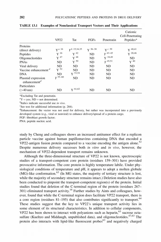
study by Cheng and colleagues shows an increased antitumor effect for a replicon
particle vaccine against human papillomavirus containing DNA that encoded a
VP22-antigen fusion protein compared to a vaccine encoding the antigen alone.52
Despite numerous delivery successes both in vitro and in vivo, however, the
mechanism of VP22-dependent transport remains unknown.
Although the three-dimensional structure of VP22 is not known, spectroscopic
studies of a transport-competent core protein (residues 159–301) have provided
provocative information. The core protein is highly temperature labile. Under phy-
siological conditions of temperature and pH, it appears to adopt a molten globule
(MG)-like conformation.63 (In MG states, the majority of tertiary structure is lost,
while the majority of secondary structure remains intact.) Deletion studies have also
been conducted to pinpoint the transport-competent region(s) of the protein. Initial
studies found that deletion of the C-terminal region of the protein (residues 267–
301) eliminated transport activity.16 Further studies by Aints and colleagues, how-
ever, found that while the C-terminal region does facilitate VP22 transport, there is
a core region (residues 81–195) that also contributes significantly to transport.64
These studies suggest that the key to VP22’s unique transport activity lies in
some element of its structural characteristics. In addition to cellular components,
VP22 has been shown to interact with polyanions such as heparin,63 sucrose octa-
sulfate (Kueltzo and Middaugh, unpublished data), and oligonucleotides.47,63 The
protein also interacts with lipid-like fluorescent probes63 and negatively charged
TABLE 13.1 Examples of Nonclassical Transport Vectors and Their Applications
Cationic
Cell-Penetrating
VP22 Tat FGFs Penetratin Peptides*
Proteins
(direct delivery) Ya 16 Y b 15,34,35 Y 36–38 Y c 39 Y 40,41
Peptides Y 16 Y 42 ND Y 43–45 Y 20,46
Oligonucleotides Y 47 Y 48 ND Y 18,49 ND
PNAs ND Y 50 ND Y 45,51 Y 40
Viral delivery ND ND ND ND ND
Vaccine enhancement d Y 52 ND ND ND ND
DNA ND Y 53,54 ND ND ND
Plasmid expression Y 55–60 ND ND ND ND
enhancementd
Particulates
(>40 nm) ND Y 61,62 ND ND ND
*Excluding Tat and penetratin.aY¼ yes; ND¼ not determined.bItalics indicate successful use in vivo.cSee text for additional information (p. 284).dEnhancement: the vector was not used for delivery, but rather was incorporated into a previously
developed system (e.g., viral or nonviral) to enhance delivery/spread of a protein cargo.
FGF: fibroblast growth factor;
PNA: peptide nucleic acid.
282 POLYCATIONIC PEPTIDES AND PROTEINS IN DRUG DELIVERY

liposomes65 in a temperature-dependent manner. The potential role of these inter-
actions in the mechanism of VP22 transport is discussed below.
13.2.2. Fibroblast Growth Factors
The fibroblast growth factor (FGF) protein family is both large (23 known mem-
bers) and diverse.66 FGFs exhibit a variety of biological functions involving the sti-
mulation of cell proliferation,67 including functions such as general mitogenic
activity (FGFs 1, 2, and 9)68,69 and stimulation of dermal proliferation (FGFs 7
and 10).70 The most intriguing FGF property, in the context of this work, is the
nonclassical secretory activity possessed by a number of FGFs (FGFs 1, 2, 9, 16,
and 20).66,67,69 This activity has been investigated in greatest detail for FGF-1
and -2. A number of studies have suggested that cellular components such as the
cytoskeleton (specifically F-actin),71 molecular chaperones,72 elements of the heat
shock system,73,74 and even a multiprotein secretory complex75,76 may be involved
in the FGF-1 and -2 secretion pathways.
Unlike the majority of potential nonclassical transport vectors, FGF import is
primarily receptor mediated, involving both specific cell surface receptors77 and
low-affinity proteoglycan binding sites.67,68,78 The differential distribution of recep-
tor subtypes in tissues is thought to provide a conventional targeting system for
FGF-based vectors.66 Internalization of FGF fusion proteins, however, has also
been observed, including such cargo proteins as Pseudomonas exotoxin,36,79,80
diphtheria toxin,37 and the constant region of human immunoglobulin G (IgG).38
These studies suggest that the FGFs may possess true nonclassical import activity
in addition to their demonstrated secretory activity. Evidence that this import is
abolished in the presence of polyanions suggests that cell surface polyanions
may play a role in the FGF import pathway37 or that partially unfolded states of
the protein may be necessary for transport (polyanions lock some FGFs into their
native states; see Section 13.4.1.2). This is not surprising, since many of the FGF
proteins strongly bind polyanions,67,81,82 and cell surface proteoglycans play a sig-
nificant role in other FGF-related processes such as formation of the FGF/FGF
receptor complex.77,83
Unlike VP22, members of the FGF family contain primarily b sheet secondary
structure.66 The similarities between the proteins exist in their gross properties (low
molecular weight, net positive charge under neutral pH conditions) as well as their
thermal stabilities. Like VP22, many of the FGFs (at least FGF-1, -2, -7, and -10)
are highly thermally labile82,84 under physiological conditions, with FGF-1 well
established as existing in an MG-like conformation under physiological conditions.
The interaction of FGF-1 with negatively charged lipid bilayers is apparently
dependent on this MG-like state, permitting partial penetration of the bilayer
when the MG conformation is induced.85 FGF-1 and -2 are also believed to localize
to the nucleus of cells, utilizing apparent NLSs,86–89 although this has been disputed.
13.2.3. HIV-1 Tat
The Tat protein was one of the first nonclassical transport vectors to be recognized.
A nuclear transcription activating protein of the HIV-1 retrovirus,90 Tat was initially
CURRENT VECTORS 283

studied in an effort to elucidate its role in the HIV-1 infection process. As Frankel,
Pabo, Green, and Lowenstein discovered independently in the late 1980s, however,
Tat also possesses potent nonclassical transport activity.91,92 Studies of an 86-
residue form of Tat have shown that the protein possesses both nonclassical secre-
tion93 and import activity,91,92 as well as postinternalization nuclear localization.94
Current research in the Tat transport field is concentrated on the Tat protein trans-
duction domain (PTD), a highly basic core peptide (residues 48–57, Table 13.2) that
appears to be the functional unit for Tat-based transport.35,95
Although both the Tat protein15 and the Tat PTD34,96 have been shown to trans-
port proteins into cells, the majority of delivery studies have been conducted with
the Tat PTD, presumably due to the simplicity of the smaller peptide-based system.
The Tat PTD has successfully transported a wide variety of macromolecular and
particulate cargoes (Table 13.1). The conjugation methods employed include cova-
lent linkage61 as well as direct fusion of the Tat PTD to the cargo molecule by pep-
tide synthesis42 or plasmid expression.35 The Tat system has been successfully
employed both in vitro and in vivo. Somewhat surprisingly, Tat-protein conjugates
have even been observed to cross the blood-brain barrier.35,97
Despite the data available from numerous investigations of the Tat delivery sys-
tem, the mechanism of import remains elusive. One potential component of the
transport process is the cell-surface proteoglycan heparan sulfate (HS). Although
an essential interaction with HS has been observed for the full-length Tat protein
import,98,99 the requirement for a similar interaction with Tat PTD import remains
ambiguous.100,101 This suggests that the Tat PTD and the full-length protein may
utilize different import pathways. This possibility is supported by a study by Silhol
and colleagues showing that the Tat PTD does not inhibit internalization of the full-
length protein.100 A second potential component in the system is an apparent lack
of dependence of import on rigid secondary or tertiary structure. This argument is
supported by evidence that (1) partial unfolding of the cargo protein is required for
translocation of full-length Tat fusion constructs,102,103 and that (2) the amphipathi-
city of the Tat peptide (suggested by modeling studies104) is not required for import.
This latter point is supported by observations that partial D-amino acid analogs
of the Tat PTD also possess highly efficient translocation activity.20
13.2.4. Homeoproteins: Penetratin
The nonclassical transport activity of the homeoprotein family of proteins was first
demonstrated by Perez and colleagues during their studies of the Drosophila anten-
napedia homeoprotein (Antp).105 A class of transactivating factors, the homeopro-
teins interact with DNA through a small (�60aa), highly conserved helical region
called the ‘‘homeodomain.’’106 It was the homeodomain of Antp that was originally
thought to enable the nonclassical internalization and nuclear localization observed
for this protein.105,107 Additional studies, however, localized the activity to a 16-
amino acid polycationic peptide (residues 43–58; Table 13.2), now designated
Penetratin.49,107,108 (Interestingly, although the Penetratin peptide now serves as
the model for nonclassical homeodomain-derived transport, it has been suggested
284 POLYCATIONIC PEPTIDES AND PROTEINS IN DRUG DELIVERY

that an extended sequence actually internalizes more efficiently than the minimal
peptide vector.108) Since the discovery of the Penetratin peptide, similar activity
has been observed in other members of the homeoprotein family, including
Hoxa-5,109 Engrailed,110 and Islet-1.39 The Engrailed protein also exhibits nonclas-
sical secretory activity,111 which may be true for the many members of the homeo-
protein family that lack secretion signal peptides.105 This suggests that the potential
for nonclassical import may exist throughout the homeoprotein family, especially in
light of the high sequence conservation of the transport-competent homeodomain
region.
Similar to the Tat PTD, Penetratin is a highly basic peptide possessing helical
character, although once again, it has been shown that the intrinsic secondary struc-
ture of the peptide is not required for internalization.106,107 As shown in Table 13.1,
Penetratin has been successfully employed to deliver protein and DNA-based
macromolecules both in vivo and in vitro.112 Unlike other nonclassical transport-
based vectors, however, the delivery capabilities of Penetratin appear to be limited
by cargo size (100 residues for peptides and 55 base pairs for oligonucleotides).112
Two exceptions to this rule are the translocation of the parent Antp protein as well
as a study by Kilk and colleagues that demonstrates transport of a �63 kDa strep-
tavidin-biotin complex.39 It is therefore not entirely clear that cargo size limitation
is indeed a property of Penetratin delivery, nor is it known if this limitation will
apply to other homeoprotein-derived vectors. Unlike other vectors, although the
precise transport pathway has not been conclusively established for Penetratin, a
specific internalization mechanism has been postulated, involving the formation
of an inverted hexagonal phase in lipid bilayers107 (see Section 13.4.2.1).
13.2.5. Cell-Penetrating Peptides
The number of peptides demonstrating nonclassical import activity has risen dra-
matically in recent years (see the reviews by Lindgren et al.,19 Wadia and
Dowdy,113 and others114–117). This now extensive group of potential delivery vec-
tors can be divided into two general groups: (1) cationic peptides and (2) amphi-
pathic helices, although some miscellaneous peptides are also included in the latter
group. In order to focus on the central subject of this chapter, polycation-based
delivery, we choose to include in this discussion only the former group of cationic
peptides. For information on the latter group, the reader is referred to selected
amphipathic peptides in Table 13.2 and references therein.
The cationic peptide family includes both synthetic and naturally derived pep-
tides, all of which have been successfully employed for both in vitro and in vivo
delivery of a variety of macromolecules (Table 13.1). The prototypic example of
a naturally derived cationic peptide is the Tat PTD, described above. Other exam-
ples include the pVEC peptide, which has been successfully used for both protein
and oligonucleotide delivery,40 as well as a series of small, RNA-binding peptides
discovered by Futaki and colleagues.20 The Penetratin vector is also included in this
group. The inclusion of Tat and Penetratin in the cationic peptide group arises from
the intrinsic definition of these peptides. In general, the cationic delivery peptides
CURRENT VECTORS 285
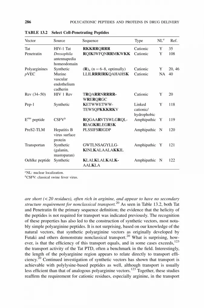
are short (< 20 residues), often rich in arginine, and appear to have no secondary
structure requirement for nonclassical transport.20 As seen in Table 13.2, both Tat
and Penetratin fit the primary sequence definition; the evidence that the helicity of
the peptides is not required for transport was indicated previously. The recognition
of these properties has also led to the construction of synthetic vectors, most nota-
bly simple polyarginine peptides. It is not surprising, based on our knowledge of the
natural vectors, that synthetic polyarginine vectors as originally developed by
Futaki and others demonstrate nonclassical transport.20 What is surprising, how-
ever, is that the efficiency of this transport equals, and in some cases exceeds,123
the transport activity of the Tat PTD, often a benchmark in the field. Interestingly,
the length of the polyarginine region appears to relate directly to transport effi-
ciency.20 Continued investigation of synthetic vectors has shown that transport is
achievable with polylysine-based peptides as well, although transport is usually
less efficient than that of analogous polyarginine vectors.123 Together, these studies
reaffirm the requirement for cationic residues, especially arginine, in the transport
TABLE 13.2 Select Cell-Penetrating Peptides
Vector Source Sequence Type NLa Ref.
Tat HIV-1 Tat RKKRRQRRR Cationic Y 35
Penetratin Drosophila
antennapedia
homeodomain
RQIKIWFQNRRMKWKK Cationic Y 108
Polyarginines Synthetic (R)n (n¼ 6–8, optimally) Cationic Y 20, 46
pVEC Murine
vascular
endothelium
cadherin
LLILRRRIRKQAHAHSK Cationic NA 40
Rev (34–50) HIV 1 Rev TRQARRNRRRR-
WRERQRGC
Cationic Y 20
Pep-1 Synthetic KETWWETWW-
TEWSQPKKKRKV
Linked
cationic/
hydrophobic
Y 118
Erns peptide CSFVb RQGAARVTSWLGRQL-
RIAGKRLEGRSK
Amphipathic Y 119
PreS2-TLM Hepatitis B
virus surface
protein
PLSSIFSRIGDP Amphipathic N 120
Transportan Synthetic
(galanin,
mastoparan)
GWTLNSAGYLLG-
KINLKALAALAKKIL
Amphipathic Y 121
Oehlke peptide Synthetic KLALKLALKALK-
AALKLA
Amphipathic N 122
aNL: nuclear localization.bCSFV: classical swine fever virus.
286 POLYCATIONIC PEPTIDES AND PROTEINS IN DRUG DELIVERY

of these peptides and suggest a specific role for a high degree of positive charge in
the transport activity of these vectors (see Section 13.4.1.1).
13.3. APPLICATIONS: VECTOR CONSTRUCTION
The vectors currently under investigation that employ nonclassical transport repre-
sent a diverse group of entities. This diversity has required that a variety of proto-
cols be developed for both the conjugation of the vectors to cargo molecules and the
presentation of the vector-cargo complex to the target cells. In this section, an over-
view of the methods most commonly employed for vector production is presented.
For more detailed information on the actual protocols, the reader is referred to the
specific references mentioned herein.
13.3.1. Plasmid Expression
In this approach, the vector cargo construct is encoded within a plasmid as a fusion
protein or peptide, depending on the exact vector/cargo combination. This method
has been used primarily with the Tat PTD and the VP22 delivery systems, although
the exact methods differ. In the Tat system, the resulting fusion construct is nor-
mally expressed and highly purified.41 The purified protein is then introduced to
cells in culture or, as shown to be highly effective by Schwarze and colleagues,
can be directly injected into a host.35 Alternatively, the VP22 system commonly
involves transfecting cells with the construct-encoding plasmid itself and mixing
these cells with target cells.55 The expressed protein is then observed to spread
from the primary expressing cell to the surrounding cells. It should be noted, how-
ever, that this spread has been postulated to be an artifact of the imaging protocols
used in these studies (see Section 13.4.4). An additional use of plasmid expression
involves incorporation of a vector-target construct into a conventional gene therapy
delivery vehicle to enhance the delivery of the expressed therapeutic.52 Again, this
has been used predominantly with the VP22 delivery system, most commonly in
viral gene therapy delivery vehicles,57,58 although this approach could quite readily
be used with the other vectors described above. It does, however, then involve the
new problem of transfection with the encoding DNA itself, something typically
accomplished with cationic vehicles such as lipids or polyethyleneimine with
only moderate to low efficiency.124
13.3.2. Direct Synthesis
A number of methods have been developed to create vector-cargo conjugates by
direct chemical synthesis. These methods are more widely applicable than plasmid
expression methods, since they do not rely on any inherent character of the delivery
vector or cargo (e.g., a requirement that both be composed of L-amino acids). The
delivery protocol for the majority of these constructs involves direct application of
the complex to cells in culture, which has been employed with significant success.
APPLICATIONS: VECTOR CONSTRUCTION 287

Although, as mentioned above, amino acid character is not always required for
direct synthetic methods, it is necessary for the first method in the group, peptide
synthesis. In this protocol, the constructs are synthesized using commercially avail-
able synthesis techniques and instruments, with or without a spacer amino acid
between the delivery and cargo sequence. Direct synthesis has been used with
the Tat PTD,42 the Penetratin peptide,43,44 and the pVEC peptide.40 A second
method that has been used extensively is disulfide bond linkage. This involves
introducing a free thiol group into both the cargo and vector molecule, either
through chemical modification or, as shown in Figure 13.1, by taking advantage
of intrinsic thiol functional groups (e.g., cysteine residues). The free thiols then
spontaneously oxidize to form a covalent disulfide bond that links the molecules.112
The ability to introduce disulfide bonds into a variety of cargoes, as demonstrated
Figure 13.1. Disulfide linkage. In this protocol, the vector and cargo are independently
synthesized, with the thiol groups of the cysteine residues protected by a pyridinium group.
After synthesis, the pyridinium is removed, in the case of peptides simply by addition of a
reducing agent, and the now free and reactive thiols quickly and efficiently oxidize to form a
disulfide bond, linking vector to cargo molecule. (Reprinted from Current Opinion in
Neurobiology, Vol. 6, Alain Prochiantz, Getting hydrophilic compounds into cells: lessons
from homeopeptides, 629–634, # 1996, with permission from Elsevier.)
288 POLYCATIONIC PEPTIDES AND PROTEINS IN DRUG DELIVERY

by Pooga and colleagues in a fusion of both transportan and Penetratin vectors to
peptide nucleic acids,51 demonstrates the applicability of this system to a wide vari-
ety of potential vector-cargo systems.
Other methods of direct conjugation have also been developed. Biotinylated
delivery vehicles have been conjugated to streptavidin-labeled cargoes.40,125 The
Tat PTD has been covalently linked to polyethylene glycol and subsequently incor-
porated into liposomes, enhancing their delivery.61 A technology recently devel-
oped by Eom and colleagues involves a novel tandem ligation protocol,
permitting linkage of multiple peptide segments via chemical ligation and resulting
in both linear and branched peptides.126 As the delivery vectors and cargoes being
investigated grow more diverse, it is probable that additional direct conjugation
methods will be developed.
13.4. MECHANISM OF ACTION: CURRENT THEORIES
A number of ideas have been proposed to explain the mechanism by which cell-
penetrating peptides and other nonclassical delivery vehicles induce transport
across lipid bilayers. Some common properties among the vectors are apparent,
suggesting that the exact mechanism of entry may become evident in the near
future. In this section, these common properties of the vectors are described and
the major mechanistic explanations that have been proposed are outlined. It should
be emphasized, however, that at this time no general consensus on an exact
mechanism has been reached.
13.4.1. Common Properties
13.4.1.1. Cationic Character: The Role of Guanidinium The most striking simi-
larity among the nonclassical vectors is without a doubt the high prevalence and
density of positive charges. Although most obvious in the cell-penetrating peptide
group, areas of high positive charge density are also observed with all known pro-
tein vectors (e.g., VP22, FGF-1). This suggests the existence of a common interna-
lization mechanism involving the cationic character of the vectors. This is further
supported by competition for internalization of some cationic vectors by others101
and reduction in efficiency among the arginine vectors as the extent of positive
charge is reduced.20 Closer examination of the cationic residues present in the vec-
tors has shown a strong predilection for arginine residues, leading to the proposal
by some groups that, in addition to its positive charge, some unique property of the
arginine side chain may be a key factor in translocation.101,113
A number of studies have been conducted in an attempt to differentiate between
a specific role for the guanidino moiety of the arginine side chain and a general
requirement for cationic amino acids (which could also be satisfied by lysine or his-
tidine residues). Examinations of polyarginine transport efficiency show either
similar41 or enhanced activity when compared to analogous polylysine,123 polyhis-
tidine, and poly-L-ornithine peptides.127 Additionally, no significant differences in
MECHANISM OF ACTION: CURRENT THEORIES 289

activity are observed in a comparison of import of polyarginine, homoarginine, and
d-guanidino a-amino butyric acid (dGABA),127 a series that differs only in the
number of side chain methylene groups. These data support the claim that transport
efficiency is related to some unique property of arginine side chains, although it is
clear that positive charge itself must play a significant role.
Although the precise molecular significance of the guanidino group in nonclas-
sical transport is not yet known, the guanidino moiety has been found to form a
number of specific interactions that could play a significant role. Most obviously,
the delocalized p-system in the positively charged region of the side chain distin-
guishes it from that of lysine. For example, arginine can self-associate in solution in
a face-to-face manner.128 Perhaps more significantly, arginine-aromatic ring inter-
actions are often observed due to the formation of discrete cation-p interactions (see
Section 13.4.2.2).129–131
13.4.1.2. Polyanion Binding It is reasonable to assume from the abundance of
cationic residues present in the majority of nonclassical transport-based vectors
that these vectors should interact, at least to some extent, with the variety of
polyanions found both at the cell surface and within the cell. In fact, such interac-
tions appear to be highly prevalent,63,82,98,132,133 being involved in the natural
biological activity of many vectors as well as having a potential role in import.
A primary example of the biological significance of the vector-polyanion interac-
tion is observed with FGF-1, whose interaction with cell-surface heparin sulfate has
been shown to be required for activity of the FGF protein-receptor complex.77
VP22 has been shown to bind cellular RNA, apparently mediating its translocation
during HSV-1 infection.134 The function of the full-length Tat protein appears to be
modified by heparan sulfate.98,135,136 The interaction of cationic vectors with poly-
anions may, however, be more general than the specific interactions normally
observed in biological systems. For example, the Penetratin peptide has been
observed to interact nonspecifically with a number of anionic lipids,137 the VP22
protein interacts preferentially with both anionic liposomes,65 and negatively
charged fatty acid mimics as well as heparin and polynucleotides,63 and FGF-1
has been shown to bind to a large number of quite different polyanions.81
The wide variety of vector-polyanion interactions described above suggests a
potential role for polyanions in nonclassical transport. This could be similar to
the role that proteoglycans have been suggested to play in the cellular entry of
cationic gene delivery.138 The idea that cell surface polyanions act as the initial
site of interaction for nonclassical vectors with cell membranes appears to be
widely accepted, based on a number of critical studies. The internalization of
both Penetratin49,105 and the Tat protein99 appears to be dependent on the cell-sur-
face polyanions heparan sulfate and polysialic acid, respectively. Similar studies
with other arginine-rich cell-penetrating peptides show that an absence of cell-sur-
face proteoglycans reduces transport.101 Polyanions other than proteoglycans have
also been implicated in transport. Some Tat PTD studies suggest that the negative
charge at the membrane surface produced by negatively charged phospholipid
headgroups may also play a role.100,101 This is further supported by a Penetratin
290 POLYCATIONIC PEPTIDES AND PROTEINS IN DRUG DELIVERY

study suggesting that charge neutralization through the interaction of the vector
with negatively charged membrane lipids is the initial and, in fact, an essential com-
ponent in the internalization pathway.139 It is interesting to note that many viruses
may also use negatively charged moieties as their initial contact point with the cell
surface.138
These data lead to the further question of additional interactions between the
vectors and other polyanions throughout the cell, a question that has not yet been
critically addressed. A variety of polyanions, as well as dense areas of negative
charge, can be found throughout the cell, including small polyanions such as
nucleotides as well as cytoskeleton components, RNA, and DNA, all of which
could serve as potential sites of interaction. Whether these intracellular polyanions
are also steps in the transport pathway, perhaps serving as transport avenues, as has
already been postulated for microtubule and microfilament networks in vesicular
transport, remains to be seen.
13.4.1.3. The Role of Nonnative States in Protein Vectors In the previous sec-
tions (13.4.1.1 and 13.4.1.2), the focus has been primarily on the highly charged
nature of nonclassical vectors and the interaction of these vectors with the polar
domain of the lipid bilayer. An additional and potentially more significant barrier
to transport lies within the apolar domain of the bilayer. Although this barrier
appears to be impassable by large hydrophilic molecules, it has been postulated
that, by adopting a partially unfolded conformation, protein molecules can interact
directly with the lipid bilayer, facilitating protein-membrane translocation.10,140
These partially unfolded conformations, often labeled ‘‘molten globule’’ states,
involve a loss of tertiary structure with retention of secondary structure, exposing
interior hydrophobic surfaces of the protein for interaction with bilayer interior.141
Although definitive evidence that these partially unfolded conformations are a
key to transport is not yet available, their potential involvement in nonclassical pro-
tein transport is becoming increasingly apparent. This is primarily due to evidence
that rigid tertiary and/or secondary structure is consistently absent in protein and
peptide vehicles and, in some cases, has been found to hinder transport. For
example, both VP22 and FGF-1 adopt partially disrupted, MG-like conformations
under physiological solution conditions.63,142 These intermediately folded confor-
mations also appear to dramatically enhance the interactions of VP22,63,65 and
FGF-1,142 as well as other proteins, with model lipid systems. Wiedlocha and col-
leagues have shown that FGF-1/diphtheria toxin fusion proteins are transport com-
petent only when in a less ordered conformation.37 Initial studies of transport by the
full-length Tat protein suggest that at least partial unfolding of the cargo protein is
required for transport. Additionally, as mentioned previously, the peptide vector
Penetratin does not require specific secondary structure for internalization;106,107
all of these studies support the hypothesis that disordered structure is necessary
for bilayer translocation. Further support of this hypothesis is found in studies of
the translocation of diphtheria toxin (DT) and Pseudomonas exotoxin, both of
which possess transmembrane (T) domains that are independently capable of mem-
brane translocation.143,144 Once again, MG-like states play an important role;
MECHANISM OF ACTION: CURRENT THEORIES 291

formation of such membrane interactive states appears to be the initial step in the
active part of the translocation process.144,145
Although the exact mechanism of MG-assisted transport has not been defined, a
simple model can be postulated. This involves an initial attraction between the
cationic vector and the negatively charged lipid surface, followed by insertion of
the vector directly into the membrane through association of the exposed hydropho-
bic surfaces of the vector with the apolar bilayer interior. Whether the membrane-
associating form of the vector and/or cargo must preexist or can be induced by con-
ditions at the cell surface, such as the lower pH present at the membrane interfacial
microenvironment146 or by other components of the lipid bilayer itself (e.g., mem-
brane proteins), is unclear; either situation, however, has the potential to satisfy this
model. Actual transport through the membrane may use simple diffusion as a driv-
ing force. The idea of diffusion of large molecules through such a restricted space is
supported by recent work showing a ‘‘jumping’’ of flexible macromolecules within
a highly restricted network while undergoing Brownian motion.147 This jumping
could be facilitated by attraction between the cationic vector and other electrostatic
surfaces on the other side (i.e., the cellular interior) of the membrane. A similar
hypothesis has been proposed for the DT transmembrane (T) domain-assisted trans-
port of MG-like proteins. In this scheme, it is suggested that translocation is
achieved through a series of nonspecific binding events between the cargo protein
and the T domain.148 The T domain then acts as a stabilizing platform for the par-
tially unfolded cargo, perhaps similarly to cellular molecular chaperones.
13.4.2. Proposed Mechanism of Transport
13.4.2.1. Inverted Hexagonal Phase Induction One of the first models of non-
classical transport, proposed by Prochiantz and colleagues, involves the induction
of an inverted hexagonal phase within the lipid bilayer.49,112,149 As shown in Figure
13.2, the vector-cargo complex is proposed to initially bind to the cell surface
Figure 13.2. Inverted hexagonal phase model of nonclassical transport. The vector-cargo
complex initially binds to the cell surface via electrostatic interactions. This induces an
inverted hexagonal phase within the lipid bilayer, permitting the complex to become encap-
sulated within an inverted micelle and subsequently shuttled to the opposite side of the mem-
brane. (Potential use of non-classical pathways for the transport of macromolecular drugs.
Kueltzo, L.A. and Middaugh, C.R. 2001. Exp Opin Invest Drugs 9(9): 2039–2050. Used with
permission.)
292 POLYCATIONIC PEPTIDES AND PROTEINS IN DRUG DELIVERY

through electrostatic attraction. This binding induces an inverted hexagonal phase
in the immediately adjacent lipid bilayer, causing the complex to be trapped in an
inverted micelle.149,150 The resulting vesicle then diffuses to the opposite side of the
bilayer, where its contents are released into the surrounding fluid (the cytosol). This
is similar to internalization methods suggested for cationic lipid/DNA gene delivery
complexes.151,152 Initial studies with the Penetratin peptide, the vector on which
this model is based, do in fact show the induction of inverted hexagonal phases
in lipid solutions, as observed by nuclear magnetic resonance.150 Unfortunately,
similar results have not been observed with other vector systems; thus, this model
has yet to receive support as a general model of nonclassical transport.
13.4.2.2. A Unified Hypothesis This mechanism, previously proposed by the
authors of this chapter,153 combines the predominant common qualities of the
nonclassical vectors. It is reasonable to assume that the cationic nonclassical
vectors can bind to a variety of polyanionic species in a somewhat nonspecific man-
ner during their approach to and passage through the cell membrane and interior.
Although this binding may be nonspecific, there would certainly be some differ-
ences in binding affinity, suggesting the potential existence of a network of poly-
anions throughout the cell that could guide the transport of polycationic vectors.
The existence of such a network as a guiding entity, either through a gradient of
the aforementioned relative binding affinities or perhaps by providing somewhat
uniform reactions surfaces, remains to be functionally established. The polyanionic
moieties of the cell surface (which could be considered the outer edge of the poly-
anion network), however, serve as the initial site of attraction for the cationic vector
(Figure 13.3).
Following initial binding to the cell surface, the vector is able to interact directly
with the lipid bilayer through an MG-like or other disordered state by direct inter-
action between protein/peptide apolar residues with lipid acyl side chains. This
state may be normally adopted by the vector under physiological solution
conditions, or may be induced by the microenvironment found at the cell surface.
The vector must then pass through the supposedly impenetrable apolar lipid bilayer.
This barrier may not, however, be as impervious as was previously thought.154–156
In addition to the diffusion studies mentioned in Section 13.4.1.3, a study of the
diphtheria toxin T domain shows that charged groups within the membrane-
spanning domain do not appear to provide the barrier to movement across the
membrane that might be expected.157
These studies support the idea that the bilayer is actually somewhat permeable to
macromolecules under certain conditions. The exact mechanism of this permeabil-
ity is not known, but the arginine specificity of nonclassical vectors (see Sec-
tion 13.4.1.1) suggests a reasonable pathway. Although these side chains are not
expected to interact with the apolar interior of the bilayer, they may interact with
the numerous transmembrane proteins that span biological membranes. The helices
of membrane-spanning proteins are often found to be studded with aromatic resi-
dues,158–160 providing an ideal environment for the formation of cation-p interac-
tions between arginine (and potentially lysine) residues of a vector and the
MECHANISM OF ACTION: CURRENT THEORIES 293

Figure 13.3. Unified model of nonclassical entry into cells. (1) The cationic nonclassical
vector, in a compact (C) or molten globule-like conformation (MG) binds nonspecifically
to the negatively charged cell membrane through electrostatic interactions. (2) The acidic
microenvironment at the cell surface induces a conversion to an expanded, MG-like confor-
mation for compact vectors. (3) The vector directly penetrates the membrane at the trans-
membrane protein/lipid bilayer interface, stabilized by interactions between exposed
hydrophobic surfaces of the vector and the interior of the bilayer in addition to (3a)
cation-p interactions with transmembrane proteins mediated by arginine residues of vectors
and aromatic side chains of membrane protein bilayer-spanning helices. (4) The vector
passes completely through the bilayer by diffusion, facilitated by an electrostatic attraction
for the negatively charged interior surface of the bilayer or other internal polyanions.
(Non-classical transport proteins and peptides: an alternative to classical macromolecule
delivery systems, Kueltzo, L.A. and Middaugh, C.R. J Pharm Sci. # 2003, Wiley-Liss,
Inc., a Wiley company, and the American Pharmacists Association.)

aforementioned aromatic residues. Such interactions are now thought to play an
important role in protein structure as well as in membrane systems.161 Therefore,
one could postulate that the interface between bilayer-spanning protein helices and
the lipid bilayer itself could serve as a potential pathway for macromolecular trans-
port (Figure 13.3, Part 3). The flexibility of the lipid bilayer could easily accommo-
date passage of even very large complexes at this interface, especially if they exist
in an extended structure. An additional advantage of extended structure is the
potential interaction of the apolar lipid interior with the exposed hydrophobic sur-
faces of the translocating complex, further reducing the energy barrier to transport.
Although this mechanism is currently highly speculative, significant support for this
hypothesis is found in the work of Xiang and Anderson, who have shown an
increase in the penetration of the lipid bilayer by polar molecules as the transmem-
brane protein content of the bilayer is increased.156 Additional supporting evidence
comes from studies showing significant insertion of basic peptides into a number of
different membrane systems.162
The partial neutralization of cationic charges within the lipid bilayer through
cation-p interactions need not be the only mechanism. Call and colleagues have
recently shown that during the assembly process of transmembrane helices, the
helices are stabilized through a multicharge interaction, with two interacting acidic
residues on one helix interacting with a single basic residue on a second helix.163
Additional studies support such interactions, indicating that interactions between
acidic residues are indeed favorable in a membrane environment.164,165 This type
of multicharge interaction could also play a part in the neutralization of cationic
residues of translocating complexes at the transmembrane protein/lipid bilayer
interface.
Following entry into and passage through the bilayer as facilitated by one of the
above mechanisms, or perhaps by another not yet recognized process, the complex
can then exit the bilayer by a combination of diffusion and electrostatic attraction to
the highly negatively charged interior membrane surface. At this point, the complex
may continue on its path into the cell via the aforementioned polyanion network or
by other as yet unknown pathways. Although this pathway may not be applicable to
all examples of nonclassical transport, specifically the transport of large particles,
which often possess quite different structural characteristics than vector-protein
complexes, it does combine the common features of currently studied vectors.
We are presently attempting to test this model using model membrane-spanning
helices in liposome-based systems with entrapped polyanions.
13.4.2.3. Secretion The majority of studies involving nonclassical delivery focus
exclusively on the import process. The initial work in this field, however, involved
proteins that were found to possess not only nonclassical import activity but also
leaderless secretory activity (secretion in the absence of a secretion signal peptide
or use of the Golgi apparatus). The mechanism of this type of secretion is no clearer
than that of import; in fact, it is uncertain at this time whether these mechanisms are
even related, especially considering the asymmetry of the cell membrane. It seems
reasonable to assume for experimental testing purposes, however, that the common
MECHANISM OF ACTION: CURRENT THEORIES 295

vector properties mentioned above may also be involved in such secretion
pathways.
Numerous hypotheses regarding the mechanism of leaderless secretion have
been postulated since its initial description. One mechanism suggests secretion
via the ABC (ATP-binding cassette) export complex.166,167 The ABC complex,
however, appears to be exclusive to yeast and bacteria, with no evidence that it
plays a part in mammalian protein secretion. The secretion of FGF-2, one of the
more studied systems, purportedly involves the membrane attack complex of the
complement system.168 A lack of complement factors in the many in vitro cell cul-
ture systems in which secretion has been demonstrated suggests, however, that they
may not be involved in a general secretion pathway. The FGF-1 export pathway
also appears to involve a protein complex.71,75 This complex includes the protein
synaptotagmin, which contains a polycationic region, providing a potential link to
the nonclassical transport proteins. Rubartelli and colleagues, one of the first
groups to study leaderless secretion, have proposed the involvement of endolyso-
somes in the secretion pathway of interleukin 1b,10,169,170 followed by translocationof a partially disordered form of the protein complexed with chaperone proteins.10
All of these potential mechanisms are protein specific; the existence of a general
mechanism of leaderless secretion, as well as any potential connection to nonclas-
sical import, remains unclear.
13.4.3. Additional Participants in Uptake and Secretion
The cellular environment is both complex and crowded; as such, it is reasonable to
assume that a vector may come into contact with a much wider variety of molecules
than those mentioned in the previous sections. These molecules may prove to be
necessary elements in the transport pathway; this is a critical consideration in stu-
dies that utilize simple lipid models in which such components are absent. For
example, in addition to the polyanions mentioned previously, molecular chaperones
may play an important role, stabilizing partially unfolded vector and vector-cargo
complexes, during and after translocation. In fact, molecular chaperones have
already been proposed to be involved in the transport of Tat97 and VP22,63 as
well as FGF-1,72 and are well established to play a role in the transport of proteins
into mitochondria.171 Other factors, such as membrane properties (curvature, lipid
composition, and spatial distribution) and differences in cell-surface proteoglycans
among cell types may also strongly affect transport events. Finally, there may be a
number of unknown factors that could be involved, all of which must ultimately be
taken into consideration when evaluating the nature of transport.
13.4.4. Potential Artifacts and Other Concerns
Despite the numerous in vivo delivery successes demonstrated with nonclassical
vehicles that provide strong evidence that the pathway does indeed exist, a few stu-
dies suggest that nonclassical transport is actually an artifact of the cell fixation pro-
cesses employed in many of the in vitro studies.172,173 Some evidence argues that
296 POLYCATIONIC PEPTIDES AND PROTEINS IN DRUG DELIVERY

transport may be occurring through traditional endocytotic pathways, rather than
via the energy-independent pathway proposed here. For example, a study of the
Engrailed vector suggests possible association with caveolae.110 Inhibition of the
caveolae pathway appears to inhibit Tat internalization, the latter study involving
Tat-linked lambda phage.53 The internalization of arginine-based peptides, how-
ever, is apparently not affected by caveolae inhibition.101 It is important to note
that internalization by a nonclassical pathway does not preclude parallel internali-
zation by conventional endocytotic pathways. In fact, such endocytotic internaliza-
tion may well be expected, considering that many delivery studies have been
conducted under physiological conditions (where endocytosis is often quite active)
following establishment of energy-independent transport during the initial investi-
gation of the vectors. Should the two pathways indeed operate simultaneously, their
relative contributions need to be determined to evaluate overall vector efficiency.
These factors highlight some of the difficulties in elucidating the nonclassical path-
way, and they must be carefully considered in a critical analysis of the data being
generated in this field.
In addition to the unresolved mechanistic questions, a number of other issues
concerning cationic nonclassical delivery vehicles must also be addressed. A pri-
mary issue is cytotoxicity, a common problem with polycationic therapeutics and
excipients. Although initial studies suggest that cytotoxicity will not be signifi-
cant,40,118 presumably due to the relatively small cationic regions employed, should
the polyanion network hypothesis hold true, a number of deleterious effects could
occur due to the variety of potential nonspecific interactions. The biological activity
of the vectors must also be examined, especially if endogenous protein vectors (e.g.,
the FGFs) are employed. Many cationic peptides also possess significant antimicro-
bial activity and are currently under development as human therapeutics. The rela-
tionship between their biological activity and potential transport-like properties
remains to be better described. Finally, as with any therapeutic, the question of
immunogenicity needs to be addressed, since the safety and efficacy issues asso-
ciated with significant immunogenicity will severely impair the use of these vectors
in the clinic, especially with repetitive use.
13.5. CONCLUSIONS
As the number of macromolecular therapeutics in clinical trials increases, the need
for efficient delivery vehicles becomes more pressing. The cationic peptides and
proteins described here provide an interesting alternative to conventional delivery
vehicles. Although the exact mechanism of nonclassical transport has not yet been
defined, the in vitro and in vivo delivery successes of these vehicles are most pro-
mising, especially considering the wide array of cargoes and protocols with which
they can be employed. Although a number of questions remain to be addressed,
research in this field is proceeding at a rapid pace, increasing the possibility that
one or more of these vector-cargo complexes could reach the clinic in the not
too distant future.
CONCLUSIONS 297

REFERENCES
1. Tan, K. B. Cytobios. 1977, 20, 143–149.
2. Triguero, D.; Buciak, J. B.; Yang, J.; Pardridge, W. M. Proc. Natl. Acad. Sci. USA 1989,
86, 4761–4765.
3. Blau, S.; Jubeh, T. T.; Haupt, S. M.; Rubinstein, A. Crit. Rev. Ther. Drug Carrier Syst.
2000, 17, 425–465.
4. Ehrlich, M.; Sarafyan, L. P.; Myers, D. J. Biochim. Biophys. Acta 1976, 454, 397–409.
5. Zelphati, O.; Wang, Y.; Kitada, S.; Reed, J. C.; Felgner, P. L.; Corbeil, J. J. Biol. Chem.
2001, 276, 35103–35110.
6. Lasic, D. D. Liposomes in Gene Delivery. CRC Press: Boca Raton, FL, 1997.
7. Kwoh, D. Y.; Coffin, C. C.; Lollo, C. P.; Jovenal, J.; Banaszczyk, M. G.; Mullen, P.;
Phillips, A.; Amini, A.; Fabrycki, J.; Bartholomew, R. M.; Brostoff, S. W.; Carlo, D. J.
Biochim. Biophys. Acta 1999, 1444, 171–190.
8. Godbey, W. T.; Wu, K. K.; Mikos, A. G. J. Control Release 1999, 60, 149–160.
9. Patri, A. K.; Majoros, I. J.; Baker, J. R. Curr. Opin. Chem. Biol. 2002, 6, 466–471.
10. Rubartelli, A.; Sitia, R. InUnusual Secretory Pathways: From Bacteria toMan.; Kuchler,
K., Ed. R.G. Landes Bioscience: Austin, TX, 1997, pp. 87–114.
11. Kueltzo, L. A.; Middaugh, C. R. Expert Opin. Invest. Drugs 2000, 9, 2039–2050.
12. Rubartelli, A.; Cozzolino, F.; Talio, M.; Sitia, R. EMBO J. 1990, 9, 1503–1510.
13. Abraham, J. A.; Mergia, A.; Whang, J. L.; Tumolo, A.; Friedman, J.; Hjerrild, K. A.;
Gospodarowicz, D.; Fiddes, J. C. Science 1986, 233, 545–548.
14. Reiness, C. G.; Seppa, M. J.; Dion, D. M.; Sweeney, S.; Foster, D. N.; Nishi, R.Mol. Cell.
Neurosci. 2001, 17, 931–944.
15. Fawell, S.; Seery, J.; Daikh, Y.; Moore, C.; Chen, L. L.; Pepinsky, B.; Barsoum, J. Proc.
Natl. Acad. Sci. USA 1994, 91, 664–668.
16. Elliott, G.; O’Hare, P. Cell 1997, 88, 223–233.
17. Perez, F.; Lledo, P. M.; Karagogeos, D.; Vincent, J. D.; Prochiantz, A.; Ayala, J. Mol.
Endocrinol. 1994, 8, 1278–1287.
18. Allinquant, B.; Hantraye, P.; Mailleux, P.; Moya, K.; Bouillot, C.; Prochiantz, A. J. Cell
Biol. 1995, 128, 919–927.
19. Lindgren, M.; Hallbrink, M.; Prochiantz, A.; Langel, U. Trends Pharmacol. Sci. 2000,
21, 99–103.
20. Futaki, S.; Suzuki, T.; Ohashi, W.; Yagami, T.; Tanaka, S.; Ueda, K.; Sugiura, Y. J. Biol.
Chem. 2001, 276, 5836–5840.
21. Alberts, B.; Bray, D.; Lewis, J.; Raff, M.; Roberts, K.; Watson, J. D. In Molecular
Biology of the Cell, 3rd ed. Garland Publishing: New York, 1994, pp. 551–99.
22. Gorlich, D.; Mattaj, I. W. Science 1996, 271, 1513–1518.
23. Alberts, B.; Bray, D.; Lewis, J.; Raff, M.; Roberts, K.; Watson, J. D. In Molecular
Biology of the Cell, 3rd ed. Garland Publishing: New York, 1994, pp. 604–54.
24. Elliott, G.; Mouzakitis, G.; O’Hare, P. J. Virol. 1995, 69, 7932–7941.
25. Elliott, G.; O’Reilly, D.; O’Hare, P. Virology 1996, 226, 140–145.
26. Blaho, J. A.; Mitchell, C.; Roizman, B. J. Biol. Chem. 1994, 269, 17401–17410.
27. Knopf, K. W.; Kaerner, H. C. J. Gen. Virol. 1980, 46, 405–414.
298 POLYCATIONIC PEPTIDES AND PROTEINS IN DRUG DELIVERY

28. Elliott, G.; O’Hare, P. J. Virol. 1998, 72, 6448–6455.
29. van Leeuwen, H.; Elliott, G.; O’Hare, P. J. Virol. 2002, 76, 3471–3481.
30. Derer, W.; Easwaran, H. P.; Knopf, C. W.; Leonhardt, H.; Cardoso, M. C. J. Mol. Med.
1999, 77, 609–613.
31. Aints, A.; Dilber, M. S.; Smith, C. I. E. J. Gene Med. 1999, 1, 275–279.
32. Phelan, A.; Elliott, G.; O’Hare, P. Nat. Biotechnol. 1998, 16, 440–443.
33. Wybranietz, W. A.; Prinz, F.; Speigel, M.; Schenk, A.; Bitzer, M.; Gregor, M.; Lauer, U.
M. J. Gene Med. 1999, 1, 265–274.
34. Caron, N. J.; Torrente, Y.; Camirand, G.; Bujold, M.; Chapdelaine, P.; Leriche, K.;
Bresolin, N.; Tremblay, J. P. Mol. Ther. 2001, 3, 310–318.
35. Schwarze, S. R.; Ho, A.; Vocero-Akbani, A.; Dowdy, S. F. Science 1999, 285, 1569–1572.
36. Gawiak, S.; Pastan, I.; Siegall, C. Bioconjug. Chem. 1993, 4, 483–489.
37. Wiedlocha, A.; Madshus, I. H.; Mach, H.; Middaugh, C. R.; Olsnes, S. EMBO J. 1992,
11, 4835–4842.
38. Dikov, M. M.; Reich, M. B.; Dworkin, L.; Thomas, J. W.; Miller, G. G. J. Biol. Chem.
1998, 273, 15811–15817.
39. Kilk, K.; Magzoub, M.; Pooga, M.; Eriksson, L. E.; Langel, U.; Graslund, A. Bioconjug.
Chem. 2001, 12, 911–916.
40. Elmquist, A.; Lindgren, M.; Bartfai, T.; Langel, U. Exp. Cell. Res. 2001, 269,
237–244.
41. Park, J.; Ryu, J.; Kim, K. A.; Lee, H. J.; Bahn, J. H.; Han, K.; Choi, E. Y.; Lee, K. S.;
Kwon, H. Y.; Choi, S. Y. J. Gen. Virol. 2002, 83, 1173–1181.
42. Li, H.; Yao, Z.; Degenhardt, B.; Teper, G.; Papadopoulos, V. Proc. Natl. Acad. Sci. USA
2001, 98, 1267–1272.
43. Bonfanti, M.; Taverna, S.; Salmona, M.; D’Incalci, M.; Broggini, M. Cancer Res. 1997,
57, 1442–1446.
44. Giorello, L.; Clerico, L.; Pescarolo, M. P.; Vikhanskaya, F.; Salmona, M.; Colella, G.;
Bruno, S.; Mancuso, T.; Bagnasco, L.; Russo, P.; Parodi, S. Cancer Res. 1998, 58, 3654–
3659.
45. Braun, K.; Peschke, P.; Pipkorn, R.; Lampel, S.; Wachsmuth, M.; Waldeck, W.;
Friedrich, E.; Debus, J. J. Mol. Biol. 2002, 318, 237–243.
46. Chen, L.; Wright, L. R.; Chen, C. H.; Oliver, S. F.; Wender, P. A.; Mochly-Rosen, D.
Chem. Biol. 2001, 8, 1123–1129.
47. Normand, N.; van Leeuwen, H.; O’Hare, P. J. Biol. Chem. 2001, 276, 15042–15050.
48. Astriab-Fisher, A.; Sergueev, D.; Fisher, M.; Shaw, B. R.; Juliano, R. L. Pharm. Res.
2002, 19, 744–754.
49. Derossi, D.; Chassaing, G.; Prochiantz, A. Trends Cell. Biol. 1998, 8, 84–87.
50. Kaushik, N.; Basu, A.; Palumbo, P.; Myers, R. L.; Pandey, V. N. J. Virol. 2002, 76, 3881–
3891.
51. Pooga, M.; Soomets, U.; Hallbrink, M.; Valkna, A.; Saar, K.; Rezaei, K.; Kahl, U.; Hao,
J. X.; Xu, X. J.; Wiesenfeld-Hallin, Z.; Hokfelt, T.; Bartfai, T.; Langel, U. Nat.
Biotechnol. 1998, 16, 857–861.
52. Cheng, W. F.; Hung, C. F.; Hsu, K. F.; Chai, C. Y.; He, L.; Polo, J. M.; Slater, L. A.; Ling,
M.; Wu, T. C. Hum. Gene Ther. 2002, 13, 553–568.
REFERENCES 299
53. Eguchi, A.; Akuta, T.; Okuyama, H.; Senda, T.; Yokoi, H.; Inokuchi, H.; Fujita, S.;
Hayakawa, T.; Takeda, K.; Hasegawa, M.; Nakanishi, M. J. Biol. Chem. 2001, 276,
26204–26210.
54. Rudolph, C.; Plank, C.; Lausier, J.; Schillinger, U.; Muller, R. H.; Rosenecker, J. J. Biol.
Chem. 2003, 278, 11411–11418.
55. Dilber, M. S.; Phelan, A.; Aints, A.; Mohamed, A. J.; Elliott, G.; Edvard Smith, C. I.;
O’Hare, P. Gene Ther. 1999, 6, 12–21.
56. Wybranietz,W. A.; Gross, C. D.; Phelan, A.; O’Hare, P.; Spiegel, M.; Graepler, F.; Bitzer,
M.; Stahler, P.; Gregor, M.; Lauer, U. M. Gene Ther. 2001, 8, 1654–1664.
57. Lai, Z.; Brady, R. O. J. Neurosci. Res. 2002, 67, 363–371.
58. Lai, Z.; Han, I.; Zirzow, G.; Brady, R. O.; Reiser, J. Proc. Natl. Acad. Sci. USA 2000, 97,
11297–11302.
59. Wills, K. N.; Atencio, I. A.; Avanzini, J. B.; Neuteboom, S.; Phelan, A.; Philopena, J.;
Sutjipto, S.; Vaillancourt, M. T.; Wen, S. F.; Ralston, R. O.; Johnson, D. E. J. Virol. 2001,
75, 8733–8741.
60. Zender, L.; Kuhnel, F.; Kock, R.; Manns, M.; Kubicka, S. Cancer Gene Ther. 2002, 9,
489–496.
61. Torchilin, V. P.; Rammohan, R.; Weissig, V.; Levchenko, T. S. Proc. Natl. Acad. Sci. USA
2001, 98, 8786–8791.
62. Lewin, M.; Carlesso, N.; Tung, C. H.; Tang, X. W.; Cory, D.; Scadden, D. T.; Weissleder,
R. Nat. Biotechnol. 2000, 18, 410–414.
63. Kueltzo, L. A.; Normand, N.; O’Hare, P.; Middaugh, C. R. J. Biol. Chem. 2000, 275,
33213–33221.
64. Aints, A.; Guven, H.; Gahrton, G.; Smith, C. I.; Dilber, M. S. Gene Ther. 2001, 8, 1051–
1056.
65. Kueltzo, L. A. In An Investigation into the Role of Molten Globule-Like States in
Non-Classical Protein Transport. Doctoral Thesis, University of Kansas, Lawrence,
2002.
66. Ornitz, D. M.; Itoh, N. Genome Biol. 2001, 2, reviews 3005.1–3005.12.
67. Middaugh, C. R.; Volkin, D.; Thomas, K. Curr. Opin. Invest. Drugs 1993, 2, 991–1005.
68. Volkin, D. B.; Middaugh, C. R. In Formulation, Characterization, and Stability of
ProteinDrugs; Pearlman,R.,Wang,Y. J., Eds.PlenumPress:NewYork,1996, pp. 181–217.
69. Miyakawa, K.; Hatsuzawa, K.; Kurokawa, T.; Asada, M.; Kuroiwa, T.; Imamura, T.
J. Biol. Chem. 1999, 274, 29352–29357.
70. Werner, S. Cytokine Growth Factor Rev. 1998, 9, 153–165.
71. Prudovsky, I.; Bagala, C.; Tarantini, F.; Mandinova, A.; Soldi, R.; Bellum, S.; Maciag, T.
J. Cell Biol. 2002, 158, 201–208.
72. Edwards, K.; Kueltzo, L.; Fisher, M.; Middaugh, C. Arch. Biochem. Biophys. 2001, 393,
14–21.
73. Jackson, A.; Friedman, S.; Zhan, X.; Engleka, K. A.; Forough, R.; Maciag, T. Proc. Natl.
Acad. Sci. USA 1992, 89, 10691–10695.
74. Piotrowicz, R. S.; Martin, J. L.; Dillman, W., H.; Levin, E. G. J. Biol. Chem. 1997, 272,
7042–7047.
75. Tarantini, F.; LaVallee, T.; Jackson, A.; Gamble, S.; Carreira, C. M.; Garfinkel, S.;
Burgess, W. H.; Maciag, T. J. Biol. Chem. 1998, 273, 22209–22216.
300 POLYCATIONIC PEPTIDES AND PROTEINS IN DRUG DELIVERY

76. Landriscina, M.; Soldi, R.; Bagala, C.; Micucci, I.; Bellum, S.; Tarantini, F.; Prodovsky,
I.; Maciag, T. J. Biol. Chem. 2001, 276, 22544–22552.
77. Stauber, D. J.; DiGabriele, A. D.; Hendrickson, W. A. Proc. Natl. Acad. Sci. USA 1999,
97, 49–54.
78. Citores, L.; Wesche, J.; Kolpakova, E.; Olsnes, S. Mol. Biol. Cell 1999, 10, 3835–3848.
79. Siegall, C. B.; Epstein, S.; Speir, E.; Hla, T.; Forough, R.; Maciag, T.; Fitzgerald, D. J.;
Pastan, I. Faseb J. 1991, 5, 2843–2849.
80. Siegall, C. B.; Gawlak, S. L.; Chace, D. F.; Merwin, J. R.; Pastan, I. Bioconjug. Chem.
1994, 5, 77–83.
81. Volkin, D. B.; Tsai, P. K.; Dabora, J. M.; Gress, J. O.; Burke, C. J.; Linhardt, R. J.;
Middaugh, C. R. Arch. Biochem. Biophys. 1993, 300, 30–41.
82. Copeland, R. A.; Ji, H.; Halfpenny, A. J.; Williams, R. W.; Thompson, K. C.; Herber, W.
K.; Thomas, K. A.; Bruner, M. W.; Ryan, J. A.; Marquis-Omer, D.; Sanyal, G.; Sitrin, R.
D.; Yamazaki, S.; Middaugh, C. R. Arch. Biochem. Biophys. 1991, 289, 53–61.
83. Plotnikov, A. N.; Schlessinger, J.; Hubbard, S. R.; Mohammadi, M. Cell 1999, 98, 641–
650.
84. Vemuri, S.; Beylin, I.; Sluzky, V.; Stratton, P.; Eberlein, G.; Wang, Y. J. J. Pharm.
Pharmacol. 1994, 46, 481–486.
85. Mach, H.; Middaugh, C. R. Biochemistry 1995, 34, 9913–9920.
86. Lin, Y.-Z.; Yao, S.; Hawinger, J. J. Biol. Chem. 1996, 271, 5305–5308.
87. Cao, Y.; Ekstrom, M.; Pettersson, R. J. Cell Sci. 1993, 104, 77–87.
88. Zhan, X.; Hu, X.; Friedman, S.; Maciag, T. Biochem. Biophys. Res. Commun. 1992, 188,
982–991.
89. Klein, S.; Carroll, J. A.; Chen, Y.; Henry, M. F.; Henry, P. A.; Ortonowski, I. E.; Pintucci,
G.; Beavis, R. C.; Burgess, W. H.; Rifkin, D. B. J. Biol. Chem. 2000, 275, 3150–3157.
90. Jeang, K.-T.; Xiao, H.; Rich, E. A. J. Biol. Chem. 1999, 274, 28837–28840.
91. Frankel, A. D.; Pabo, C. O. Cell 1988, 55, 1189–1193.
92. Green, M.; Loewenstein, P. M. Cell 1988, 55, 1179–1188.
93. Ensoli, B.; Buonaguro, L.; Barillari, G.; Fiorelli, V.; Gendelman, R.; Morgan, R. A.;
Wingfield, P.; Gallo, R. C. J. Virol. 1993, 67, 277–287.
94. Mann, D. A.; Frankel, A. D. EMBO J. 1991, 10, 1733–1739.
95. Vives, E.; Brodin, P.; Lebleu, B. J. Biol. Chem. 1997, 272, 16010–16017.
96. Ezhevsky, S. A.; Nagahara, H.; Vocero-Akbani, A. M.; Gius, D. R.; Wei, M. C.; Dowdy,
S. F. Proc. Natl. Acad. Sci. USA 1997, 94, 10699–10704.
97. Bayley, H. Nat. Biotechnol. 1999, 17, 1066–1067.
98. Rusnati, M.; Tulipano, G.; Spillmann, D.; Tanghetti, E.; Oreste, P.; Zoppetti, G.; Giacca,
M.; Presta, M. J. Biol. Chem. 1999, 274, 28198–28205.
99. Tyagi, M.; Rusnati, M.; Presta, M.; Giacca, M. J. Biol. Chem. 2000, 276, 3254–3261.
100. Silhol, M.; Tyagi, M.; Giacca, M.; Lebleu, B.; Vives, E. Eur. J. Biochem. 2002, 269, 494–
501.
101. Suzuki, T.; Futaki, S.; Niwa, M.; Tanaka, S.; Ueda, K.; Sugiura, Y. J. Biol. Chem. 2002,
277, 2437–2443.
102. Bonifaci, N.; Sitia, R.; Rubartelli, A. Aids 1995, 9, 995–1000.
103. Schwarze, S. R.; Hruska, K. A.; Dowdy, S. F. Trends Cell Biol. 2000, 10, 290–295.
REFERENCES 301

104. Ho, A.; Schwarze, S. R.; Mermelstein, S. J.; Waksman, G.; Dowdy, S. F. Cancer Res.
2001, 61, 474–477.
105. Perez, F.; Joliot, A.; Bloch-Gallego, E.; Zahraoui, A.; Triller, A.; Prochiantz, A. J. Cell
Sci. 1992, 102, 717–722.
106. Drin, G.; Demene, H.; Temsamani, J.; Brasseur, R. Biochemistry 2001, 40, 1824–1834.
107. Derossi, D.; Calvet, S.; Trembleau, A.; Brunissen, A.; Chassaing, G.; Prochiantz, A.
J. Biol. Chem. 1996, 271, 18188–18193.
108. Derossi, D.; Joliot, A. H.; Chassaing, G.; Prochaintz, A. J. Biol. Chem. 1994, 269,
10444–10450.
109. Chatelin, L.; Volovitch, M.; Joliot, A. H.; Perez, F.; Prochiantz, A.Mech. Dev. 1996, 55,
111–117.
110. Joliot, A.; Trembleau, A.; Raposo, G.; Calvet, S.; Volovitch, M.; Prochiantz, A.
Development 1997, 124, 1865–1875.
111. Joliot, A.; Maizel, A.; Rosenberg, D.; Trembleau, A.; Dupas, S.; Volovitch, M.;
Prochiantz, A. Curr. Biol. 1998, 8, 856–863.
112. Prochiantz, A. Curr. Opin. Neurobiol. 1996, 6, 629–634.
113. Wadia, J. S.; Dowdy, S. F. Curr. Opin. Biotechnol. 2002, 13, 52–56.
114. Hawiger, J. Curr. Opin. Immunol. 1997, 9, 189–194.
115. Hawiger, J. Curr. Opin. Chem. Biol. 1999, 3, 89–94.
116. Schwartz, J. J.; Zhang, S. Curr. Opin. Mol. Ther. 2000, 2, 162–167.
117. Dunican, D. J.; Doherty, P. Biopolymers 2001, 60, 45–60.
118. Morris, M. C.; Depollier, J.; Mery, J.; Heitz, F.; Divita, G. Nat. Biotechnol. 2001, 19,
1173–1176.
119. Langedijk, J. P. J. Biol. Chem. 2002, 277, 5308–5314.
120. Oess, S.; Hildt, E. Gene Ther. 2000, 7, 750–758.
121. Pooga, M.; Hallbrink, M.; Zorko, M.; Langel, U. Faseb J. 1998, 12, 67–77.
122. Oehlke, J.; Scheller, A.; Wiesner, B.; Krause, E.; Beyermann, M.; Klauschenz, E.;
Melzig, M.; Bienert, M. Biochim. Biophys. Acta. 1998, 1414, 127–139.
123. Han, K.; Jeon, M. J.; Kim, S. H.; Ki, D.; Bahn, J. H.; Lee, K. S.; Park, J.; Choi, S. Y.
Mol. Cells 2001, 12, 267–271.
124. Wiethoff, C. M.; Middaugh, C. R. J. Pharm. Sci. 2003, 92, 203–217.
125. Lee, H. J.; Pardridge, W. M. Bioconjug. Chem. 2001, 12, 995–999.
126. Eom, K. D.; Miao, Z.; Yang, J. L.; Tam, J. P. J. Am. Chem. Soc. 2003, 125, 73–82.
127. Mitchell, D. J.; Kim, D. T.; Steinman, L.; Fathman, C. G.; Rothbard, J. B. J. Pept. Res.
2000, 56, 318–325.
128. Boudon, S.; Wipff, G.; Maigret, B. J. Phys. Chem. 1990, 94, 6056–6061.
129. Burley, S. K.; Petsko, G. A. Adv. Protein Chem. 1988, 39, 125–189.
130. Karlin, S.; Zuker, M.; Brocchieri, L. J. Mol. Biol. 1994, 239, 227–248.
131. Brocchieri, L.; Karlin, S. Proc. Natl. Acad. Sci. USA 1994, 91, 9297–9301.
132. Dabora, J. M.; Sanyal, G.; Middaugh, C. R. J. Biol. Chem. 1991, 266, 23637–
23640.
133. Richardson, T. P.; Trinkaus-Randall, V.; Nugent, M. A. J. Biol. Chem. 1999, 274, 13534–
13540.
302 POLYCATIONIC PEPTIDES AND PROTEINS IN DRUG DELIVERY
134. Sciortino, M. T.; Taddeo, B.; Poon, A. P.; Mastino, A.; Roizman, B. Proc. Natl. Acad. Sci.
USA 2002, 99, 8318–8323.
135. Rusnati, M.; Coltrini, D.; Oreste, P.; Zoppetti, G.; Albini, A.; Noonan, D.; di Fagagna, F.;
Giacca, M.; Presta, M. J. Biol. Chem. 1997, 272, 11313–11320.
136. Rusnati, M.; Tulipano, G.; Urbinati, C.; Tanghetti, E.; Giuliani, R.; Giacca, M.; Ciomei,
M.; Corallini, A.; Presta, M. J. Biol. Chem. 1998, 273, 16027–16037.
137. Persson, D.; Thoren, P. E.; Herner, M.; Lincoln, P.; Norden, B. Biochemistry 2003, 42,
421–429.
138. Wiethoff, C. M.; Smith, J. G.; Koe, G. S.; Middaugh, C. R. J. Biol. Chem. 2001, 276,
32806–32813.
139. Dom, G.; Shaw-Jackson, C.; Matis, C.; Bouffioux, O.; Picard, J. J.; Prochiantz, A.;
Mingeot-Leclercq, M. P.; Brasseur, R.; Rezsohazy, R. Nucleic Acids Res. 2003, 31, 556–
561.
140. Bychkova, V. E.; Pain, R. H.; Ptitsyn, O. B. FEBS Lett. 1988, 238, 231–234.
141. Ptitsyn, O. B. Adv. Protein Chem. 1995, 47, 83–229.
142. Mach, H.; Ryan, J. A.; Burke, C. J.; Volkin, D. B.; Middaugh, C. R. Biochemistry 1993,
32, 7703–7711.
143. Oh, K. J.; Senzel, L.; Collier, R. J.; Finkelstein, A. Proc. Natl. Acad. Sci. USA 1999, 96,
8467–8470.
144. Sanyal, G.; Marquis-Omer, D.; Gress, J. O.; Middaugh, C. R. Biochemistry 1993, 32,
3488–3497.
145. Chenal, A.; Savarin, P.; Nizard, P.; Guillain, F.; Gillet, D.; Forge, V. J. Biol. Chem. 2002,
277, 43425–43432.
146. van der Goot, F. G.; Gonzalez-Manas, J. M.; Lakey, J. H.; Pattus, F. Nature 1991, 354,
408–410.
147. Nykypanchuk, D.; Strey, H. H.; Hoagland, D. A. Science 2002, 297, 987–990.
148. Ren, J.; Kachel, K.; Kim, H.; Malenbaum, S. E.; Collier, R. J.; London, E. Science 1999,
284, 955–957.
149. Prochiantz, A. Ann. NY Acad. Sci. 1999, 886, 172–179.
150. Berlose, J. P.; Convert, O.; Derossi, D.; Brunissen, A.; Chassaing, G. Eur. J. Biochem.
1996, 242, 372–386.
151. Koltover, I.; Salditt, T.; Radler, J. O.; Safinya, C. R. Science 1998, 281, 78–81.
152. Safinya, C. R. Curr. Opin. Struct. Biol. 2001, 11, 440–448.
153. Kueltzo, L. A.; Middaugh, C. R. J. Pharm. Sci. 2003, 92, 1754–72.
154. Pollack, G. H. In Cells, Gels and the Engines of Life: A New, Unifying Approach to Cell
Function.; Ebner and Sons: Seattle, 2001, pp. 11–37.
155. Subczynski, W. K.; Wisniewska, A. Acta Biochim. Pol. 2000, 47, 613–625.
156. Xiang, T.; Anderson, B. D. J. Membr. Biol. 2000, 173, 187–201.
157. Ren, J.; Sharpe, J. C.; Collier, R. J.; London, E. Biochemistry 1999, 38, 976–984.
158. Yuen, C. T.; Davidson, A. R.; Deber, C. M. Biochemistry 2000, 39, 16155–16162.
159. Tsang, S.; Saier, M. H., Jr. J. Comput. Biol. 1996, 3, 185–190.
160. Espanol, M. J.; Saier, M. H., Jr. Mol. Membr. Biol. 1995, 12, 193–200.
161. Ma, J. C.; Dougherty, D. A. Chem. Rev. 1997, 97, 1303–1324.
REFERENCES 303
162. Kim, C. S.; Kweon, D. H.; Shin, Y. K. Biochemistry 2002, 41, 10928–10933.
163. Call, M. E.; Pyrdol, J.; Wiedmann, M.; Wucherpfennig, K. W. Cell 2002, 111, 967–979.
164. Gratkowski, H.; Lear, J. D.; DeGrado, W. F. Proc. Natl. Acad. Sci. USA 2001, 98, 880–
885.
165. Zhou, F. X.; Merianos, H. J.; Brunger, A. T.; Engelman, D. M. Proc. Natl. Acad. Sci. USA
2001, 98, 2250–2255.
166. Kuchler, K. Trends Cell Biol. 1993, 3, 421–426.
167. Kuchler, K.; Egner, R. In Unusual Secretory Pathways: From Bacteria to Man; Kuchler,
K., Ed. R.G. Landes Bioscience: Austin, TX, 1997, pp. 49–86.
168. Acosta, J. A.; Benzaquen, L. R.; Goldstein, D. J.; Tosteson, M. T.; Halperin, J. A.
Mol. Med. 1996, 2, 755–765.
169. Terlecky, S. R.; Dice, J. F. J. Biol. Chem. 1993, 268, 23490–23495.
170. Andrei, C.; Dazzi, C.; Lotti, L.; Torrisi, M. R.; Chimini, G.; Rubartelli, A.Mol. Biol. Cell
1999, 10, 1463–1475.
171. Voos, W. Mol. Cell 2003, 11, 1–3.
172. Fang, B.; Xu, B.; Koch, P.; Roth, J. A. Gene Ther. 1998, 5, 1420–1424.
173. Richard, J. P.; Melikov, K.; Vives, E.; Ramos, C.; Verbeure, B.; Gait, M. J.; Cherno-
mordik, L. V.; Lebleu, B. J. Biol. Chem. 2003, 278, 585–590.
304 POLYCATIONIC PEPTIDES AND PROTEINS IN DRUG DELIVERY

14GENE THERAPY AND GENEDELIVERY
NAOKI KOBAYASHI, MAKIYA NISHIKAWA, AND YOSHINOBU TAKAKURA
Department of Biopharmaceutics and Drug Metabolism, Graduate School of Pharmaceutical
Sciences, Kyoto University, Sakyo-ku, Kyoto 606-8501, Japan
14.1. Introduction
14.2. Interaction of DNA with cells
14.2.1. Negative charge-mediated uptake
14.2.2. Activation of immune cells
14.3. Gene delivery approaches
14.3.1. Fundamentals of plasmid DNA delivery
14.3.2. Vector complex
14.3.2.1. Lipoplex
14.3.2.2. Polyplex
14.3.3. Naked plasmid DNA
14.3.3.1. Direct injection into organs
14.3.3.2. Intravascular injection/hydrodynamics-based procedure
14.3.3.3. Physical enhancement of delivery/expression
14.4. Conclusion
References
14.1. INTRODUCTION
Progress in molecular biology and biotechnology allowed the development of gene
therapy in the closing years of the twentieth century. In addition, the completion of
Drug Delivery: Principles and Applications Edited by Binghe Wang, Teruna Siahaan,
and Richard Soltero
ISBN 0-471-47489-4 # 2005 John Wiley & Sons, Inc.
305
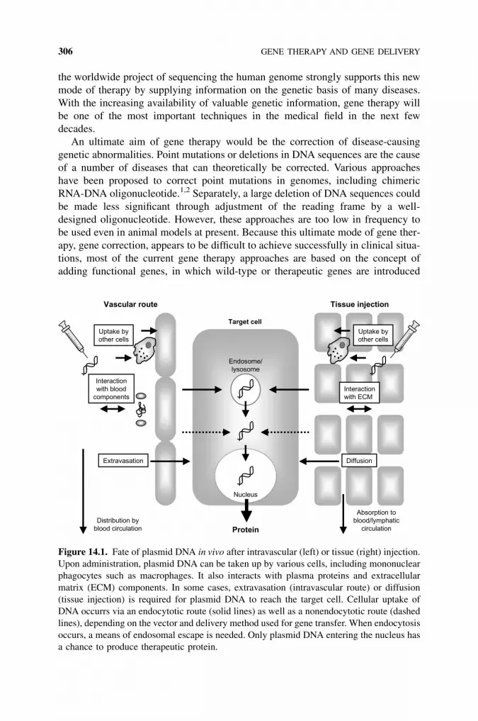
the worldwide project of sequencing the human genome strongly supports this new
mode of therapy by supplying information on the genetic basis of many diseases.
With the increasing availability of valuable genetic information, gene therapy will
be one of the most important techniques in the medical field in the next few
decades.
An ultimate aim of gene therapy would be the correction of disease-causing
genetic abnormalities. Point mutations or deletions in DNA sequences are the cause
of a number of diseases that can theoretically be corrected. Various approaches
have been proposed to correct point mutations in genomes, including chimeric
RNA-DNA oligonucleotide.1,2 Separately, a large deletion of DNA sequences could
be made less significant through adjustment of the reading frame by a well-
designed oligonucleotide. However, these approaches are too low in frequency to
be used even in animal models at present. Because this ultimate mode of gene ther-
apy, gene correction, appears to be difficult to achieve successfully in clinical situa-
tions, most of the current gene therapy approaches are based on the concept of
adding functional genes, in which wild-type or therapeutic genes are introduced
Vascular route Tissue injection
Uptake byother cells
Interactionwith ECM
Nucleus
Endosome/lysosome
Target cell
Interactionwith blood
components
Extravasation Diffusion
Absorption toblood/lymphatic
circulation
Uptake byother cells
Distribution byblood circulation Protein
Figure 14.1. Fate of plasmid DNA in vivo after intravascular (left) or tissue (right) injection.
Upon administration, plasmid DNA can be taken up by various cells, including mononuclear
phagocytes such as macrophages. It also interacts with plasma proteins and extracellular
matrix (ECM) components. In some cases, extravasation (intravascular route) or diffusion
(tissue injection) is required for plasmid DNA to reach the target cell. Cellular uptake of
DNA occurrs via an endocytotic route (solid lines) as well as a nonendocytotic route (dashed
lines), depending on the vector and delivery method used for gene transfer. When endocytosis
occurs, a means of endosomal escape is needed. Only plasmid DNA entering the nucleus has
a chance to produce therapeutic protein.
306 GENE THERAPY AND GENE DELIVERY

as pharmaceuticals. Due to the difficulties involving genes reaching their targets in
vivo, delivery is the major challenge in the development of in vivo gene therapy
protocols. In addition to gene replacement therapy, success of in vivo gene transfer
will give us an opportunity to carry out DNA vaccination, in which the antigenic
protein is delivered in a form of genetic information that ensures the optimal
immune response.3
A gene is transcribed to mRNA in the nucleus, then translated into a peptide at
the ribosome; the peptide then goes through folding, glycosylation, and/or multi-
meric formation, resulting in the production of a functional protein, that is to
say, a process of gene expression. Except for the cases in which genes encoded
in DNA do not need to be transcribed before performing their therapeutic functions,
gene therapy obviously requires the transgene expression of encoded genes. Thus,
the administered gene has no biological significance without reaching its target:
the nucleus. On its way to the nucleus from the injection site, DNA encoding a
gene will pass through several biological barriers that often preclude successful
gene delivery (Figure 14.1).4 To overcome these barriers, much effort has been
made to develop effective vectors for DNA, although most of these are not suitable
for clinical use and need to be improved.
In this chapter, the delivery issues of DNA, especially plasmid DNA, are dis-
cussed with reference to gene therapy, focusing on existing biological barriers to
be overcome and the characteristics of delivery methods and vectors to achieve
this. Current progress and the status of nonviral gene delivery approaches are
described.
14.2. INTERACTION OF DNA WITH CELLS
Upon administration to the body, plasmid DNA or its complex with any vector,
interacts with cells, proteins, and extracellular matrix. The summation of these
interactions will determine the in vivo fate of plasmid DNA and its complex, which,
in turn, determines the location and extent of gene transfer. From a pharmaceutical
and biopharmaceutical point of view, plasmid DNA is a huge macromolecule with a
strong negative charge, and it is susceptible to attack by nucleases.
14.2.1. Negative Charge-Mediated Uptake
After entering the blood circulation, plasmid DNA encounters various cells, includ-
ing blood cells, endothelial cells, and macrophages. The tissue distribution after
intravenous injection of single- or double-stranded DNA, oligonucleotide, DNA/
anti-DNA immune complex, mononucleosome, or chromatin has been examined.
Although these studies have shown that the liver is the main organ responsible
for the rapid clearance of these forms of DNA from the circulation,5–9 the uptake
mechanism and the cell type contributing to this hepatic uptake remain to be elu-
cidated. We have studied the tissue distribution of plasmid DNA radiolabeled with32P following intravenous injection into mice.10,11 After intravenous injection,
INTERACTION OF DNA WITH CELLS 307

naked plasmid DNA was rapidly eliminated from the blood circulation and taken up
predominantly by the liver nonparenchymal cells, such as liver sinusoidal endothe-
lial cells and Kupffer cells. Being administered in its free form, plasmid DNA was
also rapidly degraded by nucleases in serum as well as in tissues. The hepatic
uptake, most of which is mediated by the liver nonparenchymal cells, was as
high as the plasma flow rate to the organ. The hepatic uptake of plasmid DNA
was dependent on the concentration and decreased upon an increase of the dose.
In addition, the hepatic uptake was inhibited by calf thymus DNA, polyinosinic
acid, dextran sulfate, and heparin, but not by polycytidylic acid and chondroitin sul-
fate.10–12 These findings indicate that plasmid DNA is taken up by scavenger recep-
tors, which recognize polyanions in a charge- and/or structure-dependent manner.13
However, we excluded the possibility of class A scavenger receptors (SRA) being
involved in the uptake, based on tissue distribution and uptake experiments using
cultured macrophages from SRA-knockout mice.14 Another study supported the
conclusion that SRA are not involved in the uptake of plasmid DNA.15
When injected into skeletal muscle or solid tumor tissue, plasmid DNA under-
goes degradation as well as absorption into the blood circulation.16 A fraction of the
plasmid DNA is absorbed from the lymphatic system after injection into these tis-
sues. Plasmid DNA and its degraded products exhibit distribution profiles similar to
that obtained with intravenous plasmid DNA.
The detailed characteristics of the uptake of plasmid DNA were examined using
cultured cells. Various cells have the ability to take up plasmid DNA in a concen-
tration- and temperature-dependent manner. One example involves the brain micro-
vessel endothelial cells,17 which constitute the blood-brain barrier. However, their
contribution to the tissue distribution of plasmid DNA seems to be minor. The main
cells responsible for the distribution of plasmid DNA are immune cells such as
macrophages. In fact, Kupffer cells, liver macrophages, make a large contribution
to the clearance of plasmid DNA administered into the blood circulation. Plasmid
DNA is efficiently taken up by cultured mouse peritoneal macrophages.18 The pro-
file of uptake inhibition is similar to that observed in brain microvessel endothelial
cells. Another population of immune cells is dendritic cells (DCs); these are very
important as far as both innate and acquired immunity are concerned. The DC cell
line (DC2.4 cells) exhibits extensive uptake and degradation of plasmid DNA.19
14.2.2. Activation of Immune Cells
The unmethylated CpG sequence, i.e., the CpG motif, is a danger signal for our
immune system.20 A potent CpG motif consists of a central unmethylated CpG
dinucleotide flanked by two 50 purines and two 30 pyrimidines. Compared with
the DNA of eukaryotic cells (frequency of �1:64), bacterial genomic DNA contains
a higher frequency of the dinucleotide sequence CpG (1:16). Prokaryotic DNA is
relatively unmethylated compared with the eukaryotic form, in which approxi-
mately 80% of the cytosines are methylated, a modification known to eliminate
immunostimulation. These differences allow the mammalian immune system to
recognize and respond to foreign DNA of bacterial origin. In addition to the immu-
308 GENE THERAPY AND GENE DELIVERY

nostimulatory CpG motifs, neutralizing CpG sequences that can neutralize the
immune activating properties of the stimulatory motifs have been reported.21
Because plasmid DNA is derived from bacteria, it contains a number of sequences
that can be regarded as immunostimulatory CpG motifs.
14.3. GENE DELIVERY APPROACHES
Successful application of gene therapy to patients requires the development of a
gene delivery approach that enables in vivo transgene expression that is high
enough to produce a therapeutic benefit. Because plasmid DNA is a huge macro-
molecule with a strong negative charge, it has little access to the nucleus when
administered by routine methods. To facilitate the binding to cells, cationic vectors
are often used, and the optimization of the structure and function is a major chal-
lenge as far as nonviral vector development is concerned. Another strategy is the
use of naked plasmid DNA. In this case, the methodologies used to deliver plasmid
DNA within cells are critical for gene transfer.
14.3.1. Fundamentals of Plasmid DNA Delivery
The large size of plasmid DNA greatly limits its distribution after in vivo adminis-
tration. The distribution processes have some limitations as far as size is concerned,
such as the passage through capillaries (about 5 mm in diameter) and fenestrae (50–
300 nm) in the endothelial layers. Tissue distribution of intravascularly adminis-
tered plasmid DNA is highly restricted by the endothelial wall, which is composed
of vascular endothelial cells and basement membrane. Discontinuous endothelium,
which is present only in the liver, spleen, bone marrow, and some solid tumors,
allows plasmid DNA to come into contact with tissue cells.
Because the cell surface is negatively charged, positively charged compounds
interact more intensely with cells than do negatively charged ones. Cationic com-
plexes of plasmid DNA with any cationic vector exhibit very different pharmaco-
kinetic profiles from that of naked plasmid DNA. They avidly bind to endothelial
cells and normally accumulate in the lung after intravenous injection due to the
first-pass effect.22,23
When plasmid DNA or its complex is injected into the extracellular space of a
tissue, it distributes in an area that is very close to the injection site. Plasmid DNA
within the extracellular space will be delivered into the surrounding cells, probably
due to (1) increased pressure from the injection and/or (2) mechanical damage to
the cell membrane. Very limited distribution of transfected cells has been reported
after tissue injection of both naked and complexed plasmid DNA.24–26
14.3.2. Vector Complex
For intravenous administration, formation of a cationic complex greatly increases
the interaction of plasmid DNA with the lung endothelial cells.22,27 Transgene
GENE DELIVERY APPROACHES 309

expression is also high in these cells. This might be the result of complicated events
occurring in vivo after intravenous injection of lipoplex (see below). Serum pro-
teins28 and blood cells23 have been reported to affect the tissue distribution of intra-
vascularly administered plasmid DNA complex. When a plasmid DNA complex
can avoid first-pass filtration in the lung, only a relatively small DNA complex
can pass through the blood vessels and interact directly with the parenchymal cells
of each tissue.
Although these nonviral vectors are distinct from their viral counterparts, their
advantage of having a high affinity for various types of cells has been used to
increase the transfection efficiency by viral vectors.29,30
14.3.2.1. Lipoplex So far, an enormous number of cationic lipid/liposome sys-
tems have been developed to improve the transfection efficiency of plasmid
DNA. Some of the cationic lipids used have been summarized in a recent publica-
tion.31 Cationic liposomes associate with plasmid DNA via an electrostatic interac-
tion, which results in the formation of a complex called ‘‘lipoplex.’’32 The driving
force for lipoplex to introduce genes into cells is its electrostatic binding to nega-
tively charged cellular membranes followed by endocytotic uptake. Some studies
have shown that co-lipids, so-called helper lipids, in cationic liposomes are impor-
tant determinants of transfection efficiency. Intravenous injection of lipoplex can
lead to significant in vivo transfection activity in the lung when cholesterol is
used as a helper lipid,22,33 although such lipoplex formulations produce less trans-
fection in vitro than lipoplex containing dioleoylphosphatidylethanolamine.
To compensate for the lack of specificity of electrostatic interaction of lipoplex,
ligands can be introduced into cationic liposomes; sugars are the ligands that have
been most extensively investigated so far.34–37 On the other hand, local injection of
lipoplex has also been investigated to ensure site-specific gene transfer. A local
injection of lipoplex has been shown to be an effective approach to achieve trans-
gene expression in the lung, brain, tumor, and skin.38–41 Intratracheal administra-
tion of lipoplex is another possible way to introduce genes to the cells in the
respiratory tract.42
Because the size of lipoplex is a key factor in determining the tissue distribution
as well as the cellular uptake, it would be a challenge to reduce the size of lipoplex
to increase transfection efficiency. Recently, Dauty et al.43 succeeded in formulating
plasmid DNA into stable nanometric particles with a diameter of less than 40 nm by
synthesizing a dimerizable cationic detergent.
A major drawback of lipoplex is the production of inflammatory cytokines. We
have shown that Kupffer cells in the liver are the major source of inflammatory
cytokines after intravenous injection of lipoplex.44 A functional depletion of Kupf-
fer cells resulted in less cytokine production after lipoplex injection into mice.
Macrophages and DCs can be considered the source of cytokines, because they pro-
duce large amounts of cytokines following incubation with lipoplex composed of
plasmid DNA.45 To suppress the immune response, the depletion of CpG sequences
from plasmid DNA is a promising option. Yew et al.46 eliminated 270 of 526 CpG
dinucleotides in a reporter plasmid DNA, either by eliminating nonessential regions
310 GENE THERAPY AND GENE DELIVERY

within the plasmid backbone or by site-directed mutagenesis. A CpG-reduced plas-
mid DNA was then found to be significantly less immunostimulatory than the ori-
ginal one. The administration of dexamethasone is another approach to reduce the
production of such cytokines.47 A sequential injection of plasmid DNA and cationic
liposomes is another way of achieving a level of transgene expression in the lung
similar to that following the injection of lipoplex with the production of far fewer
inflammatory cytokines.48,49
14.3.2.2. Polyplex Cationic polymers are another class of nonviral vectors that
can be used to increase gene delivery and transfer to target cells in vivo. Various
types of polymers have been examined with respect to their ability to protect
from nuclease degradation, to deliver target cells, and to increase the transfection
efficiency of plasmid DNA. Plasmid DNA-cationic polymer complex, or polyplex,
is believed to be taken up by cells via an endocytotic pathway, so its transfection
efficiency depends on the release of plasmid DNA into the cytoplasm after cellular
uptake. Therefore, polyplex-mediated transfection can be enhanced by the applica-
tion of polymers possessing buffering ability50 or fusogenic peptides creating pores
on membranes.51
Polyethyleneimine (PEI) is one of the most extensively examined polymers
because it has a relatively high transfection efficiency. PEI is believed to enter
the cell via an endocytotic route and to possess a buffering capacity and the ability
to swell when protonated.52,53 Therefore, at low pH values, it is believed that PEI
prevents acidification of the endosome and induces a large inflow of ions and water,
subsequently leading to rupture of the endosomal or lysosomal membrane so that
the PEI-plasmid DNA complex is delivered to the cytoplasmic space.54,55 PEI is
also reported to undergo nuclear localization while retaining an ordered structure
once endocytosed.53 However, the transfection efficiency of PEI depends on its
molecular weight and structure.56–58
In another approach, the endosome-lysosome pathway of the polyplex may be
circumvented by the use of endosome-disrupting peptides. We have demonstrated
that a peptide that mimics the amino terminal of influenza virus hemagglutinin sub-
unit HA-2 can greatly increase transgene expression in the liver after intravenous
injection of hepatocyte-targeted polyplex.59 About a 70-fold increase in transgene
expression in the liver was obtained by the covalent binding of the peptide to a
galactosylated poly(L-ornithine).
Targeted gene transfer has been widely investigated using polyplex possessing
targeting ligands. These include asialoglycoproteins,60 carbohydrates,61–63 transfer-
rin,64,65 antibodies,66,67 and lung surfactant proteins.68 The attached ligand can
increase the affinity for the target cell through a receptor-ligand interaction.
When designed properly, about 80% of a polyplex can be successfully delivered
to the target organ after systemic administration.63
However, the targeted delivery of polyplex is hampered by interaction with var-
ious compounds in the body fluids such as serum proteins.69 Ogris et al.70 have
shown that, when incubated with plasma, the transferrin-PEI/plasmid DNA com-
plex undergoes aggregation, which leads to reduced delivery to the target. The
GENE DELIVERY APPROACHES 311
PEGylation also appears to be a useful method of prolonging the blood circulation
of polyplex after systemic administration, and PEGylated polyplex has resulted in
gene transfer to a tumor without significant toxicity after intravenous injection into
tumor-bearing mice.71
A series of peptides that can be recognized and transported to the nucleus are
called ‘‘nuclear localization signal’’ (NLS) peptides. A well-known example is the
basic peptide derived from the simian virus 40 large tumor antigen (PKKKRKV),
which mediates binding of the karyophilic protein to importin.72 Because of its
nature, its application to gene delivery has been extensively investigated.73–75 In
most cases, transgene expression by plasmid DNA complex was increased by the
application of NLS.
14.3.3. Naked Plasmid DNA
Although naked plasmid DNA is considered to have several problems for in vivo
use as a way of obtaining sufficient gene transfer, the problems involving the use
of DNA complex and the development of novel delivery methods allow naked plas-
mid DNA to be used as an efficient vector for gene therapy. Once it enters the cyto-
plasm or nucleus, naked plasmid DNA seems to produce much better transgene
expression than does any complex. Furthermore, naked plasmid DNA injection
using the hydrodynamics-based procedure (see below) can result in efficient trans-
gene expression while producing very few nonspecific inflammatory cytokines,76 a
property that would be favorable for many gene therapy protocols. This is quite
unlike the case in which the plasmid DNA complex is used.44
14.3.3.1. Direct Injection into Organs Because of its physicochemical proper-
ties, naked plasmid DNA has not generally been considered to possess the ability
to enter cells without any vector. However, in 1990, Wolff et al.77 demonstrated that
a needle injection of naked plasmid DNA into mouse skeletal muscle resulted in
significant transgene expression in the tissue. This was the first report showing
that no special delivery system is required for in vivo gene transfer using plasmid
DNA. Since then, this method of gene transfer has been applied to other sites of the
body, such as the heart muscle,78 liver,24 brain,39 skin,41 urological organs,79 thyr-
oid,80 and tumors.40,81 At least in skeletal muscle, transgene expression in the
organs can persist for several months,82 although the transfected DNA is not
thought to undergo chromosomal integration.
The fact that cells with transgene expression are found only in close proximity to
the track of the needle injection indicates that dispersion of the injected plasmid
DNA within the organ is an important issue and that mechanical force caused by
the needle injection is critical for the effective intracellular delivery of naked plas-
mid DNA. Direct needle injection of plasmid DNA to the target organ is apparently
simple and easy. Furthermore, muscle and skin are very attractive tissues as injec-
tion sites because surgery is not required for the administration of the DNA to these
sites.
312 GENE THERAPY AND GENE DELIVERY

Some polymers, which do not form condensed complexes with plasmid
DNA, have been reported to exhibit an enhancing effect on the transgene
expression of naked plasmid DNA in skeletal muscle. Polyvinyl pyrrolidone and
polyvinyl alcohol have been used to increase the extent and level of transgene
expression following intramuscular injection of plasmid DNA.83 In addition,
Hartikka et al.84 reported that a change of injection vehicle from saline or
phosphate-buffered saline to 150 mM sodium phosphate buffer could enhance
transgene expression by naked plasmid DNA in muscle due to inhibition of DNA
degradation.
14.3.3.2. Intravascular Injection/Hydrodynamics-Based Procedure In 1999, Liu
et al.85 and Zhang et al.86 developed a very powerful technique for in vivo gene
transfer. In this method of administration, naked plasmid DNA is injected into
the vasculature in a large volume of vehicle at high velocity. Liu et al. called this
method of gene transfer a ‘‘hydrodynamics-based procedure.’’ When injected by
this technique, plasmid DNA does not require any vector to protect it from degra-
dation. Although the mechanism of this technique is not fully understood, plasmid
DNA can reach the nucleus by this approach. We have examined the tissue distri-
bution characteristics of plasmid DNA administered either under normal conditions
or by the hydrodynamics-based procedure.11 Plasmid DNA injected by the hydro-
dynamics-based procedure was mainly recovered in the liver, which is similar to the
profile obtained after the normal injection of plasmid DNA.10 However, the hepatic
uptake was not inhibited by prior administration of polyanions, including poly I,
dextran sulfate, and heparin, suggesting a nonspecific uptake process. This
hypothesis was supported by the finding that significant hepatic uptake of proteins
such as bovine serum albumin and immunoglobulin G was observed after the
hydrodynamics-based procedure.
The large-volume injection of naked plasmid DNA has also been applied to tis-
sue-selective gene transfer, where a large volume of naked plasmid DNA solution
was injected into the vasculature that led directly to the target organ with transient
occlusion of the outflow. Budker et al.87 demonstrated successful transgene expres-
sion in the liver by injecting plasmid DNA into the portal vein in a large volume of
hypertonic solution (20% mannitol) with temporary occlusion of the hepatic vein to
increase osmotic and hydrostatic pressure in the organ. The pressure generated at
the local site may widen the sinusoid fenestrae and enhance DNA extravasation,
followed by DNA uptake by hepatocytes. A similar approach was applied to the
skeletal muscle of the rat hindlimb, where naked plasmid DNA was injected into
the femoral artery in a large volume of saline over a period of 10 seconds while
all blood vessels leading to and out of the hindlimb were occluded.88 This intravas-
cular injection increased transgene expression in muscle up to 40-fold compared
with intramuscular injection. In a similar manner, kidney-targeted gene transfer
was achieved by a retrograde injection of plasmid DNA into the renal vein of
rats.89 Transgene expression was detected only in the interstitial fibroblasts near
the peritubular capillaries of the injected kidney.
GENE DELIVERY APPROACHES 313

14.3.3.3. Physical Enhancement of Delivery/Expression In addition to the large-
volume-induced delivery of plasmid DNA into the target cells, other approaches to
achieve intracellular delivery have been widely explored. The retention time of
plasmid DNA in the tissue can be a factor determining gene transfer. Transient stop-
page of blood flow through the diaphragm of mice greatly increased transgene
expression in this muscular tissue following intravenous injection under normal
conditions.90 Significant expression of dystrophin (15–20% of the diaphragm mus-
cle) after peripheral intravenous injection of plasmid DNA encoding the mouse dys-
trophin gene was detected in dystrophin-deficient mdx mice. Recently, significant
transgene expression in the liver was achieved by simple mechanical massage after
intravenous injection of naked plasmid DNA into mice.91
Electroporation has been used to increase the transport of charged molecules,
such as plasmid DNA, across biological membranes.92,93 After initial permeation,
the pores close and plasmid DNA is trapped within the cell. Therefore, electropora-
tion following a tissue injection of plasmid DNA increases the chance of DNA
uptake by cells adjacent to the injection site. In vivo electroporation generally
increases transgene expression up to 1000-fold compared with injection of naked
plasmid DNA without electroporation in tissues such as skin,94 liver,95 melanoma,96
and muscle.97
Shooting gold particles coated with plasmid DNA into target tissues or cells can
deliver the DNA into the cytoplasm or even the nucleus. Skin, liver, and muscle have
been successfully transfected after surgical exposure of the tissue. The efficiency of
transfection varies among tissues, from 10–20% for skin epidermal cells to 1–5%
for muscle cells.98 In many cases, genes for antigens or cytokines have been introduced
by this procedure for vaccination and immunotherapy, respectively.99,100
The application of ultrasound has also been investigated in an attempt to improve
in vivo transgene expression, and this facilitated nonendocytotic uptake of plasmid
DNA into cells.101 Recently, the combined use of microbubbles was found to be
effective in increasing ultrasound-mediated gene transfer. So far, ultrasound-mediated
gene transfer has been carried out in vascular cells102,103 and heart muscle.104
14.4. CONCLUSION
Since the discoveries of target genes that are responsible for monogenetic disorders
such as the cystic fibrosis transmembrane conductance regulator gene and the dys-
trophin gene, the concept of gene therapy has been accepted as a very attractive and
promising method of treatment for these diseases. In addition, the recent comple-
tion of the Human Genome Project has linked common illnesses such as heart dis-
ease, diabetes, and asthma with certain genetic factors, so these too can be treated
by gene therapy. However, gene therapy, especially in vivo, has not so far been as
effective as was initially hoped. Solution of the problems associated with gene
delivery is a prerequisite for successful in vivo gene therapy. We believe that the
latest results of basic research on gene delivery will offer a way to develop effective
gene therapy protocols in the near future.
314 GENE THERAPY AND GENE DELIVERY

REFERENCES
1. Cole-Strauss, A.; Yoon, K.; Xiang, Y.; Byrne, B. C.; Rice, M. C.; Gryn, J.; Holloman, W.
K.; Kmiec, E. B. Science 1996, 273, 1386–1389.
2. Igoucheva, O.; Yoon, K. Hum. Gene Ther. 2000, 11, 2307–2312.
3. Gurunathan, S.; Klinman, D. M.; Seder, R. A. Annu. Rev. Immunol. 2000, 18, 927–974.
4. Nishikawa, M.; Huang, L. Hum. Gene Ther. 2001, 12, 861–870.
5. Emlen, W.; Mannik, M. J. Exp. Med. 1978, 147, 684–699.
6. Emlen, W.; Mannik, M. Clin. Exp. Immunol. 1984, 56, 185–192.
7. Emlen, W.; Burdick, G. J. Immunol. 1988, 140, 1816–1822.
8. Gauthier, V. J.; Tyler, L. N.; Mannik, M. J. Immunol. 1996, 156, 1151–1156.
9. Du Clos, T. W.; Volzer, M. A.; Hahn, F. F.; Xiao, R.; Mold, C.; Searles, R. P. Clin. Exp.
Immunol. 1999, 117, 403–411.
10. Kawabata, K.; Takakura, Y.; Hashida, M. Pharm. Res. 1995, 12, 825–830.
11. Kobayashi, N.; Kuramoto, T.; Yamaoka, K.; Hashida, M.; Takakura, Y. J. Pharmacol.
Exp. Ther. 2001, 297, 853–860.
12. Yoshida, M.; Mahato, R. I.; Kawabata, K.; Takakura, Y.; Hashida, M. Pharm. Res. 1996,
13, 599–603.
13. Terpstra, V.; van Amersfoort, E. S.; van Velzen, A. G.; Kuiper, J.; van Berkel, T. J.
Arterioscler Thromb. Vasc. Biol. 2000, 20, 1860–1872.
14. Takakura, Y.; Takagi, T.; Hashiguchi, M.; Nishikawa, M.; Yamashita, F.; Doi, T.;
Imanishi, T.; Suzuki, H.; Kodama, T.; Hashida, M. Pharm. Res. 1999, 16, 503–508.
15. Zhu, F. G.; Reich, C. F.; Pisetsky, D. S. Immunology 2001, 103, 226–234.
16. Kawase, A.; Nomura, T.; Yasuda, K.; Kobayashi, N.; Hashida, M.; Takakura, Y. J.
Pharm. Sci. 2003, 92, 1295–1304.
17. Nakamura, M.; Davila-Zavala, P.; Tokuda, H.; Takakura, Y.; Hashida M. Biochem.
Biophys. Res. Commun. 1998, 245, 235–239.
18. Takagi, T.; Hashiguchi, M.; Mahato, R. I.; Tokuda, H.; Takakura, Y.; Hashida, M.
Biochem. Biophys. Res. Commun. 1998, 245, 729–733.
19. Yoshinaga, T.; Yasuda, K.; Ogawa, Y.; Takakura, Y. Biochem. Biophys. Res. Commun.
2002, 299, 389–394.
20. Krieg, A. M. Annu. Rev. Immunol. 2002, 20, 709–760.
21. Krieg, A. M.; Wu, T.; Weeratna, R.; Efler, S. M.; Love-Homan, L.; Yang, L.; Yi, A. K.;
Short, D.; Davis, H. L. Proc. Natl. Acad. Sci. USA 1998, 95, 12631–12636.
22. Liu, Y.; Mounkes, L. C.; Liggitt, H. D.; Brown, C. S.; Solodin, I.; Heath, T. D.; Debs, R. J.
Nat. Biotechnol. 1997, 15, 167–173.
23. Sakurai, F.; Nishioka, T.; Saito, H.; Baba, T.; Okuda, A.; Matsumoto, O.; Taga, T.;
Yamashita, F.; Takakura, Y.; Hashida, M. Gene Ther. 2001, 8, 677–686.
24. Hickman, M. A.; Malone, R. W.; Lehmann-Bruinsma, K.; Sih, T. R.; Knoell, D.; Szoka,
F. C.; Walzem, R.; Carlson, D. M.; Powell, J. S. Hum. Gene Ther. 1994, 5, 1477–1483.
25. Nomura, T.; Nakajima, S.; Kawabata, K.; Yamashita, F.; Takakura, Y.; Hashida, M.
Cancer Res. 1997, 57, 2681–2686.
26. O’Hara, A. J.; Howell, J. M.; Taplin, R. H.; Fletcher, S.; Lloyd, F.; Kakulas, B.;
Lochmuller, H.; Karpati, G. Muscle Nerve 2001, 24, 488–495.
REFERENCES 315

27. Mahato, R. I.; Kawabata, K.; Nomura, T.; Takakura, Y.; Hashida, M. J. Pharm. Sci. 1995,
84, 1267–1271.
28. Li, S.; Tseng, W. C.; Stolz, D. B.; Wu, S. P.; Watkins, S. C.; Huang, L. Gene Ther. 1999, 6,
585–594.
29. Bouri, K.; Feero, W. G.; Myerburg, M. M.; Wickham, T. J.; Kovesdi, I. Hoffman, E. P.;
Clemens, P. R. Hum. Gene Ther. 1999, 10, 1633–1640.
30. Porter, C. D. J. Gene Med. 2002, 4, 622–633.
31. Audouy, S. A.; de Leij, L. F.; Hoekstra, D.; Molema, G. Pharm. Res. 2002, 19, 1599–
1605.
32. Felgner, P. L.; Barenholz, Y.; Behr, J. P.; Cheng, S. H.; Cullis, P.; Huang, L.; Jessee, J. A.;
Seymour, L.; Szoka, F.; Thierry, A. R.; Wagner, E.; Wu, G. Hum. Gene Ther. 1997, 8,
511–512.
33. Templeton, N. S.; Lasic, D. D.; Frederik, P. M.; Strey, H. H.; Roberts, D. D.; Pavlakis, G.
N. Nat. Biotechnol. 1997, 15, 647–652.
34. Remy, J. S.; Kichler, A.; Mordvinov, V.; Schuber, F.; Behr, J. P. Proc. Natl. Acad. Sci.
USA 1995, 92, 1744–1748.
35. Hara, T.; Aramaki, Y.; Takada, S.; Koike, K.; Tsuchiya, S. Gene Ther. 1995, 2, 784–788.
36. Kawakami, S.; Fumoto, S.; Nishikawa, M.; Yamashita, F.; Hashida, M. Pharm. Res.
2000, 17, 306–313.
37. Kawakami, S.; Sato, A.; Nishikawa, M.; Yamashita, F.; Hashida, M. Gene Ther. 2000, 7,
292–299.
38. Brigham, K. L.; Meyrick, B.; Christman, B.; Magnuson, M.; King, G.; Berry, L. C., Jr.
Am. J. Med. Sci. 1989, 298, 278–281.
39. Ono, T.; Fujino, Y.; Tsuchiya, T.; Tsuda, M. Neurosci. Lett. 1990, 117, 259–263.
40. Plautz, G. E.; Yang, Z. Y.; Wu, B. Y.; Gao, X.; Huang, L.; Nabel, G. J. Proc. Natl. Acad.
Sci. USA 1993, 90, 4645–4649.
41. Raz, E.; Carson, D. A.; Parker, S. E.; Parr, T. B.; Abai, A. M.; Aichinger, G.;
Gromkowski, S. H.; Singh, M.; Lew, D.; Yankauckas, M. A.; Baird, S. M.; Rhodes,
G. H. Proc. Natl. Acad. Sci. USA 1994, 91, 9519–9523.
42. McCluskie, M. J.; Chu, Y.; Xia, J. L.; Jessee, J.; Gebyehu, G.; Davis, H. L. Antisense
Nucleic Acid Drug Dev. 1998, 8, 401–414.
43. Dauty, E.; Remy, J. S.; Blessing, T.; Behr, J. P. J. Am. Chem. Soc. 2001, 123, 9227–9234.
44. Sakurai, F.; Terada, T.; Yasuda, K.; Yamashita, F.; Takakura, Y.; Hashida, M. Gene Ther.
2002, 9, 1120–1126.
45. Yasuda, K.; Ogawa, Y.; Kishimoto, M.; Takagi, T.; Hashida, M.; Takakura, Y. Biochem.
Biophys. Res. Commun. 2002, 293, 344–348.
46. Yew, N. S.; Zhao, H.; Wu, I. H.; Song, A.; Tousignant, J. D.; Przybylska, M.; Cheng, S. H.
Mol. Ther. 2000, 1, 255–262.
47. Tan, Y.; Li, S.; Pitt, B. R.; Huang, L. Hum. Gene Ther. 1999, 10, 2153–2161.
48. Song, Y. K.; Liu, F.; Liu, D. Gene Ther. 1998, 5, 1531–1537.
49. Tan, Y.; Liu, F.; Li, Z.; Li, S.; Huang, L. Mol. Ther. 2001, 3, 673–682.
50. Boussif, O.; Lezoualch, F.; Zanta, M. A.; Mergny, M. D.; Scherman, D.; Demeneix, B.;
Behr, J. P. Proc. Natl. Acad. Sci. USA 1995, 92, 7297–7301.
316 GENE THERAPY AND GENE DELIVERY

51. Wagner, E.; Plank, C.; Zatloukal, K.; Cotten, M.; Birnstiel, M. L. Proc. Natl. Acad. Sci.
USA 1992, 89, 7934–7938.
52. Boletta, A.; Benigni, A.; Lutz, J.; Remuzzi, G.; Soria, M. R.; Monaco, L. Hum. Gene
Ther. 1997, 8, 1243–1251.
53. Godbey, W. T.; Wu, K. K.; Mikos, A. G. Proc. Natl. Acad. Sci. USA 1999, 96, 5177–5181.
54. Wagner, E. J. Control. Release 1998, 53, 155–158.
55. Klemm, A. R.; Young, D.; Lloyd, J. B. Biochem. Pharmacol. 1998, 56, 41–46.
56. Ouatas, T.; Le Mevel, S.; Demeneix, B. A.; de Luze, A. Int. J. Dev. Biol. 1998, 42, 1159–
1164.
57. Fischer, D.; Bieber, T.; Li, Y.; Elsasser, H. P.; Kissel, T. Pharm. Res. 1999, 16, 1273–
1279.
58. Morimoto, K.; Nishikawa, M.; Kawakami, S.; Nakano, T.; Hattori, Y.; Fumoto, S.;
Yamashita, F.; Hashida, M. Mol. Ther. 2003, 7, 254–261.
59. Nishikawa, M.; Yamauchi, M.; Morimoto, K.; Ishida, E.; Takakura, Y.; Hashida, M. Gene
Ther. 2000, 7, 548–555.
60. Wu, G. Y.; Wu, C. H. J. Biol. Chem. 1988, 263, 14621–14624.
61. Midoux, P.; Mendes, C.; Legrand, A.; Raimond, J.; Mayer, R.; Monsigny, M.; Roche, A.
C. Nucleic Acids Res. 1993, 21, 871–878.
62. Perales, J. C.; Ferkol, T.; Beegen, H.; Ratnoff, O. D.; Hanson, R. W. Proc. Natl. Acad.
Sci. USA 1994, 91, 4086–4090.
63. Nishikawa, M.; Takemura, S.; Takakura, Y.; Hashida, M. J. Pharmacol. Exp. Ther. 1998,
287, 408–415.
64. Wagner, E.; Zenke, M.; Cotten, M.; Beug, H.; Birnstiel, M. L. Proc. Natl. Acad. Sci. USA
1990, 87, 3410–3414.
65. Kircheis, R.; Schuller, S.; Brunner, S.; Ogris, M.; Heider, K. H.; Zauner, W.; Wagner, E.
J. Gene Med. 1999, 1, 111–120.
66. Ferkol, T.; Kaetzel, C. S.; Davis, P. B. J. Clin. Invest. 1993, 92, 2394–2400.
67. Li, S.; Tan, Y.; Viroonchatapan, E.; Pitt, B. R.; Huang, L. Am. J. Physiol. 2000, 278,
L504–L511.
68. Ross, G. F.; Morris, R. E.; Ciraolo, G.; Huelsman, K.; Bruno, M.; Whitsett, J. A.; Baatz, J.
E.; Korfhagen, T. R. Hum. Gene Ther. 1995, 6, 31–40.
69. Opanasopit, P.; Nishikawa, M.; Hashida, M. Crit. Rev. Ther. Drug Carrier Syst. 2002, 19,
191–233.
70. Ogris, M.; Brunner, S.; Schuller, S.; Kircheis, R.; Wagner, E. Gene Ther. 1999, 6, 595–
605.
71. Kircheis, R.; Blessing, T.; Brunner, S.; Wightman, L.; Wagner, E. J. Control. Release
2001, 72, 165–170.
72. Adam, S. A.; Gerace, L. Cell 1991, 66, 837–847.
73. Kaneda, Y.; Iwai, K.; Uchida, T. Science 1989, 243, 375–378.
74. Fritz, J. D.; Herweijer, H.; Zhang, G.; Wolff, J. A. Hum. Gene Ther. 1996, 7, 1395–1404.
75. Ludtke, J. J.; Zhang, G.; Sebestyen, M. G.; Wolff, J. A. J. Cell Sci. 1999, 112, 2033–2041.
76. Kobayashi, N.; Kuramoto, T.; Chen, S.; Watanabe, Y.; Takakura, Y. Mol. Ther. 2002, 6,
737–744.
REFERENCES 317

77. Wolff, J. A.; Malone, R. W.; Williams, P.; Chong, W.; Acsadi, G.; Jani, A.; Felgner, P. L.
Science 1990, 247, 1465–1468.
78. Lin, H.; Parmacek, M. S.; Morle, G.; Bolling, S.; Leiden, J. M. Circulation 1990, 82,
2217–2221.
79. Yoo, J. J.; Soker, S.; Lin, L. F.; Mehegan, K.; Guthrie, P. D.; Atala, A. J. Urol. 1999, 162,
1115–1118.
80. Sikes, M. L.; O’Malley, B. W., Jr.; Finegold, M. J.; Ledley, F. D. Hum. Gene Ther. 1994,
5, 837–844.
81. Nomura, T.; Yasuda, K.; Yamada, T.; Okamoto, S.; Mahato, R. I.; Watanabe, Y.;
Takakura, Y.; Hashida, M. Gene Ther. 1999, 6, 121–129.
82. Acsadi, G.; Dickson, G.; Love, D. R.; Jani, A.; Walsh, F. S.; Gurusinghe, A.; Wolff, J. A.;
Davies, K. E. Nature 1991, 352, 815–818.
83. Alila, H.; Coleman, M.; Nitta, H.; French, M.; Anwer, K.; Liu, Q.; Meyer, T.; Wang, J.;
Mumper, R.; Oubari, D.; Long, S.; Nordstrom, J.; Rolland, A. Hum. Gene Ther. 1997, 8,
1785–1795.
84. Hartikka, J.; Bozoukova, V.; Jones, D.; Mahajan, R.; Wloch, M. K.; Sawdey, M.;
Buchner, C.; Sukhu, L.; Barnhart, K. M.; Abai, A. M.; Meek, J.; Shen, N.; Manthorpe, M.
Gene Ther. 2000, 7, 1171–1182.
85. Liu, F.; Song, Y.; Liu, D. Gene Ther. 1999, 6, 1258–1266.
86. Zhang, G.; Budker, V.; Wolff, J. A. Hum. Gene Ther. 1999, 10, 1735–173.
87. Budker, V.; Zhang, G.; Knechtle, S.; Wolff, J. A. Gene Ther. 1996, 3, 593–598.
88. Budker, V.; Zhang, G.; Danko, I.; Williams, P.; Wolff, J. Gene Ther. 1998, 5,
272–276.
89. Maruyama, H.; Higuchi, N.; Nishikawa, Y.; Hirahara, H.; Iino, N.; Kameda, S.; Kawachi,
H.; Yaoita, E.; Gejyo, F.; Miyazaki, J. Hum. Gene Ther. 2002, 13, 455–468.
90. Liu, F.; Nishikawa, M.; Clemens, P. R.; Huang, L. Mol. Ther. 2001, 4, 45–51.
91. Liu, F.; Huang, L. Hepatology 2002, 35, 1314–1319.
92. Mir, L. M.; Banoun, H.; Paoletti, C. Exp. Cell Res. 1988, 175, 15–25.
93. Somiari, S.; Glasspool-Malone, J.; Drabick, J. J.; Gilbert, R. A.; Heller, R.; Jaroszeski,
M. J.; Malone, R. W. Mol. Ther. 2000, 2, 178–187.
94. Titomirov, A. V.; Sukharev, S.; Kistanova, E. Biochim. Biophys. Acta 1991, 1088, 131–
134.
95. Heller, R.; Jaroszeski, M.; Atkin, A.; Moradpour, D.; Gilbert, R.; Wands, J.; Nicolau, C.
FEBS Lett. 1996, 389, 225–228.
96. Rols, M. P.; Delteil, C.; Golzio, M.; Dumond, P.; Cros, S.; Teissie, J. Nat. Biotechnol.
1998, 16, 168–171.
97. Aihara, H.; Miyazaki, J. Nat. Biotechnol. 1998, 16, 867–870.
98. Yang, N. S.; Burkholder, J.; Roberts, B.; Martinell, B.; McCabe, D. Proc. Natl. Acad. Sci.
USA 1990, 87, 9568–9572.
99. Lin, M. T.; Pulkkinen, L.; Uitto, J.; Yoon, K. Int. J. Dermatol. 2000, 39, 161–170.
100. Yoshida, S.; Kashiwamura, S. I.; Hosoya, Y.; Luo, E.; Matsuoka, H.; Ishii, A.; Fujimura,
A.; Kobayashi, E. Biochem. Biophys. Res. Commun. 2000, 271, 107–115.
101. Huber, P. E.; Pfisterer, P. Gene Ther. 2000, 7, 1516–1525.
318 GENE THERAPY AND GENE DELIVERY

102. Taniyama, Y.; Tachibana, K.; Hiraoka, K.; Namba, T.; Yamasaki, K.; Hashiya, N.; Aoki,
M.; Ogihara, T.; Yasufumi, K.; Morishita, R. Circulation 2002, 105, 1233–1239.
103. Teupe, C.; Richter, S.; Fisslthaler, B.; Randriamboavonjy, V.; Ihling, C.; Fleming, I.;
Busse, R.; Zeiher, A. M.; Dimmeler, S. Circulation 2002, 105, 1104–1109.
104. Shohet, R. V.; Chen, S.; Zhou, Y. T.; Wang, Z.; Meidell, R. S.; Unger, R. H.; Grayburn, P.
A. Circulation 2000, 101, 2554–2556.
REFERENCES 319

15PARENTERAL FORMULATIONFOR PEPTIDES, PROTEINS, ANDMONOCLONAL ANTIBODIESDRUGS: A COMMERCIALDEVELOPMENT OVERVIEW
JOHN A. BONTEMPO
Biopharmaceutical Product Development Consultant,
18 Benjamin Street, Somerset, NJ 08873
15.1. Development of biopharmaceutical parenteral dosage forms
15.1.1. Introduction
15.1.1.1. Key requirements to consider before preformulations and formula-
tions of biopharmaceuticals begin
15.1.1.2. Preformulation physicochemical factors
15.1.1.3. Preformulations
15.1.1.4. Initial variables to be tested
15.1.1.5. Experimental conditions for initial preformulation studies
15.2. Novel parenteral formulations and alternate delivery systems for proteins, peptides, and
monoclonal antibodies drugs
15.2.1. Introduction
15.2.2. Selection of novel delivery systems
15.2.3. Obstacles to be overcome by this technology
15.3. Summary
References
Drug Delivery: Principles and Applications Edited by Binghe Wang, Teruna Siahaan,
and Richard Soltero
ISBN 0-471-47489-4 # 2005 John Wiley & Sons, Inc.
321

15.1. DEVELOPMENT OF BIOPHARMACEUTICAL PARENTERALDOSAGE FORMS
15.1.1. Introduction
The objectives of this chapter are to give aspiring formulation scientists a brief
yet useful and meaningful overview of conventional parenteral technology as it is
applied today and the development of new delivery systems of biopharmaceutical
drugs to yield successful, sterile parenteral products for human and veterinary use.
Some of the key factors in considering specific delivery systems are safety, stabi-
lity, and efficacy. The parenteral administration of proteins and peptides today
offers assured levels of bioavailability and the ability of the product to reach the
marketplace first. It is safe to assume that over 95% of the protein therapeutics
approved by the Food and Drug Administration (FDA) today are injectable products
since parenteral administration avoids physical and enzymatic degradation.
However, intensive studies are in progress today in the biopharmaceutical indus-
try for the application of alternate delivery systems, and future advances in medical
therapies will depend on new deliveries. These new deliveries may focus on modi-
fied parenteral system(s), as well as cavitational, respiratory, gastrointestinal, nasal,
dermal, and other areas. In each case, membrane and gastrointestinal barriers must
be extensively studied in order to increase bioavailability.
The success of the development of biopharmaceutical therapeutic agents requires
close interactions of interdisciplinary sciences encompassing molecular biology,
fermentation, process development, protein chemistry, analytical biochemistry,
pharmacology, toxicology, preformulations, formulations, clinical development,
quality assurance, scale-up, bulk manufacturing, packaging, aseptic manufacturing,
regulatory affairs, marketing, and others.1
Considerations for the most applicable route of parenteral administration are
of the utmost importance, as we will see later. Most of these routes are:
� Intramuscular
� Intravenous
� Subcutaneous
� Epidural
� Intrathecal
Why choose a parenteral system? This system ensures that the drug will reach
specific target areas of the body via blood and lymphatic systems. It allows the
researcher to have control over pharmacological parameters,2 serum levels, tissue
concentrations, elimination of the drug from the body, and other factors.
15.1.1.1. Key Requirements to Consider Before Preformulations and Formu-lations of Biopharmaceuticals Begin Applied formulation scientists today face
formidable challenges in their quest to formulate stable recombinant protein
therapeutics. Proteins possess unique characteristics. We are dealing with very
322 PARENTERAL FORMULATION

complex, high molecular weight, highly purified, heat-unstable molecules with a
high tendency to aggregate. As this happens, chemical and physical changes occur,
leading to a great deal of instability.
Protein instability mechanisms have been reviewed by several investigators.3–13
Chemical reactions such as oxidation, deamidation, proteolysis, racemization, iso-
merization, disulfide exchange, photolysis, and others will give rise to chemical
instability. It is critical that when this happens, the denaturation mechanisms
must be identified in order to select appropriate stabilizing excipients. These chemi-
cal excipients may be in the form of amino acids, surfactants, polyhydric alcohols,
antioxidants, phospholipids, chelating agents, and others.
Before preformulations begin, it is critical for the formulation scientist to do
extensive research on the physicochemical properties of the active drug sub-
stance(s) protein, peptide, or monoclonal antibody.
15.1.1.2. Preformulation Physicochemical Factors All the factors listed in
Table 15.1 should be reviewed.
15.1.1.3. Preformulations From an industrial point of view, preformulation studies
are designed to cover a wide range of properties in a short time and to learn as much
as possible, but not in great depth. The studies should focus on the identification of
potential problems early enough to evaluate potential alternatives to stabilize future
formulations that could lead to a product.
Table 15.2 summarizes the bioanalytical methods employed to evaluate some
initial preformulation breakdown products.
15.1.1.4. Initial Variables to Be Tested Perhaps five to eight initial preformula-
tion combinations should be considered. Variables to be tested with varying protein
TABLE 15.1 Physicochemical Factors to Be Consideredfor Protein Drug Formulations
Structure of the protein drug Agents affecting stability
Isoelectric point pH
Temperature
Molecular weight Light
Oxygen
Amino acid composition Metal ions
Freeze-thaw
Disulfide bonds Mechanical stress
Spectral properties
Agents affecting solubility Polymorphism
Detergents Stereoisomers
Salts Filtration media compatibility
Metal ions Shear
pH Surface denaturation
DEVELOPMENT OF BIOPHARMACEUTICAL PARENTERAL DOSAGE FORMS 323

concentrations are the effects of buffer species, ionic strength, pH range, tempe-
rature, initial shear, surface denaturation, and agitation.
15.1.1.5. Experimental Conditions for Initial Preformulation Studies
� Protein drugs have been tested at varying ranges of activity. Their respective
concentrations are at the nanogram to microgram to milligram level and vary
from protein to protein.
� pH Range
Ranges of pH from 3, 5, 7 and 9 should be selected. Specific pH units will be
determined during the final formulation studies. pH changes may have vary-
ing impact on the solubility and stability of the formulation. pH control in
pharmaceutical dosage forms is critical.13 The proper pH selection is one
of the key factors in developing a stable product.
TABLE 15.2 Bioanalytical Methods Used to Evaluate Initial PreformulationDevelopment
Method Function
Bioassay Measure of activity throughout the
shelf life of a formulation
Immunoassay Purity assessment and measures
concentration of a particular
molecular species
pH Chemical stability
Sodium dodecyl sulfate–polyacrylamide
gel electrophoresis (SDS-PAGE reduced
and nonreduced)
Separation by molecular weight,
characterization of proteins and
purity
Reverse phase high-performance liquid
chromatography
Estimation of purity, identity, and
stability of proteins
Separation and analysis of protein
digests
Iso electric focusing (IEF) Determines the isoelectric point
of the protein and detects
modifications of the protein
–high-performance liquid chromatography
(SE-HPLC)
Method of separating molecules
according to their molecular size
and purity determination
N-terminal Sequencing Elucidation of the C terminus,
identity
Ultraviolet Detection of individual components,
concentration, and aggregation
Circular dichroism in the UV region Detects secondary and tertiary
conformation and quantitates
various structures
324 PARENTERAL FORMULATION

� Buffers
Buffers should be selected from the United States Pharmacopeia (USP)
physiological buffer list based upon their optimal pH range. Some of these
buffers and their pH ranges are:
Phosphates 6.2–8.2
Succinate 3.2–6.6
Citrate 2.0–6.2
Tris 7.1–9.1
Buffer concentrations14 most frequently used should be in the range of 10, 15,
or 20 mM. As buffer concentrations increase, so does the pain upon injection.
� Chelating Agents
During fermentation, purification of the bulk active protein residual metal ions
could be present as it contacts stainless steel, iron or copper surfaces. The use
of a chelating agent is a requirement, and recommended dosages may range
from 0.01 to 0.05% to bind or chelate the metal ions present in the solution.
� Antioxidants
Since oxidation is one of the major factors in protein degradation, the use of
specific antioxidants may be required. Ascorbic acid, monothio-glycerol, and
alpha tocopherols have been used for this purpose. A recommended antioxi-
dant dose14 would range from 0.05 to 0.1%.
� Preservatives
If a multidose formulation is required, an antimicrobial agent, also called a
‘‘preservative,’’ must be added to the formulation for regulatory compliance.
The preservative must meet USP requirements and the ‘‘Antimicrobial Effec-
tiveness Test’’ as stated in the USP. The most often used preservatives for
proteins or recombinant products are phenol at 0.3 to 0.5%; chlorobutanol,
also at 0.3 to 0.5%, and benzyl alcohol at 1.0 to 3.0%.
� Glass Vial Selection
The vial selected should be of type I glass, as stated in the USP. It is highly
recommended that all experimental work begin with this type of glass, and
that early in the process the interactions of the proteins with the glass surfaces
should be determined. Glass is not an inert material. Glass surfaces must be
taken into consideration to study the adsorptive properties of the respective
protein. Adsorption of proteins will be treated later in this chapter.
� Rubber Stopper Selection
Equally important in screening initial preformulations is the selection of a
rubber stopper compatible with protein solutions. The variety of composition
of rubber stoppers in parenteral formulations of biopharmaceuticals requires
studies on compatibility with proteins, involving chemical extractants from
the rubber composition into the protein solution over periods of stability at vary-
ing temperatures. We must also consider particle shedding from the stoppers
into the protein solution, adsorption, absorption, permeation through the
DEVELOPMENT OF BIOPHARMACEUTICAL PARENTERAL DOSAGE FORMS 325

stoppers, and flexibility of stopper properties for machinability. More inform-
ation on rubber composition is presented later in this chapter.
� Membrane Filter Selections
Since all protein formulations are aseptically filled for final sterilization of the
product, selection of the membrane filter and its media composition is very
important. The chemical nature of the filter membrane and the pH of the pro-
tein adsorption16 are perhaps two of the most important parameters to study.
Several issues require investigations. Membrane filters shed particles or
fibers released during filtration. This is an area where potential extractables
may occur. The potential toxicity of the filters and the product’s compatibility
with the membrane must be determined. Of all the filters tested (unpublished
data), polyvinylidene difluoride, polycarbonate, and polysulfone were found
to be most compatible with several proteins, with minimal amounts of protein
binding and deactivation.
� Degradation MechanismsThere are physical and chemical degradations to be observed and recorded
during the period of initial stability at varying temperature levels. The formu-
lators must select, from among many bioanalytical methods, those that can
differentiate physical and chemical degradation both quantitatively and quali-
tatively. In Table 15.2 the preliminary methods are listed. Both physical and
chemical degradations are found in the literature cited.
From the summary data of the preformulation studies, scientists should
obtain potential leads based on stability conditions designed in order to consi-
der them for further development. The next stage of development, which we
call ‘‘formulation,’’ should take into account all the parameters that may
achieve one or more stable formulations for marketability with acceptable
industrial stability.
� Formulation
The design of experimental formulations should be based on supportive
results obtained from preformulations. These key results must be taken into
consideration:
1. Initial compatibility testing of the active drug substance with excipients
tested.
2. Favorable stability factors such as temperature, light, and packaging
components.
3. Initial degradation products of the preformulations.
4. Performance of stability methods indicating perhaps ‘‘stability-indicating’’
testing at this time.
� Formulation of the Bulk Active Drug
Select a bulk active drug which may have as much characterization
as possible. Know the levels of homogeneity and, whenever possible,
lot-to-lot reproducibility. If any impurities are detected, there should be qua-
litative and quantitative identifications.
326 PARENTERAL FORMULATION

� Characterization of the Bulk Active Drug
Characterization of the bulk active drug is of paramount importance to a suc-
cessful formulation project. Physicochemical properties should be elucidated
in order to select the most compatible excipients with the active drug and
with the other formulation excipients. Table 15.3 summarizes the physico-
chemical properties and stabilizers most often used in protein, peptide, and
monoclonal antibody formulations.17–27
� Product Formulation
Bringing a potentially stable preformulation to an experimental formulation,
and then to a successful marketable product, is the result of all the scientific
TABLE 15.3 Stabilizers Used in Protein Formulations
Stabilizer Action/Uses
Proteins
Human serum Prevents surface adsorption
albumin (HSA) Conformational stabilizer
Complexing agent
Cryoprotectant
Amino Acids
Glycine Stabilizer
Alanine Solubilizer
Arginine Buffer
Leucine Inhibits aggregation
Glutamic acid Thermostabilizer
Aspartic acid Isomerism inhibitor
Surfactants
Polysorbate 20 and 80 Retard aggregation
Poloxamer 407 Prevents denaturation, stabilize cloudiness
Fatty Acids
Phosphotidyl choline Stabilizer
Ethanolamine
Acethyltryptophanate
Polymers
Polyethylene glycol (PEG) Stabilizer
Polyvinylpyrrolidone (PVP) 10, 24, 40 Prevent aggregation
Polyhydric Alcohol
Sorbitol Prevents denaturation aggregation
Mannitol Cryoprotectant
Glycerin May act as an antioxidant
Sucrose
Glucose Strengthens conformational
Propylene glycol Prevents aggregation
Ethylene glycol
Lactose
Trehalose (Continued)
DEVELOPMENT OF BIOPHARMACEUTICAL PARENTERAL DOSAGE FORMS 327

disciplines working together. Perhaps one of the most demanding and exact
technologies that a formulation group must use is analytical technology. The
development of quantitative methods of testing any active protein or peptide
is of paramount importance in the successful evaluation of chemical and phy-
sical stress.
These quantitative methods must eventually detect potential chemical
degradants, contaminants, and impurities induced by oxidation, deamida-
tion, proteolysis, and disulfide exchange. Physical instability such as aggre-
gation, denaturation, adsorption, and precipitation must also be detected and
quantitated.
The analytical methods used must be reproducibly validatable to ensure
regulatory compliance for the FDA and confidence in the quality of a poten-
tially successful, marketable product. Some of the most often used analyti-
cal methods are the chromatographic techniques such as size exclusion, ion
exchange, affinity chromatography, and reverse-phase chromatography.
Other equally important methods are the electrophoretic techniques, such
as polyacrylamide gel electrophoresis, isoelectric focusing, Western blots,
combined electrophoresis, and isoelectric focusing (two-dimensional elec-
trophoresis). Bioactivity methods are other key methods used in biotechno-
logy product development. These include in vivo whole animal bioassay,
cell culture bioassay, immunoassay, and biochemical assay. Many refer-
ences and several textbooks are available in many industrial and academic
libraries to provide additional and up-to-date information.
Antioxidants
Ascorbic acid Retards oxidation
Cysteine HCI
Thioglycerol
Thioglycolic acid
Thiosorbitol
Gluthathione
Reducing Agents
Several thiols Inhibit disulfide bond formation
Prevent aggregation
Chelating Agents
EDTA salts Inhibit oxidation by
Glutamic acid removing metal ions
Aspartic acid
Metal Ions
Ca2þ, Ni2þ, Mg2þ, MN2þ Stabilize protein conformation
TABLE 15.3 (Continued )
Stabilizer Action/Uses
328 PARENTERAL FORMULATION

� Package Selection
Another critical component is the package. Glass vials, and more recently
plastic vials, are the containers with which the product comes in contact. As
stated previously, glass is not inert, and there are several key points to consi-
der in choosing the correct one. Some of these factors are type I glass, USP
specifications, size requirements, protection from oxygen and moisture, com-
patibility, and adsorption as well as considerations.28,29 All possible forms
of degradation that could occur, such as adsorption, aggregation, structure,
composition of silicate glasses, depyrogenation, and others must be dealt
with to ensure industrial stability for the product.
� Elastomeric Closures
The formulator should review and select appropriate rubber stoppers for the
potential product. The screening and final selection should be based on these
considerations: the stopper should be essentially nonreactive, physically and
chemically, and with a complete barrier to vapor/gas permeation; it should
have compressibility and resealability, be resistant to coring and fragmenta-
tion, and maintain the seal interface and packaging integrity. There are also
other desirable properties30–33 to take into consideration.
� Membrane Filtration
Membrane filtration application to biopharmaceutical product development is
extremely important since sterile protein-peptide products can only be pre-
pared via sterile filtration and gamma radiation; steam cannot be used under
pressure. There are several excellent works in the field of sterile membrane
filtration.34–36 The filter media most often tested for protein formulations
with minimum adsorption and maximum compatibility are mixed esters of
cellulose acetate, cellulose nitrate, polysulfone, and nylon 66. Membrane fil-
ters must be tested for compatibility with the active drug substance and
selected for formulations if they have the lowest adsorption and maximum
compatibility with the product.
� Glass Vials, Elastomeric Closures, and Filtration Membrane Extractables
All pharmaceuticals and material for medical items are carefully screened and
tested for extractables, as required by regulatory agencies. Suppliers are also
responsible for developing extractable procedures and conducting toxicity
studies to be shared with users in the industry and government.
Some pertinent degradants from glass are silica lamination, especially
in phosphate buffer after 6 or more months of stability. Citrate and EDTA
can induce complexing agents as well as high levels of sodium, aluminum,
barium, and iron.
Elastomeric closures can leach out accelerators such as mercaptobenzo-
thiazole and tetramethyl thiuran disulfide and activators such as zinc oxide.
Lubricants will excrete stearic acid as inert components, and antioxidants
will excrete hindered phenol.
Commercial membrane filters must be tested for regulatory compliance.
Manufacturers supply extractables data to the FDA in their Drug Master File
DEVELOPMENT OF BIOPHARMACEUTICAL PARENTERAL DOSAGE FORMS 329

(DMF). Usually these extractables37 are so small that quantitatively they fall
below the level of detection of the test method(s). Another significant
review38 has focused on ‘‘Extractables/Leachables Substances from Plastic
Materials used as Pharmaceutical Product Container/Devices.’’ This is a very
comprehensive and useful work.
� Multidose Formulation
If a pharmaceutical company manufactures a multidose formulation, the time-
line for the development of this dose is significantly greater than for a unit
dose formulation. A multidose will require the incorporation of an antimicro-
bial agent (preservative) in the protein formulation.
Why does a multidose formulation require more time for development?
1. Several antimicrobial agents must be screened for compatibility with the
proteins and the excipients of the formulation. Table 15.4 summarizes the
various characteristics of preservatives.
2. Antimicrobial agents are known to be protein inhibitors; therefore, it is
important to titrate the amount of the agent to act as a preservative. This
preservative must be able to kill or inhibit microbial agents that could
contaminate the product and adulterate it. The other issue is that the con-
centration of the preservative or antimicrobial agent may inhibit the pro-
tein drug. A significant amount of laboratory time must be devoted to this
project—much more than a unit dose will require.
3. The multidose formulation will be tested to determine its efficacy accord-
ing to the Antimicrobial Effectiveness Test required by the USP.39 If the
results support the USP requirements, international requirements must
then be met. International markets require different preservatives, different
concentrations, and different excipients of the formulation. In addition, the
period required for the inhibition and/or killing of the challenge micro-
organisms may be different. International regulatory requirements for
compliance should be well researched and understood by the scientific
and the management staff.
� Stability Program
Formal stability studies must be performed to determine key parameters to
support an acceptable and successful marketed product. The following
are some of the parameters required:
1. Robustness of the formulation.
2. Shipping tests designed, performed, and validated at different tempera-
tures, and under shaking and freeze-thaw conditions.
3. Compatibility of the physical and chemical variables.
4. Recommended product specifications.
5. Characterization of degradation products.
6. Real-time stability data supporting expiration dating.
330 PARENTERAL FORMULATION

TABLE 15.4 Characteristics of Preservatives
Benzyl Alcohol Chlorobutanol
Antimicrobial activity Bacteria, weak against fungi Bacteria, fungi
Use concentrations 1.0–3.0% Up to 0.5%
Solubility 1:25 in water Soluble in water (1:125), more
soluble in hot water
Soluble in ethanol
Optimum pH 4–7 Up to 4
Stability Slowly oxides to benzaldehyde Decomposed by alkalis
Compatibility/inactivation Inactivated by nonionic Incompatible with some
surfactants (Tween 80) nonionic surfactants (10%
Tween 80)
Decomposes at 65oC
Comments Bacteriostatic Wide range of compatibility
Used for parenteral and Local anesthetic action
local anesthetic action Widely used
Parabens
(Hydroxybenzoates:
Metacresol Methyl, Propyl) Phenol
Bacteria, fungi Primarily fungi and Bacteria, fungi
gram-positive bacteria
Poor vs. pseudomonads
0.3–0.5% Methylparaben 0.18% 0.3–0.5%
Propylparaben 0.02%
1:50 in water Methylparaben (in water) 1:400 1:15 in water
Propylparaben (in water) 1:2000
Alcohol 1:2:5
2–8 3–8 Wide range (2–8)
Activity decrease at high pH Essentially good Activity decreases at high pH
May be inactivated by iron Serum reduces activity; also May be inactivated by iron,
and certain nonionic nonionic surfactants albumin, and oxidizing agents
surfactants May be incompatible with
some nonionic surfactants
The meta isomer is most Binds to PEG Mode of action is physical
effective and least toxic; Slightly soluble damage of the cell wall and
the ortho isomer is the Stable and nonirritating enzyme inactivation by free
weakest hydroxyl group
Mode of action is Proposed to block essential
apparently enzyme system of organism
related to solubility in
fatty portions of
organisms
Combine with and
denature proteins
DEVELOPMENT OF BIOPHARMACEUTICAL PARENTERAL DOSAGE FORMS 331

TABLE 15.5 Formulation Development Stability Program
Preformulation
Time: 0, 1W, 2W, 1M, 2M, 3M
Temp: �C: 2–8, 25, 37, 45
Experimental Formulation
Time: 0, 1M, 3M, 6M, 9M, 12M, 18M, 24M
Temp: �C: 2–8, 25, 37, 45
Primary Formulation
Time: 0, 1M, 3M, 6M, 12M, 18M, 24M, 36M, 48M, 60M
Temp: �C: 2–8, 25, 37, 45
Market Formulation
Time: 0, 1M, 3M, 6M, 12M, 24M, 36M, 48M, 60M
Temp: �C: 2–8, 25, 37, 45
Lots from this formulation can be qualified as conformity lots
Relative Humidity (RH) in Percent
At 25�C and 30�C, use 60% RH; at 40�C, use 75% RH
W: week; M: month.
TABLE 15.6 Proposed ICH Storage Conditions
Temperature Time
25�C/60% RH 0, 3, 6, 9, 12, 18,
24, 36 months
30�C/60% RH 0, 3, 6, 9, 12,
24, 36 months
40�C/75% RH 0, 1, 3, 6 months
Source: Federal Register, September 22, 1994.
TABLE 15.7 ICH Stability Testing
Long-term testing storage conditions (25�C/60% RH)
12 months required at time of submission
Accelerated testing storage conditions (40�C/75% RH)
6 months required at time of submission
If significant changes occurred during accelerated testing,
conduct at intermediate conditions (30�C/60% RH)
6 months of the 12 months required
Photostability
UVA/Fluorescent Cool White or Artificial Daylight ID 65
monitored by (1) lux meter, (2) radiometer
332 PARENTERAL FORMULATION

Table 15.5 summarizes appropriate stability programs. Table 15.6 presents
the proposed International Conferences on Harmonization (ICH) storage
conditions, and Table 15.7 describes the ICH stability testing program.
Table 15.8 summarizes the most frequently accepted specifications for
purified bulk drug concentrate. Table 15.9 summarizes the most frequently
accepted specifications for a finished dosage form.
� Formulation Development Scale-up Considerations
At this stage of formulation development, if the data demonstrate acceptable
stability for a potentially marketable product, the scale-up process should
begin. There are several considerations to be addressed:
1. Design of the scale-up.
2. Reproducibility from lot to lot.
3. Selection of the final container, such as vials, syringes, or ampules.
4. Selection of the key excipients.
5. International acceptance of the excipients.
6. Last, but not least, cost considerations.
TABLE 15.8 Specifications for Purified Bulk DrugConcentrate
Test Methods
1. Physical Evaluation
Appearance
pH
2. Identity
Bioassay
Peptide mapping
Amino acid analysis
3. Protein Potency
Nitrogen content
HPLC
4. Biological Potency
Specific activity
5. Purity
HPLC
SDS-PAGE
(CZE)
IEF
DNA
Endotoxins
Sterility
Other specifications can be included, depending on the
specific requirements of the individual protein.
DEVELOPMENT OF BIOPHARMACEUTICAL PARENTERAL DOSAGE FORMS 333

TABLE 15.9 Specifications for a Protein/Peptide Drugin Finished Dosage Form
Test Methods
1. Physical Evaluation
Appearance
pH
Volume/container
Moisture (lyophilized product)
Total protein
Particulates (for both liquid and lyophilized formulations)
2. Potency Tests
In vitro assays
Radioimmunoassays
Enzyme immunoassays
Chromatographic methods
Bioassays (animal model or cell-line derived)
Protein content
3. Identity
Peptide mapping
NH2 terminal analysis
Western blot
Isoelectric focusing
SDS-PAGE
Coomassie stain (reduced and unreduced)
Biological activity
4. Purity
SDS-PAGE
Coomassie stain
HPLC-RP
HPLC-SEC
HPLC-gel filtration
DNA contamination
Other specifications can be included, depending on the
specific requirement of the protein
5. Microbiological Tests
Sterility
Pyrogens
Mycoplasma
6. Safety
7. Degradation Assays
SDS-PAGE
Enzyme-linked immunosorbent assay (ELISA)
HPLC
Electrophoresis
334 PARENTERAL FORMULATION

Formulation and manufacturing, at this stage of development, need to focus
on the physical and chemical problems associated with biopharmaceuticals.
Physical adsorption of the product on surfaces such as glass, metal, plastic,
and any prefilters could mean significant loss of the product, inconsistent
concentrations per unit container, poor yield, and, ultimately, rejection of a lot.
15.2. NOVEL PARENTERAL FORMULATIONS AND ALTERNATEDELIVERY SYSTEMS FOR PROTEINS, PEPTIDES,AND MONOCLONAL ANTIBODIES DRUGS
15.2.1. Introduction
Alternate delivery systems are the focus of many laboratories throughout the world.
The major routes selected are oral, pulmonary, intranasal, transdermal, vaginal, and
others. Successful drug delivery discoveries are the result of interdisciplinary
efforts of biochemists, chemists, engineers, physicists, pharmaceutical scientists,
and clinical investigators.
Why is the biopharmaceutical community making such monumental efforts to
discover novel delivery technologies? Alternate delivery systems may have highly
desirable attributes and improved utilities40 for the clinical administration of novel
medicinal and chemotherapeutic agents. These attributed included the following:
� Targeting delivery to specific tissues
� Improved safety and efficacy
� Decreases in the frequency of dosing and possibly in the amount of drug needed
� Reduction of toxicity
� Reduction and/or elimination of pain-related administration
15.2.2. Selection of Novel Delivery Systems
How does a pharmaceutical company select one or more of these novel delivery sys-
tems? In almost all cases, a pharmaceutical company’s objectives are based upon41
� Clinical application
� Physicochemical properties of the drug
� Bioavailability
� Efficacy
� Simple formulation
� Patients acceptability
� Extending patent life
� Maximizing future product value
� Last, but not least, profitability
NOVEL PARENTERAL FORMULATIONS AND ALTERNATE DELIVERY SYSTEMS 335

The literature has cited several key delivery systems. Some of these are:41–45
� PEGylation—Increased potency of a protein, improved stability, compatibi-
lity, and higher bulk drug purity.
� Liposomes—Lipid vesicle–containing drug(s) designed to target specific
tissues. They can be charged with water-insoluble drugs (hydrophobic mem-
brane) and water-soluble (hydrophilic) drugs.
� Microparticles—Large molecules can be made transformed very small parti-
cles and delivered via a biodegradable matrix.
� Microspheres—Very small particles placed in a polymer matrix or a micro-
sphere encapsulation.
� Nanoparticles—With spray drying, protein powders can be controlled at a
uniform size requirement for injection.
� Micelles—Polymeric chains that can carry poorly soluble drugs to the target
area.
� Pumps—Devices filled with controlled release to site-specific organs.
� Osmotic implants—Controlled drug concentration release at physiological
temperature for extended periods of time.
� Inhalation—Widely used for lung delivery treatment studies of aerodynamics
of varying particle size.
� Transdermal—Small lipophilic drugs designed to cross the skin barrier with
proper solvents and penetration enhancers.
� Needleless injections—Liquid or powdered drugs propelled with pressure
through the skin.
Medicaments derived from these delivery systems are targeting diseases like cancer,
infections, cardiovascular problems, inflammations, transplants, and several other
conditions.
15.2.3. Obstacles to Be Overcome by This Technology
Although this technology has already made significant contributions in novel deli-
very to treat diabetes, tuberculosis, some forms of cancer, growth deficiency, pain
control, lung fibrosis, systemic inflammation, angina, hypertension, and many other
conditions, biopharmaceutical companies must overcome significant obstacles46 to
meet higher clinical expectations and achieve greater financial results. This novel
technology needs to address the following key product development issues:
1. Establishing the physicochemical properties of the active drug substance.
2. Preformulation of the active drug substance in various physiological buffers.
3. Determining initial compatibility of the active drug substance with
preformulation excipients.
336 PARENTERAL FORMULATION

4. Selecting compatible solvent(s) for the active drug substance and excipients
in order to establish optimal solubility.
5. Developing initial analytical and quantitative assay methods that are specific
and reproducible.
6. Designing an appropriate initial stability profile at various temperatures.
7. Evaluating and identifying initial degradation products.
8. From preformulation data, by selecting the most promising preformulation
results, a formulator can design some formulation candidates with minimal
toxicity.
9. Packaging selection and compatibility of the active drug substance with
formulation excipients.
10. Determining particle size control of the active drug substance, rate control,
and study movement of the drug particles.
11. Determining appropriate methods of sterilization, i.e., sterile filtration and
gamma radiation treatment.
12. Establishing a reproducible process in place, scaling up, and developing the
respective specifications.
13. Ensuring that the product is manufacturable.
14. Ensuring that the product meets the specifications of the regulatory agencies.
Specific product development references are listed in the References section for
several delivery systems cited in this section. Each delivery system has its own
unique procedure, from research and development to product scale-up and, finally,
manufacture. Review of some of these references can be very beneficial, as they
describe various methods, materials, and experimental procedures selected and why.
15.3. SUMMARY
Key phases of successful product development place emphasis on close, collabo-
rative, and productive interactions of the interdisciplinary sciences within a bio-
pharmaceutical group.
Physicochemical properties of proteins, peptides, and monoclonal antibodies
must be identified in order to develop preformulation and formulation studies
with rational designs. Specific stabilizers play a major role in stabilizing the product
under specific experimental conditions.
Analytical methods that determine the potential stability or degradation product
of a formulation must be developed, validated, and qualified as ‘‘stability indica-
tors.’’
The ultimate goals of the pharmaceutical scientist working on discovery, pro-
cess, purification, and production are to deliver to the health field protein drugs
which are safe, effective, pure, stable, elegant, suitable for production, cost effec-
tive, and marketable.
SUMMARY 337

REFERENCES
1. Bontempo, J. A., Ed. Development of Biopharmaceutical Parenteral Dosage Forms.
Marcel Dekker: New York, 1997, pp. 1–9.
2. Avis, K. E.; Lachman, L.; Lieberman, H. A., Eds. Pharmaceutical Dosage Forms. Marcel
Dekker: New York, 1997, pp. 1–9.
3. Hageman, M. J. Drug Dev. Industr. Pharm. 1988, 14, 2047.
4. Wang, Y. C.; Hansen, M. A. J. Parenteral Sci. Tech. 1988, 42–53.
5. Manning, M. C.; Patel, K.; Borchardt, R. T. Pharm. Res. 1989, 6, 903.
6. Geigert, J. J. Parenteral Tech. 1989, 43, 220.
7. Privalov, P. L.; Gill, S. I. Adv. Protein Chem. 39, 191.
8. Nozhaev, V. V.; Berezin, I. V.; Martinek, K. CRC Crit. Rev. Biochem. 1988, 23, 235.
9. Pace, C. N.; Shirley, B. A.; Thomson, J. H., Eds. Protein Structure: A Practical Approach.
IRL Press: Oxford, 1989, p. 311.
10. Timasheff, N. S.; Arakawa, T.; Creighton, T. E., Eds., Protein Structure: A Practical
Approach. IRL Press: Oxford. 1989, p. 34.
11. Arakawa, T., et al. Pharm. Res. 1991, 8, 225.
12. Schein, C. H. Biotechnology, 1990, 8, 308.
13. Glynn, L. G. J. Parenteral Drug Assoc. 1980, 34, 139.
14. Powell, E. M.; Nguyen, T.; Baloian, L. J. Parenteral Drug Assoc. 1998, 238–311.
15. Hensen, F. A., et al. J. Colloid Interface Sci. 1970, 32, 162.
16. Hansen, A. M.; Rowan, S. K. In Stability of Proteins Pharmaceuticals, Part B.; Achern,
J. T., Manning, M., Eds. Plenum Press: New York, 1992, p. .
17. Stewart, W. E., et al. Biochem. Biophys. Acta 1974, 359.
18. Quinn, R.; Andrade, D. J. J. Pharm. Sci 1983, 72.
19. Fukushima, T., et al. Eur Pat 37078, 1981.
20. Mikaelson, M. E., et al. Chem. Rev. 1982, 82.
21. Busby, T. E.; Ingham K.C. Biochim. Biophy. Acta. 1984, 779.
22. Anderson, L.; Hahn-Hagerdol, F. Biochim. Biophys. Acta 1987, 912.
23. Arakawa, T.; Timasheff, N. S. Biochemistry 1982, 21.
24. Geigert, J. J. Parenteral Sci. Tech. 1989, 43.
25. Privalov, P. L.; Gill, S. J. Advances in Protein Chemistry. Academic Press: New York,
1988, p. 191.
26. Yang, Y. C. J.; Hanson, M. A. J. Parenteral Sci. Tech 1988, 42.
27. Akers, M. J. J. Parenteral Sci. Tech. 1982, 36.
28. Gatlin, A. L. IBC Conference on Formulation Strategies for Biopharmaceuticals—
Ensuring Success to Market, 2001, 1–17.
29. Swift, W. R. Conference on Formulation Strategies for Biopharmaceuticals—Ensuring
Success to Market. 2001, 1–19.
30. Hopkins, G. J. J. Pharm. Sci. 1965, 54, 138.
31. Parenteral Drug Association. Tech. Methods Bull. 1981, 2.
32. Avis, E. K. In Parenteral Medications, 2nd ed., Vol. 1. Marcel Dekker: New York, 1992.
338 PARENTERAL FORMULATION

33. Wang, W. J.; Chien, Y. W. Sterile Pharmaceutical Packaging: Compatibility and Stability.
Parenteral Drug Association, 1984, Tech. Rep. 5.
34. Meltzer, T. H. Filtration in the Pharmaceutical Industry. Marcel Dekker: New York, 1987.
35. Nema, S.; Avis, K. J. Parenteral Sci. Technol. 1993, 47, 16–21.
36. Olson, W. P. Separation Technology Pharmaceutical and Biotechnology Applications.
Interpharm: Prairie View, IL, 1996.
37. Stone, T.; Goel, V.; Leszczak. Pharm. Technol. 1994, 18, 116–130.
38. Jenke, D. J. Pharm. Sci. Tech. 2002, 56, 332–371.
39. Scott, V. W.; Porter, D. J. Pharm. Sci. Tech. 2002, 56, 300–311.
40. Langer, R. Nature, Suppl. 1, 1998, 392, 5–10.
41. Davis, S. S. Am. Pharm. Rev. 2002, 5, 29–36.
42. Baker, D. E. Rev. Gastrointest. Disord. 2001, 1, 87–99.
43. Stevenson, C. L.; Tan, M. M. J. Pept. Res. 2000, 55, 129–139.
44. Russo, P. L., Harrington, G. A.; Spelman, D. W. J. Infect. Control 1999, 27, 431–434.
45. Horspool, K. IBC 2nd Int. Conf. 2002, 1–17.
46. Kannan, V.; Kandarapu, R.; Garg, S. Pharm. Tech. 2003, 27, 74–88.
REFERENCES 339

16PULMONARY DRUG DELIVERY:PHARMACEUTICAL CHEMISTRYAND AEROSOL TECHNOLOGY
ANTHONY J. HICKEY
School of Pharmacy, University of North Carolina,
Kerr Hall, Chapel Hill, NC 27599
16.1. Introduction
16.2. Aerosol technology
16.2.1. Particle preparation
16.2.2. PMDIs
16.2.3. DPIs
16.2.4. Nebulizers
16.3. Disease therapy
16.3.1. Asthma
16.3.1.1. b-Adrenergic agonists (BAAs)
16.3.1.2. Anticholinergics
16.3.1.3. Corticosteroids
16.3.1.4. Cromones
16.3.2. Emphysema
16.3.3. Cystic fibrosis
16.3.4. Other locally acting agents
16.3.5. Systemically acting agents
16.4. Formulation variables
16.4.1. Excipients
16.4.1.1. Propellant-based systems
16.4.1.2. Dry powder systems
16.4.1.3. Nebulizer solutions
Drug Delivery: Principles and Applications Edited by Binghe Wang, Teruna Siahaan,
and Richard Soltero
ISBN 0-471-47489-4 # 2005 John Wiley & Sons, Inc.
341

16.4.2. Interactions
16.4.2.1. pMDIs
16.4.2.2. Dry powder systems
16.4.2.3. Nebulizer solutions
16.4.3. Stability
16.5. Future developments
16.6. Conclusion
References
16.1. INTRODUCTION
Aerosol delivery of drugs to the lungs may be viewed sequentially as involving
physical chemistry, aerosol physics, physiological/anatomical, and pharmacological
strategies.1 The multidisciplinary nature of the approach required to develop effec-
tive therapies is daunting but reflects a high standard of achievement in both science
and technology. At least three generations of scientists, engineers, and clinicians
have expended their energies in pursuit of the most effective, safe, and elegant solu-
tion to the problems of pulmonary drug delivery. However, in terms of sheer num-
bers of researchers and quantities of resources, there has been nothing to match the
intense activity of the past decade. This is an appropriate time to ponder the achieve-
ments that define the field and the potential for future developments.
At the end of the 1980s, the aerosol products that were available reflected approxi-
mately 30 years of research and development.2 During this period, it was clear that the
propellant-driven metered dose inhaler (pMDI) had revolutionized asthma therapy,
largely due to the invention of the metering valve and actuator combination.3 In con-
trast, dry powder inhalers (DPIs) were very primitive and somewhat ineffective sys-
tems that deservedly did not compete with MDIs in terms of physician and patient
acceptance.4 Consequently, they were not a great commercial success. Nebulizers
had been available for a number of years and had already found their role in acute
care.5,6 Some questions were being asked about the susceptibility of nebulizer per-
formance to operating conditions against a backdrop of few, if any, manufacturer
specifications and little government regulation.
A growing movement for change was developing throughout the late 1980s. In
the 1990s, the scene was set for dramatic developments in the nature of aerosol pro-
ducts driven by restrictions on the use of chlorofluorocarbon (CFC) propellants and
by an upsurge in novel therapeutic agents which were intended for lung delivery.7
Alternative, non-ozone-depleting, propellant systems appeared to replace CFC
products, extending the application of MDI technology.8 A host of new DPIs was
under development based on a greater understanding of the requirements for
adequate aerosol dispersion.4 It is worth noting that very few of these products
have appeared commercially. Finally, a range of more efficient nebulizers and
342 PULMONARY DRUG DELIVERY

hand-held aqueous aerosol delivery systems were developed, bringing a standard of
performance to the systems that had previously been absent.7,9
In the following sections, the performance of recently developed devices will be
contrasted with that of earlier systems in order to emphasize the progress that has
been made. The Conclusion will focus on reasonable expectations for the future and
indicate areas in which further fundamental observations may facilitate the design
and production of new aerosol delivery systems.
16.2. AEROSOL TECHNOLOGY
The devices that are employed to deliver drugs to the lungs may be divided into
three categories: pMDIs, DPIs, and nebulizers.10 Each of these systems delivers
aerosols by a different principle, and the chemistry associated with the product
varies significantly among them.
16.2.1. Particle Preparation
Before drug particles can be incorporated into aerosol products, they must be pre-
pared in size ranges and with structures suitable for delivery to the lungs. A variety
of methods have been developed for the preparation of particles.11,12 The most
common method of particle size reduction is one in which bulk drug product,
most of which is prepared by conventional crystallization/precipitation followed
by drying techniques, is air jet milled at high pressure. Attrition of particles occurs,
leading to the micronization of the product. In recent years, methods have been
used which combine conversion of the solution to a solid with size reduction or
crystal engineering. The first of these methods, spray drying, involves forcing the
drug solution through a nozzle at high pressure into a drying airstream from which
small particles can be recovered that are suitable for inclusion in aerosol products.
These particles occasionally have unique properties that will be discussed later.
Supercritical fluid manufacture is a particle construction or crystal engineering
method. In its simplest form, this involves dispersing drug in a supercritical fluid
(usually carbon dioxide), and by controlling the conditions of temperature, pres-
sure, volume, or the presence of an antisolvent, the drug may be crystallized to
form morphologically well-defined particles.
16.2.2. pMDIs
Drugs delivered from pMDIs are initially prepared as suspensions or solutions in a
selected propellant.13,14 Often other components are added to aid in suspension of
particles or drug dispersion into solution. These additives may be cosolvents, such
as ethanol, or surfactants, such as oleic acid.
The original pMDIs employed CFC propellants 11, 114, and 12 [e.g., beclo-
methasone dipropionate (BDP) products Vanceril and Beclovent.]15 In recent years,
these propellants have been replaced with hydrofluoroalkane (HFA) propellants 134a
AEROSOL TECHNOLOGY 343

or 227 (e.g., BDP product Qvar) due to concern about atmospheric ozone depletion
associated with CFCs.
Containers are filled on a large scale with drug formulation by a variety of tech-
niques, based on high pressure or low temperature to control the state of the pro-
pellant during filling.16 Valves are crimped on the opening to the container either
before (pressure filling) or after (cold filling) propellant filling occurs.
The principle of aerosol delivery from pMDIs is based on the following sequence
of events.14,17 A small volume of a homogeneous dispersion of the drug, in solution
or suspension, in a high vapor pressure propellant or a propellant blend from a re-
servoir, is isolated. The small-volume container (the metering chamber) is opened
through an actuator nozzle. The metering chamber filling and opening to the atmo-
sphere are achieved by means of a metering valve. Once opened to the atmosphere,
the high vapor pressure contents of the metering valve immediately begin to equili-
brate with atmospheric pressure. This has the effect of propelling the contents
rapidly through the nozzle, which causes shear and droplet formation. Throughout
this process the propellant is evaporating propelling, shearing, and ultimately
reducing the size of the droplets produced. The components of an pMDI are shown
in Figure 16.1A.
16.2.3. DPIs
DPIs have been through a number of evolutionary changes over the past 40 years.4
All approved inhalers have been passive, in the sense that they employ the patient’s
inspiratory flow as the means of dispersion and entrainment of the aerosol into the
lungs. The majority of powder products are blends of respirable drug particles and
large lactose carrier particles. Early designs employed a unit dose gelatin capsule
metering system (Rotahaler, Spinhaler). This has to some extent been superseded
by multiple unit dose blister discs (Diskhaler) and rolls (Diskus) or reservoir
powder devices (Turbuhaler, Clickhaler). The general principle of powder delivery
is shown in Figure 16.1B. A powder bed is exposed to a shearing air supply (usually
the inspiratory airflow) that entrains particles. A blend will employ the fluidizing
effects of large lactose particles to help disperse the respirable particles associated
with their surfaces. The small drug particles will be carried to the lungs of the pati-
ent, while the large carrier particles will be deposited in the mouthpiece of the
inhaler or the oropharynx of the patient.
A variety of mechanisms for assisting with the dispersion of the powder have
been adopted, including impellors (Spiros), compressed air assist (Nektar), vibra-
tion (Oriel, Microdose), and impact hammers (3M, DelSys).
16.2.4. Nebulizers
Nebilizers are among the oldest devices used for delivery of therapeutic agents.18
They employ energy from compressed gas or piezoelectric ceramics to generate
droplets of water containing drug. The principle of air jet dispersion is shown in
Figure 16.1C. Drug solution (or occasionally suspension) is drawn from a reservoir
344 PULMONARY DRUG DELIVERY

CONTAINER
VALVE STEM
ACTUATOR
DEPRE SSVALVE
STEM
SUSPENSION/SOLUTIONIN PROPELLANT
METERING CHAMBER
EMIT
AEROSOL BOLUS
(A)
POWDER FORMULATION
BLISTER
AIRFLOW
INHALER HOUSING
(B)
COMPRESSED AIR
SOLUTION RESERVOIR
AIRFLOWBAFFLE
(C)
Figure 16.1. (A) Schematic diagram of a propellant-driven metered dose inhaler. (B) Sche-
matic diagram of a dry powder inhaler. (C) Schematic diagram of a air-jet nebulizer.
AEROSOL TECHNOLOGY 345

through a capillary tube by the Venturi (Bernoulli) effect. In principle, a low-
pressure region is created at the exit from the capillary tube when compressed gas
is passed at high velocity over the tube, drawing liquid into the air, where droplets
are formed. Large droplets are projected onto a baffle, where they are collected, and
small ones pass around the baffle and are delivered to the patient’s lungs on their
inspiratory flow.
16.3. DISEASE THERAPY
The lungs have been a route of drug delivery for millennia. Modern medical appli-
cations of aerosol delivery can be traced to the development of the pMDI in the
middle of the twentieth century.19
Initially drugs for asthma therapy were the prominent therapeutic category of
interest. With increased understanding of pulmonary biology and the pathogenesis
of disease, agents such as proteins and peptides, to achieve local and systemic
therapeutic effect,20 and antimicrobials for infectious disease therapy21 have been
studied. There are a number of classical texts that outline the pharmacology of
the lungs, including Goodman and Gilman,22,23 and the medicinal chemistry of
drugs.24 These general references have been drawn on for the following discussion.
Thorough descriptions of agents delivered to the lungs may also be found in the
literature.25,26
16.3.1. Asthma
The first drugs developed for asthma therapy were used to bronchodilate patients by
acting on the sympathetic or parasympathetic receptors. These agents fall broadly
into two categories: b-adrenergic agonists and anticholinergics. Subsequently, other
agents were added that acted on other manifestations of the disease. Notably,
steroids acted on the underlying inflammation. Figure 16.2 depicts the action of
some of the drugs commonly used to treat asthma by therapeutic category.27
16.3.1.1. b-Adrenergic Agonists (BAAs) The receptors for BAAs are distributed
throughout the lung but occur in increasing numbers as one approaches the peri-
phery. Consequently, aerosol BAAs must be delivered to the periphery of the lungs
to produce their pharmacodynamic effect.
A number of BAAs were evaluated approximately 50 years ago for their ability
to induce bronchodilation. The molecules are analogs of epinephrine, a very-
short-acting bronchodilator. Epinephrine is currently marketed in an over-the-counter
product, Primatine Mist (Whitehall-Robins, Richmond, VA). The safety of this pro-
duct is related to its very short duration of action in the lung following delivery.
However, early examples of nonspecific agents such as isoproterenol were longer-
acting in the lungs and at other sites were capable of inducing serious, in some cases
life-threatening, side effects. In the 1960s, more specific b2-adrenergic agonists
were developed, the most notable of which was albuterol (salbutamol, 3M, GSK,
346 PULMONARY DRUG DELIVERY

Schering Plough), which targeted airway receptors and exhibited reduced systemic
side effects. A series of short-acting agents following the same general structure
(shown in Figure 16.3A) were developed, including terbutaline (AstraZeneca)
and fenoterol (Boehringer Ingelheim).
As the chemistry of these drugs came to be understood, they were modified to
achieve several things, including local metabolism to an active agent, increased
residence time at the receptor (extended duration of action), and selection for recep-
tor binding to reduce toxicity. The first prodrugs were bitolterol and bambuterol,
which are both metabolized to active adrenergic agonists. Two long-acting agents
have been developed based on increased residence time at the receptor, salmeterol
(GSK) and formoterol (BI) (shown in Figure 16.3B). Recently, it was noted that
selected isomeric forms of BAAs may exhibit lower toxicity and enhanced efficacy.
The most prominent example of this is levalbuterol (Xopenex, Sepracor).
16.3.1.2. Anticholinergics Anticholinergic agents act on the central airways, the
location of the majority of receptors. A small number of anticholinergic agents have
been developed in the past 30 years. Ipratropium (BI) was the first of these agents to
be approved, followed by oxitropium (see Figure 16.4). Asthma was the target dis-
ease state for these agents. The success of tiotropium in the treatment of chronic
obstructive pulmonary disease, for which there had previously been no aerosol
therapy, has created the opportunity for a new generation of anticholinergics.
Allergen
MastCell
Mast Cell Degranulationand Mediator Release
Secondary Mediator Release
CyclooxygenasePathway
Arachidonic Acid
LipoxygenasePathway
Leukotrienes Prostaglandins
Bronchoconstriction
Bronchodilation
Phosphodieterase
Cholinergic Receptors
(not effective in maintainingsmooth muscle relaxation)
Primary Mediator Release
(maintains smoothmuscle relaxation)
cAMP
XANTHINE DERIVATIVES
CHOLINERGIC ANTAGONISTS
ANTIHISTAMINES
5'-AMP
CROMOLYN SODIUMCORTICOSTEROIDS
CORTICOSTEROIDS
- Histamine- Eosinophil Chemotactic Factor- Neutrophil Chemotactic Factor
β2-adrenoceptor Receptorsβ2-ADRENERGIC AGONISTS
Figure 16.2. Mechanism of action of drugs for asthma therapy.
DISEASE THERAPY 347

16.3.1.3. Corticosteroids Corticosteroids have long been known to be therapeuti-
cally beneficial in the treatment of asthma. They have the significant advantage of
treating the underlying inflammation of the lungs that causes the disease. However,
the systemic use of corticosteroids is associated with substantial systemic side ef-
fects. Consequently, orally ingested steroids are given infrequently.
Steroids, which are active following oral ingestion, such as prednisone, predni-
solone, and dexamethasone, cause significant systemic immunosuppression, with
consequent risks to the health of the patient. Since the inflammation associated
with asthma is localized in the lungs, local topical delivery with aerosols has the
General structure:
R5R1 R4
NH
OH
R2
R3
Albuterol: R1 – H R2 – OH R3 – CH2OH R4 – C(CH3)3
R5 – H
Terbutaline: R1 – OH R2 – H R3 – OH R4 – C(CH3)3 R5 – H
Fenoterol: R1 – OH R2 – H R3 – OH
R4 -
CH3
OH
R5 – H
(A)
Figure 16.3. (A) Short-acting b-agonists. (B) Long acting b-agonists.
348 PULMONARY DRUG DELIVERY

advantage of requiring low doses sufficient to achieve a local therapeutic effect but
resulting in small circulating concentrations of drug capable of causing systemic side
effects. The first drug in this class was beclomethasone dipropionate (GSK, 3M,
Schering Plough). This was followed by triamcinolone acetonide (Aventis). While
these agents exhibited some preferential local effects, the introduction of budeso-
nide (AstraZeneca), flunisolide, and fluticasone (GSK) produced maximum local
effects and minimum systemic effects based on the local binding of the steroids
in the lungs. Some examples of steroid structures are shown in Figure 16.5.
16.3.1.4. Cromones Cromones (cromolyn sodium and nedocromil sodium,
Aventis; see Figure 16.6) are a unique class of compounds that are known to cause
mast cell stabilization, thereby preventing the histamine release involved in local
hypersensitivity of the lungs. In addition, these agents are implicated in preventing
the release of other inflammatory mediators and the sensitivity of myelinated nerves
Formoterol: R1 – H R2 – OH R3 – NH-CHO
R4 -
OCH3
CH3
R5 – H Salmeterol: R1 – H R2 – OH R3 – CH2OH
O
R4 -
R5 – H
(B)
Figure 16.3. (Continued)
DISEASE THERAPY 349

in the airways. The major action of these compounds is still obscure, but they have
found a particular application in exercise-induced asthma.
16.3.2. Emphysema
A number of agents have been employed for the treatment of emphysema, asso-
ciated with the action of elastase in the lungs. The most prominent example of a
drug for the treatment of emphysema is a1-antitrypsin. Others include peptidyl
carbamates and Eglin C. These drugs reduce the free elastase in the lungs, thereby
preventing the structural remodeling of the lungs, which leads to poor gaseous
exchange and severe disability associated with the disease. Some individuals exhi-
bit a genetic predisposition for emphysema; in others, it is the result of a history of
smoking.
16.3.3. Cystic Fibrosis
Cystic fibrosis is characterized by an imbalance in airway chloride ion concen-
trations due to the absence of cystic fibrosis transmembrane regulator (CFTR)
receptor. Three approaches have been taken to the use of aerosols to treat this
Ipratropium:
O
O
CH(CH3)2
CH2OH
CH3
N+
Figure 16.4. Anticholinergic.
350 PULMONARY DRUG DELIVERY

General structure:
OHR5
CO
R4
R3
O
CH3
H
R1
R2
CH3
Beclomethasone dipropionate:R1 – HR2 – ClR3 – CH3
R4 – COOCC2H5
R5 – CH2COOC2H5
Triamcinolone acetonide: R1 – H R2 – F
CH3
O
O
CH3
R4 -
R3 -
R5 – CH2OH Budesonide: R1 – H R2 – H
CH2CH2CH3
O
OR4 -
R3 - R5 – CH2OH Fluticasone propionate:R1 – FR2 – FR3 – CH3
R4 – COOCC2H5
R5 – SCH2F
Figure 16.5. Steroids.
DISEASE THERAPY 351

disease.28 The first involves the delivery of recombinant human DNase to cleave the
tangle of leukocyte DNA that results from cell infiltration into the lungs, reducing
the viscosity of the mucus layer in the lungs and facilitating expectoration. In the
second approach, an aerosol is employed to deliver an antibacterial agent, tobramy-
cin (Figure 16.7), which acts against Pseudomonas aeruginosa, which grows
on mucus plaques in the lungs of patients. Finally, nucleic acid is employed to cor-
rect the genetic imbalance in cystic fibrosis expression and thereby correct chloride
ion transport.29
16.3.4. Other Locally Acting Agents
Amikacin and amphotericin B have both been prepared in liposomal formulations
for different reasons. Amikacin can be targeted to macrophages for the treatment of
intracellular microorganisms such as Mycobacterium avium complex (MAC).30,31
Cromolyn sodium:
C O
O O
O
O
O
OHNaOO
COONa
Nedocromyl:
CO2O2C
OO
N O
CH3
CH3
Na Na
Figure 16.6. Cromones.
352 PULMONARY DRUG DELIVERY

Amphotericin B exhibits increased solubility in liposomes capable of delivering a
dose to the lungs for the treatment of aspergillosis.32
Pentamidine (Figure 16.7) and its analogs have been delivered to the lungs for
the treatment of Pneumocystis carinii pneumonia, a secondary infection associated
with acquired immune deficiency syndrome.33
16.3.5. Systemically Acting Agents
A variety of systemically acting agents have been evaluated for delivery via the
lungs. Among these are insulin, leuprolide acetate, calcitonin, parathyroid hormone,
and growth hormone.34 Each of these agents targets a different disease state. Insulin
is employed in the control of diabetes. Leuprolide acetate is employed for the treat-
ment of prostate cancer and endometriosis. Calcitonin and parathyroid hormone are
used to prevent osteoporosis. Growth hormone, as its name implies, controls defi-
ciencies in the normal growth of children.
Pentamidine:
NH2 NH2
NHNH
O O
Tobramycin:
CH2OH
OH
NH2
OH
O
NH2
NH2 OH
O
NH2 OH
O
O
CH2NH2
Figure 16.7. Antimicrobials.
DISEASE THERAPY 353

16.4. FORMULATION VARIABLES
The above agents are delivered in a variety of forms. Their compatibility with var-
ious solvents (propellants, alcohol, water), liquids (glycerol, polyethylene glycol,
oleic acid, sorbitan trioleate, lecithin), and solid (lactose) phase excipients is key
to the chemical and physical stability of the products. The Handbook of Pharma-
ceutical Excipients lists most of these materials.35
16.4.1. Excipients
16.4.1.1. Propellant-Based Systems Propellants are relatively inert materials in
which most drugs exhibit limited solubility. Consequently, there are two favored
approaches to incorporating drug into propellants. The first is to use surfactants
to disperse respirable particles in a suspension. Three excipients were approved
for this purpose in CFC propellants oleic acid (Figure 16.8), sorbitan trioleate,
and lecithin (Figure 16.9). HFA systems are much more limited in their use of exci-
pients, and oleic acid is the only excipient currently employed in these systems. The
second approach is to use the co-solvent ethanol to bring the drug into solution and
thereby achieve a molecularly homogeneous distribution in the propellant.
Suspension formulations are prepared to achieve a controlled flocculation (DLVO
theory, ref), which will allow ease of redispersion upon shaking. A number of phy-
sicochemical phenomena may occur to disrupt the stability of the product. Moisture
ingress to the container (and association with the drug particles) will lead to hydro-
lysis for molecules that are susceptible to this mechanism of degradation. Because
of the inert hydrophobic nature of propellant, this can be controlled to some extent.
The presence of moisture will also give rise to interactions between particles, which
may result in irreversible aggregation. Related forms of aggregation may result
OH
O
CH3
Figure 16.8. Fatty acid (oleic acid).
354 PULMONARY DRUG DELIVERY

O
N+(H
3C) 3
OO
-
OO
PO
O
O
CH
3
CH
3
Fig
ure
16
.9.
Lec
ith
in.
355

from electrostatic effects. The presence of small quantities of moisture and a low
but defined solubility of drug in propellant may also result in particle growth by
Ostwald ripening. Aggregation and particle growth will change the dose delivery
and proportion of particles in the respirable range, which in turn will impact on
lung deposition and therapeutic effect.
Solution formulations generally require a semipolar cosolvent to achieve disso-
lution, and these cosolvents are more susceptible to moisture uptake. Since the drug
is molecularly dispersed, side groups are exposed which may be susceptible to rapid
degradation.
In both solution and suspension formulations, the propellant can potentially leach
extractables (nitrosamines, elastomers) from the gaskets in the containers, which may
result in instability.
16.4.1.2. Dry Powder Systems Dry powder systems are occasionally prepared
from pure drug substance. More frequently, blends with lactose (Figure 16.10)
are prepared. The lactose blends consist of respirable drug particles and large
(�50–150 mm) excipient particles. The excipient is included as a diluent to aid
in dispensing the drug and as a fluidizing agent to assist dispersion. Tertiary blends
have been prepared in which small lactose particles (<50 mm) have been used to aid
flow and dispersion of drug from the inhaler. Other sugars have been evaluated as
exipients (e.g., mannitol, trehalose), but none has yet been approved by the Food
and Drug Administration for marketed products.
Some spray-dried particles have been prepared using other generally regarded as
safe (GRAS) substances such as lecithin, human serum albumin, polylysine, and
polyarginine, but these have also not yet been included in approved products for
delivery to the lungs.
16.4.1.3. Nebulizer Solutions Solutions intended for delivery from nebulizers are
subject to the rules guiding general solution chemistry. Susceptibility to hydrolysis
mitigates against solution formulation. Light and heat may accelerate degradation.
The presence of complexing agents will influence solubility. Using concentrations
near the solubility limit of the drug may result in precipitation (e.g. pentamidine).
HOH2C
HOH2C
OH
OO
OHOH
OOH
OH OH
Figure 16.10. Lactose.
356 PULMONARY DRUG DELIVERY

Nebulizer solutions are now prepared as sterile products due to the degradation
of drugs and the potential for nosocomiosis from contaminating microorganisms.36
Historically, preservatives, such as benzalkonium chloride, were employed, but
this approach is no longer favored due to adverse events associated with these
agents.
16.4.2. Interactions
The combination of drugs and excipients often results in unforeseen interactions,
which are detrimental to the product and potentially to the health of the patient.
These interactions may result in physical or chemical changes which impact the
stability, efficacy, and toxicity of the product. It is impossible to include a compre-
hensive review of all of the circumstances in which such interactions could occur.
However, examples are given below for each of the major delivery systems.
16.4.2.1. pMDIs The incorporation of surfactants in pMDIs contributes to sus-
pension stability and lubricates the valve to facilitate the delivery of consistent
doses throughout the life span of the product. It has been noted in a model system
that side chain interactions may occur which will ultimately influence the physico-
chemical properties of the product.37 In this context, disodium fluorescein, a suit-
able model hygroscopic material for certain drugs, was shown to interact with
selected fatty acids in nonaqueous solution. This interaction was concentration
dependent and potentially initiated several interfacial phenomena ranging from
adsorption to precipitation.38 It was noted that a specific interaction occurred
between the phenolic sodium of the disodium fluorescein and the carboxylate group
of the fatty acid, which, in extreme cases, could result in the production of the salt
form of the fatty acid and a monoanion of fluorescein. This indirect observation was
superseded by studies of a variety of drugs and their interaction with the approved
excipients oleic acid and sorbitan trioleate.39 In these studies examining albuterol
(salbutamol) and two salt forms of isoproterenol (isoprenaline), it was postulated
that a proton exchange occurred between the adsorbed surfactant and the drug,
which resulted in a charge effect capable of contributing to the stability of the
suspension. A reduction in susceptibility to hygroscopic growth, both on storage
and in transit through the airways, was an incidental effect of the adsorption/asso-
ciation of the surfactant with the surface of model drug particles.40,41
Another clear effect of the presence of surfactant in a nonaqueous suspension of
particles is the potential for changes in crystal habit42 or Ostwald ripening.43
16.4.2.2. Dry Powder Systems Dry powder formulations are susceptible to a
number of potential interactions. Since there is currently only one approved exci-
pient, the drugs have to be compatible with lactose.4 In addition, dry powders are
prone to moisture sorption, which can give rise to chemical degradation by hydro-
lysis or physical instability due to capillary forces.44 As other excipients, such as
lecithin, are explored as excipients in dry powder products, a hydrophobic effect
FORMULATION VARIABLES 357

may become the source of aggregation and the stability of the excipient itself may
become a source of concern.
16.4.2.3. Nebulizer Solutions The potential interactions that can occur in nebuli-
zer solutions are subject to simple solution chemistry. A few excipients are employ-
ed in nebulizer products, notably sodium chloride to achieve isotonicity with body
fluids. There are some rare examples in which interactions with sodium chloride
and other solutes influence nebulizer performance. In studies of the delivery of
amiloride hydrochloride and trisodium uridine triphosphate (UTP), both used in
the treatment of cystic fibrosis, an interaction occurred in which a precipitate of
the two drugs was formed in the ratio 3 amiloride: 1 UTP.45 The presence of sodium
chloride suppressed the solubility of amiloride further by the common ion effect.
Suspension nebulizer formulations exhibit a complex behavior in which the par-
ticle or aggregate size in suspension may influence the size of the droplet delivered
from an air jet nebulizer, and as the size of the particles approaches and exceeds the
droplet size produced, size selectivity in dispersion of the particles may occur.46
Ultrasonic nebulizers are also susceptible to particle size in dispersion.47
16.4.3. Stability
The potential for drugs to interact with solvent, excipient, packaging materials,
atmospheric moisture, oxygen, light, and heat contributes to the overall stability
of the final product. These factors have been recognized by international regulatory
bodies. Notable among these is the Food and Drug Administration. Clear guidance
has been given for the Chemistry, Manufacturing, and control section of any sub-
mission to new drug approval.48,49
The performance of pMDIs and DPIs is scrutinized in terms of efficiency and
reproducibility of dose delivery, particle size, and distribution under a range of
storage conditions, with respect to temperature and humidity, for extended periods
of time (up to 2 years).50 Since nebulizer products do not bring the device in contact
with the drug until the point of use, a slightly different approach is taken to their
approval. Recommended devices and conditions of operation for the delivery of a
particular drug must now be stated. The solution formulation is then viewed as a
sterile parenteral product and requires concomitant testing.
16.5. FUTURE DEVELOPMENTS
In the relatively near future, the focus for aerosol delivery of drugs may shift from
the physics and chemistry associated with the product’s development to the biology
of identifying new drug candidates and targets. The driving force for such a shift is
multifaceted. As device technology is refined and the limits to efficiency and repro-
ducibility of drug delivery are approached, the ability to improve disease therapy
will refocus on the desired pharmacological effect. In this context, the desire to
locate receptor, enzyme, and transport targets will become stronger, and the
358 PULMONARY DRUG DELIVERY

notion of combining therapies to achieve a particular goal will become more wide-
spread. This opens up a significant opportunity for those involved in quantitative
structure-activity relationship research and bioinformatics to search existing
chemical libraries and reexamine the use of previously overlooked potential drugs.
It is predicted that this approach will lead to improved disease management and
new generations of drugs with desired efficacy and reduced toxicity.
16.6. CONCLUSION
The importance of generating drug particles in the 1–5 mm size range is central to
the efficient delivery of drugs. A variety of devices and mechanisms have been
developed over the past half century to achieve this goal.
A steady increase in new aerosol products has occurred in the past 10 years. The
majority of these products have come from large pharmaceutical companies with a
history of achievement in this field. It can only be hoped that the other products
currently in development will soon be commercially available.
As it becomes increasingly probable that delivery of drugs as aerosols can be
achieved readily, the focus can shift to the nature of the therapeutic agent and its
physical and chemical stability in the required dosage forms. New chemical entities
can be considered for delivery to the lungs to facilitate the control of pulmonary
diseases or diseases that may be treated by pulmonary drug delivery.
The chemical composition and structure of drug and excipient particles play a
significant role in the success of therapeutic agents in a number of diseases for
which the lungs are a target or portal. Consideration of these issues is essential to
promoting effective disease management. There are some specific sources of drug
instability, degradation, and physical properties that detract from product perfor-
mance. If these issues are considered, the likelihood of developing a commercially
viable new therapy that will meet stringent regulatory requirements is very good.
REFERENCES
1. Hickey, A. J. Inhalation Aerosols: Physical and Biological Basis for Therapy, Vol. 94.
Marcel Dekker: New York, 1996.
2. Ganderton, D.; Jones, T. C. Drug Delivery to the Respiratory Tract. John Wiley & Sons:
New York, 1988.
3. Thiel, C. In Respiratory Drug Delivery V; Dalby, R. N., Byron, P. R., Farr, S. J., Eds.,
Interpharm Press: Phoenix, AZ, 1996, pp. 115–123.
4. Dunbar, C. A.; Hickey, A. J.; Holzner, P. KONA 1998, 16, 7–44.
5. Matthys, H.; Kohler, D. Respiration 1985, 48, 269–276.
6. Clay, M. M.; Pavia, D.; Newman, S. P.; Lennard-Jones, T. L.; Clarke, S. W. Lancet 1983, 2,
592–594.
7. Dunbar, C.; Hickey, A. J. Pharm. Tech. 1997, 21, 116–125.
8. Davies, R.; Leach, C.; Lipworth, B.; Shaw, R. Hosp. Med. 1999, 60, 263–270.
REFERENCES 359

9. Crowder, T. M.; Louey, M. D.; Sethuraman, V. V.; Smyth, H. D. C.; Hickey, A. J. Pharm.
Tech. 2001, 25, 99–113.
10. Hickey, A. J. In Pharmaceutical Inhalation Aerosol Technology, Hickey, A. J., Ed. Marcel
Dekker: New York, 2003, pp. 385–421.
11. Hickey, A. J.; Ganderton, D. Pharmaceutical Process Engineering, Vol. 112. Marcel
Dekker: 2001.
12. Sacchetti, M.; Oort, M. M. V. In Inhalation Aerosols: Physical and Biological Basis for
Therapy, Vol. 94; Hickey, A. J., Ed. Marcel Dekker: New York, 1996, pp. 337–384.
13. Johnson, K.; In Inhalation Aerosols: Physical and Biological Basis for Therapy, Vol. 94;
Hickey, A. J., Ed. Marcel Dekker: New York, 1996, pp. 385–415.
14. Hickey, A. J.; Evans, R. M. In Inhalation Aerosols: Physical and Biological Basis for
Therapy, Vol. 94, Hickey, A. J., Ed. Marcel Dekker: New York, 1996, pp. 417–439.
15. Purewal, T. S.; Grant, D. J. Metered Dose Inhaler Technology. CRC Press: Boca Raton,
FL, 1997.
16. Sirand, C.; Varlet, J.-P.; Hickey, A. J. In Pharmaceutical Inhalation Aerosol Technology;
Hickey, A. J., Ed. Marcel Dekker: New York, 2003, pp. 311–343.
17. Dunbar, C. A.; Watkins, A. P.; Miller, J. F. J. Aerosol Med. 1997, 10, 351–368.
18. Niven, R. W. In Inhalation Aerosols: Physical and Biological Basis for Therapy, Vol. 94;
Hickey, A. J., Ed. Marcel Dekker: New York, 1996, pp. 273–312.
19. Dalby, R.; Suman, J. Adv. Drug Deliv. Rev. 2003, 55, 779–791.
20. Adjei, A.; Garren, J. Pharm. Res. 1990, 7, 565–569.
21. Williams, D. W. In Pharmaceutical Inhalation Aerosol Technology; Hickey, A. J., Ed.
Marcel Dekker: New York, 2003, pp. 473–488.
22. Goodman, L. S.; Gilman, A. G.; Limbird, L. E.; Hardman, J. G.; Gilman, A. G. Goodman
& Gilman’s The Pharmacological Basis of Therapeutics. McGraw-Hill: New York, 2001.
23. Katzung, B. G. Basic and Clinical Pharmacology (Lange Series). Lange Medical Books/
McGraw-Hill: New York, 2000.
24. Gringauz, A. Introduction to Medicinal Chemistry: How Drugs Act and Why, Wiley-VCH:
New York, 1997.
25. Crooks, P. A.; Al-Ghananeem, A. M. In Pharmaceutical Inhalation Aerosol Technology;
Hickey, A. J.; Ed. Marcel Dekker: New York, 2003, pp. 89–154.
26. Smith, S. J.; Bernstein, J. A. In Inhalation Aerosols: Physical and Biological Basis for
Therapy, Vol. 94; Hickey, A. J., Ed. Marcel Dekker: New York, 1996, pp. 233–269.
27. Olivieri, D.; Barnes, P. J.; Hurd, S. S.; Folco, G. C. Asthma Treatment: A Multidisciplinary
Approach, Vol. 229. Plenum Press: New York, 1992.
28. Garcia-Contreras, L.; Hickey, A. J. Adv. Drug Deliv. Rev. 2002, 54, 1491–1504.
29. Hanes, J.; Dawson, M.; Har-el, Y.-e.; Suh, J.; Fiegel, J. In Pharmaceutical Inhalation
Aerosol Technology; Hickey, A. J., Ed. Marcel Dekker: New York, 2003, pp. 489–539.
30. Ausborn, M.; Wichert, B. V.; Carvajal, M. T.; Niven, R. W.; Soucy, D. M.; Gonzalez-
Rothi, R. J.; Gao, X. Y.; Schreier, H. Proc. Program Int. Symp. Controlled Release Bioact.
Mater. 1991, 18, 371–372.
31. Wichert, B. V.; Gonzalez-Rothi, R. J.; Straub, L. E.; Wichert, B. M.; Schreier, H. Int. J.
Pharm. 1992, 78, 227–235.
32. BitMansour, A.; Brown, J. M. Y. J. Infect. Dis. 2002, 186, 134–137.
360 PULMONARY DRUG DELIVERY

33. Hickey, A. J.; Montgomery, A. B. In Pharmaceutical Inhalation Aerosol Technology;
Hickey, A. J., Ed. Marcel Dekker: New York, 2003, pp. 459–472.
34. Byron, P. R.; Patton, J. S. J. Aerosol Med. 1994, 7, 49–75.
35. Wade, A.; Weller, P. J. Handbook of Pharmaceutical Excipients, American Pharmaceu-
tical Association: Washington, 1994.
36. Simberkoff, M. S.; Santos, M. R. Curr. Opin. Pulmon. Med. 1996, 2, 228–235.
37. Hickey, A. J.; Jackson G. V.; Fildes, F. J. T. J. Pharm. Sci. 1988, 77, 804–809.
38. Cooney, D. J.; Hickey, A. J. J. Pharm. Sci. 2003, 92, 2341–2344.
39. Farr, S. J.; McKenzie, L.; Clarke, J. G. In Respiratory Drug Delivery IV; Byron, P. R.,
Dalby, R. N., Farr, S. J., Eds. Interpharm Press: Buffalo Grove, IL, 1994, pp. 221–230.
40. Hickey, A. J.; Byron, P. R. Drug Des. Deliv. 1987, 2, 35–39.
41. Hickey, A. J.; Gonda, I.; Irwin, W. J.; Fildes, F. J. T. J. Pharm. Sci. 1990, 79, 1009–1014.
42. Fults, K. A.; Miller, I. F.; Hickey, A. J. Pharm. Dev. Tech. 1997, 2, 67–79.
43. Phillips, E. M. J. Biopharm. Sci. 1992, 3, 11–18.
44. Concessio, N. M.; Hickey, A. J. Pharm. Tech. 1994, 18, 88–98.
45. Pettis, R. J.; Knowles, M. R.; Olivier, K. N.; Hickey, A. J. J. Pharm. Sci. 2003, .
46. Hickey, A. J.; Kuchel, K.; Masinde, L. E. In Repiratory Drug Delivery IV; Byron, P. R.,
Dalby, R. N., Farr, S. J., Eds. Interpharm Press: Buffalo Grove, IL, 1994, pp. 259–263.
47. Dalby, R. N.; Hickey, A. J.; Tiano, S. L. In Inhalation Aerosols, Vol. 94; Hickey, A. J., Ed.
Marcel Dekker: New York, 1996, pp. 441–473.
48. USDHHS. Draft Guidance for Industry—Nasal Spray and Inhalation Solution, Suspen-
sion and Spray Drug Products Chemistry, Manufacturing, and Controls. Food and Drug
Administration, Center for Drug Evaluation and Research: May 26, 1999.
49. USP24. The United States Pharmacopoeia and National Formulary: 2000, pp. 1895–
1912.
50. Jones, L. D.; McGlynn, P.; Bovet, L.; Hickey, A. J. Pharm. Tech. 2000, 24, 40–54.
REFERENCES 361

17ANTIBODY-DIRECTED DRUGDELIVERY
HERVE LE CALVEZ, JOHN MOUNTZOURIS, AND KOSI GRAMATIKOFF
Abgent Inc., 6310 Nancy Ridge Dr., Ste 106. San Diego, CA 92121
FANG FANG
NexBio, Inc., 6330 Nancy Ridge Dr., Ste 105, San Diego, CA 92121
17.1. Introduction
17.2. Antibody pharmacokinetics
17.3. Antibody-directed drug delivery: therapeutic immunoconjugates
17.3.1. Radioimmunoconjugates
17.3.2. Immunotoxin conjugates
17.3.3. Drug immunoconjugates
17.3.4. Antibody directed enzyme prodrug therapy (ADEPT)
17.4. Future challenges and opportunities: antibody-directed intracellular drug delivery
17.4.1. Protein transduction domains
17.4.2. Liposomal carriers
17.4.3. Antibody-mediated translocation
17.4.4. Identification of new target molecules to assist intracellular antibody delivery
17.5. Conclusion
References
Drug Delivery: Principles and Applications Edited by Binghe Wang, Teruna Siahaan,
and Richard Soltero
ISBN 0-471-47489-4 # 2005 John Wiley & Sons, Inc.
363

17.1. INTRODUCTION
The approval of 12 recombinant antibodies (Table 17.1) for therapeutic applications
by the Food and Drug Administration (FDA) since 1996 has fully established anti-
bodies in the realm of human medicine. Indeed, antibodies currently comprise 30%
of clinical trials being conducted by biopharmaceutical companies, with more than
30 drug candidates in late-stage development. The market for antibody-based drugs
is estimated to reach $50 billion by 2020. Such unbridled optimism represents a
remarkable change of fate for antibody-based therapeutics from the earlier failures
in the 1970s and 1980s. It also reflects the increasing importance of antibodies in
the postgenome era; use of antibodies is probably the quickest way to identify, char-
acterize, and validate hundreds or thousands of potential therapeutic targets
revealed by the sequence of the human genome.
The success of antibodies as therapeutic molecules has been made possible by
some significant technological progress in antibody engineering and manufacture.
In particular, methods of humanizing murine monoclonal antibodies (mAbs) have
successfully reduced the immunogenicity of mAbs. In addition, completely human
mAbs can now be generated from transgenic mice. The phage display antibody
library technology offers another way to isolate high-affinity antibodies, circum-
venting the traditional time- and labor-intensive hybridoma method. Finally, tech-
nologies that mimic molecular evolution in vitro have been successfully applied to
improve the potency, stability, and manufacturing yield of therapeutic antibody can-
didates. All these areas have been reviewed extensively and therefore will not be
addressed in this chapter.
A vast majority of the therapeutic antibodies approved by the FDA are naked
antibody molecules that exert most of their therapeutic effects by merely binding
to selected cellular targets. However, recent approval of Zevalin (IDEC Pharmaceu-
ticals) for cancer radioimmunotherapy marked a new direction in using antibodies
TABLE 17.1 Approved Antibody Drugs
Antibodies Indication
Bexxar Relapsed non-Hodgkin’s lymphoma
Campath B-cell chronic lymphocytic leukemia
Herceptin HER2þ breast cancer
Mylotarg Relapsed acute myelogenous leukemia
Orthoclone OKT3 Transplant rejection
Remicade Arthritis, Crohn’s disease
ReoPro Blood clots
Rituxan Non-Hodgkin’s lymphoma
Simulect Transplant rejection
Synagis Respiratory syncytial virus infection
Zenapax Transplant rejection
Zevalin Non-Hodgkin’s lymphoma
364 ANTIBODY-DIRECTED DRUG DELIVERY

as drug delivery molecular devices. In this regard, active research and development
is being pursued on customized antibodies conjugated to toxins, radioisotopes,
small drugs, enzymes, and genes that can be deployed to selectively destroy harm-
ful cells in the body. This exciting new technology is finding initial applications in
oncology, where current chemotherapy drugs cause high toxicity due to lack of spe-
cific targeting to tumor tissues and cells. Recent progress in this rapidly moving
area is the main focus of this chapter. As for future trends and challenges in the
field, some emerging technologies that may still be years away from the clinic
will also be covered in the last part of this review.
17.2. ANTIBODY PHARMACOKINETICS
As described later in this chapter, few antibody-based reagents have succeeded in
living up to their anticipated role as highly specific targeting agents for cancer ther-
apy. This has likely been the result of suboptimal delivery of antibody to tumors due
to a number of factors, including the physiology of the tumors and the large size of
immunoglobulin G (IgG) molecules. In many experiments using radiolabeled anti-
bodies, their pharmacokinetic behavior and homing to the target tissue have been
evaluated. In general, intact antibodies administrated intravenously bind nonspeci-
fically to the liver via Fc interactions and are then cleared following degradation
and/or excretion of immune complexes through the kidneys. It has been estimated
that only 0.001 to 0.01% of an injected dose of antitumor mAb accumulates speci-
fically in each gram of tumor.1,2
IgGs are large molecules (150 kDa) which diffuse slowly into tumors and are
slowly cleared from the circulation, resulting in low tumor:normal organ ratios.3,4
When effector functions carried by the Fc fragment are not required, smaller anti-
gen-binding constructs can be used. Smaller single-chain Fv antibody fragments
(scFv, 25 kDa) penetrate tumors better than IgG, are cleared more rapidly from
the circulation, and provide greater targeting specificity.1,5,6 For instance, radiola-
beled anti-tumor c-erbB2 scFv penetrates deeply into human tumor xenografts in
mice and is cleared rapidly from circulation and normal tissue, resulting in highly
specific tumor retention by as early as 4 hours after administration.7 scFv are typi-
cally constructed from the heavy (VH) and light (VL) chain variable region genes of
murine IgG and thus are still potentially immunogenic. In addition, scFv are mono-
valent and dissociate from tumor antigen faster than bivalent IgG molecules, which
exhibit a higher apparent affinity due to avidity.8 Loss of avidity, combined with
rapid clearance from blood, results in significantly lower quantitative retention of
scFv in tumor.9 Significant tumor retention beyond 24 hours requires a dissociation
rate constant ðkoffÞ of less than 10�4 s�1 (t1=2 ¼ 1:8 hours). Since antibodies typi-
cally have rapid (>105 M�1s�1) association rate constants ðkonÞ, this requires a
Kd < 10�9 M, which is rarely achievable by murine immunization.10 Antibody
engineering and variable domain repertoire phage display techniques have over-
come some of the full IgG molecule limitations.
ANTIBODY PHARMACOKINETICS 365
17.3. ANTIBODY-DIRECTED DRUG DELIVERY: THERAPEUTICIMMUNOCONJUGATES
Therapeutic immunoconjugates consist of a specifically tumor-targeting antibody
covalently linked or chelated to a toxic effector molecule. They can be categorized
into three groups defined by the nature of the effector molecule:
1. Radioimmunoconjugate: antibody molecules coupled to a radioisotope;
2. Immunotoxin conjugate: antibody molecules coupled to a protein toxin;
3. Drug immunoconjugate: antibody molecules coupled to a small drug or
prodrug (tumor-activated prodrug).
Radioimmunoconjugates can be prepared by direct coupling of a radionuclide
to an antibody through a bifunctional chelating agent [namely, polyaminocarboxylic
acids, e.g., ethylenediaminetetraacetic acid (EDTA), diethylenetriaminepentaacetic
acid (DTPA), or 1,4,7,10-tetrazazcyclododecane-N, N 0, N00, N
000-tetraacetic acid
(DOTA)].11 The chelating agent can also be coupled to a polymer that is then linked
to an antibody. Use of a polymer allows binding of a large number of radionuclides
per antibody molecule and also provides possibilities for additional modification of
antibody and conjugate properties.11 One strict requirement these bifunctional che-
lating agents must meet is that the metal-chelate complex must remain intact in vivo
to avoid release of the radionuclide at areas other than the targeted sites.
Preparation of immunotoxin conjugates or drug immunoconjugates involves the
coupling of drug to an antibody. A number of approaches of drug attachment to
antibody have been reviewed recently.12 A drug can be attached to an antibody
directly or through a spacer (linker). A spacer may be used merely to make the
chemistry of the coupling possible, but may have the secondary function of allow-
ing a specific type of release mechanism. The sites of an antibody for drug attach-
ment can be the E-amino group of lysine residues, sugar residues, and intrachain
disulfide bonds. The impacts of drug attachment to these sites on the structure, solu-
bility, and immunoreactivity of the antibody are reviewed in ref. 12 and will not be
discussed here. A drug can be coupled to an antibody through a peptide bond, ester
bond, aldehyde/Schiff base linkage, sulfhydryl linkage, acetal and ketal, or hydra-
zone linkage.12
17.3.1. Radioimmunoconjugates
The beta-emitting radioisotopes yttrium-90 (90Y) and iodine-131 (131I) exhibit
superior cell killing activity toward rapidly growing tumor cells, whereas the
much slower-growing normal cells are less sensitive to radiation. The ideal charac-
teristics of a radionuclide used for therapeutic applications include radiation emis-
sions of a type and a energy level such that the path length (w90) in tissue results in
optimal local energy deposition within tumors and minimal dose to distant
organs.13 131I was the earliest radionuclide investigated for therapeutic radioimmu-
noconjugates. The development of methods for attaching metal chelates to proteins
366 ANTIBODY-DIRECTED DRUG DELIVERY

has made possible the investigation of radioimmunotherapy treatments utilizing
other potentially more effective radionuclides, such as 90Y. As a pure high-energy
beta emitter, 90Y offers distinct advantages over 131I. The higher energy of 90Y
(maximum ¼ 2.3 MeV, mean ¼ 0.94 MeV) when compared to 131I (max-
imum ¼ 0.61 MeV, mean ¼ 0.18 MeV), and the longer path length (90Yw90 ¼5 mm, 131Iw90 ¼ 2 mm) allow delivery of a cytotoxic radiation dose to tumor cells
more distant from the antibody-bound cell.13 These characteristics may be espe-
cially advantageous in the treatment of bulky or poorly vascularized tumors. The
shorter half-life of 90Y (64 hours) compared to 131I (193 hours) approximates the
biological half-life of the radiolabeled antibody (47 hours), which may minimize
radiotoxicity to nontarget organs.13 Moreover, the property of emitting penetrating
gamma rays (0.36 MeV) by 131I requires measures to be taken such as shielding,
counseling, occupancy rates or proximity to others, and possibly hospitalization
when an increased whole body dose of 131I is used.13 Conversely, the pure beta-
emitting 90Y can be given on an outpatient basis with few radiation precautions.13
Nevertheless, both 131I and 90Y have been employed in radioimmunotherapy
(Table 17.2).
Radioimmunoconjugates have the advantages of allowing a radionuclide to be
delivered and concentrated to tumor sites to spare the normal cells and tissues, espe-
cially those with a high growth rate such as bone marrow. Antibody molecules con-
fer superior binding specificity and are therefore a logical solution for specific
delivery of radioisotopes to tumor cells. A number of antibodies against various
tumor-specific antigens have been coupled with radioisotopes and moved into ani-
mal and human testing in the past two decades. Although proving drug candidate
efficacy in humans has not been easy, the field is thriving, with many clinical trials
underway. The most remarkable success of radioimmunoconjugates has been in the
treatment of hematological cancers. For example, Zevalin, a 90Y-anti-CD20 anti-
body (ibritumomab tiuxetan; IDEC Pharmaceuticals) for the treatment of non-
Hodgkin’s lymphoma, is the first radioimmunotherapeutic mAb to gain FDA
approval, and Bexxar, a 131I-anti-CD20 antibody (tositumomab, Corixa Corp. and
GlaxoSmithKline) has recently been approved (Table 17.2).14–16 These drugs are
delivered via intravenous injection to patients in the clinic.
Radioimmunoguided surgery (RIGS) is a very interesting new application of
radioimmunoconjugates. Specifically, radiolabeled antibodies are given intrave-
nously before surgery to precisely mark the location of tumor. During surgery, a
hand-held gamma-detecting probe is used to locate tumor in the operative field,
and the tumor is treated by either surgical removal or local implantation of radiation
devices. For this application, scFv offer potential advantages over larger antibody
molecules due to the rapid blood clearance and good tumor penetration of scFv
antibody fragments. A Phase I clinical trial with colorectal cancer patients is
reported on RIGS with an scFv (MFE-23-his) to carcinoembryonic antigen
(CEA).17 Iodinated-MFE-23-his showed good tumor localization, with an 84%
accuracy compared to histological findings. The short interval between injection
and operation, the lack of significant toxicity, and the relatively simple production
in bacteria make this antibody suitable for RIGS.
ANTIBODY-DIRECTED DRUG DELIVERY: THERAPEUTIC IMMUNOCONJUGATES 367

TA
BL
E17
.2R
adio
imm
un
ocon
juga
tes
inC
lin
ical
Dev
elop
men
t
Rad
ioim
mu
no
-S
pec
ifici
ty/
Dev
elo
pm
ent
conju
gat
eD
rug
Can
cer
Com
pan
yS
tatu
sR
efer
ence
Zev
alin
,
ibri
tum
om
ab
tiu
xet
an
90Y
-an
ti-C
D2
0N
on
-Ho
dg
kin
’s
lym
ph
om
a
IDE
C
Phar
mac
euti
cals
FD
Aap
pro
ved
(Feb
ruar
y2
00
2)
14
Bex
xar
,
tosi
tum
om
ab
13
1I-
anti
-CD
20
Non-H
odgkin
’s
lym
ph
om
a
Co
rix
aC
orp
,an
d
Gla
xoS
mit
hK
line
FD
Aap
pro
ved
(Ju
ne
20
03
)
15
MF
E-2
3-h
issc
Fv
13
1I-
anti
-CE
AC
olo
rect
alP
has
eI
17
Th
erag
yn
pem
tum
om
ab
90Y
-an
ti-M
uc1
Gas
tric
,ovar
yA
nti
som
aan
d
Ho
ffm
an-L
aRo
che
Ph
ase
II/I
II1
8
81
C6
Ab
13
1I-
anti
-ten
asci
nG
liom
aN
CI
Phas
eII
19
368

Besides intravenous injection, some radioimmunoconjugate-based drug candi-
dates have also been directly injected into the tumor sites in clinical trials and
have reached Phase II and phase III development. TheraGyn (90Y -anti-Muc1) is
administered to colon cancer patients by intraperitoneal injection.18 A 131I-anti-
tenascin radioimmunoconjugate is injected into a cranial resection cavity of glioma
patients.19 Although benefits for the patient were observed in these trials, the
absorbed dose of radiation was still below the estimated dose needed to eradicate
solid tumors and will need to be increased in future evaluations.
17.3.2. Immunotoxin Conjugates
Some well-characterized bacterial toxins kill cells effectively upon direct contact.
Researchers have been attempting to harness this cell-killing activity and direct it to
tumor cells. Again, due to the specific binding capability of antibody molecules,
antibodies have been selected as the molecular device to direct toxin to the site
of tumors. Antibodies against some tumor-specific antigens have been conjugated
to several bacterial toxins, including Pseudomonas exotoxin (PE), staphylococcal
enterotoxin A, and ricin toxin A. Because of the better tissue penetration and
shorter systemic half-life of antibody scFv fragments compared with whole anti-
body molecules, most recombinant immunotoxins are composed of an scFv frag-
ment fused to toxin. Earlier trials using murine scFv-fused toxin resulted in human
anti-mouse antibodies (HAMA) and a human antitoxin response (HATA). Encoura-
ging results from improved conjugates have since been reported (Table 17.3).
A group led by Ira Pastan reported using PE conjugate to treat leukemia and
lymphoma. PE has three domains: a cell binding domain, a translocation domain,
and an ADP-ribosylation domain that inactivates elongation factor 2 and leads to
cell death. In this case, the PE ribosylation domain is fused to an anti-Tac [anti-
CD25 or interleukin-2 (IL2) receptor] Fv fragment. In a Phase I trial, 35 patients
with CD25-positive malignancies received intravenous injections of immunotoxin.
The trial resulted in one complete response in a patient with hairy cell leukemia
(HCL) and seven partial responses in patients with HCL (three), cutaneous lympho-
cytic leukemia (CTCL) (one), Hodgkin’s disease (one), and adult T-cell leukemia
(one). All patients with HCL and CTCL responded. Nine patients tolerated the
treatment at the maximum dose and only six patients developed neutralizing
HAMAs.20,21
Antibody conjugates with staphylococcal enterotoxin A were tested in earlier
studies and were shown to carry unacceptable toxicity due to the extraordinary
potency of staphylococcal enterotoxin A as a superantigen, which nonspecifically
stimulates the host immune system, causing systemic inflammation and an autoim-
mune response.22,23 Another serious side effect of immunotoxin therapeutics is the
potentially fatal vascular leak syndrome (VLS) due to damage to vascular endothe-
lium by the toxins. Efforts have been made to engineer toxins to eliminate toxicity
to vascular endothelial cells without compromising their tumor cell–killing potency.
Preliminary success has been reported with engineered ricin toxin A. In this
case, mutated ricin toxin A linked to an antibody that targets lymphoma induced
ANTIBODY-DIRECTED DRUG DELIVERY: THERAPEUTIC IMMUNOCONJUGATES 369

TA
BL
E17
.3Im
mu
not
oxin
Con
juga
tes
inC
lin
ical
Dev
elop
men
t
Imm
un
oto
xin
Dev
elo
pm
ent
Co
nju
gat
eS
pec
ifici
ty/D
rug
Can
cer
Co
mp
any
Sta
tus
Ref
eren
ce
BL
22
(RF
B4
(dsF
v)-
PE
38
)a-
CD
22
PE
38
(ds
scF
vfu
sio
n
toP
E3
8)
Hai
ryce
llle
uk
emia
NC
IP
has
eI
27
SG
N1
0(B
R9
6sc
Fv
-PE
40
/BR
96
-SC
IT)
a-L
ewis
Y-S
CIT
(SC
A)
Bre
ast,
colo
n,
lun
g,
pro
stat
e
Sea
ttle
Gen
etic
s/
Aven
tis
Ph
ase
Ico
mp
lete
d2
8
LM
B-9
(B3
(ds
scF
v)-
PE
38
)a-
Lew
isY-P
E3
8
(ds
scF
vfu
sio
n
toP
E3
8
Co
lore
ctu
m,p
ancr
eas,
eso
ph
agu
s,st
o-
mac
h,
bre
ast,
NS
CL
C,
GIC
,
bla
dd
er,
ovar
ian
NC
I/IV
AX
Ph
ase
I2
9
LM
B-2
(a-T
ac(F
v)-
PE
38
)a-
CD
25
-PE
38
(ds
scF
v
fusi
on
toP
E3
8)
Hem
ato
po
ieti
c
mal
ign
anci
es
NC
IP
has
eI
com
ple
ted
20
,21
SS
1(d
sFv
)-P
E3
8a-
mes
oth
elin
-PE
38
(ds
scF
vfu
sio
nto
PE
38
)
Mes
oth
elio
ma,
ovat
ian
,sq
uam
ou
s
cell
NS
CL
C
Neo
Ph
arm
Ph
ase
I3
0
ds
scF
v:
dis
ulfi
de-
stab
iliz
ed,
sing
le-c
hai
nvar
iab
led
om
ain
frag
men
t;G
IC:
gas
troin
test
inal
can
cer;
NS
CL
C:
no
n-s
mal
l-ce
lllu
ng
carc
ino
ma;
PE
38
and
PE
40
:3
8k
Da
and
40
kD
atr
un
cate
dp
Ep
oly
pep
tid
e;S
CIT
:si
ng
le-c
hai
nim
mu
no
toxin
PE
40
.
370

significantly less VLS than unmodified ricin toxin A in a mouse model.24 A new
generation of immunotoxins with modified ricin toxin A should have much lower
vascular toxicity, thereby making a higher dosage more tolerable to the patients and
significantly improving their clinical outcome.25
Besides bacterial toxins, antibodies have also been conjugated with certain cyto-
kine molecules in an attempt to promote a tumor-selective local immune response.
For instance, a fusion protein containing the human scFv fragment ML3.9 (anti-
HER2/neu) and the human complement fragment C5a was created and tested.
The molecule retains anti-HER2/neu binding capacity and promotes in vitro neutro-
phile migration and degranulation.26 Despite some encouraging clinical responses
to immunotoxins, HATA and HAMA have been commonly observed in clinical
trials, especially with solid tumor targeting, in which higher circulating doses of
drugs are needed. Therefore, there is considerable work to be done before immu-
notoxins move to the marketplace as a new class of therapeutics.
17.3.3. Drug Immunoconjugates
Antibody-drug conjugates direct small drugs to the tumor through the targeting
agent. Their purpose is to minimize the drug dosage and to avoid killing healthy
cells. However, their clear superiority to nontargeted versions of the same drugs
has not been demonstrated in vivo. Besides, conjugation of the cytotoxic agent to
an antibody often renders it noncytotoxic to cells. Antibody-drug-based therapy has
evolved toward tumor-activated prodrug therapy (TAP), in which the drug is acti-
vated upon internalization and cleavage of the complex in the cell. Using linkers
such as a disulfide bond releases the active drug into an intracellular compartment
under acidic conditions during internalization of the drug immunoconjugate.31 The
small, highly cytotoxic drugs currently used are inhibitors of tubulin polymeriza-
tion, such as maytansinoids, dolastatins, auristatin, and crytophycin; DNA-alkylating
agents such as CC-1065 analogs and duocarmycin; or enediyene antibiotics such
as calicheamin and esperamicin.32 Drugs like antifolates, vinca alkaloids, and
anthracyclines (doxorubicin) have shown lack of potency in clinical trials.33 The
FDA-approved gemtuzumab ozogamicin (Mylotarg) consists of calicheamicin con-
jugated to a humanized anti-CD33 antibody.34 In one study, 30% of patients with
CD33-positive acutemyelogenous leukemia (AML) who were treated with Mylo-
targ achieved remission. The treatment is restricted to patients with a specific clin-
ical profile (age 60 or older and not suitable for cytotoxic chemotherapy). Severe
myelosuppression and hepatotoxicity are observed in some patients.35
Conjugates currently in clinical trials comprise humanized antibodies linked via
a disulfide linker to the highly potent maytansine derivative called DM1. The con-
jugate is nontoxic in the blood system and becomes active when it reaches the
tumor site upon antigen binding. After the immunoconjugates are internalized,
the chemical moiety of the conjugate is cleaved from the antibody and then binds
to intracellular targets to exert its cytotoxic effects. The TAPs have a clear advan-
tage over conventional drugs in the dosage given to patients in producing a tumor
growth inhibitory effect.32,36–38 Clinical trials are in different phases right now
ANTIBODY-DIRECTED DRUG DELIVERY: THERAPEUTIC IMMUNOCONJUGATES 371

TA
BL
E17
.4D
rug
Imm
un
ocon
juga
tes
inC
lin
ical
Dev
elop
men
t
Dru
gS
pec
ifici
ty/D
rug
Imm
unoco
nju
gat
e(A
nti
body)
Can
cer
Com
pan
yD
evel
opm
ent
Sta
tus
Ref
eren
ce
My
lota
rg,
gem
tuzu
mab
ozo
gam
icin
a-C
D3
3-c
alic
hea
my
cin
(hu
man
ized
by
CD
R
gra
ftin
g)
AM
LW
yet
h-A
yrs
t/
Cel
ltec
hg
rou
p
FD
Aap
pro
val
(5/1
8/0
0)
35
Can
tuzu
mab
mer
tan
sin
e
(huC
242-D
M1/S
B-4
08075)
a-C
anA
g-D
M1
TA
P
(hu
man
ized
by
resu
rfac
ing
)
Co
lore
ctal
,
pan
crea
tic
Imm
un
oG
enP
has
eI
com
ple
ted
39
–4
1
BB
-10
90
1/h
uN
90
1-D
M1
a-C
D5
6-D
M1
TA
P
(hu
man
ized
by
CD
R
gra
ftin
g)
SC
LC
Bri
tish
Bio
tech
/
Imm
un
oG
en
InP
has
eI
(UK
);P
has
eI/
II(U
S)
42
ML
N2
70
4
(ML
N5
91
-DM
1)
a-P
SM
A-D
M1
TA
P
(dei
mm
un
ized
)
Pro
stat
eM
ille
niu
ma
Init
iate
dP
has
eI
(11
/21
/02
)
Biv
atu
zum
abm
erta
nsi
ne
a-C
D4
4v
6-D
M1
TA
P
(hu
man
ized
)
Un
spec
ified
Bo
ehri
ng
er
Ing
elh
eim
a
Init
iate
dcl
inic
altr
ials
(10/1
5/0
2)
Tra
stu
zum
ab-D
M1
/
Her
cep
tin
-DM
1
a-H
er2
/neu
-DM
1T
AP
(hu
man
ized
by
CD
R
gra
ftin
gan
dfr
amew
ork
chan
ges
)
Bre
ast
Gen
ente
cha
Pre
clin
ical
dev
elo
pm
ent
38
My
9–
6-D
M1
a-C
D3
3-D
M1
TA
P
(hu
man
ized
by
resu
rfac
ing
)
AM
LIm
mu
no
Gen
Pre
clin
ical
dev
elo
pm
ent
43
SG
N-1
5(B
MS
-18
22
48
/
BR
96
-do
xo
rub
icin
)
a-L
ewis
Y-d
ox
oru
bic
in
(ch
imer
ic)
Bre
ast,
colo
n,
pro
stat
e,lu
ng
Sea
ttle
Gen
etic
s/
Aven
tis
Th
ree
Ph
ase
IIcl
inic
altr
ials
;
bre
ast
tria
lco
mp
lete
d;
clin
ical
dev
elo
pm
ent
wil
ln
ot
be
pu
rsu
ed
44
,45
SG
N-2
5(B
R9
6-a
uri
stat
inE
)a-
Lew
isY-a
uri
stat
inE
(ch
imer
ic)
Bre
ast,
colo
n,
pro
stat
e,lu
ng
Sea
ttle
Gen
etic
sP
recl
inic
ald
evel
op
men
t4
6
SG
N-3
5a-
CD
30
-au
rist
atin
EH
emat
olo
gic
al
mal
ign
anci
es,
lym
ph
om
as
Sea
ttle
Gen
etic
sP
recl
inic
ald
evel
op
men
t3
1
aIm
mu
no
Gen
Inc.
tech
no
log
y,A
ML
:ac
ute
myel
og
eno
us
leu
kem
ia;
DM
1:
N20 -
dea
cety
l-N
20 -
(3-m
erca
pto
-1-o
xo
pro
py
l)-m
ayta
nsi
ne;
SC
A:
sin
gle
-ch
ain
anti
bo
dy
;S
CL
C:
smal
l
cell
lung
carc
ino
ma;
TA
P:
tum
or-
acti
vat
edp
rod
rug.
372

(Table 17.4). TAPs seem to be effective in limiting disease progression and do not
trigger anti-human antibodies. A cantuzumab mertansine is in Phase I testing for
the treatment of patients with Can-Ag-positive malignancies.39–41 An anti-CD56
DM1 is in Phase I/II testing for treatment of lung carcinoma and neuroendo-
crine-derived tumors.42
17.3.4. Antibody-Directed Enzyme Prodrug Therapy (ADEPT)
Enzyme-activating prodrug therapy is a two-step approach. In the first step, a drug-
activating enzyme is targeted and expressed in tumors. In the second step, a non-
toxic prodrug, which is a substrate of the exogenous enzyme expressed in tumors, is
administered systemically.47–49 The net gain is that a systemically administered
prodrug can be converted to a high local concentration of an active anticancer
drug in tumors. To be clinically successful, both enzymes and prodrugs should
meet certain requirements for this strategy. The enzymes should be either of nonhu-
man origin or a human protein that is absent or expressed only at low concentra-
tions in normal tissues. The protein must achieve sufficient expression in tumors
and have high catalytic activity. The prodrug should be a good substrate for the
expressed enzyme in tumors but not activated by endogenous enzyme in nontumor
tissues. It must be able to cross the tumor cell membrane for intracellular activation,
and the cytotoxicity differential between the prodrug and its corresponding active
drug should be as high as possible. It is preferable that the activated drug be highly
diffusible or actively taken up by adjacent cancer cells to produce a ‘‘bystander’’
killing effect. In addition, the half-life of the active drug should be long enough to
induce a bystander effect but short enough to avoid leakage of the drug out into the
systemic circulation. Currently, delivery methods for an enzyme/prodrug strategy
can be divided into two major classes: (1) delivery of genes that encode prodrug-
activating enzymes into tumor tissues [gene-directed enzyme prodrug therapy
(GDEPT), enzyme prodrug therapy (VDEPT), etc.] and (2) delivery of active
enzymes to tumor tissues (ADEPT). Details on clinical applications of ADEPT
are described in Chapter 11.
17.4. FUTURE CHALLENGES AND OPPORTUNITIES:ANTIBODY-DIRECTED INTRACELLULAR DRUG DELIVERY
An antibody is a useful tool to deliver a drug or radioisotope to the vicinity of a
specific cell type; antigenic cellular receptors on the cell surface condense antibody
at the exterior face of the cell. However, due to the relative impermeability of the
cell membrane to large proteins, delivering a macromolecule to the putative cyto-
plasmic or nuclear target within the cell remains a substantial technical challenge.
Several approaches used to solve this problem are described. Although it probably
will be many years before drug candidates will be identified and tested in humans,
interest in their potential is fueling current research.
FUTURE CHALLENGES AND OPPORTUNITIES 373

17.4.1. Protein Transduction Domains
The critical observation that some proteins can enter cells in the absence of endo-
cytosis led to the identification of key basic peptide sequences about 10–16 residues
in length from these proteins that can traverse the plasma membrane and thereby
smuggle the full protein across the membrane boundary.50 Well-characterized pro-
tein transduction domains (PTDs) include those identified from the Drosophilae
homeotic transcription factor Antennapedia (Antp), the herpes simplex virus
(HSV) protein VP22, and the human immunodeficiency virus 1 (HIV)-1 transcrip-
tional activator Tat.51–54 Similarities in the tertiary structure of these PTDs are not
apparent, although the propensity for multiple charged lysine and arginine residues
has been noted. Cellular ingress of the PTDs is highly specific, nondestructive to
the cell, independent of protein size, and takes place on the order of minutes. These
domains, which can be reversibly or even noncovalently attached to proteins, are
able to promote intracellular delivery of the protein at temperatures ranging from
4�C to 37�C and function in the presence of cellular transport inhibitors. A number
of synthetic PTDs have been designed to optimize the length and efficiency of pro-
tein delivery in vivo. Pep-1 is a 21-residue-long carrier consisting of three domains:
a hydrophobic, tryptophan-rich motif that targets the cell membrane and forms
hydrophobic interactions with proteins; a hydrophilic, lysine-rich domain derived
from the simian virus 40 large T antigen nuclear localization sequence, which
improves intracellular delivery and solubility of the peptide vector; and a spacer
sequence.55 Proteins up to 500 kDa, including antibodies and protein-DNA com-
plexes, have been successfully transduced into cultured cell lines by fusing with
Pep-1.55 The ability of Pep-1 to deliver proteins in vivo is yet to be demonstrated
and is a focus of current research.
17.4.2. Liposomal Carriers
Lipid-based carriers have been established to transport DNA into cells, but the effi-
ciency of protein delivery based on conventional liposomal formulations is below
5%. Zelphati et al., however, have developed a new lipid formulation that interacts
rapidly and noncovalently with protein, creating a protective vehicle for delivery.56
The protein encapsulated in the formulation binds to the negatively charged mem-
brane, is internalized in endosomal vesicles by endocytosis, and is then released
inside the cell. The system displays no significant toxicity under optimal conditions.
In spite of some interesting preliminary results, transduction of antibodies into
cells via either PTDs or liposomes remains at an early experimental stage. How-
ever, the potential application will be very attractive when the technical problems
can be surmounted.
17.4.3. Antibody-Mediated Translocation
In 1978, Alarcon-Segovia et al. reported that antibodies to nuclear ribonucleopro-
tein (RNP) penetrated live human cells.57 Internalization of certain antibodies has
374 ANTIBODY-DIRECTED DRUG DELIVERY

been shown to be mediated by antibody interaction with antigens expressed on the
cell surface. Following endocytosis, changes in cell functions have been observed
for antibodies to nuclear RNP and DNA.58 Other groups have confirmed these
results and have demonstrated that anti-RNP antibodies from human systemic lupus
erythematosus (SLE) patients, IgG anti-DNA antibodies, and anti-neural antibodies
(anti-Hu) from patients with neuropathy can penetrate lymphocytes, epithelial cells,
and hepatocytes.59,60 A key characteristic of antibodies possessing this ability is
polyreactivity against self antigens such as double-stranded and single-stranded
DNA and various proteins (actin, myoglobin, myosin, tubulin, histones).61
Although various germ-line VH gene families are used, an overrepresentation of
tyrosine, lysine, and arginine is found in the CDR3 of anti-DNA mAb with the
capacity to enter cells. Haptens (biotin, fluorescein, oligonucleotides) and
macromolecules (peroxidase, IgG) covalently coupled to the mAb or their
F(ab0)2 and Fab fragments were translocated through the cytoplasm and into the
cell nucleus. Further work on minimal peptides derived from antibody-CDRs has
shown the ability to penetrate cells and may provide a vehicle for more controlled
delivery.61
17.4.4. Identification of New Target Molecules to Assist IntracellularAntibody Delivery
One potential approach to improving antibody-directed intracellular drug delivery
is to find novel cell surface antigens that have the property of rapidly internalizing
any bound molecule, besides being highly specific for the targeted tissue or cell
types. Identification of appropriate targeting molecules has so far been performed
largely by individually screening receptor ligands or antibodies.
Recently, it has proven possible to directly select peptides and antibody frag-
ments binding cell-surface receptors from filamentous phage libraries.62–66 This
has led to a marked increase in the number of potential targeting molecules. The
ability of bacteriophage to undergo receptor-mediated endocytosis indicates that
phage libraries can be selected not only for cell binding but also for internalization
into mammalian cells.63,67,68 If the single-stranded phage genome can be tran-
scribed and translated, then it should prove possible to screen or select for phage
that bind receptors in a manner that leads to endocytosis and delivery of the phage
genome into the correct trafficking pathway, leading to expression. It has been
shown that phage can enter mammalian cells after chemical alteration of the cell
membrane, leading to reporter gene expression. More recently, Larocca et al.
demonstrated in 1998 that indirect bacteriophage-mediated gene delivery could
occur by targeting biotinylated phage via streptavidin and biotinylated fibroblast
growth factor (FGF) to mammalian cells expressing FGF receptor.69
Poul and Marks showed that filamentous phage displaying the anti-ErbB2 scFv
F5 as genetic fusion with the phage minor coat protein pIII can directly infect mam-
malian cells expressing ErbB2, leading to expression of a reporter gene contained in
the phage genome.70 This offers a new way to discover targeting molecules for
intracellular drug delivery or gene therapy by directly screening phage antibodies
FUTURE CHALLENGES AND OPPORTUNITIES 375

to identify those capable of undergoing endocytosis and delivering a gene or drug
into a cellular compartment. This should significantly facilitate the identification of
appropriate targets and targeting of molecules for gene therapy and other applica-
tions when delivery in the cytosol is required.
17.5. CONCLUSION
Antibody-directed drug delivery has firmly established its remarkable value in the
marketplace in recent years. In addition to several drugs that are already approved
by the FDA, many more drug candidates are in the development pipeline. Although
initial clinical indications for this class of therapeutic molecules are limited to
oncology, much broader applications of antibody drug delivery are anticipated in
the future, as the improved specificity and potency offered by this approach con-
tinues to be highly desirable in many therapeutic areas.
REFERENCES
1. Yokota, T.; Milenic, D.; Whitlow, M.; Schlom, J. Cancer Res. 1992, 52, 3402–3408.
2. Jain, R. K. Sci. Am. 1994, 271, 42–49.
3. Clauss, M. A.; Jain, R. K. Cancer Res. 1990, 50, 3487–3492.
4. Sharkey, R. M.; Gold, D. V.; Aninipot, R.; Vagg, R.; Ballance, C.; Newman, E. S.; Ostella,
F.; Hansen, H. J.; Goldenberg, D. M. Cancer Res. 1990, 50, 828s–834s.
5. Colcher, D.; Minelli, F. M.; Roselli, M.; Muraro, R.; Simpson-Milenic, D.; Schlom, J.
Cancer Res. 1988, 48, 4597–4603.
6. Milenic, D. E.; Yokota, T.; Filpula, D. R.; Finkelman, M. A. J.; Dodd, S. W.; Wood, J. F.;
Whitlow, M.; Snow, P.; Schlom, J. Cancer Res. 1991, 51, 6363–6371.
7. Adams, G. P.; McCartney, J. E.; Tai, M-S.; Opperman, H.; Huston, J. S.; Stafford, W. F.;
Bookman, M. A.; Fand, I.; Houston, L. L.; Weiner, L. M. Cancer Res. 1993, 53, 4026–
4034.
8. Crothers, D. M.; Metzger, H. Immunochemistry 1972, 9, 341–357.
9. Adams, G. P.; DeNardo, S. J.; Amin, A.; Kroger, L. A.; DeNardo, G. L.; Hellstrom, I.;
Hellstrom, K. E. Antibody Immunoconj. Radiopharm. 1992, 5, 81–95.
10. Foote, J.; Eisen, H. N. Proc. Natl. Acad. Sci. USA 1995, 92, 1254–1256.
11. Torchilin, V. P.; Klibanov, A. L. Crit. Rev. Ther. Drug Carrier Syst. 1991, 7, 275–308.
12. Garnett, M. C. Adv. Drug Deliv. Rev. 2001, 53,171–216.
13. Wiseman, G. A.; White, C. A.; Sparks, R. B.; Erwin, W. D.; Podoloff, D. A.; Lamonica,
D.; Bartlett, N. L.; Parker, J. A.; Dunn, W. L.; Spies, S. M.; Belanger, R.; Witzig, T. E.;
Leigh; B.R. Crit. Rev. Oncol. Hematol. 2001, 39, 181–194.
14. Ansell, S. M.; Ristow, K. M.; Habermann, T. M.; Wiseman, G. A.; Witzig, T. E. J. Clin.
Oncol. 2002, 20, 3885–3890.
15. Kaminski, M. S.; Zelenetz, A. D.; Press, O. W.; Saleh, M.; Leonard, J.; Fehrenbacher, L.;
Lister, T. A.; Stagg, R. J.; Tidmarsh, G. F.; Kroll, S.; Wahl, R. L.; Knox, S. J.; Vose, J. M. J.
Clin. Oncol. 2001, 19, 3918–3928.
376 ANTIBODY-DIRECTED DRUG DELIVERY

16. Cheson, B. D. Blood. 2003, 101, 391–398.
17. Mayer, A.; Tsiompanou, E.; O’Malley, D.; Boxer, G. M.; Bhatia, J.; Flynn, A. A.; Chester,
K. A.; Davidson, B. R.; Lewis, A. A.; Winslet, M. C.; Dhillon, A. P.; Hilson, A. J.; Begent,
R. H. Clin. Cancer. Res. 2000, 6, 1711–1719.
18. Hird, V.; Maraveyas, A.; Snook, D.; Dhokia, B.; Soutter, W. P.; Meares, C.; Stewart, J. S.;
Mason, P.; Lambert, H. E.; Epenetos, A. A. Br. J. Cancer 1993, 68, 403–406.
19. Reardon, D. A.; Akabani, G.; Coleman, R. E.; Friedman, A. H.; Friedman, H. S.;
Herndon, J. E. II; Cokgor, I.; McLendon, R. E.; Pegram, C. N.; Provenzale, J. M.;
Quinn, J. A.; Rich, J. N.; Regalado, L. V.; Sampson, J. H.; Shafman, T. D.; Wilkstrand, C.
J.; Wong, T. Z.; Zhao, X. G.; Zalutsky, M. R.; Bigner, D. D. J. Clin. Oncol. 2002, 20,
1389–1397.
20. Kreitman, R. J.; Wilson, W. H.; Robbins, D.; Marguiles, I.; Stetler-Stevenson, M.;
Waldmann, T. A.; Pastan, I. Blood. 1999, 94, 3340–3348.
21. Kreitman, R. J.; Wilson, W. H.; Bergeron, K.; Raggio, M.; Stetler-Stevenson, M.;
FitzGerald, D. J.; Pastan, I. N. Engl. J. Med. 2001, 345, 241–247.
22. Giantono, B. J.; Alpaugh, R. K.; Schultz, J.; McAleer, C.; Newton, D. W.; Shanon, B.;
Guedez, Y.; Koth, M.; Vitek, L.; Persson, R.; Gunnarsson, P. O.; Kalland, T.; Dohlsten, M.;
Persson, B.; Weiner, L. M. J. Clin. Oncol. 1997, 15, 1994–2007.
23. Alpaugh, R. K.; Schultz, J; McAleer, C.; Giantonio, B. J.; Persson, R.; Burnite, M.;
Nielsen, S. E.; Vitek, L.; Persson, B.; Weiner, L. M. Clin. Cancer Res. 1998, 4, 1903–
1914.
24. Smallshaw, J. E.; Ghetie, V.; Rizo, J.; Fulmer, J. R.; Trahan, L. L.; Ghetie, M. A.; Vietta, E.
S. Nat. Biotechnol. 2003, 21, 387–391.
25. Kreitman, R. J. Nat. Biotech. 2003, 21, 372–374.
26. Alpaugh, R. K.; Simmons, H.; Adams, G.; Weiner, L. M. IBC Antibody Eng. Conf. 1999,
Dec. 6–9, 617.
27. Kreitman, R. J.; Wilson, W. H.; White, J. D.; Stetler-Stevenson, M.; Jaffe, E. S.; Giardina,
S.; Waldmann, T. A.; Pastan, I. J. Clin. Oncol. 2000, 18, 1622–1636.
28. Posey, J.A; Khazaeli, M. B.; Bookman, M. A.; Nowrouzi, A.; Grizzle, W. E.; Thornton, J.;
Carey, D. E.; Lorenz, J. M.; Sing, A. P.; Siegall, C. B., LoBuglio, A. F.; Saleh, M. N. Clin.
Cancer Res. 2002, 8, 3092–3099.
29. Kuan, C. T.; Pai, L. H.; Pastan, I. Clin. Cancer Res. 1995, 1, 1589–1594.
30. Chang, K.; Pai, L. H.; Batra, J. K.; Pastan, I.; Willingham, M. C. Cancer Res. 1992, 52,
181–186.
31. Francisco, J. A.; Cerveny, C. G.; Meyer, D. L.; Siegall, C. B.; Senter, P. D.; Wahl, A. F.
Proc. Annu. Meet. AACR, 2003.
32. Blattler, W. A.; Chari, R. V. J. In Anticancer Agents—Frontiers in Cancer Chemotherapy;
Ojima, I., Vite, G. D., and Altmann, K.-H., Eds. American Chemical Society: Washington,
DC, 2001, pp. 317–338.
33. Chari, R. V. Adv. Drug. Deliv. Rev. 1998, 31, 89–104.
34. Sievers, E. L.; Linenburger, M. Curr. Opin. Oncol. 2001, 13, 522–527.
35. Giles, F.; Estey, E.; O’Brien, S. Cancer. 2003, 98, 2095–2104.
36. Liu, C.; Tadayoni, B. M.; Bourret, L. A.; Mattocks, K. M.; Derr, S. M.; Widdison, W. C.;
Kedersha, N. L.; Ariniello, P. D.; Goldmacher, V. S.; Lambert, J. M.; Blattler, W. A.;
Chari, R. V. Proc. Natl. Acad. Sci. USA, 1996, 93, 8618–8623.
REFERENCES 377

37. Ross, S.; Spencer, S. D.; Holcomb, I; Tan, C.; Hongo, J.; Devaus, B.; Rangell, L; Keller, G.
A.; Schow, P.; Steeves, R. M.; Lutz, R. J.; Frantz, G.; Hillan, K; Peale, F.; Tobin, P.;
Eberhard, D.; Rubin, M. A.; Lasky, L. A.; Koeppen, H. Cancer Res. 2002, 62, 2546–2553.
38. Schwall, R. H.; Dugger, D.; Erickson, S. L.; Yee, S.; Philips, G.; Chari, R.; Sliwkowski, M.
X. AARC-NCI-EORTC Int. Conf.. Miami Beach, FL. 2001, p. 132.
39. Helft, P. R.; Schilsky, R. L.; Kindler, H. L.; Bertucci, D.; Dewitte, M. H.; Martino, H. K.;
Friedman, C. J.; Erickson, J. C.; Lambert, J. M.; Ratain, M. J. AACR-NCI-EORTC Int.
Conf. Miami Beach, FL, 2001, p. 134, Abstract #657.
40. Rowinsky, E. K.; Ochoa, L.; Patnaik, A.; de Bono, J. S.; Hammond, L. A.; Takimoto, C.;
Garrison, M. A.; Schwartz, G.; Smith, L.; Hao, D.; et al. American Society of Clinical
Oncology: Orlando, FL, 2002, p. 30a, Abstract #118.
41. Tolcher, A. W.; Ochoa, L.; Hammond, L. A.; Patnaik, A.; Edwards, T.; Takimoto, C.;
Smith, L.; de Bono, J.; Schwartz, G.; Mays, T.; Jonak, Z. L.; Johnson, R.; DeWitte, M.;
Martino, H.; Audette, C.; Maes, K.; Chari, R. V.; Lambert, J. M.; Rowinsky, E. K. J. Clin.
Oncol. 2003, 21, 211–222.
42. Fosella, F. V.; Tolcher, A.; Elliott, M.; Lambert, J. M.; Lu, R.; Zinner, R.; Lu, C.; Oh, Y.;
Forouzesh, B.; McCreary, H.; et al. American Society of Clinical Oncology: Orlando, FL,
2002, p. 309a, Abstract #1232.
43. Lutz, R. J., Xie, H.; Dionne, C.; Steeves, R. M.; Goldmacher, V. S.; Leece, B.; Bartle, L.;
Chari, R. American Association for Cancer Research: San Francisco, 2002, p. 257,
Abstract #1279.
44. Ajani, J. A.; Kelsen, D. P.; Haller, D.; Hargraves, K.; Healey, D. Cancer J. 2000, 6, 78–81.
45. Tolcher, A. W.; Sugarman, S.; Gelmon, K. A.; Cohen, R.; Saleh, M.; Isaacs, C.; Young, L.;
Healy, D.; Onetto, N.; Slichenmeyer, W. J. Clin Oncol. 1999, 17, 478–484.
46. Senter, P. H.;Doronina, S. P., Cerveny, C.; Chace, D.; Francisco, J.; Klussman, K.;
Mendelsohn, B.; Meyer, D.; Siegall, C. B.; Thompson, J.; et al. American Association for
Cancer Research: San Francisco, 2002, p. 414, Abstract #2062.
47. Weyel, D.; Sedlacek, H. H.; Muller, R.,; Brusselbach, S. Gene Ther. 2000, 7, 224–231.
48. Springer, C. J.; Niculescu-Duvaz, I. J. Clin. Invest. 2000, 105, 1161–1167.
49. Hamstra, D. A.; Rehemtulla, A. Hum. Gene Ther. 1999, 10, 235–248.
50. Schwarze, S. R.; Dowdy, S. F. Trends Pharmacol. Sci. 2000, 21, 45–48.
51. Han, K.; Jeon, M. J.; Kim, K. A.; Park, J.; Choi, S. Y. Mol. Cells. 2000, 10, 728–732.
52. Torchilin, V. P. Proc. Natl. Acad. Sci. USA 2001, 98, 8786–8791.
53. Ho, A.; Schwarze, S. R.; Mermelstein, S. J.; Waksman, G.; Dowdy, S. F. Cancer Res.
2001, 61, 474–477.
54. Bonetta, L. The Scientist, 2002, 16, 38–40.
55. Morris, M. C.; Depollier, J.; Mery, J.; Heitz, F.; Divita, G. Nat. Biotech. 2001, 19, 1173–
1176.
56. Zelphati, O.; Wang, Y.; Kitada, S.; Reed, J. C.; Felgner, P. L.; Corbeil, J. J. Biol. Chem.
2001, 276, 35103–35110.
57. Alarcon-Segovia, D.; Ruiz-Arguelles, A.; Fishbein, E. Nature 1978, 271, 67–69.
58. Alarcon-Segovia, D.; Ruiz-Arguelles, A.; Llorente, L. Immunol. Today 1996, 17, 163–
164.
59. Ma, J.; Chapman, G. V.; Chen, S. L.; Melick, G.; Penny, R.; Breit, S. N. Clin. Exp.
Immunol. 1991, 84, 83–91.
378 ANTIBODY-DIRECTED DRUG DELIVERY

60. Hormingo, A.; Leiberman, F. J. Neuroimmunol. 1994, 55, 205–212.
61. Ternynck, T.; Avrameas, A.; Ragimbeau, J.; Buttin, G.; Avrameas, S. J. Autoimmun. 1998,
11, 511–521.
62. Andersen, P. S.; Stryhn, A.; Hansen, B. E.; Fugger, L.; Engberg, J.; Buus, S. Proc. Natl
Acad. Sci. USA 1996, 93, 1820–1824.
63. Barry, M. A.; Dower, W. J.; Johnston, S. A. Nat. Med. 1996, 2, 299–305.
64. Cai, X.; Garen, A. Proc. Natl Acad. Sci USA 1995, 92, 6537–6541.
65. de Kruif, J.; Terstappen, L.; Boel, E.; Logtenberg, T. Proc. Natl Acad. Sci. USA 1995, 92,
3938–3942.
66. Marks, J. D.; Ouwehand, W. H.; Bye, J. M.; Finnern, R.; Gorick, B. D.; Voak, D.; Thorpe,
S.; Hughes-Jones, N. C.; Winter, G. Bio/Tech. 1993, 11, 1145–1149.
67. Hart, S. L.; Knight, A. M.; Harbottle, R. P.; Mistry, A.; Hunger, H. D.; Cutler, D. F.;
Williamson, R.; Coutelle, C. J. Biol. Chem. 1994, 269, 12468–12474.
68. Becerill, B.; Poul, M. A.; Marks, J. D. Biochem. Biophys. Res. Commun. 1999, 255, 386–
393.
69. Larocca, D.; Witte, A.; Johnson, W.; Pierce, F. G.; Baird, A. Gene Ther. 1998, 9, 2393–
2399.
70. Poul, M. A.; Marks, J. D. J. Mol. Biol. 1999, 288, 203–211.
REFERENCES 379

18EFFLUX TRANSPORTERS INDRUG EXCRETION
SHUZHONG ZHANG AND MARILYN E. MORRIS
Department of Pharmaceutical Sciences, School of Pharmacy and Pharmaceutical Sciences,
University at Buffalo, State University of New York, Amherst, NY 14260
18.1. Introduction
18.2. P-glycoprotein
18.3. Multidrug resistance-associated proteins (MRPs)
18.4. Breast cancer resistance protein (BCRP)
18.5. Other efflux transporters (MDR3, BSEP)
18.6. Conclusions
References
18.1. INTRODUCTION
Therapeutic agents or other xenobiotic compounds exert their pharmacological or
toxicological actions only when sufficient concentrations of these compounds are
present at the site of action, where they can bind to the targeted receptors or
enzymes. Therefore, the ability of drug molecules to cross biological membranes
represents an important determinant of their absorption, distribution, elimination,
and, ultimately, their therapeutic or toxic effects. It is clear that the complex biolo-
gical membrane system is not just pure lipid bilayers, but lipid bilayers embedded
with numerous proteins, including transporters. Thus, for a large number of drug
Drug Delivery: Principles and Applications Edited by Binghe Wang, Teruna Siahaan,
and Richard Soltero
ISBN 0-471-47489-4 # 2005 John Wiley & Sons, Inc.
381

molecules, their ability to pass through biological membranes is not solely deter-
mined by their physicochemical parameters such as lipophilicity, but also governed
by the transporter activities. Among these transporters, a group of so-called efflux
transporters (Table 18.1), including P-glycoprotein, multidrug resistance–associated
proteins (MRPs), and breast cancer–resistant protein (BCRP) are of particular inter-
est in that they actively remove a wide range of structurally and functionally dis-
tinct molecules from the cells against a concentration gradient. Their transport
activities toward a number of clinically important anticancer agents, such as dox-
orubicin, paclitaxel, and vinblastine, prevent the intracellular accumulation of these
cytotoxic agents and lead to inefficient cell killing, a phenomenon known as
‘‘multidrug resistance’’ (MDR), which remains the primary obstacle to successful
cancer chemotherapy.1–4 In addition, studies characterizing the molecular and func-
tional properties and physiological functions of these transporters have revealed
that these efflux transporters, apart from mediating MDR, play an essential role
in governing the absorption and the intestinal, hepatobiliary, and renal excretion
of a variety of endogenous and exogenous compounds.5–9 The localization of these
efflux transporters on the luminal side of the blood-brain barrier, blood-testis bar-
rier, and placenta suggests their central role in regulating the entry of potentially
harmful compounds into these pharmacological sanctuaries. It is widely accepted that
at least some of these transporters constitute an essential component for the barrier
TABLE 18.1 Characteristics of Efflux Transporters
HUGO Alternative Subcellular Associated
Member Symbol Name Tissue Localization Level Disease
MDR1* ABCB1 PGY1, P-gp Liver, gut, kidney,
adrenal gland,
blood-brain
barrier, placenta
Apical Drug resistance
MDR3* ABCB4 PGY3,
MDR2/
MDR3
Liver, canalicular
membrane
Apical PFIC3
MRP1* ABCC1 MRP, GS-X Ubiquitous Basolateral Drug resistance
MRP2* ABCC2 cMOAT Liver, intestine,
kidney
Apical Dubin-Johnson
syndrome
MRP3* ABCC3 cMOAT2,
MLP2,
MOAT-D
Liver, intestine,
kidney, adrenal
gland
Basolateral ?
BCRP* ABCG2 ABCP,
MXR
Placenta, liver,
intestine, apical
membrane
Apical Drug resistance
BSEP{ ABCB11 SPGP Liver canalicular
membrane
Apical PFIC2
Source: Data from Litman et al.3 � or Trauner and Boyer.274 {
382 EFFLUX TRANSPORTERS IN DRUG EXCRETION

functions between the blood and various tissues and determine the passage of drug
molecules or other compounds into these tissues.5–9 Furthermore, considering the
impact of these transporters on drug disposition, their wide substrate spectrum, and
their potential saturability, adverse drug interactions due to competitive inhibition
or induction of these efflux transporters by coadministered drugs, ingested food, or
environmental compounds could be expected, and this has been proven in a number
of in vivo animal or clinical studies.10–14 On the other hand, these transport inter-
actions may also result in beneficial interactions and improve the therapeutic effi-
cacy of a particular drug of interest. For example, the low bioavailability of some
anticancer agents could be improved by inhibiting intestinal P-glycoprotein or other
efflux transporters.15–17 Lastly, it has been shown that the expression of these efflux
transporters varies substantially among individuals, and this variability could be due
to age and gender differences, genetic polymorphism, or prior exposure to drugs,
food, and environmental compounds.13,18–22 The impact of this variability in the
expression of these transporters on drug pharmacokinetics remains the topic of
extensive investigation, and the results of these studies will have significant impact
on future therapy. To appreciate the importance of the efflux transporters in drug
therapy, an understanding of the molecular and functional characteristics of these
transporters and their tissue distribution, as well as an appreciation of their impact
on drug disposition, is essential. This is the focus of the present overview.
18.2. P-GLYCOPROTEIN
P-glycoprotein is a membrane efflux transporter protein discovered by Juliano and
Ling in 1976 in the plasma membrane of Chinese hamster ovary cells selected for
resistance to colchicine.23 These cells also displayed cross-resistance to a wide
range of structurally and functionally unrelated drugs, a phenomenon nowadays
known as multidrug resistance (MDR). The consistent observation of this mem-
brane protein in several MDR cell lines selected with different drugs23–26 and the
positive correlation found between the level of P-glycoprotein expression and drug
resistance in a variety of MDR cell lines27,28 strongly suggested that P-glycoprotein
may play a key role in mediating MDR. This was subsequently confirmed by
studies,29,30 in which transfection of P-glycoprotein cDNA was shown to confer
the MDR phenotype upon otherwise drug-sensitive cells. The mechanism by which
P-glycoprotein mediates MDR is believed to be that P-glycoprotein functions as an
ATP-dependent efflux pump, actively extruding a wide range of cytotoxic agents,
such as anthracyclines, vinca alkaloids, epipodophyllotoxins, and taxol, from inside
the cell to the extracellular space, resulting in inadequate intracellular accumulation
of these agents for efficient cell killing.1,31–34 It is well established that P-glycopro-
tein overexpression is one of the major mechanisms responsible for the de-
velopment of MDR.2,35 The clinical relevance of this MDR mechanism was
substantiated by the findings that P-glycoprotein was often detected in numerous
resistant human tumors and that the expression of this protein represents a poor
prognosis factor.36–44
P-GLYCOPROTEIN 383

The genes encoding P-glycoprotein have been cloned and belong to a small
family of closely related genes designated mdr. The family consists of two mem-
bers (MDR1 and MDR3) in humans and three members (mdr1a, mdr1b, and mdr2)
in rodents.45–48 Despite the high homology between different members of the
family, only human MDR1 and its mouse homolog mdr1a and mdr1b protein can
confer MDR and drug transport capabilities, while human MDR3 and its mouse
homolog, mdr2 protein, apparently cannot.29,30,47,49–54 The latter was shown to
be more concentrated in the liver canalicular membranes and functions as a phos-
phatidylcholine translocase or flippase.55–58 Human P-glycoprotein has 1280 amino
acids, and the polypeptide component of the protein has a molecular weight of 120
to 140 kDa.45 The apparent molecular weight of P-glycoprotein, however, could
vary between 130 and 190 kDa, depending on the level of glycosylation. The mole-
cular structure of the protein was predicted to consist of two homologous halves,
each consisting of six transmembrane domains, and a hydrophilic nucleotide bind-
ing domain with Walker A, Walker B and ABC signature sequences, characteristic
of ABC proteins (Figure 18.1). The nucleotide binding sites are located intracellu-
larly and exhibit ATPase activity, which hydrolyzes ATP and provides the energy
for the pumping function of the protein.59,60
BCRP
P-glycoprotein
MRP1
ATPATP
ATP
ATP ATP
N
C
C
N
N
Figure 18.1. Topology of efflux transporters MRP1, P-glycoprotein and BCRP (Reproduced
with permission from ref. 3).
384 EFFLUX TRANSPORTERS IN DRUG EXCRETION

One of the distinctive features of P-glycoprotein from conventional drug trans-
porters is its broad spectrum of substrate specificity (Table 18.2). These substrates
include anticancer agents (e.g., anthracyclines, vinca alkaloids, epipodophylotox-
ins, and taxol),2 cardiac drugs (e.g., digoxin, quinidine),61,62 human immunodefi-
ciency virus (HIV) protease inhibitors (e.g., saquinavir, indinavir, ritonavir),63
immunosupressants (e.g., cyclosporine),64 antibiotics (e.g., actinomycin D),65 ster-
oids (e.g., cortisol, aldosterone, dexamethasone),66,67 and cytokines [e.g., interleu-
kin-2 (IL-2), IL-4, interferon-4 (IFN-4)].68 The list of P-glycoprotein substrates
could be expanded to include many more compounds. The only common character-
istics of these substrates are that most of these compounds are hydrophobic, posi-
tively charged or neutral compounds with planar structure2,69; however, negatively
charged compounds, such as methotrexate and phenytoin, can also serve as sub-
strates under certain circumstances.70–72 How P-glycoprotein recognizes such a
TABLE 18.2 Common Substrates and Inhibitors for the Efflux Transporters
Transporter Substrates Inhibitors
MDR1 Anthracyclines, vinca alkaloids,
epipodophyllotoxins, paclitaxel,
topotecan,* mitoxantrone,* HIV
protease inhibitors, digoxin,
Rhodamine123, methotrexate
Verapamil, diltiazem, trifuoperazine,
quinidine, reserpine, cyclosporin
A, valinomycin, terfenidine,
PSC833, VX710*, PAK-104P,
GF120918, LY35979*, XR9576*
MDR3 Phosphatidylcholine, digoxin (?),
paclitaxel (?), vinblastine (?)
verapamilþ, cyclosporin Aþ, and
PSC833þ
MRP1 Aflatoxin B1, doxorubicin,
etoposide, vincristine, methotrexate,
and various lipophilic GSH,
glucuronide, and sulfate conjugates
MK571, cyclosporin A, VX710*,
PA-104P*
MRP2 GSH conjugates, glucuronides, sulfate
conjugates, methotrexate,
temocaprilat, CPT11 carboxylate,
SN38 carboxylate, cisplatin,
pravastatin
MK571, cyclosporin A
MRP3 GSH conjugates, glucuronides,
sulfate conjugates, methotrexate,
monoanionic bile acids
(taurocholate, glycocholate),
vincristine, etoposide
MK571 (?)
BCRP Anthracyclines, epipoxophyllotoxins,
camptothecins or their active
metabolites, mitoxantrone,
bisantrene, methotrexate, flavopiri-
dol, zidovudine, lamivudine
FTC, GF120918, Ko-134
BSEP Bile salts
Source: Data from Litman et al.3 * or Taipalensuu et al.263{
P-GLYCOPROTEIN 385

wide range of structurally unrelated chemical entities still remains an enigma, but
could be partly due to the multiple drug binding sites present in the transmembrane
domains of the protein.73–76 The proposed mechanism by which P-glycoprotein per-
forms its transport function is the ‘‘hydrophobic vacuum cleaner’’ model or the
‘‘flippase’’ model.2,77,78 In the hydrophobic vacuum cleaner model, P-glycoprotein
binds directly to its substrates within the plasma membrane and pump them out of
the cells.2 In the flippase model, the binding of substrates takes place in the inner
leaflet of the plasma membrane bilayer and the substrates are flipped by P-glyco-
protein to the outer leaflet, from which they diffuse into the extracellular space.77,78
In either case, the substrates are removed directly from the cell membrane by P-gly-
coprotein before their entry into the cytoplasmic solution. The high local concen-
trations of the hydrophobic compounds in the lipid membrane may facilitate the
transport by P-glycoprotein even in the absence of high-affinity binding, and this
may also help to explain such a diverse substrate spectrum.79
A wide range of P-glycoprotein inhibitors, which are as chemically diverse as the
substrates, has also been identified. These inhibitors include calcium channel block-
ers (e.g., verapamil, diltiazem),80 calmodulin antagonists (e.g., trifluoperazine,
fluphenazine),81,82 steroidal compounds (e.g., progesterone, tamoxifen),83,84 immu-
nosuppressive agents (e.g., cyclosporin A, FK506),85,86 antibiotics (e.g., cefopera-
zone, erythromycin),87,88 and nonionic detergents (e.g., Triton-X100, Nonidet
P-40).89 Interestingly, a number of pharmaceutical excipients such as cremophor
EL, Tween 80, and polyethylene glycols were also shown to inhibit P-glycopro-
tein.90,91 More recently, the list of these inhibitors has been extended to include
many dietary compounds in a variety of natural products, such as flavonoids,92–95
curcumin,96 and piperine.97 Many of these inhibitors have undergone clinical test-
ing for their ability to restore tumor responsiveness to chemotherapeutic agents by
blocking P-glycoprotein; however, the toxicities associated with the high concentra-
tions of these inhibitors required for significant P-glycoprotein inhibition have pre-
vented their clinical use. The newly developed second and third generations of P-
glycoprotein inhibitors such as PSC833,98 GF120918,99 LY335979,100 and
XR9576101 have very high potency and low toxicity, and clinical trials using these
agents as chemosensitizers have produced some promising results.102–105
The expression of P-glycoprotein is not limited to MDR tumor cells. High levels
of expression also have been detected in a number of normal tissues, such as the
liver, kidney, gastrointestinal tract, and the blood-brain and blood-testis barriers,
as well as the adrenal glands.106–109 At the subcellular level, P-glycoprotein has
been shown to be predominantly located on the apical surface of the epithelial
(or endothelial) cells with a specific barrier function, such as the endothelial cells
of the blood capillaries in the brain, the canalicular membranes of the hepatocytes,
the brush border membranes of renal proximal tubules, and the luminal membrane
of the enterocytes in the colon and jejunum.106,108,109 The polarized expression of
this protein in the excretory organs (liver, kidney, and intestine) and blood-tissue
barriers, together with its ability to transport a wide diversity of chemicals, indi-
cates that the protein may play an important role in protecting the body or certain
tissues (such as brain and testis) from the insult of ingested toxins and toxic
386 EFFLUX TRANSPORTERS IN DRUG EXCRETION

metabolites by actively excreting these toxic agents into bile, urine, and intestine or
by restricting their entry into the brain and other pharmacological sanctuaries. P-
glycoprotein was also found in placental trophoblasts from the first trimester of
pregnancy to full term, indicating that it may be also involved in the protection
of the developing fetus.2
The role of P-glycoprotein in manipulating excretion and distribution of xeno-
biotics was initially supported by a number of in vivo animal or clinical studies
using a combination of P-glycoprotein substrate drugs and inhibitors, in which
reduced elimination and increased tissue accumulation of the substrate drugs by
the coadministered inhibitors were often observed.110–112 However, due to the
possible interactions between these inhibitors and other drug transporters or
drug-metabolizing enzymes and the fact that the inhibitors used in these early stu-
dies were relatively nonspecific, other interpretations could not be excluded. The
most convincing evidence is from a series of elegant studies conducted by Schinkel
et al.62,113,114 using knockout mice. Both mdr1a (-/-) and mdr1a/1b (-/-) knockout
mice have been created by disruption of mdr1a or both mdr1a and mdr1b genes.
These knockout mice appeared to be viable, healthy, and fertile, with normal his-
tological, hematological, and immunological parameters, indicating that mdr1-type
P-glycoprotein may not be essential for basic physiology.113,114 However, the mice
lacking mdr1-type P-glycoprotein did show hypersensitivity to xenobiotic toxins.
For example, the mdr1a (-/-) mice were 50–100-fold more sensitive to ivermectin,
an acaricide and anthelmintic drug, compared to the wild-type mice, and this
increased toxicity could be explained by the 90-fold increase in the brain accumu-
lation of ivermectin in the knockout mice, since the toxicity of ivermectin results
from its interaction with a neurotransmitter system in the central nervous system
(CNS).113 Another interesting example is related to the antidiarrheal drug lopera-
mide, a P-glycoprotein substrate. Although loperamide is a typical opioid drug, in
humans and animals this drug demonstrates peripheral opiate-like effects only on
the gastrointestinal tract, with little effect in the CNS due to its inability to pass
through the blood-brain barrier. After oral administration of loperamide, the
mdr1a (-/-) mice demonstrated markedly increased CNS opiate-like effects com-
pared with the wild-type mice, consistent with a dramatic increase in the brain
accumulation of this drug in the knockout mice (13-fold; p < :001).115 Interest-
ingly, CNS effects of loperamide in humans were also observed when it was co-
administered with quinidine, a competitive inhibitor of P-glycoprotein.10 An
increased brain accumulation of many other P-glycoprotein substrate drugs such
as vinblastine, cyclosporine, and digoxin have also been observed in the mdr1a
(-/-) or mdr1a/1b (-/-) mice.62,113–115 Taken together, these data clearly indicate
that mdr1-type P-glycoprotein plays a very important role in regulating the entry
of xenobiotics or endogenous compounds into the brain. In addition to the marked
alterations in the brain accumulation of these P-glycoprotein substrates in the
knockout mice, the blood concentrations and the accumulation of these substrates
in other tissues such as the liver, heart, and intestine were also shown to be signifi-
cantly elevated, albeit to a lesser extent, indicating a diminished elimination of
these compounds in the knockout mice.62,113–117 The high level of P-glycoprotein
P-GLYCOPROTEIN 387

found in the excretory organs in the body and the diminished elimination of P-
glycoprotein substrates observed in P-glycoprotein-deficient mice point to an impor-
tant role of the protein in the elimination of xenobiotics by these excretory routes.
Xenobiotics can be eliminated from the body by fecal excretion if they are
poorly absorbed after oral administration or following secretion into the intestinal
lumen. The polarized expression of P-glycoprotein on the apical membrane of the
enterocytes lining the intestinal wall106 suggests that this efflux transporter is
involved in the active secretion of P-glycoprotein substrates into the intestinal
lumen and thus facilitates their fecal excretion. In addition, the P-glycoprotein-
mediated active efflux of its substrates from the intestinal epithelial cells back to
the lumen limits the absorption/bioavailability of orally dosed drugs or other com-
pounds that are P-glycoprotein substrates. Significant P-glycoprotein-mediated
effects on intestinal secretion and absorption/bioavailability have been observed
in a number of studies. In mice, mdr1a P-glycoprotein is the major isoform
expressed in the intestine and brain.48,113 The area under the plasma concentration-
time curve (AUC) of paclitaxel, a known P-glycoprotein substrate, has been shown
to be two- and sixfold higher in mdr1a (-/-) knockout mice than in the wild-type
mice after IV and oral adiministration, respectively. The cumulative intestinal secre-
tion of paclitaxel (0–96 hours) was dramatically decreased from 40% in the wild-
type animal to <3% in the knockouts after IV dosing and the bioavailability of
paclitaxel increased from 11% in the wild-type mice to 35% in the knockouts after
oral dosing (10 mg/kg).118 Similar results have been obtained for a number of other
P-glycoprotein substrates, such as digoxin, grepafloxacin, vinblastine, and HIV pro-
tease inhibitors.119–123 For example, the direct intestinal secretion of 3H-digoxin
was only 2% of the dose in mdr1a (-/-) mice, in contrast to 16% in the wild-type
animals.119 Collectively, the results obtained from these knockout animal studies
provide convincing evidence of the important contribution of P-glycoprotein to
intestinal secretion and absorption of substrate compounds. The clinical relevance
of these observations in the animal studies has been demonstrated in several human
studies. For example, the intestinal secretion of talinolol, a b1-adrenergic receptor
blocker, was shown to be against a concentration gradient (5.5 (lumen): 1 (blood)),
after its IV administration, indicating the involvement of an active process. In addi-
tion, the secretion rate of talinolol in the presence of a simultaneous intraluminal
perfusion of R-verapamil, a known P-glycoprotein inhibitor, dropped to 29–59% of
the values obtained in the absence of R-verapamil.124 Similar results have also been
obtained for digoxin.125 Furthermore, the intestinal secretion of talinolol was
increased significantly in human subjects treated with rifampin, and the increased
secretion can be attributed to the 4.2-fold increase in the intestinal P-glycoprotein
expression induced by treatment with rifampin.13 The oral bioavailability of P-gly-
coprotein substrates in humans was also shown to be at least partly limited by
intestinal P-glycoprotein,20,126–131 and coadministration of P-glycoprotein inhibitors
or competitive substrates could increase the bioavailability of these substrates.11,12
Biliary excretion represents another important route for the elimination of drugs
and other xenobiotics. Following the uptake of xenobiotics into the hepatocytes,
compounds may undergo metabolic modification, or the parent compound, as
388 EFFLUX TRANSPORTERS IN DRUG EXCRETION

well as the formed metabolites, may be excreted into bile through the canalicular
membrane or effluxed back across the sinusoidal membrane into blood. The rela-
tively small surface area of the canalicular membrane (10–15% of the hepatocyte
surface area), in contrast to the sinusoidal membrane (at least 70%), and the small
intracanalicular fluid volume suggest that carrier-mediated transport may contribute
significantly to the biliary excretion of both endogenous and exogenous
compounds.7,132 Indeed, many active transporters have been identified in the cana-
licular membrane to mediate this process,132–134 including P-glycoprotein and
MRP2.106,132 The contribution of P-glycoprotein to biliary secretion has been
demonstrated by several investigations. For example, the biliary excretion of
unchanged doxorubicin decreased from 13.3% of the dose in wild-type mice to
only 2.4% in mdr1a (-/-) knockout mice after a 5 mg/kg IV dose.121 Similar results
have been obtained for a number of amphiphilic model substrates, which exhibited
markedly reduced biliary excretion in both mdr1a (-/-) and mdr1a/1b (-/-) knockout
mice compared to normal mice.117,123 Studies using P-glycoprotein inhibitors also
provided results consistent with the important contribution of P-glycoprotein to
biliary excretion. In an isolated perfused rat liver study, erythromycin significantly
decreased the biliary excretion of fexofenadine, a P-glycoprotein substrate.135
Cyclosporin A and its analog, PSC833, have been reported to decrease the biliary
excretion of both colchicine and doxorubicin136,137 in vivo. Similar results were
observed for doxorubicin and grepafloxacin when the competitive substrates ery-
thromycin (for both doxorubicin and grepafloxacin) and cyclosporin (for grepaflox-
acin) were administered simultaneously.138,139 In addition, the biliary excretion of
P-glycoprotein substrates was shown to depend on the expression level of this pro-
tein, and a significant increase in the biliary excretion of vinblastine was observed
in rats with increased levels of P-glycoprotein, which was induced by 2-acetylami-
nofluorene and phenothiazine, respectively, in two independent studies.140,141 These
data suggest that P-glycoprotein plays an important role in biliary excretion. How-
ever, other studies have failed to find significant effects on P-glycoprotein-mediated
biliary excretion in knockout mice. For example, while the intestinal secretion and
bioavailability of paclitaxel were markedly altered in mdr1a (-/-) knockout mice,
the biliary excretion of this model substrate in the knockout mice was not signifi-
cantly different from that in the wild-type animals.118 Even in the mdr1a/1b (-/-)
double knockouts, the biliary excretion of both digoxin and vinblastine was not sub-
stantially changed.114 One possible explanation of these conflicting results is the
presence of alternative transport processes responsible for the secretion of these
substrates into bile. P-glycoprotein may act in concert with other transporters in
excreting certain substrates into bile, and the loss of P-glycoprotein function could
be compensated for by other transport processes under certain circumstances.
Indeed, it has been shown that mdr1b expression in the liver and kidney was
consistently increased in mdr1a (-/-) knockout mice compared to the wild-type ani-
mals, indicating that the loss of mdr1a function could be compensated for by mdr1b
protein for their common substrates.113 Other canalicular membrane transporters
may also exhibit overlapping substrate specificity for certain P-glycoprotein
substrates.
P-GLYCOPROTEIN 389

Renal clearance represents an important route for the elimination of a large num-
ber of xenobiotic compounds. This dynamic process includes glomerular filtration,
renal tubular secretion, and tubular reabsorption. Renal secretion usually takes
place against a concentration gradient and thus is mainly an active process invol-
ving a variety of transporter mechanisms.142 In addition to the two major carrier
systems responsible for the renal handling of organic cations and organic anions,
several ATP-dependent transporters, including P-glycoprotein and MDR-associated
proteins, have been detected in the kidney.142 The transport function and the loca-
lization of P-glycoprotein on the apical membrane of the proximal tubule cells106
suggest the involvement of this protein in the renal secretion of its substrates into
urine. The observation that a classic P-glycoprotein inhibitor, cyclosporin,
decreased colchicine renal clearance after IV administration from 6.23 � 0.46 to
3.58 � 0.31 ml/(min �kg) (mean � SD; p < :05) without affecting glomerular filtra-
tion and the secretion of the organic cation ranitidine or the organic anion p-ami-
nohippurate, provided the first in vivo demonstration for this functional role of P-
glycoprotein.143 Subsequently, a significant reduction of the renal secretion of
digoxin (in rats), vinblastine, and vincristine (in dogs) by cyclosporin A was also
observed by using the isolated perfused rat kidney or the single-pass multiple indi-
cator dilution method.144,145 In humans, the renal clearance of digoxin was
decreased 20% by the concomitant use of itraconazole ðp < :01Þ. Since digoxin
is mainly excreted unchanged into urine, this reduction is most likely mediated
by the inhibition of P-glycoprotein.146 Similarly, the renal clearance of quinidine
was also decreased by 50% (p < :001) by itraconazole in a double-blind, rando-
mized, two-phase crossover study, and inhibition of P-glycoprotein is thought to
be the most likely underlying mechanism.147 Taken together, these studies demon-
strated that P-glycoprotein significantly contributes to the renal excretion of its
substrates.
18.3. MULTIDRUG RESISTANCE–ASSOCIATED PROTEINS (MRPs)
The family of MRPs is another group of ABC transporters, so far consisting of nine
members. Among these members, MRP1, MRP2, and MRP3 have been character-
ized in some detail in terms of their capability of conferring MDR and their possi-
ble physiological functions148 and so will be the focus of this discussion. The
founding member of this family, MRP1, was cloned in 1992 from the resistant
human small cell lung cancer cell line,149 which does not overexpress P-glycopro-
tein.150–153 Subsequent transfection studies demonstrated that overexpression of
this 190 kDa membrane protein can confer MDR on a number of natural product
anticancer agents such as the anthracyclines, vinca alkaloids, and epipodophyllo-
toxins by causing the active efflux of these cytotoxic agents from cells and thus
lowering their intracellular concentrations.154–157 Later, MRP2 (cMOAT) and other
members were also identified and characterized to varying extents.158–167 Among
these MRPs, MRP3 is the member most closely related to MRP1, with 58% amino
acid identity, followed by MRP2 (49%).168 These three MRPs have similar
390 EFFLUX TRANSPORTERS IN DRUG EXCRETION

topology, containing a typical ABC core structure of two segments, each consisting
of six transmembrane domains and an ATP binding domain, similar to P-glycopro-
tein, and an extra N-terminal segment of five transmembrane domains linked to the
core structure through an intracellular loop148 (Figure 18.1). Similar to MRP1, both
MRP2 and MRP3 have also been shown to be able to confer MDR on several antic-
ancer drugs.169–172 The clinical relevance for MRP1-mediated MDR has been a
topic of extensive investigation, and there is some evidence suggesting that over-
expression of MRP1 might represent a poor prognostic factor.173–181 The clinical
relevance of MRP2- and MRP3-mediated MDR is currently unknown.
In contrast to P-glycoprotein, which mainly transports large, hydrophobic catio-
nic compounds, MRP1 mainly transports amphiphilic anions, preferentially lipo-
philic compounds conjugated with glutathione (e.g., leukotrene C4, DNP-SG),
glucuronate (e.g., bilirubin, 17b-estradiol), or sulfate5 (Table 18.2). Some unconju-
gated amphiphilic anions such as methotrexate and Fluo-3, a penta-anionic fluores-
cent dye, can also serve as substrates, and they are transported in unchanged
form.182,183 In addition to the anionic compounds, MRP1 accepts amphiphilic
cations or neutral compounds, such as anthracyclines, etoposide, and vinca alka-
loids, as its substrates. But paclitaxel, a good P-glycoprotein substrate, appears
not to be transported.154–157 These cationic or neutral substrates are thought to be
transported intact but need reduced glutathione (GSH) as a cotransporting fac-
tor.184–187 As such, depletion of intracellular GSH by buthionine sulphoximine
(BSO), an inhibitor of GSH synthesis, can increase the intracellular accumulation
of these substrates in MRP1- overexpressing cells.188,189 Both MRP2 and MRP3
share a similar substrate spectrum with MRP1. They also transport conjugates of
lipophilic substances with GSH, glucuronate, and sulfate such as glutathione S-con-
jugate leukotriene C4, glucuronosyl bilirubin, and the anticancer agents methotrex-
ate, vincristine, and etoposide.5 In transporting cationic substrates, MRP2 and
MRP3 seem to function by the same mechanism as MRP1 and need GSH as a
cosubstrate.187 However, the substrate specificity of these three MRP isoforms is
not identical, and for their common substrates, the transporting efficiency of
these isoforms varies substantially5; there are substrates that can be recognized
by one isoform but not by others. For example, cisplatin has been shown to be a
substrate for MRP2 but not for MRP1154,190,191; the conjugated monoanionic bile
acids glycocholate and taurocholate are substrates for MRP3 but not for MRP1 and
MRP2.192–194 In contrast to P-glycoprotein, for which many inhibitors have been
identified, there are only a few compounds known to inhibit MRP to a significant
degree. Well-known potent P-glycoprotein inhibitors such as GF120918 and
LY335979 have little effect on MRP, while verapamil, cyclosporin A, and
PSC833 have been shown to be, at best, moderate MRP inhibitors.5,195,196 The
best-known MRP inhibitor so far appears to be MK571, a leukotriene D4 receptor
antagonist. MK571 inhibits both MRP1 and MRP2, but a mild stimulatory effect on
MRP3-mediated transport of 17b-estradiol glucuronide has been reported.159,197,198
To understand the physiological function of these energy-dependent MRP efflux
transporters, their normal tissue distribution has been extensively investigated.
While MRP1 appears to be distributed in a wide range of tissues throughout the
MULTIDRUG RESISTANCE–ASSOCIATED PROTEINS (MRPs) 391

body, MRP2 and MRP3 have been detected mainly in the gut, liver, and kidney.6,148
At the subcellular level, MRP1 is predominantly located in the cell plasma mem-
brane, and in polarized epithelial cells such as hepatocytes, enterocytes, and
endothelial cells, where its distribution is confined to the basolateral mem-
branes.6,148,199 The active transport function of MRP1 toward a number of exogen-
ous and endogenous toxic substrates, and its ubiquitous tissue distribution indicates
that MRP1 may represent a detoxifying mechanism, protecting some tissues or
organs from exposure to toxic substances.168 Recent studies using mrp1 (-/-) knock-
out mice have provided convincing evidence for this important function. It has been
shown that mice with disrupted Mrp1 (Mrp1 (-/-)) are viable, fertile, and have no
physiological or histological abnormalities, indicating that Mrp1 may not be essen-
tial for normal mouse physiology. However, these mrp1 (-/-) mice did show a two-
fold higher sensitivity to a cytotoxic agent, etoposide, with increased bone marrow
toxicity.200,201 A similar observation has also been made by Johnson et al.,202 who
demonstrated that a therapeutic dose of vincristine, which normally does not
express bone marrow toxicity and gastrointestinal damage, caused extensive
damage to these tissues in both mrp1 (-/-) and mdr1a/1b (-/-) knockout mice, indi-
cating that Mrp1, mdr1-type P-glycoprotein, and probably other related efflux trans-
porters work in concert as a detoxifying mechanism to protect tissue from damage
induced by toxic agents. In addition, the polarized localization of MRP1 in the
basolateral membrane of the choroid plexus epithelium203 suggests that it may sig-
nificantly contribute to blood-CSF (cerebrospinal fluid) barrier function, preventing
the entry of ampiphilic anions or anticancer drug substrates into CSF. This has also
been convincingly demonstrated in a knockout mouse study conducted by
Wijnholds et al.,204 in which the investigators found that after an IV dose of etopo-
side, the CSF concentration was about 10-fold higher in mdr1a/mdr1b/mrp1 (-/-/-)
triple knockout mice than in mdr1a/mdr1b (-/-) double knockout mice, indicating
the important contribution of Mrp1 to the blood-CSF barrier function in mice.
Taken together, there is strong evidence indicating that MRP1 plays an important
role in protecting tissues from damage induced by both exogenous and endogenous
toxic substances, and contributes significantly to maintaining the blood-CSF barrier
function.
Unlike MRP1, the distribution of MRP2 and MRP3 is restricted to certain tissues
such as liver, intestine, and kidney.6,148 Similar to P-glycoprotein, MRP2 is exclu-
sively localized to the apical membrane of the polarized cells such as hepatocytes,
intestinal epithelial cells, and renal proximal tubule cells,5,159,205 suggesting that it
may also play a similar role in the secretion of xenobiotics and endobiotics by these
excretory routes. The loss of MRP2 in humans is associated with Dubin-Johnson
syndrome, a benign hereditary disorder characterized by mild conjugated hyperbi-
lirubinemia and pigment disposition in the liver due to impairment in MRP2-
mediated transport function.206–209 Two naturally occurring mutants, GY/TR�
and EHBR rats from the Wistar and Sprague-Dawley rat colonies, respectively,
also lack Mrp2 expression and are considered animal models for the human
Dubin-Johnson syndrome.158,160,210,211 Many functional characterization and
substrate identification studies for MRP2 have been performed using these Mrp2-
392 EFFLUX TRANSPORTERS IN DRUG EXCRETION

deficient rats. It has been shown that the AUC (0–6 hours) of 14C-temocapril was
dramatically increased and the biliary clearance, as measured by total radioactivity,
was markedly decreased (0.25 ml/min/kg vs. 5.00 ml/min/kg) in EHBR rats com-
pared with the control Sprague Dawley rats after IV administration. Since the active
metabolite temocaprilat accounted for >95% of the total radioactivity, these data
indicate that Mrp2 plays a central role in the biliary excretion of the metabolites
of this drug.212 The biliary excretion of grepafloxacin was also markedly decreased
for both the parent compound (0.52 vs. 1.79 ml/min/kg) and glucuronide metabo-
lites (0.09 vs. 15.53 ml/min/kg) in EHBR rats compared with the Sprague Dawley
rats.213 Recently, Chen et al.214 reported that the biliary excretion of methotrexate
and probenecid was decreased 39- and 37-fold, respectively, in EHBR rats com-
pared to control rats. Similar results were also observed for several other drugs
or metabolites such as cefodizime, acetaminophen glucuronide, acetaminophen glu-
tathione conjugate and acetaminophen mercapturate, pravastatin, and indomethacin
glucuronide.215–219 Interestingly, it was shown that the biliary excretion of CPT11,
the active metabolite SN-38, and its glucuronide conjugate can be substantially
decreased by probenecid, an MRP2 inhibitor, with concomitant elevation of plasma
concentrations of these compounds in normal rats, resulting in decreased gastroin-
testinal toxicity.220 Collectively, these data strongly suggest the essential role of
MRP2 in the biliary excretion of xenobiotics or their metabolites that are MRP2
substrates. The polarized localization of MRP2 in intestinal epithelial cells also
suggests its potential contribution to intestinal secretion and to limiting the intest-
inal absorption of its substrates, leading to decreased bioavailability. This hypoth-
esis has been supported by the results of a number of studies. For example, after IV
administration of CDNB (1-chloro-2,4-dinitrobenzene), the intestinal secretion of
DNP-SG (2,4-dinitrophenyl-S-glutathione) was negligible in EHBR rats, whereas
a small amount of secretion was observed in Sprague Dawley rats, indicating the
involvement of MRP2 in the active secretion of DNP-SG into intestinal lumen. This
was also confirmed by Ussing chamber studies, in which the serosal-to-mucosal
flux of DNP-SG was shown to be 1.5-fold higher than the mucosal-to-serosal
flux in Sprague Dawley rats, and no difference in the flux in both directions was
observed in EHBR rats.221 The decreased intestinal secretion of grepafloxacin in
EHBR rats was observed in a study by Naruhashi et al.,222 which was also con-
firmed by the twofold higher flux in the serosal-to-mucosal direction compared
with that in the mucosal-to-serosal direction in the Sprague Dawley rats and no dif-
ferences in the EHBR rats. By analogy with P-glycoprotein, the impact of MRP2-
mediated efflux of its substrates from the enterocytes into the lumen can be illu-
strated by the twofold higher absorption of PhIP (p-2-amino-1-methyl-6-phenylimi-
dazo-[4,5-b]pyridine), a food-derived carcinogen, in Mrp2-deficient rats compared
with the normal rats, and the increased bioavailability of PhIP in normal rats treated
with BSO, an inhibitor of GSH synthesis.223,224 All these studies provide convin-
cing evidence for the important contribution of MRP2 to the intestinal secretion and
absorption of drugs. Whether MRP2 is also present in the brain capillary endothe-
lial cells may still need further investigation, but current evidence suggests that it
most likely is.225 Mouse Mrp2 was detected on the luminal surface of the brain
MULTIDRUG RESISTANCE–ASSOCIATED PROTEINS (MRPs) 393

capillary endothelium226 and was shown to actively transport sulforhodamine 101
and fluorescein methotrexate into the luminal compartment of isolated brain capil-
lary.226 This transport process can be inhibited by leukotriene C4, 1-chloro 2,4-dini-
trobenzene (a precursor of DNP-SG), and vanadate (an ATPase inhibitor), but not
by P-glycoprotein inhibitors such as PSC833 and verapamil. Therefore, the evi-
dence suggests that human MRP2 may also contribute to the blood-brain barrier
function in a manner similar to that of P-glycoprotein. MRP3 has a tissue distribu-
tion similar to that of MRP2 but is located on the basolateral surface of the
polarized cells.171,199,227
The expression of MRP3 in the basolateral membrane of intestinal epithelial
cells, hepatocytes, and renal proximal tubule cells suggests that it tends to remove
the substrates from the cytosol into blood. The impact of this process on drug dis-
position still remains to be clarified, especially considering its limited expression in
excretory organs under normal physiological conditions. Interestingly, it has been
shown that MRP3 is significantly up-regulated in the liver of MRP2-deficient rats
and in patients with Dubin-Johnson syndrome or in patients with primary biliary
cirrhosis,227,228 indicating that MRP3 may serve as a compensatory mechanism
to remove the conjugates from the hepatocytes through the sinusoidal membrane
under the condition in which MRP2-mediated biliary excretion is impaired.5
18.4. BREAST CANCER RESISTANCE PROTEIN (BCRP)
BCRP is a new member of the ABC transporter superfamily initially cloned from a
doxorubicin-resistant breast cancer cell line (MCF-7/AdrVp) selected with a com-
bination of adriamycin and verapamil.229 Two other groups also independently
identified this transporter from human placenta230 and human colon carcinoma cells
(S1-M1-80),231 and named the protein ABCP (ABC transporter in placenta) and
MXR (mitoxantrone resistance-associated protein), respectively. Molecular charac-
terization revealed that BCRP consists of 655 amino acids with a molecular weight
of 72.1 kDa. In contrast to P-glycoprotein and MRP1 or MRP2, which contain a
typical core structure of 12 transmembrane domains and 2 ATP binding sites,
BCRP only has 6 transmembrane domains and 1 ATP binding site (Figure 18.1),
and therefore appears to be a half ABC transporter.230 BCRP is the second member
of the ABCG subfamily, containing members such as drosophila white, brown, and
scarlet genes, and thus, ‘‘ABCG2’’ was recommended by the Human Genome
Nomenclature Committee (HUGO) to refer to this newly identified transporter.3
As a half transporter, BCRP most likely forms a homodimer to transport its sub-
strates out of the cells, utilizing the energy derived from ATP hydrolysis.3,229,232–234
The murine homolog of BCRP, Bcrp1, has also been cloned and shown to be highly
similar (81%) to BCRP, with a virtually superimposable hydrophobicity profile.235
In addition, another gene closely related to Bcrp1 has been identified in mice and
named Bcrp2, which shares 54% identity with Bcrp1.236 Whether Bcrp1 forms a
heterodimer with Bcrp2 to perform its transport function remains unknown; how-
ever, the different expression patterns of these two genes indicate that Bcrp2 is not a
394 EFFLUX TRANSPORTERS IN DRUG EXCRETION

necessary component for the transport function of Bcrp1. Distinct from other half
transporters such as TAP1 and TAP2 (the transporters associated with antigen pre-
sentation), which are localized in the intracellular membranes,237 both human
BCRP and murine Bcrp1 were shown to be present mainly in the plasma mem-
brane.16,238,239 Similar to P-glycoprotein and MRP1, both BCRP and Bcrp1 can
be overexpressed in vitro upon drug selection or by transfection of cDNAs encoding
these proteins, and they confer multidrug resistance by the energy-dependent efflux
of its substrates out of cells.229,232,233,235,240,241 Significant and variable expressions
of BCRP have been detected in human tumors such as acute leukemia and breast
cancer; however, the contribution of this efflux transporter to clinical MDR needs to
be further investigated.242–248
There is considerable overlap in the substrate specificity among P-glycoprotein,
MRP1 or MRP2, and BCRP, although the binding affinity of a particular substrate
to these transporters may vary substantially.3 The BCRP/Bcrp1 substrates identified
so far include a number of anticancer agents such as anthracyclines (e.g., doxoru-
bicin, daunorubicin, epirubicin), epipoxophyllotoxins (e.g., etoposide, teniposide),
camptothecins or their active metabolites (e.g., topotecan, SN-38, 9-aminocamp-
tothecin, CPT11), mitoxantrone, bisantrene, methotrexate, flavopiridol, and HIV-
1 nucleoside reverse transcriptase inhibitors (e.g., zidovudine, lamivudine); vincris-
tine, paclitaxel, and cisplatin appear not to be substrates.8,229,235,249–253 The amino
acid at the 482 position seems to be critical in defining substrate specificity because
mutated forms of BCRP with arginine at the 482 position changed to threonine or
glycine have shown different substrate preferences.251 Whether these mutated
forms of BCRP also occur in vivo, especially in normal physiological situations,
is currently unknown. However, a similar phenomenon observed in mouse cell lines
selected with doxorubicin indicates that the 482 position appears to be a hot muta-
tion spot; thus, similar mutations might also occur in human tumors upon drug
treatment.254 For investigating the pharmacological and physiological functions
of BCRP and for MDR reversal, substantial efforts have been made to search
for and develop potent BCRP/Bcrp1 inhibitors. Fumitremorgin C (FTC) derived
from Aspergillus fumigatus cultures appears to be the first identified potent and spe-
cific inhibitor for BCRP/Bcrp1255,256; however, its in vivo application is limited by
its neurotoxicity. The typical P-glycoprotein inhibitors, GF120918 and reserpine,
were also shown to be potent BCRP inhibitors,235,257 but many of the other P-gly-
coprotein inhibitors such as LY335979, cyclosporin A, PSC833, and verapamil
have little effect.8,258 So far, the most potent specific BCRP inhibitor appears to
be Ko134, an analog of FTC.259 The compound has been used in vivo and has
demonstrated little or low toxicity in mice at high oral or intraperitoneal doses
and could potentially be used in vivo for BCRP inhibition.259
Interestingly, the distribution of BCRP in normal tissues is similar to that of P-
glycoprotein. High levels of BCRP expression were detected in the human placenta
syncytiotrophoblast plasma membrane, facing the maternal bloodstream, in the
canalicular membrane of the liver hepatocytes, the apical membrane of the epithe-
lium in the small and large intestines, in the ducts and lobules of the breast, and in
the luminal surface of brain capillaries.260,261 In addition, significant amounts of
BREAST CANCER RESISTANCE PROTEIN (BCRP) 395

BCRP were also found in venous and capillary, but not arterial, endothelial cells in
almost all tissues investigated.261 By analogy with P-glycoprotein, it is reasonable
to speculate that one, if not the major, physiological function of BCRP is to protect
the body or certain tissues from the exposure to toxic endogenous or exogenous
compounds. The localization of BCRP in the placenta, brain, and testis may regu-
late the entry of its substrates into the developing fetus, brain, and other pharmaco-
logical sanctuaries, and therefore represents an important component of the blood-
placenta, blood-brain, and blood-testis barriers. The expression of BCRP in the
luminal side of the intestinal epithelial cells and canalicular membrane of the hepa-
tocytes suggests that the protein may play a significant role in intestinal secretion or
back efflux to the intestinal lumen and in biliary excretion, thus limiting the entry of
xenobiotic toxins into the systemic circulation or facilitating their elimination. A
recent study conducted by Jonker et al.,262 using Bcrp1 (-/-) knockout mice,
strongly supports this speculation. In the study, the authors demonstrated that the
oral bioavailability of topotecan increased about sixfold in Bcrp1 (-/-) mice com-
pared with wild-type mice and the accumulation of topotecan in Bcrp1 (-/-) fetuses
was elevated twofold compared with the accumulation in wild-type fetuses (follow-
ing normalization by the maternal plasma concentration), indicating that Bcrp1
plays a critical role in protecting the fetus from exposure to harmful substances.
In addition, the authors elegantly demonstrated that without functional Bcrp1,
mice become at least 100-fold more sensitive to pheophorbide a, a dietary chloro-
phyll breakdown product and Bcrp1 substrate, resulting in phototoxicity. The
hypersensitivity could be explained by the markedly elevated plasma concentra-
tions of pheophorbide a in these knockout mice, and therefore illustrates the impor-
tance of this transporter in providing protection against natural toxins. Studies from
the same group also demonstrated that coadministration of topotecan with
GF120918, a Bcrp1 inhibitor, increased the AUC of topotecan more than sixfold
due to the increased uptake from the intestine and the decreased biliary excretion
in the mdr1a (-/-) mice (i.e., in the absence of P-glycoprotein) (Figure 18.2).16
Similar results were obtained when topotecan was coadministered with
Ko134.259 In humans, it has been shown that the apparent oral bioavailability of
topotecan was dramatically increased, from 40.0% to 97.1%, following the coadmi-
nistration of GF120918. This change most likely resulted from the inhibition of
BCRP; it is known that topotecan is only a weak substrate of P-glycoprotein.15
Taken together, there is convincing evidence that BCRP plays an important role
in governing the body disposition of xenobiotics. It also should be noted that the
expression of BCRP and MRP2 in the human intestine is even higher than that
of P-glycoprotein263; therefore, it is possible that the contribution of BCRP to
the intestinal secretion and oral absorption of xenobiotics can be comparable
with, if not greater than, that of P-glycoprotein.
18.5. OTHER EFFLUX TRANSPORTERS (MDR3, BSEP)
MDR3 is the other human P-glycoprotein isoform, with a molecular structure vir-
tually identical to that of the human MDR1 and mouse mdr1b genes.46 MDR3 is
396 EFFLUX TRANSPORTERS IN DRUG EXCRETION

mainly present in the canalicular membrane of liver hepatocytes and functions as an
ATP-dependent phosphotidylcholine translocator.55–58 Initially, it was thought that
MDR3 protein and its mouse homolog mdr2 cannot transport drugs and confer mul-
tidrug resistance.53,54 But more recently, it has been shown that MDR3 is also cap-
able of transporting several cytotoxic drugs, such as digoxin, paclitaxel, and
vinblastine, but with low efficiency.264 A defect in MDR3 is believed to be asso-
ciated with an autosomal recessive hereditary disorder, progressive familial intrahe-
patic cholestasis type 3 (PFIC3).265,266 BSEP (SPGP, ABCB11) is another homolog
0 30
50
100
150
200
250
300
350
400
60 90 120 150 180 210 2400
0 30
50
100
150
200
250
300
350
400
60 90 120 150 180 210 2400
Mdr1a/1b (-/-) mice
VehicleGF120918
VehicleGF120918
VehicleGF120918
VehicleGF120918
time (min) time (min)
plas
ma
topo
teca
n (n
g/m
l)pl
asm
a to
pote
can
(ng/
ml)
plas
ma
topo
teca
n (n
g/m
l)
(a) (b)
(d)(c)
Wild-type mice
00 30 60 90 120 150 180 210 240 0 10 20 30 40 50 60
0
5
10
15
20
25
30
35
40
200
400
600
800
1000
1200
1400
time (min)
cum
ulat
ive
bilia
ry to
pote
can
(% o
f do
se)
time (min)
Figure 18.2. Effects of GF120918 on the plasma concentration and biliary excretion of topo-
tecan in mice. Mdr1a/1b (-/-) (a) or wild-type (b) mice were given an oral dose of GF120918
(50 mg/kg) or vehicle 15 minutes before an oral dose of topotecan (1 mg/kg). (c) Mdr1a/1b
(-/-) mice were given an i.v. dose of topotecan in combination of an oral GF120918 or vehi-
cle. (d) Cumulative biliary excretion of topotecan in mdr1a/1b (-/-) mice treated in the same
way as (c). Results are the means � SD (n 3). (Reproduced with permission from ref. 16).
OTHER EFFLUX TRANSPORTERS (MDR3, BSEP) 397
of MDR P-glycoproteins, initially identified from the pig and named ‘‘Sister of P-
glycoprotein’’ (spgp).267 Subsequently, the rat Bsep gene was also cloned and
shown to be an ATP-dependent bile salt exporter with a Km value of about 5 mM
for transporting taurocholate.268 Bsep is almost exclusively present in the liver and
localized to the canalicular microvilli and subcanalicular vesicles of the hepato-
cytes. It functions as a major bile salt export pump in mammalian livers.267 The
functional characterization of human BSEP has also been carried out recently
and is reported to have a Km value similar to that of rat Bsep for taurocholate.269,270
A defect in BSEP in humans was associated with type 2 PFIC (PFIC2).271–273 At
this time, it is generally believed that both MDR3 and BSEP may not play a sig-
nificant role in terms of drug disposition.
18.6. CONCLUSIONS
The molecular and functional characterization of efflux transporters, over the past
15 years has facilitated our understanding of how these transporters control the pas-
sage of a diverse range of substrates through biological membranes. The character-
ization of their tissue localization and function suggests a significant impact of
these transporters on the absorption, elimination, and distribution of xenobiotic
compounds as a body defense mechanism against the exposure of both endogenous
and exogenous toxins. The generation of knockout mice lacking specific transpor-
ter(s) and the identification of specific inhibitors have greatly enhanced our ability
to understand the physiological and pharmacological functions of these transpor-
ters. It has been clearly demonstrated by the studies presented here, as well as
others, that these efflux transporters play an essential role in intestinal absorption,
biliary excretion, and renal secretion and contribute to the barrier functions between
the blood and various tissues such as brain, testis, and placenta. Considering the
important impact of these efflux transporters on drug disposition, identification of
substrates and inhibitors from commonly prescribed drugs or food-derived com-
pounds, and characterization of their kinetic parameters will help to predict poten-
tial drug interactions mediated by these transport mechanisms. However, the full
appreciation of the impact of these transporters on drug disposition will depend
on our understanding of the mechanism(s) by which these transporters recognize
such a wide range of structurally distinct substances, the mechanism(s) by which
these transporters are regulated, and the influence of multiple coexisting transpor-
ters, as well as the interplay of these transporters with drug-metabolizing enzymes.
These aspects are all largely unknown and remain to be investigated.
ACKNOWLEDGMENT
We acknowledge research support through the Susan G. Komen Breast Cancer
Foundation, U. S. Army Breast Cancer Research Program Contract DAMD17-
00-1-0376 and Pfizer Global Research and Development.
398 EFFLUX TRANSPORTERS IN DRUG EXCRETION

REFERENCES
1. Gottesman, M. M.; Pastan, I. J. Biol. Chem. 1988, 263, 12163–12166.
2. Gottesman, M. M.; Pastan, I. Annu. Rev. Biochem. 1993, 62, 385–427.
3. Litman, T.; Druley, T. E.; Stein, W. D.; Bates, S. E. Cell. Mol. Life Sci. 2001, 58, 931–959.
4. Hipfner, D. R.; Deeley, R. G.; Cole, S. P. Biochim. Biophys. Acta 1999, 1461, 359–376.
5. Konig, J.; Nies, A. T.; Cui, Y.; Leier, I.; Keppler, D. Biochim. Biophys. Acta 1999, 1461,
377–394.
6. Borst, P.; Evers, R.; Kool, M.; Wijnholds, J. J. Natl. Cancer Inst. 2000, 92, 1295–1302.
7. Ayrton, A.; Morgan, P. Xenobiotica 2001, 31, 469–497.
8. Allen, J. D.; Schinkel, A. H. Mol. Cancer Ther. 2002, 1, 427–434.
9. Schinkel, A. H. Int. J. Clin. Pharmacol. Ther. 1998, 36, 9–13.
10. Sadeque, A. J.; Wandel, C.; He, H.; Shah, S.; Wood, A. J. Clin. Pharmacol. Ther. 2000,
68, 231–237.
11. Schwarz, U. I.; Gramatte, T.; Krappweis, J.; Oertel, R.; Kirch, W. Int. J. Clin. Pharmacol.
Ther. 2000, 38, 161–167.
12. Westphal, K.; Weinbrenner, A.; Giessmann, T.; Stuhr, M.; Franke, G.; Zschiesche, M.;
Oertel, R.; Terhaag, B.; Kroemer, H. K.; Siegmund, W. Clin. Pharmacol. Ther. 2000, 68,
6–12.
13. Westphal, K.; Weinbrenner, A.; Zschiesche, M.; Franke, G.; Knoke, M.; Oertel, R.; Fritz,
P.; von Richter, O.; Warzok, R.; Hachenberg, T.; Kauffmann, H. M.; Schrenk, D.;
Terhaag, B.; Kroemer, H. K.; Siegmund, W. Clin. Pharmacol. Ther. 2000, 68, 345–355.
14. Johne, A.; Brockmoller, J.; Bauer, S.; Maurer, A.; Langheinrich, M.; Roots, I. Clin.
Pharmacol. Ther. 1999, 66, 338–345.
15. Kruijtzer, C. M.; Beijnen, J. H.; Rosing, H.; ten Bokkel Huinink, W. W.; Schot, M.;
Jewell, R. C.; Paul, E. M.; Schellens, J. H. J. Clin. Oncol. 2002, 20, 2943–2950.
16. Jonker, J. W.; Smit, J. W.; Brinkhuis, R. F.; Maliepaard, M.; Beijnen, J. H.; Schellens, J.
H.; Schinkel, A. H. J. Natl. Cancer. Inst. 2000, 92, 1651–1656.
17. Kimura, Y.; Aoki, J.; Kohno, M.; Ooka, H.; Tsuruo, T.; Nakanishi, O. Cancer Chemother.
Pharmacol. 2002, 49, 322–328.
18. Morris, M. E.; Lee, H.; Predko, L. M. Pharmacol. Rev. 2003, 55, 229–240.
19. Lown, K. S.; Mayo, R. R.; Leichtman, A. B.; Hsiao, H. L.; Turgeon, D. K.; Schmiedlin-
Ren, P.; Brown, M. B.; Guo, W.; Rossi, S. J.; Benet, L. Z.; Watkins, P. B. Clin.
Pharmacol. Ther. 1997, 62, 248–260.
20. Hoffmeyer, S.; Burk, O.; von Richter, O.; Arnold, H. P.; Brockmoller, J.; Johne, A.;
Cascorbi, I.; Gerloff, T.; Roots, I.; Eichelbaum, M.; Brinkmann, U. Proc. Natl. Acad. Sci.
USA 2000, 97, 3473–3478.
21. Brumme, Z. L.; Dong, W. W.; Chan, K. J.; Hogg, R. S.; Montaner, J. S.; O’Shaughnessy,
M. V.; Harrigan, P. R. Aids 2003, 17, 201–208.
22. Hamman, M. A.; Bruce, M. A.; Haehner-Daniels, B. D.; Hall, S. D. Clin. Pharmacol.
Ther. 2001, 69, 114–121.
23. Juliano, R. L.; Ling, V. Biochim. Biophys. Acta 1976, 455, 152–162.
24. Beck, W. T.; Mueller, T. J.; Tanzer, L. R. Cancer Res. 1979, 39, 2070–2076.
25. Kartner, N.; Shales, M.; Riordan, J. R.; Ling, V. Cancer Res. 1983, 43, 4413–4419.
REFERENCES 399

26. Giavazzi, R.; Kartner, N.; Hart, I. R. Cancer Chemother. Pharmacol. 1984, 13, 145–147.
27. Kartner, N.; Evernden-Porelle, D.; Bradley, G.; Ling, V. Nature 1985, 316, 820–823.
28. Kartner, N.; Riordan, J. R.; Ling, V. Science 1983, 221, 1285–1288.
29. Ueda, K.; Cardarelli, C.; Gottesman, M. M.; Pastan, I. Proc. Natl. Acad. Sci. USA 1987,
84, 3004–3008.
30. Gros, P.; Ben Neriah, Y. B.; Croop, J. M.; Housman, D. E. Nature 1986, 323, 728–
731.
31. Dano, K. Biochim. Biophys. Acta 1973, 323, 466–483.
32. Skovsgaard, T. Cancer Res. 1978, 38, 1785–1791.
33. Horio, M.; Gottesman, M. M.; Pastan, I. Proc. Natl. Acad. Sci. USA 1988, 85, 3580–
3584.
34. Shapiro, A. B.; Ling, V. J. Biol. Chem. 1995, 270, 16167–16175.
35. Goldstein, L. J.; Pastan, I.; Gottesman, M. M. Crit. Rev. Oncol. Hematol. 1992, 12, 243–253.
36. Dalton, W. S. Curr. Opin. Oncol. 1994, 6, 595–600.
37. Gregorcyk, S.; Kang, Y.; Brandt, D.; Kolm, P.; Singer, G.; Perry, R. R. Ann. Surg. Oncol.
1996, 3, 8–14.
38. Koh, E. H.; Chung, H. C.; Lee, K. B.; Lim, H. Y.; Kim, J. H.; Roh, J. K.; Min, J. S.; Lee,
K. S.; Kim, B. S. Yonsei Med. J. 1992, 33, 137–142.
39. List, A. F. Leukemia 1996, 10, 937–942.
40. Nooter, K.; Sonneveld, P. Leuk. Res. 1994, 18, 233–243.
41. Chan, H. S.; Haddad, G.; Thorner, P. S.; DeBoer, G.; Lin, Y. P.; Ondrusek, N.; Yeger, H.;
Ling, V. N. Engl. J. Med. 1991, 325, 1608–1614.
42. Zochbauer, S.; Gsur, A.; Brunner, R.; Kyrle, P. A.; Lechner, K.; Pirker, R. Leukemia
1994, 8, 974–977.
43. van der Zee, A. G.; Hollema, H.; Suurmeijer, A. J.; Krans, M.; Sluiter, W. J.; Willemse, P.
H.; Aalders, J. G.; de Vries, E. G. J. Clin. Oncol. 1995, 13, 70–78.
44. Marie, J. P.; Zittoun, R.; Sikic, B. I. Blood 1991, 78, 586–592.
45. Chen, C. J.; Chin, J. E.; Ueda, K.; Clark, D. P.; Pastan, I.; Gottesman, M. M.; Roninson, I.
B. Cell 1986, 47, 381–389.
46. Lincke, C. R.; Smit, J. J.; van der Velde-Koerts, T.; Borst, P. J. Biol. Chem. 1991, 266,
5303–5310.
47. Gros, P.; Raymond, M.; Bell, J.; Housman, D. Mol. Cell. Biol. 1988, 8, 2770–2778.
48. Croop, J. M.; Raymond, M.; Haber, D.; Devault, A.; Arceci, R. J.; Gros, P.; Housman, D.
E. Mol. Cell. Biol. 1989, 9, 1346–1350.
49. Pastan, I.; Gottesman, M. M.; Ueda, K.; Lovelace, E.; Rutherford, A. V.; Willingham, M.
C. Proc. Natl. Acad. Sci. USA 1988, 85, 4486–4490.
50. Hsu, S. I.; Lothstein, L.; Horwitz, S. B. J. Biol. Chem. 1989, 264, 12053–12062.
51. Devault, A.; Gros, P. Mol. Cell Biol. 1990, 10, 1652–1663.
52. van der Bliek, A. M.; Kooiman, P. M.; Schneider, C.; Borst, P. Gene 1988, 71, 401–411.
53. Schinkel, A. H.; Roelofs, E. M.; Borst, P. Cancer Res. 1991, 51, 2628–2635.
54. Buschman, E.; Gros, P. Mol. Cell Biol. 1991, 11, 595–603.
55. Smith, A. J.; Timmermans-Hereijgers, J. L.; Roelofsen, B.; Wirtz, K. W.; van Blitters-
wijk, W. J.; Smit, J. J.; Schinkel, A. H.; Borst, P. FEBS Lett. 1994, 354, 263–266.
400 EFFLUX TRANSPORTERS IN DRUG EXCRETION

56. Smit, J. J.; Schinkel, A. H.; Oude Elferink, R. P.; Groen, A. K.; Wagenaar, E.; van
Deemter, L.; Mol, C. A.; Ottenhoff, R.; van der Lugt, N. M.; van Roon, M. A.; et al. Cell
1993, 75, 451–462.
57. Smit, J. J.; Schinkel, A. H.; Mol, C. A.; Majoor, D.; Mooi, W. J.; Jongsma, A. P.; Lincke,
C. R.; Borst, P. Lab. Invest. 1994, 71, 638–649.
58. Ruetz, S.; Gros, P. Cell 1994, 77, 1071–1081.
59. Germann, U. A. Eur. J. Cancer 1996, 32A, 927–944.
60. Ambudkar, S. V.; Lelong, I. H.; Zhang, J.; Cardarelli, C. O.; Gottesman, M. M.; Pastan, I.
Proc. Natl. Acad. Sci. USA 1992, 89, 8472–8476.
61. Tsuruo, T.; Iida, H.; Kitatani, Y.; Yokota, K.; Tsukagoshi, S.; Sakurai, Y. Cancer Res.
1984, 44, 4303–4307.
62. Schinkel, A. H.; Wagenaar, E.; van Deemter, L.; Mol, C. A.; Borst, P. J. Clin. Invest.
1995, 96, 1698–1705.
63. Lee, C. G.; Gottesman, M. M.; Cardarelli, C. O.; Ramachandra, M.; Jeang, K. T.;
Ambudkar, S. V.; Pastan, I.; Dey, S. Biochemistry 1998, 37, 3594–3601.
64. Meador, J.; Sweet, P.; Stupecky, M.; Wetzel, M.; Murray, S.; Gupta, S.; Slater, L. Cancer
Res. 1987, 47, 6216–6219.
65. Horio, M.; Chin, K. V.; Currier, S. J.; Goldenberg, S.; Williams, C.; Pastan, I.; Gottes-
man, M. M.; Handler, J. J. Biol. Chem. 1989, 264, 14880–14884.
66. van Kalken, C. K.; Broxterman, H. J.; Pinedo, H. M.; Feller, N.; Dekker, H.; Lankelma,
J.; Giaccone, G. Br. J. Cancer 1993, 67, 284–289.
67. Ueda, K.; Okamura, N.; Hirai, M.; Tanigawara, Y.; Saeki, T.; Kioka, N.; Komano, T.;
Hori, R. J. Biol. Chem. 1992, 267, 24248–24252.
68. Drach, J.; Gsur, A.; Hamilton, G.; Zhao, S.; Angerler, J.; Fiegl, M.; Zojer, N.; Raderer,
M.; Haberl, I.; Andreeff, M.; Huber, H. Blood 1996, 88, 1747–1754.
69. Kusuhara, H.; Suzuki, H.; Sugiyama, Y. J. Pharm. Sci. 1998, 87, 1025–1040.
70. de Graaf, D.; Sharma, R. C.; Mechetner, E. B.; Schimke, R. T.; Roninson, I. B. Proc.
Natl. Acad. Sci. USA 1996, 93, 1238–1242.
71. Norris, M. D.; De Graaf, D.; Haber, M.; Kavallaris, M.; Madafiglio, J.; Gilbert, J.; Kwan,
E.; Stewart, B. W.; Mechetner, E. B.; Gudkov, A. V.; Roninson, I. B. Int. J. Cancer 1996,
65, 613–619.
72. Potschka, H.; Loscher, W. Epilepsia 2001, 42, 1231–1240.
73. Martin, C.; Berridge, G.; Higgins, C. F.; Mistry, P.; Charlton, P.; Callaghan, R. Mol.
Pharmacol. 2000, 58, 624–632.
74. Dey, S.; Ramachandra, M.; Pastan, I.; Gottesman, M. M.; Ambudkar, S. V. Proc. Natl.
Acad. Sci. USA 1997, 94, 10594–10599.
75. Shapiro, A. B.; Ling, V. Eur. J. Biochem. 1997, 250, 130–137.
76. Shapiro, A. B.; Fox, K.; Lam, P.; Ling, V. Eur. J. Biochem. 1999, 259, 841–850.
77. Higgins, C. F.; Gottesman, M. M. Trends Biochem. Sci. 1992, 17, 18–21.
78. Raviv, Y.; Pollard, H. B.; Bruggemann, E. P.; Pastan, I.; Gottesman, M. M. J. Biol. Chem.
1990, 265, 3975–3980.
79. Shapiro, A. B.; Ling, V. Acta Physiol. Scand. Suppl. 1998, 643, 227–234.
80. Cornwell, M. M.; Pastan, I.; Gottesman, M. M. J. Biol. Chem. 1987, 262, 2166–
2170.
REFERENCES 401

81. Akiyama, S.; Cornwell, M. M.; Kuwano, M.; Pastan, I.; Gottesman, M. M. Mol.
Pharmacol. 1988, 33, 144–147.
82. Ford, J. M.; Prozialeck, W. C.; Hait, W. N. Mol. Pharmacol. 1989, 35, 105–115.
83. Naito, M.; Yusa, K.; Tsuruo, T. Biochem. Biophys. Res. Commun. 1989, 158, 1066–1071.
84. Mansouri, A.; Henle, K. J.; Nagle, W. A. SAAS Bull. Biochem. Biotech. 1992, 5, 48–52.
85. Pourtier-Manzanedo, A.; Boesch, D.; Loor, F. Anticancer Drugs 1991, 2, 279–283.
86. Gaveriaux, C.; Boesch, D.; Boelsterli, J. J.; Bollinger, P.; Eberle, M. K.; Hiestand, P.;
Payne, T.; Traber, R.; Wenger, R.; Loor, F. Br. J. Cancer 1989, 60, 867–871.
87. Gosland, M. P.; Lum, B. L.; Sikic, B. I. Cancer Res. 1989, 49, 6901–6905.
88. Hofsli, E.; Nissen-Meyer, J. Int. J. Cancer 1989, 44, 149–154.
89. Zordan-Nudo, T.; Ling, V.; Liu, Z.; Georges, E. Cancer Res. 1993, 53, 5994–6000.
90. Friche, E.; Jensen, P. B.; Sehested, M.; Demant, E. J.; Nissen, N. N. Cancer Commun.
1990, 2, 297–303.
91. Hugger, E. D.; Audus, K. L.; Borchardt, R. T. J. Pharm. Sci. 2002, 91, 1980–1990.
92. Zhang, S.; Morris, M. E. J. Exp. Pharm. Ther. 2003,
93. Ferte, J.; Kuhnel, J. M.; Chapuis, G.; Rolland, Y.; Lewin, G.; Schwaller, M. A. J. Med.
Chem. 1999, 42, 478–489.
94. Conseil, G.; Baubichon-Cortay, H.; Dayan, G.; Jault, J. M.; Barron, D.; Di Pietro, A.
Proc. Natl. Acad. Sci. USA 1998, 95, 9831–9836.
95. de Wet, H.; McIntosh, D. B.; Conseil, G.; Baubichon-Cortay, H.; Krell, T.; Jault, J. M.;
Daskiewicz, J. B.; Barron, D.; Di Pietro, A. Biochemistry 2001, 40, 10382–10391.
96. Romiti, N.; Tongiani, R.; Cervelli, F.; Chieli, E. Life Sci. 1998, 62, 2349–2358.
97. Bhardwaj, R. K.; Glaeser, H.; Becquemont, L.; Klotz, U.; Gupta, S. K.; Fromm, M. F.
J. Pharmacol. Exp. Ther. 2002, 302, 645–650.
98. Twentyman, P. R. Biochem. Pharmacol. 1992, 43, 109–117.
99. Hyafil, F.; Vergely, C.; Du Vignaud, P.; Grand-Perret, T. Cancer Res. 1993, 53, 4595–
4602.
100. Dantzig, A. H.; Shepard, R. L.; Cao, J.; Law, K. L.; Ehlhardt, W. J.; Baughman, T. M.;
Bumol, T. F.; Starling, J. J. Cancer Res. 1996, 56, 4171–4179.
101. Roe, M.; Folkes, A.; Ashworth, P.; Brumwell, J.; Chima, L.; Hunjan, S.; Pretswell, I.;
Dangerfield, W.; Ryder, H.; Charlton, P. Bioorg. Med. Chem. Lett. 1999, 9, 595–600.
102. Advani, R.; Saba, H. I.; Tallman, M. S.; Rowe, J. M.; Wiernik, P. H.; Ramek, J.; Dugan,
K.; Lum, B.; Villena, J.; Davis, E.; Paietta, E.; Litchman, M.; Sikic, B. I.; Greenberg, P.
L. Blood 1999, 93, 787–795.
103. Advani, R.; Fisher, G. A.; Lum, B. L.; Hausdorff, J.; Halsey, J.; Litchman, M.; Sikic, B. I.
Clin. Cancer Res. 2001, 7, 1221–1229.
104. Chico, I.; Kang, M. H.; Bergan, R.; Abraham, J.; Bakke, S.; Meadows, B.; Rutt, A.;
Robey, R.; Choyke, P.; Merino, M.; Goldspiel, B.; Smith, T.; Steinberg, S.; Figg, W. D.;
Fojo, T.; Bates, S. J. Clin. Oncol. 2001, 19, 832–842.
105. Thomas, H.; Coley, H. M. Cancer Control 2003, 10, 159–165.
106. Thiebaut, F.; Tsuruo, T.; Hamada, H.; Gottesman, M. M.; Pastan, I.; Willingham, M. C.
Proc. Natl. Acad. Sci. USA 1987, 84, 7735–7738.
107. Sugawara, I.; Kataoka, I.; Morishita, Y.; Hamada, H.; Tsuruo, T.; Itoyama, S.; Mori, S.
Cancer Res. 1988, 48, 1926–1929.
402 EFFLUX TRANSPORTERS IN DRUG EXCRETION

108. Cordon-Cardo, C.; O’Brien, J. P.; Casals, D.; Rittman-Grauer, L.; Biedler, J. L.;
Melamed, M. R.; Bertino, J. R. Proc. Natl. Acad. Sci. USA 1989, 86, 695–698.
109. Thiebaut, F.; Tsuruo, T.; Hamada, H.; Gottesman, M. M.; Pastan, I.; Willingham, M. C.
J. Histochem. Cytochem. 1989, 37, 159–164.
110. Fedeli, L.; Colozza, M.; Boschetti, E.; Sabalich, I.; Aristei, C.; Guerciolini, R.; Del
Favero, A.; Rossetti, R.; Tonato, M.; Rambotti, P.; et al. Cancer 1989, 64, 1805–1811.
111. Horton, J. K.; Thimmaiah, K. N.; Houghton, J. A.; Horowitz, M. E.; Houghton, P. J.
Biochem. Pharmacol. 1989, 38, 1727–1736.
112. Lum, B. L.; Kaubisch, S.; Yahanda, A. M.; Adler, K. M.; Jew, L.; Ehsan, M. N.; Brophy,
N. A.; Halsey, J.; Gosland, M. P.; Sikic, B. I. J. Clin. Oncol. 1992, 10, 1635–1642.
113. Schinkel, A. H.; Smit, J. J.; van Tellingen, O.; Beijnen, J. H.; Wagenaar, E.; van Deemter,
L.; Mol, C. A.; van der Valk, M. A.; Robanus-Maandag, E. C.; te Riele, H. P.; et al. Cell
1994, 77, 491–502.
114. Schinkel, A. H.; Mayer, U.; Wagenaar, E.; Mol, C. A.; van Deemter, L.; Smit, J. J.; van
der Valk, M. A.; Voordouw, A. C.; Spits, H.; van Tellingen, O.; Zijlmans, J. M.; Fibbe, W.
E.; Borst, P. Proc. Natl. Acad. Sci. USA 1997, 94, 4028–4033.
115. Schinkel, A. H.; Wagenaar, E.; Mol, C. A.; van Deemter, L. J. Clin. Invest. 1996, 97,
2517–2524.
116. van Asperen, J.; Schinkel, A. H.; Beijnen, J. H.; Nooijen, W. J.; Borst, P.; van Tellingen,
O. J. Natl. Cancer Inst. 1996, 88, 994–999.
117. Smit, J. W.; Schinkel, A. H.; Muller, M.; Weert, B.; Meijer, D. K. Hepatology 1998, 27,
1056–1063.
118. Sparreboom, A.; van Asperen, J.; Mayer, U.; Schinkel, A. H.; Smit, J. W.; Meijer, D. K.;
Borst, P.; Nooijen, W. J.; Beijnen, J. H.; van Tellingen, O. Proc. Natl. Acad. Sci. USA
1997, 94, 2031–2035.
119. Mayer, U.; Wagenaar, E.; Beijnen, J. H.; Smit, J. W.; Meijer, D. K.; van Asperen, J.;
Borst, P.; Schinkel, A. H. Br. J. Pharmacol. 1996, 119, 1038–1044.
120. Kim, R. B.; Fromm, M. F.; Wandel, C.; Leake, B.; Wood, A. J.; Roden, D. M.; Wilkinson,
G. R. J. Clin. Invest. 1998, 101,
121. van Asperen, J.; van Tellingen, O.; Beijnen, J. H. Drug Metab. Dispos. 2000, 28, 264–
267.
122. Yamaguchi, H.; Yano, I.; Saito, H.; Inui, K. J. Pharmacol. Exp. Ther. 2002, 300, 1063–
1069.
123. Smit, J. W.; Schinkel, A. H.; Weert, B.; Meijer, D. K. Br. J. Pharmacol. 1998, 124, 416–
424.
124. Gramatte, T.; Oertel, R. Clin. Pharmacol. Ther. 1999, 66, 239–245.
125. Drescher, S.; Glaeser, H.; Murdter, T.; Hitzl, M.; Eichelbaum, M.; Fromm, M. F. Clin.
Pharmacol. Ther. 2003, 73, 223–231.
126. Hebert, M. F. Adv. Drug Deliv. Rev. 1997, 27, 201–214.
127. Robbins, D. K.; Castles, M. A.; Pack, D. J.; Bhargava, V. O.; Weir, S. J. Biopharm. Drug
Dispos. 1998, 19, 455–463.
128. Greiner, B.; Eichelbaum, M.; Fritz, P.; Kreichgauer, H. P.; von Richter, O.; Zundler, J.;
Kroemer, H. K. J. Clin. Invest. 1999, 104, 147–153.
129. Guns, E. S.; Denyssevych, T.; Dixon, R.; Bally, M. B.; Mayer, L. Eur. J. Drug Metab.
Pharmacokinet. 2002, 27, 119–126.
REFERENCES 403

130. Verstuyft, C.; Strabach, S.; El-Morabet, H.; Kerb, R.; Brinkmann, U.; Dubert, L.; Jaillon,
P.; Funck-Brentano, C.; Trugnan, G.; Becquemont, L. Clin. Pharmacol. Ther. 2003, 73,
51–60.
131. Wetterich, U.; Spahn-Langguth, H.; Mutschler, E.; Terhaag, B.; Rosch, W.; Langguth, P.
Pharm. Res. 1996, 13, 514–522.
132. Keppler, D.; Arias, I. M. Faseb. J. 1997, 11, 15–18.
133. Hooiveld, G. J.; van Montfoort, J. E.; Meijer, D. K.; Muller, M. Eur. J. Pharm. Sci. 2000,
12, 13–30.
134. Kim, R. B. Toxicology 2002, 181–182, 291–297.
135. Milne, R. W.; Larsen, L. A.; Jorgensen, K. L.; Bastlund, J.; Stretch, G. R.; Evans, A. M.
Pharm. Res. 2000, 17, 1511–1515.
136. Speeg, K. V.; Maldonado, A. L. Cancer Chemother. Pharmacol. 1994, 34, 133–136.
137. Speeg, K. V.; Maldonado, A. L.; Liaci, J.; Muirhead, D. Hepatology 1992, 15, 899–903.
138. Kiso, S.; Cai, S. H.; Kitaichi, K.; Furui, N.; Takagi, K.; Nabeshima, T.; Hasegawa, T.
Anticancer Res. 2000, 20, 2827–2834.
139. Zhao, Y. L.; Cai, S. H.; Wang, L.; Kitaichi, K.; Tatsumi, Y.; Nadai, M.; Yoshizumi, H.;
Takagi, K.; Hasegawa, T. Clin. Exp. Pharmacol. Physiol. 2002, 29, 167–172.
140. Schrenk, D.; Gant, T. W.; Preisegger, K. H.; Silverman, J. A.; Marino, P. A.; Thor-
geirsson, S. S. Hepatology 1993, 17, 854–860.
141. Watanabe, T.; Suzuki, H.; Sawada, Y.; Naito, M.; Tsuruo, T.; Inaba, M.; Hanano, M.;
Sugiyama, Y. J. Hepatol. 1995, 23, 440–448.
142. Perri, D.; Ito, S.; Rowsell, V.; Shear, N. H. Can. J. Clin. Pharmacol. 2003, 10, 17–23.
143. Speeg, K. V.; Maldonado, A. L.; Liaci, J.; Muirhead, D. J. Pharmacol. Exp. Ther. 1992,
261, 50–55.
144. Okamura, N.; Hirai, M.; Tanigawara, Y.; Tanaka, K.; Yasuhara, M.; Ueda, K.; Komano,
T.; Hori, R. J. Pharmacol. Exp. Ther. 1993, 266, 1614–1619.
145. de Lannoy, I. A.; Mandin, R. S.; Silverman, M. J. Pharmacol. Exp. Ther. 1994, 268, 388–
395.
146. Jalava, K. M.; Partanen, J.; Neuvonen, P. J. Ther. Drug Monit. 1997, 19, 609–613.
147. Kaukonen, K. M.; Olkkola, K. T.; Neuvonen, P. J. Clin. Pharmacol. Ther. 1997, 62, 510–
517.
148. Borst, P.; Evers, R.; Kool, M.; Wijnholds, J. Biochim. Biophys. Acta 1999, 1461, 347–
357.
149. Cole, S. P.; Bhardwaj, G.; Gerlach, J. H.; Mackie, J. E.; Grant, C. E.; Almquist, K. C.;
Stewart, A. J.; Kurz, E. U.; Duncan, A. M.; Deeley, R. G. Science 1992, 258, 1650–1654.
150. Marsh, W.; Sicheri, D.; Center, M. S. Cancer Res. 1986, 46, 4053–4057.
151. Marsh, W.; Center, M. S. Cancer Res. 1987, 47, 5080–5086.
152. McGrath, T.; Center, M. S. Biochem. Biophys. Res. Commun. 1987, 145, 1171–1176.
153. McGrath, T.; Latoud, C.; Arnold, S. T.; Safa, A. R.; Felsted, R. L.; Center, M. S.
Biochem. Pharmacol. 1989, 38, 3611–3619.
154. Cole, S. P.; Sparks, K. E.; Fraser, K.; Loe, D. W.; Grant, C. E.; Wilson, G. M.; Deeley, R.
G. Cancer Res. 1994, 54, 5902–5910.
155. Grant, C. E.; Valdimarsson, G.; Hipfner, D. R.; Almquist, K. C.; Cole, S. P.; Deeley, R. G.
Cancer Res. 1994, 54, 357–361.
404 EFFLUX TRANSPORTERS IN DRUG EXCRETION

156. Zaman, G. J.; Flens, M. J.; van Leusden, M. R.; de Haas, M.; Mulder, H. S.; Lankelma, J.;
Pinedo, H. M.; Scheper, R. J.; Baas, F.; Broxterman, H. J.; et al. Proc. Natl. Acad. Sci.
USA 1994, 91, 8822–8826.
157. Kruh, G. D.; Chan, A.; Myers, K.; Gaughan, K.; Miki, T.; Aaronson, S. A. Cancer Res.
1994, 54, 1649–1652.
158. Ito, K.; Suzuki, H.; Hirohashi, T.; Kume, K.; Shimizu, T.; Sugiyama, Y. Am. J. Physiol.
1997, 272, G16–22.
159. Buchler, M.; Konig, J.; Brom, M.; Kartenbeck, J.; Spring, H.; Horie, T.; Keppler, D.
J. Biol. Chem. 1996, 271, 15091–15098.
160. Paulusma, C. C.; Bosma, P. J.; Zaman, G. J.; Bakker, C. T.; Otter, M.; Scheffer, G. L.;
Scheper, R. J.; Borst, P.; Oude Elferink, R. P. Science 1996, 271, 1126–1128.
161. Taniguchi, K.; Wada, M.; Kohno, K.; Nakamura, T.; Kawabe, T.; Kawakami, M.;
Kagotani, K.; Okumura, K.; Akiyama, S.; Kuwano, M. Cancer Res. 1996, 56, 4124–
4129.
162. Hopper, E.; Belinsky, M. G.; Zeng, H.; Tosolini, A.; Testa, J. R.; Kruh, G. D. Cancer Lett.
2001, 162, 181–191.
163. Lee, K.; Belinsky, M. G.; Bell, D. W.; Testa, J. R.; Kruh, G. D. Cancer Res. 1998, 58,
2741–2747.
164. Lee, K.; Klein-Szanto, A. J.; Kruh, G. D. J. Natl. Cancer Inst. 2000, 92, 1934–1940.
165. Kool, M.; de Haas, M.; Scheffer, G. L.; Scheper, R. J.; van Eijk, M. J.; Juijn, J. A.; Baas,
F.; Borst, P. Cancer Res. 1997, 57, 3537–3547.
166. Kool, M.; van der Linden, M.; de Haas, M.; Baas, F.; Borst, P. Cancer Res. 1999, 59,
175–182.
167. Chen, Z. S.; Hopper-Borge, E.; Belinsky, M. G.; Shchaveleva, I.; Kotova, E.; Kruh, G. D.
Mol. Pharmacol. 2003, 63, 351–358.
168. Leslie, E. M.; Deeley, R. G.; Cole, S. P. Toxicology 2001, 167, 3–23.
169. Cui, Y.; Konig, J.; Buchholz, J. K.; Spring, H.; Leier, I.; Keppler, D. Mol. Pharmacol.
1999, 55, 929–937.
170. Zeng, H.; Bain, L. J.; Belinsky, M. G.; Kruh, G. D. Cancer Res. 1999, 59, 5964–5967.
171. Kool, M.; van der Linden, M.; de Haas, M.; Scheffer, G. L.; de Vree, J. M.; Smith, A. J.;
Jansen, G.; Peters, G. J.; Ponne, N.; Scheper, R. J.; Elferink, R. P.; Baas, F.; Borst, P.
Proc. Natl. Acad. Sci. USA 1999, 96, 6914–6919.
172. Kawahara, M.; Sakata, A.; Miyashita, T.; Tamai, I.; Tsuji, A. J. Pharm. Sci. 1999, 88,
1281–1287.
173. Tada, Y.; Wada, M.; Migita, T.; Nagayama, J.; Hinoshita, E.; Mochida, Y.; Maehara, Y.;
Tsuneyoshi, M.; Kuwano, M.; Naito, S. Int. J. Cancer 2002, 98, 630–635.
174. Laupeze, B.; Amiot, L.; Drenou, B.; Bernard, M.; Branger, B.; Grosset, J. M.; Lamy, T.;
Fauchet, R.; Fardel, O. Br. J. Haematol. 2002, 116, 834–838.
175. Ito, K.; Fujimori, M.; Nakata, S.; Hama, Y.; Shingu, K.; Kobayashi, S.; Tsuchiya, S.;
Kohno, K.; Kuwano, M.; Amano, J. Oncol. Res. 1998, 10, 99–109.
176. Filipits, M.; Suchomel, R. W.; Dekan, G.; Haider, K.; Valdimarsson, G.; Depisch, D.;
Pirker, R. Clin. Cancer Res. 1996, 2, 1231–1237.
177. Nooter, K.; Brutel de la Riviere, G.; Look, M. P.; van Wingerden, K. E.; Henzen-
Logmans, S. C.; Scheper, R. J.; Flens, M. J.; Klijn, J. G.; Stoter, G.; Foekens, J. A.
Br. J. Cancer 1997, 76, 486–493.
REFERENCES 405

178. Ota, E.; Abe, Y.; Oshika, Y.; Ozeki, Y.; Iwasaki, M.; Inoue, H.; Yamazaki, H.; Ueyama,
Y.; Takagi, K.; Ogata, T.; et al. Br. J. Cancer 1995, 72, 550–554.
179. Oshika, Y.; Nakamura, M.; Tokunaga, T.; Fukushima, Y.; Abe, Y.; Ozeki, Y.; Yamazaki,
H.; Tamaoki, N.; Ueyama, Y. Mod. Pathol. 1998, 11, 1059–1063.
180. Sugawara, I.; Yamada, H.; Nakamura, H.; Sumizawa, T.; Akiyama, S.; Masunaga, A.;
Itoyama, S. Int. J. Cancer 1995, 64, 322–325.
181. Young, L. C.; Campling, B. G.; Voskoglou-Nomikos, T.; Cole, S. P.; Deeley, R. G.;
Gerlach, J. H. Clin. Cancer Res. 1999, 5, 673–680.
182. Zeng, H.; Chen, Z. S.; Belinsky, M. G.; Rea, P. A.; Kruh, G. D. Cancer Res. 2001, 61,
7225–7232.
183. Keppler, D.; Cui, Y.; Konig, J.; Leier, I.; Nies, A. Adv. Enzyme Regul. 1999, 39, 237–
246.
184. Loe, D. W.; Almquist, K. C.; Deeley, R. G.; Cole, S. P. J. Biol. Chem. 1996, 271, 9675–
9682.
185. Loe, D. W.; Deeley, R. G.; Cole, S. P. Cancer Res. 1998, 58, 5130–5136.
186. Renes, J.; de Vries, E. G.; Nienhuis, E. F.; Jansen, P. L.; Muller, M. Br. J. Pharmacol.
1999, 126, 681–688.
187. Evers, R.; de Haas, M.; Sparidans, R.; Beijnen, J.; Wielinga, P. R.; Lankelma, J.; Borst, P.
Br. J. Cancer 2000, 83, 375–383.
188. Schneider, E.; Yamazaki, H.; Sinha, B. K.; Cowan, K. H. Br. J. Cancer 1995, 71, 738–743.
189. Versantvoort, C. H.; Broxterman, H. J.; Bagrij, T.; Scheper, R. J.; Twentyman, P. R. Br. J.
Cancer 1995, 72, 82–89.
190. Kawabe, T.; Chen, Z. S.; Wada, M.; Uchiumi, T.; Ono, M.; Akiyama, S.; Kuwano, M.
FEBS Lett. 1999, 456, 327–331.
191. Itoh, Y.; Tamai, M.; Yokogawa, K.; Nomura, M.; Moritani, S.; Suzuki, H.; Sugiyama, Y.;
Miyamoto, K. Anticancer Res. 2002, 22, 1649–1653.
192. Kruh, G. D.; Zeng, H.; Rea, P. A.; Liu, G.; Chen, Z. S.; Lee, K.; Belinsky, M. G.
J. Bioenerg. Biomembr. 2001, 33, 493–501.
193. Zeng, H.; Liu, G.; Rea, P. A.; Kruh, G. D. Cancer Res. 2000, 60, 4779–4784.
194. Hirohashi, T.; Suzuki, H.; Takikawa, H.; Sugiyama, Y. J. Biol. Chem. 2000, 275, 2905–
2910.
195. Dantzig, A. H.; Law, K. L.; Cao, J.; Starling, J. J. Curr. Med. Chem. 2001, 8, 39–50.
196. Germann, U. A.; Ford, P. J.; Shlyakhter, D.; Mason, V. S.; Harding, M. W. Anticancer
Drugs 1997, 8, 141–155.
197. Bodo, A.; Bakos, E.; Szeri, F.; Varadi, A.; Sarkadi, B. Toxicol. Lett. 2003, 140–141, 133–
143.
198. Gekeler, V.; Ise, W.; Sanders, K. H.; Ulrich, W. R.; Beck, J. Biochem. Biophys. Res.
Commun. 1995, 208, 345–352.
199. Evers, R.; Zaman, G. J.; van Deemter, L.; Jansen, H.; Calafat, J.; Oomen, L. C.; Oude
Elferink, R. P.; Borst, P.; Schinkel, A. H. J. Clin. Invest. 1996, 97, 1211–1218.
200. Lorico, A.; Rappa, G.; Finch, R. A.; Yang, D.; Flavell, R. A.; Sartorelli, A. C. Cancer
Res. 1997, 57, 5238–5242.
201. Wijnholds, J.; Evers, R.; van Leusden, M. R.; Mol, C. A.; Zaman, G. J.; Mayer, U.;
Beijnen, J. H.; van der Valk, M.; Krimpenfort, P.; Borst, P. Nat. Med. 1997, 3, 1275–1279.
406 EFFLUX TRANSPORTERS IN DRUG EXCRETION

202. Johnson, D. R.; Finch, R. A.; Lin, Z. P.; Zeiss, C. J.; Sartorelli, A. C. Cancer Res. 2001,
61, 1469–1476.
203. Rao, V. V.; Dahlheimer, J. L.; Bardgett, M. E.; Snyder, A. Z.; Finch, R. A.; Sartorelli, A.
C.; Piwnica-Worms, D. Proc. Natl. Acad. Sci. USA 1999, 96, 3900–3905.
204. Wijnholds, J.; deLange, E. C.; Scheffer, G. L.; van den Berg, D. J.; Mol, C. A.; van der
Valk, M.; Schinkel, A. H.; Scheper, R. J.; Breimer, D. D.; Borst, P. J. Clin. Invest. 2000,
105, 279–285.
205. Schaub, T. P.; Kartenbeck, J.; Konig, J.; Spring, H.; Dorsam, J.; Staehler, G.; Storkel, S.;
Thon, W. F.; Keppler, D. J. Am. Soc. Nephrol. 1999, 10, 1159–1169.
206. Kartenbeck, J.; Leuschner, U.; Mayer, R.; Keppler, D. Hepatology 1996, 23, 1061–1066.
207. Paulusma, C. C.; Kool, M.; Bosma, P. J.; Scheffer, G. L.; ter Borg, F.; Scheper, R. J.;
Tytgat, G. N.; Borst, P.; Baas, F.; Oude Elferink, R. P. Hepatology 1997, 25, 1539–
1542.
208. Kajihara, S.; Hisatomi, A.; Mizuta, T.; Hara, T.; Ozaki, I.; Wada, I.; Yamamoto, K.
Biochem. Biophys. Res. Commun. 1998, 253, 454–457.
209. Toh, S.; Wada, M.; Uchiumi, T.; Inokuchi, A.; Makino, Y.; Horie, Y.; Adachi, Y.;
Sakisaka, S.; Kuwano, M. Am. J. Hum. Genet. 1999, 64, 739–746.
210. Jansen, P. L.; Peters, W. H.; Lamers, W. H. Hepatology 1985, 5, 573–579.
211. Kuipers, F.; Enserink, M.; Havinga, R.; van der Steen, A. B.; Hardonk, M. J.; Fevery, J.;
Vonk, R. J. J. Clin. Invest. 1988, 81, 1593–1599.
212. Ishizuka, H.; Konno, K.; Naganuma, H.; Sasahara, K.; Kawahara, Y.; Niinuma, K.;
Suzuki, H.; Sugiyama, Y. J. Pharmacol. Exp. Ther. 1997, 280, 1304–1311.
213. Sasabe, H.; Tsuji, A.; Sugiyama, Y. J. Pharmacol. Exp. Ther. 1998, 284, 1033–1039.
214. Chen, C.; Scott, D.; Hanson, E.; Franco, J.; Berryman, E.; Volberg, M.; Liu, X. Pharm.
Res. 2003, 20, 31–37.
215. Sathirakul, K.; Suzuki, H.; Yamada, T.; Hanano, M.; Sugiyama, Y. J. Pharmacol. Exp.
Ther. 1994, 268, 65–73.
216. Xiong, H.; Turner, K. C.; Ward, E. S.; Jansen, P. L.; Brouwer, K. L. J. Pharmacol. Exp.
Ther. 2000, 295, 512–518.
217. Yamazaki, M.; Akiyama, S.; Ni’inuma, K.; Nishigaki, R.; Sugiyama, Y. Drug Metab.
Dispos. 1997, 25, 1123–1129.
218. Chen, C.; Hennig, G. E.; Manautou, J. E. Drug Metab. Dispos. 2003, 31, 798–804.
219. Kouzuki, H.; Suzuki, H.; Sugiyama, Y. Pharm. Res. 2000, 17, 432–438.
220. Horikawa, M.; Kato, Y.; Sugiyama, Y. Pharm. Res. 2002, 19, 1345–1353.
221. Gotoh, Y.; Suzuki, H.; Kinoshita, S.; Hirohashi, T.; Kato, Y.; Sugiyama, Y. J. Pharmacol.
Exp. Ther. 2000, 292, 433–439.
222. Naruhashi, K.; Tamai, I.; Inoue, N.; Muraoka, H.; Sai, Y.; Suzuki, N.; Tsuji, A.
Antimicrob. Agents Chemother. 2002, 46, 344–349.
223. Dietrich, C. G.; de Waart, D. R.; Ottenhoff, R.; Schoots, I. G.; Elferink, R. P.
Mol. Pharmacol. 2001, 59, 974–980.
224. Dietrich, C. G.; de Waart, D. R.; Ottenhoff, R.; Bootsma, A. H.; van Gennip, A. H.;
Elferink, R. P. Carcinogenesis 2001, 22, 805–811.
225. Dombrowski, S. M.; Desai, S. Y.; Marroni, M.; Cucullo, L.; Goodrich, K.; Bingaman,
W.; Mayberg, M. R.; Bengez, L.; Janigro, D. Epilepsia 2001, 42, 1501–1506.
REFERENCES 407

226. Miller, D. S.; Nobmann, S. N.; Gutmann, H.; Toeroek, M.; Drewe, J.; Fricker, G.
Mol. Pharmacol. 2000, 58, 1357–1367.
227. Konig, J.; Rost, D.; Cui, Y.; Keppler, D. Hepatology 1999, 29, 1156–1163.
228. Hirohashi, T.; Suzuki, H.; Sugiyama, Y. J. Biol. Chem. 1999, 274, 15181–15185.
229. Doyle, L. A.; Yang, W.; Abruzzo, L. V.; Krogmann, T.; Gao, Y.; Rishi, A. K.; Ross, D. D.
Proc. Natl. Acad. Sci. USA 1998, 95, 15665–15670.
230. Allikmets, R.; Schriml, L. M.; Hutchinson, A.; Romano-Spica, V.; Dean, M. Cancer Res.
1998, 58, 5337–5339.
231. Miyake, K.; Mickley, L.; Litman, T.; Zhan, Z.; Robey, R.; Cristensen, B.; Brangi, M.;
Greenberger, L.; Dean, M.; Fojo, T.; Bates, S. E. Cancer Res. 1999, 59, 8–13.
232. Ozvegy, C.; Litman, T.; Szakacs, G.; Nagy, Z.; Bates, S.; Varadi, A.; Sarkadi, B.
Biochem. Biophys. Res. Commun. 2001, 285, 111–117.
233. Honjo, Y.; Hrycyna, C. A.; Yan, Q. W.; Medina-Perez, W. Y.; Robey, R. W.; van de Laar,
A.; Litman, T.; Dean, M.; Bates, S. E. Cancer Res. 2001, 61, 6635–6639.
234. Kage, K.; Tsukahara, S.; Sugiyama, T.; Asada, S.; Ishikawa, E.; Tsuruo, T.; Sugimoto, Y.
Int. J. Cancer 2002, 97, 626–630.
235. Allen, J. D.; Brinkhuis, R. F.; Wijnholds, J.; Schinkel, A. H. Cancer Res. 1999, 59, 4237–
4241.
236. Mickley, L.; Jain, P.; Miyake, K.; Schriml, L. M.; Rao, K.; Fojo, T.; Bates, S.; Dean, M.
Mamm. Genome 2001, 12, 86–88.
237. Townsend, A.; Trowsdale, J. Semin. Cell Biol. 1993, 4, 53–61.
238. Rocchi, E.; Khodjakov, A.; Volk, E. L.; Yang, C. H.; Litman, T.; Bates, S. E.; Schneider,
E. Biochem. Biophys. Res. Commun. 2000, 271, 42–46.
239. Scheffer, G. L.; Maliepaard, M.; Pijnenborg, A. C.; van Gastelen, M. A.; de Jong, M. C.;
Schroeijers, A. B.; van der Kolk, D. M.; Allen, J. D.; Ross, D. D.; van der Valk, P.;
Dalton, W. S.; Schellens, J. H.; Scheper, R. J. Cancer Res. 2000, 60, 2589–2593.
240. Ross, D. D.; Yang, W.; Abruzzo, L. V.; Dalton, W. S.; Schneider, E.; Lage, H.; Dietel, M.;
Greenberger, L.; Cole, S. P.; Doyle, L. A. J. Natl. Cancer Inst. 1999, 91, 429–433.
241. Maliepaard, M.; van Gastelen, M. A.; de Jong, L. A.; Pluim, D.; van Waardenburg, R. C.;
Ruevekamp-Helmers, M. C.; Floot, B. G.; Schellens, J. H. Cancer Res. 1999, 59, 4559–
4563.
242. Ross, D. D.; Karp, J. E.; Chen, T. T.; Doyle, L. A. Blood 2000, 96, 365–368.
243. Kanzaki, A.; Toi, M.; Nakayama, K.; Bando, H.; Mutoh, M.; Uchida, T.; Fukumoto, M.;
Takebayashi, Y. Jpn. J. Cancer Res. 2001, 92, 452–458.
244. Sargent, J. M.; Williamson, C. J.; Maliepaard, M.; Elgie, A. W.; Scheper, R. J.; Taylor, C.
G. Br. J. Haematol. 2001, 115, 257–262.
245. Faneyte, I. F.; Kristel, P. M.; Maliepaard, M.; Scheffer, G. L.; Scheper, R. J.; Schellens, J.
H.; van de Vijver, M. J. Clin. Cancer Res. 2002, 8, 1068–1074.
246. van der Kolk, D. M.; Vellenga, E.; Scheffer, G. L.; Muller, M.; Bates, S. E.; Scheper, R.
J.; de Vries, E. G. Blood 2002, 99, 3763–3770.
247. Sauerbrey, A.; Sell, W.; Steinbach, D.; Voigt, A.; Zintl, F. Br. J. Haematol. 2002, 118,
147–150.
248. Steinbach, D.; Sell, W.; Voigt, A.; Hermann, J.; Zintl, F.; Sauerbrey, A. Leukemia 2002,
16, 1443–1447.
408 EFFLUX TRANSPORTERS IN DRUG EXCRETION

249. Kawabata, S.; Oka, M.; Shiozawa, K.; Tsukamoto, K.; Nakatomi, K.; Soda, H.; Fukuda,
M.; Ikegami, Y.; Sugahara, K.; Yamada, Y.; Kamihira, S.; Doyle, L. A.; Ross, D. D.;
Kohno, S. Biochem. Biophys. Res. Commun. 2001, 280, 1216–1223.
250. Schellens, J. H.; Maliepaard, M.; Scheper, R. J.; Scheffer, G. L.; Jonker, J. W.; Smit, J.
W.; Beijnen, J. H.; Schinkel, A. H. Ann. NY Acad. Sci. 2000, 922, 188–194.
251. Robey, R. W.; Medina-Perez, W. Y.; Nishiyama, K.; Lahusen, T.; Miyake, K.; Litman, T.;
Senderowicz, A. M.; Ross, D. D.; Bates, S. E. Clin. Cancer Res. 2001, 7, 145–152.
252. Wang, X.; Furukawa, T.; Nitanda, T.; Okamoto, M.; Sugimoto, Y.; Akiyama, S.; Baba,
M. Mol. Pharmacol. 2003, 63, 65–72.
253. Volk, E. L.; Farley, K. M.; Wu, Y.; Li, F.; Robey, R. W.; Schneider, E. Cancer Res. 2002,
62, 5035–5040.
254. Allen, J. D.; Jackson, S. C.; Schinkel, A. H. Cancer Res. 2002, 62, 2294–2299.
255. Rabindran, S. K.; He, H.; Singh, M.; Brown, E.; Collins, K. I.; Annable, T.; Greenberger,
L. M. Cancer Res. 1998, 58, 5850–5858.
256. Rabindran, S. K.; Ross, D. D.; Doyle, L. A.; Yang, W.; Greenberger, L. M. Cancer Res.
2000, 60, 47–50.
257. de Bruin, M.; Miyake, K.; Litman, T.; Robey, R.; Bates, S. E. Cancer Lett. 1999, 146,
117–126.
258. Shepard, R. L.; Cao, J.; Starling, J. J.; Dantzig, A. H. Int. J. Cancer 2003, 103, 121–125.
259. Allen, J. D.; van Loevezijn, A.; Lakhai, J. M.; van der Valk, M.; van Tellingen, O.; Reid,
G.; Schellens, J. H.; Koomen, G. J.; Schinkel, A. H. Mol. Cancer Ther. 2002, 1, 417–
425.
260. Cooray, H. C.; Blackmore, C. G.; Maskell, L.; Barrand, M. A. NeuroReport 2002, 13,
2059–2063.
261. Maliepaard, M.; Scheffer, G. L.; Faneyte, I. F.; van Gastelen, M. A.; Pijnenborg, A. C.;
Schinkel, A. H.; van De Vijver, M. J.; Scheper, R. J.; Schellens, J. H. Cancer Res. 2001,
61, 3458–3464.
262. Jonker, J. W.; Buitelaar, M.; Wagenaar, E.; Van Der Valk, M. A.; Scheffer, G. L.;
Scheper, R. J.; Plosch, T.; Kuipers, F.; Elferink, R. P.; Rosing, H.; Beijnen, J. H.;
Schinkel, A. H. Proc. Natl. Acad. Sci. USA 2002, 99, 15649–15654.
263. Taipalensuu, J.; Tornblom, H.; Lindberg, G.; Einarsson, C.; Sjoqvist, F.; Melhus, H.;
Garberg, P.; Sjostrom, B.; Lundgren, B.; Artursson, P. J. Pharmacol. Exp. Ther. 2001,
299, 164–170.
264. Smith, A. J.; van Helvoort, A.; van Meer, G.; Szabo, K.; Welker, E.; Szakacs, G.; Varadi,
A.; Sarkadi, B.; Borst, P. J. Biol. Chem. 2000, 275, 23530–23539.
265. Deleuze, J. F.; Jacquemin, E.; Dubuisson, C.; Cresteil, D.; Dumont, M.; Erlinger, S.;
Bernard, O.; Hadchouel, M. Hepatology 1996, 23, 904–908.
266. de Vree, J. M.; Jacquemin, E.; Sturm, E.; Cresteil, D.; Bosma, P. J.; Aten, J.; Deleuze, J.
F.; Desrochers, M.; Burdelski, M.; Bernard, O.; Oude Elferink, R. P.; Hadchouel, M.
Proc. Natl. Acad. Sci. USA 1998, 95, 282–287.
267. Childs, S.; Yeh, R. L.; Georges, E.; Ling, V. Cancer Res. 1995, 55, 2029–2034.
268. Gerloff, T.; Stieger, B.; Hagenbuch, B.; Madon, J.; Landmann, L.; Roth, J.; Hofmann, A.
F.; Meier, P. J. J. Biol. Chem. 1998, 273, 10046–10050.
269. Boyer, J. L. Gastroenterology 2002, 123, 1733–1735.
REFERENCES 409

270. Byrne, J. A.; Strautnieks, S. S.; Mieli-Vergani, G.; Higgins, C. F.; Linton, K. J.;
Thompson, R. J. Gastroenterology 2002, 123, 1649–1658.
271. Strautnieks, S. S.; Bull, L. N.; Knisely, A. S.; Kocoshis, S. A.; Dahl, N.; Arnell, H.;
Sokal, E.; Dahan, K.; Childs, S.; Ling, V.; Tanner, M. S.; Kagalwalla, A. F.; Nemeth, A.;
Pawlowska, J.; Baker, A.; Mieli-Vergani, G.; Freimer, N. B.; Gardiner, R. M.; Thomp-
son, R. J. Nat. Genet. 1998, 20, 233–238.
272. Strautnieks, S. S.; Kagalwalla, A. F.; Tanner, M. S.; Knisely, A. S.; Bull, L.; Freimer, N.;
Kocoshis, S. A.; Gardiner, R. M.; Thompson, R. J. Am. J. Hum. Genet. 1997, 61, 630–
633.
273. Wang, L.; Soroka, C. J.; Boyer, J. L. J. Clin. Invest. 2002, 110, 965–972.
274. Trauner, M.; Boyer, J. L. Physiol. Rev. 2003, 83, 633–671.
410 EFFLUX TRANSPORTERS IN DRUG EXCRETION

19LIPOSOMES AS DRUG DELIVERYVEHICLES
GUIJUN WANG
Department of Chemistry, University of New Orleans, New Orleans, LA 70148
19.1. Introduction
19.2. Conventional liposomes
19.3. Stabilized liposomes
19.3.1. Sterically stabilized liposomes
19.3.2. Polymerization-stabilized vesicles
19.3.3. Archaebacterial membrane lipids and analogs
19.4. Cationic liposomes
19.5. Liposomes with triggered release functionalities
19.5.1. Thermosensitive liposomes
19.5.2. pH-sensitive liposomes
19.5.3. Light-sensitive liposomes
19.5.4. Redox-triggered liposomes
19.6. Targeted delivery
19.7. Conclusions
References
19.1. INTRODUCTION
Liposomes or vesicles are cell-like spherical aggregates formed by self-assembling
of amphiphilic molecules such as phospholipids. Liposomes were first discovered
Drug Delivery: Principles and Applications Edited by Binghe Wang, Teruna Siahaan,
and Richard Soltero
ISBN 0-471-47489-4 # 2005 John Wiley & Sons, Inc.
411

by A.D. Bangham in the early 1960s.1,2 They were envisioned as ideal drug deli-
very systems because of their high degree of biocompatibility and their ability to
encapsulate a large amount of material inside the vesicle. The lamellar vesicles are
important as models for biomembranes, for drug formulation, for gene delivery, and
as vehicles for delivering various agents used in cancer therapy. Liposomes are
especially effective in delivering drugs to the phagocytes of the immune system.
Liposome drug delivery systems can improve the therapeutic index and bioavail-
ability, increase efficacy, and reduce toxicity. They have been used as carriers for
anticancer drugs3 and antimicrobial agents,4 the delivery of macromolecules
including DNA5 and proteins,6 and the delivery of drugs for diabetes and cardiovas-
cular diseases. Since their discovery, many liposomal formulations have been in
clinical trials and a few successful liposome-based drugs have been approved by
the Food and Drug Administration (FDA).7–12 These approved formulations include
the delivery of the anticancer agents doxorubicin (Doxil and Myocet), daunorubicin
(DaunoXome) and the antifungal agent amphotericin B (AmBisome, Amphotect,
ABELCET). The liposome formulations of these drugs have achieved significant
reduction in the toxicity of the drugs and have maintained or improved the efficacy
of the active compounds.
However, the therapeutic potential of liposome drug delivery systems has not
been fully reached. The main reason for the limited success is that there are many
design and application problems with drug delivery systems. These include stabi-
lity, time of release, cost of preparation, short shelf life, and poor interaction with
certain drugs. It is still difficult to selectively increase the bioavailability of the
drug at the target tissue while maintaining stability during circulation. This chapter
will focus mainly on the most recent developments in using liposomes as drug
delivery vehicles including stabilized liposomes, triggered release, and targeted
delivery systems.
19.2. CONVENTIONAL LIPOSOMES
Conventional liposomes (CLs) are lipid vesicles composed of various phospho-
lipids, glycolipids, and other lipids without derivatization to increase the circulation
time. Figure 19.1 shows the general structures of some common membrane lipids.
Among the most important phospholipids, phosphatidylcholine 1 (PC) usually
forms a bilayer structure. Phosphatidyl ethanolamine 2 (PE) tends to form micelles
or inverted hexagonal structures because of its small head group. Because of its
strong propensity to form bilayer structures or vesicles, PC (lecithin) is the most
extensively studied phospholipid, especially as a drug delivery carrier.13–16 PE
has been studied extensively because protonation of the amino group can lead to
the transition to a lamellar packing mode, and PE is the only natural amino acid
that can be modified easily in the head group region. However, the uses of natural
membrane lipids have been limited by difficulties in their fabrication and by their
poor stability. Many synthetic lipids have been developed to form self-assembled
aggregates.
412 LIPOSOMES AS DRUG DELIVERY VEHICLES

Depending on the nature of the amphiphilic lipids, different types of liposomes,
ranging from very small to very large vesicles, unilamellar liposomes or multi-
lamellar liposomes (Figure 19.2a–c) can be formed. Because liposomes contain
both hydrophobic and hydrophilic layers alternatively, hydrophobic molecules can
be contained within the bilayer and water-soluble materials within the aqueous
compartments. Highly nonpolar drugs can be trapped within the nonpolar bilayer,
whereas more polar molecules can be encapsulated within the aqueous cavity
(Figure 19.2d). Inside a liposome, a drug is protected from the cellular enzymes
that would destroy it. This increases the lifetime of the drug in the body and there-
fore reduces the dosage required to obtain a desired effect.
Since their discovery by Bangham, a vast amount of technology has been deve-
loped using liposomes. Many applications of liposomes as delivery systems have
been developed, but only a few of them have reached the commercial stage. These
O
O
OP
OO
O
O
ON
O
O
OP
OO
O
O
ONH3
1
2
Figure 19.1. The general structures of some phospholipids, distearoyl-phosphatidylcholine
(DSPC), 1, and distearoylphosphatidylethanolamine (DSPE), 2.
Figure 19.2. (a) Small unilamellar liposome (SUV, 20–50 nm). (b) Multilamellar vesicles
(MLV, up to 10 um), (c) Large unilamellar vesicle (LUV, 50 nm–10 um). (d) A liposome
can encapsulate water-soluble drugs inside the cavity and nonpolar molecules within the
bilayers.
CONVENTIONAL LIPOSOMES 413

include enzyme-targeted delivery of anticancer drugs,17–19 a bacteriacide,20 and
moisturizers and anti-inflammatory agents21 for skin. Cationic liposomes have
been widely explored for gene therapy.22–24 The potential of liposomal delivery
seems very promising. But there are some problems with conventional liposomes.
Although the idea of using them as drug delivery systems has been around since the
early 1970s, conventional liposomes have proven to be poor carriers for drugs
because they are readily broken by contact with other surfaces (like soap bubbles)
and are easily degraded by cellular enzymes. They can also be easily disrupted by
changes in pH, temperature, or salt strength.
Conventional liposomes suffer from low stability and rapid clearance from blood.
They are rapidly taken up mainly by macrophages in the liver and spleen and to a
lesser extent in the reticuloendothelial system (RES) or the mononuclear phagocytic
system (MPS). Although liposomes have been explored to treat diseases that affect
the phagocytes of the immune system, rapid clearance has hampered their develop-
ment as drug delivery systems. The major limitations of using conventional lipo-
somes as drug delivery vehicles are rapid clearance from blood, restricted control
of encapsulated molecule release, low or nonreproducible drug loading, physical
and chemical instability, and large-scale sterile preparation. Many of these problems
have been addressed during the past two decades of research. However, liposomal
delivery systems still need further development studies, and the precise mechanisms
of their actions in the body need to be elucidated. The targeting to specific disease
sites is still challenging.
19.3. STABILIZED LIPOSOMES
Because of the stability problems with conventional liposomes, scientists have sought
many methods to stabilize them. One important development is the sterically stabi-
lized liposomes (SSLs), which are sometimes called ‘‘stealth liposomes.’’25–28
Synthetic polymers are used for steric stabilization. Another approach involves
cross-linking membrane components covalently or by the polymerization of poly-
merizable lipids.29,30 A third approach utilizes unusually stable archaebacterial
membrane lipids mimics.31
19.3.1. Sterically Stabilized Liposomes
Protecting the liposome surface by polyethylene glycol (PEG) or other polar sur-
face ligands such as carbohydrates has led to the development of stealth liposomes.
These long-circulating liposomes have improved pharmacokinetics compared to
CLs.32–34 Surface components on stealth liposomes prevent the liposomes from
sticking to each other as well as to blood cells or to vascular walls. They are in-
visible to the immune system and have shown promising results in cancer therapy.
They are also less prone to uptake by the liver. They can therefore remain in circu-
lation longer than conventional liposomes.35 A small percentage of the PEG-
modified PE 3 and some normal PCs can form sterically stabilized liposomes.
414 LIPOSOMES AS DRUG DELIVERY VEHICLES

Prolonged circulation and reduced MPS uptake has been achieved with PEG
molecules in the range of 1000 to 5000 daltons.
O
O
OP
OO
O
O
O
HN
3
O
OO
OCH3
n
SSLs have also been created in which the lipid bilayer contains glycolipids,
principally monosialoganglioside GM (1). The resulting liposomes showed decreas-
ed uptake into the MPS, increased circulation half-lives, increased stability to
contents leakage, and dose-independent pharmacokinetics.36 Sterically stabilized
liposomes can stay in the blood up to 100 times longer than conventional lipo-
somes; thus, they can increase the pharmacological efficacy of encapsulated agents.
The mechanism of the SSLs is that the surface-grafted chains of flexible and
hydrophilic polymers can form dense ‘‘conformational clouds,’’ preventing other
macromolecules from interacting with the surface even at low concentrations of
the protecting polymer. The incorporation of PEG and other hydrophilic polymers
into lipid bilayers gives rise to sterically stabilized liposomes that exhibit reduced
blood clearance and concomitant changes in tissue distribution largely because of
reduced phagocytic uptake. The polymer forms a surface coating which has been
characterized by physical measurements. It appears to function through steric
inhibition of the protein binding and cellular interactions which lead to phagocy-
tic uptake.37,38 These systems have revived the possibility of ligand-dependent
targeting to specific cells by incorporating targeting ligands on their surface,
because they are much less subject to nonspecific uptake than are the conventional
liposomes.39–41 Site-specific drug delivery can be achieved by using sterically sta-
bilized drug carriers where ligands bearing targeting information are attached on
the carrier surface or when a phase transition is induced by an external stimulus.
Some unexpected observations have been made during in vivo studies of PEG-
modified liposomes.42 Studies with PEG liposomes in patients have shown that
liposome can induce side effects such as flushing and tightness of the chest. The
PEG liposomes were cleared relatively rapidly from the bloodstream without the
supposed long circulation property.42 Despite this, the development of SSLs repre-
sented a major advance in using liposomes as drug delivery systems. Many ongoing
research efforts are devoted to bring SSLs to practical applications.43,44
19.3.2. Polymerization-Stabilized Vesicles
The practical use of self-assembled systems in technological applications requires
that the supramolecular assembly survive and function under a variety of conditions.
Another strategy for obtaining stability is by polymerization of the microstruc-
tures following self-assembly. There have been intensive studies in this area.45,46
Fatty acids with polymerizable groups have been used to prepare polymerizable
STABILIZED LIPOSOMES 415

vesicles.47,49 Phospholipids containing diacetylenes in the fatty acyl chains 50–53
and diacetylenic lipids with other polar head groups54–56 have also been synthesized
and characterized. Other polymerizable groups that have been explored include
butadienes,57 terminal vinyl and methacryloyl functionalities,58,59 and acrylates.60,61
Some structures of lipids containing polymerizable groups including methacrylate
and diacetylene groups are shown in Figure 19.3. Lipids 4 and 5 contain polymer-
izable diene and methacryloyl functional groups;57–59 lipids 6 and 7 contain
diacetylene groups that can be cross-linked with ultraviolet (UV) light.54–56 These
materials have been investigated for producing stabilized microstructures for biosen-
sors and encapsulation applications. As noted earlier, one general drawback to the
use of phospholipids in the applications discussed above is the general instability of
the supramolecular structures they form. These vesicles tend to lyse too easily and leak
the materials that are trapped therein. The use of polymeric vesicles and micelles as
drug carriers has also been reviewed recently.62,63 Cross-linking of monomer units
after formation of vesicles will lead to stabilized vesicles with improved stability.
This approach holds great potential for fabricating drug delivery carriers, but it
O
O
OP
O
O
O
O
O
N
OO
O
OP
O
O
OO
O
O
NO
O
4
5
6
7
O
OO
OHN
O
N
O
OO
OHN
ON
O
OO
O
O
HN
N
Figure 19.3. Structures of some polymerizable lipids with butadiene (4), methacryloyl (5),
and diacetylene (6, 7) functional groups.
416 LIPOSOMES AS DRUG DELIVERY VEHICLES

has not been investigated fully. Further studies to optimize the flexibility and con-
trolled stability need to be carried out before the application in drug or gene delivery
systems can be developed.
19.3.3. Archaebacterial Membrane Lipids and Analogs
The third method of obtaining stabilized liposomes is to use the archaebacterial
membrane lipids or their analogs as the lipid components of the liposomes. Archae-
bacteria and some gram-positive bacteria can survive in extreme environments such
as high temperatures, in organic solvents, and at extremes of pH (2 to 10).64–66 These
organisms survive by synthesizing membrane lipids containing very long fatty acid
chains that go through the entire membrane instead of just from the middle of the
membrane to the outside.67–69 The fatty acids are very long a,o-dicarboxylic acids(28 to 36 carbons) that are esterified to a glycerol molecule on both ends. The head
groups are phosphatidyl glycerol, monoglucosyl diacyl glycerols, or any of the
common functionalities found in typical membranes (Figure 19.4). Studies have
shown that much of the stability of the membranes of such extremophilic organisms
stems from the presence of the transmembrane fatty acyl group.
The archaebacterial lipids have been used to prepare liposomes. They form
monolayer lipid membranes70 rather than the normal bilayer type because there are
polar groups on both ends of the a,o-dicarboxylic acids. Membrane lipids extracted
from these bacteria have also been used to form liposomes. Physical studies showed
positive results such as extra stability at high temperature, at high or low pH, and in
other harsh conditions.71–74
Because isolation from bacteria yields material only in very small quantities, it
is impractical to use them for applications such as targeting drug delivery systems
and in the manufacture of biocompatible surfaces. In recent years, due to the import-
ance of these special types of membrane lipids, many synthetic efforts have been
made in this area, including the total synthesis of these archaebacterial lipids.75–78
Some bolaform amphiphilic models of archaebacterial membrane lipids 79–83 which
contain transmembrane alkyl chains and ether or ester linkage to the head groups
have also been prepared. The structures and properties of these types of lipids
were studied systematically. These bipolar lipids have the advantage of forming
lamellar systems that are stable at extreme pH,84 high temperatures,85 and
high ionic strengths.86 We have also synthesized a dimeric phosphatidyl ethanola-
mine joined by a very long chain fatty acid component.87 This lipid 6 can form
uniform, stable liposomes the surface of which can be functionalized by specific
receptors.
The potential applications of archaebacterial lipid liposomes (archaeosomes)
as novel vaccine and drug delivery systems have been reviewed recently.88–90 In
general, this type of liposomes exhibited higher stability to oxidation, high tempera-
ture, alkaline pH, and action of phopholipases. The safety profile study of these type
of liposomes had shown that they are well tolerated after intravenous or oral deli-
very in mice. The stability and safety profile of these liposomes indicated that they
may offer a superior alternative to the use of conventional liposomes.
STABILIZED LIPOSOMES 417

OO
O
O O
OOO
O
OH
O
OR
OO O
OR
PO
OH
HO
OH
O8
9O
O
O
OH
P O OO
O
O
OO
O
OH
OP O
NH
2O
H2N
10
Figure
19.4.Structuresoftwoarchaebacterial
lipid
models(8,9)andasynthetic
extrem
ophilelipid
analogwithPE-typeheadgroups(10).
418

19.4. CATIONIC LIPOSOMES
Liposomes hold especially great potential for use in gene therapy.91,92 This is a new
approach to the treatment of disease that offers promise for tomorrow’s medicine
where aberrant genes are replaced with functional ones. Once inside a liposome, the
polar DNA molecule can traverse the protective membrane of the target cell. It is
also protected from nucleases, enzymes that degrade DNA. Suitable gene delivery
carriers or vectors are crucial for successful gene therapy. It is difficult to deliver
macromolecules to cells while maintaining their activity. A suitable carrier should
be able to encapsulate the gene and protect it from nucleases and other enzymes,
deliver it to the cell, release it, and then be degraded safely. Viruses have the
capability to deliver their own genes to human cells in a pathogenic manner.
They are being exploited as gene therapy vectors. Though they are effective gene
delivery vectors, viruses have some problems. These include safety concerns, the
high cost of manufacture, difficulty of preparation, and difficulty in maintaining
activity. Viral vectors very often evoke an undesirable and potentially lethal immune
response. Synthetic vectors such as liposomes offer an alternative to viral
carriers.93–109
The most popular synthetic liposome carriers are often cationic-lipid-based
systems having amino groups in the head group region or quaternary ammonium
salts.93 Under physiological pH, the amino groups are protonated and therefore
have positive charges. The first few important lipids synthesized for use in gene
transfection are DOTAP (11), DOTMA (12), and DDAB (13) (Figure 19.5). DOTMA
was commercialized as Lipofectin as a one-to-one mixture with DOPE and has
been widely used to transfect a variety of animal and plant eukaryotic cells. The
lipopolyamine DOGS has also been commercialized as Transfectam and has been
shown to transfect many animal cells efficiently. The problem associated with
cationic liposome delivery systems is that they are rapidly cleared from the blood
before they can deliver the genes to the target sites. Also, toxicity and low transfec-
tion efficiency are major barriers limiting their clinical applications. Over the past
decades, many new cationic lipids have been synthesized and the problems involv-
ed in cationic liposome gene delivery have been addressed; the recent research
results about the structures of DNA-liposome complexes and strategies used to
design different cationic lipids have been reviewed in the literature.93–108 Several
human gene therapy clinical trials using cationic liposomes have been conducted,
and more trials will begin in the near future. Their simplicity, efficiency, and relative
safety make cationic liposomes attractive vehicles for human gene therapy.
19.5. LIPOSOMES WITH TRIGGERED RELEASE FUNCTIONALITIES
During the past few decades, numerous chemical methodologies have been deve-
loped to address drug encapsulation, retention, and stability of liposomes. Strategies
or lipid structures that allow optimal release at the target site are just beginning to
gain attention. Currently there are no such formulations in clinical trials. However,
LIPOSOMES WITH TRIGGERED RELEASE FUNCTIONALITIES 419

O
OO
N
O
11.
DO
TA
P
O
O
N
12. D
OT
MA
(lip
ofec
tin)
N
13. D
DA
B
NN
H
O
O
N
H2
NH
2
H3N
NH
3
14.D
OG
S T
rans
fect
am
Figure
19.5.Thecommonstructuresofthemost
widelystudiedcationic
lipidsusedin
genedelivery.
Dioleoyloxy-3-(trim
ethylammonio)propane
(DOTAP)11,Dioleoyloxypropyl-trim
ethylammoinium
chloride(D
OTMA)12,dim
ethyloctadecylammonium
bromide(D
DAB)13,anddioctadecylami-
doglicylspermin
(DOGS)14.
420

to fully realize the drug and gene delivery potential of liposomes, a triggering
mechanism that allows the drug to be delivered in a controlled fashion is important.
Several types of triggerable liposomes have emerged in the literature recently. These
include triggering release in response to external or cellular stimuli such as changes
of temperature, pH, and light radiation, as well as the presence of enzyme.
19.5.1. Thermosensitive Liposomes
Temperature sensitive liposomes leak more readily above the phase transition
temperature of their membrane lipids.109–111 These liposomes are designed to be
stable up to 37�C but will break down as they pass through an area of the body
where the temperature is above 40�C. Such sites include the interior of a tumor
or an area subjected to external local heating. These liposomes have been prepared
with lipids whose membranes undergo a gel-to-liquid crystalline phase transition a
few degrees above physiological temperature. Temperature sensitization of lipo-
somes has been attempted using thermosensitive polymers as well. Thermosensitive
liposomes have found applications in the treatment of benign prostatic hyperplasia
(BPH). The Prolieve Thermodilation system was approved by the FDA in February
2004 for the treatment of BPH. Other ongoing clinical trials exploring the applica-
tions of heat-sensitive liposomes are being carried out. These include a Phase I
study using Thermodox (doxorubicin encapsulated in a heat-activated liposome).110
Animal studies have shown that heat-sensitive liposomes can improve chemo-
therapy delivery to tumors. Thermosensitve liposomes110,111 represent one of the
advanced delivery methods for anticancer agents; however, they require external
heat to activate the liposome surrounding, including the tumor area. This is not
applicable to the treatment of distant metastases. It is technically difficult to produce
thermosensitve liposomes that can retain encapsulated drugs. Even though they
constitute a promising triggered delivery method, further studies are necessary to
regulate the mechanism and increase the efficacy and reliability of heat-sensitive
liposomes.
19.5.2. pH-Sensitive Liposomes
The endosomal compartment has an acidic pH (5.0–5.5), and elevated acidity is
also observed in tumor cells.112 Because of this, there has been great interest
in using pH-sensitive liposomes as drug delivery systems.113–116 pH-sensitive
liposomes were initially designed to exploit the acidic environment of tumors to
trigger destabilization of the liposome membrane. However, the tumor interstitium
(pH 6.5) is often not acidic enough. This makes the engineering of liposomes to be
disrupted at this narrow window problematic. Endosomes and lysosomes often
have acidic pH conditions lower than 5.0. pH-sensitive liposomes can be recogniz-
ed and taken up by endocytosis; one competing process is the delivery to lysosomes
where its contents can be hydrolyzed and degraded. In order to circumvent this,
pH-sensitive liposomes are designed to release their contents prior to reaching the
lysosomes. The most extensively studied pH-sensitive liposomes are various PE
LIPOSOMES WITH TRIGGERED RELEASE FUNCTIONALITIES 421

derivatives in combination with mildly acidic amphiphiles. The PE head group has a
primary amino group which can be protonated in low-pH conditions. The rationale
is that after protonation of PE at lower pH, the membrane packing will be disrupted,
and the contents released. However, though extensively studied, PE derivatives are
not likely to be useful in vivo due to their serum instability and rapid clearance.
Most investigations are still at the laboratory stage; no clinical studies have been
done.
Besides the PE derivatives, another type of acid-labile or ‘‘caged’’ lipids contain-
ing acid-cleavable groups has been investigated as triggerable drug delivery
vehicles. Triggerable lipids that can respond to different pHs might be suitable
drug delivery systems. A few systems utilizing acid-labile or caged liposomes have
been developed. These are mainly based on derivatives of naturally available lipids
such as modified PE and plasmalogens. Synthetic orthoester derivatives have also
been developed as acid-cleavable lipids. The anhydride-modified DOPEs such as
N-maleyl and N-citraconyl DOPE are sensitive to pH 5.5–6.5; the liposomes hydro-
lyze rapidly to give DOPE (Figure 19.6). The one potential problem can be the
stability of this type of liposome. Plasmalogens,114,117 another naturally occurring
lipid, have also been investigated. In a plasmalogen molecule, one of the hydrocar-
bon chains is attached to the head group by an enol ether linkage. This linkage is
broken at low pH, causing disruption of the liposome (Figure 19.7).
One drawback of these natural lipid-based systems is that they are difficult to
synthesize or purify in large quantity. PE is not available in large quantities, and
it is difficult to isolate or synthesize. The synthesis of plasmalogens is also not
trivial. In order for the acid-sensitive systems to be practical, the problem of produ-
cing material on a large scale at a reasonable cost has to be addressed. To increase
the stability of acid-labile or acid-sensitive liposomes, PEG has been used in the
design of diortho ester conjugates of PEG and distearoyl glycerol (Figure 19.8).116
Despite the progress that has been made, further research is necessary for the medi-
cal applications of these acid-triggerable liposomes.
19.5.3. Light-Sensitive Liposomes
Light-activated supramolecular systems118,119 should have potential applications as
drug delivery carriers. Light-triggered release of liposomal contents offers an attrac-
tive alternative to temperature- or pH-triggered release. There are a few approaches
to photoresponsive vesicles based on the disruption of lipid bilayer integrity.120
These include photopolymerization of lipid tails in mixed-lipid systems that results
in phase separations, thus releasing the contents, and azo benzene isomerization,120
sensitized photo-oxidation cleavage of the lipid tail of plasmalogen and diplasma-
logen121,122 that results in increased membrane permeability and a photocleavable
DOPE derivative,123 NOVC-DOPE. The mechanism of its photo-activated cleavage
is shown in Figure 19.9. NOVC-DOPE (20) is an example of applying caged com-
pounds in biological studies in the design of light-sensitive lipids.124–126 These
systems are mainly based on natural phospholipid derivatives that are restricted
of large scale availability. Compound 22 is a synthetic photosensitive lipid that
422 LIPOSOMES AS DRUG DELIVERY VEHICLES

OO
O
O
OO P
OH
3N
O
15 DO
PE
OO
O
OO
O
O
OO P
ON H
O
O OO
DO
PE
1617
H+
Figure
19.6.A
pH-sensitivelipid
N-citraconyl-dioleoylphosphatidylethanolamine16anditsmechanism
ofacid-triggered
cleavage.
423

has been developed very recently.127 The photosensitive group 3-nitroaminopyrine
is linked to the head group region of a hydrophilic phosphocholine headgroup.
19.5.4. Redox-Triggered Liposomes
Other triggered mechanisms include redox-triggered PEG conjugate containing
cysteine-cleavable dithiobenzyl urethane linkage (Figure 19.10).128 The rationale
for this approach is that the cytoplasm has a lower redox potential and substan-
tially more molecules with free sulfhydryl groups. Liposomes containing 3 mol%
of compound 23 and 97% DOPE were stable in plasma for over a day but
released most of their contents within 1 hour in the presence of dithiotreitol.
Disulfide-containing cationic lipids have been devised as improved carriers for
gene transfection.
19.6. TARGETED DELIVERY
Targeted liposome drug delivery systems should be very useful in cancer therapy,
because such liposomes should selectively localize anticancer drugs at the
tumor site, thus reducing the toxicity of the drugs to normal cells and improving
their therapeutic activity because of the higher drug levels delivered to the tumor.
The targeting of liposomes to specific tissues has been used to increase drug deli-
very efficiency.129–131 Various antibodies have been conjugated to the surface of
O OP
OO
O
R
O
ON
18
R H3O+
HO OP
OO
O
R
O
ON
+H R
O
Figure 19.7. Hydrolysis of plasmalogen lipids.
O
O
O
O
O O
O
O
O
OOO n
19
Figure 19.8. Structure of a PEGylated diorthoester distearoyl glycerol.
424 LIPOSOMES AS DRUG DELIVERY VEHICLES

O
O O
OO
POO
ON H
O
ON
O2
H3C
OO
CH
3
NO
VC
-DO
PE
for
ms
bila
yer
UV
ligh
t, >3
00nm O
O O
OO
POO
OH
3N
DO
PE
, des
tabi
lizes
bila
yer
+
NO
2
H3C
O
OC
H3
H
O
+ C
O2
20
2115
Figure
19.9.ThephotocleavageofNOVC-D
OPE.
425

stealth liposomes to produce the so called immunoliposomes for active targeting.
Extended circulation time is important for active targeting. Although the antibodies
can improve the targeting of liposome delivery, studies have shown that accumula-
tion of immunoliposome at the tumor site does not necessarily increase the tumor-
icidal activity compared to liposomes without antibodies. The main reason is that
the drugs cannot escape from the carriers after reaching the tumor site. The immu-
noliposomes also have the drawback of complexity. Each disease-antibody combi-
nation requires a special carrier and manufacturing protocol.
Tumor-specific targeting is a critical goal in research on liposomal drug deliv-
ery.130 The problems encountered when using immunoliposome-mediated targeting
of anticancer agents and potential solutions have been reviewed recently.131–136
Tumor cells aberrantly express tumor-associated antigens that can be utilized as
suitable target molecules. Monoclonal antibodies against tumor-associated antigens
have been successfully adopted for targeting cancer cells. One example of coupling
antibody-Fab0 is shown in Figure 19.12. Tumor-specific ligands other than mono-
clonal antibodies have also been investigated as in vivo tumor-directing molecules.
However, this strategy has achieved only limited success. Further studies of tumor-
specific interactions and liposomal formulations are necessary for the application of
tumor-specific drug delivery for anticancer chemotherapy or gene therapy. Other
antibodies that have been investigated include anti-HER2 antibodies for the pre-
paration of immunoliposomes.135,136
Besides antibodies, ligands for cell surface receptors are also used as active
targeting groups.137 Chapter 9 describes receptor-mediated drug delivery in detail.
Therefore, only a brief description is presented here. Tumor cells contain increased
numbers of transferrin receptors compared to normal cells. Transferrin has been used
as a ligand for delivering anticancer drugs or drug-containing liposomes.138–140
Folate receptors141–144 have been found to be overexpressed by ovarian carcinoma.
Folate-containing liposome delivery systems should benefit ovarian cancer
treatment. A method of coupling folate to DSPE through a PEG linker is shown
in Figure 19.13. Other ligands such as carbohydrates have been used to target
selectins145–149 and other cell surface receptors. Specific targeting to internalizing
cell surface receptors has increased both in vitro cytotoxicity and in vivo efficacy of
liposomal drugs.
NH S
OPO
OO
N
NO2N
S O
O
22
Figure 19.10. A new photosensitive lipid with a PC head group.
426 LIPOSOMES AS DRUG DELIVERY VEHICLES

OO
O
O
OO P
ON H
O
O
O
SS
N HO
OO
45
RS
H
SN H
OO
O
45S
R+
S
+ C
O2
+ D
SP
E
24
23
Figure
19.11.A
redox-triggered
PEG
containinglipid
(24)anditsthiolyticcleavage.
427

NO O
O
O
SS
N
OO P
O
O O
O
O
O
N HS
SO
OO P
O
O O
O
O
O
NH
2
N
HS
Ant
ibod
y-F
ab
OO P
O
O O
O
O
O
N HS
SO
Ant
ibod
y-F
ab
PE
-PD
P-F
ab c
onju
gate
+
2526
27
28
Figure
19.12.A
methodofcouplingantibodyFab
0to
PEusingN-succinim
idyl-pyridyl-dithioproprionate.
428

O
O
OP
OO
O
O O
H NO
HO
O
+N
NNN
H2N
N H
OH
N HH N
OCO
OH
O
2
N-S
ucci
nyI D
SP
E
O
O
OP
OO
O
O O
H NN H
2O
O
+
N
NN N
NH
2H N
O H
NH
N H
O
CO
OH
O
PE
G
2930
31
Fol
ate-
PE
G-N
H2
DC
C, P
yrid
ine
Fol
ate-
PE
G-D
SP
E
PE
G
NH
Figure
19.13.Synthesisoffolate-PEG-D
SPEfortargeted
drugdelivery.
429

19.7. CONCLUSIONS
Liposomes are the most widely studied colloidal drug carriers in medicines, especi-
ally for anticancer agents, antibacterial agents, and gene transfection. The problems
involved in developing good drug or gene carriers have been gradually resolved
with the advent of improved liposomes and new lipids. These include long circulat-
ing stabilized liposomes, numerous cationic lipid systems for gene delivery, new
generations of liposomes with triggered release mechanisms, and antibody- or
receptor-mediated targeting delivery. Although there are still many hurdles to over-
come before the full therapeutic potential of liposomes can be reached, there is great
promise that smart liposomal formulations will greatly benefit anticancer therapy
and the delivery of vaccines, proteins, and DNA.
REFERENCES
1. Bangham, A. D; Horne, R. W. Nature 1962, 196, 952–953.
2. Bangham, A. D.; Horne, R. W. J. Mol. Biol. 1964, 8, 660–668.
3. Drummond, D. C.; Meyer, O.; Hong, K.; Kirpotin, D. B.; Papahadjopoulos, D.
Pharmacol. Rev. 1999, 51, 691–743.
4. Pinto-Alphandary, H.; Andremont, A.; Couvreur, P. Int. J. Antimicrob. Ag. 2000, 13,
155–168.
5. Nishikawa, M.; Hashida, M. Biol. Pharm. Bull. 2002, 25, 275–283.
6. Rao, M.; Alving, C. R. Adv. Drug Deliv. Rev. 2000, 41, 171–188.
7. Gregoriadis, G. Liposomes as Drug Carriers, Recent Trends and Progress. John Wiley
& Sons: New York, 1988.
8. Lopez-Berestein, G.; Fidler, I. J. Liposomes: The Therapy of Infectious Diseases and
Cancer. Alan R. Liss: New York, 1989.
9. Fielding, R. M. Clin. Pharmacokinet. 1991, 21, 155–164.
10. Maesaki, S. Curr. Pharm. Design 2002, 8, 433–440.
11. Barenholz, Y. Curr. Opin. Colloid In. 2001, 6, 66–77.
12. Barratt, G. Cell Mol. Life Sci. 2003, 60, 21–37.
13. Lis, L. J; McAlister, M.; Fuler, N.; Rand, R. P.; Parsegian, V. A. Biophys. J. 1982, 37,
657–665.
14. Wong, M.; Thompson, T. E. Biochemistry 1982, 21, 4133–4139.
15. Larrabee, A. L. Biochemistry 1979, 15, 3321–3326.
16. Viguera, A. R.; Alonso, A.; Goni, F. M. Biochemistry 1993, 32, 3708.
17. Leyland-Jones, B. Semin. Oncol. 1993, 20, 12–17.
18. Codde, J. P.; Lumsden, A. J.; Napoli, S; Burton, M. A.; Gray, B. N. Anticancer Res. 1993,
13, 539–543.
19. Jones, M. N.; Francis, S. E.; Hutchinson, F. J.; Handley, P. S.; Lyle, I. G. Biochim.
Biophys. Acta 1993, 1147, 251–161.
20. Strauss, G. J. Soc. Cosmet. Chem. 1989, 40, 51–60.
21. Radler, J. O.; Koltover, I.; Salditt, T.; Safinya, C. R. Science 1997, 275, 810–814.
430 LIPOSOMES AS DRUG DELIVERY VEHICLES

22. Lasic, D. D.; Papahadjopoulos, D. Science 1995, 267, 1275–1276.
23. Behr, J.-P.; Demeneix, B.; Loeffler, J.-P.; Perez-Mutul, J. Proc. Natl. Acad. Sci. USA
1989, 86, 6982–6986.
24. Radler, J. O.; Koltover, I.; Jamieson, A.; Salditt, T.; Safinya, C. R. Langmuir 1998, 14,
4272–4283.
25. de Menezes, D. E. L.; Pilarski, L. M.; Allen, T. M. Cancer Res. 1998, 58, 3320–
3330.
26. Allen, T. M. Drugs 1997, 54, 8–14.
27. Allen, T. M.; Moase, E. H. Adv. Drug Deliv. Rev. 1996, 21, 117–133.
28. Johnston, D. S.; Sanghera, S.; Manjon-Rubio, A.; Chapman, D. Biochim. Biophys. Acta
1980, 602, 213–216.
29. Johnston, D. S.; Sanghera, S.; Pons, M.; Chapman, D. Biochim. Biophys. Acta 1980, 602,
57–59.
30. Cav-Con Oncology, Comparison of "sterically stabilized" liposome technology
versus LCM technology (Filmix1) for anti-tumor drug delivery; available at http://
www.ntplx.net/�cavcon/da1-5.html
31. Patel, G. B.; Agnew, B. J.; Deschatelets, L.; Fleming, L. P.; Sprott, G. D. Int. J. Pharm.
2000, 194, 39–49.
32. Allen,T.M.;Hansesn, C.B.;Demenezes,D.E.L.Adv.DrugDeliv. Rev.1995, 16, 267–284.
33. Gregoriadis, G. Trends Biotech. 1995, 13, 527–537.
34. Torchilin, V. P. J. Microencapsulation 1998, 15, 1–19.
35. Woodle, M. C.; Lasic, D. D. Biochim. Biophys. Acta 1992, 1113, 171–199.
36. Allen, T. M. Adv. Drug Deliv. Rev. 1994, 13, 285–309.
37. Woodle, M. C. Adv. Drug Deliv. Rev. 1998, 32, 139–152.
38. Ishida, T.; Harashima, H.; Kiwada, H. L. Biosci. Rep. 2002, 22, 197–224.
39. Allen, T. M. Trends Pharmacol. Sci. 1994, 15, 215–220.
40. Oku, N.; Namba, Y. Crit. Rev. Ther. Drug 1994, 11, 231–270.
41. Yang, L; Alexandridis, P. Curr. Opin. Colloid. In. 2000, 5, 132–143.
42. Laverman, P.; Boerman, O. C.; Oyen, W. J. G.; Corstens, F. H. M.; Storm, G. Crit. Rev.
Ther. Drug 2001, 18, 551–566.
43. Cattel, L.; Ceruti, M.; Dosio, F. Tumori 2003, 89, 237–249.
44. Allen,C.;DosSantos,N.;Gallagher,R.;Chiu,G.N.C.; Shu,Y.;Li,W.M.; Johnstone, S.A.;
Janoff, A. S.; Mayer, L. D.; Webb, M. S.; Bally, M. B. Biosci. Rep. 2002, 22, 225–250.
45. Mueller, A.; O’Brien, D. F. Chem. Rev. 2002, 102, 727–758.
46. Rudolph, A. S.; Calvert, J. M.; Schoen, P. E.; Schnur, J. M. In Biotechnological
Applications of Lipid Microstructures; Perseus Publishing: 1988, pp.
47. Okada, S.; Peng, S.; Spevak, W.; Charych, D. Acc. Chem. Res. 1998, 31, 229–239.
48. Tieke, B.; Bloor, D. Makromol. Chem., 1979, 180, 2275.
49. Day, D.; Ringsdorf, H. J. Polym. Sci. Polym. Lett. Edn. 1978, 16, 205.
50. Regen, S. L.; Singh, A.; Oehme, G.; Singh M. J. Am. Chem. Soc. 1982, 104, 791–795.
51. Wagner, N.; Dose, K.; Joch, H.; Ringsdorf, H. FEBS Lett. 1981, 313–318.
52. Johnston, D. S.; Mclean, L. R.; Whitam, M. A.; Clark, A. D.; Chapman, D. Biochemistry
1983, 22, 3194–3202.
REFERENCES 431

53. O’Brien, D. F.; Whitsides, T. H.; Klingbiel, R. T. J. Polym. Sci. Polym. Lett. Ed. 1981, 19,
95–101.
54. Wang, G.; Hollingsworth, R. I. Langmuir 1999, 15, 3062–3069.
55. Wang, G.; Hollingsworth, R. I. Adv. Materials. 2000, 12, 871–874.
56. Nakashima, N.; Asakuma, S.; Kunitake, T. J. Am. Chem. Soc. 1985, 107, 509–510.
57. Dorn, K.; Klinbiel, Specht, D. P.; Tyminski, P. N.; Ringsdorf, H.; O’Brien, J. Am. Chem.
Soc. 1984, 106, 1627–1633.
58. Singh, A; Price, R.; Schnur, J. M.; Schoen, P.; Yager, P. Polym. Prepr. 1986, 27, 393–394.
59. Asakuma, S.; Okada, H.; Kunitake, T. J. Am. Chem. Soc. 1991, 113, 1749–1755.
60. Marra, K. G.; Kidani, D. D. A.; Chaikof, E. L. Langmuir 1997, 13, 5697–5701.
61. Dufrene, Y. F.; Barger, W. R.; Green, J.-B. D.; Lee, G. U. Langmuir 1997, 13, 4779–4784.
62. Discher, B. M.; Hammer, D. A.; Bates, F. S.; Discher, D. E.Curr. Opin. Colloid Interface
Sci. 2000, 5, 125–131.
63. Kwon, G. S.; Okano, T. Adv. Drug Deliv. Rev. 1996, 21, 107–116.
64. Lowe, S. E.; Zeikus, J. G. Arch. Microbiol. 1991, 155, 325–329.
65. Jung, S.; Lowe, S. E.; Hollingsworth, R. I.; Zeikus, J. G. J. Biol. Chem. 1993, 268,
2828–2835.
66. Berube, L. R.; Hollingsworth, R. I. Biochemistry 1995, 34, 12005–12011.
67. Jung, S.; Hollingsworth, R. I. J. Lipid Res. 1994, 35, 1932–1945.
68. Lee, J.; Jung, S.; Lowe, S.; Zeikus, J. G.; Hollingsworth, R. I. J. Am. Chem. Soc. 1998,
120, 5855–5863.
69. Jung, S.; Zeikus, J. G.; Hollingsworth, R. I. J. Lipid Res. 1994, 35, 1057–1065.
70. Chang, E. L.; Rudolph, A.; Lo, S. L. Biophys. J. 1990, 57, 220a-220a.
71. Chang, E. L. Biochem. Biophys. Res. Commun. 1994, 202, 673–679.
72. Lo, S. L.; Chang, E. L. Biochem. Biophys. Res. Commun. 1990, 167, 238–243.
73. Komatsu, H.; Chong, P. L.-G. Biochemistry 1998, 37, 107–115.
74. Elferink, M. G. L.; de Wit, J. G.; Driessen, A. J. M.; Knonings, W. N. Biochim. Biophys.
Acta 1994, 1193, 247–254.
75. Svenson, S.; Thompson, D. H. J. Org. Chem. 1998, 63, 7180–7182.
76. Arakawa, K.; Eguchi, T.; Kakinuma, K. J. Org. Chem. 1998, 63, 4741–4745.
77. Eguchi, T.; Ibaragi, K.; Kakinuma, K. J. Org. Chem. 1998, 63, 2689–2698.
78. Eguchi, T.; Arakawa, K.; Terachi, T.; Kakinuma, K. J. Org. Chem. 1997, 62, 1924–1933.
79. Thompson, D. H.; Wong, K. F.; Humphry-Baker, R.; Wheeler, J. J.; Kim, J.-M.;
Rananavare, S. B. J. Am. Chem. Soc. 1992, 114, 9035–9042.
80. Yamauchi, K.; Sakamoto, Y.; Moriya, A.; Yamada, K.; Hosokawa, T.; Higuchi, T.;
Kinoshita, M. J. Am. Chem. Soc. 1990, 112, 3188–3191.
81. Menger, F. M.; Chen, X. Y.; Brocchini, S.; Hopkins, H. P.; Hamilton, D. J. Am. Chem.
Soc. 1993, 115, 6600–6608.
82. Moss. R. A.; Li, J.-M. J. Am. Chem. Soc. 1992, 114, 9227–9229.
83. Moss. R. A.; Fujita, T.; Okumura, Y. Langmuir 1991, 7, 2415–2418.
84. Fuhrhop, J.-H.; Liman, U.; Koesling, V. J. Am. Chem. Soc. 1988, 110, 6840–6845.
85. Kim, J-M.; Thompson, D. H. Langmuir 1992, 8, 637–644.
86. Fuhrhop, J.-H.; Hungerbuhler, H.; Siggel, U. Langmuir 1990, 6, 1295–1300.
432 LIPOSOMES AS DRUG DELIVERY VEHICLES
87. Wang, G.; Hollingsworth R. I. J. Org. Chem. 1999, 64, 4140–4147.
88. Patel, G. B.; Sprott, G. D. Crit. Rev. Biotech. 1999, 19, 317–357.
89. Hanford, M.; Peeples, T. L. Appl. Biochem. Biotech. 2002, 97, 45–62.
90. Gliozzi, A.; Relini, A.; Chong, P. L. G. J. Membrane Sci. 2002, 206, 131–147.
91. Uchegbu, I. F. Pharm. J. 1999, 263, 309–318.
92. Lasic, D. D.; Templeton, N. S. Adv. Drug Deliv. Rev. 1996, 20, 221–266.
93. Gao, X; Huang, L. Gene Ther. 1995, 2, 710–722.
94. Templeton, N. S.; Lasic, D. D. Mol. Biotech. 1999, 11, 175–180.
95. Safinya, C. R. Curr. Opin. Struct. Biol. 2001, 11, 440–448.
96. Oku, N.; Yamazaki, Y.; Matsuura, M.; Sugiyama, M.; Hasegawa, M.; Nango, M. Adv.
Drug Deliv. Rev. 2001, 52, 209–218.
97. Ilies, M. A.; Seitz, W. A; Balaban, A. T. Curr. Pharm. Design 2002, 8, 2441–
2473.
98. Audouy, S. A. L ; de Leij, L. F. M. H.; Hoekstra, D.; Molema, G. Pharm. Res. 2002, 19,
1599–1605.
99. Templeton, N. S. Curr. Med. Chem. 2003, 10, 1279–1287.
100. Niculescu-Duvaz, D.; Heyes, J.; Springer, C. J. Curr. Med. Chem. 2003, 10, 1233–
1261.
101. Hirko, A.; Tang, F. X.; Hughes, J. A. Curr. Med. Chem. 2003, 10, 1185–1193.
102. de Lima, M. C. P.; Neves, S.; Filipe, A.; Duzgunes, N.; Simoes, S. Curr. Med. Chem.
2003, 10, 1221–1231.
103. Ewert, K.; Slack, N. L.; Ahmad, A.; Evans, H. M.; Lin, A. J.; Samuel, C. E.; Safinya,
C. R. Curr. Med. Chem. 2004, 11, 133–149.
104. Miller, A. D. Curr. Med. Chem. 2003, 10, 1195–1211.
105. Duzgunes, N.; de Ilarduya, C. T; Simoes, S.; Zhdanov, R. I.; Konopka, K.; de Lima,M. C.
P. Curr. Med. Chem. 2003, 10, 1213–1220.
106. Nakanishi, M. Curr. Med. Chem. 2003, 10, 1289–1296.
107. Kumar, V. V.; Singh, R. S.; Chaudhuri, A. Curr. Med. Chem. 2003, 10, 1297–1306.
108. Liu, D. X.; Ren, T; Gao, X. Curr. Med. Chem. 2003, 10, 1307–1315.
109. Ozer, A. Y.; Farivar, M.; Hincal, A. A. Eur. J. Pharm. Biopharm. 1993, 39, 97–101.
110. Needham, D.; Dewhirst, M. W. Adv. Drug Deliv. Rev. 2001, 53, 285–305.
111. Kono, K. Adv. Drug Deliv. Rev. 2001, 53, 307–319.
112. Drummond, D. C.; Zignani, M.; Leroux, J.-C. Prog. Lipid Res. 2000, 39, 409–460.
113. Drummond, D. C.; Daleke, D. L. Chem. Phys. Lipids 1995, 75, 27–41.
114. Gerasimov, O. V.; Boomer, J. A.; Qualls, M. M.; Thompson, D. H. Adv. Drug Deliv. Rev.
1999, 38, 317–338.
115. Guo, X.; Szoka, F. C. Acc. Chem. Res. 2003, 36, 335–341.
116. Guo, X.; Szoka, F. C., Jr. Bioconjug. Chem. 2001, 12, 291–300.
117. Rui, Y.; Wang, S.; Low, P. S.; Thompson, D. H. J. Am. Chem. Soc. 2000, 64, 27–37.
118. Bondurant, B; O’Brien, D. F. J. Am. Chem. Soc. 1998, 120, 13541–13542.
119. Chesnoy, S.; Huang, L. Annu. Rev. Biophys. Biomol. Struct. 2000, 29, 27–47.
120. Bisby, R. H; Mead, C.; Morgan, C. C. Biochem. Biophys. Res. Commun. 2000, 276,
169–173.
REFERENCES 433

121. Gerasimov, O. V.; Boomer, J. A.; Qualls, M. M.; Thompson, D. H. Adv. Drug Deliv. Rev.
1999, 38, 317–338.
122. Shum, P.; Kim, J.-M.; Thompson, D. H. Adv. Drug Deliv. Rev. 2001, 54, 273–284.
123. Zhang, Z. Y.; Smith, B. Bioconjug. Chem. 1999, 10, 1150–1152.
124. Bochet, C. G. J. Chem. Soc., Perkin Trans. 2002, 1, 125–142.
125. Adams, S. R.; Tsien, R. Y. Annu. Rev. Physiol. 1993, 55, 755–784.
126. Holmes, C. P. J. Org. Chem., 1997, 62, 2370–2380.
127. Wan, Y.; Angleson, J. K.; Kutateladze, A. G. J. Am. Chem. Soc. 2002, 124, 5610–5611.
128. Zalipsky, S.; Qazen, M.; Walker, J. A.; II; Mullah, N.; Quinn, Y. P.; Huang, S. K.
Bioconjug. Chem. 1999, 10, 703–707.
129. Moghimi, S. M.; Rajabi-Siahboomi, A. R. Adv. Drug Deliv. Rev. 2000, 41, 129–133.
130. Park, Y. S. Biosci. Rep. 2002, 22, 267–281.
131. Maruyama, K. Biosci. Rep. 2002, 22, 251–266.
132. Pinto-Alphandary, H.; Andremont, A.; Couvreur, P. Int. J. Antimicrob. Agents 2000, 13,
155–168.
133. Maruyama, K.; Ishida, O.; Takizawa, T.; Moribe, K. Adv. Drug Deliv. Rev. 1999, 40,
89–102.
134. Harasym, T. O.; Bally, M. B.; Tardi, P. Adv. Drug Deliv. Rev. 1998, 32, 99–118.
135. Papahadjopoulos, D.; Kirpotin, D. B.; Park, J. W.; Hong, K. L.; Shao, Y.; Shalaby, R.;
Colbern, G.; Benz, C. C. J. Liposome Res. 1998, 8, 425–442.
136. Hung, M. C.; Chang, J. Y. J.; Xing, X. M. Adv. Drug Deliv. Rev. 1998, 30, 219–227.
137. Forssen, E.; Willis, M. Adv. Drug Deliv. Rev. 1998, 29, 249–271.
138. Singh, M. Curr. Pharm. Design 1999, 5, 443–451.
139. Wagner, E.; Curiel, D.; Cotton, M. Adv. Drug Deliv. Rev., 1994, 14, 113–135.
140. Singh, M. Curr. Pharm. Design. 1999, 5, 443–451.
141. Lee, R. J.; Huang, L. J. Biol. Chem. 1996, 271, 8481–8487.
142. Lee, R. J.; Low, P. S. Biochim. Biophys. Acta 1995, 1233, 134–144.
143. Shudimack, J.; Lee, R. J. Adv. Drug Deliv. Rev. 2000, 41, 147–162.
144. Reddy, J. A.; Low, P. S. Crit. Rev. Ther. Drug 1998, 15, 587–627.
145. Diebold, S. S.; Kursa, M.; Wagner, E.; Cotten, M.; Zenke, M. J. Biol. Chem. 1999, 274,
19087–19094.
146. Remy, J. S.; Kichler, A.; Mordvinov, V.; Schuber, F.; Behr, J. P. Proc. Natl. Acad. Sci.
USA 1995, 92, 1744–1748.
147. Wagner, E.; Zenke, M.; Cotton, M.; Beug, H.; Birnstiel, M. L. Proc. Natl. Acad. Sci. USA
1990, 87, 3410–3414.
148. Luo, Y.; Ziebell, M. R.; Prestwich, G. D. Biomacromolecules 2000, 1, 208–218.
149. Hashida, M.; Nishikawa, M.; Yamashita, F.; Takakura, Y. Adv. Drug Deliv. Rev. 2001, 52,
187–196.
434 LIPOSOMES AS DRUG DELIVERY VEHICLES

20REGULATORY AND INTELLECTUALPROPERTY ISSUES IN DRUGDELIVERY RESEARCH
SHIHONG NICOLAOU
Technology Transfer and Intellectual Property Services, University of California, San Diego,
9500 Gilman Drive, La Jolla, CA 92093
20.1. Introduction
20.2. Trends in drug delivery research
20.3. Current patent system for pharmaceuticals
20.4. Strategies for pharmaceutical patenting
20.5. Issues concerning drug delivery patents
20.5.1. Patents on oral drug delivery
20.5.2. Patents on prodrugs
20.6. Conclusions
References
20.1. INTRODUCTION
The U.S. Patent Office gives the inventor or assignee the right to exclude others, for
a limited time, from making, using, or selling an invention by granting a patent.
What is not well understood by the public is that the grant of a patent does not con-
fer on the inventor the right to make, use, or sell the invention. Rather, it permits the
Drug Delivery: Principles and Applications Edited by Binghe Wang, Teruna Siahaan,
and Richard Soltero
ISBN 0-471-47489-4 # 2005 John Wiley & Sons, Inc.
435

inventor to exclude others from such acts. For pharmaceuticals, the right comes
only with an approval from the U.S. Food and Drug Administration (FDA). In
the United States, patents can be obtained on processes, machines, articles of man-
ufacture, and compositions of matters. The inventions are patentable as long as they
are new, useful, and unobvious and as long as certain events, commonly referred to
as ‘‘statutory bars,’’ have not occurred before a patent application is filed.1
Among all the patents awarded each year by patent offices worldwide, a large
proportion concerns pharmaceutical inventions. Research-based pharmaceutical
companies rely on patents to protect their investments in researching and develop-
ing new drugs. Unlike other industries, the pharmaceutical industry is regulated by
government agencies in order to assure the safety and efficacy of the invented ther-
apeutic products. Therefore, there is a delay between filing a patent and placing a
product in the market. It is then conceivable that the pharmaceutical companies
receive a more limited patent exclusivity. Although pharmaceutical companies
have developed various strategies to protect their intellectual property and to extend
the patent terms by any available means, generic companies have been aggressively
attacking the validity of some patents in order to obtain an early market entry. One
successful strategy for many pharmaceutical companies is the development of
products with embedded drug delivery designs. These approaches target existing
drugs, which may potentially delay the expiration of the patent term of the product
and consequently hinder the generic competitors.
20.2. TRENDS IN DRUG DELIVERY RESEARCH
A wave of drug patent expirations is providing generic companies with many oppor-
tunities to develop and market less expensive generic versions of prescription drugs.
In recent years, tremendous efforts have been made to provide improved versions of
these drugs to address issues related to patient compliance, ease of drug adminis-
tration, and reduced side effects. Drug delivery technologies are developed to
address the drawbacks of conventional dosage forms and administration. Patents
for a large number of blockbuster drugs will soon expire. Many of these prescrip-
tion drugs have unfavorable physicochemical and/or pharmacokinetic properties;
thus, there are various limitations on the dosing regimen and undesirable side effects
in the conventional delivery systems. There can be increased therapeutic value when
the drugs are delivered through the appropriate delivery systems or formulations.
For example, oral controlled-release systems can better regulate blood level
concentrations of the active drug and diminish side effects caused by spikes in
blood concentrations. Nasal and pulmonary delivery systems may permit delivery
of biopharmaceuticals such as proteins and peptides that would otherwise need
to be administered by injection. Transdermal delivery systems can be effective
for certain drugs because they can help to avoid first-pass metabolism and to better
regulate systemic delivery of the active drug. Due to the need for more effective
drug delivery technologies, there is growing competition in the area of drug
delivery research.
436 REGULATORY AND INTELLECTUAL PROPERTY ISSUES

20.3. CURRENT PATENT SYSTEM FOR PHARMACEUTICALS
The patent system allows inventors or patent owners an exclusive right to market
their invention for a permitted limited time. Prior to June 8, 1995, the term of a U.S.
patent was 17 years from the date of issue. After that date, the patent term was
changed to 20 years from the earliest filing date. Thus, the patent term is being con-
sumed prior to grant while a patent application is being examined. This new patent
rule has led to a serious decrease in the term of patents. Pharmaceutical and
biotechnology-based patent applications are most affected because of delays in pro-
cessing and the frequent need to submit supplemental data to the Patent Office. As a
result of recent changes in U.S. patent law, patent term can now be adjusted if the
Patent Office unreasonably delays processing of the application and if patent issu-
ance is delayed due to patent appeals and interferences.2 In addition, the patent term
can be adjusted for pharmaceutical inventions as a result of the Hatch-Waxman Act.
Illustrated in Figure 20.1 is a general timeline of the patent prosecution activity
relative to the drug development and approval process.
The Hatch-Waxman Act, passsed in 1984, has substantially shaped the legal
environment governing FDA approval of generic drug products and U.S. patent
terms.3 It set up a regulatory framework that sought to balance incentives for con-
tinued innovation by research-based pharmaceutical companies and opportunities
for market entry by generic drug manufacturers. The law now allows generic
drug makers to file abbreviated new drug applications (ANDAs) for products cov-
ered by patents that are not scheduled to expire for years. Generic drug companies
do not have to endure costly and time-consuming safety and efficacy clinical trials
by the FDA to bring their products to market. All they need to do is to file an
ANDA. As long as a generic product is equivalent to its previously patent-protected
predecessor, the generic drug company can sell its generic version of the product
after a relatively short FDA review process. The Hatch-Waxman Act was also
designed to facilitate drug discovery and development by restoring effective patent
life to innovators. It created a system whereby any unexpired patent that covers a
pharmaceutical product subject to regulatory review prior to its first commercial
marketing can obtain a patent term extension to compensate for regulatory delays.
0 1 3-5 10-12 20
NDA Approval
Clinical Trials I-III
Discoveryof NCE
PatentGranted
Patent Expires File for
Patent TermExtension
File US
File Foreign PatentApplication
PatentApplication
Animal Tox.Study
In Vitro Evaluation
File IND
Figure 20.1. Patent Prosecution and Regulatory Timelines.
CURRENT PATENT SYSTEM FOR PHARMACEUTICALS 437

20.4. STRATEGIES FOR PHARMACEUTICAL PATENTING
Pharmaceutical patents are generally drafted in a form in which the claimed drug is
described according to its chemical composition. Additionally pharmaceutical patents
are drafted for method-of-use claims that describe the function and the therapeutic
use of the drug; for formulation claims that describe the inactive ingredients used in
the final dosage form or the delivery device; and for process claims that describe the
processes used for the drug’s production. The latter three types of claims generally
broaden the protection that is supported by the structurally defined claims. Drug
patents which claim therapeutic use, formulations, and processes may allow exclu-
sive rights well beyond the expiration of the original composition patent.
There is a hierarchy of protection in pharmaceutical patents. At the apex are
composition of matter patents, followed by method-of-use patents, formulation
patents, and manufacturing process patents. For example, claiming the chemical
structure of acetylsalicylic acid is the composition patent for aspirin. A method-
of-use patent would be directed to the relief of pain and inflammation using aspirin.
Formulation patents could cover the tablet, the capsule, or other dosage delivery
forms of aspirin in commercial use. A synthetic method of producing acetylsa-
licylic acid would be covered in the process patent. In general, composition patents
provide the strongest exclusivity and therefore are the most desirable. Method-of-
use patents are the second most important. Formulation and process patents can
help with complex and hard-to-manufacture drugs.
Various strategies are pursued by pharmaceutical companies to extend the pro-
duct life cycle. Under the Hatch-Waxman Act, patent term extensions are available
for patented drug substances, drug products, method of use, and method of manu-
facturing to compensate for the time lost in the FDA’s regulatory approval process.
However, the limitations of the statute of the following aspects have to be considered:
1. A patent term can be extended only once;
2. Only one patent can be extended for a given regulatory review period;
3. FDA authorization of the drug product must be for the first commercial
marketing or use of the drug product’s active ingredient; and
4. Extensions are limited to what is covered by FDA authorization.
An optimal patent strategy has to be designed to maximize the term extension
under all these limitations. For example, several related chemical entities can be
discovered simultaneously, including the active ingredient and its stereoisomers
or different crystalline forms. For patent purposes, each form may be considered
to be a new active ingredient, for which a patent term extension is potentially avail-
able under condition 3 above. However, if multiple forms of the active ingredient
were claimed in the same patent, the patentee’s term extension would be limited
only to the first of these approved by the FDA (condition 1). It would then be advan-
tageous to claim the different ingredients in separate patents in order to preserve the
possibility of extending the patent life for each ingredient.
438 REGULATORY AND INTELLECTUAL PROPERTY ISSUES

Most often, separate patents which cover composition claims of new active
ingredients and method-of-use claims are obtained. Because both patents will pre-
sumably have a common FDA regulatory review period, condition 2 limits term
extension to only one of the patents. Thus, the composition claims and method-
of-use claims for the single drug product should be combined, if possible, into a
single patent so that all the claims are eligible for extension. Sometimes the phar-
maceutical company must decide which patent to extend when two or more patents
that cover the same approved drug product are issued. Although a composition
patent is typically the preferred choice for the extension, this may not be the best
strategy when a formulation patent or a new drug delivery patent has a longer
remaining term.
20.5. ISSUES CONCERNING DRUG DELIVERY PATENTS
Drug delivery patents presumably create new opportunities for product exclusivity.
For instance, delivery device patents which cover nasal sprays, metered dose inha-
lers (MDIs), and dry powder inhalers (DPIs) can protect a drug long after their com-
position patent expires. Sustained-release delivery systems can also provide
extended protection for approved drugs. Special drug delivery technologies that
can potentially increase the therapeutic benefit and extend the exclusivity of the
product include prodrugs and active metabolites. Some of these delivery technolo-
gies, however, present complicated issues in terms of intellectual property protec-
tion. Litigation relating to drug delivery patents is abundant. A few examples are
provided in the following sections to demonstrate the complexities of the issue.
20.5.1. Patents on Oral Drug Delivery
Oral drug delivery inventions represent the most successful of all delivery techno-
logies. Specifically, the oral controlled-release technology is the most adaptive tech-
nology, giving the research-based pharmaceutical companies a proprietary position.
Although Paxil’s patent expires in 2006, a number of generic companies have
already filed ANDAs to market generic versions of the drug (paroxetine hydrochlor-
ide). Paxil’s patent holder, GlaxoSmithKline, has been fending off patent challenges
by filing patent-infringement suits against these generic companies. At the same
time, GlaxoSmithKline had extended the Paxil product line by launching a new
controlled-release formulation and by gaining approval for new indications. Paxil
has been approved in 28 countries for treating posttraumatic stress disorder. Paxil
CR is a controlled-release formulation of paroxetine which was launched in the
United States in April 2002. It is intended for the treatment of major depressive
disorder and panic disorder. Paxil CR combines the efficacy of paroxetine with
an advanced technology that controls dissolution and absorption of the drug in
the body. In addition to demonstrating efficacy, clinical studies of Paxil CR indi-
cated a favorable tolerability profile with a lower incidence of patient noncompli-
ance due to adverse events. GlaxoSmithKline’s efforts were successful in extending
ISSUES CONCERNING DRUG DELIVERY PATENTS 439

the product life cycle by applying the controlled-release technology and by obtain-
ing drug delivery patents.
Pfizer’s Procardia XL is a once daily calcium channel blocker (CCB) for hyper-
tension and angina. It delivers nifedipine via Alza’s osmotic pump, a patented high-
tech sustained-release (SR) formulation. Several generic companies are currently
seeking FDA approval for an ‘‘A/B’’ substitute using low-tech SR formulations.
The generic companies claim that they are not infringing Pfizer’s patent estate,
which includes:
1. Nifedipine in combination with polymeric materials (November 2000);
2. Alza’s GITS sustained-release system (September 2003); and
3. Nifedipine small crystals (November 2010).
Penwest and its partner, Mylan, were the first generic drug companies to file an
ANDA (June 1997); the FDA awarded final approval for the generic Procardia
XL 30 mg in December 1999. However, Mylan was not expected to launch their
version of the product until Pfizer’s lawsuit against Mylan was resolved. In March
2000, Pfizer settles its litigation with Mylan. Mylan agreed not to launch its generic
version of Procardia XL, but instead will sell Pfizer’s Procardia XL under a different
brand name. It appears that Pfizer has managed to escape rapid generic competition
for Procardia XL, which should now decrease managed to escape in a steady manner.
20.5.2. Patents on Prodrugs
In recent years, there have been several litigated cases focusing on prodrug patents.
The issues are related to whether infringement occurs when a prodrug is metabo-
lized into a patented compound in the body of a patient.
The prodrug issue was discussed in a lawsuit of Hoechst-Roussel Pharmaceuti-
cals, Inc. v. Lehman in 1997.4 Hoechst-Roussel owned a U.S. patent which con-
tained claims covering 1-hydroxy-tacrine, a drug for treatment of Alzheimer’s
disease. Hoechst sued Warner-Lambert for patent infringement, asserting that
tacrine hydrochloride, marketed by Warner-Lambert under the brand name
COGNEX, infringed their patent. COGNEX is metabolized into 1-hydroxy-tacrine
in the body of a user. In a consent decree, Warner-Lambert admitted that COGNEX
infringed Hoechst’s patent.4 However, the Patent Office determined that Hoechst
was not eligible for term extension based on the regulatory review period for
Warner-Lambert’s COGNEX. The ruling was based on the fact that Hoechst was
not involved directly or indirectly in the regulatory approval for tacrine hydrochlor-
ide, and their patent did not claim tacrine hydrochloride, as required by the statute.
In the appeals, both the district court and the federal circuit court ruled in favor of
the Patent Office, stating that Hoechst was not entitled to statutory extension.
In a lawsuit involving the Ortho Pharmaceutical Corporation, the issue was
whether Ortho’s norgestimate infringed the patentee’s claims to norgestrel under
the doctrine of equivalence.5 Both compounds are progestin-type steroid hormones
used as oral contraceptives. The two compounds share the same fused ring core
440 REGULATORY AND INTELLECTUAL PROPERTY ISSUES

structure but have different substituents at two positions on the ring system. At the
trial, there seemed to be no doubt that norgestimate is converted to norgestrel by
metabolism in the human body. The court found that both drugs act by the same
biochemical mechanism, i.e., induction of a progestational response through bind-
ing to progestational receptors. On this basis, the court found infringement under
the doctrine of equivalence.5 The court specifically stressed the fact that the Ortho
scientists who originally synthesized norgestimate did so by starting with norgestrel
and using well-known basic laboratory techniques. Thus, Ortho’s norgestimate was
found to infringe the patentee’s claims to norgestrel in the trial.
In 2002, another judicial trial in claim construction prevented a patentee from
covering in vivo conversion. Bristol-Myers Squibb has, upon expiration of its basic
patent to the use of buspirone to treat anxiety, obtained a patent covering the sys-
temic administration of an effective anxiolytic (anxiety-treating) dose of the
6-hydroxy metabolite of buspirone. The district court construed the claim as direc-
ted to the administration of an ‘‘externally-measured quantity of the metabolite into
the body, and not to the administration of a dose of buspirone into the body, which,
in turn, produces variable and changing levels (not doses) of the metabolite in the
bloodstream.’’6 Therefore, this patent could not be asserted against the generic man-
ufacturers and sellers of generic buspirone.
Although the prodrug issue has arisen on several occasions, it is unclear if any
U.S. court has found it necessary to rule on whether the in vivo conversion to a
patented product constitutes patent infringement.
20.6. CONCLUSIONS
As drug delivery strategies become increasingly important in extending the product
life cycle, there has been fierce competition in the area of drug delivery research.
The legal system may see a proportional increase in litigation centering on these
types of patents. Companies have traditionally used patent litigation as means
of protecting their investment in drug development. A typical model of patent
litigation is to assert ownership of one or more patents to prevent competitors
from marketing the same drug.
Patent law in the area of pharmaceuticals in general has become more complex,
both from the government regulation policy side and in terms of the details of
patent term adjustment between the Patent Office and the FDA’s approval process.
A better understanding of drug patent and regulatory issues will help to maximize
protection of an approved drug product.
REFERENCES
1. 35 U.S.C. §102.
2. The American Inventors Protection Act, 1999.
REFERENCES 441

3. Drug Price Competition and Patent Term Restoration Act, Pub. L. No. 98-417, 98 Stat.
1585, 1984.
4. Hoechst-Roussel Pharmaceuticals, Inc. v. Lehman, 109 F. 3d 756, 42 USPQ2d 1220 (Fed.
Cir. 1997).
5. Ortho Pharmaceutical Corp. v. Smith, 18 USPQ2d 1977 (E. D. PA. 1990).
6. In re. Buspirone, 185 F. Supp. 2d 340 (S.D.N.Y. 2002).
442 REGULATORY AND INTELLECTUAL PROPERTY ISSUES

INDEX
ABC complex, 296ABCG2, 382, 395ABCP, 382, 395Absorption, 74Accumulation, 81Acute dosing, 80Acyclovir, 129ADEPT, 135, 363, 373, 203, 231Adherens junctions, 18, 19, 25ADME, 74, 78Aerosol, 2, 3, 4, 5, 7, 9, 11, 13, 22, 23, 25,
26, 30Aldophosphamide, 213Alveolar epithelium, 111–114Amino-dendrimer, 280Aminotransferase, 79, 80Amphipathicity, 284Amphipathic helices, 285Amorphous, 62, 63, 65Ampicillin, 128, 143ANDA, 439, 440, 437Anthracyclines, 383, 385, 390, 391, 395Antibody-mediated translocation, 363, 374Antibody (recombinant), 363–376, 371, 372Anticholinergic, 11Antigen, 282
Antioxidant, 9, 13Antp, 284Aromatic nitro-containing anticancer
prodrugs, 206AQ4N, 134Asthma, 3, 9, 11, 12, 13, 27Azo prodrugs, 147Azoreductase, 132
BAA, 10Bacampicillin, 128, 140BBMEC, 25BCRP, 381, 382, 385, 394–396Balsalazide, 132, 147Bexxar, 368Bile salts, 1, 7, 9Biliary excretion, 382, 388, 389, 393, 394,
396–398Bioavailability, 74, 75, 76, 81, 281Bioluminescence, 76Biophase, 76Bioreductive activation, 133Blood-brain barrier, 16, 23, 109–111, 284,
259Blood-retinal barrier, 115
Drug Delivery: Principles and Applications Edited by Binghe Wang, Teruna Siahaan,
and Richard Soltero
ISBN 0-471-47489-4 # 2005 John Wiley & Sons, Inc.
443

Blood stream, 75, 76, 78Brain, 259Bronchodilation, 10Brownian motion, 292BSEP, 381, 382, 397, 398
Caco-2, 24Caco-2 cells, 107–108, 113Calcium chelators, 7Campath, 364Camptothecins, 226Cancer, 250Candesartan, 140Candesartan cilexetil, 140Carbapenems, 141Carboxypeptidase G2 (CPG2), 232, 238Cascade prodrugs, 139, 140, 149, 157CB1954, 134, 135Calcitonin, 10Catenin, 19Cathepsin B, 213, 220Cationic lipids, 412, 424Cefuroxime axetil, 140Cell adhesion, 19Cellular uptake, 280Chelating agent, 9Chemical modifications, 10, 13Chemical tractability, 6Chemotherapy, 250, 253Chitosan, 7Chloramphenicol, 131Chloramphenicol palmitate, 131Chloramphenicol sodium succinate, 131, 132(2-Chloroethyl)-(2-mesyloxygen)-
aminobenzoyl-L-glutamic acid(CMDA), 233
Chromatin, 281Ciliary neurotrophic factor, 280Cognex, 440Collagenases, 213Combinatorial chemistry, 74Combretastatin A-4, 221Complex, 307, 309–313Complexing agent, 13Conformational stabilizer, 13Controlled release, 58, 66, 67, 68Coralyne, 150Corneal epithelium, 114–115Corticosteroids, 12Claudin, 19Clevable linkers, 173Clinical trials, 2, 3, 6
Cromones, 12Cryoprotectant, 13Crystalline, 61, 62, 63CpG motif, 308, 309Cyclodextrin, 61, 64Cyclophosphamide, 230, 238Cyclosporine, 385-387, 389–391, 395CYP, 21, 22, 23CYP450 enzymes, 107-109Cystic fibrosis, 13, 20, 28Cytochrome P450, 229, 231Cytoskeleton, 283
Degradation, 308, 309, 313, 2, 9, 11, 21, 30,32
Delivery, 281, 282, 287–291, 295, 296Delivery vector, 281Desglymidodrine, 145Desmocollin, 20Desmoglein, 20Desmosomes, 18, 20Developability criteria, 5–10Development, 73, 74, 80Digoxin, 385, 387–390, 397Degradation pathways, 62–64Dinitrobenzamide mustards, 135Distribution, 307–310, 313, 74, 76Dissolution, 59, 60, 61, 62, 64, 65, 67Disulfide bond, 288Diphtheria toxin, 283DMPK, 7, 9DNA, 306–314, 280, 282, 287, 291, 293Dose regimen, 74Doxorubicin, 134, 135, 148, 216, 220, 224,
229, 234, 235DPI, 439Drug binding, 80, 81Drug delivery, 58, 64, 66–70, 77, 78, 81, 125,
131, 136, 147, 160, 259–262, 270, 271,280, 281, 411, 412, 414, 415, 417, 422
Drug discovery, 73, 74, 80Drug-drug interactions, 4, 8Drug-like, 2, 3, 5Drug metabolism, 79Drug processing, 58, 59, 64–66Drug release and activation, 139, 152–160Drug response, 76Drug targeting, 133–136, 184, 186, 187Drug transport, 79, 81, 103–119Drug transporter, 78, 79Dry powder inhalers, 3, 5, 7, 439Dry powder systems, 17
444 INDEX

E-cadherin, 19, 25EDTA, 7Efficacy, 3–8, 76Efflux, 78, 80Efflux pump, 22, 23Electrical resistance, 19Emphysema, 13Emulsions, 1, 8Enalapril, 129Enalaprilat, 129Endocytic, 280, 281, 297Endocytosis (endocytotic), 306, 310, 311,
314Endosomes, 281Energy-dependent, 280, 281, 297Enhancers, 1, 6, 7Erns Peptide, 286Enol ester, 150Enterocytes, 107–108EO9, 134Ephedrine, 151Ester prodrugs, 137–142Ethylene diamine, 215, 218, 227Etoposide, 221Excipient, 63–66Exclusivity, 436–439Excretion, 74Exocytic, 280Extravasation, 306, 313
F-Actin, 283Fibroblast growth factor (FGF), 280, 283,
289–291, 296, 297Famciclovir, 129Fatty acids, 7FDA, 441Ferredoxin-NADPþ reductase, 204Fexofenadine, 161, 162FLM5011, 150Floxuridine (FUDR), 156, 157Fluorescence, 765-Fluorocytosine (5-FC), 212, 227, 230, 236,
2375-Fluorouracil (5-FU), 212, 227, 230, 236,
237, 238Fluphenazine, 130Fluphenazine decanoate, 130Fluphenazine enanthate, 130Folate-drug conjugates, 176, 177, 181Folate receptor, 426Formulation, 24, 25, 58–60, 62–65, 68, 70Fosinopril, 141
Fosinoprilat, 141Fusion, 282–284
GABA, 290GDEPT, 135GF120918, 385, 386, 391, 395–397Gene delivery, 263, 269–271, 290, 293, 305,
307, 309, 311, 312, 314, 430, 412, 417,421, 419
Gene-directed enzyme prodrug therapy(GDEPT), 203, 231, 236,
Gene regulation, 77, 79, 80Gene therapy, 281, 287, 305-307, 309, 312,
314Glucuronidase, 203, 223, 231Glycosylation, 10
Hatch-waxman act, 437, 438Heat shock protein, 283Heparan sulfate (HS), 284, 290Heparin, 282Herceptin, 364, 372Hetacillin, 143, 161High-throughput screening, 74, 78HIV-1, 283, 284Homoarginine, 290Homeodomain, 284Homeoproteins, 284, 285Hoxa-5, 285HSV-1, 281, 290Hydrodynamics-based procedure, 312, 313Hydrocortisone, 151, 152Hydrogen-bonding potential, 23–25Hydrolysis, 62–64Hydrophilicity, 127Hyperthermia, 250, 248Hypoxia, 203Hypoxia-selective anticancer agent toxicity,
204, 207, 209
Ifosfamide, 230, 238Immunotoxin, 363, 366, 369, 371Indomethacin, 131Inhalation, 29Inhalers, 440Inhibitors, 1, 6Immunoconjugate, 363, 366–69Immunotoxin, 363, 366, 370, 371Interleukin, 280, 296,Internalization, 284, 285, 289–291, 297
INDEX 445

In silico, 78, 81Islet-I, 285Insulin, 12, 14Intellectual property, 160Intestinal mucosa, 16–18, 23Intracellular, 80Intramolecular cyclization, 155–160Intestinal epithelium, 107–109, 113Ipsalazide, 132In vitro, 78, 81, 282, 284, 285, 296, 297In vivo, 282, 284, 285, 296, 297, 307,
309–314
Kidney, 117–118
b-Lactamase, 234Levonorgestrel, 149Ligand-drug conjugates, 170, 171, 173Lipinski’s rule of 5, 127Lipophilicity, 127, 128Liposomal, 14, 29Liposomal carriers, 363, 374Liposome, 1, 12, 21, 280, 283, 289, 290,
310, 311, 411, 431, 434Low pH activated anticancer prodrugs, 213Lysosomes, 281
Magnetic resonance imaging, 76Mannich base, 142–143, 147MDCK cells, 104, 108, 117–118MDI, 439MDR1, 382–385, 396MDR3, 381, 382, 384, 385, 396–398Mdr1a/1b(�/�) knockout mice, 387, 388,
392, 396, 397Mdr1a, 384, 387, 388, 392, 396Mdr1b, 384, 387, 388, 392Mecillinam, 128Mechanism, 250, 252, 253, 258, 261–265,
271Melphalan, 235Membrane permeability, 79Mesalazine, 132, 147Metabolic barrier, 78Metabolism, 4, 7–9, 12, 74Metered dose inhaler, 3, 7, 9, 26Micelle, 9, 29Microbubble, 261Microfilaments, 291Microtubules, 281, 291Microvilli, 17Midodrine, 145
Microparticle, 29Microspheres, 29Mitogenic activity, 283Mitomycin C, 134, 205, 221, 234, 238Mitoxantrone, 385, 394, 395MK571, 385, 391Molecular chaperones, 283Molten Globule (MG), 282, 283, 291–294MRP, 22MRP1, 382, 385, 390–392, 394MRP2, 382, 385, 390–396MRP3, 382, 385, 390–392, 394MXR, 382, 394Multidrug resistance-associated proteins
(MRPs) 109, 110, 118MDCK, 25Multidrug resistance (MDR), 382–384, 386,
390, 391, 395, 397, 398Mylotarg, 364, 371, 372
N-Oxide containing anticancer prodrugs, 208E5>N-NITROSOUREA, 218Nabumetone, 142NADPH-cytochrome P450 reductase, 204,
205, 210NADH-cytochrome b5 reductase, 204NADH:ubiquinone oxidoreductase, 204NAD(P)H:quinone acceptor oxidoreductase
(DT-diaphorase), 204, 237, 238Naked plasmid DNA, 308, 309, 312–314Nanoencapsulation, 8Nanoparticles, 1, 2, 7, 8, 29Naproxen, 131Nasal epithelium, 111–112NCE, 2–5, 8, 9Nebulizer, 4, 20, 21, 31Nebulizers, 4, 5, 17, 21, 31Needless Injections, 30New chemical entity, 2Nifedipine, 440Nitrogen mustards, 206, 207, 211, 216, 226,
227, 233Nitromin, 209, 211Nitrophenyl phosphoramides, 135, 154Nitroreductase, 132, 135, 154, 155, 231, 238NLS, 281, 283Non-classical transport, 280, 281, 283, 284,
286, 287, 290, 293, 296, 297Nonviral, 307, 309–311Norethindrone, 149Norgestimate, 149Novel delivery systems, 28Nuclear pore, 281Nuclear localization, 281, 284, 286
446 INDEX
Nuclear transcription, 283Nucleotidylation, 281Nucleus, 283
o-Aminobenzyl alcohol, 215Ocular cellular barriers, 114–115Occludin, 19Oehlke Peptide, 286o-Hydroxylbenzyl alcohol, 215, 225Oligonucleotides, 282OKT3, 364Olsalazine, 132, 147Osmotic Implants, 29Oral bioavailability, 127–129Oral delivery, 16Oral drug delivery, 439Orthoclone, 364Oxazolidine, 151Oxazoline, 151Oxidation, 62, 63, 64Oxime prodrug, 149
p-Aminobenzyl alcohol, 214, 218, 223Particulate cargoes, 107–111, 113, 114, 116,
118, 284p-Glycoprotein (P-gp), 8, 12, 23, 382–390,
394 253p-Hydroxylbenzyl alcohol, 215, 225Paclitaxel, 80, 81, 215, 218, 228, 235Paracellular pathway, 17, 18, 20Paracellular transport, 6Patent, 436–441Paxil, 439Pegylation, 1, 10, 29Peyer’s patches, 7Penciclovir, 129Penetratin, 282, 284, 285, 286, 288–290,
291Pent term, 437Pep-1, 286PepT1, 22Peptide(s), 280, 282, 284, 285–291, 297PD, 74, 77–81Pegylation, 1, 10PH, 282, 283Pharmacokinetics, 3–8, 11, 73, 80Pharmacodynamics, 4, 7, 8, 11, 73, 80Pharmacophore, 6Pharmaceutical properties, 136Pharmacokinetics, 363, 365Pharmacokinetic profiles, 136Phospholipids, 424, 411–415Phosphorylation, 281
Physicochemical properties, 12, 28, 31,58–60, 63–70
Physicochemical, 75, 77, 79Pilocarpine, 158, 159Pivampicillin, 128, 140Pivmecillinam, 128, 140PK, 74, 76–81Placental barrier, 115–116Plasma concentration, 80Plasmid, 282, 284, 287, 307–314Plasmin, 213, 216, 218Polyanion(s), 282, 283, 290, 291, 293, 297Polyarginine, 286, 289, 290Polycations, 280, 285, 297Polycationic, 280, 281, 284Polyethelene imine, 280Polyhistidine, 289Poly-L-Lysine, 280, 286, 289Polymer, 280, 311, 313Polymerization, 414, 415Polymorphs, 62–65Polynucleotides, 290Polyornithine, 289Polysialic acid, 290Positron emission tomography, 76PNA, 282Preclinical Safety Assessment, 9Prednicarbate, 140Preformulation, 6, 8, 11, 15, 30, 31Prodrug, 363, 366, 371, 373Proliferation, 283PreS2-TLM, 286Preservative, 9, 18Prodrug design, 136, 137Prodrug, 24, 25, 60, 62, 64, 125–162,
439–441Protein, 263, 265, 270, 280–285, 290, 291Protein transduction domain (PTD), 284,
286–290Proteoglycans, 283, 284, 290, 296Pseudomonas exotoxin, 283, 291Promoiety, 127Prontosil, 147, 148Propellants, 15Proteases, 6, 213Pulmonary epithelium, 111–114Pumps, 29pVEC, 285, 286, 288
QSPR, 79, 80Quinone-containing anticancer prodrugs, 205
Receptor, 283Receptor affinity, 79, 80
INDEX 447

Receptor-mediated delivery, 78Receptor-mediated endocytosis, 168, 169,
175, 183Receptor-targeted therapies, 167, 183Reductive enzymes, 204Remicade, 364Renal excretion, 382, 390, 398Renal epithelium, 117–118ReoPro, 112–113, 364Respiratory airway epithelium, 112–113Retinal epithelium, 115Retrovirus, 283Reversible prodrug form, 137, 138Rilmazafone, 159Rituxan, 364Rolitetracycline, 143RNA, 285, 291S-Oxide containing anticancer prodrugs,
211Salt forms, 63Schiff base, 148Secondary structure, 282–284, 291Secretion, 284, 285, 295Secretory, 280, 283, 295Signal peptide, 285Shelf life, 59Simulect, 364Site of action, 74, 76Site-specific activation, 133–136Solubilizer, 13Solubility, 58Solid state, 58–60, 62SPGP, 382, 397, 398Stability, 58Stabilizer, 13, 14Statutory bar, 436Steric stabilization, 414Stromelysins, 213Sucrose, 282Sulfanilamide, 147, 148Sulfasalazine, 132, 147SV40 virus, 280Synagis, 364Syncytiotrophoblast, 115–116Systemic circulation, 75, 81
Talampicillin, 128Targeted bioavailability, 74, 76, 77, 79, 81Targeted delivery, 424, 412Tat, 282, 283–290, 297Terfenadine, 161Tegafur, 230
Tertiary structure, 282, 284, 291, 296Tetracycline, 143Thermostabilizer, 13Thiazolidine, 151Tight junctions, 17–19, 24, 25Tirapazamine, 208Topotecan, 385, 396, 397Toxicology, 4, 9Toxicokinetics, 4Transduction domain, 363, 374Transactivating factor, 4Transcellular, 7Transcellular pathway, 17, 20Transdermal, 9Transdermal drug delivery, 259–262Transfection, 310, 311, 313, 314Transgene expression, 307, 309–314Translocation, 284, 285, 291, 292, 296, 363,
369, 374Transepithelial electrical resistance (TEER),
110, 112–114, 118Transport, 77, 78, 281–286, 291, 295–297Transportan, 286, 289Transporter, 4, 8, 12, 107–111, 113,
115–118Tubilin, 81Tumor hypoxia, 133
Ultrasound, 270, 271Uptake, 307, 308, 310, 311, 313, 314
Valacyclovir, 129Vaccines, 281, 282Variability, 77, 81,Vesicular transport, 291Vector(s), 281, 283–290, 292, 293–297Vehicle(s), 280, 287Vidarabine, 133Vinca alkaloid, 234Vector, 307, 309–313VP22, 280–282, 287, 289–291, 296
X-ray computed tomography, 76
Zenapax, 364Zevalin, 364Zonula Adherens, 18, 19Zonula Occludens, 18Zonula occludens toxin, 7
448 INDEX