real time fluorescence analysis of the reca filament: implications of base pair fluidity in repeat...
TRANSCRIPT

Real time £uorescence analysis of the RecA ¢lament:implications of base pair £uidity in repeat realignment
Subhojit Sena, G. Krishnamoorthyb, Basuthkar J. Raoa;*aDepartment of Biological Sciences, Tata Institute of Fundamental Research, Homi Bhabha Rd., Colaba, Bombay 400005, IndiabDepartment of Chemical Sciences, Tata Institute of Fundamental Research, Homi Bhabha Rd., Colaba, Bombay 400005, India
Received 4 January 2001; accepted 28 January 2001
First published online 15 February 2001
Edited by Lev Kisselev
Abstract During recombination, when Escherichia coli RecAmediates annealing across DNA repeats, Watson^Crick chem-istry can only specify the complementarity of pairing, but not themost optimal frame of alignment. We describe that althoughstochastic alignments across poly(dA) and poly(dT) can lead tosub-optimally annealed duplexes containing ssDNA gaps/over-hangs, the same are realigned into an optimal frame by a putativemotor activity of RecA [Sen et al. (2000) Biochemistry 39,10196^10206]. In the present study, we analyze the nature ofrealignment intermediates in real time, by employing afluorescent probe, 2-aminopurine (2AP), which can not onlyreport the status of RecA on the unstacked duplex, but also thefluidity of bases in such a filament. Although known to display alower affinity for duplex DNA, RecA seems to remainfunctionally associated with these sub-optimally aligned repeatduplexes, until the realignment approaches completion. More-over, a comparison of 2AP fluorescence in repeat versus mixedsequences indicates that bases in a RecA repetitive DNA filamentexhibit higher degrees of freedom that might mediate a `non-planar hydrogen bonding cross talk' across the bases on eitherstrand. We discuss a model to explain the mechanistic basis ofrealignment and its implications in signaling the end of homologymaximization, which triggers RecA fall off. ß 2001 Federationof European Biochemical Societies. Published by Elsevier Sci-ence B.V. All rights reserved.
Key words: RecA; DNA repeat; Realignment;2-Aminopurine; Cross talk
1. Introduction
Escherichia coli RecA has been widely studied as a universalprototype of pairing enzymes [1^3]. RecA is important notonly as a housekeeping gene but also as a genome surveillanceprotein when posed with DNA damages, since it plays anactive role in recombination and repair [4,5]. The importanceof such functions is underscored by a conservation of theseactivities across many eukaryotic counterparts of RecA [3,6].In fact, the nucleoprotein ¢laments formed with RAD51^
DNA complexes are structurally very similar to RecA^DNAcomplexes [3].
By virtue of their relative abundance in all genomes [7,8],pairing is likely to encounter large stretches of repetitiveDNA. It is widely accepted that Watson^Crick complemen-tarity dictates pairing e¤ciencies across two DNA strands.However, when pairing ensues across repetitive regions, thecomplementarity principle cannot specify the most optimalframe of alignment across the paired strands, and would even-tually lead to frame misalignments. An optimal frame is de-¢ned as the one that aligns repeats such that the adjoiningmixed sequences automatically fall into a homologous regis-ter. One can envisage that any alignment that misses thisoptimal frame might lead to an abrogation of branch migra-tion at the junctions of repeats and mixed sequences, due to aheterologous register [9]. In fact, prior studies have demon-strated that strand exchange slows down markedly when itencounters microsatellite repeats and this e¡ect was directlyproportional to the length of the repeat [10].
An extended DNA helix is the hallmark of a RecA^DNA¢lament, the structure of which seems to be highly conservedacross evolutionarily distant species [11]. However, mechanis-tic implications of such a molecular model are still unclear.The fundamental signature of this RecA^DNA helix seems tobe an enhanced rotational £uidity of DNA bases due to ex-tensive base unstacking, which might play a role in homologyrecognition [12]. Here we describe another important implica-tion of such a mechanistic facet. We had demonstrated earlier,that although pairing across repeats leads to misaligned du-plexes, RecA can mediate a realignment of such mispairedframes to a maximally aligned register through a processthat requires the energy of ATP hydrolysis [9]. In the presentstudy, using 2-aminopurine, a £uorescent base analogue, as areporter of RecA density in the ¢lament, we demonstrate thatin contrast to mixed sequences RecA dissociation from therealigning is much slower and depends on the status of framealignment. In addition, RecA association on a poly(dA):(dT)repeat duplex seems to impart an element of £uidity to thepaired bases which is reported by an enhanced yield of steadystate £uorescence of the paired base. A model involving `non-planar hydrogen bonding cross talk' across the paired strandshas been described to explain the novel repeat realignmentactivity of RecA.
2. Materials and methods
2.1. MaterialsRecA protein was puri¢ed as described [13]. T4 polynucleotide ki-
nase and T4 DNA ligase were purchased from Amersham Pharmacia
0014-5793 / 01 / $20.00 ß 2001 Federation of European Biochemical Societies. Published by Elsevier Science B.V. All rights reserved.PII: S 0 0 1 4 - 5 7 9 3 ( 0 1 ) 0 2 2 1 5 - 3
*Corresponding author. Fax: (91)-22-2152110.E-mail: [email protected]
Abbreviations: ATP, adenosine triphosphate; ATPQS, adenosine 5P-O-(3-thiotriphosphate); 2AP, 2-aminopurine; BSA, bovine serum al-bumin; dsDNA, double stranded DNA; DNase I, deoxyribonucleaseI; DTT, dithiothreitol ; EDTA, ethylenediaminetetraacetic acid; SDS,sodium dodecyl sulfate; SSB, single stranded DNA binding protein;ssDNA, single stranded DNA
FEBS 24658 27-2-01
FEBS 24658 FEBS Letters 491 (2001) 289^298

Biotech. Adenosine triphosphate, phosphocreatine, creatine phospho-kinase, dithiothreitol and nuclease-free BSA were purchased fromSigma. ATPQS was from Boehringer Mannheim.
2.2. DNA substratesSingle stranded oligonucleotides were purchased either from The
Keck Biotechnology Resource Laboratory (Yale, USA) or fromDNA Technology (Denmark). All oligonucleotides were puri¢ed ondenaturing polyacrylamide gels and desalted further as described [14].The purity of an oligonucleotide was judged by 32P labeling the 5P endby T4 polynucleotide kinase, followed by analysis of a 10% denatur-ing polyacrylamide gel which revealed that they were more than 90%pure. DNA was quanti¢ed by measuring the absorbance at 260 nmand expressed as total nucleotide concentration. The oligonucleotidesused are as listed in Table 1.
2.3. End labeling of oligonucleotidesOligonucleotides were end labeled using T4 polynucleotide kinase
and [Q-32P]ATP as described [14].
2.4. Reaction conditions2.4.1. Gel assays. Reactions involving radiolabeled substrates
were performed using A30 tester (3.4 WM) and T template (8 WM)and tether (2.7 WM) (cold kinased). Pre-synapsis was done by incubat-ing the template with RecA (2.8 WM) at 37³C for 10 min in an ATPregeneration bu¡er (33 mM Tris^HCl (pH 7.5), 1.2 mM magnesiumacetate, 2 mM DTT, 1.2 mM ATP, 8 mM phosphocreatine, creatinephosphokinase (10 U/ml) and nuclease-free BSA (100 Wg/ml)) fol-lowed by annealing with tester and tether. Thermally annealed du-plexes were obtained by denaturing the oligonucleotides at 85³C for2 min in 10 mM Tris^HCl (pH 7.5) and 10 mM magnesium acetate,followed by cooling to room temperature (V25³C) over a period of45 min. Subsequently the composition of thermally annealed sampleswas adjusted to that of ATP regeneration bu¡er. Gaps were scored bythe addition of several-fold molar excess of labeled competitor, asspeci¢ed. For both thermal and RecA reactions, ligation was initiatedby the addition of T4 DNA ligase (300 U/ml) and magnesium acetate(10 mM). Aliquots were withdrawn at various time points andquenched by the addition of an equal volume of loading bu¡er (20mM EDTA, 0.5% SDS and 95% formamide) with appropriate indi-cator dyes. The samples were analyzed on a 10% denaturing polyac-rylamide gel electrophoresed at 50³C.
2.4.2. Fluorescence assays. RecA pre-synapsis was done as de-scribed above. The pairing reactions were initiated in an ATP regen-eration bu¡er at 25³C, by the addition of either naked (described asthermal) or RecA coated template (12 WM template, 4 WM RecA) tothe 2AP tester (5 WM). Subsequently the reactions were monitoredover a time course for changes in £uorescence intensities. Such atime course analysis also involved steps where non-£uorescent homol-ogous competitor (tester, 40 WM) and template DNA (120 WM) wereadded at speci¢ed stages of the reaction (see legends).
2.5. Fluorescence measurementsThe £uorescence measurements were carried out by using a SPEX
Fluorolog FL1T11 spectro£uorometer. 2AP £uorescence was moni-tored at 25³C in 150 Wl reactions (in ATP regeneration bu¡er) byexciting at 310 nm and measuring the emission at 370 nm. All £uo-rescence intensities were corrected for variable background emissionsand lamp £uctuations (signal/reference: s/r). Hence, all £uorescenceintensities are plotted as s/r values.
3. Results
Across repetitive DNA tracts, the chemistry of complemen-tary annealing cannot specify an optimal frame of alignmentbecause of which pairing is likely to be a stochastic event.However, the relative abundance of repeats in genomes de-mands that there exists a machinery by which the annealedframes can be optimized to a maximal homology register. CanE. coli RecA, a universal paradigm for pairing enzymes, bringabout such `maximization'? We tested this in a simple exper-imental system involving complementary pairing between poly-(dA)/poly(dT) repeat strands. The basic experimental designis as depicted in Fig. 1. Using the same experimental system,we had shown earlier by targeted ligation assay (TL assay)that RecA mediated annealing between DNA repeats shows aslow realignment of frames following an initial stochasticmode of pairing [9,14]. This was revealed by a time dependent
Fig. 1. A schematic representation of the experimental design forscoring gaps in misaligned duplexes. The gray and the black linesdepict the template and tester strands respectively. Both thermaland RecA mediated pairing across repeat sequences (thin lines) mayresult in sub-optimally paired duplexes with gaps (a). The gaps werescored by adding molar excess of labeled homologous tester as com-petitor; the excess of homologous tester added anneals at the gapand juxtaposes to form a migratable branch that eventually becomesligatable with a pre-annealed mixed sequence tether (thick line,branch migration assay (d)). The arrowhead indicates a ligatablenick. Although stages (a), (b) and (c) are parts of RecA mediatedrealignments, RecA ¢lament (gray circles) is shown only in (b).How does RecA ¢lament sense the presence of `short' gaps in suchsub-optimally aligned duplexes and modulate the status of RecAdensity vis a vis that of realignment ((b) to (c))?
Table 1
Oligonucleotide Sequence (5P to 3P)T template acgcacatactaggctgtatttttttttttttttttttttttttttttttttcagtacagtcatgacagtA30 tester aaaaaaaaaaaaaaaaaaaaaaaaaaaaaa2AP-A30 tester (i) aaaaaaaaaaaaaaaaaaaaaaaaa(2AP)aaaa2AP-A30 tester (ii) aaaaaaaaaaaaaa(2AP)aaaaaaaaaaaaaaa2AP-A30 tester (iii) aaaa(2AP)aaaaaaaaaaaaaaaaaaaaaaaaaTether atacagcctagtatgtgcgttagMixed seq. template ttgataagaggtcatttttgcggatggcttagagcttaattgctgaatctggtgctgtagctcaacatgttttaaatatgcaaMixed seq. tester acagcaccagattcagcaattaagctctaagcc2AP mixed seq. tester acagcaccagattcagcaattaagctct(2AP)agcc
FEBS 24658 27-2-01
S. Sen et al./FEBS Letters 491 (2001) 289^298290

emergence of `ligatable frames' speci¢cally in RecA reactions,where the labeled tester is realigned towards a `¢xed frame' ofpre-annealed tether. Thermally annealed strands showed nosuch time dependent changes. Therefore the study essentiallymonitored the progression of realignment from stage a to c, asdescribed in Fig. 1. A simple inference from such a study wasthat, in contrast to that of RecA reaction, the residual ssDNAgaps remained unrecti¢ed in thermally annealed duplexes (a inFig. 1) due to lack of frame realignment. Is there an activecorrelation of RecA density on the realigning intermediatesvis a vis the abundance of the gaps being recti¢ed? CanRecA sense the presence of ssDNA gaps/overhangs and per-sist onto such sub-optimally aligned duplexes to mediate com-
plete realignment? In this study, we designed a gap scoringassay that directly tests the presence/absence of such putativessDNA gaps during realignment. Furthermore, the study alsoelaborates on a plausible mechanistic relationship betweenrealignment vis a vis the status of RecA in these complexes,by employing a real time £uorescence assay.
3.1. Analysis of sub-optimally paired DNA repeats: gapscoring by branch migration assay
The basic premise of the gap scoring assay is that gaps inthe repeat regions (a in Fig. 1), if any, can be captured byadding several-fold molar excess of homologous competitorstrand (A30 tester), simply by law of mass action (d in Fig.1). It must be noted that, preceding such a competition, theinitial pairing between T template and the unlabeled A30 testerwas carried out at equimolar ratios, conditions under whichannealing in both thermal as well as RecA reactions went tocompletion within 5 min as monitored by parallel native gelassays performed with 32P labeled testers ([9], equivalent datain Fig. 3A). Thus the competition experiments, described be-low, were performed on preformed duplexes that are likely tocontain gaps due to frame misalignments.
Upon challenge, the molar excess of labeled competitorlands on an existing gap to form a branch migratable complexwith the pre-annealed tester. Following branch migration, thelabeled competitor (A30 tester) aligns into a ligatable framewith a tether (d in Fig. 1). Using such an assay, we studied thestatus of gaps in thermally annealed duplexes between T tem-plate and A30 tester (in the absence of RecA) and comparedthe same in RecA mediated duplexes. Thermally annealedduplexes were challenged by varying doses of labeled compet-itor for 15 min following which ligase was added to score thecompeted products. This assay follows a time course of liga-tion that captures the cumulative signal of branch migrationproducts in the population and thereby reveals an increaseover time (Fig. 2A). In addition, as expected, the mass actionof competitor reveals as a dose dependent increase of ligationproducts. Both these trends are consistent with the process of
Fig. 2. Analyses of gaps in paired frames across repeats: a compari-son of thermal pairing versus RecA mediated realignment: (A)Branch migration assay for thermally annealed duplexes: The exper-imental design is as depicted in Fig. 1 wherein the initial thermalpairing was done with T template, cold kinased tether and unki-nased A30 tester (Section 2) followed by a challenge with increasingmolar excess of labeled homologous tester in three independent re-actions (5U, 25U and 100U). After incubation for 15 min, T4DNA ligase was added (0 min) following which aliquots were with-drawn at speci¢ed time points and quenched with loading bu¡er.(B) RecA mediated realignment of sub-optimally paired duplexes toa maximized frame ^ analysis of branch migration assay. TwoRecA mediated pairing reactions ((i) and (ii)) were monitored as afunction of time using the branch migration assay as described inFig. 1 and A. Pairing was carried out using unkinased A30 testerand cold kinased tether. 100-fold molar excess of 32P labeled homol-ogous competitor (A30 tester) was added either after 10 min (i) or120 min (ii) of pairing. After 15 min of competition, T4 DNA ligasewas added (0 min) following which aliquots were withdrawn atspeci¢ed time points and quenched with loading bu¡er. Set (iii), acontrol reaction, involves initial pairing (for 120 min) using a la-beled tester followed by competition with an unlabeled tester(100U). Aliquots, withdrawn at speci¢ed time points of competition,were treated with ligase for 10 min and quenched with loading bu¡-er.6
FEBS 24658 27-2-01
S. Sen et al./FEBS Letters 491 (2001) 289^298 291

branch migration. One can note that the size of the ligatedproduct (tether plus tester) was consistent with the process ofbranch migration [9].
Since at the highest concentration of competitor, i.e. 100U,the increase in ligation product as a function of time was mostobvious, we analyzed the status of gaps in RecA mediatedreactions under this condition. More importantly, we studiedwhether such gaps that may exist in stochastically formedearly pairing intermediates, are repaired at a later time point,by a realignment process mediated by RecA, as demonstratedby us earlier [9]. Based on our own study [9] as well as others'[15,16], it is clear that a 100-fold molar excess of ssDNAcompetitor (tester) added can easily titrate RecA away fromthe paired complexes, thereby `freezing' them during realign-ment. Paired complexes that retain gaps were subsequentlyscored by the branch migration assay. We compared the sta-tus of gaps at two di¡erent time points of the RecA reaction.RecA intermediates formed after 10 min of pairing revealed aconsiderable population of gapped duplexes as revealed by theappearance of a ligation signal (Fig. 2B(i)). In contrast, whensuch complexes were analyzed after 2 h of pairing (and re-alignment), no gaps were detected (Fig. 2B(ii)). Moreover,RecA mediated realignment was complete in the entire pop-ulation as evidenced by absence of a ligation signal even afterlongest exposure to the competitor (Fig. 2B(ii), 120 min). Onemust note that failure to detect ligation products in this assaywas not due to the depletion of ATP in the reaction, whichcould simply prevent ligase action thereby reporting a nega-tive result. This was demonstrated in a parallel control wherean identical reaction (as in Fig. 2B(ii)) was carried out withthe following changes incorporated. (1) The initial pairing wascarried out with a labeled tester that was followed by com-petition with unlabeled 100U tester. (2) Ligase e¤ciency wasmonitored at speci¢c time points of competition by employinga ¢xed time of ligation as described below. Following a RecAmediated pairing reaction of 120 min, the reaction was chal-lenged with a 100U dose of unlabeled tester (Fig. 2B(iii),0 min). The ligation e¤ciency was measured by the additionof ligase to aliquots withdrawn from di¡erent time points ofcompetition and subjecting them to a ¢xed time of ligation
(10 min). This control essentially measures the e¤cacy of li-gase at di¡erent time points of competition, following the longperiod (120 min) of RecA mediated pairing (and realignment)reaction. The ligation e¤ciency remained unchanged fromstart (0 min) to ¢nish (120 min) of competition and henceruled out the possibility of ATP depletion during the assay(Fig. 2B(iii)). Therefore, the results described in Fig. 2B(i) and(ii) re£ect genuine di¡erences in the status of gaps in earlyversus late time points of RecA realignment reactions. Pre-vious experience with this oligonucleotide based RecA pairingsystem had indicated that, in spite of such long hours of in-cubation, due to relatively poor association of RecA with suchshort duplex substrates, ATP depletion is slow and hence canbe successfully sustained by the ATP regeneration system em-ployed. In fact, the control, just described, vouches for the
Fig. 3. A: Analyses of annealing e¤ciencies by native gel electro-phoresis: Two independent RecA pairing reactions were performed(Section 2), one with mixed sequence substrates (2AP mixed seq.tester with mixed seq. template, Table 1) and the other with repeats(2AP-A30 tester (i) with T template, Table 1). Both reactions wereinitiated using 5P-32P labeled testers, following which aliquots werewithdrawn at speci¢ed time points and deproteinized by proteinaseK (100 Wg/ml) in 0.5% SDS, 20 mM EDTA (at 37³C, 20 min) be-fore analysis on a 10% native polyacrylamide gel. Adjoining markerlanes depict the positions of repeat DNA substrates and the an-nealed product, where the asterisk indicates the position of the 32Plabel. B: A comparison of stacking interactions of (dA):(dT) pairsin repeat versus mixed sequences. A comparative analysis of £uores-cence intensity (s/r) of 2AP mixed seq. tester versus 2AP-A30 repeat(ii) was carried out using ssDNA (white), naked dsDNA (gray) andRecA mediated duplexes (black). Parallel reactions, where thermallyannealed duplexes (those represented by gray bars) were subse-quently coated with RecA (in 1 mM ATPQS), were also analyzed(striped bar). Templates were coated with RecA in the presence of1 mM ATPQS (under typical pre-synapsis reaction conditions, Sec-tion 2) and paired with respective 2AP testers for 15 min to gener-ate stable RecA coated duplexes. Steady state £uorescence wasmonitored and plotted with respect to 2AP^ssDNA £uorescence.
C
FEBS 24658 27-2-01
S. Sen et al./FEBS Letters 491 (2001) 289^298292

same. As described earlier [9], as well as in the precedingexperiment (Fig. 2B(ii)), RecA mediated frame realignmentis essentially complete by 2 h leaving no residual singlestranded gaps in the repeats. We corroborate the same byan independent real time assay below and study the relation-ship between DNA strand dynamics and RecA status in therealigning complexes.
3.2. Fluorescence analysis: realignment versus the status ofRecA ¢lament
We were intrigued by the fact that, given the dynamic na-ture of the ATP hydrolyzing RecA ¢lament on the short sub-strates used, RecA was still functionally associated with themisaligned duplexes, bringing about an active realignment.Our earlier DNase I protection experiments hinted to thise¡ect where RecA^DNA complexes that were populatedwith incompletely realigned frames showed better protectionof the T template strand by RecA than the correspondingcomplete duplex [9]. However, the temporal relationship be-tween RecA in the ¢lament and a repeat realignment wasunclear since DNase I probing is more of a snapshot analysis.Therefore, we wanted to use a DNA based probe that issensitive both to strand annealing as well as base stackinginteractions in order to monitor RecA status vis a vis strandrealignment, in real time. We thought that 2AP, an adenineisomer, whose £uorescence intensity is highly quenched withinduplex DNA largely due to intrastrand base stacking interac-tions [17], serves this purpose. In fact, in addition to unstack-ing, 2AP £uorescence is also sensitive to the ability of thebases to rotate and thereby £ip in and out of the helix, in-dicating higher degrees of freedom of the paired base [18,19].Therefore, 2AP substituted oligonucleotides were used as test-ers (2AP-A30 tester) to pair with T template in the experi-ments described below. We repeated native gel analysis withthese 2AP testers and con¢rmed that, as observed earlier,pairing in thermal as well as RecA reactions went to comple-tion within about 5 min (data shown only for RecA, Fig. 3A).In fact, these testers behaved exactly similar to their non-£uo-rescent counterparts, as monitored by the branch migrationassay (data not shown).
3.3. RecA mediated base unstacking leads to enhanced£uorescence emission of 2AP in repeat duplexes
To use 2AP £uorescence as a real time probe for monitor-ing RecA status on the ¢lament, we tested whether the stablebinding of RecA, in the presence of ATPQS, leads to an en-hancement in £uorescence emission due to base unstacking.More importantly, we wanted to compare the nature of the2AP base in a paired RecA^DNA complex for a mixed se-quence versus that in a (dA):(dT) repeat stretch. We predictedthat such a comparison might give us an insight about thedi¡erences in £uid nature of the 2AP base in such unstackedRecA^DNA helices. It was observed that, following thermalpairing, 2AP £uorescence in ssDNA was quenched to similarlevels in both mixed as well as repeat sequences (2AP mixedseq. tester and 2AP-A30 tester (i), Fig. 3B). On the other hand,a similar comparison between duplexes formed by RecAmediated pairing was strikingly di¡erent. In RecA reactions,quenching was observed speci¢cally in the mixed sequenceduplex but not in the repeat duplex. In the latter, £uorescenceemission was signi¢cantly higher than that of its thermalcounterpart (V90%). It must be noted that RecA binding
relieved £uorescence quenching to almost single stranded lev-els only in the repeat duplex (Fig. 3B). However, the enhance-ment observed for RecA mediated mixed sequence pairingwas only marginal (10^15%). We measured £uorescence life-times of 2AP in our experimental system by the method de-scribed earlier involving picosecond laser pulse £uorimetry[20]. The observed multi-exponential nature of £uorescencedecay of 2AP in DNA duplexes is similar to that reported[21]. Changes in the mean £uorescence lifetimes in RecA^DNA complexes (calculated from the £uorescence decay pa-rameters, unpublished observations) are consistent with theobserved changes in the steady state £uorescence intensity(Fig. 3B). Such a correspondence attests the reliability of us-ing 2AP £uorescence intensity changes in the present study.Moreover, since RecA remains stably bound to DNA inATPQS, the enhanced £uorescence observed in RecA reactionsremained stable over several minutes as monitored by a timecourse (data not shown). The increase in £uorescence ob-served speci¢cally in RecA repeat duplexes may stem froman enhanced £uidity of the unstacked bases in the repeat¢lament, assisted by an intrinsically higher propeller twist as-sociated with such homopolymeric duplexes [22]. This demon-strated that 2AP £uorescence is not only a sensitive indicatorof base pair unstacking but could be simultaneously used tomonitor RecA density in paired complexes. In addition, 2AP^ssDNA £uorescence was not enhanced any further from itssingle stranded emission when it was coated by RecA inATPQS (data not shown). However, thermally annealed du-plexes that were subsequently coated with RecA (in ATPQS),again showed an enhancement in £uorescence reiterating theobservation for RecA mediated pairing reactions above (Fig.3B).
We also studied 2AP £uorescence in repeat duplexes formedby RecA mediated pairing in ATP hydrolyzing conditions. Aspectral scan revealed that £uorescence emission from RecApaired duplexes was measurably higher than that from nakedrepeat duplexes without any signi¢cant shift in emission spec-tra (data not shown). However, it must be noted that theenhancement was not as high as in ATPQS, because underATP hydrolyzing conditions RecA ¢lament is more dynamicand hence the protein tends to fall o¡ in homologously pairedregions [9,23]. Nevertheless, we believe that the enhancementis su¤cient for monitoring RecA status in the ¢lament (inATP hydrolyzing conditions) and can be used to study thesame as a function of homologous pairing and frame realign-ments. In fact, the experiments described below bear out thesesuppositions.
3.4. RecA status in the ¢lament: a time course analysis ofpairing across mixed sequences
It is well established that RecA mediated complementarypairing across mixed sequences goes to completion very fast(Fig. 3A, [9,24]). Di¡erent approaches have demonstrated ear-lier that RecA, under ATP hydrolyzing conditions, has sig-ni¢cantly lower a¤nity for duplex DNA [25] and probablyfalls o¡ following homologous pairing and strand exchange[23]. We used the mixed sequence pairing reaction as a controlto contrast the nature of these paired complexes versus thosein repetitive sequences (that may contain gaps, Fig. 2). Giventhe uniqueness of the pairing frame involved in a mixed se-quence reaction, gaps are unlikely to exist. As a prelude tostudying repeat sequence realignments using 2AP £uores-
FEBS 24658 27-2-01
S. Sen et al./FEBS Letters 491 (2001) 289^298 293
cence, we tested the experimental protocol using mixed se-quence DNA as a control.
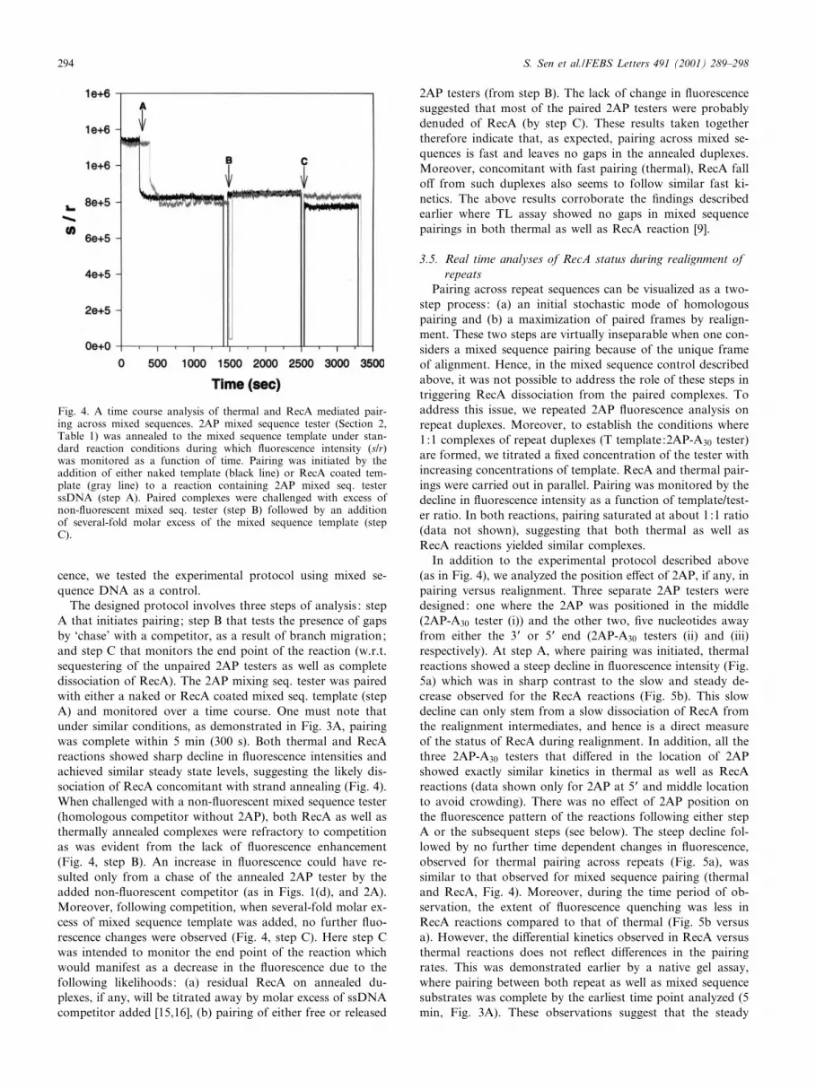
The designed protocol involves three steps of analysis : stepA that initiates pairing; step B that tests the presence of gapsby `chase' with a competitor, as a result of branch migration;and step C that monitors the end point of the reaction (w.r.t.sequestering of the unpaired 2AP testers as well as completedissociation of RecA). The 2AP mixing seq. tester was pairedwith either a naked or RecA coated mixed seq. template (stepA) and monitored over a time course. One must note thatunder similar conditions, as demonstrated in Fig. 3A, pairingwas complete within 5 min (300 s). Both thermal and RecAreactions showed sharp decline in £uorescence intensities andachieved similar steady state levels, suggesting the likely dis-sociation of RecA concomitant with strand annealing (Fig. 4).When challenged with a non-£uorescent mixed sequence tester(homologous competitor without 2AP), both RecA as well asthermally annealed complexes were refractory to competitionas was evident from the lack of £uorescence enhancement(Fig. 4, step B). An increase in £uorescence could have re-sulted only from a chase of the annealed 2AP tester by theadded non-£uorescent competitor (as in Figs. 1(d), and 2A).Moreover, following competition, when several-fold molar ex-cess of mixed sequence template was added, no further £uo-rescence changes were observed (Fig. 4, step C). Here step Cwas intended to monitor the end point of the reaction whichwould manifest as a decrease in the £uorescence due to thefollowing likelihoods: (a) residual RecA on annealed du-plexes, if any, will be titrated away by molar excess of ssDNAcompetitor added [15,16], (b) pairing of either free or released
2AP testers (from step B). The lack of change in £uorescencesuggested that most of the paired 2AP testers were probablydenuded of RecA (by step C). These results taken togethertherefore indicate that, as expected, pairing across mixed se-quences is fast and leaves no gaps in the annealed duplexes.Moreover, concomitant with fast pairing (thermal), RecA fallo¡ from such duplexes also seems to follow similar fast ki-netics. The above results corroborate the ¢ndings describedearlier where TL assay showed no gaps in mixed sequencepairings in both thermal as well as RecA reaction [9].
3.5. Real time analyses of RecA status during realignment ofrepeats
Pairing across repeat sequences can be visualized as a two-step process: (a) an initial stochastic mode of homologouspairing and (b) a maximization of paired frames by realign-ment. These two steps are virtually inseparable when one con-siders a mixed sequence pairing because of the unique frameof alignment. Hence, in the mixed sequence control describedabove, it was not possible to address the role of these steps intriggering RecA dissociation from the paired complexes. Toaddress this issue, we repeated 2AP £uorescence analysis onrepeat duplexes. Moreover, to establish the conditions where1:1 complexes of repeat duplexes (T template:2AP-A30 tester)are formed, we titrated a ¢xed concentration of the tester withincreasing concentrations of template. RecA and thermal pair-ings were carried out in parallel. Pairing was monitored by thedecline in £uorescence intensity as a function of template/test-er ratio. In both reactions, pairing saturated at about 1:1 ratio(data not shown), suggesting that both thermal as well asRecA reactions yielded similar complexes.
In addition to the experimental protocol described above(as in Fig. 4), we analyzed the position e¡ect of 2AP, if any, inpairing versus realignment. Three separate 2AP testers weredesigned: one where the 2AP was positioned in the middle(2AP-A30 tester (i)) and the other two, ¢ve nucleotides awayfrom either the 3P or 5P end (2AP-A30 testers (ii) and (iii)respectively). At step A, where pairing was initiated, thermalreactions showed a steep decline in £uorescence intensity (Fig.5a) which was in sharp contrast to the slow and steady de-crease observed for the RecA reactions (Fig. 5b). This slowdecline can only stem from a slow dissociation of RecA fromthe realignment intermediates, and hence is a direct measureof the status of RecA during realignment. In addition, all thethree 2AP-A30 testers that di¡ered in the location of 2APshowed exactly similar kinetics in thermal as well as RecAreactions (data shown only for 2AP at 5P and middle locationto avoid crowding). There was no e¡ect of 2AP position onthe £uorescence pattern of the reactions following either stepA or the subsequent steps (see below). The steep decline fol-lowed by no further time dependent changes in £uorescence,observed for thermal pairing across repeats (Fig. 5a), wassimilar to that observed for mixed sequence pairing (thermaland RecA, Fig. 4). Moreover, during the time period of ob-servation, the extent of £uorescence quenching was less inRecA reactions compared to that of thermal (Fig. 5b versusa). However, the di¡erential kinetics observed in RecA versusthermal reactions does not re£ect di¡erences in the pairingrates. This was demonstrated earlier by a native gel assay,where pairing between both repeat as well as mixed sequencesubstrates was complete by the earliest time point analyzed (5min, Fig. 3A). These observations suggest that the steady
Fig. 4. A time course analysis of thermal and RecA mediated pair-ing across mixed sequences. 2AP mixed sequence tester (Section 2,Table 1) was annealed to the mixed sequence template under stan-dard reaction conditions during which £uorescence intensity (s/r)was monitored as a function of time. Pairing was initiated by theaddition of either naked template (black line) or RecA coated tem-plate (gray line) to a reaction containing 2AP mixed seq. testerssDNA (step A). Paired complexes were challenged with excess ofnon-£uorescent mixed seq. tester (step B) followed by an additionof several-fold molar excess of the mixed sequence template (stepC).
FEBS 24658 27-2-01
S. Sen et al./FEBS Letters 491 (2001) 289^298294
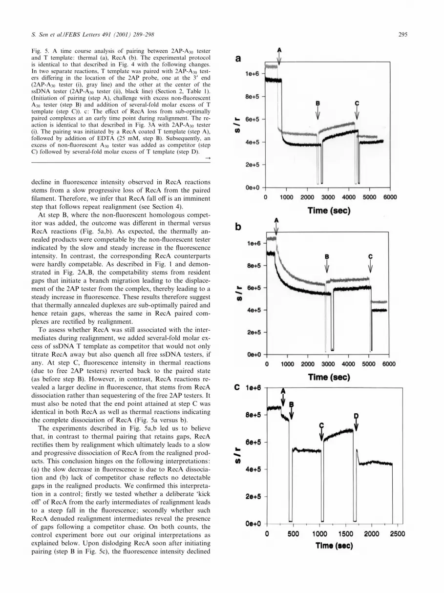
decline in £uorescence intensity observed in RecA reactionsstems from a slow progressive loss of RecA from the paired¢lament. Therefore, we infer that RecA fall o¡ is an imminentstep that follows repeat realignment (see Section 4).
At step B, where the non-£uorescent homologous compet-itor was added, the outcome was di¡erent in thermal versusRecA reactions (Fig. 5a,b). As expected, the thermally an-nealed products were competable by the non-£uorescent testerindicated by the slow and steady increase in the £uorescenceintensity. In contrast, the corresponding RecA counterpartswere hardly competable. As described in Fig. 1 and demon-strated in Fig. 2A,B, the competability stems from residentgaps that initiate a branch migration leading to the displace-ment of the 2AP tester from the complex, thereby leading to asteady increase in £uorescence. These results therefore suggestthat thermally annealed duplexes are sub-optimally paired andhence retain gaps, whereas the same in RecA paired com-plexes are recti¢ed by realignment.
To assess whether RecA was still associated with the inter-mediates during realignment, we added several-fold molar ex-cess of ssDNA T template as competitor that would not onlytitrate RecA away but also quench all free ssDNA testers, ifany. At step C, £uorescence intensity in thermal reactions(due to free 2AP testers) reverted back to the paired state(as before step B). However, in contrast, RecA reactions re-vealed a larger decline in £uorescence, that stems from RecAdissociation rather than sequestering of the free 2AP testers. Itmust also be noted that the end point attained at step C wasidentical in both RecA as well as thermal reactions indicatingthe complete dissociation of RecA (Fig. 5a versus b).
The experiments described in Fig. 5a,b led us to believethat, in contrast to thermal pairing that retains gaps, RecArecti¢es them by realignment which ultimately leads to a slowand progressive dissociation of RecA from the realigned prod-ucts. This conclusion hinges on the following interpretations:(a) the slow decrease in £uorescence is due to RecA dissocia-tion and (b) lack of competitor chase re£ects no detectablegaps in the realigned products. We con¢rmed this interpreta-tion in a control ; ¢rstly we tested whether a deliberate `kicko¡' of RecA from the early intermediates of realignment leadsto a steep fall in the £uorescence; secondly whether suchRecA denuded realignment intermediates reveal the presenceof gaps following a competitor chase. On both counts, thecontrol experiment bore out our original interpretations asexplained below. Upon dislodging RecA soon after initiatingpairing (step B in Fig. 5c), the £uorescence intensity declined
Fig. 5. A time course analysis of pairing between 2AP-A30 testerand T template: thermal (a), RecA (b). The experimental protocolis identical to that described in Fig. 4 with the following changes.In two separate reactions, T template was paired with 2AP-A30 test-ers di¡ering in the location of the 2AP probe, one at the 3P end(2AP-A30 tester (i), gray line) and the other at the center of thessDNA tester (2AP-A30 tester (ii), black line) (Section 2, Table 1).(Initiation of pairing (step A), challenge with excess non-£uorescentA30 tester (step B) and addition of several-fold molar excess of Ttemplate (step C)). c: The e¡ect of RecA loss from sub-optimallypaired complexes at an early time point during realignment. The re-action is identical to that described in Fig. 3A with 2AP-A30 tester(i). The pairing was initiated by a RecA coated T template (step A),followed by addition of EDTA (25 mM, step B). Subsequently, anexcess of non-£uorescent A30 tester was added as competitor (stepC) followed by several-fold molar excess of T template (step D).
C
FEBS 24658 27-2-01
S. Sen et al./FEBS Letters 491 (2001) 289^298 295
steeply, indicating that the original £uorescence was almostentirely due to the presence of RecA. This observation under-scores the property of RecA mediated unstacking of basesthat relieves the £uorescence quenching in paired 2AP du-plexes (Fig. 3B). Moreover, 2AP testers in such RecA de-nuded DNA intermediates were e¤ciently competable by anon-£uorescent competitor, a result highly reminiscent ofthermally annealed products (compare step C, Fig. 5c withstep B, Fig. 5a). Thus, when RecA reaction was prematurelyabrogated, it mimicked thermally annealed sub-optimallyaligned products, thereby indicating the active role of RecAin repeat realignments. Again, as expected, an addition ofexcess template scored the end point of the reaction, wherethe intensity dropped to the minimum levels (step D, Fig. 5c).
4. Discussion
Thermal pairing between T template and A30 tester leads tosub-optimally aligned duplexes as shown here by the branchmigration assay (Fig. 2A). Such sub-optimally aligned du-plexes are recti¢ed in a realignment reaction catalyzed byRecA (Fig. 2B). In fact, previous studies indicated that thisis an ATP hydrolysis dependent process and ensues acrossmono- di- as well as trinucleotide repeat stretches [9]. Thefocus of this study is to get a mechanistic insight on the
relationship between realignment vis a vis the status ofRecA in these complexes.
We used a sensitive probe that can report the density ofRecA in the ¢lament in real time. 2-Aminopurine (2AP), a£uorescent base analogue, is very sensitive to conformationalchanges in DNA structure [26,27]. 2AP £uorescence is highlyquenched in polynucleotides and duplex DNA due to stackinginteractions with the neighboring bases. However, if the DNAstacking around the 2AP base is perturbed, the £uorescenceemission is recovered as a fold enhancement which dependson the amount of unstacking and increases dramatically if thebase is £ipped out of the DNA helix [19,28]. Moreover, back-ground £uorescence from tyrosine and tryptophan residues inproteins does not interfere with 2AP £uorescence due to thenon-overlapping nature of the excitation and emission spectra.Hence, we surmised that 2AP could serve as a good probe formonitoring RecA^DNA interactions where RecA binding tothe 2AP duplex would lead to an enhancement of the £uores-cence due to base unstacking. Here we demonstrate that, un-like that of mixed sequences, RecA association (in ATPQS) ona (dA):(dT) repeat duplex uncovers almost the entire £uores-cence associated with 2AP-A30 tester, as an enhancement (Fig.3B). Although extrahelical base £ipping cannot be ruled out,the 2- to 3-fold enhancement observed here probably re£ectson the unstacking and freedom of the bases within the RecA^dsDNA ¢lament, since base £ipping is known to cause a muchhigher increase in £uorescence intensity (31^54-fold, [19]). It isknown from previous studies that 2AP £uorescence enhance-ment in protein^DNA complexes is associated with di¡erentstates (stacking) of the base within the complex. In fact, it isenvisaged that the base undergoes a series of transitions froma paired fully stacked to a paired partially stacked to an un-stacked to a ¢nal unpaired form, the latter also being able to£ip out of the helix [21,27]. A spectral scan revealed that inATP hydrolyzing conditions, £uorescence emission associatedwith RecA paired duplexes was measurably higher than thatfrom naked DNA duplexes. Furthermore, a comparison ofmixed and repeat sequences revealed that bases seem to expe-rience higher degrees of unstacked freedom in the RecAcoated repeat duplex (Fig. 3B). Does this contrast in £uores-cence enhancement point out to the enhanced £uidity of the2AP base pair speci¢cally within the repeat stretch, when it isin a RecA ¢lament? Picosecond rotational dynamics of 2APobserved through time resolved £uorescence depolarizationdoes indeed suggest that RecA binding results in an increased£uidity of the 2AP base pair (unpublished observations).
Stochastic pairing, in both thermal and RecA reactions, israpid when measured across mixed as well as repetitive se-quences. This was expected because the pairings being moni-tored here depend entirely on complementary annealing ofbases which is likely to be as e¤cient between naked DNAstrands as it is in RecA mediated reactions (Fig. 3A). 2AP£uorescence monitored in thermal versus RecA pairing reac-tions across mixed sequences showed very similar kinetics(Fig. 4). The rapid decline of £uorescence in RecA reactionssuggested that the protein dissociates from the complexessoon after pairing. Moreover, this dissociation seems to becomplete as no further decrease in £uorescence was observedfollowing RecA chase by an excess ssDNA competitor (stepC, Fig. 4). These results came in sharp contrast when wecompared RecA status during realignment of repeat duplexes.Thermal annealing across the repeats, which resulted in a
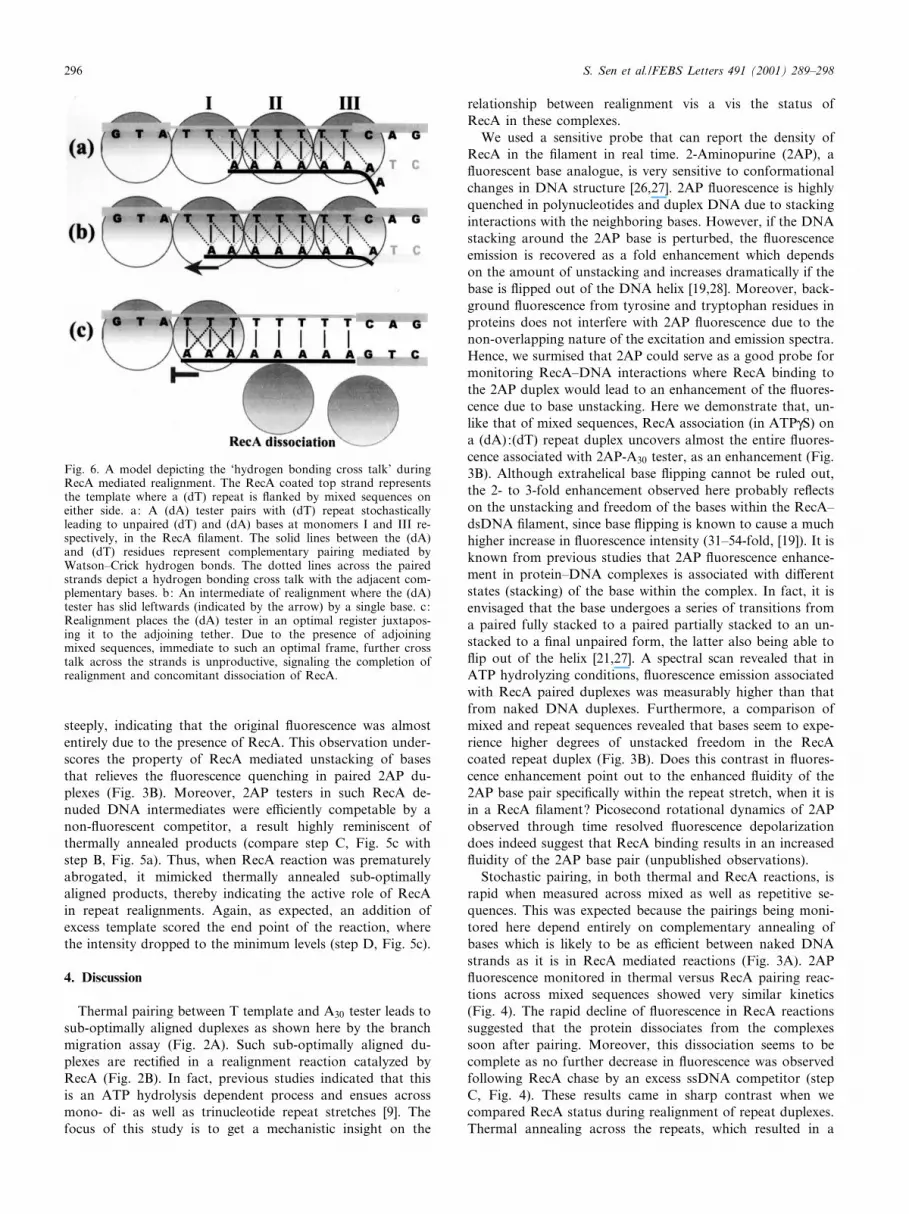
Fig. 6. A model depicting the `hydrogen bonding cross talk' duringRecA mediated realignment. The RecA coated top strand representsthe template where a (dT) repeat is £anked by mixed sequences oneither side. a: A (dA) tester pairs with (dT) repeat stochasticallyleading to unpaired (dT) and (dA) bases at monomers I and III re-spectively, in the RecA ¢lament. The solid lines between the (dA)and (dT) residues represent complementary pairing mediated byWatson^Crick hydrogen bonds. The dotted lines across the pairedstrands depict a hydrogen bonding cross talk with the adjacent com-plementary bases. b: An intermediate of realignment where the (dA)tester has slid leftwards (indicated by the arrow) by a single base. c:Realignment places the (dA) tester in an optimal register juxtapos-ing it to the adjoining tether. Due to the presence of adjoiningmixed sequences, immediate to such an optimal frame, further crosstalk across the strands is unproductive, signaling the completion ofrealignment and concomitant dissociation of RecA.
FEBS 24658 27-2-01
S. Sen et al./FEBS Letters 491 (2001) 289^298296

rapid decline in £uorescence intensity, was similar to thatobserved for mixed sequences (compare step A in Fig. 4 ver-sus Fig. 5a). This indicated that the pairing rates were similaracross both mixed as well as repeat sequences. However, thekinetics of £uorescence decline associated with RecA^repeatDNA complexes was rather slow (Fig. 5b) and not commen-surate with rapid pairing as observed by native DNA gelanalyses (Fig. 3A). Moreover, DNase I protection analysisof RecA^ssDNA complexes formed on such repetitive versusmixed sequence templates did not reveal any intrinsic a¤nitydi¡erences in the binding of RecA under similar conditions(unpublished observations). Thereby, the slow decline in £uo-rescence re£ects on the slow dissociation of RecA from thesesub-optimally paired realignment intermediates, rather thanthe intrinsic a¤nity di¡erences of the protein for such sub-strates. The slow kinetics of RecA fall o¡ observed in this£uorescence assay mirrors the slow kinetics of repeat realign-ment revealed by TL assay [9]. These results therefore corrob-orate each other suggesting an active link between RecA sta-tus and strand realignment. However, such slow kinetics is notcompatible with the expected rates in vivo. The in vitro reac-tion described here is mediated entirely by a single protein,RecA, whereas in vivo the same is likely to be assisted by amyriad of helper molecules such as RecO, RecR, SSB [29^31].One must note that RecA fall o¡ from these intermediates wasnot complete even after 5000 s (V80 min) as monitored by aRecA chase at step C (Fig. 5b). Hence, as contrasted by stepC in Fig. 5c versus step B in Fig. 5b, one could surmise thatRecA association is functionally linked to the correspondingrealignment status of the duplexes. These results suggest theability of RecA to sense sub-optimal alignments and linger onsuch complexes until realignment is complete. It is importantto note that if stochastic pairings would lead to overhangs/gaps larger than ¢ve nucleotides, the 2AP probe situated to-wards the 3P/5P end (¢fth nucleotide from the end) wouldremain in an unpaired state thereby leading to higher steadystate levels of £uorescence than compared to a probe at thecenter. However, di¡erent positions of 2AP did not show anye¡ect either on the kinetics of pairing or the steady states of£uorescence (Fig. 5). Thus it seems that RecA is able to signalrealignment in duplexes with gaps that are appreciably smallerand in fact probably lesser than even ¢ve nucleotides. Whattriggers RecA fall o¡ from such maximized duplexes in spiteof ssDNA levels almost comparable to that of the intermedi-ates during realignment (compare (c) versus (a)/(b) in Fig. 1)?How does RecA sense the incompleteness of these alignmentsand subsequently fall o¡ following the realignment of theseduplexes?
Our recent observations suggest that RecA mediated re-alignment is intimately linked with the ongoing cycles ofATP hydrolysis, where an abrogation of hydrolysis (inATPQS) leads to cessation of realignment activity [9]. Whatis the relationship between the compact/extended helix of theRecA ¢lament versus the dynamic changes associated with theDNA bases during structural transitions that ensue duringcycles of ATP hydrolysis? Nishinaka et al. have suggestedthat an inter-conversion of the backbone sugar puckers medi-ates translational and rotational motion of the bases in anactive RecA ¢lament coupled with cycles of ATP^ADP ex-change [12]. We hypothesize that the enhanced £uidity ob-served across repeat DNA stretches may stem from such in-ter-conversions and in turn allow a non-planar H bonding
geometry across the adjacent stacks thus mediating a dynamic`cross talk' (Fig. 6). In fact, crystal structure of poly(dA):poly(dT) duplexes indicates that a high propeller twist of thebases sets up a continuous bifurcating hydrogen bonding sys-tem along the major groove of the helix [32,33], which prob-ably underlies the dynamic cross talk in an active RecA ¢la-ment. As described in the model (Fig. 6), we believe that across talk at monomer I ultimately succeeds in stabilizing anew base pair at monomer III by a concerted/cumulative ac-tion of all intervening A:T cross talks, which might result inwaves of helix distortions as a function of the progressivecross talk. A higher a¤nity of RecA for such helical distor-tions in the ¢lament [34] might help retain the protein on theserealignment intermediates. One could envisage this processrecurring until all unpaired A and T bases in sub-optimallyaligned complexes are fully paired leading to a maximizationof homologous alignment, thereafter relieving distortions inthe helix and triggering RecA dissociation. Thus, RecA isable to persist on such intermediates, as long as it is able tomediate an e¤cient ATP hydrolysis coupled with a cross talkacross the bases, and falls o¡, marking the completion ofrealignment.
Acknowledgements: We thank the Department of Atomic Energy(TIFR) for ¢nancial support. We thank G.S. Lakshmikanth (TIFR)for his assistance during the course of this work. We would also liketo thank Mr. Jatin and Mr. Vijay from the photography section ofTIFR for their help.
References
[1] Radding, C.M. (1991) J. Biol. Chem. 266, 5355^5358.[2] Kowalczykowski, S.C. and Eggleston, A.K. (1994) Annu. Rev.
Biochem. 63, 991^1043.[3] Radding, C.M. (1993) Curr. Biol. 3, 358^360.[4] Cox, M.M. (1993) Bioessays 15, 617^623.[5] Roca, A.I. and Cox, M.M. (1997) Prog. Nucleic Acid Res. Mol.
Biol. 56, 129^223.[6] Camerini-Otero, R.D. and Hsieh, P. (1995) Annu. Rev. Genet.
29, 509^552.[7] Je¡reys, A.J., Barber, R., Bois, P., Buard, J., Dubrova, Y.E.,
Grant, G., Hollies, C.R., May, C.A., Neumann, R., Panayi,M., Ritchie, A.E., Shone, A.C., Signer, E., Stead, J.D. and Tam-aki, K. (1999) Electrophoresis 20, 1665^1675.
[8] Epplen, C., Santos, E.J., Maueler, W., van Helden, P. and Ep-plen, J.T. (1997) Electrophoresis 18, 1577^1585.
[9] Sen, S., Karthikeyan, G. and Rao, B.J. (2000) Biochemistry 39,10196^10206.
[10] Dutreix, M. (1997) J. Mol. Biol. 273, 105^113.[11] Ogawa, T., Yu, X., Shinohara, A. and Egelman, E.H. (1993)
Science 259, 1896^1899.[12] Nishinaka, T., Shinohara, A., Ito, Y., Yokoyama, S. and Shiba-
ta, T. (1998) Proc. Natl. Acad. Sci. USA 95, 11071^11076.[13] Shibata, T., Cunningham, R.P. and Radding, C.M. (1981) J. Biol.
Chem. 256, 7557^7564.[14] Karthikeyan, G., Wagle, M.D. and Rao, B.J. (1998) FEBS Lett.
425, 45^51.[15] Menetski, J.P. and Kowalczykowski, S.C. (1987) J. Biol. Chem.
262, 2093^2100.[16] Menetski, J.P. and Kowalczykowski, S.C. (1987) J. Biol. Chem.
262, 2085^2092.[17] Ward, D.C., Reich, E. and Stryer, L. (1969) J. Biol. Chem. 244,
1228^1237.[18] Allan, B.W., Beechem, J.M., Lindstrom, W.M. and Reich, N.O.
(1998) J. Biol. Chem. 273, 2368^2373.[19] Holz, B., Klimasauskas, S., Serva, S. and Weinhold, E. (1998)
Nucleic Acids Res. 26, 1076^1083.[20] Lakshmikanth, G.S. and Krishnamoorthy, G. (1999) Biophys. J.
77, 1100^1106.
FEBS 24658 27-2-01
S. Sen et al./FEBS Letters 491 (2001) 289^298 297

[21] Hochstrasser, R.A., Carver, T.E., Sowers, L.C. and Millar, D.P.(1994) Biochemistry 33, 11971^11979.
[22] Aymami, J., Coll, M., Frederick, C.A., Wang, A.H. and Rich, A.(1989) Nucleic Acids Res. 17, 3229^3245.
[23] Stasiak, A. and Egelman, H. (1988) in: Genetic Recombination(Kucherlapati, R. and Smith, R.G., Eds.), pp. 265^308, AmericanSociety for Microbiology, Washington, DC.
[24] Muller, B. and Stasiak, A. (1991) J. Mol. Biol. 221, 131^145.[25] Chow, S.A., Honigberg, S.M., Bainton, R.J. and Radding, C.M.
(1986) J. Biol. Chem. 261, 6961^6971.[26] Millar, D.P. (1996) Curr. Opin. Struct. Biol. 6, 322^326.[27] Lam, W.C., Van der Schans, E.J., Sowers, L.C. and Millar, D.P.
(1999) Biochemistry 38, 2661^2668.
[28] Allan, B.W., Reich, N.O. and Beechem, J.M. (1999) Biochemistry38, 5308^5314.
[29] Harmon, F.G. and Kowalczykowski, S.C. (1998) Genes Dev. 12,1134^1144.
[30] Shan, Q., Bork, J.M., Webb, B.L., Inman, R.B. and Cox, M.M.(1997) J. Mol. Biol. 265, 519^540.
[31] Webb, B.L., Cox, M.M. and Inman, R.B. (1997) Cell 91, 347^356.[32] Coll, M., Frederick, C.A., Wang, A.H. and Rich, A. (1987) Proc.
Natl. Acad. Sci. USA 84, 8385^8389.[33] Nelson, H.C., Finch, J.T., Luisi, B.F. and Klug, A. (1987) Na-
ture 330, 221^226.[34] Wang, Y.H., Bortner, C.D. and Gri¤th, J. (1993) J. Biol. Chem.
268, 17571^17577.
FEBS 24658 27-2-01
S. Sen et al./FEBS Letters 491 (2001) 289^298298