rats selectively bred for low aerobic capacity have reduced hepatic mitochondrial oxidative capacity...
TRANSCRIPT
J Physiol 587.8 (2009) pp 1805–1816 1805
Rats selectively bred for low aerobic capacity have reducedhepatic mitochondrial oxidative capacity and susceptibilityto hepatic steatosis and injury
John P. Thyfault1,2,3, R. Scott Rector2, Grace M. Uptergrove2, Sarah J. Borengasser3, E. Matthew Morris2,3,Yongzhong Wei2, Matt J. Laye4, Charles F. Burant6, Nathan R. Qi6, Suzanne E. Ridenhour2,Lauren G. Koch5, Steve L. Britton5 and Jamal A. Ibdah1,2,4
1Harry S. Truman Memorial VA Hospital, Research Service, Columbia, MO 65201, USA2Departments of Internal Medicine—Division of Gastroenterology and Hepatology, 3Nutritional Sciences and 4Medical Pharmacology and Physiology,University of Missouri, Columbia, MO 65211, USADepartments of 5Physical Medicine and Rehabilitation and 6 Internal Medicine, University of Michigan, Ann Arbor, MI 48109, USA
Fatty liver has been linked to low aerobic fitness, but the mechanisms are unknown. Wepreviously reported a novel model in which rats were artificially selected to be high capacityrunners (HCR) and low capacity runners (LCR) that in a sedentary condition have robustlydifferent intrinsic aerobic capacities. We utilized sedentary HCR/LCR rats (generation 17;max running distance equalled 1514 ± 91 vs. 200 ± 12 m for HCR and LCR, respectively) toinvestigate if low aerobic capacity is associated with reduced hepatic mitochondrial oxidativecapacity and increased susceptibility to hepatic steatosis. At 25 weeks of age, LCR livers displayedreduced mitochondrial content (reduced citrate synthase activity and cytochrome c protein) andreduced oxidative capacity (complete palmitate oxidation in hepatic mitochondria (1.15 ± 0.13vs. 2.48 ± 1.1 nm g−1 h, P < 0.0001) and increased peroxisomal activity (acyl CoA oxidase andcatalase activity) compared to the HCR. The LCR livers also displayed a lipogenic phenotypewith higher protein content of both sterol regulatory element binding protein-1c and acetylCoA carboxylase. These differences were associated with hepatic steatosis in the LCR includinghigher liver triglycerides (6.00 ± 0.71 vs. 4.20 ± 0.39 nmol g−1, P = 0.020 value), >2-fold higherpercentage of hepatocytes associated with lipid droplets (54.0 ± 9.2 vs. 22.0 ± 3.5%, P = 0.006),and increased hepatic lipid peroxidation compared to the HCR. Additionally, in rats aged tonatural death, LCR livers had significantly greater hepatic injury (fibrosis and apoptosis). Weprovide novel evidence that selection for low intrinsic aerobic capacity causes reduced hepaticmitochondrial oxidative capacity that increases susceptibility to both hepatic steatosis and liverinjury.
(Resubmitted 12 January 2009; accepted 16 February 2009; first published online 23 February 2009)Corresponding author J. P. Thyfault: Harry S. Truman Memorial VA Hospital, Department of Nutritional Sciences andDivision of Gastroenterology and Hepatology, University of Missouri, Columbia, MO 65201, USA.Email: [email protected]
Abbreviations ACC, acetyl CoA carboxylase; ALT, serum alanine aminotransferase; CPS1, anti-carbamoylphosphatesynthetase 1; FAS, fatty acid synthase; LCR, low capacity runner; β-HAD, β-hydroxyacyl-CoA dehydrogenase; HCR,high capacity runner; TAG, triglyceride; H&E, haematoxylin–eosin; 4-HNE, 4-hydroxynonenal; NAFLD, non-alcoholicfatty liver disease; PPARα, peroxisome proliferator-activated receptor α; SREBP-1c, sterol regulatory binding protein-1c;TGF-β, transforming growth factorβ; TUNEL, terminal desoxynucleotide transferase-mediated dUTP nick end labelling;PPARγ, peroxisome proliferator-activated receptor γ.
Non-alcoholic fatty liver disease (NAFLD) encompassesa gamut of liver maladaptations and is a primary causeof chronic liver disease and liver-related morbidity andmortality. Fatty liver occurs at a higher rate in individualswith obesity and type 2 diabetes and is considered
the hepatic component of the metabolic syndrome(Schindhelm et al. 2006). Currently, 34% of the generalpopulation (Browning et al. 2004) and 75–100% of obeseand extremely obese individuals are estimated to have fattyliver (Bellentani et al. 2000).
C© 2009 The Authors. Journal compilation C© 2009 The Physiological Society DOI: 10.1113/jphysiol.2009.169060
1806 J. P. Thyfault and others J Physiol 587.8
Low aerobic capacity, or low cardiorespiratory fitness,is the strongest predictor of early mortality (Myers et al.2002). Aerobic capacity is dependent upon the ability ofthe cardiorespiratory system to deliver oxygen and thecapacity of mitochondria to utilize oxygen for energyproduction. In the absence of exercise training, it isbelieved that genetic inheritance accounts for up to 70%of the variation in intrinsic aerobic capacity (Bouchardet al. 1986). In most cases, exercise training significantlyimproves aerobic capacity; however, exercise-inducedresponses are grossly heterogeneous (Kohrt et al. 1991;Bouchard & Rankinen, 2001). Recent evidence also linkslow aerobic capacity (Nguyen-Duy et al. 2003; Church et al.2006; McMillan et al. 2007) to an increased depositionof fat in the liver; however, there is no mechanisticinformation detailing these connections.
Over several generations of selective breeding for highand low treadmill endurance running (performance on3 graded exercise tests), we have developed two strainsof rats with grossly different intrinsic endurance exercisecapacities (max run distance during a graded exercise testto exhaustion is ∼7-fold different) and aerobic capacities(30% different at generation 11) (Wisloff et al. 2005). Thisnovel animal model is valuable to study the interactionbetween aerobic capacities and chronic disease for twomain reasons: (1) the contrasting aerobic capacities occurin a sedentary, caged-activity-only condition (Hoydalet al. 2007) removing the conflicting effects of dailyexercise training (Wisloff et al. 2005; Bernal-Mizrachi& Semenkovich, 2006); and (2) the strains are apolygenic model of disease that more accurately mimicsthe pathology of human chronic metabolic disease(s) thansingle gene mutations. At generation 11, the sedentary lowcapacity runners (LCR) displayed a higher incidence ofboth cardiovascular and metabolic syndrome risk factorsthan sedentary high capacity runners (HCR) (Wisloffet al. 2005). In addition, at generation 13, LCR rats weresusceptible to high-fat diet induced obesity and insulinresistance while the HCR rats were largely unaffected(Noland et al. 2007a). In both reports, skeletal musclemitochondrial oxidative capacity, which is high in themuscle of HCR, and low in the muscle of LCR, washypothesized to be the primary cause for protection orsusceptibility to metabolic dysfunction, respectively.
In the current study, we investigate the potential linksbetween intrinsic aerobic capacity, hepatic mitochondrialoxidative capacity and susceptibility to hepatic steatosisin generation 17 HCR and LCR rats. Importantly, thesestudies occurred in sedentary rats which are geneticallypredisposed to high and low aerobic capacities allowing forthe determination of hepatic metabolism independentlyof the effects of exercise training. We hypothesize thatthe phenotype of low aerobic capacity is associated withboth reduced hepatic mitochondrial content and oxidativecapacity which increases both susceptibility to early life
hepatic steatosis and significant hepatic injury at an olderage.
Methods
Animal strain and protocols
The development of LCR and HCR rats has previouslybeen described (Wisloff et al. 2005; Noland et al. 2007a).Briefly, two-way artificial selective breeding was used tocreate low capacity runner (LCR) and high capacity runner(HCR) strains that were divergent for treadmill runningcapacity (run time to exhaustion on a graded exercise test).The founder population (N : NIH stock) and generationof the HCR and LCR strains has been previously reported(Wisloff et al. 2005). In brief, the 13 lowest and 13highest running capacity rats of each sex were selectedfrom the founder population and randomly paired formating. At each subsequent generation, within-familyselection from 13 mating pairs was practiced for eachline, a number of families that maintains a relativelylow coefficient of inbreeding (<0.01/generation) andmaximizes the retention of genetic variation. After therats were phenotyped for running capacity they wereexposed to no further exercise training or testing and onlyunderwent normal cage activity.
The animal protocols were approved by the InstitutionalAnimal Care and Use Committees at the University ofMissouri and the University of Michigan and the Sub-committee for Animal Safety at the Harry S. TrumanMemorial VA Hospital.
Two separate sets of ∼25-week-old male LCR (n = 7)and HCR (n = 8) rats from generation 17 were usedfor the investigation of hepatic steatosis and hepaticmitochondrial oxidative capacity. The rats arrived at∼14 weeks of age, were provided standard rat chow andwater ad libitum, and were kept on a 12 h light–12 h darktime schedule until killed at ∼25 weeks of age. On themorning of tissue collection, rats were intraperitoneallyinjected with sodium Nembutal (100 mg kg−1), blood wascollected, the animals were exsanguinated, and tissues werefrozen with liquid nitrogen and stored at –80◦C or fixed forimmunohistochemistry. Tissue was collected in the samemanner in a second group of 25-week-old, generation 17,HCR (n = 8) and LCR (n = 8) rats and ∼500 mg of liverwas quickly removed and placed in ice-cold buffer forthe isolation of hepatic mitochondria or frozen for latermeasurement of peroxisomal enzyme activity. Additionalgeneration 17 rats (HCR, n = 4; LCR, n = 6) were ageduntil natural death or until they were killed becauseof undue suffering at approximately 24–35 months ofage at the University of Michigan where the rats weregenerated. After death animals underwent necropsy within4 h and livers were fixed for immunohistochemistry only.Weekly food consumption and changes in body weight
C© 2009 The Authors. Journal compilation C© 2009 The Physiological Society
J Physiol 587.8 Aerobic capacity and hepatic mitochondria 1807
were measured in an additional set of HCR/LCR rats(n = 10 in both groups) from 5 to 25 weeks of age. Inaddition, determinations of physical activity and oxygenconsumption by indirect calorimetry were taken in therats over a 2 day period at 25 weeks of age.
Intrahepatic lipid content
Hepatic triglycerides (TAG) were measured as referencedpreviously using a commercially available kit (Sigma,F6428) (Rector et al. 2008).
Histology analysis
Liver and epididymal adipose tissues were collected andfixed in 10% formalin for histological analysis. Routinehematoxylin–eosin (H&E) staining was performed in bothliver and adipose and sirius red staining for collagendeposition was performed in paraffin liver sectionsas previously referenced (Wei et al. 2008). Hepaticsteatosis and the percentage of hepatocytes associated withlipid droplets were determined as referenced previously(Ibdah et al. 2005) and morphometric determinationsof adipocyte cell volume were carried out as previouslyreferenced (Laye et al. 2006).
Immunohistochemistry
Liver paraffin sections were de-paraffinized andnon-specific antibody binding was blocked with 5%normal goat or rabbit serum and 5% BSA inphosphate-buffered saline (PBS). These sections wereincubated with anti-carbamoylphosphate synthetase 1(CPS1), transforming growth factor β (TGF-β) or4-hydroxynonenal (4-HNE) antibodies at 1 : 200 dilutionsfor overnight at 4◦C. After washing, the sections wereincubated with a second antibody conjugated withalexa-586 for 1 h at room temperature and mountedwith DAPI (Vector) or biotin conjugated second anti-body, following by 0.3% H2O2, ABC (Vector), NovaRed(Vector), and counterstained with hematoxylin. Theprimary antibody was omitted from this procedure foradditional sections to ensure that results were not due tonon-specific binding. Images were acquired with a lightand fluorescence microscope (Nikon, Eclipse 50i).
Apoptosis by TUNEL staining
To evaluate apoptotic cell death, terminaldesoxynucleotide transferase-mediated dUTP nickend labelling (TUNEL) was performed in liver sectionsusing an in situ Apoptosis Detection Kit (Roche AppliedScience, Indianapolis, IN, USA) as per the manufacturer’sinstructions. TUNEL-positive and -negative nuclei were
counted at five random fields for each section. Results areexpressed as number of TUNEL-positive cell/total cells ×100%.
Western blotting
Peroxisome proliferator-activated receptor (PPAR) γ,PPARα, and sterol regulatory binding protein-1c(SREBP-1c) antibodies were purchased from Santa CruzBiotechnology, Inc. (Santa Cruz, CA, USA). Antibodiesagainst Acetyl CoA carboxylase (ACC) and cytochromec were obtained from Cell Signaling Technology, Inc.(Beverly, MA, USA) and 4-HNE was purchased fromOxis International Inc. (Beverly Hills, CA, USA). Liversamples were homogenized in ice cold buffer, separatedby SDS-PAGE gels, transferred to PVDF membranes andprobed with primary antibodies. Protein bands werequantified using a densitometer and band densities werecorrected for total protein loaded by staining with 1%amido-black (Sigma) as described previously (Rector et al.2008).
Hepatic mitochondrial enzyme activities, isolation,and palmitate oxidation
Citrate synthase and β-hydroxyacyl-CoA dehydrogenase(β-HAD) activity were both measured as previouslydescribed in whole liver homogenates (Rector et al. 2008).Mitochondrial suspensions were prepared according tomodified methods of Koves et al. (2005). A portion ofthe liver was quickly excised from unconscious rats andplaced in 5 ml of fresh Buffer A (100 mM KCl, 50 mM
Mops, 5 mM MgSO4, 1 mM EGTA, 1 mM ATP, pH 7.4).The liver was then minced and suspended 7-fold (w/v)in Buffer A, and homogenized for 15 s. The homogenatewas then centrifuged at 800 g for 10 min at 4◦C and thesupernatant was filtered through gauze and set aside. Theremaining pellet was resuspended in Buffer A, homo-genized, and the homogenate was then centrifuged at800 g for 10 min at 4◦C. The remaining supernatantwas added to the previously set aside supernatant afterfiltering through gauze. The pooled supernatants werecentrifuged at 9000 g to pellet the mitochondria. Thepellet was resuspended in Buffer B (Buffer A, 0.2% BSA,pH 7.4), and centrifuged at 8000 g before the pelletwas washed and resuspended in Buffer C (100 mM KCL,50 mM Mops, 0.5 mM EGTA, pH 7.4) and centrifuged for10 min at 7000 g . The final pellet was resuspended in0.5 ml of sucrose EDTA (SET) buffer and protein contentwas determined. Palmitate oxidation was measured withradiolabelled [1-14C]palmitate (American RadiolabeledChemicals, Inc., St Louis, MO, USA) in the freshlyisolated liver mitochondria preparation using methodspreviously reported (Laye et al. 2008; Rector et al. 2008).
C© 2009 The Authors. Journal compilation C© 2009 The Physiological Society
1808 J. P. Thyfault and others J Physiol 587.8
Both 14CO2, representing complete fatty acid oxidation,and 14C labelled acid soluble metabolites, representingincomplete fatty acid oxidation, were collected in thepreviously described trapping device and then counted ona liquid scintillation counter. In addition to [14C]palmitatethe reaction buffer contained a final concentration of50 μM cold palmitate and 0.5% BSA.
Hepatic peroxisomal enzyme activity
Acyl-CoA oxidase activity was determined using amodification of methods previously described (Smallet al. 1985). Briefly, H2O2 production was measuredspectrophotometrically (520 nm) through oxidativecoupling with 3,5-dichloro-2-hydroxybenzenesulfonicacid to 4-aminoantipyrine in the presence of horse-radish peroxidase. Reaction wells contained 20 μlof liver homogenate and 200 μl of reactionbuffer consisting of 40 mM aminotriazole, 0.02%Triton X-100, 0.5 mM 4-aminoantipyrine, 4 mM
3,5-dichloro-2-hydroxybenzenesulfonic acid, 8 U horse-radish peroxidase, 11 mM potassium phosphate; pH 7.4.They were incubated for 5 min at 37◦C. The reactionwas initiated by the addition of acyl-CoA substrate(50 μM) and absorbance was monitored continuouslyover 30 min at 37◦C. Catalase activity was determined bythe spectrophotometric (520 nm) oxidative coupling of3,5-dichlorobenzene-sulfonic acid to 4-aminoantipyrinein the presence of HRP and H2O2 (Sigma) as previouslydescribed (Noland et al. 2007b).
Serum assays and blood pressure
Serum TAG (Sigma) and insulin (Linco Research, StCharles, MO, USA) were measured using commerciallyavailable kits. Serum alanine aminotransferase (ALT)concentrations were determined utilizing an OlympusAU400e Chemistry Immuno Analyzer (Olympus America,Inc., Center Valley, PA, USA). Systolic blood pressure wasmeasured in triplicate using the tail-cuff method a weekbefore sacrifice.
Food consumption, ambulatory physical activity,and oxygen consumption
Both ad libitum food consumption and total bodyweights were measured every week from the post-weaningperiod (∼5 week) until 25 weeks of age. In addition,oxygen consumption (V̇O2 ), carbon dioxide production(V̇CO2 ), and spontaneous motor activity were measuredusing a Comprehensive Laboratory Monitoring System(CLAMS, Columbus Instruments, Columbus, OH, USA)over a 2 day period at 25 weeks of age at theUniversity of Michigan Animal Phenotyping Core. The
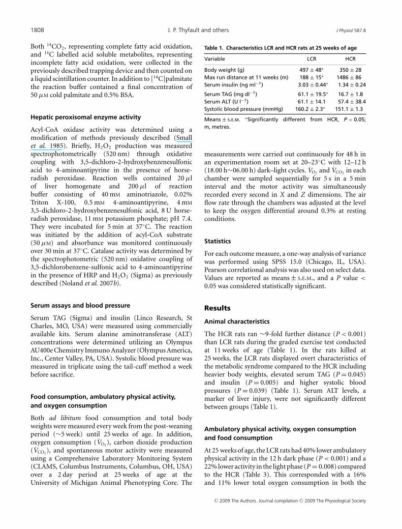
Table 1. Characteristics LCR and HCR rats at 25 weeks of age
Variable LCR HCR
Body weight (g) 497 ± 48∗ 350 ± 28Max run distance at 11 weeks (m) 188 ± 15∗ 1486 ± 86Serum insulin (ng ml−1) 3.03 ± 0.44∗ 1.34 ± 0.24
Serum TAG (mg dl−1) 61.1 ± 19.5∗ 16.7 ± 1.8Serum ALT (U l−1) 61.1 ± 14.1 57.4 ± 38.4Systolic blood pressure (mmHg) 160.2 ± 2.3∗ 151.1 ± 1.3
Means ± S.E.M. ∗Significantly different from HCR, P < 0.05;m, metres.
measurements were carried out continuously for 48 h inan experimentation room set at 20–23◦C with 12–12 h(18.00 h∼06.00 h) dark–light cycles. V̇O2 and V̇CO2 in eachchamber were sampled sequentially for 5 s in a 5 mininterval and the motor activity was simultaneouslyrecorded every second in X and Z dimensions. The airflow rate through the chambers was adjusted at the levelto keep the oxygen differential around 0.3% at restingconditions.
Statistics
For each outcome measure, a one-way analysis of variancewas performed using SPSS 15.0 (Chicago, IL, USA).Pearson correlational analysis was also used on select data.Values are reported as means ± S.E.M., and a P value <
0.05 was considered statistically significant.
Results
Animal characteristics
The HCR rats ran ∼9-fold further distance (P < 0.001)than LCR rats during the graded exercise test conductedat 11 weeks of age (Table 1). In the rats killed at25 weeks, the LCR rats displayed overt characteristics ofthe metabolic syndrome compared to the HCR includingheavier body weights, elevated serum TAG (P = 0.045)and insulin (P = 0.005) and higher systolic bloodpressures (P = 0.039) (Table 1). Serum ALT levels, amarker of liver injury, were not significantly differentbetween groups (Table 1).
Ambulatory physical activity, oxygen consumptionand food consumption
At 25 weeks of age, the LCR rats had 40% lower ambulatoryphysical activity in the 12 h dark phase (P < 0.001) and a22% lower activity in the light phase (P = 0.008) comparedto the HCR (Table 3). This corresponded with a 16%and 11% lower total oxygen consumption in both the
C© 2009 The Authors. Journal compilation C© 2009 The Physiological Society
J Physiol 587.8 Aerobic capacity and hepatic mitochondria 1809
light (P = 0.005) and dark phase (P < 0.001), respectively(Table 3). Average weekly food consumption (averagedfrom ∼4 to 25 weeks of age) was also significantly lowerin the LCR rats (P = 0.03) (Table 3). Collectively, thesedata indicate that the contrasting levels of ambulatoryactivity and oxygen consumption measured between thetwo strains corresponded with different rates of foodconsumption. Feeding efficiency (weight gain divided byfood intake) was not different between the groups between9 weeks and the time of kill at 25 weeks suggesting thatoverall energy balance was similar for 16 weeks prior tokill (data not shown).
Hepatic mitochondrial enzyme activity, palmitateoxidation and peroxisomal enzyme activity
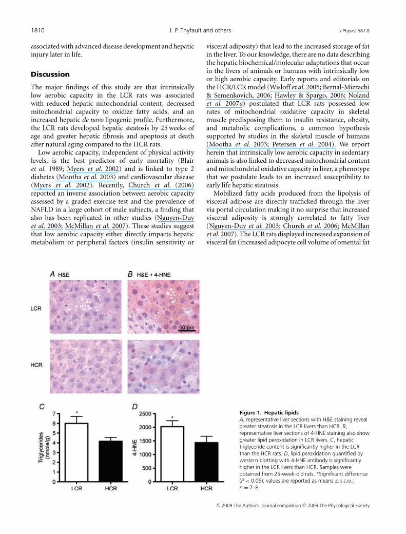
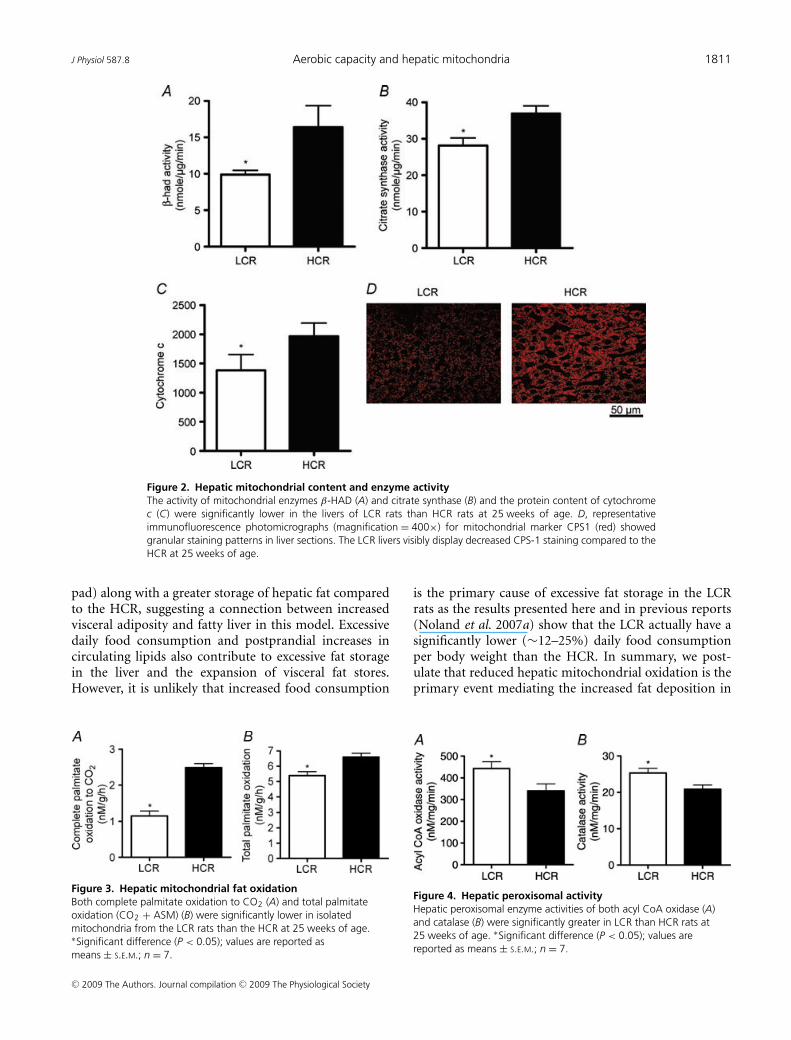
β-HAD activity, the rate limiting enzyme for β-oxidation,was 65% higher (P < 0.001) in the HCR liverscompared to LCR livers (Fig. 2A). Citrate synthaseactivity and cytochrome c protein content, markers ofmitochondrial content, were 32–42% higher (P < 0.001)in the HCR livers compared to LCR (Fig. 2B andC). Immunoflourescence staining for CPS-1, a hepaticmitochondrial marker, qualitatively revealed a greaterhepatic mitochondrial content in the HCR vs. theLCR (Fig. 2D). The HCR mitochondria also had higherrates of fatty acid oxidation than the LCR. Isolatedhepatic mitochondria from the HCR rats had a 63%(Fig. 3A; P < 0.0001) higher rate of complete palmitateoxidation to CO2 and a 22% higher (Fig. 3B; P < 0.001)rate of total palmitate oxidation (CO2 + acid solublemetabolite production) than the LCR. The LCR ratsalso displayed significantly higher peroxisomal enzymeactivity for both acyl CoA oxidase (Fig. 4A; P = 0.039)and catalase (Fig. 4B; P = 0.02) compared to the HCRrats, suggestive of a compensatory increase in peroxisomalactivity due to reduced mitochondrial oxidative capacity.Collectively these data show that LCR livers have botha lower mitochondrial content and lower mitochondrialoxidative capacity (decreased fatty acid oxidation permitochondria).
Hepatic lipogenesis
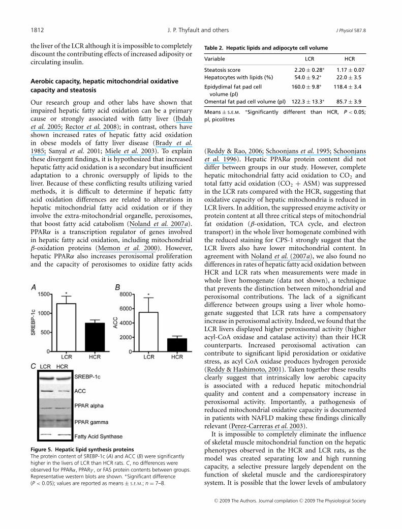
The content of SREBP-1c, a transcriptional regulator offatty acid synthesis genes (Shimano et al. 1996; Hortonet al. 2002), was significantly higher in the livers of LCRrats than HCR rats (Fig. 5A). Specifically, SREBP-1c isan upstream transcriptional regulator of both acetyl CoAcarboxylase (ACC) and fatty acid synthase (FAS). ACCprotein content, the enzyme responsible for malonyl CoAproduction (a potent inhibitor of fatty acid oxidation andthe initial substrate for FAS), was 3-fold higher (P = 0.049)in the LCR liver than the HCR liver (Fig. 5B). There
was no difference in serine79 phosphorylation of ACCprotein, a marker of ACC inhibition (data not shown),suggesting that ACC differences were due to total contentof the enzyme and not due to differences in covalentmodification. We found no difference in FAS content(Fig. 5C). Protein content of PPARγ and PPARα, vitaltranscriptional regulators of lipid synthesis and oxidationalso were not different in the livers of LCR and HCR rats(Fig. 5C).
Hepatic and extrahepatic lipid storage
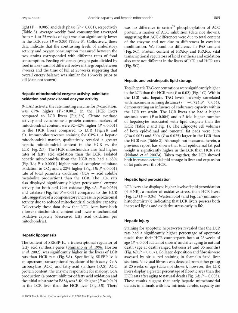
Total hepatic TAG concentrations were significantly higherin the LCR than the HCR rats (P = 0.02) (Fig. 1C). Withinthe LCR rats, hepatic TAG levels inversely correlatedwith maximum running distance (r = −0.724; P = 0.034),demonstrating an influence of endurance capacity withinthe LCR rat strain. The LCR livers also had a highersteatosis score (P = 0.004) and >2 fold higher numberof hepatocytes associated with lipid droplets than theHCR (Table 2 and Fig. 1). The adipocyte cell volumesof both epidydimal and omental fat pads were 35%(P = 0.003) and 50% (P = 0.025) larger in the LCR thanthe HCR rats (Table 2). Although not measured herein, aprevious report has shown that total epididymal fat padweight is significantly higher in the LCR than HCR rats(Noland et al. 2007a). Taken together, the LCR showedboth increased ectopic lipid storage in liver and expansionof fat pads over the HCR.
Hepatic lipid peroxidation
LCR livers also displayed higher levels of lipid peroxidation(4-HNE), a marker of oxidative stress, than HCR livers(Fig. 1D (P = 0.04) (Western blot) and Fig. 1B (immuno-histochemistry)) indicating that LCR livers possess bothincreased lipids and oxidative stress early in life.
Hepatic injury
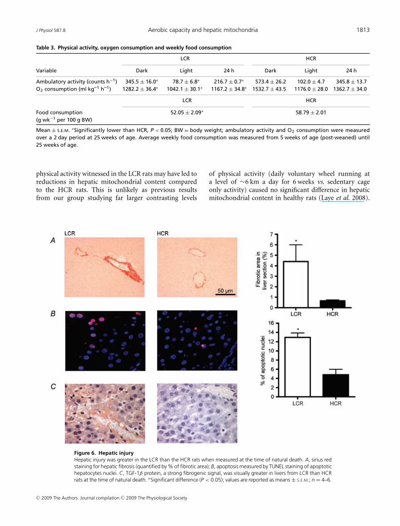
Staining for apoptotic hepatocytes revealed that the LCRrats had a significantly higher percentage of apoptoticnuclei than their HCR counterparts both at 25 weeks ofage (P < 0.001; data not shown) and after aging to naturaldeath (age at death ranged between 24 and 35 months)(Fig. 6B; P = 0.007). Collagen deposition and fibrosis wereassessed by sirius red staining in formalin-fixed liversections. No visual fibrosis was detected from either groupat 25 weeks of age (data not shown); however, the LCRlivers display a greater percentage of fibrotic area than theHCR rats after aging to natural death (Fig. 6A; P = 0.005).These results suggest that early hepatic mitochondrialdefects in animals with low intrinsic aerobic capacity are
C© 2009 The Authors. Journal compilation C© 2009 The Physiological Society
1810 J. P. Thyfault and others J Physiol 587.8
associated with advanced disease development and hepaticinjury later in life.
Discussion
The major findings of this study are that intrinsicallylow aerobic capacity in the LCR rats was associatedwith reduced hepatic mitochondrial content, decreasedmitochondrial capacity to oxidize fatty acids, and anincreased hepatic de novo lipogenic profile. Furthermore,the LCR rats developed hepatic steatosis by 25 weeks ofage and greater hepatic fibrosis and apoptosis at deathafter natural aging compared to the HCR rats.
Low aerobic capacity, independent of physical activitylevels, is the best predictor of early mortality (Blairet al. 1989; Myers et al. 2002) and is linked to type 2diabetes (Mootha et al. 2003) and cardiovascular disease(Myers et al. 2002). Recently, Church et al. (2006)reported an inverse association between aerobic capacityassessed by a graded exercise test and the prevalence ofNAFLD in a large cohort of male subjects, a finding thatalso has been replicated in other studies (Nguyen-Duyet al. 2003; McMillan et al. 2007). These studies suggestthat low aerobic capacity either directly impacts hepaticmetabolism or peripheral factors (insulin sensitivity or
Figure 1. Hepatic lipidsA, representative liver sections with H&E staining revealgreater steatosis in the LCR livers than HCR. B,representative liver sections of 4-HNE staining also showgreater lipid peroxidation in LCR livers. C, hepatictriglyceride content is significantly higher in the LCRthan the HCR rats. D, lipid peroxidation quantified bywestern blotting with 4-HNE antibody is significantlyhigher in the LCR livers than HCR. Samples wereobtained from 25-week-old rats. ∗Significant difference(P < 0.05); values are reported as means ± S.E.M.;n = 7–8.
visceral adiposity) that lead to the increased storage of fatin the liver. To our knowledge, there are no data describingthe hepatic biochemical/molecular adaptations that occurin the livers of animals or humans with intrinsically lowor high aerobic capacity. Early reports and editorials onthe HCR/LCR model (Wisloff et al. 2005; Bernal-Mizrachi& Semenkovich, 2006; Hawley & Spargo, 2006; Nolandet al. 2007a) postulated that LCR rats possessed lowrates of mitochondrial oxidative capacity in skeletalmuscle predisposing them to insulin resistance, obesity,and metabolic complications, a common hypothesissupported by studies in the skeletal muscle of humans(Mootha et al. 2003; Petersen et al. 2004). We reportherein that intrinsically low aerobic capacity in sedentaryanimals is also linked to decreased mitochondrial contentand mitochondrial oxidative capacity in liver, a phenotypethat we postulate leads to an increased susceptibility toearly life hepatic steatosis.
Mobilized fatty acids produced from the lipolysis ofvisceral adipose are directly trafficked through the livervia portal circulation making it no surprise that increasedvisceral adiposity is strongly correlated to fatty liver(Nguyen-Duy et al. 2003; Church et al. 2006; McMillanet al. 2007). The LCR rats displayed increased expansion ofvisceral fat (increased adipocyte cell volume of omental fat
C© 2009 The Authors. Journal compilation C© 2009 The Physiological Society
J Physiol 587.8 Aerobic capacity and hepatic mitochondria 1811
Figure 2. Hepatic mitochondrial content and enzyme activityThe activity of mitochondrial enzymes β-HAD (A) and citrate synthase (B) and the protein content of cytochromec (C) were significantly lower in the livers of LCR rats than HCR rats at 25 weeks of age. D, representativeimmunofluorescence photomicrographs (magnification = 400×) for mitochondrial marker CPS1 (red) showedgranular staining patterns in liver sections. The LCR livers visibly display decreased CPS-1 staining compared to theHCR at 25 weeks of age.
pad) along with a greater storage of hepatic fat comparedto the HCR, suggesting a connection between increasedvisceral adiposity and fatty liver in this model. Excessivedaily food consumption and postprandial increases incirculating lipids also contribute to excessive fat storagein the liver and the expansion of visceral fat stores.However, it is unlikely that increased food consumption
Figure 3. Hepatic mitochondrial fat oxidationBoth complete palmitate oxidation to CO2 (A) and total palmitateoxidation (CO2 + ASM) (B) were significantly lower in isolatedmitochondria from the LCR rats than the HCR at 25 weeks of age.∗Significant difference (P < 0.05); values are reported asmeans ± S.E.M.; n = 7.
is the primary cause of excessive fat storage in the LCRrats as the results presented here and in previous reports(Noland et al. 2007a) show that the LCR actually have asignificantly lower (∼12–25%) daily food consumptionper body weight than the HCR. In summary, we post-ulate that reduced hepatic mitochondrial oxidation is theprimary event mediating the increased fat deposition in
Figure 4. Hepatic peroxisomal activityHepatic peroxisomal enzyme activities of both acyl CoA oxidase (A)and catalase (B) were significantly greater in LCR than HCR rats at25 weeks of age. ∗Significant difference (P < 0.05); values arereported as means ± S.E.M.; n = 7.
C© 2009 The Authors. Journal compilation C© 2009 The Physiological Society
1812 J. P. Thyfault and others J Physiol 587.8
the liver of the LCR although it is impossible to completelydiscount the contributing effects of increased adiposity orcirculating insulin.
Aerobic capacity, hepatic mitochondrial oxidativecapacity and steatosis
Our research group and other labs have shown thatimpaired hepatic fatty acid oxidation can be a primarycause or strongly associated with fatty liver (Ibdahet al. 2005; Rector et al. 2008); in contrast, others haveshown increased rates of hepatic fatty acid oxidationin obese models of fatty liver disease (Brady et al.1985; Sanyal et al. 2001; Miele et al. 2003). To explainthese divergent findings, it is hypothesized that increasedhepatic fatty acid oxidation is a secondary but insufficientadaptation to a chronic oversupply of lipids to theliver. Because of these conflicting results utilizing variedmethods, it is difficult to determine if hepatic fattyacid oxidation differences are related to alterations inhepatic mitochondrial fatty acid oxidation or if theyinvolve the extra-mitochondrial organelle, peroxisomes,that boost fatty acid catabolism (Noland et al. 2007a).PPARα is a transcription regulator of genes involvedin hepatic fatty acid oxidation, including mitochondrialβ-oxidation proteins (Memon et al. 2000). However,hepatic PPARα also increases peroxisomal proliferationand the capacity of peroxisomes to oxidize fatty acids
Figure 5. Hepatic lipid synthesis proteinsThe protein content of SREBP-1c (A) and ACC (B) were significantlyhigher in the livers of LCR than HCR rats. C, no differences wereobserved for PPARα, PPARγ , or FAS protein contents between groups.Representative western blots are shown. ∗Significant difference(P < 0.05); values are reported as means ± S.E.M.; n = 7–8.
Table 2. Hepatic lipids and adipocyte cell volume
Variable LCR HCR
Steatosis score 2.20 ± 0.28∗ 1.17 ± 0.07Hepatocytes with lipids (%) 54.0 ± 9.2∗ 22.0 ± 3.5
Epidydimal fat pad cell 160.0 ± 9.8∗ 118.4 ± 3.4volume (pl)
Omental fat pad cell volume (pl) 122.3 ± 13.3∗ 85.7 ± 3.9
Means ± S.E.M. ∗Significantly different than HCR, P < 0.05;pl, picolitres
(Reddy & Rao, 2006; Schoonjans et al. 1995; Schoonjanset al. 1996). Hepatic PPARα protein content did notdiffer between groups in our study. However, completehepatic mitochondrial fatty acid oxidation to CO2 andtotal fatty acid oxidation (CO2 + ASM) was suppressedin the LCR rats compared with the HCR, suggesting thatoxidative capacity of hepatic mitochondria is reduced inLCR livers. In addition, the suppressed enzyme activity orprotein content at all three critical steps of mitochondrialfat oxidation (β-oxidation, TCA cycle, and electrontransport) in the whole liver homogenate combined withthe reduced staining for CPS-1 strongly suggest that theLCR livers also have lower mitochondrial content. Inagreement with Noland et al. (2007a), we also found nodifferences in rates of hepatic fatty acid oxidation betweenHCR and LCR rats when measurements were made inwhole liver homogenate (data not shown), a techniquethat prevents the distinction between mitochondrial andperoxisomal contributions. The lack of a significantdifference between groups using a liver whole homo-genate suggested that LCR rats have a compensatoryincrease in peroxisomal activity. Indeed, we found that theLCR livers displayed higher peroxisomal activity (higheracyl-CoA oxidase and catalase activity) than their HCRcounterparts. Increased peroxisomal activation cancontribute to significant lipid peroxidation or oxidativestress, as acyl CoA oxidase produces hydrogen peroxide(Reddy & Hashimoto, 2001). Taken together these resultsclearly suggest that intrinsically low aerobic capacityis associated with a reduced hepatic mitochondrialquality and content and a compensatory increase inperoxisomal activity. Importantly, a pathogenesis ofreduced mitochondrial oxidative capacity is documentedin patients with NAFLD making these findings clinicallyrelevant (Perez-Carreras et al. 2003).
It is impossible to completely eliminate the influenceof skeletal muscle mitochondrial function on the hepaticphenotypes observed in the HCR and LCR rats, as themodel was created separating low and high runningcapacity, a selective pressure largely dependent on thefunction of skeletal muscle and the cardiorespiratorysystem. It is possible that the lower levels of ambulatory
C© 2009 The Authors. Journal compilation C© 2009 The Physiological Society
J Physiol 587.8 Aerobic capacity and hepatic mitochondria 1813
Table 3. Physical activity, oxygen consumption and weekly food consumption
LCR HCR
Variable Dark Light 24 h Dark Light 24 h
Ambulatory activity (counts h−1) 345.5 ± 16.0∗ 78.7 ± 6.8∗ 216.7 ± 0.7∗ 573.4 ± 26.2 102.0 ± 4.7 345.8 ± 13.7O2 consumption (ml kg–1 h–1) 1282.2 ± 36.4∗ 1042.1 ± 30.1∗ 1167.2 ± 34.8∗ 1532.7 ± 43.5 1176.0 ± 28.0 1362.7 ± 34.0
LCR HCR
Food consumption 52.05 ± 2.09∗ 58.79 ± 2.01(g wk−1 per 100 g BW)
Mean ± S.E.M. ∗Significantly lower than HCR, P < 0.05; BW = body weight; ambulatory activity and O2 consumption were measuredover a 2 day period at 25 weeks of age. Average weekly food consumption was measured from 5 weeks of age (post-weaned) until25 weeks of age.
physical activity witnessed in the LCR rats may have led toreductions in hepatic mitochondrial content comparedto the HCR rats. This is unlikely as previous resultsfrom our group studying far larger contrasting levels
Figure 6. Hepatic injuryHepatic injury was greater in the LCR than the HCR rats when measured at the time of natural death. A, sirius redstaining for hepatic fibrosis (quantified by % of fibrotic area); B, apoptosis measured by TUNEL staining of apoptotichepatocytes nuclei. C, TGF-1β protein, a strong fibrogenic signal, was visually greater in livers from LCR than HCRrats at the time of natural death. ∗Significant difference (P < 0.05); values are reported as means ± S.E.M.; n = 4–6.
of physical activity (daily voluntary wheel running ata level of ∼6 km a day for 6 weeks vs. sedentary cageonly activity) caused no significant difference in hepaticmitochondrial content in healthy rats (Laye et al. 2008).
C© 2009 The Authors. Journal compilation C© 2009 The Physiological Society
1814 J. P. Thyfault and others J Physiol 587.8
Our results clearly show that selective breeding for lowaerobic capacity resulted in lower hepatic mitochondrialcontent and function in rats at 25 weeks of age, which wespeculate causes the increased susceptibility for hepaticsteatosis in the LCR rats. In support of this concept,preliminary results from our laboratory show that thephenotype of reduced lipid oxidation in the LCR livers isretained in primary cell culture (data not shown). Just asthe selective breeding for low running capacity reducedskeletal muscle mitochondrial ATP production neededto fuel contractions, it also likely lowered the need forhepatic mitochondrial ATP production, which is neededto fuel the energy-costly process of gluconeogenesisneeded to maintain glucose homeostasis duringexercise.
Increased lipid peroxidation can lead to the productionof reactive aldehydes that damage mitochondrial DNA(Demeilliers et al. 2002) or inactivate respiratory chainproteins (Chen et al. 2000), resulting in reducedmitochondrial content or function and hepatic injury.Despite the strong link between obesity or aging andreduced mitochondrial DNA in skeletal muscle, we wereunable to find studies linking hepatic lipid peroxidationor oxidative stress to reduced liver mitochondrial DNAthat do not involve liver damage induced by alcohol orpharmacological agents. Here we demonstrate that theaging LCR rats have increased hepatic levels of lipidperoxidation which is subsequent to reduced hepaticmitochondrial function compared with the HCR rats.This could be partially attributed to increased adiposity(Noland et al. 2007a) and potentially greater hepaticfree fatty acid flux in the LCR animals. However, theLCR rats had lower relative food consumption andsimilar feed efficiencies compared to the HCR rats,indicating similar energy balance between the two strains.Collectively, these findings support our contention thathepatic mitochondrial dysfunction plays a primary rolein non-alcoholic fatty liver disease development andprogression in animals with low intrinsic aerobic capacity.Future studies are warranted to pair feed the two strainsand/or attempt to match ambulatory activity levels acrossthe lifespan.
Aerobic capacity, hepatic lipogenesis and steatosis
Both biochemical and histological measures showedincreased hepatic fat deposition in the LCR compared tothe HCR rats at 25 weeks of age. These findings contra-dict an earlier report (Noland et al. 2007a) which studiedthe effect of high fat diet on the metabolic phenotypesof the HCR and LCR strains. That study did not find asignificant difference in hepatic fat storage between theLCR and HCR strains on a normal chow or high-fat diet;however, that study used the less sensitive and qualitativeOil Red O technique to stain neutral lipids in a notably
small subset (n = 3) of liver samples, while our report usedthe standard biochemical measure of hepatic triglyceridesin a larger sample size. In addition, rats were studied froma different generation between the two studies (generation13 in former report vs. generation 17 in this study) and it isdifficult to discern what impact further selective breedinghad upon metabolic or hepatic phenotypes. However, acomparison of reports from previous generations showscomparable values for both strains between generation11 (Wisloff et al. 2005), 13 (Noland et al. 2007a), and17 (this report) for measures of body weight, and seruminsulin and triglycerides suggesting that methodologicaldifferences, and not generational differences, were themain reason that hepatic triglycerides were not previouslyreported to be higher in the LCR rats.
Insulin increases hepatic fatty acid synthesis, in partthrough increases in SREBP-1c transcriptional regulationof FAS and ACC (Shimomura et al. 1999). In the LCRrats, it could be speculated that the elevated SREBP-1cprotein content was due to elevated serum insulin levelsresulting from peripheral insulin resistance (Noland et al.2007a). The elevated SREBP-1c content in liver of LCRrats was matched with a 2-fold higher protein content ofACC over the HCR rat, with no observed differences in FASprotein content or PPARγ. ACC synthesizes malonyl-CoA,a metabolite that is both a substrate for de novo synthesisof fatty acids through FAS and a potent inhibitor of fattyacid oxidation (McGarry et al. 1977). Although malonylCoA was not measured herein, the elevated hepatic ACCcontent also suggests that hepatic CPT-1 activity in theLCR could allosterically be reduced due to a presumedincrease in hepatic malonyl-CoA levels.
In conclusion, these results show that the hepaticphenotype associated with reduced aerobic capacity ischaracterized by both reduced mitochondrial contentand oxidative capacity, leading to early hepatic steatosisand lipid peroxidation and the later development ofelevated markers of liver injury. These findings provideinsight into the pathogenesis of hepatic steatosis andmay have important clinical implications. Lower intrinsicaerobic capacity due to physical inactivity and/or geneticheritability may lead to reduced hepatic mitochondrialoxidative capacity and increased susceptibility to hepaticsteatosis and injury. Therefore, the clinical measurementof aerobic capacity may serve as a valuable prognostictool. More importantly, because exercise training and/orincreased physical activity is the proven method to increaseaerobic capacity, it should remain the cornerstone therapyfor fatty liver disease.
References
Bellentani S, Saccoccio G, Masutti F, Croce LS, Brandi G, SassoF, Cristanini G & Tiribelli C (2000). Prevalence of and riskfactors for hepatic steatosis in Northern Italy. Ann InternMed 132, 112–117.
C© 2009 The Authors. Journal compilation C© 2009 The Physiological Society

J Physiol 587.8 Aerobic capacity and hepatic mitochondria 1815
Bernal-Mizrachi C & Semenkovich CF (2006). Fast predatorsor fast food, the fit still survive. Nat Med 12, 46–47;discussion 47.
Blair SN, Kohl HW 3rd, Paffenbarger RS Jr, Clark DG, CooperKH & Gibbons LW (1989). Physical fitness and all-causemortality. A prospective study of healthy men and women.JAMA 262, 2395–2401.
Bouchard C, Lesage R, Lortie G, Simoneau JA, Hamel P, BoulayMR, Perusse L, Theriault G & Leblanc C (1986). Aerobicperformance in brothers, dizygotic and monozygotic twins.Med Sci Sports Exerc 18, 639–646.
Bouchard C & Rankinen T (2001). Individual differences inresponse to regular physical activity. Med Sci Sports Exerc 33,S446–451; discussion S452–453.
Brady LJ, Brady PS, Romsos DR & Hoppel CL (1985). Elevatedhepatic mitochondrial and peroxisomal oxidative capacitiesin fed and starved adult obese (ob/ob) mice. Biochem J 231,439–444.
Browning JD, Szczepaniak LS, Dobbins R, Nuremberg P,Horton JD, Cohen JC, Grundy SM & Hobbs HH (2004).Prevalence of hepatic steatosis in an urban population in theUnited States: impact of ethnicity. Hepatology 40,1387–1395.
Chen J, Petersen DR, Schenker S & Henderson GI (2000).Formation of malondialdehyde adducts in livers of ratsexposed to ethanol: role in ethanol-mediated inhibition ofcytochrome c oxidase. Alcoholism Clin Exp Res 24, 544–552.
Church TS, Kuk JL, Ross R, Priest EL, Biltoft E & Blair SN(2006). Association of cardiorespiratory fitness, body massindex, and waist circumference to nonalcoholic fatty liverdisease. Gastroenterology 130, 2023–2030.
Demeilliers C, Maisonneuve C, Grodet A, Mansouri A, NguyenR, Tinel M, Letteron P, Degott C, Feldmann G, Pessayre D &Fromenty B (2002). Impaired adaptive resynthesis andprolonged depletion of hepatic mitochondrial DNA afterrepeated alcohol binges in mice. Gastroenterology 123,1278–1290.
Hawley JA & Spargo FJ (2006). It’s all in the genes, so pick yourparents wisely. J Appl Physiol 100, 1751–1752.
Horton JD, Goldstein JL & Brown MS (2002). SREBPs:activators of the complete program of cholesterol and fattyacid synthesis in the liver. J Clin Invest 109, 1125–1131.
Hoydal MA, Wisloff U, Kemi OJ, Britton SL, Koch LG, SmithGL & Ellingsen O (2007). Nitric oxide synthase type-1modulates cardiomyocyte contractility and calciumhandling: association with low intrinsic aerobic capacity. EurJ Cardiovasc Prev Rehabil 14, 319–325.
Ibdah JA, Perlegas P, Zhao Y, Angdisen J, Borgerink H,Shadoan MK, Wagner JD, Matern D, Rinaldo P & Cline JM(2005). Mice heterozygous for a defect in mitochondrialtrifunctional protein develop hepatic steatosis and insulinresistance. Gastroenterology 128, 1381–1390.
Kohrt WM, Malley MT, Coggan AR, Spina RJ, Ogawa T, EhsaniAA, Bourey RE, Martin WH 3rd & Holloszy JO (1991).Effects of gender, age, and fitness level on response of VO2max
to training in 60–71 yr olds. J Appl Physiol 71, 2004–2011.Koves TR, Noland RC, Bates AL, Henes ST, Muoio DM &
Cortright RN (2005). Subsarcolemmal and intermyofibrillarmitochondria play distinct roles in regulating skeletal musclefatty acid metabolism. Am J Physiol Cell Physiol 288,C1074–1082.
Laye MJ, Rector RS, Borengasser SJ, Naples SP, Uptergrove GM,Ibdah JA, Booth FW & Thyfault JP (2008). Cessation of dailywheel running differentially alters fat oxidation capacity inliver, muscle, and adipose tissue. J Appl Physiol 106, 161–168.
Laye MJ, Thyfault JP, Stump CS & Booth FW (2006). Inactivityinduces increases in abdominal fat. J Appl Physiol 102,1341–1347.
McGarry JD, Mannaerts GP & Foster DW (1977). A possiblerole for malonyl-CoA in the regulation of hepatic fatty acidoxidation and ketogenesis. J Clin Invest 60, 265–270.
McMillan KP, Kuk JL, Church TS, Blair SN & Ross R (2007).Independent associations between liver fat, visceral adiposetissue, and metabolic risk factors in men. Appl Physiol NutrMetab 32, 265–272.
Memon RA, Tecott LH, Nonogaki K, Beigneux A, Moser AH,Grunfeld C & Feingold KR (2000). Up-regulation ofperoxisome proliferator-activated receptors (PPAR-α) andPPAR-γ messenger ribonucleic acid expression in the liver inmurine obesity: troglitazone induces expression ofPPAR-γ-responsive adipose tissue-specific genes in the liverof obese diabetic mice. Endocrinology 141, 4021–4031.
Miele L, Grieco A, Armuzzi A, Candelli M, Forgione A,Gasbarrini A & Gasbarrini G (2003). Hepatic mitochondrialβ-oxidation in patients with nonalcoholic steatohepatitisassessed by 13C-octanoate breath test. Am J Gastroenterol 98,2335–2336.
Mootha VK, Lindgren CM, Eriksson KF, Subramanian A, SihagS, Lehar J, Puigserver P, Carlsson E, Ridderstrale M, LaurilaE, Houstis N, Daly MJ, Patterson N, Mesirov JP, Golub TR,Tamayo P, Spiegelman B, Lander ES, Hirschhorn JN,Altshuler D & Groop LC (2003). PGC-1α-responsive genesinvolved in oxidative phosphorylation are coordinatelydownregulated in human diabetes. Nat Genet 34,267–273.
Myers J, Prakash M, Froelicher V, Do D, Partington S &Atwood JE (2002). Exercise capacity and mortality amongmen referred for exercise testing. N Engl J Med 346, 793–801.
Nguyen-Duy TB, Nichaman MZ, Church TS, Blair SN & RossR (2003). Visceral fat and liver fat are independent predictorsof metabolic risk factors in men. Am J Physiol EndocrinolMetab 284, E1065–1071.
Noland RC, Thyfault JP, Henes ST, Whitfield BR, Woodlief TL,Evans JR, Lust JA, Britton SL, Koch LG, Dudek RW, DohmGL, Cortright RN & Lust RM (2007a). Artificial selection forhigh capacity endurance running is protective against highfat diet-induced insulin resistance. Am J Physiol EndocrinolMetab 293, E31–41.
Noland RC, Woodlief TL, Whitfield BR, Manning SM, EvansJR, Dudek RW, Lust RM & Cortright RN (2007b).Peroxisomal-mitochondrial oxidation in a rodent model ofobesity-associated insulin resistance. Am J Physiol EndocrinolMetab 293, E986–E1001.
Perez-Carreras M, Del Hoyo P, Martin MA, Rubio JC, MartinA, Castellano G, Colina F, Arenas J & Solis-Herruzo JA(2003). Defective hepatic mitochondrial respiratory chain inpatients with nonalcoholic steatohepatitis. Hepatology 38,999–1007.
Petersen KF, Dufour S, Befroy D, Garcia R & Shulman GI(2004). Impaired mitochondrial activity in theinsulin-resistant offspring of patients with type 2 diabetes.N Engl J Med 350, 664–671.
C© 2009 The Authors. Journal compilation C© 2009 The Physiological Society
1816 J. P. Thyfault and others J Physiol 587.8
Rector RS, Thyfault JP, Morris RT, Laye MJ, Borengasser SJ,Booth FW & Ibdah JA (2008). Daily exercise increaseshepatic fatty acid oxidation and prevents steatosis in OtsukaLong-Evans Tokushima Fatty rats. Am J Physiol GastrointestLiver Physiol 294, G619–626.
Reddy JK & Hashimoto T (2001). Peroxisomal β-oxidation andperoxisome proliferator-activated receptor α: an adaptivemetabolic system. Annu Rev Nutr 21, 193–230.
Reddy JK & Rao MS (2006). Lipid metabolism and liverinflammation. II. Fatty liver disease and fatty acid oxidation.Am J Physiol Gastrointest Liver Physiol 290, G852–858.
Sanyal AJ, Campbell-Sargent C, Mirshahi F, Rizzo WB, ContosMJ, Sterling RK, Luketic VA, Shiffman ML & Clore JN(2001). Nonalcoholic steatohepatitis: association of insulinresistance and mitochondrial abnormalities.Gastroenterology 120, 1183–1192.
Schindhelm RK, Diamant M, Dekker JM, Tushuizen ME,Teerlink T & Heine RJ (2006). Alanine aminotransferase as amarker of non-alcoholic fatty liver disease in relation to type2 diabetes mellitus and cardiovascular disease. DiabetesMetab Res Rev 22, 437–443.
Schoonjans K, Staels B & Auwerx J (1996). The peroxisomeproliferator activated receptors (PPARS) and their effects onlipid metabolism and adipocyte differentiation. BiochimBiophys Acta 1302, 93–109.
Schoonjans K, Watanabe M, Suzuki H, Mahfoudi A, Krey G,Wahli W, Grimaldi P, Staels B, Yamamoto T & Auwerx J(1995). Induction of the acyl-coenzyme A synthetase gene byfibrates and fatty acids is mediated by a peroxisomeproliferator response element in the C promoter. J Biol Chem270, 19269–19276.
Shimano H, Horton JD, Hammer RE, Shimomura I, Brown MS& Goldstein JL (1996). Overproduction of cholesterol andfatty acids causes massive liver enlargement in transgenicmice expressing truncated SREBP-1a. J Clin Invest 98,1575–1584.
Shimomura I, Bashmakov Y & Horton JD (1999). Increasedlevels of nuclear SREBP-1c associated with fatty livers in twomouse models of diabetes mellitus. J Biol Chem 274,30028–30032.
Small GM, Burdett K & Connock MJ (1985). A sensitivespectrophotometric assay for peroxisomal acyl-CoA oxidase.Biochem J 227, 205–210.
Wei Y, Clark SE, Morris EM, Thyfault JP, Uptergrove GM,Whaley-Connell AT, Ferrario CM, Sowers JR & Ibdah JA(2008). Angiotensin II-induced non-alcoholic fatty liverdisease is mediated by oxidative stress in transgenicTG(mRen2)27(Ren2) rats. J Hepatol 49, 417–428.
Wisloff U, Najjar SM, Ellingsen O, Haram PM, Swoap S,Al-Share Q, Fernstrom M, Rezaei K, Lee SJ, Koch LG &Britton SL (2005). Cardiovascular risk factors emerge afterartificial selection for low aerobic capacity. Science 307,418–420.
Acknowledgments
The authors would like to thank Daniel Link for the bloodpressure measurements, Dr Frank W. Booth for criticallyevaluating the manuscript, and Lori Gilligan and Nathan Kannerfor expert care of the rat colony. This work was supportedwith resources and the use of facilities at the Harry S. TrumanMemorial Veterans Hospital in Columbia, MO, USA and aCareer Development grant from the VA (J.P.T.). This work wasalso supported by grant RR17718 from the National Center forResearch Resources of the National Institutes of Health (L.G.K.and S.L.B.), grant 1R01DK077200 from the National Institutes ofHealth (C.F.B., N.Q. and S.L.B.), and a grant from the AmericanDiabetes Association (C.F.B. and S.L.B.).
C© 2009 The Authors. Journal compilation C© 2009 The Physiological Society