quantitative micromorphological analyses of cut marks
TRANSCRIPT
This article appeared in a journal published by Elsevier. The attachedcopy is furnished to the author for internal non-commercial researchand education use, including for instruction at the authors institution
and sharing with colleagues.
Other uses, including reproduction and distribution, or selling orlicensing copies, or posting to personal, institutional or third party
websites are prohibited.
In most cases authors are permitted to post their version of thearticle (e.g. in Word or Tex form) to their personal website orinstitutional repository. Authors requiring further information
regarding Elsevier’s archiving and manuscript policies areencouraged to visit:
http://www.elsevier.com/copyright
Author's personal copy
Quantitative micromorphological analyses of cut marksproduced by ancient and modern handaxes
Silvia M. Bello a,*, Simon A. Parfitt a,b, Chris Stringer a
a Department of Palaeontology, The Natural History Museum, Cromwell Road, London SW7 5BD, UKb Institute of Archaeology, University College London, 31–34 Gordon Square, London WC1H 0PY, UK
a r t i c l e i n f o
Article history:Received 11 December 2008Received in revised form6 April 2009Accepted 8 April 2009
Keywords:Cut marksBoxgroveButchery experimentHomo heidelbergensisHandaxeCarcase processing
a b s t r a c t
In this study, we analyse the three-dimensional micromorphology of cut marks on fossil mammalremains from a w0.5 million year old Acheulean butchery site at Boxgrove (West Sussex, southernEngland), and make comparisons with cut marks inflicted during the experimental butchery of a roe deer(Capreolus caproelus) using a replica handaxe. Morphological attributes of the cut marks were measuredusing an Alicona imaging microscope, a novel optical technique that generates three-dimensional virtualreconstructions of surface features. The study shows that high-resolution measurements of cut markscan shed light on aspects of butchery techniques, tool use and the behavioural repertoire of LowerPalaeolithic hominins. Differences between the experimental cut marks and those on the Boxgrove largemammal bones suggest variation in the angle of the cuts and greater forces used in the butchery of thelarger (rhinoceros-sized) carcasses at Boxgrove. Tool-edge characteristics may account for some of thesedifferences, but the greater robusticity of the Boxgrove hominins (attributed to Homo heidelbergensis)may be a factor in the greater forces indicated by some of the cut marks on the Boxgrove specimens.
� 2009 Elsevier Ltd. All rights reserved.
1. Introduction
Cut marks on fossil bones and teeth are an important source ofevidence in the reconstruction of prehistoric butchery practicesand have a direct bearing on subsistence strategies and thebehavioural repertoire of early humans (e.g. Binford, 1981; Blu-menschine et al., 1994; Dominguez-Rodrigo and Pickering, 2003;Shipman, 1986). Many such studies have focussed on the inter-pretation of the microscopic morphology of cut marks (Bartelinket al., 2001; Choi and Driwantoro, 2007; Gilbert and Richards,2000; Greenfield, 1999, 2004, 2006a, b; Saidel et al., 2006;Shipman, 1981; Villa et al., 1986; Walker, 1978; Walker and Long,1977; White, 1992), but with rare exceptions (e.g. Walker and Long,1977; Potts and Shipman, 1981; Shipman,1983; During and Nilsson,1991; Bartelink et al., 2001; Kaiser and Katterwe, 2001) these havebeen qualitative in nature. In recent years, there have beena number of advances in optical microscopy that enable high-resolution three-dimensional images of bone surfaces to be made.Features of the micro-topography of bone surfaces can now bemeasured that were previously unobtainable using more conven-tional techniques (e.g. scanning electron microscopy). Recently, thisapproach was applied to cut marks by Bello and Soligo (2008), who
used an Alicona imaging microscope to quantify characters such ascross-sectional shape, depth and shoulder heights. They were alsoable to infer details of the cutting-edge morphology as well ascharacteristics such as the inclination of the tool and by inferencethe tool user’s hand during cutting. While experimental work onslicing cut marks (sensu Greenfield, 1999) produced directly onbone has already been reported (Bello and Soligo, 2008), thetechnique has not been applied to the analyses of slicing cut marksproduced during butchery and only preliminary analyses have beenundertaken on fossil material (Bello et al., 2007).
In this paper, we demonstrate that this methodology using anAlicona 3D InfiniteFocus imaging microscope can be applied to thestudy of ancient slicing cut marks on bones from an Acheuleanbutchery site at Boxgrove, UK. We compared these cut marks to slicingcut marks produced during the experimental butchery of a roe deerusing a replica handaxe. Comparisons of cut marks parameters, such assharpness, depth of cut and inclination, have led to new insights intopatterns of carcass-processing and the behaviour of early hominins.
2. Materials
2.1. The site
The Boxgrove Acheulean site, near Chichester in southernEngland, has yielded some of the oldest human remains in northern
* Corresponding author. Tel.: þ44 207 942 5141.E-mail addresses: [email protected] (S.M. Bello), [email protected] (S.A. Parfitt).
Contents lists available at ScienceDirect
Journal of Archaeological Science
journal homepage: ht tp: / /www.elsevier .com/locate/ jas
0305-4403/$ – see front matter � 2009 Elsevier Ltd. All rights reserved.doi:10.1016/j.jas.2009.04.014
Journal of Archaeological Science 36 (2009) 1869–1880

Author's personal copy
Europe and one of the richest early Middle Pleistocene artefact andhumanly modified bone assemblages yet known (Roberts andParfitt, 1999). Today, the site is situated 10 km inland of the currentshoreline of the English Channel, but during the early MiddlePleistocene it was located on the coast. Subsequent tectonic activitywas responsible for raising the marine deposits ca 30 m abovepresent-day sea level (Preece et al., 1990; Bates et al., 1997). TheLower Palaeolithic land surface associated with the marine depositshas been traced in an embayment for a distance of w26 km, butextensive archaeological excavations have only been possiblewhere sand and gravel extraction has removed the overburden. AtBoxgrove, quarrying has exposed a succession of near-shore marineand terrestrial interglacial deposits overlain by cold stage collu-vium. Archaeological research at Boxgrove, led by Mark Roberts,has resulted in the recovery of extensive archaeological assem-blages from the marine beach deposits and throughout theterrestrial sequence, with the main concentration occurring in anextensive palaeosol formed within the upper part of the marinesediments (Roberts and Parfitt, 1999). The distribution of archaeo-logical material through the sequence suggests that human occu-pation at the site spanned a substantial part of the interglacial cycleand parts of the ensuing cold stage. Biostratigraphical evidenceplaces the interglacial deposits in the early Middle Pleistocene,probably towards the end of the ‘Cromerian Complex’, with an ageof about 0.5 Ma (Roberts and Parfitt, 1999).
In 1993, a largely complete hominin left tibial diaphysis wasrecovered from calcareous colluvial sediments at a site designatedQ1/B (Roberts et al., 1994; Stringer, 1996; Stringer et al., 1998;Trinkaus et al., 1999; Streeter et al., 2001). Subsequently, excava-tions in 1995–1996 recovered two incisors from freshwaterdeposits directly underlying the colluvium; these sediments arebroadly coeval with the palaeosol horizon. The Boxgrove hominidspecimens have been assigned to Homo heidelbergensis and the sizeof the tibia suggests a robust individual at least 175 cm in height(Trinkaus et al., 1999). The hominin remains were found in asso-ciation with abundant remains of butchered large mammals and anartefact assemblage dominated by finely-flaked ovate and limandehandaxes and the waste from their manufacture (Pope, 2002).Elsewhere at Boxgrove, the fine-grained sediments have preservedsmall clusters of refitting debitage (Austin, 1994; Roberts and Par-fitt, 1999) often associated with the butchered remains of a singlelarge mammal carcass (e.g. GTP 17, the Horse Butchery Site. Popeand Roberts, 2005), but at Q1/B a wide range of mammalian specieswith evidence of butchery are represented. These mammals wereprobably attracted to the freshwater pools, which were fed bysprings that emanated from the chalk cliff. The site was also a focusfor carnivore activity. Bear (Ursus deningeri), spotted hyaena(Crocuta crocuta), lion (Panthera leo) and wolf (Canis lupus) are allrepresented in the faunal assemblage, but the patterns of damagesuggest that humans had primary access to the large mammalcarcasses.
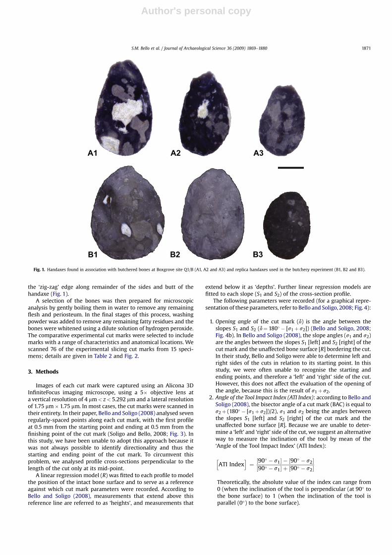
The handaxes were made from good-quality nodular flintobtained from the chalk cliff and talus some 50 m to the north ofQ1/B. The handaxes are typically between 80 and 150 mm long,flaked on both faces and knapped to maximise cutting edges.During the final stages of knapping, bone or antler hammers wereused to thin and finish the tool and the tips of many of the handaxesare characterised by the removal of a sharpening (‘tranchet’) flakefrom their tip (Bergman and Roberts, 1988; Bergman et al., 1990).The thin ‘blade-like’ cutting edge produced by the tranchet removalcontrasts with the ‘zig-zag’ morphology resulting from alternatingflaking around the edges and base of the handaxe (Fig. 1). Re-touched flakes and scrapers were extremely rare at the site and thissupports the hypothesis that acheulean handaxes functionedprimarily as butchery tools.
Microwear analysis of a sample of the w400 handaxes from Q1/B has identified use traces resulting from butchery tasks (Mitchell,1997). Significantly, these traces, together with the morphology ofthe cut marks, indicate that the handaxes were used with a slicingaction to process large mammal carcasses. Although use traces havebeen found along the edges of the Boxgrove handaxes, the tranchettip was the focus of re-sharpening thus implying that the mainte-nance of this cutting edge was of primary importance.
The research at Boxgrove was designed to place the humanactivity in an accurate chronological and environmental contextand to provide interpretations of human behaviour at differenttemporal and geographical scales. An important aspect of theresearch has been a programme of experimental studies, includingflint knapping, butchery and the replication of tool microwear,which have made important contributions to the interpretation ofthe archaeological record at the site (e.g. Wenban-Smith, 1989;Mitchell, 1995, 1997; Smith, 2003).
2.2. The fossil sample
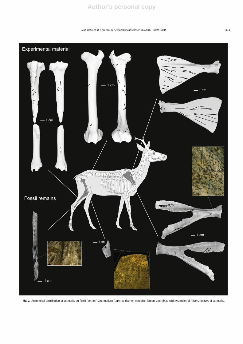
During the 1985–1986 excavations at Boxgrove, about 1050humanly modified large mammal bones were recovered froma small area of the quarry immediately to the south of the buriedcliff. These remains include a wide spectrum of taxa, ranging in sizefrom European beaver (Castor fiber) to rhinoceros (Stephanorhinushunsdheimensis and Stephanorhinus megarhinus). Although many ofthe bones are fragmentary, they were buried in fine-grained highlycalcareous sediments and show little or no alteration resulting fromtrampling or high-energy taphonomic processes. Other post-depositional processes, such as weathering and diagenesis havehad a negligible effect on cut mark morphology, which typicallyshow the characteristic features of incisions produced by a sharpflint edge used with a slicing, scraping or chopping motion. The cutmarks show a patterned distribution consistent with a range ofbutchery tasks, including skinning, dismembering, disarticulationand filleting. Intentional fracturing of limb elements indicates thatmarrow-bone processing was also undertaken at the site. Speci-mens were inspected first by light microscopy to determine thesuitability for micromorphological analysis. We then selected 13cut-marked specimens, each with at least one complete un-trun-cated cut mark, for analysis using the Alicona imaging microscope.A total of 44 slicing cut marks has been scanned. These includespecimens representing a range of skeletal elements and extremesof body size (i.e. roe deer, w59 kg and rhinoceros, w2000 kg).Details of the specimens are given in Table 1 and Fig. 2. Themaximum size of the specimens examined (<120 mm) was con-strained by the height of the Alicona microscope mounting.
2.3. The experimental sample
A comparative sample of cut marks produced during theexperimental butchery of a roe deer using flint replicas of Boxgrovehandaxes was analysed to serve as a baseline for interpreting theBoxgrove cut marks. The butchery experiments are described indetail by Mitchell (1995), with the principal aim being the efficientremoval of the meat from the carcass. Initial stages of carcasspreparation involved gutting and skinning, followed by carefulboning and jointing of the limbs to remove the meat. Theseexperiments showed that handaxes are effective butchery tools,with the long curved edges of the handaxe being particularlyeffective when used with a gentle slicing motion. Most of the cutswere made using the length of the handaxe edge from the butt totip, with the tip being used for shorter ‘filleting’ cuts during thefinal stages of meat removal. In this way, the bones were marked byboth the razor-sharp flake edge of the tranchet removal as well as
S.M. Bello et al. / Journal of Archaeological Science 36 (2009) 1869–18801870
Author's personal copy
the ‘zig-zag’ edge along remainder of the sides and butt of thehandaxe (Fig. 1).
A selection of the bones was then prepared for microscopicanalysis by gently boiling them in water to remove any remainingflesh and periosteum. In the final stages of this process, washingpowder was added to remove any remaining fatty residues and thebones were whitened using a dilute solution of hydrogen peroxide.The comparative experimental cut marks were selected to includemarks with a range of characteristics and anatomical locations. Wescanned 76 of the experimental slicing cut marks from 15 speci-mens; details are given in Table 2 and Fig. 2.
3. Methods
Images of each cut mark were captured using an Alicona 3DInfiniteFocus imaging microscope, using a 5� objective lens ata vertical resolution of 4 mm< z< 5.292 mm and a lateral resolutionof 1.75 mm� 1.75 mm. In most cases, the cut marks were scanned intheir entirety. In their paper, Bello and Soligo (2008) analysed sevenregularly-spaced points along each cut mark, with the first profileat 0.5 mm from the starting point and ending at 0.5 mm from thefinishing point of the cut mark (Soligo and Bello, 2008; Fig. 3). Inthis study, we have been unable to adopt this approach because itwas not always possible to identify directionality and thus thestarting and ending point of the cut mark. To circumvent thisproblem, we analysed profile cross-sections perpendicular to thelength of the cut only at its mid-point.
A linear regression model (R) was fitted to each profile to modelthe position of the intact bone surface and to serve as a referenceagainst which cut mark parameters were recorded. According toBello and Soligo (2008), measurements that extend above thisreference line are referred to as ‘heights’, and measurements that
extend below it as ‘depths’. Further linear regression models arefitted to each slope (S1 and S2) of the cross-section profile.
The following parameters were recorded (for a graphical repre-sentation of these parameters, refer to Bello and Soligo, 2008; Fig. 4):
1. Opening angle of the cut mark (d) is the angle between theslopes S1 and S2 (d¼ 180� � [s1þ s2]) (Bello and Soligo, 2008;Fig. 4b). In Bello and Soligo (2008), the slope angles (s1 and s2)are the angles between the slopes S1 [left] and S2 [right] of thecut mark and the unaffected bone surface [R] bordering the cut.In their study, Bello and Soligo were able to determine left andright sides of the cuts in relation to its starting point. In thisstudy, we were often unable to recognise the starting andending points, and therefore a ‘left’ and ‘right’ side of the cut.However, this does not affect the evaluation of the opening ofthe angle, because this is the result of s1þ s2.
2. Angle of the Tool Impact Index (ATI Index): according to Bello andSoligo (2008), the bisector angle of a cut mark (BAC) is equal tos2þ (180� � [s1þ s2])/2), s1 and s2 being the angles betweenthe slopes S1 [left] and S2 [right] of the cut mark and theunaffected bone surface [R]. Because we are unable to deter-mine a ‘left’ and ‘right’ side of the cut, we suggest an alternativeway to measure the inclination of the tool by mean of the‘Angle of the Tool Impact Index’ (ATI Index):
hATI Index
i¼ ½90� � s1� � ½90� � s2�½90� � s1� þ ½90� � s2�
Theoretically, the absolute value of the index can range from0 (when the inclination of the tool is perpendicular (at 90� tothe bone surface) to 1 (when the inclination of the tool isparallel (0�) to the bone surface).
Fig. 1. Handaxes found in association with butchered bones at Boxgrove site Q1/B (A1, A2 and A3) and replica handaxes used in the butchery experiment (B1, B2 and B3).
S.M. Bello et al. / Journal of Archaeological Science 36 (2009) 1869–1880 1871
Author's personal copy
3. Shoulder Height Index (SH Index): according to Bello and Soligo(2008), the height of the shoulders formed on either side of thecut (SH1¼ sin b1� L1; SH2¼ sin b2� L2, where L1 and L2 are thedistances from the tip of the shoulder to the correspondingintersection between the cut mark profile and regression line R,and where b1 is the angle between L1 and R and b2 is the anglebetween L2 and R). L1, L2, b1and b2 are determined accordinglyto S1 [left] and S2 [right] of the cut mark (Bello and Soligo, 2008;Fig. 4c). Once again, because we are unable to determine ‘left’and ‘right’ side of the cut, we calculated a ‘Shoulder HeightIndex’ (SH Index) as1:
hSH Index
i¼ S1 � S2
S1 þ S2
The absolute value of the indexes can range from 0 (when thetwo shoulders have equal height) to 1 (when only oneshoulder is present).
4. Depth of cut (DC): the perpendicular depth of the cut relative tothe unaffected bone surface (DC¼ sin a�H; where H is thedistance from the lowest point of the cut mark profile, point A,to the intersection between S1 and the regression line R, pointB, and where a is the angle between H and R; Bello and Soligo,2008; Fig. 4d).
5. Floor radius: the radius of a circle fitted to the floor of the cutmark profile, with the floor defined as lying between the twopoints where the profiles of the slopes start to converge (i.e.,where the cut mark profiles start to diverge from the regressionmodels S1 and S2; Bello and Soligo, 2008; Fig. 4c).
4. Results
Results are presented according to the specimen on whichanalyses were conducted: fossil material (Fossil roe deer, Frd; Fossillarge mammals, Flm) and Experimental roe deer (Erd). A distinctionhas also been made according to the anatomical distribution of thecut marks: long bones (e.g. tibia, fibula), ribs and scapula. Finally,a distinction was made according to the position of cut marks on ananatomical element: either on the midshaft or in proximity to (oron) the articular surface.
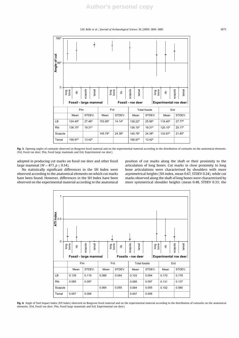
4.1. Opening angle
The mean value of the opening of the angle of cut marksobserved on the fossil roe deer was 148.2�, compared with 134.2�
for other fossil large mammals and 120.7� for the experimental cutmarks produced by the replica handaxe (Fig. 3).
The opening angle of experimentally produced cut marks can bestatistically differentiated from that observed on the fossil material,both when exclusively compared to the fossil roe deer (W¼ 1804.5,p� 1.704e�06), and when compared to the total of fossil speci-mens (W¼ 3448.5, p� 1.821e�05). Similarly, the differencesbetween the opening of the angle of cut marks observed on fossilroe deer and other fossil large mammal are statistically significant(W¼ 355, p� 0.026). This seems to indicate variability in thedegree of the opening of angle of cut marks according to the faunalspecies on which they were found. When considering the differentanatomical elements, only in the case of long bones was it possibleto statistically differentiate experimentally produced cut marksfrom fossil cut marks (W¼ 426, p� 0.035).
On the experimental material, it was possible to observea pattern in the opening of the angle according to the anatomicalposition of cut marks along the shaft or in proximity to the artic-ulation of long bones. Cut marks close to (or on) an articulation oflong bones were generally more acute (mean 80.17�, standarddeviation (STDEV) of 29.07) than those observed along the shaft(mean 124.8�, STDEV 23.25; Wilcoxon two sample test, W¼ 21,p� 0.0056). Cut marks analysed on the fossil sample had beenexclusively found along diaphysis. By comparing the experimentalsample and the fossil sample (mean 139.2�, STDEV 25.96) andexcluding in the case of the experimental sample cut marks foundin proximity of the articulation, we eliminate the differencebetween the two samples (Wilcoxon two sample test, W¼ 374,p� 0.085).
Similarly, on the experimental material, a pattern was observedon the ribs according to the anatomical position of cut marks alongthe shaft (average 126.59�, STDEV 25.76) or in proximity to thevertebral articulation (mean 101.11�, STDEV 21.45, Wilcoxon two
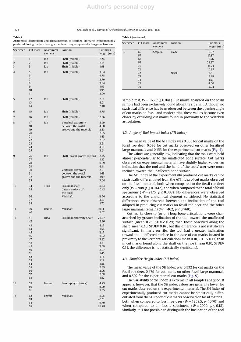
Table 1Anatomical distribution and characteristics of scanned cutmarks on early MiddlePleistocene large mammal remains from Boxgrove.
Specimennumber
Cut marknumber
Taxon Anatomicalelement
Position Cut marklength (mm)
6626 1 Rhinoceros(Stephanorhinus sp.)
Tibia Midshaft 10.522 10.61
7483 3 Rhinoceros(Stephanorhinus sp.)
Pisiform 4.63
4 2.541746 5 Rhino-sized large
mammalLong bonefragment
Midshaft 10.146 7.23
5448 7 Rhino-sized largemammal
Rib 6.36
30073 8 Rhinoceros(Stephanorhinus sp.)
Rib 6.519 6.0110 4.04
30138 11 Rhino-sized largemammal
Rib 5.3712 5.4513 4.33
2008 14 Rhino-sized largemammal
Long bone 6.0415 4.22
30396 16 Indet. large mammal Long bone? 4.9130962 17 Red deer-sized large
mammalRib 0.81
18 3.8819 2.16
30117 20 ?Rhino-sized largemammal
Rib 10.02
6322 21 Roe deer(Capreolus capreolus)
Femur 2.9822 3.4923 2.8524 3.38
7545 25 Roe deer(Capreolus capreolus)
Tibia 2.2226 14.4227 7.19
4898 28 Roe deer(Capreolus capreolus)
Scapula Blade 21.4229 27.8330 44.6131 20.6632 14.3833 11.8935 1.9036 Neck 4.2637 4.3938 9.5339 2.4240 2.7341 1.2142 1.5643 0.9744 2.6745 0.79
1 In this case, 1 and 2 have no lateral meaning, but indicate the two opposingslopes of the cut mark.
S.M. Bello et al. / Journal of Archaeological Science 36 (2009) 1869–18801872
Author's personal copy
Fig. 2. Anatomical distribution of cutmarks on fossil (bottom) and modern (top) roe deer on scapulae, femurs and tibiae with examples of Alicona images of cutmarks.
S.M. Bello et al. / Journal of Archaeological Science 36 (2009) 1869–1880 1873
Author's personal copy
sample test, W¼ 165, p� 0.041). Cut marks analysed on the fossilsample had been exclusively found along the rib shaft. Although nostatistical difference has been observed between the opening angleof cut marks on fossil and modern ribs, these values become evencloser by excluding cut marks found in proximity to the vertebralarticulation.
4.2. Angle of Tool Impact Index (ATI Index)
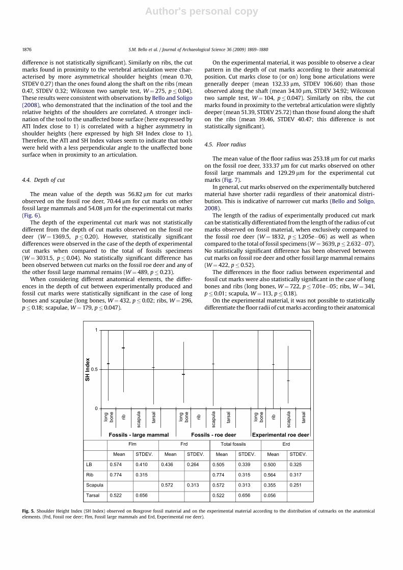
The mean value of the ATI Index was 0.065 for cut marks on thefossil roe deer, 0.096 for cut marks observed on other fossilisedlarge mammals and 0.153 for the experimental cut marks (Fig. 4).
The values are generally low, indicating that the tools were heldalmost perpendicular to the unaffected bone surface. Cut marksobserved on experimental material have slightly higher values, anindication that the tool and the hand of the tools’ user were moreinclined toward the unaffected bone surface.
The ATI Index of the experimentally produced cut marks can bestatistically differentiated from the ATI Index of cut marks observedon the fossil material, both when compared to the fossil roe deeronly (W¼ 908, p� 0.0142), and when compared to the total of fossilspecimens (W¼ 2175, p� 0.008). No differences were observedaccording to the anatomical element considered. No statisticaldifferences were observed between the inclination of the tooladopted in producing cut marks on fossil roe deer and the otherlarge mammal remains (W¼ 462, p� 0.768).
Cut marks close to (or on) long bone articulations were char-acterised by greater inclination of the tool toward the unaffectedsurface (mean 0.25, STDEV 0.29) than those observed along theshaft (mean 0.16, STDEV 0.16), but this difference is not statisticallysignificant. Similarly on ribs, the tool had a greater inclinationtoward the unaffected surface in the case of cut marks located inproximity to the vertebral articulation (mean 0.18, STDEV 0.17) thanin cut marks found along the shaft on the ribs (mean 0.10, STDEV0.11, the difference is not statistically significant).
4.3. Shoulder Height Index (SH Index)
The mean value of the SH Index was 0.532 for cut marks on thefossil roe deer, 0.679 for cut marks on other fossil large mammalsand 0.502 for the experimental cut marks (Fig. 5).
The variability of the index is extreme in all samples analysed. Itappears, however, that the SH index values are generally lower forcut marks observed on the experimental material. The SH Index ofexperimentally produced cut marks cannot be statistically differ-entiated from the SH Index of cut marks observed on fossil material,both when compared to fossil roe deer (W¼ 1258.5, p� 0.70) andwhen compared to all fossils specimens (W¼ 2909, p� 0.18).Similarly, it is not possible to distinguish the inclination of the tool
Table 2Anatomical distribution and characteristics of scanned cutmarks experimentallyproduced during the butchering a roe deer using a replica of a Boxgrove handaxe.
Specimen Cut mark Anatomicalelement
Position Cut marklength (mm)
1 1 Rib Shaft (middle) 7.26
2 2 Rib Shaft (middle) 2.213 3 Rib Shaft (middle) 1.98
4 5 Rib Shaft (middle) 3.646 6.78
7 3.708 3.949 1.9510 1.9511 2.00
5 12 Rib Shaft (middle) 2.5113 6.0114 2.48
6 15 Rib Shaft (middle) 5.75
7 16 Rib Shaft (middle) 12.36
8 17 Rib Vertebral extremity,between the costalgroove and the tubercle
2.9918 4.8819 2.3320 2.5521 1.4522 2.8723 3.9124 4.0125 2.61
9 26 Rib Shaft (costal groove region) 2.3527 1.3728 0.8929 4.4130 Vertebral extremity,
between the costalgroove and the tubercle
2.631 1.6832 1.9933 3.64
10 34 Tibia Proximal shaft(lateral surface ofthe tibia)
8.7335 10.42
36 Midshaft 11.5437 3.1538 1.76
11 39 Radius Midshaft 10.1140 2.02
12 41 Ulna Proximal extremity Shaft 28.8742 2.46
43 6.1744 1.5445 2.1746 0.9247 3.9248 3.749 2.0950 2.0751 1.4552 1.1553 1.1754 1.8655 2.5956 2.9657 2.0858 1.82
13 59 Femur Prox. epihysis (neck) 4.7360 5.0961 3.55
14 62 Femur Midshaft 5.0563 40.5164 9.7065 28.78
Table 2 (continued )
Specimen Cut mark Anatomicalelement
Position Cut marklength (mm)
15 66 Scapula Blade 6.0767 17.8768 9.7669 23.3770 11.7371 16.8372 Neck 2.673 3.4874 1.5375 4.0076 2.04
S.M. Bello et al. / Journal of Archaeological Science 36 (2009) 1869–18801874
Author's personal copy
adopted in producing cut marks on fossil roe deer and other fossillarge mammal (W¼ 477, p� 0.14).
No statistically significant differences in the SH Index wereobserved according to the anatomical elements on which cut markshave been found. However, differences in the SH Index have beenobserved on the experimental material according to the anatomical
position of cut marks along the shaft or their proximity to thearticulation of long bones. Cut marks in close proximity to longbone articulations were characterised by shoulders with moreasymmetrical heights (SH index, mean 0.67, STDEV 0.24), while cutmarks observed along the shaft of long bones were characterised bymore symmetrical shoulder heights (mean 0.48, STDEV 0.33; the
Flm Frd Total fossils Erd
Mean STDEV. Mean STDEV. Mean STDEV. Mean STDEV.
LB 124.49º 27.46º 153.95º 14.14º 139.22º 25.96º 119.40º 27.77º
Rib 136.15º 19.31º 136.15º 19.31º 120.10º 25.17º
Scapula 145.78º 24.38º 145.78º 24.38º 133.87º 21.80º
Tarsal 156.97º 13.42º 156.97º 13.42º
0
90º
180º
long
bone rib
scap
ula
tars
al
long
bone ris
scap
ula
tars
al
long
bone rib
scap
ula
tars
al
Fossil - large mammal Fossil - roe deer Experimental roe deer
An
gle o
f cu
t
Fig. 3. Opening angles of cutmarks observed on Boxgrove fossil material and on the experimental material according to the distribution of cutmarks on the anatomical elements.(Frd, Fossil roe deer; Flm, Fossil large mammals and Erd, Experimental roe deer).
Flm Frd Total fossils Erd
Mean STDEV. STDEV. Mean STDEV. Mean STDEV.
LB 0.139 0.119 0.066 0.044 0.103 0.094 0.170 0.178
Rib 0.085 0.097 0.085 0.097 0.131 0.137
Scapula 0.064 0.055 0.064 0.055 0.102 0.080
Tarsal 0.007 0.006 0.007 0.006
Mean
0
0.5
1
long
bone rib
scap
ula
tars
al
long
bone rib
scap
ula
tars
al
long
bone rib
scap
ula
tars
al
Fossil - large mammal Fossils - roe deer Experimental roe deer
AT
I In
dex
Fig. 4. Angle of Tool Impact Index (ATI Index) observed on Boxgrove fossil material and on the experimental material according to the distribution of cutmarks on the anatomicalelements. (Frd, Fossil roe deer; Flm, Fossil large mammals and Erd, Experimental roe deer).
S.M. Bello et al. / Journal of Archaeological Science 36 (2009) 1869–1880 1875
Author's personal copy
difference is not statistically significant). Similarly on ribs, the cutmarks found in proximity to the vertebral articulation were char-acterised by more asymmetrical shoulder heights (mean 0.70,STDEV 0.27) than the ones found along the shaft on the ribs (mean0.47, STDEV 0.32; Wilcoxon two sample test, W¼ 275, p� 0.04).These results were consistent with observations by Bello and Soligo(2008), who demonstrated that the inclination of the tool and therelative heights of the shoulders are correlated. A stronger incli-nation of the tool to the unaffected bone surface (here expressed byATI Index close to 1) is correlated with a higher asymmetry inshoulder heights (here expressed by high SH Index close to 1).Therefore, the ATI and SH Index values seem to indicate that toolswere held with a less perpendicular angle to the unaffected bonesurface when in proximity to an articulation.
4.4. Depth of cut
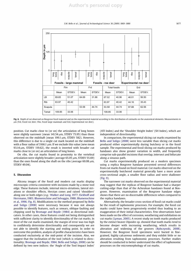
The mean value of the depth was 56.82 mm for cut marksobserved on the fossil roe deer, 70.44 mm for cut marks on otherfossil large mammals and 54.08 mm for the experimental cut marks(Fig. 6).
The depth of the experimental cut mark was not statisticallydifferent from the depth of cut marks observed on the fossil roedeer (W¼ 1369.5, p� 0.20). However, statistically significantdifferences were observed in the case of the depth of experimentalcut marks when compared to the total of fossils specimens(W¼ 3031.5, p� 0.04). No statistically significant difference hasbeen observed between cut marks on the fossil roe deer and any ofthe other fossil large mammal remains (W¼ 489, p� 0.23).
When considering different anatomical elements, the differ-ences in the depth of cut between experimentally produced andfossil cut marks were statistically significant in the case of longbones and scapulae (long bones, W¼ 432, p� 0.02; ribs, W¼ 296,p� 0.18; scapulae, W¼ 179, p� 0.047).
On the experimental material, it was possible to observe a clearpattern in the depth of cut marks according to their anatomicalposition. Cut marks close to (or on) long bone articulations weregenerally deeper (mean 132.33 mm, STDEV 106.60) than thoseobserved along the shaft (mean 34.10 mm, STDEV 34.92; Wilcoxontwo sample test, W¼ 104, p� 0.047). Similarly on ribs, the cutmarks found in proximity to the vertebral articulation were slightlydeeper (mean 51.39, STDEV 25.72) than those found along the shafton the ribs (mean 39.46, STDEV 40.47; this difference is notstatistically significant).
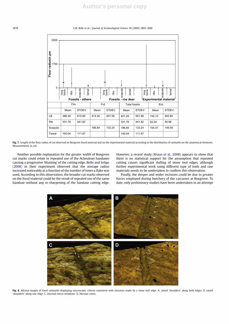
4.5. Floor radius
The mean value of the floor radius was 253.18 mm for cut markson the fossil roe deer, 333.37 mm for cut marks observed on otherfossil large mammals and 129.29 mm for the experimental cutmarks (Fig. 7).
In general, cut marks observed on the experimentally butcheredmaterial have shorter radii regardless of their anatomical distri-bution. This is indicative of narrower cut marks (Bello and Soligo,2008).
The length of the radius of experimentally produced cut markcan be statistically differentiated from the length of the radius of cutmarks observed on fossil material, when exclusively compared tothe fossil roe deer (W¼ 1832, p� 1.205e�06) as well as whencompared to the total of fossil specimens (W¼ 3639, p� 2.632�07).No statistically significant difference has been observed betweencut marks on fossil roe deer and other fossil large mammal remains(W¼ 422, p� 0.52).
The differences in the floor radius between experimental andfossil cut marks were also statistically significant in the case of longbones and ribs (long bones, W¼ 722, p� 7.01e�05; ribs, W¼ 341,p� 0.01; scapula, W¼ 113, p� 0.18).
On the experimental material, it was not possible to statisticallydifferentiate the floor radii of cut marks according to their anatomical
Frd Total fossils Erd
Mean STDEV. Mean STDEV. Mean STDEV.
LB 0.574 0.410 0.436 0.264 0.505 0.339 0.500 0.325
Rib 0.774 0.315 0.774 0.315 0.564 0.317
Scapula 0.572 0.313 0.572 0.313 0.355 0.251
Tarsal 0.522 0.656 0.522 0.656
Flm
Mean STDEV.
0.056
0
0.5
1
long
bone rib
scap
ula
tars
al
long
bone rib
scap
ula
tars
al
long
bone rib
scap
ula
tars
al
Fossils - large mammal Fossils - roe deer Experimental roe deer
SH
In
dex
Fig. 5. Shoulder Height Index (SH Index) observed on Boxgrove fossil material and on the experimental material according to the distribution of cutmarks on the anatomicalelements. (Frd, Fossil roe deer; Flm, Fossil large mammals and Erd, Experimental roe deer).
S.M. Bello et al. / Journal of Archaeological Science 36 (2009) 1869–18801876
Author's personal copy
position. Cut marks close to (or on) the articulation of long boneswere slightly narrower (mean 141.54 mm, STDEV 75.95) than thoseobserved on the midshaft (mean 199.1 mm, STDEV 582). However,this difference is due to a single cut mark located on the midshaftwith a floor radius of 3166.1 mm. If we exclude this value (new mean93.14 mm, STDEV 119.62), the result is inverted with broader cutmarks close to (or on) an articulation of long bones.
On ribs, the cut marks found in proximity to the vertebralarticulation were slightly broader (average 65.05 mm, STDEV 55.69)than the ones found along the shaft on the ribs (average 60.08 mm,STDEV 49.04).
5. Discussion
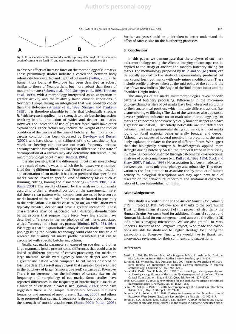
Alicona images of the fossil and modern cut marks displaymicroscopic criteria consistent with incisions made by a stone tooledge. These features include, internal micro-striations, lateral stri-ations or shoulder effects, Herzian cones and raised ‘shoulders’along one or both edges (e.g.: Walker and Long, 1977; Eickhoff andHerrmann, 1985; Blumenschine and Selvaggio, 1988; Blumenschineet al., 1996; Fig. 8). Modifications to the method proposed by Belloand Soligo (2008) were necessary because it was not alwayspossible to identify features, such as smears, oblique faulting andchipping used by Bromage and Boyde (1984) as directional indi-cators. In other cases, these features could not being distinguishedwith sufficient clarity to identify directionality of the cut marks. Inmost of the cut marks examined, the margin of error was too highto confidently determine directionality and consequently we arenot able to identify the starting and ending point. In order toovercome this problem, analysis of profile characteristics have beenconducted exclusively at the mid-point of the incision. Also, wesuggest that the inclination of the tool (which is related to direc-tionality; Bromage and Boyde, 1984; Bello and Soligo, 2008) can bedefined by two new indices: the ‘Angle of the Tool Impact Index’
(ATI Index) and the ‘Shoulder Height Index’ (SH Index), which areindependent of directionality.
In comparison, the experimental slicing cut marks examined byBello and Soligo (2008) were less variable than slicing cut marksproduced either experimentally during butchery or in the fossilsample. The experimental and fossil slicing cut marks produced byhandaxes also show greater variation in width, and frequentlycomprise sub-parallel incisions that overlap, intersect and bifurcatealong a sinuous path.
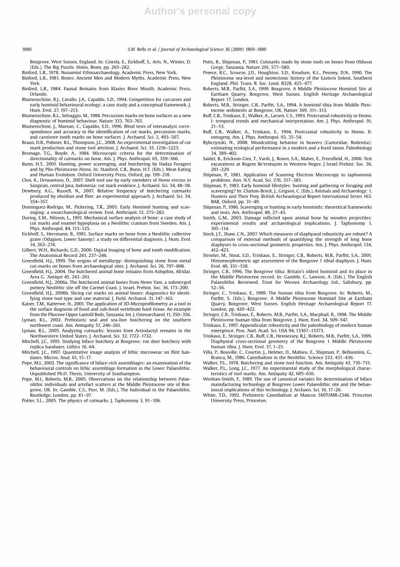
Cut marks experimentally produced on a modern specimenusing a replica Boxgrove handaxe presented several differencesfrom cut marks found on fossil material. Cut marks observed on theexperimentally butchered material generally have a more acutecross-sectional angle, a smaller floor radius and were shallower(Fig. 9).
The narrower cut marks observed on the experimental materialmay suggest that the replicas of Boxgrove handaxe had a sharpercutting-edge than that of the Acheulean handaxes found at Box-grove. However, examination of the Boxgrove handaxe edgesshows that there are no discernable differences when compared tothe replica handaxes.
Alternatively, the broader cross-section of fossil cut marks couldbe the result of taphonomic processes. For example, the fossil cutmarks could have been progressively eroded thus leading to anexaggeration of their initial characteristics. Few observations havebeen made on the effect of corrosion, weathering and exfoliation oncut marks (Lyman, 2005). A recent study on tooth marks producedby the extinct beaver Dipoides sp. showed that tooth marks tend tobecome deeper with increasing weathering, which results inalteration and widening of the grooves (Rybczynski, 2008).However, the Boxgrove fossil specimens were buried in fine-grained, highly calcareous sediments and they exhibit little or noalteration resulting from taphonomic processes. Further studiesshould be conducted to better understand the effect of taphonomicprocesses on the micromorphology of cut marks.
Flm Frd Total fossils Erd
Mean STDEV. Mean STDEV. Mean STDEV. Mean STDEV.
LB 69.65 51.07 64.69 31.86 67.0 40.96 46.01 56.50
Rib 63.97 40.42 63.97 40.42 44.16 35.43
Scapula 53.58 44.74 53.5 44.74 97.88 62.56
Tarsal 109.85 33.06 109.85 33.06
2
8
0
125
250
long
bone ribs
scap
ula
tars
al
long
bone ribs
scap
ula
tars
al
long
bone ribs
scap
ula
tars
al
Fossils - large mammal Fossils - roe deer Experimental roe deer
Dep
th
in
µ
m
Fig. 6. Depth of cut observed on Boxgrove fossil material and on the experimental material according to the distribution of cutmarks on the anatomical elements. Measurements inmm (Frd, Fossil roe deer; Flm, Fossil large mammals and Erd, Experimental roe deer).
S.M. Bello et al. / Journal of Archaeological Science 36 (2009) 1869–1880 1877
Author's personal copy
Another possible explanation for the greater width of Boxgrovecut marks could relate to repeated use of the Acheulean handaxescausing a progressive ‘blunting’ of the cutting edge. Bello and Soligo(2008) in their experiment observed that the average radiusincreased noticeably as a function of the number of times a flake wasused. According to this observation, the broader cut marks observedon the fossil material could be the result of repeated use of the samehandaxe without any re-sharpening of the handaxe cutting edge.
However, a recent study (Braun et al., 2008) appears to show thatthere is no statistical support for the assumption that repeatedcutting causes significant dulling of stone tool edges, althoughfurther experimental work using different type of tools and rawmaterials needs to be undertaken to confirm this observation.
Finally, the deeper and wider incisions could be due to greaterforces employed during butchery of the carcasses at Boxgrove. Todate, only preliminary studies have been undertaken in an attempt
Fig. 8. Alicona images of fossil cutmarks displaying microscopic criteria consistent with incisions made by a stone tool edge: A, raised ‘shoulders’ along both edges; B, raised‘shoulders’ along one edge; C, internal micro-striations; D, Herzian cones.
Flm Frd Total fossils Erd
Mean STDEV. Mean STDEV. Mean STDEV. Mean STDEV.
LB 388.28 615.85 414.30 407.56 401.29 501.89 192.12 545.80
Rib 331.76 441.82 331.76 441.82 62.04 50.96
Scapula 186.84 133.24 186.84 133.24 154.47 149.55
Tarsal 150.04 111.67 150.04 111.67
0
1750
3500
long
bone
ribs
scap
ula
tars
al
long
bone
ribs
scap
ula
tars
al
long
bone
ribs
scap
ula
tars
al
Fossils - others Fossils - roe deer Experimental material
Flo
or rad
iu
sin
µ
m
Fig. 7. Length of the floor radius of cut observed on Boxgrove fossil material and on the experimental material according to the distribution of cutmarks on the anatomical elements.Measurements in mm.
S.M. Bello et al. / Journal of Archaeological Science 36 (2009) 1869–18801878
Author's personal copy
to observe effects of increase force on the morphology of cut marks.These preliminary studies indicate a correlation between bodyrobusticity, force exerted and depth of cut marks (Potter, 2005). Thehuman tibia found at Boxgrove has been described as robust,similar to those of Neanderthals, but more robust than those ofmodern humans (Roberts et al., 1994; Stringer et al., 1998; Trinkauset al., 1999), with a morphology interpreted as an adaptation togreater activity and the relatively harsh climatic conditions inNorthern Europe during an interglacial that was probably coolerthan the Holocene (Stringer et al., 1998; Stringer and Trinkaus,1999). It is therefore plausible to infer that biologically strongerH. heidelbergensis applied more strength to their butchering action,resulting in the production of wider and deeper cut marks.However, the indication of use of greater force could have otherexplanations. Other factors may include the weight of the tool orcondition of the carcass at the time of butchery. The importance ofcarcass condition has been discussed by Dewbury and Russell(2007), who have shown that ‘stiffness’ of the carcass due to rigormortis or freezing can increase cut mark frequency becausea stronger action is required. It is likely that difference in the state ofdecomposition of a carcass may also determine differences in themicromorphology of cut marks (Binford, 1984).
It is also possible, that the differences in cut mark morphologyare a result of specific ways in which the handaxes were manipu-lated during different butchery task. From the anatomical locationand orientation of cut marks, it has been predicted that specific cutmarks can be linked to specific kind of butchery tasks, such asskinning, cutting, boning and dismembering (Binford, 1981, 1984;Bunn, 2001). The results obtained by the analyses of cut marksaccording to their anatomical position on the experimental mate-rial show a clear pattern when comparisons are made between cutmarks located on the midshaft and cut marks located in proximityto the articulation. Cut marks close to (or on) an articulation weretypically broader, deeper and have a greater inclination. Thesecharacteristics may be associated with disarticulation/dismem-bering process that require more force. Very few studies havedescribed differences in the morphology of cut marks associatedwith differences in the butchery process (Binford, 1978, 1981, 1984).We suggest that the quantitative analysis of cut marks micromor-phology using the Alicona technology could enhance this field ofresearch by quantify cut marks profile parameters that can beassociated with specific butchering actions.
Finally, cut marks parameters measured on roe deer and otherlarge mammals fossils present some differences that could also belinked to different patterns of carcass-processing. Cut marks onlarge mammal fossils were typically broader, deeper and havea greater inclination when compared to cut marks observed onfossil roe deer. This result may suggest that a greater force was usedin the butchery of larger (rhinoceros-sized) carcasses at Boxgrove.There is no agreement on the influence of carcass size on thefrequency and morphology of cut marks. Some studies havereported differences in the frequency of butchering cut marks asa function of variation in carcass size (Lyman, 2002); some havesuggested there is no simple relationship between cut markfrequency and amount of meat present (Lyman, 2005), and othershave proposed that cut mark frequency is directly proportional tothe strength of muscle attachments (Bunn, 2001; Potter, 2005).
Further analyses should be undertaken to better understand theeffect of carcass size on the butchering processes.
6. Conclusions
In this paper, we demonstrate that the analyses of cut markmicromorphology using the Alicona imaging microscope can beapplied to the study of ancient and modern butchery slicing cutmarks. The methodology proposed by Bello and Soligo (2008) canbe equally applied to the study of experimentally produced cutmarks and fossil cut marks with only minor modifications. Theseinclude profile analyses taken at the mid point of the cut and theuse of two new indices (the Angle of the Tool Impact Index and theShoulder Height Index).
The analyses of cut marks micromorphologies reveal specificpatterns of butchery processing. Differences in the micromor-phology characteristics of cut marks have been observed accordingto their anatomical position, which indicate different actions (i.e.dismembering vs filleting). The size of the carcasses also appears tohave a significant influence on cut mark micromorphology (e.g. cutmarks on rhinoceros bones were typically broader, deeper and havea greater inclination). Particularly noticeable are the differencesbetween fossil and experimental slicing cut marks, with cut marksfound on fossil material being generally broader and deeper.Although we suggested several possible interpretations, the mostlikely explanation relates to the use of different forces. We suggestthat the biologically stronger H. heidelbergensis applied morestrength during butchery. So far, the temporal trend in robusticityin Homo has been documented through osteometric cross-sectionalanalyses of post-cranial bones (e.g. Ruff et al., 1993, 1994; Stock andShaw, 2007; Trinkaus, 1997). No association had been made, so far,between cut marks micromorphology and robusticity. This obser-vation is the first attempt to associate the by-product of humanactivity to biological descriptions and may open new field ofresearch on the behavioural repertoire and anatomical character-istics of Lower Palaeolithic hominins.
Acknowledgements
This study is a contribution to the Ancient Human Occupation ofBritain Project (AHOB). We owe special thanks to the LeverhulmeTrust for their financial support of this project. SB also thank theHuman Origins Research Fund for additional financial support andNorman MacLeod for encouragement and access to the Alicona 3DInfiniteFocus imaging microscope. We are also grateful to MarkRoberts (Director of the Boxgrove Project) who made the collec-tions available for study and to English Heritage for funding theexcavations at Boxgrove. Finally, we would like to thank twoanonymous reviewers for their comments and suggestions.
References
Austin, L., 1994. The life and death of a Boxgrove biface. In: Ashton, N., David, A.(Eds.), Stories in Stone. Lithics Studies Society, London, pp. 119–126.
Bartelink, E.J., Wiersema, J.M., Demaree, R.S., 2001. Quantitative analysis of sharp-force trauma: an application of scanning electron microscopy in forensicanthropology. J. Forensic Sci. 46, 1288–1293.
Bates, M.R., Parfitt, S.A., Roberts, M.B., 1997. The chronology, palaeogeography andarchaeological significance of the marine Quaternary record of the West SussexCoastal Plain, Southern England, UK. Quat. Sci. Rev. 16, 1227–1252.
Bello, S.M., Soligo, C., 2008. A new method for the quantitative analysis of cutmarkmicromorphology. J. Archaeol. Sci. 35, 1542–1552.
Bello, S.M., Soligo, C., Parfitt, S., 2007. Micromorphology of cut-marks in PalaeolithicBritain. Am. J. Phys. Anthropol. 132, 71–72.
Bergman, C.A., Roberts, M.B., 1988. Flaking technology at the Acheulean site ofBoxgrove, West Sussex (England). Rev Archeol. de Picardie 1–2, 105–113.
Bergman, C.A., Roberts, M.B., Collcutt, S.N., Barlow, P., 1990. Refitting and spatialanalysis of artefacts from Quarry 2 at the Middle Pleistocene Acheulean site of
A B
Fig. 9. Representation of the mean values of the opening of the angle of cut, radius anddepth of cutmarks on fossil (A) and experimentally butchered specimens (B).
S.M. Bello et al. / Journal of Archaeological Science 36 (2009) 1869–1880 1879
Author's personal copy
Boxgrove, West Sussex, England. In: Cziesla, E., Eickhoff, S., Arts, N., Winter, D.(Eds.), The Big Puzzle. Holos, Bonn, pp. 265–282.
Binford, L.R., 1978. Nunamiut Ethnoarchaeology. Academic Press, New York.Binford, L.R., 1981. Bones: Ancient Men and Modern Myths. Academic Press, New
York.Binford, L.R., 1984. Faunal Remains from Klasies River Mouth. Academic Press,
Orlando.Blumenschine, R.J., Cavallo, J.A., Capaldo, S.D., 1994. Competition for carcasses and
early hominid behavioural ecology: a case study and a conceptual framework. J.Hum. Evol. 27, 197–213.
Blumenschine, R.L., Selvaggio, M., 1988. Percussion marks on bone surfaces as a newdiagnostic of hominid behaviour. Nature 333, 763–765.
Blumenschine, J., Marean, C., Capaldo, S.D., 1996. Blind tests of interanalyst corre-spondence and accuracy in the identification of cut marks, percussion marks,and carnivore tooth marks on bone surfaces. J. Archaeol. Sci. 2, 493–507.
Braun, D.R., Pobiner, B.L., Thompson, J.C., 2008. An experimental investigation of cutmark production and stone tool attrition. J. Archaeol. Sci. 35, 1216–1223.
Bromage, T.G., Boyde, A., 1984. Microscopic criteria for the determination ofdirectionality of cutmarks on bone. Am. J. Phys. Anthropol. 65, 359–366.
Bunn, H.T., 2001. Hunting, power scavenging, and butchering by Hadza Foragersand by Plio-Pleistocene Homo. In: Stanford, C.B., Bunn, H.T. (Eds.), Meat-Eatingand Human Evolution. Oxford University Press, Oxford, pp. 199–218.
Choi, K., Driwantoro, D., 2007. Shell tool use by early members of Homo erectus inSangiran, central Java, Indonesia: cut mark evidence. J. Archaeol. Sci. 34, 48–58.
Dewbury, A.G., Russell, N., 2007. Relative frequency of butchering cutmarksproduced by obsidian and flint: an experimental approach. J. Archaeol. Sci. 34,354–357.
Dominguez-Rodrigo, M., Pickering, T.R., 2003. Early Hominid hunting and scav-enging: a zooarchaeological review. Evol. Anthropol. 12, 275–282.
During, E.M., Nilsson, L., 1991. Mechanical surface analysis of bone: a case study ofcut marks and enamel hypoplasia on a Neolithic cranium from Sweden. Am. J.Phys. Anthropol. 84, 113–125.
Eickhoff, S., Herrmann, B., 1985. Surface marks on bone from a Neolithic collectivegrave (Odagsen, Lower Saxony): a study on differential diagnosis. J. Hum. Evol.14, 263–274.
Gilbert, W.H., Richards, G.D., 2000. Digital Imaging of bone and tooth modification.The Anatomical Record 261, 237–246.
Greenfield, H.J., 1999. The origins of metallurgy: distinguishing stone from metalcut-marks on bones from archaeological sites. J. Archaeol. Sci. 26, 797–808.
Greenfield, H.J., 2004. The butchered animal bone remains from Ashqelon, Afridar.Area G. ’Antiqot 45, 243–261.
Greenfield, H.J., 2006a. The butchered animal bones from Newe Yam, a submergedpottery Neolithic site off the Carmel Coast. J. Israel. Prehist. Soc. 36, 173–200.
Greenfield, H.J., 2006b. Slicing cut marks on animal bones: diagnostics for identi-fying stone tool type and raw material. J. Field. Archaeol. 31, 147–163.
Kaiser, T.M., Katterwe, H., 2001. The application of 3D-Microprofilometry as a tool inthe surface diagnosis of fossil and sub-fossil vertebrate hard tissue. An examplefrom the Pliocene Upper Laetolil Beds, Tanzania. Int. J. Osteoarchaeol. 11, 350–356.
Lyman, R.L., 2002. Prehistoric seal and sea-lion butchering on the southernnorthwest coast. Am. Antiquity 57, 246–261.
Lyman, R.L., 2005. Analyzing cutmarks: lessons from Artiodactyl remains in theNorthwestern United States. J. Archaeol. Sci. 32, 1722–1732.
Mitchell, J.C., 1995. Studying biface butchery at Boxgrove: roe deer butchery withreplica handaxes. Lithics 16, 64.
Mitchell, J.C., 1997. Quantitative image analysis of lithic microwear on flint han-daxes. Micros. Anal. 61, 15–17.
Pope, M.I., 2002. The significance of biface-rich assemblages: an examination of thebehavioural controls on lithic assemblage formation in the Lower Palaeolithic.Unpublished Ph.D. Thesis, University of Southampton.
Pope, M.I., Roberts, M.B., 2005. Observations on the relationship between Palae-olithic individuals and artefact scatters at the Middle Pleistocene site of Box-grove, UK. In: Gamble, C.S., Porr, M. (Eds.), The Individual in the Palaeolithic.Routledge, London, pp. 81–97.
Potter, S.L., 2005. The physics of cutmarks. J. Taphonomy 3, 91–106.
Potts, R., Shipman, P., 1981. Cutmarks made by stone tools on bones from OlduvaiGorge, Tanzania. Nature 291, 577–580.
Preece, R.C., Scourse, J.D., Houghton, S.D., Knudsen, K.L., Penney, D.N., 1990. ThePleistocene sea-level and neotectonic history of the Eastern Solent, SouthernEngland. Phil. Trans. R. Soc. Lond. B328, 425–477.
Roberts, M.B., Parfitt, S.A., 1999. Boxgrove. A Middle Pleistocene Hominid Site atEartham Quarry, Boxgrove, West Sussex. English Heritage ArchaeologicalReport 17, London.
Roberts, M.B., Stringer, C.B., Parfitt, S.A., 1994. A hominid tibia from Middle Pleis-tocene sediments at Boxgrove, UK. Nature 369, 311–313.
Ruff, C.B., Trinkaus, E., Walker, A., Larsen, C.S., 1993. Postcranial robusticity in Homo.I: temporal trends and mechanical interpretation. Am. J. Phys. Anthropol. 91,21–53.
Ruff, C.B., Walker, A., Trinkaus, E., 1994. Postcranial robusticity in Homo. II:ontogeny. Am. J. Phys. Anthropol. 93, 35–54.
Rybczynski, N., 2008. Woodcutting behavior in beavers (Castoridae, Rodentia):estimating ecological performance in a modern and a fossil taxon. Paleobiology34, 389–402.
Saidel, B., Erickson-Gini, T., Vardi, J., Rosen, S.A., Maher, E., Freenfield, H., 2006. Testexcavations at Rogem Be’erotayim in Western Negev. J Israel Prehist. Soc. 36,201–229.
Shipman, P., 1981. Application of Scanning Electron Microscopy to taphonomicproblems. Ann. N.Y. Acad. Sci. 276, 357–385.
Shipman, P., 1983. Early hominid lifestyles: hunting and gathering or foraging andscavenging? In: Clutton-Brock, J., Grigson, C. (Eds.), Animals and Archaeology: 1.Hunters and Their Prey. British Archeaological Report International Series 163.BAR, Oxford, pp. 31–49.
Shipman, P., 1986. Scavenging or hunting in early hominids: theoretical frameworksand tests. Am. Anthropol. 88, 27–43.
Smith, G.M., 2003. Damage inflicted upon animal bone by wooden projectiles:experimental results and archaeological implications. J. Taphonomy 1,105–114.
Stock, J.T., Shaw, C.N., 2007. Which measures of diaphyseal robusticity are robust? Acomparison of external methods of quantifying the strength of long bonediaphyses to cross-sectional geometric properties. Am. J. Phys. Anthropol. 134,412–423.
Streeter, M., Stout, S.D., Trinkaus, E., Stringer, C.B., Roberts, M.B., Parfitt, S.A., 2001.Histomorphometric age assessment of the Boxgrove 1 tibial diaphysis. J. Hum.Evol. 40, 331–338.
Stringer, C.B., 1996. The Boxgrove tibia: Britain’s oldest hominid and its place inthe Middle Pleistocene record. In: Gamble, C., Lawson, A. (Eds.), The EnglishPalaeolithic Reviewed. Trust for Wessex Archaeology Ltd., Salisbury, pp.52–56.
Stringer, C., Trinkaus, E., 1999. The human tibia from Boxgrove. In: Roberts, M.,Parfitt, S. (Eds.), Boxgrove: A Middle Pleistocene Hominid Site at EarthamQuarry, Boxgrove, West Sussex. English Heritage Archaeological Report 17,London, pp. 420–422.
Stringer, C.B., Trinkaus, E., Roberts, M.B., Parfitt, S.A., Macphail, R., 1998. The MiddlePleistocene human tibia from Boxgrove. J. Hum. Evol. 34, 509–547.
Trinkaus, E., 1997. Appendicular robusticity and the paleobiology of modern humanemergence. Proc. Natl. Acad. Sci. USA 94, 13367–13373.
Trinkaus, E., Stringer, C.B., Ruff, C.B., Hennessey, R.J., Roberts, M.B., Parfitt, S.A., 1999.Diaphyseal cross-sectional geometry of the Boxgrove 1 Middle Pleistocenehuman tibia. J. Hum. Evol. 37, 1–25.
Villa, P., Bouville, C., Courtin, J., Helmer, D., Mahieu, E., Shipman, P., Belluomini, G.,Branca, M., 1986. Cannibalism in the Neolithic. Science 233, 431–436.
Walker, P.L., 1978. Butchering and stone tool function. Am. Antiquity 43, 710–715.Walker, P.L., Long, J.C., 1977. An experimental study of the morphological charac-
teristics of tool marks. Am. Antiquity 42, 605–616.Wenban-Smith, F., 1989. The use of canonical variates for determination of biface
manufacturing technology at Boxgrove Lower Palaeolithic site and the behav-ioural implications of this technology. J. Archaeo. Sci. 16, 17–26.
White, T.D., 1992. Prehistoric Cannibalism at Mancos 5MTUMR-2346. PrincetonUniversity Press, Princeton.
S.M. Bello et al. / Journal of Archaeological Science 36 (2009) 1869–18801880