putative anticancer compounds from plant-derived ... - mdpi
TRANSCRIPT
�����������������
Citation: Hridoy, M.; Gorapi, M.Z.H.;
Noor, S.; Chowdhury, N.S.;
Rahman, M.M.; Muscari, I.; Masia, F.;
Adorisio, S.; Delfino, D.V.;
Mazid, M.A. Putative Anticancer
Compounds from Plant-Derived
Endophytic Fungi: A Review.
Molecules 2022, 27, 296. https://
doi.org/10.3390/molecules27010296
Academic Editor: Josphat Matasyoh
Received: 22 November 2021
Accepted: 28 December 2021
Published: 4 January 2022
Publisher’s Note: MDPI stays neutral
with regard to jurisdictional claims in
published maps and institutional affil-
iations.
Copyright: © 2022 by the authors.
Licensee MDPI, Basel, Switzerland.
This article is an open access article
distributed under the terms and
conditions of the Creative Commons
Attribution (CC BY) license (https://
creativecommons.org/licenses/by/
4.0/).
molecules
Review
Putative Anticancer Compounds from Plant-Derived EndophyticFungi: A ReviewMd. Hridoy 1,2 , Md. Zobayer Hossain Gorapi 3, Sadia Noor 3,4, Nargis Sultana Chowdhury 5,Md. Mustafizur Rahman 6 , Isabella Muscari 7, Francesco Masia 7, Sabrina Adorisio 8, Domenico V. Delfino 8,*and Md. Abdul Mazid 1,4,*
1 Department of Pharmacy, University of Dhaka, Dhaka 1000, Bangladesh; [email protected] Department of Pharmaceutical Sciences, School of Pharmacy, Temple University, Philadelphia, PA 19140, USA3 Department of Pharmacy, University of Asia Pacific, Dhaka 1215, Bangladesh;
[email protected] (M.Z.H.G.); [email protected] (S.N.)4 Department of Pharmaceutical Chemistry, Faculty of Pharmacy, University of Dhaka, Dhaka 1000, Bangladesh5 Department of Pharmacy, Manarat International University, Dhaka 1343, Bangladesh; [email protected] Pharmacy Discipline, Khulna University, Khulna 9208, Bangladesh; [email protected] Department of Medicine and Surgery, University of Perugia, 06132 Perugia, Italy;
[email protected] (I.M.); [email protected] (F.M.)8 Department of Medicine and Surgery, Foligno Nursing School and Section of Pharmacology,
University of Perugia, Piazzale Severi, S. Andrea delle Fratte, 06129 Perugia, Italy; [email protected]* Correspondence: [email protected] (D.V.D.); [email protected] (M.A.M.)
Abstract: Endophytic fungi are microorganisms that exist almost ubiquitously inside the varioustissues of living plants where they act as an important reservoir of diverse bioactive compounds.Recently, endophytic fungi have drawn tremendous attention from researchers; their isolation, cul-ture, purification, and characterization have revealed the presence of around 200 important anddiverse compounds including anticancer agents, antibiotics, antifungals, antivirals, immunosuppres-sants, and antimycotics. Many of these anticancer compounds, such as paclitaxel, camptothecin,vinblastine, vincristine, podophyllotoxin, and their derivatives, are currently being used clinicallyfor the treatment of various cancers (e.g., ovarian, breast, prostate, lung cancers, and leukemias).By increasing the yield of specific compounds with genetic engineering and other biotechnologies,endophytic fungi could be a promising, prolific source of anticancer drugs. In the future, compoundsderived from endophytic fungi could increase treatment availability and cost effectiveness. Thiscomprehensive review includes the putative anticancer compounds from plant-derived endophyticfungi discovered from 1990 to 2020 with their source endophytic fungi and host plants as well astheir antitumor activity against various cell lines.
Keywords: endophytic fungi; anticancer compounds; living plants
1. Introduction
In 1866, de Bary introduced the term “endophyte” [1]. An endophyte may be afungal or bacterial microorganism that colonizes various interior parts of plants causing noapparent pathogenic effects on its host plants. The endophytes, most commonly endophyticfungi, are believed to help plants adapt to abiotic factors (high temperature and salinity,drought, metal toxicity, and harmful effects of light) as well as biotic factors (herbivores,insects, nematodes, and pathogens). This is mainly achieved by the secondary bioactivemetabolites produced by the endophytic fungi. In their symbiotic relation, the endophytesare fed and protected by the host plant, and in return, these microorganisms producebioactive secondary metabolites, enhancing the growth of the host plant and protectingthe plant from pathogens and herbivores [2]. Therefore, endophytic fungal metabolites canalso be exploited as drugs for the treatment of various types of human diseases, includingcancer [3].
Molecules 2022, 27, 296. https://doi.org/10.3390/molecules27010296 https://www.mdpi.com/journal/molecules
Molecules 2022, 27, 296 2 of 37
This group of microorganisms has drawn tremendous attention from researcherssince the isolation, culture, purification, and characterization of this fascinating group ofmicroorganisms revealed the presence of hundreds of important and diverse chemicalclasses of compounds. The interest of scientists in endophytes is also growing as they are agood reservoir of bioactive metabolites [4,5]. Until now, many cytotoxic agents includingpaclitaxel (also known as Taxol) [6] have been isolated from endophytes. Secondarymetabolites with cytotoxic properties have the potential to be explored as anticancer drugs.
Recent studies revealed that naphthoquinone derivatives fusarubins including anhy-drofusarubin and fusarubin (FUS) produced by endophytic fungi Cladosporium species [7]and Fusarium species [8] showed promising cytotoxicity against cancer cells. AlthoughFUS was reported earlier to have antibacterial activity, its cytotoxic activity was reportedrecently. Very recently, for the first time, we have revealed the molecular mechanism ofcytotoxic action of fusarubin isolated from a Cladosporium species inhabiting the leavesof Rauwolfia serpentina. We have reported that fusarubin and anhydrofusarubin inhibitproliferation and increase apoptosis in leukemia and other hematological tumor cells linesin different manners through the p21/p53-mediated pathway [9]. Our findings urge usto write this review on endophytic fungal metabolites as a fascinating group of bioactivesor putative anticancer compounds. Many of these putative anticancer compounds havevery promising cytotoxicity against a broad spectrum of cancer cell lines; some compoundsare already used as treatments for different cancer types such as breast, bladder, colorectal,esophageal, lung, ovarian, prostate, melanoma, testicular, leukemia, and lymphoma.
2. Anticancer Activity of Endophytic Fungi
Endophytic fungi have been a known source of anticancer agents since the discoveryof the valuable drug Taxol (also known as paclitaxel, a diterpenoid) isolated for the firsttime from an endophytic fungus Taxomyces andreanae obtained from the Pacific Yew bark(Taxus brevifolia) [6]. Since then, other anticancer drugs have been isolated from endo-phytic fungi, and among these 9-methoxycamptothecin and 10-hydroxycamptothecin fromFusarium solani [10], camptothecin from Entrophospora infrequens [11]; the anticancer leadcompounds podophyllotoxin from Phialocephala fortinii [12] and deoxypodophyllotoxinfrom Aspergillus fumigatus [13] fueled further research on endophytic fungi to discovermany other important known and novel anticancer compounds. According to this review,until now, more than 100 different fungal species have been identified to produce more thantwo hundred putative anticancer compounds (Figures 1 and 2) reported to possess antipro-liferative and/or cytotoxic properties against more than 60 different cell lines (Tables 1–3).Figure 1 indicates that endophytic fungal-derived anticancer agents gained attention fromscientists over the past three decades. Meanwhile, Figure 2 represents the abundance ofdifferent chemical classes and diversity of fungal metabolites. The anticancer compoundsisolated from endophytic fungi are effective against diverse cell lines that could be helpfulin combating any particular type of cancer (Table 1).
Table 1. Different cell lines against which endophytic fungal derived metabolites showed cytotoxicity.
Cell Lines Cell Lines
A2780S Ovarian tumor cell line Int-407 Human intestine cancerA2058 Human melanoma Jurkat T cell leukemia
A549 Lung carcinoma epithelial KB Human nasopharyngealepidermoid tumor
A431 Skin carcinoma K562 Human leukemia cellsACHN Renal cells L5178Y Mouse lymphoma cellsAsPC-1 Human pancreatic cancer cells MIA Pa Ca-2 Pancreatic carcinomaB16F10 Skin carcinoma MiaPaka-2 Pancreatic cancer
BC Human breast cancer cell line MDA-MB-231 Breast cancer cell lineBC-1 Breast cancer MDA-MB-435 Human breast cancer cell line
Molecules 2022, 27, 296 3 of 37
Table 1. Cont.
Cell Lines Cell Lines
BEL-7402 Human hepatocellularcarcinoma/human hepatoma cell line MFC Gastric cancer cells in mice
BEL-7404 Human hepatocellularcarcinoma/human hepatoma cell line MCF-7 Breast cancer cell line
BGC-823 Gastric carcinoma MOLT-4 Lymphoblastic leukemiaBT-220 Breast cancer cell line MRC-5 Fibroblast-like fetal lung cells
BT474 Human breast cancer MV4-11 Human FLT3-ITD mutant AMLcell line
CHO Chinese hamster ovary NCI-H187 Human small-cell lung cancerDU145 Human prostate carcinoma NCI-H460 Non-small-cell lung cancer
EAC Ehrlich ascites carcinoma NEC Colorectal neuroendocrinecell carcinoma
H116 Human colon adenocarcinoma OVCAR-5 Human ovarian cancerHeLa Cervical cancer PANC-1 Human pancreatic carcinomaHEp-2 Human liver cancer P388 Murine leukemia cells
HepG2 Human hepatocellularliver carcinoma PC-3 Prostate cancer
Hep3B Human hepatoma cell line PC-3 M Metastatic prostate cancerHM02 Human gastric carcinoma RAW264.7 Mouse macrophage cell
HL-60 Human promyelocytic leukemiacell line SF-268 CNS glioma
HL251 Human lung cancer SW480 Human colon cancer cellsHL-7702 Normal hepatocyte SW-620 Colon tumor cell lineHLK 210 Human leukemia SW1116 Human colon cancer cell line
HCT-8 Human colorectal adenocarcinoma SW1990 Human pancreatic cancer cellsHCT-116 Colon tumor cell line T24 Bladder carcinoma
H22 Hepatic cancer cells in mice T47D Breast cancer
H1975 Non-small-cell lung cancercells/human lung adenocarcinoma THP-1 Human monocytic cell line
H522-T1 Non-small cell lung cancer WI-38 Normal human fibroblast cellsHT-29 Human colon cancer line U2OS Human osteosarcoma cells
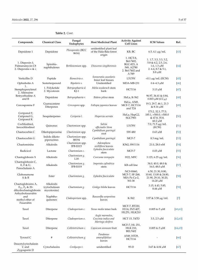
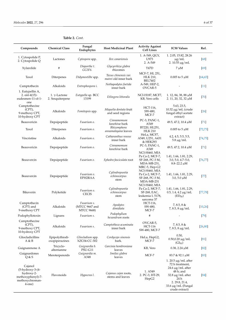
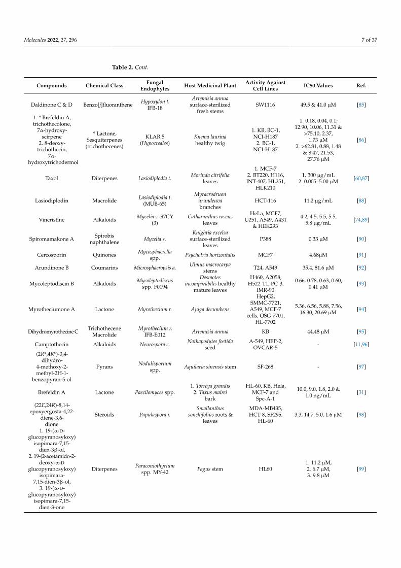
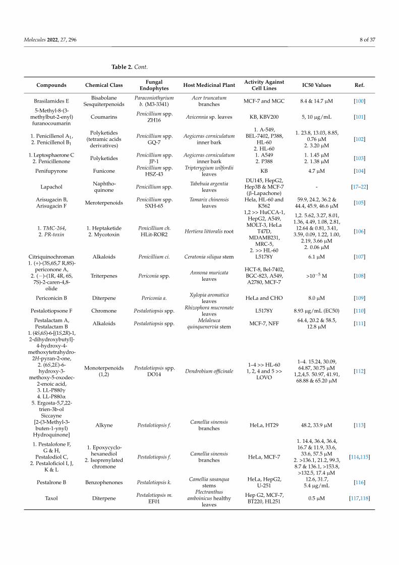
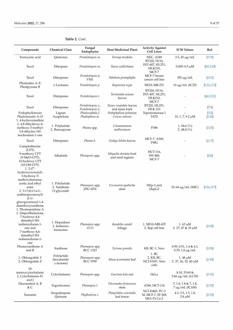
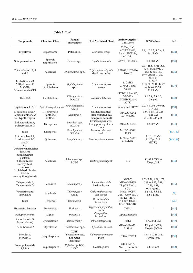
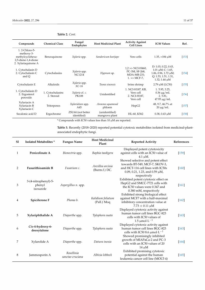
Table 2. Anticancer compounds from plant-derived endophytic fungi.
Compounds Chemical Class FungalEndophytes Host Medicinal Plant Activity Against
Cell Lines IC50 Values Ref.
Leucinostatin A Peptide Acremonium spp. Taxus baccata twig BT-20 2 nM (LD50) [14]
Allantopyrone A α-Pyrone Allantophomopsis l.KS-97 A549 cells, HL-60 >32, 0.32 µM [15]
Alternariol,Altenusin,Alternariol5-O-sulfate,Alternariol
5-O-methyl ether,Desmethylaltenusin
Polyketide Alternaria spp Polygonum senegalenseleaves L5178Y
<1 × 10−6,1 × 10−5, 1 × 10−5,1 × 10−5, 1 × 10⁻⁵
g/mL
[16]
Lapachol Naphtho-quinone Alternaria spp. Tabebuia argentea leaf
DU145, HepG2,Hep3B & MCF-7(β-Lapachone)
3.5, 3.5, 3.5 & 5 µM [17–22]
ResveratrodehydesA & B
Stilbenoid(Resveratroldervatives)
Alternaria spp. R6 Myoporum bontioidesroot
MDA-MB-435,HCT-116 <10 µM [23]
Alterporriol K,Alterporriol L Quinones Alternaria spp.
ZJ9-6B Aegiceras corniculatum MDA-MB-435,MCF-7
26.97, 29.11 & 13.11,20.04 µM [24]
Alternariol-10-methyl ether Polyketide Alternaria a. Capsicum annum
HL-60, A549,PC-3, HeLa, A431,
MiaPaka-2 andT47D
85, >100, >100, >100,95, >100 and >100
µM[25]
Molecules 2022, 27, 296 4 of 37
Table 2. Cont.
Compounds Chemical Class FungalEndophytes Host Medicinal Plant Activity Against
Cell Lines IC50 Values Ref.
Camptothecine(CPT),
9-methoxy CPT,10-hydroxy CPT
Alkaloids Alternaria a. Miquelia dentata fruitand seed regions
HCT-116,SW-480,MCF-7
6.59, 7.2,10.24 µg/mL (crudefungal ethyl acetate
extract)
[26]
Chrysin(5,7-dihydroxy
flavone)Flavone Alternaria a.
(KT380662)Passiflora incarnata
leaves MCF-7 34.066 µg/mL [27]
Alternariol9-methyl ether Dibenzopyranone Alternaria a. Camellia sinensis
branches U2OS 28.3 µM [28]
Lapachol Naphtho-quinone Alternaria a. Tabebuia argentea bark,
leaf and stem
DU145, HepG2,Hep3B & MCF-7(β-Lapachone)
3.5, 3.5, 3.5 & 5 µM [17–22]
(6aR,6bS,7S)-3, 6a,7,10-tetrahydroxy-4,9-dioxo-4, 6a, 6b,
7, 8,9-hexahydroperylene
Perylenes Alternaria t. Erythrophleum fordiibark HCT-8 1.78 µM [29]
1. Flavasperone,2. Rubrofusarin B3. Fonsecinone D
Naphthopyrones Aspergillus sp. Limonia acidissimaseeds
1. Hep 3B andU87 MG
2. SW11163. SMMC-7721
and A549
1. Between 19.92and 47.98 µM2. 4.5 µg/mL3. >10 µg/mL
[30]
Brefeldin A Lactone Aspergillus c. Torreya grandis barkHL-60, KB, Hela,
MCF-7 andSpc-A-1
1.0–10.0 ng/mL [31]
9-Deacetoxyfumigaclavine C Alkaloids Aspergillus f. Cynodon dactylon
stem K562 3.11 µM [32]
1. Fumitremorgin D,2. 4,8,10,14-
tetramethyl-6-acetoxy-14-[16-
acetoxy-19-(20,21-dimethyl)-18-ene]-phenanthrene-1-
ene-3,7-dione3. 12,13-dihydroxy-fumitremorgin C4. Verruculogen
Alkaloids Aspergillus f.Diphylleia sinensis
mainly roots,rhizomes
HepG2
1. 47.5 µM2. 139.9 µM3. 4.5 µM4. 9.8 µM
[33]
2,14-Dihydrox-7-drimen-12,11-olide Sesquiterpenes Aspergillus g. Ipomoea batatas plant Hep-G2, MCF-7 61, 41.7 µg/mL [34]
Nigerapyrones B,D & E
Asnipyrones APyrones Aspergillus n.
MA-132 Avicennia marina plantHepG2, MCF-7,A549, SW1990,MDA-MB-231
86, 105, 43, 38,48 µM [35]
Rubrofusarin B Naphtho-γ-pyrones Aspergillus n. Cynodon dactylon SW1116 4.5 µg/mL [36]
Lapachol Naphtho-quinone Aspergillus n. Tabebuia argentea
leaves
DU145, HepG2,Hep3B & MCF-7(β-Lapachone)
3.5, 3.5, 3.5 & 5 µM [17–22]
1. SequoiatonesA & B
2. SequoiamonascinA & B
Polyketide Aspergillus p. Sequoia sempervirensinner bark
1. BC2. MCF7,
NCI-H460, SF-268
1. 4 to 10 µM2. 19 × 10−4,
4 × 10−4,15 × 10−4 M
[37,38]
Butyrolactone I andButyrolactone V Butenolide Aspergillus t.—F7 Hyptis suaveolens MDA-MB-231
and MCF-734.4, 17.4 & 22.1,
31.9 µM [39]
Terrein Aspergillus t.JAS-2 Achyranthus aspera A-549 121.9 µg/mL [40]
1. Violaceoid A,2. Violaceoid C,Violaceoid D,
3. Violaceoid F
Hydroquinones Aspergillus v.Wild Moss(Bryophyta
unidentified species)
1. HeLa, MCF-7,Jurkat, MOLT-4,
HCT116,RAW264.7
2. Jurkat, MOLT-43. HCT116,RAW264.7
1. 24.6, 14.8, 3.1, 3.0,5.8, 5.6 µM (LD50)
2. 8.2, 5.9 & 8.3,6.2 µM (LD50)3. 6.4, 6.5 µM
(LD50)
[41]
Taxol Terpene Bartalinia r. Aegle marmelosleaves
BT 220, H116,Int 407, HL 251and HLK 210
- [42]
Molecules 2022, 27, 296 5 of 37
Table 2. Cont.
Compounds Chemical Class FungalEndophytes Host Medicinal Plant Activity Against
Cell Lines IC50 Values Ref.
Depsidone 1 Depsidone Pleosporales (BCC8616)
unidentified plant leafof the Hala-Bala forest
originKB, BC 6.5, 4.1 µg/mL [43]
1. Diepoxin δ,Palmarumycin C82. Diepoxins κ & ζ
Spirobis-naphthalenes Berkleasmium spp. Dioscorea zingiberensis
1. HCT-8,Bel-7402,
BGC-823, A549, A2780
2. Bel-7402 andA 549
1. 1.7, 3.3, 3.3, 3.2,5.8 & 4.2, 2.5, 2.6,
1.6, 1.3 µM2. 6.4, 8.7 & 5.1,
8.8 µM
[44]
Verticillin D Peptide Bionectria o. Sonneratia caseolarisInner leaf tissues L5178Y <0.1 µg/mL (EC50) [45]
Ophiobolin A Sesterterpenoid Bipolaris s. Unidentified MDA-MB-231 0.4–4.3 µM [46]1.
Stemphyperylenol2. Altenuene
1. Polyketide2. Mycotoxin
Botryosphaeria d.KJ-1
Melia azedarach stembark HCT116 3.13 µM [47]
Botryorhodine Aand B Depsidone Botryosphaeria r. Bidens pilosa stem HeLa, K-562 96.97, 36.41 & 0.84,
0.003 µM (CC50) [48]
Cercosporene F GuanacastaneDiterpenes Cercospora spp. Fallopia japonica leaves
HeLa, A549,MCF-7, HCT116
and T24
19.3, 29.7, 46.1, 21.3& 8.16 µM [49]
Ceriponol F,Ceriponol G,Ceriponol K
Sesquiterpenes Ceriporia l. Huperzia serrata HeLa, HepG2,SGC7901
173.2, 32.3, 77.5;185.1, >500.0, >500.0
& 47.8, 35.8,60.2 µM
[50]
Cochliodinol,Isocochliodinol Quinones Chaetomium spp. Salvia
officinalis Stem L5178Y 7.0, 71.5 µg/mL(EC50) [51]
Chaetocochin C Diketopiperazine Chaetomium spp. Cymbidium goeringiiroot SW-480 0.63 µM [52]
Chaetocochin G Indole diketo-piperazines
Chaetomium spp.88194 Cymbidium goeringii MCF-7 8.3 mg/mL [53]
Chaetominine Alkaloids Chaetomium spp.IFB-E015
Adenophoraaxilliflora leaves K562, SW1116 21.0, 28.0 nM [54]
Radicicol Lactone Chaetomium c. Ephedra fasciculatestem MCF-7 0.03 µM [55]
Chaetoglobosin X Alkaloids Chaetomium g.L18 Curcuma wenyujin H22, MFC 3.125, 6.25 µg/mL [56]
Chaetoglobosin C,E, F & U,
Penochalasin AAlkaloids Chaetomium g.
IFB-E019Imperata cylindrica
stem KB cell line 34.0, 40.0, 48.0 &16.0, 48.0 µM [57]
GlobosumoneA & B Ester Chaetomium g. Ephedra fasciculata
NCI-H460,MCF-7, SF-268,MIA Pa Ca-2,
WI-38
6.50, 21.30, 8.80,10.60, 13.00 & 24.80,21.90, 29.10, 30.20,
14.20 µM
[58]
Chaetoglobosins A,Fex, Fa & 20-
dihydrochaetoglobosin
Alkaloids(cytochalasanmycotoxins)
Chaetomium g. Ginkgo biloba leaves HCT116 3.15, 4.43, 5.85,8.44 µM [59]
Anhydrofusarubinand
methyl ether ofFusarubin
Naphtho-quinones Cladosporium spp. Rauwolfia serpentina
leaves K-562 3.97 & 3.58 µg/mL [7]
Taxol Diterpene Cladosporium c. Taxus media inner barkMCF-7, BT220,H116, INT-407,
HL251, HLK2100.005 to 5 µM [60,61]
Taxol Diterpene Cladosporium o.Aegle marmelos,
Coccinia indica andMoringa oleifera
HCT 15, T47D 3.5, 2.5 µM [62,63]
Taxol Diterpene Colletotrichum c. Capsicum annuum fruitMCF-7, HL 251,
HLK 210,BEL7402
0.005 to 5 µM [64,65]
Tyrosol C # Colletotrichum g.Pandanus
amaryllifoliusleaves
A549, HT29,HCT116 - [66]
DeacetylcytochalasinC and
Zygosporin DCytochalasins Cordyceps t. unidentified 95-D 3.67 & 4.04 µM [67]
Molecules 2022, 27, 296 6 of 37
Table 2. Cont.
Compounds Chemical Class FungalEndophytes Host Medicinal Plant Activity Against
Cell Lines IC50 Values Ref.
1. Cytospolide P,2. Cytospolide Q Lactones Cytospora spp. Ilex canariensis
1. A-549, QGY,U973
2. A-549
1. 2.05, 15.82, 28.26µg/mL
2. 10.55 µg/mL[68]
Xylarolide # Diaporthe t.GG3F6.
Glycyrrhiza glabrarhizomes T47D 7 µM [69]
Taxol Diterpenes Didymostilbe spp. Taxus chinensis var.mairei old inner bark
MCF-7, HL 251,HLK 210,BEL7402
0.005 to 5 µM [64,65]
Camptothecin Alkaloids Entrophospora i. Nothapodytes foetidainner bark
A-549, HEP-2,OVCAR-5 - [11]
1. Eutypellin A,2. ent-4(15)-
eudesmen-11-ol-1-one
1. γ-Lactone2. Sesquiterpene
Eutypella sp. BCC13199 Etlingera littoralis NCI-H187, MCF7,
KB, Vero cells1. 12, 84, 38, 88 µM2. 11, 20, 32, 32 µM [70]
Camptothecine(CPT),
9-methoxy CPT,10-hydroxy CPT
Alkaloids Fomitopsis spp. Miquelia dentata fruitand seed regions
HCT-116,SW-480,MCF-7
5.63, 23.5,10.32 µg/mL (crudefungal ethyl acetate
extract)
[26]
Beauvericin Depsipeptide Fusarium o. Cinnamomumkanehirae bark
PC-3, PANC-1,A549 49.5, 47.2, 10.4 µM [71]
Taxol Diterpenes Fusarium o. Rhizomphoraannamalayana leaves
BT220, HL251,HLK 210 0.005 to 5 µM [72,73]
Vincristine Alkaloids Fusarium o. Catharanthus roseusinner bark
HeLa, MCF7,A549, U251, A431
& HEK293
4.2, 4.5, 5.5, 5.5,5.8 µg/mL [74,75]
Beauvericin Depsipeptide Fusarium o. Cinnamomumkanehirae bark
PC-3, PANC-1,A549 49.5, 47.2, 10.4 µM [71]
Beauvercin Depsipeptide Fusarium o. Ephedra fasciculata root
NCI-H460, MIAPa Ca-2, MCF-7,SF-268, PC-3 M,MDA-MB-231,
MRC-5, Hep-G2
1.41, 1.66, 1.81, 2.29,3.0, 5.0, 4.7–5.0,
8.8–22.2 µM[76,77]
Beauvercin Depsipeptide Fusarium o.EPH2RAA
Cylindropuntiaechinocarpus
stem
NCI-H460, MIAPa Ca-2, MCF-7,SF-268, PC-3 M,MDA-MB-231
1.41, 1.66, 1.81, 2.29,3.0, 5.0 µM [77]
Bikaverin Polyketide Fusarium o.CECIS
Cylindropuntiaechinocarpus
stem
NCI-H460, MIAPa Ca-2, MCF-7,
SF-268, EAC,leukemia L 5178,
sarcoma 37
1.41, 1.66, 1.81, 2.29,0.5, 1.4, 4.2 µg/mL
(ED50)[77,78]
Camptothecin(CPT) and
9-methoxy CPTAlkaloids
Fusarium s.(MTCC 9667 and
MTCC 9668)
Apodytesdimidiata
HCT-116,SW-480,MCF-7
7, 8.5, 8 &7, 8.5, 8 µg/mL [10,26]
Podophyllotoxin Lignans Fusarium s. Podophyllumhexandrum roots # - [79]
Camptothecine(CPT),
9-methoxy CPT,10-hydroxy CPT
Alkaloids Fusarium s. Camptotheca acuminatainner bark
OVCAR-5,HCT-116
SW-480, MCF-7
7, 8.5, 8 &7, 8.5, 8 µg/mL [26,80]
GliocladicillinsA & B
Epipolythiodi-oxopiperazines
Gliocladium spp.XZC04-CC-302
Cordyceps sinensisbark.
HeLa, HepG2,MCF-7
0.50,0.50,0.20 µg/mL
(GI50)[81]
Guignarenone A Tricyclo-alternarene
Guignardia b.PSU-G11
Garcinia hombronianaleaves KB, Vero 0.38, 2.24 µM [82]
GuignardonesQ & S Meroterpenoids Guignardia m.
A348Smilax glabra
leaves MCF-7 83.7 & 92.1 µM [83]
Cajanol(5-hydroxy-3-(4-
hydroxy-2-methoxyphenyl)-7-methoxychroman-
4-one)
Flavonoids Hypocrea l. Cajanus cajan roots,stems and leaves
1. A5492. PC-3, HT-29,
HepG2
1. 20.5 µg/mL after72 h treatment,
24.6 µg/mL after48 h; and
32.8 µg/mL after24 h
2. 29.8, 21.4,33.6 µg/mL (Fungal
crude extract)
[84]
Molecules 2022, 27, 296 7 of 37
Table 2. Cont.
Compounds Chemical Class FungalEndophytes Host Medicinal Plant Activity Against
Cell Lines IC50 Values Ref.
Daldinone C & D Benzo[j]fluoranthene Hypoxylon t.IFB-18
Artemisia annuasurface-sterilized
fresh stemsSW1116 49.5 & 41.0 µM [85]
1. * Brefeldin A,trichothecolone,
7α-hydroxy-scirpene
2. 8-deoxy-trichothecin,
7α-hydroxytrichodermol
* Lactone,Sesquiterpenes(trichothecenes)
KLAR 5(Hypocreales)
Knema laurinahealthy twig
1. KB, BC-1,NCI-H187
2. BC-1,NCI-H187
1. 0.18, 0.04, 0.1;12.90, 10.06, 11.31 &
>75.10, 2.37,1.73 µM
2. >62.81, 0.88, 1.48& 8.47, 21.53,
27.76 µM
[86]
Taxol Diterpenes Lasiodiplodia t. Morinda citrifolialeaves
1. MCF-72. BT220, H116,INT-407, HL251,
HLK210
1. 300 µg/mL2. 0.005–5.00 µM [60,87]
Lasiodiplodin Macrolide Lasiodiplodia t.(MUB-65)
Myracrodruonurundeuvabranches
HCT-116 11.2 µg/mL [88]
Vincristine Alkaloids Mycelia s. 97CY(3)
Catharanthus roseusleaves
HeLa, MCF7,U251, A549, A431
& HEK293
4.2, 4.5, 5.5, 5.5,5.8 µg/mL [74,89]
Spiromamakone A Spirobisnaphthalene Mycelia s.
Knightia excelsasurface-sterilized
leavesP388 0.33 µM [90]
Cercosporin Quinones Mycosphaerellaspp. Psychotria horizontalis MCF7 4.68µM [91]
Arundinone B Coumarins Microsphaeropsis a. Ulmus macrocarpastems T24, A549 35.4, 81.6 µM [92]
Mycoleptodiscin B Alkaloids Mycoleptodiscusspp. F0194
Desmotesincomparabilis healthy
mature leaves
H460, A2058,H522-T1, PC-3,
IMR-90
0.66, 0.78, 0.63, 0.60,0.41 µM [93]
Myrotheciumone A Lactone Myrothecium r. Ajuga decumbens
HepG2,SMMC-7721,A549, MCF-7
cells, QSG-7701,HL-7702
5.36, 6.56, 5.88, 7.56,16.30, 20.69 µM [94]
Dihydromyrothecine C TrichotheceneMacrolide
Myrothecium r.IFB-E012 Artemisia annua KB 44.48 µM [95]
Camptothecin Alkaloids Neurospora c. Nothapodytes foetidaseed
A-549, HEP-2,OVCAR-5 - [11,96]
(2R*,4R*)-3,4-dihydro-
4-methoxy-2-methyl-2H-1-
benzopyran-5-ol
Pyrans Nodulisporiumspp. Aquilaria sinensis stem SF-268 - [97]
Brefeldin A Lactone Paecilomyces spp.1. Torreya grandis
2. Taxus maireibark
HL-60, KB, Hela,MCF-7 and
Spc-A-1
10.0, 9.0, 1.8, 2.0 &1.0 ng/mL [31]
(22E,24R)-8,14-epoxyergosta-4,22-
diene-3,6-dione
Steroids Papulaspora i.Smallanthus
sonchifolius roots &leaves
MDA-MB435,HCT-8, SF295,
HL-603.3, 14.7, 5.0, 1.6 µM [98]
1. 19-(α-D-glucopyranosyloxy)
isopimara-7,15-dien-3β-ol,
2. 19-(2-acetamido-2-deoxy-α-D
glucopyranosyloxy)isopimara-
7,15-dien-3β-ol,3. 19-(α-D-
glucopyranosyloxy)isopimara-7,15-
dien-3-one
Diterpenes Paraconiothyriumspp. MY-42 Fagus stem HL60
1. 11.2 µM,2. 6.7 µM,3. 9.8 µM
[99]
Molecules 2022, 27, 296 8 of 37
Table 2. Cont.
Compounds Chemical Class FungalEndophytes Host Medicinal Plant Activity Against
Cell Lines IC50 Values Ref.
Brasilamides E BisabolaneSesquiterpenoids
Paraconiothyriumb. (M3-3341)
Acer truncatumbranches MCF-7 and MGC 8.4 & 14.7 µM [100]
5-Methyl-8-(3-methylbut-2-enyl)furanocoumarin
Coumarins Penicillium spp.ZH16 Avicennia sp. leaves KB, KBV200 5, 10 µg/mL [101]
1. Penicillenol A1,2. Penicillenol B1
Polyketides(tetramic acids
derivatives)
Penicillium spp.GQ-7
Aegiceras corniculatuminner bark
1. A-549,BEL-7402, P388,
HL-602. HL-60
1. 23.8, 13.03, 8.85,0.76 µM
2. 3.20 µM[102]
1. Leptosphaerone C2. Penicillenone Polyketides Penicillium spp.
JP-1Aegiceras corniculatum
inner bark1. A5492. P388
1. 1.45 µM2. 1.38 µM [103]
Penifupyrone Funicone Penicillium spp.HSZ-43
Tripterygium wilfordiileaves KB 4.7 µM [104]
Lapachol Naphtho-quinone Penicillium spp. Tabebuia argentia
leaves
DU145, HepG2,Hep3B & MCF-7(β-Lapachone)
- [17–22]
Arisugacin B,Arisugacin F Meroterpenoids Penicillium spp.
SXH-65Tamarix chinensis
leavesHela, HL-60 and
K56259.9, 24.2, 36.2 &
44.4, 45.9, 46.6 µM [105]
1. TMC-264,2. PR-toxin
1. Heptaketide2. Mycotoxin
Penicillium ch.HLit-ROR2 Hertiera littoralis root
1,2 >> HuCCA-1,HepG2, A549,
MOLT-3, HeLaT47D,
MDAMB231,MRC-5,
2. >> HL-60
1,2. 5.62, 3.27, 8.01,1.36, 4.49, 1.08, 2.81,12.64 & 0.81, 3.41,
3.59, 0.09, 1.22, 1.00,2.19, 3.66 µM
2. 0.06 µM
[106]
Citriquinochroman Alkaloids Penicillium ci. Ceratonia siliqua stem L5178Y 6.1 µM [107]1. (+)-(3S,6S,7 R,8S)-
periconone A,2. (−)-(1R, 4R, 6S,
7S)-2-caren-4,8-olide
Triterpenes Periconia spp. Annona muricataleaves
HCT-8, Bel-7402,BGC-823, A549,A2780, MCF-7
>10−5 M [108]
Periconicin B Diterpene Periconia a. Xylopia aromaticaleaves HeLa and CHO 8.0 µM [109]
Pestalotiopsone F Chromone Pestalotiopsis spp. Rhizophora mucronateleaves L5178Y 8.93 µg/mL (EC50) [110]
Pestalactam A,Pestalactam B Alkaloids Pestalotiopsis spp. Melaleuca
quinquenervia stem MCF-7, NFF 64.4, 20.2 & 58.5,12.8 µM [111]
1. (4S,6S)-6-[(1S,2R)-1,2-dihydroxybutyl]-
4-hydroxy-4-methoxytetrahydro-
2H-pyran-2-one,2. (6S,2E)-6-hydroxy-3-
methoxy-5-oxodec-2-enoic acid,3. LL-P880γ4. LL-P880α
5. Ergosta-5,7,22-trien-3b-ol
Monoterpenoids(1,2)
Pestalotiopsis spp.DO14 Dendrobium officinale
1–4 >> HL-601, 2, 4 and 5 >>
LOVO
1–4. 15.24, 30.09,64.87, 30.75 µM
1,2,4,5. 50.97, 41.91,68.88 & 65.20 µM
[112]
Siccayne[2-(3-Methyl-3-buten-1-ynyl)
Hydroquinone]
Alkyne Pestalotiopsis f. Camellia sinensisbranches HeLa, HT29 48.2, 33.9 µM [113]
1. Pestalofone F,G & H,
Pestalodiol C,2. Pestaloficiol I, J,
K & L
1. Epoxycyclo-hexanediol
2. Isoprenylatedchromone
Pestalotiopsis f. Camellia sinensisbranches HeLa, MCF-7
1. 14.4, 36.4, 36.4,16.7 & 11.9, 33.6,
33.6, 57.5 µM2. >136.1, 21.2, 99.3,8.7 & 136.1, >153.8,
>132.5, 17.4 µM
[114,115]
Pestalrone B Benzophenones Pestalotiopsis k. Camellia sasanquastems
HeLa, HepG2,U-251
12.6, 31.7,5.4 µg/mL [116]
Taxol Diterpene Pestalotiopsis m.EF01
Plectranthusamboinicus healthy
leaves
Hep G2, MCF-7,BT220, HL251 0.5 µM [117,118]
Molecules 2022, 27, 296 9 of 37
Table 2. Cont.
Compounds Chemical Class FungalEndophytes Host Medicinal Plant Activity Against
Cell Lines IC50 Values Ref.
Torreyanic acid Quinones Pestalotiopsis m. Torreya taxifolia NEC, A549 3.5, 45 µg/mL [119]
Taxol Diterpene Pestalotiopsis m. Taxus wallichiana
BT220, H116,INT-407, HL251,
HLK210,MCF-7
0.005–0.5 µM [60,120]
Taxol Diterpenes Pestalotiopsis p.VM1 Tabebuia pentaphylla MCF-7 breast
cancer cell line 350 µg/mL [121]
Photinides A–F,Photipyrone B γ-Lactones Pestalotiopsis p. Roystonea regia MDA-MB-231 10 µg/mL (IC25) [122,123]
Taxol Diterpenes Pestalotiopsis t. Terminalia arjunaleaves
BT220, H116,INT-407, HL251,
HLK210,MCF-7
- [60,121]
Taxol Diterpenes Pestalotiopsis v.,Pestalotiopsis n.
Taxus cuspidate leavesand inner bark
BT220, HL251,HLK 210 - [73]
Podophyllotoxin Lignan Phialocephala f. Podophyllum peltatum Topoisomerase I - [12]Phialomustin A–D Azaphilone Phialophora m. Crocus sativus T47D 10, 1, 7, 9.2 µM [124]
1. 4-hydroxymellein2. 4,8-dihydroxy-6-methoxy-3-methyl-
3,4-dihydro-1H-isochromen-1-one
1. Polyketide2. Benzopyran Phoma spp. Cinnamomum
mollissimum P388 1. 94.6 (%)2. 48.8 (%) [125]
Taxol Diterpenes Phoma b. Ginkgo biloba leaves MCF-7, A549,T98G - [117]
Camptothecine(CPT)
9-methoxy CPT(9-MeO-CPT),
10-hydroxy CPT(10-OH-CPT)
Alkaloids Phomposis spp. Miquelia dentata fruitand seed regions
HCT-116,SW-480,MCF-7
- [26]
1. 2-(7′-hydroxyoxooctyl)-
3-hydroxy-5-methoxybenzene-acetic acid ethyl
ester2. 3-O-(6-O-a-L-
arabinopyranosyl)-β-D-
glucopyranosyl-1,4-dimethoxyxanthone
1. Polyketide2. XanthoneO-glycoside
Phomopsis spp.ZSU-H76
Excoecaria agallochastem
HEp-2 andHepG2 32–64 µg/mL (MIC) [126,127]
1. Phomopsidone A2. Diaporthelactone,
7-hydroxy-4,6-dimethyl-3H-
isobenzofuran-1-one and
7-methoxy-4,6-dimethyl-3H-
isobenzofuran-1-one
1. Depsidone2. Isobenzo-furanones
Phomopsis spp.A123
Kandelia candelfoliage
1. MDA-MB-4352. Raji cell line
1. 63 µM2. 27, 47 & 18 µM [128]
Phomoxanthone Aand B Xanthone Phomopsis spp.
BCC 1323 Tectona grandis KB, BC-1, Vero 0.99, 0.51, 1.4 & 4.1,0.70, 1.8 µg/mL [129]
1. Oblongolide Y2. Oblongolide Z
Polyketide(hexaketideγ-lactone)
Phomopsis spp.BCC 9789 Musa acuminata leaf
1. BC2. KB, BC,
NCI-H187, Verocells
1. 48 µM2. 37, 26, 32, 60 µM [130]
18-metoxycytochalasinJ, Cytochalasins H
and J
Cytochalasins Phomopsis spp. Garcinia kola nut HeLa 8.18, 35.69 &3.66 µg/mL (LC50) [131]
Dicerandrol A, B& C Ergochromes Phomopsis l. Dicerandra frutescens
stem A549, HCT-116 7, 1.8, 1.8 & 7, 1.8,7 µg/mL (IC100) [132]
Tauranin SesquiterpeneQuinone Phyllosticta s. Platycladus orientalis
leaf tissue
NCI-H460, PC-3M, MCF-7, SF-268,
MIA Pa Ca-2
4.3, 3.5, 1.5, 1.8,2.8 µM [133]
Molecules 2022, 27, 296 10 of 37
Table 2. Cont.
Compounds Chemical Class FungalEndophytes Host Medicinal Plant Activity Against
Cell Lines IC50 Values Ref.
Ergoflavin Ergochrome PM0651480 Mimusops elengi
TNF-a, IL-6,ACHN, H460,
Panc1, HCT116,and Calu1
1.9, 1.2, 1.2, 4, 2.4, 8,& 1.5 µM [134]
Spiropreussione A Spirobisnaphthalene Preussia spp. Aquilaria sinensis A2780, BEL-7404 2.4, 3.0 µM [135]
Cytochalasin 1, 2, 3and E Alkaloids Rhinocladiella spp. Tripterygium wilfordii
dead tree limbsA2780S, HCT-116,
SW-620
3.91, 15.6, 3.91; 15.6,62.5, 15.6; 3.91, -,15.6 & <0.0153,
0.977, 0.244 µg/mL(IC100)
[136]
1. Rhytidones B2. Rhytidones C,
MK3018,Palmarumycin CR1
Spirobisnaphthalenes
Rhytidhysteronspp.
Azima sarmentosaleaves
1. CaSKi2. MCF-7 and
CaSki
1. 22.812. 17.30, 20.10, 14.47
& 24.44, 25.59,21.95 µM
[137]
TMC-264 Heptaketide Rhizopycnis v.Nitaf22 Nicotiana tabacum
HCT-116, HepG2,BGC-823,
NCIH1650, andA2780
4.2, 5.9, 7.8, 3.2,3.6 µM [138]
Rhytidenone H & F Spirobisnaphthalenes Rhytidhysteron r.AS21B Azima sarmentosa Ramos and H1975 0.018, 0.252 & 0.048,
1.17 µM [139]
1. Secalonic acid A,Penicillixanthone A2. Hypothemycin
1. Tetrahydro-xanthone
2. RALSetophoma t.
Unidentified (leaflitter collected in amangrove habitat)
MDA-MB-435and SW-620
1. 0.16, 0.41 & 0.18,0.21 µM
2. 0.58, 2.14 µM[140]
Sphaeropsidin A,Sphaeropsidin D Diterpenes Smardaea spp.
AZ0432
Ceratodon purpureusliving photosynthetic
tissueMDA-MB-231 1.4, 3.7 µM [141]
Taxol Diterpenes Stemphylium s.SBU-16
Taxus baccata innerbark
MCF-7, A549,T98G - [117,142]
1. Altersolanol A,2. Alterporriol G
and HQuinones Stemphylium g. Mentha pulegium stem 1. K562, A549,
2. L5178Y
1. >1, >2 µM2. 2.7 µg/mL
(EC50)[143,144]
1. 3-Dehydroxymethylbisde-
thio-3,10a-bis(methylthio)-
gliotoxin2. Bisdethiobis(methylthio)-
Gliotoxin3. Didehydrobisde-
thiobis(methylthio)gliotoxin
Alkaloids Talaromyces spp.LGT-2 Tripterygium wilfordi B16 86, 82 & 78% at
500 µg/mL [145]
Talaperoxide B,Talaperoxide D Peroxides Talaromyces f. Sonneratia apetala
healthy leaves
MCF-7,MDA-MB-435,HepG2, HeLa,
PC-3
1.33, 2.78, 1.29, 1.73,0.89 & 1.92, 0.91,
0.90, 1.31,0.70 µg/mL
[146]
Vincristine andVinblastine Alkaloids Talaromyces r.
CrP20Catharanthus roseus
leaf tissuesHeLa, MCF7,
U251, A549, A4314.2, 4.5, 5.5, 5.5,
5.8 µg/mL [74]
Taxol Terpenes Taxomyces a. Taxus brevifoliainner bark
BT220, H116,INT-407, HL251,MCF-7HLK210
- [6,60]
Hypericin, Emodin Polyketides Thielavia s. Hypericum perforatumstem THP-1 - [147]
Podophyllotoxin Lignan Trametes h. Podophyllumhexandrum Topoisomerase I - [148]
Aspochalasin D,Aspochalasin J Cytochalasan Trichoderma g. Panax notoginseng HeLa 5.72, 27.4 µM [149]
Trichothecinol-A Mycotoxins Trichothecium spp. Phyllanthus amarus MDA-MBA-231,B16F10
500 µM (LC25),500 µM (LC50) [150]
Merulin AMerulin C Sesquiterpenes
XG8D(a basidiomycete,
not betteridentified)
Xylocarpus granatumplant BT474, SW620 4.98, >10 & 4.84,
>10 µg/mL [151]
Eremophilanolide1,2 & 3 Sesquiterpenes Xylaria spp. BCC
21097 Licuala spinosaKB, MCF-7,
NCI-H187, Verocells
3.8–21 µM [152]
Molecules 2022, 27, 296 11 of 37
Table 2. Cont.
Compounds Chemical Class FungalEndophytes Host Medicinal Plant Activity Against
Cell Lines IC50 Values Ref.
1. 2-Chloro-5-methoxy-3-
methylcyclohexa-2,5-diene-1,4-dione2. Xylariaquinone A
Benzoquinone Xylaria spp. Sandoricum koetjape Vero cells 1.35, >184 µM [153]
1. Cytochalasin D2. Cytochalasin C
and QCytochalasins Xylaria spp.
NC1214 Hypnum sp.
1,2 >> NCI-H460,PC-3M, SF-268,MDA-MB-231;1. >> MCF-7,
D: 1.03, 0.22, 0.43,1.01 µM; C: 1.65,
1.06, 0.96, 1.72 µM;Q: 1.53, 1.51, 1.31,
1.32; 1.44 µM
[154]
Cytochalasin E Alkaloids Xylaria spp.XC-16 Toona sinensis brine shrimp 2.79 µM (LC50) [155]
1. Cytochalasin D2. Ergosterol
peroxide
1. Cytochalasins2. Steroid
Xylaria cf. c.PK108 Unidentified
1. NCI-H187, KB,Vero cell
2. NCI-H187,Vero cell
1. 5.95, 3.25,0.36 µg/mL
2. 5.81,47.95 µg/mL
[156]
Xylariacin AXylariacin BXylariacin C
Triterpenes Xylarialean spp.A45
Annona squamosalphloem HepG2 48, 9.7, 46.7% at
20 µg/mL [157]
Secalonic acid D Ergochrome ZSU44 (not betteridentified)
(unidentified)mangrove plant HL-60, K562 0.38, 0.43 µM [158]
* Compounds with IC50 values less than 10 µM are reported.
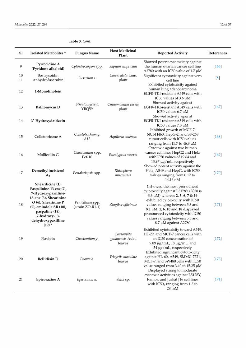
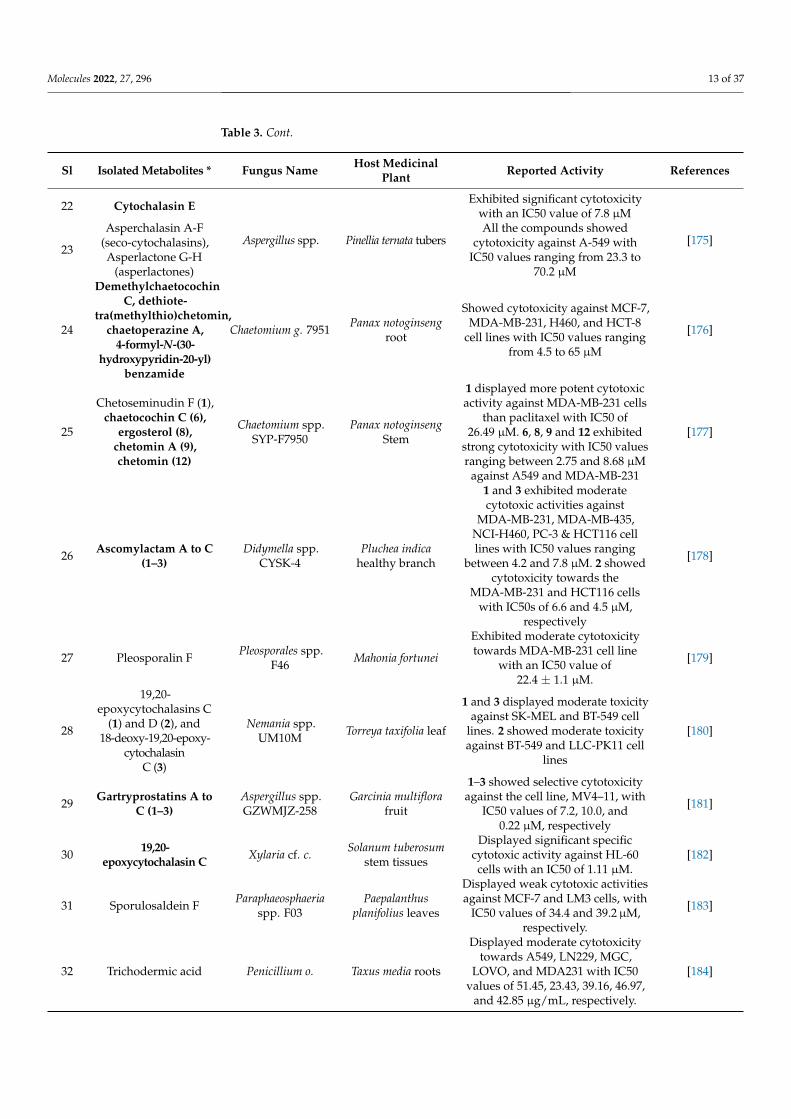
Table 3. Recently (2018–2020) reported potential cytotoxic metabolites isolated from medicinal-plant-associated endophytic fungi.
Sl Isolated Metabolites * Fungus Name Host MedicinalPlant Reported Activity References
1 Penicolinate A Bionectria spp. Raphia taedigeraDisplayed potent cytotoxicity
against cells with an IC50 value of4.1 µM.
[159]
2 Fusarithioamide B Fusarium c. Anvillea arcinia(Burm.f.) DC.
Showed selective and potent effecttowards BT-549, MCF-7, SKOV-3,and HCT-116 cell lines with IC50s
0.09, 0.21, 1.23, and 0.59 µM,respectively
[160]
33-(4-nitrophenyl)-5-
phenylisoxazole
Aspergillus n. spp.
Exhibited potent cytotoxic effect onHepG2 and SMCC-7721 cells with
the IC50 values were 0.347 and0.380 mM, respectively
[161]
4 Spiciferone F Phoma b. Kalidium foliatum(Pall.) Moq
Exhibited strong biological effectagainst MCF7 with a half-maximalinhibitory concentration value at
7.73 ± 0.11 µM
[162]
5 Xylariphthalide A Diaporthe spp. Tylophora ouata
Displayed cytotoxic activity againsthuman tumor cell lines BGC-823
cells with IC50 values of1.5 µmol·L−1
[163]
6 Cis-4-hydroxy-6-deoxytalone Diaporthe spp. Tylophora ouata
Displayed cytotoxic activity againsthuman tumor cell lines BGC-823
cells with IC50 8.6 µmol·L−1[163]
7 Xylarolide A Diaporthe spp. Datura inoxia
Showed promisingly inhibitedgrowth of MIAPaCa-2 and PC-3cells with an IC50 values of 20
14 µM
[164]
8 Jammosporin A Roselliniasanctae-cruciana Albizia lebbeck
Exhibited promising cytotoxicpotential against the human
leukemia cancer cell line (MOLT-4)[165]
Molecules 2022, 27, 296 12 of 37
Table 3. Cont.
Sl Isolated Metabolites * Fungus Name Host MedicinalPlant Reported Activity References
9 Pyrrocidine A(Pyridone alkaloid) Cylindrocarpon spp. Sapium ellipticum
Showed potent cytotoxicity againstthe human ovarian cancer cell line
A2780 with an IC50 value of 1.7 µM[166]
10 BostrycoidinFusarium s.
Cassia alata Linn.plant
Significant cytotoxicity against verocell line
[8]11 Anhydrofusarubin
12 1-Monolinolein
Streptomyces c.YBQ59
Cinnamomum cassiaplant
Exhibited cytotoxicity againsthuman lung adenocarcinoma
EGFR-TKI-resistant A549 cells withIC50 values of 3.6 µM
[167]13 Bafilomycin DShowed activity against
EGFR-TKI-resistant A549 cells withIC50 values 6.7 µM
14 3′-HydroxydaidzeinShowed activity against
EGFR-TKI-resistant A549 cells withIC50 values 7.8 µM
15 Colletotricone A Colletotrichum g.A12 Aquilaria sinensis
Inhibited growth of MCF-7,NCI-H460, HepG-2, and SF-268
tumor cells with IC50 valuesranging from 15.7 to 46.8 µM
[168]
16 Mollicellin G Chaetomium spp.Eef-10 Eucalyptus exserta
Cytotoxic against two humancancer cell lines HepG2 and Hela
withIC50 values of 19.64 and13.97 µg/mL, respectively
[169]
17 DemethylincisterolA3
Pestalotiopsis spp. Rhizophoramucronata
Showed potent activity against theHela, A549 and HepG, with IC50
values ranging from 0.17 to14.16 nM
[170]
18
Shearilicine (1),Paspalinine-13-ene (2),7-Hydroxypaxilline-
13-ene (3), ShearinineO (6), Shearinine P
(7), emindole SB (10),paspaline (18),7-hydroxy-13-
dehydroxypaxilline(19) *
Penicillium spp.(strain ZO-R1-1) Zingiber officinale
1 showed the most pronouncedcytotoxicity against L5178Y (IC50 is
3.6 µM) whereas 2, 3, 6, 7 & 19exhibited cytotoxicity with IC50values ranging between 5.3 and8.1 µM. 1, 6, 10 and 18 displayed
pronounced cytotoxicity with IC50values ranging between 5.3 and
8.7 µM against A2780
[171]
19 Flavipin Chaetomium g.Couroupita
guianensis Aubl.leaves
Exhibited cytotoxicity toward A549,HT-29, and MCF-7 cancer cells with
an IC50 concentration of9.89 µg/mL, 18 µg/mL, and
54 µg/mL, respectively
[172]
20 Bellidisin D Phoma b. Tricyrtis maculateleaves
Exhibited significant cytotoxicityagainst HL-60, A549, SMMC-7721,MCF-7, and SW480 cells with IC50value ranged from 3.40 to 15.25 µM
[173]
21 Epicorazine A Epicoccum n. Salix sp.
Displayed strong to moderatecytotoxic activities against L5178Y,
Ramos, and Jurkat J16 cell lineswith IC50s ranging from 1.3 to
28 mM
[174]
Molecules 2022, 27, 296 13 of 37
Table 3. Cont.
Sl Isolated Metabolites * Fungus Name Host MedicinalPlant Reported Activity References
22 Cytochalasin E
Aspergillus spp. Pinellia ternata tubers
Exhibited significant cytotoxicitywith an IC50 value of 7.8 µM
[175]23
Asperchalasin A-F(seco-cytochalasins),Asperlactone G-H
(asperlactones)
All the compounds showedcytotoxicity against A-549 with
IC50 values ranging from 23.3 to70.2 µM
24
DemethylchaetocochinC, dethiote-
tra(methylthio)chetomin,chaetoperazine A,
4-formyl-N-(30-hydroxypyridin-20-yl)
benzamide
Chaetomium g. 7951 Panax notoginsengroot
Showed cytotoxicity against MCF-7,MDA-MB-231, H460, and HCT-8
cell lines with IC50 values rangingfrom 4.5 to 65 µM
[176]
25
Chetoseminudin F (1),chaetocochin C (6),
ergosterol (8),chetomin A (9),chetomin (12)
Chaetomium spp.SYP-F7950
Panax notoginsengStem
1 displayed more potent cytotoxicactivity against MDA-MB-231 cells
than paclitaxel with IC50 of26.49 µM. 6, 8, 9 and 12 exhibited
strong cytotoxicity with IC50 valuesranging between 2.75 and 8.68 µM
against A549 and MDA-MB-231
[177]
26 Ascomylactam A to C(1–3)
Didymella spp.CYSK-4
Pluchea indicahealthy branch
1 and 3 exhibited moderatecytotoxic activities against
MDA-MB-231, MDA-MB-435,NCI-H460, PC-3 & HCT116 celllines with IC50 values ranging
between 4.2 and 7.8 µM. 2 showedcytotoxicity towards the
MDA-MB-231 and HCT116 cellswith IC50s of 6.6 and 4.5 µM,
respectively
[178]
27 Pleosporalin F Pleosporales spp.F46 Mahonia fortunei
Exhibited moderate cytotoxicitytowards MDA-MB-231 cell line
with an IC50 value of22.4 ± 1.1 µM.
[179]
28
19,20-epoxycytochalasins C
(1) and D (2), and18-deoxy-19,20-epoxy-
cytochalasinC (3)
Nemania spp.UM10M Torreya taxifolia leaf
1 and 3 displayed moderate toxicityagainst SK-MEL and BT-549 cell
lines. 2 showed moderate toxicityagainst BT-549 and LLC-PK11 cell
lines
[180]
29 Gartryprostatins A toC (1–3)
Aspergillus spp.GZWMJZ-258
Garcinia multiflorafruit
1–3 showed selective cytotoxicityagainst the cell line, MV4–11, with
IC50 values of 7.2, 10.0, and0.22 µM, respectively
[181]
30 19,20-epoxycytochalasin C Xylaria cf. c. Solanum tuberosum
stem tissues
Displayed significant specificcytotoxic activity against HL-60cells with an IC50 of 1.11 µM.
[182]
31 Sporulosaldein F Paraphaeosphaeriaspp. F03
Paepalanthusplanifolius leaves
Displayed weak cytotoxic activitiesagainst MCF-7 and LM3 cells, with
IC50 values of 34.4 and 39.2µM,respectively.
[183]
32 Trichodermic acid Penicillium o. Taxus media roots
Displayed moderate cytotoxicitytowards A549, LN229, MGC,
LOVO, and MDA231 with IC50values of 51.45, 23.43, 39.16, 46.97,
and 42.85 µg/mL, respectively.
[184]
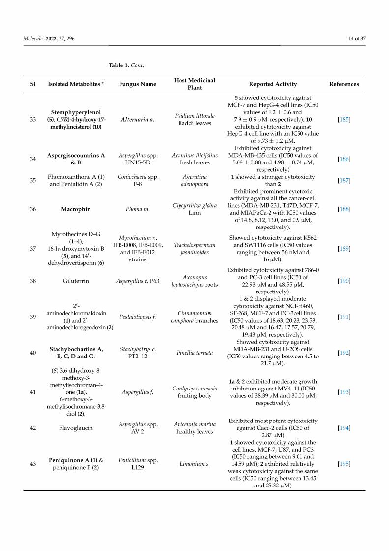
Molecules 2022, 27, 296 14 of 37
Table 3. Cont.
Sl Isolated Metabolites * Fungus Name Host MedicinalPlant Reported Activity References
33Stemphyperylenol
(5), (17R)-4-hydroxy-17-methylincisterol (10)
Alternaria a. Psidium littoraleRaddi leaves
5 showed cytotoxicity againstMCF-7 and HepG-4 cell lines (IC50
values of 4.2 ± 0.6 and7.9 ± 0.9 µM, respectively); 10exhibited cytotoxicity against
HepG-4 cell line with an IC50 valueof 9.73 ± 1.2 µM.
[185]
34 Aspergisocoumrins A& B
Aspergillus spp.HN15-5D
Acanthus ilicifoliusfresh leaves
Exhibited cytotoxicity againstMDA-MB-435 cells (IC50 values of
5.08 ± 0.88 and 4.98 ± 0.74 µM,respectively)
[186]
35 Phomoxanthone A (1)and Penialidin A (2)
Coniochaeta spp.F-8
Ageratinaadenophora
1 showed a stronger cytotoxicitythan 2 [187]
36 Macrophin Phoma m. Glycyrrhiza glabraLinn
Exhibited prominent cytotoxicactivity against all the cancer-cell
lines (MDA-MB-231, T47D, MCF-7,and MIAPaCa-2 with IC50 values
of 14.8, 8.12, 13.0, and 0.9 µM,respectively).
[188]
37
Myrothecines D–G(1–4),
16-hydroxymytoxin B(5), and 14′-
dehydrovertisporin (6)
Myrothecium r.,IFB-E008, IFB-E009,
and IFB-E012strains
Trachelospermumjasminoides
Showed cytotoxicity against K562and SW1116 cells (IC50 valuesranging between 56 nM and
16 µM).
[189]
38 Giluterrin Aspergillus t. P63 Axonopusleptostachyus roots
Exhibited cytotoxicity against 786-0and PC-3 cell lines (IC50 of
22.93 µM and 48.55 µM,respectively).
[190]
39
2′-aminodechloromaldoxin
(1) and 2′-aminodechlorogeodoxin (2)
Pestalotiopsis f. Cinnamomumcamphora branches
1 & 2 displayed moderatecytotoxicity against NCI-H460,
SF-268, MCF-7 and PC-3cell lines(IC50 values of 18.63, 20.23, 23.53,20.48 µM and 16.47, 17.57, 20.79,
19.43 µM, respectively).
[191]
40 Stachybochartins A,B, C, D and G.
Stachybotrys c.PT2–12 Pinellia ternata
Showed cytotoxicity againstMDA-MB-231 and U-2OS cells
(IC50 values ranging between 4.5 to21.7 µM).
[192]
41
(S)-3,6-dihydroxy-8-methoxy-3-
methylisochroman-4-one (1a),
6-methoxy-3-methylisochromane-3,8-
diol (2).
Aspergillus f. Cordyceps sinensisfruiting body
1a & 2 exhibited moderate growthinhibition against MV4–11 (IC50values of 38.39 µM and 30.00 µM,
respectively).
[193]
42 Flavoglaucin Aspergillus spp.AV-2
Avicennia marinahealthy leaves
Exhibited most potent cytotoxicityagainst Caco-2 cells (IC50 of
2.87 µM)[194]
43 Peniquinone A (1) &peniquinone B (2)
Penicillium spp.L129 Limonium s.
1 showed cytotoxicity against thecell lines, MCF-7, U87, and PC3(IC50 ranging between 9.01 and14.59 µM); 2 exhibited relatively
weak cytotoxicity against the samecells (IC50 ranging between 13.45
and 25.32 µM)
[195]
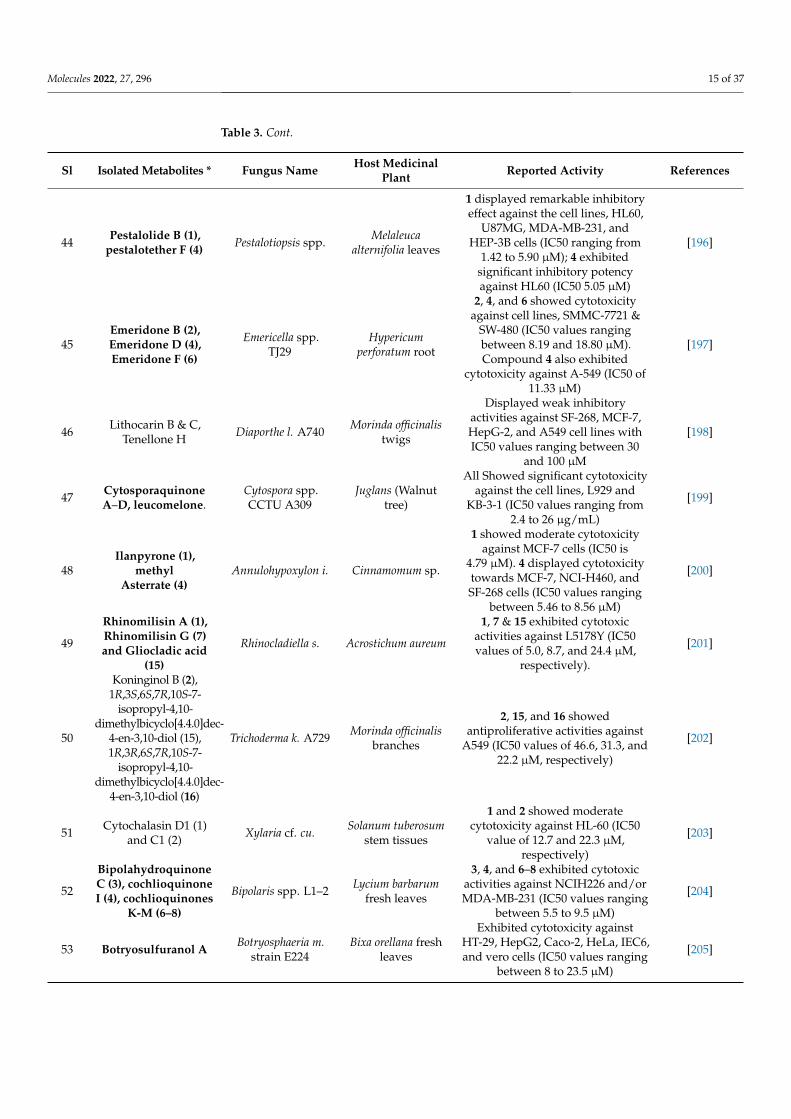
Molecules 2022, 27, 296 15 of 37
Table 3. Cont.
Sl Isolated Metabolites * Fungus Name Host MedicinalPlant Reported Activity References
44 Pestalolide B (1),pestalotether F (4) Pestalotiopsis spp. Melaleuca
alternifolia leaves
1 displayed remarkable inhibitoryeffect against the cell lines, HL60,
U87MG, MDA-MB-231, andHEP-3B cells (IC50 ranging from
1.42 to 5.90 µM); 4 exhibitedsignificant inhibitory potencyagainst HL60 (IC50 5.05 µM)
[196]
45Emeridone B (2),Emeridone D (4),Emeridone F (6)
Emericella spp.TJ29
Hypericumperforatum root
2, 4, and 6 showed cytotoxicityagainst cell lines, SMMC-7721 &
SW-480 (IC50 values rangingbetween 8.19 and 18.80 µM).Compound 4 also exhibited
cytotoxicity against A-549 (IC50 of11.33 µM)
[197]
46 Lithocarin B & C,Tenellone H Diaporthe l. A740 Morinda officinalis
twigs
Displayed weak inhibitoryactivities against SF-268, MCF-7,HepG-2, and A549 cell lines withIC50 values ranging between 30
and 100 µM
[198]
47 CytosporaquinoneA–D, leucomelone.
Cytospora spp.CCTU A309
Juglans (Walnuttree)
All Showed significant cytotoxicityagainst the cell lines, L929 and
KB-3-1 (IC50 values ranging from2.4 to 26 µg/mL)
[199]
48Ilanpyrone (1),
methylAsterrate (4)
Annulohypoxylon i. Cinnamomum sp.
1 showed moderate cytotoxicityagainst MCF-7 cells (IC50 is
4.79 µM). 4 displayed cytotoxicitytowards MCF-7, NCI-H460, andSF-268 cells (IC50 values ranging
between 5.46 to 8.56 µM)
[200]
49
Rhinomilisin A (1),Rhinomilisin G (7)and Gliocladic acid
(15)
Rhinocladiella s. Acrostichum aureum
1, 7 & 15 exhibited cytotoxicactivities against L5178Y (IC50values of 5.0, 8.7, and 24.4 µM,
respectively).
[201]
50
Koninginol B (2),1R,3S,6S,7R,10S-7-
isopropyl-4,10-dimethylbicyclo[4.4.0]dec-
4-en-3,10-diol (15),1R,3R,6S,7R,10S-7-
isopropyl-4,10-dimethylbicyclo[4.4.0]dec-
4-en-3,10-diol (16)
Trichoderma k. A729 Morinda officinalisbranches
2, 15, and 16 showedantiproliferative activities against
A549 (IC50 values of 46.6, 31.3, and22.2 µM, respectively)
[202]
51 Cytochalasin D1 (1)and C1 (2) Xylaria cf. cu. Solanum tuberosum
stem tissues
1 and 2 showed moderatecytotoxicity against HL-60 (IC50
value of 12.7 and 22.3 µM,respectively)
[203]
52
BipolahydroquinoneC (3), cochlioquinoneI (4), cochlioquinones
K-M (6–8)
Bipolaris spp. L1–2 Lycium barbarumfresh leaves
3, 4, and 6–8 exhibited cytotoxicactivities against NCIH226 and/orMDA-MB-231 (IC50 values ranging
between 5.5 to 9.5 µM)
[204]
53 Botryosulfuranol A Botryosphaeria m.strain E224
Bixa orellana freshleaves
Exhibited cytotoxicity againstHT-29, HepG2, Caco-2, HeLa, IEC6,and vero cells (IC50 values ranging
between 8 to 23.5 µM)
[205]
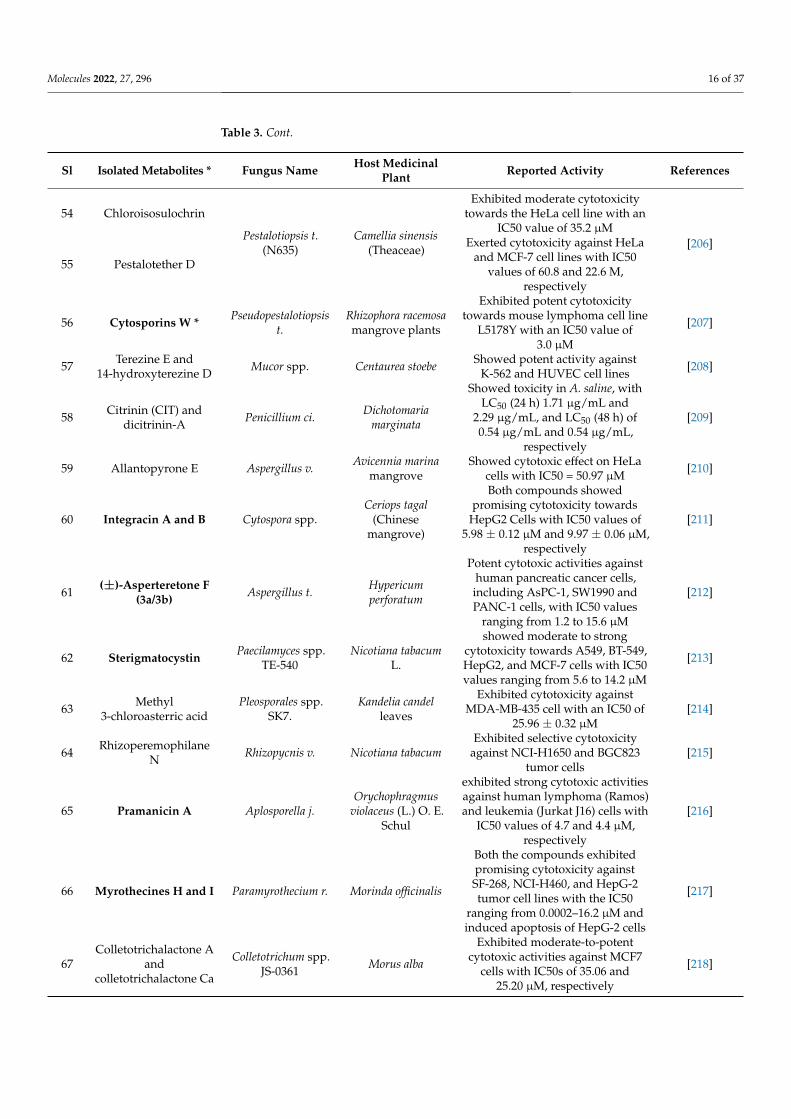
Molecules 2022, 27, 296 16 of 37
Table 3. Cont.
Sl Isolated Metabolites * Fungus Name Host MedicinalPlant Reported Activity References
54 Chloroisosulochrin
Pestalotiopsis t.(N635)
Camellia sinensis(Theaceae)
Exhibited moderate cytotoxicitytowards the HeLa cell line with an
IC50 value of 35.2 µM[206]
55 Pestalotether D
Exerted cytotoxicity against HeLaand MCF-7 cell lines with IC50
values of 60.8 and 22.6 M,respectively
56 Cytosporins W * Pseudopestalotiopsist.
Rhizophora racemosamangrove plants
Exhibited potent cytotoxicitytowards mouse lymphoma cell line
L5178Y with an IC50 value of3.0 µM
[207]
57 Terezine E and14-hydroxyterezine D Mucor spp. Centaurea stoebe Showed potent activity against
K-562 and HUVEC cell lines [208]
58 Citrinin (CIT) anddicitrinin-A Penicillium ci. Dichotomaria
marginata
Showed toxicity in A. saline, withLC50 (24 h) 1.71 µg/mL and
2.29 µg/mL, and LC50 (48 h) of0.54 µg/mL and 0.54 µg/mL,
respectively
[209]
59 Allantopyrone E Aspergillus v. Avicennia marinamangrove
Showed cytotoxic effect on HeLacells with IC50 = 50.97 µM [210]
60 Integracin A and B Cytospora spp.Ceriops tagal
(Chinesemangrove)
Both compounds showedpromising cytotoxicity towards
HepG2 Cells with IC50 values of5.98 ± 0.12 µM and 9.97 ± 0.06 µM,
respectively
[211]
61 (±)-Asperteretone F(3a/3b) Aspergillus t. Hypericum
perforatum
Potent cytotoxic activities againsthuman pancreatic cancer cells,including AsPC-1, SW1990 andPANC-1 cells, with IC50 values
ranging from 1.2 to 15.6 µM
[212]
62 Sterigmatocystin Paecilamyces spp.TE-540
Nicotiana tabacumL.
showed moderate to strongcytotoxicity towards A549, BT-549,HepG2, and MCF-7 cells with IC50values ranging from 5.6 to 14.2 µM
[213]
63 Methyl3-chloroasterric acid
Pleosporales spp.SK7.
Kandelia candelleaves
Exhibited cytotoxicity againstMDA-MB-435 cell with an IC50 of
25.96 ± 0.32 µM[214]
64 RhizoperemophilaneN Rhizopycnis v. Nicotiana tabacum
Exhibited selective cytotoxicityagainst NCI-H1650 and BGC823
tumor cells[215]
65 Pramanicin A Aplosporella j.Orychophragmus
violaceus (L.) O. E.Schul
exhibited strong cytotoxic activitiesagainst human lymphoma (Ramos)and leukemia (Jurkat J16) cells with
IC50 values of 4.7 and 4.4 µM,respectively
[216]
66 Myrothecines H and I Paramyrothecium r. Morinda officinalis
Both the compounds exhibitedpromising cytotoxicity againstSF-268, NCI-H460, and HepG-2tumor cell lines with the IC50
ranging from 0.0002–16.2 µM andinduced apoptosis of HepG-2 cells
[217]
67Colletotrichalactone A
andcolletotrichalactone Ca
Colletotrichum spp.JS-0361 Morus alba
Exhibited moderate-to-potentcytotoxic activities against MCF7
cells with IC50s of 35.06 and25.20 µM, respectively
[218]
Molecules 2022, 27, 296 17 of 37
Table 3. Cont.
Sl Isolated Metabolites * Fungus Name Host MedicinalPlant Reported Activity References
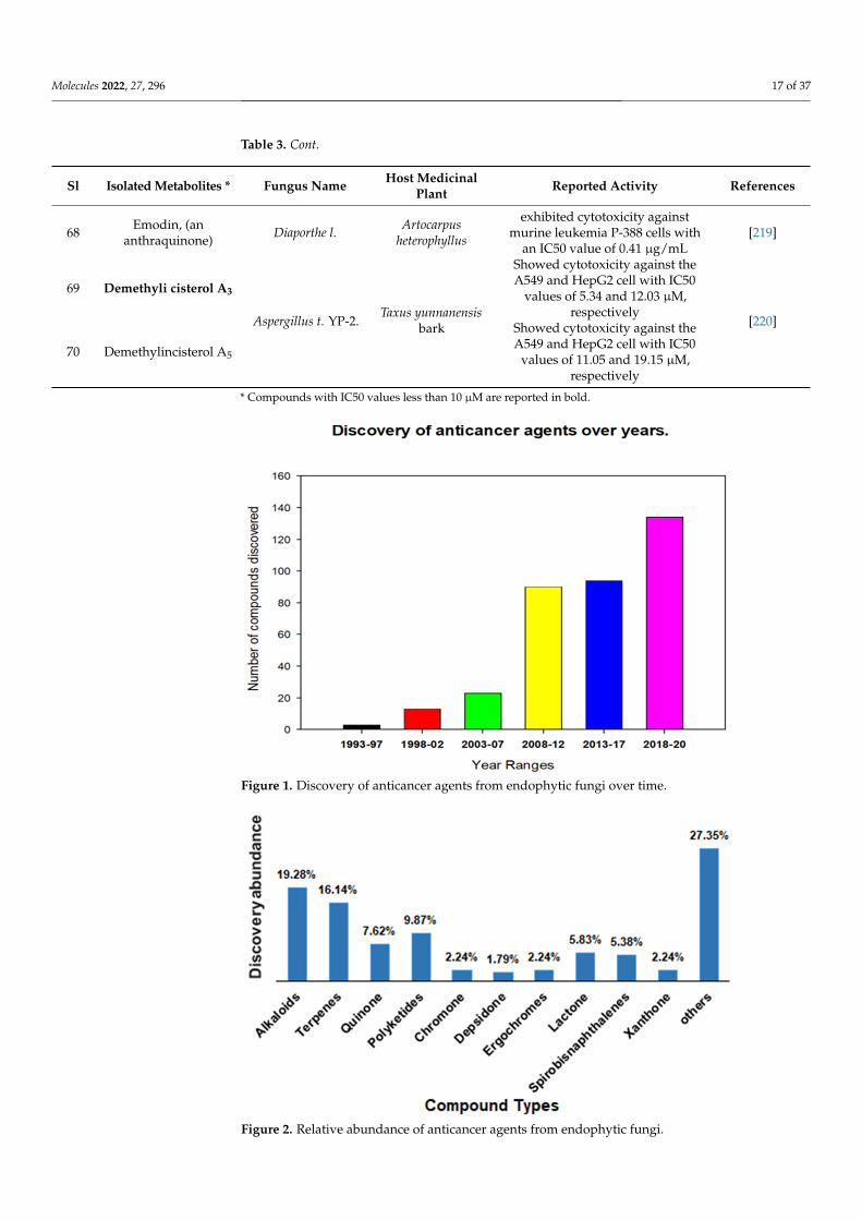
68 Emodin, (ananthraquinone) Diaporthe l. Artocarpus
heterophyllus
exhibited cytotoxicity againstmurine leukemia P-388 cells with
an IC50 value of 0.41 µg/mL[219]
69 Demethyli cisterol A3
Aspergillus t. YP-2.Taxus yunnanensis
bark
Showed cytotoxicity against theA549 and HepG2 cell with IC50
values of 5.34 and 12.03 µM,respectively
[220]
70 Demethylincisterol A5
Showed cytotoxicity against theA549 and HepG2 cell with IC50
values of 11.05 and 19.15 µM,respectively
* Compounds with IC50 values less than 10 µM are reported in bold.
Molecules 2022, 27, x FOR PEER REVIEW 17 of 40
64 Rhizoperemoph‐
ilane N Rhizopycnis v.
Nicotiana taba‐
cum
Exhibited selective cytotoxicity against NCI‐
H1650 and BGC823 tumor cells [215]
65 Pramanicin A Aplosporella j.
Orychophrag‐
mus violaceus
(L.) O. E. Schul
exhibited strong cytotoxic activities against hu‐
man lymphoma (Ramos) and leukemia (Jurkat
J16) cells with IC50 values of 4.7 and 4.4 μM, re‐
spectively
[216]
66 Myrothecines H
and I
Paramyrothecium
r.
Morinda offici‐
nalis
Both the compounds exhibited promising cyto‐
toxicity against SF‐268, NCI‐H460, and HepG‐2
tumor cell lines with the IC50 ranging from
0.0002–16.2 μM and induced apoptosis of HepG‐
2 cells
[217]
67
Colletotricha‐
lactone A and
colletotricha‐
lactone Ca
Colletotrichum
spp. JS‐0361 Morus alba
Exhibited moderate‐to‐potent cytotoxic activities
against MCF7 cells with IC50s of 35.06 and 25.20
μM, respectively
[218]
68 Emodin, (an an‐
thraquinone) Diaporthe l.
Artocarpus het‐
erophyllus
exhibited cytotoxicity against murine leukemia
P‐388 cells with an IC50 value of 0.41 μg/mL [219]
69 Demethyli cis‐
terol A3 Aspergillus t. YP‐
2.
Taxus yun‐
nanensis bark
Showed cytotoxicity against the A549 and
HepG2 cell with IC50 values of 5.34 and 12.03
μM, respectively [220]
70 Demethylincis‐
terol A5
Showed cytotoxicity against the A549 and
HepG2 cell with IC50 values of 11.05 and 19.15
μM, respectively
* Compounds with IC50 values less than 10 μM are reported in bold.
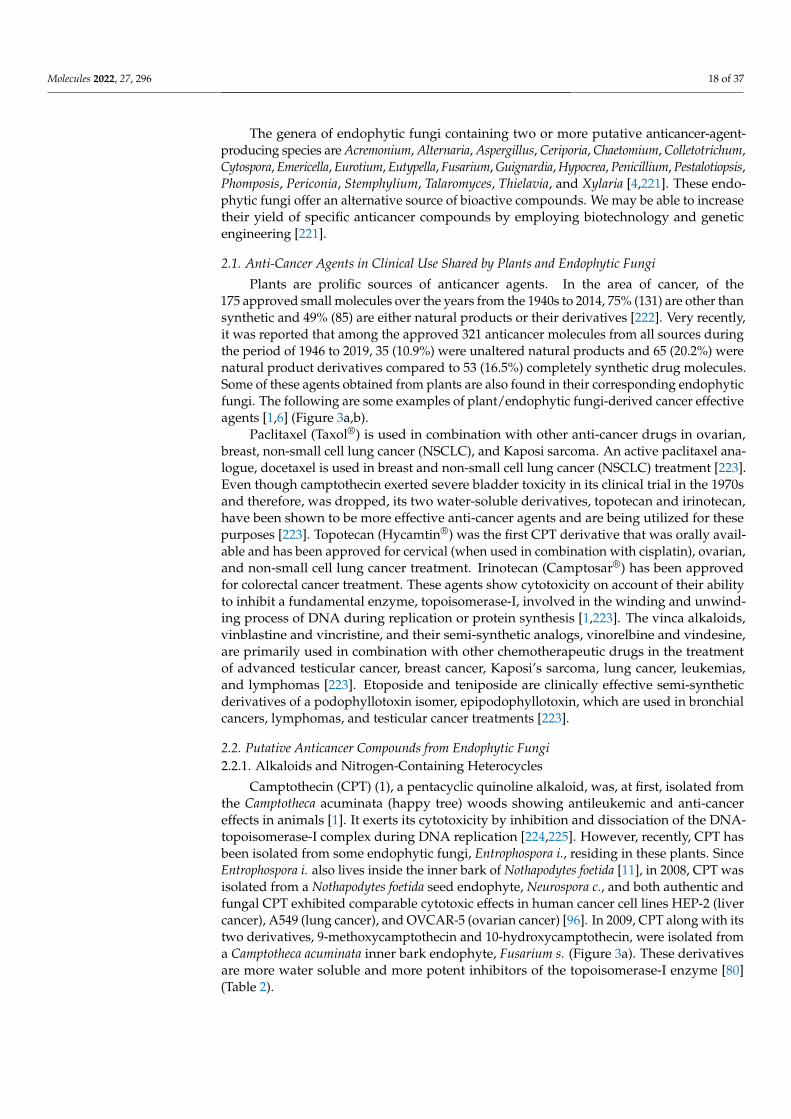
Figure 1. Discovery of anticancer agents from endophytic fungi over time.
Figure 1. Discovery of anticancer agents from endophytic fungi over time.
Molecules 2022, 27, x FOR PEER REVIEW 18 of 40
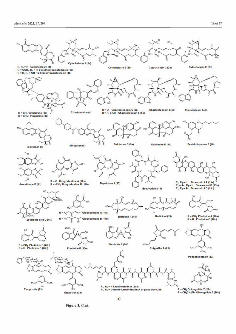
Figure 2. Relative abundance of anticancer agents from endophytic fungi.
The genera of endophytic fungi containing two or more putative anticancer‐agent‐
producing species are Acremonium, Alternaria, Aspergillus, Ceriporia, Chaetomium, Colleto‐
trichum, Cytospora, Emericella, Eurotium, Eutypella, Fusarium, Guignardia, Hypocrea, Penicil‐
lium, Pestalotiopsis, Phomposis, Periconia, Stemphylium, Talaromyces, Thielavia, and Xylaria
[4,221]. These endophytic fungi offer an alternative source of bioactive compounds. We
may be able to increase their yield of specific anticancer compounds by employing bio‐
technology and genetic engineering [221].
Figure 2. Relative abundance of anticancer agents from endophytic fungi.
Molecules 2022, 27, 296 18 of 37
The genera of endophytic fungi containing two or more putative anticancer-agent-producing species are Acremonium, Alternaria, Aspergillus, Ceriporia, Chaetomium, Colletotrichum,Cytospora, Emericella, Eurotium, Eutypella, Fusarium, Guignardia, Hypocrea, Penicillium, Pestalotiopsis,Phomposis, Periconia, Stemphylium, Talaromyces, Thielavia, and Xylaria [4,221]. These endo-phytic fungi offer an alternative source of bioactive compounds. We may be able to increasetheir yield of specific anticancer compounds by employing biotechnology and geneticengineering [221].
2.1. Anti-Cancer Agents in Clinical Use Shared by Plants and Endophytic Fungi
Plants are prolific sources of anticancer agents. In the area of cancer, of the175 approved small molecules over the years from the 1940s to 2014, 75% (131) are other thansynthetic and 49% (85) are either natural products or their derivatives [222]. Very recently,it was reported that among the approved 321 anticancer molecules from all sources duringthe period of 1946 to 2019, 35 (10.9%) were unaltered natural products and 65 (20.2%) werenatural product derivatives compared to 53 (16.5%) completely synthetic drug molecules.Some of these agents obtained from plants are also found in their corresponding endophyticfungi. The following are some examples of plant/endophytic fungi-derived cancer effectiveagents [1,6] (Figure 3a,b).
Paclitaxel (Taxol®) is used in combination with other anti-cancer drugs in ovarian,breast, non-small cell lung cancer (NSCLC), and Kaposi sarcoma. An active paclitaxel ana-logue, docetaxel is used in breast and non-small cell lung cancer (NSCLC) treatment [223].Even though camptothecin exerted severe bladder toxicity in its clinical trial in the 1970sand therefore, was dropped, its two water-soluble derivatives, topotecan and irinotecan,have been shown to be more effective anti-cancer agents and are being utilized for thesepurposes [223]. Topotecan (Hycamtin®) was the first CPT derivative that was orally avail-able and has been approved for cervical (when used in combination with cisplatin), ovarian,and non-small cell lung cancer treatment. Irinotecan (Camptosar®) has been approvedfor colorectal cancer treatment. These agents show cytotoxicity on account of their abilityto inhibit a fundamental enzyme, topoisomerase-I, involved in the winding and unwind-ing process of DNA during replication or protein synthesis [1,223]. The vinca alkaloids,vinblastine and vincristine, and their semi-synthetic analogs, vinorelbine and vindesine,are primarily used in combination with other chemotherapeutic drugs in the treatmentof advanced testicular cancer, breast cancer, Kaposi’s sarcoma, lung cancer, leukemias,and lymphomas [223]. Etoposide and teniposide are clinically effective semi-syntheticderivatives of a podophyllotoxin isomer, epipodophyllotoxin, which are used in bronchialcancers, lymphomas, and testicular cancer treatments [223].
2.2. Putative Anticancer Compounds from Endophytic Fungi2.2.1. Alkaloids and Nitrogen-Containing Heterocycles
Camptothecin (CPT) (1), a pentacyclic quinoline alkaloid, was, at first, isolated fromthe Camptotheca acuminata (happy tree) woods showing antileukemic and anti-cancereffects in animals [1]. It exerts its cytotoxicity by inhibition and dissociation of the DNA-topoisomerase-I complex during DNA replication [224,225]. However, recently, CPT hasbeen isolated from some endophytic fungi, Entrophospora i., residing in these plants. SinceEntrophospora i. also lives inside the inner bark of Nothapodytes foetida [11], in 2008, CPT wasisolated from a Nothapodytes foetida seed endophyte, Neurospora c., and both authentic andfungal CPT exhibited comparable cytotoxic effects in human cancer cell lines HEP-2 (livercancer), A549 (lung cancer), and OVCAR-5 (ovarian cancer) [96]. In 2009, CPT along with itstwo derivatives, 9-methoxycamptothecin and 10-hydroxycamptothecin, were isolated froma Camptotheca acuminata inner bark endophyte, Fusarium s. (Figure 3a). These derivativesare more water soluble and more potent inhibitors of the topoisomerase-I enzyme [80](Table 2).
Molecules 2022, 27, 296 19 of 37Molecules 2022, 27, x FOR PEER REVIEW 20 of 40
Figure 3. Cont.
Molecules 2022, 27, 296 20 of 37Molecules 2022, 27, x FOR PEER REVIEW 21 of 40
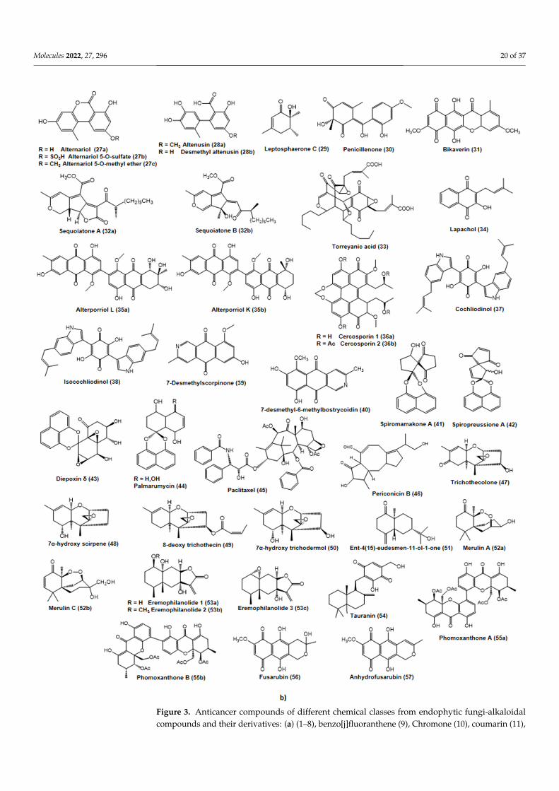
Figure 3. Anticancer compounds of different chemical classes from endophytic fungi-alkaloidalcompounds and their derivatives: (a) (1–8), benzo[j]fluoranthene (9), Chromone (10), coumarin (11),
Molecules 2022, 27, 296 21 of 37
depsidones (12, 13), depsideptide (14), ergochromes (15, 16), ester (17), lactones (18–22), lignans(23–24), peptide (25), polykedites (26); (b) polyketides (27–32), quinones (33–39), spirobisnaphthalenes(40–42), terpenes (43–54), xanthones (55), naphthoquinones (56, 57).
Cytochalasins (2a–2d) are fungal metabolites that inhibit cell division by means ofinhibiting actin filament polymerization [226]. Four cytochalasins (cytochalasin 1, 2,3, and E) have been isolated from an endophytic fungus, Rhinocladiella spp. from theTripterygium wilfordii dead tree limbs and were tested against HCT-116 (colon tumor cellline), A2780S (ovarian tumor cell line), and SW-620 (colon tumor cell line) showing cytotoxicactivities [136].
The vinca alkaloid (3a, 3b), vincristine (leurocristine), was isolated fromCatharanthus roseus [227]. This alkaloid has also been isolated from some fungal endo-phytes of Catharanthus roseus such as Fusarium o. (inner bark), Mycelia s. 97CY(3) (Leaves),and Talaromyces r. CrP20 (Leaves) [74,75,89]. Vincristine irreversibly binds to the spindleproteins and microtubules during the S-phase of cell cycle hampering mitotic spindleformation and therefore arresting tumor cell division in the metaphase [1].
Chaetominine (4) was isolated from an endophyte, Chaetomium sp. IFB-E015 from thehealthy leaves of Adenophora axilliflora, and it was cytotoxic against K562 (human leukemiacells) and SW1116 (human colon cancer cells) [54].
Cytochalasan-based alkaloids (5a–5c, 6), namely chaetoglobosin C, E, F, U, andpenochalasin A (6), were obtained from the endophyte Chaetomium g. IFB-E019 resid-ing inside the Imperata cylindrica healthy stem. Chaetoglobosin U was cytotoxically activeagainst the KB cell line (human nasopharyngeal epidermoid tumor) with an IC50 value of16.0 µM, whereas chaetoglobosin C (IC50 34.0 µM), E (IC50 40.0 µM), F (IC50 52.0 µM), andpenochalasin A (IC50 48.0 µM) were moderately active against the KB cell line [57]. Endo-phytic fungus Chaetomium g. L18 from the plant Curcuma wenyujin produces chaetoglobosinX that exerted cytotoxic activity against H22 (hepatic cancer cells in mice) and MFC (gastriccancer cells in mice) cell lines [56] (Table 2).
2.2.2. Benzo[j]fluoranthenes
Daldinone C (9a) and D (9b) were discovered from an Artemisia Artemisia annua en-dophyte, Hypoxylon t. IFB-18, where both agents exerted strong cytotoxic action againstthe human colorectal cancer SW1116 cell line at IC50 values of 49.5 and 41.0 µM, respec-tively [85] (Table 2, Figure 3a).
2.2.3. Chromones
A novel chromone, Pestalotiopsone F (10), was isolated from an endophytic fungusPestalotiopsis spp. associated with a mangrove plant Rhizophora mucronata. PestalotiopsoneF showed moderate cytotoxicity to L5178Y (murine cancer cell line) at an EC50 value of8.93 µg/mL [110]. Pestaloficiol I, J, K, and L are new isoprenylated chromone derivativesdiscovered from a Camellia sinensis endophyte, Pestalotiopsis f., that displayed cytotoxicityagainst HeLa (Cervical cancer) and MCF-7 (Breast cancer) cell lines [115] (Table 2).
2.2.4. Coumarins
Arundinone B (11) was isolated from an endophyte Microsphaeropsis a. associated withUlmus macrocarpa. The compound showed cytotoxicity to T24 (Bladder carcinoma) andA549 (Lung carcinoma epithelial) cell lines [92] (Table 2).
2.2.5. Depsidones
Botryorhodines A (12a) and B (12b), two depsidones, were isolated from the endo-phytic fungus Botryosphaeria r. associated with Bidens pilosa. These compounds exhibitedweak antitumor activity against the HeLa cell line at a concentration of 96.97 and 36.41 µM,respectivel [48]. Depsidone 1 was discovered from a fungus of the Pleosporales order (BCC8616) isolated from an unidentified plant leaf of the Hala-Bala forest origin. Depsidone 1
Molecules 2022, 27, 296 22 of 37
displayed weak cytotoxicity to KB and BC cell lines with IC50 values 6.5 and 4.1µg/mL,respectively [43] (Table 2).
2.2.6. Depsipeptides
Beauvericin (14), a depsipeptide, was isolated from two fungi, Fusarium o. EPH2RAAand Fusarium o., associated with the plants Cylindropuntia echinocarpus and Ephedra fasciculate,respectively. Beauvericin displayed cytotoxicity to NCI-H460 (human non-small cell lungcancer), MIA Pa Ca-2 (human pancreatic carcinoma), MCF-7 (human breast cancer), andSF-268 (human CNS cancer) cell lines with IC50 values of 1.41, 1.66, 1.81, and 2.29 µM,respectively, showing selective cytotoxicity toward MIA PaCa-2 and NCI-H460 (Table 2).Beauvericin also inhibited the metastasis of MDA-MB-231 (Breast cancer) and PC-3M(metastatic prostate cancer) cells at concentrations ranging between 3.0–4.0 and 2.0–2.5 µM,respectively [77]. According to other studies, beauvericin displayed cytotoxicity againstA549 (Lung carcinoma epithelial), PC-3 (Prostate cancer), and PANC-1 (human pancre-atic carcinoma) cell lines with IC50 values of 10.4 ± 1.6, 49.5 ± 3.8, and 47.2 ± 2.9 µM,respectively [71]. Additionally, in 2006, Ivanova et al. demonstrated the cytotoxicity ofbeauvericin against Hep-G2 (hepatocellular carcinoma) and MRC-5 (fibroblast-like fetallung cell line) cells as well [76].
2.2.7. Ergochromes
Phomopsis l., an endophytic fungus of Dicerandra frutescens, produced three compoundsdicerandrols A, B, and C (15a–15c), structurally related to the ergochromes and secalonicacids as they also have the same tricyclic C15 system with a similar arrangement of sub-stituents. These compounds displayed modest antitumor activities toward A549 (lungadenocarcinoma epithelial cell line) and HCT-116 (colon tumor cell line) cell lines [132](Table 2).
Secalonic acid D (16), isolated from mangrove plant endophytic fungus no. ZSU44,displayed potent cytotoxicity against HL60 (the human promyelocytic leukemia cell line)and K562 (human leukemia cells) cells with IC50 values of 0.38 and 0.43 µM, respectively.It caused apoptosis in those cell lines and cell cycle arrest in the G(1) phase as well [158].
2.2.8. Esters
Globosumones A (17a) and B (17b), isolated from the endophyte Chaetomium g. asso-ciated with Ephedra fasciculate, were shown to have cytotoxicity to MCF-7 (breast cancer),MIA PaCa-2 (pancreatic carcinoma), NCI-H460 (non-small cell lung cancer), SF-268 (CNSglioma), and WI-38 (normal human fibroblast cells) cell lines [58].
2.2.9. Lactones
The lactone compound Brefeldin A (18) was obtained from two endophytic fungi,Aspergillus c. and Paecilomyces spp., isolated from the plants Taxus mairei and Torreya grandis.Brefeldin A exhibited antitumor activities to Hela, HL-60, KB, MCF-7, and Spc-A-1 withIC50 values of 1.8, 10.0, 9.0, 2.0, and 1.0 ng/mL [31]. Brefeldin A was also obtained fromthe endophyte Acremonium spp. isolated from the healthy Knema laurina twig. It showedcytotoxicity to BC-1 (breast cancer), KB (epidermoid cancer of the mouth), and NCIH187(human small-cell lung cancer), with IC50 values of 0.04, 0.18, and 0.11 µM, respectively [86](Table 2).
Radicicol (19) was obtained from Chaetomium c. associated with Ephedra fasciculate andit is a HSP90 (heat shock protein) inhibitor, which is frequently expressed highly in cancercells. It also showed cytotoxicity to the MCF-7 (breast cancer) cell line at an IC50 value0.03 µM [55].
Photinides A–F (20a–20f) were obtained from the endophyte Pestalotiopsis p. associatedwith Roystonea regia, and all of these γ-lactones at 10 µg/mL exerted cytotoxicity againstthe MDA-MB-231 (breast cancer) cell line with inhibitory rates of 24.4, 24.2, 23.1, 24.4, and24.6%, respectively [123] (Table 2).
Molecules 2022, 27, 296 23 of 37
Eutypellin A (21), isolated from the endophyte Eutypella spp. BCC 13199 associatedwith Etlingera littoralis, showed cytotoxicity to KB, MCF-7NCI-H187 (human small-celllung cancer cells), and nonmalignant Vero cells with IC50 values of 38, 84, 12, and 88 µM,respectively [70].
2.2.10. Lignans
Podophyllotoxin (22), a precursor to the topoisomerase-I-inhibiting anticancer drugsteniposide (23), etoposide (24), and etoposide phosphate, were isolated from the endophytePhialocephala f. associated with Podophyllum peltatum [12]. This was also obtained from theendophyte Trametes h. associated with Podophyllum hexandrum and from the endophyteFusarium s. associated with Podophyllum hexandrum [1,79,148] (Table 2).
2.2.11. Peptides
Leucinostatin A was isolated from the endophyte Acremonium spp. associated withTaxus baccata and was shown to be effective against BT-20 (breast cancer) cell line withan LD50 value of 2 nM [14]. It inhibits the growth of prostate cancer cells through thesuppression of IGF-I (Insulin-Like Growth Factor-I) expression in PrSC (prostate stromalcells) [228] (Table 2).
2.2.12. Polyketides
Two novel oblongolides, Y (26a) and Z (26b) (Figure 3a), are produced by the endo-phyte Phomopsis spp. BCC 9789 housed in Musa acuminate (a wild banana). OblongolideY exhibited cytotoxicity against BC (human breast cancer) cell line (IC50 48 µM) andOblongolide Z showed cytotoxicity against BC (human breast cancer), KB (human oralepidermoid cancer), NCI-H187 (small-cell lung cancer), and nonmalignant (Vero) cell lineswith IC50 values of 26 µM, 37 µM, 32 µM, and 60 µM, respectively [130] (Table 2).
Five tricyclic lactone polyketides, alternariol (27a), alternariol 5-O-sulfate (27b), al-ternariol 5-O-methyl ether (27c), altenusin (28a), and desmethylaltenusin (28b) (Figure 3b),were isolated from the endophyte Alternaria spp. housed in the leaves of Polygonum senegalense.All these compounds manifested significant cytotoxicity against L5178Y (mouse lymphomacells) with EC50 values of 1.7, 4.5, 7.8, 6.8, and 6.2 µg/mL, respectively [16]. Accordingto another study conducted by Devari et al. in 2014, alternariol 5-O-methyl ether showedantiproliferative activity against HL-60 (human promyelocytic leukemia), A549 (lung can-cer), PC-3 (prostate cancer), HeLa (cervical cancer), A431 (skin carcinoma), MiaPaka-2(pancreatic cancer), and T47D (breast cancer) cell lines. Among all these cell lines, HL-60(human promyelocytic leukemia) cells were most sensitive (IC50 85 µM) to alternariol5-O-methyl ether [25].
Two novel polyketides, leptosphaerone C (29) and penicillenone (30), are producedby an endophytic fungus Penicillium spp. JP-1, isolated from Aegiceras corniculatum. Lep-tosphaerone C showed cytotoxicity to A549 (lung carcinoma epithelial) with an IC50 valueof 1.45 µM, and penicillenone exhibited activity against P388 (leukemia cells) with an IC50value of 1.38 µM [103].
Bikaverin (31) was isolated from an endophytic fungus Fusarium o. strain CECISassociated with Cylindropuntia echinocarpa [77]. It exerted cytotoxic activities against cancercell lines, MIA PaCa-2 (pancreatic carcinoma), NCI-H460 (non-small cell lung cancer),MCF-7 (human breast cancer), and SF-268 (human CNS cancer) with IC50 values of 0.26,0.43, 0.42, and 0.38 µM, respectively, showing selective cytotoxicity toward MIA PaCa-2and NCI-H460. Bikaverin was also proven to be cytotoxic against EAC (Erlich ascitescarcinoma), leukemia L5178, and sarcoma 37 cell lines affecting precursor utilization ofnucleic acid and protein synthesis [78].
Sequoiatone A (32a) and B (32b), two novel polyketides (Figure 3b), were isolatedfrom a Sequoia sempervirens bark endophyte, Aspergillus p. These polyketide compoundswere tested against 60 diverse human tumor cell lines, and among them, breast cancer celllines showed the greatest sensitivity [37] (Table 2).
Molecules 2022, 27, 296 24 of 37
2.2.13. Quinones
Torreyanic acid (33) (Figure 3b), a dimeric quinine, was isolated from an endophyteof Torreya taxifolia, Pestalotiopsis m. It causes cytotoxicity by apoptosis against A549 (lungcarcinoma epithelial) and NEC (human colorectal neuroendocrine cell carcinoma) cell lineswith IC50 values of 3.5 µg/mL and 45 µg/mL, respectively [119] (Table 2).
Four endophytes, Alternaria spp., Alternaria a., Aspergillus n., and Penicillium spp., asso-ciated with Tabebuia argentea, produced the antitumor and anti-metastatic agent lapachol(34) [17,20–22]. It acts by interfering with the bioactivities of the topoisomerase enzymes,which are crucial for DNA replication [22]. β-Lapachone showed activity on DU145 (humanprostate carcinoma) and MCF-7 (breast cancer cell line) cell lines [20,22]. Additionally, itsantitumor and anti-metastatic activities were evident in HepG2 (human hepatocellular livercarcinoma) and Hep3B (human hepatoma cell line) cell lines [19]. Notably, Aspergillus n.can be used to produce lapachol in a large scale within a short time [18].
Two bianthraquinone derivatives, Alterporriol K (35a) and L (35b), are produced by theendophytic fungus Alternaria spp. ZJ9-6B associated with the mangrove Aegiceras corniculatum.Alterporriol K and L exerted moderate cytotoxicity against MDA-MB-435 and MCF-7(breast cancer cell line) cell lines with IC50 values between 13.1 and 29.1 µM [24].
Cercosporin (36) was isolated from the endophytic fungus Mycosphaerella spp., associ-ated with Psychotria horizontalis, and exhibited cytotoxicity against MCF-7 [91].
Another endophytic fungus, isolated from the Salvia officinalis stem, was Chaetomiumspp., which produced the cytotoxically active agents, cochliodinol (37) and isocochliodinol(38) (Figure 3b). These compounds were tested against the L5178Y (mouse lymphomacells) cell line where cochliodinol showed higher cytotoxicity (EC50 7.0 µg/mL) thanisocochliodinol (EC50 71.5 µg/mL) [51] (Table 2).
Azaanthraquinones, 7-desmethylscorpinone (39), and 7-desmethyl-6-methylbostrycoidin(40) (Figure 3b) isolated form Fusarium s. showed cytotoxic activity against four humantumor cell lines, MDA MB 231, MIA PaCa2, HeLa, and NCI H1975 [229].
2.2.14. Spirobisnaphthalenes
Mycelia s., an endophytic fungus isolated from the leaves of Knightia excelsa, was shownto produce Spiromamakone A (41) (Figure 3b) that exhibited cytotoxicity to P388 (murineleukemia cell line) at an IC50 value 0.33 µM [90] (Table 2).
A novel spirobisnaphthalene, spiropreussione A (42), was isolated from the endophytePreussia spp. associated with Aquilaria sinensis. It displayed cytotoxicity to A2780 (humanovarian carcinoma) and BEL-7404 (human liver carcinoma) cell lines with IC50 values of2.4 and 3.0 µM, respectively [135].
Diepoxin δ (43), palmarumycin C8 (44), and diepoxins κ and ζ were isolated fromthe endophytic fungus Berkleasmium spp. associated with Dioscorea zingiberensis. Diepoxinδ and palmarumycin C8 displayed pronounced cytotoxicity to A-549, A-2780, Bel-7402,BGC-823, and HCT-8 cell lines with IC50 values between 1.28 and 5.83 µM, while diepoxinsκ and ζ selectively inhibited A-549 and Bel-7402 cells’ growth showing moderate to weakcytotoxicity [44] (Table 2).
2.2.15. Terpenes (Diterpenes, Sesquiterpenes, Triterpenes)
Several terpenes of plant and fungal origin have been established as potential an-ticancer drugs (Figure 3b, structures 45–54). Among these, paclitaxel (Taxol) (45) wasisolated from Taxus brevifolia (Pacific yew tree) [230,231]. However, due to less availabilityof the pacific yew tree and insignificant yield of this metabolite, scientist have set up otherapproaches, including tissue culture, chemical synthesis, and semi-synthesis [230,232].However, this diterpenoid was also reported to be produced by an endophytic fungus,Taxomyces a., isolated from the Taxus brevifolia [6]. Following this report, a number of pa-clitaxel producing other endophytes were reported. Some of them are Bartalinia r. fromthe leaves of Aegle marmelos [42] and Pestalotiopsis n. and Pestalotiopsis v. from the plant
Molecules 2022, 27, 296 25 of 37
Taxus cuspidate [73]. This metabolite has been found to induce apoptosis when screenedagainst INT-407, BT220, H116, HL251, and HLK210 cell lines [42] (Table 2).
A fusicoccane diterpene, periconicin B (46), was isolated from a Xylopia aromaticaendophyte, Periconia a. It exerted potent cytotoxicity against HeLa (cervical cancer) andCHO (Chinese hamster ovary) cell lines [109].
Four sesquiterpens, trichothecolone (47), 7α-hydroxy-scirpene (48), 8-deoxy-trichothecin(49), and 7α-hydroxytrichodermol (50), were isolated from an endophyte, KLAR 5, housedin the healthy twig of Knema laurina. Compounds 47 and 48 were moderately active againstBC-1 (human breast cancer cells), KB (Human nasopharyngeal epidermoid tumor), andNCI-H187 (human small-cell lung cancer cells), whereas compounds 49 and 50 showedselective cytotoxic activity against BC-1 and NCI-H187 [86].
Ent-4(15)-eudesmen-11-ol-1-one (51), an eudesmane sesquiterpene, isolated froman Etlingera littoralis endophyte, Eutypella spp. BCC 13199, showed weak cytotoxicityagainst KB, MCF7, NCI-H187, and Vero cells with IC50 values of 32, 20, 11, and 32 µM,respectively [70].
Two sesquiterpenes, Merulin A (52a) and Merulin C (52b), are produced by aXylocarpus granatum endophytic fungi, XG8D, where both of them showed significantcytotoxic activity against SW620 (colon cancer) and BT474 (breast cancer) cell lines withIC50 values of 4.84 and 4.11 µg/mL for SW620 and 4.98 and 1.57 µg/mL for BT474, respec-tively [151].
Three novel eremophilane-type sesquiterpenes (Figure 3b), eremophilanolides 1, 2,and 3 (53a–53c), were isolated from the endophytic fungi Xylaria spp. BCC 21097 of theLicuala spinose plant and were moderately cytotoxic against KB, MCF-7, and NCI-H187 celllines [152].
Tauranin (54) is produced by a Platycladus orientalis endophyte, Phyllosticta s., ex-hibiting cytotoxicity against MCF-7 (breast cancer), MIA Pa Ca-2 (pancreatic carcinoma),NCI-H460 (non-small cell lung cancer), PC-3 M (metastatic prostate cancer), and SF-268(CNS cancer- glioma) cell lines with IC50 values of 1.5, 2.8, 4.3, 3.5, and 1.8 µM, respec-tively [133] (Table 2).
2.2.16. Xanthones
Phomoxanthone A (55a) and B (55b) (Figure 3b), isolated from the endophytePhomopsis spp. BCC 1323 associated with Tectona grandis, exerted significant cytotoxic-ity against KB, BC-1, and nonmalignant Vero cells with IC50 values of 0.99, 0.51, and1.4 µg/mL, respectively, for phomoxanthone A and 4.1, 0.70, and 1.8 µg/mL, respectively,for phomoxanthone B [129] (Table 2).
2.3. Recently Reported Metabolites with Potential Cytotoxicity and the Case of Fusarubin
More than one hundred metabolites have been isolated and evaluated for putativeanticancer activities in the years 2018 to 2020. Cytotoxic activities of these endophyticmetabolites have been summarized in Table 3. Among the reported metabolites, penicoli-nate A isolated form Bionectria spp. [159] and pyrrocidine A isolated from Cylindrocarponspp. [166] exhibited potent cytotoxicity against against the human ovarian cancer cell lineA2780. Fusarithioamide B, a new type benzamide, isolated form Fusarium c., showed potentactivity against several cell lines [160]. 3-(4-nitrophenyl)-5-phenyl isoxazole was reportedto have a potent effect against HepG2 and SMCC-7721 cells [161], while spiciferone F wasreported to have a strong effect against MCF7 [162]. Liu et al. isolated two metabolies,namely xylariphthalide A and cis-4-hydroxy-6-deoxytalone, and Sharma V. et al. isolatedXylarolide A from Diaporthe spp. [163,164]. All these metabolites showed activity towardscancer cells. Three naphthaquinones, anhydrofusarubin, fusarubin, and 3-deoxyfusarubin,and one aza-anthraquinone, bostrycoidin, have potentiality as bioactive compounds againstcytotoxicity on vero cells. These metabolites were isolated from a Fusarium s. strain isolatedfrom Casia alata. [8]. Monolinolein, bafilomycin D, and 3′-hydroxydaidzein displayed astrong effect against A549 cells. These metabolites were isolated from actinomycete strain
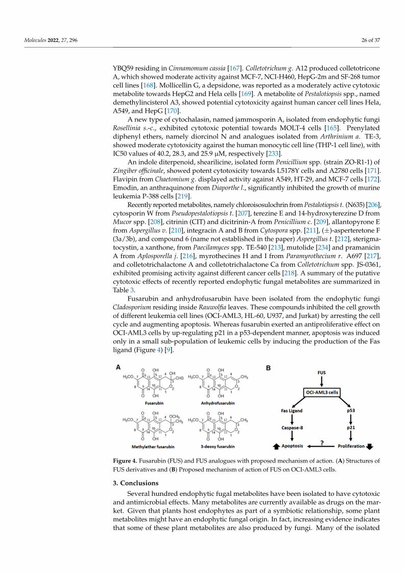
Molecules 2022, 27, 296 26 of 37
YBQ59 residing in Cinnamomum cassia [167]. Colletotrichum g. A12 produced colletotriconeA, which showed moderate activity against MCF-7, NCI-H460, HepG-2m and SF-268 tumorcell lines [168]. Mollicellin G, a depsidone, was reported as a moderately active cytotoxicmetabolite towards HepG2 and Hela cells [169]. A metabolite of Pestalotiopsis spp., nameddemethylincisterol A3, showed potential cytotoxicity against human cancer cell lines Hela,A549, and HepG [170].
A new type of cytochalasin, named jammosporin A, isolated from endophytic fungiRosellinia s.-c., exhibited cytotoxic potential towards MOLT-4 cells [165]. Prenylateddiphenyl ethers, namely diorcinol N and analogues isolated from Arthrinium a. TE-3,showed moderate cytotoxicity against the human monocytic cell line (THP-1 cell line), withIC50 values of 40.2, 28.3, and 25.9 µM, respectively [233].
An indole diterpenoid, shearilicine, isolated form Penicillium spp. (strain ZO-R1-1) ofZingiber officinale, showed potent cytotoxicity towards L5178Y cells and A2780 cells [171].Flavipin from Chaetomium g. displayed activity against A549, HT-29, and MCF-7 cells [172].Emodin, an anthraquinone from Diaporthe l., significantly inhibited the growth of murineleukemia P-388 cells [219].
Recently reported metabolites, namely chloroisosulochrin from Pestalotiopsis t. (N635) [206],cytosporin W from Pseudopestalotiopsis t. [207], terezine E and 14-hydroxyterezine D fromMucor spp. [208], citrinin (CIT) and dicitrinin-A from Penicillium c. [209], allantopyrone Efrom Aspergillus v. [210], integracin A and B from Cytospora spp. [211], (±)-asperteretone F(3a/3b), and compound 6 (name not established in the paper) Aspergillus t. [212], sterigma-tocystin, a xanthone, from Paecilamyces spp. TE-540 [213], mutolide [234] and pramanicinA from Aplosporella j. [216], myrothecines H and I from Paramyrothecium r. A697 [217],and colletotrichalactone A and colletotrichalactone Ca from Colletotrichum spp. JS-0361,exhibited promising activity against different cancer cells [218]. A summary of the putativecytotoxic effects of recently reported endophytic fungal metabolites are summarized inTable 3.
Fusarubin and anhydrofusarubin have been isolated from the endophytic fungiCladosporium residing inside Rauwolfia leaves. These compounds inhibited the cell growthof different leukemia cell lines (OCI-AML3, HL-60, U937, and Jurkat) by arresting the cellcycle and augmenting apoptosis. Whereas fusarubin exerted an antiproliferative effect onOCI-AML3 cells by up-regulating p21 in a p53-dependent manner, apoptosis was inducedonly in a small sub-population of leukemic cells by inducing the production of the Fasligand (Figure 4) [9].
Molecules 2022, 27, x FOR PEER REVIEW 28 of 40
of the putative cytotoxic effects of recently reported endophytic fungal metabolites are
summarized in Table 3.
Fusarubin and anhydrofusarubin have been isolated from the endophytic fungi
Cladosporium residing inside Rauwolfia leaves. These compounds inhibited the cell growth
of different leukemia cell lines (OCI‐AML3, HL‐60, U937, and Jurkat) by arresting the cell
cycle and augmenting apoptosis. Whereas fusarubin exerted an antiproliferative effect on
OCI‐AML3 cells by up‐regulating p21 in a p53‐dependent manner, apoptosis was induced
only in a small sub‐population of leukemic cells by inducing the production of the Fas
ligand (Figure 4) [9].
Figure 4. Fusarubin (FUS) and FUS analogues with proposed mechanism of action. (A) Structures
of FUS derivatives and (B) Proposed mechanism of action of FUS on OCI‐AML3 cells.
3. Conclusions
Several hundred endophytic fugal metabolites have been isolated to have cytotoxic
and antimicrobial effects. Many metabolites are currently available as drugs on the mar‐
ket. Given that plants host endophytes as part of a symbiotic relationship, some plant me‐
tabolites might have an endophytic fungal origin. In fact, increasing evidence indicates
that some of these plant metabolites are also produced by fungi. Many of the isolated
metabolites of endophytic fungi inhabitant medicinal plants have been proved to have
cytotoxic effects in vitro. Several of these compounds have been investigated at the mo‐
lecular level to elucidate the mechanism, since these metabolites are produced in very
small quantities by endophytes of plant origin. Due to very insignificant yields and isola‐
tion difficulties, these secondary metabolites may not be available to carry out in vivo
studies in animal models. Some laboratories applied synthetic approaches to produce nat‐
ural product derivatives, and one group also tried to synthesize some of these com‐
pounds. Optimizing derivatization and synthetic approaches is critical to attain higher
yields for animal studies. These approaches will be key for investigating and developing
these putative anticancer compounds into treatments.
Funding: This research received no external funding.
Institutional Review Board Statement: Not applicable.
Informed Consent Statement: Not applicable.
Data Availability Statement: Not applicable.
Conflicts of Interest: The authors declare no conflict of interest.
Figure 4. Fusarubin (FUS) and FUS analogues with proposed mechanism of action. (A) Structures ofFUS derivatives and (B) Proposed mechanism of action of FUS on OCI-AML3 cells.
3. Conclusions
Several hundred endophytic fugal metabolites have been isolated to have cytotoxicand antimicrobial effects. Many metabolites are currently available as drugs on the mar-ket. Given that plants host endophytes as part of a symbiotic relationship, some plantmetabolites might have an endophytic fungal origin. In fact, increasing evidence indicatesthat some of these plant metabolites are also produced by fungi. Many of the isolated

Molecules 2022, 27, 296 27 of 37
metabolites of endophytic fungi inhabitant medicinal plants have been proved to havecytotoxic effects in vitro. Several of these compounds have been investigated at the molec-ular level to elucidate the mechanism, since these metabolites are produced in very smallquantities by endophytes of plant origin. Due to very insignificant yields and isolationdifficulties, these secondary metabolites may not be available to carry out in vivo studies inanimal models. Some laboratories applied synthetic approaches to produce natural productderivatives, and one group also tried to synthesize some of these compounds. Optimizingderivatization and synthetic approaches is critical to attain higher yields for animal studies.These approaches will be key for investigating and developing these putative anticancercompounds into treatments.
Funding: This research received no external funding.
Institutional Review Board Statement: Not applicable.
Informed Consent Statement: Not applicable.
Data Availability Statement: Not applicable.
Conflicts of Interest: The authors declare no conflict of interest.
Abbreviations
Fungus Name AbbreviationAllantophomopsis lycopodina Allantophomopsis l.Alternaria alternata Alternaria a.Alternaria tenuissima Alternaria t.Aspergillus clavatus Aspergillus c.Aspergillus fumigatus Aspergillus f.Aspergillus glaucus Aspergillus g.Aspergillus niger Aspergillus n.Aspergillus parasiticus Aspergillus p.Aspergillus terreus Aspergillus t.Aspergillus violaceofuscus Aspergillus v.Bartalinia robillardoides Bartalinia r.Bionectria ochroleuca Bionectria o.Bipolaris setariae Bipolaris s.Botryosphaeria dothidea Botryosphaeria d.Botryosphaeria rhodina Botryosphaeria r.Ceriporia lacerate Ceriporia l.Chaetomium chiversii Chaetomium c.Chaetomium globosum Chaetomium g.Cladosporium cladosporioides Cladosporium c.Cladosporium oxysporum Cladosporium o.Colletotrichum capsici Colletotrichum c.Colletotrichum gloeosporioides Colletotrichum g.Cordyceps taii Cordyceps t.Diaporthe terebinthifolii Diaporthe t.Entrophospora infrequens Entrophospora i.Fusarium oxysporum Fusarium o.Fusarium solani Fusarium s.Guignardia bidwellii Guignardia b.Guignardia mangiferae Guignardia m.Hypocrea lixii Hypocrea l.Hypoxylon truncatum Hypoxylon t.Lasiodiplodia theobromae Lasiodiplodia t.Mycelia sterilia Mycelia s.Microsphaeropsis arundinis Microsphaeropsis a.Myrothecium roridum Myrothecium r.
Molecules 2022, 27, 296 28 of 37
Neurospora crassa Neurospora c.Papulaspora immersa Papulaspora i.Paraconiothyrium brasiliense Paraconiothyrium b.Penicillium chermesinum Penicillium ch.Penicillium citrinum Penicillium ci.Periconia atropurpurea Periconia a.Pestalotiopsis fici Pestalotiopsis f.Pestalotiopsis karstenii Pestalotiopsis k.Pestalotiopsis microspora Pestalotiopsis m.Pestalotiopsis pauciseta Pestalotiopsis pa.Pestalotiopsis photiniae Pestalotiopsis ph.Pestalotiopsis terminaliae Pestalotiopsis t.Pestalotiopsis versicolor Pestalotiopsis v.Pestalotiopsis neglecta Pestalotiopsis n.Phialocephala fortinii Phialocephala f.Phialophora mustea Phialophora m.Phoma betae Phoma b.Phomopsis longicolla Phomopsis l.Phyllosticta spinarum Phyllosticta s.Rhizopycnis vagum Rhizopycnis v.Rhytidhysteron rufulum Rhytidhysteron r.Setophoma terrestris Setophoma t.Stemphylium sedicola Stemphylium s.Stemphylium globuliferum Stemphylium g.Talaromyces flavus Talaromyces f.Talaromyces radicus Talaromyces r.Taxomyces andreanae Taxomyces a.Thielavia subthermophila Thielavia s.Trametes hirsuta Trametes h.Trichoderma gamsii Trichoderma g.Xylaria cf. cubensis Xylaria cf. c.
References1. Kumar, V.; Rai, S.; Gaur, P.; Fatima, T. Endophytic Fungi: Novel Sources of Anticancer Molecules. In Advances in Endophytic
Research; Verma, V.C., Gange, A.C., Eds.; Springer: New Delhi, India, 2014; pp. 389–422; ISBN 978-81-322-1574-5.2. Gunatilaka, A.A.L. Natural Products from Plant-Associated Microorganisms: Distribution, Structural Diversity, Bioactivity, and
Implications of Their Occurrence. J. Nat. Prod. 2006, 69, 509–526. [CrossRef]3. Zhang, H.W.; Song, Y.C.; Tan, R.X. Biology and Chemistry of Endophytes. Nat. Prod. Rep. 2006, 23, 753–771. [CrossRef]4. Aly, A.H.; Debbab, A.; Kjer, J.; Proksch, P. Fungal Endophytes from Higher Plants: A Prolific Source of Phytochemicals and Other
Bioactive Natural Products. Fungal Divers. 2010, 41, 1–16. [CrossRef]5. Staniek, A.; Woerdenbag, H.J.; Kayser, O. Endophytes: Exploiting Biodiversity for the Improvement of Natural Product-Based
Drug Discovery. J. Plant Interact. 2008, 3, 75–93. [CrossRef]6. Stierle, A.; Strobel, G.; Stierle, D. Taxol and Taxane Production by Taxomyces Andreanae, an Endophytic Fungus of Pacific Yew.
Sci.-N. Y. THEN Wash. 1993, 260, 214. [CrossRef]7. Khan, M.I.H.; Sohrab, M.H.; Rony, S.R.; Tareq, F.S.; Hasan, C.M.; Mazid, M.A. Cytotoxic and Antibacterial Naphthoquinones
from an Endophytic Fungus, Cladosporium sp. Toxicol. Rep. 2016, 3, 861–865. [CrossRef]8. Khan, N.; Afroz, F.; Begum, N.; Roy Rony, S.; Sharmin, S.; Moni, F.; Mahmood Hasan, C.; Shaha, K.; Sohrab, H. Endophytic
Fusarium Solani: A Rich Source of Cytotoxic and Antimicrobial Napthaquinone and Aza-Anthraquinone Derivatives. Toxicol. Rep.2018, 5, 970–976. [CrossRef]
9. Adorisio, S.; Fierabracci, A.; Muscari, I.; Liberati, A.M.; Cannarile, L.; Thuy, T.T.; Sung, T.V.; Sohrab, H.; Hasan, C.M.; Ayroldi, E.; et al.Fusarubin and Anhydrofusarubin Isolated from a Cladosporium Species Inhibit Cell Growth in Human Cancer Cell Lines. Toxins2019, 11, 503. [CrossRef]
10. Shweta, S.; Zuehlke, S.; Ramesha, B.T.; Priti, V.; Mohana Kumar, P.; Ravikanth, G.; Spiteller, M.; Vasudeva, R.; Uma Shaanker, R.Endophytic Fungal Strains of Fusarium Solani, from Apodytes Dimidiata E. Mey. Ex Arn (Icacinaceae) Produce Camptothecin,10-Hydroxycamptothecin and 9-Methoxycamptothecin. Phytochemistry 2010, 71, 117–122. [CrossRef]
11. Puri, S.C.; Verma, V.; Amna, T.; Qazi, G.N.; Spiteller, M. An Endophytic Fungus from Nothapodytes Foetida That ProducesCamptothecin. J. Nat. Prod. 2005, 68, 1717–1719. [CrossRef]
12. Eyberger, A.L.; Dondapati, R.; Porter, J.R. Endophyte Fungal Isolates from Podophyllum Peltatum Produce Podophyllotoxin. J.Nat. Prod. 2006, 69, 1121–1124. [CrossRef]
Molecules 2022, 27, 296 29 of 37
13. Kusari, S.; Lamshöft, M.; Spiteller, M. Aspergillus Fumigatus Fresenius, an Endophytic Fungus from Juniperus Communis L.Horstmann as a Novel Source of the Anticancer pro-Drug Deoxypodophyllotoxin. J. Appl. Microbiol. 2009, 107, 1019–1030.[CrossRef]
14. Strobel, G.A.; Hess, W.M. Glucosylation of the Peptide Leucinostatin A, Produced by an Endophytic Fungus of European Yew,May Protect the Host from Leucinostatin Toxicity. Chem. Biol. 1997, 4, 529–536. [CrossRef]
15. Yokoigawa, J.; Morimoto, K.; Shiono, Y.; Uesugi, S.; Kimura, K.; Kataoka, T. Allantopyrone A, an α-Pyrone Metabolite from anEndophytic Fungus, Inhibits the Tumor Necrosis Factor α-Induced Nuclear Factor KB Signaling Pathway. J. Antibiot. 2015, 68,71–75. [CrossRef]
16. Aly, A.H.; Edrada-Ebel, R.; Indriani, I.D.; Wray, V.; Müller, W.E.; Totzke, F.; Zirrgiebel, U.; Schächtele, C.; Kubbutat, M.H.;Lin, W.H.; et al. Cytotoxic Metabolites from the Fungal Endophyte Alternaria sp. and Their Subsequent Detection in Its Host PlantPolygonum Senegalense. J. Nat. Prod. 2008, 71, 972–980. [CrossRef]
17. Balassiano, I.T.; De Paulo, S.A.; Henriques Silva, N.; Cabral, M.C.; da Gloria da Costa Carvalho, M. Demonstration of the Lapacholas a Potential Drug for Reducing Cancer Metastasis. Oncol. Rep. 2005, 13, 329–333.
18. Govindappa, M. First Report of Anticancer Agent, Lapachol Producing Endophyte, Aspergillus Niger of Tabebuia Argentea andIts in Vitro Cytotoxicity Assays. Bangladesh J. Pharmacol. 2014, 9, 129–139. [CrossRef]
19. KIM, S.O.; KWON, J.I.; JEONG, Y.K.; KIM, G.Y.; KIM, N.D.; CHOI, Y.H. Induction of Egr-1 Is Associated with Anti-Metastaticand Anti-Invasive Ability of β-Lapachone in Human Hepatocarcinoma Cells. Biosci. Biotechnol. Biochem. 2007, 71, 2169–2176.[CrossRef]
20. Lee, J.H.; Cheong, J.; Park, Y.M.; Choi, Y.H. Down-Regulation of Cyclooxygenase-2 and Telomerase Activity by β-Lapachone inHuman Prostate Carcinoma Cells. Pharmacol. Res. 2005, 51, 553–560. [CrossRef]
21. Sadananda, T.S.; Nirupama, R.; Chaithra, K.; Govindappa, M.; Chandrappa, C.P.; Vinay Raghavendra, B. Antimicrobial andAntioxidant Activities of Endophytes from Tabebuia Argentea and Identification of Anticancer Agent (Lapachol). J. Med. PlantsRes. 2011, 5, 3643–3652.
22. Wuerzberger, S.M.; Pink, J.J.; Planchon, S.M.; Byers, K.L.; Bornmann, W.G.; Boothman, D.A. Induction of Apoptosis in MCF-7:WS8Breast Cancer Cells by β-Lapachone. Cancer Res. 1998, 58, 1876–1885.
23. Wang, J.; Cox, D.G.; Ding, W.; Huang, G.; Lin, Y.; Li, C. Three New Resveratrol Derivatives from the Mangrove EndophyticFungus Alternaria sp. Mar. Drugs 2014, 12, 2840–2850. [CrossRef]
24. Huang, C.-H.; Pan, J.-H.; Chen, B.; Yu, M.; Huang, H.-B.; Zhu, X.; Lu, Y.-J.; She, Z.-G.; Lin, Y.-C. Three Bianthraquinone Derivativesfrom the Mangrove Endophytic Fungus Alternaria sp. ZJ9-6B from the South China Sea. Mar. Drugs 2011, 9, 832–843. [CrossRef]
25. Devari, S.; Jaglan, S.; Kumar, M.; Deshidi, R.; Guru, S.; Bhushan, S.; Kushwaha, M.; Gupta, A.P.; Gandhi, S.G.; Sharma, J.P.; et al.Capsaicin Production by Alternaria Alternata, an Endophytic Fungus from Capsicum Annum; LC–ESI–MS/MS Analysis.Phytochemistry 2014, 98, 183–189. [CrossRef]
26. Shweta, S.; Gurumurthy, B.R.; Ravikanth, G.; Ramanan, U.S.; Shivanna, M.B. Endophytic Fungi from Miquelia Dentata Bedd.,Produce the Anti-Cancer Alkaloid, Camptothecine. Phytomed. Int. J. Phytother. Phytopharm. 2013, 20, 337–342. [CrossRef]
27. Seetharaman, P.; Gnanasekar, S.; Chandrasekaran, R.; Chandrakasan, G.; Kadarkarai, M.; Sivaperumal, S. Isolation and Character-ization of Anticancer Flavone Chrysin (5,7-Dihydroxy Flavone)-Producing Endophytic Fungi from Passiflora Incarnata L. Leaves.Ann. Microbiol. 2017, 67, 321–331. [CrossRef]
28. Wang, Y.; Yang, M.-H.; Wang, X.-B.; Li, T.-X.; Kong, L.-Y. Bioactive Metabolites from the Endophytic Fungus Alternaria Alternata.Fitoterapia 2014, 99, 153–158. [CrossRef]
29. Fang, Z.F.; Yu, S.S.; Zhou, W.Q.; Chen, X.G.; Ma, S.G.; Li, Y.; Qu, J. A New Isocoumarin from Metabolites of the EndophyticFungus Alternaria Tenuissima (Nees & T. Nees: Fr.) Wiltshire. Chin. Chem. Lett. 2012, 23, 317–320. [CrossRef]
30. Siriwardane, A.M.D.A.; Kumar, N.S.; Jayasinghe, L.; Fujimoto, Y. Chemical Investigation of Metabolites Produced by anEndophytic Aspergillus sp. Isolated from Limonia Acidissima. Nat. Prod. Res. 2015, 29, 1384–1387. [CrossRef]
31. Wang, J.; Huang, Y.; Fang, M.; Zhang, Y.; Zheng, Z.; Zhao, Y.; Su, W. Brefeldin A, a Cytotoxin Produced by Paecilomyces sp. andAspergillus Clavatus Isolated from Taxus Mairei and Torreya Grandis. FEMS Immunol. Med. Microbiol. 2002, 34, 51–57. [CrossRef]
32. Ge, H.M.; Yu, Z.G.; Zhang, J.; Wu, J.H.; Tan, R.X. Bioactive Alkaloids from Endophytic Aspergillus Fumigatus. J. Nat. Prod. 2009, 72,753–755. [CrossRef]
33. Liang, Z.; Zhang, T.; Zhang, X.; Zhang, J.; Zhao, C. An Alkaloid and a Steroid from the Endophytic Fungus Aspergillus Fumigatus.Molecules 2015, 20, 1424–1433. [CrossRef]
34. Asker, M.; Mohamed, S.F.; Mahmoud, M.G.; Sayed, O.H.E. Antioxidant and Antitumor Activity of a New Sesquiterpene Isolatedfrom Endophytic Fungus Aspergillus Glaucus. Int. J. PharmTech Res. 2013, 5.2, 391–397.
35. Liu, D.; Li, X.-M.; Meng, L.; Li, C.-S.; Gao, S.-S.; Shang, Z.; Proksch, P.; Huang, C.-G.; Wang, B.-G. Nigerapyrones A-H, α-PyroneDerivatives from the Marine Mangrove-Derived Endophytic Fungus Aspergillus Niger MA-132. J. Nat. Prod. 2011, 74, 1787–1791.[CrossRef]
36. Song, Y.C.; Li, H.; Ye, Y.H.; Shan, C.Y.; Yang, Y.M.; Tan, R.X. Endophytic Naphthopyrone Metabolites Are Co-Inhibitors ofXanthine Oxidase, SW1116 Cell and Some Microbial Growths. FEMS Microbiol. Lett. 2004, 241, 67–72. [CrossRef]
37. Stierle, A.A.; Stierle, D.B.; Bugni, T. Sequoiatones A and B: Novel Antitumor Metabolites Isolated from a Redwood Endophyte. J.Org. Chem. 1999, 64, 5479–5484. [CrossRef]
Molecules 2022, 27, 296 30 of 37
38. Stierle, D.B.; Stierle, A.A.; Bugni, T. Sequoiamonascins A–D: Novel Anticancer Metabolites Isolated from a Redwood Endophyte.J. Org. Chem. 2003, 68, 4966–4969. [CrossRef]
39. Da Silva, I.P.; Brissow, E.; Filho, L.C.K.; Senabio, J.; de Siqueira, K.A.; Filho, S.V.; Damasceno, J.L.; Mendes, S.A.; Tavares, D.C.;Magalhães, L.G.; et al. Bioactive Compounds of Aspergillus Terreus—F7, an Endophytic Fungus from Hyptis Suaveolens (L.) Poit.World J. Microbiol. Biotechnol. 2017, 33, 62. [CrossRef]
40. Goutam, J.; Sharma, G.; Tiwari, V.K.; Mishra, A.; Kharwar, R.N.; Ramaraj, V.; Koch, B. Isolation and Characterization of “Terrein”an Antimicrobial and Antitumor Compound from Endophytic Fungus Aspergillus Terreus (JAS-2) Associated from AchyranthusAspera Varanasi, India. Front. Microbiol. 2017, 8, 1334. [CrossRef]
41. Myobatake, Y.; Takemoto, K.; Kamisuki, S.; Inoue, N.; Takasaki, A.; Takeuchi, T.; Mizushina, Y.; Sugawara, F. Cytotoxic AlkylatedHydroquinone, Phenol, and Cyclohexenone Derivatives from Aspergillus Violaceofuscus Gasperini. J. Nat. Prod. 2014, 77, 1236–1240.[CrossRef]
42. Gangadevi, V.; Muthumary, J. Taxol, an Anticancer Drug Produced by an Endophytic Fungus Bartalinia Robillardoides Tassi,Isolated from a Medicinal Plant, Aegle Marmelos Correa Ex Roxb. World J. Microbiol. Biotechnol. 2008, 24, 717. [CrossRef]
43. Pittayakhajonwut, P.; Dramae, A.; Madla, S.; Lartpornmatulee, N.; Boonyuen, N.; Tanticharoen, M. Depsidones from theEndophytic Fungus BCC 8616. J. Nat. Prod. 2006, 69, 1361–1363. [CrossRef]
44. Shan, T.; Tian, J.; Wang, X.; Mou, Y.; Mao, Z.; Lai, D.; Dai, J.; Peng, Y.; Zhou, L.; Wang, M. Bioactive Spirobisnaphthalenes from theEndophytic Fungus Berkleasmium sp. J. Nat. Prod. 2014, 77, 2151–2160. [CrossRef]
45. Ebrahim, W.; Kjer, J.; El Amrani, M.; Wray, V.; Lin, W.; Ebel, R.; Lai, D.; Proksch, P. Pullularins E and F, Two New Peptides from theEndophytic Fungus Bionectria Ochroleuca Isolated from the Mangrove Plant Sonneratia Caseolaris. Mar. Drugs 2012, 10, 1081–1091.[CrossRef]
46. Bhatia, D.R.; Dhar, P.; Mutalik, V.; Deshmukh, S.K.; Verekar, S.A.; Desai, D.C.; Kshirsagar, R.; Thiagarajan, P.; Agarwal, V.Anticancer Activity of Ophiobolin A, Isolated from the Endophytic Fungus Bipolaris Setariae. Nat. Prod. Res. 2016, 30, 1455–1458.[CrossRef]
47. Xiao, J.; Zhang, Q.; Gao, Y.-Q.; Tang, J.-J.; Zhang, A.-L.; Gao, J.-M. Secondary Metabolites from the Endophytic BotryosphaeriaDothidea of Melia Azedarach and Their Antifungal, Antibacterial, Antioxidant, and Cytotoxic Activities. J. Agric. Food Chem. 2014,62, 3584–3590. [CrossRef]
48. Abdou, R.; Scherlach, K.; Dahse, H.-M.; Sattler, I.; Hertweck, C. Botryorhodines A–D, Antifungal and Cytotoxic Depsidones fromBotryosphaeria Rhodina, an Endophyte of the Medicinal Plant Bidens Pilosa. Phytochemistry 2010, 71, 110–116. [CrossRef]
49. Feng, Y.; Ren, F.; Niu, S.; Wang, L.; Li, L.; Liu, X.; Che, Y. Guanacastane Diterpenoids from the Plant Endophytic Fungus Cercosporasp. J. Nat. Prod. 2014, 77, 873–881. [CrossRef]
50. Ying, Y.-M.; Shan, W.-G.; Zhang, L.-W.; Zhan, Z.-J. Ceriponols A-K, Tremulane Sesquitepenes from Ceriporia Lacerate HS-ZJUT-C13A, a Fungal Endophyte of Huperzia Serrata. Phytochemistry 2013, 95, 360–367. [CrossRef]
51. Debbab, A.; Aly, H.A.; Edrada-Ebel, R.A.; Müller, W.E.; Mosaddak, M.; Hakiki, A.; Ebel, R.; Proksch, P. Bioactive SecondaryMetabolites from the Endophytic Fungus Chaetomium sp. Isolated from Salvia Officinalis Growing in Morocco. Biotechnol. Agron.Soc. Environ. 2009, 13, 229–234.
52. Wang, F.; Jiang, J.; Hu, S.; Ma, H.; Zhu, H.; Tong, Q.; Cheng, L.; Hao, X.; Zhang, G.; Zhang, Y. Secondary Metabolites fromEndophytic Fungus Chaetomium sp. Induce Colon Cancer Cell Apoptotic Death. Fitoterapia 2017, 121, 86–93. [CrossRef]
53. Wang, F.; Tong, Q.; Ma, H.; Xu, H.; Hu, S.; Ma, W.; Xue, Y.; Liu, J.; Wang, J.; Song, H.; et al. Indole Diketopiperazines fromEndophytic Chaetomium Sp 88194 Induce Breast Cancer Cell Apoptotic Death. Sci. Rep. 2015, 5, 9294. [CrossRef]
54. Jiao, R.H.; Xu, S.; Liu, J.Y.; Ge, H.M.; Ding, H.; Xu, C.; Zhu, H.L.; Tan, R.X. Chaetominine, a Cytotoxic Alkaloid Produced byEndophytic Chaetomium sp. IFB-E015. Org. Lett. 2006, 8, 5709–5712. [CrossRef]
55. Turbyville, T.J.; Wijeratne, E.K.; Liu, M.X.; Burns, A.M.; Seliga, C.J.; Luevano, L.A.; David, C.L.; Faeth, S.H.; Whitesell, L.;Gunatilaka, A.L. Search for Hsp90 Inhibitors with Potential Anticancer Activity: Isolation and SAR Studies of Radicicol andMonocillin I from Two Plant-Associated Fungi of the Sonoran Desert. J. Nat. Prod. 2006, 69, 178. [CrossRef]
56. Wang, Y.; Xu, L.; Ren, W.; Zhao, D.; Zhu, Y.; Wu, X. Bioactive Metabolites from Chaetomium Globosum L18, an Endophytic Fungusin the Medicinal Plant Curcuma Wenyujin. Phytomed. Int. J. Phytother. Phytopharm. 2012, 19, 364–368. [CrossRef]
57. Ding, G.; Song, Y.C.; Chen, J.R.; Xu, C.; Ge, H.M.; Wang, X.T.; Tan, R.X. Chaetoglobosin U, a Cytochalasan Alkaloid fromEndophytic Chaetomium Globosum IFB-E019. J. Nat. Prod. 2006, 69, 302–304. [CrossRef]
58. Bashyal, B.P.; Wijeratne, E.K.; Faeth, S.H.; Gunatilaka, A.L. Globosumones A- C, Cytotoxic Orsellinic Acid Esters from the SonoranDesert Endophytic Fungus Chaetomium Globosum 1. J. Nat. Prod. 2005, 68, 724–728. [CrossRef]
59. Li, H.; Xiao, J.; Gao, Y.-Q.; Tang, J.; Zhang, A.-L.; Gao, J.-M. Chaetoglobosins from Chaetomium Globosum, an Endophytic Fungusin Ginkgo Biloba, and Their Phytotoxic and Cytotoxic Activities. J. Agric. Food Chem. 2014, 62, 3734–3741. [CrossRef]
60. Gangadevi, V.; Muthumary, J. Taxol Production by Pestalotiopsis Terminaliae, an Endophytic Fungus of Terminalia Arjuna (ArjunTree). Biotechnol. Appl. Biochem. 2009, 52, 9–15. [CrossRef]
61. Zhang, P.; Zhou, P.-P.; Yu, L.-J. An Endophytic Taxol-Producing Fungus from Taxus Media, Cladosporium Cladosporioides MD2.Curr. Microbiol. 2009, 59, 227. [CrossRef]
62. Gokul Raj, K.; Manikandan, R.; Arulvasu, C.; Pandi, M. Anti-Proliferative Effect of Fungal Taxol Extracted from CladosporiumOxysporum against Human Pathogenic Bacteria and Human Colon Cancer Cell Line HCT 15. Spectrochim. Acta A Mol. Biomol.Spectrosc. 2015, 138, 667–674. [CrossRef]
Molecules 2022, 27, 296 31 of 37
63. Raj, K.G.; Sambantham, S.; Manikanadan, R.; Arulvasu, C.; Pandi, M. Fungal Taxol Extracted from Cladosporium OxysporumInduces Apoptosis in T47D Human Breast Cancer Cell Line. Asian Pac. J. Cancer Prev. 2014, 15, 6627–6632. [CrossRef]
64. Kumaran, R.S.; Jung, H.; Kim, H.J. In Vitro Screening of Taxol, an Anticancer Drug Produced by the Fungus, Colletotrichum Capsici.Eng. Life Sci. 2011, 11, 264–271. [CrossRef]
65. Wang, Y.; Tang, K. A New Endophytic Taxol-and Baccatin III-Producing Fungus Isolated from Taxus Chinensis Var. Mairei. Afr. J.Biotechnol. 2011, 10, 16379–16386.
66. Bungihan, M.; Tan, A.M.; Takayama, H.; Cruz, D.E.; Nonato, G.M. A New Macrolide Isolated from the Endophytic FungusColletotrichum sp. Philipp. Sci. Lett. 2013, 6, 57–73.
67. Li, X.-G.; Pan, W.-D.; Lou, H.-Y.; Liu, R.-M.; Xiao, J.-H.; Zhong, J.-J. New Cytochalasins from Medicinal Macrofungus CrodycepsTaii and Their Inhibitory Activities against Human Cancer Cells. Bioorg. Med. Chem. Lett. 2015, 25, 1823–1826. [CrossRef]
68. Lu, S.; Sun, P.; Li, T.; Kurtán, T.; Mándi, A.; Antus, S.; Krohn, K.; Draeger, S.; Schulz, B.; Yi, Y.; et al. Bioactive NonanolideDerivatives Isolated from the Endophytic Fungus Cytospora sp. J. Org. Chem. 2011, 76, 9699–9710. [CrossRef]
69. Yedukondalu, N.; Arora, P.; Wadhwa, B.; Malik, F.A.; Vishwakarma, R.A.; Gupta, V.K.; Riyaz-Ul-Hassan, S.; Ali, A. Diapolic AcidA–B from an Endophytic Fungus, Diaporthe Terebinthifolii Depicting Antimicrobial and Cytotoxic Activity. J. Antibiot. 2017, 70,212–215. [CrossRef]
70. Isaka, M.; Palasarn, S.; Lapanun, S.; Chanthaket, R.; Boonyuen, N.; Lumyong, S. γ-Lactones and Ent-Eudesmane Sesquiterpenesfrom the Endophytic Fungus Eutypella sp. BCC 13199. J. Nat. Prod. 2009, 72, 1720–1722. [CrossRef]
71. Wang, Q.-X.; Li, S.-F.; Zhao, F.; Dai, H.-Q.; Bao, L.; Ding, R.; Gao, H.; Zhang, L.-X.; Wen, H.-A.; Liu, H.-W. Chemical Constituentsfrom Endophytic Fungus Fusarium Oxysporum. Fitoterapia 2011, 82, 777–781. [CrossRef]
72. Elavarasi, A.; Rathna, G.S.; Kalaiselvam, M. Taxol Producing Mangrove Endophytic Fungi Fusarium Oxysporum from RhizophoraAnnamalayana. Asian Pac. J. Trop. Biomed. 2012, 2, S1081–S1085. [CrossRef]
73. Kumaran, R.S.; Kim, H.J.; Hur, B.-K. Taxol Promising Fungal Endophyte, Pestalotiopsis Species Isolated from Taxus Cuspidata. J.Biosci. Bioeng. 2010, 110, 541–546. [CrossRef]
74. Palem, P.P.C.; Kuriakose, G.C.; Jayabaskaran, C. An Endophytic Fungus, Talaromyces Radicus, Isolated from Catharanthus Roseus,Produces Vincristine and Vinblastine, Which Induce Apoptotic Cell Death. PLoS ONE 2015, 10, e0144476. [CrossRef]
75. Zhang, L.; Guo, B.; Li, H.; Zeng, S.; Shao, H.; Gu, S.; Wei, R. Preliminary Study on the Isolation of Endophytic Fungus ofCatharanthus Roseus and Its Fermentation to Produce Products of Therapeutic Value. Chin. Tradit. Herb. Drugs 2000, 31, 805–807.
76. Ivanova, L.; Skjerve, E.; Eriksen, G.S.; Uhlig, S. Cytotoxicity of Enniatins A, A1, B, B1, B2 and B3 from Fusarium Avenaceum.Toxicon Off. J. Int. Soc. Toxinology 2006, 47, 868–876. [CrossRef]
77. Zhan, J.; Burns, A.M.; Liu, M.X.; Faeth, S.H.; Gunatilaka, A.A.L. Search for Cell Motility and Angiogenesis Inhibitors withPotential Anticancer Activity: Beauvericin and Other Constituents of Two Endophytic Strains of Fusarium Oxysporum. J. Nat.Prod. 2007, 70, 227–232. [CrossRef]
78. Fuska, J.; Proksa, B.; Fusková, A. New Potential Cytotoxic and Antitumor Substances I. In Vitro Effect of Bikaverin and ItsDerivatives on Cells of Certain Tumors. Neoplasma 1975, 22, 335–338.
79. Nadeem, M.; Ram, M.; Alam, P.; Ahmad, M.M.; Mohammad, A.; Al-Qurainy, F.; Khan, S.; Abdin, M.Z. Fusarium Solani, P1, aNew Endophytic Podophyllotoxin-Producing Fungus from Roots of Podophyllum Hexandrum. Afr. J. Microbiol. Res. 2012, 6,2493–2499.
80. Kusari, S.; Zühlke, S.; Spiteller, M. An Endophytic Fungus from Camptotheca Acuminata That Produces Camptothecin andAnalogues. J. Nat. Prod. 2009, 72, 2–7. [CrossRef]
81. Chen, Y.; Gou, H.; Du, Z.; Liu, X.-Z.; Che, Y.; Ye, X. Ecology-Based Screen Identifies New Metabolites from a Cordyceps-ColonizingFungus as Cancer Cell Proliferation Inhibitors and Apoptosis Inducers. Cell Prolif. 2009, 42, 838–847. [CrossRef]
82. Sommart, U.; Rukachaisirikul, V.; Trisuwan, K.; Tadpetch, K.; Phongpaichit, S.; Preedanon, S.; Sakayaroj, J. TricycloalternareneDerivatives from the Endophytic Fungus Guignardia Bidwellii PSU-G11. Phytochem. Lett. 2012, 5, 139–143. [CrossRef]
83. Sun, Z.-H.; Liang, F.-L.; Wu, W.; Chen, Y.-C.; Pan, Q.-L.; Li, H.-H.; Ye, W.; Liu, H.-X.; Li, S.-N.; Tan, G.-H.; et al. Guignardones P–S,New Meroterpenoids from the Endophytic Fungus Guignardia Mangiferae A348 Derived from the Medicinal Plant Smilax Glabra.Molecules 2015, 20, 22900–22907. [CrossRef]
84. Zhao, J.; Li, C.; Wang, W.; Zhao, C.; Luo, M.; Mu, F.; Fu, Y.; Zu, Y.; Yao, M. Hypocrea Lixii, Novel Endophytic Fungi ProducingAnticancer Agent Cajanol, Isolated from Pigeon Pea (Cajanus Cajan [L.] Millsp.). J. Appl. Microbiol. 2013, 115, 102–113. [CrossRef]
85. Gu, W.; Ge, H.M.; Song, Y.C.; Ding, H.; Zhu, H.L.; Zhao, X.A.; Tan, R.X. Cytotoxic Benzo [j] Fluoranthene Metabolites fromHypoxylon Truncatum IFB-18, an Endophyte of Artemisia Annua. J. Nat. Prod. 2007, 70, 114–117. [CrossRef]
86. Chinworrungsee, M.; Wiyakrutta, S.; Sriubolmas, N.; Chuailua, P.; Suksamrarn, A. Cytotoxic Activities of Trichothecenes Isolatedfrom an Endophytic Fungus Belonging to Order Hypocreales. Arch. Pharm. Res. 2008, 31, 611. [CrossRef]
87. Pandi, M.; Kumaran, R.S.; Choi, Y.-K.; Kim, H.J.; Muthumary, J. Isolation and Detection of Taxol, an Anticancer Drug Producedfrom Lasiodiplodia Theobromae, an Endophytic Fungus of the Medicinal Plant Morinda Citrifolia. Afr. J. Biotechnol. 2011, 10,1428–1435.
88. Sobreira, A.C.M.; Pessoa, O.D.L.; Florêncio, K.G.D.; Wilke, D.V.; Freire, F.C.O.; Gonçalves, F.J.T.; Ribeiro, P.R.V.; Silva, L.M.A.;Brito, E.S.; Canuto, K.M. Resorcylic Lactones from Lasiodiplodia Theobromae (MUB65), a Fungal Endophyte Isolated from Myracro-druon Urundeuva. Planta Med. 2016, 82, P671. [CrossRef]
Molecules 2022, 27, 296 32 of 37
89. Yang, X.; Zhang, L.; Guo, B.; Guo, S. Preliminary Study of a Vincristine-Proudcing Endophytic Fungus Isolated from Leaves ofCatharanthus Roseus. Chin. Tradit. Herb. Drugs 2004, 35, 79–81.
90. Van der Sar, S.A.; Blunt, J.W.; Munro, M.H.G. Spiro-Mamakone A: A Unique Relative of the Spirobisnaphthalene Class ofCompounds. Org. Lett. 2006, 8, 2059–2061. [CrossRef]
91. Moreno, E.; Varughese, T.; Spadafora, C.; Arnold, A.E.; Coley, P.D.; Kursar, T.A.; Gerwick, W.H.; Cubilla-Rios, L. ChemicalConstituents of the New Endophytic Fungus Mycosphaerella sp. Nov. and Their Anti-Parasitic Activity. Nat. Prod. Commun. 2011,6, 835–840. [CrossRef]
92. Luo, J.; Liu, X.; Li, E.; Guo, L.; Che, Y. Arundinols A–C and Arundinones A and B from the Plant Endophytic Fungus Mi-crosphaeropsis Arundinis. J. Nat. Prod. 2013, 76, 107–112. [CrossRef]
93. Ortega, H.E.; Graupner, P.R.; Asai, Y.; TenDyke, K.; Qiu, D.; Shen, Y.Y.; Rios, N.; Arnold, A.E.; Coley, P.D.; Kursar, T.A.; et al.Mycoleptodiscins A and B, Cytotoxic Alkaloids from the Endophytic Fungus Mycoleptodiscus sp. F0194. J. Nat. Prod. 2013, 76,741–744. [CrossRef]
94. Lin, T.; Wang, G.; Shan, W.; Zeng, D.; Ding, R.; Jiang, X.; Zhu, D.; Liu, X.; Yang, S.; Chen, H. Myrotheciumones: Bicyclic CytotoxicLactones Isolated from an Endophytic Fungus of Ajuga Decumbens. Bioorg. Med. Chem. Lett. 2014, 24, 2504–2507. [CrossRef]
95. Shen, L.; Zhu, L.; Tan, Q.; Wan, D.; Xie, J.; Peng, J. New Cytotoxic Trichothecene Macrolide Epimers from Endophytic MyrotheciumRoridum IFB-E012. J. Antibiot. 2016, 69, 652–655. [CrossRef]
96. Rehman, S.; Shawl, A.S.; Kour, A.; Andrabi, R.; Sudan, P.; Sultan, P.; Verma, V.; Qazi, G.N. An Endophytic Neurospora sp. fromNothapodytes Foetida Producing Camptothecin. Appl. Biochem. Microbiol. 2008, 44, 203–209. [CrossRef]
97. Wu, Z.-C.; Li, D.-L.; Chen, Y.-C.; Zhang, W.-M. A New Isofuranonaphthalenone and Benzopyrans from the Endophytic FungusNodulisporium sp. A4 from Aquilaria Sinensis. Helv. Chim. Acta 2010, 93, 920–924. [CrossRef]
98. Borges Coutinho Gallo, M.; Coêlho Cavalcanti, B.; Washington Araújo Barros, F.; Odorico de Moraes, M.; Veras Costa-Lotufo, L.;Pessoa, C.; Kenupp Bastos, J.; Tallarico Pupo, M. Chemical Constituents of Papulaspora Immersa, an Endophyte from SmallanthusSonchifolius (Asteraceae), and Their Cytotoxic Activity. Chem. Biodivers. 2010, 7, 2941–2950. [CrossRef]
99. Shiono, Y.; Kikuchi, M.; Koseki, T.; Murayama, T.; Kwon, E.; Aburai, N.; Kimura, K. Isopimarane Diterpene Glycosides, Isolatedfrom Endophytic Fungus Paraconiothyrium sp. MY-42. Phytochemistry 2011, 72, 1400–1405. [CrossRef]
100. Liu, L.; Chen, X.; Li, D.; Zhang, Y.; Li, L.; Guo, L.; Cao, Y.; Che, Y. Bisabolane Sesquiterpenoids from the Plant Endophytic FungusParaconiothyrium Brasiliense. J. Nat. Prod. 2015, 78, 746–753. [CrossRef]
101. Huang, Z.; Yang, J.; Cai, X.; She, Z.; Lin, Y. A New Furanocoumarin from the Mangrove Endophytic Fungus Penicillium sp. (ZH16).Nat. Prod. Res. 2012, 26, 1291–1295. [CrossRef]
102. Lin, Z.-J.; Lu, Z.-Y.; Zhu, T.-J.; Fang, Y.-C.; Gu, Q.-Q.; Zhu, W.-M. Penicillenols from Penicillium sp. GQ-7, an Endophytic FungusAssociated with Aegiceras Corniculatum. Chem. Pharm. Bull. 2008, 56, 217–221. [CrossRef]
103. Lin, Z.; Zhu, T.; Fang, Y.; Gu, Q.; Zhu, W. Polyketides from Penicillium sp. JP-1, an Endophytic Fungus Associated with theMangrove Plant Aegiceras Corniculatum. Phytochemistry 2008, 69, 1273–1278. [CrossRef]
104. Chen, M.-J.; Fu, Y.-W.; Zhou, Q.-Y. Penifupyrone, a New Cytotoxic Funicone Derivative from the Endophytic Fungus Penicilliumsp. HSZ-43. Nat. Prod. Res. 2014, 28, 1544–1548. [CrossRef]
105. Sun, X.; Kong, X.; Gao, H.; Zhu, T.; Wu, G.; Gu, Q.; Li, D. Two New Meroterpenoids Produced by the Endophytic FungusPenicillium sp. SXH-65. Arch. Pharm. Res. 2014, 37, 978–982. [CrossRef]
106. Darsih, C.; Prachyawarakorn, V.; Wiyakrutta, S.; Mahidol, C.; Ruchirawat, S.; Kittakoop, P. Cytotoxic Metabolites from theEndophytic Fungus Penicillium Chermesinum: Discovery of a Cysteine-Targeted Michael Acceptor as a Pharmacophore forFragment-Based Drug Discovery, Bioconjugation and Click Reactions. RSC Adv. 2015, 5, 70595–70603. [CrossRef]
107. El-Neketi, M.; Ebrahim, W.; Lin, W.; Gedara, S.; Badria, F.; Saad, H.-E.A.; Lai, D.; Proksch, P. Alkaloids and Polyketides fromPenicillium Citrinum, an Endophyte Isolated from the Moroccan Plant Ceratonia Siliqua. J. Nat. Prod. 2013, 76, 1099–1104.[CrossRef]
108. Ge, H.-L.; Zhang, D.-W.; Li, L.; Xie, D.; Zou, J.-H.; Si, Y.-K.; Dai, J. Two New Terpenoids from Endophytic Fungus Periconia sp.F-31. Chem. Pharm. Bull. 2011, 59, 1541–1544. [CrossRef]
109. Teles, H.L.; Sordi, R.; Silva, G.H.; Castro-Gamboa, I.; da Silva Bolzani, V.; Pfenning, L.H.; de Abreu, L.M.; Costa-Neto, C.M.;Young, M.C.M.; Araújo, Â.R. Aromatic Compounds Produced by Periconia Atropurpurea, an Endophytic Fungus Associated withXylopia Aromatica. Phytochemistry 2006, 67, 2686–2690. [CrossRef]
110. Xu, J.; Kjer, J.; Sendker, J.; Wray, V.; Guan, H.; Edrada, R.; Lin, W.; Wu, J.; Proksch, P. Chromones from the Endophytic FungusPestalotiopsis sp. Isolated from the Chinese Mangrove Plant Rhizophora Mucronata. J. Nat. Prod. 2009, 72, 662–665. [CrossRef]
111. Davis, R.A.; Carroll, A.R.; Andrews, K.T.; Boyle, G.M.; Tran, T.L.; Healy, P.C.; Kalaitzis, J.A.; Shivas, R.G. Pestalactams A–C: NovelCaprolactams from the Endophytic Fungus Pestalotiopsis sp. Org. Biomol. Chem. 2010, 8, 1785–1790. [CrossRef]
112. Wu, L.-S.; Jia, M.; Chen, L.; Zhu, B.; Dong, H.-X.; Si, J.-P.; Peng, W.; Han, T. Cytotoxic and Antifungal Constituents Isolated fromthe Metabolites of Endophytic Fungus DO14 from Dendrobium Officinale. Molecules 2015, 21, 14. [CrossRef]
113. Liu, S.; Guo, L.; Che, Y.; Liu, L. Pestaloficiols Q–S from the Plant Endophytic Fungus Pestalotiopsis Fici. Fitoterapia 2013, 85, 114–118.[CrossRef]
114. LIU, S.-C.; YE, X.; GUO, L.-D.; LIU, L. Cytotoxic Isoprenylated Epoxycyclohexanediols from the Plant Endophyte PestalotiopsisFici. Chin. J. Nat. Med. 2011, 9, 374–379. [CrossRef]
Molecules 2022, 27, 296 33 of 37
115. Liu, L.; Liu, S.; Niu, S.; Guo, L.; Chen, X.; Che, Y. Isoprenylated Chromone Derivatives from the Plant Endophytic FungusPestalotiopsis Fici. J. Nat. Prod. 2009, 72, 1482–1486. [CrossRef]
116. Luo, D.Q.; Zhang, L.; Shi, B.Z.; Song, X.M. Two New Oxysporone Derivatives from the Fermentation Broth of the EndophyticPlant Fungus Pestalotiopsis Karstenii Isolated from Stems of Camellia Sasanqua. Molecules 2012, 17, 8554–8560. [CrossRef]
117. Kumaran, R.S.; Choi, Y.-K.; Lee, S.; Jeon, H.J.; Jung, H.; Kim, H.J. Isolation of Taxol, an Anticancer Drug Produced by theEndophytic Fungus, Phoma Betae. Afr. J. Biotechnol. 2012, 11, 950–960.
118. Rajendran, L.; Rajagopal, K.; Subbarayan, K.; Ulagappan, K.; Sampath, A.; Karthik, G. Efficiency of Fungal Taxol on Human LiverCarcinoma Cell Lines. Am. J. Res. Commun. 2013, 1, 112–121.
119. Lee, J.C.; Strobel, G.A.; Lobkovsky, E.; Clardy, J. Torreyanic Acid: A Selectively Cytotoxic Quinone Dimer from the EndophyticFungus Pestalotiopsis Microspora. J. Org. Chem. 1996, 61, 3232–3233. [CrossRef]
120. Metz, A.M.; Haddad, A.; Worapong, J.; Long, D.M.; Ford, E.J.; Hess, W.M.; Strobel, G.A. Induction of the Sexual Stage ofPestalotiopsis Microspora, a Taxol-Producing Fungus. Microbiology 2000, 146, 2079–2089. [CrossRef]
121. Vennila, R.; Kamalraj, S.; Muthumary, J. In Vitro Studies on Anticancer Activity of Fungal Taxol against Human Breast CancerCell Line MCF-7 Cells. Asian Pac. J. Trop. Biomed. 2012, 2, S1159–S1161. [CrossRef]
122. Ding, G.; Qi, Y.; Liu, S.; Guo, L.; Chen, X. Photipyrones A and B, New Pyrone Derivatives from the Plant Endophytic FungusPestalotiopsis Photiniae. J. Antibiot. 2012, 65, 271. [CrossRef]
123. Ding, G.; Zheng, Z.; Liu, S.; Zhang, H.; Guo, L.; Che, Y. Photinides A–F, Cytotoxic Benzofuranone-Derived γ-Lactones from thePlant Endophytic Fungus Pestalotiopsis Photiniae. J. Nat. Prod. 2009, 72, 942–945. [CrossRef]
124. Nalli, Y.; Mirza, D.N.; Wani, Z.A.; Wadhwa, B.; Mallik, F.A.; Raina, C.; Chaubey, A.; Riyaz-Ul-Hassan, S.; Ali, A. Phialomustin A–D,New Antimicrobial and Cytotoxic Metabolites from an Endophytic Fungus, Phialophora Mustea. RSC Adv. 2015, 5, 95307–95312.[CrossRef]
125. Santiago, C.; Sun, L.; Munro, M.H.G.; Santhanam, J. Polyketide and Benzopyran Compounds of an Endophytic Fungus Isolatedfrom C Innamomum Mollissimum: Biological Activity and Structure. Asian Pac. J. Trop. Biomed. 2014, 4, 627–632. [CrossRef]
126. Huang, Z.; Guo, Z.; Yang, R.; Yin, X.; Li, X.; Luo, W.; She, Z.; Lin, Y. Chemistry and Cytotoxic Activities of Polyketides Producedby the Mangrove Endophytic Fungus Phomopsis SP. ZSU-H76. Chem. Nat. Compd. 2009, 45, 625. [CrossRef]
127. Huang, Z.; Yang, J.; Lei, F.; She, Z.; Lin, Y. A New Xanthone O-Glycoside from the Mangrove Endophytic Fungus Phomopsis sp.Chem. Nat. Compd. 2013, 49, 27–30. [CrossRef]
128. Zhang, W.; Xu, L.; Yang, L.; Huang, Y.; Li, S.; Shen, Y. Phomopsidone A, a Novel Depsidone Metabolite from the MangroveEndophytic Fungus Phomopsis sp. A123. Fitoterapia 2014, 96, 146–151. [CrossRef]
129. Isaka, M.; Jaturapat, A.; Rukseree, K.; Danwisetkanjana, K.; Tanticharoen, M.; Thebtaranonth, Y. Phomoxanthones A and B, NovelXanthone Dimers from the Endophytic Fungus Phomopsis Species. J. Nat. Prod. 2001, 64, 1015–1018. [CrossRef]
130. Bunyapaiboonsri, T.; Yoiprommarat, S.; Srikitikulchai, P.; Srichomthong, K.; Lumyong, S. Oblongolides from the EndophyticFungus Phomopsis sp. BCC 9789. J. Nat. Prod. 2010, 73, 55–59. [CrossRef]
131. Jouda, J.-B.; Tamokou, J.-D.; Mbazoa, C.D.; Douala-Meli, C.; Sarkar, P.; Bag, P.K.; Wandji, J. Antibacterial and CytotoxicCytochalasins from the Endophytic Fungus Phomopsis sp. Harbored in Garcinia Kola (Heckel) Nut. BMC Complement. Altern.Med. 2016, 16, 462. [CrossRef]
132. Wagenaar, M.M.; Clardy, J. Dicerandrols, New Antibiotic and Cytotoxic Dimers Produced by the Fungus Phomopsis LongicollaIsolated from an Endangered Mint. J. Nat. Prod. 2001, 64, 1006–1009. [CrossRef]
133. Wijeratne, E.K.; Paranagama, P.A.; Marron, M.T.; Gunatilaka, M.K.; Arnold, A.E.; Gunatilaka, A.L. Sesquiterpene Quinones andRelated Metabolites from Phyllosticta Spinarum, a Fungal Strain Endophytic in Platycladus Orientalis of the Sonoran Desert (1). J.Nat. Prod. 2008, 71, 218–222. [CrossRef]
134. Deshmukh, S.K.; Mishra, P.D.; Kulkarni-Almeida, A.; Verekar, S.; Sahoo, M.R.; Periyasamy, G.; Goswami, H.; Khanna, A.;Balakrishnan, A.; Vishwakarma, R. Anti-Inflammatory and Anticancer Activity of Ergoflavin Isolated from an Endophytic Fungus.Chem. Biodivers. 2009, 6, 784–789. [CrossRef]
135. Chen, X.; Shi, Q.; Lin, G.; Guo, S.; Yang, J. Spirobisnaphthalene Analogues from the Endophytic Fungus Preussia sp. J. Nat. Prod.2009, 72, 1712–1715. [CrossRef]
136. Wagenaar, M.M.; Corwin, J.; Strobel, G.; Clardy, J. Three New Cytochalasins Produced by an Endophytic Fungus in the GenusRhinocladiella. J. Nat. Prod. 2000, 63, 1692–1695. [CrossRef]
137. Pudhom, K.; Teerawatananond, T.; Chookpaiboon, S. Spirobisnaphthalenes from the Mangrove-Derived Fungus Rhytidhysteronsp. AS21B. Mar. Drugs 2014, 12, 1271–1280. [CrossRef]
138. Lai, D.; Wang, A.; Cao, Y.; Zhou, K.; Mao, Z.; Dong, X.; Tian, J.; Xu, D.; Dai, J.; Peng, Y.; et al. Bioactive Dibenzo-α-PyroneDerivatives from the Endophytic Fungus Rhizopycnis Vagum Nitaf22. J. Nat. Prod. 2016, 79, 2022–2031. [CrossRef]
139. Siridechakorn, I.; Yue, Z.; Mittraphab, Y.; Lei, X.; Pudhom, K. Identification of Spirobisnaphthalene Derivatives with Anti-TumorActivities from the Endophytic Fungus Rhytidhysteron Rufulum AS21B. Bioorg. Med. Chem. 2017, 25, 2878–2882. [CrossRef]
140. El-Elimat, T.; Figueroa, M.; Raja, H.A.; Graf, T.N.; Swanson, S.M.; Falkinham, J.O.; Wani, M.C.; Pearce, C.J.; Oberlies, N.H.Biosynthetically Distinct Cytotoxic Polyketides from Setophoma Terrestris. Eur. J. Org. Chem. 2015, 2015, 109–121. [CrossRef]
141. Wang, X.-N.; Bashyal, B.P.; Wijeratne, E.M.K.; U’Ren, J.M.; Liu, M.X.; Gunatilaka, M.K.; Arnold, A.E.; Gunatilaka, A.A.L.Smardaesidins A–G, Isopimarane and 20-Nor-Isopimarane Diterpenoids from Smardaea Sp., a Fungal Endophyte of the MossCeratodon Purpureus. J. Nat. Prod. 2011, 74, 2052–2061. [CrossRef]
Molecules 2022, 27, 296 34 of 37
142. Mirjalili, M.H.; Farzaneh, M.; Bonfill, M.; Rezadoost, H.; Ghassempour, A. Isolation and Characterization of Stemphylium SedicolaSBU-16 as a New Endophytic Taxol-Producing Fungus from Taxus Baccata Grown in Iran. FEMS Microbiol. Lett. 2012, 328,122–129. [CrossRef]
143. Debbab, A.; Aly, A.H.; Edrada-Ebel, R.; Wray, V.; Müller, W.E.G.; Totzke, F.; Zirrgiebel, U.; Schächtele, C.; Kubbutat, M.H.G.; Lin,W.H.; et al. Bioactive Metabolites from the Endophytic Fungus Stemphylium Globuliferum Isolated from Mentha Pulegium. J. Nat.Prod. 2009, 72, 626–631. [CrossRef]
144. Teiten, M.-H.; Mack, F.; Debbab, A.; Aly, A.H.; Dicato, M.; Proksch, P.; Diederich, M. Anticancer Effect of Altersolanol A, aMetabolite Produced by the Endophytic Fungus Stemphylium Globuliferum, Mediated by Its pro-Apoptotic and Anti-InvasivePotential via the Inhibition of NF-KB Activity. Bioorg. Med. Chem. 2013, 21, 3850–3858. [CrossRef]
145. Zhao, Q.-H.; Yang, Z.-D.; Shu, Z.-M.; Wang, Y.-G.; Wang, M.-G. Secondary Metabolites and Biological Activities of Talaromyces sp.LGT-2, an Endophytic Fungus from Tripterygium Wilfordii. Iran. J. Pharm. Res. IJPR 2016, 15, 453–457.
146. Li, H.; Huang, H.; Shao, C.; Huang, H.; Jiang, J.; Zhu, X.; Liu, Y.; Liu, L.; Lu, Y.; Li, M.; et al. Cytotoxic Norsesquiterpene Peroxidesfrom the Endophytic Fungus Talaromyces Flavus Isolated from the Mangrove Plant Sonneratia Apetala. J. Nat. Prod. 2011, 74,1230–1235. [CrossRef]
147. Kusari, S.; Zühlke, S.; Košuth, J.; Cellárová, E.; Spiteller, M. Light-Independent Metabolomics of Endophytic Thielavia Subther-mophila Provides Insight into Microbial Hypericin Biosynthesis. J. Nat. Prod. 2009, 72, 1825–1835. [CrossRef]
148. Puri, S.C.; Nazir, A.; Chawla, R.; Arora, R.; Riyaz-ul-Hasan, S.; Amna, T.; Ahmed, B.; Verma, V.; Singh, S.; Sagar, R.; et al. TheEndophytic Fungus Trametes Hirsuta as a Novel Alternative Source of Podophyllotoxin and Related Aryl Tetralin Lignans. J.Biotechnol. 2006, 122, 494–510. [CrossRef]
149. Ding, G.; Wang, H.; Li, L.; Chen, A.J.; Chen, L.; Chen, H.; Zhang, H.; Liu, X.; Zou, Z. Trichoderones A and B: Two PentacyclicCytochalasans from the Plant Endophytic Fungus Trichoderma Gamsii. Eur. J. Org. Chem. 2012, 2012, 2516–2519. [CrossRef]
150. Taware, R.; Abnave, P.; Patil, D.; Rajamohananan, P.R.; Raja, R.; Soundararajan, G.; Kundu, G.C.; Ahmad, A. Isolation, Purificationand Characterization of Trichothecinol-A Produced by Endophytic Fungus Trichothecium sp. and Its Antifungal, Anticancer andAntimetastatic Activities. Sustain. Chem. Process. 2014, 2, 8. [CrossRef]
151. Chokpaiboon, S.; Sommit, D.; Teerawatananond, T.; Muangsin, N.; Bunyapaiboonsri, T.; Pudhom, K. Cytotoxic Nor-Chamigraneand Chamigrane Endoperoxides from a Basidiomycetous Fungus. J. Nat. Prod. 2010, 73, 1005–1007. [CrossRef]
152. Isaka, M.; Chinthanom, P.; Boonruangprapa, T.; Rungjindamai, N.; Pinruan, U. Eremophilane-Type Sesquiterpenes from theFungus Xylaria sp. BCC 21097. J. Nat. Prod. 2010, 73, 683–687. [CrossRef]
153. Tansuwan, S.; Pornpakakul, S.; Roengsumran, S.; Petsom, A.; Muangsin, N.; Sihanonta, P.; Chaichit, N. Antimalarial Benzo-quinones from an Endophytic Fungus, Xylaria sp. J. Nat. Prod. 2007, 70, 1620–1623. [CrossRef]
154. Wei, H.; Xu, Y.; Espinosa-Artiles, P.; Liu, M.X.; Luo, J.-G.; U’Ren, J.M.; Elizabeth Arnold, A.; Leslie Gunatilaka, A.A. Sesquiterpenesand Other Constituents of Xylaria sp. NC1214, a Fungal Endophyte of the Moss Hypnum sp. Phytochemistry 2015, 118, 102–108.[CrossRef]
155. Zhang, Q.; Xiao, J.; Sun, Q.-Q.; Qin, J.-C.; Pescitelli, G.; Gao, J.-M. Characterization of Cytochalasins from the Endophytic Xylariasp. and Their Biological Functions. J. Agric. Food Chem. 2014, 62, 10962–10969. [CrossRef]
156. Sawadsitang, S.; Mongkolthanaruk, W.; Suwannasai, N.; Sodngam, S. Antimalarial and Cytotoxic Constituents of Xylaria Cf.Cubensis PK108. Nat. Prod. Res. 2015, 29, 2033–2036. [CrossRef]
157. Lin, T.; Lin, X.; Lu, C.-H.; Shen, Y.-M. Three New Triterpenes from Xylarialean sp. A45, an Endophytic Fungus fromAnnona Squamosa L. Helv. Chim. Acta 2011, 94, 301–305. [CrossRef]
158. Zhang, J.; Tao, L.; Liang, Y.; Yan, Y.; Dai, C.; Xia, X.; She, Z.; Lin, Y.; Fu, L. Secalonic Acid D Induced Leukemia Cell Apoptosis andCell Cycle Arrest of G(1) with Involvement of GSK-3beta/Beta-Catenin/c-Myc Pathway. Cell Cycle Georget. Tex 2009, 8, 2444–2450.[CrossRef]
159. Kamdem, R.S.T.; Wang, H.; Wafo, P.; Ebrahim, W.; Özkaya, F.C.; Makhloufi, G.; Janiak, C.; Sureechatchaiyan, P.; Kassack, M.U.;Lin, W.; et al. Induction of New Metabolites from the Endophytic Fungus Bionectria sp. through Bacterial Co-Culture. Fitoterapia2018, 124, 132–136. [CrossRef]
160. Ibrahim, S.R.M.; Mohamed, G.A.; Al Haidari, R.A.; Zayed, M.F.; El-Kholy, A.A.; Elkhayat, E.S.; Ross, S.A. Fusarithioamide B, aNew Benzamide Derivative from the Endophytic Fungus Fusarium Chlamydosporium with Potent Cytotoxic and AntimicrobialActivities. Bioorg. Med. Chem. 2018, 26, 786–790. [CrossRef]
161. Zhang, X.; Liu, J.; Tang, P.; Liu, Z.; Guo, G.-J.; Sun, Q.-Y.; Yin, J. Identification of a New Uncompetitive Inhibitor of AdenosineDeaminase from Endophyte Aspergillus Niger sp. Curr. Microbiol. 2018, 75, 565–573. [CrossRef]
162. Tan, X.-M.; Li, L.-Y.; Sun, L.-Y.; Sun, B.-D.; Niu, S.-B.; Wang, M.-H.; Zhang, X.-Y.; Sun, W.-S.; Zhang, G.-S.; Deng, H.; et al.Spiciferone Analogs from an Endophytic Fungus Phoma Betae Collected from Desert Plants in West China. J. Antibiot. 2018, 71,613–617. [CrossRef]
163. Liu, Z.; Zhao, J.-Y.; Li, Y.; Lyu, X.-X.; Liu, Y.-B. Investigations on secondary metabolites of endophyte Diaporthe sp. hosted inTylophora ovata. Zhongguo Zhong Yao Za Zhi Zhongguo Zhongyao Zazhi China J. Chin. Mater. Medica 2018, 43, 2944–2949. [CrossRef]
164. Sharma, V.; Singamaneni, V.; Sharma, N.; Kumar, A.; Arora, D.; Kushwaha, M.; Bhushan, S.; Jaglan, S.; Gupta, P. Valproic AcidInduces Three Novel Cytotoxic Secondary Metabolites in Diaporthe Sp., an Endophytic Fungus from Datura Inoxia Mill. Bioorg.Med. Chem. Lett. 2018, 28, 2217–2221. [CrossRef]
Molecules 2022, 27, 296 35 of 37
165. Sharma, N.; Kushwaha, M.; Arora, D.; Jain, S.; Singamaneni, V.; Sharma, S.; Shankar, R.; Bhushan, S.; Gupta, P.; Jaglan, S. NewCytochalasin from Rosellinia Sanctae-Cruciana, an Endophytic Fungus of Albizia Lebbeck. J. Appl. Microbiol. 2018, 125, 111–120.[CrossRef]
166. Kamdem, R.S.T.; Pascal, W.; Rehberg, N.; van Geelen, L.; Höfert, S.-P.; Knedel, T.-O.; Janiak, C.; Sureechatchaiyan, P.; Kassack, M.U.;Lin, W.; et al. Metabolites from the Endophytic Fungus Cylindrocarpon sp. Isolated from Tropical Plant Sapium Ellipticum.Fitoterapia 2018, 128, 175–179. [CrossRef]
167. Vu, H.-N.T.; Nguyen, D.T.; Nguyen, H.Q.; Chu, H.H.; Chu, S.K.; Chau, M.V.; Phi, Q.-T. Antimicrobial and Cytotoxic Propertiesof Bioactive Metabolites Produced by Streptomyces Cavourensis YBQ59 Isolated from Cinnamomum Cassia Prels in Yen BaiProvince of Vietnam. Curr. Microbiol. 2018, 75, 1247–1255. [CrossRef]
168. Liu, H.-X.; Tan, H.-B.; Chen, Y.-C.; Li, S.-N.; Li, H.-H.; Zhang, W.-M. Secondary Metabolites from the Colletotrichum GloeosporioidesA12, an Endophytic Fungus Derived from Aquilaria Sinensis. Nat. Prod. Res. 2018, 32, 2360–2365. [CrossRef]
169. Ouyang, J.; Mao, Z.; Guo, H.; Xie, Y.; Cui, Z.; Sun, J.; Wu, H.; Wen, X.; Wang, J.; Shan, T. Mollicellins O–R, Four New DepsidonesIsolated from the Endophytic Fungus Chaetomium sp. Eef-10. Molecules 2018, 23, 3218. [CrossRef]
170. Zhou, J.; Li, G.; Deng, Q.; Zheng, D.; Yang, X.; Xu, J. Cytotoxic Constituents from the Mangrove Endophytic Pestalotiopsis sp.Induce G0/G1 Cell Cycle Arrest and Apoptosis in Human Cancer Cells. Nat. Prod. Res. 2018, 32, 2968–2972. [CrossRef]
171. Ariantari, N.P.; Ancheeva, E.; Wang, C.; Mándi, A.; Knedel, T.-O.; Kurtán, T.; Chaidir, C.; Müller, W.E.G.; Kassack, M.U.;Janiak, C.; et al. Indole Diterpenoids from an Endophytic Penicillium sp. J. Nat. Prod. 2019, 82, 1412–1423. [CrossRef]
172. Senthil Kumar, V.; Kumaresan, S.; Tamizh, M.M.; Hairul Islam, M.I.; Thirugnanasambantham, K. Anticancer Potential of NF-KB Targeting Apoptotic Molecule “Flavipin” Isolated from Endophytic Chaetomium Globosum. Phytomedicine 2019, 61, 152830.[CrossRef]
173. Wang, W.-X.; Zheng, M.-J.; Li, J.; Feng, T.; Li, Z.-H.; Huang, R.; Zheng, Y.-S.; Sun, H.; Ai, H.-L.; Liu, J.-K. Cytotoxic Polyketidesfrom Endophytic Fungus Phoma Bellidis Harbored in Ttricyrtis Maculate. Phytochem. Lett. 2019, 29, 41–46. [CrossRef]
174. Harwoko, H.; Daletos, G.; Stuhldreier, F.; Lee, J.; Wesselborg, S.; Feldbrügge, M.; Müller, W.E.G.; Kalscheuer, R.; Ancheeva, E.;Proksch, P. Dithiodiketopiperazine Derivatives from Endophytic Fungi Trichoderma Harzianum and Epicoccum Nigrum. Nat.Prod. Res. 2021, 35, 257–265. [CrossRef]
175. Xin, X.-Q.; Chen, Y.; Zhang, H.; Li, Y.; Yang, M.-H.; Kong, L.-Y. Cytotoxic Seco-Cytochalasins from an Endophytic Aspergillus sp.Harbored in Pinellia Ternata Tubers. Fitoterapia 2019, 132, 53–59. [CrossRef]
176. Wang, F.; Zhao, W.; Zhang, C.; Chang, S.; Shao, R.; Xing, J.; Chen, M.; Zhang, Y.; Si, S. Cytotoxic Metabolites from the EndophyticFungus Chaetomium Globosum 7951. RSC Adv. 2019, 9, 16035–16039. [CrossRef]
177. Peng, F.; Hou, S.-Y.; Zhang, T.-Y.; Wu, Y.-Y.; Zhang, M.-Y.; Yan, X.-M.; Xia, M.-Y.; Zhang, Y.-X. Cytotoxic and AntimicrobialIndole Alkaloids from an Endophytic Fungus Chaetomium sp. SYP-F7950 of Panax Notoginseng. RSC Adv. 2019, 9, 28754–28763.[CrossRef]
178. Chen, Y.; Liu, Z.; Huang, Y.; Liu, L.; He, J.; Wang, L.; Yuan, J.; She, Z. Ascomylactams A–C, Cytotoxic 12- or 13-Membered-RingMacrocyclic Alkaloids Isolated from the Mangrove Endophytic Fungus Didymella sp. CYSK-4, and Structure Revisions ofPhomapyrrolidones A and C. J. Nat. Prod. 2019, 82, 1752–1758. [CrossRef]
179. Li, G.; Xu, K.; Chen, W.-Q.; Guo, Z.-H.; Liu, Y.-T.; Qiao, Y.-N.; Sun, Y.; Sun, G.; Peng, X.-P.; Lou, H.-X. Heptaketides from theEndophytic Fungus Pleosporales sp. F46 and Their Antifungal and Cytotoxic Activities. RSC Adv. 2019, 9, 12913–12920. [CrossRef]
180. Kumarihamy, M.; Ferreira, D.; Croom, E.M.; Sahu, R.; Tekwani, B.L.; Duke, S.O.; Khan, S.; Techen, N.; Nanayakkara, N.P.D.Antiplasmodial and Cytotoxic Cytochalasins from an Endophytic Fungus, Nemania sp. UM10M, Isolated from a Diseased TorreyaTaxifolia Leaf. Molecules 2019, 24, 777. [CrossRef]
181. He, W.; Xu, Y.; Fu, P.; Zuo, M.; Liu, W.; Jiang, Y.; Wang, L.; Zhu, W. Cytotoxic Indolyl Diketopiperazines from the Aspergillus sp.GZWMJZ-258, Endophytic with the Medicinal and Edible Plant Garcinia Multiflora. J. Agric. Food Chem. 2019, 67, 10660–10666.[CrossRef]
182. Wang, W.-X.; Li, Z.-H.; Ai, H.-L.; Li, J.; He, J.; Zheng, Y.-S.; Feng, T.; Liu, J.-K. Cytotoxic 19,20-Epoxycytochalasans from EndophyticFungus Xylaria Cf. Curta. Fitoterapia 2019, 137, 104253. [CrossRef]
183. De Amorim, M.R.; Hilário, F.; Junior, F.M. dos S.; Junior, J.M.B.; Bauab, T.M.; Araújo, A.R.; Carlos, I.Z.; Vilegas, W.; Santos, L.C. dosNew Benzaldehyde and Benzopyran Compounds from the Endophytic Fungus Paraphaeosphaeria sp. F03 and Their Antimicrobialand Cytotoxic Activities. Planta Med. 2019, 85, 957–964. [CrossRef]
184. Zhao, T.; Xu, L.-L.; Zhang, Y.; Lin, Z.-H.; Xia, T.; Yang, D.-F.; Chen, Y.-M.; Yang, X.-L. Three New α-Pyrone Derivatives from thePlant Endophytic Fungus Penicillium Ochrochloronthe and Their Antibacterial, Antifungal, and Cytotoxic Activities. J. Asian Nat.Prod. Res. 2019, 21, 851–858. [CrossRef]
185. Xu, J.; Hu, Y.-W.; Qu, W.; Chen, M.-H.; Zhou, L.-S.; Bi, Q.-R.; Luo, J.-G.; Liu, W.-Y.; Feng, F.; Zhang, J. Cytotoxic and NeuroprotectiveActivities of Constituents from Alternaria Alternate, a Fungal Endophyte of Psidium Littorale. Bioorg. Chem. 2019, 90, 103046.[CrossRef]
186. Wu, Y.; Chen, S.; Liu, H.; Huang, X.; Liu, Y.; Tao, Y.; She, Z. Cytotoxic Isocoumarin Derivatives from the Mangrove EndophyticFungus Aspergillus sp. HN15-5D. Arch. Pharm. Res. 2019, 42, 326–331. [CrossRef]
187. Fu, J.; Hu, L.; Shi, Z.; Sun, W.; Yue, D.; Wang, Y.; Ma, X.; Ren, Z.; Zuo, Z.; Peng, G.; et al. Two Metabolites Isolated from EndophyticFungus Coniochaeta sp. F-8 in Ageratina Adenophora Exhibit Antioxidative Activity and Cytotoxicity. Nat. Prod. Res. 2021, 35,2840–2848. [CrossRef]
Molecules 2022, 27, 296 36 of 37
188. Nalli, Y.; Arora, P.; Khan, S.; Malik, F.; Riyaz-Ul-Hassan, S.; Gupta, V.; Ali, A. Isolation, Structural Modification of Macrophin fromEndophytic Fungus Phoma Macrostoma and Their Cytotoxic Potential. Med. Chem. Res. 2019, 28, 260–266. [CrossRef]
189. Shen, L.; Ai, C.-Z.; Song, Y.-C.; Wang, F.-W.; Jiao, R.-H.; Zhang, A.-H.; Man, H.-Z.; Tan, R.-X. Cytotoxic Trichothecene MacrolidesProduced by the Endophytic Myrothecium Roridum. J. Nat. Prod. 2019, 82, 1503–1509. [CrossRef]
190. Gubiani, J.R.; Oliveira, M.C.S.; Neponuceno, R.A.R.; Camargo, M.J.; Garcez, W.S.; Biz, A.R.; Soares, M.A.; Araujo, A.R.;da Bolzani, V.S.; Lisboa, H.C.F.; et al. Cytotoxic Prenylated Indole Alkaloid Produced by the Endophytic Fungus Aspergillus TerreusP63. Phytochem. Lett. 2019, 32, 162–167. [CrossRef]
191. Rao, L.; You, Y.-X.; Su, Y.; Liu, Y.; He, Q.; Fan, Y.; Hu, F.; Xu, Y.-K.; Zhang, C.-R. Two Spiroketal Derivatives with an UnprecedentedAmino Group and Their Cytotoxicity Evaluation from the Endophytic Fungus Pestalotiopsis Flavidula. Fitoterapia 2019, 135, 5–8.[CrossRef]
192. Zhang, H.; Yang, M.-H.; Zhuo, F.; Gao, N.; Cheng, X.-B.; Wang, X.-B.; Pei, Y.-H.; Kong, L.-Y. Seven New Cytotoxic Phenylspirodri-mane Derivatives from the Endophytic Fungus Stachybotrys Chartarum. RSC Adv. 2019, 9, 3520–3531. [CrossRef]
193. Li, X.-H.; Han, X.-H.; Qin, L.-L.; He, J.-L.; Cao, Z.-X.; Gu, Y.-C.; Guo, D.-L.; Deng, Y. Isochromanes from Aspergillus Fumigatus, anEndophytic Fungus from Cordyceps Sinensis. Nat. Prod. Res. 2019, 33, 1870–1875. [CrossRef]
194. Elissawy, A.M.; Ebada, S.S.; Ashour, M.L.; El-Neketi, M.; Ebrahim, W.; Singab, A.B. New Secondary Metabolites from theMangrove-Derived Fungus Aspergillus sp. AV-2. Phytochem. Lett. 2019, 29, 1–5. [CrossRef]
195. Zhang, H.-M.; Ju, C.-X.; Li, G.; Sun, Y.; Peng, Y.; Li, Y.-X.; Peng, X.-P.; Lou, H.-X. Dimeric 1,4-Benzoquinone Derivatives withCytotoxic Activities from the Marine-Derived Fungus Penicillium sp. L129. Mar. Drugs 2019, 17, 383. [CrossRef]
196. Yang, B.; Tong, Q.; Lin, S.; Guo, J.; Zhang, J.; Liu, J.; Wang, J.; Zhu, H.; Hu, Z.; Zhang, Y. Cytotoxic Butenolides and DiphenylEthers from the Endophytic Fungus Pestalotiopsis sp. Phytochem. Lett. 2019, 29, 186–189. [CrossRef]
197. Li, Q.; Chen, C.; Cheng, L.; Wei, M.; Dai, C.; He, Y.; Gong, J.; Zhu, R.; Li, X.-N.; Liu, J.; et al. Emeridones A–F, a Series of3,5-Demethylorsellinic Acid-Based Meroterpenoids with Rearranged Skeletons from an Endophytic Fungus Emericella sp. TJ29. J.Org. Chem. 2019, 84, 1534–1541. [CrossRef]
198. Liu, H.; Chen, Y.; Li, H.; Li, S.; Tan, H.; Liu, Z.; Li, D.; Liu, H.; Zhang, W. Four New Metabolites from the Endophytic FungusDiaporthe Lithocarpus A740. Fitoterapia 2019, 137, 104260. [CrossRef]
199. Narmani, A.; Teponno, R.B.; Arzanlou, M.; Surup, F.; Helaly, S.E.; Wittstein, K.; Praditya, D.F.; Babai-Ahari, A.; Steinmann, E.;Stadler, M. Cytotoxic, Antimicrobial and Antiviral Secondary Metabolites Produced by the Plant Pathogenic Fungus Cytospora sp.CCTU A309. Fitoterapia 2019, 134, 314–322. [CrossRef]
200. Cheng, M.-J.; Yang, S.-S.; Wu, M.-D.; Chang, H.-H.; Kuo, Y.-H.; Hsieh, S.-Y.; Chen, J.-J.; Wu, H.-C. Isolation and StructureElucidation of Secondary Metabolites From an Endophytic Fungus Annulohypoxylon Ilanense. Nat. Prod. Commun. 2019, 14, 9.[CrossRef]
201. Liu, S.; Zhao, Y.; Heering, C.; Janiak, C.; Müller, W.E.G.; Akoné, S.H.; Liu, Z.; Proksch, P. Sesquiterpenoids from the EndophyticFungus Rhinocladiella Similis. J. Nat. Prod. 2019, 82, 1055–1062. [CrossRef]
202. Chen, S.; Li, H.; Chen, Y.; Li, S.; Xu, J.; Guo, H.; Liu, Z.; Zhu, S.; Liu, H.; Zhang, W. Three New Diterpenes and Two NewSesquiterpenoids from the Endophytic Fungus Trichoderma Koningiopsis A729. Bioorg. Chem. 2019, 86, 368–374. [CrossRef]
203. Wang, W.-X.; Feng, T.; Li, Z.-H.; Li, J.; Ai, H.-L.; Liu, J.-K. Cytochalasins D1 and C1, Unique Cytochalasans from EndophyticFungus Xylaria Cf. Curta. Tetrahedron Lett. 2019, 60, 150952. [CrossRef]
204. Long, Y.; Tang, T.; Wang, L.-Y.; He, B.; Gao, K. Absolute Configuration and Biological Activities of Meroterpenoids from anEndophytic Fungus of Lycium Barbarum. J. Nat. Prod. 2019, 82, 2229–2237. [CrossRef]
205. Barakat, F.; Vansteelandt, M.; Triastuti, A.; Jargeat, P.; Jacquemin, D.; Graton, J.; Mejia, K.; Cabanillas, B.; Vendier, L.; Stigliani, J.-L.; et al.Thiodiketopiperazines with Two Spirocyclic Centers Extracted from Botryosphaeria Mamane, an Endophytic Fungus Isolatedfrom Bixa Orellana L. Phytochemistry 2019, 158, 142–148. [CrossRef]
206. Guo, L.; Lin, J.; Niu, S.; Liu, S.; Liu, L. Pestalotiones A–D: Four New Secondary Metabolites from the Plant Endophytic FungusPestalotiopsis Theae. Molecules 2020, 25, 470. [CrossRef]
207. Yu, X.; Müller, W.E.G.; Meier, D.; Kalscheuer, R.; Guo, Z.; Zou, K.; Umeokoli, B.O.; Liu, Z.; Proksch, P. Polyketide Derivatives fromMangrove Derived Endophytic Fungus Pseudopestalotiopsis Theae. Mar. Drugs 2020, 18, 129. [CrossRef]
208. Abdou, R.; Shabana, S.; Rateb, M.E. Terezine E, Bioactive Prenylated Tryptophan Analogue from an Endophyte of CentaureaStoebe. Nat. Prod. Res. 2020, 34, 503–510. [CrossRef]
209. de Oliveira Filho, J.W.G.; Andrade, T.d.J.A.d.S.; de Lima, R.M.T.; Silva, D.H.S.; dos Reis, A.C.; Santos, J.V.d.O.; de Meneses, A.-A.P.M.;de Carvalho, R.M.; da Mata, A.M.O.; de Alencar, M.V.O.B.; et al. Cytogenotoxic Evaluation of the Acetonitrile Extract, Citrininand Dicitrinin-A from Penicillium Citrinum. Drug Chem. Toxicol. 2020, 1–10. [CrossRef]
210. Elsbaey, M.; Tanaka, C.; Miyamoto, T. Allantopyrone E, a Rare α-Pyrone Metabolite from the Mangrove Derived FungusAspergillus Versicolor. Nat. Prod. Res. 2020, 1–5. [CrossRef]
211. Wei, C.; Deng, Q.; Sun, M.; Xu, J. Cytospyrone and Cytospomarin: Two New Polyketides Isolated from Mangrove EndophyticFungus, Cytospora sp. Molecules 2020, 25, 4224. [CrossRef]
212. Deng, M.; Tao, L.; Qiao, Y.; Sun, W.; Xie, S.; Shi, Z.; Qi, C.; Zhang, Y. New Cytotoxic Secondary Metabolites against HumanPancreatic Cancer Cells from the Hypericum Perforatum Endophytic Fungus Aspergillus Terreus. Fitoterapia 2020, 146, 104685.[CrossRef]
Molecules 2022, 27, 296 37 of 37
213. Li, X.-Q.; Dong, Q.-J.; Xu, K.; Yuan, X.-L.; Liu, X.-M.; Zhang, P. Cytotoxic Xanthones from the Plant Endophytic Fungus Paecilamycessp. TE-540. Nat. Prod. Res. 2020, 35, 6134–6140. [CrossRef]
214. Wen, S.; Fan, W.; Guo, H.; Huang, C.; Yan, Z.; Long, Y. Two New Secondary Metabolites from the Mangrove Endophytic FungusPleosporales sp. SK7. Nat. Prod. Res. 2020, 34, 2919–2925. [CrossRef]
215. Wang, A.; Yin, R.; Zhou, Z.; Gu, G.; Dai, J.; Lai, D.; Zhou, L. Eremophilane-Type Sesquiterpenoids From the Endophytic FungusRhizopycnis Vagum and Their Antibacterial, Cytotoxic, and Phytotoxic Activities. Front. Chem. 2020, 8, 596889. [CrossRef]
216. Gao, Y.; Stuhldreier, F.; Schmitt, L.; Wesselborg, S.; Guo, Z.; Zou, K.; Mándi, A.; Kurtán, T.; Liu, Z.; Proksch, P. Induction of NewLactam Derivatives From the Endophytic Fungus Aplosporella Javeedii Through an OSMAC Approach. Front. Microbiol. 2020,11, 2796. [CrossRef]
217. Liu, H.; Chen, Y.; Li, S.; Zhang, W.; Liu, Z.; Tan, H.; Zhang, W. Trichothecene Macrolides from the Endophytic FungusParamyrothecium Roridum and Their Cytotoxic Activity. Fitoterapia 2020, 147, 104768. [CrossRef]
218. Bang, S.; Kwon, H.E.; Baek, J.Y.; Jang, D.S.; Kim, S.; Nam, S.-J.; Lee, D.; Kang, K.S.; Shim, S.H. Colletotrichalactones A-Ca,Unusual 5/6/10-Fused Tricyclic Polyketides Produced by an Endophytic Fungus, Colletotrichum sp. JS-0361. Bioorganic Chem.2020, 105, 104449. [CrossRef]
219. Riga, R.; Happyana, N.; Quentmeier, A.; Zammarelli, C.; Kayser, O.; Hakim, E.H. Secondary Metabolites from DiaportheLithocarpus Isolated from Artocarpus Heterophyllus. Nat. Prod. Res. 2021, 35, 2324–2328. [CrossRef]
220. Yu, S.; Zhu, Y.-X.; Peng, C.; Li, J. Two New Sterol Derivatives Isolated from the Endophytic Fungus Aspergillus Tubingensis YP-2.Nat. Prod. Res. 2021, 35, 3277–3284. [CrossRef]
221. Chen, L.; Zhang, Q.-Y.; Jia, M.; Ming, Q.-L.; Yue, W.; Rahman, K.; Qin, L.-P.; Han, T. Endophytic Fungi with Antitumor Activities:Their Occurrence and Anticancer Compounds. Crit. Rev. Microbiol. 2014, 42, 454–473. [CrossRef]
222. Newman, D.J.; Cragg, G.M. Natural Products as Sources of New Drugs from 1981 to 2014. J. Nat. Prod. 2016, 79, 629–661.[CrossRef]
223. Cragg, G.M.; Newman, D.J. Plants as a Source of Anti-Cancer Agents. J. Ethnopharmacol. 2005, 100, 72–79. [CrossRef]224. Ling-hua, M.; Zhi-yong, L.; Pommier, Y. Non-Camptothecin DNA Topoisomerase I Inhibitors in Cancer Therapy. Curr. Top. Med.
Chem. 2003, 3, 305–320. [CrossRef]225. Pommier, Y. Topoisomerase I Inhibitors: Camptothecins and Beyond. Nat. Rev. Cancer 2006, 6, 789–802. [CrossRef]226. Haidle, A.M.; Myers, A.G. An Enantioselective, Modular, and General Route to the Cytochalasins: Synthesis of L-696,474 and
Cytochalasin B. Proc. Natl. Acad. Sci. USA 2004, 101, 12048–12053. [CrossRef]227. Svoboda, G. Alkaloids of Vinca Rosea (Catharanthus Roseus). IX. Extraction and Characterization of Leurosidine and Leurocristine.
Subj. Strain Bibliogr. 1961, 24, 173–178.228. Kawada, M.; Inoue, H.; Ohba, S.-I.; Masuda, T.; Momose, I.; Ikeda, D. Leucinostatin A Inhibits Prostate Cancer Growth through
Reduction of Insulin-like Growth Factor-I Expression in Prostate Stromal Cells. Int. J. Cancer 2010, 126, 810–818. [CrossRef]229. Chowdhury, N.S.; Sohrab, H.; Rana, S.; Hasan, C.M.; Jamshidi, S.; Rahman, K.M. Cytotoxic Naphthoquinone and Azaan-
thraquinone Derivatives from an Endophytic Fusarium Solani. J. Nat. Prod. 2017, 80, 1173–1177. [CrossRef]230. Kharwar, R.N.; Mishra, A.; Gond, S.K.; Stierle, A.; Stierle, D. Anticancer Compounds Derived from Fungal Endophytes; Their
Importance and Future Challenges. Nat. Prod. Rep. 2011, 28, 1208–1228. [CrossRef]231. Wani, M.C.; Taylor, H.L.; Wall, M.E.; Coggon, P.; McPhail, A.T. Plant Antitumor Agents. VI. Isolation and Structure of Taxol, a
Novel Antileukemic and Antitumor Agent from Taxus Brevifolia. J. Am. Chem. Soc. 1971, 93, 2325–2327. [CrossRef]232. Cragg, G.M.; Kingston, D.G.I.; Newman, D.J. (Eds.) Anticancer Agents from Natural Products, 2nd ed.; CRC Press: Boca Raton, FL,
USA, 2012; ISBN 978-0-429-13085-4.233. Zhang, P.; Li, X.; Yuan, X.-L.; Du, Y.-M.; Wang, B.-G.; Zhang, Z.-F. Antifungal Prenylated Diphenyl Ethers from Arthrinium
Arundinis, an Endophytic Fungus Isolated from the Leaves of Tobacco (Nicotiana Tabacum L.). Molecules 2018, 23, 3179. [CrossRef]234. Gao, Y.; Stuhldreier, F.; Schmitt, L.; Wesselborg, S.; Wang, L.; Müller, W.E.G.; Kalscheuer, R.; Guo, Z.; Zou, K.; Liu, Z.; et al.
Sesterterpenes and Macrolide Derivatives from the Endophytic Fungus Aplosporella Javeedii. Fitoterapia 2020, 146, 104652.[CrossRef]