cyclopentadienyl-ruthenium(ii) and iron(ii) organometallic compounds with carbohydrate derivative...
TRANSCRIPT

Seediscussions,stats,andauthorprofilesforthispublicationat:https://www.researchgate.net/publication/275660792
Cyclopentadienyl–Ruthenium(II)andIron(II)OrganometallicCompoundswithCarbohydrateDerivativeLigandsasGoodColorectalAnticancerAgents
ARTICLEinJOURNALOFMEDICINALCHEMISTRY·APRIL2015
ImpactFactor:5.45·DOI:10.1021/acs.jmedchem.5b00403·Source:PubMed
CITATION
1
READS
105
6AUTHORS,INCLUDING:
AnaCFernandes
TechnicalUniversityofLisbon
60PUBLICATIONS1,230CITATIONS
SEEPROFILE
DianeMPereira
ResearchInstituteforMedicines(iMed.ULisbo…
6PUBLICATIONS106CITATIONS
SEEPROFILE
PedroBorralho
UniversityofLisbon
44PUBLICATIONS876CITATIONS
SEEPROFILE
CeciliaRodrigues
UniversityofLisbon
211PUBLICATIONS5,753CITATIONS
SEEPROFILE
Availablefrom:PedroFlorindo
Retrievedon:04February2016

Cyclopentadienyl−Ruthenium(II) and Iron(II) OrganometallicCompounds with Carbohydrate Derivative Ligands as GoodColorectal Anticancer AgentsPedro R. Florindo,*,† Diane M. Pereira,‡ Pedro M. Borralho,‡ Cecília M. P. Rodrigues,‡
M. F. M. Piedade,†,§ and Ana C. Fernandes*,†
†Centro de Química Estrutural, Instituto Superior Tecnico, Universidade de Lisboa, Av. Rovisco Pais, 1049-001 Lisboa, Portugal‡Research Institute for Medicines (iMed.ULisboa), Faculty of Pharmacy, Universidade de Lisboa, Av. Prof. Gama Pinto, 1649-003Lisboa, Portugal§Departamento de Química e Bioquímica, Faculdade de Ciencias da Universidade de Lisboa, Campo Grande, 1749-016 Lisboa,Portugal
*S Supporting Information
ABSTRACT: New ruthenium(II) and iron(II) organometalliccompounds of general formula [(η5-C5H5)M(PP)Lc][PF6], bear-ing carbohydrate derivative ligands (Lc), were prepared and fullycharacterized and the crystal structures of five of those compoundswere determined by X-ray diffraction studies. Cell viability ofcolon cancer HCT116 cell line was determined for a total of23 organometallic compounds and SAR’s data analysis withinthis library showed an interesting dependency of the cytotoxicactivity on the carbohydrate moiety, linker, phosphane coligands,and metal center. More importantly, two compounds, 14Ruand 18Ru, matched oxaliplatin IC50 (0.45 μM), the standardmetallodrug used in CC chemotherapeutics, and our leadingcompound 14Ru was shown to be significantly more cytotoxic than oxaliplatin to HCT116 cells, triggering higher levels of caspase-3and -7 activity and apoptosis in a dose-dependent manner.
1. INTRODUCTION
Since the discovery of cisplatin in 1965,1 the quest for metal-based drugs has been driven by the hope that it might be replacedby a less-expensive metal complex with fewer side effects andimproved therapeutic value. Metal-based drugs are very attractivedue to their great versatility in terms of coordination number,oxidation state, and geometric orientation around the metalcenter and of the metal itself. Among other metals studied fortheir anticancer properties, ruthenium compounds present specialproperties, such as capacity to mimic iron by binding to biologicalmolecules, reduced general toxicity, and strong affinity to cancertissues over normal tissues, making them appealing candidates asanticancer drugs.2 Two ruthenium(III) compounds, NAMI-A([ImH][trans-Ru(III)Cl4Im(Me2SO)]; Im = imidazol)3 andKP1019 ([Hind][trans-Ru(III)Cl4(Ind)2], Ind = indazol)4 werefeatured in clinical trials as antimetastatic and anticancer agents,respectively, but problems were exposed concerning their deliveryand associated side effects. Clinical studies have shown thatNAMI-A application may cause skin blisters lasting up to severalmonths and resulting in intense pain.5 In turn, KP1019 presentslow solubility, which makes it challenging to obtain proper dosagein clinical trials.6
Organo-ruthenium(II) complexes seem suitable drug candi-dates. Ruthenium(II)−arene complexes, of general formula
[(η6-C6H6)Ru(L1)(L2)(L3)], constitute the most studied class ofruthenium compounds, exhibiting promising anticancer activityboth in vitro and in vivo. The complexes are stable, and theirframework provides a considerable scope for design optimizationin terms of biological activity and of minimizing side effects,through variations in the arene and in the other coordinatedligands.2 Two compounds of this class, RM175 and RAPTA-T(Figure 1), entered preclinical development stages due to itsactivity against primary and metastasis tumors, respectively.2d
Isoelectronic cyclopentadienyl−ruthenium(II) compounds[(η5-C5H5)Ru(L1)(L2)(L3)][X] have been also subject toprior research. Compounds of general formulas [Ru(η5-C5H5)-(PP)(L)][X] (PP = mono- or bidentate phosphanes; L = N-donorligand; X = counterion) and [Ru(η5-C5H5)(P)(N−N)][X] (P =phosphane ligand; N−N = bidentate ligand; X = counterion)showed significant toxicity against a variety of cancer cell lines,namely LoVo (human colon adenocarcinoma), MiaPaCa (pancre-atic cancer), HL-60 (human leukemia), A2780 and A2780 CisR(human ovarian, cisplatin-sensitive and cisplatin-resistant, respec-tively), MCF7 and MDAMB231 (human breast, estrogendependent and independent, respectively), PC3 (human prostate),
Received: March 13, 2015
Article
pubs.acs.org/jmc
© XXXX American Chemical Society A DOI: 10.1021/acs.jmedchem.5b00403J. Med. Chem. XXXX, XXX, XXX−XXX

and HeLa (cervical carcinoma) cancer cell lines, with IC50 valuesconsistently lower than cisplatin’s, up to 200-fold in some cases.7
Stausporine mimetic cyclopentadienyl−ruthenium compoundsexhibited affinity and selectivity for proto-oncogenic enzymessuch as GSK3, Pim-1, PI3K.7a−d In particular, compound DW1/2(Figure 1), the most studied within this subclass, inhibits eitherGSK3beta and PI3K in human melanoma cells, leading toapoptotic cell death mediated by p53 and the mitochondrialpathway. Compound [(η5-C5H5)Ru(bipy)(PPh3)]
+, TM34, andderivatives, such as its water-soluble analogue TM85 (Figure 1),were later studied for their anticancer properties, revealing highcytotoxicity against a large spectra of cancer cell lines. Similarly toDW1/2, TM34 revealed as a strong PARP-1 inhibitor, an enzymeinvolved in DNA repair mechanisms and apoptosis pathways,7h
and TM85 was found to induce cell death involving the Golgiapparatus.7k
What about iron, the biometal mimicked by ruthenium?Curiously enough, this metal has attracted much less interestregarding anticancer research. Ferrociffens, “sandwich” ferro-cene derivatives of tamoxifen, a drug used to treat breast cancer,revealed anticancer activity against estrogen-dependent(MCF7) and independent (MDA-MB231) breast cancer celllines while the parent drug is inactive in the hormone-independent cell line.8 Other classes of ferrocene derivativeshave been studied, also providing promising results.9 Ferrocenederivatives suffer, however, from bioavailability problems, re-stricting its clinical use.10 More recently, “half-sandwich” cyclo-pentadienyl−iron(II) compounds of general formula [Ru(η5-C5H5)(Dppe)(L)][CF3SO3] revealed good cytotoxic activitiesagainst A2780 (ovarian), MCF7 (breast), HeLa (cervicalcarcinoma),11a and HL-60 (human leukemia)11b cancer celllines, with IC50 values up to 30× lower than cisplatin.Similarly to ruthenium analogues, these early results envisionthis class of compounds as very promising to the developmentof anticancer metallo-drug candidates.We have been focusing in developing anticancer organo-
metallic compounds bearing bioderivative ligands. Carbohydratesare the largest class of natural compounds, readily available andrenewable, providing a large number of functional groups andseveral stereogenic centers per molecule, and each hydroxyl groupoffers the opportunity of selective modification and coordination,also granting some control over the lipophilicity/aqueous solu-bility of derivative complexes. Recently, we reported the synthesisand characterization of cyclopentadienyl−ruthenium(II) complexesbearing carbohydrate12 (galactose and fructose) derivative ligands,
which revealed outstanding cytotoxic activities against HeLa(cervical carcinoma) cancer cells, much better than cisplatin.7m
We now turn our attention to colon cancer (CC), a highprofile cancer type, displaying both high incidence and asso-ciated mortality.12,13 So far, synthetic chemical compounds such as5-fluorouracil (5-FU) and oxaliplatin, a second-generation platinumchemotherapeutic drug used specifically for CC, are still the firstchoice of treatment for CC patients in advanced stages (Figure 2).
Nonetheless, problems such as innate/acquired resistance, lackof selectivity for cancer tissues, and severe associated side-effectsconstitute major obstacles for this cornerstone chemotherapeuticagents.14 Thus, the discovery of new agents with fewer side effectsand improved therapeutic value, together with the clarification ofthe pathways by which they operate, is of great interest to thedevelopment of anticancer drugs.Here, we disclose the extension of our library of group 8
anticancer organometallic compounds, with the development ofnew cyclopentadienyl−Ru(II) and Fe(II) compounds bearingD-ribose, D-xylose, D-galactose, and D-glucose derivative ligands,obtained in one-pot synthesis from commercial raw sugars.The cytotoxicity of 23 organometallic compounds studiedwas evaluated in HCT116 CC cells, allowing the determination ofstructure−activity relationships (SARs), concerning the variationof metal centers, carbohydrate derivative ligands, and phosphanecoligands. In vitro studies of our leading compound, 14Ru, indi-cate improved therapeutic properties when compared tooxaliplatin, the standard CC chemotherapeutic metallo-drug.
2. RESULTS AND DISCUSSION2.1. Synthesis and Characterization. The carbohydrate
derivative ligands L1−L4 were prepared from commerciallyavailable D-ribose, D-xylose, D-galactose, and D-glucose, respectively,in a straightforward one-pot procedure, by reaction of the rawsugars with hydroxylamine hydrochloride in pyridine and in situdehydration of the oximes with acetic anhydride, resulting in thecorresponding O-acetyl-protected nitrile ligands (Scheme 1). Thesynthesis of ligands L5−L8 had already been previously reportedby some of us7m (Scheme 1).The organometallic compounds of general formula [(η5-C5H5)-
M(PP)Lc][PF6] were prepared by halide abstraction from theparent neutral complexes [(η5-C5H5)M(PP)X] in the presence ofa slight excess of the corresponding carbohydrate derivative ligandLc in dichloromethane at room temperature (Scheme 1). Com-pounds were recrystallized by slow diffusion of n-hexane indichloromethane solutions.All new compounds were fully characterized by FT-IR, 1H,
13C, and 31P NMR spectroscopies and by elemental analysis,corroborating the proposed formulations and structures. Thesolid state FT-IR spectra of the complexes presents character-istic bands of Cp rings (3055−3075 cm−1), the hexafluor-ophosphate anion (∼840 and 560 cm−1) and the coordinatedcarbohydrate moieties (∼1750 (νCO), ∼1210 (νC−O)). TheνCN band is not visible in the FT-IR spectra of the free ligands
Figure 1. Most studied anticancer “half-sandwich” ruthenium(II)compounds. Figure 2. Standard CC chemotherapeutic agents.
Journal of Medicinal Chemistry Article
DOI: 10.1021/acs.jmedchem.5b00403J. Med. Chem. XXXX, XXX, XXX−XXX
B

but appears as a medium intensity band at ∼2250 cm−1 forRu(II) compounds and at ∼2230 cm−1 for Fe(II) analogues.
1H and 13C resonances of the cyclopentadienyl ring arewithin the characteristic range for monocationic Ru(II)and Fe(II) complexes at 4.50−4.60 ppm for Fe andRu(P(p-YPh)3)2 (Y = H, F, Cl) compounds and at ∼4.80 ppmfor [(η5-C5H5)Ru(Dppe)]
+ analogues.7e,14 In compounds withDppe as co-ligand, the protons of Lc ligands display a generalupfield upon coordination; for P(p-YPh)3 analogues, chemicalshifts remain almost unchanged. Up-field shifts up to 1.2 ppmfor H2 of 8Ru and up to 0.9 ppm for H4 of 5Ru are attributedto the anisotropic effect of the neighbor phosphine aromaticrings. As discussed for compounds 13Ru−20Ru,7m the aliphaticnature of the ligands excludes the possibility of π-backdonationthroughout the carbohydrate backbone.7e,14 This shieldingeffect is apparently larger for [(η5-C5H5)Ru(Dppe)Lc]
+ com-pounds than to their Fe(II) analogues, but a direct comparisoncannot be made because the spectra were determined in dif-ferent solvents.
13C NMR spectra show the chemical shifts of Lc ligandscarbon atoms almost unshifted upon coordination, whichfurther supports the stereochemical nature of the shieldingeffect verified for the respective protons. Nitrile carbon atomsare the only exception, with low-field shifts upon coordinationranging from ∼9 ppm for [(η5-C5H5)Ru(Dppe)Lc]
+ com-pounds to ∼16 ppm for the Fe(II) counterparts. Concerning Ru(II)compounds, the nitrile carbon of [(η5-C5H5)Ru(P(p-YPh)3)2Lc]
+
derivatives is more deshielded upon coordination (∼13 ppm)than in the Dppe counterparts, consistent with the lowerelectronic density of the metal center in the [(η5-C5H5)Ru(p-YPPh3)2Lc]
+ moiety.14 The higher low-fieldshifts verified for Fe(II) compounds can be explained by theharder metal center and a consequently stronger σ-donationfrom the nitrile. 31P NMR spectra of the complexes present twodoublets, attributed to the phosphine coligands, due to anonequivalence of the coordinated phosphorus atoms, as aresult of the asymmetry induced in the metal center by thechiral carbohydrate-derived ligands; one exception is observedfor 4Ru and 22Ru, which presents an “uncharacteristic” singlet.2JPP coupling constants of [(η5-C5H5)Ru(P(p-YPh)3)2Lc]
+
complexes lie within the range 34.0−36.9 Hz, while for[(η5-C5H5)Ru(Dppe)Lc]
+derivatives it lies within the range22.5−25.5 Hz. This difference is explained attending thedifferent P−Ru−P angles: P(p-YPh)3 has larger cone angles,thus leading to a larger P−Ru−P angle and subsequently to alarger 2JPP.
15 For the Fe(II) compounds, the 2JPP couplingconstants lie within the range 31.4−33.5 Hz, larger than theirRu(II)-Dppe analogues. Fe−P bonds are shorter than Ru−Pbonds, and P−Fe−P angles are consequently larger thanP−Ru−P angles, explaining the larger 2JPP coupling constantsverified for Fe analogues. 31P NMR spectra of the complexesalso display a septuplet at ∼−144 ppm, a characteristic ofhexafluorophosphate counterion.
Scheme 1. Synthesis of the Carbohydrate Derivatives Lc and Organometallic Compounds 1−23 (all with PF6− Counterion)
Journal of Medicinal Chemistry Article
DOI: 10.1021/acs.jmedchem.5b00403J. Med. Chem. XXXX, XXX, XXX−XXX
C

Stability of organometallic compounds in stock solutionconditions, used in cytotoxic studies, was accessed by 31P NMRspectroscopy. The spectra were determined in DMSO-d6,following sample preparation and after 2 weeks of air andmoisture exposure, revealing no decomposition within thisperiod. Stability studies in aqueous media were attempted using31P NMR and UV−vis spectroscopies but failed due toextremely low solubility of the compounds in this media.Suitable crystals for X-ray diffraction studies were obtained
by slow diffusion of n-hexane in dichloromethane solutions of3Fe (Figure 3A), 6Fe (B), 10Ru (C), 11Ru (D). and 12Fe (E).
Compound 3Fe crystallizes in orthorhombic crystalline system,space group P212121, and all others in monoclinic system, spacegroup P21.Cationic complexes present the usual distorted three-legged
piano stool geometry for η5-monocyclopentadienyl complexes, with(η5-C5H5)
Cent-M-X angles (X = N, P) ranging from 119.38(17)° to131.86(14)°, within the range found for analogous compounds.14
Compound 10Ru presents a P−Ru−P angle of 98.26(4)°, whereasfor complexes 3Fe, 6Fe, 11Ru and 12Fe, the geometry is restrictedby the bite angle of Dppe, with P−M−P angles of 86.31(4)°,86.44(6)°, 83.83(5)°, and 86.41(5)°, respectively; the smallerP−Ru−P angle for 11Ru is rationalized by attending to longerRu−P distances (∼2.29 Å) in comparison to Fe−P distances(∼2.21 Å), in perfect agreement with 31P NMR results inter-pretation (see above).Although an exhaustive X-ray data discussion falls outside the
scope of this article, it is important to notice that, due to the
structural similarity of the compounds studied, the differencesin cytotoxic activity must be closely linked to the 3Darrangement of the cationic complexes. A structural comparisonof the three iron compounds confirms the stereochemistry andchain lengths of the different carbohydrate derivative ligands.The pentose derivative ligands in 3Fe and 6Fe adopt the samerelative orientation, with the C2−H2 bonds oriented “upwards”,roughly toward the (η5-C5H5) ring, confirming the different con-figurations at C3 stereocenter (parts A and B of Figure 3,respectively). The conformation adopted by the glucosederivative ligand L4 in compounds 10Ru, 11Ru, and 12Feseems to depend mainly on the stereochemical environmentcreated by the phosphane coligands. In compound 10Ru, theC(2)−C(6) chain extends sideward in relation to Ru-(η5-C5H5)direction, “accommodating” between two Ph rings of the samePPh3 (Figure 3C). On the other hand, in compounds 11Ruand 12Fe (parts D and E of Figure 3, respectively) L4 extendsupward, minimizing the steric interaction with Ph rings of dif-ferent P atoms and hindering the metal centers.
2.2. Cytotoxic Studies. The cytotoxic activity of theorganometallic compounds 1−23 was evaluated in HCT116CC cells. The IC50 values determined are presented in Table 1.
When analyzing compounds 1−12, bearing the linearO-acetylated carbohydrate derivative ligands L1−L4, the variationof the cytotoxic activity with the organometallic moiety withinthe same ligand is IC50 ([(η
5-C5H5)Ru(PPh3)2]+) < IC50 ([(η
5-C5H5)Ru(Dppe)]
+) < IC50 ([(η5-C5H5)Fe(Dppe)]
+), except forthe galactose derivative L3, in which case the order of Ru(II)moieties is reversed. The best IC50 value within this series of com-pounds was obtained for 10Ru, bearing the glucose derivative L4(IC50 = 1.30 μM).Although [(η5-C5H5)Fe(Dppe)]
+ derivatives have recentlybeen studied for their anticancer properties,11 this is the firststudy where a direct comparison between isostructural Ru(II)and Fe(II) compounds can be established. All Fe(II)compounds revealed less cytotoxicity against HCT116 cancercells than their Ru(II)Dppe counterparts. Compounds 3Fe,6Fe, and 9Fe, with ligands L1−L3, revealed much higher IC50than their Ru(II) analogues, while compound 12Fe, bearing theglucose derivative L4, revealed an IC50 of 4.08 μM, only ∼2×higher than the isostructural 11Ru. This value lies within the IC50range spanned by Ru(II) compounds and roughly matches 5-FUIC50 (3.80 μM),16 a current standard in CC chemotherapeutics.
Figure 3. Crystal structure of organometallic cations 3Fe (A), 6Fe(B), 10Ru (C), 11Ru (D), and 12Fe (C), with atom labeling.Displacement ellipsoids are drawn at the 50% probability level.Hydrogen atoms are omitted for picture clarity.
Table 1. IC50 Values of the Organometallic Compounds1−23, Oxaliplatin (Positive Control), and 5-FU15 inHCT116 CC Cells after 72 h of Compound Exposure
compd IC50 (μM) 95% CI compd IC50 (μM) 95% CI
1Ru 1.52 1.68−1.81 13Ru 1.32 1.22−1.432Ru 3.37 3.08−4.03 14Ru 0.45 0.44−0.463Fe 36.53 30.82−43.30 15Ru 1.60 1.52−1.684Ru 1.96 1.88−2.05 16Ru 6.88 6.54−7.245Ru 5.42 5.12−5.84 17Ru 1.16 1.10−1.236Fe 26.77 21.03−34.07 18Ru 0.44 0.43−0.467Ru 2.46 2.23−3.25 19Ru 1.31 1.24−1.398Ru 1.50 1.42−1.59 20Ru 3.95 3.79−4.129Fe 25.81 20.94−31.81 21Ru 4.10 2.70−6.6810Ru 1.30 1.22−1.38 22Ru 3.71 1.67−9.6511Ru 2.14 1.88−2.44 23Ru 9.30 4.38−18.9012Fe 4.08 3.64−4.58 oxaliplatin 0.45 0.41−0.48
5-FU 3.80
Journal of Medicinal Chemistry Article
DOI: 10.1021/acs.jmedchem.5b00403J. Med. Chem. XXXX, XXX, XXX−XXX
D
As discussed above for X-ray diffraction and NMR spectroscopicdata, 3D structural features imposed by the different carbohydrateand organometallic moieties may be at the origin of thesedifferences in cytotoxicity. Moreover, L3 and L4, obtained fromgalactose and glucose, respectively, differ only in the stereocenterat C4, giving rise to a huge difference in the cytotoxic activity ofthe corresponding iron complexes 9Fe (IC50 25.81 μM) and12Fe (IC50 4.08 μM), further supporting the importance ofstereochemical features for the high cytotoxic activity of 12Fe.With regard to compounds 13−20, the best cytotoxic results
were obtained for the [(η5-C5H5)Ru(Dppe)]+ derivatives 14Ru
(IC50 = 0.45 μM) and 18Ru (IC50 = 0.44 μM), bearing thegalactose and fructose nitrile derivative ligands L5 and L7,respectively, matching the activity of the cornerstone CCmetallo-drug oxaliplatin; the corresponding [(η5-C5H5)Ru-(PPh3)2]
+ derivatives 13Ru and 17Ru, respectively, revealedthemselves to be less cytotoxic than the Dppe counterparts.In all cases, organometallic compounds with tetrazole ligands L6
and L8 showed less inhibitory effects than the compounds bearingthe respective nitrile analogues, but while [(η5-C5H5)Ru(PPh3)2]
+
derivatives show a minimum difference, for[(η5-C5H5)Ru(Dppe)]+
derivatives, IC50s vary from 0.45 μM (14Ru) to 6.88 μM (16Ru)for compounds with L6, and slightly less for compounds with L8.So far, Dppe and PPh3 were the only phosphane coligands
used in anticancer compounds of general formula [(η5-C5H5)-Ru(PP)(L)][X]. Compounds 21Ru−23Ru were synthesizedto evaluate the effect of halogenated 4-phenylphosphanes in thecytotoxicity of derivative complexes when compared to PPh3.Fluorine is well-known to impart special physicochemical andpharmacological properties on drug candidates, being presentin 15−20% of currently approved drugs,17 including anticanceragents (e.g., 5-fluorouracil, fludarabine), and chlorine is also pres-ent in chemotherapeutic drugs (e.g., cladribine). The resultswere somehow disappointing because all three compoundsrevealed themselves to be less cytotoxic than the PPh3 analogues.Nonetheless, compounds 21Ru and 22Ru, bearing P(p-FPh)3phosphane co-ligands, revealed IC50 values within the rangespanned by Ru derivatives (4.10 and 3.71 μM, respectively),while compound 23Ru, the chlorinated analogue of 13Ru and22Ru, was revealed as the least cytotoxic of Ru derivatives(IC50 = 9.30 μM).Compounds 14Ru and 18Ru revealed the same cytotoxicity
against HCT116 CC cells (IC50 = 0.45 and 0.44 μM, respectively);for HeLa cancer cells (cervical carcinoma), the galactose derivative14Ru (IC50 = 3.58 μM) proved more cytotoxic than its fructoseanalogue 18Ru (IC50 = 6.07 μM)7m and less cytotoxic than 13Ru,bearing PPh3 co-ligands (IC50 = 2.63 μM).Next, the cytotoxicity mechanisms triggered by compounds
8Ru (IC50 = 1.50 μM) and 14Ru (IC50 = 0.45 μM), both[(η5-C5H5)Ru(Dppe)]
+ derivatives bearing galactose derivativeligands in open-chain (L3) and cyclic (L5) forms, respectively,were further explored.First, cell viability and general cell death were evaluated
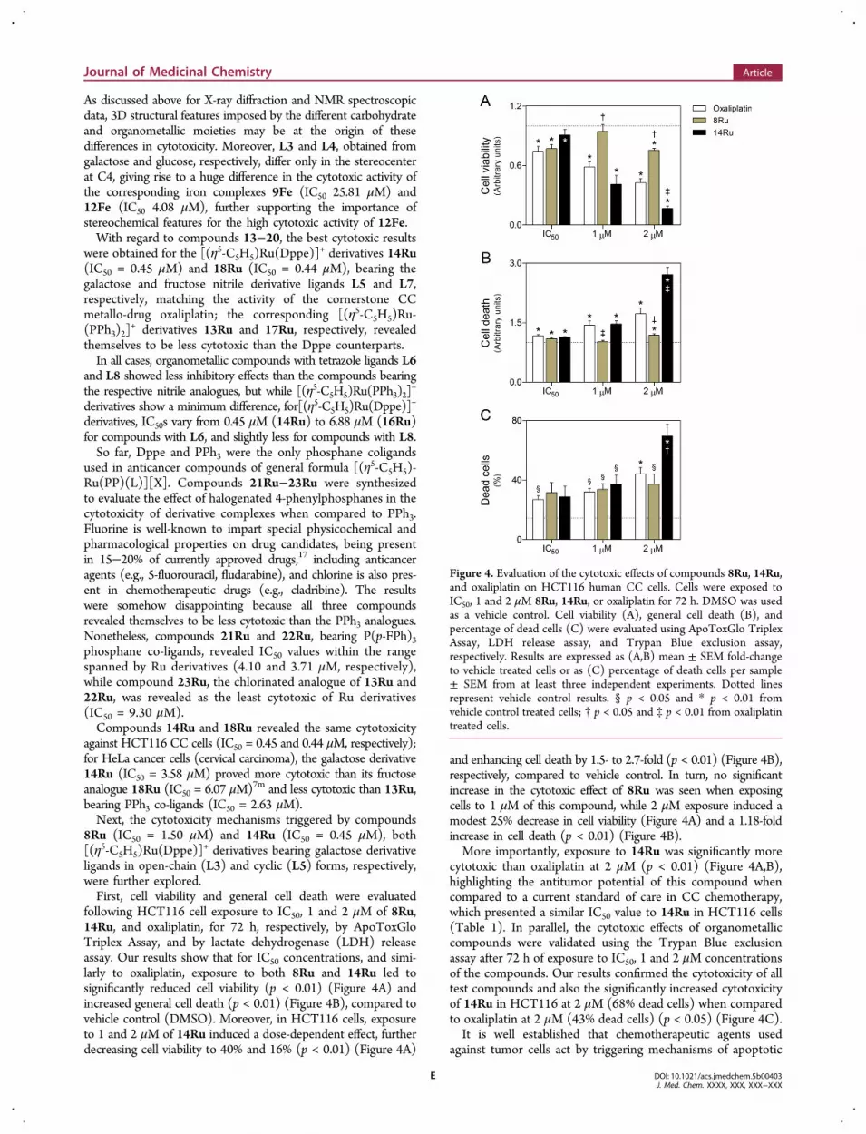
following HCT116 cell exposure to IC50, 1 and 2 μM of 8Ru,14Ru, and oxaliplatin, for 72 h, respectively, by ApoToxGloTriplex Assay, and by lactate dehydrogenase (LDH) releaseassay. Our results show that for IC50 concentrations, and simi-larly to oxaliplatin, exposure to both 8Ru and 14Ru led tosignificantly reduced cell viability (p < 0.01) (Figure 4A) andincreased general cell death (p < 0.01) (Figure 4B), compared tovehicle control (DMSO). Moreover, in HCT116 cells, exposureto 1 and 2 μM of 14Ru induced a dose-dependent effect, furtherdecreasing cell viability to 40% and 16% (p < 0.01) (Figure 4A)
and enhancing cell death by 1.5- to 2.7-fold (p < 0.01) (Figure 4B),respectively, compared to vehicle control. In turn, no significantincrease in the cytotoxic effect of 8Ru was seen when exposingcells to 1 μM of this compound, while 2 μM exposure induced amodest 25% decrease in cell viability (Figure 4A) and a 1.18-foldincrease in cell death (p < 0.01) (Figure 4B).More importantly, exposure to 14Ru was significantly more
cytotoxic than oxaliplatin at 2 μM (p < 0.01) (Figure 4A,B),highlighting the antitumor potential of this compound whencompared to a current standard of care in CC chemotherapy,which presented a similar IC50 value to 14Ru in HCT116 cells(Table 1). In parallel, the cytotoxic effects of organometalliccompounds were validated using the Trypan Blue exclusionassay after 72 h of exposure to IC50, 1 and 2 μM concentrationsof the compounds. Our results confirmed the cytotoxicity of alltest compounds and also the significantly increased cytotoxicityof 14Ru in HCT116 at 2 μM (68% dead cells) when comparedto oxaliplatin at 2 μM (43% dead cells) (p < 0.05) (Figure 4C).It is well established that chemotherapeutic agents used
against tumor cells act by triggering mechanisms of apoptotic
Figure 4. Evaluation of the cytotoxic effects of compounds 8Ru, 14Ru,and oxaliplatin on HCT116 human CC cells. Cells were exposed toIC50, 1 and 2 μM 8Ru, 14Ru, or oxaliplatin for 72 h. DMSO was usedas a vehicle control. Cell viability (A), general cell death (B), andpercentage of dead cells (C) were evaluated using ApoToxGlo TriplexAssay, LDH release assay, and Trypan Blue exclusion assay,respectively. Results are expressed as (A,B) mean ± SEM fold-changeto vehicle treated cells or as (C) percentage of death cells per sample± SEM from at least three independent experiments. Dotted linesrepresent vehicle control results. § p < 0.05 and * p < 0.01 fromvehicle control treated cells; † p < 0.05 and ‡ p < 0.01 from oxaliplatintreated cells.
Journal of Medicinal Chemistry Article
DOI: 10.1021/acs.jmedchem.5b00403J. Med. Chem. XXXX, XXX, XXX−XXX
E

cell death. In this context, activation of caspases has beenshown to be a hallmark of drug-induced cellular apoptosis.18
In addition, apoptotic cell death is characterized by distinc-tive morphological changes, including cell shrinkage, loss ofintercellular membrane contact, progressive condensation ofchromatin and cytoplasm, and subsequent nuclear fragmenta-tion. These events culminate in a characteristic formation ofapoptotic bodies, consisting of nuclear fragments and intact cellorganelles surrounded by a plasma membrane.19 Therefore,we assessed caspase-3/7 activation using Caspase-Glo 3/7assay (Promega) and apoptosis induction by evaluationof nuclear morphology under fluorescent microscopyfollowing staining with the DNA-binding stain Hoechstupon HCT116 cell exposure to IC50, 1 and 2 μM of 8Ru,14Ru, and oxaliplatin for 24 and 72 h, respectively. Our datashowed that similarly to oxaliplatin, both 8Ru and 14Rutriggered apoptotic cell death, leading to a significant in-crease in caspase-3 and -7 activity (p < 0.05) (Figure 5A)and apoptotic cells (p < 0.01) (Figure 5B,C), compared tovehicle control cells. Moreover, our results established that14Ru induced a 1.4-fold increase in caspase-3 and -7 activ-ity at IC50, which was significantly higher than oxaliplatin’s(p < 0.05), which increased caspase-3 and -7 activity only by1.27-fold (p < 0.01) (Figure 5A). Hereupon, 14Ru exposureinduced a dose-dependent increase in HCT116 apoptosis,leading to up to 16, 22, and 43% of apoptotic cells at IC50,1 and 2 μM, respectively, compared to vehicle control expo-sure (p < 0.01) (Figure 5B). In contrast, exposure to 2 μM8Ru led to less than 25% of apoptotic events (p < 0.01) (Figure5B). It should be emphasized that 14Ru also inducedsignificantly higher levels of apoptosis than oxaliplatin at 2 μM(p < 0.01), which was shown to trigger apoptosis in 30% ofHCT116 cells (p < 0.01) (Figure 5B).Collectively, our data indicate that 8Ru and 14Ru are capable
of markedly decreasing cell viability and inducing apoptoticcell death in HCT116 human CC cells. More importantly, 14Ruwas shown to trigger significantly higher levels of apoptosis whencompared to oxaliplatin, the cornerstone colon cancer chemo-therapeutic agent.
3. EXPERIMENTAL SECTIONAll experiments were carried out under inert atmosphere (N2) usingstandard Schlenk techniques. Commercial reagents were boughtfrom Sigma-Aldrich and used without further purification. All solventswere dried using standard methods.20 Starting materials were preparedfollowing the methods described in the literature for the synthesis of[(η5-C5H5)Ru(Dppe)Cl], [(η
5-C5H5)Ru(PPh3)2Cl],21 and [(η5-C5H5)-
Fe(Dppe)I].22 Compounds [(η5-C5H5)Ru(P(p-FPh)3)2Cl] and [(η5-C5H5)Ru(P(p-ClPh)3)2Cl] are here first described and were synthesizedby adopting the procedure used for the synthesis of [(η5-C5H5)Ru(PPh3)2Cl], as described below. Solid-state IR spectra were recordedin a Jasco FTIR-4100 spectrophotometer with KBr pellets; only significantbands were cited in the text. 1H, 13C, and 31P NMR spectra were recordedon Bruker Avance II 400 or Bruker Avance II 300 spectrometers at probetemperature. The 1H and 13C NMR chemical shifts are reported in partsper million (ppm) downfield from the residual solvent peak; 31P NMRspectra are reported in ppm downfield from external standard H3PO4
85%. Coupling constants are reported in Hz. Assignments of 1H and 13CNMR spectra were confirmed with the aid of two-dimensional techniques1H, 13C (COSY, HSQC). Microanalyses were performed at Laboratoriode Analises do Instituto Superior Tecnico, using a Fisons InstrumentsEA1108 system, and data acquisition, integration, and handling wereperformed using the software package Eager-200 (Carlo Erba Instru-ments), confirming ≥95% purity for all the tested compounds.
General Procedure for the Synthesis of Ligands Lc. To asolution of carbohydrate (10 mmol) in pyridine (5 mL) was addedH2NOH·HCl (12 mmol), and the mixture was stirred for 2 h. Ac2Owas then added (5 mL), and the mixture stirred at rt for a further 2 h.The solvent was then removed and the crude was extracted withAcOEt (3 × 20 mL), filtered, and pumped to dryness. The crudeobtained was purified by column chromatography (AcOEt:n-hexane),affording the pure O-acetylated nitrile ligands as a white crystallinesolids.
General Procedure for the Synthesis of Complexes [(η5-C5H5)-Ru(P(p-YPh)3)2Cl] (Y = F, Cl). To a boiling solution of RuCl3·xH2O(1.0 g) and freshly distilled cyclopentadiene (5 mL) in ethanol(80 mL) was added P(p-YPh)3 (12 mmol), and the mixture was stirredfor 2 h. After cooling to rt, the solvent was removed by filtrationand the solid products were washed with ethanol (3 × 20 mL) andn-hexane (3 × 20 mL). The products were recrystallized from dichloro-methane solutions by addition of n-hexane, affording crystallinecompounds.
Figure 5. Evaluation of the apoptotic effects of compounds 8Ru, 14Ru, and oxaliplatin on HCT116 human CC cells. Cells were exposed to IC50,1 and 2 μM of 8Ru, 14Ru, or oxaliplatin. DMSO was used as a vehicle control. (A) Caspase-3/7 activity was determined at 24 h of compoundexposure using the Caspase-Glo 3/7 assay. (B) Nuclear morphology after Hoechst staining was evaluated by fluorescence microscopy at 72 h ofcompound exposure. Dotted lines represent vehicle control results. (C) Representative images of Hoechst staining at 400× magnification. Arrowsindicate nuclear fragmentation and chromatin condensation. Results are expressed as (A) mean ± SEM fold-change to vehicle treated cells, or as(B) percentage of apoptotic cells per field ± SEM, from at least three independent experiments. § p < 0.05 and * p < 0.01 from vehicle controltreated cells; † p < 0.05 and ‡ p < 0.01 from oxaliplatin treated cells.
Journal of Medicinal Chemistry Article
DOI: 10.1021/acs.jmedchem.5b00403J. Med. Chem. XXXX, XXX, XXX−XXX
F

General Procedure for the Synthesis of Complexes [(η5-C5H5)-M(P−P)(Lc)][PF6]. To a Schlenck charged with [(η5-C5H5)M(P-P)X](0.20 mmol), TlPF6 (0.20 mmol), and Lc (0.22 mmol) was addeddichloromethane (20 mL), and the mixture was stirred overnight atroom temperature under inert atmosphere. The solutions were doublefiltered and pumped to dryness, and the crude compounds were thenwashed with n-hexane and recrystallized by slow diffusion of n-hexanein dichloromethane solutions, affording crystalline products.X-ray Diffraction Studies. Suitable crystals for X-ray diffraction
studies were obtained by slow diffusion of hexane into dichloro-methane solutions of 3Fe, 6Fe, 10Ru, 11Ru, and 12Fe. Data werecollected at 150 K using a Bruker AXS-KAPPA APEX II diffractometer.Structure resolution was performed with SHELXS97 and refinementwith SHELXL97. H atoms were calculated and constrained as riding ontheir bound atoms. CCDC 1050669−1050673 contains the supple-mentary crystallographic data for this paper (3Fe, 6Fe, 10Ru, 11Ru,and 12Fe, respectively). These data can be obtained free of charge viawww.ccdc.cam.ac.uk/data_request/cif or by emailing [email protected] or by contacting The Cambridge CrystallographicData Centre, 12, Union Road, Cambridge CB2 1EZ, UK; fax+44 1223 336033.Cell Culture. HCT116 human colon carcinoma cells were grown in
McCoy’s 5A modified medium supplemented with 10% heat-inactivatedfetal bovine serum (FBS) and 1% antibiotic/antimycotic solution(Gibco, Life Technologies, Paisley, UK) and cultured at 37 °C under ahumidified atmosphere of 5% CO2. Cells were seeded in 96-well platesat 1 × 10⁴ cells/well for dose−response curves, cell viability, and celldeath assays, and at 1.5 × 10⁴ cells/well for caspase activity studies.Additionally, cells were seeded in 24-well plates at 5 × 10⁴ cells/well forTrypan Blue exclusion assay and in 35 mm dishes at 3 × 10⁵ cells/dishfor morphological assessment of apoptosis.Exposure to Organometallic Compounds. Stock solutions of
the organometallic complexes 1−23 were prepared in sterile DMSO.Prior to all treatments, the cells were allowed to adhere for 24 h andthen exposed to test compounds for the mentioned time. To plot dose−response curves, cells were exposed to 0.1−100 μM test compounds for72 h. For cell viability, cell death, and apoptosis studies, metal-basedcompounds were tested at IC50, 1 and 2 μM, for 24 to 72 h. Allexperiments were performed in parallel with DMSO vehicle control.Oxaliplatin, a cytotoxic agent used in colon cancer treatment, was usedas a positive control in all assays. The final DMSO concentration wasalways of 0.1%.Dose−Response Curves. To plot dose−response curves, cell
viability was evaluated using the CellTiter 96 AQueous nonradioactivecell proliferation assay (Promega, Madison, WI, USA) according to themanufacturer’s instructions. This colorimetric assay is based on thebioreduction of 3-(4,5-dimethylthiazo-2-yl)-5-(3-carboxymethoxyphenyl)-2-(4-sulfophenyl)-2H-tetrazolium inner salt (MTS) to formazan bydehydrogenase enzymes found within metabolically active cells. Theamount of water-soluble formazan product can be measured by theamount of 490 nm absorbance, correlating with the number of living cellsin culture. For this purpose, changes in absorbance were assessed using amodel 680 microplate reader (Bio-Rad, Hercules, CA, USA). Best-fit IC50values from at least three independent experiments were calculated usingGraphPad Prism software (version 5.00; San Diego, CA, USA), using thelog (inhibitor) vs response (variable slope) function.Evaluation of Cell Viability. Cell viability was evaluated using the
ApoToxGlo Triplex Assay (Promega) according to the manufacturer’sinstructions. This assay uses a fluorogenic, cell-permeant, peptide substrate(glycylphenylalanyl-aminofluorocoumarin; GF-AFC) that may be cleavedby a protease found exclusively within intact cells to release AFC,generating a fluorescent signal proportional to the number of living cells.In brief, 20 μL of GF-AFC substrate solution was added to each well andplates were incubated at 37 °C for 1 h. Fluorescence emission was detect-ed using a GloMax-Multi+ detection system (Promega), with an at 405 nmexcitation filter and at 495−505 nm emission filter.Evaluation of Cell Death. General cell death was evaluated using
the Cytotoxicity Detection KitPLUS (Roche Diagnostics GmbH,Mannheim, Germany) to measure the amount of cytoplasmic lactatedehydrogenase (LDH) released from plasma membrane-damaged cells
into the extracellular medium. Thereby the amount of enzyme activityon supernatants can be proportionally determined by a coupledenzymatic reaction whereby the p-iodonitrotetrazolium salt is reducedto a red formazan product that can be spectrophotometricallyquantified at 490 nm. For the LDH assay, 50 μL of culture supernatantwas collected from each well into a new 96-well plate to evaluate LDHrelease. In parallel, the remaining cells attached to the original platewere lysed in 50 μL of medium to release the intracellular LDH.Subsequently, supernatant samples and total cell lysates wereincubated with 50 μL of assay substrate for 10−30 min, at room tem-perature, protected from light. Absorbance readings were measuredspectrophotometrically at 490 nm, with a 620 nm reference wavelength,using a model 680 microplate reader (Bio-Rad). The percentage ofLDH release was determined as the ratio between the released LDH(supernatant) and the total LDH (supernatant + cell lysate).
Additionally, cell death was evaluated using the Trypan Blue dyeexclusion test. This method relies on the principle that live cells retainintact cytoplasmic membranes that exclude Trypan Blue, remainingunstained, while dead cells incorporate this vital dye into the cytoplasmdue to loss of membrane selectivity. To evaluate Trypan Blue dye intakeupon loss of membrane integrity, cell culture supernatants and attachedcells were harvested to the same tube and next collected by centri-fugation at 500g for 5 min. Next, supernatants were discarded and cellswere resuspended and stained in 0.1% Trypan Blue solution diluted inphosphate-buffered saline (PBS). Subsequently, the relative numberof dead and live cells was obtained by optical microscopy by countingthe number of blue-stained (dead) and unstained (live) cells using aNeubauer chamber.
Evaluation of Apoptotic Cell Death. Activity of effector caspase-3and -7 was measured using the Caspase-Glo 3/7 assay (Promega).This assay is based on the cleavage of a proluminescent substratecontaining the specific DEVD sequence recognized by caspase-3 and -7to release aminoluciferin in cell lysates. The subsequent luciferasecleavage of the unconjugated aminoluciferin generates a luminescentsignal directly proportional to the amount of caspase activity present inthe sample. For this purpose, 75 μL of Caspase-Glo 3/7 reagent wasadded to each well and the mixture was incubated at room tem-perature for 30 min, leading to complete cell lysis, stabilization ofsubstrate cleavage by caspases, and accumulation of luminescent signal.The resulting luminescence was measured using the GloMax-Multi+detection system (Promega).
Nuclear morphology was assessed using the DNA-binding stainHoechst to identify apoptotic cells based on their typical morpho-logical changes. In brief, attached cells were fixed with 4%paraformaldehyde in PBS for 20 min, stained with 5 mg/mL Hoechst33258 dye (Sigma-Aldrich, St. Louis, MO, USA) in PBS for 15 min,washed with PBS, and mounted with coverslips using PBS/glycerol(3:1). Nuclear morphology was evaluated by fluorescence microscopy,under 400× magnification, and nuclei were scored and categorizedaccording to condensation and staining characteristics of chromatin.Normal nuclei showed noncondensed chromatin dispersed over theentire nucleus. Apoptotic nuclei were identified by condensed chromatin,contiguous to the nuclear membrane, as well as nuclear fragmentation.A minimum of five random microscopic fields with approximately 100nuclei were counted for each condition, and the results were expressed asthe percentage of apoptotic nuclei per field.
Statistical Analysis. All data were expressed as mean ± standarderror of mean (SEM) from at least three independent experiments.Statistical analysis was performed using Student’s t-test. Values of p < 0.05were considered significant.
4. CONCLUSIONSThis study provides a first insight on cyclopentadienyl−ruthenium(II) and iron(II) complexes with bioderivative moietiesas CC chemotherapeutic agents. The ligands Lc were obtainedfrom inexpensive raw carbohydrates using simple synthetic pro-cedures and taking advantage of both cyclic and open chain formsof these biomolecules. The cytotoxic activity of 23 organometalliccompounds of general formula [(η5-C5H5)M(PP)Lc][PF6] was
Journal of Medicinal Chemistry Article
DOI: 10.1021/acs.jmedchem.5b00403J. Med. Chem. XXXX, XXX, XXX−XXX
G

evaluated in HCT116 CC cells, with two ruthenium compounds,14Ru and 18Ru, matching the IC50 of oxaliplatin, and compound12Fe, revealing high cytotoxicity, roughly matching the IC50 of5-FU, the nonmetallic CC chemotherapeutic cornerstone. SARdata analysis shows a dependency of the cytotoxic activity on thecarbohydrate moiety (sugar and open-chain/cyclic form), linker(nitrile, tetrazole), metal (Ru, Fe), and co-ligands (Dppe, PPh3,P(p-FPh)3, P(p-ClPh)3). Our leading compound, 14Ru, showedimproved cytotoxic properties when compared to oxaliplatin, thecornerstone chemotherapeutic metallo-drug used in CC treat-ment. This compound induces dose-dependent HCT116 generalcell death, triggering higher levels of caspase-3 and -7 activities andapoptosis in a dose-dependent manner.Taken together, these results highlight the potential of
ruthenium and iron organometallic compounds bearing carbohy-drate moieties as rising opportunities to develop alternative anti-cancer drugs, with very high potential for surpassing problemssuch as general toxicity, side effects, and drug resistance, thecurrent major obstacles in colon cancer treatment.
■ ASSOCIATED CONTENT*S Supporting InformationFull spectroscopic characterization by FT-IR, 1H-, 13C-, 31PNMR (spectra description and interpretation) and elementalanalysis of compounds L1−L4, [(η5-C5H5)Ru(P(p-FPh)3)2Cl],[(η5-C5H5)Ru(P(p-ClPh)3)2Cl], and compounds 1−23 andfurther X-ray experimental details. The Supporting Informationis available free of charge on the ACS Publications website atDOI: 10.1021/acs.jmedchem.5b00403.
■ AUTHOR INFORMATIONCorresponding Authors*For P.R.F.: E-mail, [email protected].*For A.C.F.: phone, 351 218419264, fax, 351 218464455; E-mail,[email protected] authors declare no competing financial interest.
■ ACKNOWLEDGMENTSThis research was supported by FCT through projects PTDC/QUI-QUI/102114/2008 and PTDC/QUI-QUI/110532/2009and annual funds UID/QUI/00100/2013. P.R.F. and D.M.P.(SFRH/BD/96517/2013) thank FCT for grants. We thank thePortuguese NMR Network (IST-UTL Center) for providingaccess to the NMR facilities.
■ REFERENCES(1) (a) Rosenberg, B.; Van Camp, L.; Krigas, T. Inhibition of celldivision in Escherichia coli by electrolysis products from a platinumelectrode. Nature 1965, 205, 698−699. (b) Rosenberg, B.; Vancamp,L.; Trosko, J. E.; Mansour, V. H. Platinum Compounds: A New Classof Potent Antitumor Agents. Nature 1969, 222, 385−386.(2) (a) Yan, Y. K.; Melchart, M.; Habtemariam, A.; Sadler, P. J.Organometallic chemistry, biology and medicine: ruthenium areneanticancer complexes. Chem. Commun. 2005, 4764−4776. (b) Suss-Fink, G. Arene ruthenium complexes as anticancer agents. DaltonTrans. 2010, 39, 1673−1688. (c) Singha, A. K.; Pandeyb, D. S.; Xua,Q.; Braunstein, P. Recent advances in supramolecular and biologicalaspects of arene−ruthenium(II) complexes. Coord. Chem. Rev. 2014,270−271, 31−56. (d) Bergamo, A.; Gaiddon, C.; Schellens, J. H. M.;Beijnen, J. H.; Sava, G. Approaching tumour therapy beyond platinumdrugs. Status of the art and perspectives of ruthenium drug candidates.J. Inorg. Biochem. 2012, 106, 90−99.
(3) (a) Groessl, M.; Reisner, E.; Hartinger, C. G.; Eichinger, R.;Semenova, O.; Timerbaev, A. R.; Jakupec, M. A.; Arion, V. B.; Keppler,B. K. Structure−activity relationships for NAMI-A-type complexes(HL)[trans-RuCl4L(S-DMSO)ruthenate(III)] (L = imidazole, inda-zole, 1,2,4-triazole, 4-amino-1,2,4-triazole, and 1-methyl-1,2,4-triazole):aquation, redox properties, protein binding, and antiproliferativeactivity. J. Med. Chem. 2007, 50, 2185−2193.(4) Hartinger, C. G.; Zorbas-Seifried, S.; Jakupec, M. A.; Kynast, B.;Zorbas, H.; Keppler, B. K. From bench to bedside-preclinical and earlyclinical development of the anticancer agent indazolium trans-[tetrachlorobis(1H-indazole)ruthenate(III)] (KP1019 or FFC14A). J.Inorg. Biochem. 2006, 100, 891−904.(5) Rademaker-Lakhai, J. M.; van den Bongard, D.; Pluim, D.;Beijnen, J. H.; Schellens, J. H. A phase I and pharmacological studywith imidazolium-trans-DMSO-imidazole-tetrachlororuthenate. Clin.Cancer Res. 2004, 10, 3717−3727.(6) Lentz, F.; Drescher, A.; Lindauer, A.; Henke, M.; Hilger, R. A.;Hartinger, C. G.; Scheulen, M. E.; Dittrich, C.; Keppler, B. K.; Jaehde,U. Pharmacokinetics of a novel anticancer ruthenium complex(KP1019, FFC14A) in a phase I dose-escalation study. AnticancerDrugs 2009, 20, 97−103.(7) (a) Bregman, H.; Williams, D. S.; Atilla, G. E.; Carroll, P. J.;Meggers, E. An Organometallic Inhibitor for Glycogen SynthaseKinase 3. J. Am. Chem. Soc. 2004, 126, 13594−13595. (b) Debreczeni,J. E.; Bullock, A. N.; Atilla, G. E.; Williams, D. S.; Bregman, H.; Knapp,S.; Meggers, E. Extremely Tight Binding of Ruthenium Complex toGlycogen Synthase Kinase 3. Angew. Chem., Int. Ed. Engl. 2006, 45,1580−1585. (c) Smalley, K. S.; Contractor, R.; Haass, N. K.; Kulp, A.N.; Atilla-Gokcuman, G. E.; Williams, D. E.; Bregman, H.; Flaherty, K.T.; Soengas, M. S.; Meggers, E.; Herlyn, M. An OrganometallicProtein Kinase Inhibitor Pharmacologically Activates p53 and InducesApoptosis in Human Melanoma Cells. Cancer Res. 2007, 67, 209−217.(d) Anand, R.; Maksimoska, J.; Pagano, N.; Wong, E. Y.; Gimotty, P.A.; Diamond, S. L.; Meggers, E.; Marmorstein, R. Toward theDevelopment of a Potent and Selective Organoruthenium MammalianSterile 20 Kinase Inhibitor. J. Med. Chem. 2009, 52, 1602−1611.(e) Garcia, M. H.; Morais, T. S.; Florindo, P.; Piedade, M. F. M.;Moreno, V.; Ciudad, C.; Noe, V. Inhibition of cancer cell growth byruthenium(II) cyclopentadienyl derivative complexes with heteroar-omatic ligands. J. Inorg. Biochem. 2009, 103, 354−361. (f) Moreno, V.;Lorenzo, J.; Aviles, F. X.; Garcia, M. H.; Ribeiro, J.; Morais, T. S.;Florindo, P.; Robalo, M. P. Studies of the Antiproliferative Activity ofRuthenium(II) Cyclopentadienyl-Derived Complexes with NitrogenCoordinated Ligands. Bioinorg. Chem. App. 936834, 2010, ID; pp 1−11. (g) Moreno, V.; Font-Bardia, M.; Calvet, T.; Lorenzo, J.; Aviles, F.X.; Garcia, M. H.; Morais, T. S.; Valente, A.; Robalo, M. P. DNAinteraction and cytotoxicity studies of new ruthenium(II) cyclo-pentadienyl derivative complexes containing heteroaromatic ligands. J.Inorg. Biochem. 2011, 105, 241−249. (h) Tomaz, A. I.; Jakusch, T.;Morais, T. S.; Marques, F.; Almeida, R. F. M.; Mendes, F.; Enyedy, E.A.; Santos, I.; Pessoa, J. C.; Kiss, T.; Garcia, M. H. [RuII(η5-C5H5)(bipy)(PPh3)]
+, a promising large spectrum antitumor agent:cytotoxic activity and interaction with human serum albumin. J. Inorg.Biochem. 2012, 117, 261−269. (i) Morais, T. S.; Santos, F.; Corte-Real,L.; Marques, F.; Robalo, M. P.; Madeira, P. J. A.; Garcia, M. H.Biological activity and cellular uptake of [Ru(η5-C5H5)(PPh3)-(Me2bpy)][CF3SO3] complex. J. Inorg. Biochem. 2013, 122, 8−17.(j) Valente, A.; Garcia, M. H.; Marques, F.; Miao, Y.; Rousseau, C.;Zinck, P. First polymer “ruthenium−cyclopentadienyl” complex aspotential anticancer agent. J. Inorg. Biochem. 2013, 127, 79−81.(k) Morais, T. S.; Santos, F. C.; Jorge, T. F.; Corte-Real, L.; Madeira,P. J. A.; Marques, F.; Robalo, M. P.; Matos, A.; Santos, I.; Garcia, M.H. New water-soluble ruthenium(II) cytotoxic complex: biologicalactivity and cellular distribution. J. Inorg. Biochem. 2014, 130, 1−14.(l) Corte-Real, L.; Mendes, F.; Coimbra, J.; Morais, T. S.; Tomaz, A. I.;Valente, A.; Garcia, M. H.; Santos, I.; Bicho, M.; Marques, F.Anticancer activity of structurally related ruthenium(II) cyclo-pentadienyl complexes. J. Biol. Inorg. Chem. 2014, 19, 853−867.(m) Florindo, P. R.; Marques, I. J.; Nunes, C. D.; Fernandes, A. C.
Journal of Medicinal Chemistry Article
DOI: 10.1021/acs.jmedchem.5b00403J. Med. Chem. XXXX, XXX, XXX−XXX
H

Synthesis, characterization and cytotoxicity of cyclopentadienylruthenium(II) complexes containing carbohydrate-derived ligands. J.Organomet. Chem. 2014, 760, 240−247.(8) (a) Vessieres, A.; Top, S.; Beck, W.; Hillard, E.; Jaouen, G. Metalcomplex SERMs (selective oestrogen receptor modulators). Theinfluence of different metal units on breast cancer cell antiproliferativeeffects. Dalton Trans. 2006, 529−541. (b) Top, S.; Vessieres, A.;Leclercq, G.; Quivy, J.; Tang, J.; Vaissermann, J.; Huche, M.; Jaouen,G. Synthesis, biochemical properties and molecular modelling studiesof organometallic specific estrogen receptor modulators (SERMs), theferrocifens and hydroxyferrocifens: evidence for an antiproliferativeeffect of hydroxyferrocifens on both hormone-dependent andhormone-independent breast cancer cell lines. Chem.Eur. J. 2003,9, 5223−5236.(9) Braga, S. S.; Silva, A. M. S. A New Age for Iron: AntitumoralFerrocenes. Organometallics 2013, 32, 5626−5639.(10) Gasser, G.; Ott, I.; Metzler-Nolte, N. Organometallic AnticancerCompounds. J. Med. Chem. 2011, 54, 3−25.(11) (a) Goncalves, A. C.; Morais, T. S.; Robalo, M. P.; Marques, F.;Avecilla, F.; Matos, C. P.; Santos, I.; Tomaz, A. I.; Garcia, M. H.Important cytotoxicity of novel iron(II) cyclopentadienyl complexeswith imidazole based ligands. J. Inorg. Biochem. 2013, 129, 1−8.(b) Valente, A.; Santos, A. M.; Corte-Real, L.; Robalo, M. P.; Moreno,V.; Font-Bardia, M.; Calvet, T.; Lorenzo, J.; Garcia, M. H. Newiron(II) cyclopentadienyl derivative complexes: synthesis andantitumor activity against human leukemia cancer cells. J. Organomet.Chem. 2014, 756, 52−60.(12) Siegel, R.; Naishadham, D.; Jemal, A. Cancer statistics, 2013.Can. Cancer J. Clin. 2013, 63, 11−30.(13) (a) Mader, R. M.; Muller, M.; Steger, G. G. Resistance to 5-Fluorouracil. Gen. Pharmacol. 1998, 31, 661−666. (b) Alcindor, T.;Beauger, N. Oxaliplatin: a review in the era of molecularly targetedtherapy. Curr. Oncol. 2011, 18, 18−25.(14) (a) Garcia, M. H.; Florindo, P.; Piedade, M. F. M.; Maiorana, S.;Licandro, E. New organometallic Ru(II) and Fe(II) complexes withtetrathia-[7]-helicene derivative ligands. Polyhedron 2009, 28, 621−629. (b) Garcia, M. H.; Florindo, P.; Piedade, M. F. M.; Duarte, M. T.;Robalo, M. P.; Goovaerts, E.; Wenseleers, W. Synthesis and structuralcharacterization of ruthenium(II) and iron(II) complexes containing1,2-di-(2-thienyl)-ethene derived ligands as chromophores. J. Organo-met. Chem. 2009, 694, 433−445.(15) Kuhl, O. Phosphorous-31 NMR Spectroscopy: A ConciseIntroduction for the Synthetic Organic and Organometallic Chemist;Springer-Verlag: Berlin, Germany, 2008.(16) Mansoor, T. A.; Borralho, P. M.; Luo, X.; Mulhovo, S.;Rodrigues, C. M. P.; Ferreira, M.-J. U. Acetonyldihydrochelerythrineisa potent inducer of apoptosis in HCT116 and SW620 Colon Cancercells. J. Nat. Prod. 2014, 77, 1825−1830.(17) Shah, P.; Westwell, A. D. The role of fluorine in medicinalchemistry. J. Enzyme Inhib. Med. Chem. 2007, 22, 527−540.(18) Kaufmann, S. H.; Earnshaw, W. C. Induction of Apoptosis byCancer Chemotherapy. Exp. Cell Res. 2000, 256, 42−49.(19) Galluzzi, L.; Aaronson, S. A.; et al. Guidelines for the use andinterpretation of assays for monitoring cell death in higher eukaryotes.Cell Death Differ. 2009, 16, 1093−1107.(20) Perrin, D. D.; Amarego, W. L. F.; Perrin, D. R. Purification ofLaboratory Chemicals, 2nd ed.; Pergamon: New York, 1980.(21) Ashby, G. S.; Bruce, M. I.; Tomkins, I. B.; Walli, R. C.Cyclopentadienyl-ruthenium and -osmium chemistry. VII. Complexescontaining nitriles, tertiary phosphines or phosphites formed byaddition or displacement reactions. Aust. J. Chem. 1979, 32, 1003−1008.(22) Garcia, M. H.; Robalo, M. P.; Dias, A. R.; Piedade, M. F. M.;Galvao, A.; Wenseleers, W.; Goovaerts, E. Organometallic complexesfor second-order nonlinear optics: synthesis and molecular quadratichyperpolarizabilities of η5-monocyclopentadienyliron(II) nitrile deriv-atives with different phosphines. X-ray crystal structure of [FeCp-(Dppe)(p-NCC6H4NO2)][PF6]·CH2Cl2. J. Organomet. Chem. 2001,619, 252−264.
Journal of Medicinal Chemistry Article
DOI: 10.1021/acs.jmedchem.5b00403J. Med. Chem. XXXX, XXX, XXX−XXX
I