protective effects of mangifera indica l extract (vimang), and its major component mangiferin, on...
TRANSCRIPT
A
i7wapdcim©
K
1
oadp
(oee
1d
Pharmacological Research 57 (2008) 79–86
Protective effects of Mangifera indica L extract (Vimang),and its major component mangiferin, on iron-induced
oxidative damage to rat serum and liver
Gilberto L. Pardo-Andreu a,∗, Mariela Forrellat Barrios b, Carlos Curti c, Ivones Hernandez a,Nelson Merino a, Yeny Lemus a, Ioanna Martınez a, Annia Riano a, Rene Delgado a
a Departamento de Investigaciones Biomedicas, Centro de Quımica Farmaceutica,Calle 200, Esq. 21, Playa, Ciudad de La Habana, Cuba
b Instituto de Hematologıa e Inmunologıa. Apartado Postal 8070, Ciudad de La Habana, CP 10800, Cubac Departamento de Fısica e Quımica, Faculdade de Ciencias Farmaceuticas de Ribeirao Preto,
Universidade de Sao Paulo, 14040-903 Ribeirao Preto, SP, Brazil
Accepted 20 December 2007
bstract
In vivo preventive effects of a Mangifera indica L extract (Vimang) or its major component mangiferin on iron overload injury have been studiedn rats given respectively, 50, 100, 250 mg kg−1 body weight of Vimang, or 40 mg kg−1 body weight of mangiferin, for 7 days prior to, and fordays following the administration of toxic amounts of iron-dextran. Both Vimang or mangiferin treatment prevented iron overload in serum asell as liver oxidative stress, decreased serum and liver lipid peroxidation, serum GPx activity, and increased serum and liver GSH, serum SOD
nd the animals overall antioxidant condition. Serum iron concentration was decreased although at higher doses, Vimang tended to increase it;ercent tranferrin saturation, liver weight/body mass ratios, liver iron content was decreased. Treatment increased serum iron-binding capacity andecreased serum levels of aspartate-amine transferase (ASAT) and alanine-amine transferase (ALAT), as well as the number of abnormal Kupffer
ells in iron-loaded livers. It is suggested that besides acting as antioxidants, Vimang extract or its mangiferin component decrease liver iron byncreasing its excretion. Complementing earlier in vitro results from our group, it appears possible to support the hypothesis that Vimang andangiferin present therapeutically useful effects in iron overload related diseases.2007 Elsevier Ltd. All rights reserved.
ecies
o[
pif[aa
eywords: Mangifera indica L.; Vimang; Mangiferin; Iron; Reactive oxygen sp
. Introduction
Iron overload in humans causes tissue damage and possiblergan failure, liver being a primary target, and hepatic fibrosisnd cirrhosis often having been observed in patients [1]. Asemonstrated both in vitro and in vivo [2,3], lipid cell membraneeroxidation probably underlies such toxicity.
Deferoxamine and 1,2-dimethyl-3-hydroxypyrid-4-onedeferiprone, L1) are being currently prescribed for the treatment
f iron overload diseases. However, such compounds show sev-ral limitations [4–6] that indicate the need of a search for moreffective and less toxic drugs for patients that either not respond,∗ Corresponding author. Tel.: +537 2715067; fax: +537 2736471.E-mail address: [email protected] (G.L. Pardo-Andreu).
ß
sfIac
043-6618/$ – see front matter © 2007 Elsevier Ltd. All rights reserved.oi:10.1016/j.phrs.2007.12.004
; Antioxidants
r show toxic side-effects following their administration7].
The ability of polyphenols to chelate iron is a very importantart of their antioxidant activity. Their “site-specific scaveng-ng” action can occur while iron is still being catalytically activeorming free radicals, permitting it to be rapidly scavenged8]. Polyphenols therefore, could have a double, synergisticction, making them into effective antioxidants, particularlyctive in pathological situations involving iron overload, e.g.-thalassemia, Friedreich’s ataxia and hemochromatosis.
A standardized aqueous extract from the bark of selectedpecies of Mangifera indica L. (Anacardiaceae) is used as a
ood supplement in Cuba, under the brand name of Vimang.t has potent in vitro and in vivo antioxidant activities [9,10],pparently due to phenolic acids, phenolic esters, flavan-3-olsomponents and in special, mangiferin [11]. The interaction of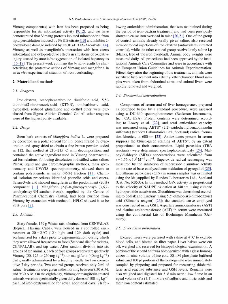
8 acolo
VrdldVai[aa
2
2
dpcw
2
boaccPtccflctPV9
2
(ratCgVdusaae
ltsoic(mttFspr
2
auIiwstsproε
moGu(thiawaum
2
bopess
0 G.L. Pardo-Andreu et al. / Pharm
imang component(s) with iron has been proposed as beingesponsible for its antioxidant activity [9,12], and we haveemonstrated that Vimang protects isolated mitochondria fromipid peroxidation induced by Fe (II)-citrate [13] and inhibits 2-eoxyribose damage induced by Fe(III)-EDTA-Ascorbate [14].imang as well as mangiferin’s interaction with iron exertsntioxidant and cytoprotective effects in situations of oxidativenjury caused by anoxia/reoxygenation of isolated hepatocytes15–19]. The present work confirms the in vitro results by char-cterizing the protective actions of Vimang and mangiferin inn in vivo experimental situation of iron overloading.
. Material and methods
.1. Reagents
Iron-dextran, bathophenanthroline disulfonic acid, 5,5′-ithiobis(2-nitrobenzoic)acid (DTNB), thiobarbituric acid,yrogallol, reduced glutathione and diethyl ether were pur-hased from Sigma–Aldrich Chemical Co. All other reagentsere of the highest purity available.
.2. Drugs
Stem bark extracts of Mangifera indica L. were preparedy decoction in a polar solvent for 1 h, concentrated by evap-ration and spray dried to obtain a fine brown powder, codeds 112, that melted at 210–215 ◦C with decomposition, andontained the active ingredient used in Vimang pharmaceuti-al formulations, following dissolution in distilled water saline.lanar, liquid and gas chromatographic methods, mass spec-
rometry and UV/VIS spectrophotometry, showed them toontain polyphenols as major (45%) fraction [11]. Chemi-al isolation procedures identified phenolic acids and esters,avan-3-ols and showed mangiferin as the predominant (20%)omponent [11]. Mangiferin (2-�-d-glucopyranosyl-1,3,6,7-etrahydroxy-9H-xanthen-9-one), supplied by the Centre ofharmaceutical Chemistry (Cuba), had been purified fromimang by extraction with methanol; HPLC showed it to be5% pure [7].
.3. Animals
Sixty female, 159 g Wistar rats, obtained from CENPALABBejucal, Havana, Cuba), were housed in a controlled envi-onment at 20 ± 2 ◦C (12 h light and 12 h dark cycle) andcclimatized for 7 days prior to experimentation, during whichhey were allowed free access to food (Standard diet for rodents,ENPALAB), and tap water. After random division into sixroups of ten animals, each of four groups received respectivelyimang (50, 125 or 250 mg kg−1), or mangiferin (40 mg kg−1)aily, orally administered by a feeding needle for two consec-tive 7 day periods. Two control groups received only 2 ml of
aline. Treatments were given in the morning between 8:30 A.M.nd 9:30 A.M. On the eighth day, Vimang or mangiferin-treatednimals were intraperitonially given daily doses of 100 mg kg−1ach, of iron-dextran/saline for seven additional days, 2 h fol-
taet
gical Research 57 (2008) 79–86
owing antioxidant administration, that was maintained duringhe period of iron-dextran treatment, and had been previouslyhown to cause iron overload in mice [20,21]. One of the groupf control animals already orally given saline, also receivedntraperitoneal injections of iron-dextran (antioxidant-untreatedontrols), while the other control group received only saline i.pblanks, free of the iron overload). Animal body weights wereeasured daily. All procedures had been approved by the insti-
utional Animals Care Committee and were in accordance withhe European Union Guidelines for Animals Experimentation.ifteen days after the beginning of the treatments, animals wereacrificed by placement into a diethyl ether chamber, blood sam-les were taken from abdominal aorta, and animal livers wereapidly removed and weighed.
.4. Biochemical determinations
Components of serum and of liver homogenates, prepareds described below by a standard procedure, were assessedsing a DU-640 spectrophotometer (Beckman Instruments,nc., CA, USA). Protein contents were determined accord-ng to Lowry et al. [22], and total antioxidant capacityas measured using ABTS+ (2,2′-azidodiethylbenzothiazolin
ulfonate) (Randox Laboratories Ltd., Scotland) radical forma-ion kinetics, at 600 nm [23]. Antioxidants present in serumuppress the bluish-green staining of ABTS+, by an extentroportional to their concentration. Lipid peroxides (TBAeactants) were determined spectrophotometricaly [24]. Mal-nyldialdeyde (MDA) concentrations were calculated from= 1.56 × 10⁵ M−1 cm−1. Superoxide radical scavenging waseasured by the inhibition of superoxide dismutase activity
n the rate of base-catalyzed auto-oxidation of pyrogallol [25].lutathione peroxidase (GPx) in serum samples was estimatedsing the kit supplied by Randox Laboratories Ltd., ScotlandCat. No. RS505). In this method GPx activity is proportionalo the velocity of NADPH oxidation at 340 nm, using cumeneydroperoxide as substrate. Glutathione was determined accord-ng to Sedlak and Lindsay, using 5,5′-dithiobis(2-nitrobenzoic)cid (Ellman’s reagent) [26]; the standard curve employedas constructed using GSH. Aspartate aminotransferase (AST)
nd alanine aminotransferase (ALT) in serum were measuredsing the commercial kits of Boehringer Mannheim (Ger-any).
.5. Liver tissue preparation
Excised livers were perfused with saline at 4 ◦C to excludelood cells, and blotted on filter paper. Liver halves were cutff, weighed and reserved for histopathological examination. Aortion of the second half was homogenized with a glass homog-nizer in nine volume of ice-cold 50 mM phosphate bufferedaline, and 100 �l portions of the homogenate were immediatelyampled by pippeting and prepared for measuring thiobarbi-
uric acid reactive substance and GSH levels. Remains werelso weighed and digested for 5–8 min over a low flame in anqual volume of a (1:1) mixture of sulfuric and nitric acids andheir iron content estimated.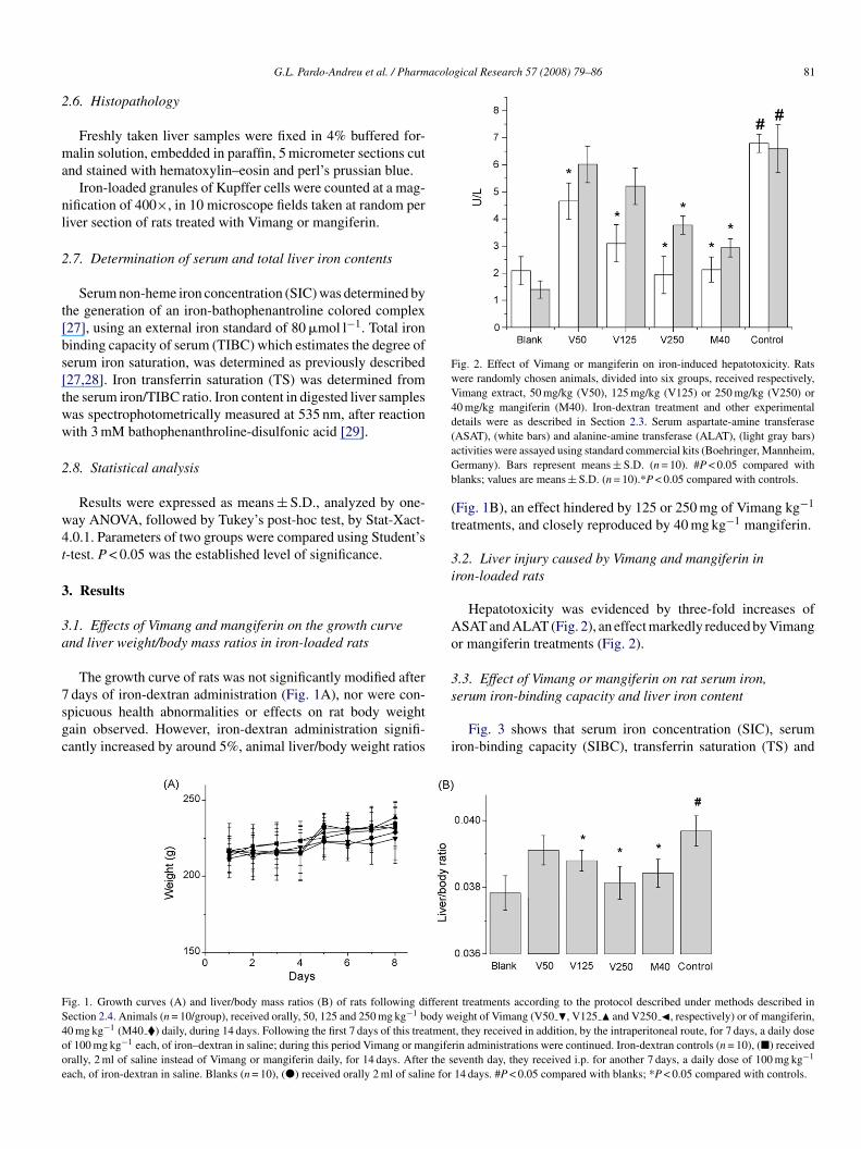
acological Research 57 (2008) 79–86 81
2
ma
nl
2
t[bs[tww
2
w4t
3
3a
7sgc
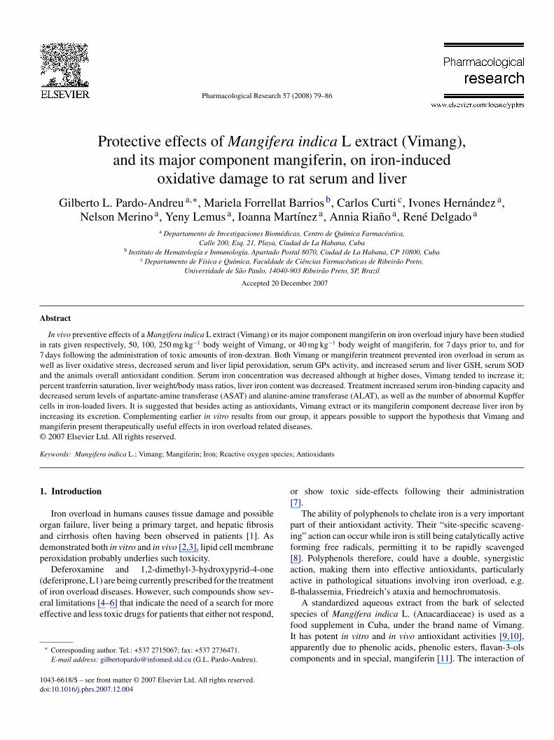
Fig. 2. Effect of Vimang or mangiferin on iron-induced hepatotoxicity. Ratswere randomly chosen animals, divided into six groups, received respectively,Vimang extract, 50 mg/kg (V50), 125 mg/kg (V125) or 250 mg/kg (V250) or40 mg/kg mangiferin (M40). Iron-dextran treatment and other experimentaldetails were as described in Section 2.3. Serum aspartate-amine transferase(aGb
(t
3i
Ao
3
FS4ooe
G.L. Pardo-Andreu et al. / Pharm
.6. Histopathology
Freshly taken liver samples were fixed in 4% buffered for-alin solution, embedded in paraffin, 5 micrometer sections cut
nd stained with hematoxylin–eosin and perl’s prussian blue.Iron-loaded granules of Kupffer cells were counted at a mag-
ification of 400×, in 10 microscope fields taken at random periver section of rats treated with Vimang or mangiferin.
.7. Determination of serum and total liver iron contents
Serum non-heme iron concentration (SIC) was determined byhe generation of an iron-bathophenantroline colored complex27], using an external iron standard of 80 �mol l−1. Total ironinding capacity of serum (TIBC) which estimates the degree oferum iron saturation, was determined as previously described27,28]. Iron transferrin saturation (TS) was determined fromhe serum iron/TIBC ratio. Iron content in digested liver samplesas spectrophotometrically measured at 535 nm, after reactionith 3 mM bathophenanthroline-disulfonic acid [29].
.8. Statistical analysis
Results were expressed as means ± S.D., analyzed by one-ay ANOVA, followed by Tukey’s post-hoc test, by Stat-Xact-.0.1. Parameters of two groups were compared using Student’s-test. P < 0.05 was the established level of significance.
. Results
.1. Effects of Vimang and mangiferin on the growth curvend liver weight/body mass ratios in iron-loaded rats
The growth curve of rats was not significantly modified after
days of iron-dextran administration (Fig. 1A), nor were con-picuous health abnormalities or effects on rat body weightain observed. However, iron-dextran administration signifi-antly increased by around 5%, animal liver/body weight ratios
s
i
ig. 1. Growth curves (A) and liver/body mass ratios (B) of rats following differenection 2.4. Animals (n = 10/group), received orally, 50, 125 and 250 mg kg−1 body w0 mg kg−1 (M40 �) daily, during 14 days. Following the first 7 days of this treatmenf 100 mg kg−1 each, of iron–dextran in saline; during this period Vimang or mangiferally, 2 ml of saline instead of Vimang or mangiferin daily, for 14 days. After the sach, of iron-dextran in saline. Blanks (n = 10), (�) received orally 2 ml of saline for
ASAT), (white bars) and alanine-amine transferase (ALAT), (light gray bars)ctivities were assayed using standard commercial kits (Boehringer, Mannheim,ermany). Bars represent means ± S.D. (n = 10). #P < 0.05 compared withlanks; values are means ± S.D. (n = 10).*P < 0.05 compared with controls.
Fig. 1B), an effect hindered by 125 or 250 mg of Vimang kg−1
reatments, and closely reproduced by 40 mg kg−1 mangiferin.
.2. Liver injury caused by Vimang and mangiferin inron-loaded rats
Hepatotoxicity was evidenced by three-fold increases ofSAT and ALAT (Fig. 2), an effect markedly reduced by Vimangr mangiferin treatments (Fig. 2).
.3. Effect of Vimang or mangiferin on rat serum iron,
erum iron-binding capacity and liver iron contentFig. 3 shows that serum iron concentration (SIC), serumron-binding capacity (SIBC), transferrin saturation (TS) and
t treatments according to the protocol described under methods described ineight of Vimang (V50 �, V125 � and V250 �, respectively) or of mangiferin,t, they received in addition, by the intraperitoneal route, for 7 days, a daily doserin administrations were continued. Iron-dextran controls (n = 10), (�) receivedeventh day, they received i.p. for another 7 days, a daily dose of 100 mg kg−1
14 days. #P < 0.05 compared with blanks; *P < 0.05 compared with controls.
82 G.L. Pardo-Andreu et al. / Pharmacological Research 57 (2008) 79–86
F serumA re assc
l(i(dlr
3G
sob4d(
Fiags(b
3as
atd7stmit
ig. 3. Effect of Vimang or mangiferin on serum iron concentration (SIC �),), and total iron liver content (Panel B) in iron loaded rats. Iron contents we
ompared with blank; *P < 0.05 compared with control.
iver iron content of treated rats, were approximately doubledSIC), or ten-fold increased (TS), following iron-dextran admin-stration (Fig. 3B), while SIBC was significantly decreasedFig. 3A). Treatment with Vimang decreased SIC but at higheroses showed a tendency to increase it, decreased TS as well asiver iron content, and increased SIBC. Mangiferin again closelyeproduced these effects.
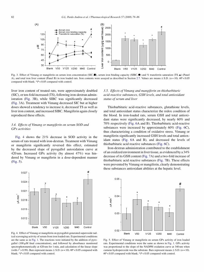
.4. Effects of Vimang or mangiferin on serum SOD andPx activities
Fig. 4 shows the 21% decrease in SOD activity in theerum of rats treated with iron-dextran. Treatment with Vimangr mangiferin significantly reversed this effect, estimated
y the decreased slope of pyrogallol autoxidation curve at20 nm. Increased GPx activity (by almost 47%) was hin-ered by Vimang or mangiferin in a dose-dependent mannerFig. 5).ig. 4. Effect of Vimang or mangiferin on pyrogallol-generated superoxide rad-cal scavenging activity of serum from iron loaded rats. Experimental conditionsre the same as in Fig. 1. The reactions were initiated by the addition of pyro-allol (100 �M final concentration), and followed by absorbance monitoredpectrophotometrically at 420 nm for 1 min, and calculation of the linear slopewith r2 > 0.99). Bars represent means ± S.D. (n = 10). #P < 0.05 compared withlank; *P < 0.05 compared with control.
odtwt
Frwc#
iron binding capacity (SIBC �) and % transferrin saturation (TS �) (Panelayed as described in Section 2.7. Values are means ± S.D. (n = 10). #P < 0.05
.5. Effects of Vimang and mangiferin on thiobarbituriccid-reactive substances, GSH levels, and total antioxidanttatus of serum and liver
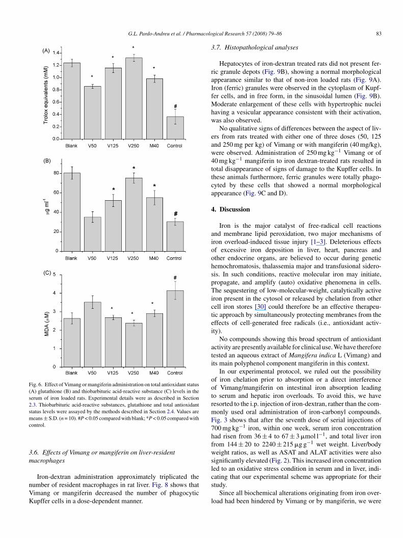
Thiobarbituric acid-reactive substances, glutathione levels,nd total antioxidant status characterize the redox condition ofhe blood. In iron-loaded rats, serum GSH and total antioxi-ant status were significantly decreased, by nearly 60% and0% respectively (Fig. 6A and B). Thiobarbituric acid-reactiveubstances were increased by approximately 60% (Fig. 6C),hus characterizing a condition of oxidative stress. Vimang or
angiferin significantly increased GSH levels and total antiox-dant status (Fig. 6A and B), and decreased the levels ofhiobarbituric acid reactive substances (Fig. 6C).
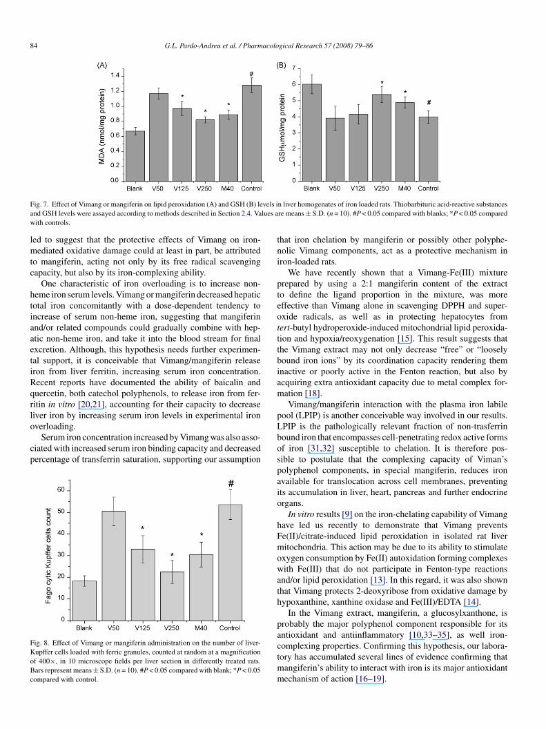
Iron-dextran administration contributed to the establishmentf an oxidized environment in liver tissue, as evidenced by a 34%
ecrease of its GSH content (Fig. 7A) and a two-fold increase ofhiobarbituric acid reactive substances (Fig. 7B). These effectsere prevented by Vimang or mangiferin, clearly demonstratinghese substances antioxidant abilities at the hepatic level.
ig. 5. Effect of Vimang or mangiferin on serum GPx activity of iron loadedats. Experimental conditions were the same as shown in Fig. 1. GPx activityas proportional to the slope of the NADPH oxidation curve at 340 nm when
umene hydroperoxide was the substrate. Bars represent means ± S.D. (n = 10).P < 0.05 compared with blank; *P < 0.05 compared with control.
G.L. Pardo-Andreu et al. / Pharmacolo
Fig. 6. Effect of Vimang or mangiferin administration on total antioxidant status(A) glutathione (B) and thiobarbituric acid-reactive substance (C) levels in theserum of iron loaded rats. Experimental details were as described in Section2.3. Thiobarbituric acid-reactive substances, glutathione and total antioxidantstatus levels were assayed by the methods described in Section 2.4. Values aremc
3m
nVK
3
raIfMhw
eaw4ttca
4
aioohspTictei
ati
ootrmF7hfwsl
eans ± S.D. (n = 10). #P < 0.05 compared with blank; *P < 0.05 compared withontrol.
.6. Effects of Vimang or mangiferin on liver-residentacrophages
Iron-dextran administration approximately triplicated theumber of resident macrophages in rat liver. Fig. 8 shows thatimang or mangiferin decreased the number of phagocyticupffer cells in a dose-dependent manner.
cs
l
gical Research 57 (2008) 79–86 83
.7. Histopathological analyses
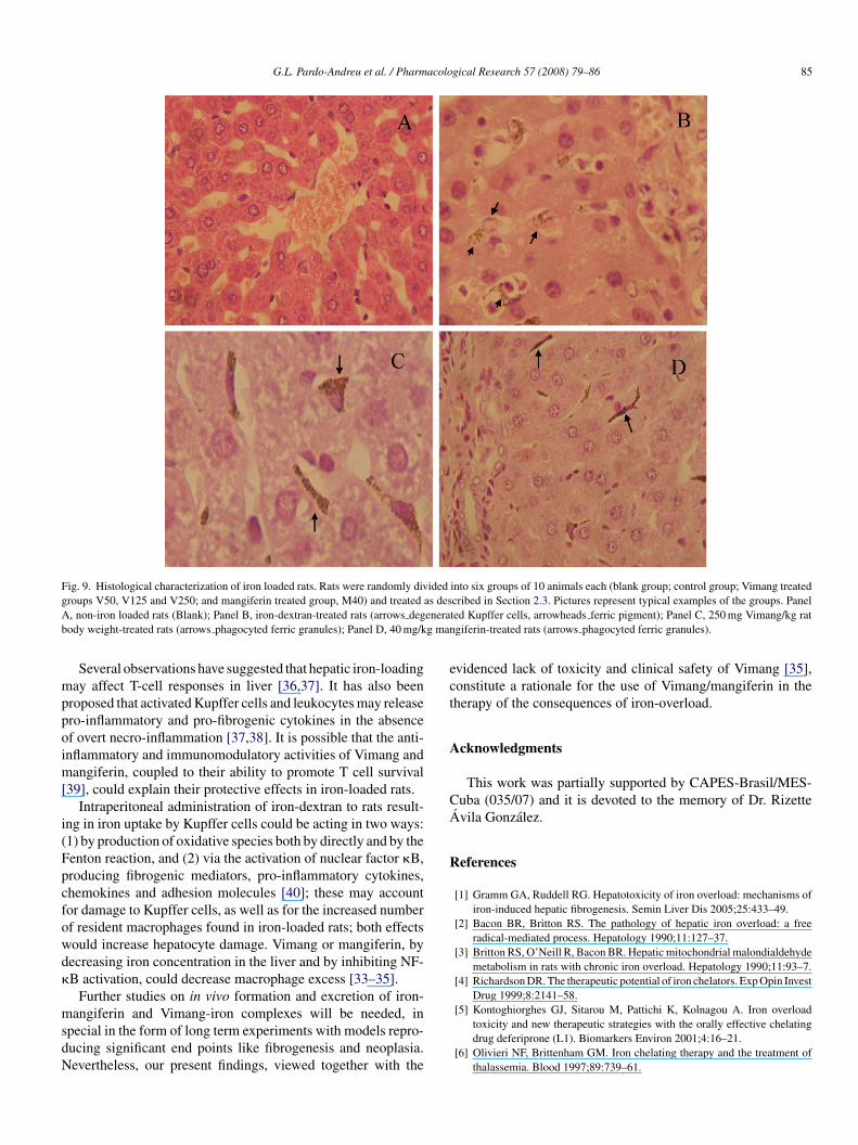
Hepatocytes of iron-dextran treated rats did not present fer-ic granule depots (Fig. 9B), showing a normal morphologicalppearance similar to that of non-iron loaded rats (Fig. 9A).ron (ferric) granules were observed in the cytoplasm of Kupf-er cells, and in free form, in the sinusoidal lumen (Fig. 9B).
oderate enlargement of these cells with hypertrophic nucleiaving a vesicular appearance consistent with their activation,as also observed.No qualitative signs of differences between the aspect of liv-
rs from rats treated with either one of three doses (50, 125nd 250 mg per kg) of Vimang or with mangiferin (40 mg/kg),ere observed. Administration of 250 mg kg−1 Vimang or of0 mg kg−1 mangiferin to iron dextran-treated rats resulted inotal disappearance of signs of damage to the Kupffer cells. Inhese animals furthermore, ferric granules were totally phago-yted by these cells that showed a normal morphologicalppearance (Fig. 9C and D).
. Discussion
Iron is the major catalyst of free-radical cell reactionsnd membrane lipid peroxidation, two major mechanisms ofron overload-induced tissue injury [1–3]. Deleterious effectsf excessive iron deposition in liver, heart, pancreas andther endocrine organs, are believed to occur during geneticemochromatosis, thalassemia major and transfusional sidero-is. In such conditions, reactive molecular iron may initiate,ropagate, and amplify (auto) oxidative phenomena in cells.he sequestering of low-molecular-weight, catalytically active
ron present in the cytosol or released by chelation from otherell iron stores [30] could therefore be an effective therapeu-ic approach by simultaneously protecting membranes from theffects of cell-generated free radicals (i.e., antioxidant activ-ty).
No compounds showing this broad spectrum of antioxidantctivity are presently available for clinical use. We have thereforeested an aqueous extract of Mangifera indica L (Vimang) andts main polyphenol component mangiferin in this context.
In our experimental protocol, we ruled out the possibilityf iron chelation prior to absorption or a direct interferencef Vimang/mangiferin on intestinal iron absorption leadingo serum and hepatic iron overloads. To avoid this, we haveesorted to the i.p. injection of iron-dextran, rather than the com-only used oral administration of iron-carbonyl compounds.ig. 3 shows that after the seventh dose of serial injections of00 mg kg−1 iron, within one week, serum iron concentrationad risen from 36 ± 4 to 67 ± 3 �mol l−1, and total liver ironrom 144 ± 20 to 2240 ± 215 �g g−1 wet weight. Liver/bodyeight ratios, as well as ASAT and ALAT activities were also
ignificantly elevated (Fig. 2). This increased iron concentrationed to an oxidative stress condition in serum and in liver, indi-
ating that our experimental scheme was appropriate for theirtudy.Since all biochemical alterations originating from iron over-oad had been hindered by Vimang or by mangiferin, we were
84 G.L. Pardo-Andreu et al. / Pharmacological Research 57 (2008) 79–86
F vels ia lues aw
lmtc
htiaaetiRqrlo
cp
FKoBc
tni
pteotttbiam
p
ig. 7. Effect of Vimang or mangiferin on lipid peroxidation (A) and GSH (B) lend GSH levels were assayed according to methods described in Section 2.4. Vaith controls.
ed to suggest that the protective effects of Vimang on iron-ediated oxidative damage could at least in part, be attributed
o mangiferin, acting not only by its free radical scavengingapacity, but also by its iron-complexing ability.
One characteristic of iron overloading is to increase non-eme iron serum levels. Vimang or mangiferin decreased hepaticotal iron concomitantly with a dose-dependent tendency toncrease of serum non-heme iron, suggesting that mangiferinnd/or related compounds could gradually combine with hep-tic non-heme iron, and take it into the blood stream for finalxcretion. Although, this hypothesis needs further experimen-al support, it is conceivable that Vimang/mangiferin releaseron from liver ferritin, increasing serum iron concentration.ecent reports have documented the ability of baicalin anduercetin, both catechol polyphenols, to release iron from fer-itin in vitro [20,21], accounting for their capacity to decreaseiver iron by increasing serum iron levels in experimental iron
verloading.Serum iron concentration increased by Vimang was also asso-iated with increased serum iron binding capacity and decreasedercentage of transferrin saturation, supporting our assumption
ig. 8. Effect of Vimang or mangiferin administration on the number of liver-upffer cells loaded with ferric granules, counted at random at a magnificationf 400×, in 10 microscope fields per liver section in differently treated rats.ars represent means ± S.D. (n = 10). #P < 0.05 compared with blank; *P < 0.05ompared with control.
Lbospaio
hFmowath
pactmm
n liver homogenates of iron loaded rats. Thiobarbituric acid-reactive substancesre means ± S.D. (n = 10). #P < 0.05 compared with blanks; *P < 0.05 compared
hat iron chelation by mangiferin or possibly other polyphe-olic Vimang components, act as a protective mechanism inron-loaded rats.
We have recently shown that a Vimang-Fe(III) mixturerepared by using a 2:1 mangiferin content of the extracto define the ligand proportion in the mixture, was moreffective than Vimang alone in scavenging DPPH and super-xide radicals, as well as in protecting hepatocytes fromert-butyl hydroperoxide-induced mitochondrial lipid peroxida-ion and hypoxia/reoxygenation [15]. This result suggests thathe Vimang extract may not only decrease “free” or “looselyound iron ions” by its coordination capacity rendering themnactive or poorly active in the Fenton reaction, but also bycquiring extra antioxidant capacity due to metal complex for-ation [18].Vimang/mangiferin interaction with the plasma iron labile
ool (LPIP) is another conceivable way involved in our results.PIP is the pathologically relevant fraction of non-trasferrinound iron that encompasses cell-penetrating redox active formsf iron [31,32] susceptible to chelation. It is therefore pos-ible to postulate that the complexing capacity of Viman’solyphenol components, in special mangiferin, reduces ironvailable for translocation across cell membranes, preventingts accumulation in liver, heart, pancreas and further endocrinergans.
In vitro results [9] on the iron-chelating capability of Vimangave led us recently to demonstrate that Vimang preventse(II)/citrate-induced lipid peroxidation in isolated rat liveritochondria. This action may be due to its ability to stimulate
xygen consumption by Fe(II) autoxidation forming complexesith Fe(III) that do not participate in Fenton-type reactions
nd/or lipid peroxidation [13]. In this regard, it was also shownhat Vimang protects 2-deoxyribose from oxidative damage byypoxanthine, xanthine oxidase and Fe(III)/EDTA [14].
In the Vimang extract, mangiferin, a glucosylxanthone, isrobably the major polyphenol component responsible for itsntioxidant and antiinflammatory [10,33–35], as well iron-
omplexing properties. Confirming this hypothesis, our labora-ory has accumulated several lines of evidence confirming thatangiferin’s ability to interact with iron is its major antioxidantechanism of action [16–19].
G.L. Pardo-Andreu et al. / Pharmacological Research 57 (2008) 79–86 85
Fig. 9. Histological characterization of iron loaded rats. Rats were randomly divided into six groups of 10 animals each (blank group; control group; Vimang treatedg as desA enerab g man
mppoim[
i(Fpcfowd�
msdN
ect
A
CA
R
roups V50, V125 and V250; and mangiferin treated group, M40) and treated, non-iron loaded rats (Blank); Panel B, iron-dextran-treated rats (arrows degody weight-treated rats (arrows phagocyted ferric granules); Panel D, 40 mg/k
Several observations have suggested that hepatic iron-loadingay affect T-cell responses in liver [36,37]. It has also been
roposed that activated Kupffer cells and leukocytes may releasero-inflammatory and pro-fibrogenic cytokines in the absencef overt necro-inflammation [37,38]. It is possible that the anti-nflammatory and immunomodulatory activities of Vimang and
angiferin, coupled to their ability to promote T cell survival39], could explain their protective effects in iron-loaded rats.
Intraperitoneal administration of iron-dextran to rats result-ng in iron uptake by Kupffer cells could be acting in two ways:1) by production of oxidative species both by directly and by theenton reaction, and (2) via the activation of nuclear factor �B,roducing fibrogenic mediators, pro-inflammatory cytokines,hemokines and adhesion molecules [40]; these may accountor damage to Kupffer cells, as well as for the increased numberf resident macrophages found in iron-loaded rats; both effectsould increase hepatocyte damage. Vimang or mangiferin, byecreasing iron concentration in the liver and by inhibiting NF-B activation, could decrease macrophage excess [33–35].
Further studies on in vivo formation and excretion of iron-
angiferin and Vimang-iron complexes will be needed, inpecial in the form of long term experiments with models repro-ucing significant end points like fibrogenesis and neoplasia.evertheless, our present findings, viewed together with the
cribed in Section 2.3. Pictures represent typical examples of the groups. Panelted Kupffer cells, arrowheads ferric pigment); Panel C, 250 mg Vimang/kg ratgiferin-treated rats (arrows phagocyted ferric granules).
videnced lack of toxicity and clinical safety of Vimang [35],onstitute a rationale for the use of Vimang/mangiferin in theherapy of the consequences of iron-overload.
cknowledgments
This work was partially supported by CAPES-Brasil/MES-uba (035/07) and it is devoted to the memory of Dr. Rizette
´ vila Gonzalez.
eferences
[1] Gramm GA, Ruddell RG. Hepatotoxicity of iron overload: mechanisms ofiron-induced hepatic fibrogenesis. Semin Liver Dis 2005;25:433–49.
[2] Bacon BR, Britton RS. The pathology of hepatic iron overload: a freeradical-mediated process. Hepatology 1990;11:127–37.
[3] Britton RS, O’Neill R, Bacon BR. Hepatic mitochondrial malondialdehydemetabolism in rats with chronic iron overload. Hepatology 1990;11:93–7.
[4] Richardson DR. The therapeutic potential of iron chelators. Exp Opin InvestDrug 1999;8:2141–58.
[5] Kontoghiorghes GJ, Sitarou M, Pattichi K, Kolnagou A. Iron overloadtoxicity and new therapeutic strategies with the orally effective chelatingdrug deferiprone (L1). Biomarkers Environ 2001;4:16–21.
[6] Olivieri NF, Brittenham GM. Iron chelating therapy and the treatment ofthalassemia. Blood 1997;89:739–61.
8 acolo
[
[
[
[
[
[
[
[
[
[
[
[
[
[
[
[
[
[
[
[
[
[
[
[
[
[
[
[
[
[
6 G.L. Pardo-Andreu et al. / Pharm
[7] Kontoghiorghes GJ. Do we need more iron-chelating drugs? Lancet2003;362:495–6.
[8] Haenen GRMM, Jansen FP, Bast A. The antioxidant properties of five O-(b-Hydroxyethyl)-rutosides of the flavonoid mixture Venoruton. Phlebology1993;Suppl.1:10–7.
[9] Martinez G, Giuliani A, Leon OS, Perez G, Nunez-Selles AJ. Effects ofMangifera indica L extract (QF808) on protein and hepatic lipoperoxida-tion. Phytother Res 2001;15:581–5.
10] Sanchez GM, Re L, Giuliani A, Nunez-Selles A, Davison GP, Leon-Hernandez OS. Protective effects of Mangifera indica L. extract,mangiferin and selected antioxidants against TPA-induced biomoleculesoxidation and peritoneal macrophage activation in mice. Pharmacol Res2000;42:565–73.
11] Nunez-Selles AJ, Velez-Castro HT, Aguero-Aguero J, Gonzalez-GonzalezJ, Naddeo F, De Simone F, Rastrelli L. Isolation and quantitative analysisof phenolic antioxidants, free sugars, and polyols from mango (Mangiferaindica L.) stem bark aqueous decoction used in Cuba as nutritional supple-ment. Agr Food Chem 2002;50:762–6.
12] Martinez G, Delgado R, Perez G, Garrido G, Nunez-Selles AJ, Leon OS.Evaluation of the in vitro antioxidant activity of Mangifera indica L. extract(VIMANG). Phytother Res 2000;14:424–7.
13] Pardo Andreu G, Delgado R, Velho J, Inada NM, Curti C, VercesiAE. Mangifera indica L. extract (Vimang) inhibits Fe2+-citrate-inducedlipoperoxidation in isolated rat liver mitochondria. Pharmacol Res2005;51:427–35.
14] Pardo-Andreu G, Delgado R, Nunez-Selles AJ, Vercesi AE. Mangiferaindica L. extract (Vimang) inhibits 2-deoxyribose damage induced by Fe(III) plus ascorbate. Phytother Res 2006;20:120–4.
15] Pardo-Andreu GL, Sanchez-Baldoquin C, Avila-Gonzalez R, YamamotoET, Revilla A, Uyemura SA, Naal Z, Delgado R, Curti C. Interaction ofVimang (Mangifera indica L. extract) with Fe(III) improves its antioxidantand cytoprotecting activity. Pharmacol Res 2006;54:389–95.
16] Andreu GP, Delgado R, Velho JA, Curti C, Vercesi AE. Iron complex-ing activity of mangiferin, a naturally occurring glucosylxanthone, inhibitsmitochondrial lipid peroxidation induced by Fe2+-citrate. Eur J Pharmacol2005;513:47–55.
17] Pardo Andreu GL, Delgado R, Nunez-Selles AJ, Vercesi AE. Dualmechanism of mangiferin protection against iron-induced damage to2-deoxyribose and ascorbate oxidation. Pharmacol Res 2006;53:253–60.
18] Pardo-Andreu GL, Sanchez-Baldoquin C, Avila-Gonzalez R, Delgado R,Naal Z, Curti C. Fe(III) improves antioxidant and cytoprotecting activitiesof mangiferin. Eur J Pharmacol 2006;547:31–6.
19] Pardo-Andreu GL, Cavalheiro RA, Naal Z, Vercesi AE, Curti C.Fe(III) shifts the mitochondria permeability transition-eliciting capac-ity of mangiferin to organelle’s protection. J Pharmacol Exper Ther2006;320:646–53.
20] Zhang Y, Li H, Zhao Y, Gao Z. Dietary supplementation of baicalin andquercetin attenuates iron overload induced mouse liver injury. E J Pharma-col 2006;535:263–9.
21] Zhao Y, Li H, Gao Z, Xu H. Effects of dietary baicalin supplementationon iron overload induced mouse liver oxidative injury. Eur J Pharmacol2005;509:195–200.
22] Lowry OH, Rosenbrough NJ, Farr AL, Randall RJ. Protein measurementwith the Folin phenol reagent. J Biol Chem 1951;193:265–75.
[
gical Research 57 (2008) 79–86
23] Rice-Evans C, Miller NJ. Total antioxidant status in plasma and body fluid.Method Enzymol 1994;234:279–93.
24] Buege JA, Aust SD. Microsomal lipid peroxidation. Method Enzymol1978;52:302–10.
25] Marklund S, Marklund G. Involvement of the superoxide anion radicalin the autoxidation of pyrogallol and a convenient assay for superoxidedismutase. Eur J Biochem 1974;47:469–74.
26] Sedlak J, Lidsay RH. Estimation of total protein bound and non-protein sulfhydryl group in tissue with Ellman’s reagent. Anal Biochem1968;25:192–205.
27] Beale RN, Broston JO, Taylor F. Improved rapid method for the deter-mination of iron content and binding capacity of serum. J Clin Path1962;15:156–9.
28] Ramsay WNM. The determination of total iron binding capacity of serum.Clin Chem Acta 1952;2:221–6.
29] Barry M, Sherlock S. Measurement of liver-iron concentration in needlebiopsy specimens. Lancet 1971;1:100–3.
30] Rothman RJ, Serroni A, Farber JL. Cellular pool of transient ferric iron,chelatable by deferoxamine and distinct from ferritin, that is involved inoxidative cell injury. Mol Pharmacol 1992;42:703–10.
31] Hershko C, Graham G, Bates GW, Rachmilewitz E. Nonspecific serum ironin thalassaemia: an abnormal serum iron fraction of potential toxicity. BritJ Haematol 1978;40:255–63.
32] Breuer W, Ermers MJJ, Pootrakul P, Abramov A, Hershko C, CabantchikZI. Desferrioxamine-chelatable iron (DCI), a component of serum non-transferrin-bound iron (NTBI) used for assessing iron chelation therapy.Transfus Sci 2000;23:241–2.
33] Garrido G, Delgado R, Lemus Y, Rodriguez J, Garcia D, Nunez-SellesAJ. Protection against septic shock and suppression of tumor necrosisfactor alpha and nitric oxide production on macrophages and microgliaby a standard aqueous extract of Mangifera indica L. (VIMANG).Role of mangiferin isolated from the extract. Pharmacol Res 2004;50:165–72.
34] Garrido G, Gonzalez D, Lemus Y, Garcia D, Lodeiro L, Quintero G, Del-porte C, Nunez-Selles AJ, Delgado R. In vivo and in vitro anti-inflammatoryactivity of Mangifera indica L. extract (VIMANG). Pharmacol Res2004;50:143–9.
35] Nunez-Selles AJ, Delgado-Hernandez R, Garrido-Garrido G, Garcia-Rivera D, Guevara-Garcia M, Pardo-Andreu GL. The paradox of naturalproducts as pharmaceuticals Experimental evidences of a mango stem barkextract. Pharmacol Res 2007;55:351–8.
36] Ramm AG, Ruddell RG. Hepatotoxicity of iron overload: Mecha-nisms of iron-induced hepatic fibrogenesis. Semin Liver Dis 2005;25:433–49.
37] Bridle KR, Crawford DHG, Fletcher LM, Smith JL, Powell LW, RammGA. Evidence for a submorphological inflammatory process in the liver inhaemochromatosis. J Hepatol 2003;38:426–33.
38] Arthur MJ. Iron overload and liver fibrosis. J Gastroenterol Hepatol1996;11:1124–9.
39] Hernandez P, Rodriguez PC, Delgado R, Hernandez R, Walczak H. Protec-
tive effect of Mangifera indica L. polyphenols on human T lymphocytesagainst activation-induced cell death. Pharmacol Res 2006;55:167–73.40] Videla LA, Fernandez V, Tapia G, Varela P. Oxidative stress-mediatedhepatotoxicity of iron and copper: role of Kupffer cells. Biometals2003;16:103–11.