protective effect of resveratrol against oxygen–glucose deprivation in organotypic hippocampal...
TRANSCRIPT

www.elsevier.com/locate/ynbdi
Neurobiology of Disease 24 (2006) 170–182Protective effect of resveratrol against oxygen–glucosedeprivation in organotypic hippocampal slice cultures:Involvement of PI3-K pathway
Lauren L. Zamin, Patrícia Dillenburg-Pilla, Ricardo Argenta-Comiran, Ana Paula Horn,Fabrício Simão, Melissa Nassif, Daniéli Gerhardt, Rudimar L. Frozza, and Christianne Salbego⁎
Departamento de Bioquímica, Instituto de Ciências Básicas da Saúde, UFRGS, 90035-003, Porto Alegre, RS, Brazil
Received 27 March 2006; revised 31 May 2006; accepted 5 June 2006Available online 24 July 2006
Here we investigated the neuroprotective effect of resveratrol in an invitro model of ischemia. We used organotypic hippocampal culturesexposed to oxygen–glucose deprivation (OGD). In OGD-vehicleexposed cultures, about 46% of the hippocampus was labeled withPI, indicating a robust percentage of cell death. When cultures weretreated with resveratrol 10, 25 and 50 μM, the cell death was reducedto 22, 20 and 13% respectively. To elucidate a possible mechanism bywhich resveratrol exerts its neuroprotective effect, we investigated thephosphoinositide3-kinase (PI3-k) pathway using LY294002 (5 μM)and mitogen-activated protein kinase (MAPK) using PD98059(20 μM). The resveratrol (50 μM) neuroprotection was preventedby LY294002 but was not by PD98059. Immunoblotting revealed thatresveratrol 50 μM induced the phosphorylation/activation of Akt andextracellular signal-regulated kinase-1 and -2 (ERK1/2) and thephosphorylation/inactivation of glycogen synthase kinase-3β (GSK-3β).Our results suggest that PI3-k/Akt pathway are involved in theneuroprotective effect of resveratrol.© 2006 Elsevier Inc. All rights reserved.
Keywords: Neuroprotection; Resveratrol; Oxygen–glucose deprivation;Hippocampus; Organotypic culture; Akt; GSK-3β; ERK1/2
Abbreviations: CA1, Cornus Ammonis1; DMSO, dimethylsulphoxide;ERK, extracellular signal-regulated kinase; ERK1/2, extracellular signal-regulated kinases-1 and -2; GSK-3β, glycogen synthase kinase-3β; HBSS,Hank's balanced salt solution; IGF-1, insulin-like growth factor-1; MAPK,mitogen-activated protein kinase; MCAO, middle cerebral artery occlusion;MEK, mitogen-activated ERK-activating kinase; OGD, oxygen–glucosedeprivation; pAkt, phospho-Akt; pERK1/2, phospho-ERK1/2; pGSK-3β,phospho-GSK-3β; PI, propidium iodide; PI3-k, phosphoinositide3-kinase;SDS, sodium dodecylsulfate.
E-mail address: [email protected] (C. Salbego).Available online on ScienceDirect (www.sciencedirect.com).
0969-9961/$ - see front matter © 2006 Elsevier Inc. All rights reserved.doi:10.1016/j.nbd.2006.06.002
Introduction
Ischemic stroke results from a transient or permanent reductionin cerebral blood flow that leads to a complex cascade of cellevents resulting in neuronal death (Dirnagl et al., 1999; Lipton,1999). Diverse in vitro and in vivo models have been used to studythe mechanisms underlying neuronal degeneration and to evaluatepotential neuroprotective effects of pharmacological treatments.Among the in vitro systems used to study ischemia-inducedinjuries, organotypic hippocampal slice cultures, combined withoxygen–glucose deprivation (OGD), offer great advantagesbecause they mimic closely the in vivo condition (Pringle et al.,1997; Tavares et al., 2001; Cimarosti et al., 2001, 2005). Culturedslices maintain their cell architecture and interneuron connections,and neurons survive during the long-term culture and physiologi-cally mature over this period, allowing an extended survival study(Muller et al., 1993; Xiang et al., 2000; Cho et al., 2004).
The notion that red wine may have potential health benefitsinitially received a great deal of attention following reports thatmoderate wine consumption was linked to a lower incidence ofcardiovascular disease—the so-called “French Paradox” (Renaudand Lorgeril, 1992). This cardioprotective effect has beenattributed to the polyphenol fraction of red wine (Das et al.,1999). A key polyphenol in red wine is resveratrol, trans-3, 5, 4trihydroxystilbene, from grape seeds and skins. Resveratrol hasbeen found to act as a strong antioxidant (Belguendouz et al., 1997;Tadolini et al., 2000) and can reduce the oxidation of lipoproteins(Frankel et al., 1993). Resveratrol can also protect the vessels fromatherosclerosis (Frankel et al., 1993; Belguendouz et al., 1997) andinhibit platelet aggregation and cyclo-oxygenase (Bertelli et al.,1995; Subbaramaiah et al., 1998).
In addition to the purported cardioprotective effects of red wine,recent evidences suggest that wine consumption may also protectagainst certain neurological disorders. Studies have demonstratedthe ability of resveratrol to exert protective effects against braininjury due to ischemia–reperfusion in gerbil model (Wang et al.,2002), and epidemiological studies have shown that moderate red

171L.L. Zamin et al. / Neurobiology of Disease 24 (2006) 170–182
wine consumption is significantly correlated with a reduction in theincidence of age-related macular degeneration (Obisesan et al.,1998), Alzheimer's disease (Luchsinger et al., 2004) and stroke(Mukamal et al., 2005).
Although molecular mechanisms underlying the pathogenesisof delayed neuronal death is unclear, histologic and biochemicalevidence suggest the involvement of apoptosis in dying cells afterischemia (MacManus et al., 1993; Nitatori et al., 1995).Phosphoinositide3-kinase (PI3-k) pathway is believed to be animportant anti-apoptotic signal pathway in neurons (Yuan andYankner, 2000; Love, 2003). Akt, also known as protein kinase B,is a downstream kinase of PI3-k in growth factor mediatedsignaling cascades, and the phosphorylation of residues Thr308and Ser473 is required for its activity (Coffer et al., 1998). Oneway by which active Akt mediates its anti-apoptotic effects is byphosphorylating and inactivating glycogen synthase kinase-3β(GSK-3β), which plays a key role in apoptosis (Pap and Cooper,1998).
Another signaling system, which has a role in mediating cellsurvival, is the extracellular signal-regulated kinase-1 and 2(ERK1/2), which belongs to the family of mitogen-activatedprotein kinase (MAPK) pathway (Ahn, 1993). The mitogen-activated ERK-activating kinase/ERK (MEK/ERK) pathway isactivated downstream of receptor tyrosine kinases, G-protein-coupled receptors and cytokine receptors (Lewis et al., 1998).Following neuronal injury, the ERK pathway is activated,suggesting a role in cell death and/or repair (Campos-Gonzalezand Kindy, 1992; Hu and Wieloch, 1994; Runden et al., 1998).Recent studies suggest that resveratrol can activate both the PI3-kand the MAPK pathway (Miloso et al., 1999; Das et al., 2005a,b;Klinge et al., 2005).
In order to investigate the neuroprotective effects of resveratrol,we used organotypic cultures of rat hippocampus exposed to an invitro model of cell death using OGD, and we also investigatedwhether such neuroprotection could be related to PI3-k signalingpathway by Akt activation and GSK-3β inactivation and/or MAPKpathway by activation of ERK1/2.
Materials and methods
Organotypic hippocampal slice cultures
Organotypic hippocampal slice cultures were preparedaccording to the method of Stoppini et al. (1991). Briefly,400-μm-thick hippocampal slices were prepared from 6- to 8-day-old male Wistar rats using a McIlwain tissue chopper (allanimal use procedures were approved by local Animal CareCommittee and were in accordance with the NIH Guide for theCare and Use of Laboratory Animals) and separated in ice-coldHank's balanced salt solution (HBSS) composed of (mM):
Fig. 1. Experimental design, indicating time-line of culture, drug exposure, OGD aERK1/2.
glucose 36, CaCl2 1.26, KCl 5.36, NaCl 136.89, KH2PO4 0.44,Na2HPO4 0.34, MgCl2 0.49, MgSO4 0.44, HEPES 25;fungizone 1% (Gibco, Grand Island, NY, USA) and gentamicine36 μl/100 ml (Schering do Brasil, São Paulo, SP, Brazil); pH7.2. The slices were placed on Millicell culture insert(Millicell®-CM, 0.4 μm, Millipore®, Bedford, MA, USA) andthe inserts were transferred to a 6-well culture plate (CellCulture Cluster, Costar®, New York, NY, USA). Each wellcontained 1 ml of tissue culture medium consisting of 50%minimum essential medium (Gibco), 25% HBSS (Gibco), 25%heat inactivated horse serum (Gibco) supplemented with (mM,final concentration): glucose 36, HEPES 25 and NaHCO3 4;fungizone 1% (Gibco) and gentamicine 36 μl/100 ml (Scheringdo Brasil); pH 7.2. Organotypic cultures were maintained in ahumidified incubator gasified with 5% CO2 atmosphere at 37°C.Culture medium was changed three times week.
OGD
OGD was achieved by combining hypoxia with aglycemia,according to the method described by Strasser and Fischer (1995),with some modifications (Cimarosti et al., 2001). Fig. 1 shows thebasic experimental design. After 14 days in vitro, the inserts weretransferred to a sterilized 6-well plate and incubated with 1 ml ofOGD medium consisting of HBSS lacking glucose for 15 min todeplete glucose from intracellular stores and extracellular space.After that, the medium was exchanged for one with the samecomposition but previously bubbled with N2 for 30 min and theplate transferred to an anaerobic chamber at 37°C with N2-enrichedatmosphere for 60 min. During this process control slices weremaintained in an incubator with 5% CO2 atmosphere at 37°C.After the deprivation period, slice cultures were incubated inculture medium under normoxic conditions for 24 h, correspondingto the recovery periods.
Drug exposition
Resveratrol was purchased from Calbiochem (San Diego, CA,USA), LY294002 was from Cell Signaling Technology (Beverly,MA, USA) and PD98059 was obtained from New EnglandBiolabs (Beverly, MA, USA). All drugs were dissolved indimethylsulphoxide (DMSO) (Sigma Chemical, St. Louis, MO,USA). Resveratrol (10, 25 and 50 μM) and DMSO (0.01%) wereadded to the OGD medium and to the culture medium during therecovery period of 24 h. LY294002 (5 μM) and PD98059(20 μM) were administered to the culture medium 2 h beforeOGD period, and maintained during the OGD and the recoveryperiod of 24 h.
Cultures were categorized in four conditions: vehicle-supple-mented cultures not exposed to OGD (control DMSO), drug-
nd subsequent measurements of cell survival and levels of Akt, GSK-3β and

172 L.L. Zamin et al. / Neurobiology of Disease 24 (2006) 170–182
supplemented group not exposed to OGD (control resveratrol,control LY294002, control resveratrol + LY294002, controlPD98059, control resveratrol+PD98059), vehicle-supplementedcultures exposed to OGD (OGD DMSO), and drug-supplementedgroup exposed to OGD (OGD resveratrol, OGD LY294002, OGDresveratrol +LY294002, OGD PD98059, OGD resveratrol +PD98059).
Quantification of cell death
Cell damage was assessed by fluorescent image analysis ofpropidium iodide (PI) uptake (Noraberg et al., 1999). After arecovery period of 22 h, 7.5 μM PI (Sigma Chemical) wasadded to the cultures and incubated for 2 h. PI uptake isindicative of significant membrane injury (Macklis andMadison, 1990). Cultures were observed with an invertedmicroscope (Nikon Eclipse TE 300) using a standard rhodaminefilter set. Images were captured and then analyzed using ScionImage software (http://www.scioncorp.com). The area where PIfluorescence was detectable above background was determined
Fig. 2. Effect of resveratrol on cell damage induced by OGD for 60 min in organostained with PI after 24 h after exposure to OGD. (B) Quantitative analysis of hipp50 μM) was added at the moment of lesion induction and maintained during the recfrom control cultures. #Significantly different from control and DMSO-treatedP<0.05).
using the “density slice” option of Scion Image software andcompared to the total hippocampus area to obtain thepercentage of damage (Valentim et al., 2003).
Western blotting assay
To investigate the phosphorylation status and immunocontentof Akt, GSK-3β and ERK 1/2 the slices were homogenized inlysis buffer (4% sodium dodecylsulfate (SDS), 2.1 mM EDTA,and 50 mM Tris) 1, 6 and 24 h after OGD. To analyze theeffect of the inhibitors, we homogenized the slices only 24 hafter OGD. Aliquots were taken for protein determination(Peterson, 1983) and β-mercaptoethanol (Sigma Chemical) wasadded to a final concentration of 5%. Proteins were separated(40 μg per lane) on 10% SDS-polyacrylamide gel electrophor-esis (Sigma Chemical). After electrophoresis, proteins wereelectrotransferred to nitrocellulose membranes using a semi-dryapparatus (Bio-Rad Trans-Blot SD, Hercules, CA, USA).
Membranes were incubated for 60 min at 4°C in blockingsolution (Tris-buffered saline containing 5% powdered milk and
typic hippocampal cultures. (A) Representative photomicrographs of slicesocampus damage 24 h after exposure to OGD. RSV: resveratrol (10, 25 andovery period. Bars represent the mean±SEM, n=12. *Significantly differentOGD cultures (one-way ANOVA followed by Student–Newman–Keuls,
173L.L. Zamin et al. / Neurobiology of Disease 24 (2006) 170–182
0.1% Tween-20) and further incubated with the appropriateprimary antibody dissolved in blocking solution overnight at4°C. The primary antibodies used were anti-phospho Akt(Ser473) (pAkt, 1:1000; Cell Signaling Technology), anti-Akt(1:1000; Cell Signaling Technology), anti-phospho GSK-3β(Ser9) (pGSK-3β, 1:1000; Cell Signaling Technology), anti-GSK-3β (1:1000; Cell Signaling Technology), anti-phosphoERK1/2 (pERK1/2, 1:1000; Cell Signaling Technology) andanti-ERK1/2 (1:1000; Upstate Biotechnology, New York, NY,USA). The membranes were then incubated with horseradishperoxidase-conjugated anti-rabbit antibody (1:1000; AmershamPharmacia Biotech, Piscataway, NJ, USA). The chemiolumines-cence (ECL, Amersham Pharmacia Biotech) was detected usingX-ray films (Kodak X-Omat, Rochester, NY, USA). The filmswere scanned and the percentage of band intensity was analyzed
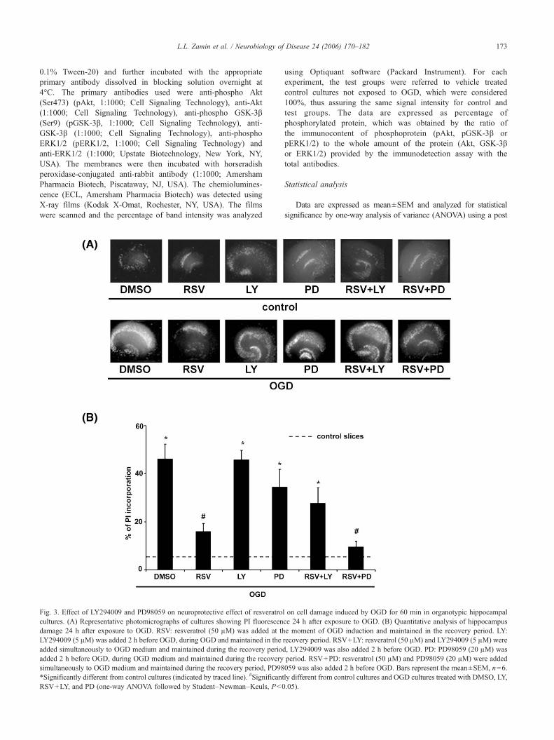
Fig. 3. Effect of LY294009 and PD98059 on neuroprotective effect of resveratrocultures. (A) Representative photomicrographs of cultures showing PI fluorescendamage 24 h after exposure to OGD. RSV: resveratrol (50 μM) was added at thLY294009 (5 μM) was added 2 h before OGD, during OGD and maintained in theadded simultaneously to OGD medium and maintained during the recovery perioadded 2 h before OGD, during OGD medium and maintained during the recoverysimultaneously to OGD medium and maintained during the recovery period, PD98*Significantly different from control cultures (indicated by traced line). #SignificantRSV+LY, and PD (one-way ANOVA followed by Student–Newman–Keuls, P<0
using Optiquant software (Packard Instrument). For eachexperiment, the test groups were referred to vehicle treatedcontrol cultures not exposed to OGD, which were considered100%, thus assuring the same signal intensity for control andtest groups. The data are expressed as percentage ofphosphorylated protein, which was obtained by the ratio ofthe immunocontent of phosphoprotein (pAkt, pGSK-3β orpERK1/2) to the whole amount of the protein (Akt, GSK-3βor ERK1/2) provided by the immunodetection assay with thetotal antibodies.
Statistical analysis
Data are expressed as mean±SEM and analyzed for statisticalsignificance by one-way analysis of variance (ANOVA) using a post
l on cell damage induced by OGD for 60 min in organotypic hippocampalce 24 h after exposure to OGD. (B) Quantitative analysis of hippocampuse moment of OGD induction and maintained in the recovery period. LY:recovery period. RSV+LY: resveratrol (50 μM) and LY294009 (5 μM) wered, LY294009 was also added 2 h before OGD. PD: PD98059 (20 μM) wasperiod. RSV+PD: resveratrol (50 μM) and PD98059 (20 μM) were added059 was also added 2 h before OGD. Bars represent the mean±SEM, n=6.ly different from control cultures and OGD cultures treated with DMSO, LY,.05).

174 L.L. Zamin et al. / Neurobiology of Disease 24 (2006) 170–182
hoc Student–Newman–Keuls test for multiple comparisons. Differ-ences between mean values were considered significant when P<0.05.
Results
Resveratrol protects against cell death induced by OGD
The exposure of the cultures to 60 min of OGD and 24 h ofrecovery caused a marked fluorescence in the hippocampus,indicating a high incorporation of PI, as presented in thephotomicrograph in Fig. 2A (OGD DMSO group). Quantifica-tion of PI fluorescence showed that OGD caused about 46% ofdamage in hippocampus, a significant increase compared tocontrol cultures with a basal hippocampus damage of 4% (Fig.2B). Treatment with 10, 25 and 50 μM of resveratrolsignificantly reduced the injury in the hippocampus from46%, to 22, 20 and 13% respectively (Fig. 2B). There wereno statistical differences among the tested doses, thus we chosethe dose of 50 μM to investigate a probable mechanism bywhich resveratrol exerts its neuroprotective effect. Also no
Fig. 4. Effect of treatment with resveratrol on the percentage of phosphorylated AktpAkt and Akt 1, 6 and 24 h after OGD revealed using specifics antibodies. (Bphosphorylation state. The densitometric values obtained to phospho- and total-Akcontrol non-exposed to OGD condition (DMSO bar) (100%). RSV: resveratrol (5recovery periods of 1, 6 and 24 h. Data are expressed as a ratio of the normal*Significantly different from controls cultures and OGD cultures treated with DM
difference was detectable between the treatments in the controlslices, indicating that resveratrol had no toxic effect in basalconditions (Fig. 2B). It was not observed cell death after arecovery period of 1 and 6 h (data not shown) as we havepreviously demonstrated (Horn et al., 2005).
The neuroprotective effect of resveratrol involves PI3-k but notMEK/ERK1/2 pathway
In order to determine whether the PI3-k signaling pathway wasinvolved in the neuroprotective effect of resveratrol, we carried outexperiments using LY294002, a specific inhibitor of PI3-k (Vlahoset al., 1994). LY294002 (5 μM) abolished the neuroprotectioninduced by resveratrol (50 μM); increasing the PI incorporation from15% (OGD RSV) to 34% (OGD RSV+LY) (Fig. 3B). To clarifyanother probable signaling pathway involved in the neuroprotectionby resveratrol we further studied the effect of PD98059, an inhibitorof MEK/ERK pathway (Alessi et al., 1995; Dudley et al., 1995).PD98059 did not prevent the neuroprotection induced by resveratrolthat showed a PI uptake of 15% in OGD cultures treated with
in organotypic hippocampal cultures. (A) Representative Western blottings of) Histograms represent the quantitative Western blotting analysis of Aktt from all treatments were first normalized to their respective vehicle-treated0 μM) was added at the moment of OGD induction and maintained in theized percentages of pAkt and Akt. Bars represent the mean±SEM, n=6.SO (one-way ANOVA followed by Student–Newman–Keuls test, P<0.05).

175L.L. Zamin et al. / Neurobiology of Disease 24 (2006) 170–182
resveratrol (Fig. 3B OGD RSV) and 10% in OGD cultures treatedwith PD98059 and resveratrol (OGD RSV+PD). No neurotoxiceffects were observed with inhibitors only (Fig. 3A).
Resveratrol increase the Akt and GSK3-β phosphorylation in OGDcultures
Considering that LY294002 abolished the neuroprotectiveeffect of resveratrol, we hypothesized that resveratrol wouldinduce Akt activation, a PI3-k-activated protein kinase. Thestatus of Akt phosphorylation was examined after 1, 6 and 24 hof recovery by Western blotting with antibodies against theactive form of Akt, phosphorylated at Ser473, as well as itstotal immunocontent. Resveratrol 50 μM significantly increasedthe percentage of pAkt by 50% in OGD cultures in all recoveryperiods (Fig. 4B). There were no differences in the pAkt
Fig. 5. Effect of treatment with resveratrol and LY294002 on the percentage of phRepresentative Western blottings of pAkt and Akt revealed using specifics antibodiephosphorylation state. The densitometric values obtained to phospho- and total-Akcontrol non-exposed to OGD condition (DMSO bar) (100%). RSV: resveratrol (5recovery period. LY: LY294009 (5 μM) was added to 2 h before OGD, during OGDLY294009 (5 μM) were added simultaneously to OGDmedium and maintained durexpressed as a ratio of the normalized percentages of pAkt and Akt. Bars represenOGD cultures treated with DMSO, LYand RSV+LY. *Significantly different from cANOVA followed by Student–Newman–Keuls test, P<0.05).
intensity neither among controls slices or in OGD DMSO-treated group. No alteration in the total amount of Akt wasobserved (representative Western blotting in Fig. 4A). Toconfirm that LY294002 was effective in the blockage of thePI3-k pathway we measured the phosphorylation state of Akt inthe presence of LY294002 after 24 h of recovery. Treatmentwith LY294002 reduced the ratio of pAkt/Akt (Fig. 5B) incontrol and OGD cultures treated with DMSO, confirming theefficacy of this inhibitor.
When Akt is activated (i.e., phosphorylated), it phosphorylatesand inactivates GSK-3β. Therefore, GSK-3β phosphorylation wasalso evaluated after 1, 6 and 24 h after OGD and resveratroltreatment. We found that resveratrol increased the GSK-3βphosphorylation by 65% in OGD cultures in all recovery periodstested (Fig. 6B). No differences were detected in the percentage ofpGSK-3β neither among controls slices or OGD DMSO-treated
osphorylated Akt in organotypic hippocampal cultures 24 h after OGD. (A)s. (B) Histograms represent the quantitative Western blotting analysis of Aktt from all treatments were first normalized to their respective vehicle-treated0 μM) was added at the moment of OGD induction and maintained in theand maintained in the recovery period. RSV+LY: resveratrol (50 μM) and
ing the recovery period, LY294009 was also added 2 h before OGD. Data aret the mean±SEM, n=6. #Significantly different from controls cultures andontrols and OGD cultures treated with DMSO, RSVand RSV+LY (one-way

Fig. 6. Effect of treatment with resveratrol on the percentage of phosphorylated GSK3-β in organotypic hippocampal cultures. (A) Representative Westernblottings of pGSK3-β and GSK3-β 1, 6 and 24 h after OGD revealed using specifics antibodies. (B) Histograms represent the quantitative Western blottinganalysis of GSK3-β phosphorylation state. The densitometric values obtained to phospho- and total-GSK3-β from all treatments were first normalized to theirrespective vehicle-treated control non-exposed to OGD condition (DMSO bar) (100%). RSV: resveratrol (50 μM) was added at the moment of OGD inductionand maintained in the recovery periods of 1, 6 and 24 h. Data are expressed as a ratio of the normalized percentages of pGSK3-β and GSK3-β. Bars represent themean±SEM, n=6. *Significantly different from controls cultures and OGD cultures treated with DMSO (one-way ANOVA followed by Student–Newman–Keuls test, P<0.05).
176 L.L. Zamin et al. / Neurobiology of Disease 24 (2006) 170–182
group. It was not detectable alteration in the total amount of GSK-3β (bottom line in Fig. 6A). Fig. 7B shows that LY294002 wasable to abolish the increase of ratio of pGSK-3β/GSK-3β mediatedby resveratrol.
Resveratrol increase ERK1/2 phosphorylation in OGD cultures
Since cell survival and differentiation in several cell types aremediated by ERK1/2 we examined whether the neuroprotectionmediated by resveratrol could involve this pathway. Furthermore,the same serine residue in GSK-3β that is targeted by Akt is nowknown to be phosphorylated by MAPK cascade (Cohen andFrame, 2001). We measured the levels of the immunocontent ofERK1/2, as well as its phosphorylation status after 1, 6 and 24 hafter OGD. Resveratrol (50 μM) was also able to increase ERK1/2phosphorylation in OGD cultures by 80% (Fig. 8B) after 6 and24 h of recovery. There were no statistical differences in the
phosphorylation status of ERK1/2 in the control cultures and OGDDMSO-treated culture, although we could observe a moderateincrease on pERK1/2 in OGD DMSO culture. No alteration in thetotal amount of ERK1/2 was observed (Fig. 8A). Fig. 3B showsthat PD98059 was ineffective in blocking the neuroprotectioninduced by resveratrol. To determine the efficacy of this inhibitorwe analyzed the phosphorylation state of ERK1/2 in the presenceof PD98059. Western blotting analyses of ERK1/2 confirm thatPD98059 was effective in reducing the ERK1/2 phosphorylation(Fig. 9B).
Discussion
In the present study, we tested whether resveratrol, an activecompound of red wine, has a neuroprotective effect in ischemicinjury and proposed an underlying mechanism by which thisneuroprotection occurs. For this purpose, we have used organo-

Fig. 7. Effect of treatment with resveratrol and LY294002 on the percentage of phosphorylated GSK-3β in organotypic hippocampal cultures 24 h after OGD.(A) Representative Western blottings of pGSK-3β and GSK-3β revealed using specifics antibodies. (B) Histograms represent the quantitative Western blottinganalysis of GSK-3β phosphorylation state. The densitometric values obtained to phospho- and total-GSK-3β from all treatments were first normalized to theirrespective vehicle-treated control non-exposed to OGD condition (DMSO bar) (100%). RSV: resveratrol (50 μM) was added at the moment of OGD inductionand maintained in the recovery period. LY: LY294009 (5 μM) was added to 2 h before OGD, during OGD and maintained in the recovery period. RSV+LY:resveratrol (50 μM) and LY294009 (5 μM) were added simultaneously to OGD medium and maintained during the recovery period, LY294009 was also added2 h before OGD. Data are expressed as a ratio of the normalized percentages of pGSK-3β and GSK-3β. Bars represent the mean±SEM, n=6. *Significantlydifferent from controls cultures and OGD cultures treated with DMSO, LY and RSV+LY (one-way ANOVA followed by Student–Newman–Keuls test,P<0.05).
177L.L. Zamin et al. / Neurobiology of Disease 24 (2006) 170–182
typic hippocampal cultures to examine its effects in an in vitromodel of ‘ischemic-like insult’ using OGD, extensively employedin our research group (Tavares et al., 2001; Valentim et al., 2003;Cimarosti et al., 2005; Horn et al., 2005). Organotypic culturesprovide good experimental access to mimic pathophysiologicalpathways in living tissues and to facilitate design of therapeuticagents (Stoppini et al., 1991). In particular, hippocampal slicecultures combined with OGD could provide a surrogate system toinvestigate cell loss following ischemic injury to the brain. Usingthis model, we have shown here that resveratrol at doses of 10, 25and 50 μM protects organotypic hippocampal slice cultures againstOGD.
These results go along with findings obtained in other in vivoand in vitro models of neurodegenerative diseases. Huang et al.(2001) suggest that resveratrol is a potent neuroprotective agent infocal cerebral ischemia caused by middle cerebral artery occlusion
(MCAO) in Long-Evans rats. It has been reported that resveratrolattenuated β-amyloid-induced cytotoxicity, apoptotic features, andintracellular reactive oxygen intermediates accumulation in PC12cells (Jang and Surh, 2003).
The mechanisms of the neuroprotective effects of resveratrolare not fully understood, although some mechanisms have beenproposed. In this study, we proposed that resveratrol could mediateits neuroprotective effect by activating of two major signalingpathways implicated in supporting neuronal survive: PI3-k andMAPK (Pettmann and Henderson, 1998). First, we investigatedwhether resveratrol could activate the PI3-k pathway. For thispurpose, we used LY294002. The results presented here show thatthe neuroprotective effect of resveratrol was prevented by thisinhibitor. Second, we investigated whether MEK/ERK1/2 could beinvolved in the neuroprotective effect of resveratrol. As weshowed, PD98055 did not prevent the neuroprotective effect of

Fig. 8. Effect of treatment with resveratrol on the percentage of phosphorylated ERK1/2 in organotypic hippocampal cultures. (A) Representative Westernblottings of pERK1/2 and ERK1/2 1, 6 and 24 h after OGD revealed using specifics antibodies. (B) Histograms represent the quantitative Western blottinganalysis of ERK1/2 phosphorylation state. The densitometric values obtained to phospho- and total-ERK1/2 from all treatments were first normalized to theirrespective vehicle-treated control non-exposed to OGD condition (DMSO bar) (100%). RSV: resveratrol (50 μM) was added at the moment of OGD inductionand maintained in the recovery periods of 1, 6 and 24 h. Data are expressed as a ratio of the normalized percentages of pGSK3-β and GSK3-β. Bars represent themean±SEM, n=6. *Significantly different from controls cultures, OGD cultures treated with DMSO and OGD RSV after 1 h of recovery (one-way ANOVAfollowed by Student–Newman–Keuls test, P<0.05).
178 L.L. Zamin et al. / Neurobiology of Disease 24 (2006) 170–182
resveratrol. These results strongly suggest that the PI3-k isinvolved in the neuroprotection by resveratrol observed in ourmodel of cell death.
It has been shown that the protective effects of PI3-k aremediated primarily by one of its downstream targets-Akt (Franke etal., 1997). Akt has direct effects on the apoptosis pathway, byinhibiting the pro-apoptotic proteins as, among others, Bad,caspase 9, forkhead factors and GSK-3β (Song et al., 2005).Several reports have suggested that apoptosis is underlying thedelayed neuronal death in ischemic brain injuries (MacManus etal., 1993; Nitatori et al., 1995). Studies have shown that blockingincreased Akt phosphorylation by treatment with LY294002,increases subsequent DNA fragmentation (Noshita et al., 2001).Likewise, reverting the decrease in Akt phosphorylation seenimmediately after brain ischemia by treatment with intraventricularadministration of insulin-like growth factor-1 (IGF-1), rescuedcells from neuronal cell death (Kawano et al., 2001). Takentogether, decreased Akt activity accounts for neuronal damagefollowing brain ischemia, and increased Akt activity observed afew hours later could participate in endogenous neuroprotectiveresponses to ischemia.
Therefore, we hypothesized that resveratrol could protectneurons against cell death induced by OGD by activating PI3-k/Akt proteins. Our results indicated that resveratrol increased Aktphosphorylation after OGD, and this increase was abolished byLY294002. No change in the amount of pAkt or Akt wasobserved in control slices treated with resveratrol. Furthermore,there was no increase in pAkt or Akt in OGD DMSO group.These results suggest that the neuroprotection mediated byresveratrol could involve an increase of Akt phosphorylationafter an insult.
Recently, Das et al. (2005a) showed that resveratrol mediatescardioprotective effect by increasing Akt phosphorylation and thiseffect was partially abolished by LY294002. Another study hasindicated a role of both adenosine A1 and A3 receptors inresveratrol preconditioning, and both use the PI3-k/Akt signalingpathway (Das et al., 2005b). Altogether, these studies pointed toPI3-k as the upstream signaling molecule for resveratrol protection.
Horn et al. (2005) did not observe any significant changes inAkt phosphorylation and immunocontent in CA1 or Dentate Gyrusareas of organotypic cultures exposed to OGD after 30 min, 6 and24 h of reperfusion. However, another group reported a

Fig. 9. Effect of treatment with resveratrol and PD98059 on the percentage of phosphorylated ERK1/2 in organotypic hippocampal cultures 24 h after OGD. (A)Representative Western blottings of pERK1/2 and ERK1/2 revealed using specifics antibodies. (B) Histograms represent the quantitative Western blottinganalysis of ERK1/2 phosphorylation state. The densitometric values obtained to phospho- and total-ERK1/2 from all treatments were first normalized to theirrespective vehicle-treated control non-exposed to OGD condition (DMSO bar) (100%). RSV: resveratrol (50 μM) was added at the moment of OGD inductionand maintained in the recovery period. PD: PD98059 (20 μM) was added to 2 h before OGD, during OGD and maintained in the recovery period. RSV+PD:resveratrol (50 μM) and PD98059 (20 μM) were added simultaneously to OGDmedium and maintained during the recovery period, PD98059 was also added 2 hbefore OGD. Data are expressed as a ratio of the normalized percentages of pERK1/2 and ERK1/2. Bars represent the mean±SEM, n=6. *Significantly differentfrom controls cultures and OGD cultures treated with DMSO, PD and RSV+PD. #Significantly different from control cultures treated with DMSO and RSVandOGD cultures treated with DMSO and RSV (one-way ANOVA followed by Student–Newman–Keuls test, P<0.05).
179L.L. Zamin et al. / Neurobiology of Disease 24 (2006) 170–182
dramatically decrease in Akt phosphorylation at Ser473 immedi-ately following global brain ischemia in the hippocampus ofWistarrats. After this transient decrease in Akt phosphorylation,phosphorylation greatly increased within the next 24 h anddeclined to basal levels within 48 h (Ouyang et al., 1999).Transient upregulation of Akt phosphorylation was also observedin the global ischemic model using 4-vessel occlusion in the rat(Jin et al., 2001). One possible explanation for these apparentcontroversies results is that the latter used an in vivo model ofglobal brain ischemia and the former used an in vitro model oforganotypic hippocampal slice cultures exposed to OGD.
Following the PI3-k/Akt pathway, we investigated whetherresveratrol had effect on GSK-3β phosphoregulation andwhether this effect could involve PI3-k using LY294002. Weare the first to demonstrate that resveratrol strongly increasephosphorylation of GSK-3β after OGD, and this increase wasabolished by LY294002, suggesting that this could be a
probable mechanism by which resveratrol protects against celldeath. GSK-3β is an enzyme initially identified as a regulator ofglycogen metabolism that also has broader functions, such asregulating signaling proteins (including amyloid precursorprotein, cyclin D1, and nerve growth factor receptor), structuralproteins (including microtubule-associated proteins-1B and-2,neural cell-adhesion protein, neurofilaments, and tau), andtranscription factors (including nuclear factor kappa B, h-catenin, activating protein-1, and heat shock factor-1) asreviewed by Cohen and Frame (2001).
There is growing evidence that GSK-3β is involved in thepathogenesis of central nervous system diseases. There are only ahandful of research reports on the expression of GSK-3β incerebral ischemia. GSK-3β is present in neurons following MCAO(Sasaki et al., 2001). Selective, but not specific GSK-3β inhibitionwith lithium, has been shown to be protective against a plethora ofneurological insults including cerebral ischemia (see reviews Manji

180 L.L. Zamin et al. / Neurobiology of Disease 24 (2006) 170–182
et al., 1999; Nonaka and Chuang, 1998), suggesting that areduction in GSK-3β activity improves brain cell survival.Furthermore, topical application of IGF-1, also a non-specificinhibitor of GSK-3β, significantly reduced infarct size following60 min MCAO and reduced the number of GSK-3β-immunor-eactive neurons (Wang et al., 2000).
Taken together, the increase in phosphorylation of Akt andGSK-3β, induced by resveratrol after OGD, and the effect of theinhibition of PI3-k by LY294002, leading to a decrease in theneuroprotection mediated by resveratrol, suggest that the PI3-k/Aktpathway and GSK-3β could be involved in the mechanism bywhich resveratrol protects organotypic hippocampal slice culturesagainst cell death induced by OGD.
Inoue et al. (2003) demonstrated that the neuroprotective effectof resveratrol on brain protection following experimental strokereside in its anti-inflammatory effect, i.e., on its effect on bloodborne cells or endothelium. In our in vitro system of OGD theabsence of blood flow as a variable and the absence of bloodvessels eliminate an important component of damage process. Ourresults added more one mechanism by which resveratrol couldexerts its neuroprotective effect.
Similarly Akt signaling, MAPK signaling pathway is requiredfor the anti-apoptotic effect and neuronal survival in the brainfollowing ischemic insult. Data from literature indicates thatresveratrol can modulate MAPK proteins, such as JNK and p38(Kutuk et al., 2006; Woo et al., 2004; Nicolini et al., 2003).Resveratrol was effective in blockage 4-hydroxynonenal-inducedapoptosis in fibroblasts cultures by blocking JNK signaling (Kutuket al., 2006) and paclitaxel-induced apoptosis in human neuro-blastoma cells by blocking JNK and p38 pathway (Nicolini et al.,2003).
Transient decrease in MAPK activity was also evident in thegerbil hippocampus following forebrain ischemia, and suchdecrease was blocked by intraventricular administration of IGF-1(Kawano et al., 2001). Recently data have showing that pERK1/2is upregulated in hippocampal organotypic cultures after OGD(Irving and Bamford, 2002; Namura et al., 2001). Our resultsshowed that resveratrol increased the ERK1/2 phosphorylationafter OGD, but the inhibition of this increase by PD98059 did notprevent the neuroprotective effect of resveratrol.
Strikingly, resveratrol only increased the phosphorylation ofAkt, GSK-3β and ERK1/2 in the OGD-treated slices, indicatingthat resveratrol could not be a direct activator of these pathways,but acts as a facilitator of their activation.
While ERK signaling plays a beneficial, neuroprotective role inmany systems (Dash et al., 2002; Cavanaugh, 2004; Hetman andGozdz, 2004), there is growing evidence implicating these kinasesin promoting cell death. For example, increased ERK1/2phosphorylation has been noted in the vulnerable penumbrafollowing acute ischemic stroke in humans (Slevin et al., 2000).Alessandrini et al. (1999) in model of cerebral ischemia–reperfusion injury provided the first in vivo evidence thatactivation of the MEK/ERK1/2 signaling pathway may contributeto acute brain injuries. In these studies, ERK1/2 activation wasblocked using inhibitors of MEK1/2 and led to reduced neuronalinjury and loss of function in mice. More compelling resultsindicating neuroprotective effects from inhibiting ERK1/2 activa-tion were subsequently obtained in hippocampal slice cultures andhippocampal dissociated neuronal cultures where protein phos-phatase inhibition was used to induce cell death (Murray et al.,1998; Runden et al., 1998).
What accounts for the seemingly contradictory effects ofMEK1/2 inhibition on neuronal cell survival following acuteinjury? Differences in outcome resulting from MEK1/2 inhibitionmay depend not only upon the nature and severity of injury, butalso upon drug dosing regimens or the cell type expressingactivated ERK1/2. Until cell type-specific inhibition of ERK1/2activation can be attained, the mechanism responsible for theneuroprotective in vivo effects of MEK1/2 inhibition will remainunresolved (Chu et al., 2004).
Although further work is needed to understand the precisemechanism of neuroprotection provided by resveratrol, the presentstudy adds one evidence that the PI3-k cascade could play a role inresveratrol-induced neuroprotective effect against ‘ischemic-likeinjury’. These findings might be important for understandingbeneficial effects of red wine intake to cerebral ischemia and themechanism of neuroprotection afforded by this beverage.
Acknowledgments
This work was supported by the Brazilian funding agenciesCNPq, PROPESQ/UFRGS, PRONEX, CAPES and FAPERGS.We thank Dr. Guido Lenz for the help with preparation of themanuscript.
References
Ahn, N.G., 1993. The MAP kinase cascade. Discovery of a new signaltransduction pathway. Mol. Cell. Biochem. 127/128, 201–209.
Alessandrini, A., Namura, S., Moskowitz, M.A., Bonventre, J.V., 1999.MEK1 protein kinase inhibition protects against damage resulting fromfocal cerebral ischemia. Proc. Natl. Acad. Sci. U. S. A. 96,12866–12869.
Alessi, D.R., Cuenda, A., Cohen, P., Dudley, D.T., Saltiel, A.R., 1995. PD098059 is a specific inhibitor of the activation of mitogen-activatedprotein kinase kinase in vitro and in vivo. J. Biol. Chem. 270,27489–27494.
Belguendouz, L., Fremont, L., Linard, A., 1997. Resveratrol inhibits metalion-dependent and independent peroxidation of porcine low-densitylipoproteins. Biochem. Pharmacol. 53, 1347–1355.
Bertelli, A.A., Giovannini, L., Giannessi, D., Migliori, M., Bernini, W.,Fregoni, M., Bertelli, A., 1995. Antiplatelet activity of synthetic andnatural resveratrol in red wine. Int. J. Tissue React. 17, 1–3.
Campos-Gonzalez, R., Kindy, M.S., 1992. Tyrosine phosphorylation ofmicrotubule-associated protein kinase after transient ischemia in thegerbil brain. J. Neurochem. 59 (5), 1955–1958.
Cavanaugh, J.E., 2004. Role of extracellular signal regulated kinase 5 inneuronal survival. Eur. J. Biochem. 271, 2056–2059.
Cho, S., Liu, D., Fairman, D., Li, P., Jenkins, L., McGonigle, P., Wood, A.,2004. Spatiotemporal evidence of apoptosis-mediated ischemic injuryin organotypic hippocampal slice cultures. Neurochem. Int. 45,117–127.
Chu, C.T., Levinthal, D.J., Kulich, S.M., Chalovich, E.M., DeFranco, D.B.,2004. Oxidative neuronal injury-the dark side of ERK1/2. Eur. J.Biochem. 271, 2060–2066.
Cimarosti, H., Rodnight, R., Tavares, A., Paiva, R., Valentim, L.,Rocha, E., Salbego, C., 2001. An investigation of the neuroprotec-tive effect of lithium in organotypic slice cultures of rathippocampus exposed to oxygen and glucose deprivation. Neurosci.Lett. 315, 33–36.
Cimarosti, H., Zamin, L.L., Frozza, R., Nassif, M., Horn, A.P., Tavares, A.,Netto, C.A., Salbego, C., 2005. Estradiol protects against oxygen andglucose deprivation in rat hippocampal organotypic cultures andactivates Akt and inactivates GSK-3β. Neurochem. Res. 30, 191–199.

181L.L. Zamin et al. / Neurobiology of Disease 24 (2006) 170–182
Coffer, P.J., Jin, J., Woodgett, J.R., 1998. Protein KinaseB (c-Akt): amultifuncional mediator of phosphatidylinositol 3-kinase activation.Biochem. J. 335, 1–13.
Cohen, P., Frame, S., 2001. The renaissance of GSK3. Nat. Rev., Mol. CellBiol. 2, 769–776.
Das, D.K., Sato, M., Ray, P.S., Maulik, G., Engelman, R.M., Bertelli, A.A.,Bertelli, A., 1999. Cardioprotection of red wine: role of polyphenolicantioxidants. Drugs Exp. Clin. Res. 25, 115–120.
Das, S., Tosaki, A., Bagchi, D., Maulik, N., Das, D.K., 2005a. Resveratrol-mediated activation of cAMP response element-binding proteinthrough adenosine A3 receptor by akt-dependent and -independentpathways. J. Pharmacol. Exp. Ther. 314 (2), 762–769.
Das, S., Cordis, G.A., Maulik, N., Das, D.K., 2005b. Pharmacologicalpreconditioning with resveratrol: role of CREB-dependent Bcl-2signaling via adenosine A3 receptor activation. Am. J. Physiol. 288(1), H328–H335.
Dash, P.K., Mach, S.A., Moore, A.N., 2002. The role of extracellular signal-regulated kinase in cognitive and motor deficits following experimentaltraumatic brain injury. Neuroscience 114, 755–767.
Dirnagl, U., Iadecola, C., Moskowitz, M.A., 1999. Pathobiology ofischaemic stroke: an integrated view. Trends Neurosci. 22, 391–397.
Dudley, D.T., Pang, L., Decker, S.J., Bridges, A.J., Saltiel, A.R., 1995. Asynthetic inhibitor of the mitogen-activated protein kinase cascade. Proc.Natl. Acad. Sci. U. S. A. 92, 7686–7689.
Franke, T.F., Kaplan, D.R., Cantley, L.C., 1997. PI3K: downstream AKTionblocks apoptosis. Cell 88, 435–437.
Frankel, E.N., Waterhouse, A.L., Kinsella, J.E., 1993. Inhibition of humanLDL oxidation by resveratrol. Lancet 341, 1103–1104.
Hetman, M., Gozdz, A., 2004. Role of extracellular signal regulated kinases1 and 2 in neuronal survival. Eur. J. Biochem. 271, 2050–2055.
Horn, A.P., Gerhardt, D., Geyer, A.B., Valetim, L., Cimarosti, H., Tavares,A., Horn, F., Lenz, G., Salbego, C., 2005. Cellular death in hippocampusin response to PI3K pathway inhibiton and oxygen and glucosedeprivation. Neurochem. Res. 30 (3), 355–361.
Hu, B.R., Wieloch, T., 1994. Tyrosine phosphorylation and activation ofmitogen-activated protein kinase in the rat brain following transientcerebral ischemia. J. Neurochem. 62, 1357–1367.
Huang, S.S., Tsai, M.C., Chih, C.L., Hung, L.M., Tsai, S.K., 2001.Resveratrol reduction of infarct size in Long-Evans rats subjected tofocal cerebral ischemia. Life Sci. 69, 1057–1065.
Inoue, H., Jiang, X.F., Katayama, T., Osada, S., Umesono, K., Namura, S.,2003. Brain protecion by resveratrol and fenofibrate against strokerequires peroxisome proliferator-activated receptor in mice. Neurosci.Lett. 352, 203–206.
Irving, E.A., Bamford, M., 2002. Role of mitogen- and stress-activatedkinases in ischemic injury. J. Cereb. Blood Flow Metab. 22 (6),631–647.
Jang, J.H., Surh, Y.J., 2003. Protective effect of resveratrol on β-amyloid-induced oxidative PC12 cell death. Free Radical Biol. Med. 34 (8),1100–1110.
Jin, K., Mao, X.O., Batteur, S.P., McEachron, E., Leahy, A.,Greenberg, D.A., 2001. Caspase-3 and the regulation of hypoxicneuronal death by vascular endothelial growth factor. Neuroscience108, 351–358.
Kawano, T., Fukunaga, K., Takeuchi, Y., Morioka, M., Yano, S., Hamada, J.,Ushio, Y., Miyamoto, E., 2001. Neuroprotective effect of sodiumorthovanadate on delayed neuronal death after transient forebrainischemia in gerbil hippocampus. J. Cereb. Blood Flow Metab. 21,1268–1280.
Klinge, C.M., Blankenship, K.A., Risinger, K.E., Bhatnagar, S., Noisin,E.L., Sumanasekera, W.K., Zhao, L., Brey, D.M., Keynton, R.S.,2005. Resveratrol and estradiol rapidly activate MAPK signalingthrough estrogen receptors α and β in endothelial cells. J. Biol.Chem. 280 (9), 7460–7468.
Kutuk, O., Poli, G., Basaga, H., 2006. Resveratrol protects against 4-hydroxynonenal-induced apoptosis by blocking JNK and c-JUN/AP-1signalling. Toxicol. Sci. 90 (1), 120–132.
Lewis, T.S., Shapiro, P.S., Ahn, N.G., 1998. Signal transduction throughMAP kinase cascades. Adv. Cancer Res. 74, 49–139.
Lipton, P., 1999. Ischemic cell death in brain neurons. Physiol. Rev. 79,1431–1568.
Love, S., 2003. Apoptosis and brain ischemia. Prog. Neuro-Psychopharma-col. 27, 267–282.
Luchsinger, J.A., Tang, M.X., Siddiqui, M., Shea, S., Mayeux, R., 2004.Alcohol intake and risk of dementia. J. Am. Geriatr. Soc. 52, 540–546.
Macklis, J.D., Madison, R.D., 1990. Progressive incorporation ofpropidium iodide in cultured mouse neurons correlates with decliningelectrophysiological status: a fluorescence scale of membrane integrity.J. Neurosci. Methods 31, 43–46.
MacManus, J.P., Buchan, A.M., Hill, I.E., Rasquinha, I., Preston, E., 1993.Global ischemia can cause DNA fragmentation indicative of apoptosis inrat brain. Neurosci. Lett. 164, 89–92.
Manji, H.K., Moore, G.J., Chen, G., 1999. Lithium at 50: have theneuroprotective effects of this unique cation been overlooked? Biol.Psychiatry 46, 929–940.
Miloso, M., Bertelli, A.A., Nicolini, G., Tredici, G., 1999. Resveratrol-induced activation of the mitogen-activated protein kinases, ERK1 andERK2, in human neuroblastoma SH-SY5Y cells. Neurosci. Lett. 264,141–144.
Mukamal, K.J., Ascherio, A., Mittleman, M.A., Conigrave, K.M., Camargo,C.A., Kawachi, I., Stampfer, M.J., Willett, W.C., Rimm, E.R., 2005.Alcohol and risk for ischemic stroke in men: the role of drinking patternsand usual beverage. Ann. Intern. Med. 142, 11–19.
Muller, D., Buchs, P.A., Stoppini, L., 1993. Time course of synapticdevelopment in hippocampal organotypic cultures. Brain Res. Dev.Brain Res. 71, 93–100.
Murray, B., Alessandrini, A., Cole, A.J., Yee, A.G., Furshpan, E.J., 1998.Inhibition of the p44/42 MAP kinase pathway protects hippocampalneurons in a cell-culture model of seizure activity. Proc. Natl. Acad. Sci.U. S. A. 95, 11975–11980.
Namura, S., Iihara, K., Takami, S., Nagata, I., Kikuchi, H., Matsushita, K.,Moskowitz, M.A., Bonventre, J.V., Alessandrini, A., 2001. Intravenousadministration of MEK inhibitor U0126 affords brain protectionagainst forebrain ischemia and focal cerebral ischemia. Proc. Natl.Acad. Sci. U. S. A. 98, 11569–11574.
Nicolini, G., Rigolio, R., Scuteri, A., Miloso, M., Saccomanno, D.,Cavaletti, G., Tredici, G., 2003. Effect of trans-resveratrol on signaltransduction pathways involved in paclitaxel-induced apoptosis inhuman neuroblastoma SH-SY5Y cells. Neurochem. Int. 42 (5),419–429.
Nitatori, T., Sato, N., Waguri, S., Karasawa, Y., Araki, H., Shibanai, K.,Kominami, E., Uchiyama, Y., 1995. Delayed neuronal death in the CA1pyramidal cell layer of the gerbil hippocampus following transientischemia is apoptosis. J. Neurosci. 15, 1001–1011.
Nonaka, S., Chuang, D.M., 1998. Neuroprotective effects of chronic lithiumon focal cerebral ischemia in rats. NeuroReport 9, 2081–2084.
Noraberg, J., Kristensen, B.W., Zimmer, J., 1999. Markers for neuronaldegeneration in organotypic slice cultures. Brain Res. Protoc. 3,278–290.
Noshita, N., Lewen, A., Sugawara, T., Chan, P.H., 2001. Evidence ofphosphorylaiton of Akt and neuronal survival after transient focalcerebral ischemia in mice. J. Cereb. Blood Flow Metab. 21, 1442–1450.
Obisesan, T.O., Hirsch, R., Kosoko, O., Carlson, L., Parrott, M., 1998.Moderate wine consumption is associated with decreased oddsdeveloping age-related macular degeneration in NHANES-1. J. Am.Geriatr. Soc. 46, 1–7.
Ouyang, Y.B., Tan, Y., Comb, M., Liu, C.L., Martone, M.E., Siesjo, B.K.,Hu, B.R., 1999. Survival- and death-promoting events after transientcerebral ischemia: phosphorylation of Akt, release of cytochrome c, andactivation of caspase-like proteases. J. Cereb. Blood Flow Metab. 19,1126–1135.
Pap, M., Cooper, G.M., 1998. Role of glycogen synthase kinase-3 in thephosphatidylinositol 3-kinase/Akt cell survival pathway. J. Biol. Chem.273, 19929–19932.
182 L.L. Zamin et al. / Neurobiology of Disease 24 (2006) 170–182
Peterson, G.L., 1983. Determination of total protein. Methods Enzymol. 91,95–119.
Pettmann, B., Henderson, C.E., 1998. Neuronal cell death. Neuron 20,633–647.
Pringle, A.K., Angunawela, R., Wilde, G.J.C., Mepham, J.A., Sund-strom, L.E., Iannotti, F., 1997. Induction of 72 KDa heat-shockprotein following sub-lethal oxygen deprivation in organotypichippocampal slice cultures. Neuropathol. Appl. Neurobiol. 23,289–298.
Renaud, S., Lorgeril, M., 1992. Wine, alcohol, platelets, and the Frenchparadox for coronary heart disease. Lancet 339, 1523–1526.
Runden, E., Seglen, P.O., Haug, F.M., Ottersen, O.P., Wieloch, T.,Shamloo, M., Laake, J.H., 1998. Regional selective neuronaldegeneration after protein phosphatase inhibition in hippocampalslice cultures: evidence for a MAP kinase-dependent mechanism.J. Neurosci. 18, 7296–7305.
Sasaki, C., Hayashi, T., Zhang, W.R., Warita, H., Manabe, Y., Sakai, K.,Abe, K., 2001. Different expression of glycogen synthase kinase-3betabetween young and old rat brains after transient middle cerebral arteryocclusion. Neurol. Res. 23, 588–592.
Slevin, M., Krupinski, J., Slowik, A., Rubio, F., Szczudlik, A., Gaffney, J.,2000. Activation of MAP kinase (ERK-1/ERK-2), tyrosine kinase andVEGF in the human brain following acute ischaemic stroke. NeuroRe-port 11, 2759–2764.
Song, G., Ouyang, G., Bao, S., 2005. The activation of Akt/PKB signalingpathway and cell survival. J. Cell. Mol. Med. 9 (1), 59–71.
Stoppini, L., Buchs, P.A., Muller, D., 1991. A simple method for organotypiccultures of nervous tissue. J. Neurosci. Methods 37, 173–182.
Strasser, U., Fischer, G., 1995. Quantitative measurement of neuronaldegeneration in organotypic hippocampal cultures after combinedoxygen/glucose deprivation. J. Neurosci. Methods 57, 177–186.
Subbaramaiah, K., Chung, W.J., Michaluart, P., Telang, N., Tanabe, T.,Inoue, H., Jang, M., Pezzuto, J.M., Dannenberg, A.J., 1998. Resveratrolinhibits cyclooxygenase-2 transcription and activity in phorbol ester-
treated human mammary epithelial cells. J. Biol. Chem. 273,21875–21882.
Tadolini, B., Juliano, C., Piu, L., Franconi, F., Cabrini, L., 2000. Resveratrolinhibition of lipid peroxidation. Free Radical Res. 33, 105–114.
Tavares, A., Cimarosti, H., Valentim, L., Salbego, C., 2001. Profile ofphosphoprotein labelling in organotypic slice culture of rat hippocam-pus. NeuroReport 12, 2705–2709.
Valentim, L.M., Rodnight, R., Geyer, A.B., Horn, A.P., Tavares, A.,Cimarosti, H., Netto, C.A., Salbego, C.G., 2003. Changes in heat shockprotein 27 phosphorylation and immunocontent in response topreconditioning to oxygen and glucose deprivation in organotypichippocampal cultures. Neuroscience 118, 379–386.
Vlahos, C.J., Matter, W.F., Hui, K.Y., Brown, R.F., 1994. A specificinhibitor of phosphatidylinositol 3-kinase, 2-(4-morpholinyl)-8-phe-nyl-4H-1-benzopyran-4-one (LY294002). J. Biol. Chem. 269,5241–5248.
Wang, J.M., Hayashi, T., Zhang, W.R., Sakai, K., Shiro, Y., Abe, K., 2000.Reduction of ischemic brain injury by topical application of insulin-likegrowth factor-I after transient middle cerebral artery occlusion in rats.Brain Res. 859, 381–385.
Wang, Q., Xu, J., Rottinghaus, G.E., Simonyi, A., Lubahn, D., Sun, G.Y.,Sun, A.Y., 2002. Resveratrol protects against global cerebral ischemicinjury in gerbils. Brain Res. 958, 439–447.
Woo, J.H., Lim, J.H., Kim, Y.H., Suh, S.I., Min, D.S., Chang, J.S., Lee,Y.H., Park, J.W., Kwon, T.K., 2004. Resveratrol inhibits phorbolmyristate acetate-induced matrix metalloproteinase-9 expression byinhibiting JNK and PKC delta signal transduction. Oncogene 23(10), 1845–1853.
Xiang, Z., Hrabetova, S., Moskowitz, S.I., Casaccia-Bonnefil, P., Young,S.R., Nimmrich, V.C., Tiedge, H., Einheber, S., Karnup, S., Bianchi,R., Bergold, P.J., 2000. Long-term maintenance of mature hippo-campal slices in vitro. J. Neurosci. Methods 98, 145–154.
Yuan, J., Yankner, A., 2000. Apoptosis in the nervous system. Nature 407,802–809.